A Practical Guide to Selecting Stable Reference Genes Using TPM Values from RNA-Seq Data
Accurate gene expression analysis in biomedical research relies heavily on the use of stable reference genes for normalization.
A Practical Guide to Selecting Stable Reference Genes Using TPM Values from RNA-Seq Data
Abstract
Accurate gene expression analysis in biomedical research relies heavily on the use of stable reference genes for normalization. This article provides a comprehensive guide for researchers and drug development professionals on leveraging Transcripts Per Million (TPM) values from RNA-sequencing data to systematically identify and validate optimal reference genes. We cover the foundational advantages of TPM over other normalization methods, detail a step-by-step bioinformatics workflow for candidate gene selection, address common troubleshooting and optimization challenges, and present rigorous validation strategies integrating RT-qPCR. By moving beyond traditional housekeeping genes, this methodology ensures more reliable and reproducible gene expression data across diverse experimental conditions, from basic research to clinical applications.
Why TPM? The Foundation of Accurate Transcriptome Comparison for Reference Gene Discovery
The Critical Role of Stable Reference Genes in Gene Expression Studies
Accurate measurement of gene expression is fundamental to advancing biological research, particularly in fields ranging from plant biology to cancer therapeutics and drug development. Quantitative real-time PCR (qRT-PCR) has emerged as the gold standard for gene expression analysis due to its high sensitivity, specificity, and reproducibility [1]. However, the reliability of qRT-PCR data critically depends on proper normalization to account for technical variations introduced during RNA quality, cDNA synthesis, and amplification efficiency [1]. Reference genes, often called housekeeping genes, serve as internal controls to normalize these variations, enabling accurate quantification of target gene expression [2] [1].
A pervasive challenge in the field is the assumption that traditional reference genes maintain constant expression across all experimental conditions. Growing evidence demonstrates that this assumption is flawed, as the expression of many commonly used reference genes varies significantly across different tissues, developmental stages, and environmental conditions [1] [3] [4]. The incorrect selection of unstable reference genes can generate misleading data, potentially invalidating experimental conclusions and hampering research progress [4]. This application note outlines a robust, transcriptome-guided framework for identifying and validating stable reference genes, with a specific focus on utilizing TPM (Transcripts Per Million) values from RNA sequencing data to enhance the reliability of gene expression studies.
The Critical Importance of Validated Reference Genes
Consequences of Improper Reference Gene Selection
Using inappropriate reference genes can significantly distort gene expression profiles, leading to erroneous biological interpretations. A striking example comes from cancer research, where inhibiting the mTOR kinase pathway to generate dormant cancer cells dramatically altered the expression of commonly used reference genes. Studies revealed that genes encoding cytoskeletal proteins (ACTB) and ribosomal proteins (RPS23, RPS18, RPL13A) underwent substantial expression changes, rendering them categorically unsuitable for normalization in these experimental conditions [4]. Similarly, in spinach research, traditionally employed genes such as GRP and PPR demonstrated significantly lower stability compared to more suitable candidates like EF1α and H3 across developmental stages [2].
The normalization errors introduced by unstable reference genes are not merely statistical artifacts but can reverse the apparent direction of gene expression changes or magnify insignificant effects. This is particularly critical in preclinical drug development, where accurate gene expression data can influence decisions on candidate therapeutic progression. Furthermore, in diagnostic applications, such imprecision could lead to false positives or negatives with direct clinical implications [1] [4].
Key Criteria for Ideal Reference Genes
An optimal reference gene should exhibit consistent expression across all experimental conditions, unaffected by tissue type, developmental stage, environmental stimuli, or pharmacological treatments [2] [1]. Beyond stability, ideal reference genes should demonstrate:
- Moderate to high expression abundance to ensure reliable detection above technical noise [2] [3]
- Low variance across biological replicates and experimental conditions [2]
- Robustness to experimental manipulations including drug treatments, stress conditions, and genetic modifications [4]
No single universal reference gene fulfills all these criteria across diverse experimental contexts, necessitating systematic validation for each specific research application [1].
Transcriptome-Based Identification of Candidate Reference Genes
Leveraging RNA-Seq Data for Candidate Selection
RNA sequencing (RNA-Seq) provides a powerful, unbiased approach for identifying novel reference gene candidates with stable expression patterns. The high-throughput nature of transcriptome sequencing enables researchers to survey genome-wide expression stability across multiple conditions, moving beyond the limited panel of traditionally employed housekeeping genes [2] [3] [5].
The following workflow illustrates the key steps for transcriptome-based identification of candidate reference genes:
Quantitative Filtering Criteria
The application of stringent quantitative filters to RNA-Seq data enables systematic identification of candidate reference genes with desirable expression characteristics:
Table 1: Quantitative Criteria for Candidate Reference Gene Selection from RNA-Seq Data
| Criterion | Threshold | Rationale | Application Example |
|---|---|---|---|
| Expression Level | Mean logâ‚‚(TPM) > 5 | Ensures moderate to high expression for reliable qRT-PCR detection | In Urechis unicinctus, candidate genes had median logâ‚‚(TPM) of 14.16-16.32 [5] |
| Expression Variance | SD logâ‚‚(TPM) < 1 | Selects genes with low variability across samples | Identified 1,196 stable candidates from 25,496 genes in spinach transcriptome [2] |
| Expression Stability | Coefficient of Variation (CV) < 0.2 | Further refines selection to most stable genes | Applied in multiple species including echiuran worm and Japanese flounder [3] [5] |
These criteria have been successfully applied across diverse organisms. In spinach, this approach identified 1,196 stable candidate genes from an initial pool of 25,496 [2]. Similarly, studies in Japanese flounder and the echiuran worm Urechis unicinctus demonstrated the effectiveness of transcriptome-based selection, where candidate reference genes significantly outperformed traditional housekeeping genes in expression stability [3] [5].
Experimental Validation of Candidate Reference Genes
Comprehensive Validation Workflow
Candidate reference genes identified through transcriptome analysis must be experimentally validated using qRT-PCR followed by rigorous statistical analysis. The following protocol outlines a comprehensive validation workflow:
Protocol 1: Reference Gene Validation Protocol
Step 1: Primer Design and Validation
- Design primers with the following characteristics:
- Amplicon length: 75-150 bp
- Primer length: 18-22 nucleotides
- Tm: 60±1°C
- GC content: 40-60%
- Validate primer specificity through:
Step 2: RNA Extraction and cDNA Synthesis
- Extract high-quality RNA (A260/280 ratio: 1.8-2.2, RIN > 7)
- Treat with DNase I to remove genomic DNA contamination
- Synthesize cDNA using reverse transcriptase with oligo(dT) and/or random primers
- Use uniform RNA input across all samples (e.g., 1 μg) [6] [8]
Step 3: qRT-PCR Amplification
- Perform reactions in technical triplicates
- Include no-template controls for contamination monitoring
- Use standardized thermal cycling conditions:
Step 4: Data Analysis
- Calculate amplification efficiencies from standard curves
- Determine Ct values using consistent threshold settings
- Analyze expression stability using multiple algorithms [2] [6]
Statistical Algorithms for Stability Assessment
No single statistical method comprehensively evaluates reference gene stability. A consensus approach utilizing multiple algorithms provides the most reliable assessment:
Table 2: Statistical Algorithms for Reference Gene Validation
| Algorithm | Primary Metric | Interpretation | Key Advantage |
|---|---|---|---|
| geNorm | M-value (average expression stability) | Lower M-value indicates greater stability; M < 1.5 generally acceptable | Determines optimal number of reference genes (Vn/Vn+1 < 0.15) [2] [6] |
| NormFinder | Stability value (intra- and inter-group variation) | Lower stability value indicates greater stability | Accounts for sample subgroups within experimental design [2] [6] |
| BestKeeper | Standard deviation (SD) and coefficient of variation (CV) of Ct values | SD < 1 indicates stable expression; lower CV preferred | Based on raw Ct values without transformation [2] [6] |
| RefFinder | Comprehensive ranking (geometric mean) | Integrates results from all above algorithms | Provides consensus ranking from multiple methods [2] [6] |
The following diagram illustrates the multi-algorithm validation process and integration of results:
Condition-Specific Reference Gene Selection
Application Across Biological Contexts
Reference gene stability is highly context-dependent, necessitating condition-specific validation. The table below summarizes optimal reference genes identified through systematic validation across diverse experimental conditions:
Table 3: Condition-Specific Stable Reference Genes
| Experimental Condition | Organism/Cell Type | Most Stable Reference Genes | Least Stable Reference Genes |
|---|---|---|---|
| Plant Development | Spinach (Spinacia oleracea) | EF1α, H3 [2] | GRP, PPR [2] |
| Abiotic Stress | Japanese Flounder (Paralichthys olivaceus) | gatd1, rpl6 [3] | 18S RNA, actb [3] |
| Human Immunology | PBMCs under Hypoxia | RPL13A, S18, SDHA [6] | IPO8, PPIA [6] |
| Cancer Biology | Dormant Cancer Cells (A549) | B2M, YWHAZ [4] | ACTB, RPS23, RPS18, RPL13A [4] |
| Medicinal Plants | Abelmoschus Manihot | eIF, PP2A1 [7] | TUA [7] |
| Fungal Studies | Inonotus obliquus | VPS (carbon sources), RPB2 (nitrogen sources) [9] | Varies by condition [9] |
These findings underscore that traditional reference genes frequently demonstrate poor stability under specific experimental conditions. For instance, in dormant cancer cells generated through mTOR inhibition, ribosomal protein genes (RPS23, RPS18, RPL13A) and ACTB underwent dramatic expression changes, rendering them unsuitable for normalization [4]. Similarly, in Japanese flounder under temperature stress, transcriptome-identified genes (gatd1, rpl6) significantly outperformed conventionally used genes (18S RNA, actb) [3].
Determining the Optimal Number of Reference Genes
The geNorm algorithm determines the optimal number of reference genes through pairwise variation analysis (Vn/Vn+1). A cut-off value of Vn/Vn+1 < 0.15 indicates that n reference genes are sufficient for reliable normalization [6] [7]. In most cases, the use of two reference genes provides sufficient normalization accuracy, though more complex experimental designs may require three or more reference genes [7].
Research Reagent Solutions
Table 4: Essential Reagents and Tools for Reference Gene Validation
| Reagent/Tool | Function | Application Notes |
|---|---|---|
| RNA Extraction Kits | High-quality RNA isolation | Select kits with DNase treatment; assess quality via RIN/RQI [8] |
| Reverse Transcription Kits | cDNA synthesis | Use consistent RNA input; include genomic DNA removal steps [6] [9] |
| qPCR Master Mixes | Amplification and detection | SYBR Green for cost-effectiveness; probe-based for multiplexing [1] [6] |
| Reference Gene Validation Algorithms | Stability assessment | Employ multiple algorithms (geNorm, NormFinder, BestKeeper) [2] [6] |
| Transcriptome Data | Candidate identification | Filter genes by TPM values (mean logâ‚‚(TPM)>5, SD<1, CV<0.2) [2] [3] |
The systematic identification and validation of stable reference genes is a critical prerequisite for accurate gene expression analysis in all biological research domains. The transcriptome-guided framework presented here, utilizing TPM values from RNA-Seq data combined with rigorous experimental validation, provides a robust methodology for selecting appropriate reference genes tailored to specific experimental conditions. This approach significantly enhances the reliability of qRT-PCR data, ensuring valid biological interpretations and supporting advances in basic research, drug development, and diagnostic applications. As research continues to explore increasingly complex biological systems and interventions, the implementation of these rigorous normalization practices will become ever more essential for generating meaningful and reproducible gene expression data.
Transcripts Per Million (TPM) has emerged as a standard normalization unit for RNA-sequencing data, enabling researchers to account for technical variations in sequencing depth and gene length. This primer provides a comprehensive overview of TPM normalization, its calculation, and its specific application in the critical process of selecting stable reference genes for accurate gene expression analysis. We detail experimental protocols for reference gene validation and present quantitative comparisons of normalization methods to guide researchers in making informed analytical decisions.
RNA-sequencing (RNA-seq) has become the predominant method for transcriptome profiling, enabling digital gene expression measurement through mapped read counts [10]. However, raw read counts are not directly comparable between genes or samples due to technical biases including sequencing depth (total number of reads per sample) and gene length (longer transcripts accumulate more reads) [10] [11]. Normalization methods are therefore essential to remove these technical artifacts and reveal true biological signals [11].
Several normalization approaches have been developed, with TPM (Transcripts per Million) representing one of the most biologically interpretable units. Unlike earlier methods such as RPKM/FPKM, TPM implements a more logical normalization sequence—first adjusting for gene length, then for sequencing depth—which makes it particularly suitable for comparing the relative abundance of transcripts across samples [12].
Table 1: Common RNA-seq Normalization Methods
| Method | Full Name | Primary Use | Key Characteristics |
|---|---|---|---|
| TPM | Transcripts per Million | Within- and between-sample comparisons (with caution) | Normalizes for gene length first, then sequencing depth; sum is constant across samples [12] |
| RPKM/FPKM | Reads/Fragments Per Kilobase per Million | Within-sample comparisons | Normalizes for sequencing depth first, then gene length; sum varies between samples [10] [12] |
| TMM | Trimmed Mean of M-values | Between-sample comparisons | Assumes most genes are not differentially expressed; robust to outliers [11] [13] |
| RLE | Relative Log Expression | Between-sample comparisons | Uses median of ratios for normalization; implemented in DESeq2 [13] |
Understanding TPM: Calculation and Interpretation
The Mathematics Behind TPM
The TPM calculation involves a two-step process that fundamentally differs from RPKM/FPKM in its operation sequence. For a gene i, TPM is calculated as follows [10] [11]:
Normalize for gene length: Divide the read counts mapped to the gene by the transcript length in kilobases: [ \text{Length-normalized reads} = \frac{\text{Reads mapped to gene}_i}{\text{Transcript length (kb)}} ]
Normalize for sequencing depth: Divide the length-normalized reads by the sum of all length-normalized reads in the sample (in millions): [ \text{TPM}i = \frac{\text{Reads mapped to gene}i / \text{Transcript length (kb)}}{\sum (\text{Reads mapped to all genes} / \text{Respective transcript lengths})} \times 10^6 ]
This calculation produces a normalized value where the sum of all TPM values in a sample equals one million, facilitating interpretation as the number of transcripts that would have been observed if exactly one million full-length transcripts had been sequenced [10] [11].
Advantages and Limitations
TPM offers several advantages over other normalization units. It fulfills the invariance property, meaning the average TPM is constant across samples (approximately 10ⶠdivided by the number of annotated transcripts) [10]. This makes TPM values more comparable across samples than RPKM/FPKM values, whose sums vary between samples.
However, a critical limitation exists: TPM represents relative abundance rather than absolute transcript counts. The relative abundance of a transcript depends on the composition of the entire RNA population in a sample [10]. When samples have significantly different transcriptome compositions—such as those prepared with different protocols (poly(A)+ selection vs. rRNA depletion)—TPM values may not be directly comparable even after normalization [10].
Figure 1: TPM Calculation Workflow. The process involves sequential normalization for transcript length followed by sequencing depth.
The Critical Role of Reference Genes and TPM in Their Selection
The Importance of Reference Genes in Gene Expression Studies
Reference genes (also called housekeeping genes or normalizing genes) serve as internal controls in gene expression studies to correct for technical variations in RNA extraction, reverse transcription efficiency, and overall cDNA input [1] [14]. The fundamental requirement for a reference gene is stable expression across all experimental conditions being studied [1]. Traditional reference genes like ACTB (actin), GAPDH, and TUBB (tubulin) were once widely assumed to have constant expression, but numerous studies have demonstrated that their expression can vary significantly across different tissues, developmental stages, and experimental conditions [1] [14].
The use of inappropriate reference genes that show systematic variation under experimental conditions can lead to inaccurate normalization and consequently, erroneous conclusions about target gene expression [1]. This is particularly critical in fields like drug development, where accurate biomarker quantification directly impacts decisions about compound efficacy and toxicity.
TPM as a Foundation for Reference Gene Selection
RNA-seq data normalized with TPM provides an ideal starting point for identifying stably expressed genes across experimental conditions. The TPM unit offers advantages for this application because it: (1) normalizes for both sequencing depth and gene length, allowing fair comparison across samples; (2) represents relative molar concentration, making it biologically interpretable; and (3) produces values where the sum is constant across samples, facilitating stability assessments [10] [15].
A key metric for evaluating gene expression stability using TPM values is the coefficient of variation (CV), calculated as the standard deviation divided by the mean TPM across samples [15] [14]. Genes with lower CV values demonstrate more stable expression and are therefore stronger candidates as reference genes. Additional statistical measures including fold change (FC) values further help identify genes with minimal expression fluctuation [14].
Table 2: Metrics for Evaluating Reference Gene Stability Using TPM Data
| Metric | Calculation | Interpretation | Optimal Value |
|---|---|---|---|
| Coefficient of Variation (CV) | Standard Deviation / Mean TPM | Lower values indicate more stable expression | < 0.3-0.5 [14] |
| Fold Change (FC) | Maximum TPM / Minimum TPM | Measures expression range across conditions | Close to 1 [14] |
| Mean TPM | Average TPM across all samples | Filters out lowly expressed genes | > 5 TPM [14] |
Experimental Protocol: Selecting Reference Genes Using TPM Values
Candidate Gene Identification from RNA-seq Data
Purpose: To identify genes with stable expression from RNA-seq data for use as reference genes in qRT-PCR experiments.
Materials and Reagents:
- RNA-seq data (FASTQ files) from all experimental conditions
- Reference genome/transcriptome appropriate for your species
- High-performance computing cluster or workstation
- RNA-seq quantification software (Salmon, Kallisto, or RSEM)
Procedure:
- Process RNA-seq data: Quantify transcript abundance using alignment-free tools like Salmon [10] or Kallisto, or alignment-based tools like RSEM [16]. These tools directly output TPM values.
- Generate TPM matrix: Compile TPM values for all genes across all samples into a single matrix.
- Filter lowly expressed genes: Remove genes with mean TPM < 5 across all samples, as low-expression genes present technical challenges in qRT-PCR validation [14].
- Calculate stability metrics: For each gene, compute:
- Coefficient of variation (CV = standard deviation/mean) of TPM values
- Fold change (FC = maximum TPM/minimum TPM) across samples
- Mean TPM value
- Rank genes by stability: Sort genes by ascending CV values. Genes with CV < 0.5 and FC < 2 typically represent good candidates [14].
- Select top candidates: Choose 10-15 genes with the lowest CV values for experimental validation.
Experimental Validation of Candidate Reference Genes
Purpose: To validate the expression stability of candidate reference genes using qRT-PCR across all experimental conditions.
Materials and Reagents:
- RNA samples from all experimental conditions and replicates
- RNA extraction kit (e.g., TRIzol, column-based kits)
- DNase I treatment kit
- Reverse transcription kit with random hexamers and/or oligo-dT primers
- qPCR machine and SYBR Green master mix
- Primer pairs for candidate reference genes and target genes of interest
Procedure:
- RNA extraction and qualification: Extract total RNA from all samples. Quantify RNA concentration and assess purity (A260/A280 ratio ~2.0) and integrity (RIN > 7) [1].
- cDNA synthesis: Perform reverse transcription with consistent RNA input (e.g., 1 µg) across all samples using a high-efficiency reverse transcription kit.
- qPCR optimization: Design and validate primer pairs for each candidate reference gene. Ensure amplification efficiency between 90-110% with single peak in melting curves [14].
- qPCR run: Perform qPCR reactions for all candidate genes across all samples in technical replicates.
- Stability analysis: Analyze Ct values using multiple algorithms:
- geNorm: Calculates expression stability value (M); lower M indicates greater stability [15] [14]
- NormFinder: Estimates intra- and inter-group variation; provides stability value [15] [14]
- BestKeeper: Uses pairwise correlations of Ct values [14]
- ΔCt method: Compares relative expression of pairs of genes [14]
- Comprehensive ranking: Use RefFinder or similar tools to integrate results from all algorithms and generate a comprehensive stability ranking [17].
- Final selection: Choose the top 2-3 most stable genes for normalization in subsequent experiments.
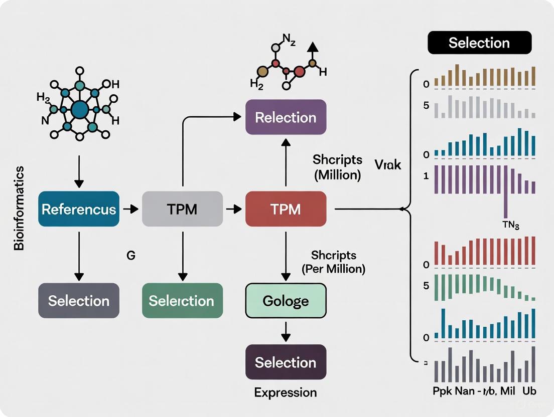
Figure 2: Reference Gene Selection and Validation Workflow. The process integrates computational identification from RNA-seq data with experimental validation using qRT-PCR.
Comparative Analysis of Normalization Methods
Performance in Downstream Applications
Multiple studies have compared the performance of TPM with other normalization methods in various analytical contexts. While TPM is excellent for within-sample comparisons and relative abundance assessment, other methods may outperform it for specific cross-sample applications.
In a comprehensive benchmark study evaluating normalization methods for mapping RNA-seq data to genome-scale metabolic models (GEMs), between-sample normalization methods (TMM, RLE) produced more consistent results than within-sample methods (TPM, FPKM) [13]. Specifically, TMM, RLE, and GeTMM (a hybrid method) enabled generation of condition-specific metabolic models with considerably lower variability in the number of active reactions compared to TPM and FPKM [13].
Similarly, in analyzing RNA-seq data from patient-derived xenograft models, hierarchical clustering based on normalized count data (using between-sample methods) grouped replicate samples more accurately than TPM and FPKM data [16]. Normalized count data also demonstrated lower median coefficient of variation and higher intraclass correlation values across replicate samples [16].
Table 3: Performance Comparison of Normalization Methods in Various Applications
| Application Context | Recommended Method(s) | Key Findings | Reference |
|---|---|---|---|
| Reference gene selection | TPM (for initial screening) | TPM enables calculation of coefficient of variation for stability assessment | [15] [14] |
| Metabolic model reconstruction | TMM, RLE, GeTMM | Between-sample methods produced models with lower variability and better captured disease-associated genes | [13] |
| Sample clustering accuracy | Normalized counts (DESeq2, edgeR) | Outperformed TPM/FPKM in grouping replicate samples from same model | [16] |
| Differential expression analysis | Normalized counts (DESeq2, edgeR) | More robust to different library sizes and compositions | [16] |
Table 4: Key Research Reagent Solutions for TPM-Based Reference Gene Studies
| Reagent/Resource | Function | Example Products/Platforms |
|---|---|---|
| RNA Extraction Kits | Isolation of high-quality total RNA with preservation of mRNA integrity | TRIzol, column-based kits (e.g., Qiagen RNeasy) |
| RNA Integrity Assessment | Quality control of RNA samples prior to library preparation | Bioanalyzer, TapeStation (RIN/RQI scores) |
| Library Preparation Kits | Construction of sequencing libraries with minimal bias | Illumina Stranded mRNA Prep, NEBNext Ultra II |
| RNA-seq Quantification Tools | Transcript abundance estimation with TPM output | Salmon, Kallisto, RSEM [10] [16] |
| qPCR Reagents | Experimental validation of candidate reference genes | SYBR Green master mixes, TaqMan assays |
| Stability Analysis Algorithms | Computational assessment of gene expression stability | geNorm, NormFinder, BestKeeper, RefFinder [14] [17] |
TPM normalization provides a robust foundation for reference gene selection in transcriptomic studies, particularly through its ability to enable cross-sample comparison of relative transcript abundance while accounting for technical variables. The integration of computational identification of stable genes from TPM-normalized RNA-seq data with experimental validation using qRT-PCR represents a powerful strategy for establishing reliable normalization standards in gene expression research. As transcriptomic applications continue to evolve in complexity—spanning diverse tissues, experimental conditions, and disease states—the rigorous selection of reference genes using TPM-based approaches will remain essential for generating accurate, reproducible biological insights, particularly in critical applications like drug development and clinical biomarker identification.
In RNA sequencing (RNA-seq) analysis, the raw read counts mapped to a gene are dependent not only on the gene's actual expression level but also on the gene's length and the library's sequencing depth [10]. Normalization is, therefore, an essential step to eliminate these technical biases and enable accurate biological interpretation [18]. Reads Per Kilobase Million (RPKM) and its paired-end counterpart, Fragments Per Kilobase Million (FPKM), were among the earliest measures developed for this purpose [19]. More recently, Transcripts Per Million (TPM) has emerged as a superior alternative, particularly for studies comparing expression profiles across different samples [19] [20]. This application note delineates the critical differences between these normalization methods, provides empirical evidence for the superiority of TPM in cross-sample comparison, and details protocols for its application in robust transcriptomic analysis, with a specific focus on the selection of reference genes.
Understanding the Normalization Methods: RPKM, FPKM, and TPM
Core Concepts and Calculations
While RPKM, FPKM, and TPM all aim to correct for sequencing depth and gene length, they differ fundamentally in their order of operations, which has profound implications for their interpretation.
RPKM (Reads Per Kilobase per Million mapped reads): Developed for single-end RNA-seq, this method calculates normalized expression as follows [19] [20]:
- Normalize for sequencing depth: Divide the read counts by the total number of reads in the sample (in millions), yielding Reads Per Million (RPM).
- Normalize for gene length: Divide the RPM values by the gene length in kilobases.
FPKM (Fragments Per Kilobase per Million mapped fragments): This is the direct equivalent of RPKM for paired-end RNA-seq data. It accounts for the fact that two reads can originate from a single fragment, thus preventing double-counting [19]. The calculation steps are identical to RPKM, with "reads" replaced by "fragments."
TPM (Transcripts Per Million): This method involves the same two normalization factors but reverses the order of operations [19] [20]:
- Normalize for gene length: Divide the read counts by the gene length in kilobases, yielding Reads Per Kilobase (RPK).
- Normalize for sequencing depth: Sum all RPK values in a sample, divide by 1,000,000 to get a scaling factor, and then divide each RPK value by this factor.
The distinction in calculation is subtle but critical. TPM normalizes for gene length first, followed by sequencing depth, which results in a normalized value that represents the relative molar concentration of a transcript within the total pool of sequenced transcripts [10].
Comparative Analysis: A Direct Comparison
The table below summarizes the key characteristics of each method to facilitate a direct comparison.
Table 1: Comparative Overview of RPKM/FPKM and TPM Normalization Methods
| Feature | RPKM/FPKM | TPM |
|---|---|---|
| Full Name | Reads/Fragments Per Kilobase per Million | Transcripts Per Million |
| Primary Use Case | Gene expression comparison within a single sample [18] | Gene expression comparison both within and between samples [18] |
| Order of Normalization | 1. Sequencing depth2. Gene length | 1. Gene length2. Sequencing depth |
| Sum of All Values | Variable across samples [19] | Constant (1,000,000) across samples [19] |
| Biological Interpretation | Measures reads/fragments per kb per million | Measures the proportion of transcripts in a million transcripts [10] |
| Recommended for Cross-Sample Comparison | No | Yes |
The Critical Advantage of TPM for Cross-Sample Comparison
The Invariance Principle and Proportionality
The most significant advantage of TPM is that the sum of all TPM values in every sample is identical—one million [19]. This invariance property means that TPM values directly represent the relative proportion of each transcript within the total transcribed mRNA pool of a sample. Consequently, if Gene A has a TPM of 5,000 in both Sample 1 and Sample 2, one can confidently conclude that the same fraction of total cellular mRNA in both samples is derived from Gene A [20].
In contrast, the sum of all RPKM or FPKM values can differ from sample to sample. Therefore, an RPKM value of 5,000 for Gene A in two different samples does not guarantee that the gene's relative abundance is the same, as the denominator used to calculate the proportion is different [19] [20]. This makes RPKM/FPKM unreliable for direct comparison of expression levels across samples.
Empirical Evidence from Benchmarking Studies
Evidence from benchmarking studies reinforces the theoretical superiority of TPM. Research evaluating normalization methods for mapping RNA-seq data onto genome-scale metabolic models (GEMs) found that within-sample normalization methods like TPM and FPKM can lead to high variability in the resulting models compared to between-sample methods [13]. This variability can obscure true biological signals and increase false positive predictions.
Furthermore, the choice of normalization method is critical when sample preparation protocols differ. For instance, poly(A)+ selection and rRNA depletion result in vastly different RNA population compositions. A study demonstrated that in such scenarios, TPM values for the same original sample are not directly comparable between the two protocols because the underlying transcript repertoires are different [10]. This highlights that even TPM values are a measure of relative abundance within the sequenced population, and researchers must be cautious when comparing data generated using different experimental workflows.
Practical Application: Selecting Reference Genes Using TPM
A pivotal application of robust cross-sample comparison is the identification of stable reference genes for downstream validation techniques like RT-qPCR. Traditional, pre-defined "housekeeping" genes often show variable expression under different experimental conditions, leading to normalization errors [1] [15] [21]. Using TPM values from an RNA-seq dataset allows for the data-driven selection of optimal, custom reference genes specific to the experimental system.
Protocol: A Workflow for Selecting Reference Genes from RNA-seq Data
This protocol describes a method to identify the most stably expressed genes from RNA-seq data for use as reference genes in RT-qPCR validation [15] [21].
1. Input Data Preparation:
- Generate a table containing TPM values for all genes across all samples in your experiment. Replicates should be averaged prior to analysis [21].
2. Software and Tools:
- The "Gene Selector for Validation (GSV)" software is an example of a tool designed for this purpose, though custom R or Python scripts can also be implemented based on the following criteria [21].
3. Step-by-Step Filtering Criteria: Apply the following sequential filters to the TPM data to identify optimal reference gene candidates [21]:
- Filter 1: Presence in All Samples. Retain only genes with a TPM value > 0 in every library analyzed.
- Filter 2: Moderate to High Expression. Retain genes with an average log2(TPM) value greater than 5. This ensures the selected genes are expressed at a level easily detectable by RT-qPCR.
- Filter 3: Stable Expression. Retain genes with a low coefficient of variation (CV) of TPM values across samples (e.g., CV < 0.2) and a standard deviation of log2(TPM) smaller than 1.
- Filter 4: Absence of Exceptional Outliers. Exclude genes where the expression in any single library is more than twice the average log2(TPM) across all libraries.
4. Output:
- The final output is a list of genes that pass all filters, ranked by their stability (lowest CV). The top-ranked genes are the most suitable candidates for use as reference genes in subsequent RT-qPCR experiments [15].
The following workflow diagram summarizes this protocol:
Validation of Selected Reference Genes
Genes selected via the above TPM-based pipeline must be empirically validated using algorithms designed for RT-qPCR data. The expression stability of the candidate genes is typically assessed using software such as geNorm [7], NormFinder [15] [7], and BestKeeper [7], which calculate stability measures based on the quantification cycle (Cq) values from the RT-qPCR experiment [21] [7]. A comprehensive validation study on Abelmoschus Manihot, for example, used these algorithms to confirm that eIF and PP2A1 were superior to traditionally used genes like TUA (tubulin alpha) [7].
The Scientist's Toolkit: Essential Reagents and Software
Successful implementation of these protocols requires a combination of specific reagents and computational tools.
Table 2: Essential Research Reagents and Software Solutions
| Item Name | Function/Application | Specific Example/Note |
|---|---|---|
| RNA-seq Library Prep Kit | Preparation of sequencing libraries from purified RNA. | Kits are often optimized for poly(A)+ selection or rRNA depletion, a choice that affects results [10]. |
| RT-qPCR Master Mix | Amplification and fluorescence detection during qPCR. | Contains DNA polymerase, dNTPs, buffers, and fluorescent dye (e.g., SYBR Green I) [1]. |
| Reference Gene Primers | Gene-specific amplification for RT-qPCR validation. | Primers must be designed for and validated on candidate genes (e.g., eIF, PP2A1) [7]. |
| GSV (Gene Selector for Validation) Software | Identifies stable reference and variable candidate genes from TPM data. | An open-source tool with a graphical interface that applies the filtering protocol [21]. |
| geNorm / NormFinder / BestKeeper | Algorithms to assess expression stability of candidates from RT-qPCR Cq data. | Used for final validation; often employed in concert for a robust conclusion [7]. |
| Oridonin | Oridonin, CAS:28957-04-2, MF:C20H28O6, MW:364.4 g/mol | Chemical Reagent |
| PI-3065 | PI-3065, MF:C27H31FN6OS, MW:506.6 g/mol | Chemical Reagent |
The choice of normalization method is not merely a technical formality but a fundamental decision that shapes all downstream biological interpretations of RNA-seq data. While RPKM and FPKM remain valid for assessing relative expression between genes within a single sample, TPM is unequivocally superior for comparing the expression of a gene across multiple samples. Its invariant sum property allows researchers to make direct, proportional comparisons, making it the unit of choice for cross-sample analysis. This advantage is particularly critical in applications like the data-driven selection of reference genes, where accurate identification of stably expressed transcripts is paramount for reliable RT-qPCR validation. Adopting TPM and associated robust protocols ensures that transcriptomic insights are both accurate and reproducible.
The GENCODE project represents a cornerstone of modern genomics, with the primary goal of identifying and classifying all gene features in the human and mouse genomes with high accuracy based on biological evidence [22]. This consortium produces a reference gene annotation that is widely recognized for its comprehensive manual curation and experimental validation, serving as the official annotation for major projects including ENCODE, the 1000 Genomes Project, and the International Cancer Genome Consortium [23]. Unlike purely computational annotations, GENCODE combines manual annotation from the HAVANA group with automated annotation from Ensembl, creating a merged gene set that leverages the strengths of both approaches [23]. This integrated annotation strategy is particularly crucial for the accurate identification and selection of reference genes in transcriptomic studies, forming the foundation for reliable gene expression analysis across diverse biological contexts and experimental conditions.
The Critical Role of Reference Genes in Genomic Analysis
Reference genes, often referred to as housekeeping genes, serve as essential internal controls in gene expression studies to correct for technical variations that occur during sample preparation and analysis. The necessity for robust normalization becomes particularly evident in quantitative real-time PCR (qPCR) and RNA sequencing (RNA-seq) experiments, where differences in RNA extraction efficiency, reverse transcription efficiency, and sample loading can significantly impact results [1]. Without proper normalization using validated reference genes, biological interpretations of gene expression data can be profoundly misleading.
Traditional reference genes such as actin, tubulin, and glyceraldehyde-3-phosphate dehydrogenase (GAPDH) have been widely used based on their presumed stable expression across conditions. However, extensive research has demonstrated that the expression of these canonical reference genes can vary considerably across different tissues, developmental stages, and experimental conditions [1] [15]. This variability has driven the field toward more systematic approaches for identifying stably expressed genes specific to particular biological contexts, moving beyond the assumption that traditionally used housekeeping genes maintain constant expression levels.
A Novel TPM-Based Methodology for Custom Reference Gene Selection
The integration of GENCODE annotations with transcriptomic data enables a powerful methodology for identifying optimal, condition-specific reference genes using Transcripts Per Million (TPM) values. This approach represents a significant advancement over traditional methods that rely on predefined candidate genes, offering instead a data-driven selection process based solely on read counts and gene sizes from RNA-seq data [15].
Computational Workflow for TPM-Based Selection
The following workflow outlines the key steps for implementing this methodology:
Detailed Protocol for Custom Reference Gene Selection
Step 1: TPM Normalization of Read Counts Begin by converting raw RNA-seq read counts to TPM values using standard normalization procedures. This critical step accounts for both sequencing depth and gene length variations, enabling meaningful cross-sample comparisons. The TPM calculation involves normalizing read counts by gene length followed by normalization for sequencing depth, producing values that represent the relative abundance of transcripts in the sample [15].
Step 2: Filtering Weakly Expressed Genes Apply the DAFS (Detection Above Background and Filtering Based on Spot Size) script or similar statistical approaches to establish an expression threshold. This filtering step eliminates genes with low, potentially unreliable expression levels that could introduce noise into the reference selection process. The specific threshold should be determined based on the distribution of TPM values in your dataset [15].
Step 3: Coefficient of Variation Calculation For each gene passing the expression filter, calculate the coefficient of variation (CV) across all samples in the experiment. The CV, defined as the standard deviation divided by the mean, quantifies expression stability, with lower values indicating more stable expression. This metric serves as the primary criterion for reference gene selection [15].
Step 4: Selection of Most Stable Genes Identify the 0.5% of genes with the lowest coefficients of variation as your custom reference gene set. This stringent selection ensures that only the most stably expressed genes are chosen for normalization purposes. The resulting gene set is specifically tailored to your experimental conditions and biological system [15].
Comparative Analysis of Reference Gene Selection Methods
The table below provides a systematic comparison of different approaches to reference gene selection, highlighting the advantages of the custom TPM-based method:
Table 1: Comparison of Reference Gene Selection Strategies
| Method Type | Basis of Selection | Key Advantages | Key Limitations | Best Suited Applications |
|---|---|---|---|---|
| Traditional Predefined Genes | Historical convention and presumed biological function | Simple to implement; requires minimal computational resources | Often unstable across different conditions; high false normalization risk | Preliminary studies; when no RNA-seq data available |
| Pre-selected Candidate Panels | Previously published stable gene sets for specific organisms | More reliable than traditional genes; some experimental validation | Limited to specific species/conditions; may not transfer well | Targeted qPCR studies with established model systems |
| Custom TPM-Based Selection | Empirical stability metrics from RNA-seq data | Organism-agnostic; condition-specific; no pre-selection bias | Requires RNA-seq data; computational resources needed | RNA-seq follow-up studies; non-model organisms; novel conditions |
This comparative analysis demonstrates that custom-selected reference genes consistently outperform predefined reference genes in transcriptomic analysis. In validation studies, custom-selected genes exhibited lower coefficients of variation and fold change values compared to commonly used reference genes, along with a broader range of expression levels [15]. When evaluated using established algorithms like NormFinder and geNorm, custom-selected genes demonstrated higher stability rankings than traditional reference genes, confirming their superior performance for normalization purposes [15].
Experimental Protocol for Reference Gene Validation
Materials and Equipment
Table 2: Essential Research Reagent Solutions for Reference Gene Analysis
| Reagent/Kit | Manufacturer | Primary Function | Application Notes |
|---|---|---|---|
| TaqMan Gene Expression Assays | Applied Biosystems | Target-specific qPCR detection | Provides pre-optimized assays for precise quantification [24] |
| TaqMan MicroRNA Arrays | Applied Biosystems | High-throughput miRNA profiling | Enables verification of miRNA expression patterns [25] |
| NCode miRNA Microarray | Invitrogen | Genome-wide miRNA screening | Useful for discovery phase of reference element identification [25] |
| Trizol Reagent | Invitrogen | RNA preservation and extraction | Maintains RNA integrity during sample processing [25] |
| Poly(A) Tailing Kit | Various | RNA labeling for microarray | Essential for microarray-based expression profiling [25] |
Step-by-Step Validation Workflow
Step 1: RNA Extraction and Quality Control Isolate total RNA using Trizol or equivalent reagents according to established protocols. Assess RNA quality using appropriate methods such as the NanoDrop spectrophotometer for purity measurements and the RNA Integrity Number (RIN) for evaluating degradation. Only proceed with samples showing high-quality RNA (A260/A280 ratio of 1.8-2.0 and RIN > 8) to ensure reliable results [25].
Step 2: Reverse Transcription and cDNA Synthesis Convert RNA to cDNA using reverse transcription kits specifically designed for gene expression analysis. For mRNA quantification, use oligo(dT) or random hexamer primers. For miRNA analysis, employ stem-loop reverse transcription primers as used in the TaqMan MicroRNA Array system to ensure specific cDNA synthesis of small RNA species [25].
Step 3: qPCR Amplification and Data Collection Perform quantitative PCR using selected reference genes and target genes of interest. Utilize either SYBR Green chemistry or TaqMan probe-based detection systems. Run all reactions in technical triplicates to account for pipetting variability, and include appropriate negative controls (no-template controls) to detect potential contamination [1].
Step 4: Stability Analysis and Final Selection Analyze the qPCR data using multiple algorithms to comprehensively evaluate reference gene stability. The following diagram illustrates this multi-algorithm validation approach:
Implement four distinct analytical approaches: the Comparative ΔCt method, which compares relative expression of gene pairs; geNorm, which employs a pairwise comparison strategy to determine the most stable genes; NormFinder, which estimates both intra- and inter-group variation; and BestKeeper, which utilizes a correlation-based approach to identify stable genes [17]. Finally, integrate results from all methods using RefFinder or similar composite tools to generate a comprehensive stability ranking, enabling selection of the optimal reference genes for your specific experimental context.
Application Notes and Best Practices
Condition-Specific Reference Gene Selection
Research has consistently demonstrated that optimal reference genes vary significantly across different experimental conditions. Studies in humpback grouper revealed distinct sets of stable reference genes for normal tissues (RPL35 and EEF1G), salinity stress (RPLP1, FH, and METAP2), embryonic development (EIF5A, EIF3F, and CCNG1), and bacterial infection (RPSA, RPL25A, and GNB211) [17]. This underscores the critical importance of validating reference genes for each specific experimental context rather than relying on universal standards.
Integration with GENCODE Annotations
GENCODE annotations provide a crucial framework for accurate reference gene selection by offering comprehensive information on transcript structures, gene biotypes, and supporting evidence. The GENCODE classification system includes three levels of annotation validity: Level 1 (manually annotated and experimentally validated), Level 2 (manually annotated), and Level 3 (automated annotation) [23]. When selecting potential reference genes, prioritize those with Level 1 or Level 2 classifications, as these have undergone more rigorous curation and validation processes.
Troubleshooting Common Issues
When encountering high variability in reference gene expression, consider these troubleshooting approaches: First, verify RNA quality and ensure minimal degradation, as RNA integrity significantly impacts expression stability metrics. Second, increase sample size to improve the statistical power for detecting stably expressed genes, particularly when working with heterogeneous tissues. Third, apply more stringent expression filters to eliminate genes with low abundance that may exhibit higher relative variability. Finally, consider expanding the candidate gene pool beyond protein-coding genes to include stable non-coding RNAs, which may exhibit more consistent expression patterns in certain contexts.
The integration of comprehensive annotation resources like GENCODE with TPM-based selection methodologies represents a significant advancement in reference gene identification. This approach moves beyond the limitations of traditional housekeeping genes by providing a data-driven, condition-specific framework for normalization that enhances the accuracy and reliability of gene expression studies. As transcriptomic technologies continue to evolve, the synergy between high-quality genome annotations and empirical stability metrics will remain fundamental to valid biological interpretation in functional genomics research.
Accurate gene expression analysis via reverse transcription quantitative polymerase chain reaction (RT-qPCR) is a cornerstone of modern molecular biology, drug discovery, and biomedical research. A critical, yet often overlooked, prerequisite for obtaining reliable results is the use of stably expressed reference genes for data normalization. The selection of inappropriate reference genes, a common pitfall, can lead to inaccurate data and misleading biological interpretations [26] [27]. Traditional housekeeping genes, such as GAPDH and β-actin, are not universally stable and their expression can vary significantly across different tissues, developmental stages, or experimental conditions [26] [28].
The advent of high-throughput RNA sequencing (RNA-seq) provides a powerful foundation for the systematic and unbiased selection of candidate reference genes. This protocol details a robust pipeline for leveraging transcriptome data to identify and validate the most stable reference genes for RT-qPCR studies, framed within the broader context of selecting reference genes using TPM (Transcripts Per Million) values. This methodology ensures that normalization standards are tailored to specific experimental conditions, thereby enhancing the accuracy and reproducibility of gene expression analyses [17] [29] [30].
The following diagram illustrates the comprehensive pipeline for systematic reference gene selection, from transcriptome analysis to experimental validation.
Materials and Reagents
Research Reagent Solutions
Table 1: Essential materials and reagents for the reference gene selection pipeline.
| Item | Function/Description | Examples/Criteria |
|---|---|---|
| RNA Extraction Kit | Isolation of high-quality, intact total RNA from samples. | TRIzol Reagent [29] [28] or equivalent. RNA integrity is critical for both RNA-seq and RT-qPCR. |
| cDNA Synthesis Kit | Reverse transcription of RNA into stable cDNA for qPCR amplification. | Kits containing reverse transcriptase, primers (oligo(dT) and/or random hexamers) [29] [31]. |
| qPCR Master Mix | Sensitive and specific detection of amplified cDNA during RT-qPCR. | SYBR Green-based mixes [29] [7] are common for gene expression analysis. |
| Gene-Specific Primers | Amplification of candidate and target genes with high efficiency and specificity. | Primers designed with tools like NCBI Primer-BLAST [29]; amplification efficiency (E) of 90–110% is ideal [7]. |
| Stability Analysis Software | Statistical evaluation of candidate gene expression stability from RT-qPCR data (Cq values). | geNorm [31] [28], NormFinder [31] [27], BestKeeper [28], and RefFinder [17] [31]. |
Computational Selection Protocol
Step 1: RNA-seq Data Processing and TPM Calculation
Begin with high-quality RNA-seq data from samples representing all experimental conditions of your study (e.g., different tissues, treatments, developmental stages). Raw sequencing reads must be processed through a standard bioinformatics pipeline, which includes quality control, adapter trimming, and alignment to a reference genome. Following alignment, calculate gene expression values.
For reference gene selection, TPM is the recommended unit over RPKM/FPKM. TPM normalizes for both sequencing depth and gene length, and its values sum to a constant (one million) across samples, allowing for more direct comparison of the relative abundance of a transcript between different samples [19] [30].
The formula for TPM is: [ TPM = \frac{\frac{Reads\ Mapped\ to\ Transcript}{Transcript\ Length (kb)}}{\sum \left( \frac{Reads\ Mapped\ to\ Transcript}{Transcript\ Length (kb)} \right)} \times 10^6 ] Where the denominator is the sum of all length-normalized read counts in the sample [19].
Step 2: Application of TPM-Based Stability Filters
With TPM values for all genes across all samples, the next step is to filter for genes with high and stable expression. Tools like GSV (Gene Selector for Validation) software can automate this process [30]. The standard filtering criteria, applied to the logâ‚‚(TPM) values, are summarized below.
Table 2: Standard TPM-based filtering criteria for selecting stable candidate reference genes [30].
| Filter Criteria | Mathematical Representation | Rationale | |
|---|---|---|---|
| Universal Expression | (TPMáµ¢) > 0 for all samples (i) | Ensures the gene is expressed in every condition. | |
| Low Variability | σ(log₂(TPMᵢ)) < 1 | Selects genes with minimal fluctuation in expression across samples. | |
| No Exceptional Outliers | |logâ‚‚(TPMáµ¢) - Mean(logâ‚‚(TPM))\ | < 2 | Removes genes with extreme expression in any single sample. |
| High Expression Level | Mean(logâ‚‚(TPM)) > 5 | Ensures the gene is expressed at a level easily detectable by RT-qPCR. | |
| Low Coefficient of Variation | σ(log₂(TPMᵢ)) / Mean(log₂(TPM)) < 0.2 | Combines stability and expression level into a single robust metric. |
After applying these stringent filters, the resulting shortlist of candidate genes should be taken forward for experimental validation. The number of candidates can vary, but studies often select around 10-12 genes for downstream testing [29] [7].
Experimental Validation Protocol
Step 3: RT-qPCR Experimental Setup
- cDNA Synthesis: Synthesize cDNA from the same RNA samples used for RNA-seq, or from a new, independent biological replicate set. Use a high-quality reverse transcription kit.
- Primer Design and Validation: Design primers for each candidate gene that are exon-spanning (to avoid genomic DNA amplification) and have an amplicon length of 80-200 bp. Validate primer specificity by checking for a single peak in the melt curve and a single band of the expected size on an agarose gel. Determine primer amplification efficiency (E) using a standard curve of serial cDNA dilutions; E between 90% and 110% with a correlation coefficient (R²) > 0.980 is acceptable [29] [7].
- qPCR Run: Perform RT-qPCR reactions for all candidate genes across all cDNA samples. Include technical replicates (at least duplicates) for each biological replicate.
Step 4: Stability Analysis and Final Selection
Compile the quantification cycle (Cq) values from the RT-qPCR runs. The stability of the candidate genes is then evaluated using multiple algorithms, as each assesses stability from a slightly different perspective [17] [31]. The combined use of these tools provides a comprehensive assessment.
Table 3: Key algorithms for evaluating reference gene stability from RT-qPCR Cq values.
| Algorithm | Primary Metric | Key Function |
|---|---|---|
| geNorm | M-value (Average pairwise variation) | Ranks genes by stability (lower M-value is better). Also determines the optimal number of reference genes (Vn/Vn+1 < 0.15) [31] [30]. |
| NormFinder | Stability Value (Intra- and inter-group variation) | Estimates expression variation both within and between sample groups, which is valuable for structured experiments [31] [27]. |
| BestKeeper | Standard Deviation (SD) & Coefficient of Variation (CV) of Cq | Uses raw Cq values; genes with SD < 1 are considered stable [28]. |
| RefFinder | Comprehensive Ranking (Geometric mean) | Integrates results from geNorm, NormFinder, BestKeeper, and the comparative ΔCq method to generate a overall final ranking [17] [31]. |
Finally, use a web-based tool like EndoGeneAnalyzer to streamline this analytical process. This tool can assist in identifying and removing outliers from the Cq data and provides a user-friendly interface for performing stability analysis and subsequent differential expression analysis of target genes [27].
Application Examples
This pipeline has been successfully applied across diverse species and experimental conditions, demonstrating that optimal reference genes are highly context-dependent.
Table 4: Examples of optimal reference genes identified through transcriptome-based pipelines.
| Species | Experimental Condition | Identified Optimal Reference Genes | Source |
|---|---|---|---|
| Humpback Grouper (Cromileptes altivelis) | Various tissues; Salinity stress; Bacterial infection | RPL35 & EEF1G (tissues); RPLP1, FH & METAP2 (salinity stress); RPSA, RPL25A & GNB211 (infection) [17] | [17] |
| Crimson Snapper (Lutjanus erythropterus) | Various tissues; Developmental stages; Astaxanthin treatment | RAB10 & PFDN2 (tissues & treatment); NDUFS7 & MRPL17 (developmental stages) [29] | [29] |
| Abelmoschus Manihot | Different tissues and flowering stages | eIF & PP2A1 (most stable); TUA (least stable) [7] | [7] |
| Wheat (Triticum aestivum) | Developing plant organs | Ref 2 (ADP-ribosylation factor) & Ta3006 [31] | [31] |
The pipeline described herein—from RNA-seq data filtering with TPM-based criteria to multi-algorithm validation of candidate genes via RT-qPCR—establishes a rigorous and systematic approach for selecting reference genes. Moving beyond the potentially flawed use of classic housekeeping genes, this method ensures that normalization standards are empirically derived and specifically tailored to the experimental system at hand. By adopting this robust workflow, researchers in drug development and basic science can significantly enhance the accuracy, reliability, and reproducibility of their gene expression analyses.
A Step-by-Step Workflow: Selecting Candidate Reference Genes from Your TPM Data
The selection of optimal reference genes is a critical step in validating transcriptomic data from RNA sequencing (RNA-seq). Transcripts Per Million (TPM) has emerged as a superior unit of measurement for this purpose, as it provides a normalized expression value that accounts for both sequencing depth and gene length [19] [10]. Unlike raw read counts, TPM values allow for a more reliable initial assessment of gene expression stability across different samples and experimental conditions [30] [32]. Proper compilation and formatting of TPM values from RNA-seq libraries establishes the essential foundation for identifying stably expressed reference genes, which are crucial for downstream validation techniques like RT-qPCR [30].
The primary advantage of TPM over other normalization methods like RPKM or FPKM lies in its order of operations and mathematical properties. With TPM, the sum of all normalized expressions in a sample is constant (one million), enabling direct comparison of the proportion of reads that mapped to a gene across different samples [19] [10]. This characteristic is particularly valuable when selecting reference genes, as it ensures that expression stability assessments are not confounded by technical variations between libraries.
Understanding TPM Calculation
Mathematical Foundation
The calculation of TPM values involves a specific two-step process that differs from other normalization methods. Understanding this calculation is essential for proper interpretation and application in reference gene selection.
Step 1: Normalize for Gene Length
Step 2: Normalize for Sequencing Depth
- Sum all RPK values in the sample and divide this number by 1,000,000 to obtain a "per million" scaling factor.
- Divide each gene's RPK value by this scaling factor to obtain TPM [19].
The formula for TPM calculation is:
[ TPMi = \frac{\frac{\text{Reads}i}{\text{Length}i (\text{kb})}}{\sum{j=1}^n \frac{\text{Reads}j}{\text{Length}j (\text{kb})}} \times 10^6 ]
This process ensures that TPM values represent the relative abundance of a transcript in the population of one million transcripts, making them directly comparable between genes within the same sample [19] [10].
Comparison with Other Normalization Methods
Table 1: Comparison of RNA-seq Expression Quantification Measures
| Measure | Full Name | Normalization Order | Primary Use Case | Key Limitation |
|---|---|---|---|---|
| TPM | Transcripts Per Million | Length → Sequencing Depth | Within-sample comparison; Reference gene selection | Not ideal for direct cross-sample DE analysis |
| RPKM/FPKM | Reads/Fragments Per Kilobase per Million | Sequencing Depth → Length | Within-sample comparison (single-end/paired-end) | Sum of normalized reads varies between samples |
| Normalized Counts | - | Advanced statistical methods (e.g., TMM, RLE) | Cross-sample comparison; Differential expression | Requires specialized statistical packages |
Compiling TPM Values from RNA-seq Libraries
TPM values can be obtained through multiple computational approaches depending on the RNA-seq analysis pipeline employed:
Pseudoalignment Tools: Modern tools like Salmon and kallisto directly output TPM values through lightweight algorithms that avoid full sequence alignment [16] [33]. These are currently preferred for their speed and accuracy.
Alignment-Based Workflows: Traditional pipelines involving alignment with tools like STAR or TopHat2 followed by transcript quantification with RSEM also generate TPM values [32]. These approaches provide alignment information but require more computational resources.
Public Data Repositories: When working with publicly available RNA-seq data, TPM values may be directly downloadable from databases like the Gene Expression Omnibus (GEO) or institutional repositories like the NCI Patient-Derived Models Repository (PDMR) [16].
Formatting and Quality Control
Proper formatting of TPM data is essential for subsequent analysis in reference gene selection:
Data Structure: Compile TPM values into a matrix with genes as rows and samples as columns. This format is ideal for stability analysis and visualization.
Quality Assessment: Before proceeding with reference gene selection, perform quality checks including:
- Ensuring no zero or negative values are present
- Verifying that all samples have similar distributions of TPM values
- Checking for outliers or anomalous patterns that may indicate technical artifacts
Data Transformation: For stability analysis, TPM values are typically log2-transformed to better meet the assumptions of statistical tests and to reduce the influence of extreme values [30].
Application to Reference Gene Selection
Selection Criteria for Reference Candidates
The GSV software implements a systematic filtering approach to identify optimal reference genes from TPM data, based on adapted criteria from Li et al. [30]. The following criteria should be applied when selecting reference candidates:
Table 2: Criteria for Selecting Reference Genes from TPM Data
| Criterion | Mathematical Representation | Rationale | Recommended Threshold |
|---|---|---|---|
| Ubiquitous Expression | ( (TPMi){i=a}^{n} > 0 ) | Ensures detection across all conditions | Expression > 0 in all samples |
| Low Variability | ( \sigma(log2(TPMi)_{i=a}^{n}) < 1 ) | Filters genes with high expression fluctuation | Standard deviation of log2(TPM) < 1 |
| Consistent Expression | ( |log2(TPMi){i=a}^{n} - \overline{log2TPM} | < 2 ) | Removes genes with outlier expression | No expression > 2x from mean in any sample |
| High Expression | ( \overline{log_2TPM} > 5 ) | Ensures easy detection by RT-qPCR | Average log2(TPM) > 5 |
| Low Coefficient of Variation | ( \frac{\sigma(log2(TPMi){i=a}^{n})}{\overline{log2TPM}} < 0.2 ) | Additional stability measure | CV < 0.2 |
Experimental Workflow for Reference Gene Validation
The following diagram illustrates the complete workflow from RNA-seq data to validated reference genes:
Case Study: Successful Application in Microbial Research
A study on Clostridium beijerinckii demonstrates the practical application of TPM-based reference gene selection. Researchers initially identified 160 genes with stable expression from RNA-seq data, then applied filtering criteria including mean TPM > 35 and coefficient of variation of TPM < 30% [32]. From these candidates, seven genes were selected for experimental validation by RT-qPCR. Statistical analysis ultimately identified zmp and greA as the most stable reference genes, highlighting how TPM-based preselection efficiently narrows candidates for costly experimental validation [32].
Essential Research Reagents and Tools
Table 3: Research Reagent Solutions for TPM-Based Reference Gene Selection
| Category | Specific Tool/Reagent | Function in Workflow |
|---|---|---|
| RNA-seq Quantification | Salmon, kallisto, RSEM | Generate TPM values from raw sequencing data |
| Data Analysis | GSV Software, Python Pandas, R/Bioconductor | Compile, format, and filter TPM matrices |
| Stability Assessment | NormFinder, GeNorm, RefFinder | Statistical validation of candidate reference genes |
| Experimental Validation | RT-qPCR reagents, specific primers | Confirm expression stability of candidate genes |
| Quality Control | FastQC, MultiQC | Assess RNA-seq data quality before TPM calculation |
Critical Considerations and Limitations
While TPM values provide an excellent starting point for reference gene selection, several important limitations must be considered:
Protocol-Dependent Comparisons: TPM values are not directly comparable between samples processed with different library preparation protocols (e.g., polyA-selection vs. ribosomal RNA depletion) [10]. The same biological sample prepared with different protocols will show different TPM distributions due to changes in the underlying RNA repertoire.
Appropriate Use Cases: TPM is ideal for within-sample comparisons and initial reference gene screening, but not recommended as direct input for differential expression analysis without additional normalization [16] [33]. Methods like DESeq2 or edgeR that use raw counts with between-sample normalization are more appropriate for identifying differentially expressed genes.
Threshold Adjustments: The filtering thresholds proposed should be considered starting points. Optimal cutoffs may vary depending on specific experimental systems and sequencing depths. The GSV software allows tuning of cutoff values to adapt to different data characteristics [30].
Proper compilation and formatting of TPM values from RNA-seq libraries establishes the critical foundation for robust reference gene selection. The systematic approach outlined here—from TPM calculation through progressive filtering to experimental validation—provides a reliable methodology for identifying stable reference genes specific to biological contexts. This TPM-based strategy addresses the significant limitation of using traditional housekeeping genes without empirical validation, ultimately enhancing the reliability of gene expression studies in diverse research applications.
In transcriptomic research, the accuracy of gene expression quantification via quantitative real-time PCR (qRT-PCR) is fundamentally dependent on reliable normalization using stably expressed reference genes [17] [34]. The advent of RNA sequencing (RNA-seq) has revolutionized the process of reference gene discovery by enabling a high-throughput, unbiased screening of the entire transcriptome, moving beyond traditionally used housekeeping genes whose expression can vary considerably across experimental conditions [17] [35]. This protocol details a robust methodology for establishing selection criteria to identify optimal reference genes from RNA-seq data, specifically utilizing Transcripts Per Million (TPM) values to filter candidates based on expression level and stability. The procedure is framed within a broader thesis that advocates for a condition-specific and systematic approach to reference gene selection, which is critical for obtaining biologically meaningful qRT-PCR results in fields ranging from aquaculture and agriculture to biomedical research and drug development [17] [34] [8].
Core Filtering Criteria from RNA-seq TPM Data
The following criteria, when applied to TPM values from RNA-seq data, enable the systematic identification of candidate reference genes with high, stable expression. These metrics should be calculated across all samples in the experimental set.
Table 1: Core Quantitative Filters for Candidate Reference Gene Selection
| Criterion | Calculation | Recommended Threshold | Biological & Statistical Rationale |
|---|---|---|---|
| Average Expression Level | Mean log2(TPM) across all samples. | log2(TPM) ≥ 6 [34] | Ensures sufficient expression for reliable detection in qRT-PCR, minimizing technical variation from low-abundance transcripts. |
| Expression Stability (Coefficient of Variation) | (Standard Deviation of log2(TPM) / Mean of log2(TPM)) * 100 | CV < 10% [34] [8] | Quantifies expression consistency; a low CV indicates minimal fluctuation across different samples or conditions. |
| Absolute Expression Cutoff | Mean TPM across all samples. | TPM ≥ 100 [8] | Provides a straightforward, non-logarithmic filter for moderate to high expression, complementing the log2(TPM) filter. |
These filters work in concert to narrow the vast transcriptome down to a manageable number of high-quality candidates. For instance, a study on Macrobrachium rosenbergii applied similar criteria to 43,155 genes, sequentially identifying 7,598 (17.61%) with high expression, and finally only 328 (0.76%) that met all stability and expression thresholds [34]. This rigorous filtering forms the foundation for a reliable shortlist of candidate genes before further stability analysis.
Experimental Protocol for Identification and Validation
This section provides a detailed, step-by-step workflow for processing RNA-seq data to identify and validate candidate reference genes.
RNA-seq Data Processing and TPM Calculation
The initial phase involves transforming raw sequencing data into normalized gene expression values.
- Raw Data Acquisition and Quality Control (QC): Begin with FASTQ files from sequencing. Perform quality control using tools like FastQC to assess read quality, adapter contamination, and GC content. Low-quality bases and adapters must be trimmed using software such as Trimmomatic or Cutadapt [36] [37].
- Read Alignment: Map the quality-filtered reads to the appropriate reference genome using a splice-aware aligner such as HISAT2 or STAR [37]. The output will be in BAM/SAM format.
- Gene Quantification: Count the number of reads aligning to each gene feature using software like featureCounts or HTSeq [38] [37]. This generates a raw count matrix for all genes across all samples.
- Normalization to TPM: Convert raw counts to TPM values. This normalization accounts for both gene length and sequencing depth, allowing for cross-sample comparison [34] [8]. The formula for TPM is:
- Reads Per Kilobase per Million (RPKM) or Fragments Per Kilobase per Million (FPKM): First, calculate RPKM/FPKM:
(gene read count / (gene length in kb * total million mapped reads)). - TPM: Then, calculate TPM:
(RPKM of a gene / sum of all RPKM values in the sample) * 1,000,000[35].
- Reads Per Kilobase per Million (RPKM) or Fragments Per Kilobase per Million (FPKM): First, calculate RPKM/FPKM:
Application of Stability and Expression Filters
Using the TPM matrix, apply the criteria defined in Table 1 to screen for candidate genes.
- Data Transformation: Calculate log2(TPM+1) for each gene in each sample to stabilize variance.
- Calculate Metrics: For every gene, compute:
- The mean of log2(TPM) across all samples.
- The standard deviation of log2(TPM) across all samples.
- The Coefficient of Variation (CV):
(Standard Deviation / Mean) * 100.
- Apply Filters: Filter the gene list to retain only those genes that meet all three thresholds simultaneously:
- Mean log2(TPM) ≥ 6
- CV of log2(TPM) < 10%
- Mean TPM ≥ 100
- Generate Candidate List: The resulting list of genes represents high-quality candidates for reference genes based on transcriptome-wide stability [34] [8].
Multi-Algorithm Stability Validation for qRT-PCR
The final phase involves experimental validation of the shortlisted candidates using qRT-PCR.
- Primer Design and QC: Design highly specific primers with an efficiency between 90-110%. Test primers to ensure a single amplicon is produced.
- cDNA Synthesis and qRT-PCR: Convert high-quality total RNA (RIN > 7.0) from all experimental conditions into cDNA [38]. Perform qRT-PCR in technical replicates for all candidate genes across all biological samples.
- Stability Analysis with Multiple Algorithms: Analyze the resulting Cq (Quantification Cycle) values using dedicated algorithms to rank the candidates by stability [17] [34] [8]:
- geNorm: Calculates a stability measure (M); lower M values indicate greater stability. It also determines the optimal number of reference genes by pairwise variation (V) [34].
- NormFinder: Uses a model-based approach to estimate intra- and inter-group variation, providing a stability value [36].
- BestKeeper: Relies on the standard deviation and coefficient of variance of the Cq values to assess stability [34].
- Comparative ΔCt Method: Evaluates stability by comparing pairwise variations between genes [34].
- Comprehensive Ranking: Use a web tool like RefFinder to integrate the results from all four methods above and generate a comprehensive final ranking of the candidate genes [17] [34].
- Final Selection and Functional Validation: Select the top-ranked genes for your specific experimental condition. Validate the selected genes by normalizing a known stress-responsive gene (e.g., a peroxidase gene) and confirming that the normalized expression profile aligns with expected biological responses [8].
Diagram 1: Workflow for selecting and validating reference genes from RNA-seq data.
The Scientist's Toolkit: Essential Research Reagents and Solutions
Successful implementation of this protocol requires specific reagents, software tools, and computational resources. The following table details the essential components.
Table 2: Key Research Reagent Solutions for Reference Gene Selection
| Category | Item / Software | Specific Function in Protocol |
|---|---|---|
| Wet-Lab Reagents | Plant/Fish/Animal Total RNA Kit (e.g., Omega Bio-tek) | High-quality total RNA isolation from tissues. |
| PrimeScript RT Reagent Kit with gDNA Eraser (Takara) | Genomic DNA removal and first-strand cDNA synthesis. | |
| SYBR Green qPCR Master Mix | Fluorescence-based detection of amplified DNA in qRT-PCR. | |
| Bioinformatics Software | FastQC, Trimmomatic | Initial quality control and adapter trimming of raw sequencing reads. |
| HISAT2, STAR | Alignment of sequencing reads to a reference genome. | |
| featureCounts, HTSeq | Quantification of reads mapped to genomic features (genes). | |
| R/Bioconductor with DESeq2, edgeR | Statistical environment for data transformation, filtering, and CV calculation. | |
| Stability Analysis Tools | geNorm, NormFinder, BestKeeper | Individual algorithms for assessing reference gene stability from Cq values. |
| RefFinder | Web tool for aggregating results from multiple algorithms into a consensus ranking. | |
| Computational Resources | High-Performance Computing (HPC) Cluster or Cloud Instance | Resource-intensive processing of RNA-seq data (alignment, quantification). |
| Ot-551 | Ot-551, CAS:627085-11-4, MF:C13H23NO3, MW:241.33 g/mol | Chemical Reagent |
| Oxamflatin | Oxamflatin|Potent HDAC Inhibitor|CAS 151720-43-3 | Oxamflatin is a potent HDAC inhibitor with antitumor activity. This product is for research use only (RUO). Not for human use. |
Establishing rigorous, quantitative filters for expression level and stability based on RNA-seq TPM data provides a powerful and objective foundation for selecting reference genes. The protocol outlined herein—combining transcriptome-wide screening with multi-algorithm validation—ensures that the final chosen internal controls are optimally suited for the specific biological context under investigation. This systematic approach mitigates a major source of error in qRT-PCR analysis, thereby enhancing the reliability and reproducibility of gene expression studies in functional genomics and drug development research.
In the selection of optimal reference genes for reverse transcription quantitative PCR (RT-qPCR) using RNA sequencing (RNA-seq) data, the application of robust statistical filters is a critical prerequisite. The stability of a reference gene is paramount for the accurate normalization of target gene expression [39]. While RNA-seq data, often quantified in Transcripts Per Million (TPM), provides a genome-wide expression profile, not all genes are suitable as internal controls [40]. This protocol details the application of three core filtering parameters—Standard Deviation (SD) of log2(TPM), Coefficient of Variation (CV), and a mean log2(TPM) cut-off—to systematically identify a candidate set of stably expressed genes from TPM data, forming the foundation for subsequent experimental validation.
Core Parameter Definitions and Rationale
The following parameters form the basis of a robust filter for identifying candidate reference genes from TPM data.
- Mean Log2(TPM): The mean of the log2-transformed TPM values for a gene across all samples. The log2 transformation stabilizes the variance of expression data, which often follows a log-normal distribution. A minimum mean expression cut-off ensures the candidate gene is expressed at a sufficient level to be reliably detected by RT-qPCR, avoiding lowly expressed genes that typically exhibit higher technical variability [40].
- Standard Deviation (SD) of Log2(TPM): Measures the absolute dispersion of a gene's log2(TPM) values around its mean. A low SD indicates consistent expression levels across all tested samples, with minimal absolute variation [39].
- Coefficient of Variation (CV): A normalized measure of dispersion, calculated as the standard deviation divided by the mean (CV = stdev/mean). For log2-transformed TPM data, this is often approximated using the SD of log2(TPM). The CV provides a relative measure of variability, independent of the unit of measurement, allowing for comparison of stability between genes with different average expression levels [40] [39].
The table below summarizes the typical cut-off values applied to TPM data for reference gene screening, as established in foundational studies.
Table 1: Core Filtering Parameters and Typical Cut-offs for Reference Gene Screening
| Parameter | Application | Typical Cut-off Value | Rationale and Citation |
|---|---|---|---|
| Mean Log2(TPM) | Expression Level Filter | > 5 | Ensures medium to high expression. Corresponds to a TPM > 32, suitable for reliable detection in RT-qPCR [40]. |
| SD of Log2(TPM) | Absolute Variation Filter | < 1 | Ensures low absolute variation in expression across all samples. A standard-deviation [log2(TPM)] < 1 implies expression does not vary by more than approximately 2-fold [40]. |
| Coefficient of Variation (CV) | Relative Variation Filter | < 0.2 (20%) | Ensures low relative variability. With a mean [log2(TPM)] > 5 and SD [log2(TPM)] < 1, the CV is inherently constrained to be less than 0.2 [40]. |
Detailed Experimental Protocol for Applying Core Filters
This section provides a step-by-step methodology for applying the core filtering parameters to an RNA-seq dataset for the purpose of reference gene selection.
Input Data Acquisition and Preprocessing
- Objective: To obtain a gene expression matrix ready for analysis.
- Procedure:
- Download RNA-seq Data: Obtain RNA-seq data from a public repository (e.g., NCBI SRA, ENA) or use in-house data. The dataset should encompass all biological conditions and replicates relevant to your study.
- Data Processing: Process raw sequencing reads (FASTQ files) through a standardized workflow. As a library, NLM provides access to scientific literature. Inclusion in an NLM database does not imply endorsement of, or agreement with, the contents by NLM or the National Institutes of Health. This typically includes:
- Quality Control: Use FastQC or similar tools to assess read quality [41].
- Trimming: Use Trimmomatic or fastp to remove adapter sequences and low-quality bases [41].
- Pseudoalignment/Quantification: Use Kallisto or Salmon for transcript-level quantification. These tools are fast and incorporate statistical models to improve accuracy, directly outputting TPM values and estimated counts [41].
- Generate TPM Matrix: Aggregate the per-sample TPM estimates into a single gene-by-sample matrix for all protein-coding genes.
Data Transformation and Filtering
- Objective: To prepare the data and apply the core statistical filters.
- Procedure:
- Log2 Transformation: Apply a log2 transformation to the entire TPM matrix. A small pseudo-count (e.g., 1) may be added prior to transformation to avoid undefined values for TPM=0.
- Calculate Summary Statistics: For each gene, calculate the following across all samples:
mean_log2TPM = mean(log2(TPM))sd_log2TPM = standard deviation(log2(TPM))cv_approx = sd_log2TPM / mean_log2TPM(Note: This is an approximation on the log-scale)
- Apply Core Filters: Filter the gene list sequentially using the criteria in Table 1.
- Filter 1 (Expression): Retain genes where
mean_log2TPM > 5. - Filter 2 (Variation): From the genes passing Filter 1, retain those where
sd_log2TPM < 1.
- Filter 1 (Expression): Retain genes where
- Generate Candidate List: The resulting list of genes represents high-quality candidates with stable and sufficient expression. As demonstrated in a study on the Yesso scallop, this method can identify hundreds of candidate reference genes from an initial set of thousands [40].
Downstream Stability Analysis and Validation
- Objective: To further rank the candidate genes and validate their stability using RT-qPCR.
- Procedure:
- Stability Ranking: Submit the list of candidate genes to specialized algorithms for final ranking. Common tools include geNorm, NormFinder, and BestKeeper, which use different models to assess expression stability [39] [7]. It is recommended to use at least three different algorithms to achieve a consensus [39].
- Primer Design: For the top-ranked candidate genes (e.g., top 6-8), design and validate RT-qPCR primers for specificity and amplification efficiency (90-110%) [7].
- Experimental Validation: Using the same RNA samples (or a new set from identical conditions), perform RT-qPCR for the candidate reference genes and a target gene of interest.
- Validation of Selection: Compare the normalized expression of your target gene using different candidate reference genes. A stable reference gene will not produce skewed expression profiles, unlike an unstable one [39].
Workflow Visualization
The following diagram illustrates the complete experimental workflow for selecting reference genes, from data acquisition to final validation.
The Scientist's Toolkit: Research Reagent Solutions
Table 2: Essential Materials and Reagents for Reference Gene Selection and Validation
| Item | Function / Application | Example / Note |
|---|---|---|
| RNA-seq Library | Provides the genome-wide transcriptome data for initial in-silico screening of candidate genes. | A dataset spanning all relevant tissues, developmental stages, or experimental conditions for the study [40]. |
| Quantification Software (Salmon/Kallisto) | Performs fast, accurate transcript-level quantification from RNA-seq reads, outputting TPM values. | Preferable to alignment-based methods for speed and accuracy in TPM estimation [41]. |
| qPCR Reagents | Essential for the experimental validation of candidate reference gene stability. | Includes reverse transcriptase, SYBR Green Master Mix, and nuclease-free water [40] [7]. |
| Validated Primer Pairs | Gene-specific primers for amplifying candidate reference genes during RT-qPCR validation. | Must be tested for specificity (single peak in melt curve) and high amplification efficiency (90-110%) [7]. |
| Statistical Software (R) | Platform for calculating summary statistics (Mean, SD, CV) and implementing filtering criteria. | Custom scripts can be used to process the TPM matrix and apply the log2(TPM) filters [40]. |
| Stability Analysis Algorithms | Specialized tools for ranking the shortlisted candidate genes by their expression stability. | geNorm, NormFinder, and BestKeeper are commonly used in combination [39] [7]. |
| Oxfenicine | Oxfenicine, CAS:32462-30-9, MF:C8H9NO3, MW:167.16 g/mol | Chemical Reagent |
| Pilsicainide Hydrochloride | Pilsicainide Hydrochloride, CAS:88069-49-2, MF:C17H25ClN2O, MW:308.8 g/mol | Chemical Reagent |
The validation of RNA-sequencing (RNA-seq) results using Real-time quantitative PCR (RT-qPCR) is a cornerstone of reliable gene expression analysis. RT-qPCR is renowned for its high sensitivity, specificity, and reproducibility, making it the gold standard for confirming transcriptomic findings [30]. However, a critical prerequisite for accurate RT-qPCR data is the use of stably expressed reference genes for normalization. The selection of inappropriate reference genes, often based on their conventional status as "housekeeping" genes rather than empirical stability, is a frequent source of erroneous results and misinterpretation in gene expression studies [30]. To address this methodological gap, the Gene Selector for Validation (GSV) software was developed. GSV is a computational tool designed to systematically identify the most suitable reference and validation candidate genes directly from RNA-seq data, thereby enhancing the robustness and reliability of subsequent RT-qPCR validation experiments [30] [42].
Theoretical Foundation: Selection Criteria and Algorithm
Core Principles of Gene Selection
GSV operates on a filtering-based methodology that uses Transcripts Per Million (TPM) values to compare gene expression across RNA-seq samples [30] [42]. The algorithm is adapted from established work [30] and is built on the principle that an ideal reference gene must exhibit high expression stability across all biological conditions of interest and possess sufficient expression levels to be reliably detected by RT-qPCR, thereby avoiding genes near the assay's detection limit.
The software systematically applies a series of mathematical filters to the transcriptome, segregating genes into two distinct categories:
- Reference Candidate Genes: Characterized by high stability and high expression.
- Validation Candidate Genes: Characterized by high variability and high expression, suitable for confirming differential expression observed in RNA-seq.
Detailed Filtering Criteria
The GSV algorithm implements a rigorous, multi-step filtering process. The criteria for identifying reference genes and validation genes are outlined below, with the standard recommended cutoff values [30].
Table 1: GSV Filtering Criteria for Candidate Gene Identification
| Filter Purpose | Equation / Description | Application |
|---|---|---|
| Ubiquitous Expression | TPM > 0 in all libraries [30]. |
Reference & Validation |
| Expression Stability | Standard Deviation (SD) of Log2(TPM) < 1 [30]. |
Reference Only |
| Absence of Outliers | Each Log2(TPM) value is within 2 units of the mean Log2(TPM) [30]. |
Reference Only |
| High Expression Level | Mean of Log2(TPM) > 5 [30]. |
Reference & Validation |
| Low Variability | Coefficient of Variation (CV) of Log2(TPM) < 0.2 [30]. |
Reference Only |
| High Variability | Standard Deviation (SD) of Log2(TPM) > 1 [30]. |
Validation Only |
The following workflow diagram illustrates the sequential application of these criteria within the GSV software.
GSV Software Logical Workflow. The algorithm filters input TPM values through distinct paths to output stable reference candidates and variable validation candidates [30] [43].
GSV Application Protocol: A Step-by-Step Guide
Software Availability and System Requirements
GSV is freely available and can be accessed from its GitHub repository: https://github.com/rdmesquita/GSV [42]. The software is distributed as a pre-compiled executable file (.exe), eliminating the need to install Python or other dependencies. The primary system requirement is a computer running the Windows 10 operating system [42]. To install, simply download the executable and the accompanying "image" folder, ensuring they remain in the same directory [42].
Input Data Preparation
GSV accepts gene expression quantification data in multiple file formats, which offers flexibility for researchers using different RNA-seq analysis pipelines.
Table 2: GSV Input File Format Specifications
| Format | Description | Replicate Handling | Required Information |
|---|---|---|---|
| Single Tabular File(.csv, .xls, .xlsx) | A single table where rows are genes and columns are TPM values for each library [42]. | Replicates must be averaged into a single column per condition prior to analysis [42]. | The name of the column containing gene identifiers [42]. |
| Multiple Salmon Files(.sf) | One file per library (or replicate), as generated by the Salmon quantification tool [42]. | The software automatically groups replicates if files are named with suffixes (e.g., SampleA_1.sf, SampleA_2.sf) [42]. |
The column names for gene identifiers and TPM values [42]. |
Protocol Execution
- Launch the Software: Double-click the
GeneSelectorforValidation.exefile to open the graphical interface [42]. - Load Input Data: Click the "Select Files" button and choose your prepared input file(s) [42].
- Configure File Settings: Click "Set Files..." and select the appropriate file extension. Provide the required information, such as the gene ID column name and, for .csv files, the separator character (e.g., ";"). Click "Apply" to confirm [42].
- Set Analysis Filters (Optional): Click "Set Filters..." to review the standard cutoff values. While the software allows modification of these values, it is highly recommended to use the default settings for optimal gene selection, especially for first-time users [42].
- Run the Analysis: Click the "Analyze" button to initiate the processing. The software will execute the filtering workflow illustrated above [42].
- Retrieve Results: Upon completion, GSV will open two separate windows displaying the ranked lists of reference candidate genes and validation candidate genes. These results can be saved in .xlsx, .xls, or .txt format for further analysis and record-keeping [42].
Experimental Validation and Case Studies
Validation in a Real-World Research Context
The efficacy of GSV was demonstrated in a study on the mosquito Aedes aegypti. The software identified eiF1A and eiF3j as the top reference candidate genes [30]. Subsequent RT-qPCR experiments confirmed that these genes exhibited superior stability compared to traditionally used mosquito reference genes, which GSV revealed to be less stable in the analyzed samples [30]. This case highlights GSV's ability to prevent the inappropriate selection of reference genes that could compromise experimental conclusions.
Performance with Complex Datasets
GSV has proven capable of handling large-scale, complex transcriptomes. In an analysis of a meta-transcriptome dataset containing over ninety thousand genes, GSV successfully processed the data and generated a ranked list of reference candidates [30] [44]. The top candidates identified by GSV showed significantly lower coefficients of variation (CV) compared to a set of traditionally used housekeeping genes, underscoring the software's utility in selecting highly stable genes from vast datasets [44].
Table 3: Top Reference Candidates vs. Housekeeping Genes in a Meta-Transcriptome
| GSV ID (Rank) | Gene ID | CV | Avg Logâ‚‚TPM |
|---|---|---|---|
| 1 | 322|1301 | 1.209E-05 | 8.41 |
| 2 | 559|3566 | 1.366E-05 | 10.44 |
| 3 | 559|904 | 1.465E-05 | 10.35 |
| ... | ... | ... | ... |
| 343 | 559|3866 (rpoC) | 0.002847 | 11.54 |
| 344 | 154|680 (dnaG) | 0.002904 | 9.87 |
| 470 | 322|1497 (adk) | 0.003822 | 10.19 |
The top GSV candidates exhibit substantially lower variation (CV) than standard housekeeping genes, making them more reliable normalizers [44].
The Scientist's Toolkit: Essential Research Reagents and Materials
The following table lists key reagents and materials required for the end-to-end process of selecting and validating reference genes using the GSV workflow and RT-qPCR.
Table 4: Essential Reagents and Materials for Reference Gene Selection and Validation
| Item | Function / Application |
|---|---|
| RNA Extraction Kit(e.g., EASYspin Plus Complex Plant RNA Kit) | For high-quality total RNA isolation from biological samples, a critical first step for both RNA-seq and RT-qPCR [45]. |
| RNA-seq Library Prep Kit | For the construction of sequencing libraries from purified RNA, compatible with platforms like DNBSEQ or Illumina [45]. |
| cDNA Synthesis SuperMix | For reverse transcription of total RNA into stable cDNA, which is used as the template for RT-qPCR [45]. |
| Sequence-Specific Primers | Designed for the candidate reference and target genes; used for amplification in RT-qPCR [45]. |
| qPCR Master Mix | A pre-mixed solution containing DNA polymerase, dNTPs, buffers, and fluorescent dye (e.g., SYBR Green) for real-time PCR amplification and detection [30]. |
| GSV Software | The tool for analyzing RNA-seq TPM data to select optimal reference and validation candidate genes [30] [42]. |
| Statistical Analysis Software(e.g., GeNorm, NormFinder) | Used post-RT-qPCR to perform a final stability assessment of the candidate reference genes using their Cq values [30] [45]. |
| Aminoguanidine | Aminoguanidine, CAS:79-17-4, MF:CH6N4, MW:74.09 g/mol |
| Pimprinine | Pimprinine, CAS:13640-26-1, MF:C12H10N2O, MW:198.22 g/mol |
Concluding Remarks
The Gene Selector for Validation (GSV) represents a significant advancement in the methodology for validating RNA-seq data. By providing a systematic, data-driven approach to selecting reference genes, it addresses a critical pain point in molecular biology. GSV enhances the accuracy and reliability of RT-qPCR results, which in turn strengthens the conclusions drawn from gene expression studies. Its user-friendly interface, compatibility with standard RNA-seq output, and proven performance in both model and non-model organisms make it an invaluable, cost-effective tool for researchers, scientists, and drug development professionals engaged in transcriptomic research.
Accurate gene expression analysis is a cornerstone of modern molecular biology, particularly in the study of complex parasitic organisms like Schistosoma mansoni. This parasitic flatworm causes schistosomiasis, a neglected tropical disease affecting over 200 million people globally [46]. The reliability of reverse-transcription quantitative real-time polymerase chain reaction (RT-qPCR), the gold standard for gene expression validation, depends critically on the use of stable reference genes for normalization [47]. Historically, researchers have relied on genes commonly used in other organisms, such as actin, tubulin, and GAPDH, without systematic validation of their expression stability across different S. mansoni life cycle stages [47].
This case study details a comprehensive research effort to identify and validate novel reference genes for S. mansoni across six different developmental stages. The work was framed within a broader thesis investigating the use of Transcripts Per Million (TPM) values from RNA-Seq data as a robust statistical approach for selecting stable reference genes. The findings demonstrate that conventionally used reference genes are often unstable in S. mansoni, while newly identified candidates provide superior normalization standards, thereby enhancing the accuracy of gene expression studies in this medically important parasite [47].
Background
The Critical Need for Validated Reference Genes in Schistosoma mansoni
Schistosoma mansoni has a complex life cycle comprising at least seven different developmental stages (eggs, miracidia, sporocysts, cercariae, schistosomula, adult males, and adult females), each with distinct transcriptional profiles [47]. This complexity is compounded by the parasite's sexual dimorphism and the fact that female sexual maturation is dependent on pairing with male worms [48]. Prior to this study, no systematic investigation had been performed to identify appropriate reference genes for RT-qPCR assays comparing gene expression across multiple S. mansoni life-cycle stages [47].
The consequences of using inappropriate reference genes are significant. Normalization with unstable reference genes can lead to inaccurate gene expression quantification, potentially resulting in erroneous biological interpretations [49]. This is particularly problematic in functional genomics studies aimed at identifying potential drug targets for novel anti-schistosomal therapies, where accurate gene expression data is essential for target validation [46].
Materials and Methods
Experimental Design and Workflow
The overall strategy for identifying optimal reference genes combined bioinformatic analysis of large-scale transcriptomic datasets with rigorous experimental validation. The workflow is summarized below:
Bioinformatics Pipeline for Candidate Gene Identification
RNA-Seq Data Collection and Processing
Researchers selected 24 RNA-Seq libraries from public repositories that represented five key developmental stages of S. mansoni: miracidium/sporocysts (n=3), cercariae (n=3), schistosomula (n=6), adult males (n=6), and adult females (n=6) [47]. These libraries met strict quality criteria, including a minimum of 4 million aligned reads and at least a 50% alignment rate [47].
Normalization and Stability Assessment
A critical innovation in this study was the application of four different normalization methods to the same dataset: DESeq2, Trimmed Mean of M-values (TMM/CPM), Upper Quartile (UQ), and Transcripts Per Million (TPM) [47]. For each of the 13,624 S. mansoni protein-coding genes analyzed, the coefficient of variation (CV = Standard Deviation/Average) across all libraries was calculated for each normalization method. The top 2% of genes (272 genes) with the lowest CV values from each method were selected as candidate stable reference genes [47].
Experimental Validation
Parasite Material and RNA Extraction
The study analyzed six developmental stages of S. mansoni: eggs, miracidia, cercariae, 48-hour schistosomula, adult males, and adult females [47]. Total RNA was extracted using TRIzol reagent (Invitrogen Life Technologies) according to the manufacturer's protocol [47]. RNA concentration and purity were assessed spectrophotometrically using a NanoDrop ND1000 spectrophotometer and an Agilent 2100 Bioanalyzer [47].
Candidate Gene Selection for RT-qPCR
From the bioinformatic analysis, 25 novel candidate reference genes were selected based on their expression stability in RNA-Seq data. Additionally, eight commonly used reference genes from previous publications (including actin, tubulin, and GAPDH) were included for comparison, resulting in a total of 33 candidate genes tested by RT-qPCR [47].
RT-qPCR Protocol
- cDNA Synthesis: 100 ng of total RNA was used for cDNA synthesis with the QuantiTect Reverse Transcription Kit (Qiagen), which includes a genomic DNA wipe-out step [47] [48].
- qPCR Reaction: Reactions were performed in 10 μL volumes using SYBR Green for detection (PerfeCTa SYBR Green Super Mix, Quanta) on a Rotor-Gene Q cycler (QIAGEN) [47] [48].
- Primer Design: All primer pairs were designed to have an annealing temperature of 60°C using Primer3 Plus, OligoCalc, and OligoAnalyzer online tools [47] [48].
- Experimental Replicates: Each gonad sample was analyzed with two biological replicates and two technical replicates [48].
Data Analysis Algorithms
The expression stability of the 33 candidate genes was evaluated using three different algorithms:
- geNorm: Determines the most stable reference genes by stepwise exclusion of the least stable gene and calculates a pairwise variation value [47] [49].
- NormFinder: Estimates expression variation and identifies optimal reference genes using an ANOVA-based model [47] [49].
- RefFinder: Integrates results from geNorm, NormFinder, and the ΔCt method to provide a comprehensive ranking [47].
Results and Analysis
Performance of Traditionally Used Reference Genes
The study demonstrated that commonly used reference genes like actin, tubulin, and GAPDH showed poor stability across S. mansoni developmental stages [47]. This finding aligns with similar research on Schistosoma japonicum, where TUBA exhibited the least stability among tested candidates [49]. The poor performance of these traditional reference genes highlights the critical need for systematic validation of reference genes in specific experimental contexts.
Identification of Optimal Reference Genes
The analysis revealed two novel reference genes with superior stability across all six developmental stages:
Table 1: Top Reference Genes for Normalization Across Six S. mansoni Developmental Stages
| Rank | Gene ID | Gene Name/Function | Stability Value | Remarks |
|---|---|---|---|---|
| 1 | Smp_101310 | Histone H4 transcription factor | Most stable | Suitable for all six stages |
| 2 | Smp_196510 | Ubiquitin recognition factor in ER-associated degradation protein 1 | Second most stable | Suitable for all six stages |
| Least stable | Actin | Actin | Variable | Traditionally used but unstable |
| Least stable | Tubulin | Tubulin | Variable | Traditionally used but unstable |
| Least stable | GAPDH | Glyceraldehyde-3-phosphate dehydrogenase | Variable | Traditionally used but unstable |
The two top-performing genes (Smp101310 and Smp196510) were consistently identified as the most stable by all three evaluation algorithms (geNorm, NormFinder, and RefFinder) [47].
Validation Under Experimental Conditions
The selected reference genes were further validated in two additional experimental contexts:
- Pairing-dependent gene expression: Using females maintained unpaired or paired with males for 8 days [47].
- RNAi experiments: Worm pairs exposed for 16 days to double-stranded RNAs targeting the protein-coding gene EED (Embryonic Ectoderm Development) [47].
In both cases, Smp101310 and Smp196510 demonstrated consistent stable expression, confirming their utility as reliable normalizers for RT-qPCR experiments under various experimental conditions [47].
Comparative Analysis of Normalization Methods
Table 2: Comparison of Normalization Methods for Reference Gene Selection
| Normalization Method | Principle | Advantages | Disadvantages | Suitability for Reference Gene Selection |
|---|---|---|---|---|
| TPM (Transcripts Per Million) | Normalizes for gene length and sequencing depth | Allows direct comparison between samples | Different gene group from other methods | Recommended as part of comprehensive approach |
| DESeq2 | Based on negative binomial distribution | Handles biological replicates well | Requires replicate samples | Complementary to TPM |
| TMM/CPM (Trimmed Mean of M-values/Counts Per Million) | Assumes most genes are not differentially expressed | Robust to highly variable genes | May be sensitive to composition bias | Useful cross-validation method |
| UQ (Upper Quartile) | Uses upper quartile of counts | Less sensitive to outliers than TMM | May not suit all data distributions | Additional validation approach |
Notably, the TPM normalization method identified a distinct set of candidate genes, with only 13% overlap with the other three methods, suggesting that TPM may capture different aspects of gene expression stability [47].
The Scientist's Toolkit: Research Reagent Solutions
Table 3: Essential Research Reagents for Reference Gene Validation Studies
| Reagent/Kit | Manufacturer | Function | Application in Protocol |
|---|---|---|---|
| TRIzol Reagent | Invitrogen Life Technologies | Total RNA extraction | RNA isolation from parasite samples |
| QuantiTect Reverse Transcription Kit | Qiagen | cDNA synthesis with gDNA removal | First-strand cDNA synthesis for RT-qPCR |
| PerfeCTa SYBR Green Super Mix | Quanta | Fluorescent detection in qPCR | Real-time PCR amplification monitoring |
| NanoDrop ND1000 Spectrophotometer | Thermo Fisher Scientific | Nucleic acid quantification | RNA and DNA concentration measurements |
| Agilent 2100 Bioanalyzer | Agilent Technologies | RNA quality assessment | RNA integrity number (RIN) determination |
| Rotor-Gene Q cycler | QIAGEN | Thermal cycling for qPCR | Real-time PCR amplification and detection |
| NCGC00029283 | NCGC00029283, CAS:714240-31-0, MF:C18H12FN3O3, MW:337.3 g/mol | Chemical Reagent | Bench Chemicals |
Application Notes and Protocols
Recommended Workflow for Reference Gene Selection
Based on the successful approach outlined in this case study, the following protocol is recommended for identifying stable reference genes in non-model organisms:
Phase 1: Bioinformatic Screening
- Data Collection: Compile RNA-Seq datasets representing the biological conditions of interest (e.g., different developmental stages, tissues, or experimental treatments).
- Multi-Method Normalization: Apply at least three different normalization methods (including TPM) to identify candidate genes with low coefficients of variation.
- Candidate Selection: Select the top-performing 2-5% of genes from each normalization method for experimental validation.
Phase 2: Experimental Validation
- RT-qPCR Assay Design: Design primers with annealing temperatures of 60°C and amplicon lengths of 70-150 bp.
- Comprehensive Sampling: Include all relevant biological conditions in the validation study.
- Multi-Algorithm Analysis: Evaluate expression stability using at least two different algorithms (e.g., geNorm and NormFinder).
Phase 3: Implementation
- Use Multiple Reference Genes: Always use at least two validated reference genes for normalization.
- Context-Specific Validation: Re-validate reference genes when introducing new experimental conditions.
- Quality Control: Regularly monitor reference gene stability in ongoing experiments.
Troubleshooting Guide
- High variation in RT-qPCR results: Check RNA integrity and ensure consistent cDNA synthesis conditions.
- Discrepancies between RNA-Seq and RT-qPCR data: Verify that primer pairs are specific and efficiently amplify the target.
- Inconsistent stability rankings across algorithms: Use a comprehensive tool like RefFinder that integrates multiple algorithms.
This case study demonstrates a robust framework for identifying and validating reference genes in Schistosoma mansoni using TPM values and other normalization methods from RNA-Seq data as a primary screening tool. The successful identification of novel reference genes (Smp101310 and Smp196510) that outperform traditionally used genes marks a significant advancement in the field of schistosome molecular biology. The integrated approach of bioinformatic analysis followed by experimental validation provides a model that can be applied to other non-model organisms with complex life cycles or diverse tissue types.
The findings emphasize that reference gene selection must be context-specific and that genes traditionally considered "housekeeping" may exhibit significant variation across different biological conditions. By providing more reliable normalization for gene expression studies, these newly validated reference genes will enhance the accuracy of functional genomics research in S. mansoni, ultimately supporting drug discovery efforts against this important human parasite [46] [47].
Optimizing Your Selection: Troubleshooting Common Pitfalls in TPM-Based Workflows
In quantitative real-time PCR (qRT-PCR), accurate normalization is foundational for reliable gene expression data. This process traditionally depends on housekeeping genes (HKGs)—genes responsible for basic cellular maintenance that are presumed to be stably expressed across various experimental conditions. However, a growing body of evidence conclusively demonstrates that this presumption is often false. The "trap" of traditional HKGs lies in their uncritical selection, frequently based on historical precedent rather than empirical validation. This practice can introduce significant bias, leading to inaccurate data and erroneous biological conclusions. This Application Note synthesizes recent evidence on the instability of commonly used HKGs and provides a robust, data-driven framework—centered on the use of Transcripts Per Million (TPM) values from RNA-Seq data—for identifying and validating superior reference genes, ensuring the integrity of gene expression studies.
The Evidence: Documented Instability of Traditional Housekeeping Genes
Numerous studies across diverse biological models have systematically evaluated traditional HKGs and found them to be unstable.
Table 1: Instability of Traditional Housekeeping Genes Across Different Biological Systems
| Biological System | Traditional HKG | Documented Instability | Citation |
|---|---|---|---|
| Yersinia enterocolitica (Bacterium) | 16S rRNA | Recommended against for exclusive use; outperformed by novel candidates like glnS and nuoB. | [50] |
| Glioblastoma (Human Brain Tumor) | GAPDH, HPRT | Showed significant variation; deemed unsuitable compared to stable genes RPL13A and TBP. | [51] |
| Tomato-Ralstonia Pathosystem | ACT, EF1α, TUB | Showed less stability compared to TIP41, UBI3, and PDS in specific interactions. | [52] |
| Volvariella volvacea (Fungus) | GAPDH, CYP, 18S | Among the least stable genes across developmental stages. | [53] |
| iPS Cell Reprogramming (Mouse) | Rps18, Hprt, Actb | Classified as the least stable genes during the dynamic reprogramming process. | [54] |
| Wound Healing (Mouse) | ACTB, GAPDH, 18S | Exhibited contrasting stability in wounded vs. unwounded tissue. | [55] |
| Human Pancreatic Organoids | Varies by condition | No single HKG was universally stable; the best gene depended on the specific comparison. | [56] |
The consistent theme across these studies is that no single HKG is universally stable. A gene that performs well in one cell type, tissue, or experimental condition may be highly variable in another. For instance, in mouse induced pluripotent stem (iPS) cell reprogramming, Atp5f1, Pgk1, and Gapdh were the most stable, while Rps18, Hprt, and Actb fluctuated significantly [54]. This underscores the absolute necessity of condition-specific validation.
A Robust Strategy: TPM-Based Screening for Candidate Identification
To escape the trap of traditional genes, researchers must adopt a systematic approach for discovering new, stably expressed candidate genes. RNA-Seq data and the subsequent TPM values provide an ideal starting point for this discovery process.
TPM is a unit of measurement that normalizes for both gene length and sequencing depth, allowing for direct cross-sample comparison of transcript abundance. This makes it a powerful tool for pre-screening potential reference genes before costly and time-consuming qRT-PCR validation.
The following workflow outlines a comprehensive protocol for identifying and validating stable reference genes, integrating TPM-based screening with statistical validation.
Detailed Protocol: TPM-Based Screening
This protocol is adapted from a 2024 study on Yersinia enterocolitica [50] and can be generalized to other organisms.
Objective: To identify a set of stably expressed candidate reference genes from RNA-Seq data for subsequent qRT-PCR validation.
Materials & Reagents:
- RNA-Seq Datasets: Raw FASTQ files from samples representing all key experimental conditions (e.g., different temperatures, treatments, time points).
- Computational Tools:
- SRA Toolkit: For downloading and extracting raw sequencing data from public repositories like the Sequence Read Archive (SRA).
- Fastp (v0.23.2): For quality control and adapter trimming of raw reads.
- Bakta (v1.8.1) or similar: For genome annotation.
- Kallisto (v0.48.0): For pseudocount-based transcript quantification, generating TPM values efficiently and accurately.
- R or Python Environment: For statistical filtering and analysis of TPM data.
Procedure:
- Data Acquisition and Preprocessing: Download RNA-Seq datasets using the SRA Toolkit. Use
fastpwith default settings to remove low-quality reads and adapters, ensuring high-quality data for quantification [50]. - Transcriptome Indexing and Quantification: Build a transcriptome index from a well-annotated reference genome using
Kallisto. Quantify transcript abundances for each sample against this index to obtain TPM values for every gene [50]. - Stability Filtering: Import the TPM matrix into an R or Python script. Apply the following sequential filters to identify stably expressed genes [50]:
- Low Variance Filter: Calculate the standard deviation (SD) of
log2(TPM)across all samples. Retain genes withSD < 0.5. - No Outlier Filter: For each gene, ensure that in no single sample does
|log2(TPM) - mean[log2(TPM)]| > 1. - Adequate Expression Filter: Retain genes with a mean
log2(TPM) > 5to ensure the gene is expressed at a sufficient level for reliable qRT-PCR detection.
- Low Variance Filter: Calculate the standard deviation (SD) of
- Conservation Assessment (Optional but Recommended): For studies intending to apply reference genes across strains or closely related species, assess the conservation of the filtered genes using a tool like
blastn. A common threshold is the presence of a >95% identity match in at least 90% of the available genomes for that organism [50]. - Candidate Gene Selection: Rank the genes that pass all filters by their coefficient of variation (CV) of TPM values. Select the top 8-10 genes with the lowest CV as the most promising candidates for experimental validation.
Experimental Validation: From Candidate to Confirmed Reference Gene
Candidates identified via TPM screening must be validated via qRT-PCR across a representative set of biological samples.
Detailed Protocol: qRT-PCR Validation and Stability Analysis
Objective: To experimentally confirm the expression stability of candidate genes using qRT-PCR and determine the optimal gene(s) for normalization.
Materials & Reagents:
- Biological Samples: RNA extracted from a panel of samples that captures the planned experimental variation (e.g., different serotypes, time points, treatments). The Yersinia study used 12 strains representing four prevalent serotypes [50].
- RNA Extraction Kit: e.g., RNeasy Mini Kit (Qiagen) or TRIzol reagent (Invitrogen) [51] [55].
- cDNA Synthesis Kit: e.g., High Capacity cDNA Reverse Transcription Kit (Applied Biosystems) or equivalent, including DNase I treatment to remove genomic DNA [51] [54].
- qPCR Master Mix: SYBR Green or TaqMan-based master mix.
- qPCR Instrument: Any standard real-time PCR instrument.
- Stability Analysis Software:
- geNorm: Determines a stability measure (M); lower M means higher stability. Also calculates the pairwise variation (V) to determine the optimal number of reference genes [55] [53].
- NormFinder: Evaluates intra- and inter-group variation, providing a stability value [52] [57].
- BestKeeper: Relies on pairwise correlation analysis of Cq values [52] [57].
- RefFinder: A comprehensive tool that integrates the results from geNorm, NormFinder, BestKeeper, and the comparative ΔCq method to generate an overall consensus ranking [57] [56].
Procedure:
- RNA Extraction and cDNA Synthesis: Isolate high-quality total RNA from all biological samples. Assess RNA purity and concentration using a spectrophotometer (e.g., NanoDrop). Synthesize cDNA from a fixed amount of RNA (e.g., 1 µg) using a reverse transcription kit with random hexamers [51] [54].
- qPCR Amplification: Perform qPCR reactions for all candidate genes across all cDNA samples. Run all reactions in duplicate or triplicate. Include a no-template control (NTC) for each gene. Record the quantification cycle (Cq) values.
- Stability Analysis:
- Input Data: Compile the Cq values into a format suitable for each algorithm.
- Run Multiple Algorithms: Analyze the Cq data using geNorm, NormFinder, and BestKeeper independently.
- Generate Consolidated Ranking: Use the RefFinder tool to aggregate the results from the three algorithms into a single, robust ranking of gene stability [57].
- Final Selection: Select the top-ranked genes from the consolidated ranking. geNorm's pairwise variation analysis (Vn/n+1) can be used to determine if multiple genes are needed for optimal normalization. A common threshold is V < 0.15, below which the inclusion of an additional reference gene is not required [52] [53].
Table 2: Example Outcome from a Validation Study in Yersinia enterocolitica
| Gene Symbol | Function | Ranking (via RRAâ•) | Recommendation |
|---|---|---|---|
| glnS | Glutaminyl-tRNA synthetase | 1 | Most Stable |
| nuoB | NADH dehydrogenase subunit | 2 | Most Stable |
| glmS | Glutamine-fructose-6-phosphate transaminase | 3 | Stable |
| gyrB | DNA gyrase subunit B | 4 | Stable |
| dnaK | Chaperone protein | 5 | Stable |
| thrS | Threonyl-tRNA synthetase | 6 | Stable |
| 16S rRNA | Ribosomal RNA | 16 (Lowest) | Not Recommended Alone |
| â•RRA: Robust Rank Aggregation, a method similar to RefFinder. | [50] |
Table 3: Research Reagent Solutions for Reference Gene Validation
| Category | Product Examples | Function in Workflow | |
|---|---|---|---|
| RNA Extraction | RNeasy Mini Kit (Qiagen), TRIzol Reagent (Invitrogen) | Isolation of high-quality, intact total RNA from biological samples. | [51] [55] |
| cDNA Synthesis | High Capacity cDNA Kit (Applied Biosystems), Omniscript RT Kit (Qiagen) | Reverse transcription of RNA into stable cDNA for qPCR amplification. | [51] [54] |
| qPCR Master Mix | SYBR Green Master Mix (e.g., Bio-Rad, Thermo Fisher), TaqMan Gene Expression Assays (Applied Biosystems) | Provides enzymes, buffers, and fluorescent probes/dyes for real-time PCR detection. | [51] [52] |
| Stability Analysis | geNorm, NormFinder, BestKeeper, RefFinder | Algorithms and software for statistical evaluation of gene expression stability from Cq values. | [52] [57] [56] |
The historical reliance on a narrow set of traditional housekeeping genes is a significant pitfall in molecular biology. As evidenced by studies from bacteria to human organoids, these genes are frequently unstable and their use without validation risks generating misleading data. The integrated strategy outlined here—leveraging TPM values from RNA-Seq as a discovery engine followed by rigorous multi-algorithm validation of candidate genes via qRT-PCR—provides a robust, evidence-based path forward. Adopting this systematic approach is no longer a best practice but an essential requirement for ensuring the accuracy, reproducibility, and biological relevance of gene expression studies.
In reverse transcription quantitative PCR (RT-qPCR), the selection of reference genes is a fundamental step for accurate gene expression normalization. This process is particularly crucial when working within a research framework that utilizes transcripts per million (TPM) values from RNA sequencing (RNA-seq) to identify candidate genes [40]. A primary, and often overlooked, challenge is ensuring these computationally identified candidates are sufficiently expressed to be reliably detected by the sensitive, yet limited, dynamic range of RT-qPCR assays [1] [30]. A reference gene that is stably expressed but has low expression levels is problematic because its quantification cycle (Cq) value may fall near or beyond the detection limit of the assay, leading to high variability and inaccurate normalization [30]. Such inaccurate normalization can profoundly skew the expression profile of the target gene, potentially invalidating experimental conclusions [1] [14]. This application note details a robust, two-tiered strategy—combining in silico filtering with experimental validation—to ensure selected reference genes are both stable and robustly detectable.
Computational Screening: Identifying High-Expression Candidates from TPM Data
The first line of defense against low-expression issues is a rigorous computational filter applied to transcriptome data. This pre-screening step eliminates genes that are unlikely to amplify efficiently in a qPCR reaction before costly and time-consuming lab work begins.
TPM-Based Filtering Criteria
Systematic identification of reference genes from RNA-seq data involves applying specific thresholds to TPM values to select candidates with high, stable expression [40] [30]. The following criteria, adapted from established methodologies, are recommended for initial candidate selection:
- Expression in All Samples: The gene must have a TPM value greater than zero in all libraries or biological conditions analyzed [30].
- Minimum Expression Level: The average log2(TPM) across all samples should be greater than 5 [40] [30]. This translates to a TPM of approximately 32, ensuring a moderate-to-high expression level.
- Low Expression Variability: The standard deviation of log2(TPM) values should be less than 1, indicating stable expression across the tested conditions [40] [30].
- No Exceptional Outliers: No single log2(TPM) value should differ from the mean log2(TPM) by more than 2, ensuring no extreme fluctuations in specific samples [40].
- Low Coefficient of Variation (CV): The CV (standard deviation / mean) should be less than 0.2, providing an additional measure of stability [30].
The workflow below illustrates the sequential application of these filters to identify optimal candidate genes from a full transcriptome dataset.
Tools for Automated Selection
To streamline this screening process, bioinformatics tools can be employed. Software like GSV (Gene Selector for Validation) automates the application of these TPM-based filters to identify the most suitable reference and variable candidate genes from RNA-seq data, while explicitly removing stable low-expression genes from consideration [30].
Experimental Validation: Confirming Detectability and Stability
Candidates that pass computational screening must be experimentally validated to confirm their expression stability and detectability in the RT-qPCR system.
Primer Design and Efficiency Testing
A critical step is the design and validation of primers for the candidate genes [14].
- Specificity: Primer pairs must yield a single, specific amplification product, confirmed by a single peak in the melt curve analysis [14].
- Efficiency: The amplification efficiency (E) of each primer pair must be determined using a standard curve from a serial dilution of cDNA. The reaction efficiency should be between 90% and 110% [58]. The efficiency is calculated using the formula: ( E = (10^{-1/slope} - 1) \times 100\% ).
Assessing Expression Level and Stability with Cq Values
The raw quantification cycle (Cq) values from the RT-qPCR run provide the first experimental insight into gene expression levels [14].
- Cq Distribution: Genes with very high Cq values (e.g., >30-35) indicate low abundance and may be difficult to detect consistently across all samples, especially if RNA input is limited [30].
- Stability Analysis: The expression stability of the shortlisted candidate genes is then rigorously evaluated using specialized algorithms such as geNorm, NormFinder, and BestKeeper [40] [59] [14]. These tools rank genes based on their stability, and the most stable genes are selected for normalization.
Table 1: Key Reagents and Materials for RT-qPCR Validation of Reference Genes
| Reagent/Material | Function | Key Considerations |
|---|---|---|
| High-Quality RNA | Template for cDNA synthesis | Purity (A260/A280 ratio ~2.0) and integrity (RIN > 7) are critical [1]. |
| Reverse Transcriptase | Synthesizes cDNA from RNA | Choice of priming (oligo-dT vs. random hexamers) affects cDNA representation [58]. |
| qPCR Master Mix | Contains polymerase, dNTPs, buffer | Select SYBR Green or probe-based (e.g., TaqMan) chemistry based on needs [58]. |
| Validated Primers | Amplify specific gene target | Must have high efficiency (90-110%) and specificity [14] [58]. |
| qPCR Instrument | Amplifies and detects DNA | Must be capable of detecting the chosen fluorescence chemistry [58]. |
A Practical Workflow from RNA-seq to Validated Reference Genes
Integrating the computational and experimental phases is key to a successful outcome. The end-to-end protocol below ensures the selection of detectable and stable reference genes.
Table 2: Summary of TPM and Cq Thresholds for Ensuring Detectability
| Parameter | Computational (TPM) | Experimental (Cq) | Rationale |
|---|---|---|---|
| Expression Level | Mean log2(TPM) > 5 [40] | Cq < 30-35 (sample-dependent) | Ensures transcript is sufficiently abundant for reliable detection [30]. |
| Expression Stability | SD log2(TPM) < 1 [40] | Low stability value (M) in geNorm [59] | Confirms minimal variation across all test conditions. |
| Amplification Efficiency | N/A | 90% - 110% [58] | Guarantees precise and reproducible quantification. |
Protocol: End-to-End Identification and Validation
Step 1: Candidate Identification from RNA-seq Data
- Obtain TPM values for all genes across all samples in your experimental design [40].
- Apply the five filtering criteria outlined in Section 2.1 and the associated workflow diagram to generate a shortlist of candidate reference genes [30].
- Select 6-15 top candidates for experimental validation.
Step 2: RT-qPCR Experimental Setup
- RNA Extraction & cDNA Synthesis: Extract high-quality RNA from your sample set, ensuring no genomic DNA contamination. Synthesize cDNA using a reverse transcription kit suitable for your downstream application (e.g., using random hexamers to ensure full transcript coverage) [58].
- Primer Validation: Design or acquire primers for the candidate genes. Test primers for specificity (single band on gel or single peak in melt curve) and determine amplification efficiency via a standard curve [14].
- qPCR Run: Perform qPCR on all candidate genes across all cDNA samples. Include no-template controls (NTCs) to check for contamination.
Step 3: Data Analysis and Final Selection
- Assess Cq Values: Examine the raw Cq values. Candidates with consistently low Cq values (indicating high expression) are generally preferable, provided they are stable.
- Analyze Stability: Input the Cq value data into stability analysis programs like geNorm and NormFinder [40] [14].
- Select Gene Combination: Based on the stability analysis, select the most stable genes. It is often recommended to use a combination of two or three genes for normalization, as this has been shown to outperform the use of a single gene [59].
The entire process, from transcriptome data to validated reference genes, is summarized in the following workflow.
The Scientist's Toolkit: Essential Reagent Solutions
Successful validation hinges on the use of appropriate, high-quality reagents. The following table details essential solutions for the experimental phase.
Table 3: Research Reagent Solutions for RT-qPCR Validation
| Category | Product Examples | Function in Workflow |
|---|---|---|
| RNA Isolation Kits | RNeasy Kit, TRIzol reagent | Purify intact, genomic DNA-free total RNA from tissues or cells [1]. |
| Reverse Transcription Kits | High-Capacity cDNA Kit | Reliably convert RNA to cDNA with high efficiency; choice of priming method is key [58]. |
| qPCR Master Mixes | SYBR Green Master Mix, TaqMan Universal Master Mix | Provide optimized buffer, enzymes, and dyes for efficient and specific amplification [58]. |
| Pre-Designed Assays | TaqMan Gene Expression Assays | Pre-validated primer-probe sets with guaranteed specificity and efficiency [58]. |
| Reference Gene Panels | TaqMan Endogenous Control Plates | Pre-formulated assays for common housekeeping genes, useful for initial comparisons [58]. |
Within a thesis or research framework utilizing TPM values for reference gene selection, proactively addressing the detectability of candidates is not optional—it is a foundational requirement for data integrity. By implementing a stringent in silico filter based on TPM thresholds (log2(TPM) > 5) followed by meticulous experimental validation of Cq values and amplification efficiencies, researchers can confidently select reference genes that are both stable and abundantly expressed. This two-pronged approach mitigates the risk of normalization errors arising from low-expression genes, thereby ensuring the accuracy and reliability of gene expression data generated by RT-qPCR.
In the selection of reference genes for quantitative real-time PCR (qRT-PCR) using transcriptome data, the use of transcripts per million (TPM) values has become a fundamental methodology. TPM normalization accounts for sequencing depth and gene length, providing a relative abundance measure that is proportional to the average RNA molar concentration within a sample [19]. However, a common misconception is that TPM values are directly comparable across all samples and experimental conditions. In reality, the stability of candidate reference genes is highly condition-dependent, necessitating careful adaptation of standard selection filter values to avoid inaccurate normalization and erroneous research conclusions [10] [60]. This application note provides a structured framework for researchers to adjust filtering criteria based on specific experimental variables, supported by quantitative data and practical protocols.
Theoretical Foundation: TPM Normalization and Its Limitations
TPM represents a slight modification of RPKM/FPKM normalization but with a crucial difference in the order of operations. While RPKM normalizes for sequencing depth first and then gene length, TPM reverses this process: it first divides read counts by gene length (yielding reads per kilobase), then sums these values across all genes, and finally normalizes by this total to achieve transcripts per million [19]. This approach ensures that the sum of all TPM values in each sample is constant, facilitating comparison of the proportion of reads mapped to a particular gene across samples.
Despite this advantage, TPM values represent relative abundance within the sequenced transcript population and therefore depend heavily on the composition of the RNA repertoire in each sample [10]. When sample preparation protocols, experimental conditions, or tissue types differ significantly, the underlying transcript populations may vary substantially, making direct TPM comparisons problematic. For instance, studies have demonstrated that the same sample prepared with poly(A)+ selection versus rRNA depletion protocols shows dramatically different TPM distributions, with protein-coding genes having artificially deflated TPM values in rRNA-depleted samples due to the high abundance of non-coding RNAs [10]. These technical and biological variabilities necessitate condition-specific adaptation of reference gene selection criteria.
Condition-Specific Filter Value Adaptation: Quantitative Framework
Table 1: Stability Filter Values Across Experimental Conditions
Table summarizing appropriate stability measure thresholds for different biological contexts based on published studies.
| Experimental Condition | Recommended CV Threshold | Optimal M-value (geNorm) | Minimum Reference Genes | Exemplary Stable Genes |
|---|---|---|---|---|
| Normal Tissues | < 0.15 [34] | < 0.5 [17] | 2 [17] [7] | RPL35, EEF1G [17] |
| Abiotic Stress | < 0.10 [8] | < 0.6 [8] | 2-3 [8] | RPLP1, FH, METAP2 (salinity); MD10B, PP2A (drought) [17] [8] |
| Pathogen Challenge | < 0.12 [17] | < 0.55 [17] | 2 [17] | RPSA, RPL25A, GNB211 (Vibrio infection) [17] |
| Developmental Stages | < 0.15 [17] | < 0.5 [17] | 3 [17] | EIF5A, EIF3F, CCNG1 (embryonic development) [17] |
| Multiple Tissues | < 0.15 [7] [8] | < 0.6 [8] | 3 [7] | 60S, UBP1 (bamboo); eIF, PP2A1 (A. manihot) [7] [8] |
Decision Framework for Filter Adjustment
Experimental Protocols for Condition-Specific Validation
Protocol 1: Multi-Algorithm Stability Assessment
Purpose: To comprehensively evaluate candidate reference gene stability under specific experimental conditions using multiple computational algorithms.
Materials:
- RNA-seq data (TPM values) or qRT-PCR Ct values
- Computational tools: geNorm, NormFinder, BestKeeper, RefFinder
- Sample sets representing biological conditions of interest
Procedure:
- Candidate Identification: Select candidate reference genes from transcriptome data with relatively stable TPM values across replicates. Calculate coefficient of variation (CV) with condition-appropriate thresholds (see Table 1) [8] [34].
- geNorm Analysis:
- Input expression data and calculate average expression stability values (M)
- Rank genes by increasing M-values (lower M = more stable)
- Determine optimal number of reference genes using pairwise variation (Vn/Vn+1 < 0.15) [7]
- NormFinder Analysis:
- Calculate stability values considering both intra- and inter-group variation
- Identify genes with minimal combined variation [8]
- BestKeeper Analysis:
- Compute standard deviation (SD) and coefficient of variation (CV) of Ct values
- Exclude genes with SD > 1.0 as unstable [7]
- Comprehensive Ranking:
Expected Results: Condition-specific ranking of reference genes by stability, with identification of optimal gene combinations for normalization.
Protocol 2: Experimental Validation Using Stress-Responsive Markers
Purpose: To validate selected reference genes by normalizing known stress-responsive genes under experimental conditions.
Materials:
- Plant or animal specimens under controlled stress conditions
- RNA extraction kit (e.g., Plant RNA Kit, Omega Bio-tek)
- cDNA synthesis kit (e.g., PrimeScript FAST RT Reagent with gDNA Eraser)
- qRT-PCR instrumentation and reagents
- Primers for candidate reference genes and validation genes (e.g., peroxidase for stress studies) [8]
Procedure:
- Stress Application:
- RNA Extraction and cDNA Synthesis:
- Extract total RNA using standardized protocols
- Verify RNA integrity (clear 28S/18S rRNA bands, A260/280 = 1.8-2.2)
- Synthesize cDNA from 1μg total RNA with genomic DNA removal [8]
- qRT-PCR Analysis:
- Perform amplification with validated primer sets (efficiency = 90-105%)
- Include no-template controls and standard curves
- Analyze using comparative ΔΔCt method with candidate reference genes
- Validation Assessment:
- Compare expression patterns of stress-responsive genes (e.g., peroxidase)
- Assess variation in normalized expression values between stable and unstable reference genes
- Confirm that optimal reference genes show minimal variation across conditions [8]
Expected Results: Reliable normalization of target gene expression with minimal technical variation when using condition-appropriate reference genes.
The Scientist's Toolkit: Essential Research Reagents
Table 2: Key Reagents for Reference Gene Validation Studies
Critical materials and their functions for implementing condition-specific reference gene selection protocols.
| Reagent/Kit | Function | Exemplary Product | Application Notes |
|---|---|---|---|
| RNA Extraction Kit | Isolation of high-quality total RNA | Plant RNA Kit (Omega Bio-tek) | Ensure RNA integrity (RIN > 8.0) for reliable downstream analysis [8] |
| cDNA Synthesis Kit | Reverse transcription with gDNA removal | PrimeScript FAST RT with gDNA Eraser | Essential for eliminating genomic DNA contamination in qRT-PCR [8] |
| qRT-PCR Reagents | Fluorescence-based quantification | SYBR Green Master Mix | Verify primer specificity with melt curve analysis [7] |
| Stressor Compounds | Induction of experimental conditions | PEG-6000 (drought), NaCl (salt) | Use concentration ranges relevant to biological system [8] |
| Stability Analysis Software | Computational assessment of gene stability | geNorm, NormFinder, BestKeeper, RefFinder | Apply multiple algorithms for robust consensus rankings [17] [7] |
Technical Implementation Workflow
The adaptation of standard filter values for reference gene selection is not merely recommended but essential for accurate gene expression normalization across varying experimental conditions. By implementing the condition-specific thresholds, validation protocols, and decision frameworks outlined in this application note, researchers can significantly enhance the reliability of their qRT-PCR results. The integration of transcriptome-based pre-screening with multi-algorithm stability assessment and experimental validation provides a robust methodology for identifying optimal reference genes tailored to specific biological contexts. As research continues to reveal the condition-dependent nature of gene expression stability, these adaptive approaches will become increasingly fundamental to molecular biology research and drug development programs.
The selection of stably expressed reference genes is a critical step in the accurate normalization of gene expression data from qRT-PCR experiments. This process becomes significantly more complex in experimental designs that incorporate multiple tissues, developmental stages, and drug treatments. Variations in these biological and experimental conditions can profoundly influence the expression of commonly used reference genes. This Application Note provides detailed protocols and data presentation guidelines for evaluating reference gene stability using TPM (Transcripts Per Million) values within such multifaceted studies, ensuring reliable and reproducible results for researchers and drug development professionals.
Experimental Workflow for Reference Gene Validation
The following diagram outlines the comprehensive workflow for evaluating candidate reference genes across complex experimental conditions.
Diagram 1: Workflow for reference gene validation in complex studies.
Research Reagent Solutions
The table below details the essential materials and reagents required for the execution of the protocols described in this document.
| Item | Function/Brief Explanation |
|---|---|
| Total RNA Extraction Kit | For the isolation of high-quality, intact total RNA from diverse tissue types. Critical for downstream sequencing and qRT-PCR applications. |
| RNA-Seq Library Prep Kit | For the construction of sequencing libraries from purified RNA, enabling genome-wide transcriptome analysis and TPM calculation. |
| qRT-PCR Master Mix | A ready-to-use mix containing reverse transcriptase, DNA polymerase, dNTPs, and buffers for the quantitative PCR amplification of candidate reference genes. |
| Stability Analysis Software | Algorithms like geNorm and NormFinder are used with TPM values to statistically determine the most stably expressed reference genes across all test conditions. |
Summarized TPM Values for Candidate Reference Genes
The following table provides a template for summarizing TPM values across experimental conditions, allowing for easy visual comparison of gene expression stability. Numerical data should be consistently formatted and right-aligned for optimal scannability [61] [62].
Table 1: TPM Values of Candidate Reference Genes Across Different Conditions
| Gene Symbol | Tissue A (Mean TPM) | Tissue B (Mean TPM) | Developmental Stage 1 (Mean TPM) | Developmental Stage 2 (Mean TPM) | Control Treatment (Mean TPM) | Drug Treatment (Mean TPM) |
|---|---|---|---|---|---|---|
| ACTB | 150.5 | 85.2 | 120.3 | 115.8 | 110.1 | 95.4 |
| GAPDH | 200.7 | 210.5 | 180.9 | 230.1 | 205.5 | 215.2 |
| 18S rRNA | 5000.0 | 4800.0 | 5100.0 | 4900.0 | 4950.0 | 5050.0 |
| HPRT1 | 45.3 | 48.1 | 42.7 | 50.9 | 46.5 | 47.2 |
| YWHAZ | 75.6 | 78.9 | 73.4 | 81.1 | 77.0 | 77.5 |
| B2M | 60.1 | 25.4 | 55.2 | 30.3 | 42.8 | 40.1 |
Note: The values in this table are illustrative. Researchers must replace them with empirically derived data from their own RNA-seq experiments. A key finding is often that no single gene is stable across all conditions, necessitating the use of a pair of optimal reference genes [63].
Detailed Protocol for Reference Gene Evaluation
This section provides a step-by-step methodology for the workflow outlined in Diagram 1.
Sample Collection and RNA Extraction
- Experimental Design: Define and collect samples from all relevant conditions: multiple tissues (e.g., liver, kidney, heart), key developmental stages (e.g., embryonic, juvenile, adult), and treatment groups (e.g., vehicle control, low/high dose of drug). Include appropriate biological replicates (recommended n ≥ 3 per condition) [63].
- RNA Extraction: Using the Total RNA Extraction Kit, homogenize tissue samples and isolate total RNA according to the manufacturer's instructions.
- Quality Control: Assess RNA integrity and purity using spectrophotometry (e.g., NanoDrop) and/or automated electrophoresis systems (e.g., Bioanalyzer). Only proceed with samples having an A260/A280 ratio of ~2.0 and a high RNA Integrity Number (RIN > 8.0).
RNA-Sequencing and TPM Calculation
- Library Preparation and Sequencing: Using the RNA-Seq Library Prep Kit, prepare sequencing libraries from the high-quality RNA samples. Perform sequencing on an appropriate platform (e.g., Illumina) to a sufficient depth (e.g., 30 million paired-end reads per sample).
- Bioinformatic Analysis: Map sequenced reads to the reference genome using a suitable aligner (e.g., STAR, HISAT2). Quantify transcript abundances.
- Calculate TPM Values: Use quantification software (e.g., StringTie, Salmon) to generate TPM values for all genes in each sample. TPM normalizes for gene length and sequencing depth, allowing for cross-sample comparison [63].
Stability Analysis and Validation
- Select Candidate Genes: Compile a list of candidate reference genes (e.g., ACTB, GAPDH, HPRT1) from literature and the TPM data table.
- Input Data for Stability Analysis: Create an input file containing the TPM values for each candidate gene across all samples and conditions.
- Run Stability Algorithms: Use specialized software (e.g., RefFinder) or run algorithms like geNorm and NormFinder independently. These programs will rank the candidate genes based on their expression stability, with a lower stability value (M-value in geNorm) indicating higher stability.
- Identify Optimal Gene(s): Select the top-ranked stable gene or, preferably, a pair of genes as recommended by geNorm for optimal normalization.
- qRT-PCR Validation: Design and validate qPCR assays for the selected reference genes. Use them to normalize target gene expression in a subset of samples to confirm the robustness of the normalization strategy.
Data Presentation and Visualization Guidelines
Effective presentation of complex data is essential for clarity and communication. Adhere to the following guidelines for tables and diagrams:
- Table Titles and Headers: Provide a concise but explanatory title for each table [61]. Ensure all columns have clear headings, and align headers with their column content (e.g., right-align headers for numerical data) [62].
- Numerical Alignment: Always right-align numerical data, including TPM values, to facilitate easy comparison and mental calculation [62]. Consider using a monospace (tabular) font for numbers to improve scannability [62].
- Table Notes: Use footnotes to explain abbreviations, symbols, or specific experimental details, ensuring the table is intelligible on its own [63].
- Color Contrast in Diagrams: As demonstrated in the workflow diagram, always ensure sufficient color contrast between foreground elements (text, arrows) and their backgrounds. This is critical for accessibility and readability [64] [65]. For nodes with text, explicitly set the
fontcolorto contrast highly with the node'sfillcolor. - Simplicity in Design: Avoid unnecessary gridlines, decorations, or complex 3D effects in visuals. The primary goal is to support communication, not to distract from the data [61] [63].
Beyond the Screen: Validating Your Candidate Genes with RT-qPCR and Stability Algorithms
In the contemporary genomics landscape, high-throughput RNA sequencing (RNA-seq) has become an indispensable tool for generating comprehensive transcriptome data, enabling the identification of thousands of differentially expressed genes and novel transcripts [66]. A common application of this technology is the in-silico selection of candidate reference genes using normalized expression values such as Transcripts Per Million (TPM), which provides a theoretical foundation for gene expression studies [30] [67]. However, the transition from computational predictions to reliable biological conclusions represents a significant vulnerability in the research pipeline if not properly validated. The essential validation step bridges this critical gap, transforming in-silico candidates into biologically confirmed findings through rigorous laboratory techniques.
The validation process is particularly crucial in the context of reference gene selection for real-time quantitative PCR (RT-qPCR), which remains the gold standard for gene expression analysis despite the proliferation of RNA-seq technologies [30] [68]. RT-qPCR offers superior sensitivity, specificity, and reproducibility but requires stable reference genes for accurate normalization [69] [7]. Numerous studies have demonstrated that improperly validated reference genes can lead to significant errors in gene expression quantification—sometimes exceeding 20-fold differences—highlighting the non-negotiable necessity of experimental confirmation [30] [8]. This application note provides a comprehensive framework for validating in-silico selected candidates, with a specific focus on reference genes identified through TPM-based selection strategies.
Computational Selection: Identifying Candidates from TPM Data
TPM-Based Selection Criteria
The initial in-silico phase utilizes TPM values from RNA-seq data to identify genes with stable expression patterns across experimental conditions. The "Gene Selector for Validation" (GSV) software exemplifies a systematic approach by applying five filtering criteria to select optimal reference genes [30]:
- Universal Expression: TPM values must be greater than zero across all analyzed libraries
- Low Variability: Standard deviation of logâ‚‚(TPM) must be less than 1
- Consistent Expression: No individual library expression should exceed twice the average of logâ‚‚(TPM)
- High Expression Level: Average logâ‚‚(TPM) must be above 5
- Low Coefficient of Variation: Coefficient of variation must be less than 0.2
For selecting variable genes suitable for validation experiments, GSV applies more general filters: expression greater than zero in all libraries, standard variation of logâ‚‚(TPM) greater than 1, and average logâ‚‚(TPM) above 5 [30].
Transcriptome-Wide Screening Approaches
Beyond specific software tools, researchers can conduct comprehensive screening of transcriptome datasets to identify candidate reference genes. One effective methodology involves calculating the coefficient of variation (CV) for each gene across multiple samples and experimental conditions [67]. This approach was successfully implemented in cassava, where researchers analyzed 32 transcriptome datasets encompassing different varieties, organs, and stress conditions. Genes with the smallest CV values across all experimental series were selected as superior candidates, outperforming traditional housekeeping genes in stability [67].
Table 1: Stability Criteria for In-Silico Selection of Reference Genes Using TPM Data
| Criterion | Calculation | Threshold | Purpose |
|---|---|---|---|
| Universal Expression | TPM > 0 | Across all samples | Ensures detectable expression |
| Expression Stability | σ(log₂(TPM)) | < 1 | Filters genes with low variation |
| Expression Consistency | |logâ‚‚(TPM) - mean(logâ‚‚(TPM))| | < 2 | Removes outliers |
| Expression Level | mean(logâ‚‚(TPM)) | > 5 | Ensures high expression |
| Coefficient of Variation | σ(log₂(TPM)) / mean(log₂(TPM)) | < 0.2 | Normalized measure of stability |
Experimental Design for Validation Studies
Power Analysis and Replication
Robust experimental design begins with appropriate sample size determination to ensure statistical power. For RNA-seq experiments, including at least three biological replicates per condition is typically recommended, though increasing to 4-8 replicates enhances reliability, particularly for detecting subtle expression changes [66]. Biological replicates—independent samples from the same experimental group—are essential for accounting for natural variation between individuals, tissues, or cell populations [70]. Technical replicates, which involve multiple measurements of the same biological sample, primarily assess technical variation in the workflow but are less critical than biological replicates [70].
Pilot studies represent a valuable strategy for determining appropriate sample sizes for main experiments by providing preliminary data on variability [70]. When working with precious samples, such as patient tissues, where large replicate numbers may be impractical, consultation with bioinformaticians or data experts becomes crucial for optimizing study design within constraints [70].
Controlling for Technical Variation
Technical variation in gene expression studies arises from multiple sources, including RNA quality, library preparation batch effects, and sequencing lane effects [66]. Several strategies can mitigate these confounding factors:
- Sample Randomization: Randomize samples during preparation and normalize to identical concentrations before processing [66]
- Multiplexing: Use indexing to multiplex samples across sequencing lanes, ensuring all experimental groups are represented on each lane/flow cell [66]
- Blocking Designs: When complete multiplexing is impossible, implement blocking designs that include samples from each group on every processing unit [66]
- Spike-In Controls: Incorporate artificial spike-in RNAs (e.g., SIRVs) as internal standards to monitor technical performance, including dynamic range, sensitivity, and reproducibility [70]
Laboratory Protocols for Reference Gene Validation
RNA Extraction and Quality Control
The foundation of reliable RT-qPCR validation begins with high-quality RNA extraction. The following protocol ensures RNA integrity:
- Homogenization: Grind approximately 100 mg of tissue to a fine powder in liquid nitrogen using RNase-free mortar and pestle [8]
- RNA Extraction: Use commercial plant RNA kits (e.g., Omega Bio-tek Plant RNA Kit) following manufacturer protocols [8]
- Quality Assessment:
- Evaluate RNA integrity via 1% agarose gel electrophoresis, selecting samples with clear 28S and 18S rRNA bands without degradation [8]
- Measure concentration and purity using a microvolume spectrophotometer (e.g., NanoDrop); acceptable samples have A260/280 ratios of 1.8-2.2 and A260/230 ratios above 2.0 [8]
- cDNA Synthesis: Synthesize first-strand cDNA from 1 μg of total RNA using reverse transcription kits (e.g., PrimeScript FAST RT Reagent Kit) with gDNA eraser to remove genomic DNA contamination [8]
Primer Design and Validation
Specific primer design is critical for accurate RT-qPCR results:
- Primer Design Parameters: Design primers using tools such as Primer3 to generate amplicons of 90-250 bp, with melting temperatures of 58-62°C [68] [67]
- Specificity Verification:
- Efficiency Calculation: Generate standard curves through serial dilutions; acceptable primers have amplification efficiencies of 90-110% with regression coefficients (R²) >0.980 [69] [7]
Table 2: Essential Research Reagents for Reference Gene Validation
| Reagent Category | Specific Examples | Function/Purpose |
|---|---|---|
| RNA Extraction Kits | Plant RNA Kit (Omega Bio-tek) | High-quality RNA isolation from various tissues |
| Reverse Transcription Kits | PrimeScript FAST RT with gDNA Eraser | cDNA synthesis with genomic DNA removal |
| qPCR Master Mixes | SYBR Green PCR mixes | Fluorescent detection of amplified DNA |
| Spike-In Controls | SIRVs (Spike-in RNA Variants) | Monitoring technical performance and normalization |
| Nuclease-Free Water | Molecular biology grade | Dilution of primers and reactions |
| Positive Control RNA | Universal RNA references | Assay performance verification |
Stability Analysis with Multiple Algorithms
Comprehensive stability assessment requires multiple statistical algorithms, as each employs different mathematical approaches to evaluate expression consistency:
- geNorm Analysis: Calculates the average expression stability value (M), where lower M values indicate greater stability. Genes with M < 1.5 are generally considered stable. geNorm also determines the optimal number of reference genes through pairwise variation (Vn/Vn+1), with values below 0.15 indicating that n genes are sufficient for normalization [69] [68] [7]
- NormFinder Analysis: Uses a model-based approach to estimate both intra- and inter-group variation, providing a stability value (SV) where lower values indicate greater stability. NormFinder can identify the best combination of two genes [69] [68] [67]
- BestKeeper Analysis: Evaluates stability based on the standard deviation (SD) and coefficient of variation (CV) of Cq values. Genes with SD < 1 are considered stable, with lower values indicating higher stability [69] [7]
- ΔCt Method: Compares relative expression of pairs of genes within each sample, sequentially eliminating genes with large variation in ΔCt values [69]
- RefFinder Integration: A web-based tool that integrates results from geNorm, NormFinder, BestKeeper, and the ΔCt method to generate a comprehensive ranking based on the geometric mean of all methods [69] [68]
Case Studies and Applications
Successful Validation in Plant Species
Multiple studies demonstrate the critical importance of proper reference gene validation in plant research:
- In Indocalamus tessellatus, transcriptome analysis identified 3,801 relatively stable genes, from which researchers selected 11 novel candidates alongside 9 traditional reference genes. Under drought stress conditions, MD10B and PP2A emerged as the most stable genes, while commonly used UBI and Actin7 showed poor stability [8]
- For Abelmoschus manihot, evaluation of 11 candidate genes across different tissues and developmental stages revealed eIF and PP2A1 as the most stable references, while tubulin alpha (TUA) displayed the lowest stability [7]
- In cassava, researchers combined orthologs of stable Arabidopsis genes with transcriptome-derived candidates to identify optimal reference genes. The study recommended different gene combinations for specific conditions: cassava4.1017977 with cassava4.1006391 for general use, and cassava4.1014335 with cassava4.1006884 for drought-stressed samples [67]
Validation in Bacterial Pathogen Studies
The validation principle extends beyond plant and animal systems to microbial pathogens. In Erwinia amylovora, the fire blight pathogen, seven candidate reference genes were evaluated using multiple algorithms. The comprehensive analysis identified proC and recA as the most stable genes, followed by ffh and pykA. The combination of proC, recA, and ffh enabled accurate expression analysis of virulence genes (amsB and hrpN) during infection of apple cultivars [68].
Advanced Validation Techniques
Sanger Sequencing Confirmation
For genetic studies involving variant discovery, Sanger sequencing remains a valuable validation tool despite the accuracy of modern NGS platforms. One study reported that while 99.9% of high-quality NGS variants were confirmed by Sanger sequencing, occasional discrepancies occurred primarily due to allelic dropout (ADO) during PCR amplification rather than NGS errors [71]. This highlights that both validation methods have limitations, and researchers should consider Sanger confirmation essential for variants with borderline quality metrics or when unexpected results emerge [71].
Functional Validation with Target Genes
The ultimate test of reference gene suitability involves demonstrating their performance in actual expression studies of target genes. For example:
- In minipig tissues, normalization with stable reference genes (HPRT1 and 18S) revealed distinct ACE2 expression patterns in intestine and kidney at PND28, consistent with human expression patterns, while less stable genes produced inconsistent results [69]
- In I. tessellatus, the stable reference genes identified through multi-algorithm analysis were used to normalize expression of a stress-responsive peroxidase (POD) gene, confirming their utility for reliable expression quantification under abiotic stress conditions [8]
The transition from in-silico candidates to biologically confirmed reference genes represents a critical bottleneck in gene expression studies that demands rigorous experimental validation. This application note outlines a comprehensive framework encompassing computational selection using TPM data, careful experimental design, meticulous laboratory protocols, and multi-algorithm stability assessment. The case studies presented demonstrate that condition-specific validation is essential, as even traditionally stable housekeeping genes can show significant variability under different experimental conditions. By implementing this systematic approach, researchers can ensure the reliability of their gene expression data, ultimately strengthening the biological conclusions drawn from their transcriptomic studies. The validation protocols detailed here provide a robust foundation for confirming in-silico predictions, bridging the gap between computational biology and experimental confirmation.
The accurate normalization of quantitative real-time PCR (qPCR) data is a cornerstone of reliable gene expression analysis in molecular biology, particularly in critical fields such as drug development. This process fundamentally depends on the use of stable reference genes. Transcripts per million (TPM) from RNA sequencing (RNA-seq) have emerged as a powerful genome-wide metric for identifying these stable genes, moving beyond the traditional use of potentially variable housekeeping genes like GAPDH and ACTB [21] [30]. The quantification cycle (Cq) value from qPCR represents the cycle number at which the amplification curve crosses a detection threshold and is inversely proportional to the starting concentration of the target nucleic acid [72] [73].
Establishing a predictive relationship between TPM and Cq values allows researchers to leverage the comprehensive nature of RNA-seq to intelligently select and prioritize candidate reference genes for qPCR validation. This approach streamlines experimental design, reduces costs, and enhances the reliability of gene expression data by ensuring that selected reference genes are both stable and sufficiently expressed [21] [74]. This Application Note details a standardized protocol for correlating these two fundamental measures, framed within a robust strategy for reference gene selection.
Computational Selection of Candidate Genes from TPM Data
The initial phase involves the bioinformatic screening of RNA-seq data to identify genes with expression characteristics suitable for reference candidates.
RNA-seq Data Pre-processing and Analysis
RNA-seq data must be processed through a standardized workflow to generate accurate, gene-level TPM values. This typically involves quality control, adapter trimming, alignment to a reference genome, and quantification. Studies have benchmarked various workflows (e.g., STAR-HTSeq, Kallisto, Salmon) and found that while they show high overall concordance with qPCR data, careful processing is essential for reliable TPM calculation [75].
Criteria for Selecting Candidate Reference Genes
The following criteria, implemented using tools such as the Gene Selector for Validation (GSV) software, should be applied to TPM values across all samples to identify ideal reference gene candidates [21] [30]:
- High Expression Level: The average log2(TPM) across all samples should be greater than 5. This ensures the gene is expressed at a level easily detectable by qPCR, avoiding low-expression genes that may exhibit high Cq values and greater variability [21] [74].
- Low Expression Variance: The standard deviation of log2(TPM) should be less than 1. A coefficient of variation (CV) of TPM values less than 0.2 is also a strong indicator of stability across the tested conditions [21] [74].
- Consistent Expression: The gene must be expressed (TPM > 0) in all libraries or samples analyzed [21] [30].
Table 1: Statistical Criteria for Filtering Reference Genes from TPM Data
| Filtering Criteria | Metric | Threshold Value | Rationale |
|---|---|---|---|
| Expression Level | Mean log2(TPM) | > 5 | Ensures high abundance for reliable qPCR detection |
| Expression Stability | Standard Deviation of log2(TPM) | < 1 | Selects genes with low variation across samples |
| Expression Stability | Coefficient of Variation (CV) of TPM | < 0.2 | Identifies genes with consistent expression relative to their mean |
| Expression Presence | TPM value in all samples | > 0 | Confirms the gene is expressed in all test conditions |
The following workflow diagram illustrates the sequential process for the computational selection of candidate reference genes from raw RNA-seq data.
Experimental Validation via qPCR and Correlation Analysis
Candidate genes selected from TPM analysis must be experimentally validated using qPCR to confirm their stability.
qPCR Experimental Protocol
- cDNA Synthesis: Synthesize cDNA from the same RNA used for sequencing. Use a high-quality reverse transcription kit that includes a genomic DNA removal step. A uniform input amount of total RNA (e.g., 1 μg) is recommended for each reaction to ensure consistency [8].
- qPCR Reaction Setup: Perform reactions in triplicate (technical replicates) for each candidate gene and each biological sample. Use a reaction volume of 10-20 μL containing a master mix, primers (100-300 nM final concentration), and cDNA template (diluted 1:5 to 1:20). The use of a passive reference dye (e.g., ROX) is recommended to normalize for non-PCR related fluorescence fluctuations [76].
- Thermocycling Conditions: Standard two-step cycling conditions are sufficient for most assays: initial denaturation (95°C for 2-5 min), followed by 40 cycles of denaturation (95°C for 5-15 s) and combined annealing/extension (60°C for 30-60 s). A melt curve analysis should be performed post-amplification to verify primer specificity [76].
- Cq Determination: Set the quantification threshold within the exponential phase of the amplification plot, consistently across all plates. The software will report the Cq value for each well [72] [73].
Establishing the TPM-Cq Relationship
With TPM and Cq values in hand, the correlation and predictive relationship can be established.
- Data Transformation: Convert TPM values to log2(TPM) to linearize the data for correlation with Cq values, which are exponential in nature.
- Correlation Analysis: Calculate the Pearson correlation coefficient (r) to assess the strength of the linear relationship between log2(TPM) and Cq across all tested samples for each candidate gene. A strong negative correlation is expected, as higher TPM values (more transcript) should result in lower Cq values (earlier detection) [75].
- Regression Modeling: Perform simple linear regression with Cq as the dependent variable and log2(TPM) as the independent variable: Cq = -k * log2(TPM) + C. The slope (k) indicates the strength of the relationship, and the model can be used to predict the expected Cq range for a gene of a given TPM value.
Table 2: Benchmarking Correlation Between RNA-seq and qPCR from Published Data
| Study / Context | RNA-seq Workflow | qPCR Correlation (Pearson R²) | Key Finding |
|---|---|---|---|
| MAQC Consortium [75] | Salmon | 0.845 | High overall correlation but identified systematic discrepancies for a small set of genes. |
| MAQC Consortium [75] | Kallisto | 0.839 | Performance was nearly identical across different RNA-seq processing workflows. |
| MAQC Consortium [75] | Tophat-HTSeq | 0.827 | Alignment-based and pseudoalignment methods showed comparable correlation with qPCR. |
| Spinach Reference Genes [74] | Not Specified | Applied TPM filtering | Using TPM (SD <1, CV<0.2) successfully identified stable reference genes like EF1α. |
The following diagram summarizes the integrated experimental workflow for qPCR validation and correlation analysis.
The Scientist's Toolkit: Essential Reagents and Materials
Table 3: Key Research Reagent Solutions for TPM-Cq Correlation Studies
| Item | Function / Application | Example / Note |
|---|---|---|
| High-Throughput RNA-seq Kit | Library preparation for transcriptome sequencing. | Strand-specific kits are recommended for accurate transcript assignment. |
| RNA Extraction Kit | Isolation of high-quality, intact total RNA. | Kits with DNase I treatment to remove genomic DNA contamination. |
| Reverse Transcription Kit | Synthesis of first-strand cDNA from RNA templates. | Kits including a gDNA eraser or wipeout buffer are essential [8]. |
| qPCR Master Mix | Provides enzymes, buffers, and dNTPs for efficient amplification. | Mixes with a passive reference dye (e.g., ROX) enhance precision [76]. |
| Gene Selection Software (GSV) | Identifies stable reference & variable candidate genes from TPM data. | Filters genes based on expression level and stability [21] [30]. |
| Stability Analysis Algorithms | Evaluate candidate gene stability from Cq values. | geNorm, NormFinder, BestKeeper, and RefFinder are widely used [74] [8]. |
Application in Reference Gene Selection
The correlation between TPM and Cq is directly applied to the final selection of optimal reference genes. Genes that demonstrate a strong negative correlation and whose Cq values align with predictions are high-confidence candidates. Their expression stability should be formally confirmed using established algorithms such as geNorm, NormFinder, and BestKeeper, which analyze the Cq values themselves to generate a comprehensive stability ranking [74] [8]. This TPM-guided approach ensures that the final selected reference genes are not only stable but also abundantly expressed, providing a robust foundation for normalizing target gene expression in subsequent qPCR experiments.
The selection of stable reference genes is a critical prerequisite for obtaining accurate and reliable results in gene expression analysis using quantitative real-time PCR (qRT-PCR). No single reference gene maintains constant expression under all experimental conditions, making it essential to systematically validate candidate genes for specific experimental settings [34] [77]. The utilization of Transcripts Per Million (TPM) values from RNA sequencing (RNA-seq) data has emerged as a powerful preliminary approach for identifying candidate reference genes before qRT-PCR validation [78] [21]. This methodology leverages the comprehensive nature of transcriptome datasets to screen for genes with inherently stable expression patterns across biological conditions of interest.
The stability analysis algorithms geNorm, NormFinder, BestKeeper, and the comparative ΔCt method each employ distinct statistical approaches to evaluate gene expression stability. RefFinder serves as an integrative web-based tool that incorporates all four of these algorithms, calculating a geometric mean of their stability rankings to provide a comprehensive final ranking of candidate reference genes [79] [80]. This integrated approach has been successfully applied across diverse research contexts, including studies on Macrobrachium rosenbergii under overmating stress and aging [34], Floccularia luteovirens under abiotic stresses [81], Japanese flounder under temperature stress [78], and Indocalamus tessellatus under multiple tissue types and abiotic stresses [8].
Algorithm Specifications and Comparative Analysis
Core Algorithm Principles and Methodologies
Table 1: Fundamental Characteristics of Stability Analysis Algorithms
| Algorithm | Statistical Principle | Output Metrics | Key Advantages | Primary Limitations |
|---|---|---|---|---|
| geNorm | Pairwise comparison with stepwise exclusion; geometric mean of reference genes for normalization factor [82] | M value (lower = more stable); V value for optimal number of genes [82] | Determines optimal number of reference genes; implemented in user-friendly software [82] | Identifies best gene pair rather than single best gene in original implementation [82] |
| NormFinder | Model-based variance estimation; separates intra- and inter-group variation [79] [77] | Stability value (lower = more stable) [79] | Accounts for sample subgroups; less sensitive to co-regulation than geNorm [79] | Does not suggest optimal number of reference genes |
| BestKeeper | Pairwise correlation analysis based on raw Cq values and PCR efficiencies [79] | Standard deviation (SD) and coefficient of variance (CV) [79] | Works with raw Cq values; incorporates PCR efficiency | Limited to small sets of genes; sensitive to outliers |
| Comparative ΔCt | Comparative cycle threshold method; compares relative expression of pairs of genes [79] | Mean of SDs (lower = more stable) [79] | Simple calculation; no specialized software required | Less sophisticated than other methods |
| RefFinder | Geometric mean of rankings from all four methods [79] [80] | Comprehensive final ranking | Integrates multiple approaches; web-based accessibility [79] | Dependent on quality of individual algorithm outputs |
Advanced Algorithm Implementation
Recent methodological advances include equivalence test-based approaches that address the compositional nature of RT-qPCR data. This method employs pairwise equivalence tests on ratios of gene expressions and uses graph theory to identify maximal cliques of genes with equivalent expression patterns [77]. The procedure controls the error of selecting inappropriate genes and has demonstrated consistency with geNorm rankings while differing from NormFinder outcomes in some applications [77].
For transcriptome-wide identification of reference genes, the GSV (Gene Selector for Validation) software implements a filtering-based methodology using TPM values with specific criteria: expression greater than zero in all libraries, standard variation of log2(TPM) < 1, no exceptional expression (difference < 2 from mean log2(TPM)), mean log2(TPM) > 5, and coefficient of variation < 0.2 [21]. This approach effectively removes stable low-expression genes from candidate lists, addressing a significant limitation of traditional selection methods.
Experimental Protocols and Workflows
RNA-seq Based Candidate Gene Identification
Table 2: TPM-Based Filtering Criteria for Reference Gene Selection
| Criterion | Mathematical Formula | Rationale | Standard Threshold |
|---|---|---|---|
| Ubiquitous Expression | TPM > 0 in all libraries [21] | Ensures detectability across all experimental conditions | Strict (zero tolerance) |
| Low Variability | SD (log2(TPM)) < 1 [78] [21] | Selects genes with minimal expression fluctuation | 1.0 |
| Expression Consistency | |log2(TPM) - mean(log2(TPM))| < 2 [21] | Eliminates genes with outlier expression in specific conditions | 2.0 |
| High Expression Level | mean(log2(TPM)) > 5 [78] [21] | Ensures practical utility for qRT-PCR with low Cq values | 5.0 |
| Low Coefficient of Variation | CV = Stdev/Mean < 0.1-0.2 [34] [21] | Additional stability metric accounting for expression magnitude | 0.1-0.2 |
Protocol: Transcriptome-Wide Reference Gene Screening
Data Preparation: Compile TPM values from RNA-seq datasets representing all experimental conditions and tissues of interest. Include sufficient biological replicates (recommended n ≥ 3 per condition) [78] [8].
Data Transformation: Convert TPM values to log2(TPM) to normalize variance and improve statistical properties for downstream analysis [21].
Initial Filtering: Apply sequential filters as defined in Table 2 to identify candidate reference genes. Implement using custom scripts or specialized software such as GSV [21].
Candidate Selection: Select 8-12 top candidate genes based on the filtering criteria for experimental validation. Include traditionally used reference genes (e.g., actb, gapdh, 18S RNA) as benchmarks [34] [78].
Experimental Design: Ensure candidate genes represent diverse functional classes to avoid co-regulation artifacts (e.g., ribosomal proteins, elongation factors, cytoskeletal genes) [34].
qRT-PCR Validation Workflow
Figure 1: Experimental workflow for reference gene validation
Protocol: qRT-PCR Experimental Validation
Sample Preparation and RNA Extraction:
- Collect tissues/cells representing all experimental conditions (include at least 3-5 biological replicates per condition) [78].
- Extract total RNA using TRIzol or commercial kits with DNase I treatment to remove genomic DNA contamination [78].
- Assess RNA quality using agarose gel electrophoresis (clear 28S/18S rRNA bands) and measure concentration/purity with spectrophotometry (A260/280 ratio 1.8-2.2) [8].
cDNA Synthesis:
Primer Design and Validation:
- Design primers with the following characteristics: amplicon size 80-200 bp, Tm 58-62°C, GC content 40-60% [78].
- Verify primer specificity by blast analysis against respective transcriptome and by melt curve analysis.
- Determine PCR efficiency (90-105%) using standard curves with serial dilutions of cDNA [78].
qRT-PCR Execution:
- Perform reactions in technical triplicates for each biological sample.
- Use standardized thermal cycling conditions appropriate for the chemistry.
- Include no-template controls (NTC) and reverse transcription controls for contamination assessment.
- Record quantification cycle (Cq) values using the same threshold for all reactions.
Data Analysis and Interpretation
Algorithm Application Protocol
Protocol: Stability Analysis with Multiple Algorithms
Data Preparation for Analysis:
geNorm Analysis:
- Input efficiency-corrected Cq values into geNorm (available in qbase+ software or web implementations) [82].
- Record M values for all genes (lower M value indicates higher stability).
- Determine the optimal number of reference genes by identifying the point where pairwise variation (Vn/Vn+1) falls below the recommended threshold of 0.15 [82].
NormFinder Analysis:
- Input sample group information along with expression values.
- Obtain stability values that consider both intra-group and inter-group variation.
- Lower stability values indicate more stable expression [79].
BestKeeper Analysis:
- Input raw Cq values and PCR efficiencies.
- Analyze pairwise correlations and calculate standard deviations.
- Genes with lower standard deviation (SD < 1) are considered more stable [79].
Comparative ΔCt Method:
- Calculate ΔCq values by comparing pairs of genes within each sample.
- Compute standard deviations of ΔCq values across samples.
- Rank genes by mean standard deviation (lower values indicate higher stability) [79].
Comprehensive Analysis with RefFinder:
- Input stability rankings from all four methods into the RefFinder web tool (http://www.heartcure.com.au/reffinder/ or https://blooge.cn/RefFinder/) [79] [80].
- Obtain the geometric mean of rankings for a comprehensive final ranking.
- Select the top-ranked genes for normalization of target gene expression.
Validation and Implementation
Protocol: Final Validation of Selected Reference Genes
Normalization Factor Calculation:
- Calculate the geometric mean of the top-ranked reference genes (typically 2-3 genes) to create a normalization factor for each sample [82].
Target Gene Normalization:
- Normalize expression of target genes using the normalization factor: ΔCq = Cq(target) - Cq(normalization factor).
- For cross-sample comparisons: ΔΔCq = ΔCq(sample) - ΔCq(calibrator).
- Calculate relative expression as 2^(-ΔΔCq) for fold-change analysis [82].
Validation with Positive Control:
Research Reagent Solutions
Table 3: Essential Research Reagents and Computational Tools
| Category | Specific Product/Tool | Application Purpose | Implementation Notes |
|---|---|---|---|
| RNA Extraction | TRIzol Reagent [78] | Total RNA isolation | Includes DNase I treatment for genomic DNA removal |
| Reverse Transcription | Reverse Transcriptase M-MLV Kit [78] | cDNA synthesis | Use with random hexamers and/or oligo-dT primers |
| qRT-PCR Master Mix | SYBR Green qPCR SuperMix Plus [78] | Fluorescence-based detection | Optimize for primer concentrations and annealing temperatures |
| Stability Analysis Software | geNorm (via qbase+) [82] | Reference gene stability assessment | Free Windows version available after CellCarta acquisition |
| Comprehensive Analysis Platform | RefFinder [79] [80] | Integrated stability ranking | Web-based tool combining four algorithms |
| Candidate Gene Identification | GSV Software [21] | TPM-based reference gene screening | Python-based with graphical interface |
| Normalization Method | TPM normalization [13] | RNA-seq data normalization | Enables cross-sample comparison of expression levels |
The integrated application of geNorm, NormFinder, BestKeeper, and RefFinder algorithms provides a robust framework for identifying optimal reference genes in qRT-PCR studies. Combining TPM values from RNA-seq data with experimental validation through these algorithms represents a comprehensive approach that enhances the reliability of gene expression analysis. The protocols outlined in this document provide researchers with detailed methodologies for implementing these stability analysis algorithms across diverse experimental contexts, ultimately contributing to improved accuracy and reproducibility in molecular research.
Determining the Optimal Number of Reference Genes for Accurate Normalization
In reverse transcription-quantitative polymerase chain reaction (RT-qPCR) studies, accurate normalization is a critical prerequisite for obtaining reliable gene expression data. A cornerstone of this process is the use of stably expressed reference genes to control for technical variations across samples [1]. While the selection of appropriate reference genes has received significant attention, determining the optimal number of such genes for robust normalization represents an equally important yet often overlooked consideration in experimental design.
The MIQE guidelines explicitly recommend against normalizing against a single reference gene unless clear evidence of its uniform expression is provided [83] [84]. This recommendation stems from numerous studies demonstrating that the expression of commonly used housekeeping genes, such as β-actin (ACTB) and glyceraldehyde-3-phosphate dehydrogenase (GAPDH), fluctuates considerably across different tissues, developmental stages, and experimental conditions [85] [34] [84]. Consequently, the use of multiple reference genes has become standard practice, though the exact number required for specific experimental contexts varies.
This Application Note synthesizes current methodological frameworks for determining the optimal number of reference genes, with particular emphasis on transcript-based approaches utilizing TPM values. We provide detailed protocols and data interpretation guidelines to assist researchers in establishing statistically sound normalization strategies for their RT-qPCR experiments.
Theoretical Framework and Key Principles
The Statistical Rationale for Multiple Reference Genes
Normalization using multiple reference genes improves statistical reliability by averaging out individual gene expression fluctuations. However, the relationship between the number of reference genes and statistical efficiency is not always linear or predictable. As demonstrated by Gu and colleagues, selecting the most stably expressed reference gene does not automatically guarantee reduced variance or improved statistical efficiency for all target genes [86].
Their research established a crucial mathematical principle: normalization increases the variance of an estimated treatment effect when the correlation (Ïij) between the target gene (Xi) and reference gene (Hj) is less than half the ratio of their variances:
Ïij < ½ × Var(Hj)/Var(Xi)
This relationship explains why normalization can occasionally increase variation despite using stable reference genes, particularly for target genes with exceptionally low inherent variability [86]. Consequently, the optimal number of reference genes depends not only on the stability of candidate references but also on the expression characteristics of the target genes under investigation.
Reference Genes in the Transcriptomics Era
Transcriptome sequencing data, particularly TPM values, provide a powerful resource for preliminary screening of candidate reference genes. The coefficient of variation (CV) of log2(TPM) values across samples effectively identifies genes with stable expression profiles before RT-qPCR validation [34] [8]. For instance, in Macrobrachium rosenbergii, researchers successfully identified eif5a and rps18 as optimal reference genes by selecting candidates with CV values below 0.1 from transcriptome data [34].
Table 1: Advantages of TPM-Based Pre-Screening of Reference Genes
| Advantage | Description | Application Example |
|---|---|---|
| High-Throughput Screening | Enables evaluation of thousands of genes across multiple conditions simultaneously | Screening 43,155 genes to identify 9 stable candidates in M. rosenbergii [34] |
| Condition-Specific Stability | Reveals how gene expression stability varies across tissues, developmental stages, and treatments | Identifying different optimal genes for normal tissues, salinity stress, and Vibrio infection in C. altivelis [17] |
| Reduced Validation Burden | Focuses RT-qPCR validation on a limited set of promising candidates | Selecting 11 candidate genes from 3,801 stable transcripts in I. tessellatus [8] |
Methodological Approaches and Algorithms
Comprehensive Workflow for Determination of Optimal Reference Gene Number
The following diagram illustrates the integrated workflow for determining the optimal number of reference genes, combining transcriptomic pre-screening with experimental validation:
Computational Algorithms for Stability Assessment
Four primary algorithms are routinely employed to evaluate reference gene stability, each with distinct statistical approaches:
geNorm calculates the average expression stability value (M) for each candidate gene and performs pairwise variation analysis (Vn/Vn+1) to determine the optimal number of reference genes. The widely accepted cutoff is Vn/Vn+1 < 0.15, indicating that 'n' reference genes are sufficient [7] [87]. For example, in a study on Abelmoschus manihot, V2/V3 values below 0.15 across tissue samples indicated that two reference genes were sufficient, while mixed samples required three genes (V2/V3 = 0.21, V3/V4 = 0.14) [7].
NormFinder uses a model-based approach to estimate both intra- and inter-group expression variations, providing a stability value for each candidate gene [84]. This method is particularly effective for avoiding the selection of co-regulated genes.
BestKeeper relies on the standard deviation (SD) and coefficient of variation (CV) of raw Cq values. Genes with SD < 1 are generally considered stable [85] [87]. In Toxoplasma gondii exposed to broxaldine, most candidate genes showed SD < 1 across treatment groups [87].
RefFinder integrates results from geNorm, NormFinder, BestKeeper, and the comparative ΔCt method to generate a comprehensive stability ranking [34] [87] [8]. This web-based tool provides a consensus ranking that minimizes algorithm-specific biases.
Table 2: Comparison of Algorithms for Reference Gene Evaluation
| Algorithm | Statistical Approach | Primary Output | Cut-off Values | Key Advantage |
|---|---|---|---|---|
| geNorm | Pairwise comparisons | M value (stability) and V value (pairwise variation) | M < 1.5; Vn/Vn+1 < 0.15 | Directly recommends optimal number of genes [7] [87] |
| NormFinder | Model-based variance estimation | Stability value (SV) | Lower SV = greater stability | Accounts for sample subgroups; avoids co-regulated genes [84] |
| BestKeeper | Descriptive statistics of Cq values | Standard deviation (SD) and coefficient of variation (CV) | SD < 1 | Uses raw Cq values without transformation [85] [87] |
| RefFinder | Comprehensive ranking | Geometric mean of rankings | Comprehensive ranking | Integrates multiple algorithms for robust consensus [34] [87] |
Experimental Protocol and Application
Step-by-Step Protocol for Determining Optimal Reference Gene Number
Step 1: Candidate Gene Selection from Transcriptome Data
- Procedure: Calculate TPM values from RNA-seq data across all experimental conditions. Select genes with low coefficient of variation (CV < 0.1 for log2(TPM)) and moderate to high expression levels (mean TPM > 100) [34] [8].
- Rationale: This pre-screening step eliminates highly variable genes before resource-intensive RT-qPCR validation.
Step 2: RNA Extraction and cDNA Synthesis
- Materials:
- RNA extraction kit (e.g., Plant RNA Kit, Omega Bio-tek)
- DNase I treatment for genomic DNA removal
- High-Capacity cDNA Reverse Transcription Kit or equivalent
- Quality Control: Assess RNA integrity via agarose gel electrophoresis (clear 18S and 28S rRNA bands) and measure purity (A260/280 ratio of 1.8-2.2) [8].
Step 3: RT-qPCR Amplification
- Reaction Setup: Perform triplicate reactions for each candidate gene using gene-specific primers.
- Primer Validation: Verify primer specificity through melt curve analysis (single peak) and agarose gel electrophoresis (single band of expected size) [7] [85].
- Efficiency Calculation: Generate standard curves through serial dilutions. Acceptable amplification efficiency ranges from 90% to 110% with correlation coefficients (R²) > 0.990 [7] [87].
Step 4: Stability Analysis and Number Determination
- Data Input: Compile Cq values for all candidate genes across experimental conditions.
- Multi-Algorithm Analysis:
- Run geNorm, NormFinder, and BestKeeper analyses
- Calculate pairwise variation (Vn/Vn+1) in geNorm
- Use RefFinder for comprehensive ranking
- Interpretation:
- If Vn/Vn+1 < 0.15, 'n' reference genes are sufficient
- If Vn/Vn+1 ≥ 0.15, include additional reference genes until the threshold is met [7]
Step 5: Experimental Validation
- Procedure: Normalize expression of target genes using different numbers of reference genes (n, n+1, etc.)
- Assessment: Compare normalized expression patterns with expected results based on independent validation methods (e.g., RNA-seq data) [87]
- Confirmation: Verify that additional reference genes beyond the optimal number do not significantly alter normalization outcomes
The Scientist's Toolkit: Essential Research Reagents
Table 3: Key Research Reagents for Reference Gene Studies
| Reagent/Category | Specific Examples | Function/Application | Validation Criteria |
|---|---|---|---|
| RNA Extraction Kits | Plant RNA Kit (Omega Bio-tek) | High-quality RNA isolation from diverse sample types | A260/280 ratio: 1.8-2.2; clear 18S/28S rRNA bands [8] |
| Reverse Transcription Kits | Maxima First Strand cDNA Synthesis Kit; High-Capacity cDNA Reverse Transcription Kit | cDNA synthesis with high efficiency and linearity | Linear range of 100-800 ng input RNA [84] |
| qPCR Master Mixes | SYBR Green Master Mix; TaqMan Gene Expression Master Mix | Fluorescence-based detection of amplification | Efficiency: 90-110%; R² > 0.990 [7] [85] |
| Reference Gene Analysis Software | geNorm, NormFinder, BestKeeper, RefFinder | Stability analysis and optimal number determination | Vn/Vn+1 < 0.15; SD < 1; comprehensive ranking [7] [34] |
Case Studies and Data Interpretation
Condition-Specific Optimization
The optimal number of reference genes varies significantly across experimental conditions, as demonstrated by these recent studies:
In Abelmoschus manihot, two reference genes (eIF and PP2A1) were sufficient for normalizing gene expression across different tissues, with V2/V3 values below the 0.15 threshold. However, for mixed tissue samples, the V2/V3 value exceeded 0.15 (0.21), indicating that three reference genes were necessary for reliable normalization [7].
For Toxoplasma gondii exposed to broxaldine, comprehensive analysis revealed that two reference genes (TGME49_247220 and TGME49_235930) were sufficient across all experimental conditions, with all V2/3 values remaining below 0.15 [87].
In the MCF-7 breast cancer cell line, significant variations in reference gene expression were observed between sub-clones cultured under identical conditions. While a pair of reference genes (GAPDH-CCSER2) was initially identified as stable, validation with target genes revealed this combination to be unsuitable. Ultimately, a triplet combination (GAPDH-CCSER2-PCBP1) successfully normalized expression data, highlighting the importance of experimental validation [83].
Impact of Reference Gene Number on Experimental Outcomes
The choice of reference gene number directly influences statistical power and experimental conclusions. Gu et al. demonstrated that while normalization typically reduces variance (as indicated by RSE > 1), it can occasionally increase variation for specific target genes [86]. This phenomenon occurred even when using the most stable reference genes, emphasizing the need to verify the effect of normalization on variance for each target gene.
In studies of Anthonomus eugenii, different optimal reference gene combinations were identified for various experimental conditions (developmental stages, temperature stress, starvation, dsRNA exposure), with the number of required genes varying according to the specific biological context [85].
Determining the optimal number of reference genes is a critical component of robust RT-qPCR experimental design. Based on current evidence and methodological frameworks, we recommend:
Transcriptomic Pre-Screening: Utilize TPM values from RNA-seq data to identify candidate reference genes with stable expression across experimental conditions before RT-qPCR validation.
Multi-Algorithm Approach: Employ at least three stability assessment algorithms (geNorm, NormFinder, BestKeeper) with RefFinder integration for comprehensive evaluation.
Condition-Specific Validation: Always determine the optimal number of reference genes for specific experimental conditions rather than relying on universal recommendations.
Statistical Verification: Use the pairwise variation (Vn/Vn+1) threshold of 0.15 from geNorm as a primary guide, but validate this selection with target genes of interest.
Experimental Confirmation: Verify that the selected reference gene number produces biologically plausible results when normalizing target gene expression.
This integrated approach, combining transcriptomic pre-screening with multi-algorithm validation and experimental confirmation, provides a robust framework for determining the optimal number of reference genes, ultimately enhancing the reliability and reproducibility of RT-qPCR-based gene expression studies.
Accurate gene expression analysis is a cornerstone of molecular biology, with quantitative real-time PCR (qRT-PCR) serving as a widely used method for targeted gene expression validation due to its sensitivity, specificity, and cost-effectiveness [1] [88]. The reliability of qRT-PCR data critically depends on proper normalization to correct for technical variations introduced during RNA isolation, reverse transcription, and PCR amplification [1]. This normalization typically involves using reference genes—ideally stably expressed across all experimental conditions [88].
Traditionally, reference genes have been selected from housekeeping genes (HKGs) involved in basic cellular maintenance, such as β-actin (ACTB), glyceraldehyde-3-phosphate dehydrogenase (GAPDH), and 18S ribosomal RNA (18S rRNA) [88]. However, substantial evidence confirms that the expression of these traditional reference genes can vary significantly across different tissues, developmental stages, and experimental conditions, potentially leading to inaccurate normalization and erroneous biological conclusions [1] [88] [8].
The advent of high-throughput transcriptome sequencing (RNA-Seq) has enabled a paradigm shift from this assumptive approach to a data-driven selection of reference genes. Transcriptome-wide expression data, normalized as Transcripts Per Million (TPM), provides a powerful resource for identifying genes with exceptionally stable expression patterns [17] [15]. This application note provides a comparative analysis of novel, transcriptome-derived reference genes against traditional HKGs, detailing protocols for their identification and validation, framed within the context of a thesis on TPM-based reference gene selection.
Comparative Analysis of Reference Gene Performance
Quantitative Stability Assessment Across Species and Conditions
Systematic evaluations across diverse species consistently demonstrate that novel, transcriptome-derived reference genes outperform traditional housekeeping genes in stability. The following table summarizes key findings from recent studies.
Table 1: Performance Comparison of Novel and Traditional Reference Genes
| Species | Experimental Condition | Most Stable Novel/Target Genes | Traditional HKGs Assessed | Performance Summary | Source |
|---|---|---|---|---|---|
| Humpback Grouper(Cromileptes altivelis) | Various Tissues | RPL35, EEF1G | Not Specified | Condition-dependent optimal references; traditional genes insufficient. | [17] |
| Humpback Grouper(C. altivelis) | Embryonic Development | EIF5A, EIF3F, CCNG1 | Not Specified | Specific genes identified for developmental stages. | [17] |
| Humpback Grouper(C. altivelis) | Salinity Stress | RPLP1, FH, METAP2 | Not Specified | Specific genes identified for stress response. | [17] |
| Mediterranean Mussel(M. galloprovincialis) | Multiple Adult Tissues | Rpl14, Rpl32, Rpl34 | Act, Cyp-A, Ef1α, Gapdh, 18S, 28S | Novel genes (e.g., Rpl32) showed high stability; Act/Cyp-A pair was best for cross-tissue analysis. | [88] |
| Bamboo(I. tessellatus) | Abiotic Stresses & Tissues | MD10B, PP2A, eIF1A, 60S | UBI, Actin7 | Novel genes were more stable; traditional genes like UBI and Actin7 were less stable. | [8] |
| Arabidopsis thaliana | Effector Expression Studies | Custom-selected genes from RNA-Seq | Classic HKGs (e.g., TUB6), 104-gene set [11] | Custom genes had lower coefficient of variation (CV) and fold change than pre-defined sets. | [15] |
Key Evidence and Advantages of the Novel Approach
- Condition Dependency of Reference Genes: A critical finding across studies is that there is no single universal reference gene. The most stable gene is highly dependent on the specific experimental conditions, including tissue type, developmental stage, and environmental stressors [17] [8]. For instance, in humpback grouper, different sets of genes (RPL35/EEF1G for tissues, EIF5A/EIF3F/CCNG1 for embryogenesis, RPLP1/FH/METAP2 for salinity stress) were identified as optimal for different conditions [17].
- Systematic Identification from Transcriptome Data: The novel protocol involves analyzing RNA-Seq data from multiple samples to calculate gene expression levels in TPM. Genes with the lowest coefficient of variation (CV) in TPM values across samples are selected as candidate reference genes [15]. This method is unbiased, organism-agnostic, and does not rely on pre-selected candidate genes [15].
- Superior Stability Metrics: When evaluated using specialized algorithms like geNorm, NormFinder, and BestKeeper, novel candidate genes consistently achieve better stability rankings than traditional HKGs [88] [15]. In mussels, novel candidates like Rpl32 showed lower stability scores (0.24) compared to traditional genes [88]. In bamboo, traditional genes like UBI and Actin7 were explicitly found to be unstable [8].
Experimental Protocols for Identification and Validation
This section provides a detailed workflow for identifying and validating stable reference genes using transcriptome data.
Protocol 1: Identification of Candidate Reference Genes from RNA-Seq Data
Objective: To genome-widely identify genes with stable expression from transcriptome datasets. Principle: Stable genes exhibit minimal variation in TPM values across different samples within an experiment [15].
Diagram: Workflow for Identifying Candidate Reference Genes
Materials & Reagents:
- RNA-Seq Datasets: Multiple datasets representing the biological conditions of interest (e.g., different tissues, treatments, developmental stages).
- Computational Resources: High-performance computing cluster or workstation.
- Bioinformatics Software:
Procedure:
- Data Collection & Quality Control: Obtain RNA-Seq datasets (e.g., from SRA) or generate new data. Ensure sequences pass quality checks (e.g., Q30 > 90%) [89] [90].
- Read Alignment & Quantification: Map cleaned reads to the appropriate reference genome (e.g., GRCh38 for human, T2T-CHM13 for more complete assembly) using a splice-aware aligner like STAR [89] [91]. Generate raw count data for each gene in each sample.
- TPM Calculation: Normalize raw counts to TPM values. This corrects for gene length and sequencing depth, allowing cross-sample comparison [15] [10]. The formula for TPM is: TPM = (Reads Mapped to Gene / Gene Length in kb) / (Sum of all (Reads Mapped/Gene Length in kb)) * 10^6 [10].
- Filtering and Stability Calculation: Filter out genes with consistently low expression (e.g., TPM < 1 in all samples) to avoid noise [15]. For each remaining gene, calculate the mean and standard deviation of its log2(TPM) values across all samples. The Coefficient of Variation (CV) is calculated as (Standard Deviation / Mean).
- Candidate Selection: Rank all genes by their CV from lowest to highest. The top-ranked genes (e.g., top 0.5% or top 10-20 genes) are the candidate stable reference genes for experimental validation [15].
Protocol 2: Experimental Validation of Candidate Genes by qRT-PCR
Objective: To confirm the expression stability of candidate genes identified from RNA-Seq using an independent method (qRT-PCR). Principle: Even stable genes from RNA-Seq must be validated under specific laboratory conditions. This protocol uses multiple algorithms to rank gene stability from qRT-PCR data [17] [88] [8].
Diagram: Workflow for qRT-PCR Validation of Candidates
Materials & Reagents:
- Biological Samples: Tissues or cells representing all experimental conditions planned for future studies.
- RNA Extraction Kit: e.g., RNeasy Mini Kit (Qiagen) or Plant RNA Kit (Omega Bio-tek) [91] [8].
- cDNA Synthesis Kit: e.g., PrimeScript RT Reagent Kit with gDNA Eraser (Takara) [8].
- qPCR Master Mix: SYBR Green-based chemistry [88].
- Real-Time PCR System: e.g., QuantStudio 5.
- Analysis Software: geNorm, NormFinder, BestKeeper, and RefFinder.
Procedure:
- Sample Preparation and RNA Extraction: Collect samples under relevant conditions. Homogenize tissues in liquid nitrogen. Extract total RNA using a commercial kit, including a DNase digestion step to remove genomic DNA contamination. Assess RNA quality and integrity using agarose gel electrophoresis (clear 18S/28S bands) or instruments like Bioanalyzer (RIN ≥ 7) [91] [8].
- cDNA Synthesis: Synthesize first-strand cDNA from a fixed amount of high-quality RNA (e.g., 1 µg) using reverse transcriptase and oligo(dT) and/or random primers. Include a no-reverse-transcriptase control to check for genomic DNA contamination.
- qPCR Primer Design and Amplification: Design intron-spanning primers for each candidate gene with high amplification efficiency (90–110%) and specificity. Perform qPCR in triplicate for each sample-candidate gene combination using SYBR Green chemistry.
- Data Collection and Analysis: Record the Cycle threshold (Ct) values. Analyze the Ct value data using at least three different algorithms to evaluate expression stability:
- geNorm: Calculates a stability measure (M-value); lower M-value indicates greater stability. Also determines the optimal number of reference genes by pairwise variation [17] [88] [8].
- NormFinder: Uses a model-based approach to estimate intra- and inter-group variation, providing a stability value [17] [88].
- BestKeeper: Relies on raw Ct values and pairwise correlations to determine the most stable genes [17] [88].
- Comprehensive Ranking: Use a tool like RefFinder to integrate the results from all algorithms and generate a comprehensive final ranking of the candidate genes [17]. The top-ranked gene or a combination of the top two genes is recommended for normalization in subsequent gene expression studies.
Table 2: Key Research Reagent Solutions for Reference Gene Studies
| Item Name | Function / Application | Example Products / Kits |
|---|---|---|
| RNA Extraction Kit | Isolation of high-quality, intact total RNA from various biological sources. | RNeasy Mini Kit (Qiagen) [91], Plant RNA Kit (Omega Bio-tek) [8], PAXgene Blood RNA Kit [92] [91] |
| cDNA Synthesis Kit | Reverse transcription of RNA into stable cDNA, often including genomic DNA removal. | PrimeScript RT Reagent Kit with gDNA Eraser (Takara) [8] |
| qPCR Master Mix | Sensitive detection and quantification of amplified cDNA during qRT-PCR. | SYBR Green master mixes [88] |
| RNA Integrity Number (RIN) | Assessment of RNA quality to ensure only high-quality samples are used. | Agilent 2100 Bioanalyzer with RNA Nano Kit [89] [92] |
| Stability Analysis Algorithms | Software tools to determine the most stably expressed reference genes from qRT-PCR data. | geNorm, NormFinder, BestKeeper, RefFinder [17] [88] [8] |
| Transcriptome Alignment Tool | Mapping of RNA-Seq reads to a reference genome for expression quantification. | STAR aligner [89] [91] |
| Reference Genome | Genomic sequence used as a template for aligning sequencing reads. | GRCh38 (human), T2T-CHM13 (more complete human), species-specific assemblies [89] [92] |
The comparative analysis unequivocally demonstrates that novel reference genes, selected through a data-driven analysis of transcriptome-wide TPM data, provide superior normalization for gene expression studies compared to traditional housekeeping genes. The stability of traditional genes is highly context-dependent and often suboptimal, risking the introduction of normalization errors. The detailed protocols outlined herein provide a robust framework for researchers to systematically identify and validate the most stable reference genes for their specific experimental systems. Adopting this TPM-based approach is essential for ensuring the accuracy, reliability, and reproducibility of gene expression data in basic research and drug development.
Conclusion
Selecting reference genes using TPM values from RNA-seq data represents a powerful, data-driven strategy that moves beyond the assumption-laden use of traditional housekeeping genes. This systematic approach, which integrates specific filtering criteria and rigorous multi-algorithm validation, significantly enhances the accuracy and reliability of gene expression quantification in RT-qPCR. As we enter the pangenome and long-read sequencing era, these methodologies will become even more critical for producing robust, reproducible data. For biomedical and clinical research, adopting this refined practice is a fundamental step toward ensuring that gene expression findings—from biomarker discovery to therapeutic target validation—are built on a solid and trustworthy foundation, ultimately accelerating the pace of translational discovery.
References
- 1. Reference genes in real-time PCR - PMC [https://pmc.ncbi.nlm.nih.gov/articles/PMC3825189/]
- 2. Transcriptome-guided selection of stable reference genes for ... [https://pmc.ncbi.nlm.nih.gov/articles/PMC11436919/]
- 3. Evaluation of reference genes for gene expression analysis in ... [https://bmcgenomics.biomedcentral.com/articles/10.1186/s12864-025-11285-7]
- 4. Selection and validation of reference genes for RT-qPCR ... [https://pubmed.ncbi.nlm.nih.gov/40450038/]
- 5. Identification and validation of the reference genes in the ... [https://bmcgenomics.biomedcentral.com/articles/10.1186/s12864-023-09358-6]
- 6. Selection of Stable Reference Genes for Gene Expression ... [https://pmc.ncbi.nlm.nih.gov/articles/PMC12294867/]
- 7. Screening and validation of optimal real-time PCR ... [https://www.nature.com/articles/s41598-025-96110-7]
- 8. Screening and Validation of Stable Reference Genes for ... [https://www.mdpi.com/1999-4907/16/10/1607]
- 9. Selection and evaluation of reference genes for qRT-PCR ... [https://www.frontiersin.org/journals/microbiology/articles/10.3389/fmicb.2025.1500043/full]
- 10. Misuse of RPKM or TPM normalization when comparing ... [https://pmc.ncbi.nlm.nih.gov/articles/PMC7373998/]
- 11. RNA-Seq Normalization: Methods and Stages [https://bigomics.ch/blog/why-how-normalize-rna-seq-data/]
- 12. How to choose normalization methods (TPM/RPKM/FPKM) ... [https://www.rna-seqblog.com/how-to-choose-normalization-methods-tpm-rpkm-fpkm-for-mrna-expression/]
- 13. A benchmark of RNA-seq data normalization methods for ... [https://www.nature.com/articles/s41540-024-00448-z]
- 14. Selection and Validation of Reference Genes for ... [https://www.nature.com/articles/s41598-020-58328-5]
- 15. Custom selected reference genes outperform pre-defined ... [https://bmcgenomics.biomedcentral.com/articles/10.1186/s12864-019-6426-2]
- 16. TPM, FPKM, or Normalized Counts? A Comparative Study of ... [https://translational-medicine.biomedcentral.com/articles/10.1186/s12967-021-02936-w]
- 17. Selection and evaluation of new reference genes for qRT ... [https://www.sciencedirect.com/science/article/pii/S235251342500167X]
- 18. How to choose Normalization methods (TPM/RPKM/FPKM) ... [https://www.novogene.com/us-en/resources/blog/how-to-choose-normalization-methods-tpm-rpkm-fpkm-for-mrna-expression/]
- 19. RPKM, FPKM and TPM, clearly explained [https://www.rna-seqblog.com/rpkm-fpkm-and-tpm-clearly-explained/]
- 20. What is the difference between RPKM, FPKM and TPM - BTEP [https://bioinformatics.ccr.cancer.gov/btep/questions/what-is-the-difference-between-rpkm-fpkm-and-tpm]
- 21. RNA-seq validation: software for selection of reference and ... [https://pmc.ncbi.nlm.nih.gov/articles/PMC11251314/]
- 22. GENCODE - Home page [https://www.gencodegenes.org/]
- 23. GENCODE: The reference human genome annotation for The ... [https://pmc.ncbi.nlm.nih.gov/articles/PMC3431492/]
- 24. Verification of NGS and Microarray Results using TaqMan ... [https://www.thermofisher.com/us/en/home/life-science/pcr/real-time-pcr/real-time-pcr-applications/gene-expression-using-real-time-pcr/verification-ngs-microarray-results.html]
- 25. Comparison of microarray and quantitative real-time PCR ... [https://pmc.ncbi.nlm.nih.gov/articles/PMC4442613/]
- 26. A systematic review on the selection of reference genes for ... [https://pubmed.ncbi.nlm.nih.gov/39327364/]
- 27. A tool for selection and validation of reference genes [https://journals.plos.org/plosone/article?id=10.1371/journal.pone.0299993]
- 28. Selection and Validation of Reference Genes for RT-qPCR ... [https://pmc.ncbi.nlm.nih.gov/articles/PMC7875621/]
- 29. Systematic selection of suitable reference genes for ... [https://www.nature.com/articles/s41598-024-63335-x]
- 30. RNA-seq validation: software for selection of reference and ... [https://bmcgenomics.biomedcentral.com/articles/10.1186/s12864-024-10511-y]
- 31. Validated reference genes for normalization of RT-qPCR in ... [https://www.nature.com/articles/s41598-025-08295-6]
- 32. Frontiers | Identification and Validation of Reference in... Genes [https://www.frontiersin.org/journals/microbiology/articles/10.3389/fmicb.2021.640054/full]
- 33. A benchmark of RNA-seq data normalization methods for ... [https://pmc.ncbi.nlm.nih.gov/articles/PMC11502818/]
- 34. Optimal Reference Genes for Gene Expression Analysis of ... [https://pmc.ncbi.nlm.nih.gov/articles/PMC12027232/]
- 35. RNA Sequencing and Analysis - PMC - PubMed Central - NIH [https://pmc.ncbi.nlm.nih.gov/articles/PMC4863231/]
- 36. Systematic comparison and assessment of RNA-seq ... [https://www.nature.com/articles/s41598-020-76881-x]
- 37. A Guide to Basic RNA Sequencing Data Processing and ... [https://en.bio-protocol.org/en/bpdetail?id=5295&type=0]
- 38. A Beginner's Guide to Analysis of RNA Sequencing Data [https://pmc.ncbi.nlm.nih.gov/articles/PMC6096346/]
- 39. Optimal use of statistical methods to validate reference gene stability in longitudinal studies - PMC [https://pmc.ncbi.nlm.nih.gov/articles/PMC6650036/]
- 40. Systematic identification and validation of the reference genes ... [https://pmc.ncbi.nlm.nih.gov/articles/PMC6460854/]
- 41. a practical guide to RNA sequencing data analysis [https://pmc.ncbi.nlm.nih.gov/articles/PMC12599993/]
- 42. rdmesquita/GSV: Gene Selector for Validation [https://github.com/rdmesquita/GSV]
- 43. RNA-seq validation – software for selection of reference ... [https://www.rna-seqblog.com/rna-seq-validation-software-for-selection-of-reference-and-variable-candidate-genes-for-rt-qpcr/]
- 44. RNA-seq validation: software for selection of reference and ... [https://bmcgenomics.biomedcentral.com/articles/10.1186/s12864-024-10511-y/tables/6]
- 45. Selection and Validation of Reference Genes for ... [https://www.mdpi.com/2073-4425/15/1/131]
- 46. A genome-scale drug discovery pipeline uncovers therapeutic ... [https://pmc.ncbi.nlm.nih.gov/articles/PMC12415213/]
- 47. Assessment of reference genes at six different developmental ... [https://pmc.ncbi.nlm.nih.gov/articles/PMC8376997/]
- 48. A gene expression atlas of adult Schistosoma mansoni and ... [https://www.nature.com/articles/sdata2017118]
- 49. Evaluation of Suitable Reference Gene During the ... [https://pmc.ncbi.nlm.nih.gov/articles/PMC12566883/]
- 50. Stability assessment of housekeeping genes for qRT-PCR ... [https://pmc.ncbi.nlm.nih.gov/articles/PMC11536982/]
- 51. Validation of Housekeeping Genes for Gene Expression ... [https://pmc.ncbi.nlm.nih.gov/articles/PMC4426273/]
- 52. Stability analysis of reference genes for RT-qPCR assays ... [https://www.nature.com/articles/s41598-021-97854-8]
- 53. Identification of novel and robust internal control genes ... [https://www.nature.com/articles/srep29236]
- 54. Validation of Common Housekeeping Genes as Reference ... [https://www.nature.com/articles/s41598-018-26707-8]
- 55. Importance of Housekeeping gene selection for accurate ... [https://pmc.ncbi.nlm.nih.gov/articles/PMC2939911/]
- 56. Identification of the best housekeeping gene for RT-qPCR ... [https://journals.plos.org/plosone/article?id=10.1371/journal.pone.0260902]
- 57. Identification and validation of reference genes for qPCR of ... [https://bmcmicrobiol.biomedcentral.com/articles/10.1186/s12866-025-04112-2]
- 58. Introduction to Quantitative PCR (qPCR) Gene Expression ... [https://www.thermofisher.com/us/en/home/life-science/pcr/real-time-pcr/real-time-pcr-learning-center/gene-expression-analysis-real-time-pcr-information/introduction-gene-expression.html]
- 59. A stable combination of non-stable genes outperforms ... [https://www.nature.com/articles/s41598-024-82651-w]
- 60. Misuse of RPKM or TPM normalization when comparing ... [https://www.rna-seqblog.com/misuse-of-rpkm-or-tpm-normalization-when-comparing-across-samples-and-sequencing-protocols/]
- 61. Understanding the anatomy and proper construction of tables [https://www.decisionfoundry.com/misc/articles/understanding-the-anatomy-and-proper-construction-of-tables/]
- 62. Data Table Design UX Patterns & Best Practices [https://www.pencilandpaper.io/articles/ux-pattern-analysis-enterprise-data-tables]
- 63. APA Tables and Figures - Purdue OWL [https://owl.purdue.edu/owl/research_and_citation/apa_style/apa_formatting_and_style_guide/apa_tables_and_figures.html]
- 64. Text has enhanced contrast | ACT Rule | WAI [https://www.w3.org/WAI/standards-guidelines/act/rules/09o5cg/]
- 65. Elements must meet minimum color contrast ratio thresholds [https://dequeuniversity.com/rules/axe/4.8/color-contrast]
- 66. RNA-seq Data: Challenges in and Recommendations for ... [https://pmc.ncbi.nlm.nih.gov/articles/PMC4230301/]
- 67. Validation of Reference Genes for Relative Quantitative ... [https://www.frontiersin.org/journals/plant-science/articles/10.3389/fpls.2016.00680/full]
- 68. Validation of reference genes for the normalization ... [https://www.nature.com/articles/s41598-017-02078-4]
- 69. Identification and validation of stable reference genes for ... [https://pmc.ncbi.nlm.nih.gov/articles/PMC9374586/]
- 70. RNA-Seq Experimental Design Guide for Drug Discovery [https://www.lexogen.com/blog/planning-for-success-a-strategic-design-guide-for-rna-seq-experiments-in-drug-discovery/]
- 71. Sanger Validation of High-Throughput Sequencing in ... [https://www.frontiersin.org/journals/genetics/articles/10.3389/fgene.2020.592588/full]
- 72. Understanding Cq values in PCR [https://www.idtdna.com/page/support-and-education/decoded-plus/understanding-cq-values-in-pcr/]
- 73. Use and Misuse of Cq in qPCR Data Analysis and Reporting [https://pmc.ncbi.nlm.nih.gov/articles/PMC8229287/]
- 74. Transcriptome-guided selection of stable reference genes ... [https://www.nature.com/articles/s41598-024-73444-2]
- 75. Benchmarking of RNA-sequencing analysis workflows ... [https://www.nature.com/articles/s41598-017-01617-3]
- 76. Quantitative PCR (qPCR) Data Analysis [https://www.thermofisher.com/us/en/home/life-science/pcr/real-time-pcr/real-time-pcr-learning-center/gene-expression-analysis-real-time-pcr-information/precision-qpcr.html]
- 77. Selecting reference genes in RT-qPCR based on ... [https://www.nature.com/articles/s41598-019-52217-2]
- 78. Evaluation of reference genes for gene expression analysis in ... [https://pmc.ncbi.nlm.nih.gov/articles/PMC11804088/]
- 79. RefFinder: a web-based tool for comprehensively ... [https://pubmed.ncbi.nlm.nih.gov/37060478/]
- 80. RefFinder - CIIDIR Sinaloa [https://www.ciidirsinaloa.com.mx/RefFinder-master/]
- 81. Selection and validation of reference genes for ... [https://www.sciencedirect.com/science/article/pii/S1878614623001368]
- 82. geNorm normalization of real-time PCR expression data [https://genorm.cmgg.be/]
- 83. a comprehensive analysis in MCF-7 breast cancer cell line [https://bmcmolcellbiol.biomedcentral.com/articles/10.1186/s12860-020-00313-x]
- 84. Identification of new reference genes with stable ... [https://www.nature.com/articles/s41598-021-98869-x]
- 85. Selection of reference genes for normalization of RT-qPCR ... [https://www.nature.com/articles/s41598-020-61739-z]
- 86. The choice of reference gene affects statistical efficiency in ... [https://pmc.ncbi.nlm.nih.gov/articles/PMC4102495/]
- 87. Reference Gene Selection for RT-qPCR Normalization in ... [https://www.mdpi.com/1422-0067/25/21/11403]
- 88. Comparative analysis of novel and common reference genes ... [https://bmcgenomics.biomedcentral.com/articles/10.1186/s12864-022-08553-1]
- 89. Clinical and analytical validation of a combined RNA ... [https://www.nature.com/articles/s43856-025-00934-3]
- 90. A real-world multi-center RNA-seq benchmarking study ... [https://www.nature.com/articles/s41467-024-50420-y]
- 91. Clinical validation of RNA sequencing for Mendelian ... [https://pmc.ncbi.nlm.nih.gov/articles/PMC12081282/]
- 92. Evaluation of Long-Read RNA Sequencing Procedures for ... [https://pmc.ncbi.nlm.nih.gov/articles/PMC12469794/]