Differential Expression Analysis: A Beginner's Guide from Foundations to Clinical Applications
This guide provides a comprehensive introduction to differential expression (DE) analysis, a pivotal technique in transcriptomics for identifying genes associated with diseases and treatments.
Differential Expression Analysis: A Beginner's Guide from Foundations to Clinical Applications
Abstract
This guide provides a comprehensive introduction to differential expression (DE) analysis, a pivotal technique in transcriptomics for identifying genes associated with diseases and treatments. Tailored for researchers, scientists, and drug development professionals, it covers foundational concepts of bulk and single-cell RNA-seq, explores state-of-the-art methodologies like limma, edgeR, and DESeq2, and addresses key challenges such as data sparsity and normalization. The article offers practical insights for troubleshooting analysis pipelines and emphasizes the critical importance of experimental validation and method benchmarking to ensure robust, reproducible findings that can reliably inform biomedical research and therapeutic development.
What is Differential Expression? Core Concepts and Experimental Design
Defining Differential Expression Analysis and Its Role in Biomedical Research
Differential expression analysis is a foundational technique in molecular biology used to identify genes or transcripts that exhibit statistically significant changes in expression levels between two or more biological conditions or sample types [1]. This methodology is a cornerstone of genomics, transcriptomics, and proteomics, enabling researchers to decipher the molecular mechanisms underlying biological processes such as disease progression, treatment response, and environmental adaptation [1]. By comparing gene expression patterns between different states (e.g., healthy versus diseased tissue, treated versus untreated cells), scientists can pinpoint specific molecular players involved in these processes, leading to discoveries with profound implications for basic research and clinical applications.
The analytical process involves rigorous statistical comparison of gene abundance measurements, typically derived from high-throughput technologies like RNA sequencing (RNA-seq) [2]. The core objective is to determine whether observed differences in read counts for each gene are greater than what would be expected due to natural random variation alone [2]. When properly executed, differential expression analysis provides a powerful hypothesis-generating engine that drives discovery across biomedical research domains, from cancer biology to infectious disease studies.
Key Applications in Biomedical Research and Drug Development
Biomarker and Therapeutic Target Discovery
Differential expression analysis plays a pivotal role in identifying disease-associated genes that may serve as diagnostic, prognostic, or predictive biomarkers [3]. In precision medicine, these biomarkers enable more precise patient stratification and treatment selection [3]. Furthermore, comparing expression profiles between diseased and healthy tissues helps reveal genes critically involved in pathogenesis, potentially unveiling new therapeutic targets for drug development [1]. For instance, in cancer research, differential expression analysis can identify oncogenes that are upregulated in tumors or tumor suppressor genes that are downregulated, both of which may represent promising targets for pharmacological intervention [1] [4].
Mechanistic Studies and Treatment Response
Understanding how different treatments or interventions alter gene expression patterns provides crucial insights into their mechanisms of action [1]. In pharmaceutical research, this approach helps identify genes that respond to drug treatment, potentially revealing both intended therapeutic pathways and unintended off-target effects [1]. Additionally, differential expression analysis can elucidate how environmental factors—such as toxin exposure or dietary changes—influence gene expression, bridging the gap between external stimuli and biological responses [1].
Cross-Domain Applications
The versatility of differential expression analysis extends across multiple omics domains, including transcriptomics (RNA), proteomics (proteins), and epigenomics (epigenetic modifications) [1]. This cross-domain applicability enables a more comprehensive understanding of molecular mechanisms, as researchers can integrate findings from different biological layers to build more complete models of biological systems and disease processes [1].
Methodological Framework: From Data to Discovery
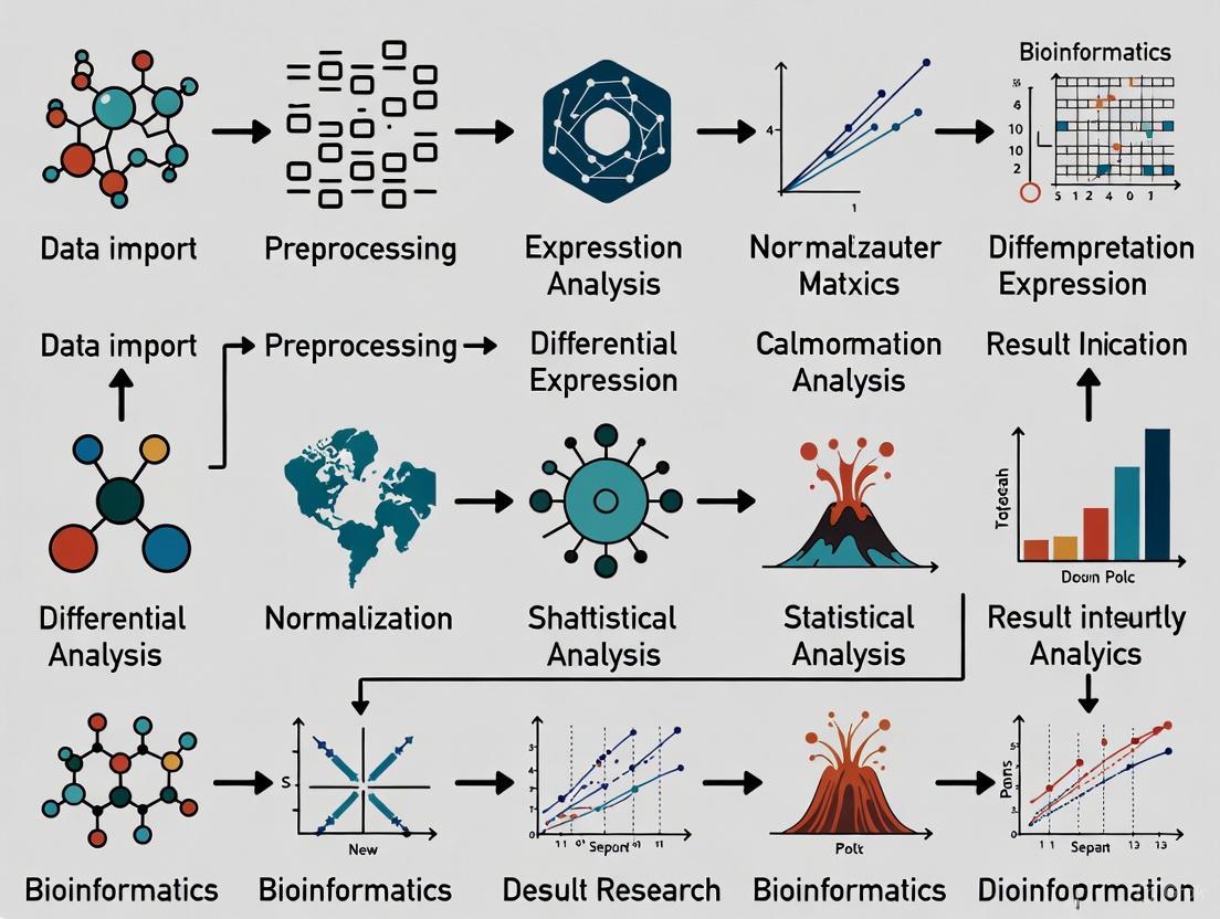
The differential expression analysis pipeline follows a structured sequence of steps to ensure robust and interpretable results. Figure 1 illustrates the complete workflow from sample preparation through biological interpretation.
Figure 1. Complete workflow for differential expression analysis, spanning from sample preparation to biological interpretation.
Core Analytical Steps
The analytical phase of differential expression analysis consists of several critical computational and statistical procedures:
Data Pre-processing: Raw sequencing reads (FASTQ files) undergo quality assessment and filtering to remove technical artifacts and low-quality data [1] [2]. The quality-filtered reads are then aligned to a reference genome or transcriptome using tools such as HISAT2 or TopHat [2] [5].
Expression Quantification: Aligned reads are summarized and aggregated over genomic features (genes or transcripts) using tools like HTSeq, producing a count matrix where each value represents the number of reads mapped to a particular feature in a specific sample [2].
Normalization: This crucial step adjusts for technical variations between samples, particularly differences in sequencing depth (total number of reads) and RNA composition [3]. Common methods include the Trimmed Mean of M-values (TMM) used by edgeR and the geometric mean approach employed by DESeq2 [3].
Statistical Testing: Normalized data undergoes statistical analysis to identify genes with significant expression changes. Tools like DESeq2 and edgeR use models based on the negative binomial distribution to account for biological variability and technical noise in count data [3] [6].
Multiple Test Correction: Since thousands of statistical tests are performed simultaneously (one per gene), specialized correction methods such as the Benjamini-Hochberg procedure are applied to control the false discovery rate (FDR) [1]. The resulting adjusted p-values (q-values) determine which genes are declared differentially expressed.
Interpretation: The final step involves biological interpretation of the resulting differentially expressed gene list, typically through functional enrichment analysis to identify overrepresented biological processes, pathways, or functional categories [1].
Tool Selection Framework
Choosing an appropriate analytical tool depends on multiple experimental factors. Figure 2 provides a decision framework for selecting the most suitable differential expression method.
Figure 2. Decision framework for selecting differential expression analysis methods based on experimental design and data characteristics.
Comparative Analysis of Differential Expression Tools
Tool Performance and Characteristics
The selection of an appropriate differential expression tool significantly impacts results interpretation. Different packages employ distinct statistical models, normalization approaches, and data handling procedures. Table 1 summarizes the key features of major differential expression analysis tools.
Table 1. Comparison of Major Differential Expression Analysis Tools
| Tool | Publication Year | Statistical Distribution | Normalization Method | Key Features | Best Suited For |
|---|---|---|---|---|---|
| DESeq2 | 2014 [3] | Negative binomial [3] [4] | DESeq method [3] | Shrinkage estimation for fold changes and dispersion; robust for low count genes [3] | Bulk RNA-seq with small sample sizes; paired comparisons [3] [7] |
| edgeR | 2010 [3] | Negative binomial [3] [4] | TMM [3] | Empirical Bayes estimation; exact tests for differential expression [3] | Bulk RNA-seq with multiple conditions; complex experimental designs [3] |
| limma | 2015 [3] | Log-normal [3] | TMM [3] | Linear models with empirical Bayes moderation; fast computation [8] [4] | Bulk RNA-seq and microarray data; multiple group comparisons [8] [4] |
| NOIseq | 2012 [3] | Non-parametric [3] | RPKM [3] | Noise distribution estimation; no replication requirement [7] | Data with limited replicates; when distribution assumptions are violated [3] |
| SAMseq | 2013 [3] | Non-parametric [3] | Internal [3] | Mann-Whitney test with Poisson resampling; robust to outliers [3] | Large sample sizes; non-normally distributed data [3] |
| MAST | 2015 [9] | Two-part generalized linear model [9] | TPM/FPKM [9] | Accounts for dropout events in single-cell data; models both discrete and continuous components [9] | Single-cell RNA-seq data with high zero-inflation [9] |
Tool Selection Guidelines
Based on comparative studies and tool characteristics, researchers can follow these evidence-based guidelines for method selection:
For typical bulk RNA-seq experiments: DESeq2 and limma+voom generally provide the most consistent and reliable results across various experimental conditions [7]. DESeq2 is particularly noted for its robust handling of studies with small sample sizes, a common scenario in RNA-seq experiments [3].
When distributional assumptions may be violated: Non-parametric methods like NOIseq perform well without requiring specific distributional assumptions, making them suitable for data with complex characteristics [3] [7].
For single-cell RNA-seq data: Specialized methods like MAST, SCDE, and scDD that explicitly model single-cell specific characteristics (e.g., zero-inflation, high heterogeneity) are recommended over bulk RNA-seq tools [9].
Consensus approaches: Combining results from multiple methods (e.g., consensus across DESeq2, limma, and NOIseq) can yield more reliable differentially expressed genes with reduced false positives [7].
Essential Research Reagents and Computational Tools
Successful differential expression analysis requires both wet-lab reagents and computational resources. Table 2 outlines key components of the researcher's toolkit.
Table 2. Essential Research Reagents and Computational Tools for Differential Expression Analysis
| Category | Item | Specification/Example | Function/Purpose |
|---|---|---|---|
| Wet-Lab Reagents | RNA Isolation Kits | Column-based or magnetic bead systems | High-quality RNA extraction with integrity number (RIN) >8 for reliable sequencing [2] |
| Library Preparation Kits | Strand-specific, poly-A selection or rRNA depletion | Convert RNA to sequence-ready libraries; determine transcript coverage and strand specificity [2] | |
| RNA-Seq Platforms | Illumina, PacBio, or Oxford Nanopore | Generate raw sequencing reads (FASTQ files); determine read length, depth, and error profiles [5] | |
| Quality Control Tools | Bioanalyzer, Qubit, Nanodrop | Assess RNA quality, quantity, and purity before library preparation [2] | |
| Computational Tools | Alignment Software | HISAT2, STAR, TopHat2 [2] [5] | Map sequencing reads to reference genome/transcriptome [2] |
| Quantification Tools | HTSeq, featureCounts | Generate count matrices from aligned reads [2] | |
| Differential Expression Packages | DESeq2, edgeR, limma | Statistical analysis to identify differentially expressed genes [3] [4] | |
| Functional Analysis Tools | clusterProfiler, Enrichr | Biological interpretation of DEGs through pathway and ontology enrichment [3] |
Differential expression analysis represents a powerful methodological framework for identifying molecular changes across biological conditions. When properly executed with appropriate statistical tools and validated through follow-up experiments, this approach continues to drive fundamental discoveries in biomedical research. As sequencing technologies evolve and new computational methods emerge, differential expression analysis remains an indispensable tool for unraveling the complexity of biological systems and advancing human health.
The field continues to evolve with emerging methodologies such as machine learning approaches that may complement traditional differential expression analysis, particularly for identifying complex patterns in large, heterogeneous datasets [3]. However, regardless of methodological advances, careful experimental design, appropriate quality control, and biologically informed interpretation remain fundamental to generating meaningful insights from differential expression studies.
RNA sequencing (RNA-seq) has revolutionized transcriptomics, enabling researchers to capture detailed snapshots of gene expression. Two primary methodologies have emerged: bulk RNA-seq and single-cell RNA-seq (scRNA-seq). Both are powerful tools for exploring gene activity, but they offer fundamentally different perspectives and are suited to distinct biological questions [10] [11]. Bulk RNA-seq provides a population-averaged gene expression profile, measuring the combined RNA from all cells in a sample. In contrast, single-cell RNA-seq dissects this population to examine gene expression at the resolution of individual cells [12]. This in-depth technical guide will compare these two approaches, highlighting their differences, applications, and methodologies to help you select the right strategy for your research, particularly within the context of differential expression analysis.
Core Principles and Key Differences
Understanding the fundamental distinctions between bulk and single-cell RNA-seq is crucial for experimental design.
Bulk RNA-Sequencing is a well-established method that analyzes the average gene expression from a population of thousands to millions of cells [10] [13]. In this workflow, RNA is extracted from the entire tissue or cell population, converted to complementary DNA (cDNA), and sequenced to generate a single, composite gene expression profile for the sample [14]. This approach is ideal for homogeneous samples or when the research goal is to understand the overall transcriptional state of a tissue.
Single-Cell RNA-Sequencing takes gene expression analysis to a higher resolution by examining the transcriptome of each individual cell separately [10]. The process begins with the dissociation of tissue into a viable single-cell suspension [14]. Individual cells are then isolated, often using microfluidic devices like the 10x Genomics Chromium system, where each cell is partitioned into a nanoliter-scale droplet containing a barcoded gel bead [14] [15]. The RNA from each cell is tagged with a unique cellular barcode, allowing sequencing reads to be traced back to their cell of origin, thus enabling the reconstruction of individual transcriptomes [14].
The table below summarizes the critical differences between the two technologies:
Table 1: Key Differences Between Bulk and Single-Cell RNA-Seq
| Feature | Bulk RNA Sequencing | Single-Cell RNA Sequencing |
|---|---|---|
| Resolution | Average of cell population [10] | Individual cell level [10] |
| Cost per Sample | Lower (~$300 per sample) [10] | Higher (~$500 to $2000 per sample) [10] |
| Data Complexity | Lower, easier to process [10] [11] | Higher, requires specialized computational methods [10] [15] |
| Cell Heterogeneity Detection | Limited, masks diversity [10] [16] | High, reveals diverse cell types and states [10] [16] |
| Rare Cell Type Detection | Limited or impossible [10] | Possible, can identify rare populations [10] [12] |
| Gene Detection Sensitivity | Higher per sample [10] | Lower per cell, due to technical noise and dropout events [10] [15] |
| Sample Input Requirement | Higher amount of total RNA [10] | Lower, can work with just a few picograms of RNA from a single cell [10] |
| Ideal Application | Homogeneous populations, differential expression between conditions [10] [17] | Heterogeneous tissues, cell type discovery, developmental trajectories [10] [12] |
Applications and Use Cases
Each sequencing method is suited to different research needs, as evidenced by numerous scientific studies.
Applications of Bulk RNA-Seq
Bulk RNA-seq is best for large-scale studies focused on the overall transcriptomic landscape.
- Differential Gene Expression Analysis: It is widely used to compare gene expression profiles between different experimental conditions (e.g., diseased vs. healthy, treated vs. control) to identify genes that are upregulated or downregulated [14].
- Biomarker Discovery: Bulk RNA-seq has been instrumental in identifying RNA-based biomarkers for disease diagnosis, prognosis, or patient stratification [10] [14]. A study on pancreatic cancer, for instance, used bulk RNA-seq to identify differential expression of simple repetitive sequences as potential tumor biomarkers [10].
- Transcriptome Characterization: It is a powerful tool for annotating novel transcripts, including isoforms, non-coding RNAs, alternative splicing events, and gene fusions [10] [14]. A comprehensive analysis of nearly 7,000 cancer samples from The Cancer Genome Atlas used bulk RNA-seq to detect novel and clinically relevant gene fusions involved in cancer [10].
Applications of Single-Cell RNA-Seq
Single-cell RNA-seq is ideal for investigating cellular complexity and heterogeneity.
- Characterizing Heterogeneous Cell Populations: scRNA-seq can identify novel cell types, cell states, and rare cell subtypes [14]. For example, a study of mouse embryonic stem cells identified a rare subpopulation that highly expressed Zscan4 genes, revealing cells with greater differentiation potential [10].
- Reconstructing Developmental Trajectories: By analyzing transcriptomes from individual cells, researchers can infer developmental pathways and lineage relationships, understanding how cellular heterogeneity evolves over time [14] [12].
- Immune and Cancer Profiling: scRNA-seq has been pivotal in discovering new immune cell subsets and unraveling tumor heterogeneity [10] [14]. A study of metastatic lung cancer used scRNA-seq to reveal changes in plasticity induced by non-small cell lung cancer, providing insights not possible with bulk sequencing [10].
- Rare Disease-Associated Cells: This technology can identify extremely rare cells that are critical to disease pathology, such as CFTR-expressing pulmonary ionocytes in cystic fibrosis, which occur at a rate of only 1 in 200 cells [10].
Experimental Design and Workflow
A successful transcriptomics study requires careful planning and execution. Key considerations include sequencing depth, the number of biological replicates, and strategies to minimize batch effects [18]. For bulk RNA-seq, a crucial design factor is the RNA-extraction protocol, which typically involves either poly(A) selection to enrich for mRNA or ribosomal RNA depletion [18]. For scRNA-seq, the first and most critical step is the preparation of a high-quality single-cell suspension with high cell viability, free of clumps and debris [14].
The workflows for bulk and single-cell RNA-seq diverge significantly at the initial stages, as illustrated below.
Differential Expression Analysis: Methods and Considerations
Differential expression (DE) analysis aims to identify genes with statistically significant expression changes between conditions. The approach differs markedly between bulk and single-cell data.
Analyzing Bulk RNA-Seq Data
For bulk RNA-seq, DE analysis typically involves comparing gene expression counts between sample groups using methods that account for the count-based nature of the data and its variability.
- Common Tools: Widely used tools like DESeq2 [19] and edgeR [13] [18] employ a negative binomial generalized log-linear model to test for differential expression [13].
- Workflow: After raw read processing, alignment, and quantification, a counts table is generated. Following quality control and normalization, these tools statistically assess each gene for expression changes between conditions, providing fold-change values and adjusted p-values [18].
Analyzing Single-Cell RNA-Seq Data
The analysis of scRNA-seq data is more complex due to its high dimensionality, sparsity, and the hierarchical structure where cells are nested within samples [15] [16]. Treating individual cells as independent replicates leads to pseudoreplication and false discoveries, as cells from the same sample are more similar to each other than to cells from different samples [16]. Therefore, the sample, not the cell, must be treated as the experimental unit. Several methodological approaches have been developed to address this:
- Pseudobulk Methods: This approach involves summing the UMI counts for a specific cell type across all cells within each biological sample to create a "pseudobulk" expression profile per sample [16]. Standard bulk RNA-seq DE tools like DESeq2 or edgeR can then be applied to these aggregated profiles [19] [16]. This is a computationally efficient and robust way to account for sample-to-sample variability.
- Mixed-Effects Models: These models explicitly account for the correlation of cells from the same sample by including a random effect for sample identity, while the condition of interest (e.g., case vs. control) is modeled as a fixed effect [16]. Tools like NEBULA and MAST (with a random effect term) implement this approach, though it can be computationally intensive for large datasets [16].
- Differential Distribution Testing: Single-cell data enables researchers to look beyond differences in mean expression. Methods like IDEAS and distinct test whether the entire distribution of a gene's expression (including its variance or modality) differs between conditions [19] [16]. IDEAS, for instance, estimates the expression distribution for each individual and then tests whether these distributions are different between groups of individuals [19].
Table 2: Key Reagents and Tools for RNA-Seq Analysis
| Item Name | Function / Description | Example Use Case |
|---|---|---|
| Oligo(dT) Beads | Enriches for polyadenylated mRNA from total RNA by hybridization [18] [17]. | mRNA enrichment in bulk RNA-seq library prep. |
| Unique Molecular Identifiers (UMIs) | Molecular tags that label individual mRNA molecules during reverse transcription to correct for PCR amplification bias [15]. | Accurate transcript counting in single-cell and bulk protocols. |
| Chromium Controller (10x Genomics) | A microfluidic instrument that partitions single cells into Gel Beads-in-emulsion (GEMs) for barcoding [14]. | High-throughput single-cell library preparation. |
| Cell Hashtag Oligos | Antibody-derived tags that label cells from individual samples with unique barcodes, enabling sample multiplexing [14]. | Pooling multiple samples in a single scRNA-seq run to reduce costs and batch effects. |
| DESeq2 | A bulk RNA-seq statistical package using a negative binomial model for differential expression testing [19] [18]. | Identifying DEGs from bulk data or pseudobulk data from scRNA-seq. |
| NEBULA | A fast negative binomial mixed model for differential expression analysis of multi-subject single-cell data [16]. | Cell-type-specific DE analysis across individuals while accounting for subject-level effects. |
Bulk and single-cell RNA-seq are complementary, not competing, technologies. Bulk RNA-seq remains a cost-effective and powerful choice for identifying overall gene expression changes in large cohort studies or when working with homogeneous samples [10] [11]. Single-cell RNA-seq is indispensable for deconvoluting cellular heterogeneity, discovering rare cell types, and reconstructing cellular trajectories [10] [12].
The future of transcriptomics lies in integrated approaches [10] [17]. Combining bulk, single-cell, and emerging technologies like spatial transcriptomics—which preserves the spatial context of gene expression—provides a more holistic view of biological systems [12] [17]. Furthermore, multi-omics approaches that combine scRNA-seq with assays for chromatin accessibility (scATAC-seq) or protein abundance are becoming increasingly common, offering a more comprehensive view of cellular functions [10]. As these technologies continue to evolve and become more accessible, they will undoubtedly deepen our understanding of the molecular underpinnings of health and disease.
Differential gene expression analysis represents a cornerstone of modern transcriptomics, enabling researchers to identify molecular mechanisms underlying health and disease. The journey from raw sequencing data to a reliable count matrix is a critical, yet often overlooked, phase where choices in quantification directly impact all downstream analyses and biological conclusions [20]. This technical guide details the process of transforming raw sequencing reads into a gene count matrix, explaining the core principles, methodologies, and sources of uncertainty inherent in RNA-seq quantification, framed for beginners embarking on differential expression research [21].
RNA sequencing (RNA-seq) leverages next-generation sequencing to measure the abundance of RNA transcripts in a biological sample at a given time [22]. The primary data output is millions of short sequence reads. The fundamental goal of quantification is to assign these reads to specific genomic features (like genes or transcripts) and summarize them into a count table that serves as the starting point for differential expression analysis [23].
This process is non-trivial. Raw read counts cannot be directly used to compare expression between samples due to differences in sequencing depth, gene length, and RNA composition [24]. Furthermore, the data is subject to various technical and biological variances that must be accounted for to make accurate biological inferences [25]. Understanding how a count matrix is generated, and the uncertainties introduced during this process, is essential for correctly interpreting differential expression results.
From Raw Data to Read Alignments
The initial phase of RNA-seq analysis involves processing the raw sequence data into a format suitable for quantification.
Raw Data and Quality Control
RNA-seq data typically begins as FASTQ files, which contain the nucleotide sequences for millions of fragments and an associated quality score for each base call [23]. Key considerations at this stage include:
- Read Type: Single-end vs. paired-end protocols. Paired-end sequencing reads both ends of a fragment, providing more information for alignment and isoform identification [23].
- Quality Assessment: Checks for per-base sequence quality, adapter contamination, and overall read quality are crucial before proceeding [23].
Alignment and Quantification Strategies
There are three principal computational approaches for determining the origin of RNA-seq reads [23]. The choice of strategy is a fundamental methodological decision.
Table 1: Comparison of RNA-seq Quantification Approaches
| Approach | Core Methodology | Key Tools | Advantages | Considerations |
|---|---|---|---|---|
| Genome Alignment & Counting | Aligns reads to the genome, then counts overlaps with gene exons. | STAR, Rsubread featureCounts [23] | Simplicity; preferred for poorly annotated genomes. | Can be computationally intensive; requires a reference genome and GTF annotation file. |
| Transcriptome Alignment & Quantification | Aligns reads to the transcriptome, estimates transcript expression. | RSEM [23] | Can produce accurate quantification, good for samples without DNA contamination. | Requires a well-annotated transcriptome (e.g., from GENCODE or Ensembl). |
| Pseudoalignment | Rapidly assigns reads to transcripts without full alignment. | Salmon, kallisto [23] | Computational efficiency; mitigates effects of DNA contamination and GC bias. | Speed comes from a different algorithmic approach; transcript-level quantification can be noisier. |
The following workflow diagram illustrates the three main paths from raw sequencing data to a gene count matrix:
Normalization and Quantification Measures
After read assignment, expression levels must be normalized to enable cross-sample comparison. Different quantification measures have been developed, but they are not all suitable for differential expression analysis.
Common Quantification Measures
- FPKM/RPKM: Fragments (or Reads) Per Kilobase of exon per Million mapped fragments. This is a within-sample normalization method that corrects for gene length and sequencing depth, allowing comparison of expression between different genes within the same sample [24].
- TPM: Transcripts Per Million is similar to FPKM but with the calculation order changed. It is considered a more stable metric because the sum of all TPMs is constant across samples [24].
- Normalized Counts: These are raw counts that have been normalized by a statistical method, such as the median-of-ratios method in DESeq2 or the TMM (Trimmed Mean of M-values) in edgeR, specifically designed to account for library size and composition differences between samples for the purpose of cross-sample comparison and differential testing [21] [24].
Why Normalized Counts are Preferred for Differential Expression
A comparative study on RNA-seq data from patient-derived xenograft models provided compelling evidence that normalized count data are the best choice for cross-sample analysis [24]. The study found that hierarchical clustering based on normalized counts grouped replicate samples from the same model together more accurately than TPM or FPKM. Furthermore, normalized counts demonstrated the lowest median coefficient of variation and the highest intraclass correlation values across replicates [24]. This is because FPKM and TPM "normalize away" factors that are essential for between-sample statistical testing, and they can perform poorly when transcript distributions differ significantly between samples [24].
Table 2: Key Quantification Measures for RNA-seq Data
| Measure | Definition | Primary Use | Suitable for Cross-Sample DE Analysis? |
|---|---|---|---|
| Raw Counts | Unaltered number of reads assigned to a feature. | Input for statistical normalizers (DESeq2, edgeR). | No, requires normalization. |
| FPKM/RPKM | Fragments per kilobase per million mapped fragments. | Within-sample gene comparison. | Not recommended [24]. |
| TPM | Transcripts per million. | Within-sample gene comparison. | Not recommended [24]. |
| Normalized Counts | Raw counts normalized for library size/composition (e.g., DESeq2, edgeR). | Cross-sample comparison and DE analysis. | Yes, this is the recommended measure. |
Uncertainty in Quantification and Experimental Design
Quantification is an estimation process with inherent uncertainty, which can be classified into technical and biological variance.
Statistical Modeling of RNA-seq Data
Differential expression packages like DESeq2 and edgeR model expression data using the negative binomial distribution [21]. This distribution is chosen because it accommodates the "over-dispersed" nature of count data where the variance is greater than the mean [21]. These tools create a model and perform statistical testing for the expected true value of gene expression, rather than testing directly on the input counts.
The Critical Role of Experimental Design
Proper experimental design is paramount for ensuring that quantification uncertainty can be accurately measured and accounted for.
- Replicates: Biological replicates (samples from different biological units) are essential to measure biological variance and make inferences about a population. True replication requires independent experimental units [23].
- Confounding and Batch Effects: RNA-seq experiments are sensitive to technical variations (e.g., different days, analysts, or reagent batches). A well-designed experiment balances conditions across these batches to prevent confounding, making it possible to disentangle technical effects from the primary biological effects of interest [23].
The following table details key resources, both data and software, required for RNA-seq quantification.
Table 3: Essential Resources for RNA-seq Quantification
| Resource Category | Item | Function / Description |
|---|---|---|
| Reference Sequences | Genome FASTA File | Contains the full sequence of the reference genome for alignment. |
| Annotation (GTF/GFF) File | Specifies the genomic locations of annotated features (genes, exons, etc.). | |
| Transcriptome FASTA File | Contains the sequences of all known transcripts for transcriptome-based methods. | |
| Software & Pipelines | Read Aligner (e.g., STAR) | Aligns sequencing reads to a reference genome. |
| Quantification Tool (e.g., Salmon, RSEM) | Assigns reads to features and estimates abundance. | |
| Differential Expression Suite (e.g., DESeq2, edgeR) | Performs statistical normalization and identifies differentially expressed genes. | |
| Data Sources | GENCODE | Provides high-quality reference gene annotations for human and mouse. |
| Ensembl | Provides reference files for a wide range of organisms, including plants and fungi. |
Visualization and Quality Control
Post-quantification, it is vital to perform quality control (QC) on the normalized count data to ensure the normalization process was effective and that samples cluster as expected.
- PCA Plots: Verify that biological replicates cluster together and that treatment groups separate. Normalization should improve this clustering [21].
- Distance Plots: Show the dissimilarity between samples, with replicates expected to have smaller distances [21].
- Other Plots: Density and box plots of normalized expression help verify that distributions across samples have been successfully aligned by the normalization procedure [21].
Advanced visualization methods like parallel coordinate plots and scatterplot matrices can further help researchers detect normalization issues, identify problematic genes or samples, and confirm that the data shows greater variability between treatments than between replicates [25].
The path from raw sequencing data to a count matrix is a foundational process in RNA-seq analysis. Selecting an appropriate quantification strategy (genome, transcriptome, or pseudoalignment) based on the organism and experimental goals, and using normalized counts from robust statistical methods like those in DESeq2 or edgeR for differential expression, is critical for generating reliable biological insights. A well-designed experiment with adequate replication, coupled with rigorous quality control and visualization, allows researchers to accurately quantify and account for uncertainty, ensuring that the subsequent differential expression analysis truly reflects the underlying biology.
In RNA sequencing (RNA-seq) analysis, accurate normalization of gene expression data is a critical prerequisite for obtaining biologically meaningful results. Normalization corrects for technical variations, enabling meaningful comparisons within and between samples. This technical guide provides an in-depth examination of three foundational normalization metrics—Reads Per Kilobase Million (RPKM), Fragments Per Kilobase Million (FPKM), and Transcripts Per Million (TPM). Framed within the broader context of differential expression analysis for beginners, this review clarifies the calculations, applications, and limitations of each metric. We summarize quantitative data in structured tables, detail experimental protocols for calculation, and provide clear visualizations of workflows. The content is designed to equip researchers, scientists, and drug development professionals with the knowledge to select appropriate normalization methods for their RNA-seq studies, thereby ensuring robust and interpretable results in downstream analyses.
The raw output of an RNA-seq experiment consists of counts of reads or fragments mapped to each gene in the genome. These raw counts are not directly comparable due to several technical biases:
- Sequencing Depth: Samples sequenced to a greater depth will yield higher counts for a gene expressed at the same level, simply because more reads are generated [26] [27].
- Gene Length: For a given level of expression, longer genes will generate more reads than shorter genes because they provide a larger template [26] [27].
- RNA Composition: In certain experiments, a few highly expressed genes can consume a substantial fraction of the sequencing library. This can skew the count distribution for other genes and make them appear less expressed in that sample compared to others, even if their true expression level is unchanged [28] [27].
Normalization is the computational process of adjusting the raw count data to account for these technical factors. The primary goal is to produce abundance measures that reflect true biological differences in gene expression rather than technical artifacts [26] [28]. This is an essential step for both exploratory data analysis and formal differential expression testing. Without proper normalization, the identification of differentially expressed genes (DEGs) is unreliable, potentially leading to false discoveries and incorrect biological conclusions [28].
Understanding the Metrics: Definitions and Calculations
This section dissects the three core metrics, their intended purposes, and their mathematical formulations.
RPKM (Reads Per Kilobase Million)
RPKM was one of the earliest measures developed for normalizing RNA-seq data from single-end experiments [29] [30]. It is designed to facilitate the comparison of gene expression levels within a single sample by normalizing for both sequencing depth and gene length.
- Calculation Steps: The calculation of RPKM follows a specific order of operations:
- Formula: ( RPKM = \frac{\text{Reads mapped to gene}}{\text{(Gene length in kb)} \times \text{(Total mapped reads in millions)} } = \frac{\text{Reads mapped to gene}}{\text{Gene length} \times \text{Total mapped reads}} \times 10^9 ) [26] [24]
FPKM (Fragments Per Kilobase Million)
FPKM is the direct counterpart to RPKM for paired-end RNA-seq experiments. In paired-end sequencing, two reads are sequenced from a single cDNA fragment. FPKM accounts for this by counting fragments instead of individual reads, thus avoiding double-counting [29] [30].
- Relationship to RPKM: The conceptual meaning and calculation of FPKM are identical to RPKM, with the word "Fragments" replacing "Reads" [31]. For a given transcript, if each fragment is represented by two reads, the FPKM value will be half the RPKM value. In single-end sequencing, where each fragment yields one read, RPKM and FPKM are equivalent [32].
- Formula: ( FPKM = \frac{\text{Fragments mapped to gene}}{\text{(Gene length in kb)} \times \text{(Total mapped fragments in millions)} } = \frac{\text{Fragments mapped to gene}}{\text{Gene length} \times \text{Total mapped fragments}} \times 10^9 ) [24]
TPM (Transcripts Per Million)
TPM is a more recent metric that addresses a critical limitation of RPKM and FPKM. While the formulas appear similar, the order of operations is reversed, leading to a desirable property for cross-sample comparison [29].
- Calculation Steps:
- Normalize for gene length first: Divide the raw read count by the length of the gene in kilobases. This gives Reads Per Kilobase (RPK).
- Sum all RPK values: Sum the RPK values for all genes in the sample to get the total RPK per sample.
- Normalize for sequencing depth: Divide the RPK value for each gene by the "per million" scaling factor, which is the total RPK from step 2 divided by one million. This yields TPM [29] [26].
- Formula: ( TPM = \frac{\text{Reads mapped to gene / Gene length in kb}}{\text{Sum(Reads mapped to all genes / Their lengths in kb)}} \times 10^6 ) [31]
The key advantage of TPM is that the sum of all TPM values in a sample is always constant (one million). This means TPM values represent the relative proportion of each transcript within the total sequenced transcript pool, making them directly comparable across samples [29]. In contrast, the sum of all RPKM or FPKM values in a sample can vary, complicating inter-sample comparisons [29] [27].
The table below provides a consolidated comparison of RPKM, FPKM, and TPM to highlight their key characteristics and appropriate use cases.
Table 1: Comparative overview of RNA-seq normalization metrics
| Feature | RPKM | FPKM | TPM |
|---|---|---|---|
| Full Name | Reads Per Kilobase Million [26] | Fragments Per Kilobase Million [26] | Transcripts Per Million [29] |
| Sequencing Type | Single-end [29] | Paired-end [29] | Both |
| What is Counted? | Reads | Fragments | Reads (conceptually transcripts) |
| Order of Normalization | Depth then length [29] | Depth then length [29] | Length then depth [29] |
| Sum of Values in a Sample | Variable [27] | Variable [27] | Constant (1 million) [29] |
| Recommended Use | Compare different genes within a sample [30] | Compare different genes within a sample [30] | Compare genes within a sample and relative abundance across samples [29] [30] |
The following workflow diagram illustrates the distinct calculation paths for RPKM/FPKM versus TPM.
Diagram 1: RPKM/FPKM vs. TPM Calculation Workflows
Experimental Protocols and Practical Implementation
Computational Calculation
The metrics discussed can be calculated using various bioinformatics programming languages. Below is an example of how to compute them using Python with the bioinfokit package [26].
Protocol 1: Normalization with Python's bioinfokit package
- Prerequisites: Ensure Python and the
bioinfokitpackage (v0.9.1 or later) are installed. Data should be in a dataframe with genes as rows and samples as columns, and a column for gene length in base pairs. - RPKM/FPKM Calculation:
- TPM Calculation:
A Note on Differential Expression Analysis
For formal differential expression analysis (e.g., identifying genes expressed at different levels between two conditions), specialized statistical methods implemented in tools like DESeq2 and edgeR are the gold standard [24] [27]. These tools use sophisticated normalization approaches (e.g., DESeq2's "median of ratios" [27] or edgeR's "trimmed mean of M-values" [26]) that are more robust for between-sample comparisons than TPM, RPKM, or FPKM.
- DESeq2's Median of Ratios Method: This method does not use gene length in its normalization, as it compares the same gene between samples. Instead, it: a. Calculates a pseudo-reference sample for each gene via the geometric mean across all samples. b. Computes the ratio of each gene's count to its pseudo-reference. c. The median of these ratios for each sample is used as the size factor (normalization factor). d. Raw counts are divided by this size factor to generate normalized counts [27].
- When to Use TPM/RPKM/FPKM: These metrics are highly useful for data visualization, exploratory analysis, and when a normalized expression measure is needed for other downstream tasks (e.g., estimating metabolic fluxes). However, their raw values should not be used as direct input for statistical tests in differential expression tools like DESeq2 or edgeR, which operate on raw counts and perform their own internal normalization [27].
Limitations and Common Misconceptions
A widespread misconception is that because RPKM, FPKM, and TPM are "normalized" values, they are directly comparable across all samples and experimental conditions. This is not always true [31] [33].
- Dependence on Transcriptome Composition: RPKM, FPKM, and TPM measure relative abundance, not absolute concentration. The value for a gene represents its proportion relative to the total population of sequenced transcripts in that sample. If the transcriptome composition changes drastically between samples (e.g., one condition has a few genes that are extremely highly expressed), the relative proportions of all other genes will be compressed, making their TPM values non-comparable between those samples [31] [28].
- Sensitivity to Experimental Protocol: The sample preparation protocol can significantly impact normalized expression values. For example, in a study comparing poly(A)+ selection and rRNA depletion protocols on the same biological sample, the resulting TPM values were vastly different. The rRNA depletion protocol captured a large amount of structural non-coding RNAs, which dramatically altered the composition of the transcriptome and thus the relative proportion (TPM) of protein-coding genes [31]. Therefore, comparing TPM values from studies using different library preparation protocols can be highly misleading.
Table 2: Essential research reagents and solutions for RNA-seq
| Reagent / Solution | Function in RNA-seq Workflow |
|---|---|
| Oligo(dT) Magnetic Beads | Selection and enrichment of polyadenylated mRNA from total RNA by binding to the poly(A) tail [31]. |
| rRNA Depletion Probes | Selective removal of abundant ribosomal RNA (rRNA) from total RNA to increase the sequencing coverage of mRNA and other RNA species [31]. |
| Fragmentation Buffer | Chemically or enzymatically breaks full-length RNA or cDNA into shorter fragments of a size suitable for high-throughput sequencing [31]. |
| Reverse Transcriptase Enzyme | Synthesizes complementary DNA (cDNA) from the RNA template, creating a stable library for sequencing [27]. |
| Size Selection Beads (e.g., SPRI) | Solid-phase reversible immobilization beads are used to purify and select for cDNA fragments within a specific size range, ensuring library uniformity [24]. |
The Scientist's Toolkit for RNA-Seq
A successful RNA-seq experiment relies on a suite of specialized reagents and computational tools. The table below details key materials used in a typical workflow.
RPKM, FPKM, and TPM are fundamental metrics for understanding and interpreting RNA-seq data. While RPKM/FPKM are suitable for assessing relative gene expression within a single sample, TPM is generally preferred for making comparisons of a gene's abundance across different samples due to its constant sum property. However, it is critical to remember that all three metrics represent relative expression that depends on the transcriptome composition of each sample. For robust differential expression analysis, dedicated statistical methods like DESeq2 and edgeR, which operate on raw counts and employ more advanced normalization techniques, are strongly recommended. By selecting the appropriate metric and understanding its assumptions and limitations, researchers can draw more accurate and reliable biological insights from their transcriptomic studies.
The reliability of scientific findings, particularly in high-throughput biology, hinges on rigorous experimental design. This technical guide examines the foundational principles of replicates, statistical power, and the often-overlooked issue of pooling bias, framed within the context of differential expression analysis. Proper implementation of these principles is critical for generating biologically relevant results that can withstand rigorous scrutiny and contribute meaningfully to the scientific record. We provide a comprehensive overview of methodologies that empower researchers to design robust experiments, avoid common pitfalls, and optimize resource allocation while maintaining statistical integrity.
The modern biology toolbox continues to evolve, with cutting-edge molecular techniques complementing classic approaches. However, statistical literacy and experimental design remain the bedrock of empirical research success, regardless of the specific methods used for data collection [34]. A well-designed experiment ensures that observed differences in gene expression can be reliably attributed to biological conditions of interest rather than technical artifacts or random variation.
The quote from Ronald A. Fisher succinctly captures this imperative: "To consult the statistician after an experiment is finished is often merely to ask him to conduct a post mortem examination. He can perhaps say what the experiment died of" [34]. This perspective is particularly relevant in the context of differential expression analysis, where decisions made during experimental planning fundamentally determine what conclusions can be drawn from the data.
In RNA-sequencing experiments, the basic task involves analyzing count data to detect differentially expressed genes between sample groups. The count matrix represents the number of sequence fragments uniquely assigned to each gene, with higher counts indicating higher expression levels [35]. However, proper interpretation requires careful consideration of multiple factors, including sequencing depth, gene length, and RNA composition, all of which must be accounted for through appropriate normalization and statistical approaches.
The Critical Role of Replication
Biological vs. Technical Replicates
A fundamental distinction in experimental design is between biological replicates and technical replicates. Biological replicates are measurements from biologically distinct samples (e.g., cells, tissues, or animals) that capture the natural biological variation within a condition. Technical replicates are repeated measurements of the same biological sample to account for technical noise introduced during library preparation or sequencing [34].
In differential expression analysis, biological replicates are essential for drawing conclusions about biological phenomena, as they allow researchers to distinguish consistent biological effects from random variation. The misconception that large quantities of data (e.g., deep sequencing or measurement of thousands of genes) ensure precision and statistical validity remains prevalent. In reality, it is the number of biological replicates that determines the reliability of biological inferences [34].
Pseudoreplication and Its Consequences
Pseudoreplication occurs when measurements are treated as independent observations despite originating from the same biological unit or being subject to confounding factors. This practice artificially inflates sample size and increases the risk of false positive findings [34] [36]. Hurlbert coined the term "pseudoreplication" for situations where researchers treat non-independent data points as independent in their analysis, a problem he identified as widespread across biological disciplines [36].
In a typical example involving nested designs, such as testing a greenhouse treatment effect on tomato plants with multiple plants per greenhouse, the greenhouse rather than individual plants constitutes the experimental unit. Any hypothesis test of the treatment should use an error term based on variation among greenhouses rather than variation among individual plants to avoid pseudoreplication [36].
Statistical Power and Sample Size
Power Analysis Fundamentals
Statistical power represents the probability that a test will correctly reject a false null hypothesis (i.e., detect a true effect). Underpowered experiments, which are prevalent across biological research, waste resources and generate unreliable results [37]. Power analysis allows researchers to determine the sample size needed to detect an effect of a given size with a specified degree of confidence [34].
The relationship between power, sample size, effect size, and significance threshold follows established statistical principles. Low statistical power remains a pervasive issue compromising replicability across scientific fields, with an estimated $28 billion wasted annually on irreproducible preclinical science in the United States alone [37].
Power Considerations in High-Throughput Experiments
In RNA-seq experiments, power analysis must account for multiple testing, as thousands of genes are evaluated simultaneously. The false discovery rate (FDR) rather than family-wise error rate is typically controlled in exploratory analyses to balance false positives with statistical power [38]. Tools such as the R package micropower facilitate power analysis for microbiome studies, while similar principles apply to RNA-seq experimental design [34].
Table 1: Replication Success Rates Across Scientific Fields
| Field | Original Positive Outcomes | Replication Success Rate | Original Effect Size | Replication Effect Size |
|---|---|---|---|---|
| Psychology | 97 | 36% | 0.403 (SD: 0.188) | 0.197 (SD: 0.257) |
| Economics | 18 | 61% | 0.474 (SD: 0.239) | 0.279 (SD: 0.234) |
| Social Sciences | 21 | 62% | 0.459 (SD: 0.229) | 0.249 (SD: 0.283) |
| Cancer Biology | 97 | 43% | 6.15 (SD: 12.39) | 1.37 (SD: 3.01) |
| Ditridecylamine | Ditridecylamine, CAS:101012-97-9, MF:C26H55N, MW:381.7 g/mol | Chemical Reagent | Bench Chemicals | |
| 1-Bromo-2-methoxy-2-methylpropane | 1-Bromo-2-methoxy-2-methylpropane|19752-21-7 | 1-Bromo-2-methoxy-2-methylpropane (CAS 19752-21-7), a bifunctional halogenated ether for mechanistic studies. For Research Use Only. Not for human or veterinary use. | Bench Chemicals |
Data adapted from large-scale replication projects [37]
Understanding and Avoiding Pooling Bias
Test-Qualified Pooling
Test-qualified pooling represents a common but controversial practice in biological research where non-significant terms are dropped from a statistical model, effectively pooling their variation with the error term used to test hypotheses [36]. This approach is often motivated by the desire to increase degrees of freedom and statistical power for testing effects of interest.
The process typically involves fitting a full model based on the experimental design, then removing non-significant terms through sequential testing. The simplified model is then used for final hypothesis testing. Despite being recommended in some statistical textbooks for biologists, this practice introduces substantial risks [36].
Consequences of Inappropriate Pooling
Test-qualified pooling can severely compromise statistical inference through several mechanisms:
Inflated Type I Error Rates: Pooling based on non-significance in preliminary tests biases p-values downward and confidence intervals toward being too narrow, increasing false positive rates [36].
Reduced Reliability: The hoped-for improvement in statistical power is often small or non-existent, while the reliability of statistical procedures is substantially reduced [36].
Pseudoreplication: In nested designs, pooling errors constitutes a form of pseudoreplication that Hurlbert termed "test-qualified sacrificial pseudoreplication" [36].
Table 2: Common Normalization Methods for RNA-seq Data
| Normalization Method | Accounted Factors | Recommended Use | Limitations |
|---|---|---|---|
| CPM (Counts Per Million) | Sequencing depth | Gene count comparisons between replicates of the same sample group | Not for within-sample comparisons or DE analysis |
| TPM (Transcripts Per Kilobase Million) | Sequencing depth, Gene length | Gene count comparisons within a sample | Not for between-sample comparisons or DE analysis |
| RPKM/FPKM | Sequencing depth, Gene length | Gene count comparisons between genes within a sample | Not recommended for between-sample comparisons |
| DESeq2's Median of Ratios | Sequencing depth, RNA composition | Gene count comparisons between samples and for DE analysis | Not for within-sample comparisons |
| EdgeR's TMM (Trimmed Mean of M-values) | Sequencing depth, RNA composition | Gene count comparisons between samples and for DE analysis | Not for within-sample comparisons |
Adapted from RNA-seq methodology guides [35]
Practical Implementation for Differential Expression Analysis
Experimental Workflow Design
The following diagram illustrates key decision points in designing a robust differential expression experiment:
The Scientist's Toolkit: Essential Research Reagents and Materials
Table 3: Essential Research Reagents and Materials for RNA-seq Experiments
| Item | Function | Technical Considerations |
|---|---|---|
| RNA Isolation Kit | Extracts high-quality RNA from biological samples | Select based on sample type (cells, tissues, etc.); assess RNA integrity number (RIN >7.0 recommended) [13] |
| mRNA Enrichment Reagents | Enriches for messenger RNA from total RNA | Poly(A) selection kits preferentially capture protein-coding transcripts [13] |
| cDNA Library Prep Kit | Prepares sequencing libraries from RNA | Fragmentation, adapter ligation, and index ligation create sequenceable libraries [13] |
| Sequencing Controls | Monitor technical performance of sequencing run | Include external RNA controls consortium (ERCC) spikes to assess technical variability [34] |
| Alignment Software | Aligns sequence reads to reference genome | STAR aligner provides accurate splice-aware alignment for RNA-seq data [38] |
| Gene Quantification Tool | Generates count matrix from aligned reads | HTSeq-count assigns reads to genomic features using reference annotation [38] |
| Linadryl H | Linadryl H, CAS:19732-39-9, MF:C20H25NO2, MW:311.4 g/mol | Chemical Reagent |
| 2,2-Dibromohexane | 2,2-Dibromohexane | High-Purity Reagent | For RUO | High-purity 2,2-Dibromohexane for research. Used in organic synthesis & mechanistic studies. For Research Use Only. Not for human or veterinary use. |
Quality Control Assessment
Quality control represents a critical step in the differential expression workflow. Both sample-level and gene-level QC checks help ensure data quality before proceeding with formal statistical testing [35].
Principal Component Analysis (PCA) reduces the high-dimensionality of count data while preserving original variation, allowing visualization of sample relationships in two-dimensional space. In ideal experiments, replicates for each sample group cluster together, while sample groups separate clearly. Unexplained sources of variation may indicate batch effects or other confounding factors that should be accounted for in the statistical model [35].
Hierarchical clustering heatmaps display correlation of gene expression for all pairwise sample combinations. Since most genes are not differentially expressed, samples typically show high correlations (>0.80). Samples with correlations below this threshold may indicate outliers or contamination requiring further investigation [35].
Statistical Analysis Considerations
Model Specification and Analysis
For bulk RNA-seq analyses, DESeq2 has emerged as a preferred tool for differential expression testing [38]. The method requires a count matrix of integer values and employs a negative binomial distribution to model count data, addressing overdispersion common in sequencing data [38].
DESeq2 internally corrects for library size using sample-specific size factors determined by the median ratio of gene counts relative to the geometric mean per gene [35]. This approach accounts for both sequencing depth and RNA composition, making it appropriate for comparisons between samples.
The statistical testing in DESeq2 uses the Wald test by default, which evaluates the precision of log fold change values to determine whether genes show significant differential expression between conditions [38]. To address multiple testing, the Benjamini-Hochberg false discovery rate (FDR) correction is typically applied, controlling the expected proportion of false positives among significant findings [38].
Addressing Batch Effects and Covariates
Batch effects represent systematic technical differences between groups of samples processed at different times, by different personnel, or using different reagents [13]. These non-biological factors can introduce substantial variation that confounds biological signals if not properly addressed.
Table 4: Common Sources of Batch Effects and Mitigation Strategies
| Source | Examples | Mitigation Strategies |
|---|---|---|
| Experimental | Multiple researchers, time of day, animal cage effects | Standardize protocols, harvest controls and experimental conditions simultaneously, use littermate controls [13] |
| RNA Processing | Different RNA isolation days, freeze-thaw cycles, reagent lots | Process all samples simultaneously when possible, minimize handling variations [13] |
| Sequencing | Different sequencing runs, lanes, or facilities | Sequence controls and experimental conditions together, balance conditions across runs [13] |
When known sources of variation are identified through PCA or other methods, they can be incorporated into the DESeq2 model using the design formula. For example, including both condition and batch in the design formula (e.g., ~ batch + condition) accounts for batch effects while testing for condition-specific effects [35].
Robust experimental design remains the cornerstone of reliable differential expression analysis. By implementing appropriate replication strategies, conducting power analysis during experimental planning, and avoiding problematic practices like test-qualified pooling, researchers can significantly enhance the validity and reproducibility of their findings. These principles empower scientists to design experiments that become useful contributions to the scientific record, even when generating negative results, while reducing the risk of drawing incorrect conclusions or wasting resources on experiments with low chances of success [34]. As high-throughput technologies continue to evolve, maintaining rigorous attention to these foundational elements will ensure that biological insights gleaned from large datasets reflect underlying biology rather than methodological artifacts.
From Data to Insights: A Practical Guide to DE Analysis Tools and Workflows
Differential expression (DE) analysis is a fundamental step in transcriptomics, enabling researchers to identify genes whose expression levels change significantly between different biological conditions, such as disease states or treatment groups [39]. RNA sequencing (RNA-seq) has become the predominant technology for this purpose, offering high reproducibility and a dynamic range for transcript quantification [4]. The power of DE analysis lies in its ability to systematically identify these changes across tens of thousands of genes simultaneously, while accounting for the biological variability and technical noise inherent in RNA-seq experiments [39].
The selection of an appropriate statistical method is crucial for robust and reliable DE analysis. Among the numerous tools developed, three have emerged as the most widely used and cited: limma, edgeR, and DESeq2 [39] [40] [4]. Each employs distinct statistical approaches and modeling assumptions, leading to differences in performance, sensitivity, and specificity under various experimental conditions. This guide provides an in-depth technical overview of these three prominent tools, detailing their underlying statistical foundations, practical workflows, and relative strengths to help researchers and drug development professionals make informed analytical choices within the context of their research objectives.
Statistical Foundations and Core Algorithms
The core difference between limma, edgeR, and DESeq2 lies in the statistical models they employ to handle count-based RNA-seq data, particularly their approaches to normalization and variance estimation.
limma (withvoomTransformation)
Originally developed for microarray data, the limma (Linear Models for Microarray Data) package has been adapted for RNA-seq analysis primarily through the voom (variance modeling at the observational level) transformation [41] [42]. limma utilizes a linear modeling framework with empirical Bayes moderation [39]. The voom method converts RNA-seq count data into log-counts per million (log-CPM) values and estimates the mean-variance relationship for each gene. It then calculates precision weights for each observation, allowing the transformed data to be analyzed using limma's established linear modeling pipeline [39] [41]. This empirical Bayes approach moderates the gene-specific variance estimates by borrowing information from the entire dataset, which is particularly beneficial for experiments with small sample sizes [39].
edgeR
edgeR (Empirical Analysis of Digital Gene Expression in R) is based on a negative binomial (NB) distribution model, which is well-suited for representing the overdispersion (variance greater than the mean) characteristic of RNA-seq count data [39] [41] [42]. It employs the Trimmed Mean of M-values (TMM) method for normalization, which assumes that most genes are not differentially expressed [39] [41] [42]. edgeR offers multiple strategies for estimating dispersion (common, trended, or tagwise) and provides several testing frameworks, including exact tests, generalized linear models (GLMs), and quasi-likelihood F-tests (QLF) [39]. The QLF approach is recommended for complex experimental designs as it accounts for both biological and technical variation [39].
DESeq2
DESeq2 (DESeq2 is an R package available via Bioconductor and is designed to normalize count data from high-throughput sequencing assays such as RNA-Seq and test for differential expression [43].) Like edgeR, it uses a negative binomial distribution to model count data [39] [41]. However, its normalization approach relies on calculating size factors (geometric means) for each sample, rather than TMM [41] [42]. DESeq2 incorporates empirical Bayes shrinkage for both dispersion estimates and log2 fold changes, which improves the stability and interpretability of results, particularly for genes with low counts or high dispersion [39] [44]. This shrinkage approach helps to prevent false positives arising from poorly estimated fold changes and provides better performance for genes with small counts [44].
Table 1: Core Statistical Foundations of limma, edgeR, and DESeq2
| Aspect | limma | DESeq2 | edgeR |
|---|---|---|---|
| Core Statistical Approach | Linear modeling with empirical Bayes moderation [39] | Negative binomial modeling with empirical Bayes shrinkage [39] | Negative binomial modeling with flexible dispersion estimation [39] |
| Data Transformation/Normalization | voom transformation converts counts to log-CPM values [39] |
Internal normalization based on geometric mean (size factors) [41] [42] | TMM normalization by default [39] [41] [42] |
| Variance Handling | Empirical Bayes moderation improves variance estimates for small sample sizes [39] | Adaptive shrinkage for dispersion estimates and fold changes [39] | Flexible options for common, trended, or tagwise dispersion [39] |
| Key Components | • voom transformation• Linear modeling• Empirical Bayes moderation• Precision weights [39] |
• Normalization• Dispersion estimation• GLM fitting• Hypothesis testing [39] | • Normalization• Dispersion modeling• GLM/QLF testing• Exact testing option [39] |
Practical Implementation and Workflows
The following section outlines the standard analytical workflows for each tool, providing reproducible code snippets for implementation in R.
Data Preparation and Quality Control
Proper data preparation is essential for all three methods. The initial steps involve reading the count data, filtering low-expressed genes, and creating a metadata table describing the experimental design.
DESeq2 Analysis Pipeline
DESeq2 performs internal normalization and requires raw, un-normalized count data as input [43].
edgeR Analysis Pipeline
edgeR offers multiple testing approaches; the quasi-likelihood F-test (QLF) is recommended for complex designs.
limma (voom) Analysis Pipeline
The voom transformation enables the application of limma's linear modeling framework to RNA-seq data.
The following diagram illustrates the parallel workflows of these three tools, highlighting their key analytical steps:
Figure 1: Comparative Workflows of DESeq2, edgeR, and limma-voom
Performance Comparison and Method Selection
Understanding the relative performance characteristics of each tool is essential for appropriate method selection based on specific experimental conditions and research goals.
Sample Size Considerations
Sample size significantly influences the performance and suitability of each DE tool:
- limma: Demonstrates robust performance with at least 3 replicates per condition and excels in handling complex experimental designs with multiple factors [39].
- DESeq2: Suitable for moderate to large sample sizes and shows strong control of false discovery rates (FDR) across various conditions [39].
- edgeR: Efficient even with very small sample sizes (as few as 2 replicates) and performs well with large datasets [39] [41].
Notably, a critical study revealed that for population-level RNA-seq studies with large sample sizes (dozens to thousands of samples), parametric methods like DESeq2 and edgeR may exhibit inflated false discovery rates, sometimes exceeding 20% when the target FDR is 5% [45]. In such scenarios, non-parametric methods like the Wilcoxon rank-sum test demonstrated better FDR control [45].
Agreement and Complementarity Across Methods
Despite their methodological differences, the three tools typically show substantial agreement in their results. Benchmark studies using both real and simulated datasets have demonstrated that they identify overlapping sets of differentially expressed genes, particularly for strongly expressed genes with large fold changes [39] [44]. This concordance across methods, each with distinct statistical approaches, strengthens confidence in the biological validity of the identified DEGs [39].
However, important differences exist in their sensitivity to detect subtle expression changes or genes with specific expression characteristics. edgeR may exhibit advantages in detecting differentially expressed genes with low counts, while DESeq2's conservative approach provides strong fold change estimates [39] [44].
Table 2: Performance Characteristics and Optimal Use Cases
| Aspect | limma | DESeq2 | edgeR |
|---|---|---|---|
| Ideal Sample Size | ≥3 replicates per condition [39] | ≥3 replicates, performs well with more [39] | ≥2 replicates, efficient with small samples [39] [41] |
| Best Use Cases | • Small sample sizes• Multi-factor experiments• Time-series data• Integration with other omics [39] | • Moderate to large sample sizes• High biological variability• Subtle expression changes• Strong FDR control [39] | • Very small sample sizes• Large datasets• Technical replicates• Flexible modeling needs [39] |
| Computational Efficiency | Very efficient, scales well [39] | Can be computationally intensive [39] | Highly efficient, fast processing [39] |
| Limitations | • May not handle extreme overdispersion well• Requires careful QC of voom transformation [39] | • Computationally intensive for large datasets• Conservative fold change estimates [39] | • Requires careful parameter tuning• Common dispersion may miss gene-specific patterns [39] |
The Scientist's Toolkit: Essential Research Reagents and Materials
Successful differential expression analysis requires both computational tools and appropriate experimental materials. The following table outlines key reagents and resources essential for RNA-seq experiments.
Table 3: Essential Research Reagents and Materials for RNA-seq DE Analysis
| Reagent/Resource | Function/Purpose | Considerations |
|---|---|---|
| RNA Extraction Kits | Isolation of high-quality RNA from biological samples | Select based on sample type (tissues, cells, FFPE); maintain RNA integrity (RIN > 8) |
| Library Preparation Kits | Conversion of RNA to sequencing-ready libraries | Consider strand-specificity, input RNA amount, and compatibility with sequencing platform |
| Sequencing Platforms | Generation of raw sequence reads | Illumina most common; consider read length, depth (typically 20-40 million reads/sample), and paired-end vs single-end |
| Reference Genome & Annotation | Mapping reads and determining gene identities | Use most recent versions (e.g., GENCODE, Ensembl) with comprehensive gene models |
| Alignment Software | Mapping sequencing reads to reference genome | Options include STAR, HISAT2; consider speed, accuracy, and splice awareness |
| Count Matrix Generation | Quantifying gene-level expression | Tools like HTSeq-count, featureCounts; require GTF/GFF annotation file |
| R/Bioconductor Packages | Statistical analysis of count data | DESeq2, edgeR, limma require R environment; ensure correct version compatibility |
| High-Performance Computing | Processing large datasets | Sufficient RAM (16GB+), multi-core processors for efficient analysis of multiple samples |
| 2-Hydroxy-5-methylpyrazine | 2-Hydroxy-5-methylpyrazine | High Purity | RUO | High-purity 2-Hydroxy-5-methylpyrazine for flavor chemistry & pharmaceutical research. For Research Use Only. Not for human or veterinary use. |
| 2-(Bromomethyl)benzaldehyde | 2-(Bromomethyl)benzaldehyde | High-Purity Reagent | High-purity 2-(Bromomethyl)benzaldehyde, a key bifunctional building block for organic synthesis and medicinal chemistry research. For Research Use Only. |
limma, edgeR, and DESeq2 represent sophisticated statistical frameworks for identifying differentially expressed genes from RNA-seq data, each with distinct strengths and optimal application domains. While they share the common goal of robust DE detection, their differing approaches to normalization, variance estimation, and statistical testing make them uniquely suited to particular experimental contexts.
The selection among these tools should be guided by specific experimental parameters, including sample size, study design, and biological questions of interest. limma excels in complex designs and offers computational efficiency, edgeR provides flexibility for small sample sizes and large datasets, while DESeq2 offers robust performance for moderate to large studies with careful FDR control. Recent research highlighting FDR control issues with large sample sizes underscores the importance of method validation and consideration of non-parametric alternatives when appropriate [45].
For researchers embarking on DE analysis, the convergence of results across multiple methods can provide increased confidence in biological findings, while divergent results may highlight genes sensitive to specific statistical assumptions. By understanding the theoretical foundations and practical implementations of these prominent tools, researchers can make informed analytical decisions that enhance the reliability and interpretability of their transcriptomic studies.
Bulk RNA sequencing (RNA-seq) is a powerful genome-wide gene expression analysis tool in genomic studies that enables researchers to quantify transcript levels across biological samples under different conditions [46] [47]. For researchers and drug development professionals, mastering this workflow is essential for investigating disease mechanisms, identifying therapeutic targets, and validating drug responses. The fundamental goal of differential expression analysis is to identify genes with statistically significant changes in expression between experimental conditions, such as treated versus untreated cells or healthy versus diseased tissue [48]. This technical guide provides a comprehensive, step-by-step framework for analyzing bulk RNA-seq data, from raw sequencing files to biologically meaningful insights, with a focus on implementation for beginners in the field.
The following diagram illustrates the complete end-to-end workflow for bulk RNA-seq analysis, showing the key stages from raw data to biological interpretation:
Step 1: Data Acquisition and Preparation
The RNA-seq analysis workflow begins with obtaining raw sequencing data in FASTQ format, which contains the nucleotide sequences of each read along with corresponding quality scores at each position [47]. These files can be generated in-house through sequencing experiments or downloaded from public repositories like the Sequence Read Archive (SRA) or The Cancer Genome Atlas (TCGA) using tools such as fasterq-dump or TCGAbiolinks in R [46] [49].
A critical early step is creating a comprehensive sample metadata table that links each FASTQ file to its experimental conditions. This table typically includes sample identifiers, experimental groups, and other relevant variables that will inform the differential expression analysis. Proper sample annotation is essential for meaningful biological interpretation later in the workflow [47].
Step 2: Quality Control and Preprocessing
Raw FASTQ files often contain adapter sequences, low-quality bases, and other artifacts that can compromise downstream analysis. Quality control assesses data quality, while preprocessing removes these technical artifacts.
Essential QC Metrics:
- Per base sequence quality
- Adapter contamination
- GC content distribution
- Sequence duplication levels
- Overrepresented sequences
Tools like FastQC provide comprehensive quality assessment, while Trimmomatic, Cutadapt, or fastp perform preprocessing tasks including adapter trimming, quality filtering, and read cropping [50] [49]. The example below shows a typical command for quality control:
After preprocessing, it's crucial to repeat quality assessment to verify successful artifact removal and ensure data integrity before proceeding to alignment.
Step 3: Read Alignment to Reference Genome
Read alignment involves mapping each sequencing read to its correct location in the reference genome. For RNA-seq data, "splice-aware" aligners are essential because they can properly handle reads that span exon-exon junctions [50].
Alignment Software Comparison
| Tool | Best For | Key Features | Considerations |
|---|---|---|---|
| HISAT2 | Standard RNA-seq | Fast, memory-efficient, handles splicing | Good balance of speed and accuracy [50] |
| STAR | Complex splicing patterns | Accurate junction mapping, sensitive | Higher computational resources required [47] |
| Bowtie2 | DNA-seq, simple RNA-seq | Fast, memory-efficient | Less ideal for complex splicing [49] |
The alignment process typically involves first building a genome index, then mapping reads. For example with HISAT2:
Alignment outputs are typically in SAM format, which is then converted to compressed BAM format for efficiency. The resulting BAM files contain the mapped reads along with their genomic coordinates and mapping quality scores [47] [50].
Step 4: Read Quantification
Once reads are aligned, the next step is to quantify how many reads map to each gene. This generates the count matrix necessary for differential expression analysis, where rows represent genes and columns represent samples [47].
Quantification Methods Comparison
| Method | Tools | Input | Output | Advantages |
|---|---|---|---|---|
| Traditional Counting | featureCounts, htseq-count | BAM + GTF | Gene counts | Simple, direct counting [47] [49] |
| Transcript-level Quantification | RSEM, StringTie | BAM + GTF | Transcript counts | Handles isoform-level expression [49] |
| Pseudoalignment | Salmon, Kallisto | FASTQ + Transcriptome | Gene counts | Fast, avoids alignment [49] |
The traditional counting approach with featureCounts or htseq-count uses the alignment results (BAM files) and an annotation file (GTF or GFF3 format) that defines genomic features like genes and exons [47]. The resulting count matrix must contain raw, unnormalized counts, as differential expression tools like DESeq2 and edgeR rely on the statistical properties of raw count data for their internal normalization procedures [47].
Step 5: Differential Expression Analysis
Differential expression analysis identifies genes with statistically significant changes in expression between experimental conditions. This step uses the count matrix and sample metadata to model expression changes while accounting for biological and technical variability.
Experimental Design Considerations
Proper experimental design is crucial for meaningful differential expression results. Biological replicates (multiple independent samples per condition) are essential to capture natural biological variation and enable statistical testing [48]. Without adequate replication, distinguishing true biological signals from random variation becomes impossible.
Differential Expression Tools
| Tool | Statistical Approach | Normalization | Best For |
|---|---|---|---|
| DESeq2 | Negative binomial model | Median of ratios | Standard experiments, low expression genes [46] [47] |
| edgeR | Negative binomial model | TMM normalization | Studies with many low-count genes [46] [47] |
| limma-voom | Linear modeling + precision weights | voom transformation | Complex experimental designs [51] [47] |
The following diagram illustrates the statistical framework underlying differential expression analysis:
Example DESeq2 Analysis
Below is a basic example of differential expression analysis using DESeq2 in R:
DESeq2 internally performs normalization using the "median of ratios" method, models counts using a negative binomial distribution, and performs hypothesis testing for each gene [46].
Multiple Testing Correction
Because thousands of statistical tests are performed simultaneously (one per gene), multiple testing correction is essential to control false discoveries. The Benjamini-Hochberg procedure is commonly used to control the False Discovery Rate (FDR), which results in adjusted p-values (padj) [48]. A typical significance threshold of FDR < 0.05 indicates that approximately 5% of the significant genes in the results are expected to be false positives.
Step 6: Results Interpretation and Visualization
Once differentially expressed genes (DEGs) are identified, proper interpretation and visualization are crucial for biological insights.
Visualization Approaches
- Volcano plots: Display statistical significance (-log10 p-value) versus magnitude of change (log2 fold change), allowing simultaneous assessment of effect size and significance
- Heatmaps: Show expression patterns of DEGs across samples, revealing co-expressed genes and sample clustering
- MA plots: Visualize the relationship between average expression intensity and fold change
Functional Enrichment Analysis
Functional enrichment analysis helps interpret the biological meaning of DEGs by identifying over-represented biological pathways, processes, or functions. Common approaches include:
- Gene Ontology (GO) analysis: Encompasses biological processes, molecular functions, and cellular components
- Pathway analysis: Identifies affected biological pathways using databases like KEGG or Reactome
- Gene set enrichment analysis (GSEA): Detects subtle but coordinated changes in predefined gene sets
Tools like clusterProfiler, topGO, or GOseq are commonly used for these analyses [52] [49]. These tools typically output enriched terms with statistical measures (p-values, FDR) that help prioritize biologically relevant findings.
Successful RNA-seq analysis requires both computational tools and appropriate biological resources. The following table details key components needed for implementation:
Research Reagent Solutions
| Resource | Function | Examples/Sources |
|---|---|---|
| Reference Genome | Template for read alignment | Ensembl, GENCODE, UCSC Genome Browser [47] [50] |
| Annotation File | Defines gene/transcript features | GTF or GFF3 files from Ensembl [47] |
| Analysis Software | Data processing and statistical analysis | DESeq2, edgeR, HISAT2, STAR [46] [47] [50] |
| Computing Environment | Execution of computational workflows | High-performance computing, cloud platforms [53] |
| Experimental Metadata | Links samples to experimental conditions | Sample information table in CSV format [47] |
| Pathway Databases | Biological interpretation of results | Gene Ontology, KEGG, Reactome [52] [49] |
The bulk RNA-seq workflow from FASTQ to differential genes represents a standardized but flexible framework for uncovering biologically meaningful expression changes. This guide has outlined the key stages of this process, emphasizing proper experimental design, appropriate tool selection, and careful statistical interpretation. As the field evolves, newer approaches like pseudoalignment and integrated cloud platforms are making RNA-seq analysis more accessible [53] [49]. However, the fundamental principles remain: rigorous quality control, appropriate normalization, proper statistical modeling, and thoughtful biological interpretation. By following this structured approach and utilizing the provided resource tables, researchers can confidently extract meaningful biological insights from their RNA-seq data, advancing drug discovery and basic research objectives.
Single-cell RNA sequencing (scRNA-seq) has revolutionized biological research by enabling the characterization of gene expression at unprecedented resolution. However, this technological advancement introduces significant analytical challenges for differential expression (DE) analysis. Single-cell data are characterized by high cell-to-cell variability, low sequencing depth per cell, and an abundance of zero counts [54]. These characteristics stem from both biological phenomena, such as transcriptional bursting, and technical artifacts, including so-called "drop-out" events where transcripts are missed during reverse transcription [55].
A critical consideration often overlooked in single-cell analysis is that cells from the same biological sample are not independent statistical observations but rather "pseudoreplicates." Treating individual cells as independent samples violates fundamental statistical assumptions, leading to inflated p-values and an overestimated false discovery rate (FDR) [56] [57]. This issue, known as pseudoreplication bias, has prompted rigorous evaluation of DE methodologies, revealing significant shortcomings in many single-cell-specific tools and highlighting the emergence of pseudobulk approaches as statistically robust alternatives [58] [59].
This technical guide examines the core principles, performance benchmarks, and practical implementation of pseudobulk methods alongside single-cell-specific tools, providing a framework for researchers to select appropriate methodologies for their experimental designs.
Understanding the Methodological Divide
Pseudobulk Approaches: Leveraging Bulk RNA-seq Wisdom
Pseudobulk analysis bridges single-cell and bulk RNA-seq methodologies by aggregating gene expression data from groups of cells within the same biological replicate. This approach involves:
- Sample-Level Aggregation: Cells are grouped by cell type or cluster and biological sample (e.g., individual patient or mouse), then summed or averaged to create "pseudobulk" samples [56] [54].
- Bulk Tool Application: These pseudobulk samples are analyzed using established bulk RNA-seq tools like edgeR, DESeq2, or Limma [57].
- Variance Preservation: By maintaining the sample as the unit of observation, pseudobulk methods properly account for biological variability between replicates [58].
The two primary pseudobulk aggregation strategies are:
- Sum of Counts: Raw UMI counts are summed across cells within each sample-cell type combination, then normalized using bulk methods like TMM (edgeR) or Median of Ratios (DESeq2) [54].
- Mean of Normalized Values: Single-cell normalized expression values are averaged across cells, providing inherent normalization but potentially losing information about intra-individual variance [58] [54].
Single-Cell Specific Tools: Modeling Cellular Complexity
Single-cell-specific methods operate directly on individual cells as observations and employ specialized statistical models to address single-cell data characteristics:
- Zero-Inflation Models: Tools like MAST use two-part hurdle models that separately model the probability of a gene being expressed (binary component) and the expression level when detected (continuous component) [55] [60].
- Mixed Effects Models: Advanced implementations incorporate random effects for biological replicates to account for within-sample correlation, such as MAST with random effects (MAST_RE) [59] [57].
- Multimodal Distribution Testing: Methods like scDD specifically test for differences in distribution patterns beyond mean expression, including unimodal vs. bimodal shifts [55].
Comprehensive Performance Benchmarking
Statistical Performance Metrics
Rigorous benchmarking studies have evaluated DE methods using multiple statistical metrics:
Table 1: Key performance metrics for DE method evaluation
| Metric | Definition | Interpretation |
|---|---|---|
| Type I Error Rate | Proportion of non-DE genes incorrectly flagged as DE | Lower values indicate better false positive control |
| Sensitivity (Power) | Proportion of true DE genes correctly identified | Higher values indicate better detection capability |
| Matthews Correlation Coefficient (MCC) | Balanced measure considering true/false positives/negatives | Range: -1 to 1; higher values indicate better overall performance |
| Area Under ROC Curve (AUROC) | Ability to distinguish DE from non-DE genes across thresholds | Range: 0-1; higher values indicate better classification |
Comparative Performance Evidence
Multiple independent studies have demonstrated the statistical advantages of pseudobulk approaches:
Table 2: Summary of benchmarking study findings
| Study | Methods Compared | Key Findings |
|---|---|---|
| Murphy et al. 2022 (Nature Communications) [58] | Pseudobulk (mean/sum), mixed models, pseudoreplication methods | Pseudobulk approaches achieved highest Matthews Correlation Coefficient across variations in individuals and cells |
| Junttila et al. 2022 [59] | 18 methods (6 pseudobulk, 3 mixed models, 5 naïve methods) | Pseudobulk methods performed generally best; naïve methods achieved higher sensitivity but with more false positives |
| Squair et al. 2022 [58] | Multiple pseudobulk and single-cell methods | Pseudobulk methods demonstrated superior performance compared to mixed models |
| Single-Cell Best Practices [57] | edgeR, MAST, Wilcoxon | Pseudobulk and mixed models superior to naïve methods; consensus favors pseudobulk approaches |
A critical reanalysis by Murphy et al. demonstrated that pseudobulk methods achieve the best performance when evaluated using the Matthews Correlation Coefficient, which provides a balanced assessment of both Type I and Type II error control [58]. The study showed pseudobulk methods maintained strong performance even with imbalanced cell numbers between conditions, a common scenario in real experimental data.
Computational Considerations
Pseudobulk methods offer significant computational advantages for large-scale datasets. By reducing hundreds of thousands of cells to dozens or hundreds of pseudobulk samples, these methods dramatically decrease memory requirements and processing time compared to single-cell-specific methods that model each cell individually [54]. This efficiency enables more rapid iteration and analysis of large-scale single-cell studies.
Practical Implementation Protocols
Pseudobulk Workflow with edgeR
The following diagram illustrates the complete pseudobulk analysis workflow:
Diagram 1: Pseudobulk analysis workflow from single-cell data to DE results.
Detailed Protocol for Pseudobulk Analysis Using Decoupler and edgeR
Based on established best practices [56] [57], a robust pseudobulk workflow includes these critical steps:
Data Preparation and Quality Control
- Start with a raw count matrix stored in an AnnData object with complete observation metadata
- Verify presence of essential annotations: cell type (
annotated), sample identifier (batch), and condition (tissue) - Ensure raw counts are preserved in a dedicated layer (
raw_counts)
Pseudobulk Matrix Generation with Decoupler
- Use Decoupler's pseudobulk tool with the following parameters [56]:
Groupby column:annotated(cell type annotations)Sample Key column:batch(sample identifiers)Layer:raw_counts(raw count matrix)Factor Fields:tissue(primary condition for comparison)Minimum Cells: 10 (minimum cells per aggregation)Minimum Counts: 10 (minimum counts per gene)Minimum Total Counts: 1000 (minimum total counts per pseudobulk sample)
- Use Decoupler's pseudobulk tool with the following parameters [56]:
Differential Expression Analysis with edgeR
- Filter genes with minimal expression across samples
- Normalize using TMM (Trimmed Mean of M-values) to account for compositional differences
- Estimate dispersions using empirical Bayes shrinkage
- Perform quasi-likelihood F-tests for robust differential expression detection
Single-Cell Specific Analysis with MAST
MAST with Random Effects Protocol
For researchers requiring single-cell resolution analysis, MAST with random effects provides a statistically sound approach [57]:
Data Preprocessing
- Normalize counts using log(CPM + 1) or sctransform
- Calculate cellular detection rate (CDR): fraction of genes detected per cell
- Include CDR as a covariate in the model to account for technical variation
Model Specification
- Implement a two-part hurdle model with random effects for sample identity
- Binary component: logistic regression for detection probability
- Continuous component: Gaussian model for log2(TPM + 1) conditional on detection
- Include fixed effects for condition and CDR, plus random intercept for sample
Statistical Testing
- Use likelihood ratio tests for fixed effects
- Apply FDR correction across genes using Benjamini-Hochberg procedure
Essential Research Reagents and Computational Tools
Table 3: Key research reagents and computational tools for single-cell DE analysis
| Tool/Resource | Type | Function | Application Context |
|---|---|---|---|
| edgeR [59] [57] | Software Package | Negative binomial models for DE analysis | Pseudobulk analysis following count aggregation |
| DESeq2 [59] | Software Package | Negative binomial GLMs with shrinkage estimation | Pseudobulk analysis alternative to edgeR |
| MAST [59] [57] | Software Package | Hurdle models with random effects | Single-cell specific DE with sample correlation adjustment |
| Decoupler [56] | Software Tool | Pseudobulk matrix generation | Preparation of aggregated counts from single-cell data |
| AnnData Objects [56] | Data Structure | Structured single-cell data container | Storage of expression matrices and metadata |
| Raw UMI Counts [56] [61] | Data Type | Unnormalized molecular identifiers | Essential input for valid pseudobulk analysis |
| Cell Type Annotations [56] | Metadata | Cell classification labels | Required for cell-type specific DE analysis |
| Sample Metadata [56] | Metadata | Experimental design information | Critical for proper group comparisons and covariate adjustment |
Recommendations and Future Directions
Method Selection Guidelines
Based on comprehensive benchmarking evidence, the following guidelines support appropriate method selection:
For most multi-sample, multi-condition studies: Implement pseudobulk approaches with edgeR or DESeq2, as they provide the best balance of statistical rigor, computational efficiency, and false discovery control [58] [59] [57].
When analyzing rare cell populations: Consider mean aggregation pseudobulk or MAST with random effects if sample sizes are limited, as these approaches may be more robust to small cell numbers per sample [58].
For exploratory analysis without biological replicates: Naïve methods like Wilcoxon rank-sum test may provide preliminary insights but require validation using replicate-based methods [59].
When investigating binary expression patterns: Consider dedicated differential detection workflows that specifically test for differences in detection frequency alongside traditional DE analysis [60].
Emerging Methodological Developments
The field continues to evolve with new statistical approaches addressing recognized limitations:
GLIMES framework: A recently developed method that leverages UMI counts and zero proportions within generalized Poisson/Binomial mixed-effects models, showing promise for improved performance across diverse experimental scenarios [61].
Differential Detection workflows: New methodologies that specifically test for differences in the fraction of cells expressing genes, providing complementary information to traditional mean expression analysis [60].
The ongoing development and benchmarking of DE methods underscores the importance of selecting statistically principled approaches that properly account for the hierarchical structure of multi-sample single-cell studies, with pseudobulk methods currently representing the most robust choice for most experimental designs.
For researchers, scientists, and drug development professionals, the ability to extract meaningful insights from complex biological data is paramount. The field of bioinformatics, particularly the analysis of high-throughput data like RNA sequencing (RNA-seq), has traditionally required significant computational expertise. However, a transformative shift is underway with the rise of end-to-end analysis platforms, encompassing both no-code solutions and sophisticated cloud-based environments. These platforms are democratizing data analysis by making powerful analytical capabilities accessible to non-programmers, thereby accelerating the pace of scientific discovery [62] [63].
This guide frames these technological advancements within the context of a broader thesis on understanding differential expression (DE) analysis for beginner researchers. DE analysis is a fundamental step in understanding how genes respond to different biological conditions, such as disease states or experimental treatments [39]. The power of DE analysis lies in its ability to systematically identify expression changes across tens of thousands of genes simultaneously, while accounting for biological variability and technical noise inherent in experiments [39]. The emergence of user-friendly platforms is making this powerful technique more accessible than ever before.
The No-Code Revolution in Data Analytics
What are No-Code Analytics Platforms?
No-code data analytics is a modern approach that eliminates the need for traditional programming. It allows individuals to analyze and interpret data through user-friendly interfaces and drag-and-drop features, enabling them to manipulate and visualize data without writing a single line of code [63]. The primary goal is to democratize data analytics, making it accessible to everyone in an organization, not just those with technical skills [63]. These platforms empower users to build applications and automate complex processes, offering tailored solutions that address specific research needs without depending on off-the-shelf software [64].
Key Impacts on Research and Development
The adoption of no-code platforms has significant implications for research environments:
- Faster Research Cycles: These platforms integrate pre-built capabilities for effective analysis, enabling researchers to craft analytics models and generate meaningful insights quickly. This contributes to faster hypothesis testing and validation [63].
- Democratization of Data: No-code tools are not just for bioinformaticians. They are for every researcher in the lab, regardless of their computational expertise. This fosters a data-driven culture where decisions at all levels are backed by data-derived insights [63].
- Enhanced Collaboration: No-code platforms often include collaboration features, enabling better teamwork on data analysis tasks. This leads to more comprehensive insights and fosters a culture of transparency and shared understanding [63].
- Shift in Expert Roles: Rather than making bioinformaticians redundant, the role of computational experts evolves. They can focus on more complex analyses and strategic tasks, while routine analyses can be performed by wet-lab scientists and other non-technical users [63].
Top No-Code and Cloud-Based Platforms for Research Analytics
The market offers a diverse range of no-code and cloud-based platforms suitable for research applications. The table below summarizes key platforms relevant to scientific data analysis.
Table 1: Comparison of No-Code and Cloud-Based Analytics Platforms
| Platform Name | Primary Use Case | Key Features | Pros | Cons |
|---|---|---|---|---|
| BlazeSQL [62] | Business & Research Analytics | Natural language query, AI-powered insights, visualization & dashboards | Gentle learning curve, secure direct database connections | 300-table cap per database, limited to SQL databases |
| Power BI [62] [65] | Business Intelligence & Data Science | Broad data integration, AI-generated reports, strong visualization | Strong data visualization, broad learning resources | Can have an overwhelming user interface |
| Userpilot [65] | In-app SaaS Analytics | Event tracking, analytics dashboards, funnel & path analysis | Tracks user behavior and product engagement | Pricing starts at $299/month, can be costly |
| Tableau [62] [65] | Advanced Data Analysis | Drag-and-drop authoring, hundreds of native connectors, predictive analytics | Handles large data volumes effectively, professional reporting | Steep learning curve, not very flexible for tailored workflows |
| Amplitude [65] | Product Analytics with Predictive Capabilities | A/B testing, predictive analytics, user journey mapping | Uses machine learning to predict user actions | Pricing for Growth and Enterprise plans available on request |
| Google Cloud AutoML [64] | Custom Machine Learning | Suite of machine learning tools leveraging Google's expertise | Enables building custom models without code | Requires Google Cloud Platform integration |
| Alteryx [64] | Data Blending and Advanced Analysis | Prepare, blend, and analyze data from different sources using drag-and-drop | Powerful platform for complex data workflows | Commercial platform with associated costs |
| KNIME [64] | Open-Source Data Analytics | Create data workflows using a visual programming interface | Open-source, highly accessible, visual workflow creation | May require setup and configuration expertise |
| Polymer [62] | Marketing Data Visualization | AI-powered insights, supports major data connectors, drag-and-drop builder | Beginner-friendly, intuitive interface | Complex initial setup, limited AI in lower plans |
For researchers specifically working with RNA-seq data, cloud-based implementations of established tools like DESeq2, edgeR, and limma are also available through platforms like Galaxy or as managed services on cloud providers (AWS, Google Cloud, Azure), offering a more guided experience without the need for local command-line installation and configuration [39].
Connecting to Differential Expression Analysis: Core Methodologies
To understand how analysis platforms can be applied, it is crucial to grasp the core methodologies of Differential Expression (DE) analysis, a cornerstone of transcriptomics.
Statistical Foundations of Differential Expression
The three most widely-used tools for DE analysis of RNA-seq data are limma, DESeq2, and edgeR [39]. They address specific challenges in RNA-seq data, including count data overdispersion, small sample sizes, and complex experimental designs [39]. The table below compares their core statistical approaches.
Table 2: Comparison of Differential Expression Analysis Tools
| Aspect | limma | DESeq2 | edgeR |
|---|---|---|---|
| Core Statistical Approach | Linear modeling with empirical Bayes moderation | Negative binomial modeling with empirical Bayes shrinkage | Negative binomial modeling with flexible dispersion estimation |
| Data Transformation | voom transformation converts counts to log-CPM values |
Internal normalization based on geometric mean | TMM normalization by default |
| Ideal Sample Size | ≥3 replicates per condition | ≥3 replicates, performs well with more | ≥2 replicates, efficient with small samples |
| Best Use Cases | Small sample sizes, multi-factor experiments, time-series data | Moderate to large sample sizes, high biological variability | Very small sample sizes, large datasets, technical replicates |
| Computational Efficiency | Very efficient, scales well | Can be computationally intensive | Highly efficient, fast processing |
A key concept is the use of the negative binomial distribution by DESeq2 and edgeR. This distribution is chosen because the assumption that mean equals variance under the Poisson distribution is not valid for RNA-seq data, as the variance is typically greater than the mean (a property known as overdispersion) [21]. The negative binomial distribution accommodates this unequal mean-variance relationship by modeling the expression data with both a mean and a variance parameter [21].
Experimental Protocol: A Standard DESeq2 Workflow
The following provides a detailed methodology for performing a standard differential expression analysis using the DESeq2 package in R, which can be executed in cloud-based R environments like RStudio Cloud.
1. Software and Data Preparation:
- Install R and necessary packages: DESeq2, and optionally, tidyverse for data manipulation [39].
- Load the raw count matrix and sample metadata (phenotype data) into R. The count matrix should have genes as rows and samples as columns. The metadata must describe the experimental conditions for each sample [66].
2. Creating the DESeq2 Object and Preprocessing:
- Create a DESeqDataSet object from the count matrix and sample metadata, specifying the experimental design (e.g.,
design = ~ Treatment) [39]. - Filter out lowly expressed genes to reduce noise and improve multiple testing correction. A common method is to keep genes with a minimum number of counts in a certain proportion of samples [39].
3. Normalization and Model Fitting:
- DESeq2 performs an internal normalization using the "median of ratios" method to account for differences in sequencing depth and RNA composition between samples [21].
- The core analysis is performed with the
DESeq()function, which estimates dispersions, fits negative binomial generalized linear models, and performs hypothesis testing [39].
4. Extraction and Interpretation of Results:
- Extract results for a specific contrast (e.g., tumor vs. normal) using the
results()function, which provides for each gene: baseMean (average normalized expression), log2FoldChange, standard error, p-value, and adjusted p-value (padj) for multiple testing [21] [39]. - The results can be sorted and filtered based on statistical significance (e.g., padj < 0.05) and magnitude of effect (e.g., |log2FoldChange| > 1) [39].
- Quality control (QC) should be performed on the normalized expression data using PCA plots and other visualizations to ensure normalization did not introduce artifacts and that samples cluster as expected based on biology [21].
DESeq2 Differential Expression Analysis Workflow
The Scientist's Toolkit: Essential Research Reagents and Solutions
Successful execution of a bioinformatics analysis, particularly in a cloud or no-code environment, relies on a foundation of key materials and data components.
Table 3: Essential Research Reagents and Solutions for Differential Expression Analysis
| Item Name | Function/Purpose | Specific Examples / Notes |
|---|---|---|
| Raw Count Matrix | The primary data input for DE tools; contains read counts mapped to each gene for every sample. | Typically a table file (CSV/TSV) from tools like HTSeq, featureCounts, or Salmon [21] [66]. |
| Sample Metadata (Targets File) | Provides the experimental design and phenotypic information for statistical modeling. | A tab-delimited or CSV file with one row per sample, detailing conditions, demographics, batch info, etc. [67]. |
| Genome Annotation File | Provides gene models and genomic coordinates for assigning reads to features. | GTF or GFF3 format from sources like Ensembl or GENCODE [67]. |
| Statistical Software Environment | Provides the computational engine for performing statistical tests and modeling. | R/Bioconductor with packages like DESeq2, edgeR, limma [39]; or Python with PyDESeq2 [68]. |
| High-Performance Computing (HPC) Resources | Essential for handling the large data volumes and computationally intensive processes of NGS data. | A computer with a multi-core processor, >32GB RAM, and several TB of free hard drive space for NGS data [67]. Cloud platforms are a viable alternative. |
| Quality Control (QC) Reports | Assess the technical quality of sequencing runs and sample preparation before analysis. | Reports from FastQC, MultiQC, or RSeQC indicating base quality, GC content, adapter contamination, etc. |
| N(alpha)-Dimethylcoprogen | N(alpha)-Dimethylcoprogen Siderophore | Research-grade N(alpha)-Dimethylcoprogen, a fungal trihydroxamate siderophore. For Research Use Only. Not for human or veterinary use. |
| Bipenamol | Bipenamol | Research Chemical | RUO Supplier | Bipenamol for research applications. This compound is For Research Use Only. Not for human or veterinary diagnostic or therapeutic use. |
The landscape of data analysis in life sciences is rapidly evolving towards greater integration and accessibility. End-to-end platforms, both no-code and cloud-based, are breaking down traditional barriers, allowing a broader range of scientific professionals to engage directly with complex data. For beginner researchers embarking on differential expression analysis, these platforms offer a manageable entry point without sacrificing analytical power.
Future trends point towards increased automation and AI integration, with algorithms identifying patterns and trends not immediately apparent to the human eye [63]. Furthermore, the rise of industry-specific tools and enhanced focus on data security and governance will continue to shape these platforms, making them even more robust and tailored to the unique needs of biomedical research and drug development [63]. By leveraging these powerful tools, researchers can focus more on biological interpretation and hypothesis generation, ultimately accelerating the translation of data into discoveries.
Differential expression (DE) analysis is a fundamental step in transcriptomics, crucial for identifying genes whose expression changes significantly between different biological conditions (e.g., diseased vs. healthy, treated vs. untreated) [39] [4]. For researchers, scientists, and drug development professionals, mastering the computational tools for DE analysis is essential for deriving meaningful biological insights from RNA sequencing (RNA-seq) data. This technical guide provides an in-depth framework for performing DE analysis using three of the most widely used R packages: limma, edgeR, and DESeq2 [69] [39] [4]. While all three methods aim to identify differentially expressed genes (DEGs), they employ distinct statistical models and normalization strategies, making them suitable for different experimental contexts [42] [41]. This guide, framed within a broader thesis on understanding DE analysis for beginner researchers, will detail the core methodologies, provide executable code protocols, and compare the results from these tools to equip you with the practical skills needed to implement robust and reproducible analyses in your research.
Statistical Foundations and Tool Selection
The power of DE analysis lies in its ability to systematically identify expression changes across tens of thousands of genes while accounting for biological variability and technical noise inherent in RNA-seq experiments [39]. The choice of analytical tool depends on the data's characteristics and the experimental design.
Core Statistical Models
RNA-seq data consists of integer read counts, which exhibit specific properties such as a dependence of variance on the mean and a large number of zeros or low counts [70]. These properties make standard parametric models (e.g., normal distribution) unsuitable.
- DESeq2 and edgeR both use the negative binomial distribution to model count data. This distribution is ideal because it can handle overdispersion, where the variance in the data exceeds the mean [39] [41]. Both tools rely on the hypothesis that most genes are not differentially expressed to stabilize their estimates [42].
- limma was originally developed for microarray data but has been effectively adapted for RNA-seq using the
voomtransformation.voomconverts counts to log2-counts per million (log2-CPM) and estimates the mean-variance relationship, which allows the application of limma's established linear modeling and empirical Bayes moderation framework [39] [41].
The following table summarizes the key differences, strengths, and ideal use cases for each method to guide your selection.
Table 1: Comparative overview of limma, DESeq2, and edgeR.
| Aspect | limma (with voom) | DESeq2 | edgeR |
|---|---|---|---|
| Core Statistical Approach | Linear modeling with empirical Bayes moderation on voom-transformed data [39] [41] |
Negative binomial modeling with empirical Bayes shrinkage for dispersion and fold changes [39] | Negative binomial modeling with flexible dispersion estimation [39] |
| Normalization Method | Primarily uses the TMM (Trimmed Mean of M-values) method, often calculated via edgeR [39] [41] | Internal normalization based on the geometric mean of gene counts [42] [41] | TMM (Trimmed Mean of M-values) by default [42] [41] |
| Variance Handling | Precision weights based on the mean-variance trend from voom [39] |
Empirical Bayes shrinkage of gene-wise dispersions towards a fitted trend [39] | Empirical Bayes moderation of gene-wise dispersions towards a common, trended, or tagwise dispersion [39] |
| Ideal Sample Size | ≥3 replicates per condition [39] | ≥3 replicates; performs well with more [39] | ≥2 replicates; highly efficient with small samples [39] [41] |
| Best Use Cases | Complex multi-factor experiments, time-series data, integration with other omics data [39] | Moderate to large sample sizes, data with high biological variability, strong false discovery rate (FDR) control [39] [41] | Very small sample sizes, large datasets, experiments with technical replicates [39] |
| Computational Efficiency | Very efficient and scales well with large datasets [39] | Can be computationally intensive for large datasets [39] | Highly efficient and fast processing [39] |
| Key Output | log2-fold changes, moderated t-statistics, p-values, adjusted p-values [39] | log2-fold changes, p-values, adjusted p-values (padj) [71] | log2-fold changes (logFC), p-values, often requires manual FDR adjustment [69] |
The Scientist's Toolkit: Essential Research Reagents and Software
Before beginning the analytical workflow, ensure you have the following software and packages installed and loaded in your R environment. These are the essential "research reagents" for a computational RNA-seq experiment.
Table 2: Essential software and packages for differential expression analysis.
| Item Name | Type | Function / Purpose |
|---|---|---|
| R | Software Environment | A programming language and environment for statistical computing and graphics. The foundation for all analyses. |
| RStudio | Software / IDE | An integrated development environment for R that simplifies scripting and visualization. |
| BiocManager | R Package | A package manager used to install and update packages from the Bioconductor repository. |
| DESeq2 | R/Bioconductor Package | Performs differential expression analysis using a negative binomial generalized linear model [71] [70]. |
| edgeR | R/Bioconductor Package | Performs differential expression analysis using negative binomial models, excellent for small sample sizes [4]. |
| limma | R/Bioconductor Package | Provides a framework for linear models and empirical Bayes moderation for differential expression, used with voom for RNA-seq [4]. |
| tidyverse | R Package Collection | A collection of packages (e.g., dplyr, ggplot2) for data manipulation, transformation, and visualization. |
| tximport | R/Bioconductor Package | Used to import and summarize transcript-level abundance estimates (e.g., from Salmon) to the gene-level for use with DESeq2 or other count-based tools [71]. |
| Count Matrix | Data File | A table where rows represent genes, columns represent samples, and values are raw integer read counts. This is the primary input. |
| Metadata Table | Data File | A table describing the experimental design, with rows corresponding to samples and columns describing variables (e.g., condition, batch). |
| Kliostom | Kliostom | Research Compound Supplier | Kliostom for research applications. This compound is For Research Use Only (RUO). Not for human or veterinary diagnostic or therapeutic use. |
| 1-Iodo-2,3,4-trimethoxybenzene | 1-Iodo-2,3,4-trimethoxybenzene|25245-37-8 | 1-Iodo-2,3,4-trimethoxybenzene (CAS 25245-37-8), a key aryl iodide building block for cross-coupling reactions. For Research Use Only. Not for human or veterinary use. |
Initial R Environment Setup
Execute the following code to install and load the required packages.
General Data Preparation and Quality Control
A robust DE analysis begins with careful data preparation. The following steps are common prerequisites for all three tools.
Loading and Inspecting Data
The analysis starts with a count matrix and a metadata file. The count matrix should contain raw, un-normalized integer counts [70] [43].
Filtering Low-Expressed Genes
Genes with very low counts across samples provide little statistical power for DE detection and should be removed. A common method is to use Counts Per Million (CPM) to account for different library sizes.
After data preparation, the workflow diverges based on the chosen tool. The following sections provide detailed, code-based protocols for each method.
Detailed Protocol I: Differential Expression with DESeq2
DESeq2 is a widely adopted tool known for its robust normalization and conservative approach to estimating fold changes and dispersion [39] [4].
Workflow and Code Implementation
The DESeq2 analysis pipeline involves creating a DESeqDataSet object, running the core DESeq function, and extracting results.
Workflow Visualization
The following diagram illustrates the key steps in the DESeq2 analysis pipeline.
Diagram 1: Key analytical steps in the DESeq2 workflow.
Detailed Protocol II: Differential Expression with edgeR
edgeR is highly efficient and particularly powerful for analyses with very small sample sizes [39] [41].
Workflow and Code Implementation
The edgeR workflow typically uses the quasi-likelihood (QL) framework, which provides stricter error rate control.
Workflow Visualization
The following diagram outlines the key steps in the edgeR quasi-likelihood analysis pipeline.
Diagram 2: Key analytical steps in the edgeR quasi-likelihood workflow.
Detailed Protocol III: Differential Expression with limma-voom
The limma-voom approach combines the precision of count-based modeling with the power of limma's empirical Bayes moderation, making it highly effective for complex designs [39] [41].
Workflow and Code Implementation
The voom transformation is the critical first step that adapts RNA-seq data for use with limma.
Workflow Visualization
The following diagram illustrates the key steps in the limma-voom analysis pipeline.
Diagram 3: Key analytical steps in the limma-voom workflow.
Results Comparison and Integration
After running all three methods, it is crucial to compare their results to gauge consensus and identify high-confidence DEGs.
Identifying Significant Genes and Creating Venn Diagrams
The first step is to extract the lists of significant genes from each tool, typically using a threshold on the adjusted p-value and an absolute log2 fold change.
Comparative Analysis of Results
Extensive benchmark studies have shown that while these tools use distinct statistical approaches, they often arrive at similar biological conclusions, which strengthens confidence in the shared results [39]. The level of agreement can vary; with simple, small datasets, differences might be more pronounced, while complex, well-replicated experiments often show higher concordance [39]. The following table summarizes common comparative findings.
Table 3: Typical comparative outcomes between limma, DESeq2, and edgeR.
| Comparison Aspect | Typical Observation | Interpretation and Recommendation |
|---|---|---|
| Number of DEGs | DESeq2 is often more conservative, reporting fewer DEGs. edgeR and limma-voom may report more [39]. | DESeq2's conservatism can be beneficial for reducing false positives. The "best" result depends on the study's goals. |
| Overlap of DEGs | There is usually a significant core set of DEGs identified by all three methods [39] [4]. | High-confidence DEGs are found in the overlapping consensus set. These are prime candidates for validation. |
| Handling of Low Counts | edgeR is often noted for its sensitivity in detecting DE among low-abundance transcripts [39]. | If lowly expressed genes are of biological interest, edgeR might be a preferable choice. |
| Computational Speed | limma-voom is generally the fastest, especially with large sample sizes, while DESeq2 can be slower [39]. | For quick iterations or very large datasets, limma-voom offers a performance advantage. |
The code-based approaches with limma, edgeR, and DESeq2 detailed in this guide provide a solid foundation for performing professional-grade differential expression analysis. While all three tools are powerful, their differing statistical philosophies lead to variations in results. DESeq2 is renowned for its robustness and conservative fold change estimates, making it an excellent default choice for many studies. edgeR excels in scenarios with limited replicates and for analyzing genes with low expression levels. limma-voom stands out for its computational efficiency and flexibility in handling complex experimental designs with multiple factors.
For beginner researchers, the following best practices are recommended:
- Always Perform Quality Control: Use PCA and other plots to inspect your data for outliers and batch effects before DE analysis [72].
- Use the Tool that Fits Your Design: Let your experimental design (number of replicates, complexity of factors) guide your choice of tool.
- Compare Multiple Methods: Running more than one tool and focusing on the consensus DEGs can greatly increase the confidence in your findings [39] [4].
- Document Your Workflow: Keep a detailed and commented R script to ensure your analysis is reproducible.
- Validate Biologically: Always couple computational findings with experimental validation (e.g., RT-qPCR) for key genes to confirm their biological relevance.
By mastering the implementations of limma, edgeR, and DESeq2, researchers can confidently unlock the biological stories contained within their RNA-seq data, driving forward discoveries in basic research and drug development.
Navigating Pitfalls: Overcoming Common Challenges in DE Analysis
Differential expression (DE) analysis in single-cell transcriptomics is fundamental for identifying cell-type–specific responses to stimuli. However, as population-level single-cell studies become more common, significant shortcomings in current DE methods threaten the validity of their findings [61]. This technical guide dissects the four major challenges—or "curses"—plaguing single-cell DE analysis: excessive zeros, normalization, donor effects, and cumulative biases [61] [73]. We detail the conceptual pitfalls inherent in existing workflows and present a paradigm shift towards robust statistical frameworks that mitigate these issues. By providing clear methodologies, visualizations, and best practices, this review serves as an essential resource for researchers and drug development professionals aiming to derive biologically meaningful insights from their single-cell data.
Single-cell RNA sequencing (scRNA-seq) has revolutionized biological research by providing unprecedented resolution for exploring cellular heterogeneity, rare cell populations, and dynamic processes, particularly in complex fields like neuroscience [74]. The core of many such analyses is differential expression, which aims to pinpoint genes expressed at different levels between conditions, cell types, or cell states.
Despite the availability of numerous computational methods, recent evaluations reveal critical performance shortcomings in both single-cell-specific tools and methods adapted from bulk RNA-seq studies [61] [73]. These shortcomings stem from four interconnected challenges that we term the "four curses". Failure to adequately address these curses can lead to a proliferation of false discoveries, obscuring genuine biological signals and compromising experimental conclusions [61]. This guide dissects each curse, explains its impact on analysis, and provides a roadmap for robust and interpretable single-cell DE analysis, forming a critical component of a foundational thesis on DE analysis for beginner researchers.
The Curse of Excessive Zeros: Biological Signal or Technical Noise?
The Nature and Interpretation of Zeros
A defining characteristic of scRNA-seq data is its sparsity, marked by a high proportion of genes with zero UMI counts. Correctly interpreting these zeros is the first major challenge in DE analysis. A zero count can arise from three distinct scenarios:
- Genuine Biological Zero: The gene is not expressed in the cell.
- Sampled Zero (Drop-out): The gene is expressed at a very low level and was not captured during sequencing.
- Technical Zero: The gene is expressed but not detected due to technical artifacts in the assay [61].
A prevalent misconception is that zeros are predominantly technical artifacts ("drop-outs"). However, a growing body of evidence indicates that cell-type heterogeneity is a major driver of the zeros observed in 10X UMI data [61]. This has critical implications, as genes that are exclusively expressed in a rare cell type—often the most sought-after markers—may appear to have an excessive number of zeros in the overall dataset.
Flawed Pre-processing Approaches and Their Consequences
Many standard pre-processing workflows employ aggressive strategies to handle zero inflation, which can inadvertently discard valuable biological information [61]. These flawed approaches include:
- Overzealous Feature Selection: Removing genes based on zero detection rates (e.g., requiring non-zero counts in 10% of all cells) risks filtering out rare but biologically critical cell-type-specific genes.
- Data Imputation: Imputing zero values and performing DE analysis on imputed data can introduce unwanted noise and obscure true biological signals.
- Explicit Zero Modeling: Some methods model zeros as an extra component, effectively performing DE analysis on non-zero values alone, which discards a significant portion of the dataset's information [61].
A more principled approach recognizes that zeros often represent genuine biological absence of expression. Therefore, analytical frameworks that leverage information from both zero and non-zero counts, rather than correcting or removing them, are essential for preserving biological sensitivity.
The Curse of Normalization: Erasing Biological Signal
The Misapplication of Bulk Normalization Strategies
Normalization aims to remove technical variations to make samples comparable, but inappropriate normalization in scRNA-seq can erase meaningful biological differences. A critical flaw occurs when methods designed for bulk RNA-seq are misapplied to single-cell data.
Bulk RNA-seq protocols cannot track absolute RNA abundance, making library-size normalization (e.g., Counts Per Million - CPM) necessary to estimate relative RNA abundances. In contrast, scRNA-seq protocols using UMIs enable the absolute quantification of RNA molecules. Applying CPM normalization to UMI data converts absolute counts into relative abundances, thereby discarding the quantitative information that UMIs provide [61]. This process can mask true biological variation, such as differences in total RNA content between cell types, which is often biologically meaningful.
Table 1: Common Normalization Methods and Their Pitfalls in scRNA-seq
| Normalization Method | Core Principle | Recommended Use | Pitfalls in scRNA-seq DE Analysis |
|---|---|---|---|
| CPM | Counts scaled by total number of reads | Gene count comparisons between replicates of the same sample group [35] | Converts absolute UMI counts to relative abundances; erases information on absolute RNA content [61] |
| TPM/FPKM | Accounts for sequencing depth and gene length | Gene count comparisons within a sample [35] | Normalized counts are not comparable between samples; not suitable for DE analysis [35] |
| DESeq2's Median of Ratios | Counts divided by sample-specific size factors | Gene count comparisons between samples and for DE analysis in bulk RNA-seq [21] [35] | Can be problematic for single-cell data due to its different noise structure and sparsity. |
| SCTransform (VST) | Regularized negative binomial regression | A popular method for scRNA-seq normalization and feature selection [61] | If the underlying data deviates from the model, it can introduce bias; transforms zeros to negative values [61] |
Empirical Evidence of Normalization Effects
The impact of normalization is not merely theoretical. As shown in Figure 1, different normalization methods drastically alter data structure. When analyzing post-menopausal fallopian tube data, raw UMI counts revealed that macrophages and secretory epithelial cells had significantly higher RNA content than other cell types—a biologically plausible finding. However, after batch integration normalization, these critical differences in library size distribution across cell types were obscured [61].
Furthermore, normalization profoundly affects the distribution of gene counts. Raw UMI counts show a right-skewed, exponential decline in frequency as counts increase. In contrast, Variance Stabilizing Transformation (VST) data forms a bell-shaped curve, and zeros in the raw data are transformed into negative values [61]. These fundamental shifts in data distribution raise serious concerns about performing downstream DE analysis on such normalized values, as the underlying statistical assumptions may be violated.
The Curse of Donor Effects and Cumulative Biases
The Problem of Unaccounted Biological Replication
The third curse, donor effects, is a major source of false discoveries. Many single-cell DE methods fail to properly account for variation between biological replicates (e.g., different donors or mice) [61]. In typical study designs, cells from one biological sample are processed together, confounding donor effects with batch effects [61]. If the analysis does not model this hierarchical data structure (cells nested within donors), it risks overstating the significance of results. Essentially, the analysis treats all cells as independent technical replicates, ignoring the biological reality that cells from the same donor are more similar to each other than to cells from another donor. This inflated degree of freedom leads to anti-conservative p-values and a flood of false positives.
The Compounding Nature of Cumulative Biases
Finally, cumulative biases refer to the snowball effect of small, sequential errors or inappropriate choices throughout the analytical workflow. From cell quality control to doublet detection, normalization, and feature selection, each step introduces its own assumptions and potential biases [61] [75]. For instance, overly stringent filtering during quality control can remove rare cell populations, while permissive filtering retains low-quality cells that confound downstream analysis [75]. These minor biases compound, leading to a final result that may be substantially skewed from the true biological state. Mitigating this curse requires careful, documented workflows and robustness checks at every stage.
A New Paradigm: Mitigating the Curses with GLIMES
Introducing the GLIMES Framework
To address these limitations, a new statistical framework, GLIMES, has been proposed. GLIMES leverages UMI counts and zero proportions within a generalized Poisson/Binomial mixed-effects model [61]. Its design directly confronts the four curses:
- For Excessive Zeros: It jointly models zero proportions and UMI counts, treating zeros as a source of information rather than noise.
- For Normalization: It uses absolute RNA expression from UMI counts, avoiding the pitfalls of relative abundance measures like CPM.
- For Donor Effects: Its mixed-effects model structure explicitly accounts for batch effects and within-sample correlation, preventing false discoveries.
Rigorous benchmarking against six existing DE methods across multiple case studies and simulations has demonstrated that GLIMES is more adaptable to diverse experimental designs and better preserves biologically meaningful signals [61].
Experimental Protocol for Robust Single-Cell DE Analysis
The following workflow integrates tools like the Seurat R package and best practices to mitigate the four curses in a practical analysis.
Step 1: Rigorous Quality Control (QC)
- Objective: Remove low-quality cells without filtering out rare populations.
- Method: Calculate QC metrics per barcode: total counts, number of genes detected, and fraction of mitochondrial counts [75]. Cells with broken membranes often show low counts/genes and high mitochondrial fraction. Use automatic outlier detection (e.g., Median Absolute Deviations - MAD) rather than arbitrary thresholds to avoid bias [75].
Step 2: Judicious Normalization and Feature Selection
- Objective: Normalize data without erasing biological variation.
- Method: For UMI data, consider methods that preserve absolute abundance information. The field has moved beyond simple TPM-like normalization [76]. Methods like
SCTransformare popular but require careful evaluation of their effect on your data [61].
Step 3: Differential Expression Analysis Accounting for Donor Effects
- Objective: Identify DEGs while controlling for batch and biological replication.
- Method: Use a DE tool that supports mixed models or pseudo-bulk approaches. For a pseudo-bulk analysis:
- Aggregate: Sum counts for each gene within each donor and each cell-type/condition combination.
- DESeq2 on Aggregates: Use the aggregated counts to create a DESeq2 object where samples represent donors.
This pseudo-bulk strategy effectively mitigates donor effects by treating donors as true biological replicates.
Visualizing the Analytical Workflow
The following diagram, generated using Graphviz, illustrates the interconnected nature of the four curses and the strategic solutions required at each step of a single-cell DE analysis workflow.
The Scientist's Toolkit: Essential Reagents and Computational Solutions
Table 2: Key Research Reagent and Computational Solutions for Robust Single-Cell DE Analysis
| Item / Tool Name | Type | Primary Function | Rationale for Use |
|---|---|---|---|
| UMI (Unique Molecular Identifier) | Wet-lab reagent / Computational concept | Tags individual mRNA molecules during library prep to correct for PCR amplification bias and enable absolute molecule counting [61]. | Prevents overestimation of transcript abundance; foundational for using absolute counts and avoiding problematic CPM normalization [61]. |
| GLIMES | Computational tool / R package | Performs DE analysis using a generalized Poisson/Binomial mixed-effects model [61]. | Directly addresses all four curses by modeling zeros, using absolute counts, and incorporating donor-level random effects. |
| DESeq2 | Computational tool / R package | A widely used package for differential expression analysis of sequence count data [77] [21] [2]. | Effective for pseudo-bulk analysis, where aggregated counts per donor are used as input, thereby accounting for donor effects. |
| Seurat | Computational tool / R package | A comprehensive toolkit for single-cell genomics data analysis, including QC, clustering, and DE analysis [74]. | Provides an integrated environment for the entire pre-processing and analysis workflow, including data integration and visualization. |
| Scanpy | Computational tool / Python package | A scalable toolkit for analyzing single-cell gene expression data built on AnnData data structures [75]. | The Python equivalent to Seurat, ideal for large-scale data and for analysts working in the Python ecosystem. |
| 4-Iodo-3,5-dimethylaniline | 4-Iodo-3,5-dimethylaniline | High-Purity Reagent | High-purity 4-Iodo-3,5-dimethylaniline for research. A key building block in synthesis. For Research Use Only. Not for human or veterinary use. | Bench Chemicals |
Confronting the "four curses" of single-cell differential expression analysis is not merely a technical exercise but a fundamental requirement for scientific rigor. The curses of excessive zeros, normalization, donor effects, and cumulative biases are interconnected, and overcoming them requires a shift in mindset: from viewing scRNA-seq data as a bulk-like dataset that needs cleaning, to treating it as a complex, hierarchical measurement of absolute biological states.
The path forward lies in the adoption of robust statistical frameworks like GLIMES that are built from the ground up to handle the specific challenges of single-cell data [61]. Furthermore, employing best-practice workflows—such as using MAD-based filtering, pseudo-bulk analysis for complex designs, and tools that preserve absolute UMI information—will significantly enhance the reliability and interpretability of research findings. As the field continues to evolve with new multi-modal technologies [78], these foundational principles will ensure that differential expression analysis remains a powerful tool for unlocking the profound complexities of biology at single-cell resolution.
Normalization is a critical preprocessing step in the analysis of high-throughput genomic data, such as RNA sequencing (RNA-seq). Its primary purpose is to remove technical sources of variation, allowing researchers to distinguish true biological signals from artifacts. Technical variations can arise from differences in sequencing depth (library size), RNA composition, and other experimental conditions that create batch effects. Without proper normalization, these technical discrepancies can lead to false conclusions in downstream analyses, including differential expression testing. This guide explores the core principles and methodologies for normalization, framed within the context of differential expression analysis for beginner researchers, scientists, and drug development professionals. The process typically begins with a raw count matrix (genes × samples) and applies specific normalization techniques to produce a corrected matrix ready for robust statistical analysis [79] [21].
The following conceptual workflow outlines the key decision points in a standard normalization procedure:
Library Size Normalization
The Need for Library Size Normalization
In RNA-seq experiments, the total number of reads or counts generated for each sample—known as the library size—can vary significantly due to technical reasons rather than biological differences. These variations occur during capture, reverse transcription, and sequencing of molecules. If left unadjusted, a gene in a sample with a larger library size might appear more highly expressed than an identical gene in a sample with a smaller library size, leading to incorrect inferences. Normalization aims to scale the counts to account for these differences, creating a foundation where expression levels can be compared across samples [79]. The core model for unique molecular identifier (UMI)-based single-cell data is the Gamma-Poisson distribution, which implies a quadratic mean-variance relationship, formally expressed as Var[Y] = μ + αμ², where μ is the mean and α is the overdispersion [79].
Common Methods and Their Applications
Several methods have been developed to estimate size factors or scaling factors for library size normalization. The table below summarizes key methods, their underlying principles, and typical use cases.
Table 1: Common Library Size Normalization Methods
| Method | Core Principle | Formula/Implementation | Best For |
|---|---|---|---|
| Counts Per Million (CPM) | Simple scaling by total counts | ( \text{CPM} = \frac{\text{gene counts} \times 10^6}{\text{total counts}} ) | Quick exploration; not recommended for DE testing [79] |
| DESeq2's Median of Ratios | Models RNA composition bias; uses a pseudo-reference sample [21] | ( sc = \text{median} ( \frac{gc}{ \prod{v=1}^m gv ^{1/m} } ) ) where ( g_c ) are gene counts for cell ( c ) [80] | Bulk RNA-seq DE analysis; works directly on raw counts [21] [80] |
| Trimmed Mean of M-values (TMM) | Assumes most genes are not DE; trims extreme log-fold changes and average expression [81] | Implemented in edgeR::calcNormFactors() [82] |
Bulk RNA-seq, particularly when RNA composition differs greatly [81] |
| SCTransform (Pearson Residuals) | Regularized negative binomial regression; models technical noise [79] | scTransform(seurat_object) in R; sc.experimental.pp.normalize_pearson_residuals() in Scanpy [79] |
Single-cell RNA-seq; variable gene selection; rare cell type identification [79] |
| Shifted Logarithm | Delta method applying a non-linear function to stabilize variance [79] | ( f(y) = \log( \frac{y}{s} + y0 ) ) where ( s ) is size factor, ( y0 ) is pseudo-count [79] | Stabilizing variance for PCA and DE analysis [79] |
Experimental Protocol: Median of Ratios Normalization (DESeq2)
DESeq2's median of ratios method is a cornerstone normalization technique for bulk RNA-seq differential expression analysis. The following protocol details its implementation [21] [80]:
- Input Data: Begin with a raw count matrix where rows are genes and columns are samples. Do not use pre-normalized counts like TPM or FPKM, as DESeq2 models the precision of measurements based on raw counts [80].
- Calculate Geometric Mean per Gene: For each gene, compute the geometric mean of its counts across all samples. This creates a pseudo-reference sample.
- Compute Ratios: For each gene in each sample, calculate the ratio of its count to the gene's geometric mean.
- Calculate Size Factor per Sample: For each sample, take the median of all gene ratios (excluding genes with a geometric mean of zero). This median is the size factor for that sample.
- Normalize Counts: Divide the raw counts of each sample by its calculated size factor.
In R, this entire process is efficiently handled within the DESeq2 workflow [80]:
Batch Effect Correction
Understanding and Identifying Batch Effects
Batch effects are systematic technical variations introduced when data is collected or processed in different batches—for example, on different days, by different personnel, using different sequencing lanes, or with different reagent kits. These non-biological signals can obscure true biological differences and lead to both false positives and false negatives [83]. In a plot of normalized expression, such as a PCA, batch effects often manifest as samples clustering primarily by batch rather than by biological condition [81]. The intraclass correlation coefficient (ICC) quantifies this problem by measuring the fraction of total variance attributable to between-sample (or between-batch) heterogeneity. A high ICC indicates strong batch effects that must be addressed [84].
Correction Techniques and Their Implementation
Several statistical methods are available to correct for batch effects. The choice of method depends on factors such as data distribution, sample size, and whether batch information is known.
Table 2: Batch Effect Correction Methods
| Method | Category | Key Principle | Considerations |
|---|---|---|---|
| ComBat [81] | Empirical Bayes | Uses an empirical Bayes framework to estimate and adjust for batch effects. Can be parametric or non-parametric. | Assumes data is normally distributed; powerful for small sample sizes; requires known batch information [83] [81]. |
| removeBatchEffect (limma) [81] [82] | Linear Model | Fits a linear model to the data, including batch as a covariate, then removes the component associated with the batch. | Fast and flexible; good when the batch effect is additive; requires known batch information [81] [82]. |
| Harmony [84] | Integration | Iterative clustering and correction to align datasets in a reduced dimension space. | Effective for complex integrations (e.g., single-cell atlases); does not require direct use of raw counts. |
| Linear Mixed Effects (LME) Models (e.g., in DREAM) [84] | Mixed Model | Models batch as a random effect, explicitly accounting for the hierarchical structure of the data. | Ideal for nested designs (e.g., cells within samples, samples within batches); preserves biological variance better. |
Experimental Protocol: Batch Effect Correction using ComBat
The following protocol uses the ComBat function from the sva package in R to remove batch effects from a normalized expression matrix [81].
Input Preparation: You will need:
- A normalized expression matrix (e.g., after TMM or VST). Log-transforming the data first is often recommended.
- A metadata frame containing the known batch variable for each sample.
- An optional model matrix for biological conditions of interest (e.g.,
model.matrix(~condition, data=metadata)). This protects these biological signals from being removed during correction.
Run ComBat:
Validation: After correction, always perform PCA and visualize the results. The corrected data should show samples clustering by biological condition rather than by batch. Compare the pre- and post-correction PCA plots to assess the effectiveness of the procedure [81].
Data Transformation for Stabilizing Variance
The Role of Data Transformation
Many downstream statistical methods, such as clustering and dimensionality reduction, assume that the variance of data is relatively stable and does not depend on the mean. However, in RNA-seq count data, there is a strong mean-variance relationship where highly expressed genes show much greater variance than lowly expressed genes. Data transformation aims to stabilize the variance across the dynamic range of expression, making the data more amenable to these analyses [80] [82]. It is crucial to note that these transformations are typically applied after library size normalization and are intended for visualization and exploratory analysis, not as direct input for differential expression tools like DESeq2 or edgeR, which use raw counts with their internal normalization [80] [82].
Key Transformation Methods
The table below compares the most commonly used variance-stabilizing transformations.
Table 3: Common Data Transformation Techniques
| Method | Mechanism | Use Case | Code Example |
|---|---|---|---|
| Variance Stabilizing Transformation (VST) [80] [84] | Uses the fitted dispersion-mean relationship to create a transformation where variance is approximately independent of the mean. | Ideal for sample-level distances and visualizations (e.g., heatmaps, PCA) when working with DESeq2. | vsd <- vst(dds, blind=FALSE) (DESeq2) |
| Regularized Logarithm (rlog) [80] | Similar to VST; transforms the data to the log2 scale in a way that stabilizes the variance across the mean. | Good for small datasets (n < 30); has been largely superseded by VST for speed. | rld <- rlog(dds, blind=FALSE) (DESeq2) |
| log2(x + 1) (Shifted Logarithm) [79] [82] | A simple log transformation with a pseudo-count of 1 to avoid taking the log of zero. | A fast and general-purpose transformation for visualization in both bulk and single-cell RNA-seq. | log_normalized <- log2(normalized_counts + 1) |
| voom [82] [84] | Transforms count data into log2 counts per million with precision weights for the linear modeling pipeline in limma. |
Essential for using the powerful limma package for differential expression analysis of RNA-seq data. |
v <- voom(counts, design, plot=TRUE) (limma) |
Experimental Protocol: Variance Stabilizing Transformation (VST) with DESeq2
This protocol describes how to apply the VST to a DESeqDataSet for use in downstream exploratory analyses [80].
Prepare the DESeqDataSet: Ensure you have a
DESeqDataSetobject (dds) that has been created from raw counts. It is recommended to runDESeqon the object first, as this allows the VST to use the fitted dispersion-mean trend, which improves the stability of the transformation.Apply the VST:
Utilize Transformed Data: The transformed matrix
vsd_matrixis now suitable for functions likeplotPCA(also from DESeq2) or for generating heatmaps of sample-to-sample distances.
Successful normalization and analysis require both computational tools and an understanding of the experimental reagents that can minimize technical variation from the start.
Table 4: Key Research Reagent Solutions and Computational Tools
| Category | Item/Reagent | Function/Purpose |
|---|---|---|
| Wet-Lab Reagents | UMI Barcodes | Unique Molecular Identifiers added during library prep to correct for PCR amplification bias and accurately count original mRNA molecules [79]. |
| Spike-in RNAs | Exogenous RNA controls of known concentration added to samples to monitor technical performance and sometimes aid normalization. | |
| Library Prep Kits | Commercial kits (e.g., from Illumina, Takara) for converting RNA into sequenceable libraries. Consistent use of the same kit batch helps minimize batch effects. | |
| Software & Packages | DESeq2 [21] [80] | Primary tool for bulk RNA-seq DE analysis; performs internal median-of-ratios normalization and offers VST. |
| edgeR & limma [82] [84] | Provides TMM normalization (edgeR::calcNormFactors) and the voom transformation for linear modeling (limma). |
|
| sva [81] | Contains the ComBat function for batch effect correction using empirical Bayes frameworks. |
|
| Single-Cell Suites (Scanpy, Seurat) [79] [82] | Provide specialized normalization methods for single-cell data, such as scran pooling-based size factors and analytic Pearson residuals. |
Differential expression (DE) analysis represents a cornerstone of modern genomics, enabling researchers to identify genes that change in expression between different biological conditions, such as disease states or treatment responses. However, this powerful approach is susceptible to a critical problem: false discoveries. These occur when statistical methods incorrectly identify genes as differentially expressed, potentially leading researchers down erroneous biological pathways and wasting valuable research resources. At the heart of this issue lies the proper accounting for biological replicates—independent biological samples within each experimental condition. False discoveries arise systematically when analyses fail to distinguish biological variation from technical variation, a distinction that becomes particularly critical in single-cell RNA sequencing (scRNA-seq) where the data's inherent complexity can amplify methodological shortcomings [85] [86].
The tension between discovering biologically relevant signals and controlling false positives has become increasingly apparent as transcriptomic technologies have evolved. While bulk RNA sequencing provided initial insights, the advent of single-cell transcriptomics revealed cellular heterogeneity previously obscured by population-level averaging. This advancement, however, introduced new analytical challenges. Biological replicates serve as the essential foundation for distinguishing reproducible biological effects from random variation, yet their proper implementation in statistical models remains inconsistent across analysis methods [85] [16]. This whitepaper examines the principles underlying false discoveries in differential expression analysis, demonstrates how accounting for biological variation mitigates these errors, and provides practical guidance for implementing robust analytical workflows.
The Critical Role of Biological Replicates in Experimental Design
Defining Biological Replicates and Their Statistical Importance
In transcriptomic studies, biological replicates are distinct, independent biological units (e.g., cells, tissues, or organisms) that represent the natural variability within a population. Crucially, they differ from technical replicates, which are repeated measurements of the same biological unit. Biological replicates enable researchers to estimate the inherent biological variation present in a system, which forms the basis for statistically robust conclusions about differences between experimental conditions [48] [87]. Without adequate biological replication, studies cannot distinguish whether observed differences reflect consistent biological effects or random variation between individuals.
The statistical power of a differential expression analysis—its ability to detect true biological effects—depends critically on the number of biological replicates. Empirical studies have demonstrated that with only three biological replicates, most differential expression tools identify just 20-40% of the significantly differentially expressed genes detected with larger replicate numbers [87]. This limitation has profound implications for experimental conclusions, as the majority of true biological signals remain undetected in underpowered studies. To achieve >85% sensitivity for detecting differentially expressed genes regardless of their fold change, more than 20 biological replicates per condition are typically required [87].
Quantitative Impact of Replicate Numbers on Detection Power
Table 1: Impact of Biological Replicate Number on Differential Expression Detection
| Replicates per Condition | Percentage of DE Genes Detected | Recommended Use Cases |
|---|---|---|
| 3 | 20-40% | Pilot studies; limited tissue availability |
| 6 | ~60% (varies by tool) | Standard research studies |
| 12+ | >85% | Comprehensive profiling; subtle expression changes |
| 20+ | >90% | Clinical studies; detection of small effect sizes |
The relationship between replicate number and detection sensitivity is further modulated by the magnitude of expression changes. Studies have shown that for genes with large expression changes (greater than fourfold), detection sensitivity remains high even with moderate replication [87]. However, for biologically important but subtler expression changes, higher replication becomes essential. For researchers planning experiments, these findings suggest a replication strategy tailored to both the expected effect sizes and the research goals. If identifying subtle expression changes is critical, investing in larger replicate numbers provides substantially greater returns than simply increasing sequencing depth [88].
Mechanisms of False Discoveries in Single-Cell Differential Expression
Systematic Biases in Single-Cell Methods
Single-cell RNA sequencing technologies have revolutionized our ability to study cellular heterogeneity, but they have also introduced specific vulnerabilities to false discoveries. A comprehensive evaluation of differential expression methods revealed that the most widely used approaches can identify hundreds of differentially expressed genes even in the complete absence of biological differences [85] [86]. This systematic error stems primarily from a failure to properly account for variation between biological replicates, treating individual cells as independent observations rather than accounting for their nested structure within replicates.
The fundamental issue lies in what constitutes an experimental unit. In single-cell studies, the experimental unit is the biological sample (replicate), not individual cells [16]. Cells from the same sample share common influences—both biological and technical—that make their expression profiles more similar to each other than to cells from different samples. When analytical methods treat cells as independent observations, they commit pseudoreplication, violating core statistical assumptions of independence and artificially inflating the apparent sample size. This, in turn, leads to underestimated variances and overconfident p-values, producing false discoveries at an alarming rate [85] [16].
Expression Level-Dependent Bias
A particularly pernicious form of bias in single-cell differential expression analysis is the systematic preference for highly expressed genes. When methods fail to account for biological replicates, they demonstrate a marked tendency to falsely call highly expressed genes as differentially expressed, even when their expression remains unchanged between conditions [85]. This bias was definitively demonstrated using spike-in experiments, where synthetic mRNAs were added in equal concentrations to single-cell libraries. Single-cell methods incorrectly identified many abundant spike-ins as differentially expressed, while methods properly accounting for replicate structure avoided this bias [85].
Table 2: Performance Comparison of Differential Expression Methods
| Method Type | Accounts for Biological Replicates | False Discovery Control | Bias Toward Highly Expressed Genes | Concordance with Bulk RNA-seq |
|---|---|---|---|---|
| Pseudobulk | Yes | Strong | Minimal | High |
| Single-cell (no replicate accounting) | No | Weak | Substantial | Low |
| Mixed models | Yes | Strong | Minimal | High |
This expression-level-dependent bias has profound consequences for biological interpretation. It directs attention toward highly expressed housekeeping genes and away from potentially important but lowly expressed regulatory genes. Across a compendium of 46 scRNA-seq datasets encompassing disparate species, cell types, technologies, and biological perturbations, single-cell DE methods consistently displayed this systematic preference for highly expressed genes [85]. The bias was universal, persisting regardless of the specific biological context or technological platform.
Pseudobulk Methods: A Robust Framework for Accounting for Biological Replicates
Principles and Implementation of Pseudobulk Approaches
Pseudobulk methods provide a powerful framework for addressing the false discovery problem in single-cell differential expression analysis. The core principle involves aggregating counts across cells within each biological replicate to form "pseudobulk" profiles before applying statistical tests designed for bulk RNA-seq analysis [85] [16]. This aggregation step explicitly acknowledges that biological replicates, not individual cells, represent the independent units of observation, thereby properly accounting for biological variation.
The pseudobulk approach follows a specific workflow. First, for each biological sample and cell type of interest, gene expression counts are summed across all cells belonging to that sample. This creates a pseudobulk expression matrix where rows represent genes and columns represent biological replicates—the appropriate structure for conventional bulk RNA-seq tools like edgeR, DESeq2, and limma [85] [16]. These established tools then model biological variation between replicates, providing robust statistical tests for differential expression. The aggregation process dramatically reduces the number of zeros in the data, particularly for lowly expressed genes, while preserving the replicate structure essential for valid statistical inference [85].
Performance Advantages of Pseudobulk Methods
Comparative evaluations across multiple gold-standard datasets have consistently demonstrated the superiority of pseudobulk approaches. In a comprehensive benchmarking study using eighteen datasets with matched bulk and single-cell RNA-seq data, all six top-performing methods shared the pseudobulk property [85]. The performance advantage was substantial and statistically significant, with pseudobulk methods showing significantly higher concordance with bulk RNA-seq results—considered the ground truth—than methods analyzing individual cells [85].
The advantages of pseudobulk approaches extend beyond simple gene-level differential expression to functional interpretation. When researchers compared Gene Ontology term enrichment analyses in bulk versus single-cell DE results, pseudobulk methods again more faithfully reflected the biological ground truth captured in bulk RNA-seq [85]. In one representative example, single-cell methods failed to identify relevant GO terms when comparing mouse phagocytes stimulated with poly(I:C)—a synthetic double-stranded RNA—while pseudobulk methods successfully recovered the expected biological response [85]. This demonstrates how false discoveries at the gene level can propagate to erroneous biological interpretations at the pathway level.
Experimental Design and Methodological Recommendations
Optimal Experimental Design for Robust Differential Expression
The foundation for controlling false discoveries begins with appropriate experimental design rather than post hoc analytical corrections. Evidence consistently indicates that the number of biological replicates has a greater impact on differential expression detection power than sequencing depth [88]. While optimal design parameters vary by biological system and experimental question, general principles emerge from empirical studies.
For standard RNA-seq experiments, a minimum of six biological replicates per condition provides a reasonable balance between practical constraints and statistical power [87]. When identifying subtle expression changes is critical, or when biological variability is high, increasing to 12 or more replicates substantially improves detection power [87]. In single-cell studies, researchers should aim for multiple biological replicates (typically 3-5) per condition, with sufficient cells per replicate to properly characterize the cell populations of interest [16]. This replicate structure enables the application of pseudobulk or mixed-effects approaches that properly account for biological variation.
Method Selection and Implementation Guidelines
Method selection critically influences false discovery rates in differential expression analysis. Based on comprehensive benchmarking studies, the following guidelines emerge:
For bulk RNA-seq: edgeR and DESeq2 provide an optimal balance of true positive detection and false positive control, particularly with smaller sample sizes [87]. Both tools use negative binomial distributions to model count data and incorporate empirical Bayes methods to stabilize variance estimates.
For single-cell RNA-seq: Pseudobulk approaches consistently outperform methods treating cells as independent observations [85] [16]. These can be implemented using dedicated packages (e.g., muscat, scran's pseudobulkDGE, aggregateBioVar) or by manually aggregating counts followed by standard bulk tools.
For complex experimental designs: Linear models with empirical Bayes moderation (as implemented in limma) provide flexibility for handling multiple factors while maintaining false discovery control [87].
Regardless of the specific tool, researchers must apply multiple testing corrections to control the false discovery rate across the thousands of simultaneous hypothesis tests in transcriptomic studies [48] [1]. The Benjamini-Hochberg procedure, which controls the expected proportion of false discoveries among called significant genes, represents the most widely adopted approach.
Visualizing Analytical Workflows and Their Impact on False Discoveries
Comparative Workflow Diagram: Single-Cell vs. Pseudobulk Approaches
The following diagram illustrates the critical differences between standard single-cell and pseudobulk differential expression workflows, highlighting where each approach accounts for biological variation:
This diagram illustrates how proper accounting for biological replicates shapes statistical inference in differential expression analysis:
Computational Tools and Statistical Packages
Table 3: Essential Tools for Differential Expression Analysis with Biological Replicates
| Tool/Package | Application Domain | Key Features | Replicate Handling |
|---|---|---|---|
| edgeR | Bulk RNA-seq, Pseudobulk | Negative binomial model, Empirical Bayes moderation | Models variation between biological replicates |
| DESeq2 | Bulk RNA-seq, Pseudobulk | Negative binomial model, Shrinkage estimation | Explicitly models replicate variation |
| limma-voom | Bulk RNA-seq, Pseudobulk | Linear models with precision weights | Flexible replicate modeling |
| muscat | Single-cell RNA-seq | Wraps multiple methods including pseudobulk | Specialized for multi-sample, multi-condition designs |
| NEBULA | Single-cell RNA-seq | Fast negative binomial mixed models | Directly incorporates sample-level random effects |
| aggregateBioVar | Single-cell RNA-seq | Facilitates pseudobulk creation | Creates replicate-structured SummarizedExperiment objects |
Beyond computational tools, several methodological resources support proper experimental design and analysis:
Power analysis frameworks: Tools like scPower help optimize the design of multi-sample single-cell transcriptomic studies by modeling the relationships between sample size, cells per individual, sequencing depth, and statistical power [89].
Spike-in controls: Synthetic mRNA spikes (e.g., ERCC controls) enable monitoring of technical variability and can support normalization in specialized experiments, though they are not always required for standard differential expression analyses [88].
Quality control metrics: Measures like sequencing depth distribution, mapping rates, and intra-sample correlation help identify problematic replicates before differential expression analysis.
The evidence presented in this whitepaper underscores an urgent need for paradigm shift in how researchers approach differential expression analysis, particularly in single-cell transcriptomics. The longstanding practice of treating individual cells as independent observations fundamentally misrepresents the structure of biological variation and systematically produces false discoveries. By properly accounting for biological replicates—either through pseudobulk approaches or mixed models—researchers can dramatically improve the reliability of their conclusions while maintaining sensitivity to true biological signals.
This methodological refinement takes on particular importance in translational research and drug development, where erroneous identification of differentially expressed genes can misdirect resource-intensive validation experiments and clinical development programs. The adoption of robust analytical frameworks that properly account for biological replicates represents not merely a technical consideration, but an essential component of rigorous, reproducible biomedical research. As single-cell technologies continue to evolve and find new applications, maintaining this focus on statistical fundamentals will ensure that biological interpretations remain grounded in valid statistical inference rather than analytical artifact.
Optimizing Feature Selection and Handling of Lowly Expressed Genes
In differential expression (DE) analysis, the transcriptome's complexity requires careful selection of informative features (genes/transcripts) for efficient and accurate downstream analysis. This process is particularly critical for handling lowly expressed genes, which are often dominated by technical noise rather than biological signal. Effective feature selection enhances clustering accuracy, improves data visualization, and enables reliable biological interpretation, forming the foundation for valid scientific conclusions in transcriptomics research [90] [91].
This guide addresses the pivotal challenge of distinguishing biological signal from technical noise, especially for low-abundance transcripts. We provide researchers with practical methodologies for optimizing feature selection, grounded in both theoretical principles and empirical evidence from current literature.
The Critical Impact of Feature Selection on Analysis Quality
Feature selection profoundly influences all subsequent analytical steps in RNA-seq workflows. Studies demonstrate that the choice of feature selection method can significantly alter both the identification of differentially expressed genes and the accuracy of cell type identification in single-cell RNA-seq (scRNA-seq) data [90].
Discordance Between Selection Criteria
Research has revealed a critical discordance between two common evaluation criteria for feature selection methods:
- Ground-truth gene inclusion: The proportion of known marker genes captured by the selection method
- Clustering accuracy: The correctness of cell clustering using known cell types as reference
Systematic comparisons show that methods excelling at the first criterion often perform poorly on the second. For instance, while SeuratVst includes more ground-truth features than high-deviation gene (HDG) and high-expression gene (HEG) methods, it exhibits markedly lower clustering accuracy as measured by adjusted rand index (ARI) [90]. This discordance underscores the importance of aligning feature selection strategies with ultimate analytical goals.
The Low Expression Challenge
A fundamental challenge in feature selection stems from the statistical properties of lowly expressed genes. Analysis of both simulated and real scRNA-seq data reveals that lowly expressed genes exhibit seriously high coefficients of variation (CV) compared to highly expressed genes [90].
The technical noise dominating low-expression genes can detrimentally impact downstream analyses. In real scRNA-seq data, the CVs for lowly expressed genes (bottom 25% by mean expression) were on average 143 times higher than those for highly expressed genes (top 25%) [90]. This noise dominance explains why simply excluding the lowest 20-70% of expressed genes can considerably improve clustering performance [90].
Quantitative Comparison of Feature Selection Methods
Performance Metrics Across Methods
Table 1: Performance comparison of feature selection methods in scRNA-seq analysis
| Method | Ground-Truth Gene Inclusion | Clustering Accuracy (ARI) | Primary Selection Basis | Recommended Normalization |
|---|---|---|---|---|
| SeuratVst | High | Low | Mean-variance trend | sctransform, log-normalization |
| HDG | Intermediate | High | Deviation-based | sctransform |
| HEG | Intermediate | High | Expression-based | sctransform |
| HDGSeuratVst | High | Intermediate | Combinatorial | sctransform |
| HEGSeuratVst | High | Intermediate | Combinatorial | sctransform |
| DUBStepR | Variable | High (with log-normalization) | Gene-gene correlations | Log-normalization |
| NBDrop | Intermediate | Intermediate | Dropout-based | Both |
| HIPPO | Low | High | Dropout-based | sctransform |
Characterization of High-Performance Methods
Recent benchmarking of 11 feature selection methods reveals distinct patterns among top-performing approaches. Methods prioritizing highly expressed genes (HEG) or high-deviation genes (HDG) consistently outperform the widely used SeuratVst in both clustering accuracy and data visualization quality [90].
The superior performance of HEG and HDG methods stems from their selective emphasis on genes with stronger biological signals. These approaches enable:
- Clearer separation of the same cell type across different tissues
- More accurate estimation of cell trajectories
- Enhanced visualization of subtle cell types without using lowly expressed features [90]
Combinatorial methods (HDGSeuratVst, HEGSeuratVst) represent a balanced approach, capturing more ground-truth genes while maintaining intermediate clustering accuracy [90].
Experimental Protocols for Robust Feature Selection
Preprocessing and Quality Control Workflow
Protocol Details
Quality Control with FastQC
- Assess sequence quality scores across all bases
- Detect adapter contamination and overrepresented sequences
- Identify unusual base composition or GC bias
- Generate HTML reports for visual inspection [91]
Read Trimming with Trimmomatic
Alignment with STAR
- Generate genome index with annotated splice junctions
- Map reads using two-pass method for novel junction discovery
- Set outFilterMultimapNmax to 10 for multimapping reads
- Adjust outSAMtype to BAM SortedByCoordinate for sorted output [91]
Post-Alignment QC with SAMtools
- Remove poorly aligned reads (MAPQ < 10)
- Filter out reads mapping to multiple locations
- Calculate mapping statistics and coverage metrics
- Identify PCR duplicates using markdup [91]
Quantification with featureCounts
Differential Expression Analysis with DESeq2
Protocol Details
Creating the DESeqDataSet
- Construct from count matrices, SummarizedExperiment objects, or transcript abundance files
- Define design formula based on experimental variables (e.g., ~ condition + batch)
- Filter genes with fewer than 10 reads total across all samples to reduce memory usage [93]
Normalization with EstimateSizeFactors()
- Apply "Relative Log Expression" (RLE) method to account for library size differences
- Address compositional biases between samples
- Generate scaling factors for each sample based on median ratios [93]
Dispersion Estimation with estimateDispersions()
- Estimate gene-wise dispersions to model within-group variability
- Fit curve to capture mean-dispersion relationship
- Shrink dispersions toward trended values using empirical Bayes methods
- Visualize dispersion estimates using plotDispEsts(dds) [93]
Statistical Testing with nbinomWaldTest()
- Fit negative binomial generalized linear models (GLM)
- Calculate log2 fold changes and standard errors
- Test significance using Wald tests with appropriate contrasts
- Apply multiple testing correction using Benjamini-Hochberg procedure [93]
The Scientist's Toolkit: Research Reagent Solutions
Table 2: Essential tools and reagents for RNA-seq feature selection analysis
| Tool/Reagent | Function | Application Notes |
|---|---|---|
| DESeq2 (R package) | Differential expression analysis | Uses negative binomial GLM; implements shrinkage estimation [93] |
| edgeR (R package) | Differential expression analysis | Offers flexibility for complex designs; empirical Bayes methods [92] |
| Seurat (R package) | Single-cell analysis | Provides SeuratVst and dispersion-based feature selection [90] |
| FastQC | Quality control | Generates reports on read quality, adapter contamination, GC content [91] |
| Trimmomatic | Read trimming | Removes adapters and low-quality bases; handles paired-end data [91] |
| STAR | Read alignment | Splice-aware aligner for accurate junction mapping [91] |
| featureCounts | Read quantification | Assigns reads to features; works with strand-specific protocols [91] |
| sctransform | Normalization | Normalization method that filters lowly expressed genes [90] |
| Targeted RNA-seq Probes | Gene enrichment | Enriches for genes of interest; improves detection sensitivity [94] |
Experimental Design Considerations for Reliable Feature Selection
Replication and Statistical Power
The reliability of differential expression analysis fundamentally depends on appropriate experimental design, particularly regarding biological replication. While three replicates per condition is often considered the minimum standard, recent evidence suggests this may be insufficient for robust detection of differentially expressed genes [95].
Studies examining replicability of RNA-seq results found that experiments with fewer than five biological replicates per condition produce results with low replicability, though not necessarily low precision [95]. Power analysis indicates that 6-12 biological replicates per condition are necessary for robust detection of DEGs, with higher numbers required to identify the majority of differentially expressed genes across all fold changes [95].
Sequencing Depth and Protocol Choices
Sequencing depth represents another critical parameter in experimental design. For standard differential expression analysis, approximately 20-30 million reads per sample is often sufficient, though this varies with transcriptome complexity and research goals [91].
The choice between single-end versus paired-end sequencing and stranded versus non-stranded protocols significantly impacts feature selection and downstream analysis. Research demonstrates:
- Single-end reads result in reduced numbers of uniquely mapped reads and systematically lower counts per gene compared to paired-end reads
- Strand-specific protocols resolve ambiguity in read assignment, particularly for overlapping genomic features
- While single-end reads can reduce sequencing costs, they introduce approximately 5% false positives and false negatives in differential expression analysis [96]
Validation and Interpretation of Feature Selection Results
Analytical Validation Approaches
Effective validation of feature selection requires both analytical and experimental approaches:
Clustering Validation
- Assess clustering accuracy using adjusted rand index (ARI) when ground truth cell types are available
- Compare average silhouette width (ASW) across feature selection methods
- Evaluate separation of known cell types in visualization (UMAP/t-SNE) [90]
Quantitative PCR Validation
- Select subset of differentially expressed genes representing various expression levels
- Design gene-specific primers with appropriate efficiency testing
- Perform reverse transcription and qPCR in technical triplicates
- Analyze using ΔΔCt method with validated reference genes [92]
Biological Replication
- Validate findings in independent biological cohorts when possible
- Assess consistency of results across different feature selection methods
- Perform functional assays to confirm biological implications [92]
Interpretation and Visualization
Effective interpretation of feature selection results employs multiple visualization strategies:
Volcano Plots
- Display -log10(p-value) versus log2(fold change) for all genes
- Identify genes with both statistical significance and large effect sizes
- Facilitate prioritization of genes for further investigation [92]
Heatmaps
- Visualize expression patterns across selected features and samples
- Apply hierarchical clustering to identify co-expressed gene groups
- Reveal sample subgroups and expression trends [92]
Gene Set Enrichment Analysis
- Contextualize differential expression within biological pathways
- Identify overrepresented functional categories among selected features
- Overcome limitations of arbitrary significance thresholds [92]
Optimizing feature selection represents a critical step in RNA-seq analysis that significantly influences all downstream interpretations. The evidence consistently demonstrates that methods prioritizing highly expressed and high-deviation genes (HDG, HEG) generally outperform approaches that include lowly expressed genes with high technical noise.
Successful implementation requires:
- Appropriate experimental design with sufficient biological replication
- Informed selection of sequencing protocols and analysis methods
- Systematic validation of results using both computational and experimental approaches
- Consistent interpretation within biological context using appropriate visualization
By applying these principles, researchers can enhance the reliability and biological relevance of their transcriptomic studies, leading to more robust scientific discoveries in genomics research.
Differential expression (DE) analysis is a fundamental methodology in genomics that identifies genes expressed at different levels between biological conditions, offering crucial insights into processes affected by disease, treatment, or developmental stages [35]. In RNA-sequencing (RNA-seq) workflows, the data progresses from a table of raw counts to a finalized list of differentially expressed genes [97]. Quality control (QC) represents a critical phase in this pipeline, ensuring that technical artifacts do not obscure genuine biological signals and that subsequent statistical testing produces reliable, interpretable results [35] [98]. This guide details the implementation and interpretation of QC measures for DE analysis, framing them within a comprehensive workflow designed to generate technically sound and biologically meaningful findings for researchers and drug development professionals.
The core challenge addressed by QC stems from the nature of RNA-seq data itself. Unlike continuous microarray data, RNA-seq generates count data modeled using discrete distributions, most commonly the negative binomial distribution, which accounts for the fact that variance in count data typically exceeds the mean [21] [99]. Furthermore, multiple technical factors can introduce bias, including differing sequencing depths (library sizes) between samples, variability in RNA composition, and the presence of batch effects [35]. Failure to account for these factors can lead to both false positive and false negative results, potentially misdirecting downstream research and drug target identification.
Foundational QC Concepts and the Scientist's Toolkit
Normalization: The Cornerstone of Comparability
The initial step in the DE workflow involves normalizing raw count data to account for technical variations, thereby making expression levels comparable between samples [35]. Normalization adjusts for several "uninteresting" factors, with the primary ones being sequencing depth and RNA composition.
Table 1: Common Normalization Methods in RNA-seq Analysis
| Normalization Method | Description | Accounted Factors | Recommendations for Use |
|---|---|---|---|
| CPM (Counts Per Million) | Counts scaled by the total number of reads | Sequencing depth | Gene count comparisons between replicates of the same sample group; NOT for within-sample comparisons or DE analysis. |
| TPM (Transcripts per Kilobase Million) | Counts per length of transcript (kb) per million reads mapped | Sequencing depth and gene length | Gene count comparisons within a sample or between samples of the same group; NOT for DE analysis. |
| RPKM/FPKM | Similar to TPM | Sequencing depth and gene length | Gene count comparisons between genes within a sample; NOT for between-sample comparisons or DE analysis. |
| DESeq2's Median of Ratios | Counts divided by sample-specific size factors determined by the median ratio of gene counts relative to geometric mean per gene | Sequencing depth and RNA composition | Gene count comparisons between samples and for DE analysis; NOT for within-sample comparisons. |
| EdgeR's TMM (Trimmed Mean of M-values) | Uses a weighted trimmed mean of the log expression ratios between samples | Sequencing depth, RNA composition | Gene count comparisons between samples and for DE analysis; NOT for within-sample comparisons. |
For differential expression analysis, DESeq2's median of ratios and edgeR's TMM are the most appropriate and widely adopted methods because they effectively account for the confounding factors most relevant to cross-condition comparisons [35] [41]. A key assumption underlying these methods is that the majority of genes are not differentially expressed, allowing for a robust estimation of scaling factors [99].
The Scientist's Toolkit: Essential Research Reagents and Solutions
Successful execution of a DE analysis requires not only statistical knowledge but also familiarity with key software tools and data structures.
Table 2: Essential Research Reagent Solutions for Differential Expression Analysis
| Item | Function | Example Tools/Packages |
|---|---|---|
| Raw Count Matrix | The primary input file containing the number of reads mapping to each gene (rows) in each sample (columns). | Output from HTSeq, featureCounts, or pseudo-alignment tools like Salmon/Kallisto (with tximport). |
| Statistical Software Environment | Provides the computational framework for data manipulation, normalization, modeling, and testing. | R/Bioconductor |
| Differential Expression Analysis Packages | Implement specialized statistical models (e.g., negative binomial) to test for expression differences between conditions. | DESeq2, edgeR, LIMMA (with voom transformation) [66] [41] |
| Sample Metadata File | A table describing the experimental design, linking each sample (column in count matrix) to its condition(s) (e.g., treatment, batch, sex). | Comma-separated value (CSV) file |
| Quality Control & Visualization Packages | Generate plots for assessing data quality, sample similarity, and interpreting results. | PCAtools, pheatmap, RColorBrewer, ggplot2 [46] |
The QC Workflow: From Raw Counts to Analysis-Ready Data
The following diagram outlines the major stages of the quality control workflow in a differential expression analysis.
Gene-Level Filtering
A crucial first step is to remove genes that are unexpressed or very lowly expressed across all samples, as these genes provide no statistical power for detecting differences and can interfere with multiple testing corrections [97] [46]. A simple yet effective method is to filter based on the median logâ‚‚-transformed counts per million (CPM). For example, one might retain only genes with a median logâ‚‚ CPM greater than -1 [97]. The DESeq2 package workflow can also perform automatic gene-level QC, removing genes with zero counts in all samples or those with extremely low and skewed read counts [46].
Sample-Level QC and Exploratory Analysis
After filtering and normalization, the next phase involves assessing overall similarity between samples using logâ‚‚-transformed normalized counts [35]. The primary goals are to determine which samples are similar, identify any unexpected outliers, and verify that the major sources of variation align with the experimental design.
Principal Component Analysis (PCA)
PCA is a dimensionality reduction technique that identifies the greatest sources of variation in a dataset and projects them onto new axes called principal components (PCs) [35]. The first principal component (PC1) explains the most variation, PC2 the second most, and so on. In an ideal experiment, samples from the same condition should cluster tightly together, and different conditions should separate from one another, ideally along the first few PCs.
PCA is also a powerful tool for diagnosing technical biases and batch effects. If samples do not separate by the condition of interest, it is critical to explore other factors in the metadata (e.g., sex, strain, cage, or batch) by coloring the PCA plot with these variables [35]. Identifying and subsequently accounting for these confounding sources of variation in the statistical model is essential for increasing the power to detect true biological differences.
Hierarchical Clustering Heatmap
A hierarchical clustering heatmap visualizes the correlation of gene expression for all pairwise combinations of samples [35]. Since most genes are not differentially expressed, samples typically show high correlations with each other (e.g., values higher than 0.80). This visualization provides a complementary view to PCA. The resulting dendrogram indicates which samples are most similar to each other, and you would expect biological replicates to cluster together. Samples with correlations below 0.80 may indicate outliers or potential contamination and warrant further investigation [35].
Interpreting Differential Expression Results
Once QC is complete and DE testing is performed, interpreting the results requires both statistical and biological reasoning. Key outputs from tools like DESeq2 and edgeR include the log2FoldChange, which indicates the magnitude and direction of expression change, and the padj (adjusted p-value), which measures statistical significance after correcting for multiple testing [21].
The Volcano Plot
A volcano plot is a compact, intuitive visualization that combines effect size and statistical evidence for all tested genes in a single scatter plot [100].
Features with both large effect sizes (high |logâ‚‚FC|) and strong statistical support (high -logâ‚â‚€(q)) appear in the upper-left (downregulated) or upper-right (upregulated) regions. Common, pre-defined thresholds for significance are |logâ‚‚FC| ≥ 1 (indicating a two-fold change) and a q-value < 0.05 (5% False Discovery Rate) [100]. These thresholds are visualized on the plot with vertical and horizontal lines. Volcano plots are invaluable for biomarker screening, candidate selection, and generating hypotheses for pathway enrichment analyses [100] [21].
Expression Heatmaps
An expression heatmap is used to visualize the normalized expression levels of a subset of significantly differentially expressed genes across all samples [21]. The heatmap provides a direct visual assessment of the expression patterns driving the DE results. It should show clear blocks of color corresponding to the experimental conditions, confirming that the identified genes consistently distinguish the groups. This plot is useful for verifying that the DE signature is robust and for communicating the final results.
A Practical QC Checklist
Before proceeding to biological interpretation, ensure your analysis meets the following technical criteria:
- Normalization: Raw counts have been normalized using a method appropriate for DE analysis, such as DESeq2's median of ratios or edgeR's TMM [35] [41].
- Filtering: Lowly expressed genes have been filtered out to reduce noise and improve power [97] [46].
- Sample Clustering: In PCA and hierarchical clustering, biological replicates cluster together, and the condition of interest is a major source of variation [35].
- Outlier Investigation: Any samples that do not cluster with their expected group have been investigated and, if necessary, removed.
- Confounding Factors: Major sources of technical variation (e.g., batch, sex) identified in PCA plots have been accounted for in the statistical model [35].
- Thresholds: Statistical thresholds (e.g., for logâ‚‚FC and q-value) were defined prior to analysis and applied consistently [100].
By rigorously applying this QC framework, researchers can have high confidence that their differential expression results reflect true biological phenomena, providing a solid foundation for subsequent validation experiments and mechanistic studies in the drug development pipeline.
Ensuring Robustness: Method Validation, Benchmarking, and Interpretation
Differential Expression (DE) analysis is a foundational technique in molecular biology, used to compare gene expression levels between different sample groups, such as healthy versus diseased tissues or treated versus untreated cells [3]. For researchers beginning to explore transcriptomics, understanding that various statistical methods can yield different results from the same dataset is crucial. These methods, each with their own underlying assumptions and statistical approaches, vary significantly in their error rates—particularly in their tendencies to generate false positives (incorrectly identifying non-DEGs as significant) or false negatives (failing to identify truly DEGs) [101] [102].
Validation moves DE analysis from a computational exercise to biologically meaningful discovery. Without proper validation, researchers risk building subsequent experiments, hypotheses, and potential clinical applications on unreliable foundations. This guide examines the false positive and negative rates of popular DE methods, provides experimental protocols for validation, and offers practical recommendations for researchers, particularly those in drug development who depend on accurate biomarker identification.
Core Concepts: False Positivity and Negativity in DE Analysis
In DE analysis, a false positive occurs when a statistical method incorrectly identifies a gene as differentially expressed when no true biological difference exists [103]. Conversely, a false negative occurs when a method fails to identify a genuinely differentially expressed gene [103]. The balance between these errors has real-world consequences: false positives can misdirect research resources toward validating non-existent biomarkers, while false negatives can cause researchers to overlook genuinely important biological mechanisms or therapeutic targets [103].
In statistical terms, false positives relate to Type I errors and false negatives to Type II errors [103]. The standard metrics for evaluating DE methods include:
- Sensitivity (True Positive Rate): Proportion of true DEGs correctly identified
- Specificity (True Negative Rate): Proportion of non-DEGs correctly identified
- False Positivity Rate: Proportion of non-DEGs incorrectly flagged as significant
- False Negativity Rate: Proportion of true DEGs missed by the analysis
- Positive Predictive Value (PPV): Probability that a gene identified as DEG is truly differential
The Negative Binomial Foundation and Its Limitations
Most traditional bulk RNA-seq DE methods (DESeq2, edgeR) model count data using the negative binomial distribution to handle biological variability and overdispersion where variance exceeds the mean [21] [102]. While these methods revolutionized DE analysis with small sample sizes, their parametric assumptions become problematic when the data violates these underlying models. When model assumptions are violated, both false positive and false negative rates can increase substantially [45].
Comparative Performance of Differential Expression Methods
Experimental Validation of False Positive and Negative Rates
A critical 2015 experimental study validated four DE methods using RNA-seq data from mouse amygdalae micro-punches, with confirmation through high-throughput qPCR on independent biological replicates [101]. The results revealed striking differences in method performance:
Table 1: Experimental Validation of DE Methods Using qPCR Confirmation
| Method | Sensitivity (%) | Specificity (%) | False Positivity Rate (%) | False Negativity Rate (%) | Positive Predictive Value (%) |
|---|---|---|---|---|---|
| edgeR | 76.67 | 90.91 | 9.09 | 23.33 | 90.20 |
| Cuffdiff2 | 51.67 | 12.73 | 87.27 | 48.33 | 39.24 |
| TSPM | 5.00 | 90.91 | 9.09 | 95.00 | 37.50 |
| DESeq2 | 1.67 | 100.00 | 0.00 | 98.33 | 100.00 |
This validation demonstrated that edgeR provided the best balance with relatively high sensitivity (76.67%) and specificity (90.91%), while DESeq2 was overly conservative with near-zero sensitivity but perfect specificity [104]. Cuffdiff2 showed a alarmingly high false positivity rate of 87.27%, meaning most of its identified DEGs were false positives when validated [101].
The Large-Sample-Size Challenge: Population-Level RNA-seq Studies
As RNA-seq studies have expanded to larger sample sizes (dozens to thousands of samples), new challenges in false positive control have emerged. A 2022 study published in Genome Biology revealed that popular methods like DESeq2 and edgeR can exhibit unexpectedly high false discovery rates in population-level human studies [45].
Permutation analysis on an immunotherapy dataset (109 patients) showed DESeq2 and edgeR had 84.88% and 78.89% chances, respectively, of identifying more DEGs from permuted datasets (where no true differences exist) than from the original dataset [45]. Even more concerning, genes with larger fold changes in the original data were more likely to be identified as false positives in permuted data, potentially misdirecting validation efforts toward biologically compelling but statistically spurious targets [45].
Table 2: Method Performance in Population-Level Studies (Permutation Analysis)
| Method | Chance of More DEGs in Permuted vs Original Data | FDR Control | Recommended Sample Size |
|---|---|---|---|
| DESeq2 | 84.88% | Often fails | Small to moderate |
| edgeR | 78.89% | Often fails | Small to moderate |
| limma-voom | Moderate | Variable | Moderate to large |
| NOISeq | Moderate | Variable | Large |
| dearseq | Moderate | Variable | Large |
| Wilcoxon rank-sum | Low | Consistent | Large (>8 per condition) |
This study found that only the Wilcoxon rank-sum test consistently controlled FDR across large-sample datasets, though it requires substantial sample sizes (≥8 per condition) to achieve reasonable power [45].
Single-CRNA-seq Considerations
In single-cell RNA-seq, the statistical landscape becomes more complex. Methods designed for bulk RNA-seq (DESeq2, edgeR, limma-voom) are often applied to pseudobulk data (aggregated cell-type-specific counts per sample), while specialized single-cell methods (MAST, glmmTMB) use generalized linear mixed models at the cell level [57].
Recent benchmarking has revealed that bulk methods applied to pseudobulk data generally outperform single-cell specific methods in false positive control, particularly because they better account for within-sample correlation that, if ignored, causes pseudoreplication and FDR inflation [57].
Experimental Protocols for DE Method Validation
Permutation Testing for False Positive Rate Assessment
Purpose: To evaluate the false positive rate of a DE method by randomly shuffling condition labels, thereby creating datasets where no systematic biological differences exist.
Workflow:
- Start with a count matrix and sample metadata with condition labels
- Randomly permute condition labels across samples
- Run DE analysis on permuted dataset
- Record number of genes called significant at chosen threshold
- Repeat process multiple times (typically 100-1000 permutations)
- Calculate empirical false positive rate as percentage of permutations where significant genes are detected
This approach revealed that in the immunotherapy dataset, DESeq2 identified 30 genes as significant in 50% of permuted datasets, while edgeR identified 233 genes under the same conditions, indicating substantial false positive inflation [45].
Permutation Testing Workflow for FPR Assessment
qPCR Validation Protocol
Purpose: To experimentally confirm computational DE predictions using an orthogonal measurement technology.
Experimental Design:
- Select genes for validation from DE analysis results, including both significant and non-significant genes
- Design primers for selected genes and housekeeping controls
- Prepare cDNA from independent biological replicates (not used in RNA-seq)
- Run quantitative PCR with technical replicates
- Analyze using ΔΔCt method to calculate fold changes
- Compare RNA-seq and qPCR fold changes using correlation analysis
The 2015 validation study used this approach to test 115 genes initially identified by various DE methods, finding that only 60 (52%) were confirmed by qPCR, highlighting the critical need for experimental validation [101].
qPCR Validation Workflow for DE Results
Semi-Synthetic Dataset Generation for Power Assessment
Purpose: To create datasets with known true positives and true negatives for comprehensive method evaluation.
Methodology:
- Begin with real RNA-seq dataset from two conditions
- Randomly select subset of genes to be "true positives"
- Spiked-in artificial fold changes to true positive genes
- Maintain remaining genes as "true negatives"
- Apply DE methods to semi-synthetic data
- Compare identified DEGs to known true status to calculate sensitivity and specificity
This approach enabled researchers to evaluate how sample size affects method performance, demonstrating that the Wilcoxon test requires at least 8 samples per condition for reasonable power while maintaining FDR control [45].
Essential Research Reagents and Tools
Table 3: Research Reagent Solutions for DE Validation Studies
| Reagent/Tool | Function | Application Notes |
|---|---|---|
| RNA extraction kits | Isolate high-quality RNA from tissues/cells | Critical for both RNA-seq and qPCR; quality affects both data types |
| RNA-seq library prep kits | Prepare sequencing libraries | Choice affects 3' bias, coverage uniformity, and duplicate rates |
| qPCR reagents | Quantitative PCR validation | SYBR Green or probe-based; require optimization and validation |
| DESeq2 (Bioconductor) | DE analysis for RNA-seq | Uses negative binomial model with shrinkage estimators [102] |
| edgeR (Bioconductor) | DE analysis for RNA-seq | Negative binomial model with empirical Bayes estimation [102] |
| limma-voom (Bioconductor) | DE analysis for RNA-seq | Linear modeling with precision weights [102] |
| Wilcoxon rank-sum test | Nonparametric DE analysis | Recommended for large sample sizes with complex distributions [45] |
Recommendations for Robust Differential Expression Analysis
Method Selection Guidelines
Based on comprehensive validation studies:
- For small sample sizes (n < 5-8 per group): edgeR generally provides the best balance between sensitivity and specificity, though all methods have low power with very small samples [101] [102]
- For large population studies (n > 8 per group): Wilcoxon rank-sum test provides the most consistent FDR control, though with potential power trade-offs [45]
- For single-cell RNA-seq: Use pseudobulk approaches with edgeR, DESeq2, or limma rather than cell-level methods to avoid pseudoreplication artifacts [57]
- When model assumptions are violated: Nonparametric methods (Wilcoxon, NOISeq) provide more robust error control [45] [102]
Validation Best Practices
- Always include independent validation when possible, either through qPCR on additional biological replicates or using published datasets from similar systems
- Perform permutation testing to assess false positive rates in your specific dataset and biological context
- Use multiple DE methods and focus on genes consistently identified across approaches
- Apply stringent multiple testing correction (Benjamini-Hochberg FDR control) with significance thresholds appropriate for your research goals
- Consider biological effect size alongside statistical significance – large fold changes in key pathways may warrant investigation even with borderline statistical support
Validation is not merely an optional step in differential expression analysis but a fundamental requirement for producing biologically meaningful results. Different statistical methods exhibit substantially different error profiles, with false positive rates sometimes exceeding 20% even when target FDR is set at 5% in large-scale studies [45]. The optimal approach combines thoughtful method selection based on sample size and data characteristics with rigorous validation through either experimental follow-up or computational cross-validation. For researchers in drug development and biomarker discovery, where decisions have significant clinical and financial implications, robust validation protocols ensure that resources are directed toward genuine biological signals rather than statistical artifacts.
High-throughput RNA sequencing (RNA-seq) has become the method of choice for comprehensive transcriptome analysis, with differential expression (DE) analysis serving as a cornerstone for identifying genes involved in specific biological processes, disease states, or drug responses [105] [101]. The selection of an appropriate statistical method is critical, as it directly impacts the validity, reproducibility, and biological interpretation of results. For researchers and drug development professionals, navigating the landscape of available tools—primarily edgeR, DESeq2, and limma-voom—can be challenging due to their differing statistical underpinnings and performance characteristics.
This whitepaper provides an in-depth benchmark of these key tools, focusing on the core metrics of sensitivity (the ability to correctly identify true differentially expressed genes) and specificity (the ability to avoid false positives). By synthesizing evidence from major consortium studies like SEQC/MAQC and independent experimental validations, we aim to offer a clear, evidence-based guide for beginners and seasoned researchers alike to select the right tool for their experimental context.
Performance at a Glance: Key Benchmarking Findings
Extensive benchmarking studies, utilizing validated datasets and qPCR confirmation, have consistently revealed distinct performance profiles for each differential expression analysis tool. The table below summarizes the core findings on their sensitivity and specificity.
Table 1: Performance Summary of Major Differential Expression Analysis Tools
| Tool | Reported Sensitivity | Reported Specificity | Key Strengths | Key Weaknesses |
|---|---|---|---|---|
| edgeR | 76.67% [101] | 90.91% [101] | High sensitivity and good specificity; performs well with small sample sizes and low-count genes [101] [39]. | Can be too liberal, leading to inflated false discovery rates (FDR) in some cases [105] [106]. |
| DESeq2 | 1.67% (overly conservative) [101] | 100% [101] | Excellent specificity and control of false positives; robust with moderate to large sample sizes [101] [107]. | Can be overly conservative, leading to high false negativity rates [101] [106]. |
| limma-voom | Not explicitly quantified in validation | Not explicitly quantified in validation | High computational efficiency; excellent for complex experimental designs and integration with other omics data [39]. | May not handle extreme overdispersion as effectively as negative binomial-based methods [39]. |
| Cuffdiff2 | 51.67% (for true positives) [101] | Low (high false positives) [101] | Capable of detecting differential isoform usage and expression. | Very high false positivity rate; not recommended for standard gene-level DE analysis [101]. |
A critical finding from the MAQC/SEQC consortium benchmarks is that with proper data processing—including the removal of hidden confounders via factor analysis and the application of filters for effect strength and average expression—the reproducibility of differential expression calls between different tool combinations can exceed 80% [108] [109]. This highlights the overall robustness of the field's primary methods when used appropriately.
Experimental Protocols from Benchmarking Studies
To ensure the reliability of the findings summarized above, benchmark studies follow rigorous experimental and computational protocols.
Experimental Validation with qPCR
One of the most robust validation methods involves testing RNA-seq findings against an independent, gold-standard technology. The following workflow was used to validate DEGs identified by Cuffdiff2, edgeR, DESeq2, and TSPM [101]:
- RNA Extraction & Sequencing: RNA was extracted from amygdalae micro-punches of genetically modified (Brd1 +/−) and wild-type mice (8 biological replicates per group). These RNA samples were sequenced.
- Computational Analysis: The resulting sequence reads were aligned (e.g., with Tophat2), and gene counts were generated. The four differential expression methods were applied to this data to generate lists of DEGs (adjusted p-value < 0.05).
- Independent Validation via High-Throughput qPCR: From the combined list of 199 DEGs, 115 genes were randomly selected. Their differential expression was tested using high-throughput qPCR on independent biological replicate samples that were not used in the RNA-seq experiment.
- Performance Calculation: The qPCR results served as the reference standard. Sensitivity, specificity, and positive predictive values for each tool were calculated based on how well their DEG calls were confirmed by qPCR [101].
Consortium-Led Benchmarking with Synthetic Controls
The SEQC/MAQC consortium provides a benchmark dataset where RNA from two human reference samples (A and B) is mixed in known ratios (e.g., 3:1 for sample C) [108] [109]. This design creates a "truth set" where changes in expression are known a priori.
- Data Generation: The samples were sequenced across multiple laboratory sites with several replicates, allowing for the assessment of both technical and inter-site reproducibility.
- Analysis Pipeline: The raw data was processed through a variety of gene expression estimation tools (e.g., STAR, Subread, TopHat2/Cufflinks2, kallisto). The resulting count or abundance estimates were then analyzed by the three main differential expression calling tools: limma, edgeR, and DESeq2.
- Metric Calculation: Performance was assessed by calculating the empirical False Discovery Rate (eFDR) by comparing cross-site same-same comparisons (A-vs-A) to A-vs-C comparisons. The reproducibility of DEG lists between sites and between analysis pipelines was also quantified [108] [109].
Visualizing the Analytical Workflow
The process of conducting a differential expression analysis, from raw data to biological insight, involves a series of critical steps. The following diagram outlines a robust, generalized workflow that incorporates best practices from the benchmark studies.
Diagram 1: Differential Expression Analysis Workflow with QC. This workflow highlights the essential steps for a robust analysis, emphasizing the critical role of Quality Control (QC) and post-testing filters in mitigating false discoveries.
The Scientist's Toolkit: Essential Reagents & Materials
Successful RNA-seq experiments require a suite of trusted reagents and computational resources. The table below lists key solutions used in the benchmarked studies.
Table 2: Essential Research Reagents and Tools for RNA-seq Analysis
| Item Name | Function / Description | Example Use in Context |
|---|---|---|
| Universal Human Reference RNA (UHRR) | A standardized RNA pool from multiple human cell lines, used as a benchmark sample. | Serves as sample "A" in the SEQC/MAQC consortium studies to assess accuracy and reproducibility [108] [109]. |
| Human Brain Reference RNA (HBRR) | A standardized RNA pool from human brain tissue, used as a benchmark sample. | Serves as sample "B" in the SEQC/MAQC consortium studies; mixed with UHRR to create samples with known expression differences [109]. |
| High-Throughput qPCR System | An independent, highly quantitative platform for measuring gene expression. | Used for experimental validation of DEGs identified by computational tools to determine true/false positives [101]. |
| Alignment & Counting Tools (STAR, Subread) | Software that maps sequencing reads to a reference genome and counts reads per gene. | Generate the raw count matrix from FASTQ files, which is the input for DESeq2, edgeR, and limma [108] [109]. |
| Factor Analysis Tools (svaseq, SVA, PEER) | Computational methods to identify and remove hidden technical or biological confounders. | Critical preprocessing step to substantially improve the empirical False Discovery Rate (eFDR) before differential testing [108] [109]. |
Recommendations for Tool Selection
Choosing the right tool is context-dependent. The following diagram provides a decision guide based on the benchmarked performance and experimental goals.
Diagram 2: A Guide for Selecting a Differential Expression Tool. This flowchart assists in selecting an appropriate method based on sample size, analytical priorities, and experimental design.
Benchmarking studies consistently show that while no single tool is universally superior, each has a clear profile. edgeR often provides a strong balance of sensitivity and specificity, making it a popular and reliable choice, especially for smaller studies. DESeq2 is an excellent choice when minimizing false positives is the highest priority, offering very robust and conservative results. limma-voom brings powerful efficiency and flexibility for complex designs. Ultimately, the key to successful differential expression analysis lies not only in choosing an appropriate tool but also in rigorous experimental design, adequate replication, and careful data preprocessing, including normalization and confounding factor removal. By applying these evidence-based practices, researchers in both basic and drug development science can generate more reliable and interpretable transcriptomic insights.
Differential expression analysis (DEA) of RNA-seq data identifies genes involved in biological processes and disease, but the reliability of results varies significantly across analytical methods [101]. Independent experimental validation is crucial to distinguish true biological signals from statistical artifacts, serving as the "gold standard" for confirming findings [101]. This technical guide examines the experimental framework for validating DEA results using independent biological replicates and high-throughput qPCR, providing researchers with methodologies to enhance the credibility of their genomic findings. We demonstrate that among common DEA tools, edgeR shows superior sensitivity (76.67%) and specificity (91%) compared to Cuffdiff2, DESeq2, and TSPM when validated against qPCR confirmation [101].
RNA-seq has superseded microarrays as the primary tool for transcriptome analysis due to its ability to detect novel transcripts, splicing events, and precise gene expression measurement across a wide dynamic range [101]. Despite these advantages, the complexity of RNA-seq data analysis has prompted development of numerous algorithms and methodological approaches, creating challenges in consensus and reproducibility [110]. The fundamental steps in RNA-seq analysis include trimming, alignment, counting, normalization, and finally differential expression analysis [110]. At each step, researchers must choose between multiple algorithms, with studies showing that different computational workflows can yield substantially different results [110].
Within this context, experimental validation of computational predictions becomes essential, particularly for studies informing clinical decisions or drug development pipelines. Validation using independent biological replicates—samples not used in the initial RNA-seq experiment—provides the most rigorous assessment of differential expression results [101]. This approach controls for both technical artifacts and cohort-specific findings, ensuring that identified differentially expressed genes (DEGs) represent consistent biological phenomena rather than methodological artifacts or sampling bias.
The Critical Need for Experimental Validation
Methodological Variations in DEG Analysis
Substantial evidence demonstrates that different DEG analysis methods yield divergent results, with concerning rates of both false positives and false negatives. A systematic comparison of four common methods—Cuffdiff2, edgeR, DESeq2, and Two-stage Poisson Model (TSPM)—revealed minimal overlap in identified DEGs, with none of the 199 total DEGs being identified by any three of these methods [101]. This lack of consensus highlights the inherent methodological biases and underlying statistical assumptions that differentiate these tools.
The variation between methods extends beyond simple gene lists to the estimation of expression fold changes. While correlation coefficients between logarithmic fold changes estimated by different methods range from 0.680 to 0.932, the actual values—particularly for genes expressed in only one group—vary substantially [101]. Cuffdiff2 assigned infinite values for such genes, while other methods estimated values ranging from 0 to ±20 [101]. These discrepancies fundamentally impact biological interpretation and subsequent experimental planning.
Performance Limitations of Computational Methods
Comprehensive experimental validation has revealed significant limitations in the performance of DEG analysis methods. When validated against high-throughput qPCR using independent biological replicates, these methods demonstrated concerning error rates [101]:
Table 1: Performance Metrics of DEG Analysis Methods Based on Experimental Validation
| Method | Sensitivity | Specificity | False Positivity Rate | False Negativity Rate | Positive Predictive Value |
|---|---|---|---|---|---|
| Cuffdiff2 | 51.67% | 13% | 87% | 48.33% | 39.24% |
| edgeR | 76.67% | 91% | 9% | 23.33% | 90.20% |
| DESeq2 | 1.67% | 100% | 0% | 98.33% | 100% |
| TSPM | 5% | 90.91% | 9.09% | 95% | 37.50% |
DESeq2 exhibited extremely high specificity but alarmingly low sensitivity (1.67%), missing nearly all true DEGs [101]. Conversely, Cuffdiff2 identified more than half of true positives but contributed 87% of false positives [101]. edgeR demonstrated the most balanced performance with 76.67% sensitivity and 91% specificity [101]. These findings underscore the necessity of experimental confirmation, as relying solely on computational predictions risks both missed discoveries and false leads.
Experimental Design for DEG Validation
The Validation Framework
The core principle of experimental validation involves testing computational predictions using independent biological replicates and orthogonal measurement technologies. This approach controls for methodological artifacts and ensures that findings represent generalizable biological phenomena rather than cohort-specific patterns or analytical artifacts.
Selection of Independent Biological Replicates
Independent biological replicates refer to samples collected from separate biological entities (e.g., different animals, human subjects, or culture flasks) that were not included in the original RNA-seq experiment [101]. These replicates should match the original experimental conditions but originate from completely independent biological material. This approach distinguishes biological reproducibility from technical replicability, providing a more rigorous assessment of generalizability.
The number of independent replicates should provide sufficient statistical power for validation studies. Research suggests that 8 biological replicates per group offer substantially better reliability compared to 3 replicates, with significantly improved correlation between pooled and individual sample results (Spearman Ï = 0.517 vs. 0.380) [101]. Larger sample sizes help account for biological variability that is inherent in complex systems.
High-Throughput qPCR Methodology
Quantitative reverse-transcription PCR (qRT-PCR) serves as the validation gold standard due to its sensitivity, specificity, and quantitative accuracy [101]. The validation protocol involves:
RNA Extraction and Quality Control: Total RNA is extracted using kits such as RNeasy Plus Mini, with RNA integrity verified using systems like the Agilent 2100 Bioanalyzer [110]. High RNA quality (RIN > 8.0) is essential for reliable results.
cDNA Synthesis: One microgram of total RNA is reverse transcribed using oligo dT primers with systems like SuperScript First-Strand Synthesis [110]. This ensures representative cDNA populations for subsequent amplification.
qPCR Assay Design: TaqMan qRT-PCR mRNA assays provide specific detection of target genes [110]. Primers and probes should be designed to span exon-exon junctions where possible to minimize genomic DNA amplification.
Validation Data Normalization: Appropriate normalization is critical for accurate qPCR interpretation. Three approaches have been systematically evaluated:
- Endogenous control normalization using the mean of traditional reference genes (e.g., GAPDH, ACTB) [110]
- Global median normalization using the median value of all genes with Ct < 35 for each sample [110]
- Most stable gene method identified using algorithms like BestKeeper, NormFinder, GeNorm, and comparative delta-Ct method [110]
Research indicates that global median normalization or the most stable gene method outperforms endogenous control normalization, particularly when reference genes like GAPDH and ACTB may be affected by experimental conditions [110]. The gene ECHS1 has been identified as a stable reference in some validation studies [110].
Experimental Results and Interpretation
Validation Rates Across DEG Methods
Experimental validation reveals substantial differences in the reliability of DEGs identified by various computational methods. In a comprehensive study, only 60 of 115 randomly selected DEGs (52%) were replicated by qPCR in independent biological samples, while 55 (48%) failed confirmation [101]. This highlights that nearly half of computationally identified DEGs may represent false positives, emphasizing the critical importance of experimental validation.
The positive predictive value (PPV)—the proportion of identified DEGs that represent true positives—varied dramatically between methods. DESeq2 showed perfect PPV (100%) but identified only one true DEG, while edgeR demonstrated both high sensitivity and PPV (90.20%) [101]. Cuffdiff2 and TSPM showed concerningly low PPVs of 39.24% and 37.50% respectively, indicating that most of their identified DEGs were false positives [101].
Table 2: qPCR Validation Rates of DEGs Identified by Different Computational Methods
| Method | DEGs Identified | Genes Selected for Validation | Validated by qPCR | Validation Rate |
|---|---|---|---|---|
| Cuffdiff2 | 152 | 79 | 31 | 39.24% |
| edgeR | 51 | 51 | 46 | 90.20% |
| DESeq2 | 1 | 1 | 1 | 100% |
| TSPM | 8 | 8 | 3 | 37.50% |
Accuracy of Fold Change Estimation
Beyond simple detection of differential expression, the accuracy of fold change estimation is crucial for biological interpretation. Correlation coefficients between logarithmic fold changes (LFC) estimated by computational methods and those measured by qPCR ranged from 0.453 to 0.541 [101]. While statistically significant (p < 0.001), these moderate correlations indicate substantial variability in effect size estimation.
Root-mean-square deviation (RMSD) analysis provides further insight into quantification accuracy. DESeq2 showed the best RMSD accuracy (1.18) despite its low sensitivity, followed by edgeR (1.88), Cuffdiff2 (2.11), and TSPM (2.50) [101]. These findings suggest that while some methods may identify fewer true DEGs, their fold change estimates may be more reliable.
Practical Implementation Guide
Recommended Validation Workflow
Based on experimental evidence, we recommend the following workflow for DEG validation:
The Scientist's Toolkit: Essential Research Reagents
Table 3: Essential Research Reagents for DEG Validation Experiments
| Reagent/Kit | Specific Example | Function in Validation Workflow |
|---|---|---|
| RNA Extraction Kit | RNeasy Plus Mini Kit (QIAGEN) | High-quality total RNA extraction with genomic DNA removal [110] |
| RNA Quality Control System | Agilent 2100 Bioanalyzer | Assessment of RNA Integrity Number (RIN) to ensure sample quality [110] |
| cDNA Synthesis Kit | SuperScript First-Strand Synthesis System (Thermo Fisher) | Reverse transcription of RNA to cDNA for qPCR analysis [110] |
| qPCR Assays | TaqMan qRT-PCR mRNA Assays (Applied Biosystems) | Specific and sensitive quantification of target gene expression [110] |
| RNA-seq Library Prep Kit | TruSeq Stranded Total RNA Library Prep Kit (Illumina) | Preparation of sequencing libraries for original DEG discovery [110] |
Troubleshooting Common Validation Challenges
Low Validation Rates: If few DEGs validate experimentally, consider whether independent replicates truly match original conditions, assess potential batch effects, and verify RNA quality. Low validation rates may also indicate overfitting in the computational analysis or insufficient statistical stringency.
Discrepant Fold Changes: Substantial differences between computational and experimental fold changes may indicate normalization issues, platform-specific biases, or non-linear amplification effects in qPCR. Re-examine normalization strategies and consider using digital PCR for absolute quantification of problematic targets.
Technical Replication: All qPCR reactions should be performed in duplicate or triplicate to control for technical variability. Coefficient of variation between technical replicates should be <5% for reliable results.
Experimental validation of differentially expressed genes using independent biological replicates represents the gold standard for confirming RNA-seq findings. The substantial discrepancies between computational predictions and experimental results highlight the limitations of relying solely on statistical approaches for biological discovery. Among available tools, edgeR demonstrates the most favorable balance of sensitivity and specificity, while methods like Cuffdiff2 generate concerning rates of false positives that can misdirect research efforts.
The validation framework presented here provides a rigorous methodology for confirming DEGs, emphasizing the importance of independent biological replicates, appropriate qPCR normalization strategies, and careful experimental design. By implementing this gold standard approach, researchers can significantly enhance the reliability of their findings, particularly in translational research and drug development contexts where accurate identification of differentially expressed genes informs critical decisions.
As RNA-seq technologies continue to evolve and new computational methods emerge, the fundamental need for experimental validation remains constant. This practice ensures that genomic discoveries reflect genuine biological phenomena rather than methodological artifacts, maintaining the scientific rigor essential for advancing our understanding of complex biological systems.
Differential Expression (DE) analysis is a fundamental methodology in transcriptomics used to identify genes whose expression levels change significantly between different experimental conditions, such as healthy versus diseased tissue or treated versus untreated samples [61]. For researchers and drug development professionals, accurately identifying these differentially expressed genes is crucial for understanding molecular mechanisms of disease, identifying potential drug targets, and advancing personalized medicine approaches. Traditional methods for DE analysis, primarily developed for bulk RNA-sequencing data, typically model count data using negative binomial distributions and rely heavily on data normalization procedures to account for technical variations between samples [21] [7] [80].
The emergence of single-cell RNA-sequencing (scRNA-seq) technologies has introduced new analytical challenges that expose limitations in traditional DE methods. These challenges include handling excessive zero counts, accounting for donor effects, and selecting appropriate normalization strategies [61]. In response, new statistical paradigms like GLIMES (Generalized LInear Mixed-Effects model) have been developed specifically to address these shortcomings [61] [111]. This technical guide provides a comprehensive framework for evaluating such emerging statistical methods within the context of single-cell differential expression analysis.
The Fundamental Challenges in Single-Cell DE Analysis
Single-cell transcriptomics introduces several analytical challenges that differentiate it from traditional bulk RNA-seq approaches. Recent research has dissected these into four primary shortcomings, often termed the "four curses" of single-cell DE analysis [61].
The Curse of Excessive Zeros
Zero inflation in scRNA-seq data presents a significant analytical challenge. Zero counts can arise from three distinct scenarios: genuine absence of gene expression (biological zero), low expression that wasn't captured (sampled zero), or technical failures in detection (technical zero or "drop-out") [61]. While many existing methods treat zeros as technical artifacts requiring imputation or removal, this approach discards potentially meaningful biological information. Cell-type heterogeneity is now recognized as a major driver of zeros in UMI-based scRNA-seq data, yet prevailing preprocessing methods often remove genes based on zero detection rates or perform imputation, potentially obscuring true biological signals [61]. Ironically, the most desirable marker genes—those exclusively expressed in rare cell types—may be eliminated by these aggressive preprocessing steps.
The Curse of Normalization
The term "normalization" encompasses multiple distinct processes in genomics, including library size normalization, batch effect correction, and data distribution transformation [61]. While bulk RNA-seq relies heavily on library size normalization to estimate relative RNA abundances, single-cell protocols with UMIs enable absolute quantification of RNA molecules. Traditional size-factor-based normalization methods (e.g., counts per million) convert this valuable absolute quantification into relative abundances, potentially erasing biologically meaningful information [61]. Experimental data from post-menopausal fallopian tube samples demonstrates that different normalization methods substantially alter the distribution of both non-zero and zero UMI counts, potentially introducing biases in downstream DE analysis [61].
The Curse of Donor Effects
Many single-cell DE analysis methods are susceptible to false discoveries due to inadequate accounting for variations between biological replicates ("donor effects") [61]. In single-cell studies, donor effects are often confounded with batch effects, as cells from one biological sample are typically processed together. Failure to properly model these nested sources of variation can lead to inflated false positive rates and reduced reproducibility.
The Curse of Cumulative Biases
The sequential nature of preprocessing steps in single-cell analysis pipelines can result in cumulative biases, where decisions made at early stages (e.g., filtering, normalization) propagate and amplify through subsequent analytical steps [61]. This emphasizes the need for integrated statistical approaches that simultaneously address multiple sources of variation.
The GLIMES Paradigm: A New Statistical Framework
Conceptual Foundation
GLIMES represents a paradigm shift in single-cell DE analysis by leveraging raw UMI counts without pre-normalization within a generalized linear mixed-effects modeling framework [61] [111]. This approach uses absolute RNA expression rather than relative abundance, potentially improving sensitivity, reducing false discoveries, and enhancing biological interpretability [61].
The framework implements two complementary models:
- Poisson-glmm: Models UMI counts directly
- Binomial-glmm: Models zero proportions independently [111]
By preserving the absolute quantification information inherent in UMI counts and explicitly modeling multiple sources of variation, GLIMES challenges conventional workflows that rely heavily on normalization procedures [61].
Methodological Implementation
GLIMES operates on raw UMI counts without pre-normalization, incorporating both fixed effects (experimental conditions) and random effects (donor variability) within a unified modeling framework [111]. The implementation requires a SingleCellExperiment object with UMI counts accessible in the @assays@data$counts slot [111].
For determining differentially expressed genes, GLIMES employs modified criteria that extend conventional thresholds (adjusted p-value < 0.05 and absolute log2 fold change > log2(1.5)) by incorporating additional filters based on gene mean expression and difference in means between groups [111]. Specifically, genes with log2 mean expression below -2.25 in both groups and log2 mean difference smaller than -1 are excluded from DEG classification, regardless of their statistical significance, thereby reducing false positives from low-abundance genes [111].
Table 1: Key Features of the GLIMES Framework
| Feature | Description | Advantage |
|---|---|---|
| Input Data | Raw UMI counts without normalization | Preserves absolute quantification information |
| Statistical Model | Generalized linear mixed-effects model | Accounts for donor effects and within-sample variation |
| Model Variants | Poisson-GLMM (counts) and Binomial-GLMM (zero proportions) | Comprehensive modeling of different aspects of scRNA-seq data |
| DEG Criteria | Modified conventional thresholds with expression-level filters | Reduces false discoveries from low-abundance genes |
| Experimental Design | Adaptable to diverse designs including cross-cell-type and cross-tissue comparisons | Enhanced flexibility for complex single-cell studies |
Comparative Framework: GLIMES vs. Established Methods
Benchmarking Methodology
Rigorous evaluation of DE methods requires comprehensive benchmarking across diverse experimental scenarios. An effective benchmarking strategy should include:
- Multiple Dataset Types: Case studies comparing cell types, tissue regions, and cell states
- Varied Experimental Designs: Different sample sizes, sequencing depths, and effect sizes
- Performance Metrics: Sensitivity, false discovery rate, computational efficiency, and robustness
- Reference Standards: Where possible, validation against orthogonal data (e.g., qRT-PCR, spike-in controls) [61] [7]
GLIMES has been benchmarked against six existing DE methods using three case studies and simulations, demonstrating improved adaptability to diverse experimental designs in single-cell studies [61].
Performance Comparison
Table 2: Method Comparison in Differential Expression Analysis
| Method | Statistical Foundation | Single-Cell Optimized | Handles Donor Effects | Key Limitations |
|---|---|---|---|---|
| GLIMES | Generalized linear mixed-effects models | Yes | Explicitly models as random effects | Computational intensity for very large datasets |
| DESeq2 | Negative binomial generalized linear models | Limited | Requires specialized design | Relies on normalization; converts absolute to relative expression [61] |
| edgeR | Negative binomial models | Limited | Quasi-likelihood methods | Normalization-dependent; may discard zero-inflation information [61] |
| limma-voom | Linear models with precision weights | Limited | Empirical Bayes moderation | Transformation-based; may not capture full single-cell characteristics [61] |
| NOIseq | Non-parametric noise distribution | Limited | Incorporated in noise model | May have reduced power with small sample sizes |
| SAMseq | Non-parametric resampling | Limited | Accounted for in resampling | Computationally intensive for large datasets |
Normalization Approach Comparison
A critical differentiator between GLIMES and traditional methods is their approach to normalization:
Traditional Methods (DESeq2, edgeR): Employ size-factor normalization (e.g., median ratio normalization) to account for sequencing depth and RNA composition [21] [80]. This converts absolute counts to relative abundances, potentially obscuring biological differences in total RNA content between cell types [61].
GLIMES: Uses raw UMI counts without normalization, instead incorporating technical and biological sources of variation directly in the statistical model [111]. This preserves information about absolute RNA abundances, which may be biologically meaningful when comparing different cell types or states [61].
Experimental evidence demonstrates that different normalization methods substantially alter expression distributions. For example, in fallopian tube scRNA-seq data, macrophages and secretory epithelial cells show significantly higher RNA content than other cell types in raw UMI counts, but these differences are obscured following CPM normalization or batch integration [61].
Experimental Protocols and Workflows
GLIMES Implementation Protocol
Implementing GLIMES for differential expression analysis involves the following workflow:
Step-by-Step Protocol:
Data Preparation
- Format data as a SingleCellExperiment object
- Ensure UMI counts are accessible via
sce@assays@data$counts - Include metadata columns for conditions, donors, and batch information
Model Specification
- Define group comparison using the
comparisonparameter - Specify donor effects with the
replicatesparameter - Identify batch effects with the
exp_batchparameter
- Define group comparison using the
Model Execution
- Run Poisson-GLMM for count data:
poisson_glmm_DE(sce, comparison="stim", replicates="ind", exp_batch="sampleID") - Run Binomial-GLMM for zero proportions:
binomial_glmm_DE(sce, comparison="stim", replicates="ind", exp_batch="sampleID")
- Run Poisson-GLMM for count data:
DEG Identification
- Apply modified criteria considering statistical significance, fold change, and expression levels
- Adjust thresholds based on dataset characteristics and research goals
Traditional DE Analysis Workflow
For comparative purposes, the standard DESeq2 workflow exemplifies traditional approaches:
Table 3: Essential Computational Tools for Differential Expression Analysis
| Tool/Resource | Function | Application Context |
|---|---|---|
| SingleCellExperiment | Data structure for single-cell genomics | Container for scRNA-seq data in GLIMES analysis [111] |
| DESeq2 | Negative binomial-based DE analysis | Traditional bulk and single-cell DE analysis [80] |
| edgeR | Negative binomial models with robust dispersion estimation | RNA-seq DE analysis with complex experimental designs |
| limma-voom | Linear modeling of transformed count data | DE analysis with precision weighting [7] |
| Salmon/kallisto | Pseudoalignment for transcript quantification | Fast transcript-level quantification for input to DE tools [80] |
| tximport/tximeta | Import of transcript-level abundances | Bridging quantification tools with DE analysis frameworks [80] |
| UMI-tools | Processing of UMI-based scRNA-seq data | Deduplication and quantification for single-cell data |
Evaluation Metrics and Interpretation Guidelines
Performance Metrics
When evaluating DE methods, researchers should consider multiple performance dimensions:
- Sensitivity: Proportion of true DEGs correctly identified
- False Discovery Control: Ability to minimize false positives
- Robustness: Consistent performance across different experimental designs
- Computational Efficiency: Runtime and memory requirements
- Biological Interpretability: Relevance of results to underlying biology
Interpretation Framework
For interpreting GLIMES results specifically:
- Assess Model Convergence: Check for warning messages about model fitting
- Evaluate Zero Inflation: Consider whether Binomial-GLMM results complement Poisson-GLMM findings
- Contextualize Effect Sizes: Interpret log fold changes in context of absolute expression levels
- Validate Biologically: Use pathway analysis or orthogonal validation for top hits
The evaluation framework presented here enables systematic comparison between emerging statistical paradigms like GLIMES and established differential expression methods. GLIMES represents a significant advancement in addressing the specific challenges of single-cell transcriptomics, particularly through its use of raw UMI counts, explicit modeling of donor effects, and integrated approach to handling zero inflation.
For researchers and drug development professionals, selecting an appropriate DE method requires careful consideration of experimental design, data characteristics, and research objectives. Methods like GLIMES show particular promise for single-cell studies with multiple biological replicates, where accounting for donor effects is crucial for valid statistical inference. Traditional methods like DESeq2 and edgeR remain valuable for bulk RNA-seq analyses or when computational efficiency is prioritized.
As single-cell technologies continue to evolve, further methodological innovations will likely emerge. The comparative framework outlined here provides a foundation for evaluating these future developments, emphasizing the importance of benchmarking across diverse experimental scenarios and validation against biological ground truths.
In the analysis of high-throughput genomic data, accurately identifying biologically significant findings from a background of noise is a fundamental challenge. This guide details the core statistical concepts—Log Fold Change (LFC), P-values, and the False Discovery Rate (FDR)—that researchers use to navigate this complexity. We provide a structured framework for interpreting these metrics in tandem, ensuring that conclusions drawn from differential expression analyses are both statistically robust and biologically meaningful. Framed within the context of a broader thesis on bioinformatics for beginner researchers, this document serves as an essential primer for scientists and drug development professionals embarking on the analysis of transcriptomic data.
Differential expression (DE) analysis is a cornerstone of modern molecular biology, enabling the identification of genes or proteins that change in abundance between different experimental conditions, such as healthy versus diseased tissue or treated versus untreated samples [112]. The core challenge in these experiments is to distinguish true biological signals from the inherent technical and biological variability present in high-throughput data.
In a typical DE analysis, thousands of features (e.g., genes) are tested simultaneously. Each test produces a P-value, which quantifies the probability of observing the data if no true difference exists (the null hypothesis). However, when conducting thousands of tests, relying on P-values alone is problematic, as it guarantees a large number of false positives. This multiplicity problem led to the development of the False Discovery Rate (FDR), which is the expected proportion of false discoveries among all features declared significant [113]. Alongside these measures of statistical significance, the Log Fold Change (LFC) provides an estimate of the effect size, or the magnitude of the expression difference, which is crucial for assessing biological relevance [114].
This guide delves into the interpretation of this triad of metrics—LFC, P-value, and FDR—within the workflow of differential expression analysis.
Core Statistical Concepts and Definitions
The P-value: A Measure of Statistical Significance
The P-value is a fundamental concept in statistical hypothesis testing.
- Definition: In the context of DE analysis, a P-value answers the question: "If there were truly no differential expression for this gene (the null hypothesis is true), what is the probability that we would observe an expression difference at least as extreme as the one we actually measured in our experiment?" [112].
- Interpretation: A very small P-value (e.g., < 0.05) indicates that the observed data is unlikely under the null hypothesis, providing evidence against it. However, it is not a direct measure of the probability that the gene is differentially expressed.
The False Discovery Rate (FDR): Correcting for Multiple Testing
When testing thousands of genes, using a standard P-value threshold of 0.05 would result in hundreds of false positive genes. The False Discovery Rate (FDR) was developed to address this issue.
- Definition: The FDR is the expected proportion of incorrectly rejected null hypotheses (false discoveries) among all hypotheses that are rejected. Formally, FDR = E(V/R | R > 0) * P(R > 0), where V is the number of false positives and R is the total number of declared significant genes [113].
- Controlling the FDR: The most common method for controlling the FDR is the Benjamini-Hochberg (BH) procedure. This method ranks all P-values from smallest to largest and uses a step-up procedure to determine the largest rank k for which P_(k) ≤ (k/m) * α, where m is the total number of tests and α is the desired FDR level (e.g., 0.05). All hypotheses with a rank up to k are declared significant [113]. The adjusted P-values resulting from this procedure are often called q-values. A q-value of < 0.05 means that among all genes with a q-value this small, an average of 5% are expected to be false discoveries [115].
- FDR vs. Family-Wise Error Rate (FWER): Unlike stricter measures like the FWER (e.g., the Bonferroni correction), which controls the probability of at least one false discovery, FDR control is more adaptive and powerful, especially in high-dimensional biological experiments where researchers are willing to tolerate a small proportion of false positives to discover more true effects [115] [113].
Log Fold Change (LFC): A Measure of Effect Size
While P-values and FDR indicate statistical significance, they do not convey the biological importance of a change. This is where the Log Fold Change comes in.
- Definition: The Fold Change (FC) is a simple ratio of the normalized expression values in two conditions. The Log Fold Change is typically the base-2 logarithm of this ratio: LFC = logâ‚‚(expressiontreatment / expressioncontrol) [114]. An LFC of 1 indicates a 2-fold increase, and an LFC of -1 indicates a 2-fold decrease.
- Interpretation: The LFC quantifies the magnitude and direction of the expression change. A large absolute LFC suggests a substantial biological effect. However, for genes with very low expression levels, a large LFC can be technically unreliable, which is why it must be interpreted alongside measures of statistical significance.
The Interplay of LFC, P-value, and FDR in Result Interpretation
No single metric should be used in isolation to declare a gene differentially expressed. The most robust interpretations come from considering all three together.
The Volcano Plot: A Visual Synthesis
A volcano plot is a standard visualization that effectively displays the relationship between statistical significance and effect size for all genes tested in an experiment.
- Axes: The x-axis represents the Log Fold Change, and the y-axis represents the -logâ‚â‚€(P-value) or -logâ‚â‚€(FDR) [116]. Using the negative log transforms small P-values/FDRs into large positive values, making significant genes appear at the top of the plot.
- Interpretation: Genes in the top-left and top-right corners are the most noteworthy. They have both a large magnitude of change (far from zero on the x-axis) and high statistical significance (high on the y-axis). Genes can be significant with a small LFC (top, near center) or have a large LFC but not be significant (sides, near bottom), highlighting the need for both metrics [112] [116].
The following diagram illustrates the logical decision-making process for interpreting genes based on their LFC and FDR values.
A Framework for Prioritizing Genes
When faced with a long list of DE results, researchers can use the following framework to prioritize genes for validation and further study. The table below summarizes how to interpret different combinations of LFC and FDR.
Table 1: A Framework for Interpreting Differential Expression Results
| FDR / Q-value | Log Fold Change (LFC) | Interpretation | Recommended Action | ||
|---|---|---|---|---|---|
| Significant (e.g., < 0.05) | Large (e.g., | LFC | > 1) | High-confidence hit. Strong statistical evidence of a large-magnitude change. | High priority for validation and functional analysis. |
| Significant (e.g., < 0.05) | Small (e.g., | LFC | < 0.5) | Statistically significant, but biologically subtle. The change is reliable but may have a minor functional impact. | Consider for pathway analysis where combined small effects matter. |
| Not Significant (e.g., ≥ 0.05) | Large (e.g., | LFC | > 1) | Potential false negative or highly variable gene. The effect size is large, but statistical power may be low (e.g., due to high variance or low counts). | Investigate further; do not immediately dismiss. Check expression levels and variance. |
| Not Significant (e.g., ≥ 0.05) | Small (e.g., | LFC | < 0.5) | Not differentially expressed. No compelling evidence for a change. | Low priority. |
Best Practices and Common Pitfalls
Setting Appropriate Thresholds
There are no universal thresholds for LFC and FDR; they depend on the biological question and experimental design.
- FDR Threshold: An FDR of 5% (q-value < 0.05) is a common standard, providing a reasonable balance between discovery and false positives [115].
- LFC Threshold: Applying a minimum LFC threshold (e.g., |LFC| > 0.5 or |LFC| > 1) helps filter out statistically significant but biologically irrelevant changes. This is sometimes referred to as a "biological significance" filter and can be integrated into the testing procedure itself or applied during results filtering.
Accounting for Technical and Biological Variation
- Replicates are Crucial: Biological replicates (samples from different individuals) are essential for reliably estimating variability and generalizing findings to a population. Studies with only technical replicates (repeated measurements of the same sample) cannot account for biological variation and lead to inflated significance [57].
- Pseudobulk Methods for Single-Cell RNA-seq: For single-cell data, analyzing cells individually without accounting for the fact that cells come from the same donor (pseudoreplication) inflates the FDR. Best practices now recommend pseudobulk approaches, which aggregate counts per sample before analysis with bulk RNA-seq tools like edgeR or DESeq2, as they provide more robust error control [57].
The Pitfall of Interpreting Metrics in Isolation
- P-value without LFC: A very small P-value does not imply a large or biologically important effect. A gene can have a tiny, functionally irrelevant change that is statistically significant due to very low variability or very high sequencing depth.
- LFC without FDR: A large fold change is not necessarily real. It can arise from technical artifacts or random chance, especially for lowly expressed genes. The FDR provides the necessary context to trust that the observed LFC is reproducible.
Experimental Protocols and Workflow
A robust differential expression analysis follows a structured workflow from raw data to biological interpretation. The protocol below outlines the key steps for a bulk RNA-seq analysis using tools from the Bioconductor project, such as DESeq2 [117] or edgeR [57].
Protocol: A Standard Bulk RNA-seq Differential Expression Analysis
Objective: To identify genes differentially expressed between two biological conditions with controlled false discovery rates.
Materials and Reagents:
- Computational Environment: R statistical software and Bioconductor.
- Primary Input Data: A matrix of raw read counts per gene per sample (e.g., from HTseq-count [117] or similar tools).
- Metadata: A table describing the experimental design, linking each sample to its condition.
Methodology:
- Data Preprocessing and Quality Control (QC):
- Load the raw count matrix and metadata.
- Perform basic filtering to remove lowly expressed genes (e.g., genes with less than 10 counts in a minimum number of samples). This reduces the multiple testing burden and improves power [117].
- Conduct QC checks using visualization (e.g., PCA plots) to identify potential outliers or batch effects.
Model Fitting and Statistical Testing:
- For DESeq2: Create a
DESeqDataSetfrom the count matrix and metadata, specifying the experimental design (e.g.,~ condition). Run theDESeq()function, which performs in a single step [117]: a. Estimation of size factors (for library size normalization). b. Estimation of dispersion (measure of variance per gene). c. Fitting of a Generalized Linear Model (GLM) and Wald test for each gene. - For edgeR: Create a
DGEListobject. Perform normalization (e.g., TMM), estimate dispersions, fit a quasi-likelihood GLM, and conduct a quasi-likelihood F-test [57].
- For DESeq2: Create a
Result Extraction and Multiple Testing Correction:
- Extract a results table containing Log Fold Changes, standard errors, test statistics, and raw P-values.
- Both DESeq2 and edgeR automatically apply the Benjamini-Hochberg procedure to raw P-values to calculate adjusted P-values (q-values) that control the FDR [117].
Interpretation and Visualization:
- Apply thresholds for FDR (e.g., < 0.05) and LFC to generate a list of candidate differentially expressed genes.
- Create a volcano plot to visualize the relationship between LFC and significance for all genes.
- Perform downstream functional analysis (e.g., Gene Ontology enrichment) on the candidate gene list.
The following workflow diagram summarizes this multi-stage analytical process.
Table 2: Key Analytical Tools and Resources for Differential Expression Analysis
| Tool / Resource | Function | Application Notes |
|---|---|---|
| DESeq2 [117] | An R/Bioconductor package for DE analysis of count-based data (e.g., RNA-seq). Uses a negative binomial model and empirical Bayes shrinkage for stable LFC estimates. | Ideal for bulk RNA-seq. Performs well with small sample sizes. Provides built-in LFC shrinkage. |
| edgeR [57] | An R/Bioconductor package for DE analysis. Similar in scope to DESeq2, it uses a negative binomial model and offers robust quasi-likelihood tests. | Ideal for bulk RNA-seq. Often used in pseudobulk analysis of single-cell data. |
| HTseq-count [117] | A Python script to generate the raw count matrix from aligned sequencing reads (BAM files) and genomic annotations (GTF file). | Provides the primary input data for DESeq2 and edgeR. |
| Limma [57] | An R/Bioconductor package for linear models of microarray and RNA-seq data. The voom function transforms count data for use with linear models. |
A highly versatile tool for complex experimental designs. |
| Benjamini-Hochberg Procedure [113] | The standard algorithm for controlling the False Discovery Rate (FDR). | Implemented by default in most DE analysis software (e.g., DESeq2, edgeR). |
| Pseudobulk Approach [57] | A method for single-cell RNA-seq analysis where counts are aggregated to the sample level before DE testing. | Critical for controlling FDR inflation in single-cell studies by accounting for donor-level effects. |
Interpreting differential expression analysis accurately requires a nuanced understanding of the interplay between Log Fold Change (LFC), P-values, and the False Discovery Rate (FDR). LFC provides the measure of biological effect size, the P-value gives the initial statistical evidence against the null hypothesis, and the FDR offers a reliable threshold for deciding which findings to trust in a high-throughput context. By applying the structured framework and best practices outlined in this guide—including the use of appropriate thresholds, robust statistical methods like pseudobulk for complex data, and integrative visualization tools like the volcano plot—researchers can confidently prioritize candidate genes. This ensures that subsequent validation experiments and biological insights are grounded in both statistical rigor and biological relevance, effectively advancing research in drug development and fundamental biology.
Conclusion
Mastering differential expression analysis requires a solid grasp of its foundational principles, a practical understanding of robust methodological workflows, the ability to troubleshoot common pitfalls, and a commitment to rigorous validation. As the field evolves with new technologies like single-cell multi-omics and advanced statistical models such as GLIMES, the core emphasis must remain on rigorous experimental design and independent validation to ensure biological discoveries are reproducible and meaningful. For biomedical and clinical research, this translates into more reliable biomarker identification, clearer insights into disease mechanisms, and a stronger foundation for developing targeted therapeutics. Future directions will likely involve greater method standardization, improved integration of multi-omic data, and the development of even more powerful tools to decipher cellular heterogeneity and dynamic responses in health and disease.
References
- 1. Pluto Bio • Differential ... | Pluto Expression Analysis Bio [https://pluto.bio/resources/Learning%20Series/differential-expression-analysis-techniques-and-benefits]
- 2. Differential gene expression analysis | Functional genomics II [https://www.ebi.ac.uk/training/online/courses/functional-genomics-ii-common-technologies-and-data-analysis-methods/rna-sequencing/performing-a-rna-seq-experiment/data-analysis/differential-gene-expression-analysis/]
- 3. gene Differential pipelines and bioinformatic... expression analysis [https://pmc.ncbi.nlm.nih.gov/articles/PMC10951429/]
- 4. Three Differential Methods for RNA Sequencing... Expression Analysis [https://www.jove.com/t/62528/three-differential-expression-analysis-methods-for-rna-sequencing]
- 5. gene and transcript Differential of RNA-seq... expression analysis [https://pubmed.ncbi.nlm.nih.gov/22383036/]
- 6. Differential expression analysis for sequence count data - PubMed [https://pubmed.ncbi.nlm.nih.gov/20979621/]
- 7. - RNA : An extended... | PLOS One Seq differential expression analysis [https://journals.plos.org/plosone/article?id=10.1371/journal.pone.0190152]
- 8. Differential Expression Analysis: Limma [https://mdozmorov.github.io/BIOS567/assets/presentation_diffexpression/DiffExpr_Limma.html]
- 9. Comparative analysis of differential gene expression tools... analysis [https://bmcbioinformatics.biomedcentral.com/articles/10.1186/s12859-019-2599-6]
- 10. Bulk RNA Sequencing vs Single-Cell RNA Sequencing: Choosing the Right ... [https://www.cd-genomics.com/resource-bulk-rna-sequencing-vs-single-cell-rna-sequencing.html]
- 11. What is the difference between bulk and single-cell RNA-seq analysis? [https://synapse.patsnap.com/article/what-is-the-difference-between-bulk-and-single-cell-rna-seq-analysis]
- 12. Frontiers Publishing Partnerships | Advancements in single-cell RNA sequencing and spatial transcriptomics: transforming biomedical research [https://www.frontierspartnerships.org/journals/acta-biochimica-polonica/articles/10.3389/abp.2025.13922/full]
- 13. A Beginner’s Guide to Analysis of RNA Sequencing Data - PMC [https://pmc.ncbi.nlm.nih.gov/articles/PMC6096346/]
- 14. vs. single cell Bulk - RNA : Experimental differences... | 10x Genomics seq [https://www.10xgenomics.com/blog/bulk-vs-single-cell-RNA-seq-experimental-differences-and-advantages-of-single-cell-resolution]
- 15. Single-Cell RNA-Seq Technologies and Related Computational Data Analysis - PMC [https://pmc.ncbi.nlm.nih.gov/articles/PMC6460256/]
- 16. Differential Gene Expression Analysis in scRNA-seq Data between Conditions with Biological Replicates | 10x Genomics [https://www.10xgenomics.com/analysis-guides/differential-gene-expression-analysis-in-scrna-seq-data-between-conditions-with-biological-replicates]
- 17. What is Bulk Sequencing? - CD Genomics RNA [https://www.cd-genomics.com/resource-bulk-rna.html]
- 18. A survey of best practices for RNA-seq data analysis | Genome Biology | Full Text [https://genomebiology.biomedcentral.com/articles/10.1186/s13059-016-0881-8]
- 19. IDEAS: individual level differential expression analysis for single-cell RNA-seq data | Genome Biology | Full Text [https://genomebiology.biomedcentral.com/articles/10.1186/s13059-022-02605-1]
- 20. Gene Differential Methods and Steps Expression Analysis [https://bigomics.ch/blog/how-to-perform-differential-gene-expression-analysis/]
- 21. Lesson 14: Differential for Bulk RNA... Expression Analysis [https://bioinformatics.ccr.cancer.gov/docs/bioinformatics-for-beginners-2025/Module2_RNA_Sequencing/Lesson14/]
- 22. Exploring RNA-Seq Data Analysis Through Visualization Techniques and Tools: A Systematic Review of Opportunities and Limitations for Clinical Applications - PMC [https://pmc.ncbi.nlm.nih.gov/articles/PMC11760846/]
- 23. - RNA analysis with Bioconductor: Introduction to seq - RNA seq [https://carpentries-incubator.github.io/bioc-rnaseq/01-intro-to-rnaseq.html]
- 24. TPM, FPKM, or Normalized Counts ? A Comparative Study of... [https://translational-medicine.biomedcentral.com/articles/10.1186/s12967-021-02936-w]
- 25. Visualization methods for differential expression analysis | BMC Bioinformatics | Full Text [https://bmcbioinformatics.biomedcentral.com/articles/10.1186/s12859-019-2968-1]
- 26. Gene expression units explained: RPM, RPKM , FPKM , TPM , DESeq... [https://www.reneshbedre.com/blog/expression_units.html]
- 27. Count normalization with DESeq2 | Introduction to DGE - ARCHIVED [https://hbctraining.github.io/DGE_workshop_salmon/lessons/02_DGE_count_normalization.html]
- 28. Selecting between-sample RNA-Seq normalization methods from the perspective of their assumptions - PMC [https://pmc.ncbi.nlm.nih.gov/articles/PMC6171491/]
- 29. , RPKM and FPKM , clearly explained | RNA-Seq Blog TPM [https://www.rna-seqblog.com/rpkm-fpkm-and-tpm-clearly-explained/]
- 30. How to choose Normalization methods ( TPM / RPKM / FPKM ) for mRNA... [https://www.novogene.com/eu-en/resources/blog/how-to-choose-normalization-methods-tpm-rpkm-fpkm-for-mrna-expression/]
- 31. Misuse of RPKM or TPM normalization when comparing across samples and sequencing protocols - PMC [https://pmc.ncbi.nlm.nih.gov/articles/PMC7373998/]
- 32. RPM, RPKM , FPKM , and TPM : Normalization Methods in RNA... [https://bioinfo.cd-genomics.com/rpm-rpkm-fpkm-and-tpm-normalization-methods-in-rna-sequencing.html]
- 33. Misuse of RPKM or TPM normalization when comparing across samples and sequencing protocols - PubMed [https://pubmed.ncbi.nlm.nih.gov/32284352/]
- 34. How thoughtful experimental design can empower biologists ... [https://pmc.ncbi.nlm.nih.gov/articles/PMC12328601/]
- 35. Differential gene expression (DGE) analysis | Training-modules [https://hbctraining.github.io/Training-modules/planning_successful_rnaseq/lessons/sample_level_QC.html]
- 36. Statistical model specification and power [https://pmc.ncbi.nlm.nih.gov/articles/PMC5378071/]
- 37. Questionable Research Practices, Low Statistical Power ... [https://pmc.ncbi.nlm.nih.gov/articles/PMC9351632/]
- 38. Bulk RNAseq Methodology and Analysis Guide [https://www.cores.emory.edu/eicc/_includes/documents/sections/resources/RNAseq_Methodology.html]
- 39. How to Analyze RNAseq Data for Absolute Beginners Part 20... [https://ngs101.com/how-to-analyze-rnaseq-data-for-absolute-beginners-part-20-comparing-limma-deseq2-and-edger-in-differential-expression-analysis/]
- 40. Three Differential Expression Analysis Methods for RNA Sequencing: limma, EdgeR, DESeq2 - PubMed [https://pubmed.ncbi.nlm.nih.gov/34605806/]
- 41. Step-by-Step Guide: Differences Between edgeR, LIMMA, and DESeq2 for Differential Expression Analysis - Omics tutorials [https://omicstutorials.com/step-by-step-guide-differences-between-edger-limma-and-deseq2-for-differential-expression-analysis/]
- 42. How do I explain the difference between edgeR, LIMMA, DESeq etc. to experimental Biologist/non-bioinformatician [https://www.biostars.org/p/284775/]
- 43. DE Analysis — DEBrowser 1.14.0 documentation [https://debrowser.readthedocs.io/en/master/deseq/deseq.html]
- 44. or DESeq – Mike Love’s blog 2 edgeR [https://mikelove.wordpress.com/2016/09/28/deseq2-or-edger/]
- 45. Exaggerated false positives by popular differential expression methods when analyzing human population samples | Genome Biology | Full Text [https://genomebiology.biomedcentral.com/articles/10.1186/s13059-022-02648-4]
- 46. GitHub - hamidghaedi/ RNA - seq - differential - expression : Differential ... [https://github.com/hamidghaedi/RNA-seq-differential-expression]
- 47. - RNA workflow: gene-level exploratory Seq and analysis ... differential [https://pmc.ncbi.nlm.nih.gov/articles/PMC4670015/]
- 48. - Bioinformatics for Differential 2022 Expression Beginners [https://bioinformatics.ccr.cancer.gov/docs/b4b/RNASeq_Overview/07.Differential_Expression/]
- 49. RNA-Seq Data Analysis: A Step-by-Step Overview [https://olvtools.com/en/documents/rnaseq-dry]
- 50. Lesson 9: Aligning FASTQ Sequences to Reference Genome - Bioinformatics for Beginners, January 2025 [https://bioinformatics.ccr.cancer.gov/docs/bioinformatics-for-beginners-2025/Module2_RNA_Sequencing/Lesson10-NCI-02090020-M/]
- 51. Example Workflow for Bulk - RNA Analysis with limma-voom seq [https://cran.r-universe.dev/articles/easybio/example_limma.html]
- 52. GitHub - yk8000/MMB-RNAseq- Workflow : BulkRNA- seq for... workflow [https://github.com/yk8000/MMB-RNAseq-Workflow]
- 53. - Step - by Series: Step - RNA analysis in Seqera Seq [https://seqera.io/blog/step-by-step-rna-seq/]
- 54. Navigating the Single-cell Ship Through the Pseudo-bulk Route [https://www.elucidata.io/blog/navigating-the-single-cell-ship-through-the-pseudo-bulk-route]
- 55. Comparative analysis of differential gene expression ... analysis tools [https://pmc.ncbi.nlm.nih.gov/articles/PMC6339299/]
- 56. Hands-on: Pseudobulk Analysis with Decoupler and EdgeR... [https://training.galaxyproject.org/training-material/topics/single-cell/tutorials/pseudobulk-analysis/tutorial.html]
- 57. 16 Differential gene expression analysis — Single - cell best practices [https://www.sc-best-practices.org/conditions/differential_gene_expression.html]
- 58. A balanced measure shows superior performance of pseudobulk methods in single-cell RNA-sequencing analysis | Nature Communications [https://www.nature.com/articles/s41467-022-35519-4]
- 59. Benchmarking methods for detecting differential states between... [https://pmc.ncbi.nlm.nih.gov/articles/PMC9487674/]
- 60. detection workflows for multi-sample Differential - single RNA-seq... cell [https://bmcgenomics.biomedcentral.com/articles/10.1186/s12864-025-12102-x]
- 61. Exploring and mitigating shortcomings in single-cell differential expression analysis with a new statistical paradigm | Genome Biology | Full Text [https://genomebiology.biomedcentral.com/articles/10.1186/s13059-025-03525-6]
- 62. Top 8 No - Code Data Analytics Platforms in 2025 [https://www.blazesql.com/blog/no-code-data-analytics-platforms]
- 63. - No Data Analytics: A Comprehensive Guide - Designveloper Code [https://www.designveloper.com/blog/no-code-data-analytics/]
- 64. Unlocking the Power of Data: Top 7 No - Code for... | Medium Platforms [https://medium.com/@oussama.elmerrahi/unlocking-the-power-of-data-top-7-no-code-platforms-for-data-analysis-d7cd5a60ee03]
- 65. Best No Analytics Code for Growth Insights Platform [https://userpilot.com/blog/no-code-analytics-platform/]
- 66. to Introduction | R differential expression analysis [https://campus.datacamp.com/courses/rna-seq-with-bioconductor-in-r/exploratory-data-analysis-2?ex=1]
- 67. Differential Expression and Functional Analysis of High-Throughput -Omics Data Using Open Source Tools - PMC [https://pmc.ncbi.nlm.nih.gov/articles/PMC6554642/]
- 68. PyDESeq2: a python package for bulk RNA-seq differential expression analysis - PubMed [https://pubmed.ncbi.nlm.nih.gov/37669147/]
- 69. GitHub - mindy-tran/Differential-Expression-Methods-Comparison... [https://github.com/mindy-tran/Differential-Expression-Methods-Comparison]
- 70. Set up and overview for gene-level differential expression analysis | Introduction to DGE - ARCHIVED [https://hbctraining.github.io/DGE_workshop/lessons/01_DGE_setup_and_overview.html]
- 71. 12. Differential Expression and Visualization in R — angus 6.0 documentation [https://angus.readthedocs.io/en/2019/diff-ex-and-viz.html]
- 72. GitHub - amarinderthind/RNA-seq-tutorial-for-gene-differential-expression-analysis: This RNAseq data analysis tutorial is created for educational purpose [https://github.com/amarinderthind/RNA-seq-tutorial-for-gene-differential-expression-analysis]
- 73. The curses of performing differential expression analysis using... [https://pubmed.ncbi.nlm.nih.gov/38853843/]
- 74. A practical guide for single - cell transcriptome data analysis in... [https://pubmed.ncbi.nlm.nih.gov/40164433/]
- 75. 6. Quality Control — Single - cell best practices [https://www.sc-best-practices.org/preprocessing_visualization/quality_control.html]
- 76. in normalization RNAseq single cell [https://www.biostars.org/p/364856/]
- 77. A Beginner’s Guide to Differential Expression Analysis with DESeq2 in R on Open OnDemand — TTS Research Technology Guides [https://rtguides.it.tufts.edu/bio/tutorials/de-seq2-in-r-ood.html]
- 78. Benchmarking single-cell multi-modal data integrations | Nature Methods [https://www.nature.com/articles/s41592-025-02737-9]
- 79. 7 Normalization — Single-cell best practices [https://www.sc-best-practices.org/preprocessing_visualization/normalization.html]
- 80. Analyzing RNA-seq data with DESeq2 [https://bioconductor.org/packages/devel/bioc/vignettes/DESeq2/inst/doc/DESeq2.html]
- 81. GitHub - Bio-protocol/ Normalization -and- Batch - Effect -Removal [https://github.com/Bio-protocol/Normalization-and-Batch-Effect-Removal]
- 82. Differential Expression Analysis: Complete Guide with R and Python Code – Runcell Blog [https://www.runcell.dev/blog/differential-expression-analysis]
- 83. Correction: Fixing Batch Between 0-100 Data [https://tellqa.com/blog/batch-correction-fixing-data-between]
- 84. Single-cell differential expression analysis between conditions within nested settings - PMC [https://pmc.ncbi.nlm.nih.gov/articles/PMC12343076/]
- 85. Confronting false in single-cell discoveries differential expression [https://www.nature.com/articles/s41467-021-25960-2?error=cookies_not_supported&code=8fe02bd7-55d8-4ce5-95f8-06cecd61cd24]
- 86. Confronting false in single-cell discoveries differential expression [https://pubmed.ncbi.nlm.nih.gov/34584091/]
- 87. How many biological replicates are needed in an RNA-seq experiment and which differential expression tool should you use? - PMC [https://pmc.ncbi.nlm.nih.gov/articles/PMC4878611/]
- 88. Frontiers | Optimization of an RNA-Seq Differential Gene Expression ... [https://www.frontiersin.org/articles/10.3389/fpls.2018.00108/full]
- 89. (Open Access) Confronting false in single-cell discoveries ... differential [https://scispace.com/papers/confronting-false-discoveries-in-single-cell-differential-2pxk9t1uxz]
- 90. Characterizing efficient feature for single-cell selection ... expression [https://pmc.ncbi.nlm.nih.gov/articles/PMC11229035/]
- 91. Frontiers | From bench to bytes: a practical guide to RNA sequencing data analysis [https://www.frontiersin.org/journals/genetics/articles/10.3389/fgene.2025.1697922/full]
- 92. | Bioinformatics... | Fiveable Differential gene expression analysis [https://fiveable.me/bioinformatics/unit-7/differential-gene-expression-analysis/study-guide/grZ6LKCghZxrNIpP]
- 93. RNA-seq analysis with Bioconductor: Differential expression analysis [https://carpentries-incubator.github.io/bioc-rnaseq/instructor/05-differential-expression.html]
- 94. Targeted RNA sequencing enhances gene expression profiling of ultra-low input samples - PubMed [https://pubmed.ncbi.nlm.nih.gov/32597303/]
- 95. Replicability of bulk RNA-Seq differential expression and enrichment analysis results for small cohort sizes - PMC [https://pmc.ncbi.nlm.nih.gov/articles/PMC12077797/]
- 96. from RNA-Seq and functional... Differentially expressed genes [https://bmcgenomics.biomedcentral.com/articles/10.1186/s12864-017-3797-0]
- 97. rnaseq-de- tutorial · GitHub [https://gist.github.com/jdblischak/11384914]
- 98. RNA-Seq Data Analysis: From Raw Data Quality Control to Differential Expression Analysis - PubMed [https://pubmed.ncbi.nlm.nih.gov/28936667/]
- 99. A comparison of methods for differential of... expression analysis [https://bmcbioinformatics.biomedcentral.com/articles/10.1186/1471-2105-14-91]
- 100. in Metabolomics & Proteomics... - MetwareBio Volcano Plots [https://www.metwarebio.com/volcano-plot-metabolomics-proteomics-guide/]
- 101. Experimental validation of methods for differential gene expression analysis and sample pooling in RNA-seq | BMC Genomics | Full Text [https://bmcgenomics.biomedcentral.com/articles/10.1186/s12864-015-1767-y]
- 102. Statistical Methods for RNA Sequencing Data Analysis - Computational Biology - NCBI Bookshelf [https://www.ncbi.nlm.nih.gov/books/NBK550334/]
- 103. and false - GeeksforGeeks Positives false Negatives [https://www.geeksforgeeks.org/machine-learning/false-positives-and-false-negatives/]
- 104. Experimental validation of methods for differential gene expression... [https://bmcgenomics.biomedcentral.com/articles/10.1186/s12864-015-1767-y/tables/1]
- 105. Choice of library size normalization and statistical methods for... [https://bmcgenomics.biomedcentral.com/articles/10.1186/s12864-020-6502-7]
- 106. Choice of library size normalization and statistical methods for differential gene expression analysis in balanced two-group comparisons for RNA-seq studies - PMC [https://pmc.ncbi.nlm.nih.gov/articles/PMC6986029/]
- 107. Moderated estimation of fold change and dispersion for RNA-seq data with DESeq2 | Genome Biology | Full Text [https://genomebiology.biomedcentral.com/articles/10.1186/s13059-014-0550-8]
- 108. Sensitivity, specificity, and reproducibility of RNA-Seq differential expression calls | Biology Direct | Full Text [https://biologydirect.biomedcentral.com/articles/10.1186/s13062-016-0169-7]
- 109. , Sensitivity , and reproducibility of RNA-Seq differential... specificity [https://pmc.ncbi.nlm.nih.gov/articles/PMC5168849/]
- 110. Systematic comparison and assessment of RNA - seq procedures for... [https://www.nature.com/articles/s41598-020-76881-x?error=cookies_not_supported]
- 111. GitHub - C-HW/ GLIMES : GLIMES – Generalized LInear Mixed-Effects... [https://github.com/C-HW/GLIMES]
- 112. Selection of differentially expressed genes in microarray data analysis | The Pharmacogenomics Journal [https://www.nature.com/articles/6500412]
- 113. False discovery rate - Wikipedia [https://en.wikipedia.org/wiki/False_discovery_rate]
- 114. Fold changes [https://yulijia.net/en/bioinformatics/2015/11/17/fold-changes.html]
- 115. A practical guide to methods controlling false discoveries in computational biology | Genome Biology | Full Text [https://genomebiology.biomedcentral.com/articles/10.1186/s13059-019-1716-1]
- 116. Volcano plot p - value or FDR ? [https://www.biostars.org/p/190779/]
- 117. with DESeq2 | Griffith Lab Differential Expression [https://rnabio.org/module-03-expression/0003/03/03/Differential_Expression-DESeq2/]