DNA Methylation Analysis: A 2025 Researcher's Guide from Foundations to Clinical Applications
This comprehensive guide provides researchers, scientists, and drug development professionals with a complete framework for DNA methylation analysis.
DNA Methylation Analysis: A 2025 Researcher's Guide from Foundations to Clinical Applications
Abstract
This comprehensive guide provides researchers, scientists, and drug development professionals with a complete framework for DNA methylation analysis. Covering foundational epigenetic principles through advanced methodologies, troubleshooting, and validation strategies, it synthesizes current technologies including bisulfite sequencing, enrichment techniques, microarrays, and emerging tools like meCUT&RUN. The content enables informed selection of analytical approaches based on project requirements for resolution, coverage, sample type, and budget, while addressing practical challenges in experimental execution and data interpretation for basic research and clinical translation.
DNA Methylation Fundamentals: From Basic Biology to Epigenetic Regulation
The Role of DNA Methylation in Development and Disease Etiology
DNA methylation is a fundamental enzymatic covalent modification of DNA, involving the addition of methyl groups to DNA, with DNA methyltransferases (DNMTs) performing this reaction using S-adenosylmethionine as the methyl group donor [1]. This epigenetic mechanism plays a crucial role in genome regulation across both prokaryotic and eukaryotic organisms without altering the underlying DNA sequence [2] [3]. The process primarily occurs at cytosine residues in CpG dinucleotides, where a methyl group is attached to the C-5 position of cytosine, forming 5-methylcytosine (5-mC) [3] [4]. CpG islands—genomic regions with high C+G content (>50%) and an observed:expected CpG ratio >0.6—represent important regulatory sites where methylation patterns fashion gene expression profiles during development and can be altered in response to environmental experiences and exposures [2] [3].
The establishment and maintenance of DNA methylation patterns are essential for normal cellular function, with aberrations in these patterns noted in various diseases, particularly cancer [2]. The degree of DNA methylation directly influences gene expression, typically leading to decreased transcription when promoter regions are hypermethylated [3]. This regulation affects critical biological processes including cellular differentiation, embryonic development, genomic imprinting, and X-chromosome inactivation [4]. Cancer risk increases with specific methylation patterns, particularly tumor suppressor gene hypermethylation and oncogene hypomethylation, making DNA methylation analysis an important approach for understanding disease etiology and developing diagnostic biomarkers [3].
DNA Methylation in Normal Development
Mechanisms and Establishing Patterns
During mammalian development, DNA methylation patterns undergo dynamic changes through carefully orchestrated processes. Following fertilization, a wave of genome-wide demethylation occurs to reset epigenetic marks, with subsequent remethylation establishing new patterns during implantation and gastrulation [3]. These patterns are fashioned through the coordinated action of DNA methyltransferases, with DNMT3A and DNMT3B responsible for de novo methylation, while DNMT1 maintains methylation patterns during DNA replication [2]. The establishment of cell-type-specific methylation signatures enables the differentiation of diverse cellular lineages from a common genome, with pluripotent stem cells exhibiting distinct methylation profiles compared to their differentiated counterparts.
DNA methylation regulates gene expression through multiple mechanisms, primarily by inhibiting transcription factor binding to promoter regions or recruiting methyl-binding proteins that promote chromatin condensation into transcriptionally inactive states [3]. The positioning of methyl groups in major grooves of DNA creates physical barriers to protein-DNA interactions, while methyl-CpG-binding domain proteins (MBDs) recruit additional chromatin-modifying complexes that establish repressive chromatin environments. These mechanisms work in concert to silence genes in a tissue-specific manner, allowing the same genetic code to produce diverse cellular phenotypes during organogenesis and tissue maturation.
Developmental Programming and Tissue-Specific Regulation
The developmental programming established through DNA methylation creates stable gene expression patterns that persist throughout the lifespan. Imprinted genes represent a specialized class where methylation marks are established in a parent-of-origin-specific manner, leading to monoallelic expression that is critical for normal growth and neurodevelopment [3]. Tissue-specific methylation occurs at regulatory elements beyond promoters, including enhancers and insulators, fine-tuning spatiotemporal gene expression programs. For instance, methylation patterns in hematopoietic stem cells direct lineage commitment decisions, while in neural stem cells, they regulate neurogenesis and gliogenesis.
Recent evidence indicates that DNA methylation provides a molecular memory that records developmental exposures to hormones, nutrients, and environmental factors [2]. These recorded experiences can shape long-term health trajectories through metabolic programming, immune system calibration, and stress response tuning. The plasticity of methylation patterns during critical developmental windows allows the integration of environmental information into the genome, creating phenotypic diversity beyond genetic determinants while maintaining cellular identity through mitotic divisions.
DNA Methylation in Disease Etiology
Cancer-Associated Methylation Alterations
Aberrant DNA methylation patterns represent a hallmark of cancer, contributing to both tumor initiation and progression through multiple mechanisms. Cancer cells typically exhibit global hypomethylation, which promotes genomic instability and oncogene activation, alongside site-specific hypermethylation of tumor suppressor gene promoters that silences their protective functions [3]. These alterations occur early in carcinogenesis, making methylation biomarkers valuable for early detection, particularly for cancers with low survival rates such as pancreatic (10% five-year survival), esophageal (20%), liver (20%), lung (21%), and brain (27%) cancers [3].
Research has identified specific methylation biomarkers across multiple cancer types. A recent study integrating genome-wide DNA methylation profiles and comorbidity patterns identified ALX3, HOXD8, IRX1, HOXA9, HRH1, PTPRN2, TRIM58, and NPTX2 as important methylation biomarkers for the five cancers characterized by low five-year survival rates [3]. The combination of ALX3, NPTX2, and TRIM58—selected from distinct functional groups—achieved 93.3% accuracy in predicting the ten most common cancers, including the initial five low-survival-rate types [3]. This approach demonstrates how methylation biomarkers can be leveraged for effective diagnostic tools targeting early-stage cancer detection.
Table 1: DNA Methylation Biomarkers in Low-Survival-Rate Cancers
| Biomarker | Associated Cancers | Methylation Change | Potential Functional Impact |
|---|---|---|---|
| ALX3 | Pancreatic, Esophageal, Liver, Lung, Brain | Hypermetrylation | Developmental regulation disruption |
| HOXD8 | Pancreatic, Esophageal, Liver, Lung, Brain | Hypermetrylation | Homeobox gene silencing |
| IRX1 | Pancreatic, Esophageal, Liver, Lung, Brain | Hypermetrylation | Transcription factor inactivation |
| HOXA9 | Pancreatic, Esophageal, Liver, Lung, Brain | Hypermetrylation | Developmental pathway alteration |
| HRH1 | Pancreatic, Esophageal, Liver, Lung, Brain | Hypermetrylation | Histamine signaling disruption |
| PTPRN2 | Pancreatic, Esophageal, Liver, Lung, Brain | Hypermetrylation | Protein tyrosine phosphatase loss |
| TRIM58 | Pancreatic, Esophageal, Liver, Lung, Brain | Hypermetrylation | Ubiquitination pathway alteration |
| NPTX2 | Pancreatic, Esophageal, Liver, Lung, Brain | Hypermetrylation | Neuronal signaling disruption |
Molecular Mechanisms in Disease Pathogenesis
The mechanistic links between DNA methylation alterations and disease pathology involve multiple pathways. In cancer, hypermethylation of tumor suppressor genes such as BRCA1, MLH1, and p16INK4a directly contributes to uncontrolled cell proliferation, defective DNA repair, and evasion of apoptosis [3]. Simultaneously, hypomethylation of repetitive elements and proto-oncogenes promotes chromosomal instability and activates growth-promoting pathways. These coordinated changes create a permissive environment for tumor development and progression.
Beyond cancer, methylation dysregulation contributes to various complex diseases. In autoimmune disorders, hypomethylation of immune response genes leads to overexpression of inflammatory mediators, while in neurological diseases, aberrant methylation of genes involved in synaptic function, oxidative stress response, and protein aggregation contributes to neuronal dysfunction [2]. Environmental exposures can induce persistent methylation changes that mediate disease risk, with nutritional factors, toxins, stress, and infections all capable of reprogramming the epigenome toward pathological states. The stability of methylation marks makes them both useful biomarkers and potential therapeutic targets for chronic diseases.
Analytical Methods for DNA Methylation Research
Targeted DNA Methylation Analysis
Targeted approaches focus on quantifying DNA methylation states of specific genes or genomic regions, providing precise, base-resolution data suitable for validation studies and diagnostic applications. The most commonly used methods include Pyrosequencing, Quantitative Methylated DNA Immunoprecipitation (qMeDIP), and methylation-sensitive high resolution melting (MS-HRM) [2]. Each technique offers distinct advantages and limitations, making them suitable for different research scenarios depending on the required throughput, resolution, and available resources.
Pyrosequencing provides highly quantitative data on methylation percentages at individual CpG sites through sequential nucleotide incorporation and light detection [2]. qMeDIP utilizes antibodies specific to 5-methylcytosine to immunoprecipitate methylated DNA fragments, followed by quantitative PCR analysis of target regions [2]. MS-HRM exploits differential melting properties of methylated versus unmethylated DNA after bisulfite conversion, with melting curve analysis indicating methylation status without the need for sequencing [2]. The selection of an appropriate method depends on factors including the number of target regions, required quantitative precision, sample quality and quantity, and available instrumentation.
Table 2: Comparison of Targeted DNA Methylation Analysis Methods
| Method | Principle | Resolution | Advantages | Limitations |
|---|---|---|---|---|
| Pyrosequencing | Sequencing-by-synthesis with light detection | Single CpG site | High accuracy and reproducibility; Quantitative; Simple data analysis | Limited multiplexing; Short read length; Medium throughput |
| qMeDIP | Immunoprecipitation with anti-5mC antibodies | ~100-1000bp regions | No bisulfite conversion needed; Compatible with degraded DNA; Good for genome-wide screening | Antibody specificity issues; Relative quantification only; Region-specific primers required |
| MS-HRM | Melting curve analysis after bisulfite conversion | Methylation status of region | No sequencing required; High sensitivity; Cost-effective for few targets | Semi-quantitative; Optimization intensive; Limited multiplexing capability |
| Bisulfite Sequencing | Conversion with sodium bisulfite followed by sequencing | Single base pair | Gold standard; High accuracy; Comprehensive data | Extensive bioinformatics; PCR bias; DNA degradation |
Global and Genome-Wide Methylation Analysis
Global methylation analysis methods provide information on the overall methylation content in a sample, useful for comparative studies and screening applications. Approaches based on high-performance liquid chromatography coupled to mass spectrometry (HPLC-MS) of hydrolyzed DNA enable direct, rapid, cost-efficient, and sensitive quantification of methylated nucleobases alongside their unmodified counterparts [4]. This method accurately quantifies 5-methylcytosine and 6-methyladenine, requiring only small amounts of DNA without lengthy bioinformatic analyses [4]. Chemical hydrolysis using HCl efficiently releases methylated and unmethylated nucleobases from DNA, avoiding limitations of enzymatic digestion that can fail with highly methylated DNA [4].
For comprehensive mapping of methylation patterns across the genome, several high-throughput approaches are available. Bisulfite sequencing represents the gold standard, converting unmethylated cytosines to uracils while leaving methylated cytosines unchanged, allowing single-base-resolution mapping through subsequent sequencing [2]. The Infinium HumanMethylation450K BeadChip and newer platforms probe hundreds of thousands of CpG sites throughout the genome, balancing coverage with cost-effectiveness for large cohort studies [3]. Emerging long-read sequencing technologies from PacBio and Oxford Nanopore can indirectly detect multiple forms of DNA modifications in native DNA, though they require substantial bioinformatic resources and high DNA input [4].
Standardized Analytical Workflow
A typical DNA methylation analysis workflow involves several standardized steps, regardless of the specific quantification method employed. The process begins with DNA extraction using methods such as proteinase K digestion and phenol-chloroform extraction, followed by quality assessment through UV spectrophotometry [2]. For bisulfite-based methods, DNA treatment with sodium bisulfite represents a critical step, converting unmethylated cytosine to uracil while leaving methylated cytosine unchanged [2]. This conversion enables downstream discrimination based on methylation status.
Following bisulfite conversion, target-specific amplification employs primers designed to avoid CpG sites in their sequence [2]. PCR conditions often incorporate touchdown protocols to increase specificity and sensitivity, with careful optimization of annealing temperatures to overcome sequence bias introduced by bisulfite treatment [2]. The quantification step then utilizes the chosen analytical platform (pyrosequencing, MS-HRM, etc.), followed by data analysis including normalization and statistical evaluation. Quality control measures throughout this workflow are essential, particularly for clinical applications, with the MIQE guidelines providing minimum information standards for publication of quantitative experiments [2].
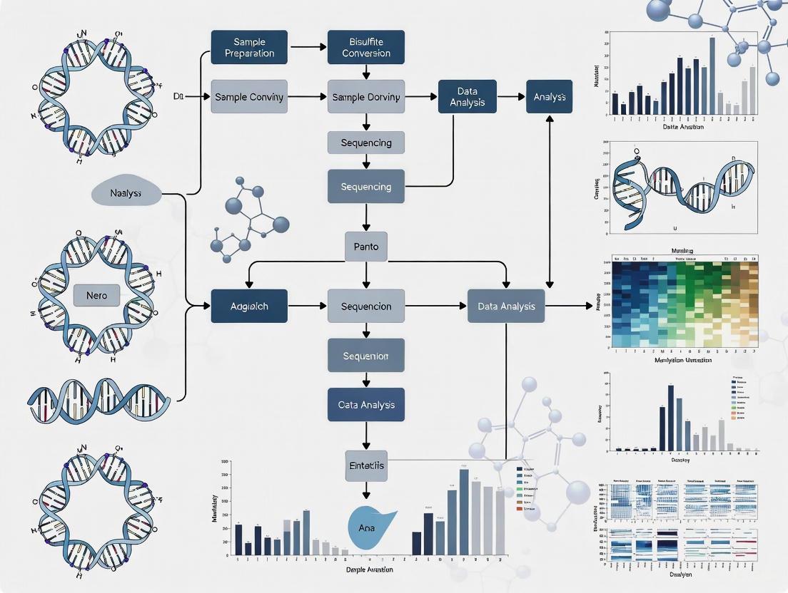
Diagram 1: DNA Methylation Analysis Workflow
Bioinformatics Databases and Tools
Several specialized databases provide comprehensive DNA methylation data across multiple species and experimental conditions. MethBank represents a knowledge base featuring manually curated bio-contexts related to differentially methylated genes (DMGs) and methylation tools [5]. This continuously updated database incorporates normal human cell type DNA methylation datasets and contains methylation profiles for Homo sapiens, Arabidopsis thaliana, and other model organisms [5]. The Cancer Genome Atlas (TCGA) represents another essential resource, providing DNA methylation profiles for over 50 cancer types acquired from the Infinium HumanMethylation450K BeadChip platform, with each profile including methylation levels (β-values) for approximately 480,000 CpG probes [3].
For gene ontology analysis and functional annotation, resources such as the Gene Ontology database, DisGeNet, and OMIM provide valuable information for linking methylation changes to biological processes and disease associations [3]. Analytical toolkits like the Chip Analysis Methylation Pipeline (ChAMP) facilitate quality control, normalization, and differential methylation analysis of array-based data, incorporating BMIQ normalization procedures to correct probe design biases [3]. Primer design for methylation-specific PCR benefits from specialized tools such as MethPrimer, while the BiSearch web server offers additional capabilities for designing bisulfite-conversion-based assays [2].
Essential Research Reagents and Kits
Table 3: Essential Research Reagents for DNA Methylation Analysis
| Reagent/Kits | Function | Application Notes |
|---|---|---|
| Proteinase K | Digests proteins and nucleases during DNA extraction | Critical for removing contaminating proteins; From Tritirachium album Limber [2] |
| Sodium Bisulfite | Converts unmethylated cytosine to uracil | Distinguishes methylated vs unmethylated cytosines; Critical parameter optimization required [2] |
| DNA Methyltransferases (DNMTs) | Catalyzes methylation using SAM donor | For controlled methylation experiments; DNMT1, DNMT3A, DNMT3B have distinct functions [1] |
| Anti-methylcytosine antibody | Immunoprecipitation of methylated DNA | Used in MeDIP protocols; specificity validation essential [2] |
| Bisulfite Conversion Kits | Standardized bisulfite treatment | Commercial kits available from multiple vendors; performance varies [2] |
| Methylation-Specific Restriction Enzymes | Differential digestion based on methylation status | Used in HELP, MSRE and other restriction-based approaches |
| PCR Reagents | Amplification of bisulfite-converted DNA | Polymerase selection critical for bias-free amplification of converted templates [2] |
| DNA Quality Assessment Kits | UV spectrophotometry, fluorometry | A260/A280 ratios ~1.8 indicate pure DNA; quality critical for bisulfite conversion [2] |
DNA methylation represents a crucial regulatory mechanism that shapes development and contributes to disease etiology through stable alterations of gene expression potential. The technical advances in methylation analysis, from global mass spectrometry-based approaches to targeted bisulfite sequencing, have enabled precise mapping of epigenetic patterns across biological contexts. The integration of methylation data with comorbidity patterns and functional annotations, as demonstrated in the identification of ALX3, NPTX2, and TRIM58 as multi-cancer biomarkers, highlights the translational potential of epigenetic research [3]. As databases such as MethBank continue to expand with additional samples and cancer-specific modules, the research community gains increasingly powerful resources for epigenetic discovery [5].
The future of DNA methylation research lies in integrating multi-omics approaches to understand the interplay between epigenetic marks, genetic variants, and transcriptomic outputs across developmental trajectories and disease processes. Methodological innovations in long-read sequencing, single-cell epigenomics, and computational prediction will further enhance our ability to decipher the epigenetic code. For researchers and drug development professionals, DNA methylation analysis offers not only biomarkers for early detection but also potential targets for epigenetic therapies that may eventually reverse pathological epigenetic states, representing a promising frontier for precision medicine across cancer and complex diseases.
CpG Islands, Promoters, and Genomic Distribution Patterns
CpG islands (CGIs) are fundamental cis-regulatory elements in vertebrate genomes, characterized as contiguous, non-methylated segments with a significantly higher than average level of CpG dinucleotides and GC content [6]. The standard formal definition specifies a region of at least 200 base pairs (bp), with a GC percentage greater than 50%, and an observed-to-expected CpG ratio exceeding 60% [7]. These sequences stand in stark contrast to the general genomic landscape, where CpG dinucleotides are markedly underrepresented due to the elevated mutation rate of methylated cytosines, which spontaneously deaminate to thymines [7]. In mammalian genomes, approximately 70–80% of CpG dinucleotides are methylated, but CpG islands remain refractory to this modification [6].
The genomic distribution of CpG islands is highly non-random and strongly associated with gene regulatory regions. It is estimated that the human genome contains approximately 28,890 CpG islands [7]. A substantial majority of mammalian gene promoters are encompassed within these regions, with about 70% of proximal promoters (those located near the transcription start site) containing a CpG island [7]. This association extends beyond housekeeping genes to include many tissue-specific genes, challenging the initial perception that CpG island promoters were exclusively a feature of constitutive gene expression [6]. Furthermore, over 60% of human genes and almost all house-keeping genes have their promoters embedded in CpG islands, highlighting their central role in transcriptional regulation [7].
Table 1: Quantitative Characteristics of CpG Islands in the Human Genome
| Feature | Metric | Genomic Background |
|---|---|---|
| Formal Definition | ≥200 bp length, >50% GC content, >0.6 Observed/Expected CpG ratio | - |
| Estimated Count | ~28,890 islands | - |
| CpG Dinucleotide Frequency | ~4-6% (High,接近预期值) | ~1% (Suppressed) |
| Typical Methylation Status | Mostly unmethylated in normal cells | ~70-80% of CpGs methylated |
| Promoter Association | ~70% of proximal promoters; >60% of all human genes | - |
The functional relationship between CpG islands and promoters is complex and multifaceted. Unlike classical TATA box-containing promoters, CpG island promoters generally utilize dispersed transcription start sites, suggesting that the CpG island may act as a generalized platform for transcriptional initiation [6]. The methylation status of CpG islands within promoters is a critical determinant of gene activity, with hypermethylation typically leading to stable gene silencing—a mechanism frequently exploited in cancer cells to turn off tumor suppressor genes [7].
Characteristics and Genomic Distribution Patterns
The distribution of CpG islands across the genome follows distinct patterns that provide insights into their functional significance. These regions typically span 300–3,000 base pairs in length and are disproportionately located at or near transcription start sites of genes [7]. A key characteristic of CpG islands is their resistance to the CG suppression observed in the rest of the genome, maintaining a CpG dinucleotide content of at least 60% of that which would be statistically expected (approximately 4–6%), compared to the ~1% frequency in the genomic background [7].
Advanced genomic analyses have revealed finer distribution patterns, leading to a revised understanding of CpG island characteristics. An extensive study of human chromosomes 21 and 22 suggested that DNA regions greater than 500 bp with a GC content exceeding 55% and an observed-to-expected CpG ratio of 65% are more likely to represent "true" CpG islands associated with the 5' regions of genes [7]. This refinement helps distinguish promoter-associated CpG islands from other GC-rich genomic sequences such as Alu repeats. Interestingly, most tissue-specific methylation differences occur not in the CpG islands themselves, but in flanking regions termed "CpG island shores," located up to 2 kilobases away from the traditional island boundaries [7].
The genomic distribution of CpG islands is not uniform across all gene types. Based on CpG density variation, CpG islands can be classified into high-CpG (HCGI), intermediate-CpG (ICGI), and low-CpG (LCGI) density categories [8]. This classification has functional implications, as HCGI-associated genes are most likely to be housekeeping genes, while different HCGI/TATA-box combinations show distinct Gene Ontology (GO) enrichment patterns [8]. The HCGI/TATA± and LCGI/TATA± combinations display different GO enrichment profiles, whereas the ICGI/TATA± combination is less characteristic based on GO enrichment analysis [8].
Table 2: Classification of CpG Islands by Density and Functional Associations
| Classification | CpG Density | Common Gene Associations | Functional Characteristics |
|---|---|---|---|
| High-CpG (HCGI) | Very High | Housekeeping (HK) genes | Strong, constitutive expression; distinct GO enrichment with TATA-box combinations |
| Intermediate-CpG (ICGI) | Moderate | Mixed | Less characteristic GO enrichment with TATA-box combinations |
| Low-CpG (LCGI) | Lower but still significant | Tissue-specific genes | Distinct GO enrichment patterns with TATA-box combinations |
The positioning of CpG islands within gene structures extends beyond proximal promoters. Distal promoter elements also frequently contain CpG islands, as exemplified by the DNA repair gene ERCC1, where a CpG island-containing element is located about 5,400 nucleotides upstream of the transcription start site [7]. Additionally, CpG islands occur frequently in promoters for functional noncoding RNAs, including microRNAs, expanding their regulatory influence beyond protein-coding genes [7].
Functional Mechanisms and Regulatory Roles
The functional role of CpG islands in gene regulation is mediated through sophisticated mechanisms involving specialized protein domains and chromatin modifications. Central to this process are ZF-CxxC domain-containing proteins, which specifically recognize and bind to non-methylated CpG dinucleotides [6]. This domain acts as a CpG island targeting module, with proteins like KDM2A and CFP1 binding to over 90% of CpG islands genome-wide [6]. The recognition is highly specific, as binding is blocked when the CpG sequence is methylated, providing a direct mechanism for interpreting the epigenetic information encoded in the methylation pattern.
These ZF-CxxC domain proteins are associated with histone-modifying activities that create a unique chromatin architecture characteristic of CpG islands. KDM2A catalyzes the removal of methylation from histone H3 lysine 36 (H3K36me2), leading to depletion of this mark at CpG islands [6]. Conversely, CFP1 associates with a histone H3 K4 methyltransferase complex (SET1 complex) to catalyze the addition of the tri-methyl modification (H3K4me3) [6]. The resulting chromatin environment—depleted of H3K36me2 and enriched with H3K4me3—effectively differentiates CpG island elements from surrounding chromatin and creates a configuration that is highly permissive for transcriptional initiation.
The following diagram illustrates how non-methylated CpG islands are recognized and translated into a unique chromatin architecture:
This chromatin architecture establishes what can be considered a default "permissive state" for transcription. In this state, RNA polymerase II is enriched at promoters, and short bidirectional transcripts are often produced, even from genes that show no detectable full-length mRNA [6]. This suggests that CpG island chromatin creates an accessible environment that favors binding of the basal transcription machinery. However, transition to a fully active state characterized by productive, directional transcription requires additional regulatory signals from sequence-specific DNA binding transcription factors [6]. The permissive state may function to highlight promoter regions within the vast expanse of the mammalian genome and focus nucleation of the transcriptional machinery at the 5' ends of genes.
The regulatory impact of CpG island methylation is profound, particularly in the context of disease states. Methylation of CpG islands in promoter regions leads to stable, long-term gene silencing [7]. In cancer, promoter CpG island hypermethylation represents a major mechanism for loss of tumor suppressor gene expression, occurring approximately 10 times more frequently than inactivating mutations [7]. For example, in colorectal cancer, hundreds to over a thousand genes may show aberrant promoter methylation compared to normal adjacent mucosa, illustrating the massive epigenetic disruption in malignancy [7].
Analytical Methods and Experimental Protocols
The analysis of CpG island methylation employs diverse methodological approaches, ranging from targeted assays to genome-wide profiling techniques. These methods can be broadly categorized into bisulfite sequencing-based methods, array-based platforms, and enrichment-based techniques, each with distinct applications, advantages, and limitations [9]. The selection of an appropriate method depends on the specific research question, required resolution, scale of analysis, and available resources.
Bisulfite Sequencing Methods
Bisulfite treatment represents the gold standard for DNA methylation analysis, converting unmethylated cytosines to uracils (read as thymines during sequencing) while leaving methylated cytosines unchanged [9]. Whole-genome bisulfite sequencing (WGBS) provides comprehensive, single-base resolution methylation maps across the entire genome [10]. However, WGBS is costly and computationally intensive for large genomes. Reduced Representation Bisulfite Sequencing (RRBS) offers a more cost-effective alternative by using restriction enzymes to enrich for CpG-rich regions prior to bisulfite treatment and sequencing [11]. RRBS covers approximately 1% of the total DNA methylome but captures about 30% of all CpG sites and 65% of promoter CpGs, making it highly efficient for analyzing CpG islands [11].
A typical RRBS protocol involves the following key steps [11]:
- DNA Digestion: 5 μg of genomic DNA is digested with the MspI restriction enzyme, which cuts at CCGG sites, effectively enriching for CpG-rich genomic regions.
- Library Preparation: The digested DNA fragments undergo end repair, A-tailing, and adapter ligation using standard library preparation kits.
- Bisulfite Conversion: The adapter-ligated library is treated with bisulfite using commercial kits (e.g., EpiTect Bisulfite Kit), converting unmethylated cytosines to uracils.
- PCR Amplification: The bisulfite-converted DNA is amplified using a low number of PCR cycles (e.g., 4 cycles) with polymerases suitable for bisulfite-converted templates.
- Sequencing and Analysis: The final library is sequenced on platforms such as Illumina HiSeq, and the resulting data are processed using specialized alignment and methylation calling software.
Array-Based Platforms and Emerging Methods
For human studies, Illumina's Infinium Methylation BeadChip arrays (450K and EPIC) provide a cost-effective solution for profiling methylation at predetermined CpG sites. The EPIC array covers over 850,000 CpG sites, including more than 90% of the CpG islands from the 450K array with enhanced coverage of regulatory regions [3]. The standard analytical workflow for array data includes quality control, normalization, and differential methylation analysis using packages such as ChAMP, minfi, or RnBeads [12] [3].
Emerging computational approaches demonstrate that methylation status can be predicted from ordinary whole-genome sequencing (WGS) data by analyzing read distribution biases. This method, implemented in tools like WGS2meth, leverages the finding that methylated CpG dinucleotides are approximately 30% more susceptible to fragmentation during library preparation than unmethylated CpGs [10]. The workflow involves:
- Read Coordinate Extraction: Mapping 5'-end coordinates of reads and identifying the dinucleotide at each breakpoint.
- Dinucleotide Frequency Calculation: Measuring the observed versus expected fragmentation rates for each dinucleotide type in CpG islands.
- Machine Learning Classification: Using trained models (e.g., XGBoost) to predict methylation status based on the fragmentation bias patterns [10].
Table 3: Key Experimental Methods for CpG Island Methylation Analysis
| Method | Resolution | Throughput | Key Applications | Common Tools/Pipelines |
|---|---|---|---|---|
| Whole-Genome Bisulfite Sequencing (WGBS) | Single-base | Genome-wide | Comprehensive methylation mapping; novel discovery | Bismark, BS-Seeker, MethylKit [12] |
| Reduced Representation Bisulfite Sequencing (RRBS) | Single-base (CpG-rich regions) | Targeted (~1% of genome) | Cost-effective profiling of promoter regions | Trim Galore, Bismark, MethylKit [11] |
| Illumina Infinium BeadChip | Single CpG site (850K sites) | High-throughput population studies | EWAS; biomarker validation | ChAMP, minfi, RnBeads [12] [3] |
| Computational Prediction (from WGS) | CpG island level | Genome-wide | Methylation status from existing WGS data | WGS2meth [10] |
The following workflow diagram outlines the key steps in a comprehensive CpG island methylation analysis, integrating both experimental and computational approaches:
Research Reagents and Computational Tools
Advancing research in CpG island biology requires a comprehensive toolkit of specialized reagents, assays, and computational resources. These tools enable researchers to profile methylation patterns, manipulate methylation states, and interpret the resulting data in a biological context. The field has developed robust pipelines and databases that facilitate standardized analysis and integration with other genomic data types.
Essential Research Reagents and Assays
Key experimental reagents form the foundation of CpG island methylation research. Bisulfite conversion kits, such as the EpiTect Bisulfite Kit, are essential for most sequencing-based methods, enabling the discrimination between methylated and unmethylated cytosines [11]. Restriction enzymes like MspI are critical for RRBS protocols, providing selective enrichment of CpG-rich regions while reducing sequencing costs and complexity [11]. For array-based approaches, the Infinium HumanMethylation450K and EPIC BeadChip arrays (Illumina) offer standardized platforms for profiling over 850,000 CpG sites across the genome, with extensive coverage of CpG islands and regulatory regions [3]. Antibodies specific to 5-methylcytosine enable enrichment-based methods such as MeDIP-seq (Methylated DNA Immunoprecipitation followed by sequencing), which is particularly useful when working with limited DNA input or when bisulfite conversion is undesirable [12].
Bioinformatics Tools and Databases
The analysis of DNA methylation data relies heavily on specialized bioinformatics tools and pipelines. For bisulfite sequencing data, packages like DMRichR and methylKit provide comprehensive solutions for identifying differentially methylated regions (DMRs) from whole-genome bisulfite sequencing data [12]. The Chip Analysis Methylation Pipeline (ChAMP) offers a complete analysis suite for Illumina Infinium array data, including quality control, normalization, and DMR detection [3]. Integration of methylation data with other omics datasets can be achieved using tools like FEM and ELMER, which correlate methylation patterns with gene expression to identify putative regulatory relationships [12].
For functional interpretation, enrichment analysis tools such as GOfuncR and GREAT provide biological context by associating methylation changes with Gene Ontology terms, pathways, and regulatory annotations [12]. The Genomic Regions Enrichment of Annotations Tool (GREAT) is particularly valuable for analyzing genomic coordinates from methylation studies, as it assigns biological meaning to non-coding regions by analyzing annotations of nearby genes [12].
Table 4: Essential Research Reagents and Computational Tools for CpG Island Analysis
| Category | Item | Specific Function | Example Tools/Products |
|---|---|---|---|
| Wet-Lab Reagents & Kits | Bisulfite Conversion Kit | Converts unmethylated C to U for sequence discrimination | EpiTect Bisulfite Kit [11] |
| Restriction Enzymes | Enriches CpG-rich regions for targeted approaches | MspI (for RRBS) [11] | |
| Methylation Arrays | Genome-wide profiling of predefined CpG sites | Infinium Methylation450K/EPIC BeadChip [3] | |
| Computational Tools & Pipelines | Bisulfite Seq Analysis | Alignment, methylation calling, DMR detection from WGBS/RRBS | DMRichR, methylKit, Bismark [12] |
| Methylation Array Analysis | Quality control, normalization, DMR detection from array data | ChAMP, minfi, RnBeads [12] [3] | |
| Integrative Analysis | Correlates DNA methylation with gene expression data | FEM, ELMER [12] | |
| Functional Interpretation | Enrichment Analysis | Provides biological context (GO, pathways) for gene lists | GOfuncR, GREAT, Enrichr [12] |
Applications in Disease Research and Biomarker Discovery
The analysis of CpG island methylation patterns has profound implications for understanding disease mechanisms and developing clinical biomarkers. In cancer research, DNA methylation profiling has revealed extensive reprogramming of the epigenome, with specific methylation signatures associated with diagnosis, prognosis, and treatment response. Cancers with low five-year survival rates—including pancreatic (10%), esophageal (20%), liver (20%), lung (21%), and brain (27%) cancers—have been particularly targeted for methylation biomarker discovery [3].
Integrated analysis of genome-wide DNA methylation profiles and comorbidity patterns across these five cancer types has identified key methylation biomarkers, including ALX3, HOXD8, IRX1, HOXA9, HRH1, PTPRN2, TRIM58, and NPTX2 [3]. The combination of ALX3, NPTX2, and TRIM58—selected from distinct functional groups through gene ontology clustering—achieved 93.3% accuracy in predicting cancer status across the ten most common cancers, demonstrating the power of multi-functional biomarker panels [3]. This approach combines primary biomarkers identified through differential methylation analysis (comparing tumor vs. normal tissue, with |Δβ-value| > 0.2 and p < 0.05) with secondary biomarkers derived from comorbidity-associated genes, creating robust diagnostic signatures.
In basic research, studies examining the relationship between CpG island methylation and gene expression across diverse adult tissues have provided insights into the fundamental principles of epigenetic regulation. Analysis of 20 pairs of DNA methylomes and transcriptomes from adult Ogye chicken tissues identified 3,133 CpG islands potentially affecting downstream genes [11]. Among these, 121 significant CpG island-gene pairs showed statistically correlated expression, with six genes (CLDN3, DECR2, EVA1B, NME4, NTSR1, and XPNPEP2) demonstrating highly significant changes associated with DNA methylation alterations [11]. These findings confirm that DNA methylation levels and gene expression are generally negatively correlated in normal adult tissues, with important tissue-specific variations.
The translational potential of CpG island methylation analysis extends to early cancer detection, monitoring disease progression, and predicting treatment response. The stability of DNA methylation marks in circulating cell-free DNA (cfDNA) makes them particularly attractive as non-invasive biomarkers [10]. Furthermore, the distinct fragmentation patterns of methylated DNA in cfDNA—where fragments more frequently begin with CpG dinucleotides when those CpGs are methylated—provide an additional layer of information that can be leveraged with machine learning approaches [10]. These advances highlight the growing importance of CpG island methylation analysis in both basic research and clinical applications, offering powerful tools for understanding gene regulation and developing epigenetic-based diagnostics and therapies.
DNA Methyltransferases (DNMTs) and Ten-Eleven Translocation (TET) Enzymes in Methylation Dynamics
DNA methylation and demethylation constitute a dynamic epigenetic layer crucial for regulating gene expression, genomic stability, and cellular differentiation. This balance is orchestrated by the opposing activities of DNA methyltransferases (DNMTs) and Ten-Eleven Translocation (TET) dioxygenases. DNMTs establish and maintain cytosine methylation, while TET enzymes catalyze its iterative oxidation, initiating active demethylation pathways. This technical guide delves into the structure, function, and regulatory mechanisms of these enzyme families, underscoring their roles in mammalian development and disease pathogenesis, particularly cancer. Furthermore, it provides a comprehensive overview of modern analytical methodologies and computational tools, framing this knowledge within the context of resources for DNA methylation research.
DNA methylation, the covalent addition of a methyl group to the fifth carbon of cytosine (5-methylcytosine, 5mC), primarily within cytosine-phosphate-guanine (CpG) dinucleotides, is a fundamental epigenetic mark in mammals [13] [14]. This modification is dynamically regulated and influences cellular processes including transcriptional repression, X-chromosome inactivation, genomic imprinting, and suppression of transposable elements [13] [15]. The mammalian "methylome" is not static; it is maintained by a delicate equilibrium between methylation, catalyzed by DNA methyltransferases (DNMTs), and active demethylation, facilitated by Ten-Eleven Translocation (TET) dioxygenases [14]. Disruption of this balance is a hallmark of various human diseases, most notably cancer, which often exhibits global hypomethylation coupled with site-specific hypermethylation of tumor suppressor gene promoters [13] [16]. Understanding the enzymes governing this cycle is therefore paramount for both basic research and therapeutic development.
DNA Methyltransferases (DNMTs): Architects of Methylation
Enzyme Types and Functional Roles
The DNMT family in mammals comprises three canonical, catalytically active enzymes: DNMT1, DNMT3A, and DNMT3B, alongside regulatory factors like DNMT3L [14] [17].
- DNMT1 is often termed the "maintenance" methyltransferase. It exhibits a strong preference for hemi-methylated DNA, making it essential for copying DNA methylation patterns from the parent strand to the newly synthesized daughter strand during DNA replication, thereby ensuring the fidelity of epigenetic inheritance across cell divisions [14] [15].
- DNMT3A and DNMT3B are responsible for de novo methylation, targeting unmethylated CpG sites to establish new methylation patterns during embryonic development and gametogenesis [13] [14]. Despite their primary role, they also contribute to methylation maintenance in specific contexts [14].
- DNMT3L is a catalytically inactive paralog that stimulates the activity of DNMT3A and DNMT3B by forming a complex with them, enhancing their binding affinity for DNA [14] [17].
Structure and Catalytic Mechanism
All catalytically active DNMTs share a common catalytic mechanism. They utilize S-adenosyl methionine (SAM) as the methyl group donor [14]. The enzyme catalyzes the transfer of a methyl group to the C5 position of cytosine, resulting in the formation of 5mC and S-adenosylhomocysteine (SAH). A key step in this reaction involves the enzyme flipping the target cytosine base out of the DNA helix and into its catalytic pocket, a process critical for the modification to occur [17].
Table 1: Core DNA Methyltransferases in Mammals
| Enzyme | Primary Role | Key Structural Features | Associated Human Diseases |
|---|---|---|---|
| DNMT1 | Maintenance methylation | N-terminal regulatory domain, C-terminal catalytic domain [14] | Hereditary sensory autonomic neuropathy, Autosomal dominant cerebellar ataxia, Breast Cancer [14] |
| DNMT3A | De novo methylation | PWWP domain, ADD domain, C-terminal catalytic domain [14] | Acute Myeloid Leukemia, Tatton–Brown–Rahman syndrome [14] |
| DNMT3B | De novo methylation | PWWP domain, ADD domain, C-terminal catalytic domain [14] | Immunodeficiency, Centromere instability, and Facial anomalies (ICF) syndrome [14] |
| DNMT3L | Regulation of DNMT3A/B | Lacks catalytic activity, forms heterotetramers with DNMT3A [14] [17] | - |
Ten-Eleven Translocation (TET) Enzymes: Catalysts of Demethylation
Enzyme Family and Oxidative Function
The TET family of proteins, comprising TET1, TET2, and TET3, are Fe(II)/α-ketoglutarate (α-KG)-dependent dioxygenases that initiate active DNA demethylation [18] [19]. They catalyze the sequential oxidation of 5mC to 5-hydroxymethylcytosine (5hmC), then to 5-formylcytosine (5fC), and finally to 5-carboxycytosine (5caC) [18] [16]. The 5hmC mark is not merely an intermediate; it also serves as a stable epigenetic mark with distinct regulatory functions, particularly abundant in embryonic stem cells and neuronal tissues [18] [16].
Pathways to DNA Demethylation
TET-mediated oxidation leads to demethylation via two principal pathways:
- Active Demethylation: The oxidized bases 5fC and 5caC are recognized and excised by thymine-DNA glycosylase (TDG). The resulting abasic site is then restored to an unmethylated cytosine through the Base Excision Repair (BER) pathway [18] [14].
- Passive Demethylation: The presence of 5hmC (and further oxidized derivatives) impairs the ability of DNMT1 to recognize and methylate the cytosine on the newly synthesized DNA strand during replication. This leads to a dilution of the methylation mark over subsequent cell divisions [18] [16].
Structural Insights and Regulatory Diversity
All TET proteins contain a conserved C-terminal catalytic domain that includes a double-stranded β-helix (DSBH) fold and a cysteine-rich domain, which together coordinate the Fe(II) and α-KG cofactors [18] [19]. A key structural difference lies in the N-terminus: TET1 and TET3 possess a CXXC zinc finger domain that binds unmethylated CpG-rich DNA, whereas TET2 lacks this domain. The CXXC domain of TET2 exists as a separate gene, IDAX (CXXC4), which regulates TET2 activity and recruitment [18] [19]. Furthermore, each TET gene expresses multiple isoforms through alternative splicing and promoter usage, adding a layer of regulatory complexity and tissue-specific function [19].
Table 2: The TET Enzyme Family
| Enzyme | Key Domains | Oxidation Products | Genomic Preference | Role in Disease |
|---|---|---|---|---|
| TET1 | CXXC, Catalytic Domain | 5hmC, 5fC, 5caC [18] | Promoters [18] | - |
| TET2 | Catalytic Domain | 5hmC, 5fC, 5caC [18] | Gene bodies, Enhancers [18] | Frequently mutated in myeloid malignancies [18] [16] |
| TET3 | CXXC, Catalytic Domain | 5hmC, 5fC, 5caC [18] | - | - |
The Methylation Cycle: An Integrated View
The following diagram illustrates the integrated cycle of DNA methylation and demethylation, highlighting the central roles of DNMT and TET enzymes.
Analytical Methods for DNA Methylation Research
Selecting the appropriate method for DNA methylation analysis is critical and depends on the research question, required resolution, and available resources [20] [21].
Core Techniques Based on Bisulfite Conversion
Treatment of DNA with sodium bisulfite deaminates unmethylated cytosines to uracils, which are then converted to thymidines during PCR amplification, while methylated cytosines remain unchanged. This sequence conversion forms the basis of many gold-standard methods [20].
- Whole-Genome Bisulfite Sequencing (WGBS): This is the most comprehensive method, providing single-base resolution methylation status for nearly all cytosines in the genome. It is ideal for discovery-based studies but is costly and computationally intensive [20].
- Reduced Representation Bisulfite Sequencing (RRBS): This method uses restriction enzymes to digest genomic DNA, enriching for CpG-dense regions (e.g., promoters and CpG islands) before bisulfite treatment and sequencing. It is more cost-effective than WGBS for focused analyses [20].
- Methylation-Specific PCR (MS-PCR): After bisulfite conversion, PCR primers are designed to specifically amplify either the methylated or unmethylated sequence. This is a simple, qualitative method for assessing the methylation status of a specific gene region [20].
- Bisulfite Pyrosequencing: This is a quantitative method that analyzes a short sequence of DNA following bisulfite PCR. It provides highly accurate, base-resolution quantification of methylation levels at consecutive CpG sites and is widely used for validation studies [21].
Global Methylation Analysis
For assessing genome-wide methylation levels, techniques like HPLC-UV (the gold standard) and the more sensitive LC-MS/MS can precisely quantify the total levels of 5mC and 5hmC in hydrolyzed DNA samples [21]. ELISA-based methods offer a rapid, albeit less accurate, alternative for global methylation screening [21].
Table 3: Key Methods for DNA Methylation Analysis
| Method | Resolution | Throughput | Key Advantage | Key Limitation |
|---|---|---|---|---|
| WGBS | Single-base | High | Comprehensive genome coverage [20] | High cost, complex data analysis [20] |
| RRBS | Single-base | High | Cost-effective for CpG-rich regions [20] | Limited to a fraction of the genome [20] |
| Bisulfite Pyrosequencing | Quantitative, single-base | Medium | High accuracy and quantitative precision [21] | Limited to short, predefined sequences [21] |
| MS-PCR | Locus-specific | Low | Simple, accessible, no sequencing required [20] | Qualitative or semi-quantitative only [20] |
| LC-MS/MS | Global (total 5mC/5hmC) | Low | High sensitivity and accuracy [21] | Requires specialized, expensive equipment [21] |
| ELISA | Global | High | Very fast and simple [21] | Low accuracy and high variability [21] |
Experimental Workflow: From Sample to Analysis
A typical workflow for a genome-wide DNA methylation study using bisulfite sequencing is outlined below and visualized in the accompanying diagram.
Detailed Protocol: Whole-Genome Bisulfite Sequencing (WGBS)
- DNA Extraction & Quality Control: High-quality, high-molecular-weight genomic DNA is isolated from cells or tissue. Quality and quantity are assessed using spectrophotometry or fluorometry [20].
- Library Preparation & Bisulfite Conversion: The DNA is fragmented (e.g., by sonication) and adapters are ligated to the ends. The library is then treated with sodium bisulfite, which deaminates unmethylated C to U, leaving 5mC and 5hmC unchanged [20]. The converted DNA is purified to remove reagents.
- PCR Amplification: The bisulfite-converted DNA library is amplified using a DNA polymerase that is efficient at amplifying uracil-containing templates [20].
- High-Throughput Sequencing: The final library is sequenced on a next-generation sequencing platform (e.g., Illumina), generating millions of short reads [20].
- Bioinformatic Analysis:
- Alignment: Reads are aligned to a reference genome using specialized bisulfite-aware aligners (e.g., Bismark [12]), which account for the C-to-T conversion.
- Methylation Calling: The methylation status of each cytosine is determined by comparing the sequencing read to the reference genome. A C in the read that aligns to a C in the reference indicates a methylated cytosine, while a T indicates an unmethylated one [20].
- Differential Methylation Analysis: Statistical packages (e.g.,
dmrseq[12]) are used to identify genomic regions that show significant differences in methylation levels between sample groups.
Research Reagent Solutions
Table 4: Essential Reagents and Kits for DNA Methylation Analysis
| Item | Function | Example Application |
|---|---|---|
| Sodium Bisulfite | Chemical conversion of unmethylated cytosine to uracil [20] | Fundamental reagent for bisulfite-based methods (WGBS, RRBS, MSP) [20] |
| Bisulfite Conversion Kits | Commercial kits for efficient and controlled bisulfite conversion and cleanup (e.g., from Zymo Research, Qiagen) [21] | Standardizing the critical conversion step for reproducibility |
| Anti-5mC / Anti-5hmC Antibodies | Immunoprecipitation or immuno-detection of modified cytosines [16] | MeDIP-seq, hMeDIP-seq, ELISA-based global quantification [21] |
| DNMT/TET Inhibitors | Small molecules to modulate enzyme activity (e.g., Decitabine, AZA) [15] | Functional studies to probe the role of DNA methylation in cellular processes |
| LC-MS/MS System | High-sensitivity quantification of nucleosides (dC, 5mC, 5hmC) [21] | Gold-standard measurement of global DNA methylation/hydroxymethylation levels |
Bioinformatics Tools and Databases
A robust bioinformatics pipeline is indispensable for interpreting methylation data. Key resources include:
- Alignment & QC:
Bismarkis a widely used tool for aligning bisulfite sequencing reads and performing methylation extraction [12].CpG_Meis a pipeline for WGBS alignment and quality control [12]. - Differential Methylation Analysis:
DMRichRis an R package for identifying and visualizing differentially methylated regions (DMRs) from whole-genome data [12].methylKitandRnBeadsare comprehensive R packages for analyzing bisulfite sequencing and microarray data, respectively [12]. - Annotation and Visualization:
GREATassigns biological meaning to non-coding genomic regions by analyzing nearby gene annotations [12].WandererandMethylation plotterare interactive tools for visualizing methylation data in genomic contexts [12]. - Public Data Repositories: Databases such as the Gene Expression Omnibus (GEO) and The Cancer Genome Atlas (TCGA) host vast amounts of publicly available DNA methylation data for comparative and meta-analysis.
DNMT and TET enzymes form a sophisticated, dynamic system for the precise control of the DNA methylome. Their integrated activity is fundamental to normal development and cellular function, and its dysregulation is a key driver of disease, particularly in cancer. Contemporary research, powered by high-throughput sequencing technologies and a growing suite of bioinformatic tools, continues to unravel the complexity of this regulatory network. This guide provides a foundational resource for researchers and drug development professionals, equipping them with the knowledge of core principles, experimental methodologies, and analytical resources needed to advance the field of epigenetic research and therapeutics.
Key Biological Processes Regulated by DNA Methylation
DNA methylation is a fundamental epigenetic mechanism involving the addition of a methyl group to a DNA molecule, typically at the fifth carbon of a cytosine residue to form 5-methylcytosine (5-mC) [22] [23]. This modification regulates gene expression without altering the underlying DNA sequence and is essential for normal development, cellular differentiation, and genomic stability [23]. In mammals, DNA methylation occurs primarily at CpG dinucleotides—regions where a cytosine is followed by a guanine [22] [24]. The distribution of CpG sites is not uniform across the genome; they are often clustered in regions known as CpG islands, which are frequently located in gene promoter regions [23] [24]. This technical guide details the key biological processes governed by DNA methylation, provides methodologies for its measurement, and outlines essential resources for research, serving as a foundational resource for scientists and drug development professionals engaged in epigenetics research.
Core Mechanisms and Functional Impact
DNA methylation dynamics are governed by dedicated enzymes and have a direct mechanistic impact on gene transcription.
The DNA Methylation and Demethylation Machinery
The establishment, maintenance, and removal of DNA methylation marks are catalyzed by specific enzymes [22] [25]:
- De Novo Methyltransferases (DNMT3A & DNMT3B): These enzymes establish new methylation patterns on previously unmodified DNA [22] [23].
- Maintenance Methyltransferase (DNMT1): This enzyme copies the methylation pattern from the parent DNA strand to the daughter strand during DNA replication, ensuring the methylation landscape is propagated through cell divisions [22] [23].
- Demethylation Pathways: DNA demethylation can occur passively when methylation patterns are not maintained during replication. Active demethylation involves the Ten-Eleven Translocation (TET) family of enzymes, which oxidize 5-mC to 5-hydroxymethylcytosine (5-hmC), initiating a pathway that leads to the restoration of an unmodified cytosine [22] [23].
How Methylation Regulates Transcription
The presence of 5-mC in gene promoter regions typically leads to transcriptional repression through two primary mechanisms [22] [24]:
- Direct Obstruction: The methyl group can physically impede the binding of transcription factors to their target DNA sequences.
- Recruitment of Repressor Complexes: Methylated DNA is recognized by methyl-CpG-binding domain proteins (MBDs), such as MeCP2. These proteins then recruit additional chromatin-modifying complexes, including histone deacetylases, which lead to a more condensed, transcriptionally inactive chromatin state known as heterochromatin [22] [24].
Table 1: Enzymatic Regulators of DNA Methylation
| Enzyme/Protein | Type | Primary Function | Key Characteristics |
|---|---|---|---|
| DNMT1 | DNA Methyltransferase | Maintenance Methylation | Copies methylation pattern during DNA replication; ensures heritability of epigenetic marks [22] [23]. |
| DNMT3A & DNMT3B | DNA Methyltransferase | De Novo Methylation | Establishes new methylation patterns during embryonic development and cellular differentiation [22] [23]. |
| TET Family | Dioxygenase | Active Demethylation | Initiates demethylation by oxidizing 5-mC to 5-hmC; crucial for dynamic methylation control in neurons and stem cells [22] [23]. |
| MeCP2 | Methyl-Binding Domain Protein | Transcription Repression | Binds methylated CpGs and recruits histone modifiers to silence gene expression [22] [24]. |
Key Biological Processes Regulated by DNA Methylation
DNA methylation is indispensable for several critical biological processes, with dysregulation being a hallmark of many diseases.
Cellular Differentiation and Embryonic Development
During mammalian embryonic development, the genome undergoes widespread epigenetic reprogramming [24]. Global DNA methylation patterns are largely erased and then re-established in a cell- and tissue-specific manner [23]. This process allows pluripotent stem cells to differentiate into the diverse array of cell types that constitute an organism. DNA methylation helps define and lock in cell identity by stably silencing genes that are unnecessary for a specific cell lineage [22] [23].
Genomic Imprinting
Genomic imprinting is an epigenetic phenomenon that results in the monoallelic expression of a subset of genes based on their parental origin. DNA methylation marks at imprinting control regions (ICRs) are established in the parental germlines and maintained throughout development to ensure that only one allele (either the maternal or paternal) is active [22] [24]. This process is critical for normal growth and development.
X-Chromosome Inactivation
In female mammals, one of the two X chromosomes is transcriptionally silenced to achieve dosage compensation with males who have only one X chromosome. DNA methylation plays a crucial role in the stable maintenance of this silencing. The Xist RNA coats the future inactive X chromosome, leading to the recruitment of DNMTs and subsequent methylation of the promoter regions of genes on that chromosome, ensuring their long-term repression [22] [24].
Silencing of Repetitive Elements
A substantial portion of the mammalian genome consists of transposable elements and retroviral sequences. The pervasive methylation of these intergenic regions is critical for maintaining genomic stability by preventing the transcription and mobilization of these potentially harmful elements, which could cause mutations and DNA damage [23] [24].
Gene Body Methylation
Contrary to promoter methylation, methylation within the transcribed region of actively expressed genes (gene body methylation) is often associated with efficient transcription [24]. While its function is less understood, it is thought to suppress spurious transcription from cryptic start sites within the gene or to play a role in alternative splicing [24].
The following diagram illustrates how DNA methylation regulates gene silencing, a process central to many of these biological functions:
DNA Methylation in Human Disease
Aberrant DNA methylation patterns are a universal feature of many human diseases, particularly in cancer [22] [26].
Table 2: DNA Methylation Alterations in Human Disease
| Disease Category | Methylation Status | Key Genes/Regions Affected | Functional Consequence |
|---|---|---|---|
| Cancer | Global Hypomethylation | Repetitive Elements, Intergenic Regions | Genomic instability, activation of oncogenes [22]. |
| Promoter Hypermethylation | Tumor Suppressor Genes (e.g., BRCA1, MLH1) | Silencing of genes that control cell cycle, DNA repair, and apoptosis [22] [26]. | |
| Neurological Disorders | Hypermethylation | Alzheimer's disease-related genes | Repression of genes critical for neuronal function [22]. |
| Hypomethylation | SNCA (Alpha-synuclein) | Overexpression of SNCA, linked to Parkinson's disease pathology [22]. | |
| MeCP2 Mutation | MECP2 Gene | Rett syndrome; loss of function in reading DNA methylation marks [22]. | |
| Autoimmune Disease (e.g., SLE) | Global Hypomethylation | T-cell DNA | Promotes autoreactivity and inflammation [22]. |
Measurement and Analysis of DNA Methylation
Accurate measurement of DNA methylation is crucial for research and clinical applications. The choice of method depends on the research question, required resolution, and available resources [25].
Genome-Wide Profiling Techniques
- Whole-Genome Bisulfite Sequencing (WGBS): This is the gold standard for DNA methylation analysis, providing single-base resolution methylation levels across the entire genome [25] [26]. It involves treating DNA with sodium bisulfite, which converts unmethylated cytosines to uracils (read as thymines in sequencing), while methylated cytosines remain unchanged [25].
- Reduced Representation Bisulfite Sequencing (RRBS): This method enriches for CpG-dense regions (like CpG islands) by using restriction enzymes, followed by bisulfite sequencing. It is a cost-effective alternative to WGBS that provides high-resolution data for key regulatory regions [25] [26].
- Methylation Arrays (e.g., Illumina Infinium): These arrays interrogate the methylation status of hundreds of thousands of pre-selected CpG sites. They are a high-throughput, cost-effective solution for large-scale epidemiological studies [27] [25].
A Protocol for Methylation Analysis in Cancer Biospecimens
The following workflow, adapted from a detailed protocol for processing human cancer biospecimens, outlines the key steps for generating high-quality genome-scale DNA methylation data using RRBS [26]:
- DNA Extraction: Extract genomic DNA from biospecimens (e.g., fresh-frozen tissue, FFPE tissue, cell lines). For FFPE tissue, this includes a deparaffinization step and proteinase K digestion to reverse cross-links and recover DNA [26].
- Restriction Digestion: Digest DNA with the MspI restriction enzyme. This enzyme cuts at CCGG sites, which are abundant in CpG-rich regions, thereby creating a reduced representation of the genome [26].
- Bisulfite Conversion: Treat the digested DNA with sodium bisulfite using a commercial kit (e.g., Zymo Research EZ DNA Methylation-Direct Kit). This critical step converts unmethylated cytosines to uracils [25] [26].
- Library Preparation and Sequencing: Prepare a sequencing library from the bisulfite-converted DNA. This involves end-repair, adapter ligation, and PCR amplification. The library is then sequenced on an Illumina platform [26].
- Bioinformatic Analysis:
- Quality Control & Trimming: Use tools like FastQC and Trim Galore to assess read quality and remove adapter sequences [26].
- Alignment: Align the processed reads to a bisulfite-converted reference genome using an aligner like Bismark [26].
- Methylation Calling: The same Bismark tool is used to extract the methylation status of each cytosine in the genome, comparing the sequenced bases to the reference to determine if they were methylated (C) or unmethylated (T post-conversion) [26].
- Differential Analysis: Use statistical packages in R or specialized tools like DMAP to identify regions of significant methylation difference between sample groups [26].
Successful DNA methylation research relies on a suite of specialized reagents, tools, and databases.
Table 3: Research Reagent Solutions for DNA Methylation Analysis
| Item | Function | Example Products/Resources |
|---|---|---|
| Bisulfite Conversion Kits | Chemically converts unmethylated C to U; critical for bisulfite-based methods. | Zymo Research EZ DNA Methylation-Direct Kit [26]. |
| Methylation-Sensitive Enzymes | Restriction enzymes used in methods like RRBS to enrich for CpG-rich regions. | MspI [26]. |
| DNA Methyltransferases (Recombinant) | For in vitro methylation assays and control experiments. | Commercial recombinant DNMT enzymes. |
| Methylated & Unmethylated Control DNA | Essential positive and negative controls for bisulfite conversion and assay validation. | Commercially available from various suppliers (e.g., Zymo Research). |
| Methylation Arrays | High-throughput profiling of pre-defined CpG sites. | Illumina Infinium MethylationEPIC array [27] [28]. |
| Bioinformatics Software | For alignment, methylation calling, and differential analysis of sequencing data. | Bismark, FastQC, Trim Galore, DMAP, SAMtools [26]. |
| Public Databases | For exploring methylation quantitative trait loci (meQTLs) and reference data. | EPIGEN MeQTL Database [28]; Gene Expression Omnibus (GEO). |
Connecting DNA Methylation to Transcriptional Regulation and Cellular Identity
DNA methylation, the addition of a methyl group to the fifth carbon of cytosine, constitutes a fundamental epigenetic mechanism that dynamically regulates gene expression without altering the underlying DNA sequence [29]. This modification plays a pivotal role in determining mammalian cell development, lineage identity, and transcriptional programs, serving as a crucial interface between the genome and environmental influences [30]. In the immune system, for example, fine-tuned DNA methylation patterns control myeloid and lymphoid cell differentiation and function, shaping both innate and adaptive immune responses [30]. Dysregulation of these epigenetic controls leads to significant human pathology, including blood malignancies, infections, and autoimmune diseases [30]. This technical guide examines the molecular mechanisms connecting DNA methylation to transcriptional regulation, surveys cutting-edge profiling technologies, and explores how these mechanisms establish and maintain cellular identity across biological contexts.
Molecular Mechanisms of Methylation-Mediated Regulation
Canonical Transcriptional Repression
The predominant model of DNA methylation-mediated gene silencing involves multiple interconnected mechanisms that render chromatin inaccessible to transcriptional machinery. Methylation primarily occurs at CpG dinucleotides, with CpG-rich regions known as CpG islands frequently found at gene promoters [31]. When these promoters become methylated, the modification can directly prevent transcription factor binding by steric hindrance or by recruiting transcriptional repressor complexes [31] [29]. A key mechanism involves methyl-CpG-binding domain proteins (MBPs) such as MeCP2, which deck on methylated DNA and recruit co-repressor complexes including histone methyltransferases and histone deacetylases [29]. This collaboration between DNA methylation and histone modifications establishes an inactive chromatin state characterized by condensed nucleosomes that physically obstruct transcription factor accessibility [29].
Context-Dependent Regulatory Effects
Recent epigenome engineering approaches have revealed that transcriptional responses to DNA methylation are more complex and context-specific than previously appreciated. While promoter hypermethylation is common in cancer and frequently associated with tumor-suppressor gene silencing, some regulatory networks can override DNA methylation, and promoter methylation can sometimes cause alternative promoter usage rather than complete silencing [31]. Surprisingly, induced DNA methylation can exist simultaneously on promoter nucleosomes possessing the active histone modification H3K4me3 or DNA bound by the initiated form of RNA polymerase II [31]. In some cases, increased gene expression has been observed following methylation induction, potentially driven by the eviction of methyl-sensitive transcriptional repressors [31].
Genomic Distribution and Functional Consequences
The genomic context of DNA methylation significantly determines its functional impact. While promoter methylation typically correlates with gene silencing, gene body methylation has been associated with active transcription and may affect alternative intragenic promoters, enhancers, non-coding RNA expression, transposable element mobility, and alternative splicing or polyadenylation [29]. Intergenic methylation changes can affect enhancers or insulators, leading to gene silencing or activation, respectively [29]. This complex relationship is bidirectional, as certain transcription factors can place epigenetic marks upon binding to DNA and subsequently alter DNA methylation patterns [29].
Table 1: Functional Consequences of DNA Methylation by Genomic Context
| Genomic Context | Methylation State | Typical Effect | Primary Mechanism |
|---|---|---|---|
| Promoter CpG Island | Hypermethylation | Gene silencing | Chromatin condensation, transcription factor exclusion |
| Gene Body | Methylation | Transcription elongation, splice regulation | Unknown, potentially affects histone modifications |
| Intergenic Enhancer | Hypermethylation | Enhancer silencing | Disrupted transcription factor binding |
| Imprinted DMRs | Allele-specific methylation | Parent-of-origin expression | Monoallelic transcriptional regulation |
| Repetitive Elements | Hypermethylation | Genomic stability | Transposon silencing |
Advanced Methodologies for Methylation Analysis
Sequencing-Based Profiling Technologies
Recent technological advances have revolutionized our capacity to profile DNA methylation patterns at various resolutions and scales. The table below compares key modern methodologies for methylation analysis.
Table 2: DNA Methylation Profiling Technologies and Applications
| Technology | Resolution | Throughput | Key Advantage | Primary Application |
|---|---|---|---|---|
| Whole Genome Bisulfite Sequencing (WGBS) | Single-base | Genome-wide | Gold standard for base resolution | Comprehensive methylome mapping [32] |
| meCUT&RUN | Regional | Targeted | 20-fold fewer reads than WGBS; low input (10,000 cells) [33] | Efficient methylome profiling [33] |
| Spatial-DMT | Near single-cell | Spatial multi-omics | Simultaneous methylome and transcriptome on tissue section [34] | Tissue context methylation-transcription relationships [34] |
| Nanopore T-LRS | Single-molecule | Targeted (0.1-10% of genome) | Phasing of methylation haplotypes; no bisulfite conversion [35] | Imprinting disorders, allele-specific methylation [35] |
| Mass Spectrometry | Global quantification | N/A | Absolute quantification independent of sequence [4] | Global methylation levels; non-model organisms [4] |
Spatial Multi-Omics Integration
A groundbreaking advancement in methylation analysis is the recent development of spatial joint profiling of DNA methylome and transcriptome (spatial-DMT), which enables simultaneous measurement of both epigenetic and transcriptional states in intact tissue sections at near single-cell resolution [34]. This technology combines microfluidic in situ barcoding, cytosine deamination conversion, and high-throughput sequencing to map methylation patterns within the native tissue architecture [34]. Applied to mouse embryogenesis and postnatal mouse brains, spatial-DMT has revealed intricate spatiotemporal regulatory mechanisms, showing how methylation and transcription patterns collectively define cell identity during mammalian development [34]. This approach addresses the critical limitation of previous methods that lost spatial context, enabling researchers to investigate interactive molecular hierarchies in development, physiology, and pathogenesis with spatial resolution.
Long-Read Sequencing for Methylation Haplotyping
Single-molecule long-read sequencing technologies from Oxford Nanopore and Pacific Biosciences now enable simultaneous measurement of epigenetic states alongside genomic variation, providing phasing information that reveals allele-specific methylation patterns [32] [35]. These technologies have proven particularly valuable for studying imprinted genomic regions, which contain differentially methylated regions (DMRs) with parent-of-origin-specific 5-methylcytosine patterns that control monoallelic expression [35]. Targeted long-read sequencing using adaptive sampling enriches specific genomic regions, providing cost-effective methylation haplotyping that can distinguish paternal and maternal alleles without statistical inference [35]. This approach has been successfully applied to imprinting disorders such as Beckwith-Wiedemann syndrome, Silver-Russell syndrome, and Temple syndrome, where it can identify multi-locus imprinting disturbances and structural variants affecting methylation patterns [35].
Experimental Approaches for Functional Validation
Targeted Methylation Manipulation
Epigenome engineering techniques enable direct testing of causal relationships between induced DNA methylation and transcriptional outcomes. Targeted methylation approaches include customized zinc finger domains, transcription activator-like effectors (TALEs), or nuclease-inactive Cas9 fused to the catalytic domain of DNA methyltransferases like DNMT3A or bacterial methyltransferases such as M.SssI [31]. These tools allow researchers to deposit methylation at specific endogenous loci and assess the resulting effects on transcription, chromatin accessibility, and histone modifications. Large-scale manipulation of promoter methylation has revealed that transcriptional responses are highly context-specific, with some promoters resistant to methylation-induced silencing while others show strong repression [31]. Importantly, induced methylation at regulatory elements can be rapidly erased after removing the methyltransferase fusion protein, through processes combining passive dilution and TET-mediated active demethylation [31].
Integrative Analysis Workflows
Comprehensive analysis of methylation-dependent regulation requires integrated experimental designs that couple methylation profiling with complementary assays. The diagram below illustrates a workflow for simultaneous spatial profiling of DNA methylation and gene expression.
Spatial co-profiling workflow for simultaneous DNA methylome and transcriptome analysis
The Scientist's Toolkit: Essential Research Reagents
Table 3: Key Research Reagents for DNA Methylation Studies
| Reagent/Kit | Primary Function | Application Context |
|---|---|---|
| CUTANA meCUT&RUN Kit | Methylated DNA enrichment using engineered MeCP2 protein | Targeted methylome profiling with reduced sequencing depth [33] |
| ZF-DNMT3A/DNMT3B fusions | Targeted methylation deposition | Epigenome engineering to test methylation effects [31] |
| EM-seq Conversion Kit | Enzymatic bisulfite alternative | Preservation of DNA integrity during methylation detection [34] |
| DNA Ligation Sequencing Kit (ONT) | Library prep for nanopore sequencing | Long-read methylation haplotyping [35] |
| Infinium MethylationEPIC Kit | Array-based methylation screening | Cost-effective population epigenomics [3] |
| 3-Oxooctanoic acid | 3-Oxooctanoic acid|CAS 13283-91-5|For Research | 3-Oxooctanoic acid is a medium-chain keto acid for research. This product is for laboratory research use only (RUO) and not for human use. |
| Beryllium selenide | Beryllium selenide, CAS:12232-25-6, MF:BeSe, MW:87.98 g/mol | Chemical Reagent |
DNA Methylation in Cellular Identity and Disease
Establishing and Maintaining Cellular Identity
DNA methylation serves as a fundamental mechanism for establishing and maintaining cellular identity throughout development and differentiation. During mammalian embryogenesis, carefully orchestrated methylation dynamics define lineage specification and tissue patterning, as revealed by spatial multi-omics approaches [34]. In the immune system, DNA methylation patterning precisely modulates cell type- and stimulus-specific transcriptional programs that preserve host defense and organ homeostasis [30]. The relationship between methylation and cellular identity is particularly evident in imprinted genes, which maintain parent-of-origin-specific expression through germline-derived methylation marks that are protected from genome-wide demethylation events after fertilization [35]. Maintenance of these identity-defining methylation patterns requires both faithful copying during DNA replication through DNMT1 and protection against unauthorized demethylation by factors like ZFP57 and ZNF445 [35].
Methylation Dysregulation in Human Disease
Aberrant DNA methylation patterns represent a hallmark of various human diseases, particularly cancer. Global hypomethylation coupled with locus-specific hypermethylation constitutes a common epigenomic landscape in transformed cells, driving oncogenesis through simultaneous activation of growth-promoting genes and silencing of tumor suppressors [29]. In lung cancer, for example, promoter hypermethylation of tumor suppressor genes contributes to disease progression and has been exploited for biomarker development [29]. Beyond cancer, methylation dysregulation is implicated in autoimmune diseases, inflammation, and imprinting disorders [34] [35]. The discovery of distinct epigenotypes linked to pathogenesis holds significant potential for validating therapeutic targets in disease prevention and management [30].
Diagnostic and Therapeutic Applications
The clinical relevance of DNA methylation patterns is increasingly recognized in diagnostic and therapeutic contexts. Methylation biomarkers show particular promise for early cancer detection, with panels including ALX3, HOXD8, IRX1, HOXA9, HRH1, PTPRN2, TRIM58, and NPTX2 demonstrating diagnostic potential for cancers with low survival rates such as pancreatic, esophageal, liver, lung, and brain cancers [3]. The combination of ALX3, NPTX2, and TRIM58 has achieved 93.3% accuracy in validating the ten most common cancers [3]. From a therapeutic perspective, the reversible nature of epigenetic modifications makes DNA methylation an attractive target for pharmacological intervention, with DNA methyltransferase inhibitors already employed in clinical practice for certain hematological malignancies [30].
DNA methylation represents a dynamic, context-dependent regulatory layer that profoundly influences transcriptional programs and cellular identity. While traditionally viewed primarily as a transcriptional repressive mark, recent evidence from advanced profiling technologies and epigenome engineering approaches reveals a more complex relationship between methylation and gene expression. The integration of spatial multi-omics, long-read sequencing, and targeted manipulation techniques continues to refine our understanding of how methylation patterns establish and maintain cell-type-specific identities throughout development and how their dysregulation contributes to human disease. As these methodologies become increasingly accessible and comprehensive, they promise to unlock new diagnostic and therapeutic opportunities based on the precise interpretation and modification of the epigenetic landscape.
DNA Methylation Technologies: Choosing the Right Method for Your Research Goals
Whole-Genome Bisulfite Sequencing (WGBS) represents the gold standard method for detecting DNA methylation at single-base resolution across entire genomes. This technical guide details the core principles, wet-lab methodologies, bioinformatic workflows, and applications of WGBS within the broader context of DNA methylation analysis resources. By providing comprehensive protocols, analytical pipelines, and practical considerations, this whitepaper serves as an essential resource for researchers, scientists, and drug development professionals seeking to implement this powerful epigenetic profiling technology in their investigative work.
DNA methylation, specifically the addition of a methyl group to the fifth carbon of cytosine (5-mC), constitutes a fundamental epigenetic mechanism regulating gene expression, genomic imprinting, X-chromosome inactivation, and suppression of transposable elements [36] [37]. As a stable epigenetic modification that can be inherited through DNA replication, it represents a crucial interface between genetic inheritance and environmental influence. The distribution of 5-methylcytosine across the genome—the methylome—provides critical insights into cellular identity, differentiation states, and disease processes. Among various technologies developed for methylome profiling, Whole-Genome Bisulfite Sequencing has emerged as the most comprehensive approach, enabling unbiased, genome-wide detection of methylation states at single-nucleotide resolution, making it indispensable for advanced epigenetic research and biomarker discovery [38] [39].
Principles of Bisulfite Sequencing
The fundamental principle underlying WGBS relies on the differential sensitivity of cytosines to bisulfite conversion based on their methylation status. When genomic DNA is treated with sodium bisulfite, unmethylated cytosines undergo chemical deamination to form uracils, which are subsequently amplified as thymines during PCR. In contrast, methylated cytosines (5-mC) are protected from this conversion and remain as cytosines through subsequent sequencing steps [38] [37]. This bisulfite-induced sequence difference allows for the discrimination between methylated and unmethylated cytosines when comparing treated sequences to a reference genome.
The standard WGBS workflow encompasses several critical stages: DNA extraction from biological samples, bisulfite conversion of the extracted DNA, library preparation specifically optimized for bisulfite-converted DNA, high-throughput sequencing, and comprehensive bioinformatic analysis [37]. The method provides single-base resolution, covers CpG and non-CpG methylation contexts (CHG and CHH, where H is A, C, or T), and achieves a high conversion rate typically exceeding 99% with appropriate quality control measures [37]. While originally developed for organisms with small genomes like Arabidopsis thaliana, WGBS has been successfully applied to diverse species including humans, mice, plants, and microorganisms, provided a reference genome is available [37] [39].
Methodological Approaches in Bisulfite Sequencing
Core WGBS Protocol and Variants
The standard WGBS protocol involves fragmenting genomic DNA, followed by bisulfite treatment and sequencing library construction. Traditional methods require microgram quantities of input DNA, but recent advancements have addressed limitations for low-input samples. Several methodological variants have been developed to address specific research needs, each with distinct advantages and applications, as summarized in Table 1.
Table 1: Comparison of Bisulfite Sequencing Methodologies
| Method | Principles | Advantages | Limitations |
|---|---|---|---|
| WGBS [38] [37] | Whole-genome sequencing of bisulfite-converted DNA | Single-base resolution; genome-wide coverage; detects CpG and non-CpG methylation | High sequencing cost; DNA degradation; reduced sequence complexity |
| RRBS/ scRRBS [38] | Restriction enzyme digestion followed by bisulfite sequencing | Cost-effective; focused on CpG-rich regions; suitable for limited samples | Biased coverage (~10-15% of CpGs); misses non-CpG regions |
| oxBS-Seq [38] | Oxidation before bisulfite treatment to distinguish 5mC from 5hmC | Differentiates 5mC vs. 5hmC; base resolution for both modifications | Complex protocol; same alignment challenges as standard BS-seq |
| T-WGBS [38] | Tagmentation using Tn5 transposase before bisulfite conversion | Minimal DNA input (~20 ng); fast protocol with fewer steps | Does not distinguish 5mC from 5hmC; alignment challenges |
| scBS-Seq [38] | Single-cell adaptation of BS-seq with random priming | Enables methylome profiling at single-cell resolution | Very low input DNA; technical amplification artifacts |
Wet-Lab Workflow and Reagent Solutions
The experimental workflow for WGBS requires specific reagents and kits optimized for bisulfite-converted DNA. Key stages include:
DNA Extraction: High-purity, high-molecular-weight DNA is essential, typically requiring ≥5μg mass, ≥50ng/μL concentration, and OD260/280 ratio of 1.8-2.0 [37]. Suitable for eukaryotic samples with reference genomes assembled to at least scaffold level.
Bisulfite Conversion: Commercial kits employ different denaturation and conversion conditions. The Zymo EZ DNA Methylation Lightning Kit uses heat-based (99°C) or alkaline-based (37°C) denaturation with 65°C conversion temperature for 90 minutes, while the Qiagen EpiTect Bisulfite Kit uses heat-based denaturation (99°C) with 55°C conversion for 10 hours [37].
Library Preparation: The EpiGnome Methyl-Seq Kit exemplifies an optimized approach where bisulfite-treated single-stranded DNA undergoes random priming with a polymerase capable of reading uracil nucleotides, synthesizing DNA containing specific sequence tags [37]. Illumina P7 and P5 adapters are subsequently added by PCR prior to sequencing.
Sequencing: Illumina platforms (e.g., HiSeq) employing sequencing-by-synthesis (SBS) technology with paired-end 150bp strategies are commonly used for 250-300bp insert bisulfite-treated DNA libraries [37]. Alternative platforms include PacBio SMRT, Nanopore, and Roche 454.
Table 2: Essential Research Reagent Solutions for WGBS
| Reagent/Kit | Function | Key Features |
|---|---|---|
| Sodium Bisulfite | Chemical conversion of unmethylated C to U | Selective deamination; methylation-dependent protection |
| Zymo EZ DNA Methylation Lightning Kit | Bisulfite conversion | 90-minute protocol; heat or alkaline denaturation |
| Qiagen EpiTect Bisulfite Kit | Bisulfite conversion | Standardized protocol; suitable for various inputs |
| EpiGnome Methyl-Seq Kit | Library preparation | Random priming with uracil-tolerant polymerase |
| Illumina P5/P7 Adapters | Library indexing and sequencing | Platform-specific compatibility; dual indexing available |
Bioinformatics Analysis Pipeline
The computational analysis of WGBS data presents unique challenges due to bisulfite-induced sequence simplification, with an estimated 10% of CpG sites difficult to align after conversion and potential DNA degradation up to 90% [38]. A standardized bioinformatics workflow addresses these challenges through sequential steps:
WGBS Bioinformatics Pipeline
Pre-processing and Quality Control
The initial analysis stage involves quality assessment and read cleaning. FastQC generates quality reports including base quality scores and sequence overrepresentation analysis [36] [40]. Adapter contamination and low-quality bases are removed using tools like TrimGalore! or Trimmomatic, with specific attention to removing adapter sequences such as the Illumina TruSeq adapter (AGATCGGAAGAGCACACGTCTGAACTCCAGTCAC) [36] [41]. Post-trimming quality verification ensures data integrity before alignment.
Read Alignment and Methylation Calling
Bisulfite-treated reads require specialized alignment strategies due to C→T conversions. The three primary mapping approaches include:
- Three-letter alignment (Bismark, BS-seeker2): Converts all Cs to Ts in both reads and reference genome before alignment using standard mappers like Bowtie2 [42].
- Wild-card alignment (BSMAP): Converts reference genome Cs to Y (representing C/T) and aligns reads allowing C/T to Y matching [42].
Bismark generally provides higher mapping accuracy, particularly for repeat regions, while BSMAP offers higher mapping rates but potentially lower accuracy, especially for hypomethylated regions with high T content [42]. After alignment, PCR duplicates are removed, and methylation information is extracted for each cytosine in CpG, CHG, and CHH contexts using methylation extractors like those in Bismark with parameters such as --no_overlap --comprehensive --CX --cytosine_report [41].
Differential Methylation Analysis
Differentially methylated regions (DMRs) are identified using specialized statistical packages that account for the binomial distribution of methylation data. The R package DSS is specifically recommended for detecting DMRs in WGBS experiments [36]. Alternative tools include methylKit and MethylSeekR [41]. The resulting DMRs are annotated based on genomic features (promoters, gene bodies, enhancers) using annotation packages like ChIPseeker, with functional enrichment analysis (GO, KEGG) revealing biological implications of methylation changes [40].
Integrated Analysis Pipelines
End-to-end pipelines like msPIPE streamline WGBS analysis by seamlessly connecting pre-processing, alignment, methylation calling, and downstream analyses [41]. msPIPE supports all reference genome assemblies available in the R package BSgenome, generates publication-quality figures, and utilizes Docker containers for reproducibility and ease of use. For improved accuracy, integrative approaches combining multiple mappers (Bismark, BSMAP, BS-seeker2) through scoring schemes have demonstrated enhanced detection accuracy and robustness against sequencing artifacts [42].
Applications in Biomedical Research
WGBS has enabled groundbreaking discoveries across diverse biological domains:
Stem Cell Research: WGBS revealed that approximately 25% of methylation in human embryonic stem cells occurs in non-CG contexts (CHG/CHH), contrasting with somatic cells where 99.98% of methylation is in CG context [39]. Non-CG methylation disappears upon differentiation but is restored in induced pluripotent stem cells, establishing it as a pluripotency marker.
Developmental Biology: Mouse oocytes show substantial non-CG methylation (up to two-thirds of total methylation), which accumulates during oocyte growth and depends on specific methyltransferases (Dnmt3s-Dnmt3L complex) [39].
Disease Diagnostics: WGBS detects abnormal methylation patterns of tumor suppressor genes in cancers including acute promyelocytic leukemia and gastric cancer, enabling early diagnosis [39].
Forensic Science: Application to dried blood spot samples improves DNA methylation analysis from forensic stains [39].
Limitations and Alternative Technologies
Despite its comprehensive coverage, WGBS faces several limitations. The bisulfite conversion process causes DNA degradation, reduces sequence complexity complicating alignment, and cannot distinguish between 5mC and 5-hydroxymethylation (5hmC) without additional modifications like oxBS-Seq [38]. Additionally, approximately 10% of CpG sites remain difficult to map after bisulfite conversion [38].
Emerging technologies address these limitations. The Illumina 5-base solution employs novel chemistry to directly convert only 5mC to T in a simple, single-step process that is non-damaging to DNA and retains library complexity, enabling simultaneous genetic variant and methylation detection [38]. Third-generation sequencing platforms like PacBio SMRT and Nanopore detect modified bases without bisulfite conversion through direct electronic or kinetic signatures.
Whole-Genome Bisulfite Sequencing remains the gold standard for base-resolution methylome profiling, providing unparalleled comprehensive coverage of methylation patterns across all genomic contexts. While methodological challenges persist regarding DNA degradation, alignment complexity, and 5mC/5hmC discrimination, ongoing computational and wet-lab innovations continue to enhance its capabilities. As a fundamental resource in the epigenetic analysis toolkit, WGBS empowers researchers to unravel the complex relationship between methylation patterns and phenotypic outcomes, accelerating discovery in basic biology, clinical diagnostics, and therapeutic development.
Reduced Representation Bisulfite Sequencing (RRBS) for Cost-Effective CpG Island Coverage
Reduced Representation Bisulfite Sequencing (RRBS) is a high-throughput technique designed for genome-wide DNA methylation profiling at single-nucleotide resolution. Developed by Meissner et al. in 2005, this method strategically reduces genomic complexity by enriching for CpG-rich regions before sequencing, thereby lowering costs significantly compared to whole-genome bisulfite sequencing (WGBS) while still capturing a substantial portion of functionally relevant genomic areas [43] [44]. The power of RRBS lies in its combination of restriction enzyme digestion and bisulfite conversion, enabling researchers to efficiently analyze methylation patterns across millions of CpG sites, making it particularly valuable for biomarker discovery, clinical applications, and large-scale epigenetic studies [43] [45].
The fundamental rationale behind RRBS stems from the observation that CpG dinucleotides are not randomly distributed throughout the genome but are concentrated in specific regions such as promoters and CpG islands. By targeting these areas, RRBS achieves approximately 80% coverage of CpG islands in promoters while only sequencing about 1-5% of the entire genome [43]. This targeted approach has established RRBS as a cornerstone technology in epigenetics research, balancing comprehensive methylation assessment with practical resource constraints.
RRBS Methodology and Protocol
Core Technical Principles
The RRBS protocol leverages several key biochemical principles to achieve its selective enrichment of CpG-rich regions. The process begins with digestion using methylation-insensitive restriction enzymes that recognize sequences commonly found in CpG-dense areas [44]. The most commonly used enzyme, MspI, cuts at CCGG sites regardless of the methylation status of the internal cytosine, ensuring both methylated and unmethylated regions are equally represented in the initial digestion [43]. Following digestion, the protocol incorporates bisulfite conversion, which deaminates unmethylated cytosines to uracils while leaving methylated cytosines unchanged, allowing for subsequent discrimination between methylation states during sequencing [44].
The strategic size selection of fragments (typically 40-220 bp) further enriches for CpG-rich genomic portions, as these regions tend to be more compact due to the nature of restriction sites surrounding CpG islands [43] [45]. Additional optimizations include using methylated adapters during library preparation, where all cytosines are replaced with 5'methyl-cytosines to prevent their deamination during bisulfite treatment, thereby preserving the adapter sequences for subsequent amplification and sequencing [44]. These methodological refinements collectively enable RRBS to provide quantitative DNA methylation measurements with high sensitivity and single-nucleotide resolution while requiring minimal input DNA (as low as 10-300 ng) [45].
Step-by-Step Experimental Protocol
The standard RRBS protocol extends over approximately three days and involves the following key steps [46] [45]:
Restriction Enzyme Digestion: Genomic DNA is digested with MspI (or other selected restriction enzymes) at 37°C for several hours, often overnight, to ensure complete digestion. This step generates fragments with CpG dinucleotides at their ends. For plant species, which exhibit different CpG distribution patterns, alternative enzymes such as SacI/MseI may be employed [43].
End Repair and A-tailing: The fragment ends are repaired and a single adenosine base is added to the 3' ends in the same reaction mixture using dCTP, dGTP, and an excess of dATP deoxyribonucleotides. This creates compatible ends for adapter ligation [46] [44].
Methylated Adapter Ligation: Methylated sequencing adapters are ligated to the A-tailed fragments. These adapters contain 5'methyl-cytosines instead of regular cytosines to protect them from bisulfite-mediated deamination [44].
Size Selection: The ligated fragments are size-selected (typically 40-220 bp) using gel electrophoresis or bead-based methods. This critical step enriches for CpG-rich fragments and determines the final genomic coverage [46] [44]. Size selection aims to capture the majority of promoters and other relevant genomic regions while excluding larger, CpG-poor fragments [43].
Bisulfite Conversion: The size-selected DNA undergoes bisulfite treatment, which deaminates unmethylated cytosines to uracils while methylated cytosines remain protected. This step requires careful optimization of temperature and denaturing conditions to ensure complete conversion while minimizing DNA degradation [44].
PCR Amplification: The bisulfite-converted DNA is amplified using PCR with primers complementary to the methylated adapters. A non-proofreading polymerase must be used, as proofreading enzymes would stall at uracil residues [44].
Sequencing and Analysis: The final library is sequenced using next-generation sequencing platforms, typically Illumina systems. The resulting reads are then aligned to a reference genome using specialized bioinformatics tools designed to handle bisulfite-converted sequences [43] [47].
Table 1: Key Reagents and Their Functions in RRBS Library Preparation
| Reagent Category | Specific Examples | Function in Protocol |
|---|---|---|
| Restriction Enzymes | MspI, TaqαI, ApeKI, DpnII | Digests genomic DNA at specific sites to enrich CpG-rich regions |
| DNA Modification Enzymes | End-repair enzymes, A-tailing enzyme, Ligase | Prepares fragment ends for adapter ligation |
| Adapter Sequences | Methylated Illumina adapters | Provides platform-specific sequences for amplification and sequencing |
| Bisulfite Conversion Reagents | Sodium bisulfite | Deaminates unmethylated cytosines to uracils for methylation detection |
| PCR Components | Non-proofreading polymerase, dNTPs | Amplifies bisulfite-converted libraries while handling uracil residues |
Diagram 1: RRBS Experimental Workflow. The process begins with genomic DNA extraction, followed by sequential enzymatic treatments, size selection, bisulfite conversion, and culminates in sequencing and data analysis.
Advantages and Limitations of RRBS
Key Advantages
RRBS offers several compelling advantages that explain its widespread adoption in epigenetic research:
Cost-Effectiveness: By sequencing only 1-5% of the genome while capturing the majority of promoters and CpG islands, RRBS provides substantial cost savings compared to WGBS, making large-scale methylation studies economically feasible [43] [44]. This efficiency enables researchers to process more samples within the same budget, enhancing statistical power in comparative studies.
Single-Nucleotide Resolution: Unlike array-based methods or enrichment-based techniques like MeDIP-Seq, RRBS provides base-pair resolution methylation data, allowing for precise mapping of methylation boundaries and identification of discrete differentially methylated cytosines [43] [44].
Low Input Requirements: The protocol requires only 10-300 ng of input DNA, facilitating studies with limited starting material, including clinical biopsies, rare cell populations, and precious historical samples [45]. Furthermore, RRBS has been successfully applied to formalin-fixed paraffin-embedded (FFPE) samples, expanding its utility in retrospective clinical studies [45].
Comprehensive CpG Coverage: In human genomes, RRBS captures approximately 12% of all genomic CpG sites, including about 84% of CpG islands in promoter regions [43]. This targeted coverage efficiently focuses sequencing power on functionally relevant genomic regions where methylation changes are most likely to impact gene regulation.
Multiplexing Capability: The reduced genome representation allows for higher levels of sample multiplexing in sequencing runs, further reducing per-sample costs and processing time [43].
Technical Limitations and Considerations
Despite its numerous advantages, researchers should consider several limitations when designing RRBS experiments:
Incomplete Genomic Coverage: Since RRBS relies on restriction enzymes for genome reduction, it inherently misses some CpG sites located outside the targeted fragments. MspI digestion alone does not cover all CG-rich regions, particularly those lacking CCGG recognition sites [44]. This limitation can be partially addressed using alternative enzyme combinations, but complete methylome coverage still requires WGBS [48].
PCR Artifacts: The requirement for a non-proofreading polymerase during PCR amplification increases the risk of sequencing errors, as these enzymes lack the ability to correct misincorporated bases [44]. Additionally, PCR amplification can introduce biases in representation, particularly for extreme GC-content fragments.
Bisulfite Conversion Challenges: Incomplete bisulfite conversion can lead to false positive methylation calls, while over-conversion or DNA degradation during the harsh bisulfite treatment conditions can reduce library complexity and quality [44]. The process typically results in significant DNA loss (up to 90% in the first hour), emphasizing the need for careful optimization [44].
Bioinformatics Complexity: Analysis of RRBS data requires specialized bioinformatics tools that account for the non-random base composition resulting from bisulfite conversion and the specific characteristics of restriction enzyme-based libraries [47] [44]. Standard alignment software cannot be used directly due to the C-to-T transitions in the sequencing reads.
Species-Specific Considerations: While highly effective for human, mouse, and rat genomes, RRBS protocols may require optimization for other species, particularly plants, which exhibit different CpG distribution patterns and methylation contexts (including CHG and CHH methylation) [43] [49].
Bioinformatics Analysis Pipeline
The unique nature of bisulfite-converted sequencing data necessitates specialized computational tools for accurate alignment and methylation calling. The bioinformatics pipeline for RRBS data encompasses several critical steps, each requiring careful consideration of analytical parameters.
Data Processing Workflow
The standard RRBS data analysis workflow consists of the following stages [47]:
Quality Control: Raw sequencing data is first assessed for quality using tools like FastQC to evaluate base quality distribution, GC content, sequence length distribution, and potential contamination. This step identifies issues requiring filtering or trimming before alignment.
Read Alignment: Filtered reads are aligned to a reference genome using specialized bisulfite-aware aligners such as Bismark, BS-Seeker2, or BSMAP. These tools account for the C-to-T conversions in the sequencing reads by performing in silico bisulfite conversion of the reference genome or reads before alignment [47]. The alignment must also consider the specific restriction enzyme sites used in library preparation.
Methylation Calling: Following alignment, methylated sites are identified by comparing the methylation status of each cytosine in the genomic context. Methylation levels are typically quantified as beta-values (β-values), calculated as the ratio of methylated reads to total reads covering each cytosine position (β = readsC / (readsC + readsT)) [47] [3].
Differential Methylation Analysis: Statistical comparisons between sample groups (e.g., treated vs. control, disease vs. normal) identify differentially methylated regions (DMRs). Commonly used tools for this analysis include limma, edgeR, and DMRcate, which employ various statistical models to account for biological variability and multiple testing [47].
Functional Annotation and Integration: DMRs are annotated with genomic features (genes, promoters, enhancers, etc.) and integrated with functional databases to identify biological pathways and processes potentially influenced by methylation changes. Tools like DAVID, Enrichr, and GSEA are frequently used for pathway enrichment analysis [47] [3].
Diagram 2: RRBS Bioinformatics Pipeline. The analysis workflow progresses from raw data quality assessment through alignment, methylation quantification, differential analysis, and culminates in functional interpretation.
Analysis Tools and Databases
Table 2: Comparison of Bioinformatics Tools for RRBS Data Analysis
| Tool Name | Mapping Strategy | Supported Aligners | Key Features | Considerations |
|---|---|---|---|---|
| Bismark | Three-letter | Bowtie, Bowtie2 | High accuracy, handles both directional and undirectional libraries | Slower processing for large datasets [47] |
| BS-Seeker2 | Three-letter | Bowtie, Bowtie2, SOAP | Includes adapter trimming, flexible aligner support | More complex installation and configuration [47] |
| BSMAP | Wildcard | SOAP | Simple usage, high accuracy for small-scale data | Less effective with complex methylation patterns [47] |
| bwa-meth | Three-letter | BWA | Fast alignment speed, specifically designed for methylation data | Limited handling of specialized cases [47] |
| GSNAP | Wildcard | GSNAP | Versatile for DNA and RNA data, high alignment accuracy | Slower with large RRBS datasets [47] |
Several specialized databases support RRBS data analysis and interpretation by providing reference methylation data and functional annotations:
- UCSC Genome Browser: Offers comprehensive methylation data across multiple species, tissues, and cell types, allowing comparison with published datasets [47].
- ENCODE (Encyclopedia of DNA Elements): Provides extensive reference epigenomes from various cell types, facilitating the biological interpretation of RRBS findings [47].
- The Cancer Genome Atlas (TCGA): Contains methylation data from numerous cancer types, enabling comparative analysis in cancer research contexts [3].
Applications in Biomedical Research
RRBS has been widely applied across diverse research domains, particularly where cost-effective, high-resolution methylation profiling provides critical insights into biological processes and disease mechanisms.
Cancer Research and Biomarker Discovery
In oncology, RRBS has proven invaluable for identifying cancer-specific methylation markers by comparing methylation patterns between cancerous and healthy tissues [43] [3]. This approach has revealed aberrant methylation patterns associated with tumor initiation, progression, and metastasis across various cancer types. For example, a recent study focusing on cancers with low five-year survival rates (pancreatic, esophageal, liver, lung, and brain cancers) identified ALX3, HOXD8, IRX1, HOXA9, HRH1, PTPRN2, TRIM58, and NPTX2 as important methylation biomarkers shared across these aggressive malignancies [3]. The cost-efficiency of RRBS makes it particularly suitable for screening large patient cohorts to validate potential epigenetic biomarkers for early cancer detection, prognosis, and treatment response prediction.
Developmental Biology and Neuroscience
RRBS enables the investigation of dynamic methylation changes during embryonic development, cellular differentiation, and tissue specification [43]. The technique has revealed stage-specific and cell-type-specific methylation patterns that contribute to fate determination and gene expression regulation during development [43]. In neuroscience, RRBS has been applied to study DNA methylation profiles in neurological disorders such as Alzheimer's disease and autism, uncovering epigenetic mechanisms underlying these conditions [43]. Furthermore, RRBS has illuminated experience-dependent methylation changes associated with learning, memory formation, and neural plasticity, providing mechanistic insights into how environmental influences shape brain function through epigenetic modifications.
Agricultural and Evolutionary Studies
In agricultural science, RRBS serves as a powerful tool for analyzing DNA methylation patterns related to economically important traits in crops and livestock [43]. By comparing methylation profiles across different varieties or breeds, researchers can identify epigenetic markers associated with yield, quality, disease resistance, and environmental stress tolerance, informing breeding strategies for crop improvement [43]. In evolutionary and ecological research, RRBS facilitates comparative studies of DNA methylation patterns across diverse species or populations, revealing epigenetic influences on evolutionary dynamics and adaptive responses [43]. This approach has also been used to investigate how environmental factors such as pollution, nutrition, and climate change influence epigenetic regulation across generations.
Comparative Analysis with Other Methylation Profiling Techniques
Understanding the position of RRBS within the landscape of DNA methylation analysis technologies requires comparison with alternative approaches, each with distinct strengths and limitations.
Table 3: Comparison of DNA Methylation Analysis Methods
| Method | Resolution | Genome Coverage | Relative Cost | Key Advantages | Key Limitations |
|---|---|---|---|---|---|
| RRBS | Single-nucleotide | ~12% of CpGs, ~84% of CpG islands | Medium | Cost-effective, high resolution for CpG-rich regions | Incomplete genomic coverage [43] [44] |
| Whole Genome Bisulfite Sequencing (WGBS) | Single-nucleotide | >95% of CpGs | High | Comprehensive coverage, gold standard | High cost, requires deep sequencing [44] |
| Methylation Arrays (Infinium) | Single-CpG site | ~480,000 predefined CpG sites | Low | High-throughput, cost-effective for large cohorts | Limited to predefined sites, no novel discovery [3] |
| MeDIP-Seq | ~150 bp | Enriched methylated regions | Low to Medium | Good for highly methylated regions | Lower resolution, antibody-dependent biases [44] |
| meCUT&RUN | ~200 bp | ~80% of methylation sites | Low | Very low input, minimal sequencing required | Lower resolution than bisulfite methods [33] |
| Long-Read Sequencing | Single-nucleotide | Potentially all CpGs | High | Detects methylation and variation simultaneously | Higher error rate, specialized equipment [32] |
Recent methodological advances continue to expand the utility of RRBS. Novel restriction enzyme combinations (such as MspI-DpnII or MspI-ApeKI) can increase CpG coverage to almost half of the human genome, addressing one of the primary limitations of traditional RRBS [48] [49]. Plant-specific RRBS protocols have been developed to accommodate the different CpG distribution and methylation contexts in plant genomes, where methylation occurs not only in CG but also in CHG and CHH contexts (where H is A, T, or C) [49]. These protocol variations demonstrate the adaptability of the RRBS framework to diverse research needs and biological systems.
Emerging Technologies and Future Perspectives
While RRBS remains a widely used method for cost-effective methylation profiling, the field of epigenomics continues to evolve with new technologies offering complementary capabilities.
Long-read sequencing platforms from Oxford Nanopore Technologies and Pacific Biosciences now enable simultaneous detection of DNA methylation and genetic variation without requiring bisulfite conversion [32]. These methods detect methylation through alterations in electrical signals (Nanopore) or polymerase kinetics (PacBio) during native DNA sequencing, potentially simplifying library preparation and providing phasing information to distinguish maternal and paternal methylation patterns [32]. However, these approaches currently involve higher equipment costs and different bioinformatics challenges compared to RRBS.
Enrichment-based methods like meCUT&RUN represent another emerging approach, using engineered methyl-binding proteins (such as MeCP2) to target methylated DNA for cleavage and sequencing [33]. This technique claims to capture approximately 80% of DNA methylation sites with dramatically reduced sequencing requirements (20-fold fewer reads than WGBS) and compatibility with low-input samples (as low as 10,000 cells) [33]. While not providing single-base resolution, such methods offer alternatives for specific applications where extreme cost-efficiency or sample preservation are priorities.
Despite these innovations, RRBS maintains its position as a balanced solution that combines single-nucleotide resolution, cost-effectiveness, and established protocols. Future applications will likely see RRBS integrated with other omics technologies (transcriptomics, proteomics) in multi-layered epigenetic studies, and its continued use in large-scale clinical and population studies where balancing comprehensive methylation assessment with practical constraints remains essential.
Reduced Representation Bisulfite Sequencing stands as a mature, robust, and cost-effective technology for DNA methylation analysis that strategically targets functionally relevant genomic regions. By enriching for CpG-rich areas through restriction enzyme digestion, RRBS provides single-nucleotide resolution methylation data for a substantial portion of the methylome while significantly reducing sequencing costs compared to whole-genome approaches. Despite limitations in complete genomic coverage, its efficiency, sensitivity, and compatibility with low-input samples have established RRBS as a cornerstone method in diverse research domains, from cancer biomarker discovery to developmental biology and agricultural science. As the field of epigenomics advances, RRBS continues to offer a practical balance between comprehensive methylation assessment and resource efficiency, maintaining its relevance amidst emerging technologies for researchers seeking to unravel the epigenetic mechanisms underlying biological processes and disease states.
DNA methylation (DNAm) is a fundamental epigenetic mechanism involving the addition of a methyl group to the fifth carbon of a cytosine residue, primarily at cytosine-guanine dinucleotides (CpGs). This modification regulates gene expression without altering the underlying DNA sequence and plays crucial roles in normal development, aging, and disease pathogenesis [50]. The emergence of high-throughput microarray technologies has revolutionized the scale at which researchers can investigate these epigenetic marks across large populations, enabling discoveries that were previously limited to candidate-gene approaches.
Illumina's Infinium Methylation BeadChip technology has powered over a decade of groundbreaking research in epigenome-wide association studies (EWAS) [51]. These arrays provide a cost-effective alternative to sequencing-based methods, making large-scale epidemiological studies financially feasible. The evolution from the HumanMethylation450 ("450K") to the EPIC versions ("850K" and "900K") has progressively expanded genomic coverage while maintaining the throughput necessary for population-scale investigations into the epigenetic basis of complex diseases, environmental exposures, and aging processes [50] [52].
Technology Evolution and Array Specifications
Array Generations and Technical Progression
The Infinium methylation array platform has undergone significant evolution since its inception, with each generation offering improved coverage and technical enhancements:
HumanMethylation450 BeadChip (450K): Launched in 2011, this arrayinterrogated approximately 485,577 CpG sites with single-base resolution, covering >99% of RefSeq genes and 96% of CpG islands. It utilized a dual-chemistry approach (Infinium I and II) to enhance data stability and reproducibility [50] [53]. This platform formed the basis for major projects like The Cancer Genome Atlas (TCGA), with methylation analysis of over 75,000 samples [50].
MethylationEPIC v1.0 BeadChip (EPICv1/850K): Released in 2016, this version extended coverage to over 850,000 CpG sites while maintaining the core content of the 450K array. The expanded content provided enhanced coverage of regulatory regions, including enhancers identified through ENCODE and FANTOM5 projects [50] [52].
MethylationEPIC v2.0 BeadChip (EPICv2/900K): Launched in 2023, this latest iteration targets approximately 930,000 unique methylation sites. It incorporates 186,000 new probes informed by cancer research, with enriched content targeting enhancers, CTCF-binding sites, CpG islands, and improved copy number variation detection for clinical applications [51] [52]. This version maintains high backwards compatibility with previous BeadChips and is compatible with FFPE samples, enabling studies on large biorepositories of tumors [51].
Table 1: Comparison of Illumina Infinium Methylation BeadChip Platforms
| Feature | 450K Array | EPIC v1.0 Array | EPIC v2.0 Array |
|---|---|---|---|
| Total CpG Probes | ~485,577 [52] | ~866,552 [52] | ~937,690 [52] |
| Coverage of RefSeq Genes | >99% [50] | >99% (maintains 450K content) [50] | Extensive coverage (enhanced functional content) [51] |
| Coverage of CpG Islands | 96% [50] | >95% (maintains 450K content) [50] | Dense coverage with additional probes [51] |
| Key Enhanced Content | Promoter regions, gene coding sequences | Enhancer regions from ENCODE/FANTOM5 | Expert-selected content from cancer research; enhancers, CTCF sites [51] |
| Sample Throughput | 12 samples/array [50] | 8 samples/array [51] | 8 samples/array [51] |
| Input DNA Requirement | 250 ng (typical) | 250 ng (typical) | 250 ng [51] |
| Specialized Sample Types | - | - | Blood, FFPE tissue [51] |
Comparison with Alternative Methylation Profiling Technologies
While methylation arrays dominate large-scale epidemiological research, other technologies offer complementary capabilities:
Sequencing-Based Methods: Whole-genome bisulfite sequencing (WGBS) provides the most comprehensive coverage (~28 million CpGs) at single-nucleotide resolution but remains cost-prohibitive for large cohorts [52]. Reduced-representation bisulfite sequencing (RRBS) offers a more targeted approach. A key limitation of these bisulfite-based methods is DNA degradation, which can be problematic for precious samples [4].
Enrichment-Based Long-Read Sequencing: Emerging techniques like CUTANA meCUT&RUN use an engineered MeCP2 protein to capture methylated DNA for sequencing, requiring 20-fold fewer sequencing reads than WGBS and working with low input (as low as 10,000 cells) [33]. Long-read sequencing technologies (Oxford Nanopore, PacBio) can simultaneously detect methylation and genetic variation on a single molecule, enabling haplotype-phased methylation analysis, which is valuable for studying imprinted genes [32] [35].
Global Methylation Analysis: Techniques like acid hydrolysis coupled with liquid chromatography-mass spectrometry (LC-MS) provide accurate quantification of the global proportion of methylated cytosine but lack locus-specific information. This method is ideal for rapid comparison of overall methylation states across many samples [4].
Table 2: Methylation Profiling Technologies Comparison
| Technology | Resolution & Coverage | Throughput & Cost | Primary Applications |
|---|---|---|---|
| Infinium BeadChips | Medium (~450K-930K CpGs); single-base resolution | Very High throughput; Low cost per sample | Large-scale EWAS, longitudinal studies, clinical biomarker discovery [51] [52] |
| Whole-Genome Bisulfite Sequencing | High (~28 million CpGs); single-base resolution | Low throughput; High cost per sample | Comprehensive discovery, building reference methylomes [52] |
| Long-Read Sequencing | High; detects methylation + sequence variation | Medium throughput; Medium cost | Haplotype-phased methylation, structural variant analysis, imprinted genes [32] [35] |
| Global MS Analysis | None (global percentage only) | High throughput; Low cost | Rapid screening, monitoring global methylation shifts [4] |
Experimental Design and Protocols
Core Workflow for BeadChip Analysis
The standard experimental workflow for Infinium methylation arrays involves sequential steps from sample preparation to data generation.
Sample Preparation and DNA Extraction: The process begins with sample collection from relevant tissues (e.g., whole blood, FFPE tissue). Genomic DNA is extracted using standard kits (e.g., Qiagen DNeasy, Gentra Puregene, or Monarch HMW DNA Extraction Kit), with input requirements of 250-750ng for the Infinium assay [35] [52]. DNA quality and quantity assessment is critical for success.
Bisulfite Conversion: Extracted DNA undergoes bisulfite treatment using kits such as the Zymo EZDNA Methylation Kit. This chemical conversion deaminates unmethylated cytosines to uracils, while methylated cytosines remain unchanged, creating sequence differences that correspond to methylation status [52].
Microarray Processing: Bisulfite-converted DNA is whole-genome amplified, enzymatically fragmented, and hybridized to the BeadChip. On the array, probes bind to the complementary sequence adjacent to target CpGs. A single-base extension step incorporates fluorescently labeled nucleotides, with the fluorescence signal indicating the methylation state at each CpG site [53].
Scanning and Data Generation: The BeadChip is scanned using Illumina's iSCAN system or compatible scanners, generating raw data files (IDAT format) containing fluorescence intensities for each probe [3] [53].
Quality Control and Preprocessing
Robust quality control and preprocessing are essential for generating reliable methylation data. The Bioconductor minfi package provides a comprehensive analytical toolkit for this purpose [53].
Quality Assessment: Initial quality control involves detecting low-quality samples with high probe detection p-values (>0.01) or low bead numbers (<3). Samples failing these metrics should be excluded. The
minfipackage includes tools for visualizing quality metrics and identifying outliers [53].Normalization: Technical variation between arrays is minimized using normalization procedures. Common methods include:
- Subset Quantile Normalization (SWAN): Implemented in
minfi, this method accounts for the different probe types (Infinium I and II) [53]. - Functional Normalization: A between-array method that regresses out technical variation explained by control probes, effectively removing batch effects and other non-biological variation [52].
- BMIQ Normalization: Corrects for the bias introduced by the different probe type designs, ensuring comparable β-value distributions [3].
- Subset Quantile Normalization (SWAN): Implemented in
Probe Filtering: Probes are filtered based on quality metrics, including:
Data Analysis and Computational Methods
Differential Methylation Analysis
The primary analysis goal in population studies is identifying differentially methylated positions (DMPs) or regions (DMRs) associated with exposures, traits, or disease states.
Methylation Quantification: Methylation level at each CpG is typically represented as a β-value, calculated as the ratio of the methylated signal intensity to the total signal intensity (methylated + unmethylated). β-values range from 0 (completely unmethylated) to 1 (completely methylated). For statistical testing, M-values (logit transformation of β-values) are often preferred as they demonstrate more homoscedasticity [53].
Statistical Modeling: Differential methylation is typically identified using linear regression models for continuous outcomes or logistic regression for binary outcomes, with methylation value as the dependent variable and adjusting for relevant covariates including age, sex, cell type composition, and technical batch effects. The Benjamini-Hochberg procedure is commonly applied to control the false discovery rate (FDR) from multiple testing [3].
Regional Analysis: DMRs (clusters of adjacent significant CpGs) can be identified using methods like
bumphunterin theminfipackage. DMRs are often more biologically meaningful than individual DMPs, as they may reflect broader epigenetic regulatory changes. Studies have shown that DMRs identified by bump hunting are more likely to be located near differentially expressed genes compared to single-CpG DMPs [53].
Analytical Pipeline and Advanced Applications
Functional Annotation and Interpretation: Significant CpGs and DMRs are annotated to genomic features (promoters, gene bodies, CpG islands, enhancers) and linked to nearby genes. Gene ontology (GO) and pathway analysis (KEGG) tools help identify biological processes and pathways enriched for differential methylation, providing functional context to the findings [3].
Cell Type Composition Deconvolution: In heterogeneous tissues like blood, methylation patterns reflect the proportional mix of cell types. Reference-based deconvolution algorithms estimate cell type proportions from bulk methylation data, allowing researchers to adjust for cellular heterogeneity, which is a major potential confounder in EWAS [52].
Epigenetic Clocks: Methylation arrays enable the calculation of epigenetic age estimators (e.g., Horvath's clock, Hannum's clock) from specific CpG panels. Discrepancies between epigenetic age and chronological age (age acceleration) associate with various health outcomes, mortality, and environmental exposures. Studies have shown that principal component versions of epigenetic clocks demonstrate greater stability across different array generations [52].
The Scientist's Toolkit
Table 3: Essential Research Reagents and Computational Tools
| Item/Resource | Function/Role | Specifications & Examples |
|---|---|---|
| Infinium MethylationEPIC v2.0 Kit | Genome-wide methylation profiling | ~930K CpG sites; 8 samples/array; 250 ng DNA input; compatible with blood and FFPE samples [51] |
| Bisulfite Conversion Kits | Chemical conversion of unmethylated cytosines | Zymo EZDNA Methylation Kit; required step before array processing [52] |
| minfi Bioconductor Package | Comprehensive analysis of Infinium arrays | Quality control, normalization, DMP/DMR identification, visualization [53] |
| ChAMP Toolkit | Preprocessing and analysis of methylation data | Data quality control, BMIQ normalization, differential methylation analysis [3] |
| Reference Methylomes | Cell type deconvolution, normalization | Reference datasets for specific tissues/cell types; e.g., Loyfer et al. 2023 methylation atlas [32] |
| Genomic Annotation Resources | Functional interpretation of results | GO, KEGG, ENCODE, FANTOM5 enhancer databases [3] [51] |
| Strontium phosphate | Strontium Phosphate|Sr3(PO4)2|Research Chemicals | |
| 4-Hydroxytryptophan | 4-Hydroxytryptophan, CAS:16533-77-0, MF:C11H12N2O3, MW:220.22 g/mol | Chemical Reagent |
Considerations for Population Studies
Longitudinal and Multi-Generational Array Data
Population studies often span years or decades, potentially incorporating data generated across different array versions. Careful harmonization is required when combining 450K, EPICv1, and EPICv2 data:
Probe Overlap: The EPICv2 array maintains high backwards compatibility with previous versions, but new content has been added and some poor-quality probes removed. A recent comparison study identified 369,639 CpGs present on all three major arrays (450K, EPICv1, EPICv2), providing a core set for longitudinal analysis [52].
Technical Variability: Empirical studies comparing 450K, EPICv1, and EPICv2 arrays within the same participants found that while sample-level correlations are high, notable discrepancies can occur at individual CpG sites. CpGs with lower replicability across arrays tend to have higher array-based variance, which should inform probe selection for replication studies [52].
Harmonization Strategies: Processing data from different arrays together using functional normalization can help minimize technical variability. Creating annotation resources that document probe quality and performance across arrays facilitates appropriate filtering and analysis decisions for combined datasets [52].
Application in Disease-Specific Research
Methylation arrays have proven particularly valuable for studying cancers with low survival rates, where early detection biomarkers are urgently needed. For example, a 2025 study identified methylation biomarkers (ALX3, HOXD8, IRX1, HOXA9, HRH1, PTPRN2, TRIM58, and NPTX2) across five low-survival-rate cancers (pancreatic, esophageal, liver, lung, and brain cancers) by integrating genome-wide DNA methylation profiles from TCGA with comorbidity patterns. The combination of ALX3, NPTX2, and TRIM58 achieved 93.3% accuracy in predicting multiple cancer types, demonstrating the potential of methylation arrays in developing multi-cancer early detection tests [3].
Furthermore, methylation arrays have been successfully applied to non-human primate models, with studies demonstrating that up to 165,847 probes on the 450K array and 261,545 probes on the EPIC array can be reliably used for DNA methylation analysis in rhesus macaques and African green monkeys, facilitating translational epigenetic research [54].
Infinium methylation arrays remain a cornerstone technology for high-throughput population epigenetics, offering an optimal balance of coverage, throughput, and cost-effectiveness. The continuous evolution of the platform, coupled with robust computational tools and analytical frameworks, has enabled unprecedented scale in EWAS. While emerging sequencing technologies offer superior resolution for specific applications, microarrays continue to be the preferred platform for large-scale epidemiological studies investigating the role of DNA methylation in human health, disease, and environmental adaptation. As the field progresses, careful attention to technical variability across array generations and integration with other omics data will maximize the scientific value of these powerful tools.
DNA methylation, the covalent addition of a methyl group to the fifth carbon of cytosine (5-methylcytosine, 5mC), constitutes a fundamental epigenetic mechanism regulating gene expression, genomic imprinting, and cellular differentiation [55] [56]. In mammalian genomes, this modification predominantly occurs at cytosine-guanine dinucleotides (CpG sites), which are often clustered in regions known as CpG islands (CGIs) [55] [57]. Aberrant DNA methylation patterns are hallmark features of various human diseases, particularly cancer, where global hypomethylation coexists with localized hypermethylation of tumor suppressor gene promoters [55] [58].
The analytical landscape for DNA methylation features two principal approaches: bisulfite conversion-based methods and enrichment-based techniques [55] [59]. While bisulfite sequencing (e.g., WGBS, RRBS) can provide single-base resolution, it requires harsh chemical treatment that degrades DNA, necessitates high sequencing coverage, and poses challenges in data interpretation due to reduced sequence complexity [60] [57]. Enrichment-based methods, including Methylated DNA Immunoprecipitation Sequencing (MeDIP-seq) and Methyl-CpG Binding Domain (MBD)-based techniques, offer a powerful, cost-effective alternative for genome-wide methylation profiling without bisulfite conversion [61] [55]. These methods selectively isolate methylated genomic regions prior to sequencing, making them particularly valuable for studies requiring analysis of limited DNA material or focusing on regional methylation patterns rather than single-base resolution [60] [62].
Core Principles and Molecular Mechanisms
MeDIP-seq: Antibody-Based Enrichment
MeDIP-seq utilizes immunoprecipitation with an antibody specific to 5-methylcytosine (5mC) to isolate methylated DNA fragments [60] [57]. The fundamental workflow begins with genomic DNA extraction and fragmentation, typically through sonication, producing fragments ranging from 300-1000 base pairs (with 400-600 bp being typical) [57]. The fragmented DNA is then denatured into single strands to expose methylated cytosines for antibody recognition [60] [57]. These single-stranded DNA fragments are incubated with monoclonal or polyclonal 5mC-specific antibodies, and antibody-bound methylated DNA is captured using magnetic beads conjugated to anti-mouse IgG [60]. After washing away unbound DNA, the enriched methylated fragments are released through proteinase K digestion, purified, and used for library preparation and high-throughput sequencing [57] [62].
The molecular mechanism of MeDIP relies on the specific affinity of antibodies for the 5mC epitope. This antibody-based approach offers the advantage of recognizing methylated cytosines irrespective of their sequence context, enabling detection of both CpG and non-CpG methylation [62]. However, a critical consideration is that antibody binding efficiency depends on methylcytosine density, with higher affinity for regions containing multiple adjacent methylated CpGs [60] [57]. This density dependence introduces a bias toward hypermethylated regions while potentially underrepresenting sparsely methylated areas [60].
MBD-Based Techniques: Protein Domain-Mediated Capture
MBD-based techniques exploit the natural function of methyl-CpG binding domain proteins, which specifically recognize and bind methylated CpG dinucleotides in double-stranded DNA [55] [63]. The MBD family comprises 11 proteins containing a highly conserved ~70 amino acid domain that binds asymmetrically to DNA around symmetrically methylated CpGs [55]. Structural analyses reveal that this domain is rich in positively charged amino acids, with two arginine residues forming critical hydrogen bonds with the guanine base while packing against the methyl group of 5mC—a configuration known as the 5mC-Arg-G triad [55].
Various MBD-based methods have been developed, including MBD-seq (MBD-isolated Genome Sequencing), MIRA (Methylated-CpG Island Recovery Assay), and MethylCap-seq [55] [62]. These protocols typically involve incubating fragmented double-stranded genomic DNA with MBD proteins (often MBD1, MBD2, or MBD4) immobilized on solid supports such as paramagnetic beads [55] [63]. After binding, methylated DNA fragments are washed under specific salt conditions to remove non-specifically bound DNA, then eluted and prepared for sequencing [55]. The binding specificity of different MBD domains varies, with MBD1 exhibiting high affinity for densely methylated DNA, while MBD3 shows reduced methylation selectivity due to a tyrosine-to-phenylalanine mutation [55].
Unlike MeDIP, MBD-based approaches can capture methylated DNA without denaturation, preserving native DNA structure [63]. However, similar to MeDIP, they display preferential binding to regions with higher CpG density, though this bias can be modulated by adjusting binding and washing stringencies [55] [64].
Comparative Technical Specifications
Table 1: Technical comparison of major enrichment-based methylation profiling methods
| Feature | MeDIP-seq | MBD-seq | MRE-seq | Integrated Approaches |
|---|---|---|---|---|
| Enrichment Principle | Immunoprecipitation with anti-5mC antibody [60] [57] | Capture by methyl-CpG binding domain proteins [55] [63] | Digestion with methylation-sensitive restriction enzymes [56] [59] | Combines MeDIP-seq and MRE-seq [59] |
| DNA State | Single-stranded (denatured) [60] | Double-stranded (native) [63] | Double-stranded (native) [59] | Both single and double-stranded |
| CpG Density Bias | Strong bias toward high density regions [60] [62] | Moderate to strong bias, depending on protein and stringency [55] | Bias toward recognition sites [59] | Compensates for individual method biases |
| Typical Resolution | ~100-150 bp [62] | ~50-100 bp [55] | Restriction site resolution [59] | Single CpG resolution possible with computational integration [59] |
| Input DNA Requirement | 100 ng (standard), as low as 1-10 ng for cfMeDIP-seq [60] [57] | Varies by protocol, typically >100 ng [55] | Varies by protocol [59] | Varies by component methods |
| Primary Applications | Genome-wide methylation patterns, low-input samples [62] | Genome-wide methylation, focused on CpG-rich regions [55] | Identification of unmethylated regions [59] | Comprehensive methylome mapping [59] |
Table 2: Performance characteristics in research contexts
| Characteristic | MeDIP-seq | MBD-seq | Bisulfite Sequencing (Reference) |
|---|---|---|---|
| Genome Coverage | ~90% of CpG sites in promoters, gene bodies, islands [57] | High coverage of CpG-rich regions [55] | Nearly complete [62] |
| Concordance with Bisulfite Methods | 82% for CpGs, 99% for non-CpG cytosines [61] | 99% with binary methylation calls [61] | Gold standard |
| Non-CpG Methylation Detection | Yes [62] | Limited, depends on MBD protein [55] | Yes [62] |
| Cost Relative to WGBS | Substantially lower [59] | Substantially lower [55] | Reference (highest) |
| Best Suited For | Differential methylated regions, low DNA input studies [62] | Hypermethylated regions, quantitative applications [55] [64] | Single-base resolution, complete methylome |
Experimental Workflows and Protocols
MeDIP-seq Protocol
The standard MeDIP-seq protocol involves sequential steps that can be completed over 2-3 days [60] [57]:
DNA Fragmentation: Purified genomic DNA is sheared by sonication or enzymatic digestion to generate random fragments of 300-1000 bp. Sonication is preferred as it avoids sequence-specific biases introduced by restriction enzymes [57].
DNA Denaturation: The fragmented DNA is heat-denatured (typically at 95°C) to produce single-stranded DNA, which is immediately placed on ice to prevent reannealing. Denaturation is crucial for antibody access to methylated cytosines [60] [57].
Immunoprecipitation: Denatured DNA is incubated with 5mC-specific antibody (monoclonal or polyclonal) in appropriate buffer conditions. The antibody-DNA complexes are captured using magnetic beads conjugated with species-specific secondary antibodies [60] [57].
Washing and Elution: Beads are washed with buffers of varying stringency to remove non-specifically bound DNA. Methylated DNA is then eluted either by proteinase K digestion to degrade the antibodies or under denaturing conditions [57].
Library Preparation and Sequencing: Eluted DNA is purified and processed for next-generation sequencing using standard library preparation protocols, followed by sequencing on platforms such as Illumina [57] [62].
A critical adaptation is cell-free MeDIP-seq (cfMeDIP-seq), optimized for low-input DNA (1-10 ng) from liquid biopsies [57]. This protocol incorporates "filler DNA" (e.g., bacteriophage lambda DNA) as a carrier to improve immunoprecipitation efficiency and includes synthetic spike-in controls for quantification normalization [57].
MBD-Based Capture Protocol
Standard MBD-based enrichment follows this general workflow [55] [63]:
DNA Fragmentation: Genomic DNA is fragmented similarly to MeDIP-seq, typically to 100-500 bp fragments.
MBD Capture: Fragmented DNA is incubated with MBD proteins (commonly MBD2 or MBD1) immobilized on magnetic beads or chromatography columns. Binding occurs in specific salt conditions that promote specific interactions.
Stringency Washes: Bound DNA is washed with buffers containing increasing salt concentrations to elute fragments based on methylation density. This step can be optimized to isolate particular methylation density fractions [55].
Elution: Methylated DNA is eluted using high-salt buffers or proteinase K treatment. For quantitative applications, stepwise elution with increasing salt concentrations can separate differentially methylated fractions [55] [64].
Library Preparation and Sequencing: Eluted DNA is purified, converted to sequencing libraries, and sequenced. For locus-specific analysis, eluted DNA can be analyzed by qPCR rather than sequencing [55].
MBD protocols can be adapted for various applications, from genome-wide sequencing (MBD-seq) to quantitative PCR (MBD-qPCR) for specific loci [55]. Recent adaptations include colorimetric and electrochemical detection platforms that use MBD-HRP (horseradish peroxidase) conjugates for rapid methylation assessment without sequencing [64].
Computational Analysis Pipelines
The analysis of enrichment-based methylation data requires specialized bioinformatics approaches to address the unique characteristics of these datasets. The fundamental challenge lies in distinguishing true methylation variation from confounding effects of CpG density and sequencing biases [56].
Primary Analysis Steps
A standard MeDIP-seq or MBD-seq computational pipeline includes these key stages [56] [62]:
Quality Control and Preprocessing: Raw sequencing reads are assessed for quality using tools like FastQC, followed by adapter trimming and quality filtering.
Alignment to Reference Genome: Processed reads are aligned to a reference genome using aligners such as Bowtie2, BWA, or HISAT2, with considerations for potentially ambiguous mappings.
Methylation Enrichment Quantification: The core analytical step involves quantifying enrichment across genomic regions. This includes calculating coverage depth in windows or predefined regions and normalizing for technical variations.
Differential Methylation Analysis: Statistical comparisons between sample groups identify differentially methylated regions (DMRs), accounting for multiple testing.
Biological Interpretation: DMRs are annotated to genomic features (promoters, genes, CpG islands) and integrated with complementary data (e.g., transcriptomics) for functional insights.
Specialized Analytical Tools
Several specialized software packages have been developed to address the specific analytical challenges of enrichment-based methylation data [56] [62]:
Batman (Bayesian Tool for Methylation Analysis): One of the first tools developed for MeDIP-seq data, it implements a Bayesian deconvolution strategy to estimate absolute methylation levels based on methylated CpG density [56] [57]. Though powerful, it is computationally intensive.
MEDIPS: An R/Bioconductor package that provides a comprehensive framework for MeDIP-seq data analysis, including quality control, normalization, and DMR detection [56]. It significantly improves computational efficiency compared to Batman while maintaining analytical rigor.
MeDUSA (Methylated DNA Utility for Sequence Analysis): A pipeline that performs complete analysis from sequence alignment to DMR identification and annotation [56] [62].
M&M and methylCRF: Advanced frameworks that integrate MeDIP-seq with MRE-seq data. M&M detects differentially methylated regions between samples, while methylCRF uses conditional random fields to predict methylation levels at single-CpG resolution, achieving coverage comparable to WGBS at a fraction of the cost [59].
Table 3: Key research reagents and resources for enrichment-based methylation studies
| Category | Specific Examples | Function and Application Notes |
|---|---|---|
| Capture Reagents | Anti-5-methylcytosine antibodies (monoclonal/polyclonal) [60] [57] | Immunoprecipitation of methylated DNA in MeDIP; specificity and lot consistency should be validated |
| Recombinant MBD proteins (MBD1, MBD2, MBD4) [55] [63] | Methylated DNA capture in MBD-based methods; different MBDs vary in binding affinity and specificity | |
| Solid Supports | Magnetic beads (protein A/G, streptavidin) [60] [57] | Immobilization of antibodies or MBD fusion proteins for target capture |
| Library Preparation | DNA fragmentation reagents (sonication equipment, enzymes) [57] | Generation of appropriately sized DNA fragments for enrichment and sequencing |
| Library preparation kits (Illumina, NEB) [62] | Preparation of sequencing libraries following methylated DNA enrichment | |
| Controls | Methylated lambda phage DNA [63] [64] | Positive control for methylation capture efficiency |
| Synthetic spike-in controls [57] | Normalization for technical variation, particularly in low-input protocols | |
| Unmethylated genomic DNA [64] | Negative control for specificity assessment | |
| Bioinformatics Tools | MEDIPS, Batman, MeDUSA [56] [62] | Specialized software for processing and analyzing enrichment-based methylation data |
| Alignment software (Bowtie2, BWA) [57] [62] | Mapping sequenced reads to reference genomes | |
| Genome browsers (WashU EpiGenome Browser, Ensembl) [57] [59] | Visualization and exploration of methylation data in genomic context |
Applications in Biomedical Research
Enrichment-based methylation profiling methods have enabled diverse applications across biomedical research:
Cancer Methylome Analysis: MeDIP-seq and MBD-seq have been extensively used to characterize aberrant methylation patterns in various cancers [57] [62]. These techniques can identify both hypermethylated tumor suppressor genes and global hypomethylation events, providing insights into cancer pathogenesis and potential biomarkers [58]. The first cancer methylome was characterized using MeDIP-seq, demonstrating its utility in oncology research [56].
Developmental Biology: These methods have been crucial for mapping dynamic methylation changes during embryonic development, cellular differentiation, and tissue specification [63] [62]. The low DNA input requirements make them suitable for studying precious samples like oocytes and early embryos [62].
Liquid Biopsy Applications: The adaptation of MeDIP-seq for cell-free DNA (cfMeDIP-seq) has enabled non-invasive cancer detection and monitoring through liquid biopsies [57] [58]. This approach leverages the stability of methylation patterns and tissue-specific methylation signatures to detect tumor-derived DNA in circulation [57].
Neurological and Complex Diseases: MeDIP-seq and MBD-seq have contributed to understanding the epigenetic basis of neurological disorders such as Rett syndrome, which involves mutations in the MECP2 gene [55]. These methods help identify methylation alterations associated with complex disease pathogenesis.
Environmental Epigenetics: Enrichment-based methods facilitate studies investigating how environmental exposures (diet, toxins, stress) influence the epigenome by comparing methylation patterns between exposed and control groups [62].
Method Selection Guidelines
Choosing between MeDIP-seq and MBD-based approaches depends on specific research objectives and practical considerations:
Select MeDIP-seq when:
- Studying non-CpG methylation, as antibodies can recognize methylated cytosines irrespective of sequence context [62]
- Working with limited DNA input (<100 ng), particularly for cfMeDIP-seq applications [60] [57]
- Targeting genome-wide coverage without restriction to specific genomic features [62]
- Budget constraints exist, as MeDIP-seq generally offers lower costs than comprehensive bisulfite sequencing [59]
Choose MBD-based methods when:
- Preferring to work with native double-stranded DNA without denaturation [63]
- Focusing on CpG-rich regions such as promoters and CpG islands [55]
- Quantitative assessment of methylation density is needed through fractional elution [55] [64]
- Implementing non-sequencing applications like colorimetric detection or electrochemical sensing [64]
Consider integrated approaches when comprehensive methylome characterization is needed. Combining MeDIP-seq with MRE-seq provides complementary information that compensates for individual method biases, with computational integration enabling single-CpG resolution approaching that of WGBS at substantially reduced cost [59].
Emerging Innovations and Future Directions
The field of enrichment-based methylation analysis continues to evolve with several promising developments:
Single-Cell Applications: While current enrichment methods typically require bulk DNA, emerging adaptations aim to enable methylation profiling at single-cell resolution, potentially revealing cellular heterogeneity in epigenetic states [55].
Multimodal Omics Integration: Combining MeDIP-seq/MBD-seq with other genomic assays (e.g., chromatin accessibility, transcription factor binding) provides more comprehensive epigenetic characterization [56] [59]. Computational methods for such integration are rapidly advancing.
Point-of-Care Diagnostics: The adaptation of MBD-based capture for colorimetric and electrochemical detection enables rapid methylation assessment without sequencing, potentially facilitating clinical translation [64]. These platforms offer simplicity and speed suitable for diagnostic applications.
Enhanced Specificity Reagents: Development of improved antibodies and engineered MBD domains with reduced sequence context biases and better discrimination between methylation and hydroxymethylation continues to address current methodological limitations [55] [64].
Long-Read Sequencing Compatibility: Coupling enrichment-based methylation capture with long-read sequencing technologies (Oxford Nanopore, PacBio) may enable haplotype-specific methylation analysis and improved mapping in repetitive regions [58].
As these innovations mature, enrichment-based methods will continue to provide valuable tools for deciphering the epigenetic code in health and disease, complementing rather than being replaced by bisulfite-based approaches.
The analysis of DNA methylation, a fundamental epigenetic mark, is crucial for understanding gene regulation, cellular differentiation, and disease pathogenesis. Traditional methods like bisulfite sequencing have long been the gold standard but come with significant limitations including DNA degradation and biased sequencing data. This technical guide explores three innovative approaches—meCUT&RUN, enzymatic conversion, and long-read sequencing—that are revolutionizing DNA methylation research by offering superior resolution, accuracy, and compatibility with challenging sample types.
These emerging technologies are particularly valuable for researchers and drug development professionals seeking to unravel complex epigenetic patterns in cancer, developmental disorders, and neurological diseases. By providing higher-quality data with less input material and longer range epigenetic phasing, these methods open new avenues for biomarker discovery and therapeutic development.
Enzymatic Conversion Methods: A Gentle Alternative to Bisulfite Treatment
Fundamental Principles of Enzymatic Conversion
Enzymatic conversion represents a breakthrough approach for distinguishing methylated from unmethylated cytosines without the damaging effects of bisulfite treatment. This method employs a series of enzymes to selectively convert unmethylated cytosines to uracils while protecting and identifying methylated forms. The core enzymatic process involves:
- TET2 Oxidation: Tet methylcytosine dioxygenase 2 (TET2) oxidizes 5-methylcytosine (5mC) through 5-hydroxymethylcytosine (5hmC) and 5-formylcytosine (5fC) to 5-carboxylcytosine (5caC) [65].
- T4-BGT Glucosylation: T4 phage beta-glucosyltransferase (T4-BGT) glucosylates 5hmC to form 5-(β-glucosyloxymethyl)cytosine (5gmC), protecting it from deamination [65].
- APOBEC3A Deamination: Apolipoprotein B mRNA editing enzyme catalytic subunit 3A (APOBEC3A) deaminates unmodified cytosines to uracils while leaving oxidized and glucosylated methylcytosines unchanged [65] [66].
The end product preserves the same readout as bisulfite sequencing—methylated cytosines remain as cytosines while unmethylated cytosines appear as thymines after PCR amplification—but achieves this through gentle enzymatic reactions that maintain DNA integrity.
Experimental Protocol for Enzymatic Methyl Sequencing (EM-seq)
The EM-seq protocol provides a robust workflow for whole-genome methylation analysis at single-base resolution:
DNA Input Preparation:
Enzymatic Conversion Reaction:
- Set up first reaction with TET2 and T4-BGT in appropriate buffer
- Incubate to oxidize and glucosylate methylated cytosines
- Set up second reaction with APOBEC3A to deaminate unmethylated cytosines
- Purify DNA using magnetic bead-based cleanups [67]
Library Preparation and Sequencing:
- Construct sequencing libraries using standard Illumina-compatible protocols
- Amplify with limited PCR cycles (6-18 cycles) to minimize bias [66]
- Sequence on appropriate platform with sufficient coverage
Data Analysis:
- Process data using standard bisulfite sequencing tools like Bismark
- Assess conversion efficiency using control regions
- Analyze methylation patterns with tools like SeqMonk [68]
Figure 1: EM-seq Workflow - Enzymatic conversion process for DNA methylation detection
Performance Comparison: Enzymatic vs. Bisulfite Conversion
Recent comprehensive studies directly comparing enzymatic and bisulfite conversion reveal distinct performance characteristics that inform method selection for specific applications.
Table 1: Performance Comparison of Enzymatic vs. Bisulfite Conversion Methods
| Parameter | Bisulfite Conversion | Enzymatic Conversion | Experimental Evidence |
|---|---|---|---|
| DNA Recovery | 61-81% | 34-47% | 50 ng cfDNA input [67] |
| Fragment Length | Shorter fragments due to degradation | Longer fragments preserved | Electrophoretic separation [67] |
| Conversion Efficiency | 100% | 99-100% | ddPCR with Chr3/MYOD1 assays [67] |
| Coverage of CpGs | Lower (reference) | 22-23.5% more CH sites | Arabidopsis thaliana genome [66] |
| Background Noise | Higher, requires filtering | Lower, minimal non-conversion | Chloroplast genome controls [66] |
| GC Bias | Significant bias in representation | Even GC distribution | Analysis of dinucleotide frequency [65] |
| Input DNA Requirements | Higher inputs recommended | Effective with 100 pg | Titration experiments [65] |
The superior DNA recovery of bisulfite conversion (61-81% vs. 34-47% for enzymatic methods) makes it particularly suitable for limited samples like circulating tumor DNA when analyzed with droplet digital PCR [67] [69]. However, enzymatic conversion preserves longer DNA fragments and provides more uniform coverage, especially in extreme cytosine-rich contexts [66].
Long-Read Sequencing Technologies for Methylation Analysis
Nanopore and SMRT Sequencing Platforms
Long-read sequencing technologies have transformed methylation analysis by enabling direct detection of base modifications without pretreatment, while providing long-range epigenetic phasing information. The two primary platforms are:
- Oxford Nanopore Technologies (ONT): Detects DNA modifications through changes in electrical current signals as DNA passes through protein nanopores [70] [71]. The latest R10.4 flowcell chemistry with dual-reader head design improves basecalling accuracy and modification detection [70].
- Pacific Biosciences SMRT Sequencing: Identifies base modifications through polymerase kinetics alterations during DNA synthesis [72] [71]. Requires high coverage (~250×) for confident 5mC detection [72].
Nanopore sequencing demonstrates remarkable concordance with oxidative bisulfite sequencing (oxBS), with Pearson correlation coefficients of 0.9594 across 7,179 samples compared to 132 oxBS samples from the same blood draws [70]. The accuracy improves significantly with coverage, with approximately 12× coverage recommended for reliable detection and 20× or greater for highly accurate results [70].
Tet-Assisted Pyridine Borane Sequencing (lrTAPS)
lrTAPS combines enzymatic oxidation with chemical reduction to enable accurate long-read methylation sequencing:
- TET Enzyme Oxidation: TET2 enzyme oxidizes 5mC and 5hmC to 5caC [72]
- Pyridine Borane Reduction: 5caC is reduced to dihydrouracil (DHU) [72]
- PCR Amplification: DHU is read as thymine during amplification, creating T/C signatures for unmethylated/methylated cytosines [72]
- Long-Read Sequencing: Compatible with both Nanopore and PacBio platforms [72]
This method achieves accuracy comparable to Illumina bisulfite sequencing (Pearson correlation coefficient of 0.992-0.999) while maintaining read lengths up to 10 kb [72]. Unlike native nanopore sequencing, lrTAPS does not require control DNA samples or complex computational analysis for methylation calling.
Experimental Protocol for Long-Read Methylation Sequencing
A standardized protocol for long-read methylation analysis using nanopore sequencing:
DNA Quality Control:
- Assess DNA integrity using fragment analyzers
- Ensure high molecular weight DNA for optimal read lengths
- Quantity using fluorometric methods
Library Preparation:
- Use ligation sequencing kits (e.g., LSK108/109)
- Input 4 μg genomic DNA for optimal yield
- Optional shearing with Covaris G-Tube for size selection
Sequencing:
- Load onto PromethION or MinION flow cells
- Sequence for appropriate coverage (minimum 12×, ideally >20×)
- Basecall with latest algorithms (e.g., Guppy, Dorado)
Methylation Calling:
Table 2: Performance of DNA Methylation Calling Tools for Nanopore Sequencing
| Tool | Methodology | Genomic Context | Coverage Requirements | Strengths |
|---|---|---|---|---|
| Nanopolish | HMM-based signal alignment | All contexts | ~12× for reliable calls | Established, widely used [70] |
| Megalodon | Deep neural network | CpG islands, promoters | >20× for high accuracy | High precision, active development [71] |
| DeepSignal | Machine learning | Singleton CpGs | Moderate to high | Excellent for single molecules [71] |
| Guppy | Integrated basecaller | All contexts | Varies | Real-time capability [71] |
| METEORE | Ensemble method | Problematic regions | Higher for training | Combines multiple tools [71] |
meCUT&RUN for Targeted Methylation Analysis
Principles and Applications
meCUT&RUN (methylation-specific cleavage under targets and release using nuclease) represents an innovative approach for targeted methylation profiling that combines antibody-based enrichment with enzymatic conversion. While detailed protocols for meCUT&RUN were not covered in the available literature, the method conceptually builds on CUT&RUN technology that uses protein A-Micrococcal Nuclease (MNase) fusion proteins targeted to specific chromatin features.
This technique offers several advantages for methylation analysis:
- Targeted assessment of specific genomic regions
- Compatibility with low-input samples
- Combination with other epigenetic features
- Reduced sequencing costs compared to whole-genome methods
Research Reagent Solutions for DNA Methylation Studies
Table 3: Essential Research Reagents for Advanced Methylation Analysis
| Reagent/Kits | Manufacturer | Primary Function | Key Applications |
|---|---|---|---|
| NEBNext Enzymatic Methyl-seq Kit | New England Biolabs | Enzymatic conversion of unmethylated cytosines | Whole-genome methylation sequencing [67] |
| EpiTect Plus DNA Bisulfite Kit | Qiagen | Bisulfite conversion of DNA | Standard bisulfite sequencing [67] |
| Oxford Nanopore Ligation Kits | Oxford Nanopore | Library prep for native methylation detection | Direct methylation sequencing [70] |
| TET2 Enzyme | Various suppliers | Oxidation of 5mC to 5caC | TAPS, EM-seq workflows [65] [72] |
| APOBEC3A Enzyme | Various suppliers | Deamination of unmethylated C to U | EM-seq, ACE-seq workflows [65] [66] |
| T4-BGT Enzyme | Various suppliers | Glucosylation of 5hmC | Protection against deamination [65] |
| Magnetic Beads (AMPure XP) | Beckman Coulter | Size selection and cleanup | Post-conversion purification [67] |
Integrated Workflows and Future Directions
Selecting the Appropriate Method for Research Goals
The optimal methylation analysis method depends on specific research requirements, sample type, and available resources. The following decision framework guides method selection:
Figure 2: Method Selection Framework - Decision pathway for choosing appropriate DNA methylation analysis methods
Emerging Trends and Future Developments
The field of DNA methylation analysis continues to evolve rapidly with several promising directions:
- Multi-omic Integration: Combining methylation data with chromatin accessibility, histone modifications, and transcriptomics from the same samples [73]
- Single-Cell Applications: Adapting enzymatic and long-read methods for single-cell methylome analysis
- Clinical Translation: Developing robust clinical assays based on enzymatic conversion for liquid biopsy applications [67]
- Computational Advancements: Improved algorithms for methylation calling from long-read data, especially for problematic genomic regions [71]
- Modified Base Discrimination: Techniques to distinguish between 5mC, 5hmC, and other cytosine modifications [72]
These innovations will further enhance our understanding of epigenetic regulation in development, disease, and therapeutic interventions, providing drug development professionals with powerful tools for biomarker discovery and validation.
Innovative approaches including enzymatic conversion, long-read sequencing, and targeted methylation profiling are transforming the landscape of DNA methylation research. While bisulfite conversion remains suitable for specific applications like droplet digital PCR analysis of cfDNA [67], enzymatic methods offer significant advantages for whole-genome methylation sequencing with better coverage, less bias, and compatibility with low-input samples [65] [66]. Long-read technologies provide unprecedented access to long-range epigenetic patterns and difficult-to-sequence genomic regions [70] [72]. As these technologies mature and integrate, they will accelerate both basic research and clinical applications in epigenetics, particularly in cancer management, developmental disorders, and precision medicine initiatives.
DNA methylation, the covalent addition of a methyl group to cytosine bases, is a fundamental epigenetic mechanism governing gene regulation, genomic stability, and cellular differentiation [74] [75]. Accurate profiling of this mark is indispensable for advancing our understanding of developmental biology, disease mechanisms, and therapeutic discovery. The selection of an appropriate methylation profiling method is a critical strategic decision for researchers, as it directly impacts data quality, biological insights, and resource allocation. This technical guide provides an in-depth comparative analysis of current DNA methylation technologies, evaluating them across the core dimensions of resolution, genomic coverage, cost-effectiveness, and sample requirements. Framed within a broader thesis on resources for epigenomics education, this review synthesizes recent methodological advancements to equip researchers and drug development professionals with the knowledge to design efficient and robust methylation studies.
Core DNA Methylation Profiling Technologies
The landscape of DNA methylation analysis is diverse, with methods ranging from targeted assays to whole-genome approaches. The following sections detail the core technologies, their underlying principles, and their performance characteristics.
Bisulfite Sequencing-Based Methods
Whole-Genome Bisulfite Sequencing (WGBS) is widely regarded as the gold standard for DNA methylation analysis, providing single-base resolution and an unbiased representation of up to 90% of CpGs in the human genome [76] [75]. Its principle relies on the harsh chemical treatment of DNA with sodium bisulfite, which deaminates unmethylated cytosines to uracils (read as thymines during sequencing), while methylated cytosines remain protected [76]. A significant drawback is the substantial DNA degradation and fragmentation caused by the process, which can be particularly problematic for samples with limited or fragmented DNA, such as in liquid biopsies [77] [75]. Furthermore, WGBS is the most expensive technique due to the high sequencing depth required for confident methylation calling and is often not applied to large sample cohorts [76].
Reduced Representation Bisulfite Sequencing (RRBS) offers a cost-effective alternative by focusing on a "reduced representation" of the genome. It uses restriction enzymes (typically MspI) to digest DNA at CCGG sites, thereby enriching for CpG-rich regions like promoters and CpG islands [76] [78]. This method profiles between 1.5 to 2 million CpG sites in the human genome, providing substantial coverage of regulatory regions while drastically reducing sequencing costs compared to WGBS [79] [78]. However, its coverage is non-uniform and can miss variable regions outside the restriction enzyme's cutting pattern [79].
Microarray-Based Profiling
The Illumina MethylationEPIC BeadChip is a popular platform for population-scale epigenome-wide association studies. The latest EPIC v2 array interrogates over 935,000 predefined CpG sites, with extensive coverage of gene promoters and enhancer regions [75]. Its key advantages are low per-sample cost, simple and standardized data processing, and high reproducibility, making it suitable for studies involving thousands of samples [80]. The primary limitation is its targeted nature, as it captures only about 3% of the 28 million CpG sites in the human genome, potentially missing crucial methylation events outside its predefined probe set [79].
Emerging and Alternative Technologies
Enzymatic Methyl-sequencing (EM-seq) has been developed to circumvent the DNA damage inherent in bisulfite treatment. This method uses the TET2 enzyme to oxidize 5-methylcytosine (5mC) and 5-hydroxymethylcytosine (5hmC), protecting them from subsequent deamination by the APOBEC enzyme. In contrast, unmodified cytosines are deaminated to uracils [74] [75]. EM-seq demonstrates high concordance with WGBS, provides more uniform genomic coverage, preserves DNA integrity, and requires lower DNA input, positioning it as a powerful robust alternative [74] [77].
Oxford Nanopore Technologies (ONT) Sequencing represents a paradigm shift as it directly detects DNA modifications from native DNA without prior bisulfite or enzymatic conversion. As DNA strands pass through a protein nanopore, alterations in the electrical current reveal the base identity and its modification status [74] [75]. This technology offers the advantage of long-read sequencing, enabling the resolution of complex genomic regions and haplotype-specific methylation. A current limitation is its lower per-base accuracy compared to Illumina sequencing, and it requires high-molecular-weight native DNA (approximately 1 µg) [75].
Ultra-Mild Bisulfite Sequencing (UMBS-seq) is a recent breakthrough that re-engineers traditional bisulfite chemistry. By refining the chemical formulation and reaction parameters, UMBS-seq achieves near-complete cytosine conversion under ultra-mild conditions that dramatically reduce DNA degradation [77]. This method is particularly promising for liquid biopsy and low-input applications, as it combines the high confidence of bisulfite chemistry with superior preservation of DNA integrity, outperforming both conventional bisulfite sequencing and EM-seq in these metrics [77].
Mass Spectrometry (LC-MS) provides a complementary approach for global methylation analysis. This technique involves the quantitative hydrolysis of DNA into individual nucleobases, followed by liquid chromatography and mass spectrometry to directly measure the relative abundances of cytosine and 5-methylcytosine [4]. It requires only small amounts of DNA, is cost-efficient, and does not require complex bioinformatics analysis. However, it only provides a global methylation percentage and yields no information on the locus-specific distribution of methylation marks [4].
Table 1: Summary of DNA Methylation Profiling Methods
| Method | Resolution | Genomic Coverage | DNA Input | Relative Cost |
|---|---|---|---|---|
| WGBS | Single-base | ~90% of CpGs (unbiased) | High (≥1 µg) [75] | Very High |
| RRBS | Single-base | ~1.5-2 million CpGs (enriched for CpG islands) [79] [78] | Medium (100-500 ng) | Medium |
| EPIC Array | Single pre-defined site | ~935,000 CpGs (promoters, enhancers) [75] | Low (100-500 ng) | Low |
| EM-seq | Single-base | Comparable to WGBS, uniform coverage [74] | Low | High |
| Nanopore (ONT) | Single-base | Whole genome, excels in complex regions [74] | High (≥1 µg of native DNA) [75] | Medium-High |
| UMBS-seq | Single-base | Whole genome, high complexity [77] | Low (ideal for cfDNA) | Not Specified |
| LC-MS | Global (no locus info) | Genome-wide average [4] | Low | Low |
Detailed Comparative Analysis of Key Metrics
Resolution and Coverage
Resolution refers to the granularity of methylation measurement, while coverage defines the proportion of the methylome that is assayed.
- Single-Base Resolution Methods: WGBS, EM-seq, and ONT sequencing provide the highest level of detail, revealing the methylation status of every cytosine in their respective coverage areas. This is crucial for identifying heterogeneous methylation patterns, such as allele-specific methylation or methylation mosaicism [74] [75].
- Breadth of Coverage: WGBS and EM-seq offer the most comprehensive and unbiased genome-wide coverage. In contrast, RRBS and the EPIC array provide targeted coverage, focusing on functionally relevant but limited subsets of the genome. ONT sequencing uniquely provides long-range methylation context, allowing researchers to phase methylation status across haplotypes and resolve challenging repetitive regions that are often inaccessible to short-read technologies [74].
Cost and Practical Implementation
The total cost of a methylation study includes reagents, sequencing, and bioinformatic analysis. The EPIC array is the most cost-effective for large cohorts, whereas WGBS is the most expensive per sample. RRBS and targeted sequencing offer a middle ground. EM-seq and UMBS-seq may have higher reagent costs than WGBS but can provide better data quality from limited samples, potentially offering higher value in specific contexts like clinical diagnostics [74] [77]. From a practical standpoint, microarray data analysis is the most straightforward. The analysis of sequencing data is computationally intensive and requires specialized bioinformatics skills and pipelines, such as Bismark for alignment and methylKit or bsseq in R for downstream differential methylation analysis [76] [81].
Sample Requirements and Integrity
Sample requirements are a critical factor in method selection, especially for clinical samples.
- DNA Input: WGBS and ONT typically require microgram quantities of DNA, while EM-seq, UMBS-seq, and RRBS are compatible with lower inputs (nanogram scale). The EPIC array also requires low input, making it and the low-input sequencing methods suitable for biobank samples or fine-needle aspirates [74] [77] [75].
- DNA Quality: The harsh conditions of traditional bisulfite treatment make WGBS and RRBS suboptimal for fragmented DNA, such as that from formalin-fixed paraffin-embedded (FFPE) tissue or cell-free DNA (cfDNA). EM-seq, UMBS-seq, and enzymatic methods are far gentler and better suited for such challenging samples. ONT sequencing requires high-molecular-weight DNA for the best performance [77] [75].
Table 2: Method Comparison for Common Research Scenarios
| Research Scenario | Recommended Method(s) | Key Rationale |
|---|---|---|
| Discovery-based studies | WGBS, EM-seq | Unbiased, genome-wide coverage for novel biomarker discovery. |
| Large cohort studies (EWAS) | EPIC BeadChip | Cost-effectiveness and standardized analysis for thousands of samples. |
| Targeted validation | Targeted Bisulfite Sequencing, RRBS | High depth at specific candidate regions in a cost-effective manner [79]. |
| Liquid biopsy / Low-input samples | UMBS-seq, EM-seq | Superior DNA integrity preservation and low-input requirements [77]. |
| Long-range phasing / Structural variants | Oxford Nanopore (ONT) | Long reads enable methylation profiling in context of haplotypes and complex regions [74]. |
| Global methylation quantification | LC-MS / HPLC-MS | Rapid, cost-effective measurement of overall 5mC levels without need for location data [4]. |
Experimental Workflows and Data Analysis
A successful DNA methylation study requires a robust experimental protocol and a corresponding bioinformatics pipeline. Below is a generalized workflow for a sequencing-based method like WGBS or EM-seq.
DNA Methylation Analysis Workflow
Detailed Protocol: Targeted Bisulfite Sequencing with Long Reads
The following protocol, adapted from a study on preterm birth, demonstrates a cost-effective approach for deep methylation profiling of specific gene promoters [79].
- DNA Extraction and Bisulfite Conversion: Extract genomic DNA using a standardized salting-out method. Treat 500 ng of DNA with sodium bisulfite using a commercial kit (e.g., Zymo EZ-96 DNA Methylation Kit).
- PCR Amplification of Target Regions: Design primers for the promoters of candidate genes using specialized software (e.g., Methyl Primer Express). Perform long-range, nested PCR to amplify fragments of approximately 1 kb from the bisulfite-converted DNA.
- First PCR Round: 1 cycle of 96°C for 5 sec, gene-specific annealing for 1 min, and 64°C for 4 min; followed by 35 cycles of 95°C for 20 sec, gene-specific annealing for 30 sec, and 64°C for 2 min.
- Second PCR Round: Add barcodes and universal adapters (e.g., Oxford Nanopore tails) to the amplicons in a second PCR reaction.
- Library Preparation and Sequencing: Pool the barcoded amplicons from multiple samples in equimolar ratios. Prepare the library for sequencing on a long-read platform, such as Oxford Nanopore's MinION flow cell.
- Bioinformatic Analysis: Basecall the raw data to generate FASTQ files. Align reads to the bisulfite-converted reference genome and call methylation states for each CpG site. Differentially methylated regions (DMRs) can be identified using statistical tests that account for read depth and biological variation.
The Scientist's Toolkit: Essential Research Reagents and Materials
Table 3: Key Research Reagents and Kits for DNA Methylation Analysis
| Item | Function | Example Use Case |
|---|---|---|
| Bisulfite Conversion Kit | Chemically converts unmethylated C to U for sequencing. | Foundational step for WGBS, RRBS, and targeted bisulfite sequencing [79]. |
| EM-seq Kit | Enzymatically converts unmethylated C to U, preserving DNA integrity. | A robust alternative to WGBS, especially for low-quality or low-input samples [74]. |
| UMBS-seq Reagents | Ultra-mild bisulfite chemistry for high-fidelity, low-degradation conversion. | Optimal for liquid biopsy analyses using cell-free DNA [77]. |
| MspI Restriction Enzyme | Methylation-insensitive enzyme that cuts at CCGG sites for RRBS. | Creates the reduced representation genome fraction in RRBS protocols [76] [78]. |
| Infinium MethylationEPIC Kit | Microarray-based profiling of >935,000 CpG sites. | Large-scale epigenome-wide association studies (EWAS) [75]. |
| Zymo EZ DNA Methylation Kit | A widely used commercial kit for bisulfite conversion. | Used in both sequencing and microarray studies for consistent conversion [79] [75]. |
| Bismark / bwa-meth | Bioinformatics tools for aligning bisulfite-treated sequencing reads. | Essential first step in the computational analysis of WGBS, RRBS, and EM-seq data [76] [80]. |
| methylKit / DSS | R/Bioconductor packages for differential methylation analysis. | Statistical identification of DMPs and DMRs from coverage files [76] [81]. |
| Barium antimonate | Barium Antimonate (BaSb₂O₆) | |
| Neononanoic acid | Neononanoic Acid|C9H18O2|For Research Use Only | Neononanoic Acid (CAS 3302-10-1) is a branched-chain fatty acid for research in lubricants, polymers, and cosmetics. This product is for research use only (RUO). |
The choice of a DNA methylation profiling technology is a strategic decision that balances experimental goals, sample characteristics, and budgetary constraints. WGBS remains the comprehensive gold standard, but emerging methods like EM-seq and UMBS-seq offer superior performance for delicate samples. Microarrays are unmatched for population-scale screening, while Nanopore sequencing opens unique possibilities for long-range epigenetic analysis. Mass spectrometry provides a simple solution for global quantification. By understanding the detailed comparative landscape presented in this guide, researchers can make informed decisions, selecting the most appropriate tool to illuminate the epigenetic mechanisms underlying their biological questions and advance the frontier of personalized medicine.
DNA methylation, the process of adding a methyl group to the cytosine ring in CpG dinucleotides, represents a fundamental epigenetic mechanism for regulating gene expression without altering the DNA sequence [82] [83]. This modification plays crucial roles in normal cellular processes including embryonic development, genomic imprinting, and chromosome stability maintenance [83]. In disease states, particularly cancer, aberrant DNA methylation patterns—including hypermethylation of tumor suppressor genes and global hypomethylation—serve as valuable biomarkers for early detection, diagnosis, and prognosis [84] [85]. The advancement of technologies for profiling these epigenetic marks has revolutionized our understanding of their biological significance and clinical utility.
The selection of appropriate detection methods is paramount for successful research outcomes across different applications. Techniques vary significantly in their resolution, coverage, throughput, DNA input requirements, and cost structures [84] [86]. This guide provides a comprehensive framework for selecting optimal DNA methylation analysis methods based on specific research goals, whether for genome-wide discovery in basic research, targeted validation in biomarker development, or clinical diagnostic implementation.
DNA Methylation Detection Technologies
DNA methylation analysis methods can be broadly categorized into three principal approaches based on their underlying biochemical principles: bisulfite conversion-based methods, affinity enrichment-based techniques, and restriction enzyme-based approaches [86]. Bisulfite conversion methods, considered the "gold standard," provide single-base resolution by chemically deaminating unmethylated cytosines to uracils while leaving methylated cytosines unchanged [87] [83]. Affinity enrichment methods utilize antibodies or methyl-binding proteins to isolate methylated DNA fragments prior to sequencing [84] [86]. Restriction enzyme-based approaches employ methylation-sensitive enzymes to cleave DNA at specific recognition sites, thereby revealing methylation status [86].
Next-generation sequencing (NGS) platforms have dramatically advanced epigenomic research by enabling comprehensive profiling of methylation patterns across the genome [83] [86]. Unlike earlier microarray-based technologies, NGS provides unbiased coverage, single-base resolution, and the ability to detect novel methylation sites without prior knowledge of their existence [85] [86]. The evolution of these technologies has facilitated the translation of DNA methylation biomarkers from basic research to clinical applications.
Comparative Analysis of Major Platforms
Table 1: Comparison of DNA Methylation Detection Technologies
| Method | Technology Principle | Coverage | Resolution | DNA Input | Best For Applications |
|---|---|---|---|---|---|
| Whole-Genome Bisulfite Sequencing (WGBS) | Bisulfite conversion + NGS | Whole-genome | Single-base | ≥100 ng | Comprehensive methylation profiling, discovery research [84] |
| Reduced Representation Bisulfite Sequencing (RRBS) | Enzyme digestion + bisulfite sequencing | CpG-rich regions | Single-base | ≥30 ng | Large-scale, cost-effective methylation analysis [84] [82] |
| Infinium MethylationEPIC v2.0 | Microarray hybridization | 930,000 predefined CpG sites | Single CpG site | ≥250 ng | Epigenome-wide association studies, large cohorts [84] |
| Targeted Methylation Sequencing | Bisulfite conversion + targeted NGS | Custom CpG panels | Single-base | ≥100 ng | Liquid biopsy, cancer biomarker validation [84] [85] |
| Enzymatic Methylation Sequencing (EM-Seq) | Enzymatic conversion + NGS | Whole-genome | Single-base | ≥10 ng | Bisulfite-free analysis preserving DNA integrity [84] |
| Methylated DNA Immunoprecipitation Sequencing (MeDIP-Seq) | Antibody-based enrichment + NGS | Epigenome-wide | ~150 bp | ≥50 ng | Analyzing large genomic regions, cost-effective profiling [84] [86] |
| Pyrosequencing | Sequencing-by-synthesis | Targeted regions | Single CpG site | ≥20 ng | Clinical assays, biomarker validation [84] [85] |
Experimental Workflow and Protocol Guidance
Whole-Genome Bisulfite Sequencing Protocol:
- DNA Quality Control: Assess DNA integrity and purity using spectrophotometry or fluorometry; input requirement ≥100 ng high-quality genomic DNA [84].
- Library Preparation: Fragment DNA by sonication or enzymatic digestion, followed by end-repair, A-tailing, and adapter ligation [83].
- Bisulfite Conversion: Treat library with sodium bisulfite using commercial kits (e.g., Zymo EZ DNA Methylation-Gold); optimize conversion efficiency to >99% while minimizing DNA degradation [83] [86].
- PCR Amplification: Amplify converted libraries with methylation-aware polymerases; limit cycles to reduce duplication artifacts [83].
- Sequencing: Perform high-throughput sequencing on Illumina platforms; aim for ≥30x coverage to achieve ~99% sensitivity [84].
- Bioinformatic Analysis: Align reads using specialized tools (Bismark, BS-Seeker); call methylation status with MethylKit or similar packages [86].
Targeted Methylation Sequencing Protocol:
- Panel Design: Select cancer-specific genomic regions with differential methylation; target 10-100 genomic regions simultaneously [84] [85].
- Library Preparation: Prepare sequencing libraries from ≥100 ng genomic DNA with unique dual indexing for sample multiplexing [84].
- Bisulfite Conversion: Convert libraries as described above.
- Target Enrichment: Perform hybrid capture or amplicon-based enrichment using custom probes; optimize for bisulfite-converted sequences [84].
- Sequencing: Sequence on Illumina platforms with sufficient depth (500-1000x) for sensitive detection of rare methylation events [84].
- Analysis: Use targeted analysis pipelines (e.g., BSMAP) for quantitative methylation assessment at each CpG site [86].
Figure 1: DNA Methylation Method Selection Workflow
Method Selection by Research Application
Biomarker Discovery Applications
Genome-wide discovery of novel DNA methylation biomarkers requires technologies offering comprehensive coverage and high resolution to identify differentially methylated regions (DMRs) across the epigenome. Whole-genome bisulfite sequencing (WGBS) represents the most comprehensive approach, providing single-base resolution methylation data across the entire genome, including intergenic and repetitive regions [84] [86]. This method is particularly valuable for discovering methylation patterns in previously uncharacterized genomic regions and for identifying non-CpG methylation events [83]. However, WGBS demands substantial bioinformatics resources and higher sequencing costs, with recommended coverage of ≥30x for ~99% sensitivity [84].
For large-scale epigenome-wide association studies (EWAS) with sample sizes numbering in the hundreds to thousands, the Infinium MethylationEPIC BeadChip array offers a cost-effective solution, interrogating over 930,000 predefined CpG sites [84] [3]. This platform balances comprehensive coverage with throughput, making it ideal for population-level studies. Reduced representation bisulfite sequencing (RRBS) provides a targeted yet extensive approach by enriching for CpG-dense regions, covering approximately 10% of CpGs in promoters, enhancers, and CpG islands at a lower cost than WGBS [84]. RRBS is particularly effective for biomarker discovery focused on gene regulatory regions.
Emerging enzymatic conversion methods like EM-Seq and TAPS offer promising alternatives to bisulfite-based approaches, minimizing DNA degradation while maintaining high sensitivity [84]. These bisulfite-free technologies are especially valuable when working with limited or degraded DNA samples, such as those from formalin-fixed paraffin-embedded (FFPE) tissues or liquid biopsies [84] [85].
Clinical Diagnostics and Translation
Clinical diagnostic applications prioritize accuracy, reproducibility, throughput, and cost-effectiveness. Targeted methylation sequencing panels represent the leading technology for liquid biopsy-based cancer detection, enabling simultaneous assessment of multiple validated biomarkers with high sensitivity [84] [85]. These panels can detect aberrant methylation in circulating tumor DNA (ctDNA) even at low abundances characteristic of early-stage cancers [84]. Commercial liquid biopsy tests increasingly combine targeted methylation analysis with machine learning algorithms to enhance diagnostic accuracy and enable tissue-of-origin prediction [82].
Pyrosequencing provides a robust, quantitative method for clinical validation of specific methylation biomarkers, offering high reproducibility and sensitivity for detecting as little as 5% methylation [84] [85]. This technology is widely implemented in clinical laboratories for analyzing candidate genes with established diagnostic utility. Digital droplet PCR (ddPCR) offers exceptional sensitivity for ultra-rare methylation events and is particularly valuable for monitoring minimal residual disease or treatment response [84].
For routine clinical screening applications, methylation-specific PCR (MSP) remains widely used due to its simplicity, rapid turnaround time, and minimal equipment requirements [85]. While less quantitative than other methods, MSP provides sufficient sensitivity for well-validated biomarkers in applications like cervical cancer screening from Pap smears or colorectal cancer detection from stool samples [85].
Table 2: Application-Based Method Selection Guide
| Research Application | Recommended Methods | Key Considerations | Typical Sample Types |
|---|---|---|---|
| Biomarker Discovery | WGBS, RRBS, Methylation Arrays | Coverage, novelty, budget | Fresh-frozen tissue, cell lines [84] [3] |
| Clinical Validation | Targeted Sequencing, Pyrosequencing | Reproducibility, sensitivity, cost | FFPE tissue, plasma/serum [84] [85] |
| Liquid Biopsy Applications | Targeted Panels, ddPCR | Sensitivity for low-abundance ctDNA | Blood plasma, urine, saliva [84] [85] |
| Single-Cell Analysis | scBS-seq, scRRBS | Cellular heterogeneity, amplification bias | Suspended cells, sorted nuclei [82] |
| Multi-Omics Integration | WGBS, EM-Seq, Arrays | Data compatibility, computational resources | Tissue, blood, primary cells [84] [82] |
Basic Research Applications
Basic research investigating fundamental mechanisms of epigenetic regulation demands technologies that provide comprehensive coverage, high resolution, and the ability to detect subtle methylation changes. WGBS remains the gold standard for de novo methylation pattern characterization, enabling researchers to study methylation dynamics during development, cellular differentiation, and disease progression [83] [86]. For studies focusing specifically on gene regulatory regions, RRBS offers substantial cost savings while maintaining high resolution in functionally relevant genomic areas [84].
Single-cell DNA methylation profiling technologies, including scBS-seq and scRRBS, have transformed our understanding of epigenetic heterogeneity by enabling methylation analysis at the individual cell level [82]. These approaches are particularly valuable for characterizing rare cell populations, studying embryonic development, and investigating tumor heterogeneity [82]. However, they present technical challenges including DNA amplification bias and limited genomic coverage compared to bulk sequencing methods.
Long-read sequencing technologies from Oxford Nanopore and PacBio enable direct detection of DNA methylation without bisulfite conversion while providing valuable information about haplotype-specific methylation and methylation patterns in repetitive regions [82]. These platforms can simultaneously detect base modifications and sequence variants, facilitating integrated analysis of genetic and epigenetic variation.
Advanced Applications and Integrative Approaches
Multi-Omics Integration and Machine Learning
The integration of DNA methylation data with other molecular profiles—including genomic, transcriptomic, and proteomic data—has emerged as a powerful approach for comprehensive biological understanding [84]. Multi-omics studies can reveal coordinated epigenetic and genetic alterations in cancer, providing insights into disease mechanisms and potential therapeutic targets [84] [3]. Successful integration requires careful consideration of data compatibility, with methylation microarrays often preferred for their cost-effectiveness in large multi-omics cohorts [84].
Machine learning algorithms have dramatically enhanced the analysis of DNA methylation patterns for diagnostic and prognostic applications [82]. Conventional supervised methods including support vector machines and random forests have been widely employed for cancer classification and subtype stratification based on methylation profiles [82]. More recently, deep learning approaches such as convolutional neural networks and transformer models have demonstrated superior performance in capturing complex, non-linear relationships between methylation patterns and clinical outcomes [82].
Foundation models pretrained on large-scale methylation datasets (e.g., MethylGPT, CpGPT) enable efficient transfer learning for applications with limited sample sizes [82]. These models generate context-aware embeddings of CpG sites that can be fine-tuned for specific prediction tasks, often achieving robust cross-cohort generalization [82]. The combination of targeted methylation assays with machine learning has proven particularly successful in liquid biopsy applications, providing both early cancer detection and accurate tissue-of-origin localization [82].
Specialized Research Applications
DNA Methylation Clocks and Aging Research: Epigenetic clocks based on DNA methylation patterns have emerged as powerful biomarkers of biological aging [88] [89]. These algorithms, including Hannum, PhenoAge, and GrimAge clocks, estimate biological age based on methylation profiles at specific CpG sites [88]. Research indicates that epigenetic age acceleration (EAA) is significantly associated with age-related conditions including frailty, with GrimAge EAA showing the strongest predictive value [88]. Importantly, clock performance varies across tissue types, with blood-based clocks generally providing the most reliable estimates [89]. Future development of tissue-specific aging clocks may improve biological age prediction and its clinical utility [89].
Liquid Biopsy and Early Cancer Detection: Liquid biopsy approaches analyzing ctDNA methylation have demonstrated remarkable potential for non-invasive cancer detection and monitoring [84] [85]. Targeted methylation panels optimized for plasma cfDNA can detect multiple cancer types with high specificity, with some assays achieving area under the curve (AUC) values exceeding 0.97 for early-stage breast cancer [84] [85]. The combination of methylation analysis with fragmentomics and other cfDNA features further enhances detection sensitivity, particularly for early-stage diseases where ctDNA abundance is extremely low [84].
The Scientist's Toolkit: Essential Research Reagents and Materials
Table 3: Essential Research Reagents for DNA Methylation Analysis
| Category | Specific Products/Kits | Function | Application Notes |
|---|---|---|---|
| Bisulfite Conversion Kits | Zymo EZ DNA Methylation-Lightning, Qiagen EpiTect Fast | Chemical conversion of unmethylated C to U | Critical for bisulfite-based methods; optimize for complete conversion while preserving DNA integrity [83] [86] |
| Enzymatic Conversion Kits | NEBNext EM-Seq | Enzymatic conversion of unmethylated C to U | Alternative to bisulfite; reduced DNA degradation [84] |
| Methylation-Specific PCR Reagents | MSP primers, methylation-aware polymerases | Amplification of methylated/unmethylated sequences | Requires careful primer design to distinguish methylation states [85] |
| Library Preparation Kits | Illumina DNA Prep, Accel-NGS Methyl-Seq | Sequencing library construction from converted DNA | Select kits optimized for bisulfite-converted DNA [84] |
| Target Enrichment Systems | Illumina TruSeq Custom Panels, Agilent SureSelectXT | Capture of targeted methylation regions | Essential for focused studies; design probes accounting for bisulfite conversion [84] [85] |
| Methylation Standards | Fully methylated/unmethylated control DNA | Quality control and assay calibration | Critical for quantifying conversion efficiency and detection sensitivity [83] |
| Bioinformatics Tools | Bismark, MethylKit, SeSAMe | Data processing, alignment, and differential analysis | Specialized tools required for bisulfite sequencing data analysis [86] |
| (R)-tropic acid | (R)-tropic acid, CAS:17126-67-9, MF:C9H10O3, MW:166.17 g/mol | Chemical Reagent | Bench Chemicals |
| Scandium hydroxide | Scandium Hydroxide High-Purity Reagent | High-purity Scandium Hydroxide for research applications in catalysis, alloys, and electronics. For Research Use Only. Not for human or veterinary use. | Bench Chemicals |
The selection of appropriate DNA methylation analysis methods requires careful consideration of research objectives, sample characteristics, and resource constraints. For discovery-phase research, comprehensive approaches like WGBS and methylation arrays provide the breadth needed to identify novel methylation patterns. For clinical translation and validation, targeted methods offering high sensitivity, reproducibility, and cost-effectiveness are preferred. Emerging technologies including enzymatic conversion methods and long-read sequencing continue to expand the methodological toolkit, addressing limitations of traditional bisulfite-based approaches.
The integration of DNA methylation analysis with other molecular data types and the application of advanced machine learning algorithms represent the frontier of epigenetic research. As these technologies mature and standards for clinical implementation emerge, DNA methylation biomarkers are poised to play an increasingly important role in precision medicine, particularly for early cancer detection, biological age assessment, and therapeutic monitoring. Future methodological developments will likely focus on improving sensitivity for liquid biopsy applications, reducing costs for population-scale studies, and enhancing multi-omics integration capabilities.
Solving Common DNA Methylation Analysis Challenges: Practical Troubleshooting Strategies
Bisulfite conversion is the cornerstone of DNA methylation analysis, enabling the base-resolution discrimination between methylated and unmethylated cytosines that is critical for epigenetic research [80] [25]. This chemical process selectively deaminates unmethylated cytosines to uracils, which are then read as thymines during subsequent PCR amplification, while methylated cytosines remain unchanged [90] [91]. However, for decades, researchers have faced a fundamental trade-off: achieving complete conversion efficiency often comes at the cost of significant DNA degradation, particularly problematic for precious low-input samples like cell-free DNA (cfDNA) and archival tissues [92] [93]. This technical guide examines the latest advances in bisulfite conversion methodologies, providing detailed protocols and quantitative data to help researchers maximize conversion efficiency while preserving DNA integrity. Within the broader context of DNA methylation analysis resources, mastering these optimization techniques is essential for generating reliable, high-quality data in studies ranging from basic biological mechanisms to clinical biomarker discovery [94] [80].
Core Principles and Challenges in Conventional Protocols
The bisulfite conversion reaction relies on a series of nucleophilic attacks and hydrolytic deaminations that are highly dependent on reaction conditions. The process involves DNA denaturation to make cytosines accessible, sulfonation at the C-6 position of cytosine, hydrolytic deamination of the resulting cytosine-bisulfite adduct to form a uracil-bisulfite adduct, and finally alkaline desulfonation to yield uracil [25]. The efficiency of this process is governed by several factors, including bisulfite concentration, pH, temperature, and reaction duration [92].
Traditional bisulfite sequencing (CBS-seq) suffers from three primary drawbacks that limit its application, especially for low-input and fragmented DNA samples. First, the harsh chemical treatment with high concentrations of bisulfite at elevated temperatures for extended periods (often 12-16 hours) causes severe DNA fragmentation, reducing the average fragment size to approximately 600 bases in genomic DNA and further degrading already-short cfDNA fragments [93] [90]. Second, incomplete conversion of cytosines, particularly in GC-rich regions, leads to background noise and overestimation of methylation levels [92]. Third, the process results in substantial DNA loss during purification steps, with recovery rates sometimes falling below 50% for low-input samples, severely impacting downstream analysis sensitivity [93] [90]. These limitations have prompted the development of optimized protocols that fundamentally rethink bisulfite chemistry.
Advanced Optimization Strategies
Ultra-Mild Bisulfite Formulation
Recent breakthroughs in bisulfite conversion chemistry have led to the development of Ultra-Mild Bisulfite Sequencing (UMBS-seq), which substantially improves DNA preservation while maintaining high conversion efficiency. The key innovation lies in optimizing the bisulfite reagent composition to maximize conversion efficiency under milder conditions [92]. Researchers have identified that maximizing bisulfite concentration at an optimal pH enables efficient cytosine deamination while minimizing DNA damage.
The optimized formulation consists of:
- 100 μL of 72% ammonium bisulfite as the active conversion reagent
- 1 μL of 20 M KOH to adjust pH to the optimal range
- Reaction conditions: 55°C for 90 minutes [92]
This formulation achieves complete conversion of cytosine-containing model DNA oligonucleotides while preserving 5-methylcytosine integrity [92]. The inclusion of an alkaline denaturation step and DNA protection buffer further enhances bisulfite efficiency and preserves DNA integrity. When compared to conventional bisulfite treatment, UMBS-seq demonstrates significantly less DNA fragmentation and higher DNA recovery rates, making it particularly suitable for low-input samples [92].
Accelerated Thermal Cycling Protocols
Alternative approaches have focused on reducing incubation times through elevated temperatures. One optimized protocol achieves complete cytosine conversion in just 10 minutes at 90°C or 30 minutes at 70°C, dramatically reducing the exposure time to degrading conditions [93]. The step-by-step protocol involves:
- Sample Preparation: 20 μL cfDNA mixed with 130 μL of 10 M (NH₄)HSO₃-NaHSO₃ bisulfite solution
- Thermal Incubation: 90°C for 10 minutes or 70°C for 30 minutes in a PCR machine
- Rapid Cooling: Immediate cooling to 4°C post-incubation
- Purification: Using Zymo-Spin IC Columns with 20 μL elution volume [93]
This accelerated approach maintains approximately 65% recovery of bisulfite-treated cell-free DNA, significantly higher than many conventional methods [93]. The recovery rate is crucial for analyzing limited samples such as clinical cfDNA specimens, where maximizing output from minimal input is essential for reliable results.
Table 1: Performance Comparison of Bisulfite Conversion Methods
| Method | Reaction Conditions | Conversion Efficiency | DNA Recovery | DNA Fragmentation | Best Application |
|---|---|---|---|---|---|
| Conventional BS-seq | 16h, 50°C | >99.5% | Low (varies) | Severe | Standard genomic DNA with ample input |
| Accelerated Protocol [93] | 10min, 90°C or 30min, 70°C | >99.5% | ~65% (cfDNA) | Moderate | Cell-free DNA, limited samples |
| UMBS-seq [92] | 90min, 55°C | >99.9% | High | Minimal | Low-input DNA, cfDNA, clinical samples |
| Enzymatic Conversion (EM-seq) [92] | 4.5h, 37°C | >99% (but higher background at low input) | Low-Medium (40%) [90] | Minimal | Degraded DNA, but concerns about cost and robustness |
Comparative Performance Metrics
Independent benchmarking studies provide quantitative comparisons between optimization approaches. When evaluating library yield across decreasing input amounts (from 5 ng to 10 pg), UMBS-seq consistently produces higher yields than both conventional bisulfite and enzymatic methods, indicating superior DNA preservation [92]. In terms of library complexity, UMBS-seq demonstrates substantially lower duplication rates than conventional bisulfite sequencing and performs comparably to or better than enzymatic conversion methods [92].
Conversion background levels also show significant differences between methods. UMBS-seq maintains consistently low background levels of unconverted cytosines (~0.1%) across all input amounts, while enzymatic methods can exhibit substantially higher background signals exceeding 1% at the lowest inputs [92]. This consistent performance across varying input levels makes optimized bisulfite methods particularly valuable for analyzing limited clinical samples.
Experimental Protocols for Method Validation
Quality Control Using Droplet Digital PCR
Implementing rigorous quality control measures is essential for validating optimized bisulfite conversion protocols. Droplet digital PCR (ddPCR) provides absolute quantification of conversion efficiency and DNA recovery using specially designed primer sets [93]. The validation protocol involves:
Primer Design Strategy:
- MLH1 UF and MLH1 R: Detect DNA regardless of deamination status (total DNA quantification)
- MLH1 DF and MLH1 R: Specifically detect deaminated DNA (converted DNA quantification)
- MLH1 UDF and MLH1 R: Detect undeaminated DNA (incomplete conversion detection) [93]
ddPCR Reaction Setup:
- Template DNA: 5 μL in 20 μL total reaction volume
- Reaction composition: 10 μL 2× ddPCR Supermix, 2 μL primers, 1 μL probe mix, 2 μL DNase-free water
- Thermal cycling: 95°C for 10 min, 40 cycles of (94°C for 30s, 52-58°C for 1 min), 98°C for 10 min
- Droplet generation and reading using QX200 system with QuantaSoft analysis [93]
This method enables precise measurement of conversion efficiency by comparing the concentration of deaminated DNA to total DNA, ensuring that conversion rates exceed 99.5% for reliable results [93].
Multiplex qPCR Assessment (qBiCo)
For comprehensive performance evaluation, the qBiCo (quantitative Bisulfite Conversion) multiplex qPCR assay assesses three critical parameters simultaneously: conversion efficiency, converted DNA recovery, and DNA fragmentation [90]. The assay employs:
Target Regions:
- Conversion Efficiency: Genomic and converted versions of the LINE-1 repetitive element (~200 genome copies)
- DNA Concentration: Converted version of the single-copy hTERT gene
- DNA Fragmentation: Short (hTERT) and long (TPT1) converted single-copy targets compared to determine fragmentation index [90]
This standardized approach allows researchers to objectively compare different conversion methods and optimize protocols for specific sample types, particularly valuable for analyzing degraded DNA from clinical or forensic contexts [90].
Visualization of Optimization Workflows
Bisulfite Conversion Optimization Strategy
DNA Methylation Analysis Decision Pathway
The Scientist's Toolkit: Essential Research Reagents
Table 2: Essential Reagents for Optimized Bisulfite Conversion
| Reagent/Category | Specific Examples | Function & Importance in Optimization |
|---|---|---|
| Bisulfite Reagents | 72% Ammonium Bisulfite [92], Sodium Metabisulfite [93] | Active conversion reagent; higher concentrations enable milder reaction conditions |
| pH Modifiers | 20 M KOH [92], HCl | Optimizes bisulfite/sulfite equilibrium; critical for efficient deamination at lower temperatures |
| DNA Protection Additives | Commercial DNA protection buffers [92], Quinoline [25] | Reduces DNA degradation during conversion; essential for preserving long fragments |
| Purification Systems | Zymo-Spin IC Columns [93], Magnetic bead-based cleanups [90] | Maximizes recovery of converted DNA; minimized steps reduce sample loss |
| Quality Control Tools | ddPCR assays [93], qBiCo multiplex qPCR [90], λ-phage DNA spike-ins [25] | Validates conversion efficiency (>99.5%) and quantifies DNA recovery and fragmentation |
| Commercial Kits | EZ DNA Methylation-Gold Kit (Zymo) [92] [90], EpiTect Bisulfite Kits (QIAGEN) [91] | Standardized protocols with optimized reagent formulations for consistent results |
| Quinol sulfate | Quinol sulfate, CAS:17438-29-8, MF:C6H6O5S, MW:190.18 g/mol | Chemical Reagent |
| 1,4-Diamino-2-butene | 1,4-Diamino-2-butene, CAS:18231-61-3, MF:C4H10N2, MW:86.14 g/mol | Chemical Reagent |
Optimizing bisulfite conversion represents a critical methodological refinement that enables more reliable DNA methylation analysis across diverse sample types, particularly valuable for clinical applications using limited or degraded DNA. The latest advances in ultra-mild bisulfite chemistry, accelerated thermal protocols, and rigorous quality control measures provide researchers with powerful tools to maximize conversion efficiency while minimizing DNA degradation. As DNA methylation continues to gain importance as a biomarker for disease detection, prognosis, and therapeutic monitoring [92] [95], these optimized protocols will play an increasingly vital role in generating robust, reproducible data. Future developments will likely focus on further reducing input requirements through single-cell bisulfite sequencing, integrating bisulfite conversion with other multi-omics approaches, and automating protocols for high-throughput clinical applications [95]. By implementing these optimized bisulfite conversion strategies, researchers can overcome traditional limitations and unlock the full potential of DNA methylation analysis in both basic research and translational applications.
Addressing Amplification Issues with Bisulfite-Converted DNA
The analysis of DNA methylation is a cornerstone of epigenetic research, providing critical insights into gene regulation, cellular differentiation, and disease mechanisms [96]. Bisulfite conversion remains the gold-standard technique for detecting 5-methylcytosine at single-base resolution, enabling researchers to distinguish methylated from unmethylated cytosines through selective deamination [96] [97] [98]. However, the very process that makes methylation analysis possible also introduces significant challenges for subsequent PCR amplification. The conversion treatment damages DNA, reduces complexity, and creates sequence properties that complicate primer design and enzymatic amplification [99] [93]. This technical guide addresses the most prevalent amplification obstacles encountered with bisulfite-converted DNA and provides evidence-based solutions to ensure experimental success, serving as an essential resource within the broader context of DNA methylation analysis methodology.
How Bisulfite Conversion Impacts DNA and Subsequent Amplification
Bisulfite conversion fundamentally alters DNA structure and composition through a series of chemical reactions that convert unmethylated cytosines to uracils, while methylated cytosines remain unchanged [96] [98]. This process involves sulfonation, deamination, and desulfonation steps that ultimately result in thymine residues following PCR amplification [100]. While this chemical transformation enables methylation detection, it simultaneously creates three major challenges for successful amplification:
DNA Fragmentation: The bisulfite reaction causes strand breaks and backbone damage, particularly notable in already-fragmented samples like cell-free DNA or FFPE tissues [93]. Studies indicate average DNA fragment lengths of approximately 600 bases after conventional bisulfite treatment, with even greater fragmentation observed in cell-free DNA [93].
Sequence Complexity Reduction: The conversion of most cytosines to thymines dramatically reduces sequence complexity and creates high percentages of thymine-rich sequences, complicating primer design and increasing non-specific binding potential [99] [96].
Template Quality Issues: The chemical treatment introduces lesions and base modifications that can inhibit polymerase activity and processivity, while incomplete purification of bisulfite reagents can carry over inhibitors into PCR reactions [99] [93].
The following workflow diagram illustrates the critical checkpoints where amplification issues commonly arise in the bisulfite conversion and analysis pipeline:
Primary Amplification Challenges and Troubleshooting Solutions
Primer Design Considerations
Primer design represents the most critical factor for successful amplification of bisulfite-converted DNA. The dramatic reduction in sequence complexity following conversion necessitates specialized design strategies distinct from conventional PCR [99] [96].
Length and Composition: Design primers 24-32 nucleotides in length to compensate for reduced sequence complexity and provide adequate binding specificity. Primers should contain no more than 2-3 degenerate bases (addressing C or T residues) to maintain effective annealing [99].
3' End Specificity: Ensure the 3' end of primers does not contain cytosine/thymine degenerate sites or end in residues whose conversion state is unknown. The 3' terminus should be perfectly complementary to the target to prevent amplification failure [99].
CpG Site Placement: Avoid placing CpG sites within the primer sequence when possible. If necessary, incorporate degeneracy (Y for C/T, R for A/G) to account for potential methylation variability, though this may reduce specificity [96].
Strand Specificity: Remember that bisulfite treatment destroys DNA complementarity, so primers must be designed specifically for either the sense or antisense strand [100].
Bioinformatics Verification: Utilize specialized bisulfite primer design tools and always verify primer specificity against in silico bisulfite-converted sequences before experimental use.
Polymerase and Reaction Component Optimization
The choice of DNA polymerase and reaction conditions significantly impacts amplification success due to the unique template properties of bisulfite-converted DNA.
Polymerase Selection: Employ hot-start Taq polymerase such as Platinum Taq DNA Polymerase, Platinum Taq High Fidelity, or AccuPrime Taq DNA Polymerase [99]. Avoid proof-reading polymerases as they cannot efficiently read through uracil residues present in the converted DNA template [99].
Template Volume and Quality: Use 2-4 μL of eluted bisulfite-converted DNA per PCR reaction, ensuring total template DNA does not exceed 500 ng to prevent inhibitor carryover [99]. Verify complete removal of bisulfite salts during purification, as residual reagents can inhibit polymerase activity.
Magnesium Concentration: Optimize MgClâ‚‚ concentration, as bisulfite-converted DNA may require slightly higher magnesium levels (typically 1.5-2.5 mM) to compensate for reduced template quality and increased uracil content.
PCR Additives: Consider incorporating PCR enhancers such as betaine, DMSO, or formamide to improve amplification efficiency, particularly for GC-rich regions or difficult templates. Betaine (1-1.5 M) can help equalize thymine-adenine and guanine-cytosine base pairing stability in biased sequences.
Template Integrity and Amplicon Size
The extensive degradation caused by bisulfite treatment directly constrains feasible amplicon sizes and requires careful template quality assessment.
Amplicon Length: Target 200 bp or less for optimal amplification efficiency [99]. While larger amplicons can be generated with optimized protocols, success rates diminish significantly with increasing length due to bisulfite-induced strand breaks [99].
Template Quality Assessment: Evaluate bisulfite-converted DNA quality using agarose gel electrophoresis or bioanalyzer systems to assess fragmentation levels. Adapt amplicon size expectations based on observed fragment distribution.
Input DNA Quality: Begin with high-quality, pure DNA free of contaminants. Particulate matter present after adding conversion reagent should be removed by centrifugation, using only the clear supernatant for conversion reactions [99].
Conversion Efficiency Verification: Include controls to verify complete bisulfite conversion using unmethylated genomic DNA or synthetic oligonucleotides. Incomplete conversion leads to false positive methylation detection and may complicate amplification.
Quantitative Optimization Data for Bisulfite Conversion
Method optimization studies provide critical quantitative guidance for balancing conversion efficiency with DNA recovery. The following table summarizes key experimental findings from systematic assessments of bisulfite conversion parameters:
Table 1: Optimized Bisulfite Conversion Conditions for Maximum DNA Recovery and Conversion Efficiency
| Parameter | Standard Protocol | Optimized Rapid Protocol | Impact on Amplification |
|---|---|---|---|
| Incubation Time | 12-16 hours [96] | 10 min at 90°C or 30 min at 70°C [93] | >65% DNA recovery with optimized vs. significant loss with extended incubation |
| Temperature | 50°C [96] | 70-90°C [93] | Higher temperatures accelerate deamination while maintaining completeness |
| Conversion Completeness | >99% with overnight incubation | >99.5% with optimized conditions [93] | Near-complete conversion prevents false methylation signals |
| DNA Recovery | Very poor, especially for cfDNA [93] | ~65% with optimized method [93] | Critical for low-input samples like cell-free DNA |
| Fragment Size Preservation | Average ~600 bp [93] | Improved integrity with shorter protocols | Enables larger amplicon design |
The data demonstrate that optimized rapid protocols using higher temperatures for shorter durations can achieve complete conversion while significantly improving DNA recovery—particularly crucial for limited samples such as cell-free DNA or biopsy material [93].
Successful amplification of bisulfite-converted DNA requires specialized reagents and kits specifically designed to address the unique challenges of this application. The following table catalogues essential research tools referenced in the literature:
Table 2: Essential Research Reagents for Bisulfite Conversion and Amplification
| Reagent Category | Specific Examples | Function and Application |
|---|---|---|
| Bisulfite Conversion Kits | EpiTect Bisulfite Kit (Qiagen) [96] [98], MethylEdge Bisulfite Conversion System (Promega) [97] [101], EZ DNA Methylation-Lightning Kit (Zymo) [93] | Convert unmethylated cytosines to uracils while preserving 5-methylcytosines; optimized for DNA recovery |
| Specialized Polymerases | Platinum Taq DNA Polymerase, Platinum Taq High Fidelity, AccuPrime Taq DNA Polymerase [99], GO Taq master mix (Promega) [97] | Efficient amplification of uracil-containing templates; hot-start capabilities prevent non-specific amplification |
| DNA Purification Systems | Wizard DNA clean-up system (Promega) [96], AllPrep DNA/RNA Micro Kit (Qiagen) [97], Zymo-Spin IC Columns (Zymo) [93] | Remove bisulfite salts and concentrate converted DNA while minimizing fragment loss |
| Cloning & Sequencing Systems | pGEM-T Easy Vector System (Promega) [96] [97], BigDye Terminator v3.1 Cycle sequencing Kit (Thermo Fisher Scientific) [97] | Enable single-molecule methylation pattern analysis through cloning and Sanger sequencing |
| Quantification Methods | AccuBlue High Sensitivity dsDNA Quantitation Kit (Biotium) [97], droplet digital PCR (ddPCR) [93] | Accurate quantification of degraded, bisulfite-converted DNA for input normalization |
Advanced Applications and Future Directions
As DNA methylation analysis continues to evolve, amplification of bisulfite-converted DNA remains fundamental to emerging applications in both basic research and clinical diagnostics. Genome-wide bisulfite sequencing (WGBS) provides comprehensive methylation profiling but demands high-quality conversion and amplification [102]. Targeted bisulfite sequencing approaches offer cost-effective alternatives for specific gene panels, while techniques like pyrosequencing enable quantitative methylation analysis without cloning [100].
The growing importance of liquid biopsy applications using cell-free DNA presents particular amplification challenges due to extremely limited template quantities [93]. Here, optimized bisulfite methods achieving high DNA recovery are essential for detecting cancer-associated methylation markers in plasma. Similarly, single-cell methylome analysis pushes the boundaries of sensitivity, requiring specialized whole-genome amplification methods following bisulfite conversion.
Future methodology developments will likely focus on bisulfite-free methylation detection approaches, such as nanopore sequencing, which can directly identify 5-methylcytosine without conversion [102]. However, until these technologies mature, bisulfite-based methods will remain the cornerstone of DNA methylation analysis, making robust amplification protocols essential for advancing epigenetic research and its applications in drug discovery and clinical diagnostics.
Amplification of bisulfite-converted DNA presents unique technical challenges stemming from template degradation, sequence complexity reduction, and polymerase compatibility issues. Successful outcomes require integrated optimization spanning primer design, polymerase selection, reaction conditions, and template quality assessment. The systematic troubleshooting approaches outlined in this guide provide a framework for addressing common amplification failures, while the compiled reagent toolkit offers practical solutions for implementation. As DNA methylation analysis continues to drive discoveries in basic research and therapeutic development, mastering these fundamental techniques remains essential for generating robust, reproducible epigenetic data.
Enrichment-based methods are a cornerstone of epigenomic profiling, enabling researchers to isolate methylated DNA sequences without subjecting DNA to the degradative effects of bisulfite conversion. These techniques, primarily Methylated DNA Immunoprecipitation (MeDIP) and Methyl-CpG-Binding Domain (MBD) capture, rely on affinity-based purification to enrich for methylated genomic regions [103]. Their utility is well-established in epigenome-wide association studies, cancer biomarker discovery, and developmental biology. However, widespread adoption is sometimes hampered by technical challenges relating to specificity—the accurate enrichment of truly methylated regions without background noise—and yield—the quantity of recovered methylated DNA sufficient for downstream analysis. This guide provides a structured, technical framework for diagnosing and resolving these common issues, thereby enhancing the reliability and efficiency of enrichment-based DNA methylation studies. The recommendations herein are designed to fit within a broader workflow for DNA methylation analysis, ensuring researchers have the practical knowledge to generate high-quality data for subsequent interpretation.
Core Principles and Comparative Methodologies
A fundamental understanding of how MeDIP and MBD capture function is a prerequisite for effective troubleshooting. Although both aim to enrich methylated DNA, their underlying mechanisms and performance characteristics differ significantly.
- MeDIP (Methylated DNA Immunoprecipitation): This method employs an antibody specific for 5-methylcytosine (5mC) to immunoprecipitate methylated DNA fragments. Its performance is heavily influenced by the density of methylated CpGs within a genomic region. MeDIP demonstrates higher sensitivity in regions of low CpG density, such as CpG shores, making it ideal for studying tissues or disease states where methylation outside of traditional islands is biologically significant [103] [104].
- MBD Capture: This technique utilizes a recombinant methyl-CpG-binding domain protein (often from MBD2) bound to a solid matrix (e.g., beads) to capture methylated DNA. In contrast to MeDIP, MBD capture exhibits a strong preference for regions with high CpG density, such as CpG islands, and requires multiple adjacent methylated CpGs for strong binding [103].
A critical and often overlooked source of bias in both protocols is a whole-genome amplification step prior to microarray hybridization or sequencing. This step can introduce a systematic bias against CpG-rich regions, skewing the representation of the methylome [103]. Furthermore, while these methods do not detect 5-hydroxymethylcytosine (5hmC), this can be an advantage when the research goal is to specifically interrogate 5mC.
Table 1: Core Characteristics of Enrichment-Based Methods
| Feature | MeDIP | MBD Capture |
|---|---|---|
| Binding Principle | Antibody against 5-methylcytosine | MBD protein binding methylated DNA |
| Optimal CpG Density | Low to Moderate | High (CpG Islands) |
| Sensitivity Bias | More sensitive in low CpG density regions | More sensitive in high CpG density regions |
| 5hmC Cross-Reactivity | No | No |
| DNA Damage | No bisulfite-induced degradation | No bisulfite-induced degradation |
| Primary Technical Bias | Bias against low-methylation-density regions | Bias against low-CpG-density regions |
| Common Downstream Analysis | Microarray (MeDIP-chip), Sequencing (MeDIP-seq) | Sequencing (MBD-seq, MethylCap-seq) |
Systematic Troubleshooting of Specificity and Yield
Poor outcomes in enrichment protocols manifest as high background in downstream assays (low specificity) or insufficient material for library preparation (low yield). The following section provides a diagnostic framework.
Diagnosing and Improving Specificity
Low specificity results in the co-enrichment of unmethylated DNA, confounding downstream analysis. Key factors and corrective actions are detailed below.
- Optimize Antibody/Protein Binding Conditions: For MeDIP, the antibody-to-DNA ratio is critical. An excessive amount of antibody can lead to non-specific binding, while too little will fail to capture the target. For MBD capture, the salt concentration during binding and washing is paramount. The MBD protein has a defined affinity that can be tuned with a salt gradient; lower salt concentrations may retain unmethylated DNA, while progressively higher concentrations can elute DNA with increasing methylation density.
- Validate Antibody and Protein Quality: The specificity of any immunoprecipitation-based method hinges on reagent quality. Use validated, high-specificity antibodies for MeDIP. For MBD capture, ensure the recombinant protein is fresh and functional. Batch-to-batch variability can significantly impact results, so include a control sample with known methylation status when setting up a new experiment or using a new reagent lot.
- Fragment DNA to an Optimal Size: DNA fragment size dramatically impacts resolution and background. Overly large fragments (>500 bp) can lead to the co-precipitation of unmethylated regions linked to methylated ones via linear proximity. Sonication to 100-300 bp is ideal, creating fragments that are small enough to isolate discrete methylated loci while providing sufficient space for antibody or protein access.
- Implement Stringent Washes: Post-capture washes are the primary step for removing non-specifically bound DNA. Increase the number and stringency of washes (e.g., using buffers with mild detergent or slightly elevated salt concentrations) to reduce background. However, balance is key, as overly stringent washes can also elute weakly bound, genuinely methylated fragments, reducing yield.
Diagnosing and Improving Yield
Insufficient yield prevents successful library construction for sequencing or leads to noisy microarray data. The following strategies can mitigate this issue.
- Quantity Input DNA Accurately: Start with sufficient high-quality input DNA. While protocols are often optimized for 100 ng to 1 µg, the required amount depends on genome size and expected methylation levels. Use fluorometric methods for accurate DNA quantification, as spectrophotometry can be skewed by contaminants.
- Minimize Sample Loss: Enrichment protocols involve multiple purification steps (bead binding, washing, elution), each incurring sample loss. Use carrier molecules like glycogen or linear acrylamide during precipitation steps. Consider magnetic bead-based systems, which often have higher recovery rates than traditional column-based methods. When eluting, use a low-salt buffer or pure water and perform two elutions to maximize recovery.
- Check Enrichment Efficiency with qPCR: Before proceeding to library prep, validate the success of the enrichment using quantitative PCR. Design primers for known methylated and unmethylated genomic regions. A successful enrichment will show a strong amplification signal for the methylated control and a minimal signal for the unmethylated control. This quality control step can prevent wasting resources on failed libraries.
- Address Preamplification Bias: If a whole-genome amplification step is necessary for your platform, be aware that it can introduce significant bias and reduce effective yield for CpG-rich regions [103]. Consider methods like multiple displacement amplification (MDA), which may offer more uniform coverage, though some sequence-dependent bias will remain.
An Integrated Approach: methylCRF
For researchers seeking single-CpG resolution from enrichment-based data, a combined computational and experimental strategy can be highly effective. The methylCRF algorithm integrates data from two complementary enrichment methods—MeDIP-seq (sensitive to methylated regions) and MRE-seq (which uses methylation-sensitive restriction enzymes to cut unmethylated DNA)—to predict absolute methylation levels at single-CpG resolution [104]. This integration provides comprehensive genome-wide coverage equivalent to whole-genome bisulfite sequencing but at a fraction of the cost. Benchmarked against multiple technologies, methylCRF has demonstrated high accuracy, resolving discrepancies even with WGBS data in some cases [104]. This approach effectively mitigates the inherent biases of any single enrichment method by leveraging their complementary nature.
The following workflow diagram illustrates a robust, integrated protocol that incorporates key troubleshooting steps to maximize both specificity and yield.
Diagram 1: Integrated MeDIP and MRE workflow.
The Scientist's Toolkit: Essential Reagents and Materials
Successful execution of enrichment-based methylation studies requires careful selection of reagents and materials. The following table details key components and their functions.
Table 2: Research Reagent Solutions for Enrichment-Based Methylation Analysis
| Reagent/Material | Function | Technical Considerations |
|---|---|---|
| 5-methylcytosine Antibody | Binds methylated cytosine for immunoprecipitation in MeDIP. | Select a monoclonal antibody with high specificity and lot-to-lot consistency. Verify performance with a positive control. |
| MBD2 Protein / MBD Magnetic Beads | Captures methylated DNA via affinity binding in MBD methods. | Check binding capacity and optimize salt conditions for elution. Beads offer easier handling than column-based formats. |
| Methylation-Sensitive Restriction Enzymes (MREs) | Cleave unmethylated CpG sites for MRE-seq. | Use a cocktail of enzymes (e.g., HpaII, Hin6I, AciI) for greater genomic coverage. |
| Magnetic Protein A/G Beads | Solid support for antibody capture in MeDIP. | Ensure beads are thoroughly resuspended and matched to the host species of the 5mC antibody. |
| Sonication System | Fragments genomic DNA to optimal size for enrichment. | Aim for a tight distribution of 100-300 bp fragments. Verify size with a fragment analyzer or bioanalyzer. |
| DNA Clean-up Beads | Purifies DNA after enzymatic reactions, washes, and elution. | Magnetic beads compatible with low-concentration DNA are preferred for high recovery. |
| Methylated & Unmethylated Control DNA | Positive and negative controls for qPCR validation of enrichment. | Use commercially available controls or locus-specific primers for a well-characterized genomic region. |
| methylCRF Software | Computational integration of MeDIP-seq and MRE-seq data. | Used to generate high-resolution, base-pair-level methylation maps from enrichment data [104]. |
| Rubidium cyanide | Rubidium cyanide, CAS:19073-56-4, MF:RbCN, MW:111.485 g/mol | Chemical Reagent |
Mastering enrichment-based DNA methylation analysis requires a meticulous approach that acknowledges and mitigates the inherent biases of each method. By systematically addressing factors such as CpG density bias, antibody and protein binding efficiency, DNA fragment size, and wash stringency, researchers can significantly improve the specificity and yield of their experiments. Furthermore, the strategic integration of complementary techniques like MeDIP and MRE, coupled with powerful computational tools such as methylCRF, offers a path to cost-effective, high-resolution methylome mapping. This guide provides a foundational framework for troubleshooting; however, continued optimization tailored to specific biological systems and research questions remains the key to generating robust and biologically meaningful epigenetic data.
Quality control (QC) is a foundational step in DNA methylation analysis, crucial for ensuring data integrity and the validity of subsequent biological conclusions. Effective QC minimizes technical artifacts, identifies outlier samples, and confirms that experimental procedures have been performed correctly. For bisulfite-based methods, the conversion rate of unmethylated cytosines to uracils is a primary indicator of successful bisulfite treatment, while signal intensity metrics are essential for evaluating hybridization efficiency and overall data quality in microarray platforms. In next-generation sequencing, metrics such as mapping rates, coverage depth, and bisulfite conversion efficiency are equally critical. This guide details the core QC metrics, experimental protocols, and analytical tools necessary for robust methylation analysis, providing researchers with a framework for reliable epigenetic investigation.
Core Quality Control Metrics
Bisulfite Conversion Rates
Bisulfite conversion is the cornerstone of most methylation detection protocols. It involves treating DNA with bisulfite, which deaminates unmethylated cytosines to uracils (later read as thymines during sequencing), while methylated cytosines remain as cytosines. The conversion rate measures the efficiency of this reaction.
A low conversion rate indicates incomplete conversion, leading to false positives for methylation as unconverted unmethylated cytosines are misinterpreted as methylated. For sequencing-based methods like Whole-Genome Bisulfite Sequencing (WGBS) and Reduced Representation Bisulfite Sequencing (RRBS), the conversion rate is typically assessed by:
- Spike-in Controls: Using unmethylated lambda phage DNA and calculating the percentage of cytosines converted to thymines at non-CpG contexts (CHH and CHG sites), which are expected to be unmethylated in mammalian genomes [105] [76]. A conversion rate exceeding 99% is often considered optimal.
- Computational Estimation: In the absence of spike-ins, the conversion rate can be estimated from the sequence data itself by examining the methylation levels in non-CpG contexts.
For methods like TET-assisted pyridine borane sequencing (TAPS), which employs a different chemistry, a high C-to-T conversion rate (approximately 95% has been reported) is similarly used to validate the process [105].
Signal Intensity Metrics
In microarray-based platforms like the Illumina Infinium BeadChip, signal intensity is a direct measure of a successful assay. It reflects the quantity of probe-target hybridization and the subsequent fluorescent detection.
Key metrics and their interpretations include:
- Detection P-value: This is the most critical intensity-based metric. It measures the probability that the signal for a given probe is indistinguishable from background noise. Samples with a high proportion of probes failing to meet a detection p-value threshold (e.g., p < 0.01 or p < 0.05) should be flagged or excluded. The proportion of probes passing this threshold is a fundamental sample-level QC metric [106] [107].
- Average Signal Intensity: The overall mean intensity of all probes, often separated into methylated (M) and unmethylated (U) channels. Significant deviations from the experiment's average may indicate a technical issue.
- Probe Detection Rate: The percentage of CpG probes on the array that yield a signal above the background. This is highly dependent on DNA quality. Studies using the EPIC v2.0 array show this rate can drop from ~90% with high-quality DNA to ~43% with highly fragmented DNA, ultimately leading to sample failure if the rate is too low [108].
- Background Level: The level of fluorescence detected in the absence of specific hybridization. High background can reduce the signal-to-noise ratio and compromise data quality.
Table 1: Summary of Key Quality Control Metrics and Their Thresholds
| Metric | Description | Typical Target/Threshold | Platform |
|---|---|---|---|
| Bisulfite Conversion Rate | Percentage of unconverted cytosines in non-CpG contexts | >99% [105] [76] | WGBS, RRBS |
| Detection P-value | Probability signal is background noise | >90% of probes with p < 0.05 [106] | Illumina BeadChip |
| Probe Detection Rate | Percentage of CpG probes with detectable signal | Varies with DNA quality; >85% is good, <50% may fail QC [108] | Illumina BeadChip |
| Number of Detected CpGs | CpG sites with sufficient coverage | >50,000 per single cell in scEpi2-seq [105] | Single-cell sequencing |
| DNA Input & Quality | DNA quantity and fragment size | Input as low as 20 ng with 165 bp fragment size is feasible on EPIC v2.0 [108] | Illumina BeadChip |
Experimental Protocols for Quality Assessment
Protocol: Assessing EPIC BeadChip Data Quality with SeSAMe
This protocol uses the Bioconductor package SeSAMe for end-to-end processing and QC of Illumina Infinium Methylation BeadChip data, which is considered a best-practice approach [106] [108].
1. Load Raw Data: Begin by reading the raw .idat files into R, along with any sample metadata.
2. Execute Preprocessing and Quality Masking: Run the openSesame() pipeline, which performs multiple QC and normalization steps.
3. Evaluate Control Probes and Generate QC Report: SeSAMe and associated Illumina software (like DRAGEN Array Methylation QC) provide quantitative reports on 21 control metrics. These assess bisulfite conversion efficiency, specific hybridization, staining, and extension steps [106].
4. Perform Sample-Level Filtering: Exclude samples where more than 5-10% of probes have a detection p-value above the significance threshold (e.g., 0.05). Hierarchical clustering and Principal Component Analysis (PCA) plots of control metrics can further identify batch effects and outliers [106] [108].
Protocol: Quality Control for Bisulfite Sequencing Data
This protocol outlines QC for sequencing-based methods, starting from the output of alignment tools like Bismark [76].
1. Pre-alignment QC: Use FastQC to assess raw read quality, followed by adapter trimming and quality filtering with tools like Cutadapt, Trimmomatic, or the integrated tool fastp [109].
2. Calculate Bisulfite Conversion Efficiency: If unmethylated lambda phage DNA was spiked-in, calculate the conversion rate from its sequence.
3. Post-alignment QC and Methylation Calling:
- Use the
Bismark_methylation_extractortool to generate a coverage file that lists counts of methylated and unmethylated reads per CpG. - Assess coverage distribution to ensure sufficient depth (e.g., >10x) across the genome or regions of interest.
4. Downstream Analysis in R: Use the methylKit package to load data and perform further QC.
Data Visualization for Quality Control
Visualization is indispensable for intuitive and effective quality control. The following workflow diagrams the standard QC process for microarray and sequencing data, highlighting key decision points.
Tools like shinyMethyl provide an interactive interface for visualizing Illumina array data, allowing researchers to quickly assess sample quality, investigate batch effects, and perform sex prediction checks by clicking on sample outliers in various plots [110]. For sequencing data, multi-sample correlation plots and coverage histograms generated in R (methylKit) or Python are standard for identifying low-quality samples.
The Scientist's Toolkit: Essential Research Reagents and Materials
Successful methylation analysis relies on a suite of specialized reagents and materials. The following table details key solutions used in the featured experiments and their critical functions.
Table 2: Research Reagent Solutions for DNA Methylation Analysis
| Item | Function/Application | Example Use Case |
|---|---|---|
| Illumina Infinium MethylationEPIC v2.0 BeadChip | Genome-wide DNA methylation profiling of ~935,000 CpG sites. | Large cohort studies in clinical and population epigenetics [108]. |
| Bisulfite Conversion Kit (e.g., EZ DNA Methylation Kit) | Chemically converts unmethylated cytosine to uracil for downstream detection. | Mandatory preprocessing step for WGBS, RRBS, and BeadChip analysis [108]. |
| Methylation-Sensitive Restriction Enzymes (MSREs) (e.g., HhaI) | Enzymatically digest unmethylated DNA at specific sequences, enabling methylation detection without bisulfite conversion. | Used in dPCR-based methylation analysis workflows [111]. |
| Methylation-Free Control DNA (e.g., Lambda Phage DNA) | Serves as an internal control for accurately calculating the bisulfite conversion efficiency. | Spike-in control for WGBS and RRBS protocols [76]. |
| Digital PCR Systems (e.g., Digital LightCycler) | Provides absolute quantification of DNA molecules, allowing for highly sensitive methylation analysis of specific loci. | Targeted methylation validation and liquid biopsy applications [111]. |
| Single-Cell Multi-omic Kits (e.g., for scEpi2-seq) | Enable simultaneous profiling of DNA methylation and histone modifications from the same single cell. | Investigating epigenetic heterogeneity and interplay in complex tissues [105]. |
Rigorous quality control, centered on conversion rates and signal intensity metrics, is non-negotiable for generating reliable DNA methylation data. As the field advances with new technologies like single-cell multi-omics and long-read sequencing, QC methodologies will continue to evolve. Adherence to the principles and protocols outlined in this guide—leveraging established tools like SeSAMe for arrays and methylKit for sequencing—provides a solid foundation. This ensures that researchers can confidently identify and mitigate technical artifacts, paving the way for robust and biologically meaningful discoveries in epigenetics.
Handling Low-Input and Degraded Samples (FFPE Tissues)
Formalin-fixed paraffin-embedded (FFPE) tissues represent an invaluable resource in biomedical research, particularly in cancer epigenetics and biomarker development. These samples, routinely collected and stored in pathology departments worldwide, are accompanied by extensive clinical data and long-term outcome information, making them indispensable for translational research [112]. However, the formalin fixation process introduces significant challenges for molecular analyses, including DNA fragmentation, protein cross-linking, and nucleic acid modifications that compromise DNA integrity [113]. Despite these challenges, DNA methylation profiling has emerged as a particularly promising approach for FFPE samples, as methylation patterns are chemically stable and withstand long-term storage better than other molecular features [114]. This technical guide provides comprehensive methodologies for extracting reliable DNA methylation data from FFPE tissues, enabling researchers to leverage these precious clinical resources for epigenomic studies, biomarker discovery, and clinical diagnostics.
Technical Challenges in FFPE Methylation Analysis
The analysis of DNA methylation from FFPE tissues presents multiple technical hurdles that must be addressed for successful profiling. Formalin fixation causes DNA fragmentation through protein-DNA cross-linking and chemical modification, typically yielding DNA fragments smaller than 300 base pairs [113]. This fragmentation poses particular challenges for library preparation and sequencing applications. Additionally, the bisulfite conversion process—a cornerstone of most methylation analysis methods—further degrades DNA, potentially exacerbating fragmentation issues [113]. The degree of degradation can vary substantially based on multiple factors including fixation duration, formalin composition (concentration, pH, salt concentration), temperature, and tissue type [113]. Research indicates that DNA Integrity Numbers (DIN) significantly impact downstream results, with lower DIN values correlating with extended sequencing requirements and increased misclassification risk in methylation-based tumor classification [115]. Despite these challenges, studies have demonstrated that with optimized protocols, FFPE samples can yield methylation data with high concordance to matched fresh-frozen tissues, with correlation values (R²) reaching up to 0.97 in properly restored samples [116].
Methodological Approaches for FFPE Methylation Analysis
DNA Extraction and Quality Control
Successful methylation analysis begins with optimized DNA extraction protocols specifically designed for FFPE tissues. The Maxwell RSC DNA FFPE Kit (Promega) has demonstrated superior performance in covering the highest number of CpG sites compared to alternative methods, despite sometimes yielding lower DNA quantities [115]. Critical steps in the extraction process include efficient deparaffinization using xylene washes, extended proteinase K digestion to reverse formaldehyde cross-links (including overnight incubation at 56°C), and careful purification using MinElute columns [113]. A crucial quality control measure involves assessing cellularity before extraction; this can be achieved through hematoxylin and eosin (H&E) staining of adjacent sections, digital scanning, and image analysis using tools like ImageJ to estimate cell counts and expected DNA yield [113]. For accurate quantification of fragmented DNA, fluorometric methods (Qubit) are preferred over spectrophotometry, and the Infinium HD FFPE QC Assay can assess suitability for subsequent array-based methylation analysis [116].
Bisulfite Conversion-Based Methods
Bisulfite pyrosequencing and amplicon bisulfite sequencing have demonstrated the best all-round performance for locus-specific methylation analysis according to a comprehensive benchmarking study [114]. These methods provide quantitative measurements at single-CpG resolution and show good sensitivity on low-input samples. The MethyLight technology offers a high-throughput solution for analyzing multiple biomarkers from limited DNA extracted from a single microscope slide of FFPE tissue [117]. This method uses PCR amplification of bisulfite-converted DNA with fluorescently labeled probes that hybridize specifically to predefined DNA methylation patterns. For genome-wide analysis, the Infinium MethylationEPIC BeadChip provides coverage of approximately 850,000 CpG sites, including enhancer regions and gene bodies relevant to cancer research [112]. Successful application to FFPE samples requires DNA restoration using repair and ligation steps prior to the whole-genome amplification stage of the assay, enabling detection rates exceeding 99.65% despite DNA fragmentation [116].
Reduced representation bisulfite sequencing (RRBS) offers a cost-effective approach for genome-scale methylation analysis from FFPE samples. An optimized RRBS protocol using 50 ng of input DNA incorporates a PCR-based test to assess bisulfite conversion efficiency prior to sequencing, addressing the particular challenges of FFPE-derived DNA [113]. This method enriches for CpG-rich regions, reducing sequencing costs while maintaining comprehensive coverage of functionally relevant genomic regions.
Bisulfite-Free Approaches: Nanopore Sequencing
Emerging technologies such as Oxford Nanopore Technologies (ONT) sequencing enable direct detection of methylated bases without bisulfite conversion, thereby avoiding the associated DNA degradation [115] [118]. This approach is particularly advantageous for FFPE samples, as it preserves DNA integrity while providing both methylation status and copy number variation (CNV) information from the same sequencing run. The Reduced Representation Methylation Sequencing (RRMS) protocol using adaptive sampling enriches for CpG-rich regions (islands, shores, shelves, and promoters) covering 310 Mb of the human genome and containing approximately 7.18 million CpG sites [118]. This method has demonstrated robust performance with FFPE-derived DNA, achieving high-confidence methylation calls for 7.3-8.5 million CpGs per sample—significantly surpassing the 1.7-2.5 million CpGs typically covered by RRBS [118]. For clinical applications, ONT sequencing has enabled methylation-based classification of central nervous system tumors from FFPE samples within 24 hours, with robust classification possible in as little as 20-60 minutes for samples with adequate DNA quality [115].
Table 1: Comparison of DNA Methylation Analysis Methods for FFPE Samples
| Method | Resolution | Throughput | DNA Input | Key Advantages | Limitations |
|---|---|---|---|---|---|
| Bisulfite Pyrosequencing | Single CpG | Medium | 10-50 ng | High accuracy, quantitative | Locus-specific, limited multiplexing |
| MethyLight | Region-specific | High | <10 ng | Sensitive, high-throughput | Relative quantification, predefined targets |
| Infinium MethylationEPIC | ~850,000 CpGs | High | 250-500 ng | Genome-wide coverage, standardized | Requires DNA restoration, fixed content |
| RRBS | CpG-rich regions | Medium | 50 ng | Cost-effective genome-scale | Bias in covered regions |
| Nanopore Sequencing | Single base | Flexible | 2 μg | No bisulfite conversion, simultaneous CNV detection | Specialized equipment required |
Experimental Protocols
Optimized DNA Extraction from FFPE Tissues
The following protocol, adapted from Chatterjee et al., provides a streamlined workflow for obtaining high-quality DNA from FFPE samples suitable for methylation analysis [113]:
- Deparaffinization: Place 10 μm FFPE tissue sections in a 1.5 mL microcentrifuge tube. Add 1 mL xylene and vortex for 10 seconds. Centrifuge at 13,000 rpm for 2 minutes and carefully remove supernatant without disturbing the pellet.
- Ethanol Wash: Add 1 mL of 96% ethanol to the tube, vortex for 10 seconds, and centrifuge at 13,000 rpm for 2 minutes. Remove all traces of ethanol using a fine pipette and incubate the tube at 37°C with the lid open for 20 minutes to evaporate residual ethanol.
- Proteinase K Digestion: Resuspend the pellet in 180 μL buffer ATL with 20 μL proteinase K (20 mg/mL). Vortex thoroughly and incubate at 56°C overnight.
- Additional Digestion: The next day, add 100 μL ATL and 20 μL proteinase K, then continue incubation at 56°C for at least 2 hours until the solution appears clear.
- DNA Purification: Follow the manufacturer's protocol for the QIAamp DNA FFPE Tissue Kit, including incubation at 90°C to reverse formaldehyde modifications, RNase A treatment, ethanol precipitation, and binding to a MinElute column.
- Elution: Elute DNA in 20 μL EB buffer and quantify using fluorometric methods.
Nanopore Sequencing for Methylation Analysis
For nanopore sequencing of FFPE-derived DNA using the Reduced Representation Methylation Sequencing approach [118]:
- DNA Extraction and Fragmentation: Extract DNA using the Maxwell RSC FFPE DNA purification kit. Fragment 2 μg of DNA using Covaris g-TUBE and verify fragment size distribution.
- Library Preparation: Perform DNA repair and end-preparation (35 minutes), followed by native barcode ligation (60 minutes) using the SQK-NBD114.24 kit.
- Adapter Ligation: Attach sequencing adapters (50 minutes) and purify the library.
- Sequencing: Prime the PromethION flow cell and load the library. Sequence for 96 hours with flow cell washes at approximately 24 and 48 hours. Enable adaptive sampling with a BED file specifying CpG-rich regions.
- Basecalling and Modification Detection: Perform basecalling using Dorado with modified basecalling enabled to detect 5-methylcytosine without bisulfite conversion.
Diagram 1: FFPE Methylation Analysis Workflow. This flowchart outlines the key steps in processing FFPE tissues for DNA methylation analysis, from sample preparation to data interpretation.
The Scientist's Toolkit: Essential Reagents and Equipment
Table 2: Essential Research Reagents and Equipment for FFPE Methylation Analysis
| Item | Specific Product Examples | Function | Application Notes |
|---|---|---|---|
| DNA Extraction Kit | Maxwell RSC DNA FFPE Kit (Promega), QIAamp DNA FFPE Tissue Kit (Qiagen) | DNA purification from FFPE tissue | Maxwell kit provides superior CpG coverage despite lower yields [115] |
| Bisulfite Conversion Kit | EZ DNA Methylation Kit (Zymo Research) | Chemical conversion of unmethylated cytosines | Critical step for bisulfite-based methods [112] |
| DNA Restoration Kit | Infinium HD FFPE DNA Restore Kit (Illumina) | Repair of fragmented FFPE DNA | Essential for array-based methylation analysis [116] |
| Library Prep Kit | SQK-LSK114 (ONT), SQK-NBD114.24 (ONT) | Sequencing library preparation | Native barcoding enables multiplexing [118] |
| Methylation Array | Infinium MethylationEPIC BeadChip (Illumina) | Genome-wide methylation profiling | Covers ~850,000 CpG sites [112] |
| Quantification System | Qubit Fluorometer (Invitrogen) | Accurate DNA quantification | Preferred over spectrophotometry for fragmented DNA [113] |
Data Analysis and Normalization Considerations
The analysis of methylation data from FFPE samples requires careful consideration of potential biases and artifacts. For array-based approaches, probe design bias must be addressed using normalization methods such as MGMIN (M-values Gaussian-MIxture Normalization) or BMIQ (Beta MIxture Quantile dilation) [119]. These methods correct for the different distributions of methylation values obtained from type I and type II probes on the Illumina platform. When working with FFPE data, additional quality control steps should include:
- Detection P-value Filtering: Remove probes with detection p-values > 0.01 [112]
- SNP Filtering: Eliminate probes affected by common single nucleotide polymorphisms
- Cross-reactive Probe Removal: Exclude probes that map to multiple genomic locations
- Batch Effect Correction: Account for technical variations between processing batches
For sequencing-based approaches, mapping rates may be lower for FFPE samples compared to fresh-frozen tissues due to increased fragmentation. However, optimized protocols can achieve mapping efficiencies exceeding 96% for samples passing quality thresholds [113].
Diagram 2: Data Analysis Pipeline. This workflow outlines the key steps in processing methylation data from FFPE samples, highlighting critical quality control procedures.
The analysis of DNA methylation from FFPE tissues, while challenging, is not only feasible but increasingly robust with current methodologies. Successful implementation requires careful attention to DNA extraction, appropriate selection of analysis platforms, and specialized data processing approaches. Bisulfite-based methods remain widely used and validated, while emerging technologies like nanopore sequencing offer compelling alternatives by eliminating bisulfite conversion and enabling simultaneous assessment of genetic and epigenetic variation. As these methodologies continue to evolve, FFPE tissues will remain indispensable for unlocking the clinical and research potential of DNA methylation biomarkers, particularly in cancer research and personalized medicine. By following optimized protocols and implementing rigorous quality control measures, researchers can reliably extract valuable epigenetic information from these challenging yet invaluable clinical specimens.
Primer Design Best Practices for Bisulfite-Based Methods
DNA methylation analysis is a cornerstone of epigenetic research, playing a critical role in understanding gene regulation, development, and disease mechanisms such as cancer. Bisulfite conversion-based polymerase chain reaction (PCR) remains one of the most widely employed techniques for detecting and quantifying DNA methylation at specific genomic loci. The fundamental principle underlying this method is the selective chemical modification of DNA by sodium bisulfite, which converts unmethylated cytosines to uracils while leaving methylated cytosines unchanged. This process creates sequence distinctions between methylated and unmethylated DNA that can be detected through subsequent PCR amplification and analysis [120] [121].
The successful implementation of bisulfite-based methylation analysis depends almost entirely on appropriate primer design. This process is considerably more complex than conventional PCR primer design due to the extreme sequence composition changes following bisulfite treatment. The conversion reduces sequence complexity by transforming most cytosines to thymines (through uracil intermediates), causing the two DNA strands to lose complementarity and creating challenges for specific primer binding. Furthermore, the bisulfite treatment itself is harsh and fragments DNA, imposing additional constraints on amplicon size and PCR conditions. Proper primer design must account for these fundamental changes to ensure specific amplification, minimize PCR bias, and generate accurate, reproducible methylation data [122] [121] [123].
This technical guide provides comprehensive best practices for designing primers for bisulfite-based methods, focusing on two primary approaches: bisulfite sequencing PCR (BSP) primers for amplifying converted DNA regardless of methylation status, and methylation-specific PCR (MSP) primers for selectively amplifying methylated or unmethylated sequences. The guidelines presented here are essential for researchers aiming to generate reliable DNA methylation data for both basic research and clinical applications.
Bisulfite Conversion: Fundamental Principles and Implications
The Bisulfite Conversion Process
Bisulfite conversion begins with the deprotonation of the cytosine amino group, followed by nucleophilic addition of bisulfite to the 5-6 double bond of cytosine, forming a cytosine-bisulfite adduct. This intermediate is then hydrolytically deaminated to form a uracil-bisulfite adduct, which finally undergoes alkaline desulfonation to yield uracil. This series of reactions effectively converts unmethylated cytosine residues to uracil, which are subsequently amplified as thymine during PCR. Critically, 5-methylcytosine residues react significantly more slowly with bisulfite and largely remain as cytosine, creating sequence differences that reflect the original methylation status [121] [123].
The conversion process has several important implications for downstream analysis. First, it dramatically alters the physical and chemical properties of DNA. Double-stranded DNA becomes predominantly single-stranded after conversion, with approximately 98% of cytosines in mammalian DNA converted to uracils in unmethylated regions. This results in a DNA population that is highly fragmented, predominantly single-stranded, and rich in thymine and adenine bases. These changes fundamentally impact how the DNA must be quantified, quality-assessed, and amplified [121] [123].
Quality Assessment of Bisulfite-Converted DNA
Accurate assessment of bisulfite-converted DNA quality and quantity requires modifications to standard molecular biology techniques. For spectrophotometric quantification (e.g., NanoDrop), converted DNA should be quantified as RNA using an A260 absorbance of 1.0 equivalent to 40 μg/mL, because the chemical properties of the converted DNA more closely resemble RNA. Recovery often appears low for two primary reasons: actual sample loss during the conversion process (especially with degraded input DNA), and overestimation of input DNA due to RNA contamination that is subsequently removed during conversion. Despite apparent low yields, the recovered material is generally sufficient for downstream PCR-based applications if starting with intact, RNA-free DNA [121].
Agarose gel electrophoresis assessment requires a 2% gel with a 100 bp DNA marker. A common concern is the initial invisibility of DNA bands after electrophoresis, which occurs because the converted DNA is predominantly single-stranded and therefore poorly intercalated with ethidium bromide. Chilling the gel for several minutes in an ice bath promotes sufficient base-pairing to allow visualization. The converted DNA typically appears as a smear ranging from >1,500 bp down to 100 bp, with approximately 100 ng of DNA needed for adequate visualization [121].
Table 1: Key Differences Between Native and Bisulfite-Converted DNA
| Property | Native DNA | Bisulfite-Converted DNA |
|---|---|---|
| Structure | Double-stranded | Predominantly single-stranded |
| Cytosine content | All cytosines present | Only methylated cytosines remain |
| Sequence complexity | 4-base complexity | Effectively 3-base complexity (A, T, G) |
| Physical state | High molecular weight | Fragmented (100-1500 bp) |
| Quantification method | As DNA (A260=1.0 ~ 50μg/mL) | As RNA (A260=1.0 ~ 40μg/mL) |
General Primer Design for Bisulfite Sequencing PCR (BSP)
Core Design Parameters
Bisulfite sequencing PCR aims to amplify converted DNA regardless of its methylation status for subsequent analysis such as sequencing, cloning, or pyrosequencing. The design of BSP primers requires careful attention to multiple parameters to ensure successful and unbiased amplification. The following core parameters must be considered [120] [121]:
Primer Length: Primers should be 26-30 base pairs long to compensate for reduced sequence complexity and maintain sufficient binding specificity. This represents a significant increase compared to conventional PCR primers (typically 18-22 bp).
Amplicon Size: Target amplicons between 150-300 base pairs. Bisulfite treatment fragments DNA, making amplification of longer products challenging. Shorter amplicons increase the probability of amplifying intact target sequences.
CpG Content in Primers: Ideally, primers should not contain CpG sites. If unavoidable, locate CpG sites toward the 5' end and incorporate degenerate bases (Y for C/T, R for G/A) at cytosine positions to ensure unbiased amplification of both methylated and unmethylated templates.
Melting Temperature (Tm): Aim for primer Tm values of approximately 65°C, with forward and reverse primers matched within 1°C of each other. This enables PCR amplification at higher annealing temperatures (55-60°C), which improves specificity.
GC Content: Although the overall template becomes AT-rich after conversion, primers should maintain sufficient GC content (without creating secondary structures) to achieve appropriate melting temperatures.
3' End Specificity: The 3' end of primers should ideally terminate with a thymine residue derived from a converted non-CpG cytosine in the original sequence. This increases specificity for properly bisulfite-converted DNA.
PCR Conditions and Optimization
PCR amplification of bisulfite-converted DNA requires modified cycling conditions to address the challenges of the converted template. The following conditions are recommended [120] [121]:
Cycle Number: Implement 35-40 amplification cycles to compensate for lower amplification efficiency from fragmented templates and reduced primer binding specificity.
Polymerase Selection: Use hot-start DNA polymerases to minimize primer-dimer formation and non-specific amplification, which are common problems with AT-rich bisulfite-converted DNA.
Annealing Temperature: Employ annealing temperatures between 55-60°C. When testing new primer sets, always perform an annealing temperature gradient to identify optimal conditions.
Strand Specificity: Note that a given primer set will amplify only one strand of the bisulfite-converted DNA because the strands are no longer complementary. The reverse primer binds directly to the converted template, while the forward primer binds to the complementary strand synthesized during PCR.
Figure 1: Bisulfite PCR Primer Design Workflow
Primer Design for Methylation-Specific PCR (MSP)
Fundamental MSP Design Principles
Methylation-specific PCR represents a distinct approach where the primers themselves interrogate the methylation status of specific CpG sites within the target sequence. Unlike BSP primers that amplify regardless of methylation status, MSP primers are designed to selectively amplify either methylated or unmethylated sequences based on sequence differences at CpG sites created by bisulfite conversion. This method requires two separate primer sets for each locus: one specific for methylated templates and another for unmethylated templates [120] [121].
The core principle of MSP design involves positioning CpG dinucleotides at the 3' end of primers to maximize methylation discrimination. For methylated-specific primers, cytosines in CpG dinucleotides remain as cytosines in the primer sequence. For unmethylated-specific primers, these cytosines are replaced with thymines to match the converted sequence of unmethylated DNA. The 3' positioning is critical because extension by DNA polymerase is more efficient when the 3' end perfectly matches the template, providing the basis for methylation discrimination [121].
Key MSP Design Considerations
Successful MSP primer design requires attention to several specialized parameters [120] [121]:
CpG Positioning: Include multiple CpG sites (typically 3-7) toward the 3' end of each primer. This ensures specific amplification based on methylation status, as mismatches at the 3' end dramatically reduce amplification efficiency.
Primer Specificity: Methylated and unmethylated primer sets must be highly specific for their respective templates. The methylated primer sequence should match the converted sequence where only non-CpG cytosines have been converted to thymines, while CpG cytosines remain unchanged.
Control Reactions: Always include control reactions with known methylated and unmethylated DNA templates to verify primer specificity and reaction conditions.
Amplicon Size: Keep MSP products small (typically 80-150 bp) to ensure efficient amplification, especially when working with degraded clinical samples or low-quality DNA.
Prevention of False Positives: Design primers to include non-CpG cytosines that must be converted to thymines in the template for priming to occur. This ensures amplification only occurs from successfully bisulfite-converted DNA, preventing false positives from incomplete conversion.
Table 2: Comparison of BSP and MSP Primer Design Characteristics
| Design Parameter | Bisulfite Sequencing PCR (BSP) | Methylation-Specific PCR (MSP) |
|---|---|---|
| Primary Purpose | Amplification for subsequent methylation analysis | Direct detection of methylation status |
| CpG Handling | Avoid or place at 5' end with degenerate bases | Required at 3' end for specificity |
| Primer Sets Required | One set per locus | Two sets per locus (M and U) |
| Amplicon Size | 150-300 bp | 80-150 bp |
| Strand Specificity | Amplifies one strand | Amplifies one strand |
| Analysis Method | Sequencing, cloning, pyrosequencing | Gel electrophoresis, real-time detection |
| Information Content | All CpG sites in amplicon | Only CpG sites within primers |
Addressing PCR Bias in Methylation-Independent Amplification
Understanding PCR Bias
A significant challenge in bisulfite-based methylation analysis is PCR bias, which refers to the preferential amplification of certain templates over others during PCR. In methylation studies, this typically manifests as preferential amplification of unmethylated templates over methylated ones, potentially leading to underestimation of methylation levels. This bias was first systematically described by Warnecke et al. and remains a critical consideration for accurate methylation quantification [122].
PCR bias in methylation studies arises from multiple factors. First, unmethylated templates after bisulfite conversion contain more thymine residues (from converted cytosines), which may alter DNA secondary structure and polymerase processivity. Second, sequence differences between methylated and unmethylated templates can affect primer binding efficiency. Third, the stochastic nature of early PCR cycles can disproportionately influence final product ratios, particularly when template input is low. The combined effect of these factors can strongly favor amplification of unmethylated sequences, potentially leading to failure to detect methylation at biologically significant levels [122].
Strategies for Bias Control
Traditional approaches to minimizing PCR bias involved excluding CpG sites from primer sequences entirely or replacing cytosine bases in CpG dinucleotides with degenerate bases to ensure equal binding to both methylated and unmethylated templates. However, these approaches have proven insufficient for eliminating bias. A more effective strategy involves the intentional inclusion of a limited number of CpG sites in primer sequences to deliberately introduce counter-bias that compensates for the inherent amplification bias favoring unmethylated templates [122].
The controlled inclusion of CpGs follows these principles [122]:
Limited CpG Inclusion: Include one (or rarely two) CpG dinucleotides in each primer sequence. More than three CpGs typically makes primers entirely specific for methylated templates.
Strategic Positioning: Place included CpGs as far as possible from the 3' end of the primer to maintain some capacity for amplifying both methylated and unmethylated templates.
Temperature Optimization: Use annealing temperatures between 60-65°C to increase stringency. Higher temperatures favor methylated template amplification, while lower temperatures favor unmethylated templates.
Empirical Validation: Test primers with control mixtures of known methylated and unmethylated DNA at various ratios to quantify and correct for any remaining bias.
This approach recognizes that complete elimination of bias is often unrealistic, and instead aims to achieve proportional amplification where methylated and unmethylated templates are amplified with similar efficiencies, allowing accurate quantification of methylation ratios in mixed samples.
Experimental Protocols and Validation
Primer Validation Protocol
Proper validation of bisulfite primers is essential for generating reliable methylation data. The following protocol outlines a comprehensive approach to primer validation [120] [122]:
Specificity Testing: Amplify unconverted genomic DNA with the primer set to confirm no amplification occurs, ensuring specificity for bisulfite-converted templates.
Control Templates: Test primers with fully methylated and fully unmethylated control DNA (commercially available or prepared using SssI methyltransferase treatment). Both BSP and MSP primers should only amplify their appropriate templates after bisulfite conversion.
Methylation Ratio Series: For BSP primers, test with dilution series of methylated DNA in unmethylated DNA (e.g., 100%, 10%, 1%, 0.1% methylated) to assess detection sensitivity and potential bias. Analyze products by sequencing or restriction digestion to quantify actual ratios.
Annealing Temperature Optimization: Perform PCR with an annealing temperature gradient (typically 50-65°C) to identify the optimal temperature for specificity and efficiency.
Cross-Platform Validation: Where possible, verify methylation results with an alternative method (e.g., verify MSP results with BSP and sequencing) to confirm technical accuracy.
Bisulfite PCR Amplification Protocol
The following standard protocol is recommended for amplifying bisulfite-converted DNA [120] [121]:
Reaction Setup:
- 10-50 ng bisulfite-converted DNA
- 1X PCR buffer (optimized for bisulfite templates)
- 2.5-3.0 mM MgClâ‚‚ (higher than conventional PCR)
- 0.2 mM each dNTP
- 0.2-0.4 μM each primer
- 1.0-1.5 U hot-start DNA polymerase
- Nuclease-free water to 25-50 μL
Thermal Cycling Conditions:
- Initial denaturation: 95°C for 10 minutes (activates hot-start polymerase)
- 35-40 cycles of:
- Denaturation: 95°C for 30 seconds
- Annealing: 55-65°C for 30-45 seconds (determine empirically)
- Extension: 72°C for 60 seconds (adjust based on amplicon size)
- Final extension: 72°C for 7 minutes
- Hold: 4°C
Product Analysis:
- Analyze 5-10 μL PCR product on 2-3% agarose gel
- Expect single, sharp bands of expected size
- Purify appropriate bands for downstream applications (sequencing, cloning, etc.)
Computational Tools for Bisulfite Primer Design
Specialized Software Solutions
Given the complexity of bisulfite primer design, several specialized software tools have been developed to assist researchers. These tools incorporate the unique requirements of bisulfite-converted templates and implement algorithms specifically tuned for methylation analysis. The following table summarizes key available tools and their features [124] [125] [126]:
Table 3: Computational Tools for Bisulfite Primer Design
| Tool Name | Primary Function | Unique Features | Access |
|---|---|---|---|
| MethPrimer | BSP and MSP primer design | Digital bisulfite conversion, graphical output | Web-based |
| BiSearch | Primer design and mispriming check | Mispriming analysis on bisulfite genomes | Web-based |
| BisPrimer | Primer design for plants and mammals | Plant-specific methylation contexts | Standalone |
| Primer3 | General primer design | Customizable for bisulfite applications | Web-based/standalone |
| MSP-HTPrimer | High-throughput MSP design | Integrated BS and MSRE approaches | Web-based |
In Silico Validation Steps
Regardless of the software used, all designed primers should undergo rigorous in silico validation before experimental use:
Specificity Checking: Use BLAST or similar tools to verify primer specificity against the appropriate genome database to ensure binding only to the intended target.
Secondary Structure Analysis: Check for hairpins, self-dimers, and hetero-dimers using tools like OligoAnalyzer or Amplify.
Melting Temperature Calculation: Precisely calculate Tm using salt-adjusted formulas rather than simple (A+T)×2 + (G+C)×4 approximations.
Bisulfite Alignment: Verify that primers properly align with in silico bisulfite-converted sequences for both methylated and unmethylated versions.
Essential Reagents and Materials
Successful bisulfite-based methylation analysis requires specific reagents optimized for the unique challenges of bisulfite-converted DNA. The following toolkit represents essential materials for robust experimentation [120] [121] [123]:
Table 4: Essential Reagent Solutions for Bisulfite-Based Methylation Analysis
| Reagent Category | Specific Examples | Function and Importance |
|---|---|---|
| Bisulfite Conversion Kits | EZ DNA Methylation-Lightning, EZ DNA Methylation-Direct | Standardized chemical conversion with optimized recovery |
| Hot-Start DNA Polymerases | ZymoTaq, AmpliTaqGold | Reduced primer-dimers and non-specific amplification |
| DNA Clean-up Kits | DNA Clean & Concentrator | Post-concentration and contaminant removal |
| Methylated/Unmethylated Control DNA | SssI-treated DNA, Commercial controls | Assay validation and optimization |
| Bisulfite-Constituted PCR Buffers | Manufacturer-specific optimized buffers | Enhanced amplification efficiency for converted DNA |
| Quantitation Standards | RNA standards for spectrophotometry | Accurate quantification of converted DNA |
Proper primer design remains the most critical factor for successful bisulfite-based DNA methylation analysis. The fundamental differences between conventional PCR and bisulfite PCR – including reduced sequence complexity, DNA fragmentation, strand non-complementarity, and potential amplification bias – demand specialized design approaches and careful experimental validation. By adhering to the guidelines presented in this technical guide for primer length, amplicon size, CpG handling, and computational design strategies, researchers can overcome the unique challenges of bisulfite-converted DNA templates.
The continuing development of specialized software tools and optimized reagents has significantly improved the reliability and accessibility of bisulfite-based methylation analysis. However, the principles of careful design and thorough validation remain paramount. As DNA methylation continues to emerge as a critical biomarker in development, disease, and therapeutic monitoring, mastery of these bisulfite-specific primer design techniques becomes increasingly essential for researchers across biological and medical disciplines.
Validation Methods and Comparative Analysis: Ensuring Data Accuracy and Reproducibility
Within the broader context of DNA methylation analysis research, the demand for accurate, quantitative sequencing of specific genomic regions is paramount. Pyrosequencing, a sequencing-by-synthesis technology, fulfills this need by providing a robust platform for the quantitative analysis of targeted areas, such as CpG islands in epigenetic studies [127]. Unlike traditional Sanger sequencing, which relies on electrophoretic separation, pyrosequencing monitors DNA synthesis in real-time through the detection of light emitted from a series of enzymatic reactions [128] [127]. This fundamental difference allows researchers to obtain quantifiable data on allele frequencies or methylation percentages, making it an invaluable tool for scientists and drug development professionals working in fields like cancer research, biomarker discovery, and personalized medicine [127]. This guide details the core principles, experimental protocols, and validation metrics essential for implementing pyrosequencing for targeted quantitative analysis.
Core Principles and Biochemistry
The principle of pyrosequencing is based on the sequencing-by-synthesis method. The process quantitatively detects nucleotide incorporation by converting the release of an inorganic pyrophosphate (PPi) molecule into a detectable light signal [127]. The core enzymatic cascade is what allows for this real-time, quantitative detection.
The Enzymatic Reaction Cascade
The following diagram illustrates the core biochemical pathway that enables sequence determination in pyrosequencing.
The cascade begins when DNA polymerase incorporates a complementary deoxynucleoside triphosphate (dNTP) into the growing DNA strand, releasing a pyrophosphate (PPi) molecule [129] [127]. The released PPi is then converted to adenosine triphosphate (ATP) by the enzyme ATP sulfurylase, using adenosine 5' phosphosulfate (APS) as a substrate [128] [127]. The newly synthesized ATP drives the conversion of luciferin to oxyluciferin by the enzyme luciferase, producing visible light in direct proportion to the amount of ATP [128] [127]. The intensity of this light signal is detected by a charge-coupled device (CCD) camera and is represented as a peak on a pyrogram, with the height of the peak being proportional to the number of nucleotides incorporated [128] [127]. A critical modification to this system is the substitution of deoxyadenosine alpha-thiotriphosphate (dATPαS) for dATP, as natural dATP is also a substrate for luciferase and would cause false-positive signals [129] [127]. To enable the sequential addition of nucleotides, the enzyme apyrase is used to degrade any unincorporated nucleotides and remaining ATP, effectively quenching the light signal and preparing the system for the next nucleotide addition [128] [127].
Experimental Design for Quantitative Validation
Ensuring the quantitative accuracy of pyrosequencing, especially for applications like DNA methylation quantification, requires careful experimental design. Key considerations include the selection of the targeted 16S rRNA region, management of technology-specific errors, and rigorous bioinformatic processing.
Target Region Selection
The choice of which hypervariable region of the 16S rRNA gene to target can introduce significant bias in the resulting community profile [130]. One study demonstrated that targeting different regions led to substantially different taxonomic compositions from the same sample. For instance, the genera Prevotella and Fusobacterium were abundant when the V1–V3 region was targeted, whereas Streptococcus and Veillonella predominated in communities generated by V7–V9 primers [130]. Furthermore, certain taxa like Fusobacterium were not detected at all when the V4–V6 region was targeted [130]. To obtain a representative characterization, it is recommended to use primers targeted to multiple regions, such as V1–V3 and V7–V9, and to average the resulting community fingerprints [130].
Table 1: Primer Selection for 16S rRNA Targeted Pyrosequencing
| Target Region | Forward Primer (Sequence 5'→3') | Reverse Primer (Sequence 5'→3') | Key Taxa Detected | Potential Blind Spots |
|---|---|---|---|---|
| V1-V3 | AGAGTTTGATCCTGGCTCAG [130] | GTTTGA TCC TGG CTC AG [130] | Prevotella, Fusobacterium, Streptococcus [130] | Profile differs significantly from other regions [130] |
| V4-V6 | GTG CCA GCT GCC GCG GTA ATA C [130] | GGG TTG CGC TCG TTG C [130] | Streptococcus, Treponema, Prevotella [130] | Failed to detect Fusobacterium [130] |
| V7-V9 | GCA ACG AGC GCA ACC C [130] | AAG GAG GTG ATC CAG GC [130] | Veillonella, Streptococcus, Eubacterium [130] | Selenomonas, TM7, Mycoplasma not detected [130] |
Sequencing errors can lead to an overestimation of microbial diversity and compromise quantitative accuracy [131]. The primary sources of error include PCR artifacts (e.g., chimeras), polymerase errors, and platform-specific errors from the pyrosequencing technology itself [131]. Pyrosequencing errors are particularly prevalent in homopolymer regions (stretches of identical nucleotides), where determining the exact length of the homopolymer is challenging, and are also influenced by the position of the bead on the sequencing plate [131]. To correct these errors, specialized denoising algorithms have been developed. These include flowgram-based clustering algorithms like PyroNoise, and sequence-based algorithms like AmpliconNoise, Acacia, and NoDe [131]. NoDe, for example, uses a support vector machine trained to predict erroneous positions in sequencing reads and subsequently clusters these error-prone reads with correct ones, achieving a 75% higher error detection rate in benchmarking studies compared to other algorithms while maintaining a low computational cost [131].
Detailed Experimental Protocols
This section provides a detailed, step-by-step methodology for conducting a pyrosequencing experiment, from sample preparation through sequence analysis, with a focus on quantitative validation of targeted regions.
Sample Preparation and PCR Amplification
The initial steps are critical for generating a high-quality, unbiased template for sequencing.
- DNA Extraction and Fragmentation: Extract genomic DNA from biological samples using appropriate methods (e.g., mechanical disruption, chemical lysis). The extracted DNA is then fragmented into small pieces, typically 300-800 base pairs, using restriction enzymes or mechanical methods [127].
- Adapter Ligation: Ligate specific "A" and "B" adapters to the blunt-ended fragments using DNA ligase. These adapters serve as universal priming sites for subsequent amplification and sequencing. The B adapter often contains a 5' biotin tag [128].
- Generation of Single-Stranded Template (sstDNA): Denature the adapter-ligated DNA library using sodium hydroxide to produce single-stranded DNA (sstDNA) [128].
- Emulsion PCR (emPCR): Hybridize the sstDNA library onto primer-coated beads under limiting dilution conditions to ensure that most beads capture only a single DNA molecule [128]. Each bead is then encapsulated in a water-in-oil emulsion droplet, forming a micro-reactor containing all reagents necessary for PCR [128]. Amplify the template on the bead surface via emulsion PCR, resulting in each bead being coated with millions of identical copies of the original DNA fragment [128].
- Template Strand Isolation: After amplification and breaking the emulsion, isolate the biotinylated template strands by binding them to streptavidin-coated beads. Denature the DNA to release the non-biotinylated strand, which is washed away, leaving a single-stranded template bound to the beads ready for sequencing [128] [127].
The Sequencing Workflow
The core sequencing process is summarized in the workflow below, detailing the steps from prepared template to sequence output.
- Plate Loading: Load the DNA beads into individual wells of a PicoTiterPlate (a fiber-optic slide) [128]. The well size is designed to hold only one DNA bead. Smaller enzyme beads containing sulfurylase and luciferase are also added to the wells [128].
- Sequencing-by-Synthesis: Place the PicoTiterPlate into the sequencer, which contains a flow channel above the wells for dispensing nucleotides and reagents [128]. The four nucleotide types (dATPαS, dTTP, dCTP, dGTP) are flowed sequentially and cyclically over the plate in a fixed order (e.g., A, T, G, C) [128].
- Signal Detection and Nucleotide Degradation: For each nucleotide flow, if the nucleotide is complementary to the template, it is incorporated by DNA polymerase, releasing PPi and generating a light signal [128] [129]. The CCD camera records the light from each well. After a set time, apyrase is flowed through to degrade any unincorporated nucleotides and residual ATP, effectively "washing" the system and preparing it for the next nucleotide flow [128] [127].
- Data Output: The sequence of light pulses for each well is translated into a DNA sequence, known as a read. The collective data from all wells in a single run generates hundreds of thousands of reads in parallel [128].
Data Analysis and Denoising
Raw sequencing data must be processed to correct errors before biological interpretation.
- Initial Processing and Quality Filtering: Trim adaptor sequences and bin sequences by their sample-specific barcodes. Remove sequences that are too short (e.g., <300 bp), contain ambiguous bases, or have an abnormal length distribution [131] [130].
- Error Correction (Denoising): Apply a denoising algorithm such as NoDe, PyroNoise, or AmpliconNoise to the sequence data or raw flowgrams to correct for homopolymer errors and other platform-specific inaccuracies [131] [130]. This step is crucial for reducing overestimation of biodiversity.
- Chimera Removal: Deplete chimeric sequences using tools like B2C2 to remove artificial sequences formed during PCR from two different parent sequences [130].
- Clustering and Taxonomic Assignment: Cluster the denoised and filtered sequences into Operational Taxonomic Units (OTUs) at a defined sequence similarity threshold (e.g., 97%) [130]. Assign a taxonomic identity to each OTU by aligning the sequences to a reference database (e.g., Greengenes) using algorithms like BLASTn [130].
The Scientist's Toolkit: Research Reagent Solutions
Successful implementation of pyrosequencing relies on a specific set of reagents and materials. The following table details the essential components and their functions.
Table 2: Essential Research Reagents for Pyrosequencing
| Reagent/Material | Function | Critical Notes |
|---|---|---|
| Biotinylated PCR Primer | Labels one strand of the PCR amplicon with biotin for subsequent immobilization on streptavidin-coated beads [128] [127]. | Key for solid-phase separation and generation of single-stranded template. |
| Streptavidin-Coated Beads | Solid support that binds with high affinity to biotin, allowing for the immobilization and purification of the DNA template [128] [127]. | Foundation of the solid-phase and emulsion PCR workflows. |
| Enzyme Mixture | A cocktail containing DNA polymerase, ATP sulfurylase, luciferase, and apyrase [128] [129]. | Drives the core sequencing-by-synthesis reaction cascade. |
| Substrate Mixture | Contains adenosine 5' phosphosulfate (APS) and luciferin [129]. | APS is a substrate for ATP sulfurylase; luciferin is a substrate for luciferase. |
| dNTPs (dATPαS, dTTP, dCTP, dGTP) | The nucleotides added by DNA polymerase to elongate the DNA strand [128] [127]. | dATPαS is used instead of dATP to prevent false light signals with luciferase [129] [127]. |
| PicoTiterPlate | A fiber-optic slide with hundreds of thousands of individual wells. Each well holds a single DNA bead and functions as a separate sequencing reactor [128]. | Enables massive parallel sequencing. |
| emPCR Reagents | Components for creating water-in-oil emulsion and performing PCR, including primers, polymerase, and nucleotides [128]. | Allows for clonal amplification of single DNA molecules on beads. |
Performance Metrics and Validation
For quantitative validation, specific performance characteristics of the pyrosequencing run must be assessed. The following table summarizes key metrics and their implications for data quality.
Table 3: Quantitative Performance Metrics for Pyrosequencing Validation
| Performance Metric | Typical Range/Value | Impact on Data Quality & Validation |
|---|---|---|
| Read Length | 200-300 bases [128], up to 700 bp for 16S rRNA studies [131] | Longer reads improve taxonomic resolution and alignment accuracy for biodiversity studies [131]. |
| Output per Run | Up to 100 Mb [128] | Higher throughput allows for deeper sampling of microbial communities or more multiplexed samples. |
| Run Time | ~7.5 hours [128] | Affects workflow turnaround time; faster runs enable higher productivity. |
| Reads per Run | ~400,000 [128] | Greater read numbers enable detection of rare taxa in a community [130]. |
| Error Rate (Key Limitation) | Higher in homopolymer regions [131] [127] | Leads to overestimation of OTUs and biodiversity; necessitates denoising [131]. |
| Quantitative Accuracy | High for SNP and methylation frequency analysis [127] | Light signal is proportional to number of incorporated nucleotides, enabling allele quantification [128] [127]. |
The quantitative nature of pyrosequencing is its principal advantage for targeted validation. The direct proportionality between the number of nucleotides incorporated and the intensity of the light signal allows for precise measurement of allele frequencies or methylation percentages at specific loci [128] [127]. This makes it exceptionally suitable for applications like SNP scoring and DNA methylation analysis, where the percentage of a particular variant in a sample is a critical data point [127]. However, the technology's main limitation is its difficulty in accurately determining the length of homopolymers (stretches of identical nucleotides), which can lead to insertion or deletion errors [131] [127]. This inherent limitation must be accounted for during experimental design and data analysis, particularly when targeting genomic regions rich in repetitive sequences.
Methylation-Specific High-Resolution Melting (MS-HRM) represents a significant advancement in the landscape of DNA methylation analysis, providing researchers with an in-tube, PCR-based method for detecting methylation levels at specific loci of interest with remarkable sensitivity. This technique has established itself as a cornerstone approach in epigenetic studies, particularly for rapid screening applications where cost-effectiveness, throughput, and sensitivity are paramount considerations. The fundamental principle underlying MS-HRM is the differential melting behavior of PCR amplification products derived from methylated and unmethylated templates after bisulfite treatment [132]. This methodology enables sensitive and high-throughput assessment of methylation, making it particularly valuable for both diagnostic and research applications where large sample numbers need to be processed efficiently [133].
The technological simplicity and robustness of MS-HRM have positioned it as a preferred method for many research and clinical applications, especially for single-locus methylation studies that require rapid turnaround times. Unlike whole-genome bisulfite sequencing approaches that involve massive costs and require deep sequencing to obtain comprehensive results [33], MS-HRM focuses on specific loci of interest, making it ideal for targeted epigenetic investigations. The method's unique primer design facilitates a high sensitivity of the assays, enabling detection of down to 0.1-1% methylated alleles in an unmethylated background [134], a level of sensitivity that is crucial for early cancer detection and other applications where rare methylated alleles must be identified against a predominantly unmethylated background.
Principles and Mechanisms
Fundamental Basis of MS-HRM
The analytical power of MS-HRM stems from the fundamental biochemical differences that arise from bisulfite conversion of DNA, a process that selectively deaminates unmethylated cytosines to uracils while leaving methylated cytosines unchanged. This sequence-dependent conversion creates distinct templates for PCR amplification based on their original methylation status [135]. Following bisulfite treatment, methylated DNA retains its cytosine content at CpG sites, while unmethylated DNA undergoes a C→T transition, resulting in PCR products with markedly different base compositions [133]. These composition differences directly influence the thermodynamic properties of the amplified products, specifically their melting behavior when subjected to controlled temperature denaturation.
During the high-resolution melting analysis, PCR products are gradually heated in the presence of a saturating DNA dye, and their fluorescence is continuously monitored as they denature. The melting temperature (Tm) of each amplicon is determined by its GC content, with methylated sequences (higher GC content due to retained cytosines) exhibiting higher melting temperatures compared to unmethylated sequences (lower GC content due to C→T conversions) [136]. This differential melting behavior forms the basis for distinguishing methylation status without the need for separation techniques or post-PCR processing, making MS-HRM a true "closed-tube" methodology that minimizes contamination risk and streamlines workflow.
MS-HRM Workflow
The following diagram illustrates the standardized MS-HRM workflow, from sample preparation through data interpretation:
Experimental Protocols
Sample Preparation and Bisulfite Conversion
The initial and most critical step in MS-HRM analysis is proper sample preparation and bisulfite conversion. High-quality genomic DNA should be extracted using standard methodologies appropriate for the sample type (e.g., blood, tissue, or cell lines). The bisulfite conversion process utilizes specific kits designed to maximize conversion efficiency while minimizing DNA degradation, such as the Cells-to-CpG Bisulfite Conversion Kit [135]. During this process, approximately 200-500 ng of genomic DNA is treated with bisulfite reagents, converting unmethylated cytosines to uracils while leaving methylated cytosines unchanged [137]. The converted DNA is then purified and eluted in an appropriate buffer, with careful attention to maintaining DNA concentration and purity for subsequent PCR amplification.
The efficiency of bisulfite conversion must be rigorously controlled, as incomplete conversion can lead to false-positive results by misinterpretating unconverted unmethylated cytosines as methylated ones. Recommended quality control measures include the use of completely unmethylated DNA (often from blood mononuclear cells or whole genome amplified DNA) and universally methylated DNA (commercially available) as negative and positive controls, respectively [137] [135]. These controls should be included in every conversion batch to ensure consistent performance across experiments. The converted DNA can be stored at -20°C for extended periods, though repeated freeze-thaw cycles should be avoided to prevent degradation.
Primer Design Considerations
Proper primer design is arguably the most crucial aspect of developing a successful MS-HRM assay. Primers for MS-HRM are strategically designed to be complementary to the methylated allele, with a specific annealing temperature that enables these primers to anneal both to methylated and unmethylated alleles, thereby increasing the sensitivity of the assays [134]. Several key considerations guide this process:
- Amplicon Length: Optimal amplicon length typically ranges from 80-200 base pairs. Shorter amplicons generally provide better resolution in melting analysis but must contain sufficient CpG sites to generate detectable melting differences.
- CpG Positioning: Primers should be placed in regions relatively devoid of CpG sites to ensure unbiased amplification. If CpG sites must be included in primer sequences, they should be positioned near the 5' end rather than the 3' end to minimize amplification bias [137].
- Sequence Specificity: Primers must be specific to bisulfite-converted DNA and should not amplify unmodified genomic DNA. This typically involves designing primers that contain multiple thymine residues corresponding to converted cytosines in non-CpG contexts.
- Melting Domain Management: The amplified region should ideally constitute a single melting domain, as complex multi-domain melting profiles can complicate interpretation.
Software tools such as Methyl Primer Express Software v1.0 can assist in optimizing primer design parameters for MS-HRM applications [135]. Once designed, primers should be rigorously validated using control DNA samples with known methylation status to ensure specificity and sensitivity.
PCR Amplification and Melting Analysis
The PCR amplification phase utilizes specialized reagents such as MeltDoctor HRM Reagents that include all PCR components and the saturating DNA dye required for high-resolution melting analysis [135]. A typical reaction setup includes:
- Reaction Volume: 10-20 μL final volume
- DNA Template: 10 ng of bisulfite-converted DNA
- Primer Concentration: 200 nM of each primer
- PCR Cycling Parameters: Initial denaturation at 95°C for 10-15 minutes, followed by 45-60 cycles of denaturation (95°C for 10 seconds), annealing (primer-specific temperature for 30 seconds), and extension (72°C for 30 seconds) [137]
The annealing temperature represents a critical optimization point, as it must allow for near-proportional amplification of both methylated and unmethylated templates to avoid bias [137]. Following amplification, the HRM step is performed with precise temperature control, typically ramping from 65°C to 90°C at 0.1-0.2°C per second with continuous fluorescence acquisition [137] [136]. Modern real-time PCR systems equipped with HRM capabilities, such as the QuantStudio series or Rotor-Gene 6000, provide the instrument precision necessary to detect the subtle melting differences that distinguish methylation states [137] [135].
Data Analysis and Interpretation
Melting Profile Analysis
The interpretation of MS-HRM data relies on comparing the melting profiles of unknown samples to those of standards with known methylation levels [136]. The normalized melting curves and their derivative plots (dissociation curves) provide distinctive patterns that reveal the methylation status of each sample:
- Fully Methylated Samples: Exhibit higher melting temperatures, shifting the melting curve to the right
- Fully Unmethylated Samples: Display lower melting temperatures, shifting the melting curve to the left
- Heterogeneously Methylated Samples: Show broader, more complex melting profiles often with multiple peaks, indicative of a mixture of differently methylated molecules [137]
This analysis enables semi-quantitative estimation of methylation levels by comparing curve shapes and positions relative to standards. Reconstruction experiments have demonstrated that MS-HRM can detect methylation at levels as low as 0.1% for some loci, such as the MGMT promoter region [133]. The ability to distinguish heterogeneous methylation represents a particular strength of MS-HRM compared to other methylation analysis methods, as the formation of heteroduplexes in samples with mixed methylation status creates characteristic melting profiles that are readily identifiable [137] [132].
Digital MS-HRM (dMS-HRM)
For applications requiring absolute quantification of methylation levels or analysis of highly heterogeneous samples, digital MS-HRM (dMS-HRM) provides enhanced analytical power. This approach involves limiting dilution of the DNA template followed by amplification of single molecules, effectively enabling methylation analysis at single-allele resolution [137] [132]. The digital methodology eliminates both PCR and cloning bias toward either methylated or unmethylated DNA, providing a more accurate representation of the true methylation distribution in the sample [137].
The dMS-HRM workflow involves:
- Preparing a dilution series to achieve approximately one amplifiable template per reaction
- Performing multiple replicate PCR amplifications (typically 60 replicates)
- Analyzing melting curves from single templates, which show smooth, sharp signals
- Calculating methylation percentage based on the ratio of methylated to unmethylated reactions
This approach has proven particularly valuable for analyzing challenging loci such as the CDKN2B (p15) gene, which often shows heterogeneous methylation patterns in hematological malignancies [137]. The digital format simplifies complex information into a countable output, allowing precise quantification of methylated and unmethylated alleles while providing a comprehensive picture of methylation at the target locus.
Performance Characteristics and Applications
Technical Performance Specifications
MS-HRM technology offers a balanced combination of sensitivity, throughput, and cost-effectiveness that makes it suitable for various research and diagnostic applications. The table below summarizes key performance characteristics based on validation studies:
Table 1: MS-HRM Performance Characteristics
| Parameter | Specification | Application Significance |
|---|---|---|
| Sensitivity | Detection of 0.1-1% methylated alleles [133] [134] | Suitable for early detection applications |
| Throughput | 96 samples in 2-3 hours [136] | Compatible with medium-throughput screening |
| Quantification | Semi-quantitative (standard MS-HRM); Quantitative (dMS-HRM) [137] [132] | Flexible based on precision requirements |
| Methylation Type | Homogeneous and heterogeneous methylation detection [137] | Comprehensive profiling capability |
| Sample Input | 200 ng genomic DNA (pre-conversion) [137] | Compatible with limited clinical samples |
Research and Clinical Applications
The combination of technical capabilities outlined above has enabled MS-HRM implementation across diverse research areas and clinical applications:
- Cancer Biomarker Detection: MS-HRM has proven valuable for detecting cancer biomarkers in a noninvasive manner, including urine from bladder cancer patients, stool from colorectal cancer patients, and buccal mucosa from breast cancer patients [134]. The method's sensitivity allows detection of rare methylated alleles in background of normal DNA, a crucial requirement for liquid biopsy applications.
- Imprinting Disorders Diagnosis: The technology provides a rapid method to diagnose imprinted diseases and clinically validate results from whole-epigenome studies [134]. Applications include screening for disorders such as Prader-Willi and Angelman syndromes through analysis of loci like SNRPN [134].
- Environmental Epigenetics: The ability to detect few copies of methylated DNA makes MS-HRM valuable for establishing links between environmental exposure, epigenetic changes, and disease [134].
- Pharmacoepigenetics: Methylation status of genes like MGMT serves as important predictive biomarkers for treatment response, with MS-HRM providing a cost-effective method for routine assessment [133].
Research Reagent Solutions
Successful implementation of MS-HRM requires specific reagents and instrumentation optimized for methylation analysis. The following table outlines essential solutions and their applications in the MS-HRM workflow:
Table 2: Essential Research Reagents for MS-HRM
| Reagent/Instrument | Function | Application Notes |
|---|---|---|
| Bisulfite Conversion Kits (e.g., Cells-to-CpG, EpiTect Bisulfite Kit) | Converts unmethylated C to U while preserving 5mC [137] [135] | Critical step requiring complete conversion with minimal DNA damage |
| HRM-Optimized PCR Reagents (e.g., MeltDoctor HRM Reagents) | Provides PCR components and saturating DNA dye for melting analysis [135] | Dye must saturate DNA without inhibiting PCR or affecting melting |
| Methylation Standards (0%, 50%, 100% methylated DNA) | Reference for methylation quantification [135] | Essential for semi-quantitative analysis and assay validation |
| Real-time PCR Systems with HRM (e.g., QuantStudio series, Rotor-Gene 6000) | Precise temperature control and fluorescence detection [137] [135] | Requires instrument capability for high-resolution melting (0.1-0.2°C increments) |
| Primer Design Software (e.g., Methyl Primer Express) | Optimizes primers for methylation-specific amplification [135] | Critical for assay sensitivity and specificity |
Comparative Analysis with Alternative Methods
MS-HRM Versus Other Methylation Analysis Platforms
While MS-HRM offers numerous advantages for rapid screening applications, researchers should consider its performance relative to alternative methodologies when selecting the appropriate platform for specific research questions. The table below provides a comparative overview:
Table 3: Method Comparison for DNA Methylation Analysis
| Method | Resolution | Throughput | Cost | Best Applications |
|---|---|---|---|---|
| MS-HRM | Locus-specific | Medium | Low | Rapid screening, clinical validation, large cohorts |
| Whole-Genome Bisulfite Sequencing | Base-level, genome-wide | Low | High | Discovery studies, comprehensive methylome mapping |
| Methylation Arrays (e.g., Infinium) | CpG-site specific, genome-wide | High | Medium | Population studies, biomarker discovery [3] |
| Pyrosequencing | Quantitative, base-level | Medium | Medium | Validation studies, precise quantification |
| Enrichment-based Methods (e.g., meCUT&RUN) | Regional, genome-wide | Medium | Low-medium | Transcription factor studies, histone modifications [33] |
| Long-read Sequencing (e.g., Nanopore) | Base-level, can span repeats | Low-medium | High | Haplotype-resolution, structural variant integration [32] [35] |
Emerging Methodologies in Methylation Analysis
The field of DNA methylation analysis continues to evolve with emerging technologies that complement established methods like MS-HRM. Long-read sequencing technologies from Oxford Nanopore and Pacific Biosciences now enable simultaneous measurement of epigenetic states and genomic variation, providing haplotype-resolved methylation information [32] [35]. These approaches are particularly valuable for studying imprinted regions and disorders, where parent-of-origin specific methylation patterns play crucial functional roles [35].
Additionally, enzyme-based approaches such as EpiCypher's CUTANA meCUT&RUN kit harness engineered methyl-CpG-binding proteins like MeCP2 to capture methylated DNA regions with high efficiency [33]. This methodology offers advantages in mapping DNA methylation across the genome with 20-fold fewer sequencing reads compared to whole-genome bisulfite sequencing, potentially bridging the gap between targeted and genome-wide methylation analysis [33].
Despite these advancements, MS-HRM maintains its position in the methodological landscape due to its unmatched cost-effectiveness, technical simplicity, and rapid turnaround time for focused research questions and clinical applications requiring analysis of specific loci across many samples.
Methylation-Specific High-Resolution Melting (MS-HRM) represents a mature, robust, and cost-effective technology for locus-specific DNA methylation analysis that continues to offer distinct advantages for rapid screening applications. Its exceptional sensitivity, ability to detect heterogeneous methylation, and closed-tube workflow make it ideally suited for clinical validation studies, large cohort screenings, and diagnostic applications where throughput and cost considerations are paramount. As the field of epigenetics continues to advance, with emerging technologies enabling increasingly comprehensive methylome analyses, MS-HRM maintains its relevance as a specialized tool for focused investigations, demonstrating that methodological value is determined not only by technological sophistication but also by practical utility in addressing specific biological and clinical questions.
Methylation-Specific Restriction Enzyme (MSRE) analysis is a foundational, bisulfite-free method for detecting DNA methylation, an crucial epigenetic mark involved in gene regulation, embryonic development, and disease processes such as cancer [80] [138]. This technique leverages bacterial restriction enzymes that selectively cleave DNA only at unmethylated recognition sites, while leaving methylated sites intact [138] [21]. The core principle is straightforward: the presence of a methylated cytosine within the enzyme's recognition sequence blocks digestion, allowing researchers to differentiate methylated from unmethylated DNA based on cleavage patterns [139].
MSRE methods stand in contrast to bisulfite-based approaches, which rely on the chemical conversion of unmethylated cytosine to uracil under harsh conditions that can degrade DNA [140] [139]. By avoiding this damaging step, MSRE techniques preserve DNA integrity and maintain the original genetic sequence, enabling concurrent analysis of genetic variants and methylation patterns from the same sample [141]. This makes MSRE particularly valuable for applications involving degraded samples, such as formalin-fixed paraffin-embedded (FFPE) tissues, and for multi-omics approaches that combine epigenomic and genomic profiling [141] [139].
Table: Key Characteristics of MSRE Analysis
| Feature | Description |
|---|---|
| Core Principle | Methylation-sensitive enzymes cleave only unmethylated recognition sites [138] |
| DNA Treatment | No bisulfite conversion required [139] |
| Sequence Preservation | Maintains original genetic sequence for variant analysis [141] |
| Resolution | Site-specific for restriction sites; regional for adjacent CpGs [138] |
| Optimal Applications | Multi-omics, degraded DNA samples, targeted methylation analysis [141] [139] |
Core Principles and Enzyme Mechanics
The molecular mechanism of MSRE analysis centers on the exquisite specificity of restriction enzymes for both DNA sequence context and methylation status. Enzymes such as HpaII (recognition site: CCGG) will cleave DNA only when the central cytosine in their recognition sequence is unmethylated; the presence of a methyl group at this position sterically hinders the enzyme's ability to cut the DNA backbone [138] [142]. This discrimination extends to various recognition sequences, with different enzymes targeting distinct CpG-containing motifs and providing complementary coverage of the methylome.
The selection of appropriate restriction enzymes is critical for experimental design. HhaI (recognition site: GCGC) is particularly well-suited for mammalian epigenomics due to several advantageous properties: it generates 3' CG overhangs that are efficiently tailed by terminal deoxynucleotidyl transferase; it is completely blocked by CpG methylation on one or both strands; and the human genome contains approximately 1.69 million HhaI recognition sites, providing superior genome-wide coverage [141]. Notably, CpG islands and transcription start sites are strongly enriched for HhaI sites, making this enzyme ideal for studying regulatory regions [141]. For broader coverage, researchers often employ enzyme combinations, such as using HpaII, HinP1I, and AciI in parallel to target different recognition sequences and increase the number of analyzable CpG sites [142].
The fundamental readout of MSRE digestion is straightforward: digested fragments indicate unmethylated sites, while intact fragments reflect methylated loci. However, this simple relationship becomes more complex with partially methylated or heterogeneous samples, where a mixture of digested and undigested fragments may be present. The technique typically requires at least two restriction sites within the amplicon to reliably measure DNA methylation, meaning it cannot investigate single CpG sites in isolation but rather provides information about the methylation status of small regions containing the restriction sites [138].
Advanced MSRE Methodologies
epi-gSCAR for Single-Cell Multi-Omics
The epi-gSCAR (epigenomics and genomics of Single Cells Analyzed by Restriction) method represents a significant advancement in single-cell multi-omics by enabling simultaneous, genome-wide analysis of DNA methylation and genetic variants from individual cells [141]. This single-tube workflow minimizes DNA loss and contamination risk while providing accurate and reproducible characterization of DNA methylation alongside genetic information. The technique has been successfully applied to acute myeloid leukemia-derived cells, yielding DNA methylation measurements of up to 506,063 CpGs and up to 1,244,188 single-nucleotide variants from single cells [141].
The epi-gSCAR protocol begins with HhaI digestion of single-cell DNA, followed by terminal deoxynucleotidyl transferase (TdT) treatment to efficiently add 3' poly(d)A tails to the generated DNA ends. These tagged restriction enzyme scars then serve as priming sites for GAT-oligo(dT)12-adapters containing a constant nucleotide 5' sequence, which are ligated to the free 5' scar end [141]. A second adapter with the same constant sequence followed by seven random 3' nucleotides facilitates quasilinear amplification of the whole genome, conserving both epigenetic information (as intact or scar-tagged HhaI sites) and genetic information. The resulting primary library amplicons are PCR-amplified and can be analyzed by conventional or next-generation sequencing.
Validation studies demonstrate that epi-gSCAR generates DNA methylation profiles that closely resemble cell-bulk controls from 450K arrays and whole-genome bisulfite sequencing [141]. The method shows high digestion efficiency (≥98.3% as assessed by non-methylated spike-in DNA) and can clearly differentiate between cell lines based on their distinct DNA methylation and genetic profiles [141]. This makes it particularly valuable for studying cellular heterogeneity in complex tissues and cancers.
IMPRESS for Diagnostic Applications
The IMPRESS (Improved Methylation Profiling using Restriction Enzymes and smMIP sequencing) methodology combines MSRE digestion with single-molecule Molecular Inversion Probes (smMIPs) to create a highly multiplexed, targeted approach for DNA methylation analysis [140]. This technique was specifically developed for diagnostic applications, enabling the creation of a multi-cancer detection assay that distinguishes tumor from normal tissue based on DNA methylation signatures.
The IMPRESS protocol begins with combined digestion of 50 ng DNA using four MSREs, which cleave unmethylated DNA at their recognition sites while methylated CpG regions remain unaffected [140]. The intact, methylated regions are then captured by smMIPs through hybridization of specific binding arms. Following elongation and ligation, circular DNA fragments are created, and remaining linear fragments are degraded by an exonuclease reaction. The circular molecules are subsequently amplified by PCR, pooled, and sequenced. A critical quality control component involves spiked-in lambda phage DNA as an internal digestion control, with a threshold of 5% non-digested fragments considered acceptable [140].
In validation studies, IMPRESS demonstrated impressive diagnostic performance, with a classifier model discriminating tumor from normal samples reaching a sensitivity of 0.95 and specificity of 0.91 using 358 CpG target sites [140]. The method also shows significant potential for liquid biopsy applications, highlighting its clinical utility for non-invasive cancer detection.
MREBS for Enhanced Genome Coverage
MREBS (Methylation-Sensitive Restriction Enzyme Bisulfite Sequencing) represents a hybrid approach that combines MSRE digestion with bisulfite sequencing to overcome limitations of both traditional MRE-seq and reduced representation bisulfite sequencing (RRBS) [142]. This method utilizes three methylation-sensitive restriction endonucleases in parallel (HpaII, HinP1I, and AciI) to digest DNA, followed by size selection (50-300 bp), library preparation, and bisulfite treatment before sequencing [142].
The key innovation of MREBS is its computational model that integrates two types of data: read coverage (which anti-correlates with DNA methylation levels at restriction sites) and bisulfite conversion ratios of individual cytosines [142]. This dual approach allows differential methylation estimation across approximately 60% of the genome using read count data alone, with improved accuracy in high-coverage regions (~1.5-3% of the genome) through incorporation of single-CpG conversion information [142].
When compared to established methods, MREBS provides CpG coverage similar to RRBS but at lower sequencing costs, while offering more comprehensive genome-wide coverage through the read count component. Validation studies show that differential DNA methylation values based on MREBS data correlate well with those from whole-genome bisulfite sequencing and RRBS, making it suitable for large-scale mammalian epigenomic studies [142].
Table: Comparison of Advanced MSRE Methodologies
| Method | Key Features | Applications | Performance Metrics |
|---|---|---|---|
| epi-gSCAR [141] | Single-tube workflow; HhaI digestion; TdT tailing; simultaneous genetic and epigenetic profiling | Single-cell multi-omics; cellular heterogeneity studies | Up to 506,063 CpGs and 1.24M SNVs per cell; ≥98.3% digestion efficiency |
| IMPRESS [140] | 4-enzyme MSRE digestion; smMIP capture; internal lambda phage control | Diagnostic biomarker panels; multi-cancer detection; liquid biopsies | 95% sensitivity, 91% specificity; 5% non-digestion threshold |
| MREBS [142] | 3-enzyme digestion; bisulfite conversion; combined coverage and conversion analysis | Large-scale epigenomic studies; differential methylation analysis | ~60% genome coverage with counts; correlates well with WGBS/RRBS |
The Scientist's Toolkit: Research Reagent Solutions
Successful implementation of MSRE analysis requires careful selection of enzymes, validation controls, and specialized reagents. The following essential components constitute the core toolkit for researchers in this field:
Methylation-Sensitive Restriction Enzymes: HhaI (GCGC recognition) provides excellent coverage of CpG islands and transcription start sites [141]. HpaII (CCGG recognition) is among the most frequently used enzymes and is often combined with its methylation-insensitive isoschizomer MspI for control experiments [138] [142]. Enzyme combinations such as HpaII, HinP1I, and AciI significantly expand genomic coverage [142].
Digestion Controls: Lambda phage DNA serves as an critical internal control for monitoring digestion efficiency, with specific smMIPs targeting CpG-containing and reference sites in the phage genome [140]. Spike-in controls with known methylation status allow quantitative assessment of digestion completeness, with demonstrated efficiency ≥98.3% in optimized protocols [141].
Specialized Enzymes for Library Preparation: Terminal deoxynucleotidyl transferase (TdT) enables efficient poly(d)A tailing of restriction enzyme-generated ends for subsequent adapter ligation in epi-gSCAR and similar methods [141].
Capture Probes: Single-molecule Molecular Inversion Probes (smMIPs) contain binding arms complementary to target regions, common backbone sequences, and unique molecular tags for duplicate removal in targeted approaches like IMPRESS [140].
Methylated/Unmethylated DNA Standards: Commercially available fully methylated and unmethylated DNA (e.g., from Zymo Research) are essential for assay validation and standardization across experiments [140] [21].
Methylated DNA Binding Proteins: Recombinant MBD2-GST fusion protein (MethylMagnet) enables separation of methylated from unmethylated DNA fractions in capture-based methods like MethylMeter [139].
MSRE Analysis in the Context of Alternative Methods
When compared to other DNA methylation analysis techniques, MSRE methods offer distinct advantages and limitations that guide their appropriate application. Bisulfite-based approaches, including whole-genome bisulfite sequencing (WGBS) and methylation-specific PCR (MSP), are considered gold standards for single-base resolution methylation mapping but cause substantial DNA degradation and preclude concurrent genetic variant analysis [80] [139]. Affinity-based methods like MeDIP-seq provide enrichment-based methylation profiles but lack single-CpG resolution [80].
MSRE analysis occupies a unique niche, providing a balance between resolution, DNA preservation, and cost-effectiveness. Quantitative comparisons reveal that RE-digestion PCR can accurately discriminate differences of ≥25% in methylation status, though it struggles with more subtle variations [143]. The precision of digital PCR platforms for MSRE analysis has shown mixed results, with some studies reporting higher variability compared to qPCR approaches [143].
The field continues to evolve with emerging bisulfite-free enzymatic methods such as TET-assisted pyridine borane sequencing (TAPS) and Enzymatic Methyl sequencing (EM-seq) that offer alternative pathways for methylation detection without DNA damage [140]. However, MSRE methods maintain advantages in cost-effectiveness and methodological simplicity, particularly for targeted applications and clinical diagnostics [140].
Table: MSRE Performance Compared to Alternative Methylation Analysis Methods
| Method | Resolution | DNA Damage | Multiplexing Capability | Best Applications |
|---|---|---|---|---|
| MSRE Analysis [141] [138] | Restriction site + regional | Minimal (no bisulfite) | Moderate to high | Multi-omics, degraded samples, targeted diagnostics |
| Whole-Genome Bisulfite Sequencing [80] [142] | Single-base | Extensive (bisulfite) | Genome-wide | Comprehensive methylome mapping |
| RRBS [142] [143] | Single-base (limited genomic coverage) | Extensive (bisulfite) | Targeted (6-12% of CpGs) | CpG island and promoter methylation |
| Affinity Enrichment (MeDIP) [80] | Regional (100-500 bp) | Minimal | Genome-wide | Methylated region discovery |
| Methylation Arrays [80] [144] | Single-base (predefined sites) | Minimal (optional bisulfite) | High (3,000-850,000 CpGs) | Epigenome-wide association studies |
Methylation-Specific Restriction Enzyme analysis represents a powerful and versatile approach in the epigenomics toolkit, with particular strength in applications requiring DNA preservation, multi-omics integration, and clinical diagnostics. The continued refinement of MSRE methodologies—from single-cell multi-omics approaches like epi-gSCAR to diagnostic platforms like IMPRESS and enhanced coverage methods like MREBS—demonstrates the ongoing innovation in this field. As bisulfite-free technologies gain prominence for their DNA-preserving qualities and compatibility with genetic analysis, MSRE methods are poised to play an increasingly important role in both basic research and clinical applications. Their unique combination of specificity, practicality, and cost-effectiveness ensures they will remain relevant amid the rapidly expanding landscape of epigenetic analysis technologies.
Quantitative Methylation-Specific PCR (qMSP) is a highly sensitive real-time PCR technique for detecting and quantifying DNA methylation at specific CpG sites within gene promoter regions. This method combines the specificity of methylation-sensitive primer design with the quantitative capabilities of real-time PCR, enabling precise measurement of methylation levels that are crucial for both basic research and clinical diagnostics [145] [146]. As aberrant DNA methylation serves as a fundamental epigenetic mechanism in gene silencing and is increasingly recognized as a valuable biomarker for cancer detection and prognosis, understanding the technical capabilities and constraints of qMSP becomes essential for researchers and drug development professionals [147].
The significance of qMSP lies in its application potential across various clinical domains. In cancer research, DNA methylation biomarkers have demonstrated utility in early detection, risk stratification, and monitoring treatment response. For instance, methylation markers such as SEPT9 have received FDA approval for colorectal cancer screening, while multiplexed qMSP assays for gene panels including CADM1, MAL, and hsa-miR-124-2 show promising performance in cervical cancer detection [148] [149]. The technique's ability to work with diverse sample types—including liquid biopsies, formalin-fixed paraffin-embedded (FFPE) tissues, and cervical scrapings—further enhances its translational relevance in molecular diagnostics [147] [146].
This technical guide examines the core principles, methodological considerations, and performance characteristics of qMSP, with particular emphasis on its sensitivity limitations and approaches to mitigate them. By framing this discussion within the broader context of DNA methylation analysis research, we aim to provide researchers with a comprehensive resource for experimental design and implementation of robust qMSP assays.
Fundamental Principles and Workflow
qMSP operates on the principle of selectively amplifying methylated DNA sequences following bisulfite conversion of genomic DNA. The critical procedural stages encompass sample preparation, bisulfite conversion, assay design, and quantitative PCR, each contributing significantly to the technique's ultimate sensitivity and specificity [145] [146].
Bisulfite Conversion Chemistry: Treatment of DNA with sodium bisulfite facilitates the deamination of unmethylated cytosines into uracils, which are subsequently amplified as thymines during PCR. In contrast, methylated cytosines (5-methylcytosines) remain unaltered through this process. This differential chemical modification creates sequence polymorphisms that enable the design of primers and probes specifically targeting methylated alleles [145] [147]. The conversion efficiency is paramount, as incomplete conversion can yield false positive results by misinterpreting residual unmethylated cytosines as methylated ones. Contemporary commercial bisulfite conversion kits have substantially improved this process, achieving conversion efficiencies exceeding 99% while minimizing DNA degradation—a historical limitation of earlier bisulfite treatment protocols [145].
Primer and Probe Design Considerations: Effective qMSP assays necessitate careful design of oligonucleotides that specifically recognize the methylated sequence following bisulfite conversion. Optimal design parameters include:
- Inclusion of at least two CpG sites within the primer sequence, preferably positioned at the 3'-end to enhance methylation specificity
- Amplicon length typically under 150 base pairs to accommodate bisulfite-converted DNA, which tends to be fragmented
- Meticulous avoidance of primer dimerization, self-complementarity, and cross-dimers between multiple primers in multiplex assays
- Verification of target specificity through specialized tools like methBLAST to prevent non-specific amplification [148]
The incorporation of locked nucleic acid (LNA) residues at crucial discrimination sites can further enhance primer specificity for methylated alleles, as demonstrated in assays detecting DAPK1, IGSF4, SPARC, and TFPI2 methylation in cervical specimens [146].
Quantification Approach: qMSP employs the comparative Cq (quantification cycle) method for relative quantification of methylation levels. Target gene methylation values are normalized to a reference gene (e.g., ACTB) to account for variations in DNA input and bisulfite conversion efficiency, using the formula 2^(-ΔCq) where ΔCq = Cq(target gene) - Cq(reference gene) [148] [146]. This normalization strategy provides a relative methylation value that enables comparison across samples.
The following workflow diagram illustrates the key procedural stages in qMSP analysis:
Performance Characteristics and Limitations
Sensitivity and Specificity Parameters
qMSP demonstrates exceptional sensitivity for detecting methylated alleles amidst an excess of unmethylated DNA, theoretically capable of identifying as little as 0.1% methylated DNA in a sample [145]. This characteristic renders it particularly suitable for clinical applications where methylated DNA represents only a minor fraction of total DNA, such as in early cancer detection from liquid biopsies. However, this extreme sensitivity also constitutes a vulnerability, as even minimal contamination or incomplete bisulfite conversion can generate false positive signals [145].
The technique's specificity originates from the dual selection process of bisulfite conversion followed by methylation-specific primer binding. When properly optimized, qMSP can discriminate between single CpG site methylation states, though this resolution depends on careful primer placement and stringent amplification conditions [148]. Specificity challenges frequently emerge in multiplex assays where numerous primer sets may interact, potentially causing cross-reactivity and amplified background noise [148].
Technical Limitations and Constraints
DNA Input and Quality Requirements: qMSP typically requires substantial DNA input (approximately 50-100 ng per reaction) compared to other methylation analysis methods [145]. This requirement poses a significant constraint when analyzing limited clinical material, such as biopsy samples or liquid biopsies with low DNA yields. Bisulfite conversion exacerbates this limitation by fragmenting DNA and reducing overall recovery, potentially diminishing assay sensitivity for scarce targets [147].
Primer Design Complexities: The design of effective methylation-specific primers presents considerable challenges. Primers must encompass sufficient CpG sites to ensure methylation specificity while maintaining appropriate melting temperatures and amplification efficiency. Furthermore, the reduced sequence complexity of bisulfite-converted DNA (where cytosines are predominantly converted to thymines) increases the likelihood of non-specific amplification or primer dimer formation [148]. These factors necessitate extensive empirical validation of primer sets, often requiring multiple design iterations and rigorous optimization of annealing temperatures and magnesium concentrations.
Limited Quantitative Dynamic Range: While qMSP provides quantitative data, its dynamic range remains constrained compared to alternative techniques like pyrosequencing. The amplification efficiency differences between methylated and unmethylated sequences, combined with potential preferential amplification of specific alleles, can compromise quantitative accuracy, particularly at methylation level extremes [145]. This limitation becomes especially pertinent when attempting to distinguish intermediate methylation states or monitor subtle methylation changes in longitudinal studies.
Table 1: Comparison of qMSP with Other DNA Methylation Analysis Techniques
| Technique | Sensitivity | Quantitative Capability | Throughput | Multiplexing Capacity | Key Limitations |
|---|---|---|---|---|---|
| qMSP | High (0.1% methylated alleles) | Relative quantification | Moderate | Limited without optimization | Primer design demanding; limited dynamic range [145] |
| Pyrosequencing | Moderate | Excellent quantitative accuracy | Moderate to high | Low | Equipment cost; limited read length (~100bp) [145] |
| MS-HRM | Moderate | Semi-quantitative | High | Low | Does not provide site-specific information [145] [150] |
| MSRE Analysis | Variable | Semi-quantitative | High | Moderate | Limited to restriction enzyme sites; not suitable for intermediately methylated regions [145] |
| Whole-Genome Bisulfite Sequencing | High | Absolute quantification at single-base resolution | Low | Genome-wide | High cost; computationally intensive [150] [147] |
Methodological Optimization Strategies
Multiplex qMSP Development
Multiplex qMSP assays, which simultaneously detect multiple methylation targets in a single reaction, offer significant advantages for clinical applications by conserving sample material, reducing processing time, and minimizing inter-assay variability. Successful development of these assays, however, requires meticulous optimization of several parameters [148]:
Fluorescent Dye Selection: Careful selection of fluorophores with distinct emission spectra is essential to minimize spectral overlap between different detection channels. The ABI7500Fast Real-Time PCR System, for instance, accommodates four detection channels (FAM, JOE, Dragon Fly Orange, and CY5) alongside the ROX passive reference dye. Researchers must account for variations in fluorescence intensity between these dyes, as these differences can affect Cq values and quantification accuracy [148].
Primer Compatibility Optimization: In multiplex formats, all primer pairs must exhibit similar annealing temperatures to ensure comparable amplification efficiencies across targets. When previously established qMSP assays for CADM1, MAL, and hsa-miR-124-2 with divergent annealing temperatures (54-57°C versus 58-60°C) were combined, CADM1 and MAL demonstrated suboptimal amplification [148]. This challenge necessitated primer redesign to achieve annealing temperature compatibility while preserving methylation specificity and amplification efficiency.
Reaction Condition Standardization: Identification of appropriate master mixes and thermal cycling parameters represents another critical optimization step. Comparative evaluations of various commercial multiplex PCR mixes (QuantiTect Multiplex, EpiTect MethyLight, iQ Multiplex Powermix, and Genotyping Master Mix) have revealed substantial performance differences in multiplex qMSP applications [148]. Similarly, extension time and temperature adjustments may be required to ensure complete amplification of all targets without compromising specificity.
Enhanced Detection Approaches
Locked Nucleic Acid (LNA) Technology: Incorporating LNA residues into primers and probes enhances the specificity of methylated allele discrimination by increasing the thermal stability of perfectly matched duplexes. In cervical cancer methylation studies, LNA-modified probes targeting DAPK1, IGSF4, SPARC, and TFPI2 have demonstrated improved discrimination between methylated and unmethylated templates, thereby reducing background signal and increasing assay robustness [146].
Multi-Target Methylation Panels: Combining multiple methylation markers significantly improves clinical sensitivity compared to single-marker assays. For colorectal cancer detection, a dual-target SEPT9 assay (ColonUSK) demonstrated enhanced sensitivity (77.34% for CRC, 54.29% for high-grade intraepithelial neoplasia) compared to single-target approaches [149]. Similarly, in cervical cancer screening, a four-gene panel (DAPK1, IGSF4, SPARC, TFPI2) achieved superior discrimination of high-grade squamous intraepithelial lesions (HSIL) from low-grade lesions and normal samples (AUC = 0.76) compared to individual gene assays (AUC range: 0.6-0.67) [146].
Table 2: Research Reagent Solutions for qMSP Assay Development
| Reagent Category | Specific Examples | Function and Application | Technical Considerations |
|---|---|---|---|
| Bisulfite Conversion Kits | EZ DNA Methylation-GOLD Kit (Zymo Research) [146] | Converts unmethylated cytosines to uracils while preserving methylated cytosines | Conversion efficiency >99% is critical; newer kits minimize DNA degradation |
| Multiplex PCR Master Mixes | QuantiTect Multiplex PCR Kit (Qiagen) [148] | Provides optimized buffer components for simultaneous amplification of multiple targets | Different mixes show variable performance; requires empirical testing |
| DNA Polymerases | HotStart Taq DNA Polymerase | Reduces non-specific amplification during reaction setup | Particularly important for methylation-specific amplification |
| Fluorescent Probes | Hydrolysis (TaqMan) probes with various fluorophores (FAM, JOE, DFO, CY5) [148] | Enable real-time detection of amplification products | Spectral overlap must be minimized in multiplex assays |
| Reference Gene Assays | ACTB (β-actin) primers/probes [148] [146] | Normalizes for DNA input quantity and bisulfite conversion efficiency | Should target bisulfite-converted sequence without CpG sites in primer regions |
| Methylated Controls | Commercially available methylated DNA or cell line DNA (e.g., SiHa) [146] | Serves as positive control for assay performance | Enables standardization across experiments and laboratories |
Advanced Applications and Integration with Emerging Technologies
Clinical Implementation Examples
qMSP has transitioned from a research tool to clinical applications, particularly in oncology diagnostics. The FDA-approved Epi proColon assay, which detects SEPT9 methylation in blood plasma, represents a landmark achievement for non-invasive colorectal cancer screening [149]. While this single-target assay demonstrated clinical utility, its modest sensitivity for early-stage lesions (particularly stages I and II) prompted development of enhanced approaches. The subsequently developed ColonUSK assay, which simultaneously targets two CpG-rich subregions within the SEPT9 promoter, achieves significantly improved sensitivity for early-stage CRC and high-grade intraepithelial neoplasia while maintaining 95.95% specificity [149].
In cervical cancer screening, multiplex qMSP assays detecting CADM1, MAL, and hsa-miR-124-2 methylation show potential for triaging human papillomavirus (HPV)-positive women [148]. This application addresses a critical clinical need, as HPV testing has high sensitivity but limited specificity for identifying women with precancerous lesions. The strong correlation between singleplex and multiplex qMSP results (R² = 0.944-0.986 for individual markers) validates the technical robustness of multiplex approaches while providing practical advantages for high-throughput screening implementations [148].
Integration with Machine Learning and Bioinformatics
The growing complexity of methylation data, particularly from multi-gene panels, has prompted integration of qMSP with computational analysis approaches. Machine learning algorithms can enhance the diagnostic and prognostic value of qMSP data by identifying optimal marker combinations and weighting schemes [150]. In neurodevelopmental disorders and rare diseases, DNA methylation episignatures detected by targeted approaches like qMSP have been combined with machine learning classifiers to improve diagnostic accuracy [150].
The emergence of foundation models pretrained on extensive methylome datasets (e.g., MethylGPT, CpGPT) offers promising avenues for refining qMSP data interpretation [150]. These models can provide physiologically interpretable insights and demonstrate robust cross-cohort generalization, potentially overcoming some limitations of traditional quantitative approaches. As these computational methods advance, they may help address key qMSP limitations, including batch effects, platform-specific biases, and interlaboratory variability [150].
The following diagram illustrates the clinical application workflow of qMSP in cancer detection:
qMSP remains a powerful technique for targeted DNA methylation analysis, offering exceptional sensitivity and practical utility for both basic research and clinical applications. Its limitations—including primer design challenges, quantitative range constraints, and technical variability—can be effectively mitigated through methodological optimizations such as multiplex assay design, incorporation of LNA technology, and implementation of multi-gene panels. The integration of qMSP with emerging computational approaches, particularly machine learning classifiers, further enhances its potential in precision medicine.
As DNA methylation continues to establish its role as a valuable biomarker across various disease states, particularly in oncology, qMSP provides a strategically balanced approach that bridges the gap between discovery-oriented genome-wide methods and clinically implementable targeted assays. Future advancements in reagent technology, instrumentation, and computational analytics will likely address current limitations while expanding the clinical utility of this established methodology. For researchers engaged in DNA methylation analysis, qMSP represents an essential tool in the methodological arsenal, particularly when precise quantification of specific CpG sites is required in sample-limited or clinical diagnostic contexts.
DNA methylation, the covalent addition of a methyl group to the fifth carbon of a cytosine base (5-methylcytosine), represents a fundamental epigenetic mark crucial for embryonic development, genomic imprinting, and gene expression regulation [145] [25]. In mammalian genomes, this modification occurs predominantly at cytosine-phospho-guanine (CpG) dinucleotides, with approximately 60-80% of CpG sites methylated in a cell-type-specific manner [25]. The biological effect of DNA methylation depends not only on its presence or absence but primarily on its exact genomic location [145]. Aberrant DNA methylation patterns are strongly associated with various diseases, including cancer, metabolic disorders, and neurodegenerative conditions, making this epigenetic mark an attractive biomarker for diagnosis, prognosis, and therapeutic monitoring [145] [85].
While high-throughput technologies like whole-genome bisulfite sequencing and methylation arrays enable genome-wide discovery of methylation patterns, validation of specific loci using targeted methods remains an essential step in biomarker development [145] [80]. The ideal validation method should demonstrate high sensitivity, specificity, cost-effectiveness, and throughput suitable for screening large clinical cohorts [145]. This technical guide provides a comprehensive comparison of established DNA methylation validation methods, focusing on their accuracy, practical considerations, and applicability in research and diagnostic contexts.
Core Principles of DNA Methylation Analysis
DNA methylation analysis techniques primarily rely on one of three fundamental principles to distinguish methylated from unmethylated cytosines: bisulfite conversion, methylation-sensitive restriction enzymes, or affinity enrichment [25] [80]. Bisulfite conversion represents the most widely used approach, where treatment with sodium bisulfite deaminates unmethylated cytosines to uracils (which are amplified as thymines in PCR), while methylated cytosines remain protected from conversion [145] [25]. This treatment effectively transforms epigenetic information into sequence information that can be analyzed by various downstream applications [25]. The completeness of bisulfite conversion is critical, as unconverted cytosines can be misinterpreted as methylated sites, potentially biasing results [145].
Methods based on methylation-sensitive restriction enzymes (MSRE) utilize enzymes that cleave specific DNA sequences only when they are unmethylated, thereby allowing methylated DNA to remain intact [145] [80]. This approach does not require bisulfite conversion, preserving DNA integrity, but is limited to analyzing CpG sites within specific restriction enzyme recognition sequences [145]. Affinity enrichment methods, such as methylated DNA immunoprecipitation (MeDIP), use antibodies or methyl-binding proteins to selectively capture methylated DNA fragments [25] [80]. While useful for genome-wide studies, affinity-based techniques generally offer lower resolution than bisulfite-based methods and may exhibit biases related to CpG density and copy number variations [25].
Figure 1: Fundamental Workflows in DNA Methylation Analysis. Three primary approaches (bisulfite conversion, methylation-sensitive restriction enzymes-MSRE, and affinity enrichment) form the basis for most DNA methylation analysis methods, each with distinct downstream applications.
Comprehensive Method Comparison
Technical Performance Metrics
The selection of an appropriate DNA methylation validation method requires careful consideration of multiple performance parameters, including resolution, accuracy, sensitivity, DNA input requirements, and cost. Different methods offer distinct advantages and limitations, making them suitable for specific research or clinical applications [145] [151].
Table 1: Performance Comparison of DNA Methylation Validation Methods
| Method | Resolution | Accuracy | Throughput | DNA Input | Cost per Sample | Bisulfite Conversion Required |
|---|---|---|---|---|---|---|
| Pyrosequencing | Single CpG | High (Quantitative) | Medium | 10-50 ng | $$-$$$ | Yes |
| MS-HRM | Regional | High (Semi-quantitative) | High | 5-20 ng | $-$$ | Yes |
| Amplicon Bisulfite Sequencing | Single CpG | High (Quantitative) | Medium | 10-50 ng | $$-$$$ | Yes |
| qMSP | Single CpG site pattern | Medium (Semi-quantitative) | High | 1-10 ng | $-$$ | Yes |
| MSRE-qPCR | Restriction sites only | Low for intermediate methylation | Medium | 50-100 ng | $$ | No |
| EpiTyper | Single CpG | Medium (Quantitative) | Low | 100-500 ng | $$$ | Yes |
In a comprehensive community-wide benchmarking study that evaluated the performance of widely used DNA methylation assays, amplicon bisulfite sequencing and bisulfite pyrosequencing demonstrated the best all-round performance across multiple metrics, including sensitivity, reproducibility, and accuracy [151]. This multicenter analysis involved 18 laboratories across seven countries and evaluated 21 locus-specific assays, providing robust comparative data to inform method selection for biomarker development and clinical applications [151].
Practical Implementation Considerations
Beyond technical performance, practical considerations significantly influence method selection for specific research or clinical applications. These include equipment requirements, assay development time, scalability, and compatibility with different sample types.
Table 2: Practical Considerations for DNA Methylation Validation Methods
| Method | Equipment Requirements | Assay Development Complexity | Multiplexing Capacity | Suitable for Intermediate Methylation | Best Applications |
|---|---|---|---|---|---|
| Pyrosequencing | Specialized instrument | Medium | Low | Excellent | Validation of specific CpG sites; clinical diagnostics |
| MS-HRM | Real-time PCR system | Low | Medium | Good | Screening; sample stratification |
| Amplicon Bisulfite Sequencing | NGS platform | High | High | Excellent | High-resolution regional analysis |
| qMSP | Real-time PCR system | High | Low | Poor | Detection of rare methylated alleles |
| MSRE-qPCR | Standard PCR/qPCR equipment | Low | Low | Poor | High methylation detection; rapid screening |
| EpiTyper | Mass spectrometer | High | Medium | Good | Multiplex CpG analysis |
Pyrosequencing and methylation-specific high-resolution melting (MS-HRM) have been identified as particularly convenient methods for validation studies [145]. Pyrosequencing provides quantitative data for every CpG in a chosen region but requires specialized instrumentation that may represent a significant investment [145] [152]. MS-HRM offers a simpler, cost-effective PCR-based approach that requires only a real-time PCR system capable of high-resolution melting analysis, making it more accessible to laboratories with standard molecular biology equipment [145].
Methylation-sensitive restriction enzyme (MSRE) analysis followed by qPCR provides a non-bisulfite approach that is straightforward to implement but is less suitable for quantifying intermediate methylation levels and requires multiple restriction sites within the amplicon for reliable detection [145]. Quantitative methylation-specific PCR (qMSP), while highly sensitive for detecting low levels of methylated DNA, demonstrated lower accuracy in comparative studies and requires meticulous primer design and optimization to ensure specificity [145] [151].
Figure 2: Method Selection Strategy Based on Research Objectives. Different research questions and practical constraints lead to optimal selection of different DNA methylation validation methods.
Detailed Methodologies
Bisulfite Pyrosequencing
Bisulfite pyrosequencing represents a gold standard for quantitative DNA methylation analysis at single-CpG resolution [145] [151]. The methodology involves three principal steps: (1) PCR amplification of bisulfite-converted DNA using a biotinylated primer, (2) isolation of the PCR product using streptavidin-coated beads and hybridization with a sequencing primer, and (3) sequential nucleotide dispensing that generates a light signal upon incorporation [145]. The methylation percentage is calculated from the ratio of cytosine peak height (representing methylated alleles) to the sum of cytosine and thymine peaks (representing both methylated and unmethylated alleles) at each CpG dinucleotide [145].
Critical considerations for pyrosequencing assay design include amplicon length (optimally 80-200 base pairs), primer positioning to avoid CpG sites that could cause amplification bias, and incorporation of at least four non-CpG cytosines in each primer to ensure amplification of completely bisulfite-converted DNA [145]. One significant limitation is the gradual signal degradation after 90-100 sequencing cycles due to increasing reaction volume and incomplete nucleotide degradation, restricting analysis to relatively short regions [145]. However, this can be mitigated by using multiple sequencing primers or serial pyrosequencing approaches [145].
Methylation-Specific High-Resolution Melting (MS-HRM)
MS-HRM provides a rapid, cost-effective method for semi-quantitative assessment of regional methylation patterns [145]. The technique involves PCR amplification of bisulfite-converted DNA with primers that flank the region of interest, followed by precise monitoring of DNA dissociation (melting) as the temperature increases [145] [151]. The melting profile of the amplified product is determined by its sequence composition, which differs between methylated (retaining cytosines) and unmethylated (converted to thymines) alleles after bisulfite treatment [145].
Methylation levels are estimated by comparing sample melting curves to those of standards with known methylation percentages [145]. MS-HRM requires careful optimization of PCR conditions and primer design to ensure amplification of both methylated and unmethylated sequences without bias [145]. The method is particularly suitable for rapid screening of sample sets and classification based on methylation thresholds, though it provides regional rather than single-CpG resolution [145] [151].
Enzymatic vs. Chemical Bisulfite Conversion
Traditional bisulfite conversion relies on harsh chemical conditions that can cause significant DNA fragmentation and degradation, particularly affecting unmethylated cytosines [153]. Enzymatic methyl sequencing (EM-seq) has emerged as an alternative approach that uses enzymatic rather than chemical conversion to detect methylation status, resulting in substantially less DNA damage and lower input requirements [153]. Studies demonstrate that EM-seq recovers more CpG sites, exhibits lower duplication rates, and shows better between-replicate correlations compared to whole-genome bisulfite sequencing [153].
Recent advancements have combined enzymatic conversion with targeted capture approaches, such as Targeted Methylation Sequencing (TMS), which profiles approximately 4 million CpG sites at a fraction of the cost of whole-genome methods [153]. These approaches show strong agreement with both microarray-based methods (R² = 0.97) and whole-genome bisulfite sequencing (R² = 0.99), providing a robust solution for population-scale studies [153].
The Scientist's Toolkit: Essential Research Reagents
Table 3: Essential Reagents and Resources for DNA Methylation Analysis
| Reagent/Resource | Function | Examples/Notes |
|---|---|---|
| Bisulfite Conversion Kits | Converts unmethylated C to U | Zymo Research EZ DNA Methylation kits; Qiagen Epitect Bisulfite kits |
| Methylation-Sensitive Restriction Enzymes | Cleaves unmethylated recognition sites | HpaII, AatII, ClaI; Often used in combinations |
| PCR Primers for Bisulfite-Converted DNA | Amplifies converted DNA | Must be designed for converted sequence; Avoid CpGs in primer sites |
| Methylation-Specific PCR Assays | Detects specific methylation patterns | Pre-designed assays available for common human genes |
| Pyrosequencing Kits | Enables sequence-based methylation quantification | Qiagen PyroMark kits include all necessary reagents |
| DNA Methylation Standards | Quantification controls | Pre-mixed ratios of methylated/unmethylated DNA |
| Bioinformatics Tools | Data analysis and visualization | Bismark, MethPrimer, BiQ Analyzer, R packages |
Successful DNA methylation analysis requires not only laboratory reagents but also specialized bioinformatics tools for experimental design and data analysis [145] [80]. Primer design for bisulfite-based methods presents particular challenges due to the sequence complexity reduction after conversion, and tools like MethPrimer, Bisearch, and MethylPrimer Express provide specialized solutions for this purpose [145]. For data analysis, pipelines such as Bismark, BSMAP, and BS Seeker facilitate alignment and methylation calling from bisulfite sequencing data, while specialized packages in R and Python enable differential methylation analysis and visualization [80] [154].
Application in Cancer Research and Diagnostics
DNA methylation biomarkers offer significant advantages for cancer diagnostics, including early detection capability, precision, and traceability [85]. In cancer cells, DNA methylation patterns are characterized by global hypomethylation accompanied by localized hypermethylation of specific CpG islands, particularly in promoter regions of tumor suppressor genes [85] [155]. These changes often occur in the earliest stages of tumor development, making them valuable targets for early detection biomarkers [85].
Liquid biopsy approaches that detect tumor-derived methylated DNA in blood, urine, or other body fluids represent particularly promising applications [85]. For example, studies have identified methylation biomarkers in circulating tumor DNA that enable early detection of breast cancer (AUC = 0.971) and colorectal cancer (86.4% sensitivity, 90.7% specificity) [85]. Compared to conventional serum protein markers, DNA methylation biomarkers often demonstrate superior sensitivity for early cancer detection [85].
The selection of appropriate validation methods is crucial for translating methylation biomarkers into clinical applications. Methods must demonstrate robust performance across different sample types, including formalin-fixed paraffin-embedded (FFPE) tissues and low-input samples such as circulating tumor DNA [151]. The community-wide benchmarking study confirmed that several targeted methods, particularly amplicon bisulfite sequencing and pyrosequencing, provide the accuracy and reproducibility required for clinical applications [151].
The selection of an appropriate DNA methylation validation method requires careful consideration of research objectives, required resolution, sample type, and available resources. Pyrosequencing and amplicon bisulfite sequencing provide the highest accuracy and single-CpG resolution, making them ideal for validation studies and clinical applications [145] [151]. MS-HRM offers an excellent balance of cost, throughput, and accuracy for screening applications [145]. Emerging technologies like enzymatic conversion methods address limitations of traditional bisulfite treatment and show promise for future applications [153].
As DNA methylation continues to gain importance as a biomarker for disease detection and monitoring, understanding the strengths and limitations of each validation method becomes increasingly critical for researchers and clinical investigators. The comprehensive comparison presented in this guide provides a framework for selecting optimal methodologies based on specific research needs and practical constraints.
Establishing Validation Pipelines for Clinical and Translational Research
In clinical and translational research, a validation pipeline is a structured and reproducible framework that ensures analytical and biological findings are accurate, reliable, and suitable for informing clinical decisions. For DNA methylation (DNAm) studies, which are increasingly used for disease classification, early detection, and prognostic assessment, robust validation is not merely a final step but a critical component integrated throughout the research lifecycle. The stability of DNA methylation patterns and their correlation with disease states make them powerful biomarkers; however, this potential can only be realized through rigorous validation that accounts for technical variability, biological heterogeneity, and clinical applicability [156] [150].
The transition from a research finding to a clinically actionable tool is fraught with challenges. These include batch effects from sample processing, platform discrepancies between different microarray or sequencing technologies, and the challenge of detecting true biological signals in the presence of low abundance of targets, such as circulating tumor DNA (ctDNA) in early-stage cancer [156] [150]. Furthermore, models trained on small or imbalanced cohorts risk poor generalizability. Consequently, a systematic validation pipeline is essential to control these variables, assess performance rigorously, and build the evidence base required for clinical adoption. This guide outlines the core components of such a pipeline, providing detailed methodologies and resources for researchers in the field.
Core Components of a DNA Methylation Validation Pipeline
A comprehensive validation pipeline for DNA methylation research can be conceptualized as a multi-stage process, from initial data generation to final clinical interpretation. The following diagram illustrates the interconnected stages and key decision points.
Stage 1: Data Generation and Acquisition
The foundation of any reliable study is high-quality data. The choice of technology imposes constraints on resolution, coverage, and input material, which must be aligned with the research question.
- Technology Selection: For discovery-phase studies, Whole-Genome Bisulfite Sequencing (WGBS) offers single-base resolution and comprehensive coverage but at a high cost and computational burden [76] [150]. Reduced Representation Bisulfite Sequencing (RRBS) provides a cost-effective alternative by focusing on CpG-rich regions [156] [76]. In contrast, Illumina Infinium Methylation BeadChips (e.g., EPIC v2.0) are a robust, high-throughput, and affordable solution for large-scale cohort studies,interrogating over 930,000 CpG sites [156] [157] [150].
- Sample Quality and Study Design: For liquid biopsy applications, special attention must be paid to the low abundance and fragmented nature of ctDNA. Methods like low-pass WGBS and Enhanced Linear-Splinter Amplification Sequencing (ELSA-seq) have been optimized for low-input cfDNA, improving sensitivity for early cancer detection [156] [150]. Study design should incorporate balanced cohorts and pre-plan for independent validation cohorts to mitigate overfitting and ensure generalizability [150].
Stage 2: Data Preprocessing and Quality Control (QC)
Raw data requires extensive preprocessing and QC to minimize technical artifacts. This stage is critical for generating reliable, analyzable data.
- Sequencing Data Workflow: For bisulfite sequencing data, a standardized pipeline involves read processing, conversion-aware alignment, and methylation calling. The nf-core/methylseq pipeline provides a best-practice framework for this, utilizing aligners like
Bismarkorbwa-meth[76]. Key QC metrics include bisulfite conversion efficiency, alignment rates, and coverage depth distribution. - Microarray Data Workflow: Data from Illumina BeadChips requires normalization and rigorous QC to address batch effects and probe-type bias. The
RnBeadsR package is a comprehensive tool for this purpose, facilitating normalization, detection of low-quality probes, and identification of confounding technical variables [158] [157]. - Data Harmonization: In studies integrating data from multiple platforms (e.g., 450K and EPIC arrays) or longitudinal studies, data harmonization is essential. Methods that train predictors on principal components (PCA) rather than individual CpGs have demonstrated higher agreement between replicate samples run on alternate platforms, enhancing reliability for longitudinal analysis [157] [159].
Stage 3: Analytical Validation
This stage confirms that the measurement technique itself is accurate, precise, and reproducible.
- Assay Performance: For a targeted assay, this involves establishing sensitivity (detection limit), specificity (false-positive rate), and precision (repeatability and reproducibility) using control samples. In liquid biopsy, droplet digital PCR (ddPCR) is often used for high-sensitivity validation of methylation at specific loci [156] [160].
- Predictor Performance: When developing a DNA methylation-based predictor (e.g., an epigenetic clock or a disease classifier), performance is evaluated by comparing predictions to a gold standard. Standard metrics include correlation coefficients (e.g., Pearson's r) and median absolute error (MAE) for continuous traits, or area under the curve (AUC), sensitivity, and specificity for classifiers [157] [159] [150].
Table 1: Key Performance Metrics for Analytical Validation
| Metric | Definition | Interpretation in Context |
|---|---|---|
| Sensitivity | Proportion of true positives correctly identified. | Critical for early cancer detection tests; often lower for Stage I cancers [156]. |
| Specificity | Proportion of true negatives correctly identified. | Essential for screening to avoid false positives and overtreatment [156]. |
| Median Absolute Error (MAE) | Median of absolute differences between predicted and observed values. | Key metric for epigenetic age predictors; lower values indicate higher accuracy [157] [159]. |
| Area Under the Curve (AUC) | Measure of a classifier's ability to distinguish between classes. | AUC of 1.0 is perfect, 0.5 is random; high AUC indicates robust classification [150]. |
Stage 4: Biological and Clinical Validation
A predictor that is analytically sound must also be biologically and clinically meaningful.
- Biological Validation: This involves establishing that the DNA methylation signature is associated with the underlying biology. Techniques include:
- Correlation with established biomarkers or clinical phenotypes.
- Independent confirmation using a different molecular method (e.g., pyrosequencing) or in a different sample type (e.g., tissue vs. blood) [156] [160].
- Functional enrichment analysis of genes associated with differentially methylated regions to link findings to relevant biological pathways.
- Clinical Validation: This assesses the predictor's performance in the intended clinical population. It requires testing in a large, independent, and prospectively collected cohort that represents the target population. Clinical validation answers whether the biomarker provides information that improves upon current standard of care and is useful for clinical decision-making [156] [150]. For example, a DNAm classifier for central nervous system tumors was validated across over 100 subtypes and changed the initial diagnosis in 12% of prospective cases, demonstrating clear clinical utility [150].
Advanced Methodologies and Machine Learning Integration
Machine learning (ML) has become indispensable for analyzing high-dimensional DNA methylation data, moving beyond simple differential analysis to building powerful predictive models.
Machine Learning Approaches
- Traditional Supervised ML: Algorithms like support vector machines, random forests, and penalized regression (elastic net) are widely used for classification and prediction. Elastic net, in particular, is the traditional method for building epigenetic clocks, as it selects a sparse set of predictive CpG sites [157] [159] [150].
- Novel Pipeline Development: Recent advancements propose moving beyond individual CpGs. A SuperLearner-based pipeline trains an ensemble model on the principal components of the DNAm data. This approach has been shown to improve the fit with observed phenotypes and, crucially, enhances agreement between duplicate samples run on different array types, addressing a major challenge in longitudinal studies [157] [159].
- Deep Learning and Foundation Models: Deep learning models, such as convolutional neural networks, can capture non-linear relationships between CpGs. Recently, foundation models like MethylGPT and CpGPT have been pre-trained on hundreds of thousands of human methylomes. These models can be fine-tuned for specific clinical tasks with limited data, showing robust generalization across cohorts and offering more physiologically interpretable insights [150].
The following diagram outlines the key decision points in selecting and applying a machine learning methodology for DNA methylation predictor development.
Benchmarking Computational Workflows
The choice of bioinformatic workflow significantly impacts results. A comprehensive benchmark of ten DNAm sequencing workflows revealed substantial variation in performance, particularly for low-input protocols. Top-performing workflows were consistent with a data-driven consensus, while others showed systematic biases, such as under-reporting methylation levels [161]. Continuous benchmarking platforms are being established to help researchers select the most accurate and efficient tools, which is a critical component of analytical validation [161].
Successful execution of a validation pipeline relies on a suite of trusted reagents, computational tools, and educational resources.
Table 2: Essential Research Reagent Solutions and Resources
| Category | Item/Platform | Function and Application |
|---|---|---|
| Wet-Lab Technologies | Illumina Infinium BeadChip (EPIC v2.0) | High-throughput, cost-effective methylation profiling of >900,000 CpGs in large cohorts [156] [158]. |
| Bisulfite Sequencing Kits | Convert unmethylated cytosine to uracil for subsequent sequencing; specialized kits minimize DNA degradation [156] [76]. | |
| ddPCR Assays | Ultra-sensitive, absolute quantification of methylation at specific loci for validation of NGS findings [156] [160]. | |
| Computational Tools | nf-core/methylseq | A standardized, portable Nextflow pipeline for preprocessing bisulfite sequencing data, ensuring reproducibility [76] [162]. |
R/Bioconductor Packages (methylKit, RnBeads) |
methylKit for differential methylation analysis from sequencing; RnBeads for comprehensive array analysis and QC [158] [76]. |
|
| SuperLearner Pipeline Code | Code for developing ensemble predictors on DNAm principal components, improving reliability [157] [159]. | |
| Educational Resources | Columbia University Epigenetics Boot Camp | Intensive training on designing DNAm studies and analyzing array data using R, led by field experts [158]. |
| de.NBI "DNA Methylation: Design to Discovery" | Course covering bioinformatic processing of DNAm data from sequencing, including alignment and DMR calling [162]. |
Establishing a rigorous validation pipeline is the cornerstone of translating DNA methylation research into clinically useful tools. This process demands more than a single validation step; it requires a holistic framework encompassing robust data generation, stringent quality control, analytical and biological performance assessment, and ultimately, demonstration of clinical utility. The integration of advanced machine learning methods, particularly those designed for improved reliability across platforms and cohorts, offers promising pathways to overcome current limitations. As the field progresses, adherence to such comprehensive validation standards will be paramount for building trust in epigenetic biomarkers and successfully integrating them into clinical and translational research to improve patient outcomes.
Conclusion
DNA methylation analysis has evolved into a sophisticated toolbox offering researchers multiple pathways to investigate epigenetic regulation, from cost-effective targeted methods to comprehensive whole-genome approaches. The optimal methodology depends on specific research objectives, balancing resolution, coverage, sample requirements, and computational resources. As the field advances, emerging technologies like meCUT&RUN and long-read sequencing are addressing previous limitations in cost and resolution. Future directions point toward single-cell methylation analysis, multi-omics integration, and refined clinical applications for disease biomarkers and aging research. By understanding both the capabilities and limitations of current technologies, researchers can design robust methylation studies that generate biologically meaningful and clinically actionable insights, ultimately advancing personalized medicine and therapeutic development.
References
- 1. MeSH Browser [https://meshb.nlm.nih.gov/record/ui?ui=D019175]
- 2. Targeted DNA Analysis Methods | Springer Nature... Methylation [https://experiments.springernature.com/articles/10.1007/978-1-4939-6743-8_3?error=cookies_not_supported&code=1ee0744e-7652-4243-90f5-e399c6807485]
- 3. Frontiers | DNA biomarker methylation from low-survival-rate... analysis [https://www.frontiersin.org/journals/bioinformatics/articles/10.3389/fbinf.2025.1523524/full]
- 4. Frontiers | Global DNA - methylation in quantitative epigenetics... [https://www.frontiersin.org/journals/molecular-biosciences/articles/10.3389/fmolb.2025.1681568/full]
- 5. Home - MethBank [https://ngdc.cncb.ac.cn/methbank/]
- 6. chromatin: A platform for CpG - PMC island gene regulation [https://pmc.ncbi.nlm.nih.gov/articles/PMC3278783/]
- 7. CpG site - Wikipedia [https://en.wikipedia.org/wiki/CpG_site]
- 8. LncRNA Recognition-Associated CpG Detection and... Island [https://pubmed.ncbi.nlm.nih.gov/39702713/]
- 9. Current Advances in DNA Methylation Analysis Methods - PubMed [https://pubmed.ncbi.nlm.nih.gov/33824878/]
- 10. Predicting the methylation status of CpG from read... islands [https://bmcgenomics.biomedcentral.com/articles/10.1186/s12864-025-12257-7]
- 11. Frontiers | Identification of DNA-Methylated CpG Islands Associated With Gene Silencing in the Adult Body Tissues of the Ogye Chicken Using RNA-Seq and Reduced Representation Bisulfite Sequencing [https://www.frontiersin.org/journals/genetics/articles/10.3389/fgene.2019.00346/full]
- 12. Epigenetic Tools and Databases for Bioinformatic Analyses [https://epigenie.com/epigenetic-tools-and-databases/]
- 13. The Roles of Human DNA and Their Isoforms in... Methyltransferases [https://pmc.ncbi.nlm.nih.gov/articles/PMC6409524/]
- 14. The Role of DNMT Methyltransferases and TET Dioxygenases in the... [https://pmc.ncbi.nlm.nih.gov/articles/PMC11430729/]
- 15. sciencedirect.com/topics/medicine-and-dentistry/ dna - methyltransferase [https://www.sciencedirect.com/topics/medicine-and-dentistry/dna-methyltransferase]
- 16. proteins and the control of cytosine demethylation in cancer TET [https://genomemedicine.biomedcentral.com/articles/10.1186/s13073-015-0134-6]
- 17. The DNA family... | Nature Reviews Genetics methyltransferase [https://www.nature.com/articles/nrg.2017.80?error=cookies_not_supported&code=2509eac1-2cad-456f-80f4-5c929dad54b8]
- 18. ( TET - Ten ) family proteins: structure, biological... eleven translocation [https://www.nature.com/articles/s41392-023-01537-x?error=cookies_not_supported&code=092d7d4f-d5e8-4425-866d-73a589ec6245]
- 19. - Wikipedia TET enzymes [https://en.wikipedia.org/wiki/TET_enzymes]
- 20. Current Advances in DNA Methylation Analysis Methods - PMC [https://pmc.ncbi.nlm.nih.gov/articles/PMC8007345/]
- 21. DNA Methylation Analysis: Choosing the Right Method - PMC [https://pmc.ncbi.nlm.nih.gov/articles/PMC4810160/]
- 22. , Gene Expression, and Human Health | The Scientist DNA Methylation [https://www.the-scientist.com/dna-methylation-gene-expression-and-human-health-73367]
- 23. and Its Basic Function - PMC DNA Methylation [https://pmc.ncbi.nlm.nih.gov/articles/PMC3521964/]
- 24. - Wikipedia DNA methylation [https://en.wikipedia.org/wiki/DNA_methylation]
- 25. A Practical Guide to the Measurement and Analysis of DNA Methylation - PMC [https://pmc.ncbi.nlm.nih.gov/articles/PMC6775954/]
- 26. Protocol for generating high-quality genome-scale DNA methylation sequencing data from human cancer biospecimens - PMC [https://pmc.ncbi.nlm.nih.gov/articles/PMC10682265/]
- 27. -based predictors of... | Nature DNA Genetics methylation Reviews [https://www.nature.com/articles/s41576-022-00465-w?error=cookies_not_supported&code=8f64319a-140d-407d-a1be-51f326d9241a]
- 28. EPIGEN MeQTL Database [https://epicmeqtl.kcl.ac.uk/]
- 29. : its role in DNA and associatio methylation transcriptional regulation [https://www.dovepress.com/dna-methylation-its-role-in-transcriptional-regulation-and-association-peer-reviewed-fulltext-article-RRBC]
- 30. as a DNA of the immune system methylation transcriptional regulator [https://pubmed.ncbi.nlm.nih.gov/30170004/]
- 31. Large-scale manipulation of promoter DNA reveals... methylation [https://genomebiology.biomedcentral.com/articles/10.1186/s13059-022-02728-5]
- 32. Computational analysis of DNA methylation from long-read sequencing | Nature Reviews Genetics [https://www.nature.com/articles/s41576-025-00822-5]
- 33. Redefining High-Resolution DNA Methylation Mapping | The Scientist [https://www.the-scientist.com/redefining-high-resolution-dna-methylation-mapping-73604]
- 34. Spatial joint profiling of DNA methylome and transcriptome in... | Nature [https://www.nature.com/articles/s41586-025-09478-x?error=cookies_not_supported&code=5a1ed92c-1051-442d-be49-f45c5a61fc31]
- 35. A comprehensive long-read sequencing system to assess DNA methylation at differentially methylated regions and imprinting-disorder-related genes | Genome Medicine | Full Text [https://genomemedicine.biomedcentral.com/articles/10.1186/s13073-025-01559-w]
- 36. Bioinformatics of Whole-Genome Bisulfite Methylation: Overview and... [https://bioinfo.cd-genomics.com/bioinformatics-of-whole-genome-bisulfite-methylation-overview-and-detailed-workflow.html]
- 37. Principles and Workflow of Whole Genome Bisulfite Sequencing [https://www.cd-genomics.com/principles-and-workflow-of-whole-genome-bisulfite-sequencing.html]
- 38. (BS-Seq)/ Bisulfite Sequencing WGBS [https://emea.illumina.com/techniques/sequencing/methylation-sequencing/bisulfite-sequencing.html]
- 39. ( Whole ) Applications Genome Bisulfite Sequencing WGBS [https://www.news-medical.net/life-sciences/Applications-of-Whole-Genome-Bisulfite-Sequencing-(WGBS).aspx]
- 40. Visualization integrated Precision Medicine Analytics Platform [https://www.biosino.org/vipmap/wgbs]
- 41. msPIPE: a pipeline for the analysis and visualization of whole-genome bisulfite sequencing data | BMC Bioinformatics | Full Text [https://bmcbioinformatics.biomedcentral.com/articles/10.1186/s12859-022-04925-2]
- 42. An integrative approach for efficient analysis of whole genome bisulfite... [https://bmcgenomics.biomedcentral.com/articles/10.1186/1471-2164-16-S12-S14]
- 43. Reduced Representation Bisulfite Sequencing - CD Genomics [https://www.cd-genomics.com/reduced-representation-bisulfite-sequencing.html]
- 44. Reduced representation bisulfite sequencing - Wikipedia [https://en.wikipedia.org/wiki/Reduced_representation_bisulfite_sequencing]
- 45. Preparation of reduced representation bisulfite sequencing libraries for genome-scale DNA methylation profiling | Nature Protocols [https://www.nature.com/articles/nprot.2010.190]
- 46. A Rapid and Robust Protocol for Reduced Representation Bisulfite Sequencing in Multiple Myeloma - PubMed [https://pubmed.ncbi.nlm.nih.gov/29797260/]
- 47. Overview of RRBS Analysis: Pipeline, Alignment... - CD Genomics Data [https://www.cd-genomics.com/overview-of-rrbs-data-analysis-pipeline-alignment-tools-databases-and-challenges.html]
- 48. In silico analysis identifies novel restriction enzyme combinations that... [https://bmcresnotes.biomedcentral.com/articles/10.1186/1756-0500-7-534]
- 49. Plant- RRBS , a bisulfite and next-generation sequencing-based... [https://bmcplantbiol.biomedcentral.com/articles/10.1186/s12870-017-1070-y]
- 50. Evolution of Methylation Arrays: From Basic Profiling... - CD Genomics [https://www.cd-genomics.com/resource-methylation-arrays-evolution-basic-profiling-high-resolution.html]
- 51. MethylationEPIC v2.0 Kit | Infinium profiling array Methylation [https://www.illumina.com/products/by-type/microarray-kits/infinium-methylation-epic.html]
- 52. Technical variability across the 450 , EPICv1, and EPICv2 DNA... K [https://clinicalepigeneticsjournal.biomedcentral.com/articles/10.1186/s13148-024-01761-4]
- 53. Minfi: a flexible and comprehensive Bioconductor package for the... [https://www.bohrium.com/paper-details/minfi-a-flexible-and-comprehensive-bioconductor-package-for-the-analysis-of-infinium-dna-methylation-microarrays/813164139131174913-9134]
- 54. Analysis and annotation of DNA methylation in two nonhuman primate... [https://pubmed.ncbi.nlm.nih.gov/33471557/]
- 55. Biotechnological Applications of MBD Domain Proteins for DNA Methylation Analysis - ScienceDirect [https://www.sciencedirect.com/science/article/abs/pii/S0022283619305443]
- 56. Computational Analysis and Integration of MeDIP-seq Methylome Data - Next Generation Sequencing - NCBI Bookshelf [https://www.ncbi.nlm.nih.gov/books/NBK402335/]
- 57. Methylated DNA immunoprecipitation - Wikipedia [https://en.wikipedia.org/wiki/Methylated_DNA_immunoprecipitation]
- 58. The technology landscape for detection of DNA methylation in cancer liquid biopsies - PMC [https://pmc.ncbi.nlm.nih.gov/articles/PMC11792835/]
- 59. Comprehensive Whole DNA Methylome Analysis by... | SpringerLink [https://link.springer.com/protocol/10.1007/978-1-4939-7481-8_12]
- 60. Principles of MeDIP [https://blog.benchsci.com/medip-principle]
- 61. of sequencing- Comparison methods to profile based ... DNA [https://research-repository.uwa.edu.au/en/publications/comparison-of-sequencing-based-methods-to-profile-dna-methylation/]
- 62. Overview of Methylated DNA Immunoprecipitation Sequencing (MeDIP-seq) - CD Genomics [https://www.cd-genomics.com/overview-of-methylated-dna-immunoprecipitation-sequencing-medip-seq.html]
- 63. A semi-quantitative assay of overall DNA methylation status using Methyl-CpG binding protein (MBD1) - PMC [https://pmc.ncbi.nlm.nih.gov/articles/PMC3437204/]
- 64. Colorimetric detection of both total genomic and loci-specific DNA methylation from limited DNA inputs - PMC [https://pmc.ncbi.nlm.nih.gov/articles/PMC4498563/]
- 65. Enzymatic methyl sequencing detects DNA methylation at single-base resolution from picograms of DNA - PMC [https://pmc.ncbi.nlm.nih.gov/articles/PMC8256858/]
- 66. Efficient and accurate determination of genome-wide DNA methylation patterns in Arabidopsis thaliana with enzymatic methyl sequencing | Epigenetics & Chromatin | Full Text [https://epigeneticsandchromatin.biomedcentral.com/articles/10.1186/s13072-020-00361-9]
- 67. Comparison of enzymatic and bisulfite conversion of circulating cell-free tumor DNA for DNA methylation analyses | Clinical Epigenetics | Full Text [https://clinicalepigeneticsjournal.biomedcentral.com/articles/10.1186/s13148-025-01901-4]
- 68. Whole Genome Methylation Sequencing via Enzymatic Conversion (EM-seq): Protocol, Data Processing, and Analysis - PubMed [https://pubmed.ncbi.nlm.nih.gov/39546198/]
- 69. Comparison of enzymatic and bisulfite conversion of circulating cell-free tumor DNA for DNA methylation analyses - PubMed [https://pubmed.ncbi.nlm.nih.gov/40468374/?otool=bibsys]
- 70. A comparison of methods for detecting DNA methylation from long-read sequencing of human genomes | Genome Biology | Full Text [https://genomebiology.biomedcentral.com/articles/10.1186/s13059-024-03207-9]
- 71. DNA methylation-calling tools for Oxford Nanopore sequencing: a survey and human epigenome-wide evaluation | Genome Biology | Full Text [https://genomebiology.biomedcentral.com/articles/10.1186/s13059-021-02510-z]
- 72. Accurate targeted long-read DNA methylation and hydroxymethylation sequencing with TAPS | Genome Biology | Full Text [https://genomebiology.biomedcentral.com/articles/10.1186/s13059-020-01969-6]
- 73. Using long-read sequencing to detect imprinted DNA methylation - PMC [https://pmc.ncbi.nlm.nih.gov/articles/PMC6486641/]
- 74. of current methods for genome-wide Comparison ... DNA methylation [https://pubmed.ncbi.nlm.nih.gov/40855329/]
- 75. of current methods for genome-wide Comparison ... DNA methylation [https://link.springer.com/article/10.1186/s13072-025-00616-3]
- 76. : Bisulfite Sequencing Workflow — Epigenomics... DNA Methylation [https://nbis-workshop-epigenomics.readthedocs.io/en/latest/content/tutorials/methylationSeq/Seq_Tutorial.html]
- 77. Breakthrough “Ultra-Mild†Sequencing Technique Overcomes Key [https://bioengineer.org/breakthrough-ultra-mild-sequencing-technique-overcomes-key-limitations-in-cancer-dna-methylation-analysis/]
- 78. of genome-scale, base- Comparative ... analysis resolution DNA [https://www.nature.com/articles/s41467-022-34828-y?error=cookies_not_supported&code=b70a5837-c753-46e7-83a8-7c2a59833490]
- 79. -effective promoter Cost via target long-read... methylation analysis [https://bmcmedgenomics.biomedcentral.com/articles/10.1186/s12920-025-02193-6]
- 80. DNA methylation data by sequencing: experimental approaches and recommendations for tools and pipelines for data analysis | Clinical Epigenetics | Full Text [https://clinicalepigeneticsjournal.biomedcentral.com/articles/10.1186/s13148-019-0795-x]
- 81. How To Analyze Data For Absolute Beginners Part... DNA Methylation [https://ngs101.com/how-to-analyze-dna-methylation-data-for-absolute-beginners-part-3-from-wgbs-and-rrbs-methylation-calls-to-biological-insights/]
- 82. DNA methylation and machine learning: challenges and perspective toward enhanced clinical diagnostics | Clinical Epigenetics | Full Text [https://clinicalepigeneticsjournal.biomedcentral.com/articles/10.1186/s13148-025-01967-0]
- 83. The Application of Next Generation Sequencing in DNA Methylation Analysis - PMC [https://pmc.ncbi.nlm.nih.gov/articles/PMC3960863/]
- 84. in breast cancer: early detection and DNA ... methylation biomarker [https://pmc.ncbi.nlm.nih.gov/articles/PMC12020129/]
- 85. Advancements in DNA methylation technologies and their application in cancer diagnosis - PMC [https://pmc.ncbi.nlm.nih.gov/articles/PMC12309559/]
- 86. Profiling DNA Methylation Based on Next-Generation Sequencing Approaches: New Insights and Clinical Applications - PMC [https://pmc.ncbi.nlm.nih.gov/articles/PMC6162482/]
- 87. Next-Generation Bisulfite Sequencing for Targeted DNA Methylation Analysis - PubMed [https://pubmed.ncbi.nlm.nih.gov/35103961/]
- 88. Biological age measured by DNA clocks and frailty... methylation [https://pubmed.ncbi.nlm.nih.gov/41207303/]
- 89. explores Research clock accuracy across tissues DNA methylation [https://www.news-medical.net/news/20250212/Research-explores-DNA-methylation-clock-accuracy-across-tissues.aspx]
- 90. Comparative performance evaluation of bisulfite - and enzyme-based... [https://clinicalepigeneticsjournal.biomedcentral.com/articles/10.1186/s13148-025-01855-7]
- 91. DNA Methylation Analysis [https://www.qiagen.com/us/knowledge-and-support/knowledge-hub/technology-and-research/pyrosequencing-resource-center/pyrosequencing-applications/dna-methylation-analysis]
- 92. Ultra-mild bisulfite outperforms existing... | Nature Communications [https://www.nature.com/articles/s41467-025-66033-y?error=cookies_not_supported&code=2f631cc4-6938-4ad1-ba09-95b54c203892]
- 93. An optimized rapid bisulfite method with high recovery of... conversion [https://bmcmolbiol.biomedcentral.com/articles/10.1186/s12867-017-0101-4]
- 94. DNA Methylation Analysis | MD Anderson Cancer Center [https://www.mdanderson.org/research/research-resources/core-facilities/epigenomics-profiling-core/services/dna-methylation-analysis.html]
- 95. Sequencing: A Deep Dive into Epigenetic Analysis Bisulfite [https://www.numberanalytics.com/blog/bisulfite-sequencing-deep-dive-epigenetic-analysis]
- 96. DNA methylation detection: Bisulfite genomic sequencing analysis - PMC [https://pmc.ncbi.nlm.nih.gov/articles/PMC3233226/]
- 97. Bisulfite Sequencing for DNA Methylation Analysis of Primary Muscle Stem Cells - PMC [https://pmc.ncbi.nlm.nih.gov/articles/PMC6433475/]
- 98. Direct bisulfite sequencing for examination of DNA methylation patterns with gene and nucleotide resolution from brain tissues - PMC [https://www.ncbi.nlm.nih.gov/pmc/articles/PMC3395468/]
- 99. Analysis... | Thermo Fisher Scientific - RU DNA Methylation [https://www.thermofisher.com/ru/en/home/technical-resources/technical-reference-library/epigenetics-support-center/dna-methylation-analysis-support/dna-methylation-analysis-support-troubleshooting.html?open=nr676]
- 100. Bisulfite sequencing - Wikipedia [https://en.wikipedia.org/wiki/Bisulfite_sequencing]
- 101. Faster, cleaner bisulfite that yields more intact conversion for... DNA [https://www.nature.com/articles/nmeth.f.362?error=cookies_not_supported&code=f6947f77-2384-43a3-99af-cb5296e81132]
- 102. Comprehensive Insights into DNA Methylation Analysis - CD Genomics [https://www.cd-genomics.com/resource-comprehensive-insights-dna-methylation-analysis.html]
- 103. DNA Methylation Enrichment Methods [https://epigenie.com/epigenetics-research-methods-and-technology/methylcytosine-5mc-analysis/dna-methylation-enrichment-methods/]
- 104. Estimating absolute methylation levels at single-CpG resolution from... [https://pubmed.ncbi.nlm.nih.gov/23804401/]
- 105. Single-cell multi-omic detection of DNA ... | Nature Methods methylation [https://www.nature.com/articles/s41592-025-02847-4?error=cookies_not_supported&code=163ead44-892d-4576-99d9-a48c026f2d89]
- 106. Methylation Array Data Analysis Tips [https://www.illumina.com/techniques/microarrays/methylation-arrays/methylation-array-data-analysis-tips.html]
- 107. A cross-package Bioconductor workflow for analysing methylation array data [https://www.bioconductor.org/packages/release/workflows/vignettes/methylationArrayAnalysis/inst/doc/methylationArrayAnalysis.html]
- 108. Evaluation of the HumanMethylationEPIC v2.0 Bead Chip Using Low... [https://link.springer.com/article/10.1186/s12575-025-00292-3]
- 109. for germline variant and Best of... practices DNA methylation analysis [https://pmc.ncbi.nlm.nih.gov/articles/PMC11536923/]
- 110. shinyMethyl: interactive visualization of Illumina 450K methylation arrays [https://www.bioconductor.org/packages/devel/bioc/vignettes/shinyMethyl/inst/doc/shinyMethyl.html]
- 111. dPCR Approach for DNA Methylation Analysis [https://www.news-medical.net/whitepaper/20250106/A-digital-PCR-based-approach-for-DNA-methylation-analysis.aspx]
- 112. DNA Methylation in Ovarian Tumors—a Comparison Between Fresh Tissue and FFPE Samples | Reproductive Sciences [https://link.springer.com/article/10.1007/s43032-021-00589-0]
- 113. A streamlined method for analysing genome-wide DNA methylation ... [https://bmcmedgenomics.biomedcentral.com/articles/10.1186/s12920-017-0290-1]
- 114. Quantitative comparison of DNA methylation assays for biomarker development and clinical applications | Nature Biotechnology [https://www.nature.com/articles/nbt.3605]
- 115. Employing nanopore sequencing on FFPE -derived DNA for CNS tumor... [https://actaneurocomms.biomedcentral.com/articles/10.1186/s40478-025-02172-z]
- 116. Validation of DNA methylation profiling in formalin-fixed paraffin-embedded samples using the Infinium HumanMethylation450 Microarray - PMC [https://www.ncbi.nlm.nih.gov/pmc/articles/PMC4065180/]
- 117. of Analysis in DNA Tissues Using... | SpringerLink Methylation FFPE [https://link.springer.com/protocol/10.1007/978-1-61779-055-3_13]
- 118. Reduced representation methylation multiplex sequencing... [https://nanoporetech.com/document/reduced-representation-methylation-multiplex-sequencing-v14]
- 119. Frontiers | MGMIN: A Normalization Method for Correcting Probe ... [https://www.frontiersin.org/articles/10.3389/fgene.2020.538492/full]
- 120. How to Design | ZYMO RESEARCH Primers [https://www.zymoresearch.com/blogs/blog/how-to-design-primers-for-pcr-experiments]
- 121. Bisulfite Beginners Guide 101 | ZYMO RESEARCH [https://www.zymoresearch.com/pages/bisulfite-beginner-guide]
- 122. A new approach to primer for the control of PCR bias in... design [https://bmcresnotes.biomedcentral.com/articles/10.1186/1756-0500-1-54]
- 123. Zymo Bisulfite Converted DNA Amplification Guide [https://epigenie.com/guide-bisulfite-converted-dna-amplification/]
- 124. PCR Designing for Primer Mapping | SpringerLink DNA Methylation [https://link.springer.com/protocol/10.1007/978-1-59745-528-2_19]
- 125. BiSearch: primer-design and search tool for PCR on bisulfite-treated genomes - PMC [https://pmc.ncbi.nlm.nih.gov/articles/PMC546182/]
- 126. (DOC) Genome-wide Primer for Design ... Analyzing DNA Methylation [https://www.academia.edu/1029911/Genome_wide_Primer_Design_for_Analyzing_DNA_Methylation_Patterns_of_CpG_Island]
- 127. : Principle, Steps, Reactions, Types, Uses, Diagram Pyrosequencing [https://microbenotes.com/pyrosequencing/]
- 128. | PPT Pyrosequencing [https://www.slideshare.net/slideshow/pyrosequencing-87620649/87620649]
- 129. - Stanford SSI Wiki Pyrosequencing [https://ssi-wiki.stanford.edu/Pyrosequencing]
- 130. Selection Is a Critical Determinant of Community... Target Region [https://pmc.ncbi.nlm.nih.gov/articles/PMC3126800/]
- 131. NoDe: a fast error-correction algorithm for pyrosequencing amplicon reads | BMC Bioinformatics | Full Text [https://bmcbioinformatics.biomedcentral.com/articles/10.1186/s12859-015-0520-5]
- 132. -sensitive Methylation for the High Analysis... Resolution Melting Rapid [https://www.caister.com/hsp/abstracts/epigenetics/18.html]
- 133. -sensitive Methylation ( high - resolution ): a new... melting MS HRM [https://pubmed.ncbi.nlm.nih.gov/17289753/]
- 134. -Sensitive Methylation ( High - Resolution ) | Springer... Melting MS HRM [https://experiments.springernature.com/articles/10.1007/978-1-4939-7481-8_28?error=cookies_not_supported&code=fc85b5c8-64af-475f-a9ee-6efc20f54223]
- 135. Methylation Analysis | Thermo Fisher Scientific - RU [https://www.thermofisher.com/ru/en/home/life-science/pcr/real-time-pcr/real-time-pcr-applications/genetic-variation-analysis-using-real-time/high-resolution-melting-hrm/methylation-analysis.html]
- 136. -sensitive Methylation - high | Nature Protocols resolution melting [https://www.nature.com/articles/nprot.2008.191?error=cookies_not_supported&code=d6d02041-1381-456d-ad1f-40b79d456e2f]
- 137. analysis of heterogeneously methylated DNA using digital... Rapid [https://epigeneticsandchromatin.biomedcentral.com/articles/10.1186/1756-8935-1-7]
- 138. DNA Methylation Validation Methods: a Coherent Review with Practical Comparison | Biological Procedures Online [https://link.springer.com/article/10.1186/s12575-019-0107-z]
- 139. MethylMeter®: bisulfite-free quantitative and sensitive DNA methylation profiling and mutation detection in FFPE samples - PMC [https://pmc.ncbi.nlm.nih.gov/articles/PMC5066135/]
- 140. IMPRESS: Improved methylation profiling using restriction enzymes and smMIP sequencing, combined with a new biomarker panel, creating a multi-cancer detection assay | British Journal of Cancer [https://www.nature.com/articles/s41416-024-02809-1]
- 141. - Bisulfite epigenomics and genomics of single cells through... free [https://www.nature.com/articles/s42003-021-01661-w?error=cookies_not_supported&code=6b19adf9-0b70-43d0-8840-a003138c3a3f]
- 142. DNA methylation estimation using methylation -sensitive restriction ... [https://journals.plos.org/plosone/article?id=10.1371/journal.pone.0214368]
- 143. Quantification of epigenetic biomarkers: an evaluation of established and emerging methods for DNA methylation analysis | BMC Genomics | Full Text [https://bmcgenomics.biomedcentral.com/articles/10.1186/1471-2164-15-1174]
- 144. DNA Methylation Analysis | Genome-wide methylation profiling [https://www.illumina.com/techniques/multiomics/epigenetics/dna-methylation-analysis.html]
- 145. DNA Methylation Validation Methods: a Coherent Review with Practical Comparison | Biological Procedures Online | Full Text [https://biologicalproceduresonline.biomedcentral.com/articles/10.1186/s12575-019-0107-z]
- 146. Quantitative Methylation-specific PCR for the Detection of Aberrant DNA Methylation in Liquid-based Pap Tests - PMC [https://pmc.ncbi.nlm.nih.gov/articles/PMC2648805/]
- 147. DNA Methylation in Cancer Research [https://www.thermofisher.com/us/en/home/life-science/sequencing/sequencing-learning-center/next-generation-sequencing-information/cancer-research/dna-methylation.html]
- 148. Development of a multiplex methylation-specific PCR as candidate triage test for women with an HPV-positive cervical scrape | BMC Cancer | Full Text [https://bmccancer.biomedcentral.com/articles/10.1186/1471-2407-12-551]
- 149. A novel dual-target Septin9 methylation assay for improved detection of... [https://bmccancer.biomedcentral.com/articles/10.1186/s12885-024-12645-4]
- 150. DNA methylation and machine learning: challenges and perspective toward enhanced clinical diagnostics - PMC [https://pmc.ncbi.nlm.nih.gov/articles/PMC12512482/]
- 151. Quantitative comparison of DNA ... | Nature Biotechnology methylation [https://www.nature.com/articles/nbt.3605?error=cookies_not_supported]
- 152. : a Coherent Review with... DNA Methylation Validation Methods [https://pubmed.ncbi.nlm.nih.gov/31582911/]
- 153. Cost-effective solutions for high-throughput enzymatic DNA methylation sequencing - PMC [https://pmc.ncbi.nlm.nih.gov/articles/PMC12162101/]
- 154. Analysing and interpreting DNA methylation data | Nature Reviews Genetics [https://www.nature.com/articles/nrg3273]
- 155. Frontiers | DNA in esophageal cancer: technological... methylation [https://www.frontiersin.org/journals/oncology/articles/10.3389/fonc.2025.1543190/full]
- 156. in breast cancer: early detection and biomarker... DNA methylation [https://translational-medicine.biomedcentral.com/articles/10.1186/s12967-025-06495-2]
- 157. A SuperLearner-based pipeline for the development of DNA methylation-derived predictors of phenotypic traits - PMC [https://pmc.ncbi.nlm.nih.gov/articles/PMC11801726/]
- 158. Epigenetics Boot Camp | Columbia Public Health | Columbia University Mailman School of Public Health [https://www.publichealth.columbia.edu/academics/non-degree-special-programs/professional-non-degree-programs/skills-health-research-professionals-sharp-training/trainings/epigenetics]
- 159. A SuperLearner-based pipeline for the development of DNA ... [https://journals.plos.org/ploscompbiol/article?id=10.1371/journal.pcbi.1012768]
- 160. Advanced Diagnostic Tools for Assessing Methylation Status [https://www.rupahealth.com/post/advanced-diagnostic-tools-for-assessing-methylation-status]
- 161. Evaluating DNA Sequencing Workflows - Simple Science Methylation [https://scisimple.com/en/articles/2025-04-22-evaluating-dna-methylation-sequencing-workflows--a3j5r77]
- 162. DNA Methylation: Design to Discovery 2021 [https://www.denbi.de/training-courses-2025/772-dna-methylation-design-to-discovery-2021]