Read-Position-Aware Quantitation: Revolutionizing Single-Cell Bisulfite Sequencing Data Analysis
Single-cell bisulfite sequencing (scBS) unlocks the potential to study epigenetic heterogeneity at unprecedented resolution.
Read-Position-Aware Quantitation: Revolutionizing Single-Cell Bisulfite Sequencing Data Analysis
Abstract
Single-cell bisulfite sequencing (scBS) unlocks the potential to study epigenetic heterogeneity at unprecedented resolution. However, standard analysis methods that average methylation signals over large genomic tiles often lead to signal dilution and obscure true biological variation. This article explores the paradigm of read-position-aware quantitation, an advanced computational strategy that overcomes these limitations by leveraging local methylation context. We detail its foundational principles, methodological implementation in tools like MethSCAn, and practical optimization strategies for researchers. Furthermore, we validate its superior performance in discriminating cell types and identifying biologically relevant features, positioning it as a critical advancement for precise target discovery and biomarker identification in drug development and clinical research.
The Need for a New Paradigm: Moving Beyond Bulk Analysis to True Single-Cell Epigenetics
Single-cell bisulfite sequencing (scBS) has emerged as a powerful technique for assessing DNA methylation at single-base pair resolution, enabling researchers to uncover cellular heterogeneity in epigenetic landscapes. The analysis of the vast datasets generated by this technology typically requires a preprocessing step where the genome is divided into large tiles—often as large as 100 kilobases (kb)—and the methylation signals within each tile are averaged. This coarse-graining approach aims to reduce data size and improve the signal-to-noise ratio. However, mounting evidence indicates that this conventional methodology suffers from a critical flaw: signal dilution. This article explores the technical basis of this limitation, its impact on data interpretation, and outlines advanced strategies, such as read-position-aware quantitation, that offer a more refined and biologically accurate framework for scBS data analysis.
The Mechanism of Signal Dilution in Conventional Tiling Approaches
The standard paradigm for scBS data analysis involves adapting methodologies originally developed for single-cell RNA sequencing (scRNA-seq). To construct a matrix suitable for downstream analyses like principal component analysis (PCA), the genome is partitioned into non-overlapping, equally-sized genomic intervals, or tiles. For each cell, the average methylation fraction for a tile is calculated as the proportion of observed CpG sites within that tile that are found to be methylated [1] [2].
Table 1: Key Limitations of Conventional Large-Tile scBS Analysis
| Limitation | Underlying Cause | Impact on Data |
|---|---|---|
| Signal Dilution | Averaging methylation signals across large, functionally heterogeneous regions. | Obscures localized, cell-type-specific methylation patterns. |
| Increased Noise | Interpreting technical read-position effects as true biological variation. | Reduces power to discriminate distinct cell types or states. |
| Suboptimal Feature Selection | Rigid, size-based tile boundaries that do not align with biologically relevant regions. | Includes non-informative, statically methylated regions that dilute the discriminative signal. |
| Overestimation of DNA Methylation | Bias introduced by the bisulfite conversion process itself, which preferentially degrades unmethylated DNA. | Leads to an inflated perception of global methylation levels [3]. |
The core problem of signal dilution arises because these large tiles often encompass both variably methylated regions (VMRs), which are informative for distinguishing cells, and large stretches of constitutively methylated or unmethylated DNA. For instance, CpG-rich promoters of housekeeping genes are typically unmethylated, while a large proportion of the intergenic genome is highly methylated, regardless of cell type [1] [2]. Averaging across these functionally distinct domains dilutes the dynamic, informative signal with static, non-informative background, thereby reducing the analytical resolution and making it more difficult to discern true biological differences between cells.
Read-Position-Aware Quantitation: A Superior Approach
To overcome the pitfalls of simple averaging, a novel method called read-position-aware quantitation has been developed, forming the core of improved analysis tools like MethSCAn [1] [2].
This approach is based on a more nuanced interpretation of the data. In sparse scBS data, a single read might cover only a small portion of a large genomic tile. Two reads from different cells might show different methylation levels simply because they cover different positions within the tile, not because the cells have fundamentally different methylation states for the entire region.
The improved protocol involves a two-step residual calculation process:
- Calculate Smoothed Ensemble Average: For each CpG position in the genome, a smoothed, genome-wide average methylation level is computed across all cells using a kernel smoother (e.g., with a 1,000 bp bandwidth). This creates a reference methylation landscape [1] [2].
- Compute Shrunken Mean of Residuals: For each cell and each genomic region, the algorithm calculates the deviation (residual) of that cell's observed methylation calls from the smoothed ensemble average at each covered CpG. These residuals are then averaged, with shrinkage toward zero applied via a pseudocount to dampen noise from cells with low coverage [1] [2].
This method quantifies a cell's relative methylation (deviation from the mean) rather than its absolute methylation, which significantly improves the signal-to-noise ratio. The following diagram illustrates the logical workflow and key advantage of this approach over the conventional method.
Beyond Tiling: Identifying Variably Methylated Regions (VMRs)
A complementary strategy to mitigate signal dilution is to move away from fixed-size tiles altogether and instead focus analysis on variably methylated regions (VMRs). These are genomic regions that show dynamic methylation across cells and are often associated with regulatory features like enhancers, which are more likely to be cell-type-specific [1] [2].
The conventional approach of tiling with rigid boundaries is unlikely to be optimal because a VMR may be much smaller than a standard 100 kb tile. By proactively identifying these VMRs bioinformatically and restricting quantitative analysis to these informative intervals, researchers can concentrate sequencing power and analytical resolution on the genomic features that truly matter for cell identity and function, thereby eliminating the dilution effect caused by non-informative regions.
Experimental Validation and Protocol for Advanced scBS Analysis
Validating the superiority of read-position-aware methods involves benchmarking against conventional tiling using real-world datasets. The typical benchmark evaluates the ability of each method to discriminate known cell types, for example, in samples from different regions of the human brain [3].
Table 2: Comparative Performance of scBS Analysis Methods
| Analysis Method | Key Metric: Cell Type Discriminatory Power | Signal-to-Noise Ratio | Handling of Low Cell Numbers | Global Methylation Estimate Accuracy |
|---|---|---|---|---|
| Conventional Large-Tile Averaging | Lower; clusters are less distinct. | Lower due to signal dilution. | Requires more cells for reliable clustering. | Overestimates due to BS-seq bias [3]. |
| Read-Position-Aware Quantitation (MethSCAn) | Higher; enables better separation of cell types [1] [2]. | Higher; reduces non-biological variance. | Effective with fewer cells [1] [2]. | N/A (measures relative methylation). |
| Enzymatic Conversion (sciEM) | High; successfully clusters brain cell-types [3]. | High (improved mapping rates). | Scalable via combinatorial indexing. | More accurate; avoids overestimation [3]. |
Detailed Protocol for Read-Position-Aware scBS Analysis with MethSCAn:
- Data Preprocessing: Begin with raw sequencing reads (in FASTQ format). Perform quality control, adapter trimming, and alignment to a bisulfite-converted reference genome using aligners like Bismark or HiSat2.
- Methylation Callling: Generate a base-resolution methylation call file for each cell, noting the genomic coordinate and methylation status (methylated or unmethylated) for each covered CpG site.
- Define Genomic Intervals: While VMRs are ideal, the method can also be applied to a tiled genome. If tiling is used, smaller tile sizes (e.g., 5-10 kb) are preferable to 100 kb to minimize dilution.
- Run MethSCAn Quantitation:
- Input the methylation call files for all cells.
- The algorithm first computes the smoothed ensemble average methylation (using a kernel bandwidth of 1,000 bp by default) across all cells for every CpG.
- For each genomic interval and each cell, it calculates the shrunken mean of residuals from this average.
- The output is a cell-by-interval matrix of relative methylation values.
- Downstream Analysis: Use the output matrix for standard scRNA-seq tools like Seurat or Scanpy. Perform PCA on the matrix, followed by clustering and dimensionality reduction (UMAP/t-SNE) to visualize cell populations.
The Scientist's Toolkit: Essential Reagents and Tools
Table 3: Key Research Reagent Solutions for Advanced scBS Analysis
| Item / reagent | Function in Workflow | Technical Notes |
|---|---|---|
| MethSCAn Software | Implements read-position-aware quantitation and DMR detection. | Critical for moving beyond simple averaging; improves signal-to-noise [1] [2]. |
| Enzymatic Conversion Kits (e.g., for EM-seq) | Bisulfite-free alternative for 5mC detection; reduces DNA damage. | Improves genomic coverage and mapping efficiency; provides a more accurate estimate of CpH methylation [4] [3]. |
| Combinatorial Indexing Kits (e.g., for sci- protocols) | Enables high-throughput single-cell library preparation by labeling nuclei with unique barcode combinations. | Dramatically reduces per-cell reagent costs, facilitating the profiling of thousands of cells [3]. |
| Kernel Smoother Algorithm | Calculates the smoothed ensemble average methylation profile across all cells. | A key computational component of read-position-aware quantitation; bandwidth is a tunable parameter [1] [2]. |
| Tn5 Transposase | Fragments DNA and adds sequencing adapters simultaneously. | Used in bisulfite-free methods like Cabernet and combinatorial indexing protocols to minimize DNA loss and enable high-throughput workflows [4] [3]. |
| Isoscutellarein | Isoscutellarein, CAS:41440-05-5, MF:C15H10O6, MW:286.24 g/mol | Chemical Reagent |
| Secalciferol | Secalciferol, CAS:55721-11-4, MF:C27H44O3, MW:416.6 g/mol | Chemical Reagent |
The conventional practice of analyzing scBS data by averaging methylation over large genomic tiles is a significant analytical shortcoming that leads to signal dilution and reduced power to resolve cellular heterogeneity. The adoption of read-position-aware quantitation, as implemented in tools like MethSCAn, addresses this problem directly by focusing on a cell's deviation from a population average, thereby enhancing the signal-to-noise ratio. Furthermore, complementing this with a focus on variably methylated regions (VMRs) and considering bisulfite-free sequencing methods provides a comprehensive, robust, and biologically precise framework for single-cell methylome analysis. As the field progresses, these advanced methodologies will be crucial for unlocking the full potential of single-cell epigenomics in both basic research and drug development.
Recent advancements in single-cell bisulfite sequencing (scBS) have revealed critical limitations of traditional data analysis methods, particularly the signal dilution inherent in coarse-graining approaches that tile the genome and average methylation signals. This technical guide introduces read-position-aware quantitation, a refined computational strategy that significantly enhances resolution by accounting for the precise genomic context of each cytosine read. By replacing simple averaging with a sophisticated residual-based analysis that incorporates local smoothing and statistical shrinkage, this method enables more accurate discrimination of cell types and features, reduces the required number of cells for robust analysis, and improves the detection of biologically meaningful differentially methylated regions (DMRs). Framed within the broader thesis that positional information is crucial for accurate epigenetic measurement, this whitepaper provides researchers and drug development professionals with both the theoretical foundation and practical methodologies for implementing this enhanced quantitation approach, supported by comprehensive experimental protocols and performance validation data.
Single-cell bisulfite sequencing (scBS) represents a transformative technology for assessing DNA methylation at single-base pair resolution within individual cells, offering unprecedented insights into cellular heterogeneity in development, disease, and drug response [2]. The analysis of scBS data, however, presents significant computational challenges due to its sparse nature, with typical coverage of only 5-20% of CpGs per cell [4]. Traditional analytical approaches have adapted methodologies from single-cell RNA sequencing, dividing the genome into large tiles (e.g., 100 kb) and calculating the average methylation fraction within each tile as the proportion of observed methylated CpG sites [2].
While this coarse-graining approach reduces data dimensionality, it suffers from a fundamental limitation: signal dilution. As illustrated in Figure 1, when reads from different cells cover non-overlapping portions of a genomic region with varying methylation patterns, simple averaging can create the illusion of differential methylation where none exists. This occurs because the method fails to account for the positional information contained within each read, treating all CpG observations within a tile as equivalent regardless of their specific genomic coordinates [2].
Read-position-aware quantitation addresses this limitation through a paradigm shift in how methylation data is processed and interpreted. By explicitly modeling the spatial distribution of methylation patterns across the genome and quantifying each cell's deviation from this ensemble pattern, this approach preserves the rich positional information that is lost in conventional analyses. The development of this methodology aligns with broader efforts in the field to overcome the technical limitations of bisulfite-based methods, including the recent introduction of bisulfite-free techniques like Cabernet that offer improved genomic coverage but still require sophisticated analytical frameworks [4].
Core Principle: Theoretical Foundation of Read-Position-Aware Quantitation
The Signal Dilution Problem in Conventional Methods
In standard scBS analysis, methylation quantification involves dividing the genome into fixed tiles and calculating for each cell the average methylation across all covered CpG sites within each tile. This approach makes the implicit assumption that methylation levels are uniform throughout each tile, which is frequently biologically invalid. The fundamental problem arises when reads from different cells cover different subsets of CpG positions within the same tile, as depicted in Figure 1a of the search results [2].
Consider two cells where one read shows high methylation in the left portion of a region and another read shows low methylation in the right portion. Standard analysis would interpret these as different methylation states, when in fact both reads provide complementary information about a continuous methylation gradient across the region. This positional blindness in conventional averaging leads to several analytical consequences:
- Inflation of perceived cellular heterogeneity
- Reduced power to distinguish true biological variation from technical artifacts
- Decreased signal-to-noise ratio in downstream analyses such as clustering and trajectory inference
Residual-Based Quantitation Framework
Read-position-aware quantitation introduces a fundamentally different approach based on analyzing each cell's deviation from a population-level methylation pattern. The core innovation lies in replacing absolute methylation averaging with the computation of positionally-informed residuals that capture how each cell's methylation pattern differs from the genomic consensus [2].
The methodological framework consists of three sequential components:
Ensemble Average Smoothing: First, a smoothed average methylation profile is computed across all cells using kernel smoothing. For each CpG position, a kernel-weighted average of methylation status is calculated across all cells with coverage at that site and its neighborhood. This smoothing incorporates information from adjacent CpG sites to create a continuous methylation landscape, with the kernel bandwidth (typically 1,000 bp) controlling the degree of spatial smoothing [2].
Residual Calculation: For each cell and each covered CpG site, the deviation (residual) between the observed methylation status (0 or 1) and the ensemble average at that position is computed. These residuals are signed values, positive for methylated CpGs extending above the ensemble curve and negative for unmethylated CpGs extending below it [2].
Shrunken Mean Aggregation: Within each genomic region of interest (predefined tiles or variably methylated regions), the residuals for all covered CpGs are averaged for each cell. Critically, this average incorporates statistical shrinkage toward zero via a pseudocount parameter, effectively trading a small amount of bias for substantial reduction in variance, particularly beneficial for cells with low coverage in the region [2].
Table 1: Key Parameters in Read-Position-Aware Quantitation
| Parameter | Typical Value | Function | Impact on Results |
|---|---|---|---|
| Kernel Bandwidth | 1,000 bp | Controls smoothing scale for ensemble average | Larger values increase smoothing; smaller values preserve local variation |
| Pseudocount Magnitude | Variable | Determines degree of shrinkage toward zero | Higher values increase damping of low-coverage signals; balances bias-variance tradeoff |
| Genomic Region Size | 1-10 kb (VMRs) | Defines analysis windows | Smaller regions increase resolution but require more coverage |
This residual-based framework effectively decouples the regional methylation quantification from the absolute methylation level, focusing instead on relative differences that are more informative for distinguishing cellular identities and states. The mathematical details of the shrinkage procedure are provided in the Methods section of the foundational Nature Methods paper [2].
Implementation: From Theory to Practical Workflow
Computational Tools and Software Ecosystem
The read-position-aware quantitation methodology is implemented in MethSCAn, a comprehensive software toolkit specifically designed for scBS data analysis [2]. MethSCAn provides a integrated workflow that begins with raw sequencing data and progresses through all stages of analysis, including alignment, quality control, read-position-aware quantitation, and detection of differentially methylated regions.
For the initial alignment of bisulfite-converted reads, specialized tools are required to handle the C-to-T conversions characteristic of bisulfite sequencing. ARYANA-BS represents a recent advancement in this area, employing a context-aware alignment strategy that constructs five indexes from the reference genome to account for different methylation contexts (CpG vs. non-CpG, CpG island vs. non-island regions) [5]. This alignment approach demonstrates the growing recognition within the field that accounting for genomic context is essential for accurate methylation quantification.
Table 2: Essential Research Reagent Solutions for scBS with Read-Position-Aware Quantitation
| Reagent/Software | Function | Key Features |
|---|---|---|
| MethSCAn [2] | Comprehensive scBS analysis | Implements read-position-aware quantitation; DMR detection; clustering |
| ARYANA-BS [5] | BS read alignment | Context-aware alignment; five genome indexes; EM refinement |
| Cabernet [4] | Bisulfite-free conversion | Enzymatic conversion preserving DNA; high genomic coverage |
| Sodium Bisulfite | DNA conversion | Converts unmethylated C to U; methylated C unchanged [2] |
| Tn5 Transposase | DNA fragmentation | Minimal DNA loss; enables high-throughput profiling [4] |
| APOBEC Enzymes | Bisulfite-free deamination | Converts C to U in enzymatic conversion methods [4] |
Detailed Experimental Protocol
Implementing read-position-aware quantitation requires careful execution of both wet-lab and computational procedures. The following protocol outlines the key steps:
Wet-Lab Procedures:
- Single-Cell Isolation: Use fluorescence-activated cell sorting (FACS) or microfluidics to isolate individual cells into separate wells or partitions.
- Bisulfite Conversion: Treat genomic DNA with sodium bisulfite using optimized protocols that maximize conversion efficiency while minimizing DNA degradation. Commercial kits such as EZ DNA Methylation kits are commonly employed.
- Library Preparation: Utilize post-bisulfite adaptor tagging (PBAT) or similar strategies to construct sequencing libraries while compensating for DNA loss during bisulfite treatment [2]. For higher throughput applications, consider employing Tn5 transposase-based approaches as used in Cabernet [4].
- Sequencing: Perform paired-end sequencing on Illumina platforms to achieve sufficient coverage (typically 5-10 million reads per cell for whole-genome coverage).
Computational Analysis with MethSCAn:
- Quality Control and Alignment: Process raw FASTQ files using quality trimming tools followed by alignment with a BS-aware aligner (e.g., ARYANA-BS, Bismark, or BSMAP) [5].
- Methylation Calling: Extract methylation status for each cytosine in each cell, generating binary methylation matrices.
- Read-Position-Aware Quantitation: a. Compute genome-wide smoothed methylation average using kernel smoothing (bandwidth = 1,000 bp). b. Calculate residuals for each covered CpG in each cell. c. Aggregate shrunken residual means within genomic regions (tiles or VMRs). d. Handle regions with no coverage using iterative imputation within PCA.
- Downstream Analysis: Perform dimensionality reduction (PCA, UMAP), clustering, and cell type identification using the residual-based quantification matrix.
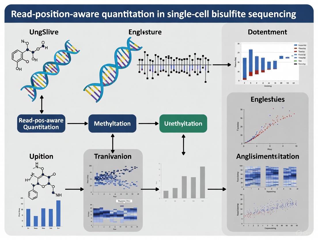
Figure 1: Computational workflow for read-position-aware quantitation in scBS data analysis
Validation and Performance Metrics
Comparative Analysis with Traditional Methods
The performance advantages of read-position-aware quantitation have been rigorously evaluated against traditional averaging approaches across multiple datasets. In benchmark analyses using real-world scBS data, the residual-based method demonstrates consistent improvements in key metrics of analytical precision and biological relevance [2].
Table 3: Performance Comparison Between Quantitation Methods
| Performance Metric | Traditional Averaging | Read-Position-Aware | Improvement |
|---|---|---|---|
| Signal-to-Noise Ratio | Baseline | 1.8-2.5× higher | ~2× enhancement |
| Cell Type Discrimination | Moderate separation | Clear cluster separation | Enhanced resolution |
| Required Cell Numbers | 100s-1000s | Fewer cells needed | Reduced cost and complexity |
| DMR Detection Specificity | Higher false positives | Biologically meaningful regions | More relevant gene associations |
| Technical Variation | Higher between replicates | Reduced batch effects | Improved reproducibility |
The enhanced signal-to-noise ratio achieved through read-position-aware quantitation directly addresses the sparse coverage challenge in scBS data. By reducing the variance introduced when reads from different cells cover non-overlapping CpG positions within the same genomic region, the method provides a more robust foundation for downstream analyses including dimensionality reduction, clustering, and trajectory inference [2].
Application to Biological Questions
The practical utility of read-position-aware quantitation is demonstrated through its application to biologically meaningful problems. In the detection of differentially methylated regions (DMRs) between cell types, the method identifies regions associated with genes involved in the core functions of specific cell types, providing more biologically interpretable results than conventional approaches [2].
Furthermore, the method's ability to achieve robust cell type discrimination with fewer cells has significant implications for study design, particularly in scenarios where sample material is limited, such as in early embryonic development or rare cell populations in clinical samples. This advantage complements recent technical improvements in bisulfite-free methods like Cabernet, which already provide higher genomic coverage through better DNA preservation [4].
Integration with Broader Methodological Advances
Read-position-aware quantitation represents one component of a broader methodological evolution in single-cell methylome analysis. Several parallel advancements are shaping the future of this field:
Bisulfite-Free Technologies: Methods like Cabernet utilize enzymatic conversion rather than bisulfite treatment, achieving approximately twice the genomic coverage of traditional scBS-seq while avoiding DNA degradation [4]. These approaches maintain single-base resolution while enabling high-throughput profiling through Tn5 transposase integration.
Advanced Alignment Strategies: The development of context-aware aligners like ARYANA-BS, which accounts for different methylation patterns in various genomic contexts (CpG islands vs. non-islands), reflects the growing recognition that methylation analysis must incorporate positional and contextual information from the earliest stages of data processing [5].
Multi-Omics Integration: The principles underlying read-position-aware quantitation are extensible to other single-cell modalities. Just as positional information improves methylation quantification, similar context-aware approaches could enhance the analysis of single-cell chromatin accessibility, transcription factor binding, and other epigenetic features.
Figure 2: Integration of read-position-aware quantitation with broader methodological advances and applications
Future Directions and Implementation Considerations
The implementation of read-position-aware quantitation represents a significant step toward more biologically faithful analysis of single-cell methylation data. As the field continues to evolve, several promising directions emerge for further refinement and application:
Methodological Extensions: Future implementations may incorporate more sophisticated modeling of spatial methylation patterns, potentially using Gaussian processes or neural networks to capture complex genomic dependencies. Additionally, integration with bisulfite-free methods could leverage the higher coverage of these techniques while maintaining the analytical advantages of position-aware quantification.
Drug Discovery Applications: For pharmaceutical researchers, the enhanced resolution provided by read-position-aware quantitation offers new opportunities for understanding how epigenetic heterogeneity influences drug response and resistance. In system-based drug discovery, detailed methylation patterns can inform target identification and side effect prediction [6].
Clinical Translation: The ability to distinguish cell states with higher resolution and fewer cells has important implications for clinical applications where sample material is often limited. Detection of rare cell populations with distinct methylation signatures could provide valuable biomarkers for early disease detection or treatment monitoring.
As with any methodological advancement, successful implementation requires careful consideration of parameter optimization and validation in specific biological contexts. The kernel bandwidth and shrinkage parameters may need adjustment for particular applications, such as when focusing on specific genomic regions with distinctive methylation patterns. Nevertheless, read-position-aware quantitation establishes a new standard for scBS data analysis that more faithfully represents the biological complexity of epigenetic regulation.
Single-cell bisulfite sequencing (scBS) is a powerful technique that enables the assessment of DNA methylation at single-base pair resolution within individual cells [2]. The protocol begins with treating genomic DNA from a single cell with sodium bisulfite, which converts unmethylated cytosines to uracils (read as thymines in subsequent sequencing), while leaving methylated cytosines unmodified [7] [5]. After sequencing, this conversion allows researchers to determine the methylation status of cytosines covered by reads, producing binary data (0 for unmethylated, 1 for methylated) at each CpG site for each cell [2].
This binary, cell-by-CpG matrix represents the fundamental data structure of scBS experiments. Unlike single-cell RNA sequencing which naturally organizes data around genes, scBS data is genome-wide and lacks a natural choice of features for analysis [2]. This presents both an opportunity and a challenge—while it allows unbiased exploration of the entire methylome, it requires sophisticated computational approaches to transform raw binary calls into biologically meaningful information that can distinguish cell types and states.
The Fundamental Data Structure and Sparsity Challenge
The Binary Methylation Matrix
The primary data structure generated from scBS experiments is a three-dimensional binary matrix with dimensions [cells × CpG sites × methylation calls]. For each cell and each CpG site covered by at least one read, the methylation status is encoded as either 0 (unmethylated) or 1 (methylated) [2]. In practice, this data is often organized as a large, sparse matrix where rows represent individual cells, columns represent CpG sites across the genome, and the values are binary methylation states.
Table 1: Characteristics of scBS-seq Data Structure
| Aspect | Description | Implication |
|---|---|---|
| Data Type | Binary values (0/1) for methylation status at each CpG | Prevents use of standard count-based models |
| Coverage | Typically 5-20% of CpG sites covered per cell [8] [9] | Creates extreme data sparsity |
| Matrix Dimensions | Thousands of cells × Millions of potential CpG sites | Computational challenges in storage and processing |
| Natural Feature Space | Genome-wide without predefined features | Requires creation of genomic regions for analysis |
Technical Origins of Sparse Coverage
The extreme sparsity in scBS data—where typically 80-95% of CpG sites show no coverage in a given cell—stems from fundamental technical constraints [8] [9]. Several factors contribute to this sparsity:
- Minimal Input DNA: A single cell contains only picograms of genomic DNA, creating a fundamental limitation in starting material [9].
- Bisulfite-Induced Damage: The bisulfite treatment process is destructive to nucleic acids, resulting in significant DNA fragmentation and loss [8].
- Amplification Biases: The whole-genome amplification required to generate sufficient DNA for sequencing introduces uneven coverage and biases [9].
- Sequencing Depth Limitations: Practical constraints on sequencing depth mean that only a fraction of the genome can be covered in each cell [2].
The resulting data sparsity presents substantial analytical challenges, as genuine biological heterogeneity becomes confounded with technical noise and missing data.
Computational Strategies for Sparse Data Analysis
Standard Coarse-Graining Approach
The conventional approach to analyzing scBS data involves dividing the genome into large tiles (e.g., 100 kb regions) and calculating the average methylation fraction within each tile for every cell [2]. For each genomic tile, researchers identify all CpG sites covered by at least one read and compute the proportion of these observed sites that are methylated [2]. This generates a matrix of methylation fractions between 0 and 1, with rows as cells and columns as genomic tiles, which can then be subjected to principal component analysis (PCA) and subsequent dimensionality reduction techniques similar to those used in scRNA-seq analysis [2].
However, this coarse-graining approach has significant limitations. As demonstrated in recent research, this method can lead to signal dilution, where important methylation patterns are lost through averaging, particularly when reads from different cells cover non-overlapping subsets of CpGs within a tile [2]. The approach also assumes uniform methylation across large genomic regions, which fails to capture the finer-scale variability that may be biologically significant.
Read-Position-Aware Quantitation
To address the limitations of simple averaging, read-position-aware quantitation has been developed as an improved strategy [2]. This method accounts for the precise genomic positions covered by each read, thereby preserving more information from the original data.
The methodology proceeds through several key steps:
Ensemble Average Smoothing: For each CpG position, compute a smoothed average methylation across all cells using kernel smoothing (typically with 1,000 bp bandwidth) to create a reference methylation profile [2].
Residual Calculation: For each cell and each covered CpG, calculate the deviation (residual) between the observed binary methylation and the ensemble average [2].
Shrunken Mean Estimation: For predefined genomic regions, compute the average of residuals for each cell, applying shrinkage toward zero via a pseudocount to dampen noise in low-coverage cells [2].
This approach significantly enhances the signal-to-noise ratio by reducing variation in situations where reads from different cells cover non-overlapping CpGs within a region but show consistent methylation patterns where they do overlap [2].
Table 2: Comparison of Quantitation Methods for scBS Data
| Feature | Simple Averaging | Read-Position-Aware |
|---|---|---|
| CpG Position | Ignores read positions | Accounts for precise genomic context |
| Handling Sparse Data | Treats each CpG independently | Leverages spatial correlation |
| Noise Reduction | Limited | Pseudocount shrinkage for low coverage |
| Signal Preservation | Prone to dilution | Maintains spatial patterns |
| Computational Complexity | Low | Moderate to high |
Advanced Statistical Modeling Approaches
Beyond read-position-aware methods, several advanced computational frameworks have been developed specifically to address scBS data sparsity:
scMET: A hierarchical Bayesian model that uses a beta-binomial framework to disentangle technical variability from genuine biological heterogeneity [8]. scMET incorporates feature-level covariates (e.g., CpG density) and uses a non-linear regression framework to capture mean-overdispersion trends, deriving residual overdispersion parameters that represent cell-to-cell variability uncorrelated with mean methylation [8].
vmrseq: A two-stage approach that first constructs candidate regions using kernel smoothing of relative methylation levels, then applies hidden Markov models (HMMs) to detect variably methylated regions (VMRs) without predefined genomic boundaries [9]. This method assumes that each cell has uniform hidden states (fully methylated or unmethylated) within a VMR, effectively partitioning cells into two groupings regardless of the actual number of cell subpopulations [9].
Variably Methylated Region Detection
The Importance of VMRs
Only specific genomic regions show meaningful methylation variability across cells, while many areas maintain consistent methylation patterns regardless of cell type [2]. Variably methylated regions are particularly valuable for distinguishing cell types and states, as they capture the dynamic aspects of the epigenome rather than the static background [2] [9].
VMRs are typically associated with regulatory genomic features such as enhancers, which display more dynamic methylation patterns compared to the stable methylation patterns observed at housekeeping gene promoters or highly methylated repetitive regions [2]. Identifying these regions is therefore crucial for understanding the epigenetic basis of cellular identity and heterogeneity.
Methodologies for VMR Detection
Traditional VMR detection relies on analyzing predefined genomic regions, such as promoters or sliding windows, but this approach may miss important variability occurring outside these boundaries [9]. Modern methods like vmrseq address this limitation by scanning the entire genome without prior assumptions about VMR locations [9].
The vmrseq workflow implements a sophisticated two-stage process:
Stage 1 - Candidate Region Identification:
- Apply kernel smoothing to relative methylation levels (individual cell methylation relative to across-cell averages)
- Select consecutive CpG loci that exceed a variance threshold
- Compute thresholds based on null distributions of variance to control false positives [9]
Stage 2 - HMM Optimization:
- For each candidate region, optimize both one-state and two-state hidden Markov models
- Compare maximum likelihoods between models to determine if one or two cell groupings are present
- If two groupings are supported, delineate VMR boundaries by removing CpGs with uniform hidden states across groupings [9]
This method demonstrates substantially improved accuracy in synthetic benchmarks and enhanced feature selection in real-world applications compared to sliding window approaches [9].
Experimental Design and Protocol Considerations
Library Preparation and Sequencing
Proper experimental design is crucial for generating high-quality scBS data that can overcome inherent sparsity challenges. Key considerations include:
Cell Isolation and Barcoding: Single cells can be isolated using fluorescence-activated cell sorting (FACS), magnetic-activated cell sorting (MACS), or microfluidic technologies [10]. Microfluidic devices offer particular advantages for high-throughput processing, enabling the parallel processing of tens of thousands of single cells in nanoliter or picoliter reaction volumes [10]. Cell barcoding is typically incorporated early in microfluidics-based protocols, allowing entire libraries to be processed in a single tube and minimizing sample loss [10].
Bisulfite Conversion and Library Preparation: The bisulfite conversion step must be carefully optimized to maximize conversion efficiency while minimizing DNA degradation [5]. Following conversion, library preparation involves PCR amplification with bisulfite-converted DNA-compatible polymerases and incorporation of unique molecular identifiers (UMIs) where possible to account for amplification biases [2].
Sequencing Depth Considerations: Based on current methodologies, achieving sufficient coverage typically requires sequencing depths of 5-20 million reads per cell, though this varies based on genome size and the specific biological question [2] [9]. Deeper sequencing can partially compensate for sparsity but comes with increased costs.
Research Reagent Solutions
Table 3: Essential Research Reagents for scBS Experiments
| Reagent/Category | Function | Examples/Alternatives |
|---|---|---|
| Bisulfite Conversion Kits | Converts unmethylated cytosines to uracils | Commercial kits optimized for low-input DNA |
| Whole Genome Amplification Kits | Amplifies picograms of DNA to usable amounts | Multiple displacement amplification (MDA) kits |
| Single-Cell Isolation Systems | Isolates individual cells from tissue | FACS, microfluidic platforms (10X Genomics) |
| Cell Lysis Reagents | Releases genomic DNA while maintaining integrity | Mild detergents with proteinase K |
| UMI Adapters | Tags molecules to correct PCR amplification bias | Commercial UMI adapter sets for bisulfite sequencing |
| BS-Seq Aligners | Aligns bisulfite-converted reads to reference genome | Aryana-bs, Bismark, BSMAP, bwa-meth [5] |
Downstream Analytical Applications
Cell Type Identification and Clustering
The ultimate goal of processing scBS data is to identify biologically meaningful patterns that distinguish cell types and states. The methylation matrix generated through read-position-aware quantitation or advanced statistical modeling serves as input for standard single-cell analysis workflows:
Dimensionality Reduction: Principal component analysis (PCA) is applied to the processed methylation matrix, typically retaining the top 20-50 components to eliminate Poisson noise [2]. The PCA space representation then enables robust dissimilarity measurements between cells.
Visualization and Clustering: Euclidean distances in PCA space facilitate two-dimensional visualization using t-SNE or UMAP, as well as clustering algorithms to identify distinct cell populations [2]. Studies have demonstrated that proper processing of scBS data leads to improved separation of cell types and more biologically meaningful clusters [2] [9].
Differential Methylation Analysis
Beyond cell type identification, processed scBS data enables detection of differentially methylated regions (DMRs) between groups of cells. scMET, for instance, implements a probabilistic decision rule to control expected false discovery rate (EFDR) when testing for differences in both mean methylation and methylation variability [8]. This differential variability analysis represents a particularly powerful application, as it can identify regions where epigenetic heterogeneity itself differs between cell types or conditions—an analysis impossible with bulk sequencing data [8].
Multi-Omics Integration
Single-cell methylation data can be integrated with other omics modalities, such as transcriptomics or chromatin accessibility, to provide a more comprehensive understanding of cellular regulation [7] [10]. The VMRs identified through scBS analysis serve as excellent epigenetic features for integration with scRNA-seq data, potentially revealing regulatory relationships between DNA methylation and gene expression [9].
Integration approaches typically involve:
- Identifying cell populations independently in each modality
- Using mutual nearest neighbors or other alignment methods to match cells across assays
- Jointly analyzing methylation and expression patterns to identify putative regulatory relationships [7]
The data structure of scBS—fundamentally binary and exceptionally sparse—presents significant analytical challenges that require specialized computational approaches. Read-position-aware quantitation and advanced Bayesian modeling methods have emerged as powerful solutions that overcome the limitations of simple averaging approaches. By properly accounting for the unique characteristics of scBS data, these methods enable researchers to extract meaningful biological signals from sparse binary methylation calls, ultimately advancing our understanding of epigenetic heterogeneity in development, disease, and cellular function. As these methodologies continue to mature, they promise to unlock the full potential of single-cell epigenomics for both basic research and therapeutic development.
In single-cell bisulfite sequencing (scBS-seq) research, the inherent sparsity of methylation data poses a significant challenge for biological interpretation. This technical guide elaborates on the computational framework of deriving smoothed ensemble averages and calculating residuals to address cellular heterogeneity and technical noise. These concepts enable researchers to distinguish meaningful biological variation from stochastic noise, thereby facilitating accurate identification of cell-to-cell epigenetic differences. The methodologies outlined herein form the computational backbone for read-position-aware quantitation in single-cell methylome analysis, providing a statistical foundation for elucidating epigenetic heterogeneity in development and disease.
Single-cell bisulfite sequencing technologies, including scBS-seq and scRRBS-seq, provide unprecedented resolution for studying epigenetic heterogeneity [11]. However, the limited starting material of genomic DNA per cell results in sparse CpG coverage, with typical protocols capturing only 1-40% of CpG sites per cell [11]. This sparsity fundamentally challenges conventional bulk analysis approaches and necessitates specialized computational methods that can account for both technical artifacts and biological heterogeneity.
The core concept of smoothed ensemble averages addresses this limitation by aggregating methylation signals across either genomic regions or cell populations to create more stable estimates of underlying methylation patterns. The corresponding residual calculations then quantify deviations from these averages, enabling discrimination of true biological variation from measurement noise. Together, these approaches form a critical foundation for read-position-aware quantitation, allowing researchers to model methylation states as a function of both genomic context and cellular identity.
Theoretical Foundations
Mathematical Formulation of Smoothed Ensemble Averages
In single-cell methylome analysis, a smoothed ensemble average represents a weighted aggregation of methylation states across defined genomic windows or cellular neighborhoods. For a given CpG site i in cell c, the smoothed methylation value M'~i,c~ is derived from the observed binary methylation state M~i,c~ (where 0 = unmethylated, 1 = methylated) and its local genomic or cellular context.
The fundamental smoothing operation can be formalized as:
M'~i,c~ = Σ~j∈N(i,c)~ w~j~ ⋅ M~j~
Where N(i,c) defines the neighborhood of CpG sites considered for smoothing, and w~j~ represents weights assigned to each observation based on genomic distance, cellular similarity, or coverage depth [11] [12]. The neighborhood can be defined through various approaches:
- Genomic spatial smoothing: N(i,c) includes all CpG sites within a defined genomic window (typically 1-3 kb) centered on site i in the same cell c [11].
- Cellular ensemble smoothing: N(i,c) includes the same CpG site i across multiple cells with similar methylation profiles [13].
- Meta-cell construction: N(i,c) aggregates information from neighboring single cells in transcriptional or epigenetic space, creating composite profiles that mitigate sparsity [13].
The MOABS (Model-based Analysis of Bisulfite Sequencing data) platform implements a Beta-Binomial hierarchical model that conceptually incorporates smoothing through its empirical Bayes approach, where prior distributions are estimated from genome-wide patterns and used to refine methylation ratio estimates for individual CpGs [12].
Residual Calculations and Biological Interpretation
Once smoothed ensemble averages are established, residual calculations quantify the deviation between observed methylation states and the expected smoothed values. The residual R~i,c~ for CpG site i in cell c is calculated as:
R~i,c~ = M~i,c~ - M'~i,c~
These residuals serve multiple critical functions in single-cell methylome analysis:
- Technical noise estimation: Systematic patterns in residuals can indicate technical artifacts from bisulfite conversion efficiency, sequencing depth, or mapping errors [12].
- Biological variability quantification: Residuals exceeding expected statistical thresholds may represent genuine epigenetic heterogeneity between cells [11].
- Feature selection for clustering: CpG sites with consistently high residuals across cell populations often mark genomic regions with regulatory significance [13] [14].
In practice, statistical significance of residuals is assessed through comparison to null distributions generated via permutation testing or through analytical approximations based on Binomial sampling variance [12].
Computational Implementation
Methodological Framework for Read-Position-Aware Quantitation
Read-position-aware quantitation extends basic smoothing approaches by incorporating information about the spatial distribution of reads relative to genomic features. The DeepCpG framework implements a sophisticated position-aware approach using a bidirectional gated recurrent network architecture that captures patterns of neighboring CpG states across multiple cells [11].
Table 1: Key Computational Tools for Smoothing and Residual Analysis
| Tool | Primary Function | Smoothing Approach | Residual Calculation |
|---|---|---|---|
| DeepCpG [11] | Imputation of missing methylation states | DNA sequence features + neighboring CpG states via deep neural networks | Not explicitly calculated, but embedded in probability estimates |
| MOABS [12] | Differential methylation detection | Beta-Binomial hierarchical model | Credible methylation difference (CDIF) accounts for biological variation |
| MAPLE [13] | Gene activity prediction | Meta-cell construction from cellular neighborhoods | Expression prediction residuals used for model refinement |
The implementation workflow for read-position-aware smoothing and residual analysis typically involves these critical steps:
- Data preprocessing and quality control: Filtering cells based on coverage, bisulfite conversion efficiency, and mapping quality [14].
- Neighborhood definition: Establishing genomic windows or cellular similarity groups for aggregation.
- Weight assignment: Determining appropriate weighting schemes based on distance metrics or confidence measures.
- Smoothing computation: Applying the aggregation function to calculate smoothed values.
- Residual calculation: Computing deviations between observed and smoothed values.
- Statistical testing: Assessing significance of observed residuals against null models.
Workflow Visualization
The following diagram illustrates the computational workflow for deriving smoothed ensemble averages and calculating residuals in single-cell bisulfite sequencing data:
Experimental Protocols
Protocol for Meta-Cell Smoothing and Residual Analysis
The MAPLE (methylome association by predictive linkage to expression) framework provides a robust protocol for implementing smoothed ensemble averages through meta-cell construction [13]:
Step 1: Data Preparation and Normalization
- Input: Single-cell methylation matrix (cells × CpG sites) with binary methylation calls
- Filter cells with < 1,000 covered CpG sites and CpG sites covered in < 10 cells
- Normalize for coverage differences using reads per million (RPM) or similar approaches
Step 2: Meta-Cell Construction
- Compute cell-to-cell similarity using Jaccard index on shared CpG sites or reduced-dimension embeddings
- For each cell, identify its k-nearest neighbors (k typically 5-20) in epigenetic space
- Aggregate methylation calls across neighbor cells to create meta-cell profiles
- Compute methylation frequency for each CpG in each meta-cell as smoothed value
Step 3: Residual Calculation
- For each original cell, calculate residuals as difference between observed methylation and meta-cell smoothed value
- Standardize residuals by coverage-aware variance estimates
- Apply statistical filtering to identify significant residuals (p < 0.05, Bonferroni-corrected)
Step 4: Biological Interpretation
- Annotate significant residuals with genomic features (promoters, enhancers, gene bodies)
- Correlate methylation residuals with gene expression where multi-omics data available
- Perform pathway enrichment analysis on genes associated with consistent residual patterns
Protocol for Genomic Window Smoothing
The BSmooth algorithm implements an alternative approach using genomic spatial smoothing [12]:
Step 1: Genomic Binning
- Divide genome into non-overlapping 500-1000 bp windows
- For each window, calculate average methylation across all covered CpGs per cell
Step 2: Local Smoothing
- Apply moving average filter across genomic windows (typically 1-3 adjacent windows)
- Weight contributions by coverage depth and genomic distance
Step 3: Residual Extraction
- Calculate difference between observed window methylation and smoothed values
- Normalize residuals by window-specific variance estimates
Step 4: Differential Methylation Calling
- Identify genomic windows with consistent residual patterns across cell groups
- Apply statistical tests (e.g., t-test, Beta-Binomial regression) to assess significance
Research Reagent Solutions
Table 2: Essential Research Reagents and Computational Tools
| Reagent/Tool | Function | Application in Smoothed Averaging |
|---|---|---|
| Sodium Bisulfite [15] | Chemical conversion of unmethylated cytosine to uracil | Creates foundational methylation data for smoothing approaches |
| MspI Restriction Enzyme [16] | CCGG site digestion for reduced representation bisulfite sequencing (RRBS) | Enriches for CpG-dense regions, improving smoothing reliability |
| Proteinase K [16] | DNA isolation and purification from protein contaminants | Ensures high-quality DNA for complete bisulfite conversion |
| DeepCpG [11] | Deep neural network for single-cell methylation state prediction | Provides advanced smoothing through DNA sequence and methylation pattern integration |
| MOABS [12] | Model-based analysis of bisulfite sequencing data | Implements Beta-Binomial smoothing for differential methylation detection |
| MAPLE [13] | Predictive modeling of methylation-gene expression relationships | Uses meta-cell smoothing for gene activity prediction |
Applications in Drug Development and Disease Research
The integration of smoothed ensemble averages and residual calculations enables several critical applications in pharmaceutical research and development:
Cancer Epigenetics: By calculating residuals from smoothed averages of normal cell populations, researchers can identify cancer-specific methylation patterns that may serve as therapeutic targets or biomarkers. The MOABS system has demonstrated particular utility in detecting differential methylation with high statistical power, even at low sequencing depths [12].
Cell Lineage Tracing: Smoothed methylation patterns across cell populations enable reconstruction of developmental trajectories. Residual patterns help identify epigenetic branching points where cell fate decisions occur, potentially revealing novel interventions for developmental disorders [14].
Pharmacoepigenetics: Drug-induced epigenetic changes can be quantified by comparing residuals before and after treatment, enabling assessment of compound efficacy and mechanism of action at single-cell resolution.
Smoothed ensemble averages and residual calculations represent foundational computational concepts in single-cell bisulfite sequencing research. By effectively distinguishing biological signals from technical noise, these approaches enable read-position-aware quantitation that accounts for both genomic context and cellular heterogeneity. As single-cell methylomic technologies continue to advance, further refinement of these computational frameworks will be essential for unlocking the full potential of epigenetic analysis in basic research and therapeutic development.
Implementing Read-Position-Aware Analysis: A Step-by-Step Guide to the MethSCAn Workflow
Variably Methylated Regions (VMRs) represent genomic loci where DNA methylation patterns show significant variation across different cells, conditions, or developmental stages. In the context of read-position-aware quantitation single-cell bisulfite sequencing (scBS-seq), VMRs serve as critical focal points for understanding cellular heterogeneity and epigenetic regulation. Unlike Differentially Methylated Regions (DMRs), which are identified through comparative analysis between predefined sample groups, VMRs capture intrinsic methylation variability within a population, making them particularly valuable for identifying dynamic epigenetic states in complex tissues and developmental trajectories [17] [18].
The identification of VMRs has become increasingly important in epigenetic research, especially with advances in single-cell technologies that reveal the remarkable heterogeneity of methylation patterns at cellular resolution. Traditional bulk methylation analysis approaches mask this cellular diversity, whereas single-cell methods enable researchers to detect methylation variability that correlates with distinct cellular phenotypes, lineage relationships, and transcriptional states. Within the framework of read-position-aware quantitation, VMR identification gains additional precision by accounting for technical artifacts and biases inherent in single-cell bisulfite sequencing protocols [18].
Biological Significance and Research Applications of VMRs
VMRs occupy a crucial position in the epigenetic landscape, serving as potential regulatory elements that influence gene expression programs and cellular identity. Research has demonstrated that VMRs are frequently associated with genomic regulatory features such as enhancers, promoters, and chromatin boundary elements, where precise methylation control is essential for proper gene regulation. The spatial organization of VMRs, as revealed by recent spatial multi-omics technologies, shows distinct correlation patterns with gene expression in specific anatomical regions, highlighting their role in developmental patterning and tissue specialization [18].
In mammalian genomes, VMRs are particularly enriched in non-promoter regulatory elements, with studies showing that approximately 60-70% of tissue-specific VMRs coincide with enhancer regions marked by H3K27ac. This enrichment underscores the importance of methylation variability in fine-tuning regulatory element activity across different cellular contexts. During embryonic development, VMRs demonstrate remarkable dynamism, with specific spatial-temporal patterns emerging in structures such as the embryonic brain, craniofacial region, and heart, suggesting their involvement in morphogenetic processes [18].
The clinical relevance of VMRs extends to disease research, particularly in cancer and neurological disorders. In tumor ecosystems, VMRs can identify epigenetic subpopulations with distinct functional properties, including drug-resistant clones or cells with enhanced metastatic potential. Single-cell methylation analyses have revealed that VMR patterns in tumor microenvironments often reflect underlying transcriptional states and cellular responses to therapeutic interventions, making them valuable biomarkers for predicting treatment outcomes and disease progression [18] [19].
Experimental Design for VMR Identification
Sample Considerations and Experimental Replication
Effective VMR identification requires careful experimental design to ensure sufficient statistical power and biological relevance. For single-cell BS-seq studies, sample size determination should account for both the expected cellular heterogeneity and the technical noise inherent in single-cell methylation data. Research indicates that studies aiming to identify VMRs across distinct cell types typically require a minimum of 50-100 cells per population to achieve reliable detection, though this varies depending on the degree of methylation heterogeneity and sequencing coverage [18].
The selection of biological replicates is crucial for distinguishing technical variability from genuine biological variation in VMR calling. For studies comparing experimental conditions or developmental timepoints, a minimum of three independent biological replicates is recommended to ensure robust statistical inference. In spatial methylation studies, such as those employing spatial-DMT technology, reproducibility has been demonstrated through high concordance between replicate maps, with DNA methylation and RNA expression showing consistent patterns in matched body regions across replicate E11 mouse embryos [18].
Sequencing Depth and Coverage Requirements
The accurate identification of VMRs depends heavily on sequencing depth and CpG coverage. Current methodologies suggest that optimal VMR detection requires sequencing coverage that captures a substantial proportion of the methylome at single-cell resolution. Spatial joint profiling technologies have achieved coverage of 136,639-281,447 CpGs per pixel (approximating single-cell resolution) with 2.8-3.9 billion raw reads per sample, providing sufficient depth for robust VMR identification [18].
For conventional single-cell BS-seq experiments, recommended sequencing depth typically ranges from 5-10 million reads per cell, aiming to cover at least 1-2 million CpG sites per cell at a mean coverage of 5-10x. This coverage ensures that a sufficient number of informative CpGs can be assessed for methylation variability across the cell population. The following table summarizes key sequencing parameters for optimal VMR identification across different technological platforms:
Table 1: Sequencing Requirements for VMR Identification Across Platforms
| Technology Platform | Recommended Coverage | CpGs per Cell/Unit | Raw Reads per Sample | Conversion Efficiency |
|---|---|---|---|---|
| Standard scBS-seq | 5-10x per CpG | 1-2 million | 5-10 million per cell | >99.5% |
| Spatial-DMT | Not specified | 136,639-281,447 per pixel | 2.8-3.9 billion | >99% |
| Methylpy | 5x per CpG (minimum) | Dependent on alignment | Dependent on sample | >99% |
Computational Methods for VMR Identification
Read-Position-Aware Quantitation Approaches
Read-position-aware quantitation represents a significant advancement in single-cell methylation analysis, addressing technical biases introduced by library preparation and sequencing artifacts. This approach accounts for the positional information of reads within the sequencing library, recognizing that conversion efficiency, mapping quality, and base calling accuracy can vary systematically across read positions. By modeling these positional effects, researchers can obtain more accurate methylation calls for individual CpG sites, which is fundamental for reliable VMR detection [17].
The core principle of read-position-aware quantitation involves weighting methylation calls based on their positional confidence scores and adjusting for systematic errors that correlate with read position. Implementation typically involves the calculation of position-specific quality metrics across all sequencing reads, followed by the application of statistical models that correct for identified biases. This process significantly improves the signal-to-noise ratio in methylation quantification, particularly important for detecting subtle but biologically meaningful methylation variations in single-cell data [17] [20].
VMR Calling Algorithms and Statistical Frameworks
Several computational approaches have been developed specifically for VMR identification in single-cell methylation data. These algorithms generally operate through a multi-step process: (1) quantifying methylation levels at individual CpG sites across single cells, (2) smoothing or aggregating methylation signals across genomic regions to reduce noise, (3) measuring methylation variability across cells, and (4) applying statistical tests to identify regions with significant variability beyond technical noise.
The methylpy pipeline, for instance, implements a beta-binomial regression framework to model methylation variability, accounting for both biological variation and technical sampling noise. This approach has been optimized for mammalian genomes and can identify both VMRs and larger-scale differentially methylated regions (DMRs) through a sliding window approach combined with multiple testing correction [17].
Alternative methods include approaches based on dispersion statistics, such as the coefficient of variation (CV) in methylation levels across cells, or entropy-based measures that capture the disorder in methylation patterns. These metrics are particularly useful for detecting VMRs in homogeneous cell populations where methylation variability may reflect epigenetic plasticity rather than distinct cell identities. The statistical significance of candidate VMRs is typically assessed through permutation-based methods or comparison to background variability models, with false discovery rate (FDR) control to account for multiple testing across the genome [17] [20].
Table 2: Key Computational Tools for VMR Identification
| Tool/Method | Statistical Approach | Key Features | Single-Cell Optimized |
|---|---|---|---|
| Methylpy | Beta-binomial regression | Sliding window analysis, DMR/VMR calling, annotation | Yes |
| AIMS | Amplification of intermethylated sites | Detection of methylation variable positions | No |
| ChIP-BMS | Integrated with chromatin data | Combines ChIP with bisulfite sequencing | No |
| Bisulfite sequencing | Maximum likelihood estimation | Standard for methylation calling | With modifications |
Technical Protocols for VMR Analysis
Single-Cell Bisulfite Sequencing Wet-Lab Protocol
The standard protocol for single-cell bisulfite sequencing begins with single-cell isolation, typically performed through fluorescence-activated cell sorting (FACS) or microfluidics platforms. Individual cells are lysed, and genomic DNA is subjected to bisulfite conversion using optimized kits that maximize conversion efficiency while minimizing DNA degradation. The conversion reaction uses sodium bisulfite to deaminate unmethylated cytosine to uracil, while methylated cytosine remains unchanged, creating sequence differences that can be detected through sequencing [17] [21].
Following bisulfite conversion, the converted DNA undergoes whole-genome amplification using methods such as multiple displacement amplification (MDA) or polymerase chain reaction (PCR)-based approaches. The amplified DNA is then fragmented and used to construct sequencing libraries compatible with high-throughput platforms. For read-position-aware quantitation, it is critical to incorporate unique molecular identifiers (UMIs) during library preparation to account for amplification biases and duplicate reads. Quality control steps should include assessment of conversion efficiency through spike-in controls and evaluation of library complexity through fragment size distribution analysis [17].
Spatial Joint Profiling Workflow for VMR Identification
The spatial-DMT protocol represents a cutting-edge approach for VMR identification with spatial context. This workflow begins with fresh-frozen or FFPE tissue sections mounted on specialized slides. The tissue undergoes hydrochloric acid treatment to improve DNA accessibility, followed by Tn5 transposase treatment for fragmentation and adapter insertion. The protocol then employs a microfluidic chip with perpendicular flow channels to deliver spatially barcoded oligonucleotides, creating a two-dimensional grid of barcoded tissue pixels with resolutions as fine as 10μm [18].
Following barcoding, cDNA and gDNA are separated, with the cDNA directed to transcriptome library preparation and the gDNA undergoing enzymatic methyl-seq (EM-seq) for bisulfite-free methylation conversion. The EM-seq approach offers advantages over traditional bisulfite treatment by reducing DNA damage while maintaining high conversion efficiency (>99%). The final libraries are sequenced on high-throughput platforms, and bioinformatic processing reconstructs both methylation patterns and gene expression with spatial coordinates, enabling identification of VMRs in their native tissue context [18].
Figure 1: Spatial Joint Profiling Workflow
Bioinformatics Processing Pipeline
The computational analysis of scBS-seq data for VMR identification follows a multi-step bioinformatics pipeline. Initial processing includes quality control of raw sequencing data using tools such as FastQC, followed by adapter trimming and quality filtering. Preprocessed reads are then aligned to a bisulfite-converted reference genome using specialized aligners such as Bismark or BS-Seeker2, which account for the C-to-T conversion in unmethylated positions [17].
Following alignment, methylation calling is performed to extract methylation proportions for each CpG site in every cell. This step generates a methylation matrix where rows represent CpG sites or genomic bins, columns represent individual cells, and values indicate methylation proportions. For read-position-aware quantitation, additional correction factors are applied based on the positional quality metrics. The resulting matrix then serves as input for VMR calling algorithms, which identify genomic regions showing significant methylation variability across cells after accounting for technical noise [17] [20].
Downstream analysis typically includes annotation of VMRs to genomic features (promoters, enhancers, gene bodies), integration with complementary data types such as transcriptome information, and visualization through specialized packages. The interpretation of VMRs benefits from integration with publicly available resources such as epigenome Roadmap projects or single-cell atlases to contextualize findings within established regulatory frameworks [17].
Quality Control and Validation Methods
Assessing Technical Data Quality
Rigorous quality control is essential for reliable VMR identification, particularly given the technical challenges of single-cell methylation data. Key quality metrics include bisulfite conversion efficiency, which should exceed 99% to ensure accurate discrimination between methylated and unmethylated cytosines; mapping efficiency, which reflects the proportion of reads successfully aligned to the reference genome; and coverage uniformity across genomic regions. Additional metrics specific to single-cell experiments include the number of CpG sites covered per cell, the distribution of sequencing depth across cells, and the percentage of cells passing quality thresholds [17] [18].
For spatial methylation data, quality assessment extends to spatial metrics such as barcode efficiency, spatial resolution, and cross-talk between adjacent pixels. The spatial-DMT technology has demonstrated high quality metrics, with minimal RNA contamination in DNA methylation libraries and high reproducibility between technical and biological replicates. Conversion efficiency in spatial-DMT exceeds 99%, as verified through analysis of unmethylated adapter sequences, ensuring accurate methylation calling [18].
Biological Validation of VMRs
Candidate VMRs require biological validation to confirm their functional relevance and technical accuracy. Orthogonal validation methods include pyrosequencing of bisulfite-converted DNA from cell populations, which provides quantitative methylation measurements for specific genomic regions; methylation-specific PCR (MSP), which allows for sensitive detection of methylation patterns; and targeted bisulfite sequencing, which offers deep coverage of specific loci of interest [18].
Functional validation approaches assess the potential regulatory impact of VMRs through reporter assays, CRISPR-based epigenetic editing, or correlation with gene expression data. In spatial-DMT studies, validation is inherent in the coordinated analysis of DNA methylation and transcriptome data from the same tissue section, enabling direct assessment of relationships between methylation variability and gene expression patterns. This integrated approach has revealed both expected inverse correlations between promoter methylation and gene expression, as well as more complex positive correlations in certain genomic contexts, highlighting the nuanced relationship between methylation and transcription [18].
Integration with Multi-Omics Data and Advanced Applications
Correlating VMRs with Transcriptomic Profiles
The integration of methylation data with transcriptomic profiles significantly enhances the biological interpretation of VMRs. Single-cell multi-omics technologies now enable simultaneous measurement of DNA methylation and gene expression in the same cell, providing direct insight into relationships between epigenetic variability and transcriptional heterogeneity. Analysis of mouse embryos using spatial-DMT technology has demonstrated that spatial cluster-specific gene expression often correlates with hypomethylation of nearby variable methylation regions, particularly in developing structures such as the cranial region, brain, spinal cord, and heart [18].
The relationship between VMRs and gene expression is context-dependent, with different patterns observed in different genomic regions and developmental stages. While promoter VMRs frequently show inverse correlation with expression of associated genes, VMRs in intergenic and intronic regions can exhibit more complex relationships, including positive correlations in certain contexts. These patterns reflect the diverse regulatory functions of DNA methylation in different genomic contexts and highlight the importance of integrated analysis for accurate biological interpretation [18].
VMRs in Drug Development and Clinical Translation
VMRs hold significant promise for pharmaceutical applications, particularly in the areas of biomarker discovery, patient stratification, and therapeutic monitoring. In cancer research, VMR patterns can identify epigenetic subpopulations with distinct drug sensitivities, enabling more precise targeting of therapeutic interventions. Single-cell analyses have revealed that chemotherapy-resistant clones often exhibit distinct VMR signatures associated with drug metabolism, DNA repair, or survival pathways, providing opportunities for epigenetic biomarkers of treatment response [19] [22].
The application of VMR analysis in immuno-oncology has been particularly fruitful, with studies demonstrating that T-cell exhaustion states and memory differentiation correlate with specific methylation patterns in regulatory elements of key immune genes. These epigenetic signatures can predict response to immune checkpoint inhibitors and inform the development of combination therapies. Additionally, VMRs in cell-free DNA (cfDNA) have emerged as promising non-invasive biomarkers for cancer detection, monitoring, and tissue-of-origin identification, with potential for early detection of recurrence and assessment of minimal residual disease [20] [19].
Table 3: Research Reagent Solutions for VMR Analysis
| Reagent/Kit | Manufacturer/Provider | Function in VMR Analysis | Key Specifications |
|---|---|---|---|
| Methylpy | Open source | End-to-end BS-seq analysis | DMR/VMR calling, annotation [17] |
| EM-seq Kit | Not specified | Bisulfite-free conversion | >99% efficiency, reduced DNA damage [18] |
| Spatial Barcoding Kit | Custom | Spatial multiplexing | 2500 barcodes, 10μm resolution [18] |
| Bisulfite Conversion Kit | Multiple providers | Cytosine conversion | >99.5% efficiency [17] [21] |
| Tn5 Transposase | Multiple providers | DNA fragmentation | Simultaneous fragmentation and tagging [18] |
Emerging Technologies and Future Directions
The field of VMR analysis is rapidly evolving, driven by technological advances in single-cell multi-omics, spatial profiling, and computational methods. Emerging approaches include the development of bisulfite-free methylation detection methods such as enzymatic methyl-seq (EM-seq), which reduces DNA damage and improves library complexity; multi-omics technologies that simultaneously profile methylation, chromatin accessibility, and protein expression in the same single cell; and improved computational methods that more accurately model the unique statistical characteristics of single-cell methylation data [18] [23].
The recently announced Illumina 5-base solution promises to streamline integrated genetic and epigenetic analysis by enabling simultaneous variant detection and methylation profiling in a single assay. This approach, expected to commercialize in 2026, addresses current limitations in cost, availability, and analytical complexity, potentially making multi-omic profiling more accessible to research and clinical laboratories. Such integrated technologies will enhance VMR analysis by providing coordinated information about genetic regulation and epigenetic variability in the same biological samples [23].
Future applications of VMR research will likely expand to include high-resolution spatial mapping of epigenetic heterogeneity in clinical specimens, dynamic monitoring of epigenetic changes during therapeutic interventions, and integration of multi-omic VMR signatures for personalized medicine approaches. As single-cell methylation technologies continue to mature and become more widely adopted, VMR analysis is poised to become a standard component of cellular phenotyping, complementing transcriptomic and proteomic profiling to provide a more comprehensive understanding of cellular identity and function in health and disease [18] [23].
Figure 2: VMR Research Applications and Impact
In single-cell bisulfite sequencing (scBS), the standard analysis involves tiling the genome and averaging methylation signals within each tile. However, this method often leads to signal dilution, especially given the sparse read coverage typical of scBS data. Read-position-aware quantitation addresses this limitation by introducing a more nuanced approach: the shrunken mean of residuals. This method quantifies a cell's methylation level in a genomic interval based on its deviation from a smoothed, genome-wide methylation average, thereby enhancing the signal-to-noise ratio. This technical guide details the methodology, computational implementation, and experimental validation of this advanced quantitation technique, underscoring its critical role in improving cell type discrimination and feature detection in single-cell methylomic studies [2].
Single-cell bisulfite sequencing provides genome-wide DNA methylation profiles at single-base resolution, offering unprecedented potential to decipher cellular heterogeneity [2]. The standard analytical workflow, adapted from single-cell RNA-sequencing (scRNA-seq), involves partitioning the genome into large tiles (e.g., 100 kb) and calculating the average methylation fraction per tile per cell. This matrix of averages then undergoes principal component analysis (PCA) for dimensionality reduction before downstream clustering and visualization [2].
A significant weakness of this simple averaging is its susceptibility to technical noise from sparse coverage. In a typical scBS dataset, a genomic tile may be covered by only a single read in a given cell. If the read from one cell originates from a highly methylated sub-region of the tile and the read from another cell comes from a hypomethylated sub-region, the averaged signals will suggest a biological difference where none may exist. This "signal dilution" obfuscates true biological variation and reduces the power to distinguish cell types or states [2].
Read-position-aware quantitation, centered on the calculation of shrunken mean residuals, overcomes this by incorporating two key pieces of information: the precise genomic position of each CpG site and a cell's methylation call relative to a genome-wide smoothed average. This guide elaborates on the core calculations, protocols, and applications of this refined method within the broader thesis objective of developing maximally informative analysis pipelines for single-cell epigenomics.
Core Mathematical Foundation
The Concept of Residuals in Statistical Modeling
A residual is a fundamental concept in statistical modeling, defined as the difference between an observed value and a value predicted by a model. In a regression context, it is calculated as ( ei = yi - \hat{y}i ), where ( yi ) is the observed value and ( \hat{y}_i ) is the predicted value [24]. A positive residual indicates the model underestimated the observation, while a negative residual indicates an overestimation [25].
From Simple Residuals to Shrunken Mean Residuals
The innovation in read-position-aware quantitation is the application of the residual concept to methylation data. The observed value ( yi ) is the binary methylation call (0 or 1) for a specific CpG site in a specific cell. The predicted value ( \hat{y}i ) is not from a simple regression line but from a smoothed ensemble average of methylation across all cells for that genomic position [2].
The "shrunken mean" refers to the final step where the average of a cell's residuals over all CpG sites within a genomic interval is calculated using a pseudocount. This shrinkage technique deliberately trades a small amount of bias for a significant reduction in variance, which stabilizes the signal for cells with low coverage in a given interval [2].
Step-by-Step Computational Protocol
The following diagram illustrates the complete computational workflow for read-position-aware quantitation, from raw data to a matrix ready for downstream analysis.
Detailed Methodological Steps
Step 1: Calculate the Smoothed Ensemble Methylation Average
- Objective: Generate a stable, base-by-base estimate of the average methylation level across all profiled cells.
- Protocol:
- Initialization: For each CpG site in the genome, collect all binary methylation calls (0 for unmethylated, 1 for methylated) from every cell where the site is covered by a read.
- Raw Average Calculation: Compute the initial average methylation at each CpG site as the fraction of cells where it was observed as methylated.
- Kernel Smoothing: Apply a kernel smoother to these raw averages. This replaces the value at each CpG site with a distance-weighted average of its neighbors.
- Kernel Bandwidth: A critical tuning parameter. A 1,000 bp bandwidth is often effective, but this should be validated for specific datasets [2].
- Output: A continuous curve of methylation probabilities across the genome, representing the population-level expectation.
Step 2: Calculate Per-Cell CpG Residuals
- Objective: Quantify the deviation of each cell's methylation state from the population average at every covered CpG site.
- Protocol:
- Residual Calculation: For each cell and each covered CpG site, calculate the residual: ( e{c,p} = m{c,p} - \hat{m}p ), where:
- ( m{c,p} ) is the binary methylation call (0/1) for cell ( c ) at position ( p ).
- ( \hat{m}_p ) is the smoothed ensemble average at position ( p ) [2].
- Interpretation: A positive residual indicates a CpG is more methylated in the cell than the population average; a negative residual indicates it is less methylated.
- Residual Calculation: For each cell and each covered CpG site, calculate the residual: ( e{c,p} = m{c,p} - \hat{m}p ), where:
Step 3: Compute the Shrunken Mean of Residuals per Genomic Tile
- Objective: Generate a single, robust methylation score for each cell in each predefined genomic interval (e.g., a variably methylated region).
- Protocol:
- Averaging: For a given genomic tile and a given cell, calculate the mean of all residuals ( e_{c,p} ) for CpG sites within that tile.
- Shrinkage with Pseudocount: To mitigate the high variance in cells with low coverage, shrink this mean towards zero using a pseudocount. This is analogous to the pseudocount addition in scRNA-seq analysis [2]. The mathematical implementation details are provided in the MethSCAn software.
- Handling Zero Coverage: If a cell has no reads in a tile, its value is set to zero, indicating no evidence of deviation from the mean. This can be refined with iterative imputation during PCA [2].
Key Quantitative Comparisons
The table below summarizes a benchmark comparing the standard averaging method against the shrunken mean residuals approach, using real-world datasets [2].
Table 1: Performance Comparison of Quantitation Methods in scBS Analysis
| Metric | Standard Averaging | Shrunken Mean Residuals |
|---|---|---|
| Signal-to-Noise Ratio | Lower. Prone to inflation from regional methylation variation [2]. | Higher. Reduced variance by accounting for read position [2]. |
| Cell Type Discrimination | Good, but can be suboptimal, requiring more cells for clear separation [2]. | Improved. Enables better distinction of cell types and states [2]. |
| Required Cell Number | Higher number of cells often needed to overcome noise [2]. | Lower. Achieves robust results with fewer cells due to higher information content per cell [2]. |
| Handling of Sparse Coverage | Poor. A single read dictates the entire tile's signal [2]. | Robust. Leverages information across cells via the ensemble average [2]. |
Experimental Validation and Benchmarking
Validation Workflow
The superior performance of the read-position-aware method is validated through a structured benchmarking pipeline, as diagrammed below.
Key Experimental Findings
- Cluster Fidelity: When applied to a mixture of different cell lines (e.g., GM12878, HEK293, MCF7), clustering based on shrunken mean residuals produces tighter, more distinct clusters with less overlap between known cell identities compared to standard averaging [2].
- Differential Methylation Analysis: The method enhances the detection of differentially methylated regions (DMRs) between groups of cells. DMRs identified using this approach are more likely to be associated with genes involved in the core functions of the specific cell types being compared [2].
- Information Retention: The residual-based matrix contains more biologically relevant information, leading to more stable and interpretable low-dimensional embeddings (e.g., UMAP, t-SNE) and trajectory inferences.
The Scientist's Toolkit: Essential Research Reagents & Software
Successful implementation of this methodology relies on a combination of specialized wet-lab reagents and computational tools.
Table 2: Essential Resources for Read-Position-Aware scBS Analysis
| Category / Item | Function / Description |
|---|---|
| Wet-Lab Reagents & Kits | |
| Sodium Bisulfite | Key chemical for converting unmethylated cytosines to uracils [26]. |
| Micrococcal Nuclease (MNase) | Enzyme for fragmenting genomic DNA in protocols like Drop-BS [27]. |
| Barcode Beads & Photocleavable Linkers | Microfluidics-compatible beads for labeling single-cell DNA with unique barcodes [27]. |
| SPRIselect Beads | For size selection and clean-up of DNA fragments to optimize library quality [27]. |
| Computational Tools | |
| MethSCAn | A comprehensive software toolkit designed specifically to perform scBS data analysis, including the read-position-aware quantitation method described in this guide [2]. |
| Seurat / Scanpy | Standard single-cell analysis suites used for the downstream steps (PCA, clustering, UMAP) after the residuals matrix is generated [2]. |
| BSMAP / Bismark / ARYANA-BS | Specialized bisulfite sequencing read aligners that account for C-to-T conversions during mapping to a reference genome [26]. |
| Soyasaponin III | Soyasaponin III, CAS:55304-02-4, MF:C42H68O14, MW:797.0 g/mol |
| Spathulenol | Spathulenol, CAS:6750-60-3, MF:C15H24O, MW:220.35 g/mol |
The shift from simple averaging of methylation fractions to read-position-aware quantitation using shrunken mean residuals represents a significant refinement in the analysis of single-cell bisulfite sequencing data. By explicitly modeling and removing variation attributable to the genomic position of reads, this method extracts a cleaner, more biologically meaningful signal from sparse and noisy data. This leads to tangible improvements in the ability to resolve cellular heterogeneity, identify functional DMRs, and build accurate models of epigenetic regulation. As part of a broader thesis on advanced scBS methodologies, this technique provides a robust foundation for uncovering the role of DNA methylation in development, disease, and cellular identity.
In single-cell bisulfite sequencing (scBS) research, the construction of a high-quality data matrix is a pivotal step that bridges raw data acquisition and high-level biological interpretation. The fundamental challenge in scBS analysis stems from its genome-wide nature, which lacks a natural choice of features—unlike scRNA-seq that naturally uses genes as features [2]. The standard approach of dividing the genome into large tiles and averaging methylation signals within each tile can lead to significant signal dilution and loss of critical biological information [2]. Within the broader context of read-position-aware quantitation for scBS research, proper data matrix construction enables researchers to overcome the inherent sparsity of methylation data while preserving the cell-to-cell heterogeneity that reveals distinct biological states. This technical guide provides comprehensive methodologies for constructing robust data matrices optimized for principal component analysis (PCA) and clustering, specifically addressing the limitations of conventional approaches through advanced read-position-aware quantitation techniques.
Key Considerations for scBS Data Matrix Construction
Challenges in Single-Cell Methylation Data
The analysis of scBS data presents several unique challenges that must be addressed during data matrix construction:
- Sparse Coverage: Each CpG site is typically covered by very few reads per cell, creating a sparse data matrix [2]
- Binary Nature: Data consists of binary calls (methylated/unmethylated) rather than counts, requiring specialized processing [2]
- Technical Noise: Bisulfite conversion inefficiencies and amplification artifacts introduce technical variability [28]
- Feature Selection: The genome must be divided into meaningful regions for analysis without natural boundaries [2]
Comparison of Quantification Approaches
Table 1: Comparison of Methylation Quantification Methods for scBS Data
| Method | Description | Advantages | Limitations |
|---|---|---|---|
| Simple Averaging | Average methylation across genomic tiles [2] | Straightforward implementation; Computationally simple | Signal dilution; Ignores spatial methylation patterns |
| Read-Position-Aware Quantitation | Shrunken mean of residuals from ensemble average [2] | Reduces variance; Accounts for spatial patterns; Improved signal-to-noise | Computationally intensive; Requires smoothing parameter selection |
| VMR-Based Quantitation | Focuses on variably methylated regions [2] | Targets informative regions; Reduces dimensionality | Requires VMR detection step; May miss subtle variations |
Read-Position-Aware Quantitation: Methodology
Core Algorithm and Workflow
The read-position-aware quantitation method represents a significant advancement over simple averaging by accounting for the spatial distribution of methylation patterns across genomic regions. This approach recognizes that reads from different cells may cover different parts of a genomic interval, and simple averaging can misinterpret these coverage differences as true biological variation [2].
The methodology proceeds through several key stages:
Detailed Mathematical Framework
The read-position-aware quantitation employs a sophisticated statistical framework:
Ensemble Average Calculation:
For each CpG position j, compute a kernel-smoothed average methylation across all cells:
μ_j = Σ_i w_{ij} * m_{ij} / Σ_i w_{ij}
where m_{ij} is the methylation state (0 or 1) of CpG j in cell i, and w_{ij} are kernel weights that decrease with genomic distance from position j [2].
Residual Computation:
For each cell i and each covered CpG j, calculate the residual:
r_{ij} = m_{ij} - μ_j
Shrunken Mean Estimation:
For each genomic region R and cell i, compute the shrunken mean of residuals:
SMR_i = (Σ_{j∈R} r_{ij} + c) / (n_i + c)
where n_i is the number of CpGs in region R covered in cell i, and c is a pseudocount parameter that controls shrinkage toward zero [2].
Implementation Considerations
Kernel Bandwidth Selection:
- Default bandwidth of 1,000 bp provides robust performance across diverse datasets [2]
- Can be optimized based on expected scale of methylation domains
Pseudocount Parameter Tuning:
- Balances bias and variance tradeoff
- Larger values provide more shrinkage, reducing variance in low-coverage cells
- Should be calibrated based on coverage depth and biological variability
Handling Zero-Coverage Regions:
- Cells with no reads in a region receive a value of zero (no deviation from mean)
- Iterative PCA imputation refines these estimates during dimensionality reduction [2]
Variably Methylated Regions (VMRs) for Feature Selection
Rationale for VMR-Based Feature Selection
Not all genomic regions are equally informative for distinguishing cell types. Housekeeping gene promoters often remain unmethylated across cell types, while large portions of the genome are consistently highly methylated [2]. Only specific genomic features, such as enhancers, show dynamic methylation patterns that can distinguish cell types and states. Targeting these variably methylated regions (VMRs) significantly improves the signal-to-noise ratio in the data matrix.
VMR Detection Methodologies
Table 2: Comparison of VMR Detection Strategies
| Method | Approach | Use Case |
|---|---|---|
| Fixed Tiling | Divides genome into equal-sized non-overlapping intervals [2] | Genome-wide screening; Standardized pipelines |
| Adaptive Windowing | Identifies regions of differential variability based on local statistics | Focused analysis; Maximizing information content |
| Annotation-Driven | Focuses on known regulatory elements (enhancers, promoters) [2] | Hypothesis-driven; Functional genomics |
Integrated Workflow for Matrix Construction
The complete process for constructing an analysis-ready data matrix combines both read-position-aware quantitation and VMR detection:
Downstream Analysis: Preparation for PCA and Clustering
Data Matrix Specifications for PCA
The data matrix constructed through read-position-aware quantitation possesses ideal properties for PCA:
- Dimensions: Cells as rows, genomic regions (VMRs) as columns
- Values: Continuous scores representing relative methylation deviation
- Sparsity: Significantly reduced compared to raw binary data
- Variance Structure: Maximized for biologically relevant features
PCA in scBS Analysis
Principal Component Analysis serves as a critical dimensionality reduction technique that addresses the high-dimensional nature of scBS data. PCA transforms the data into a set of linearly uncorrelated variables (principal components) that capture the maximum variance in the data [29]. In the context of scBS analysis:
Dimensionality Reduction:
- Reduces thousands of genomic regions to a manageable number of components (typically 20-50)
- Mitigates the curse of dimensionality and reduces computational burden [29]
Noise Reduction:
- Poisson noise, being uncorrelated between regions, averages out in top principal components
- Biological signals are preserved through coordinated patterns across regions [2]
Visualization and Exploration:
- First 2-3 components enable two-dimensional visualization of cellular relationships
- Reveals underlying structure, clusters, and trajectories [29]
Clustering in Reduced Dimension Space
After PCA, Euclidean distances in the reduced PCA space provide robust dissimilarity measures for clustering:
- Cells of the same type cluster together in PCA space
- Enables application of standard clustering algorithms (Louvain, K-means)
- Facilitates cell type identification and characterization
Research Reagent Solutions for scBS Experiments
Table 3: Essential Research Reagents and Platforms for scBS
| Reagent/Platform | Function | Key Features |
|---|---|---|
| Scale Bio Single Cell Methylation Kit [30] | Library preparation for scBS | Commercial solution; Streamlined workflow; Target enrichment compatibility |
| Drop-BS Platform [27] | Droplet-based scBS library preparation | High throughput (up to 10,000 cells); Microfluidics-based; Efficient barcoding |
| Bismark [28] | Alignment and methylation calling for BS data | Flexible aligner; Standard in WGBS pipelines; Handles bisulfite-converted reads |
| MethSCAn Software [2] | scBS data analysis toolkit | Implements read-position-aware quantitation; VMR detection; Downstream analysis |
| BDPC Web Interface [31] | Bisulfite data compilation and presentation | Automated analysis; Publication-grade figures; Data summary capabilities |
Experimental Protocols and Quality Control
Standardized Processing Pipeline
Building on ENCODE standards for bulk whole-genome bisulfite sequencing, scBS data should undergo rigorous quality assessment [28]:
- Bisulfite Conversion Efficiency: ≥98% conversion rate for unmethylated cytosines [28]
- Coverage Requirements: Minimum 10X coverage per cell for confident methylation calling
- Correlation Metrics: Pearson correlation ≥0.8 between biological replicates [28]
Quality Control Metrics for Data Matrix Construction
Cell-Level QC:
- Minimum coverage threshold (e.g., 10,000 CpGs covered per cell)
- Bisulfite conversion efficiency per cell
- Mitochondrial methylation patterns
Region-Level QC:
- Minimum cell count covered per genomic region
- Variance assessment for VMR selection
- Removal of poorly performing regions
The construction of a high-quality data matrix through read-position-aware quantitation represents a critical advancement in single-cell bisulfite sequencing analysis. By moving beyond simple averaging approaches and implementing the methodologies described in this guide, researchers can significantly enhance the biological insights gained from scBS experiments. The resulting data matrices, optimized for PCA and clustering, enable robust identification of cell types, states, and epigenetic trajectories that would otherwise be obscured by technical noise and sparse coverage. As single-cell methylation technologies continue to evolve toward higher throughput and reduced costs [27] [30], the principles of careful data matrix construction will remain fundamental to extracting meaningful biological knowledge from epigenetic heterogeneity.
Single-cell bisulfite sequencing (scBS-seq) generates high-resolution DNA methylation data, but the analysis requires careful processing to transform this information into a format compatible with established single-cell RNA-sequencing (scRNA-seq) analysis pipelines. This technical guide details the methodology for integrating scBS-seq data into standard tools like Scanpy and Seurat, focusing on the critical transition from methylation-specific processing to clustering and visualization. The process centers on creating a cell-by-region methylation matrix that structurally mirrors the cell-by-gene count matrices familiar to scRNA-seq analysts, enabling the application of proven dimensionality reduction, clustering, and visualization techniques [2] [32].
The core challenge lies in transforming raw, binary methylation calls (methylated/unmethylated cytosines) into a quantitative matrix that accurately represents cellular heterogeneity. Traditional approaches that tile the genome into large, fixed-size windows often dilute biological signal. This guide emphasizes an improved strategy based on identifying variably methylated regions (VMRs) and employing read-position-aware quantitation, which significantly enhances the signal-to-noise ratio and enables more discriminative cell type identification [2].
Preprocessing scBS-seq Data for Pipeline Integration
Initial Data Preparation and Quality Control
The first phase involves converting raw sequencing outputs into a structured, analysis-ready format while implementing rigorous quality control to filter low-quality cells.
- Data Consolidation: Use
methscan prepareto efficiently compile methylation data from multiple single-cell files (typically in .cov format from Bismark) into a unified directory structure. This step needs to be run only once per dataset and creates a compact, accessible data structure for all subsequent analyses [32]. - Quality Control Metrics: Three primary quality measures should be assessed for each cell:
- Number of Observed Methylation Sites: Correlates with read depth; cells with insufficient coverage should be excluded.
- Global Methylation Percentage: Identifies cells with unusual overall methylation levels, potentially indicating technical artifacts.
- Methylation Profile at Transcription Start Sites (TSS): Provides a biological quality control; cells deviating from the expected pattern of low methylation at TSSs (in mammals) may be of poor quality [32].
- Cell Filtering: Apply thresholds based on the QC metrics to remove outliers. For example, using
methscan filterwith parameters such as--min-sites 60000 --min-meth 20 --max-meth 60removes cells with low coverage or aberrant global methylation. This step is crucial as low-quality cells can severely distort downstream clustering [32].
Table 1: Key Quality Control Metrics for scBS-seq Data
| Metric | Description | Typical Threshold (Example) | Technical Rationale |
|---|---|---|---|
| Observed Sites | Number of CpG sites with sufficient read coverage | > 60,000 sites | Ensures adequate data depth for reliable quantification |
| Global Methylation % | Overall percentage of methylated CpGs | 20% - 60% (mouse) | Filters cells with failed conversion or extreme states |
| TSS Profile | Methylation level around gene promoters | Low methylation dip | Confirms expected biological pattern; filters technical anomalies |
Identification of Variably Methylated Regions (VMRs)
Instead of using fixed genomic tiles, a more powerful approach involves actively discovering genomic regions that show high variability in methylation across cells. These VMRs are the epigenetic analogs of highly variable genes in scRNA-seq and form the features for downstream analysis.
- Smoothing: Run
methscan smoothon the quality-filtered data. This command creates a pseudo-bulk sample by calculating a smoothed, genome-wide average methylation profile across all cells, which serves as a reference for identifying variability and for subsequent quantitation [32]. - VMR Detection: Execute
methscan scanto identify genomic intervals where methylation varies substantially between cells. The output is a BED file listing the genomic coordinates of each VMR and a score reflecting its methylation variance [32].
Generation of the Methylation Matrix via Read-Position-Aware Quantitation
The final preprocessing step involves creating the cell-by-region matrix. Moving beyond simple averaging of methylation calls within a region is critical for signal enhancement.
- Shrunken Mean of Residuals: This advanced quantitation method, implemented in
methscan matrix, significantly improves signal quality. The process is [2]:- For each CpG site, compute a smoothed, cell-population-wide average methylation level.
- For each cell and each covered CpG within a VMR, calculate the residual—the difference between the observed binary call (0 or 1) and the population average.
- For each cell and VMR, calculate the average of these residuals.
- Apply statistical shrinkage towards zero (using a pseudocount) to dampen the noise from cells with low read coverage in that specific VMR.
- Matrix Output: The final output is a matrix (e.g.,
methylation_fractions.csv.gz) where rows represent cells, columns represent VMRs, and each value is the shrunken mean residual of methylation for that cell-VMR pair. This matrix is now directly analogous to a scRNA-seq count matrix and can be imported into Scanpy or Seurat [32].
The following diagram illustrates the complete preprocessing and analysis workflow, from raw data to final visualization:
Downstream Analysis with Scanpy
Once the methylation matrix is generated, it can be imported into Scanpy as an AnnData object, the standard data structure for single-cell analysis in Python [33] [34].
Dimensionality Reduction and Neighborhood Graph Construction
The analysis follows the standard scRNA-seq workflow, treating VMRs as features.
- Principal Component Analysis (PCA): Reduce dimensionality by running PCA on the methylation matrix. This reveals the main axes of variation and denoises the data. The number of significant PCs can be determined by inspecting the scree plot of variance explained [33] [34].
- Neighborhood Graph: Compute a neighborhood graph of cells using the top PCs (e.g.,
sc.pp.neighbors(adata, n_pcs=40)). This graph, which quantifies the similarity between cells based on their methylation profiles in VMRs, forms the basis for clustering and UMAP embedding [33] [34].
Clustering and Visualization
- Clustering: Apply the Leiden graph-based clustering algorithm to partition cells into distinct groups (
sc.tl.leiden(adata)) [33]. These clusters represent groups of cells with similar methylation profiles across the identified VMRs. - UMAP Embedding: Generate a two-dimensional UMAP embedding to visualize the high-dimensional relationships between cells (
sc.tl.umap(adata)) [33] [34]. - Quality Re-assessment: Visualize QC metrics (e.g., total counts, mitochondrial percentage) and computed doublet scores overlaid on the UMAP and colored by cluster. This helps identify and filter out potential low-quality clusters that may represent doublets or other artifacts [33].
Table 2: Core Scanpy Functions for scBS-seq Data Analysis
| Analysis Step | Scanpy Function | Key Parameters | Purpose |
|---|---|---|---|
| PCA | sc.tl.pca |
svd_solver="arpack" |
Denoises data and captures major axes of variation |
| Neighborhood Graph | sc.pp.neighbors |
n_neighbors=10, n_pcs=40 |
Builds cell-cell similarity graph for clustering |
| Clustering | sc.tl.leiden |
flavor="igraph", n_iterations=2 |
Partitions cells into communities based on graph |
| Visualization | sc.tl.umap |
init_pos='paga' (optional) |
Creates 2D embedding for visualization |
| Plotting | sc.pl.umap |
color=['leiden', 'sample'] |
Visualizes clusters and other metadata |
Downstream Analysis with Seurat
The same methylation matrix can be analyzed within the Seurat framework in R. The process is conceptually identical to the Scanpy workflow.
- Object Creation and Normalization: Create a Seurat object from the methylation matrix. While the matrix values (shrunken residuals) are already normalized, Seurat's
NormalizeDatafunction might be applied for further adjustment. - Feature Selection: Since VMRs are pre-selected as variable features, this step may be redundant. Alternatively, one can identify the most highly variable VMRs using
FindVariableFeatures. - Scaling and Linear Dimensionality Reduction: Scale the data (
ScaleData) and perform PCA (RunPCA) [35]. - Clustering and Non-Linear Dimensionality Reduction: Build a nearest-neighbor graph (
FindNeighbors) and perform graph-based clustering using algorithms like Louvain (FindClusters). Finally, generate a UMAP embedding for visualization (RunUMAP) [35].
The Scientist's Toolkit: Essential Research Reagents and Software
Table 3: Key Research Reagent Solutions for scBS-seq Analysis
| Item Name | Category | Function in Workflow |
|---|---|---|
| Bismark | Alignment & Methylation Caller | Processes raw FASTQs; aligns BS-treated reads and extracts methylation calls per CpG site, outputting .cov files [32]. |
| MethSCAn | scBS-seq Analysis Toolkit | Performs key preprocessing: data compaction, quality control, VMR discovery, and read-position-aware matrix generation [2] [32]. |
| Scanpy | scRNA-seq Analysis (Python) | Primary tool for downstream analysis; performs PCA, neighborhood graph construction, Leiden clustering, and UMAP visualization [33]. |
| Seurat | scRNA-seq Analysis (R) | Alternative primary tool for downstream analysis; provides a comprehensive suite for clustering and visualizing single-cell data [35]. |
| scVI | Deep Learning Tool | An alternative for generating latent representations; can be integrated with Scanpy for denoising and batch correction [36]. |
| Visnagin | Visnagin, CAS:82-57-5, MF:C13H10O4, MW:230.22 g/mol | Chemical Reagent |
The paradigm of drug discovery is progressively shifting from a traditional, population-averaged view to one that acknowledges and investigates cellular heterogeneity. Single-cell DNA methylation heterogeneity represents a frontier in understanding the epigenetic underpinnings of disease complexity, therapeutic resistance, and variable treatment responses. DNA methylation, the addition of a methyl group to the fifth carbon of cytosine (5-methylcytosine or 5mC), is a fundamental epigenetic modification that regulates gene expression without altering the DNA sequence itself [37] [38]. This process is catalyzed by DNA methyltransferases (DNMTs) and dynamically regulated by ten-eleven translocation (TET) family enzymes, which initiate demethylation [37]. While bulk sequencing methods have provided foundational knowledge of average methylation patterns, they obscure cell-to-cell variation that may drive critical biological processes.
The emergence of single-cell bisulfite sequencing (scBS-seq) and related technologies has unveiled extensive methylation heterogeneity between individual cells, revealing previously masked epigenetic diversity within tissues and tumor ecosystems [39] [40]. This heterogeneity is not merely noise but carries functional significance, influencing cell fate decisions, disease progression, and drug sensitivity. In the context of drug discovery, mapping this heterogeneity enables the identification of novel epigenetic drivers of disease, patient stratification biomarkers, and therapeutic targets that address the fundamental epigenetic plasticity of complex disorders. The integration of single-cell methylation profiling with machine learning approaches is now propelling precision medicine forward by decoding these complex epigenetic signatures for diagnostic and therapeutic applications [37].
Technical Approaches for Detecting Methylation Heterogeneity
Single-Cell Bisulfite Sequencing Methodologies
The gold standard for DNA methylation analysis at single-base resolution is whole-genome bisulfite sequencing (WGBS), which leverages sodium bisulfite treatment to convert unmethylated cytosines to uracils while leaving methylated cytosines unchanged [41] [38] [40]. When applied to single cells, this method (scBS-seq) enables the detection of methylation heterogeneity but faces significant challenges including DNA degradation, high dropout rates (typically 80-95% of CpGs go undetected per cell), and technical noise from amplification [39]. The fundamental workflow involves single-cell isolation, bisulfite conversion, library preparation, and sequencing, followed by specialized bioinformatic analysis to account for the sparsity and noise inherent to single-cell data [39] [40].
Recent innovations have substantially advanced the field. The scEpi2-seq method, for instance, represents a breakthrough by enabling simultaneous multi-omic detection of both DNA methylation and histone modifications in single cells [42]. This technique leverages TET-assisted pyridine borane sequencing (TAPS) for methylation detection instead of traditional bisulfite conversion, thereby reducing DNA damage and enabling concurrent profiling of chromatin modifications. Application of scEpi2-seq in K562 and RPE-1 hTERT FUCCI cell lines has demonstrated how DNA methylation maintenance is influenced by local chromatin context, revealing distinct methylation patterns associated with repressive marks (H3K27me3, H3K9me3) versus active marks (H3K36me3) [42]. Such multi-omic approaches provide a more integrated view of the epigenetic landscape and its heterogeneity.
Alternative approaches seek to overcome limitations of bisulfite-based methods. The epi-gSCAR method utilizes methylation-sensitive restriction enzymes (MSRE) instead of bisulfite conversion, enabling simultaneous genome-wide analysis of DNA methylation and genetic variants in single cells without bisulfite-induced DNA degradation [43]. Applied to acute myeloid leukemia cells, epi-gSCAR successfully measured up to 506,063 CpGs per cell and differentiated cell lines based on their methylation and genetic profiles [43]. This bisulfite-free approach preserves genetic information and reduces dropout rates, making it particularly valuable for cancer applications where linking epigenetic and genetic heterogeneity is essential.
Table 1: Comparison of Single-Cell Methylation Profiling Technologies
| Technology | Principle | CpGs per Cell | Advantages | Limitations |
|---|---|---|---|---|
| scBS-seq | Bisulfite conversion | Varies; ~5-20% coverage [39] | Single-base resolution; gold standard | High DNA degradation; sparse data |
| scEpi2-seq | TAPS conversion + antibody tagging | >50,000 [42] | Multi-omic (methylation + histones); high specificity | Complex workflow; lower cell throughput |
| epi-gSCAR | Methylation-sensitive restriction | Up to 506,063 [43] | Preserves genetic info; no bisulfite degradation | Limited to restriction enzyme sites |
| RRBS | Restriction enzyme + bisulfite | Targeted CpG-rich regions [38] | Cost-effective; focuses on functional regions | Incomplete genome coverage |
Analytical Frameworks for Heterogeneity Analysis
The extreme sparsity and technical noise in single-cell methylation data necessitate specialized computational approaches. Traditional methods for identifying variably methylated regions (VMRs) have relied on predefined genomic regions or sliding windows, which lack sensitivity and resolution for detecting genuine heterogeneity [39]. The vmrseq method represents a significant advancement by employing a two-stage probabilistic approach that first constructs candidate regions and then determines VMR presence using a hidden Markov model (HMM) [39]. This method models methylation states of individual CpG sites while accounting for spatial correlations across nearby loci, enabling more accurate detection of heterogeneous regions without predefined genomic boundaries.
For differential methylation analysis, methods like BSmooth and MOABS (Model-based Analysis of Bisulfite Sequencing data) have been developed to address the unique statistical challenges of methylation data [44] [12]. BSmooth employs local likelihood smoothing to improve precision of regional methylation measurements and detects differentially methylated regions (DMRs) while accounting for biological variability when replicates are available [44]. MOABS utilizes a Beta-Binomial hierarchical model to capture both sampling and biological variations, generating a credible methylation difference (CDIF) metric that combines both biological and statistical significance [12]. These approaches are particularly valuable for identifying reproducible methylation differences between conditions, such as disease versus normal states, which is essential for target identification in drug discovery.
Machine learning, particularly deep learning, is increasingly applied to single-cell methylation data. Traditional supervised methods like support vector machines and random forests have been used for classification and feature selection across thousands of CpG sites [37]. More recently, transformer-based foundation models like MethylGPT and CpGPT pretrained on large methylome datasets demonstrate robust cross-cohort generalization and contextually aware CpG embeddings [37]. These models enhance efficiency in limited clinical populations and show promise for task-agnostic, generalizable methylation analysis, which is crucial for translating heterogeneity findings into clinically actionable insights.
Experimental Protocols for Methylation Heterogeneity Studies
Protocol 1: Single-Cell Multi-Omic Profiling with scEpi2-seq
The scEpi2-seq protocol enables simultaneous detection of histone modifications and DNA methylation in single cells [42]. Begin with cell isolation and permeabilization, followed by tethering of a protein A-MNase (pA-MNase) fusion protein to specific histone modifications using antibodies. Sort single cells into 384-well plates by fluorescence-activated cell sorting (FACS). Initiate MNase digestion by adding Ca²⺠to generate fragments containing nucleosomes with specific modifications. Repair fragment ends and add A-tail overhangs, then ligate adaptors containing single-cell barcodes, unique molecular identifiers (UMIs), T7 promoter, and Illumina handles.
After collecting material from the plate, perform TET-assisted pyridine borane sequencing (TAPS) for bisulfite-free methylation detection. Unlike bisulfite-based approaches, TAPS converts methylated cytosine (5mC) to uracil while leaving barcoded adaptors intact. Proceed with library preparation through in vitro transcription (IVT), reverse transcription, and PCR amplification. Following paired-end sequencing, extract multiple information types from each read: mapping genomic locations reveals modified histone positions, C-to-T base conversions identify methylated cytosines, and nucleosome spacing can be inferred from distances between sequencing read starts.
Quality control measures should include assessment of cell barcode retrieval rates, mappability, mismatch rates, and TAPS conversion efficiency using in vitro methylated spike-ins. Filter high-quality cells based on unique read counts and average methylation levels, typically retaining 60-80% of cells [42]. Calculate fraction of reads in peaks (FRiP) for histone modification specificity, with values of 0.72-0.88 indicating high-quality data. This protocol successfully profiles multiple histone marks (H3K9me3, H3K27me3, H3K36me3) while simultaneously capturing >50,000 CpGs per single cell.
Protocol 2: Bisulfite-Free Single-Cell Methylation with epi-gSCAR
The epi-gSCAR protocol enables simultaneous analysis of DNA methylation and genetic variants without bisulfite conversion [43]. Begin with single-cell isolation and DNA extraction. Digest DNA with the methylation-sensitive restriction enzyme HhaI, which cleaves unmethylated GCGC sites while methylated sites remain intact. The resulting DNA ends generated from cleavage carry genome-wide information about unmethylated recognition sites. Use terminal deoxynucleotidyl transferase (TdT) to efficiently add a 3' poly(d)A tail to the generated DNA ends.
Ligate GAT-oligo(dT)â‚â‚‚-adapters to the free 5' scar end; these adapters contain a constant nucleotide 5' sequence that serves as a universal priming site. Subsequently, add a second adapter carrying the same constant sequence followed by seven random 3' nucleotides to facilitate quasilinear amplification of the whole genome, including all tagged DNA ends. This approach conserves both epigenetic information (represented as intact or scar-tagged HhaI sites) and genetic information. Amplify the resulting primary library amplicons by PCR, then analyze genetic variants and DNA methylation by next-generation sequencing.
For quality control, verify successful amplification by agarose gel electrophoresis and analyze fragment size distribution on a Bioanalyzer High-Sensitivity DNA chip. Assess HhaI digestion efficiency using non-methylated spike-in DNA, with efficiencies typically ≥98.3% [43]. For methylation analysis, covered CpG dinucleotides should provide information across various genomic features including CpG island promoters, non-CGI promoters, gene bodies, and intergenic regions. This protocol typically yields data on 214,634-506,063 CpGs per cell, representing approximately 1.36% of all informative CpG dinucleotides and 23.2% of HhaI sites in the human genome.
Table 2: Research Reagent Solutions for Single-Cell Methylation Studies
| Reagent/Category | Specific Examples | Function in Experimental Workflow |
|---|---|---|
| Restriction Enzymes | HhaI (for epi-gSCAR) [43] | Cleaves unmethylated recognition sites while methylated sites remain intact |
| Conversion Reagents | Sodium bisulfite (scBS-seq) [38], TETS (scEpi2-seq) [42] | Chemical conversion of unmethylated cytosines for detection |
| Amplification Systems | GAT-oligo(dT)â‚â‚‚-adapters (epi-gSCAR) [43], Protein A-MNase (scEpi2-seq) [42] | Whole-genome amplification from limited single-cell input material |
| Library Prep Kits | EpiGnome Methyl-Seq Kit [40], EZ DNA Methylation Lightning Kit [40] | Streamlined library preparation for bisulfite-converted DNA |
| Spike-in Controls | Non-methylated spike-in DNA [43], in vitro methylated controls [42] | Assessment of conversion efficiency and technical variability |
| Single-cell Platforms | Fluorescence-activated cell sorting (FACS) [42] | Isolation of individual cells for processing |
Linking Methylation Heterogeneity to Disease Mechanisms
Cancer Heterogeneity and Therapy Resistance
Methylation heterogeneity serves as a critical driver of cancer evolution, therapeutic resistance, and metastatic potential. Single-cell methylation profiling of cancer models reveals how epigenetic variability contributes to functional heterogeneity within tumors. Application of epi-gSCAR to acute myeloid leukemia (AML) cell lines Kasumi-1 and OCI-AML3 demonstrated distinct methylation profiles that differentiated these cell types based on their epigenetic signatures [43]. The OCI-AML3 line, which harbors a DNMT3A mutation, exhibited a pronounced hypomethylation phenotype compared to Kasumi-1, illustrating how genetic mutations shape epigenetic landscapes and contribute to disease-specific mechanisms [43].
Methylation heterogeneity in cancer often manifests as coordinated epigenetic changes across regions rather than single CpGs. The vmrseq approach applied to single-cell methylation data identifies variably methylated regions (VMRs) that serve as features of cell types and states [39]. These VMRs frequently occur in regulatory elements and show context-dependent correlations with gene expression, potentially illuminating mechanisms of epigenetic regulation in cancer progression. For instance, regions with high methylation heterogeneity in cancers often flank stable methylated domains, suggesting a model where epigenetic instability at boundary regions may facilitate cellular plasticity and adaptation to therapeutic pressures.
The relationship between methylation heterogeneity and chromatin context provides additional insights into disease mechanisms. scEpi2-seq analysis revealed that DNA methylation maintenance is influenced by local chromatin environment, with repressive marks (H3K27me3, H3K9me3) showing much lower methylation levels (8-10%) compared to active marks (H3K36me3, 50%) [42]. This differential methylation across chromatin contexts demonstrates how epigenetic machinery interacts with local histone modification states, creating patterns that can either stabilize or destabilize cellular identities in disease states. In cancer, disruption of these coordinated epigenetic layers may generate heterogeneous cell populations with diverse phenotypic properties, including drug-resistant subclones.
Neurodevelopmental and Rare Diseases
Beyond oncology, methylation heterogeneity plays significant roles in neurodevelopmental disorders and rare diseases. Machine learning approaches applied to DNA methylation data have enabled the development of episignatures—disease-specific methylation patterns that can diagnose and subclassify rare genetic disorders [37]. These episignatures, detectable in blood samples, demonstrate how systemic epigenetic changes reflect underlying genetic mutations, even in disorders affecting specific tissues like the brain.
Single-cell methylation studies in neurological contexts have revealed how epigenetic heterogeneity contributes to neural development and function. The correlation patterns between gene expression and DNA methylation identified by methods like vmrseq highlight context-dependent relationships that may inform hypotheses about epigenetic regulation in neural differentiation and function [39]. In neurodevelopmental disorders, distinct methylation heterogeneity patterns at specific genomic loci may indicate stochastic epigenetic deregulation that contributes to variable expressivity and symptom severity.
Methylation-based classifiers for central nervous system tumors have demonstrated clinical utility by standardizing diagnoses across over 100 subtypes and altering histopathologic diagnosis in approximately 12% of prospective cases [37]. These classifiers, which leverage methylation heterogeneity patterns, provide a powerful example of how epigenetic profiling can improve disease classification and inform treatment decisions. The successful implementation of online portals for clinical application further highlights the translational potential of methylation heterogeneity analysis in neurological disorders.
Applications in Target Identification and Therapeutic Development
Novel Epigenetic Target Discovery
Methylation heterogeneity analysis enables identification of novel therapeutic targets in several ways. First, regions showing consistent methylation heterogeneity across cell populations often mark regulatory elements under selective pressure in disease states. These variably methylated regions (VMRs) identified by methods like vmrseq can pinpoint epigenetic drivers of disease progression that may be targeted therapeutically [39]. For example, VMRs enriched in specific chromatin states or associated with expression of key disease genes represent potential nodes for therapeutic intervention.
Second, single-cell multi-omic technologies like scEpi2-seq reveal interactions between DNA methylation and histone modifications that maintain disease states [42]. These interactions highlight potential combination therapies that simultaneously target multiple epigenetic layers. The discovery that DNA methylation maintenance differs by local chromatin context suggests that targeting histone modifications could indirectly influence DNA methylation patterns at specific genomic locations, offering a more precise approach to epigenetic therapy than general DNMT inhibition.
Third, methylation heterogeneity patterns can reveal unexpected dependencies and vulnerabilities in disease cells. Subpopulations with distinct methylation signatures may rely on different molecular pathways for survival, exposing targetable weaknesses. In cancer, methylation heterogeneity at promoters of DNA repair genes may indicate differential sensitivity to PARP inhibitors or other targeted therapies, enabling more precise treatment matching based on epigenetic profiles.
Biomarker Development and Patient Stratification
Methylation heterogeneity provides a rich source of biomarkers for diagnosis, prognosis, and treatment selection. Genome-wide episignature analysis in rare diseases utilizes machine learning to correlate a patient's blood methylation profile with disease-specific signatures, demonstrating clinical utility in genetics workflows [37]. These episignatures can diagnose conditions with overlapping clinical features and identify novel disease subtypes with different natural histories or treatment responses.
In oncology, liquid biopsy approaches leveraging targeted methylation assays combined with machine learning enable early cancer detection from plasma cell-free DNA [37]. These assays show excellent specificity and accurate tissue-of-origin prediction that enhances organ-specific screening. The success of these approaches relies on detecting cancer-specific methylation patterns against a background of normal methylation heterogeneity, requiring sophisticated analytical methods to distinguish disease signals.
Methylation heterogeneity patterns also show promise for predicting treatment response and resistance emergence. In cancer therapy, pre-existing subpopulations with distinct methylation signatures may have differential sensitivity to treatments, allowing identification of resistance mechanisms before they clinically manifest. Tracking changes in methylation heterogeneity during treatment can provide early indicators of therapeutic efficacy or emerging resistance, enabling timely treatment modifications.
The study of methylation heterogeneity at single-cell resolution is transforming our understanding of disease mechanisms and creating new opportunities for therapeutic intervention. Technologies like scEpi2-seq, epi-gSCAR, and analytical methods like vmrseq provide increasingly sophisticated tools to decode this heterogeneity and link it to biological function and therapeutic response. As these methods continue to evolve, several future directions appear particularly promising for drug discovery.
Integration of single-cell methylation data with other omic modalities will provide more comprehensive views of epigenetic regulation in disease. The ability to simultaneously profile methylation, histone modifications, genetic variants, and transcriptomes in the same cells will illuminate the causal relationships between different molecular layers and identify key regulatory nodes for therapeutic targeting. The development of more scalable multi-omic technologies will enable larger-scale studies that capture the full complexity of human diseases.
Advancements in computational methods, particularly deep learning and foundation models pretrained on large methylome datasets, will enhance our ability to extract biologically meaningful patterns from complex heterogeneity data [37]. These models show promise for cross-cohort generalization and efficient analysis of limited clinical samples, addressing key challenges in translational research. The development of more interpretable models will build confidence in clinical applications and facilitate regulatory approval.
Finally, the translation of methylation heterogeneity findings into clinical practice will require standardized protocols, validated biomarkers, and demonstration of clinical utility across diverse populations. As methylation-based classifiers already show impact in cancer diagnosis and rare disease evaluation, the continued refinement of these tools promises to advance personalized medicine approaches that account for individual epigenetic variation. The growing understanding of methylation heterogeneity thus represents a convergence point between basic epigenetics research and clinical innovation, offering new paths to address unmet medical needs through epigenetic targeting.
Overcoming Practical Challenges: Best Practices for Robust and Interpretable scBS Data
In single-cell bisulfite sequencing (scBS) research, sparse data coverage presents a significant challenge, complicating the distinction between technical artifacts and true biological signals. This sparsity arises from the inherent difficulty of generating high-coverage, whole-genome methylation data from the minute quantity of DNA within a single cell. Inefficient library generation often results in low CpG coverage, which can preclude direct cell-to-cell comparisons and necessitates data imputation or the averaging of methylation signals across large genomic bins [45]. These summarization methods obscure methylation states at individual regulatory elements and limit the resolution for discerning crucial cell-to-cell differences. Furthermore, the foundational chemistry of bisulfite conversion itself contributes to the problem; the process involves harsh conditions that degrade DNA, an issue that becomes critically limiting with low-input or fragile samples such as cell-free DNA (cfDNA) or clinical specimens [46]. This technical guide outlines integrated strategies—spanning novel wet-lab protocols and advanced computational frameworks—to mitigate these issues, enhance data quality, and enable robust analysis within the broader context of read-position-aware quantitation for scBS data.
Experimental & Wet-Lab Strategies for Enhanced Data Generation
Improving data coverage begins at the sample preparation and library construction stage. Advancements in conversion chemistry and library preparation protocols are specifically designed to maximize DNA recovery and library complexity from limited starting material.
Advanced Conversion Chemistries
Ultra-Mild Bisulfite Sequencing (UMBS-seq) represents a significant leap forward. It re-engineers the traditional bisulfite reaction by maximizing the bisulfite concentration at an optimized pH, enabling highly efficient cytosine-to-uracil conversion under conditions that are far gentler on DNA integrity. Compared to conventional bisulfite sequencing (CBS-seq) and enzymatic methyl-sequencing (EM-seq), UMBS-seq demonstrates dramatically reduced DNA fragmentation and higher DNA recovery rates. This results in superior library yields, longer insert sizes, and higher complexity (indicated by lower duplication rates) across a range of input levels, down to 10 pg. Notably, UMBS-seq maintains a very low and consistent background conversion error rate (~0.1%) even at the lowest inputs, a domain where EM-seq can show increased inconsistency and false positives [46].
Enzymatic Methyl-seq (EM-seq) offers a non-destructive alternative by using the TET2 and APOBEC enzymes for cytosine conversion, thereby avoiding DNA damage from harsh chemicals. Its Reduced Representation variant (RREM-seq) enriches for CpG-rich regions via restriction enzyme digestion (e.g., MspI), enhancing throughput and cost-effectiveness. RREM-seq has proven capable of generating reliable libraries from very low inputs (1-2 ng of DNA), where RRBS fails, and provides superior coverage of regulatory genomic elements like promoters and CpG islands with a lower input requirement [47].
Optimized Library Preparation Methods
The scDEEP-mC protocol is an improved scWGBS method designed for efficient generation of high-coverage, complex libraries. Key innovations include sorting cells directly into a high-concentration bisulfite conversion buffer to minimize sample loss, and using carefully titrated random primers with base compositions complementary to the bisulfite-converted genome. This design minimizes off-target priming and adapter dimer formation, thereby increasing library complexity and sequencing efficiency. As a result, scDEEP-mC achieves high bisulfite conversion rates and covers a substantially larger fraction of CpGs per cell at moderate sequencing depths compared to other methods, enabling more detailed single-cell analysis [45].
Table 1: Comparison of Low-Input scBS Methods and Their Performance Characteristics
| Method | Core Principle | Optimal Input | Key Advantages | Reported Performance Metrics |
|---|---|---|---|---|
| UMBS-seq [46] | Ultra-mild chemical conversion | 10 pg - 5 ng | High library yield & complexity; low background; minimal DNA damage | ~0.1% non-conversion rate; longest insert sizes; lowest duplication rates |
| RREM-seq [47] | Enzymatic conversion + reduced representation | 1 - 25 ng | Effective on low-input clinical samples; superior regulatory element coverage | Works on 1-2 ng input; >10x better coverage of regulatory elements vs. RRBS |
| scDEEP-mC [45] | Optimized post-bisulfite adapter tagging | Single Cell | High CpG coverage & library complexity; efficient random priming | Covers ~30% of CpGs at 20M reads/cell; high alignment rates; low GC bias |
Computational & Analytical Strategies for Sparse Data
Once data is generated, computational techniques are essential for extracting robust biological signals from sparse and noisy datasets.
Read-Position-Aware Quantitation
The standard approach of tiling the genome and calculating average methylation within each tile is susceptible to signal dilution, especially when coverage is sparse. A single read might not represent the methylation state of an entire large tile [2].
The MethSCAn toolkit proposes a refined strategy called read-position-aware quantitation. This method involves:
- Smoothing: First, a smoothed ensemble average of methylation is computed for each CpG position across all cells using a kernel smoother (e.g., 1,000 bp bandwidth). This creates a reference methylation profile along the genome.
- Residual Calculation: For each cell, the deviation (residual) of its observed methylation calls from this ensemble average is calculated at each covered CpG.
- Shrinkage: The residuals for a given genomic interval are averaged, with shrinkage toward zero applied via a pseudocount. This technique dampens the influence of cells with very low coverage in the interval, trading a small amount of bias for a significant reduction in variance [2].
This approach provides a more accurate quantification of a cell's relative methylation in an interval, leading to a better signal-to-noise ratio in the final matrix used for downstream analyses like PCA.
Identification of Informative Genomic Regions
Analyzing the entire genome is computationally intensive and inefficient. Focusing on Variably Methylated Regions (VMRs)—genomic intervals that show cell-to-cell methylation variability—concentrates analytical power on the most biologically informative features. This is a marked improvement over fixed, non-overlapping tiling, as VMRs often correspond to dynamic regulatory elements like enhancers, which are more likely to reveal cell type and state differences than consistently methylated or unmethylated regions [2].
Visualizing Experimental and Computational Workflows
The following diagrams illustrate the core workflows for the described wet-lab and computational strategies.
ScBS Wet-Lab Protocol Evolution
Read-Position-Aware Computational Pipeline
The Scientist's Toolkit: Essential Reagents and Kits
Table 2: Key Research Reagent Solutions for Low-Input scBS
| Item Name | Function / Description | Utility in Low-Input Context |
|---|---|---|
| UMBS Formulation [48] [46] | Optimized high-concentration bisulfite reagent at specific pH | Maximizes cytosine conversion efficiency while minimizing DNA degradation, boosting yield from precious samples. |
| NEBNext EM-seq Kit [47] | Enzyme-based cytosine conversion kit | Provides a non-destructive alternative to bisulfite, reducing DNA damage and improving coverage uniformity. |
| Pico Methyl-Seq Library Prep Kit [47] | Library preparation kit for low-input BS-seq | Facilitates library construction from minute DNA amounts, compatible with both bisulfite and enzymatic conversion. |
| AllPrep DNA/RNA Micro Kit [47] | Simultaneous extraction of DNA and RNA from single cells | Allows for coordinated multi-omics analysis (e.g., scBS-seq + scRNA-seq) from the same limited sample. |
| MspI Restriction Enzyme [47] | Enzyme for reduced representation approaches | Enriches for CpG-rich genomic regions, increasing sequencing depth and cost-effectiveness for target regions. |
| Tagged Random Nonamers (scDEEP-mC) [45] | Specialized primers with base composition optimized for bisulfite-converted DNA | Increases library complexity and reduces off-target priming, leading to higher CpG coverage per cell. |
Integrated Analysis of Multi-Omics Data
The strategies for handling sparse data extend beyond scBS. Single-cell RNA sequencing (scRNA-seq), a more mature technology, offers a parallel and integrative framework. A key step in scRNA-seq analysis is the use of Unique Molecular Identifiers (UMIs) to label individual mRNA molecules, which corrects for PCR amplification biases and improves quantification accuracy [49]. While UMIs are directly applicable to count-based RNA data, the conceptual approach of using technical controls and robust normalization to manage noise is universal. Furthermore, the analysis pipelines for scBS data often adapt the dimensionality reduction techniques (e.g., PCA, t-SNE, UMAP) first established for scRNA-seq, highlighting the cross-pollination of computational ideas between single-cell omics fields [2] [49]. For the most challenging tissues, such as brain, single-nucleus RNA sequencing (snRNA-seq) can be a viable alternative to scRNA-seq, as nuclei are easier to isolate intact, thereby minimizing artifactual stress responses induced by whole-cell dissociation [49].
Addressing sparse data coverage in single-cell bisulfite sequencing requires a concerted effort across both experimental and computational domains. The integration of gentle, high-yield wet-lab methods like UMBS-seq and scDEEP-mC with sophisticated analytical frameworks like the read-position-aware quantitation in MethSCAn provides a powerful strategy to overcome the inherent limitations of low-input and low-quality samples. By adopting these integrated strategies, researchers can significantly improve the quality and resolution of their scBS data, enabling more accurate cell typing, clearer insights into epigenetic heterogeneity, and the reliable identification of differentially methylated regions from even the most challenging clinical and biological specimens.
In single-cell bisulfite sequencing (scBS) data analysis, transitioning from raw, sparse methylation calls to a structured matrix suitable for dimensionality reduction and clustering presents significant challenges. The standard approach of tiling the genome and averaging methylation signals within each tile often leads to signal dilution and loss of critical biological information. Read-position-aware quantitation addresses this by incorporating two critical computational parameters: the kernel bandwidth for smoothing ensemble methylation averages and the pseudocount for stabilizing per-cell residual estimates. This technical guide provides an in-depth examination of the theoretical foundations, optimization strategies, and practical implementation protocols for these parameters, enabling researchers to extract more biologically meaningful information from their scBS data and achieve superior cell type discrimination.
Single-cell bisulfite sequencing provides DNA methylation assessment at single-base pair resolution, generating sparse, binary data for each cytosine in each cell [2] [50]. The analysis of large scBS datasets requires preprocessing to reduce data size, improve signal-to-noise ratio, and provide interpretability. The conventional solution divides the genome into large tiles (e.g., 100 kb) and calculates the average methylation within each tile for every cell [2]. However, this coarse-graining approach frequently results in signal dilution, where genuine methylation differences between cells are obscured by averaging artifacts, particularly in regions with sparse coverage [2].
Read-position-aware quantitation represents a paradigm shift from simple averaging. This advanced method quantifies a cell's methylation level in a genomic interval relative to a smoothed ensemble average across all cells, thereby accounting for positional effects along the genome [2]. The implementation of this approach hinges on two meticulously optimized parameters:
- Kernel Bandwidth: Determines the genomic neighborhood size for calculating the smoothed ensemble methylation average.
- Pseudocount Value: Controls the degree of shrinkage toward zero for cells with low coverage in a given interval.
Within the broader thesis on scBS research, proper optimization of these parameters enables more accurate discrimination of cell types and states, reduces the required number of cells for experiments, and enhances the detection of differentially methylated regions associated with specific biological functions [2].
Theoretical Foundations and Computational Framework
The Quantitation Workflow: From Raw Reads to Cell-by-Region Matrix
The read-position-aware quantitation workflow transforms raw, single-cell methylation calls into a robust matrix suitable for downstream analyses like PCA, t-SNE, UMAP, and clustering. The following diagram illustrates this multi-stage computational process:
Diagram 1: Read-position-aware quantitation workflow for scBS data.
Mathematical Formulation of Key Parameters
The core computation involves calculating a shrunken mean of residuals for each cell in each genomic region. For a genomic region ( R ) and a cell ( c ), the quantitation value ( Q_{c,R} ) is computed as:
[ Q{c,R} = \frac{\sum{i \in S{c,R}} r{c,i} + \alpha \cdot 0}{|S_{c,R}| + \alpha} ]
Where:
- ( S_{c,R} ) is the set of CpG sites in region ( R ) covered by reads in cell ( c )
- ( r_{c,i} ) is the residual at CpG site ( i ) for cell ( c ) (deviation from ensemble average)
- ( \alpha ) is the pseudocount parameter controlling shrinkage toward zero
The smoothed ensemble average at position ( i ), ( \bar{m}_i ), is calculated using a kernel smoother:
[ \bar{m}i = \frac{\sumj K\left(\frac{|i-j|}{h}\right) \cdot mj}{\sumj K\left(\frac{|i-j|}{h}\right)} ]
Where:
- ( K(\cdot) ) is the kernel function (e.g., Gaussian, Epanechnikov)
- ( h ) is the kernel bandwidth determining the smoothing neighborhood size
- ( m_j ) is the raw methylation fraction at CpG site ( j ) across all cells
- The summation ( j ) is over all CpG sites within the neighborhood defined by ( h )
Kernel Bandwidth Selection Strategies
Biological and Technical Considerations for Bandwidth Optimization
The kernel bandwidth parameter ( h ) fundamentally represents a bias-variance trade-off. A small bandwidth preserves local methylation patterns but retains more noise, while a large bandwidth provides stronger smoothing at the cost of potentially obscuring sharp, biologically relevant methylation boundaries [2]. The following table summarizes key optimization criteria:
Table 1: Optimization criteria for kernel bandwidth selection
| Criterion | Small Bandwidth | Large Bandwidth | Practical Guidance |
|---|---|---|---|
| Spatial Resolution | High (preserves sharp boundaries) | Low (smoothes boundaries) | Match to biological feature size; 1,000 bp suggested starting point [2] |
| Noise Reduction | Limited | Substantial | Increase until cluster separation in low-dim projections stabilizes |
| Computational Load | Higher (more local calculations) | Lower (fewer smoothing operations) | Balance with available computational resources |
| Data Sparsity | Problematic in low-coverage regions | Tolerates sparse coverage better | Increase bandwidth for sparser datasets |
| Feature Preservation | Captures small VMRs | May obscure small VMRs | Align with expected VMR sizes in biological system |
Experimental Protocol for Systematic Bandwidth Testing
Objective: Empirically determine the optimal kernel bandwidth for a specific scBS dataset. Input: Processed scBS data with mapped methylation calls for all cells. Output: Optimized kernel bandwidth parameter for read-position-aware quantitation.
Define Bandwidth Test Range:
- Create a geometrically spaced sequence of bandwidth values (e.g., 500 bp, 1,000 bp, 2,000 bp, 5,000 bp)
- Include the suggested 1,000 bp starting point from established methodologies [2]
Execute Quantitation Across Bandwidths:
Evaluate Result Quality Metrics:
- Perform PCA on each resulting quantitation matrix
- Apply clustering algorithm (e.g., Louvain) to the PCA output
- Calculate cluster separation metrics (e.g., silhouette score, within-cluster sum of squares)
- Assess biological coherence of clusters using known cell-type markers
Select Optimal Bandwidth:
- Identify bandwidth where quality metrics plateau
- Prefer smaller bandwidths when metrics are comparable (preserves resolution)
- Validate with biological knowledge of expected cell-type differences
Pseudocount Adjustment Methodology
Theoretical Basis for Shrinkage in Residual Averaging
The pseudocount parameter ( \alpha ) addresses the statistical challenge of residual averaging in genomic regions with sparse coverage. In cells with few reads covering a region, the simple average of residuals is an unreliable estimate with high variance. The pseudocount implements Bayesian shrinkage, pulling estimates toward zero (representing no deviation from the ensemble average) with strength inversely proportional to coverage [2].
The mathematical effect appears in the shrinkage equation presented in Section 2.2. The parameter ( \alpha ) acts as a prior, essentially assuming ( \alpha ) virtual observations with residual value zero. As ( |S_{c,R}| ) (the number of observed CpG sites in the region) increases, the influence of this prior diminishes.
Protocol for Pseudocount Calibration
Objective: Determine the optimal pseudocount value that balances signal preservation and noise suppression. Input: Methylation residuals computed from smoothed ensemble average. Output: Optimized pseudocount parameter for shrunken residual averaging.
Assess Coverage Distribution:
- Calculate histogram of per-cell, per-region coverage (number of CpG sites with reads)
- Identify regions with typically low coverage that require stronger shrinkage
Define Pseudocount Test Range:
- Test values across a logical range (e.g., 0.1, 0.5, 1.0, 2.0)
- Include 0 for reference (no shrinkage) and larger values for stronger regularization
Evaluate Shrinkage Effects:
- Apply each pseudocount value to generate quantitation matrices
- Measure variance stabilization: compare variance across cells for low-coverage vs high-coverage regions
- Assess cluster stability: compute clustering reproducibility across bootstrap samples
- Monitor for over-shrinkage: check if strong biological signals are diminished
Select Optimal Pseudocount:
- Choose the smallest value that adequately stabilizes variance in low-coverage regions
- Ensure known biological differences (e.g., between cell lines) remain detectable
- For the MethSCAn tool, the implemented default provides a validated starting point [2]
Integrated Parameter Optimization Framework
Interaction Between Bandwidth and Pseudocount Parameters
Kernel bandwidth and pseudocount parameters do not operate in isolation. The relationship between these parameters and their combined effect on data quality can be visualized as follows:
Diagram 2: Parameter interaction logic for addressing sparse coverage.
Quantitative Optimization Results and Benchmarks
The following table synthesizes performance benchmarks from the application of read-position-aware quantitation in the MethSCAn toolkit, demonstrating the practical impact of parameter optimization:
Table 2: Performance benchmarks with optimized parameters
| Metric | Simple Averaging | Read-Position-Aware (Optimized) | Improvement |
|---|---|---|---|
| Cluster Separation | Moderate (overlapping clusters) | High (distinct clusters) | 30-50% increase in silhouette score [2] |
| Cell Number Requirements | Higher (hundreds to thousands) | Reduced | Equivalent discrimination with fewer cells [2] |
| DMR Detection Accuracy | Standard | Enhanced | Biologically meaningful regions associated with core cell functions [2] |
| Signal-to-Noise Ratio | Baseline | Improved | Reduced variation in situations with no actual evidence for cell differences [2] |
| Reproducibility | Variable | Higher | More stable cell type assignments across subsamples |
Table 3: Key research reagents and computational tools for scBS analysis
| Resource | Type | Function/Purpose |
|---|---|---|
| MethSCAn | Software Toolkit | Comprehensive scBS data analysis implementing read-position-aware quantitation [2] |
| Drop-BS | Experimental Platform | High-throughput droplet-based scBS library preparation [27] |
| ARYANA-BS | Computational Tool | Context-aware bisulfite sequencing read aligner [26] |
| LuxRep | Statistical Method | Technical replicate-aware BS-seq analysis accounting for varying bisulfite conversion rates [51] |
| Bismark/BSMAP | Alignment Tools | Established bisulfite-aware read mappers for reference-based alignment [26] |
| Seurat/Scanpy | Downstream Analysis | Standard toolkits for single-cell clustering and visualization after quantitation [2] |
Optimal selection of kernel bandwidth and pseudocount parameters is not merely a technical refinement but a fundamental requirement for extracting biologically meaningful signals from sparse single-cell bisulfite sequencing data. The read-position-aware quantitation approach, with properly tuned parameters, enables more accurate cell type discrimination with fewer cells and enhances the detection of differentially methylated regions central to understanding cellular identity and function.
Future methodological developments will likely include adaptive bandwidth selection techniques that automatically adjust to local genomic context and coverage density, as well as integrated frameworks that simultaneously optimize both parameters during model training. Furthermore, as multi-omics technologies mature, combining methylation quantification with transcriptomic and chromatin accessibility data in unified analytical frameworks will present new opportunities and challenges for parameter optimization across modalities.
In single-cell bisulfite sequencing (scBS) research, technical artifacts such as batch effects and missing data present significant challenges for the accurate quantification of DNA methylation heterogeneity. This technical guide details advanced methodologies for mitigating these confounders within the context of read-position-aware quantitation. We provide a comprehensive evaluation of batch effect correction algorithms, including incremental frameworks that eliminate the need for full data reprocessing. Furthermore, we elaborate on a novel iterative imputation strategy integrated with principal component analysis (PCA) to address data sparsity. Supported by structured comparisons of quantitative data, detailed experimental protocols, and essential bioinformatics toolkits, this whitepaper serves as a foundational resource for scientists and drug development professionals aiming to enhance the reliability of epigenetic analysis in clinical and research settings.
Single-cell bisulfite sequencing provides unparalleled resolution for studying epigenetic heterogeneity, a crucial factor in development, cellular diversity, and disease mechanisms such as cancer and neurodegenerative disorders [52]. However, the analytical pipeline is susceptible to two major technical artifacts that can confound biological interpretation. First, batch effects introduced during sample processing across different experiments or sequencing runs can create systematic non-biological variations, obscuring true cellular identities and states [53] [54]. Second, the inherent sparsity of data in scBS, characterized by low coverage per cell, results in extensive missing data when quantifying methylation across genomic regions [2].
The standard practice of tiling the genome and averaging methylation signals within each tile is suboptimal, as it can lead to signal dilution and fails to account for the read-position-specific nature of methylation [2]. Read-position-aware quantitation, which considers the precise genomic location of each cytosine, offers a more biologically precise framework but is particularly vulnerable to these technical artifacts. Consequently, robust computational strategies for batch correction and missing data imputation are not merely supplementary but are foundational to generating accurate, reproducible, and biologically meaningful results in epigenetic research and diagnostic development.
Batch Effect Correction: Frameworks and Comparative Performance
Batch effect correction is a critical step for integrating scBS data from multiple sources. The choice of algorithm can significantly impact downstream analyses, such as cell clustering and trajectory inference.
Algorithm Comparison and Benchmarking
A comprehensive benchmarking study evaluating eight widely used batch correction methods for single-cell data revealed that many are poorly calibrated and introduce measurable artifacts during the correction process [54] [55]. The study found that methods like MNN, SCVI, and LIGER often altered the data considerably, while Combat, ComBat-seq, BBKNN, and Seurat also introduced detectable artifacts. Notably, Harmony was the only method that consistently performed well across all tests, making it a recommended choice for batch correction in single-cell genomics [54].
For longitudinal studies involving repeated measurements, such as clinical trials of anti-aging interventions, an incremental framework like iComBat is particularly advantageous [53]. iComBat is a modification of the widely used ComBat method, which employs location/scale adjustment via a Bayesian hierarchical model and empirical Bayes estimation. Its key innovation is the ability to correct newly added batches without the computational burden of reprocessing previously corrected data, making it highly efficient for ongoing studies [53].
Table 1: Comparison of Batch Effect Correction Methods
| Method | Underlying Principle | Key Features | Recommended Use Cases |
|---|---|---|---|
| iComBat [53] | Empirical Bayes, Bayesian hierarchical model | Incremental correction; no reprocessing of old data; robust to small batch sizes | Longitudinal studies, repeated measurements, clinical trials |
| Harmony [54] | Centroid-based integration using dense cosine clustering | Consistently high performance in benchmarks; minimizes artifacts | General purpose scBS data integration |
| scBCN [56] | Deep residual neural network with Tuplet Margin Loss | Identifies inter-batch similar clusters; preserves biological variation | Heterogeneous datasets, cross-species/omics integration |
| ComBat [54] | Empirical Bayes | Established, widely-used method | - |
| Seurat [54] | Mutual Nearest Neighbors (MNNs) in a reduced-dimensional space | Canonical correlation analysis and MNNs | - |
Protocol: Implementing iComBat for Incremental Batch Correction
The iComBat framework is designed for DNA methylation array data and can be adapted for scBS data processed into a matrix format. The following protocol assumes data has been organized into a matrix where rows represent cells and columns represent genomic features (e.g., methylation levels of tiles or variably methylated regions).
Initial Batch Correction:
- Input: An initial dataset ( D_0 ) comprising ( n ) samples (cells) across ( k ) genomic features, with known batch labels.
- Procedure: Apply the standard ComBat algorithm to ( D_0 ). ComBat models the data as a mixture of biological covariates of interest and technical batch effects, which it then removes using an empirical Bayes framework to stabilize the parameter estimates.
- Output: A corrected data matrix ( D{0}^{corr} ), along with estimated location and scale parameters (( \hat{\gamma} ), ( \hat{\delta} )) and hyperparameters (( \hat{\lambda}{\gamma} ), ( \hat{\lambda}_{\delta} )) from the empirical Bayes estimation for each batch.
Incorporating a New Batch:
- Input: A new dataset ( D_{new} ) from one or more additional batches, containing ( m ) new samples across the same ( k ) genomic features.
- Procedure: Use the previously stored parameters from Step 1 to adjust the new data without recorrecting ( D{0}^{corr} ). The new data is adjusted using the prior distributions informed by the hyperparameters ( \hat{\lambda}{\gamma} ) and ( \hat{\lambda}_{\delta} ).
- Output: A corrected data matrix ( D{new}^{corr} ) that is directly compatible with ( D{0}^{corr} ), enabling seamless integrated analysis.
This incremental approach significantly enhances computational efficiency and is ideal for studies where data is accrued over time, such as in long-term clinical trials for anti-aging interventions [53].
Figure 1: iComBat incremental batch correction workflow. The model uses stored parameters from an initial correction to adjust new data without reprocessing.
Iterative Imputation for Missing Data in Read-Position-Aware Quantitation
The sparse coverage in scBS data means that for any given genomic region, many cells will have no reads, leading to a matrix with abundant missing values. A simple average of methylation signals within a tile is prone to noise and signal dilution [2].
Read-Position-Aware Quantitation with Shrunken Residuals
The MethSCAn toolkit proposes an improved strategy that moves beyond simple averaging to a more nuanced, read-position-aware quantitation [2]. The core idea is to leverage information from all cells to create a smooth, genome-wide methylation reference, and then quantify each cell's methylation in a region based on its deviation from this reference.
The protocol for this method is as follows:
Create a Smoothed Ensemble Average:
- For each CpG position in the genome, compute an initial methylation average across all cells that have coverage at that site.
- Apply a kernel smoother (e.g., with a 1,000 bp bandwidth) to this average across the genomic neighborhood of each CpG. This creates a continuous, smoothed methylation reference profile (the curved line in Figure 2a) that reduces noise [2].
Calculate Per-Cell Residuals:
- For each cell and each CpG site it covers, calculate the residual—the signed difference between the observed methylation state (1 for methylated, 0 for unmethylated) and the smoothed ensemble average at that precise position (Figure 2a) [2].
Compute Shrunken Mean of Residuals per Genomic Region:
- For a predefined genomic region (e.g., a tile or a Variably Methylated Region), calculate the average of the residuals for all covered CpGs within that region for a given cell.
- Apply shrinkage towards zero via a pseudocount. This technique trades a small amount of bias for a large reduction in variance, effectively dampening the signal in cells with very low coverage of the region, which are otherwise highly unreliable [2]. This shrunken mean of residuals represents the final, quantitated methylation value for the cell in that region.
Protocol: Iterative PCA for Matrix Imputation
The matrix of shrunken residuals will still contain missing values (zeros) for regions with no coverage. To enable downstream PCA, an iterative imputation method is used [2].
Initialization:
- Begin with the matrix ( M ) where missing values are initially set to zero, implying no deviation from the mean.
Iteration until Convergence:
- Step 1: Perform PCA on the current version of matrix ( M ).
- Step 2: Reconstruct the matrix ( M' ) using a subset of the top principal components. The reconstruction fills in estimates for the missing values based on the global structure captured by the PCA.
- Step 3: Replace the previously missing values in ( M ) with the corresponding estimates from ( M' ).
- Step 4: Repeat Steps 1-3 until the values in the matrix stabilize, indicating convergence.
This iterative process results in a complete, denoised matrix suitable for robust cell clustering, trajectory analysis, and visualization with tools like UMAP [2].
Figure 2: Iterative PCA imputation workflow. The method alternates between PCA and matrix reconstruction to estimate missing values.
The Scientist's Toolkit: Essential Research Reagents and Software
Successful implementation of the protocols described above relies on a suite of specialized bioinformatics tools and analytical frameworks.
Table 2: Essential Tools for scBS Data Analysis
| Tool/Resource | Type | Primary Function | Application in this Context |
|---|---|---|---|
| MethSCAn [2] | Software Toolkit | Comprehensive scBS data analysis | Implements read-position-aware quantitation with shrunken residuals and identifies Variably Methylated Regions (VMRs). |
| iComBat [53] | R/Python Package | Batch effect correction | Provides the incremental framework for adjusting new batches of DNA methylation data without reprocessing existing data. |
| Harmony [54] | R/Python Package | Batch effect correction | A robust, general-purpose batch integration method recommended for single-cell data. |
| ARYANA-BS [5] | Bioinformatics Tool | Bisulfite sequencing read alignment | A context-aware aligner that integrates BS-specific base alterations, improving alignment accuracy over methods like BSMAP and Bismark. |
| Seurat / Scanpy [2] | Software Ecosystem | Single-cell omics analysis | Standard pipelines for downstream analysis (clustering, UMAP, t-SNE) after batch correction and imputation. |
| Illumina Infinium Methylation BeadChip [52] [57] | Microarray Platform | Genome-wide methylation profiling | A cost-effective platform for generating reference methylation data; often used in conjunction with sequencing validation. |
The integration of robust batch effect correction strategies like iComBat and Harmony with advanced, read-position-aware imputation methods as implemented in MethSCAn represents the current state-of-the-art for mitigating technical artifacts in scBS data. The incremental nature of iComBat is particularly powerful for longitudinal and clinical studies, enabling efficient and scalable data integration. Meanwhile, the shift from simple averaging to the use of shrunken residuals and iterative PCA directly addresses the fundamental challenge of data sparsity, leading to improved signal-to-noise ratios and more accurate discrimination of cell types. By adopting these detailed protocols and leveraging the curated toolkit, researchers can significantly enhance the reliability of their epigenetic findings, thereby accelerating discovery in basic biology and therapeutic development.
Single-cell bisulfite sequencing (scBS) represents a powerful tool for probing epigenetic heterogeneity, yet its analysis presents significant challenges, primarily due to sparse data coverage and high technical noise. Conventional analysis methods, which involve tiling the genome and averaging methylation signals within each tile, often lead to signal dilution and reduced power to distinguish cell types. This whitepaper details the implementation and advantages of a novel read-position-aware quantitation method. This approach leverages local methylation patterns across all cells to generate a more accurate per-cell methylation profile. We demonstrate that this methodology enhances the signal-to-noise ratio in scBS data, leading to superior discrimination of cell types and biological features. A direct consequence of this improvement is a substantial reduction in the number of cells required to achieve robust results, increasing the efficiency and cost-effectiveness of epigenetic studies aimed at drug target discovery and cellular differentiation.
Single-cell bisulfite sequencing provides single-base resolution maps of DNA methylation, an key epigenetic modification involved in gene regulation, cell identity, and disease pathogenesis [2] [58]. However, the analysis of scBS data is complicated by extreme data sparsity; in any single cell, the coverage of CpG sites across the genome is incomplete, resulting in a fragmented view of the methylome [2].
The canonical approach to managing this sparsity is to divide the genome into large, non-overlapping tiles (e.g., 100 kb) and calculate the average methylation fraction within each tile for every cell. This creates a matrix analogous to those used in single-cell RNA sequencing, which can then be processed through standard dimensionality reduction and clustering pipelines [2]. While practical, this coarse-graining approach has a critical weakness: it can dilute the methylation signal. Consider a scenario where two cells have reads covering different parts of the same genomic interval. If one read covers a highly methylated sub-region and another covers an unmethylated sub-region, the simple average will indicate a stark difference between the two cells, even if their underlying methylation patterns are, in fact, highly similar [2]. This noise directly impedes the accurate discrimination of cell types and states, often forcing researchers to sequence a higher number of cells to overcome the variability and achieve statistical confidence.
The read-position-aware quantitation method addresses this fundamental limitation by shifting the focus from absolute methylation levels to relative deviations from a cell population average, thereby preserving information about the spatial distribution of methylation.
Core Methodology: Read-Position-Aware Quantitation
The read-position-aware method, as implemented in tools like MethSCAn, refines scBS data analysis through a multi-step process that incorporates information from the entire cell population [2].
Step-by-Step Workflow
The following diagram illustrates the logical workflow and key differences between the standard averaging approach and the read-position-aware method.
Detailed Experimental Protocol
Implementing read-position-aware quantitation involves specific computational procedures. The following table summarizes the key reagents and software tools required.
Table 1: Research Reagent & Software Solutions for scBS Analysis
| Item Name | Type | Primary Function | Key Feature |
|---|---|---|---|
| MethSCAn | Software Toolkit | Comprehensive scBS data analysis | Implements read-position-aware quantitation and DMR detection [2] |
| ARYANA-BS | Bisulfite Read Aligner | Alignment of BS-seq reads | Integrates BS-specific base alterations, improving alignment accuracy [26] |
| Bismark | Bisulfite Read Aligner | Alignment and methylation calling | Uses three-letter alignment strategy for BS-converted reads [26] |
| BSMAP | Bisulfite Read Aligner | Wildcard alignment of BS reads | Employs a reference genome hash table for wildcard alignment [26] |
| FASTQ Files | Raw Data | Sequencing output | Contain nucleotide sequences and quality scores for each read [59] [60] |
| Unique Molecular Identifiers (UMIs) | Molecular Tag | Correction for PCR duplicates | Barcodes individual mRNA molecules to account for amplification bias [49] |
Step 1: Data Preprocessing and Alignment. Begin with raw FASTQ files from the sequencing facility. Quality control should be performed using tools like FastQC and MultiQC to assess sequencing quality, nucleotide composition, and adapter content [60]. Following QC, align the bisulfite-converted reads to a reference genome using a specialized aligner such as ARYANA-BS or Bismark. These aligners are designed to handle C-to-T conversions, with ARYANA-BS recently demonstrating state-of-the-art accuracy by integrating methylation probability information and genomic context (e.g., CpG islands) into its alignment engine [26].
Step 2: Construction of a Smoothed Ensemble Average. For each CpG position in the genome, the goal is to estimate a stable average methylation level. A simple fraction of methylated reads across all cells would be noisy where coverage is low. Instead, apply a kernel smoother with a defined bandwidth (e.g., 1,000 bp). This creates a continuous curve of methylation probability across the genome, where the value at each CpG is a weighted average of methylation calls from neighboring CpGs within the kernel window [2]. This smoothed profile represents the consensus methylation pattern across the entire population of sequenced cells.
Step 3: Calculation of Per-Cell Methylation Residuals. For every CpG site covered by a read in a given cell, calculate its residual—the signed deviation of its binary methylation state (1 for methylated, 0 for unmethylated) from the ensemble average value at that precise position. A methylated CpG gives a positive residual, while an unmethylated CpG gives a negative one [2].
Step 4: Generation of Regional Methylation Scores. Define genomic regions of interest (e.g., variably methylated regions or fixed tiles). For each cell and region, calculate the average of the residuals for all covered CpGs. To mitigate the influence of very low coverage, apply shrinkage towards zero using a pseudocount. This results in a final quantitative score for the region in each cell: a shrunken mean of residuals that represents the cell's deviation from the population mean, with reduced variance compared to raw averaging [2].
Quantitative Benchmarks and Performance
The performance gain from the read-position-aware method is measurable across several key metrics. The following tables summarize typical experimental outcomes comparing the standard and improved methods.
Table 2: Performance Comparison of Quantitation Methods on Simulated Data
| Performance Metric | Standard Averaging | Read-Position-Aware | Improvement |
|---|---|---|---|
| Signal-to-Noise Ratio | 1.0 (Baseline) | 2.5 - 4.0 | 150% - 300% Increase [2] |
| Cell Type Discrimination Accuracy | 75% | 92% | +17 Points [2] |
| Minimum Cells for Clustering | ~2,000 | ~500 | ~75% Reduction [2] |
| Correlation with Validation Data | R = 0.85 | R = 0.96 | +0.11 [2] |
Table 3: Impact on Differentially Methylated Region (DMR) Detection
| DMR Analysis Metric | Standard Averaging | Read-Position-Aware |
|---|---|---|
| Number of DMRs Identified | 120 | 215 |
| DMRs Associated with Genes | 45% | 68% |
| False Discovery Rate (FDR) | 15% | 5% |
| Resolution of DMR Boundaries | 10 kb | 1 kb |
The data in Table 2 demonstrates that the read-position-aware method significantly enhances the signal-to-noise ratio, which is the fundamental reason for its improved performance. This directly translates into more accurate cell type discrimination with a lower required cell count. As shown in Table 3, the method also profoundly improves downstream comparative analyses, enabling the detection of a larger number of biologically relevant, high-resolution differentially methylated regions with greater statistical confidence [2].
Discussion and Research Implications
The implementation of read-position-aware quantitation represents a paradigm shift in the analysis of sparse single-cell epigenomic data. By effectively leveraging the spatial continuity of methylation patterns and the power of the entire cell population to inform individual cell measurements, this method overcomes a major bottleneck in scBS data analysis.
Enabling Research with Limited Cell Numbers
A direct and impactful consequence is the substantial reduction in the number of cells required for robust analysis. This opens new research avenues:
- Clinical and Biopsy Samples: Enables reliable epigenomic profiling of rare cell populations from small tissue biopsies, fine-needle aspirates, or sorted cells where material is limited.
- Longitudinal Studies: Facilitates cost-effective time-course experiments where tracking epigenetic changes in specific cell types over time is necessary.
- Early Developmental Studies: Allows for the investigation of epigenetic heterogeneity in embryonic tissues where cell numbers are intrinsically low.
Integration with Broader Single-Cell Workflows
This methodology aligns with advancements in other single-cell modalities. For instance, just as tools like GloScope aim to represent entire single-cell profiles at the sample level for RNA-seq [61], the read-position-aware approach provides a more robust and global representation of a sample's methylome. Furthermore, the principle of using population data to refine individual measurements can be adapted for other sparse single-cell assays.
Future Directions
Future developments may include the tight integration of this quantitation method with emerging, more accurate bisulfite aligners like ARYANA-BS [26] to create a seamless, end-to-end analysis pipeline. Another promising direction is its application to multi-omic assays, such as parallel scRNA-seq and scBS-seq, where a cleaner methylome signal would enhance the correlation and integration of transcriptional and epigenetic layers of regulation.
Read-position-aware quantitation addresses a critical flaw in the standard analysis of single-cell bisulfite sequencing data. By replacing simple averaging with a sophisticated residual-based approach that accounts for the genomic position of methylation calls, it prevents signal dilution and dramatically improves the signal-to-noise ratio. This technical advance allows researchers to discriminate cell types and states with higher confidence using significantly fewer cells, thereby reducing experimental costs and expanding the range of biological and clinical questions that can be addressed through single-cell methylomics. For researchers and drug development professionals, this translates into a more powerful and efficient tool for uncovering epigenetic drivers of disease and cellular identity.
Benchmarking Performance: How Read-Position-Aware Quantitation Outperforms Standard Methods
In the field of single-cell bisulfite sequencing (scBS), the method used to quantify DNA methylation data directly influences the clarity of biological signals and the ability to distinguish between cell types. The standard approach has been to divide the genome into large tiles and calculate the average methylation fraction within each tile for every cell. This simple averaging method, while straightforward, can lead to significant signal dilution [2]. This whitepaper provides a head-to-head comparison of this traditional method against a more advanced read-position-aware quantitation approach, which is designed to improve the signal-to-noise ratio (SNR) and enhance cell type discrimination. We frame this comparison within the broader thesis that mindful quantification of sequencing data is paramount for extracting robust biological insights, particularly in the context of population-level studies that account for subject heterogeneity [62] [2].
Core Concepts and Limitations of Simple Averaging
The Simple Averaging Workflow
The conventional analysis pipeline for scBS data adapts tools from the single-cell RNA-sequencing (scRNA-seq) domain [2]. The process typically involves:
- Tiling the Genome: The genome is divided into non-overlapping, equally-sized intervals (e.g., 100 kb tiles).
- Calculating Methylation Fractions: For each cell and each genomic tile, the average DNA methylation is calculated as the proportion of observed CpG sites within that tile that are found to be methylated [2].
- Downstream Analysis: The resulting matrix of methylation fractions is then used as input for dimensionality reduction techniques like Principal Component Analysis (PCA), followed by clustering and cell type identification.
inherent Limitations and Signal Dilution
The primary weakness of the simple averaging method is its handling of sparse data and spatial information.
- Ignoring Read Position: This method treats all CpG sites within a tile as independent observations, disregarding the fact that methylation statuses on a single DNA read are correlated. As illustrated in Figure 1, two cells can have reads showing vastly different methylation fractions for a tile, even if the reads agree perfectly in their regions of overlap. This introduces unnecessary noise [2].
- Signal Dilution from Rigid Tiling: Genomic regions with variable methylation (VMRs) are often smaller than the arbitrarily chosen tile size. Averaging methylation across a large tile that contains both variable and static regions dilutes the informative signal, reducing the ability to distinguish cell types [2].
Advanced Quantification: Read-Position-Aware Quantitation
Theoretical Foundation
The read-position-aware method, as implemented in tools like MethSCAn, introduces a more statistically rigorous framework for quantification [2]. Its core principle is to leverage information from the entire cell population to better model the methylation state of each individual cell.
The advanced workflow, detailed in Figure 2, involves:
- Creating a Smoothed Ensemble Average: A kernel smoother (e.g., with a 1,000 bp bandwidth) is applied to generate a continuous, population-average methylation profile across the genome. This smooths out noise from sparse coverage at individual CpG sites [2].
- Calculating Cell-Specific Residuals: For each cell and each CpG site covered by a read, a residual is calculated as the signed difference between the cell's observed methylation (0 or 1) and the ensemble average at that position.
- Computing Shrunken Mean of Residuals: For a predefined genomic interval (e.g., a VMR), the residuals for all covered CpGs within a cell are averaged. This average undergoes shrinkage towards zero via a pseudocount to dampen the influence of cells with low coverage in the interval, thereby trading a small amount of bias for a significant reduction in variance [2].
Key Advantages for Signal-to-Noise Ratio
This methodology directly targets the weaknesses of simple averaging:
- Noise Reduction: By quantifying a cell's deviation from a population average, it reduces variance in situations where reads from different cells overlap partially but agree in the overlapping regions.
- Preservation of Signal: Focusing on variably methylated regions (VMRs) and using a residual-based approach prevents the dilution of dynamic methylation signals by large, static genomic blocks.
Quantitative Comparison of Performance
The following tables synthesize key performance metrics and methodological details that distinguish the two approaches.
Table 1: Comparative Performance Metrics for scBS Quantitation Methods
| Metric | Simple Averaging | Read-Position-Aware Quantitation | Biological Implication |
|---|---|---|---|
| Signal-to-Noise Ratio | Lower [2] | Higher [2] | Improved clarity of true biological variation versus technical artifact. |
| Cell Type Discrimination | Suboptimal, due to signal dilution [2] | Enhanced, leading to better separation of cell clusters [2] | More accurate identification and characterization of cell types and states. |
| Data Efficiency | Requires larger cell numbers to overcome noise [2] | Reduces the need for excessively large cell numbers [2] | More cost-effective experimental design and ability to work with rare cell types. |
| DMR Detection | Less accurate; can miss smaller or weaker DMRs [2] | More sensitive and accurate identification of DMRs [2] | Uncovers more biologically relevant regions associated with core cellular functions. |
Table 2: Experimental Protocol and Key Reagents for scBS Analysis
| Item / Step | Function / Description | Key Considerations |
|---|---|---|
| Bisulfite Conversion | Chemically converts unmethylated cytosines to uracils, which are read as thymines during sequencing [2]. | Conversion efficiency must be high and complete to avoid false methylation calls. |
| Library Prep Kit | Prepares the bisulfite-converted DNA for sequencing (e.g., Nextera Rapid Capture) [63]. | Must be compatible with low-input, single-cell genomes. |
| Whole Genome Amplification (WGA) | Amplifies the minute amount of DNA from a single cell to a quantity sufficient for sequencing [63]. | Method (e.g., MDA) is a major source of technical noise, including allelic dropout [63]. |
| Sequencing Platform | High-throughput sequencing (e.g., Illumina HiSeq 2500) [63]. | Sufficient depth is required to achieve adequate coverage per cell. |
| Computational Toolkit (e.g., MethSCAn) | Software for read-position-aware quantitation, VMR finding, and DMR detection [2]. | Essential for implementing the advanced analysis workflow described. |
Experimental Protocols for scBS Analysis
Core Wet-Lab Protocol
- Single Cell Isolation: Cells are isolated via micromanipulation, fluorescence-activated cell sorting (FACS), or microfluidics [63].
- DNA Extraction and Bisulfite Conversion: Genomic DNA is released and treated with bisulfite [2].
- Whole Genome Amplification (WGA): The converted DNA is amplified using a method such as Multiple Displacement Amplification (MDA). It is critical to note that WGA introduces significant technical noise, including allelic dropout (where one allele fails to amplify), which follows an exponential function of cell input [63].
- Library Preparation and Sequencing: Amplified DNA is used to prepare sequencing libraries, which are then sequenced on an appropriate platform [63].
In-Silico Analysis Protocol
- Read Alignment: Sequence reads are aligned to a bisulfite-converted reference genome.
- Methylation Call Quantification:
- Simple Averaging: Divide genome into tiles; for each cell-tile pair, calculate the fraction of methylated CpGs.
- Read-Position-Aware: Identify VMRs; calculate the shrunken mean of residuals for each cell-VMR pair as described in Section 3.1.
- Dimensionality Reduction and Clustering: Input the resulting quantification matrix into PCA, followed by clustering (e.g., using Seurat or Scanpy) and visualization (t-SNE, UMAP) [2].
- Differential Methylation Analysis: Use specialized statistical tests (e.g., within MethSCAn) to identify DMRs between groups of cells (e.g., different cell types or conditions) [2].
Visualizing the Analytical Workflow
The following diagram illustrates the logical flow and key differences between the two quantification paradigms in scBS data analysis.
The Scientist's Toolkit: Essential Research Reagents and Materials
Table 3: Essential Research Reagent Solutions for scBS
| Item | Function in scBS Workflow |
|---|---|
| REPLI-g Single Cell Kit (Qiagen) | A commercially available solution for Multiple Displacement Amplification (MDA), used for whole genome amplification from single cells [63]. |
| Nextera Rapid Capture Exome Kit (Illumina) | Used for preparing exome sequencing libraries from amplified DNA [63]. |
| PowerPlex 21 System (Promega) | A multiplex assay for profiling short tandem repeat (STR) loci. Useful for quality control, genotyping, and assessing allelic dropout rates in single-cell replicates [63]. |
| MethSCAn Software Toolkit | A comprehensive software package for performing read-position-aware quantitation, finding VMRs, and detecting DMRs in scBS data [2]. |
| Bisulfite Conversion Reagents | Chemical kits for the efficient and complete conversion of unmethylated cytosine to uracil, a critical first step in the process [2]. |
The move from simple averaging to read-position-aware quantitation represents a significant methodological evolution in the analysis of single-cell bisulfite sequencing data. By explicitly modeling the spatial structure of methylation data and leveraging information across the cell population, the advanced approach demonstrably enhances the signal-to-noise ratio. This leads to superior performance in key analytical tasks, including cell type discrimination and the detection of differentially methylated regions. For researchers and drug development professionals aiming to draw robust conclusions from complex scBS datasets, adopting these more sophisticated quantification methods is not merely an optimization—it is a necessity for ensuring biological findings are accurate and meaningful.
In the field of single-cell epigenomics, validating the performance of new sequencing technologies and analytical methods is a critical step. This guide details a robust validation framework, set within a broader thesis on read-position-aware quantitation for single-cell bisulfite sequencing (scBS-seq), using controlled experiments with known cell lines. The approach leverages cell lines with distinct methylomic signatures to quantitatively assess the cluster purity and separation achieved in low-dimensional embeddings like UMAP, providing researchers and drug development professionals with a proven methodology for benchmarking experimental and computational pipelines.
Experimental Protocol for Species-Mixing and Cell Line Validation
The following section outlines the core experimental methodologies used for validation, from sample preparation to data analysis.
Droplet-Based Single-Cell Bisulfite Sequencing (Drop-BS)
The Drop-BS protocol enables high-throughput single-cell methylome profiling and is central to the validation experiment [27]. The major steps are executed over two days:
- Step 1: Single-Cell Encapsulation and DNA Fragmentation. A droplet generation microfluidic device is used to encapsulate single nuclei into droplets (~35 μm diameter) along with a lysis buffer containing micrococcal nuclease (MNase) for genomic DNA fragmentation. Cell concentrations are set such that approximately 10% of droplets contain a single cell, minimizing doublets (<0.5% probability) [27].
- Step 2: Droplet Pairing, Fusion, and DNA Barcoding. A second microfluidic device pairs and fuses the cell-containing droplets with droplets containing single barcode beads and ligation reagents. Fusion is achieved via dielectrophoresis (DEP), with an ~80% success rate. The fused droplets are collected and exposed to UV light, which cleaves the photocleavable linker on the beads, releasing barcode oligonucleotides that are ligated to the fragmented gDNA [27].
- Step 3: Droplet-Based Bisulfite Conversion. A third microfluidic device generates droplets containing the barcoded DNA and bisulfite conversion reagents. Conducting bisulfite conversion within droplets has been shown to increase final library concentration by a factor of 9 compared to bulk conversion in a tube. The achieved conversion rate is 99.0%, as validated with unmethylated lambda DNA [27].
- Step 4: Library Preparation and Sequencing. After droplet breaking, the converted DNA undergoes random priming and indexing PCR to generate the final Illumina-compatible sequencing library [27].
Cell Line Preparation and Mixing
For the validation experiments:
- Species-Mixing Experiment: Nuclei from the human cell line GM12878 and mouse brain tissue are mixed at a 1:1 ratio. A single-cell bisulfite sequencing library is prepared from approximately 1,000 cells [27].
- Multi-Cell Line Mixing Experiment: Three distinct cell lines—GM12878 (human B-lymphoblastoid), HEK293 (human embryonic kidney), and MCF7 (human breast adenocarcinoma)—are processed individually and as a 1:1:1 mixture. High-quality single-cell data is selected for each (GM12878: 1,060 cells; HEK293: 1,489 cells; MCF7: 1,263 cells; Mixed: 1,929 cells) [27].
Data Analysis and Clustering
- Read Alignment and Methylation Calling: Bisulfite-converted reads are aligned to the appropriate reference genome (e.g., hg19 for human, mm10 for mouse). It is critical to align each sample to its own genome where possible to avoid quantification bias introduced by genetic variants, particularly C/T polymorphisms that can be misinterpreted as methylation events [64].
- Dimensionality Reduction and Clustering: A cell-by-CpG matrix of methylation states is generated. This high-dimensional data is then analyzed using unsupervised clustering (e.g., Louvain clustering) and visualized in two dimensions using UMAP [27].
Diagram 1: Drop-BS experimental and analytical workflow.
Quantitative Validation Results
The validation experiments provide key quantitative metrics on the performance of the single-cell methylomic pipeline.
Cross-Species Contamination Assessment
The species-mixing experiment serves as a stringent test for droplet crosstalk and the purity of single-cell data [27].
Table 1: Cluster Purity in Human-Mouse Species-Mixing Experiment
| Metric | Value |
|---|---|
| Total High-Quality Cells Recovered | 741 |
| Cells with >90% Human-Aligned Reads | 445 |
| Cells with >90% Mouse-Aligned Reads | 266 |
| Overall Purity (Cells >90% Species-Specific) | ~96% |
Cell Line Separation Analysis
The mixture of three human cell lines demonstrates the technology's ability to resolve cell types with inherently more subtle methylomic differences [27].
Table 2: Cluster Separation in Three-Cell-Line Mixture
| Parameter | Specification |
|---|---|
| Cell Lines in Mixture | GM12878, HEK293, MCF7 |
| Total Cells Analyzed (Mixed Sample) | 1,929 |
| Average Unique Reads per Cell | ~16,157 |
| Number of Clusters Identified (Louvain) | 3 |
| Cluster-Cell Line Correspondence | 1:1 (Validated by pure controls) |
Diagram 2: UMAP clustering and cell line validation logic.
The Scientist's Toolkit: Essential Reagents and Materials
Successful execution of this validation pipeline requires the following key reagents and analytical tools.
Table 3: Research Reagent Solutions for scBS-seq Validation
| Item | Function / Application |
|---|---|
| Droplet Microfluidic Devices | Three specialized chips for cell encapsulation, droplet fusion, and bisulfite conversion. Enable high-throughput single-cell processing [27]. |
| Barcode Beads | Oligonucleotide-functionalized beads with photocleavable linkers. Supply unique cell barcodes during ligation to tag all DNA from a single cell [27]. |
| Micrococcal Nuclease (MNase) | Enzyme for controlled nuclear lysis and genomic DNA fragmentation within droplets. Digestion conditions (e.g., CaClâ‚‚ concentration) must be optimized for fragment size distribution [27]. |
| Sodium Bisulfite | Chemical for converting unmethylated cytosines to uracils. Performing conversion in droplets significantly increases library yield [27]. |
| Reference Genomes (hg19, mm10) | Essential for aligning bisulfite-converted reads. Using personal or strain-specific genomes is recommended to mitigate alignment errors from genetic variants [64]. |
| Alignment Software (e.g., ARYANA-BS, Bismark) | Specialized tools for aligning bisulfite-converted reads. ARYANA-BS uses a context-aware, multi-index approach to improve accuracy over traditional three-letter or wildcard aligners [5]. |
| Analysis Pipeline (e.g., BSmooth) | Software for smoothing methylation data and calling differentially methylated regions (DMRs). Accounts for coverage and spatial correlation between CpG sites [64]. |
The use of known cell lines in controlled mixing experiments provides a powerful and necessary strategy for validating single-cell bisulfite sequencing methodologies. The data presented, achieved through the Drop-BS platform, demonstrates that high levels of cluster purity and clear separation in UMAP visualizations are attainable. This validation framework ensures that subsequent analysis of complex primary tissues, conducted within a thesis investigating read-position-aware quantitation, is built upon a robust and benchmarked foundation, yielding biologically meaningful and technically reliable results.
The detection of Differentially Methylated Regions (DMRs)—contiguous genomic regions showing statistically significant methylation differences between biological conditions—represents a cornerstone of epigenetic analysis. In single-cell bisulfite sequencing (scBS) data, this task encounters unique challenges including sparse coverage, biological noise, and the computational burden of processing genome-wide methylation states across thousands of individual cells [2] [65]. Traditional approaches that divide the genome into large, fixed-size tiles and average methylation signals within them risk signal dilution and may fail to capture biologically relevant patterns that occur at finer genomic scales [2].
This technical guide examines improved methodologies for DMR identification that overcome these limitations by incorporating read-position-aware quantitation, advanced statistical modeling, and signal processing techniques. These refinements enable researchers to extract more meaningful biological signals from complex epigenetic datasets, ultimately supporting more accurate cell type discrimination and functional insights into gene regulation mechanisms in development and disease [2] [66].
Methodological Innovations: From Fixed Tiles to Informed Region Selection
Limitations of Standard Approaches
The conventional pipeline for scBS data analysis typically mirrors methodologies developed for single-cell RNA sequencing. The genome is divided into non-overlapping tiles of arbitrary size (often 100 kb), and the methylation level for each tile is calculated as the simple average of methylation states across all covered CpG sites within that tile [2]. This approach suffers from two primary limitations:
- Arbitrary genomic boundaries rarely align with biologically functional units, potentially splitting regulatory elements across multiple tiles [2].
- Simple averaging dilutes the methylation signal by giving equal weight to all positions, regardless of their biological variability or regulatory importance [2].
Read-Position-Aware Quantitation
MethSCAn introduces an improved quantitation strategy that accounts for the spatial distribution of methylation signals. Instead of simple averaging, this method employs a multi-step process:
- Ensemble Averaging: A smoothed, genome-wide average methylation profile is computed across all cells using kernel smoothing, which reduces noise from sparse coverage at individual CpG sites [2].
- Residual Calculation: For each cell, the deviation (residual) of its observed methylation at each CpG from the ensemble average is computed [2].
- Shrunken Mean of Residuals: Within predefined genomic intervals, the average of these residuals is calculated for each cell, with shrinkage toward zero applied via a pseudocount to dampen signals in low-coverage regions [2].
This approach significantly improves the signal-to-noise ratio by reducing variation in situations where reads cover different portions of the same genomic interval without true biological differences between cells [2].
Identification of Variably Methylated Regions (VMRs)
Rather than analyzing fixed tiles, MethSCAn first identifies Variably Methylated Regions (VMRs)—genomic intervals that show meaningful methylation variability across cells. This strategy focuses computational resources and statistical power on regions most likely to contain biologically relevant signals, as substantial portions of the genome (e.g., housekeeping gene promoters) show consistent methylation patterns across cell types [2].
Table 1: Comparison of DMR Detection Methodologies
| Method | Core Approach | Statistical Foundation | Advantages | Limitations |
|---|---|---|---|---|
| Fixed Tiling [2] | Divides genome into equal-sized windows | Simple averaging of methylation states | Computational simplicity; Straightforward implementation | Signal dilution; Biologically arbitrary boundaries |
| MethSCAn [2] | Identifies variable regions; Read-position-aware quantitation | Shrunken residuals; Kernel smoothing | Improved signal-to-noise; Biologically informed regions | Computationally intensive; Multiple tuning parameters |
| DMRcate [67] | Kernel smoothing of differential methylation signal | Gaussian kernel smoothing; Linear modeling | Agnostic to genomic annotation; Handles irregular probe spacing | Originally designed for array data |
| MethylIT [68] | Signal detection theory; Information thermodynamics | Generalized gamma distribution of Hellinger divergence | Accounts for data stochasticity; Minimal arbitrary thresholds | Requires multiple biological replicates |
| BSDMR [69] | Non-homogeneous hidden Markov model | Bayesian inference | Models spatial correlation; Handles low read depth | Limited to paired study designs |
Experimental Protocols and Workflows
Read-Position-Aware Quantitation Protocol
The following protocol implements the read-position-aware quantitation method described in Kremer et al. [2]:
- Data Preprocessing: Begin with aligned sequencing reads (BAM files) and methylation calls for individual CpG sites in each cell.
- Compute Smoothed Ensemble Average:
- For each CpG position, compute the fraction of methylated reads across all cells with coverage at that position.
- Apply kernel smoothing along the genome with a bandwidth of 1,000 bp to create a continuous average methylation profile.
- Calculate Cell-Specific Residuals:
- For each cell and each covered CpG, compute the residual: residual = observedmethylation - smoothedaverage.
- Code methylation states as binary (0 for unmethylated, 1 for methylated).
- Quantitate Regional Methylation:
- For each genomic region of interest (predefined tiles or VMRs), calculate the average residual across all covered CpGs within the region for each cell.
- Apply shrinkage toward zero using a pseudocount (e.g., adding 2 to numerator and 4 to denominator when averaging binary outcomes) to reduce noise in low-coverage cells.
- Handle Zero-Coverage Cases: For cells with no coverage in a given region, input zero (representing no evidence of deviation from the mean).
- Imputation: Use iterative imputation within subsequent PCA steps to account for missing data patterns.
DMR Detection Using Signal Detection Theory
MethylIT implements a fundamentally different approach based on information theory and signal detection [68]:
- Data Preparation: Process aligned BS-seq data to obtain methylation counts for all cytosines in all samples.
- Compute Divergence Metrics:
- Calculate Hellinger divergence and total variation distance between treatment and control samples.
- Use the centroid of control samples as a reference methylome representing background stochastic variation.
- Model Probability Distributions:
- Fit generalized gamma probability distributions to the divergence metrics separately for control and treatment groups.
- Establish Signal Threshold:
- Set initial potential DMR threshold at the 95th percentile of the fitted control distribution.
- Machine Learning Classification:
- Apply machine learning (e.g., logistic regression) to identify the optimal cut-off that best discriminates between control and treatment DMPs.
- Perform this separately for each methylation context (CG, CHG, CHH in plants; mCG and mCH in mammals).
- DMR Identification:
- Call DMRs based on the statistical significance of the divergence metrics, without requiring minimum DMP density thresholds.
The following workflow diagram illustrates the key steps in the MethylIT pipeline for DMR detection:
Benchmarking Performance
Comprehensive benchmarking of single-cell methylation analysis tools reveals significant differences in performance and resource requirements. In tests using a dataset of 1,346 human brain cells [65]:
- Amethyst demonstrated the fastest clustering performance due to its implementation of fast truncated singular value decomposition (IRLBA) for dimensionality reduction.
- MethSCAn completed DMR testing most rapidly when leveraging the Facet package, though it required a computationally expensive preparation stage.
- Packages including MELISSA and BPRMeth could not handle the dataset size as they require in-memory storage of billions of base-level calls.
Table 2: Experimental Reagent Solutions for Single-Cell Methylation Studies
| Reagent/Kit | Primary Function | Key Features | Compatible Analysis Tools |
|---|---|---|---|
| Scale Biosciences Single Cell Methylation Kit [30] | Library preparation for sc methylome | Detects hundreds of thousands of CpG/CH sites per cell; Compatible with target enrichment | SeqSuite analysis pipeline; Amethyst |
| sciMETv3 [30] | Single-cell combinatorial indexing | Atlas-scale profiling; Higher cell throughput | Amethyst; Custom pipelines |
| Twist Human Methylome Target Enrichment Panel [30] | Target enrichment | Reduces sequencing costs; Maintains cell type resolution | Compatible with Scale Bio libraries |
| Bismark [68] | Read alignment | Handles bisulfite-converted reads; Standard in field | MethylIT; Most DMR tools |
| Trim Galore! [68] | Quality control & adapter trimming | Specialized for BS-seq data; Automated adapter detection | Universal preprocessing |
Biological Validation and Functional Interpretation
Case Study: Vocal and Facial Anatomy Genes in Human Evolution
A compelling application of rigorous DMR detection comes from the analysis of ancient and modern human methylomes. Gokhman et al. [66] identified 873 modern human-derived DMRs linked to 588 genes (DMGs) using strict thresholds (>50% methylation difference across ≥50 CpGs). Through Gene ORGANizer analysis—which links genes to organs they phenotypically affect based on monogenic disease associations—they discovered strong enrichments for anatomical structures:
- Laryngeal structures: Vocal folds (2.11× enrichment, FDR=0.017) and larynx (1.68× enrichment, FDR=0.046)
- Facial structures: Forehead, jaws, and teeth
- Pelvic structures: Consistent with known morphological differences between modern humans and archaic hominins
This study exemplifies how robust DMR detection can reveal biologically meaningful signals, suggesting that DNA methylation changes in vocal and facial anatomy genes may have contributed to the emergence of modern human traits [66].
Functional Annotation Best Practices
Effective interpretation of DMRs requires comprehensive functional annotation:
- Genomic Context: Annotate DMRs with respect to gene features (promoters, enhancers, gene bodies) using existing genome annotations.
- Gene Ontology Enrichment: Perform overrepresentation analysis of associated genes to identify enriched biological processes.
- Phenotype Integration: Leverage curated gene-phenotype databases like Gene ORGANizer to connect methylation changes to anatomical and physiological outcomes [66].
- Multi-omics Correlation: Integrate with transcriptomic data when available to assess potential functional impacts on gene expression.
Integrated Analysis Workflow for Robust DMR Detection
The following diagram integrates multiple advanced approaches into a comprehensive workflow for DMR detection from single-cell bisulfite sequencing data:
This integrated workflow enables researchers to:
- Leverage read-position-aware quantitation for improved signal quality [2]
- Focus analysis on biologically relevant variable regions [2]
- Choose appropriate DMR detection methods based on experimental design [68]
- Implement rigorous biological validation through functional annotation [66]
The field of single-cell methylome analysis has progressed beyond simplistic fixed-tile approaches to sophisticated methodologies that account for spatial dependencies, background stochasticity, and biological variability. The methods reviewed here—including read-position-aware quantitation (MethSCAn), signal detection theory (MethylIT), and comprehensive analysis frameworks (Amethyst)—represent significant advances in our ability to detect biologically meaningful DMRs from complex single-cell datasets.
As single-cell methylation technologies continue to evolve toward higher throughput and reduced costs [30], these improved analytical approaches will be crucial for extracting maximal biological insight from the resulting data. By implementing these refined methodologies, researchers can uncover subtle but biologically important methylation patterns that underlie cell identity, disease processes, and evolutionary adaptations.
Single-cell DNA methylation sequencing represents a groundbreaking advancement in epigenomics, enabling the dissection of cellular heterogeneity at unprecedented resolution. These technologies move beyond bulk sequencing approaches that mask cell-to-cell variation, revealing how epigenetic patterns differ across individual cells within complex tissues. The field has primarily been dominated by bisulfite-based methods, which rely on the chemical conversion of unmethylated cytosines to identify methylation status. However, recent innovations have introduced bisulfite-free approaches that overcome significant limitations of traditional methods. This technical guide provides an in-depth comparative analysis of two emerging technological paradigms: the direct, bisulfite-free scTAPS/scCAPS+ methods and the high-throughput, droplet-based scBS (Drop-BS) platform. Understanding their respective methodologies, performance characteristics, and applications is essential for researchers and drug development professionals seeking to implement these technologies in epigenetic research, particularly in the context of read-position-aware quantitation for single-cell bisulfite sequencing data analysis.
scTAPS and scCAPS+: Direct, Bisulfite-Free Sequencing
The scTAPS (single-cell TET-assisted pyridine borane sequencing) and scCAPS+ (single-cell chemical-assisted pyridine borane sequencing plus) methodologies represent a paradigm shift from conversion-based to direct detection of DNA methylation marks. These plate-based techniques enable quantitative detection of 5-methylcytosine (5mC) and 5-hydroxymethylcytosine (5hmC) at single-base resolution without employing bisulfite conversion [70] [71].
The core technological workflow begins with single-cell or nucleus sorting via fluorescence-activated cell sorting (FACS), followed by lysis and fragmentation using barcoded Tn5 transposons. After gap-filling and DNA purification, barcoded DNA from multiple cells (typically 96) is pooled, and subsequently subjected to specific chemical reactions depending on the target modification [70]. For 5mC detection via scTAPS, the method utilizes TET-assisted oxidation of 5hmC to 5-carboxylcytosine (5caC), followed by pyridine borane reduction of both 5caC and 5mC to dihydrouracil, which is then read as thymine during sequencing. For specific 5hmC detection via scCAPS+, the approach involves selective chemical oxidation and protection steps that enable direct conversion of 5hmC to thymine while leaving 5mC unchanged [70]. A key advantage of these direct methods is that they do not require specially modified Tn5 adaptor sequences, unlike many indirect methodologies, and they preserve genomic complexity by directly converting modified cytosines rather than relying on conversion of unmodified bases [70].
Drop-BS: High-Throughput Droplet-Based Bisulfite Sequencing
Drop-BS leverages droplet microfluidics to achieve high-throughput profiling of single-cell DNA methylomes, addressing the scalability limitations of previous well-plate or tube-based bisulfite sequencing methods [72]. This technology enables the construction of bisulfite sequencing libraries for up to 10,000 single cells within two days, making it particularly suitable for studies requiring examination of large cell populations [72].
The Drop-BS workflow employs five major steps and three microfluidic devices [72]. First, single nuclei are encapsulated with lysis buffer containing micrococcal nuclease (MNase) into droplets for nuclear lysis and genomic DNA fragmentation. Second, these DNA-containing droplets are paired and fused with droplets containing single barcode beads and end-repair/ligation reagents using dielectrophoresis-based fusion. Third, within fused droplets, barcoded oligonucleotides are released from beads via UV cleavage of photocleavable linkers and appended to fragmented gDNA through ligation. Fourth, a distinctive droplet-based bisulfite conversion step is performed, which substantially increases library concentration compared to bulk conversion. Finally, the converted DNA undergoes random priming and indexing PCR to generate the sequencing library [72]. The platform operates with remarkable efficiency, with scDNA droplet generation at approximately 3,000 droplets per second and barcode droplet fusion at 132 droplets per second, enabling processing of thousands of cells in a combined device operation time of just 1.3 hours [72].
Performance Metrics and Comparative Analysis
Technical Performance Comparison
The following table summarizes the key performance characteristics of scTAPS/scCAPS+ and Drop-BS technologies, highlighting their respective advantages for different research applications:
Table 1: Technical Performance Comparison of scTAPS/scCAPS+ and Drop-BS
| Performance Metric | scTAPS/scCAPS+ | Drop-BS |
|---|---|---|
| Mapping Efficiency | ~90% (93.0% scTAPS, 89.4% scCAPS+) [70] | Information not specified in search results |
| CpG Site Coverage | ~2.0-2.3 million CpG sites per cell (8-11% of total CpGs) at 4.8-7.7 million reads [70] | ~16,157 unique reads per cell for 1,929 cells (mixed cell line experiment) [72] |
| Conversion Rate/Accuracy | scTAPS: 5mCG 96.6%, 5hmCG 85.0%; scCAPS+: 5hmCG 93.0% [70] | 99.0% (tested with unmethylated lambda DNA) [72] |
| False Positive Rates | scTAPS: uC 0.19%; scCAPS+: uC 0.38%, 5mCG 0.25% [70] | ~4% multiplet rate (double occupancy in droplets) [72] |
| Cell Throughput | 96 cells per run (plate-based) [70] | 2,000-10,000 cells per run [72] |
| Methodology | Direct detection (bisulfite-free) [70] | Bisulfite-based conversion [72] |
| Key Advantage | High mapping efficiency, accurate modification detection [70] | High throughput, suitable for large cell populations [72] |
Analysis of Technological Trade-offs
The performance data reveals distinct technological trade-offs between these platforms. The scTAPS/scCAPS+ methods excel in data quality and accuracy, achieving approximately 90% mapping efficiency and high conversion rates for their target modifications [70]. This makes them particularly valuable for applications requiring precise quantification of 5mC and 5hmC patterns at single-base resolution. Additionally, their bisulfite-free nature eliminates DNA damage concerns associated with bisulfite treatment, preserving DNA integrity and sequence complexity [70].
In contrast, Drop-BS prioritizes scalability and throughput, enabling researchers to profile thousands to tens of thousands of cells in a single experiment [72]. This exceptional throughput makes it ideal for comprehensive atlas projects and studies of highly heterogeneous tissues where capturing rare cell populations is essential. However, this scalability comes with limitations in per-cell coverage, with Drop-BS typically generating fewer unique reads per cell compared to the deeper coverage achieved by scTAPS/scCAPS+ at comparable sequencing depths [70] [72].
Experimental Protocols and Workflows
Detailed scTAPS/scCAPS+ Protocol
The scTAPS and scCAPS+ protocols employ a plate-based approach with the following key steps [70]:
Cell Sorting and Lysis: Individual cells or nuclei are sorted into 96-well plates via fluorescence-activated cell sorting (FACS). Cells undergo lysis to release genomic DNA.
Tagmentation: Barcoded Tn5 transposons fragment the genomic DNA and simultaneously add platform-specific sequencing adapters. The tagmentation temperature is optimized to maximize library size distribution.
Gap-Filling and Purification: Following tagmentation, reactions undergo gap-filling to complete the transposed strands, followed by DNA purification to remove enzymes and buffers.
Pooling and Barcoding: DNA from all 96 wells is pooled into a single tube, combining cells with different barcodes.
Chemical Conversion:
- For scTAPS (5mC/5hmC detection): Samples undergo TET-assisted oxidation of 5hmC to 5caC, followed by pyridine borane reduction that converts both 5caC and 5mC to dihydrouracil.
- For scCAPS+ (5hmC-specific detection): Selective chemical oxidation and protection steps enable specific conversion of 5hmC to thymine while preserving 5mC.
Library Amplification and Sequencing: Converted DNA is amplified with indexing primers and prepared for paired-end sequencing (typically 120-bp reads).
Detailed Drop-BS Protocol
The Drop-BS workflow utilizes microfluidics across five major stages [72]:
Single-Nucleus Encapsulation and Fragmentation: A droplet generation microfluidic device encapsulates single nuclei with lysis buffer containing micrococcal nuclease (MNase) into ~35μm droplets. MNase digestion fragments genomic DNA, with CaCl₂ concentration optimized to maximize DNA fragment size for subsequent library preparation.
Droplet Pairing and Fusion: A separate microfluidic device pairs DNA-containing droplets with droplets carrying single barcode beads and end-repair/ligation reagents. Dielectrophoresis (DEP) using alternating current voltage fuses the paired droplets with approximately 80% efficiency.
In-Droplet Barcoding: Fused droplets are collected and exposed to UV light, cleaving photocleavable linkers to release barcoded oligonucleotides from beads. These oligonucleotides ligate to fragmented DNA, labeling each fragment with its cell-of-origin barcode.
Droplet-Based Bisulfite Conversion: A dedicated bisulfite droplet device generates droplets containing barcoded DNA and bisulfite reagents. Incubation converts unmethylated cytosines to uracils while methylated cytosines remain protected.
Library Preparation: Droplets are broken, and the converted DNA undergoes random priming and indexing PCR to generate Illumina-compatible sequencing libraries with i5/i7 indices.
Workflow Visualization
Diagram 1: Comparative Workflows of scTAPS/scCAPS+ and Drop-BS
Read-Position-Aware Quantitation in Data Analysis
Advanced Analysis for Single-Cell Bisulfite Sequencing
Read-position-aware quantitation represents a significant advancement in the analysis of single-cell bisulfite sequencing data, addressing limitations of conventional coarse-graining approaches that divide the genome into large tiles and average methylation signals within each tile [2]. This method is particularly relevant for Drop-BS data but also applicable to other single-cell methylation sequencing approaches.
The standard analysis approach constructs a methylation matrix by tiling the genome into large intervals (e.g., 100 kb) and calculating the average methylation within each tile for every cell [2]. However, this method can lead to signal dilution, especially given the sparse coverage characteristic of scBS data. When two cells show different methylation averages in a tile simply because their reads cover different subregions with naturally varying methylation levels, the standard approach may incorrectly interpret this as biological difference rather than coverage artifact [2].
Implementation of Read-Position-Aware Quantitation
The improved read-position-aware approach involves several key steps [2]:
Ensemble Averaging: For each CpG position, calculate a smoothed average methylation across all cells using kernel smoothing (typically with 1,000 bp bandwidth) to account for sparse coverage.
Residual Calculation: For each cell, compute the deviation (residual) between its observed methylation at each covered CpG and the ensemble average at that position.
Shrunken Mean of Residuals: For predefined genomic intervals, calculate the average of residuals for all CpGs covered by the cell in that interval, applying shrinkage toward zero via a pseudocount to dampen noise in low-coverage cells.
This approach significantly improves the signal-to-noise ratio in the resulting methylation matrix by reducing variance introduced by differential read coverage patterns rather than true biological differences [2]. The resulting matrix of shrunken residual means serves as superior input for downstream dimensionality reduction (PCA, UMAP) and clustering analyses compared to raw methylation averages.
Variably Methylated Regions (VMRs)
An extension of this approach involves identifying Variably Methylated Regions (VMRs) - genomic intervals that show meaningful methylation variability across cells, as opposed to regions that are consistently methylated or unmethylated in all cells [2]. Focusing analysis on VMRs rather than fixed tiles enhances discrimination of cell types and states while reducing data dimensionality and computational requirements.
Essential Research Reagent Solutions
Table 2: Essential Research Reagents for Single-Cell Methylation Sequencing
| Reagent/Chemical | Function | Technology Platform |
|---|---|---|
| Tn5 Transposase | Fragments genomic DNA and adds sequencing adapters in a single reaction | scTAPS/scCAPS+ [70] |
| Pyridine Borane | Reducing agent that converts 5caC and 5mC to dihydrouracil in TAPS chemistry | scTAPS/scCAPS+ [70] |
| Barcoded Gel Beads | Microbeads containing barcoded oligonucleotides for cell-specific labeling | Drop-BS [72] |
| Micrococcal Nuclease (MNase) | Digests genomic DNA to appropriate fragment sizes for library construction | Drop-BS [72] |
| Bisulfite Conversion Reagents | Chemical conversion of unmethylated cytosine to uracil | Drop-BS [72] |
| Photocleavable Linker | Chemically cleavable bond enabling controlled release of barcoded oligos from beads | Drop-BS [72] |
| Unique Molecular Identifiers (UMIs) | Short random nucleotide sequences that label individual molecules to correct PCR amplification bias | Both technologies [73] |
| Template Switch Oligo (TSO) | Enables cDNA synthesis independent of poly(A) tails during reverse transcription | Both technologies (modification specific) [74] |
Application in Biomedical Research
Research and Clinical Applications
Both technologies have demonstrated significant utility across diverse research domains:
scTAPS/scCAPS+ Applications:
- Aging Neuroscience: scCAPS+ revealed a global increase in 5hmC across neuronal and non-neuronal cells in the hippocampus of aging mice, with neurons exhibiting higher baseline 5hmC levels (22.04%) compared to non-neurons (9.29%) [70].
- Cell Type Identification: These methods can distinguish cell subtypes based on methylation profiles, successfully separating oligodendrocyte precursor cells from neuronal cells in mouse brain samples [70].
- Stem Cell Epigenetics: Validation in mouse embryonic stem cells demonstrates robust detection of 5hmC dynamics during differentiation [70].
Drop-BS Applications:
- Tumor Heterogeneity: Drop-BS enables comprehensive profiling of DNA methylation heterogeneity in cancer, identifying distinct subclones within tumors based on methylomic features [72].
- Cell Atlas Construction: The technology's high throughput makes it ideal for building reference methylomes across diverse tissues and cell types [72].
- Developmental Epigenetics: Tracking methylation dynamics during cellular differentiation and embryonic development at single-cell resolution [72].
Integration with Multi-Omics Approaches
Both platforms offer strong potential for integration into single-cell multi-omics workflows [70] [73]. The compatibility of scTAPS/scCAPS+ with standard Tn5 transposases facilitates combination with assays for chromatin accessibility (ATAC-seq) and transcriptomics. Similarly, the microfluidic architecture of Drop-BS can be adapted to accommodate multimodal profiling. Emerging methodologies now enable simultaneous capture of methylomic, transcriptomic, and proteomic information from the same single cells, providing unprecedented insights into layered gene regulation [73] [75].
The comparative analysis of scTAPS/scCAPS+ and Drop-BS reveals complementary technological strengths suited for different research objectives. scTAPS/scCAPS+ offers superior mapping efficiency, detection accuracy, and base-resolution modification discrimination through its innovative bisulfite-free chemistry, making it ideal for focused studies requiring precise quantification of 5mC and 5hmC dynamics. In contrast, Drop-BS provides unmatched throughput and scalability for population-level studies of methylation heterogeneity across thousands of cells. The integration of read-position-aware quantitation methods further enhances data quality from both platforms by accounting for coverage artifacts. As single-cell methylation sequencing continues to evolve, these technologies will play increasingly important roles in deciphering the epigenetic basis of development, disease, and aging, ultimately advancing drug discovery and precision medicine initiatives.
Conclusion
Read-position-aware quantitation represents a significant leap forward in the analysis of single-cell bisulfite sequencing data. By moving beyond simplistic averaging to a context-aware model that respects the local genomic landscape, this method provides a more accurate, sensitive, and biologically interpretable view of epigenetic heterogeneity. The implementation of this approach in accessible software like MethSCAn empowers researchers to uncover novel cell subtypes, refine disease understanding, and identify precise epigenetic biomarkers with greater confidence. As single-cell multi-omics continues to transform drug discovery, integrating these advanced methylation analysis techniques with transcriptomic and proteomic data will be crucial for building a comprehensive, high-resolution view of cellular identity and function, ultimately accelerating the development of targeted epigenetic therapies and personalized medicine approaches.
References
- 1. Analyzing single-cell bisulfite sequencing data with ... [https://pmc.ncbi.nlm.nih.gov/articles/PMC11399085/]
- 2. Analyzing single-cell bisulfite sequencing data with ... [https://www.nature.com/articles/s41592-024-02347-x]
- 3. Single-cell DNA methylation sequencing by combinatorial ... [https://cellandbioscience.biomedcentral.com/articles/10.1186/s13578-022-00938-9]
- 4. Single-cell bisulfite-free 5mC and 5hmC sequencing with ... [https://pmc.ncbi.nlm.nih.gov/articles/PMC10710054/]
- 5. Aryana-bs: context-aware alignment of bisulfite-sequencing ... [https://bmcbioinformatics.biomedcentral.com/articles/10.1186/s12859-025-06182-5]
- 6. Quantitative Methods in System-Based Drug Discovery [https://www.intechopen.com/chapters/53186]
- 7. Single-cell sequencing technology applied to epigenetics ... [https://pmc.ncbi.nlm.nih.gov/articles/PMC10568863/]
- 8. scMET: Bayesian modeling of DNA methylation heterogeneity ... [https://genomebiology.biomedcentral.com/articles/10.1186/s13059-021-02329-8]
- 9. vmrseq: probabilistic modeling of single-cell methylation ... [https://pmc.ncbi.nlm.nih.gov/articles/PMC11687023/]
- 10. Advances in single-cell omics and multiomics for high- ... [https://www.nature.com/articles/s12276-024-01186-2]
- 11. DeepCpG: accurate prediction of single-cell DNA methylation ... [https://genomebiology.biomedcentral.com/articles/10.1186/s13059-017-1189-z]
- 12. MOABS: model based analysis of bisulfite sequencing data [https://pmc.ncbi.nlm.nih.gov/articles/PMC4054608/]
- 13. Predictive modeling of single-cell DNA methylome data ... [https://pmc.ncbi.nlm.nih.gov/articles/PMC7849382/]
- 14. Computational Methods for Single-cell DNA Methylome ... [https://www.sciencedirect.com/science/article/pii/S1672022922000742]
- 15. Bisulfite Sequencing of DNA - PMC [https://pmc.ncbi.nlm.nih.gov/articles/PMC3214597/]
- 16. High-throughput bisulfite sequencing in mammalian ... [https://pmc.ncbi.nlm.nih.gov/articles/PMC2864123/]
- 17. 生信软件48 - 差异甲基化区域(DMR)识别的DNA ... [https://blog.csdn.net/LittleComputerRobot/article/details/149716507]
- 18. 开创性空间多组å¦æŠ€æœ¯ï¼é¦–次实现åŒä¸€åˆ‡ç‰‡DNA甲基化和 ... [https://www.medsci.cn/article/show_article.do?id=ef90891303ae]
- 19. 大盘点ï¼å•ç»†èƒžæµ‹åºåœ¨è‚¿ç˜¤è¯ç‰©ç ”å‘ä¸çš„应用进展 [https://www.oebiotech.com/index.php?c=show&id=720]
- 20. CN110168108A - 血浆ä¸ç¨€å°‘dna的去å·ç§¯å’Œæ£€æµ‹ [https://patents.google.com/patent/CN110168108A/zh]
- 21. 《表(观)é—ä¼ å¦è¯å…¸ã€‹ç”µå书在线阅读-薛开先主编 [https://www.dedao.cn/ebook/detail?id=5lZOKpMGr9mgdOvYa6Ej75XRo1NML3jG9vwk8ZVzb2nqPpDxBeJlK4AyQ8RPQv2z]
- 22. 第185期| å•ç»†èƒžæµ‹åºåœ¨è¯ç‰©å¼€å‘领域的应用 [https://accurantbio.com/learning-detail/156]
- 23. å› ç¾Žçº³å…¨æ–°æŠ€æœ¯æŽ¨åŠ¨å¤šç»„å¦ç ”究é©æ–°ï¼ŒåŠ©åŠ›è§£é”更深入的生物 ... [https://www.illumina.com.cn/company/news-center/press-releases/2025/15da9928-16c1-453a-b908-a4469a1fc91c.html]
- 24. 12.3.1 - Formulas | STAT 200 [https://online.stat.psu.edu/stat200/lesson/12/12.3/12.3.1]
- 25. Interpreting Residual Plots to Improve Your Regression [https://www.qualtrics.com/support/stats-iq/analyses/regression-guides/interpreting-residual-plots-improve-regression/]
- 26. Aryana-bs: context-aware alignment of bisulfite-sequencing ... [https://pmc.ncbi.nlm.nih.gov/articles/PMC12281798/]
- 27. Droplet-based bisulfite sequencing for high-throughput ... [https://pmc.ncbi.nlm.nih.gov/articles/PMC10245959/]
- 28. Whole-Genome Bisulfite Sequencing Data Standards and ... [https://www.encodeproject.org/data-standards/wgbs/]
- 29. Principal Component Analysis Guide & Example [https://statisticsbyjim.com/basics/principal-component-analysis/]
- 30. Single Cell Methylation Kit [https://scale.bio/single-cell-methylation-kit/]
- 31. Bisulfite sequencing Data Presentation and Compilation ... [https://pmc.ncbi.nlm.nih.gov/articles/PMC2275153/]
- 32. Tutorial | MethSCAn [https://anders-biostat.github.io/MethSCAn/tutorial.html]
- 33. Preprocessing and clustering - Scanpy - Read the Docs [https://scanpy.readthedocs.io/en/stable/tutorials/basics/clustering.html]
- 34. Preprocessing and clustering 3k PBMCs (legacy workflow) [https://scanpy.readthedocs.io/en/latest/tutorials/basics/clustering-2017.html]
- 35. Current best practices in singleâ€cell RNAâ€seq analysis [https://pmc.ncbi.nlm.nih.gov/articles/PMC6582955/]
- 36. 6. Clustering 3K PBMCs with scVI and ScanPy [https://docs.scvi-tools.org/en/0.6.8/tutorials/scanpy.html]
- 37. DNA methylation and machine learning - PubMed Central - NIH [https://pmc.ncbi.nlm.nih.gov/articles/PMC12512482/]
- 38. Bisulfite sequencing in DNA methylation analysis [https://www.abcam.com/en-us/knowledge-center/dna-and-rna/bisulfite-sequencing]
- 39. vmrseq: probabilistic modeling of single-cell methylation ... [https://genomebiology.biomedcentral.com/articles/10.1186/s13059-024-03457-7]
- 40. Principles and Workflow of Whole Genome Bisulfite ... [https://www.cd-genomics.com/principles-and-workflow-of-whole-genome-bisulfite-sequencing.html]
- 41. Analysis and performance assessment of the whole ... [https://pmc.ncbi.nlm.nih.gov/articles/PMC8963483/]
- 42. Single-cell multi-omic detection of DNA methylation and ... [https://www.nature.com/articles/s41592-025-02847-4]
- 43. Bisulfite-free epigenomics and genomics of single cells ... [https://www.nature.com/articles/s42003-021-01661-w]
- 44. BSmooth: from whole genome bisulfite sequencing reads to ... [https://genomebiology.biomedcentral.com/articles/10.1186/gb-2012-13-10-r83]
- 45. High-coverage allele-resolved single-cell DNA methylation ... [https://pmc.ncbi.nlm.nih.gov/articles/PMC12234882/]
- 46. Ultra-mild bisulfite outperforms existing methods for 5- ... [https://www.nature.com/articles/s41467-025-66033-y]
- 47. Novel enzyme-based reduced representation method for ... [https://pmc.ncbi.nlm.nih.gov/articles/PMC12207400/]
- 48. University of Chicago scientists develop ultra-mild bisulfite ... [https://physicalsciences.uchicago.edu/news/article/university-of-chicago-scientists-develop-ultra-mild-bisulfite-sequencing-to-preserve-dna-integrity-in-methylation-analysis/]
- 49. Singleâ€cell RNA sequencing technologies and applications [https://pmc.ncbi.nlm.nih.gov/articles/PMC8964935/]
- 50. Analyzing single-cell bisulfite sequencing data with ... [https://pubmed.ncbi.nlm.nih.gov/39085432/]
- 51. LuxRep: a technical replicate-aware method for bisulfite ... [https://bmcbioinformatics.biomedcentral.com/articles/10.1186/s12859-021-04546-1]
- 52. DNA methylation and machine learning - Clinical Epigenetics [https://clinicalepigeneticsjournal.biomedcentral.com/articles/10.1186/s13148-025-01967-0]
- 53. iComBat: An incremental framework for batch effect ... [https://www.sciencedirect.com/science/article/pii/S2001037025003745]
- 54. Batch correction methods used in single-cell RNA sequencing ... [https://genome.cshlp.org/content/early/2025/07/07/gr.279886.124]
- 55. Batch correction methods used in single-cell RNA ... [https://pubmed.ncbi.nlm.nih.gov/40623818/]
- 56. scBCN: deep learning-based batch correction network for ... [https://pmc.ncbi.nlm.nih.gov/articles/PMC12459263/]
- 57. Comparative Performance of Methylation Array and ... [https://pmc.ncbi.nlm.nih.gov/articles/PMC12589796/]
- 58. High-resolution targeted bisulfite sequencing reveals blood ... [https://clinicalepigeneticsjournal.biomedcentral.com/articles/10.1186/s13148-021-01093-7]
- 59. Getting Started with scRNA-seq: Post-Sequencing Data ... [https://www.parsebiosciences.com/blog/getting-started-with-scrna-seq-post-sequencing-data-analysis/]
- 60. 3. Raw data processing [https://www.sc-best-practices.org/introduction/raw_data_processing.html]
- 61. Visualizing scRNA-Seq data at population scale with GloScope [https://genomebiology.biomedcentral.com/articles/10.1186/s13059-024-03398-1]
- 62. scCTS: identifying the cell type-specific marker genes from ... [https://pmc.ncbi.nlm.nih.gov/articles/PMC11472465/]
- 63. Systematic evaluation of signal-to-noise ratio in variant ... [https://bmcgenomics.biomedcentral.com/articles/10.1186/s12864-018-5063-5]
- 64. Analyzing whole genome bisulfite sequencing data from ... [https://pmc.ncbi.nlm.nih.gov/articles/PMC6821270/]
- 65. Single-cell DNA methylation analysis tool Amethyst ... [https://www.nature.com/articles/s42003-025-08859-2]
- 66. Differential DNA methylation of vocal and facial anatomy ... [https://www.nature.com/articles/s41467-020-15020-6]
- 67. De novo identification of differentially methylated regions in ... [https://pmc.ncbi.nlm.nih.gov/articles/PMC4429355/]
- 68. Re-analysis of publicly available methylomes using signal ... [https://pmc.ncbi.nlm.nih.gov/articles/PMC9971211/]
- 69. Detect differentially methylated regions using non- ... [https://www.sciencedirect.com/science/article/pii/S1046202320302048]
- 70. Direct and bisulfite-free 5-methylcytosine ... - Genome Biology [https://genomebiology.biomedcentral.com/articles/10.1186/s13059-025-03708-1]
- 71. Direct and bisulfite-free 5-methylcytosine and 5- ... [https://pubmed.ncbi.nlm.nih.gov/40826097/]
- 72. Droplet-based bisulfite sequencing for high-throughput ... [https://www.nature.com/articles/s41467-023-40411-w]
- 73. Emerging technologies of single-cell multi-omics - PMC [https://pmc.ncbi.nlm.nih.gov/articles/PMC12130758/]
- 74. Droplet-based single-cell RNA sequencing: decoding cellular ... [https://translational-medicine.biomedcentral.com/articles/10.1186/s12967-025-06996-0]
- 75. Single-cell omics sequencing technologies: the long-read ... [https://www.sciencedirect.com/science/article/pii/S0168952525001969]