Uncharted Epigenetic Territories: A Guide to Methylation Analysis in Non-Model Organisms
This article provides a comprehensive roadmap for researchers and drug development professionals embarking on DNA methylation studies in non-model organisms.
Uncharted Epigenetic Territories: A Guide to Methylation Analysis in Non-Model Organisms
Abstract
This article provides a comprehensive roadmap for researchers and drug development professionals embarking on DNA methylation studies in non-model organisms. It covers the foundational principles of epigenetic exploration in species lacking extensive genomic resources, detailing cutting-edge methodologies from non-invasive sampling to AI-driven analysis. The content addresses common troubleshooting and optimization challenges, and establishes rigorous frameworks for data validation and cross-species comparison. By synthesizing recent technological advances and analytical strategies, this guide aims to empower the scientific community to unlock the vast, untapped potential of non-model organisms for evolutionary biology, biomarker discovery, and clinical insights.
The Unexplored Epigenome: Establishing Foundational Methylation Principles in Non-Model Systems
The field of epigenetics has traditionally been dominated by research on a limited number of model organisms, such as mice, fruit flies, and the Arabidopsis plant. However, evolution has yielded an amazing array of biological traits and capabilities across the tree of life that remain largely unexplored [1]. Non-model organisms—species not traditionally established in laboratory settings—represent an untapped frontier for epigenetic research. These organisms often possess unique biological features, occupy diverse ecological niches, and hold distinctive positions in the evolutionary tree, offering unparalleled opportunities to understand the fundamental principles of epigenetic regulation beyond conventional models [1]. The study of DNA methylation patterns in non-model organisms is particularly promising for revealing how environmental interactions shape genomes and influence phenotypic diversity.
This technical guide examines the emerging opportunities and significant challenges in studying epigenetic mechanisms, with a particular focus on DNA methylation, in non-model organisms. Framed within the context of a broader thesis on methylation patterns and exploratory analysis, this review provides researchers with methodological frameworks, analytical tools, and practical considerations for advancing epigenetics beyond traditional model systems. By leveraging innovative technologies and adapted protocols, scientists can now explore epigenetic phenomena in organisms ranging from marine algae to wild primates, potentially transforming our understanding of gene regulation, environmental adaptation, and evolutionary processes.
Defining Non-Model Organisms in Epigenetic Research
Characteristics and Scientific Value
Non-model organisms in epigenetic research are typically characterized by several key attributes: the absence of established laboratory cultivation methods, lack of high-quality reference genomes, and limited availability of genetic and molecular tools [2] [1]. Despite these limitations, they offer exceptional scientific value for epigenetic studies. For instance, the green macroalga Ulva mutabilis, a marine species with remarkably high global DNA methylation levels, provides insights into how epigenetic mechanisms operate in densely methylated genomes and in response to bacterial symbionts [2]. Similarly, wild capuchin monkeys enable the study of age-associated epigenetic changes in natural environments, revealing how social and ecological factors shape DNA methylation patterns throughout life [3].
The distinctive biological traits found in non-model organisms are particularly valuable for understanding epigenetic regulation. Regenerative species like planarians and apple snails offer models for studying epigenetic control during complete tissue and organ regeneration [1]. Long-lived species such as bats, naked mole-rats, and certain fish varieties provide opportunities to investigate epigenetic correlates of longevity and negligible senescence. Similarly, extremophiles that thrive in harsh environments (e.g., high salinity, temperature extremes, or toxic conditions) can reveal how epigenetic mechanisms facilitate rapid environmental adaptation without genetic changes.
Comparative Epigenetics and Evolutionary Insights
Comparative studies across diverse non-model species are crucial for understanding the evolution of epigenetic regulatory mechanisms [4]. By examining DNA methylation patterns in species with different evolutionary histories, life history strategies, and ecological adaptations, researchers can distinguish conserved epigenetic features from lineage-specific innovations. For example, plants exhibit DNA methylation in three sequence contexts (CG, CHG, and CHH, where H represents A, T, or C), while animals predominantly show methylation at CG dinucleotides [4]. Such comparative approaches reveal how epigenetic machinery has been adapted to different genomic environments and biological needs across the tree of life.
Methodological Approaches for DNA Methylation Analysis in Non-Model Organisms
Global Methylation Analysis Techniques
For non-model organisms where reference genomes may be incomplete or unavailable, global methylation analysis provides a valuable alternative to locus-specific methods. These approaches quantify overall methylation levels without requiring positional information, making them particularly suitable for initial epigenetic characterization.
Acid Hydrolysis with Orbitrap Mass Spectrometry: This method employs highly efficient acid hydrolysis of DNA followed by liquid chromatography and high-resolution mass spectrometry detection to accurately quantify methyl-modified nucleobases (5-methylcytosine and 6-methyladenine) along with their unmodified counterparts [2]. The protocol involves hydrolyzing DNA with hydrochloric acid, which releases methylated and unmethylated nucleobases that are then separated by ultra-high-performance liquid chromatography (UHPLC) and detected by Orbitrap mass spectrometry. This approach enables direct, rapid, cost-efficient, and sensitive quantification requiring only small amounts of DNA [2]. Unlike sequencing techniques, it provides quantitative information on the overall degree of methylation without depending on lengthy bioinformatic analyses, making it ideal for rapid methylome screening and comparison across biological contexts [2].
Liquid Chromatography-Mass Spectrometry (LC-MS) of Hydrolyzed DNA: Similar in principle to the acid hydrolysis method, LC-MS-based approaches analyze hydrolyzed DNA nucleosides or nucleobases, allowing detection of any DNA modification and absolute quantification independent of sequence context [2]. While enzymatic hydrolysis is more common, it faces efficiency constraints with highly methylated DNA, whereas the chemical hydrolysis approach avoids enzyme-related limitations including matrix effects and nucleoside background [2].
Table 1: Global DNA Methylation Analysis Methods for Non-Model Organisms
| Method | Resolution | DNA Input | Advantages | Limitations |
|---|---|---|---|---|
| Acid Hydrolysis + Orbitrap MS | Global | Low (nanogram scale) | Sequence-independent; absolute quantification; detects various modifications | No locus-specific information |
| LC-MS of Hydrolyzed DNA | Global | Low to moderate | Broad modification detection; quantitative | No genomic context |
| Luminometric Methylation Assay (LUMA) | Global | Moderate | Cost-effective; high-throughput | Limited to CG methylation; requires specific restriction sites |
| Immunochemical Detection | Global | Low | Simple workflow; low-cost | Semi-quantitative; antibody specificity issues |
Bisulfite Sequencing and Adaptations for Non-Model Systems
Bisulfite sequencing remains the gold standard for DNA methylation detection at single-base resolution, with several adaptations making it suitable for non-model organisms.
Whole-Genome Bisulfite Sequencing (WGBS): This approach provides the most comprehensive view of cytosine methylation, covering nearly all CpG sites in the genome [5]. For non-model organisms, WGBS offers the advantage of not requiring prior genomic annotation, as it detects methylation patterns across the entire genome. However, it demands high sequencing depth (>30× for diploid methylation calling) and suffers from bisulfite-induced DNA degradation, which reduces sequence complexity and complicates alignment [5]. The technique is particularly challenging for non-model organisms with large or complex genomes where reference sequences may be incomplete.
Reduced Representation Bisulfite Sequencing (RRBS): RRBS offers a cost-effective alternative to WGBS by focusing sequencing efforts on CpG-rich regions through methylation-insensitive restriction enzyme digestion (typically MspI) and size selection [5]. This method enables efficient profiling of approximately 4 million CpG sites in mammalian genomes, making it well-suited for large-cohort studies and non-model organisms [5]. However, RRBS has limitations in genome coverage, excluding distal enhancers, low-CpG-density intergenic regions, and repetitive elements that may harbor functionally relevant methylation changes [5]. Its dependence on restriction sites also introduces sequence bias.
Epigenotyping-by-Sequencing (epiGBS): This RRBS-based method enables methylation analysis without relying on a reference genome, making it highly applicable in ecological studies of non-model plant species [5]. By combining restriction enzyme digestion with bisulfite conversion and subsequent sequencing, epiGBS allows for simultaneous SNP discovery and methylation analysis in populations without prior genomic information.
Emerging Technologies for Epigenetic Profiling
Enzymatic Methyl-Sequencing (EM-seq): This bisulfite-free method uses the TET2 enzyme to convert 5-methylcytosine to 5-carboxylcytosine and APOBEC to deaminate unmodified cytosines, thereby preserving DNA integrity and reducing sequencing bias [6]. EM-seq demonstrates high concordance with WGBS while offering improved CpG detection and lower DNA input requirements [6]. For non-model organisms, its gentler treatment of DNA can yield higher-quality data from suboptimal samples.
Oxford Nanopore Technologies (ONT) Sequencing: This third-generation sequencing approach directly detects DNA modifications including 5mC and 5hmC through deviations in electrical signals as DNA passes through protein nanopores [6]. The technology benefits from long-read sequencing, enabling resolution of highly repetitive genomic regions that are challenging for short-read methods [6]. For non-model organisms, ONT allows for simultaneous genome assembly and methylation calling, though it requires relatively high DNA amounts (approximately 1μg of 8kb fragments) [6].
Table 2: Genome-Wide Methylation Profiling Methods for Non-Model Organisms
| Method | Resolution | Genomic Coverage | Input DNA | Pros for Non-Models | Cons for Non-Models |
|---|---|---|---|---|---|
| WGBS | Single-base | ~80% of CpGs | High (μg) | No prior annotation needed | Expensive; complex analysis |
| RRBS | Single-base | CpG-rich regions | Moderate | Cost-effective; focused on functional regions | Reference bias; incomplete coverage |
| EM-seq | Single-base | Comprehensive | Low to moderate | Gentle on DNA; high accuracy | Enzymatic cost; protocol complexity |
| Nanopore | Single-base | Comprehensive | High (μg) | Long reads; direct detection | High error rate; computational demands |
| Methylation Arrays | Probe-based | Predefined sites | Low | Cost-effective for large cohorts | Limited to predefined sites |
Analytical Frameworks and Bioinformatics Challenges
Bioinformatics Tools for Non-Model Organisms
The analysis of DNA methylation data from non-model organisms presents unique bioinformatics challenges, particularly when reference genomes are incomplete or poorly annotated. Specialized tools have been developed to address these limitations.
BSXplorer is specifically designed for exploratory analysis of bisulfite sequencing data in non-model systems [4]. This lightweight tool provides graphical analysis of methylation levels in metagenes or user-defined regions, enables comparative analyses across experimental samples and species, and identifies modules with similar methylation signatures at functional genomic elements [4]. BSXplorer processes methylation data quickly and offers both API and command-line capabilities, creating high-quality publication-ready figures without requiring extensive bioinformatics expertise [4].
Key features of BSXplorer include:
- Profiling methylation levels using line plots and heatmaps
- Generation of summary statistics charts
- Comparative analysis of methylation patterns across samples and species
- Identification of co-methylated genomic regions
- Support for multiple input formats (cytosine report, bedGraph, CGmap)
- Compatibility with poorly annotated genomes [4]
Methylation Analysis Pipelines: For more comprehensive analyses, integrated pipelines like RnBeads 2.0, msPIPE, MethylC-analyzer, and the EpiDiverse Toolkit offer all-in-one solutions for methylation data processing [4]. However, these tools are often optimized for model organisms with well-annotated genomes and may require adaptation for non-model systems.
Reference Genome Considerations
The quality and completeness of reference genomes significantly impact methylation analysis in non-model organisms. When only draft genomes are available, several strategies can improve analytical outcomes:
- Focus on global patterns: Prioritize analyses that don't require precise genomic localization, such as overall methylation levels or methylation in repetitive elements
- Utilize synteny: Leverage genomic conservation with related species to infer positional information
- Iterative improvement: Use methylation data to improve genome assembly, particularly in repetitive regions
- De novo epiallele discovery: Identify consistently methylated regions across samples without reference to annotation
Case Studies: Epigenetic Research in Non-Model Organisms
Marine Macroalga Ulva mutabilis
In a proof-of-concept study, researchers applied acid hydrolysis coupled with Orbitrap mass spectrometry to investigate DNA methylation levels in the green macroalga Ulva mutabilis under standardized culture conditions [2]. This marine organism exhibits exceptionally high global DNA methylation levels, attributed to its densely methylated CpG content [2]. The method successfully quantified cytosine methylation in highly methylated DNA samples where enzymatic approaches might fail, demonstrating the utility of global methylation analysis for non-model organisms with extreme epigenetic features [2]. The study further revealed changes in methylation signatures in Ulva grown in the presence or absence of co-occurring bacterial symbionts that release growth- and morphogenesis-promoting factors, illustrating how epigenetic analysis can elucidate organism-environment interactions in non-model systems [2].
Wild Capuchin Monkey Fecal Epigenetics
Researchers developed a novel protocol for quantifying DNA methylation in non-invasively collected fecal samples from wild white-faced capuchin monkeys (Cebus imitator), demonstrating the feasibility of field epigenetics in wild populations [3]. By combining Fluorescence-Activated Cell Sorting (fecalFACS) with Twist Targeted Methylation Sequencing, they efficiently captured host DNA methylation profiles from fecal matter, covering approximately 905,950 CpG sites despite the fragmented nature of fecal DNA [3]. The resulting epigenetic clock predicted chronological age to within 1.59 years (~3.5% of capuchin lifespan), comparable to highly accurate blood-based clocks in humans [3]. This approach opens new avenues for studying ecological and social determinants of aging in natural populations without requiring invasive sampling.
Non-Invasive Sampling and Field Applications
The expansion of epigenetic research to non-model organisms has driven innovation in non-invasive and minimally invasive sampling techniques. These approaches are particularly valuable for studying endangered species, wild populations, and organisms where traditional tissue sampling is impractical.
Table 3: Non-Invasive Sampling Methods for Epigenetic Studies
| Sample Type | Target Cells | DNA Yield/Quality | Applications | Limitations |
|---|---|---|---|---|
| Fecal | Intestinal epithelium | Moderate/fragmented | Age estimation; population studies | Host DNA enrichment needed |
| Urine | Urothelial | Low/fragmented | Developmental studies; health monitoring | Low epithelial cell count |
| Feather pulp | Mesenchymal | Low/moderate | Avian studies; migration | Seasonal availability |
| Hair/bristle | Follicle cells | Low/moderate | Mammalian studies; stress response | Contamination risk |
| Shed skin | Epidermal | Low/fragmented | Reptile and amphibian studies | Degradation issues |
The Scientist's Toolkit: Research Reagent Solutions
Successful epigenetic research in non-model organisms requires careful selection of reagents and methodologies adapted to the specific challenges of these systems. The following toolkit highlights essential solutions for overcoming common obstacles.
Table 4: Research Reagent Solutions for Non-Model Organism Epigenetics
| Reagent/Method | Function | Application in Non-Models | Key Considerations |
|---|---|---|---|
| Acid hydrolysis protocol [2] | Chemical DNA hydrolysis | Global methylation analysis without reference genome | Avoids enzymatic limitations in highly modified DNA |
| EpiTect Fast DNA Bisulfite Kit [7] | Rapid bisulfite conversion | Preservation of DNA quality from suboptimal samples | Faster processing reduces DNA degradation |
| Infinium MethylationEPIC BeadChip [7] | Genome-wide methylation screening | Cross-species application with conserved CpGs | Limited to species with established probe alignment |
| TET2/APOBEC enzyme mix (EM-seq) [6] | Enzymatic conversion | Gentle alternative to bisulfite for degraded samples | Higher cost but better DNA preservation |
| Nanopore sequencing adapters [6] | Direct methylation detection | Simultaneous genome assembly and methylation calling | Optimal for organisms without reference genomes |
| Bismark bisulfite mapper [4] | Read alignment and methylation calling | Flexible reference genome requirements | Compatible with draft-quality assemblies |
| BSXplorer software [4] | Data visualization and exploration | Analysis without comprehensive annotation | User-friendly for non-bioinformaticians |
| Relcovaptan-d6 | Relcovaptan-d6|Stable Isotope (unlabeled) | Relcovaptan-d6 is a deuterated, selective V1a vasopressin receptor antagonist for research. For Research Use Only. Not for human or veterinary use. | Bench Chemicals |
| rac-Pregabalin-d4 | rac-Pregabalin-d4, MF:C₈H₁₃D₄NO₂, MW:163.25 | Chemical Reagent | Bench Chemicals |
Signaling Pathways and Experimental Workflows
DNA Methylation Analysis Workflow for Non-Model Organisms
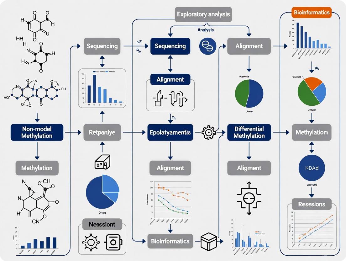
The following diagram illustrates a generalized workflow for DNA methylation analysis in non-model organisms, highlighting critical decision points and methodological alternatives at each stage:
Non-Invasive Epigenetic Analysis Pathway
For ecological and conservation applications, non-invasive sampling requires specialized processing pathways as demonstrated in wild capuchin research [3]:
Challenges and Future Directions
Technical and Analytical Limitations
Epigenetic research in non-model organisms faces several significant challenges that require methodological innovation and adapted approaches:
Genomic Resource Limitations: The absence of high-quality reference genomes remains a primary obstacle for precise methylation mapping in non-model organisms. While global methylation analyses circumvent this limitation, they sacrifice genomic context and locus-specific information. Potential solutions include using chromosomal-level assemblies from related species, long-read sequencing technologies for de novo genome assembly, and reference-free analysis methods that identify consistent methylation patterns across samples without positional mapping [4].
Sample Quality and Quantity Issues: Non-model organisms often present challenges in sample collection, particularly for wild populations, endangered species, or organisms with small body sizes. Non-invasive sampling techniques yield fragmented DNA in limited quantities, requiring specialized protocols for DNA extraction and library preparation [3]. Methods like multiple displacement amplification can increase DNA yield but may introduce biases in methylation patterns. EM-seq and optimized bisulfite conversion protocols offer gentler alternatives that preserve DNA integrity better than standard WGBS approaches [6].
Analytical Framework Gaps: Most bioinformatics tools for DNA methylation analysis were developed for model organisms with well-annotated genomes and may perform poorly on non-model systems [4]. There is a critical need for specialized software that accommodates incomplete genomes, supports comparative analyses across diverse taxa, and enables exploratory (rather than hypothesis-driven) research approaches. Tools like BSXplorer represent steps in this direction, but more comprehensive solutions are needed [4].
Standardization and Reproducibility
The lack of standardized protocols for non-model organism epigenetics presents challenges for reproducibility and cross-study comparisons. Variation in DNA extraction methods, bisulfite conversion efficiency, sequencing depth, and bioinformatic pipelines can significantly impact results. Future efforts should focus on establishing best practice guidelines for:
- Sample collection and preservation under field conditions
- DNA quality assessment for degraded samples
- Reference-free methylation analysis
- Cross-species comparative frameworks
- Data reporting standards for improved reproducibility
Integration with Other Omics Approaches
The full potential of non-model organism epigenetics will be realized through integration with complementary omics technologies. Multi-omics approaches combining DNA methylation analysis with transcriptomics, proteomics, and metabolomics can provide mechanistic insights into how epigenetic variation influences phenotype. For example, studies linking methylation patterns to gene expression changes in response to environmental stressors can reveal functional epigenetic mechanisms in ecological contexts. Similarly, connecting epigenetic markers with physiological measurements or behavioral observations can illuminate the functional consequences of epigenetic variation in natural populations.
Non-model organisms represent both a challenge and tremendous opportunity for advancing epigenetic research. The methodological frameworks presented in this review provide multiple entry points for exploring DNA methylation patterns in organisms lacking extensive genomic resources or established laboratory protocols. From global methylation analysis using mass spectrometry to adapted sequencing approaches and specialized bioinformatics tools, researchers now have an expanding toolkit for epigenetic discovery beyond traditional model systems.
The unique biological features, ecological adaptations, and evolutionary diversity of non-model organisms offer unparalleled opportunities to understand how epigenetic mechanisms operate across the tree of life. By embracing these opportunities and addressing the associated methodological challenges, researchers can uncover fundamental principles of epigenetic regulation that may be invisible within the constrained context of traditional model organisms. As technologies continue to advance and methodologies become more accessible, non-model organism epigenetics promises to transform our understanding of gene-environment interactions, adaptive evolution, and the dynamic regulation of genomes across diverse biological contexts.
DNA methylation, the addition of a methyl group to the fifth carbon of a cytosine base (5-methylcytosine, 5mC), constitutes a fundamental epigenetic mechanism that regulates gene expression without altering the underlying DNA sequence [8] [9]. This reversible modification is crucial for a myriad of biological processes, including genomic imprinting, repression of transposable elements, cell differentiation, and the maintenance of cellular identity [8] [10]. In mammals, DNA methylation occurs primarily at cytosine-guanine dinucleotides (CpG sites), with genomic patterns that are dynamically regulated by opposing enzymatic activities [9].
The interpretation and maintenance of these methylation patterns are governed by three principal classes of proteins: "writers" that install the methyl mark, "erasers" that remove it, and "readers" that recognize and translate it into functional biological states [11] [12]. This tripartite system facilitates a responsive and plastic regulatory layer over the genetic code. While these core principles are largely conserved across the animal kingdom, recent comparative epigenomic studies across 580 animal species have revealed both deeply conserved and lineage-specific features, underscoring the dynamic evolution of DNA methylation machinery and its role in shaping species-specific traits [13] [10]. This whitepaper provides an in-depth technical guide to these core components, with a specific focus on implications for research in non-model organisms.
The Core Machinery: Writers, Erasers, and Readers
The establishment, interpretation, and removal of DNA methylation marks are executed by a coordinated set of enzymatic and binding proteins.
Writers: DNA Methyltransferases (DNMTs)
DNA methyltransferases (DNMTs) are the "writer" enzymes that catalyze the transfer of a methyl group from the universal methyl donor, S-adenosyl-methionine (SAM), to the fifth carbon of a cytosine base, producing 5-methylcytosine (5mC) [9] [11].
- DNMT1 is the primary maintenance methyltransferase in somatic cells. It exhibits a strong preference for hemi-methylated DNA, making it highly effective at copying DNA methylation patterns from the parent strand to the newly synthesized daughter strand during DNA replication, thereby ensuring the faithful inheritance of methylation patterns across cell divisions [9].
- DNMT3A and DNMT3B are de novo methyltransferases responsible for establishing new methylation patterns ab initio, particularly during embryonic development when the methylome is erased and reprogrammed [9]. Their activity is essential for setting up cell-type-specific methylation landscapes.
- DNMT3L is a catalytically inactive regulatory cofactor that stimulates the de novo methylation activity of DNMT3A and DNMT3B, especially in the germline [11].
Table 1: Core DNA Methylation "Writer" Enzymes in Mammals
| Enzyme | Primary Type | Key Function | Catalytic Activity |
|---|---|---|---|
| DNMT1 | Maintenance | Copies methylation patterns after DNA replication | Active |
| DNMT3A | De novo | Establishes new methylation patterns | Active |
| DNMT3B | De novo | Establishes new methylation patterns | Active |
| DNMT3L | Regulatory cofactor | Stimulates DNMT3A/B activity | Inactive |
Erasers: Active DNA Demethylation Pathways
While DNA methylation was long considered a stable modification, the discovery of enzymes capable of active DNA demethylation revealed the dynamic nature of this epigenetic mark. The erasure of 5mC is not performed by a direct "demethylase" but rather through a multi-step process involving the TET (Ten-Eleven Translocation) family of enzymes [11].
TET enzymes (TET1, TET2, TET3) are α-ketoglutarate-dependent dioxygenases that iteratively oxidize 5mC to form 5-hydroxymethylcytosine (5hmC), 5-formylcytosine (5fC), and 5-carboxylcytosine (5caC) [11]. These oxidized methylcytosine derivatives are not recognized by the maintenance methyltransferase DNMT1 and can be replaced by an unmodified cytosine via base excision repair (BER) pathways, thereby achieving active DNA demethylation [11]. This pathway is critical for both targeted gene activation and global epigenetic reprogramming events, such as those occurring in early embryogenesis and in primordial germ cells.
Readers: Methyl-Binding Proteins (MBPs)
The biological message encoded by DNA methylation is interpreted by "reader" proteins that specifically recognize and bind to methylated DNA. These readers then recruit additional protein complexes to execute downstream transcriptional outcomes, primarily gene silencing [9].
The classic readers are the Methyl-CpG-Binding Domain (MBD) family of proteins, which includes MeCP2, MBD1, MBD2, and MBD4 [9] [12]. Upon binding to methylated CpG sites, these proteins recruit co-repressor complexes containing factors such as histone deacetylases (HDACs) and histone methyltransferases [9]. HDACs remove activating acetyl marks from histone tails, leading to a more condensed chromatin state. This mechanism exemplifies the profound crosstalk between DNA methylation and histone modifications, where DNA methylation readers directly influence the histone code to reinforce a repressive chromatin environment [9] [14].
Evolutionary Perspectives Across Species
Comparative epigenomic analyses are revolutionizing our understanding of how DNA methylation systems have evolved and how they contribute to phenotypic diversity across the tree of life.
Conservation and Divergence of Methylation Patterns
A landmark study profiling DNA methylation in 580 animal species (535 vertebrates and 45 invertebrates) revealed a broadly conserved link between DNA methylation and the underlying genomic DNA sequence, but with two major evolutionary transitions: one at the emergence of the first vertebrates and another with the rise of reptiles [10]. This suggests significant shifts in how the methylation machinery interacts with the genome at these pivotal points.
Despite these shifts, tissue-specific DNA methylation signatures are deeply conserved. Cross-species comparisons demonstrate that methylation patterns can distinguish tissue types (e.g., heart vs. liver) more strongly than they distinguish individuals within the same species for fish, birds, and mammals [10]. This indicates a fundamental and evolutionarily ancient role for DNA methylation in defining and maintaining cellular identity.
Role in Species-Trait Evolution and Plasticity
DNA methylation is increasingly recognized not just as a static mark but as a dynamic mediator of phenotypic plasticity and evolutionary adaptation [15].
- Short-Term Acclimation: Methylation states can change rapidly in response to environmental stressors such as temperature, salinity, or diet, potentially facilitating acclimation within a single generation [15]. These changes are often transient, highlighting the plasticity of the epigenome.
- Stable Phenotypic Evolution: In some cases, environmentally induced methylation changes can lead to stable, ecologically important phenotypes within a generation, such as sex determination in some fish and reptiles, or caste determination in social insects [15].
- Genomic Evolution: There is a reciprocal relationship between genetic and epigenetic evolution. DNA methylation can be genetically controlled, but it can also promote mutations (e.g., deamination of 5mC to T) and contribute to genomic sequence evolution over longer timescales [15]. Furthermore, promoter methylation changes have been correlated with the evolution of species-specific traits, such as body patterning [13].
Table 2: Evolutionary Roles of DNA Methylation Across Timescales
| Timescale | Role of DNA Methylation | Example |
|---|---|---|
| Short-Term (Acclimation) | Rapid, reversible response to environmental cues. | Thermal stress response in fish [15]. |
| Medium-Term (Phenotypic Evolution) | Stable encoding of functional phenotypes within a generation. | Environmental sex determination in reptiles [15]. |
| Long-Term (Genomic Evolution) | Contribution to mutation rates and genomic sequence change; species-specific trait evolution. | Correlation with body patterning evolution in mammals [13]. |
Methodologies for Exploratory Analysis in Non-Model Organisms
For researchers investigating methylation in non-model organisms, where high-quality reference genomes are often unavailable, specific methodologies have been developed.
Reference-Genome-Independent Profiling with RRBS
Reduced Representation Bisulfite Sequencing (RRBS) is a powerful, cost-effective method for genome-scale DNA methylation profiling that is particularly suited for cross-species studies [10]. The protocol is as follows:
- Digestion: Genomic DNA is digested with the MspI restriction enzyme (cuts at CCGG sites regardless of methylation). This enzyme has a short, highly common target sequence, ensuring consistent performance across a wide range of species [10].
- Size Selection: The digested fragments are size-selected (typically 40-220 bp) via gel electrophoresis or bead-based clean-up. This step enriches for CpG-rich regions, including many gene promoters and regulatory elements.
- Bisulfite Conversion: The size-selected fragments are treated with sodium bisulfite, which deaminates unmethylated cytosines to uracils, while methylated cytosines remain unchanged. During subsequent PCR amplification, uracils are read as thymines (T).
- Library Preparation and Sequencing: Adapters are ligated, and the library is sequenced on a high-throughput platform.
- Bioinformatic Analysis: The sequencing reads are analyzed by comparing C-to-T conversion rates. A C in the original sequence that is read as a T indicates an unmethylated cytosine, while a C that remains a C indicates a methylated cytosine. Because RRBS fragments start and end at defined MspI sites, reads can be grouped and analyzed based on their sequence context alone, without the need for a reference genome assembly [10].
Emerging Single-Cell Multi-Omic Technologies
Cutting-edge technologies now allow for the simultaneous profiling of DNA methylation and histone modifications in the same single cell. The scEpi2-seq (single-cell Epi2-seq) method exemplifies this advance [16].
- Cell Permeabilization and Antibody Binding: Single cells are permeabilized, and a protein A-micrococcal nuclease (pA-MNase) fusion protein is tethered to specific histone modifications (e.g., H3K27me3, H3K9me3) using antibodies.
- MNase Digestion: MNase digestion is activated by adding Ca²âº, cleaving DNA around the bound nucleosomes. This releases fragments bearing the histone mark of interest.
- Adaptor Ligation and TAPS: The released fragments are repaired, A-tailed, and ligated to adaptors containing cell barcodes and unique molecular identifiers (UMIs). The material is then subjected to TET-assisted pyridine borane sequencing (TAPS). Unlike bisulfite sequencing, TAPS chemically converts 5mC to uracil, leaving the barcoded adaptors intact, which improves library complexity and quality [16].
- Library Prep and Multi-Omic Readout: The library is prepared via in vitro transcription, reverse transcription, and PCR. Sequencing data provides simultaneous readouts: mapped genomic locations reveal histone modification patterns, while C-to-T conversions identify methylated cytosines, all attributable to a single cell [16].
The Scientist's Toolkit: Key Research Reagents and Solutions
This section details essential reagents and tools for studying DNA methylation, drawing from the experimental protocols discussed.
Table 3: Essential Research Reagents for DNA Methylation Analysis
| Reagent / Tool | Function | Example Application |
|---|---|---|
| MspI Restriction Enzyme | Restriction enzyme for CpG-rich locus enrichment. | DNA fragmentation in RRBS protocol [10]. |
| Sodium Bisulfite | Chemical conversion of unmethylated C to U. | Distinguishing methylated vs. unmethylated cytosines in BS-seq/RRBS [10]. |
| Protein A-MNase (pA-MNase) | Fusion protein for targeted chromatin cleavage. | tethering to antibodies for histone mark profiling in scEpi2-seq [16]. |
| TET Enzymes / Pyridine Borane | Chemical conversion of 5mC to U. | Non-destructive 5mC detection in TAPS-based methods [16]. |
| Anti-Histone Modification Antibodies | Specific recognition of epigenetic marks. | Immunoprecipitation or tethering in scEpi2-seq (e.g., H3K27me3, H3K9me3) [16]. |
| DNMT Inhibitors | Small molecule inhibition of DNA methyltransferases. | Experimental demethylation (e.g., 5-Azacytidine) [12]. |
| MBD Domain Proteins | Affinity enrichment of methylated DNA. | Methylated DNA pulldown for downstream analysis [9]. |
| 2-Azidoethanol-d4 | 2-Azidoethanol-d4, MF:C₂HD₄N₃O, MW:91.11 | Chemical Reagent |
| 10Z-Vitamin K2-d7 | 10Z-Vitamin K2-d7|Deuterated Research Standard | High-purity 10Z-Vitamin K2-d7 for research. An internal standard for LC-MS/MS analysis of Vitamin K2 metabolites. For Research Use Only. Not for human consumption. |
The core principles of DNA methylation—governed by the coordinated actions of writers, erasers, and readers—form a dynamic and complex regulatory system that is fundamental to genomic function and integrity. Research in non-model organisms, facilitated by reference-free and multi-omic technologies like RRBS and scEpi2-seq, is critically enriching our understanding of this system. These studies reveal that while the basic machinery is conserved, its deployment and evolutionary impact are fluid, contributing to both stable cellular memory and remarkable phenotypic plasticity. Integrating this epigenetic perspective is therefore indispensable for a unified understanding of evolution, development, and disease.
Evolutionary Conservation and Divergence in Methylation Patterns
Deoxyribonucleic acid (DNA) methylation, the addition of a methyl group to cytosine bases, is a fundamental epigenetic mechanism regulating gene expression and genomic stability across eukaryotic life [17]. Its study has evolved from a focus on model organisms to an expansive field that leverages non-model organisms to uncover the evolutionary principles governing epigenetic regulation. Research in diverse species—from marsupials and flatfish to wild primates—reveals a complex interplay between deeply conserved functions and lineage-specific adaptations in methylation systems. This whitepaper synthesizes recent findings from comparative epigenomics to provide a technical guide for researchers exploring methylation patterns in non-model systems, framing them within the context of a broader thesis on evolutionary epigenetics. It details conserved and divergent dynamics, provides standardized protocols for cross-species analysis, and offers a toolkit for exploratory research, aiming to bridge the gap between fundamental epigenetic knowledge and its application in comparative and translational biology.
Core Concepts: Conservation and Divergence in Methylation Systems
DNA methylation predominantly targets cytosine-phosphate-guanine (CpG) dinucleotides, though non-CpG methylation is also observed in some contexts [18]. Its functional impact is tightly linked to genomic location: methylation within gene promoter regions typically suppresses gene expression, whereas gene body methylation often associates with active transcription and plays a role in splicing and genomic stability [18] [17]. The distribution of CpG sites and their methylation status across the genome varies significantly among species, forming the basis for comparative evolutionary studies.
Conserved Patterns in Higher Vertebrates: Among higher vertebrates (amniotes), a "global" methylation pattern is highly conserved. This pattern is characterized by near-complete methylation of the genome, with the crucial exception of CpG islands located in promoter regions [17]. These promoter-associated CpG islands are typically unmethylated, allowing for potential gene activation. This system is so fundamental that the regulatory logic of promoter methylation is shared across humans, mice, rats, cows, dogs, and chickens [17]. Furthermore, the development of epigenetic clocks—predictive models of biological age based on DNA methylation patterns—has been successfully demonstrated in wild capuchin monkeys using non-invasive fecal samples. The high accuracy of these clocks (predicting age to within ~3.5% of lifespan) underscores a deeply conserved link between methylation changes and the aging process across mammals, including humans [3].
Divergent Patterns Across Evolutionary Lineages: In contrast to the global pattern of amniotes, many invertebrates, plants, and fungi exhibit a "mosaic" methylation pattern, where heavily methylated domains are interspersed with unmethylated regions [17]. A striking example of evolutionary divergence in methylation dynamics comes from embryogenesis. In eutherian mammals (placental mammals), the paternal genome undergoes active demethylation immediately after fertilization, followed by global passive demethylation, resulting in a hypomethylated early blastocyst [19]. This reprogramming is thought to be essential for resetting epigenetic memory and establishing totipotency. However, recent work in the marsupial Monodelphis domestica (the opossum) reveals a different paradigm. The opossum genome remains hypermethylated during cleavage stages, with demodification occurring later, being transient and modest in the epiblast but sustained in the trophectoderm [19]. This suggests that global erasure is not an absolute requirement for mammalian embryogenesis and highlights divergent uses of DNA demethylation during development.
Table 1: Comparative DNA Methylation Patterns Across Eukaryotes
| Organism Group | Example Species | Methylation Pattern | Key Features and Functional Roles |
|---|---|---|---|
| Higher Vertebrates (Amniotes) | Human, Mouse, Cow, Chicken [17] | Global | Genome-wide methylation except for promoter CpG islands; role in promoter-driven gene regulation is conserved. |
| Marsupials | Opossum (Monodelphis domestica) [19] | Divergent Embryonic | Genome remains hypermethylated during early cleavage; no global erasure; sustained hypomethylation in trophectoderm. |
| Flatfish | Turbot (Scophthalmus maximus) [20] | Dynamic Developmental | Stage-specific hypermethylation during metamorphosis climax; role in visual system remodeling. |
| Plants & Invertebrates | Arabidopsis, Fruit Fly (D. melanogaster) [17] | Mosaic | Methylation targeted to gene bodies and transposable elements; crucial for transcriptional silencing of repeats. |
| Non-Methylators | S. cerevisiae (Yeast), C. elegans (Nematode) [17] | Absent | Lack DNA methyltransferase genes and therefore genomic DNA methylation. |
Detailed Experimental Protocols for Cross-Species Methylation Analysis
Non-Invasive DNA Methylation Profiling for Wild Populations
Studying methylation in wild or non-model species often precludes invasive tissue sampling. The following protocol, adapted from research on wild capuchin monkeys, enables robust methylome analysis from fecal samples [3].
- Step 1: Sample Collection and Cell Sorting. Collect fresh fecal samples and immediately preserve them in a stabilizing buffer (e.g., RNAlater) to prevent DNA degradation. To enrich for host epithelial cells, the protocol employs Fluorescence-Activated Cell Sorting (FACS). A fluorescence-activated cell sorter is used to isolate specific cell populations from the homogenized fecal matter, effectively separating host intestinal cells from the complex microbial background [3].
- Step 2: DNA Extraction and Quality Control. Extract genomic DNA from the sorted cell population using a commercial kit designed for complex or low-quality samples. Assess DNA purity and quantity via spectrophotometry (e.g., NanoDrop) and fluorometry (e.g., Qubit). Verify DNA integrity using gel electrophoresis or a Fragment Analyzer [3] [18].
- Step 3: Targeted Methylation Sequencing. For non-invasive samples with fragmented DNA, whole-genome bisulfite sequencing can be inefficient. Instead, use a capture-based targeted approach like Twist Targeted Methylation Sequencing (TTMS). This method uses a probe set (e.g., designed for the human genome but with demonstrated cross-species applicability) to capture ~4 million CpG sites. Following capture, the library is prepared for sequencing using enzymatic methyl-sequencing (EM-seq), which avoids the DNA degradation associated with traditional bisulfite treatment by using enzymes to distinguish methylated from unmethylated cytosines [3] [18].
- Step 4: Bioinformatic Analysis. Process the sequenced reads: align to a reference genome (or a closely related species' genome), and perform methylation calling to generate a methylation level (beta-value) for each CpG site. Downstream analysis can include building an epigenetic clock with an elastic net regression model, identifying differentially methylated regions (DMRs), and annotating DMRs to genomic features [3] [21].
Base-Resolution Methylation Mapping in Embryonic Tissues
Investigating dynamic processes like embryogenesis requires high-resolution maps from low-input material.
- Step 1: Low-Input Sample Preparation. Manually isolate gametes or preimplantation embryos under a microscope. For marsupial models like the opossum, embryos are collected at precise embryonic days corresponding to key developmental milestones (e.g., embryonic genome activation, lineage specification) [19].
- Step 2: Library Preparation and Sequencing. Utilize low-input bisulfite sequencing (BS-seq). This involves bisulfite converting the minimal genomic DNA, which deaminates unmethylated cytosines to uracils (read as thymines during sequencing), while methylated cytosines remain unchanged. Libraries are prepared with protocols optimized for low DNA quantities and sequenced on a high-throughput platform [19].
- Step 3: Single-Cell Multi-Omics for Lineage Resolution. To dissect cell-type-specific methylation within an embryo, employ single-cell multi-omics. This technique allows for the simultaneous profiling of DNA methylation and transcriptome from the same single cell. Cells are isolated from embryos at different stages (e.g., pre- and post-lineage divergence) and processed using a commercial single-cell multi-ome kit. This enables the direct correlation of methylation status with gene expression in individual cells of the epiblast and trophectoderm [19].
- Step 4: Data Integration and Comparative Analysis. Generate base-resolution methylation maps for each developmental stage and cell type. Perform differential methylation analysis between stages and lineages. Compare these dynamics to known benchmarks from model eutherians like mice to identify conserved and species-specific reprogramming events [19].
The Scientist's Toolkit: Essential Reagents and Technologies
Selecting the appropriate methodology is critical for successful methylation analysis, especially with challenging samples from non-model organisms. The table below summarizes key solutions.
Table 2: Research Reagent Solutions for DNA Methylation Analysis
| Tool Category | Specific Product/Technology | Function and Application | Key Considerations |
|---|---|---|---|
| Methylation Profiling | Twist Targeted Methylation Sequencing (TTMS) [3] | Targeted capture of ~4 million CpG sites; ideal for fragmented DNA (e.g., from feces). | Uses human probes with cross-species applicability; combines with EM-seq. |
| Bisulfite-Free Sequencing | Enzymatic Methyl-Sequencing (EM-seq) [18] | Bisulfite-free whole-methylome profiling; preserves DNA integrity. | Higher concordance with WGBS; better for low-input/long-range methylation. |
| Third-Generation Sequencing | Oxford Nanopore Technologies (ONT) [18] [22] | Direct detection of modifications during sequencing; no conversion needed. | Enables long-reads, access to complex regions; requires high DNA input. |
| Global Methylation Analysis | Acid Hydrolysis & UHPLC-HRMS [2] | Quantifies global 5mC levels; does not provide locus-specific data. | Rapid, cost-effective; ideal for highly methylated DNA where enzymes fail. |
| Bioinformatic Tools | MethylomeMiner [22] | Processes nanopore methylation calls; assigns sites to genomic features. | Python-based, integrates with pangenome data for population-level analysis. |
| (Z)-Roxithromycin-d7 | (Z)-Roxithromycin-d7, MF:C₄₁H₆₉D₇N₂O₁₄, MW:828.09 | Chemical Reagent | Bench Chemicals |
| 5-Carboxy Imazapyr | 5-Carboxy Imazapyr | High-purity 5-Carboxy Imazapyr for research. This product is For Research Use Only (RUO). Not for human or veterinary use. | Bench Chemicals |
Signaling and Metabolic Pathways Involving DNA Methylation
Methylation Dynamics in Flatfish Metamorphosis
The dramatic remodeling of the visual system during turbot metamorphosis provides a powerful model of how methylation regulates adaptive development. Research using Reduced Representation Bisulfite Sequencing (RRBS) has revealed that the climax stage of metamorphosis is marked by widespread stage-specific hypermethylation, coinciding with the upregulation of the de novo methyltransferase gene dnmt3ab [20]. This wave of methylation is implicated in the remodeling of both the migrating and non-migrating eyes as the fish adapts to a benthic lifestyle.
A key finding is the divergent methylation and expression of transcription factors essential for retinal ganglion cell (RGC) development, such as eomesa and tbr1b, between the migrating and non-migrating eyes [20]. RGCs form the optic nerve, connecting the eye to the brain. The differential epigenetic regulation of these genes likely underlies the asymmetric development of the visual pathway, potentially explaining anatomical differences like the shorter optic nerve in the migrating eye. Furthermore, while genes involved in the phototransduction cascade did not show methylation-linked regulation, their expression profiles shifted as expected: rod-specific genes (for low-light vision) increased, and cone-specific genes decreased post-metamorphosis [20]. This indicates that methylation's primary role in this context is in guiding the structural remodeling of the visual system rather than directly regulating the light-sensing apparatus itself.
The comparative analysis of DNA methylation across the tree of life reveals a nuanced picture of evolutionary dynamics. Deeply conserved mechanisms, such as the regulatory logic of promoter methylation in higher vertebrates and the link between methylation and aging, underscore the fundamental role of this epigenetic mark in animal biology. Simultaneously, profound divergences, exemplified by the alternative reprogramming strategies in marsupial embryos or the context-specific methylation dynamics during flatfish metamorphosis, highlight the plasticity of epigenetic systems. For researchers engaged in exploratory analysis of non-model organisms, this duality is paramount. It necessitates robust, adaptable methodologies—from non-invasive sampling to bisulfite-free sequencing—that can be applied across diverse species. The findings and protocols detailed herein provide a framework for such investigations, emphasizing that the integration of evolutionary context with advanced epigenetic tools is key to unlocking the full functional significance of DNA methylation patterns in shaping biological diversity, health, and disease. The ongoing expansion of epigenomics into wild and non-traditional species promises to further refine our understanding of what is fundamental and what is flexible in the epigenetic regulation of life.
Linking Methylation to Phenotypic Diversity and Environmental Adaptation
DNA methylation, the addition of a methyl group to cytosine or adenine bases, represents a fundamental epigenetic mechanism that influences gene expression without altering the underlying DNA sequence [23]. In the context of ecology and evolutionary biology, DNA methylation provides a potential molecular mechanism for organisms to rapidly respond to environmental challenges, potentially facilitating adaptation [24]. While earlier epigenetic research focused on model organisms and biomedical applications, advances in sequencing technologies and analytical methods now enable detailed investigation of DNA methylation in non-model species [25]. This technical guide explores the current methodologies, key findings, and analytical frameworks for studying the role of DNA methylation in generating phenotypic diversity and promoting environmental adaptation in natural populations.
The study of DNA methylation in non-model organisms presents unique challenges and opportunities. Unlike traditional model systems, non-model species often lack reference genomes, standardized protocols, and established bioinformatic pipelines [25]. However, they offer unparalleled insights into how epigenetic mechanisms operate in natural environments and under realistic selective pressures. This guide synthesizes current approaches for investigating the link between methylation variation, phenotypic diversity, and environmental adaptation, with particular emphasis on technical considerations for research in non-model systems.
Core Concepts: Methylation as an Adaptive Mechanism
Functional Roles of DNA Methylation
DNA methylation serves distinct biological functions across taxa. In eukaryotes, cytosine methylation predominantly occurs in CpG dinucleotides and plays crucial roles in gene regulation, genomic imprinting, and silencing transposable elements [23] [26]. In plants, methylation in CHG and CHH contexts (where H is A, T, or C) provides additional regulatory complexity, particularly for controlling transposable elements [27]. Bacterial systems utilize additional methylation forms, including 6-methyladenine (6mA) and 4-methylcytosine (4mC), primarily involved in restriction-modification systems and gene regulation [28].
The potential for DNA methylation to contribute to adaptive processes stems from several key characteristics: its responsiveness to environmental cues, influence on gene expression, and the heritable nature of certain methylation marks [24]. Environmentally induced methylation changes can create phenotypic heterogeneity that provides substrate for selection, potentially leading to consistent methylation patterns across generations in stable environmental conditions [24]. Additionally, methylation can increase mutation rates at targeted cytosines, potentially capturing beneficial epigenetic variants as genetic mutations over evolutionary time [24].
Evidence for Environmentally Induced Methylation Variation
Recent studies across diverse taxa provide compelling evidence for environmentally associated methylation variation. Research on Arabidopsis lyrata transplanted between lowland and alpine field sites revealed that gene expression is highly plastic, with many more genes differentially expressed between field sites than between populations [27]. While DNA methylation at genic regions was largely insensitive to the environment in this system, transposable elements (TEs) showed significant environmental effects, with higher expression and methylation levels at high-altitude sites [27]. This suggests a broad-scale TE activation under environmental stress, potentially creating novel heritable variation.
In marine macroalga Ulva mutabilis, methylation patterns change in the presence or absence of co-occurring bacterial symbionts that release growth- and morphogenesis-promoting factors [2]. Similarly, wild baboon populations exhibit methylation differences associated with early life experiences and resource base variation [25]. These consistent findings across diverse systems highlight the potential for methylation to mediate organism-environment interactions.
Table 1: Documented Cases of Environmentally Associated DNA Methylation Variation
| Species | Environmental Factor | Methylation Response | Functional Consequence |
|---|---|---|---|
| Arabidopsis lyrata [27] | Altitude (lowland vs alpine) | Increased TE methylation and expression | Potential creation of novel heritable variation |
| Ulva mutabilis [2] | Bacterial symbionts | Global changes in cytosine methylation | Altered growth and morphogenesis |
| Baboons (Papio cynocephalus) [25] | Resource base (wild vs human-food) | Differential methylation at specific loci | Unknown fitness consequences |
| Three-spine stickleback [29] | Freshwater vs saltwater adaptation | Population-specific methylation patterns | Potential contribution to local adaptation |
| Heliosperma plants [29] | Alpine vs sub-alpine habitats | Conserved methylation profiles despite ecological divergence | Developmental constraints on methylation |
Quantitative Evidence: Methylation Patterns in Environmental Adaptation
Empirical studies reveal consistent patterns in how methylation variation associates with environmental gradients. A critical finding across multiple systems is that despite the potential for rapid epigenetic change, methylation patterns often show remarkable conservation, suggesting evolutionary constraints [29].
Research on Heliosperma plants adapted to divergent ecological conditions (alpine vs. sub-alpine habitats) revealed surprisingly consistent methylation profiles between species, pointing to significant molecular or developmental constraints acting on DNA methylation variation [29]. This constitutive stability indicates that not all genomic regions are equally prone to environmentally induced methylation changes, with certain loci potentially buffered against epigenetic perturbation.
In humans, a comprehensive methylation atlas of normal cell types demonstrated that methylation patterns are extremely robust across individuals, with less than 0.5% of genomic blocks showing substantial variation across donors [26]. This conservation highlights the fundamental role of DNA methylation in maintaining cell identity and suggests that most interindividual methylation variation occurs at specific regulatory loci rather than affecting the entire genome.
Table 2: Quantitative Patterns of Methylation Variation in Response to Environmental Factors
| Pattern Category | Example System | Key Finding | Technical Approach |
|---|---|---|---|
| Tissue/Cell Specificity | Human cell types [26] | >99.5% methylation conservation across individuals; tissue-specific patterns at enhancers | Whole-genome bisulfite sequencing |
| Environmental Plasticity | Arabidopsis lyrata [27] | Gene expression highly plastic; TE methylation responsive to altitude | Whole-genome bisulfite & transcriptome sequencing |
| Evolutionary Divergence | Heliosperma species [29] | High methylation conservation despite ecological divergence | bsRADseq |
| Taxonomic Variation | Bacteria vs Eukaryotes [28] | 6mA and 4mC dominant in bacteria; 5mC predominant in eukaryotes | Nanopore sequencing |
| Temporal Stability | Baboons [25] | Early life adversity associated with stable methylation differences later in life | Reduced representation bisulfite sequencing |
Methodological Approaches for Non-Model Systems
Sequencing-Based Methylation Analysis
The advent of high-throughput sequencing technologies has revolutionized methylation analysis in non-model organisms. The following experimental workflows represent the most widely applied approaches:
Whole-Genome Bisulfite Sequencing (WGBS) provides base-resolution methylation data across the entire genome but requires substantial sequencing depth and computational resources [27] [26]. The standard protocol involves: (1) DNA extraction and quality control; (2) bisulfite conversion using sodium bisulfite (converts unmethylated cytosines to uracil); (3) library preparation and high-throughput sequencing; (4) alignment to a reference genome; and (5) methylation calling and differential analysis [27]. This approach is particularly valuable for organisms with reference genomes and when comprehensive methylation mapping is required.
Reduced Representation Bisulfite Sequencing (RRBS) offers a cost-effective alternative by targeting CpG-rich regions through restriction enzyme digestion (typically Mspl) [25]. This method reduces sequencing costs while capturing methylation information at functionally relevant genomic regions, making it suitable for population-level studies with multiple individuals.
Bisulfite-Converted Restriction Site Associated DNA Sequencing (bsRADseq) combines RADseq with bisulfite sequencing, providing a flexible reduced-representation approach that doesn't require a reference genome [29]. The methodology involves: (1) genomic DNA digestion with selected restriction enzymes; (2) bisulfite conversion of restriction fragments; (3) library preparation and sequencing; and (4) construction of synthetic references for mapping if no reference genome is available. This approach is particularly advantageous for non-model organisms with large genomes or when studying many individuals across populations.
Single-Molecule Real-Time Bisulfite Sequencing (SMRT-BS) leverages third-generation sequencing to achieve long read lengths (up to ~1.5 kb) for targeted CpG methylation analysis [30]. The protocol includes: (1) bisulfite conversion of genomic DNA; (2) amplification of bisulfite-treated DNA using region-specific primers; (3) re-amplification with sample-specific barcodes for multiplexing; (4) SMRT sequencing; and (5) CpG methylation quantitation. This method excels when haplotypic information or long-range methylation patterns are needed.
Nanopore Sequencing enables direct detection of DNA modifications without bisulfite conversion by monitoring changes in electrical current as DNA passes through protein nanopores [28]. This approach can detect all three common bacterial methylation types (5mC, 4mC, and 6mA) with equivalent sequencing depth and is particularly valuable for organisms where bisulfite conversion may be challenging [28].
Mass Spectrometry-Based Approaches
As an alternative to sequencing-based methods, mass spectrometry provides quantitative analysis of global DNA methylation levels without sequence context. A recently developed approach uses acid hydrolysis of DNA followed by liquid chromatography and Orbitrap mass spectrometry to directly quantify methyl-modified nucleobases (5-methylcytosine and 6-methyladenine) along with their unmodified counterparts [2].
This method offers several advantages for non-model organisms: (1) it requires only small amounts of DNA (as little as 100 ng); (2) it provides absolute quantification of modification levels; (3) it is independent of the total methylation rate; and (4) it doesn't require reference genomes or complex bioinformatics [2]. The limitations include the lack of locus-specific information and inability to detect sequence context of modifications. This approach is ideal for rapid screening of global methylation differences across multiple samples or treatment conditions.
Bioinformatics and Statistical Considerations
Analysis of methylation data from non-model organisms presents unique bioinformatic challenges. For bisulfite sequencing data, specialized alignment tools like Bismark or BS-Seeker account for C-to-T conversions following bisulfite treatment [31]. For species without reference genomes, de novo assembly of bisulfite-converted reads is particularly challenging, making reduced-representation approaches like bsRADseq advantageous [29].
Statistical analysis must account for the compositional nature of methylation data (percentages between 0-100%) and the potential confounding effects of genetic variation. Methods like MACAU incorporate kinship and population structure into methylation analysis, reducing false positives in structured natural populations [25]. Power analysis is particularly important, as studies with insufficient samples often yield unreliable results—generally, investing in more samples provides better returns than deeper sequencing [25].
Visualizing Methylation-Environment Relationships: Conceptual Framework and Technical Workflows
Conceptual Framework for Methylation in Environmental Adaptation
The relationship between environmental variation, DNA methylation, and phenotypic outcomes can be visualized as a conceptual framework that integrates molecular, organismal, and evolutionary processes:
Technical Workflow for Methylation Analysis in Non-Model Organisms
The experimental pipeline for studying methylation in non-model systems involves multiple steps from sample collection to biological interpretation:
The Scientist's Toolkit: Essential Research Reagents and Solutions
Successful investigation of methylation-environment relationships requires careful selection of laboratory reagents and computational tools. The following table summarizes key solutions for studying DNA methylation in non-model organisms:
Table 3: Essential Research Reagents and Solutions for Methylation Studies
| Category | Specific Solution | Function/Application | Considerations for Non-Model Organisms |
|---|---|---|---|
| Bisulfite Conversion Kits | Epigentek Methylamp, Qiagen EpiTect | Convert unmethylated cytosines to uracil for sequencing-based methods | Test conversion efficiency without reference genome using spike-in controls [30] |
| Restriction Enzymes | Mspl (RRBS), various (bsRADseq) | Genome reduction for cost-effective population studies | Enzyme selection affects genomic coverage; test multiple enzymes for optimal representation [29] |
| Library Prep Kits | NEBNext Enzymatic Methyl-seq (EM-seq) | Alternative to bisulfite conversion with less DNA damage | Enables use of degraded samples (e.g., field collections) [31] |
| Sequencing Platforms | Illumina (WGBS, RRBS), PacBio (SMRT-BS), Oxford Nanopore | Detection of methylation patterns | Nanopore detects all modification types without conversion; ideal for bacterial methylation [28] |
| Mass Spectrometry | Orbitrap LC-MS/MS | Global quantification of modified bases | Requires minimal genomic information; ideal for highly methylated genomes [2] |
| Bioinformatic Tools | Bismark, MethylKit, MACAU, wgbstools | Read alignment, methylation calling, differential analysis | MACAU accounts for population structure in natural populations [25] |
| Reference Databases | Custom genome assemblies, synthetic references | Mapping and annotation of methylation data | For species without genomes, create synthetic references from RAD loci [29] |
| GNE-0877-d3 | GNE-0877-d3, MF:C₁₄H₁₃D₃F₃N₇, MW:342.34 | Chemical Reagent | Bench Chemicals |
| 22Z-Paricalcitol | 22Z-Paricalcitol|C27H44O3 | 22Z-Paricalcitol is a stereoisomer for research. This product is for Research Use Only (RUO) and is not intended for personal use. | Bench Chemicals |
The study of DNA methylation in environmental adaptation continues to evolve with rapid methodological advancements. Current evidence suggests that DNA methylation contributes to phenotypic diversity and environmental adaptation through multiple mechanisms: (1) direct regulation of environmentally responsive genes; (2) control of transposable element activity under stress conditions; and (3) creation of heritable variation that can be subject to selection. However, the relative importance of genetic versus epigenetic variation in adaptive processes remains a rich area for future investigation.
Emerging technologies like long-read sequencing and mass spectrometry-based approaches are overcoming previous limitations in studying non-model organisms. These tools, combined with sophisticated statistical methods that account for population structure and genetic relatedness, promise to provide unprecedented insights into the role of epigenetic mechanisms in evolution. Future research should focus on integrating multiple molecular approaches (epigenomic, transcriptomic, and genomic) with field-based phenotypic measurements to establish causal links between methylation variation, phenotypic traits, and fitness outcomes in natural environments.
As the field progresses, standardization of methodologies and data analysis pipelines will be crucial for comparing results across studies and taxa. Particular attention should be paid to distinguishing between causative epigenetic changes and correlated consequences of genetic variation or environmental induction. Through carefully designed studies that leverage the methodologies outlined in this guide, researchers can continue to unravel the complex relationship between DNA methylation, phenotypic diversity, and environmental adaptation across the tree of life.
The study of epigenetic mechanisms in non-model organisms is crucial for understanding the evolutionary landscape of gene regulation and environmental adaptation. DNA methylation, a key epigenetic mark, plays a fundamental role in controlling gene activity without altering the underlying DNA sequence. This case study focuses on the marine macroalga Ulva mutabilis, a pivotal species in coastal ecosystems worldwide and a emerging model organism for epigenetic research in non-model systems [32] [2] [33]. U. mutabilis possesses a remarkably high level of global DNA methylation, characterized by densely methylated CpG content, making it an excellent subject for investigating methylation dynamics [32] [2]. The exploration of these dynamics provides critical insights into how environmentally responsive epigenetic mechanisms operate in organisms beyond traditional models, bridging fundamental knowledge gaps in epigenetics.
Global DNA Methylation Analysis inUlva mutabilis
Quantitative Epigenetics Approach
Recent methodological advances have enabled precise quantification of DNA methylation in highly methylated algal genomes. A novel approach based on acid hydrolysis of DNA coupled with ultra-high-performance liquid chromatography and high-resolution mass spectrometry (UHPLC-HRMS) has been developed specifically for global methylome analysis in Ulva mutabilis [32] [2]. This method offers significant advantages over conventional sequencing techniques for quantitative assessment, providing direct, rapid, cost-efficient, and sensitive quantification of the methyl-modified nucleobase 5-methylcytosine (5mC) along with unmodified nucleobases [32].
Table 1: Key Features of the UHPLC-HRMS Global Methylation Analysis Method
| Feature | Description | Advantage |
|---|---|---|
| Hydrolysis Method | HCl-based chemical hydrolysis | Avoids incomplete digestion issues of enzymatic approaches; robust for highly methylated DNA |
| Detection Technique | UHPLC-HRMS | Enables absolute quantification independent of sequence context |
| Data Output | Global methylation percentage | Simple comparison across samples and conditions |
| Sample Requirement | Small DNA amounts | Suitable for limited biological material |
| Bioinformatic Demand | Minimal | No complex sequencing data analysis required |
| Throughput | High | Enables comparison of multitude of samples |
This technical approach addresses a critical methodological gap, as conventional enzymatic hydrolysis methods often demonstrate constrained efficiency with highly methylated DNA samples like those from U. mutabilis [2]. The chemical hydrolysis protocol effectively releases methylated and unmethylated nucleobases without destroying methylation patterns, enabling accurate global methylation assessment.
Research Reagent Solutions
Table 2: Essential Research Reagents for Ulva Methylation Studies
| Reagent/Category | Specific Examples | Function/Application |
|---|---|---|
| DNA Standards | 100% unmodified or methylated cytosines (Zymo Research) | Quantification standards and method calibration |
| Internal Standards | 2ˈ-deoxycytidine-13C1, 15N2; 2ˈ-deoxy-5-methylcytidine-13C1, 15N2 | Isotope-labeled internal standards for precise quantification |
| Chemical Standards | Cytosine, 5-methylcytosine, 2ˈ-deoxycytidine (Sigma-Aldrich) | Reference compounds for method development |
| DNA Extraction | Qiagen DNeasy Plant Mini Kit with RNase I | High-quality, RNA-free DNA preparation |
| Cultivation Media | Ulva Culture Medium (UCM) | Standardized algal growth conditions |
| Bacterial Symbionts | Roseovarius sp. MS2, Maribacter sp. MS6 | Tripartite community studies; morphogenesis-promoting factors |
Experimental Design and Cultivation Conditions
Standardized Cultivation Protocol
The experimental framework for studying methylation dynamics in Ulva mutabilis requires strictly controlled cultivation conditions. The standard protocol involves maintaining the 'slender' morphotype (strain FSU-UM5-1) in Ulva Culture Medium (UCM) under defined parameters [32] [2]:
- Temperature: 18 ± 2°C
- Light Cycle: 17-hour light/7-hour dark period
- Light Intensity: 40-80 μmol photons mâ»Â²sâ»Â¹
- Culture Types: Axenic conditions or in presence of specific bacterial symbionts (Roseovarius sp. MS2 and Maribacter sp. MS6)
This highly standardized approach yields synchronized clonal populations with minimal variance among biological replicates, essential for robust epigenetic analysis [32]. The ability to culture U. mutabilis under both axenic and symbiotic conditions provides a unique opportunity to investigate cross-kingdom interactions and their epigenetic implications.
DNA Extraction and Hydrolysis Methodology
The sample preparation workflow involves critical steps to ensure accurate methylation quantification:
DNA Extraction: Genomic DNA is extracted from freeze-dried and homogenized algal tissue using the Qiagen DNeasy Plant Mini Kit with addition of 50 μg RNase I during cell lysis to ensure RNA-free preparation [32].
Acid Hydrolysis: One μg of extracted DNA undergoes HCl-based hydrolysis, optimizing the release of methylated and unmethylated nucleobases without formylated side-products that complicate analysis [2].
UHPLC-HRMS Analysis: The hydrolyzed samples are directly submitted to chromatographic separation and mass spectrometric detection, allowing simultaneous quantification of 5-methylcytosine and unmodified cytosine [32] [2].
Diagram 1: Experimental workflow for global DNA methylation analysis in Ulva mutabilis
Comparative Methylation Dynamics in Related Ulva Species
DNA Methylation Patterns in Ulva prolifera Under Stress
Research on the related species Ulva prolifera provides valuable insights into methylation dynamics under environmental stress. Whole-genome bisulfite sequencing (WGBS) analysis revealed that the U. prolifera genome exhibits approximately 1.18% cytosine methylation with distinct distribution patterns [34] [35]:
- CpG context: ~72% methylation
- CHG context: ~10% methylation
- CHH context: ~3% methylation
Under elevated temperature-light stress (30°C and 300 μmol photons mâ»Â²sâ»Â¹), U. prolifera showed significant hypomethylation in CpG contexts, while CHG and CHH methylation remained relatively stable [34] [35]. This stress-induced demethylation was particularly associated with transcriptionally active regions, revealing a negative correlation between CG methylation and gene expression patterns.
Functional Implications of Methylation Changes
The methylation changes observed in Ulva species under stress conditions have significant functional implications:
Transcriptional Regulation: CG hypomethylation under abiotic stress provokes transcriptional responses, facilitating expression of stress-responsive genes [34] [35]
Transposon Control: CHG and CHH methylation predominantly found in transposable elements and intergenic regions possibly contribute to genetic stability by restricting transposon activity during stress [34]
Metabolic Reprogramming: Stress conditions trigger upregulation of glycolytic pathway genes, with methylation changes potentially influencing this metabolic shift [34] [35]
Table 3: Stress-Induced Molecular Changes in Ulva Species
| Parameter | Normal Conditions | Stress Conditions | Functional Impact |
|---|---|---|---|
| Global CG Methylation | Higher (~72%) | Decreased (hypomethylation) | Enhanced transcriptional responsiveness |
| Glycolysis Genes | Basal expression | Upregulated (GCK, G6PC, GPI, etc.) | Metabolic adaptation to stress |
| Transposable Elements | Controlled methylation | Maintained CHG/CHH methylation | Genome stability preservation |
| Regenerative Capacity | Standard | Rapid spore ejection, new thalli formation | Stress memory and resilience |
| Antioxidant Systems | Basal levels | Increased peroxidase, variable catalase | Oxidative stress management |
Methodological Comparisons in DNA Methylation Analysis
Technical Approaches for Methylation Assessment
The study of DNA methylation in algae employs diverse methodological approaches, each with distinct advantages and limitations. Whole-genome bisulfite sequencing (WGBS) provides comprehensive, base-resolution methylation mapping but requires substantial resources and complex bioinformatics [5]. Reduced representation bisulfite sequencing (RRBS) offers a cost-effective alternative by focusing on CpG-rich regions but excludes distal regulatory elements [5]. The novel acid hydrolysis/UHPLC-HRMS approach enables rapid global methylation quantification but lacks locus-specific information [32] [2].
Emerging Technologies and Future Directions
Advanced computational approaches are increasingly being applied to DNA methylation research. Artificial intelligence and machine learning methods show promise for analyzing complex methylation datasets, with models like DeepCpG, MethylNet, and Deep6mA demonstrating capabilities in pattern recognition and prediction [36]. The integration of long-read sequencing technologies (Oxford Nanopore, PacBio SMRT) further expands the toolbox for epigenetic investigation in non-model organisms like Ulva [36] [2].
Diagram 2: Proposed mechanism of methylation-mediated stress response in Ulva
The investigation of methylation dynamics in Ulva mutabilis provides a paradigm for epigenetic research in non-model marine organisms. The high global methylation level characteristic of this species, coupled with responsive methylation changes under environmental stimuli, positions Ulva as an excellent model for understanding epigenetic mechanisms in aquatic environments. The methodological advancement of acid hydrolysis/UHPLC-HRMS for global methylation quantification addresses a critical technical need for studying highly methylated genomes, offering a robust alternative to sequencing-based approaches for quantitative assessment.
Future research directions should focus on integrating global methylation quantification with locus-specific analyses to comprehensively understand the spatial organization of epigenetic marks in the Ulva genome. Furthermore, exploring the transmission of stress-induced methylation changes across generations would provide valuable insights into the potential for transgenerational epigenetic inheritance in marine macroalgae. The tripartite community system of U. mutabilis with its bacterial symbionts offers an additional fascinating dimension for investigating cross-kingdom epigenetic interactions. These research avenues collectively advance our understanding of epigenetic regulation in non-model organisms and its role in environmental adaptation.
For decades, a fundamental dogma has governed the field of epigenetics: DNA methylation patterns are regulated primarily by pre-existing chromatin features rather than underlying DNA sequences. This understanding centered on self-reinforcing loops where existing methylation and histone modifications guide the maintenance of these same epigenetic marks during cell division. While this model effectively explains the stability of epigenetic states, it fails to account for how novel methylation patterns are generated during development and cellular differentiation [37] [38]. Recent groundbreaking research has uncovered a new mode of epigenetic targeting that represents a paradigm shift in our understanding of how DNA methylation is established. Studies of plant reproductive tissues have revealed that specific genetic sequences can directly instruct the establishment of DNA methylation patterns through the action of specialized transcription factors [37] [38] [39].
This discovery emerged from investigations into how distinct epigenomes are generated in reproductive tissues of Arabidopsis thaliana, where the RNA-directed DNA methylation (RdDM) machinery is targeted to different genomic locations by the CLASSY protein family. Researchers discovered that several REPRODUCTIVE MERISTEM (REM) transcription factors are required for methylation at CLASSY3-dependent loci [37]. These factors, designated REM INSTRUCTS METHYLATION (RIMs), directly bind to specific DNA sequences and recruit the methylation machinery, demonstrating for the first time that genetic information can directly guide epigenetic patterning in plants [37] [38] [39]. This whitepaper examines the mechanistic insights from this discovery, provides detailed experimental protocols for studying these phenomena, and discusses the implications for epigenetic research in non-model organisms.
Mechanistic Insights: The RIM-CLSY3 Regulatory Module
Core Components of the Genetic Targeting System
The genetic regulation of DNA methylation in plant reproductive tissues centers on a specialized molecular module comprising specific DNA sequences, transcription factors, and epigenetic machinery components. Through forward genetic screens in Arabidopsis, researchers identified that several REM transcription factors are essential for establishing tissue-specific methylation patterns [37] [38]. These RIM proteins function with remarkable specificity—RIM16 and RIM22 predominantly regulate HyperTE loci in anthers, while a triple CRISPR knockout of RIM11, RIM12, and RIM46 selectively affects siren loci in ovules [37]. This tissue-specific functionality enables the generation of distinct epigenomes in different reproductive tissues despite the presence of the same underlying genome sequence.
The molecular mechanism involves direct DNA binding by RIM proteins through their B3 DNA-binding domains, followed by recruitment of CLASSY3 (CLSY3), which in turn directs the RNA-directed DNA methylation (RdDM) machinery to specific genomic targets [37]. When researchers disrupted either the DNA-binding domains of the RIM proteins or the specific DNA motifs they recognize, the entire RdDM pathway failed at these target loci, demonstrating that both components are indispensable for methylation establishment [37]. Furthermore, mis-expression experiments confirmed the sufficiency of this system for initiating methylation—expression of RIM12 in anthers was sufficient to initiate siRNA production at ovule-specific targets [37] [38]. This genetic-epigenetic interface provides a precise targeting mechanism that operates outside the previously described self-reinforcing loops between chromatin modifications.
Table 1: Core Components of the Genetic Methylation Targeting System
| Component | Type | Function | Tissue Specificity |
|---|---|---|---|
| RIM16 | REM transcription factor | Targets HyperTE loci via RdDM | Anther-specific |
| RIM22 | REM transcription factor | Targets HyperTE loci via RdDM | Anther-specific |
| RIM11,12,46 | REM transcription factors | Target siren loci via RdDM | Ovule-specific |
| CLASSY3 | SNF2-like chromatin remodeler | Recruits RdDM machinery to RIM-bound sites | Reproductive tissues |
| RIM-binding motifs | DNA sequence elements | Docking sites for RIM transcription factors | Genome-wide at target loci |
Functional Validation and Quantitative Effects
The functional significance of the RIM-CLSY3 module is demonstrated by its substantial quantitative impact on the siRNA landscape in reproductive tissues. Genetic disruption of RIM22 alone strongly reduced siRNA levels at 502 specific clusters while modestly increasing levels at 533 others, demonstrating its specific rather than global function [37]. The RIM-dependent loci show remarkable overlap with CLSY3 targets, with approximately 78-86% of RIM22-dependent clusters also requiring CLSY3 function [37]. In total, the identified RIM mutants affect approximately 85% of HyperTE loci and 47% of siren loci, reducing siRNA levels to similar extents as observed in clsy3 mutants [37]. These quantitative effects highlight the essential nature of these transcription factors in establishing the unique epigenetic landscapes of reproductive tissues.
Table 2: Quantitative Effects of RIM Mutations on siRNA Clusters
| Genetic Background | siRNA Clusters Affected | Overlap with CLSY3 Targets | Biological Context |
|---|---|---|---|
| rim22 mutants | 502 reduced, 533 increased | ~78-86% (390/502 clusters) | Flower tissues |
| rim16/rim22 | ~85% of HyperTE loci | Strong overlap with CLSY3 | Anther-specific |
| rim11,12,46 triple mutant | ~47% of siren loci | Strong overlap with CLSY3 | Ovule-specific |
| clsy3 mutant | Reference for comparison | 100% | All reproductive tissues |
Figure 1: Genetic Regulation of DNA Methylation. Specific DNA motifs serve as docking sites for RIM transcription factors, which recruit CLASSY3 to target the RNA-directed DNA methylation (RdDM) machinery and establish de novo DNA methylation patterns.
Experimental Approaches and Methodologies
Genetic Screening and Molecular Validation
The discovery of RIM transcription factors emerged from a well-designed forward genetic screen using ethyl methanesulfonate (EMS) mutant lines in Arabidopsis [37] [38]. The experimental workflow began with screening homozygous EMS mutant (HEM) lines for DNA methylation defects using PCR-based methyl-cutting assays that specifically distinguished between clsy3-dependent and clsy1,2-dependent loci in flowers [37]. Candidate mutants were validated through low-pass sequencing and allelism tests, which successfully identified known RdDM components (nrpd1, nrpe1, ago4, clsy3) alongside the novel rim22 mutant [37]. Mapping the causal mutation in rim22 revealed a missense mutation within the putative DNA-binding domain of the REM22 transcription factor [37].
Molecular validation involved multiple complementary approaches. Allelism tests with a second allele of RIM22 (SALK_091149/rim22-2) confirmed its causal role in regulating methylation at specific CLSY3-dependent loci [37]. Small RNA sequencing (smRNA-seq) experiments in flowers quantified the precise effects on siRNA populations, revealing that rim22 mutants specifically affect a subset of RdDM loci rather than causing global siRNA reduction [37]. Comparative analysis with various clsy mutants demonstrated the specific partnership between RIM22 and CLSY3, with minimal overlap with loci dependent on other CLASSY family members [37]. Finally, domain-specific mutagenesis confirmed the functional importance of the DNA-binding domain—mutating key residues within the RIM22 DNA-binding domain abolished RdDM at HyperTE loci, while disrupting the DNA motifs recognized by RIM proteins prevented CLSY3 recruitment and siRNA production [37].
Figure 2: Experimental Workflow for Identifying Genetic Regulators of Methylation. The multi-step approach from mutagenesis to functional validation that identified RIM transcription factors.
Analytical Framework for Methylation Analysis
Comprehensive analysis of DNA methylation patterns relies on bisulfite sequencing technologies, which detect and quantify methylation patterns at single-base resolution [4]. For non-model organisms or exploratory research, specialized analytical tools like BSXplorer provide crucial capabilities for visualizing and interpreting methylation data [4]. This framework supports multiple file formats (cytosine report, bedGraph, CGmap) and offers both API and command-line interfaces, making it adaptable to diverse research environments [4]. Key analytical steps include:
Data Processing and Quality Control: Processed alignment files from bisulfite sequencing are imported, with quality control metrics including bisulfite conversion efficiency and sequencing depth. Cytosines with at least 5-read coverage per base are typically retained for reliable methylation calling, with statistical filtering (p≤0.005) to distinguish true methylation from background [40] [4].
Differential Methylation Analysis: Differentially methylated regions (DMRs) are identified using statistical approaches such as Fisher's exact test (p<0.01) with FDR adjustment (<0.05), considering regions with at least five cytosines and methylation level variance of >1.5-fold change as significant [40]. For comparative analyses, linear regression models can be applied while controlling for covariates such as age, sex, and cellular composition [7].
Visualization and Interpretation: BSXplorer enables generation of methylation profiles across genomic features (e.g., gene bodies, transposable elements) through line plots and heatmaps, facilitating pattern recognition across experimental conditions, methylation contexts (CG, CHG, CHH), and species [4]. Clustering analysis identifies genomic regions sharing similar methylation signatures, revealing functionally relevant epigenetic modules [4].
Table 3: Essential Research Reagents and Analytical Tools
| Category | Specific Items/Reagents | Function/Application | Technical Notes |
|---|---|---|---|
| Genetic Resources | Arabidopsis EMS mutants (HEM lines), T-DNA insertion lines (e.g., SALK_091149), CRISPR/Cas9 constructs for multiple gene knockouts | Forward and reverse genetic screening; validation through allelism tests; functional analysis of gene families | Rim22-2 (SALK_091149) provides independent allele for validation [37] |
| Molecular Biology Reagents | Methyl-cutting assays (MSAP, McrBC), Bisulfite conversion kits (EpiTect Fast DNA Bisulfite Kit), DNA extraction kits (QIAmp Blood Mini Kit) | Detection of methylation status; DNA preparation for methylation analysis; bisulfite conversion for sequencing | Methyl-cutting assays distinguish clsy3-dependent vs clsy1,2-dependent loci [37] [7] |
| Sequencing & Analysis | Whole-genome bisulfite sequencing, Small RNA sequencing, BSXplorer, Bismark, methylKit | Genome-wide methylation profiling; siRNA quantification; data visualization and DMR detection | BSXplorer specifically useful for non-model organisms [40] [4] |
| Validation Tools | Domain-specific mutagenesis (DNA-binding domains), Motif disruption constructs, Tissue-specific mis-expression vectors | Mechanistic validation of DNA-binding function; testing sufficiency for methylation initiation | RIM12 mis-expression in anthers tests sufficiency for ovule targets [37] [38] |
Implications for Non-Model Organisms and Exploratory Research
The discovery of sequence-directed DNA methylation establishment has profound implications for epigenetic research in non-model organisms. Previously, the focus on self-reinforcing epigenetic mechanisms limited our ability to explain how novel methylation patterns emerge during development, particularly in species with less-characterized epigenomes. The RIM-CLSY3 paradigm demonstrates that genetic information can directly instruct epigenetic patterning, providing a framework for investigating methylation establishment in diverse taxa [37] [38] [4].
For non-model organisms, tools like BSXplorer become particularly valuable as they enable exploratory analysis of methylation patterns without requiring extensive genomic annotations [4]. This approach has already revealed important insights in comparative studies—for example, research in drought-tolerant and drought-sensitive rice cultivars has shown that inherent differences in sequence preferences for hyper- and hypo-methylation persist even under stress conditions, suggesting genetically encoded methylation biases [40]. Approximately 90% of drought-induced differentially methylated regions (DMRs) are cultivar-specific, and about 70% of cultivar differences under stress are unique compared to control conditions [40].
The ability to use DNA sequences to target methylation has broad implications for agriculture and medicine, potentially enabling epigenetic engineering strategies to correct defective methylation patterns associated with disease or to enhance crop resilience [38] [41]. As this field advances, the integration of genetic and epigenetic information will be essential for a comprehensive understanding of how methylation patterns are established, maintained, and modified across diverse biological contexts.
Future Directions and Concluding Perspectives
The discovery that specific transcription factors can instruct DNA methylation patterns through recognition of defined genetic sequences represents a fundamental expansion of our understanding of epigenetic regulation. This genetic-epigenetic interface provides a precise targeting mechanism that operates alongside the well-characterized self-reinforcing maintenance mechanisms, finally explaining how novel methylation patterns can be established during development and cellular differentiation.
Future research will likely identify additional RIM family members and similar genetic regulators across plant species, potentially revealing conserved principles that extend to animal systems. The demonstrated ability to engineer methylation patterns through manipulation of these targeting factors [37] [38] [41] opens exciting possibilities for epigenetic engineering with applications in agriculture, medicine, and basic research. As we continue to unravel the complex interactions between genetic and epigenetic information, the field moves closer to a comprehensive understanding of how cellular diversity emerges from a single genetic blueprint—a fundamental question in biology with far-reaching implications for both basic science and applied biotechnology.
From Field to Lab: Advanced Methodologies for Methylation Profiling in Diverse Species
Non-invasive sampling refers to the collection of biological materials without the need for capturing, restraining, or causing significant stress or harm to the subject organism. This approach involves gathering DNA and other biomarkers from materials that animals leave behind in their environment, including feces (scat), urine, hair, feathers, saliva, or shed skin [42]. Environmental DNA (eDNA) extends this concept to collecting genetic material directly from environmental substrates such as water and soil, which contains DNA from the species inhabiting those areas [43] [42].
In the specific context of methylation pattern research in non-model organisms, non-invasive sampling provides crucial access to epigenetic material while addressing fundamental practical and ethical constraints. These samples are not equivalent to richer DNA sources like blood or tissue, and typically yield lower DNA quantity and quality [42], presenting unique challenges for epigenetic applications. However, technological advances in molecular analysis have made it possible to apply a variety of techniques to these samples, including mitochondrial and nuclear DNA sequencing, microsatellite analyses, sex identification, and pathogen diagnosis [42]. The utility of non-invasive sampling for population size estimation, individual identification, and understanding ecological relationships has been well-established [43] [44], creating a foundation for its application in the more demanding realm of epigenetic analysis.
Sample Types and Comparative Analysis
The effectiveness of a non-invasive sampling strategy depends on selecting the appropriate sample type for the specific research questions and analytical techniques.
Table 1: Comparison of Non-Invasive Biological Sample Sources for Methylation Research
| Sample Type | Key Advantages | Limitations for Methylation Studies | Primary Applications | DNA Yield & Quality |
|---|---|---|---|---|
| Feces (Scat) | - Provides DNA from gut epithelial cells and microbiome- Easily collected in field settings- Allows for individual identification | - DNA is often degraded and of low quality- High contamination risk from gut bacteria- Inhibitors can complicate PCR | - Diet analysis- Microbiome characterization- Individual genotyping- Hormone monitoring | Low to moderate; highly degraded [44] [42] |
| Urine | - Painless, convenient, and low-risk collection [45]- Can detect transrenal DNA and antigens [45]- Well-suited for longitudinal studies | - Low concentration of target DNA- Dilution factor affects consistency- Limited application to non-urinary species | - Disease diagnostics (e.g., helminths) [45]- Hormone analysis- Metabolic studies | Low; requires sensitive detection methods [45] |
| Hair & Feathers | - Contains follicular or pulp DNA- Stable at ambient temperatures- Easy to transport and store | - Limited to species with accessible hair/feathers- Low quantity of nuclear DNA- Root/pulp may be absent | - Individual identification- Population genetics- Species detection | Moderate if roots/pulp are present; otherwise low [43] [42] |
| Environmental DNA (eDNA) | - No direct interaction with organism required- Can detect rare/elusive species- Provides community-level data | - Cannot typically identify individuals- Source and age of DNA is ambiguous- Complex environmental inhibitors | - Species presence/absence- Biodiversity inventories- Community composition analysis | Highly variable and mixed [43] |
| Shed Skin & Saliva | - Direct source of host DNA- Saliva can contain buccal epithelial cells | - Difficult to collect in wild settings- Small sample quantities- Saliva requires specific collection devices | - Individual genetics- Health monitoring- Diet from prey DNA in saliva | Low to moderate [42] |
Experimental Protocols and Workflows
Standardized Field Collection and Preservation
Proper collection and preservation are paramount for downstream methylation analysis, as epigenetic marks can be altered post-sampling if not stabilized.
- Feces Collection: For fecal DNA, collect fresh samples using sterile gloves and instruments. Avoid ground contact to prevent soil contamination. Sub-samples for DNA analysis should be immediately preserved in 95% ethanol, DNA/RNA shield buffer, or silica gel desiccant [44]. For microbiome studies, flash-freezing in liquid nitrogen is optimal but often impractical in the field. Store samples in a consistent, cool environment until DNA extraction.
- Urine Collection: For non-invasive urine collection in wildlife, soil or snow containing fresh urine can be collected. In laboratory or captive settings, automated systems can be employed. For example, one proof-of-concept system integrated with toilet plumbing used solid/liquid separation and spray-erosion to reliably collect fecal suspensions from wastewater, demonstrating the feasibility of automated collection [46]. Preserve urine samples with EDTA or similar stabilizers to prevent DNA degradation.
- Hair/Feather Collection: Collect hairs with intact follicles whenever possible using clean forceps. Store in paper envelopes to avoid moisture buildup, which allows for stable storage at ambient temperature. For feathers, the pulp from the calamus provides the best DNA source.
- eDNA Water Sampling: Filter a standardized volume of water (250ml to 2L) through sterile membrane filters (0.22-45µm pore size) in the field. Filters should be immediately preserved in lysis buffer or ethanol and kept cool. Negative controls (filtered purified water) are essential to monitor contamination.
DNA Extraction from Challenging Samples
Robust DNA extraction is critical for methylation analysis. The following protocol is optimized for low-quality/quantity samples like feces and urine.
Materials:
- DNeasy PowerSoil Pro Kit (Qiagen) or similar kits designed for inhibitor removal.
- Proteinase K for enhanced cell lysis.
- Beta-mercaptoethanol (optional) to digest tough proteins.
- Centrifuge, water bath/heat block, and sterile microcentrifuge tubes.
Procedure:
- Lysis: Transfer 180-220 mg of fecal material or the precipitate from urine/water samples to a PowerBead Tube. Add recommended buffers and Proteinase K. Incubate at 56°C for 30-60 minutes with occasional vortexing.
- Inhibitor Removal: Add the inhibitor removal solution and vortex thoroughly. Centrifuge at high speed (≥10,000 x g) for 1-3 minutes.
- DNA Binding and Wash: Transfer the supernatant to a new collection tube. Add a binding solution and pass through a silica membrane column. Wash the column twice with wash buffers.
- Elution: Elute DNA in a small volume (50-100 µL) of nuclease-free water or TE buffer. Pre-heat the elution buffer to 65°C for higher yield.
Global DNA Methylation Analysis via Mass Spectrometry
For an exploratory analysis of methylation in non-model organisms, global methylation analysis provides a quantitative assessment of the overall methylation level without requiring a reference genome. A highly effective method is acid hydrolysis followed by Ultra-High-Performance Liquid Chromatography coupled with High-Resolution Mass Spectrometry (UHPLC-HRMS) [2]. This method accurately quantifies the methylated nucleobase 5-methylcytosine (5mC) and its unmodified counterpart, cytosine, to calculate a global methylation percentage.
Materials:
- Hydrochloric Acid (HCl): For quantitative DNA hydrolysis.
- Internal Standards: Isotopically labeled nucleobases, e.g., 2ˈ-deoxycytidine-13C1,15N2.
- UHPLC-HRMS System: Equipped with a reverse-phase column.
- DNA Standards: With known methylation levels for calibration.
Procedure:
- DNA Hydrolysis: Incubate 1 µg of purified DNA with 2M HCl at 100°C for 30-60 minutes. This quantitatively hydrolyzes DNA into individual nucleobases without destroying methylation patterns, offering an advantage over enzymatic methods for highly methylated DNA [2].
- Neutralization and Preparation: Neutralize the hydrolysate and add internal standards for quantification.
- UHPLC-HRMS Analysis: Inject the sample. Nucleobases are separated by UHPLC and detected by HRMS. The mass spectrometer is set to detect the specific mass-to-charge ratios of cytosine and 5-methylcytosine.
- Data Analysis: Calculate the global methylation percentage using the peak areas from the mass spectrometry data:
%5mC = (Area_{5mC} / (Area_{C} + Area_{5mC})) * 100.
Data Analysis Considerations for Methylation
Analyzing methylation data from non-invasive samples requires careful consideration of several factors. The low-quality DNA inherent to such samples can lead to biased representation of the original methylome due to degradation and non-random fragmentation. For bisulfite sequencing methods, the damaged DNA from non-invasive sources is particularly vulnerable to degradation during the harsh bisulfite conversion step, leading to increased data loss.
A key decision point is the choice between global methylation analysis and sequencing-based methods. Global analysis, such as the UHPLC-HRMS method described, provides a single, quantitative measure of the total proportion of methylated cytosines in the sample. It is rapid, cost-effective, not dependent on a reference genome, and ideal for initial exploratory studies or comparing methylation levels across groups or conditions [2]. In contrast, sequencing methods (e.g., Whole Genome Bisulfite Sequencing) reveal the precise genomic locations of methylated sites but are more expensive, computationally intensive, and require a reference genome for mapping, which is often unavailable for non-model organisms.
When working with non-model organisms, a lack of reference genome can limit the depth of analysis. In such cases, global methylation analysis or reduced-representation bisulfite sequencing (RRBS) can be viable alternatives. Furthermore, for fecal samples, it is critical to differentiate between host DNA methylation and the methylation patterns of the gut microbiome, which requires careful experimental design, such as the use of probes to enrich for host DNA.
The Scientist's Toolkit: Essential Reagents and Materials
Successful implementation of non-invasive sampling for methylation studies relies on a suite of specialized reagents and tools.
Table 2: Essential Research Reagents and Materials for Non-Invasive Methylation Studies
| Item Name | Function/Application | Technical Notes |
|---|---|---|
| DNA/RNA Shield | Preserves DNA and RNA integrity in field-collected samples by inactivating nucleases. | Critical for stabilizing methylation marks in feces and urine before extraction. |
| PowerSoil Pro DNA Kit | DNA extraction optimized for difficult samples containing PCR inhibitors. | Effectively removes humic acids and other inhibitors common in feces and soil [44]. |
| Proteinase K | Broad-spectrum serine protease for digesting contaminating proteins and nucleases. | Enhances lysis of tough cells and inactivates nucleases that degrade DNA. |
| HCl (High Purity) | Used for quantitative acid hydrolysis of DNA for global methylation analysis. | Preferable to enzymatic digestion for highly methylated DNA [2]. |
| Internal Standards (13C, 15N) | Isotopically labeled cytosine/5mC for absolute quantification in mass spectrometry. | Corrects for instrument variability and enables precise measurement of %5mC [2]. |
| Bisulfite Conversion Kit | Chemically converts unmethylated cytosines to uracils for sequencing-based methylation analysis. | Use kits designed for low-input/degraded DNA to maximize conversion efficiency. |
| UHPLC-HRMS System | Platform for separating and detecting hydrolyzed nucleobases with high sensitivity and accuracy. | Allows for direct quantification of 5mC and C without the need for amplification [2]. |
| Silica Gel Desiccant | Preserves DNA in hair, feather, and scat samples by removing moisture. | Allows for stable, long-term storage of samples at room temperature. |
| Saxitoxin-13C,15N2 | Saxitoxin-13C,15N2 Isotope|RUO|Sodium Channel Blocker | |
| Lyngbyatoxin-d8 | Lyngbyatoxin-d8, MF:C₂₇H₃₁D₈N₃O₂, MW:445.66 | Chemical Reagent |
Non-invasive sampling strategies offer a powerful and ethical pathway for conducting exploratory methylation analysis in non-model organisms. The successful application of this approach hinges on a carefully considered workflow: selecting the most appropriate sample type, implementing rigorous field collection and preservation protocols to stabilize labile epigenetic marks, choosing a DNA analysis method (global or locus-specific) that aligns with the research goals and genomic resources available, and being cognizant of the specific analytical challenges, such as low DNA quality and potential contamination.
Future developments in this field will likely focus on enhancing the sensitivity of protocols for low-input DNA, creating more robust bioinformatic tools for analyzing epigenetic data from non-model organisms, and further integrating automated sampling technologies [46]. By adhering to the detailed methodologies and considerations outlined in this guide, researchers can reliably leverage non-invasive samples to uncover the roles of epigenetic mechanisms in evolution, ecology, and the biology of a vast array of species that have, until now, been largely inaccessible to epigenetic research.
Understanding DNA methylation is crucial for exploring epigenetic regulation in biological processes, from development to environmental adaptation. While sequencing-based methods can map methylation sites, global methylation analysis provides a quantitative measure of the overall methylation level, which is particularly valuable for initial screenings and studies on non-model organisms. This technique quantifies the total proportion of modified bases, such as 5-methylcytosine (5mC), without the need for a reference genome, making it a powerful first step in epigenetic research on ecologically or phylogenetically diverse species [32] [47].
The analysis of non-model organisms presents unique challenges, including the absence of standardized reference genomes, potential for high levels of unknown DNA modifications, and the need for methods that are robust against variations in genome size and composition [48] [25]. In this context, methods that provide a direct, quantitative output of global methylation levels are indispensable. Techniques based on acid hydrolysis coupled with UHPLC-HRMS (Ultra-High-Performance Liquid Chromatography-High-Resolution Mass Spectrometry) have emerged as a solution, enabling rapid, sensitive, and cost-effective profiling of DNA methylation, even in species with highly methylated genomes where enzymatic methods might fail [32] [49] [47].
Core Principles of Acid Hydrolysis and UHPLC-HRMS
The Role of Acid Hydrolysis in DNA Demolition
The first critical step in global methylation analysis via LC-MS is the complete breakdown of the DNA polymer into its constituent nucleobases. Acid hydrolysis achieves this through a chemical process that severs the glycosidic bonds and the phosphodiester backbone. This method offers a key advantage over enzymatic approaches: it is not hindered by high levels of DNA modification. Enzymatic digestion can suffer from incomplete hydrolysis when confronted with densely methylated DNA, leading to inaccurate quantification [32]. In contrast, optimized acid hydrolysis provides a robust and efficient means to release all nucleobases, including 5-methylcytosine (5mC) and 6-methyladenine (6mA), into solution for subsequent analysis [47].
Recent methodological advances have moved away from formic acid, which can create formylated side-products, toward hydrochloric acid (HCl)-based protocols [32] [47]. This hydrolysis is typically performed at elevated temperatures (e.g., 130°C) for a short duration (30 minutes), effectively converting the DNA into a mixture of free nucleobases without destroying the methylation marks [47]. This efficient and unbiased breakdown is fundamental to achieving accurate quantitation of the global methylation state.
UHPLC-HRMS for Precise Separation and Detection
Following hydrolysis, the complex mixture of nucleobases must be separated and identified. UHPLC-HRMS is ideally suited for this task, combining high-resolution chromatographic separation with accurate mass detection. The UHPLC system, often equipped with a polar-modified reversed-phase C18 column, achieves baseline separation of highly polar nucleobases like cytosine and 5-methylcytosine within minutes. This rapid separation is critical for high-throughput applications [47].
The high-resolution mass spectrometer, particularly an Orbitrap-based system, detects the eluted nucleobases with high mass accuracy and sensitivity. Detection is typically performed in positive ionization mode, monitoring the precise mass-to-charge ratio (m/z) of the protonated molecules [M+H]+ [47]. This setup allows for the unambiguous identification and quantification of not only 5mC but also other modifications like 4-methylcytosine (4mC) and 6mA, based on their distinct molecular masses and fragmentation patterns, providing a versatile platform for global methylome analysis [32] [49].
Comparative Advantages for Non-Model Systems
This combined approach is exceptionally well-suited for research on non-model organisms for several reasons:
- Sequence Independence: The method quantifies nucleobases regardless of their genomic context, eliminating the need for a reference genome [32] [25].
- High Tolerance for Methylation: Acid hydrolysis efficiently processes genomes with very high or unusual methylation patterns, which are common in many plants, algae, and invertebrates [32].
- Small DNA Input: The technique requires only small amounts of DNA (e.g., 1 µg or less), a significant advantage when working with organisms where sample collection is challenging [47].
- Direct Quantification: It provides an absolute measure of the proportion of modified bases, offering a straightforward metric for comparing different species, tissues, or environmental conditions [32] [47].
Experimental Protocol: From DNA to Data
DNA Hydrolysis and Sample Preparation
The protocol begins with the preparation of DNA samples. It is critical to use RNA-free DNA to avoid confounding signals from ribonucleosides, which can co-elute or have identical masses to their deoxy counterparts [50]. The hydrolysis process is as follows:
- Acid Hydrolysis: Combine 1 µg of DNA with 100 µL of 6 M hydrochloric acid (HCl) in a sealed vial [47].
- Incubation: Heat the mixture at 130°C for 30 minutes to achieve complete hydrolysis into free nucleobases [47].
- Neutralization and Dilution: After cooling, neutralize the reaction and dilute the hydrolysate with an appropriate solvent, such as water. Internal standards, notably stable isotope-labeled compounds like 2ˈ-deoxycytidine-13C1,15N2 and 2ˈ-deoxy-5-methylcytidine-13C1,15N2, should be added at this stage to correct for instrument variability and ensure quantification accuracy [32] [47].
- Filtration: Finally, the sample is filtered through a 0.22 µm PVDF syringe filter to remove any particulates prior to UHPLC-HRMS analysis [47].
UHPLC-HRMS Analysis Parameters
For optimal separation and detection, the following instrument parameters are recommended based on established methodologies:
Table 1: UHPLC-HRMS Instrument Parameters for Global Methylation Analysis
| Parameter | Specification | Function |
|---|---|---|
| UHPLC Column | Polar-modified C18 (e.g., Thermo Accucore C-18, 100 x 2.1 mm, 2.6 µm) | Separates polar nucleobases |
| Mobile Phase A | Water + 2% acetonitrile + 0.1% formic acid | Aqueous solvent |
| Mobile Phase B | Acetonitrile | Organic solvent |
| Gradient | 0% B to 50% B over 4 minutes | Elutes analytes |
| Flow Rate | 0.4 mL/min | Maintains separation pressure |
| Injection Volume | 1-2 µL | Introduces sample |
| MS Detection | Orbitrap (Q Exactive Plus) | High-resolution mass detection |
| Ionization Mode | Heated Electrospray Ionization (HESI), positive mode | Generates ions for detection |
| Scan Range | 80-800 m/z | Monitors target nucleobases |
The gradient elution is designed for speed, typically achieving baseline separation of cytosine and 5mC in under 3 minutes [47]. The high-resolution mass spectrometer is set to a resolving power of around 70,000 to distinguish between isobaric compounds like 4mC and 5mC, which have the same nominal mass but can be differentiated based on exact mass and fragmentation patterns [47].
Data Processing and Quantification
Data analysis involves extracting the chromatographic peaks for the target nucleobases and their internal standards. Quantification is performed using a calibration curve constructed from standards with known ratios of modified to unmodified bases. The global methylation level is calculated as the relative abundance of the modified base. For example, the percentage of 5-methylcytosine is determined using the formula:
%5mC = [c(5mC) / (c(5mC) + c(C))] × 100
where c(5mC) and c(C) are the concentrations of 5-methylcytosine and unmodified cytosine, respectively [47]. This relative quantitation provides a clear, intuitive measure of the global methylation state in the sample.
Performance and Validation Data
The acid hydrolysis UHPLC-HRMS method has been rigorously validated, demonstrating high performance suitable for sensitive epigenetic research.
Table 2: Quantitative Performance Metrics for 5-Methylcytosine (5mC) Analysis
| Performance Metric | Result | Experimental Context |
|---|---|---|
| Linearity | R² > 0.999 (0-100 nM range) | External calibration with internal standards [47] |
| Limit of Detection (LOD) | In the sub-nanomolar range | Highly sensitive detection [47] |
| Required DNA Input | As low as 1 µg | Analysis of algal DNA [32] [47] |
| Analysis Speed | < 5 minutes per sample | Rapid UHPLC separation [47] |
| Hydrolysis Efficiency | Superior to enzymatic digestion for highly methylated DNA | Comparison with DNA Degradase Plus on methylated standards [47] |
The method's robustness was confirmed in a biological case study on the marine macroalga Ulva mutabilis, a non-model organism with a highly methylated genome. The analysis successfully quantified changes in global methylation signatures in algae cultured with and without their bacterial symbionts, demonstrating the method's applicability to real-world ecological and developmental questions [32] [47]. Furthermore, the technique's versatility is shown by its ability to simultaneously quantify other modifications, such as 6-methyladenine (6mA), making it a comprehensive tool for global epigenomic assessment [49].
Essential Research Reagent Solutions
A successful global methylation analysis experiment relies on a set of key reagents and materials. The following table details these essential components and their critical functions in the workflow.
Table 3: Research Reagent Solutions for Acid Hydrolysis UHPLC-HRMS
| Reagent / Material | Function in the Protocol | Examples / Specifications |
|---|---|---|
| Hydrochloric Acid (HCl) | Primary reagent for chemical hydrolysis of DNA into nucleobases | 6 M concentration, high purity [47] |
| Stable Isotope-Labeled Internal Standards | Normalization for quantification accuracy and correction of matrix effects | 2ˈ-deoxycytidine-13C1,15N2; 2ˈ-deoxy-5-methylcytidine-13C1,15N2 [32] [47] |
| DNA Standards | Method development and calibration | Fully methylated and unmethylated genomic DNA (e.g., from Zymo Research) [47] |
| UHPLC Column | Chromatographic separation of polar nucleobases | Polar-modified C18 column (e.g., Thermo Accucore C-18, 100 x 2.1 mm, 2.6 µm) [47] |
| MS-Compatible Solvents | Mobile phase preparation for UHPLC | LC-MS grade water and acetonitrile with 0.1% formic acid [47] |
| Syringe Filter | Clarification of hydrolysate prior to injection | 0.22 µm PVDF membrane [47] |
Experimental Workflow Visualization
The following diagram illustrates the complete end-to-end workflow for global methylation analysis using acid hydrolysis and UHPLC-HRMS, highlighting the key stages from sample preparation to data interpretation.
Figure 1: From DNA Sample to Methylation Result
The integration of acid hydrolysis with UHPLC-HRMS provides a robust, sensitive, and efficient platform for global DNA methylation analysis. Its sequence-agnostic nature and minimal DNA requirement make it an indispensable tool for exploratory research in non-model organisms, from marine algae to wild animal populations. By offering a direct quantitative measure of epigenetic modifications, this method facilitates rapid screening and comparison across diverse biological contexts, paving the way for deeper investigations into the role of epigenetics in evolution, development, and environmental adaptation.
DNA methylation, the addition of a methyl group to cytosine bases, is a fundamental epigenetic mechanism that regulates gene expression without altering the underlying DNA sequence [51] [6]. In plants, this modification occurs in three sequence contexts: symmetric CG and CHG, and asymmetric CHH (where H is A, T, or C), each maintained by distinct enzymatic pathways [51]. The detection of these methylation marks is crucial for understanding biological processes such as genome integrity, stress response, environmental adaptation, and cellular differentiation [51] [6]. For non-model organisms and exploratory research on novel genomes, selecting appropriate methylation profiling techniques presents unique challenges and considerations. The absence of high-quality reference genomes, coupled with potential variations in genome size and ploidy, necessitates careful methodological planning [52] [53] [54].
Sequencing-based technologies have become the cornerstone of modern methylome analysis, offering varying degrees of resolution, coverage, and technical requirements. This technical guide provides an in-depth comparison of three primary approaches: Whole Genome Bisulfite Sequencing (WGBS), Reduced Representation Bisulfite Sequencing (RRBS), and long-read sequencing technologies from Oxford Nanopore and PacBio. We frame this discussion within the context of non-model organism research, highlighting practical considerations for experimental design, protocol optimization, and data analysis when a reference genome is incomplete or unavailable.
Technology Comparison and Selection Framework
Technical Specifications and Comparative Analysis
The choice of a methylation profiling technique involves balancing multiple factors, including resolution, genomic coverage, DNA input requirements, cost, and bioinformatic complexity. The table below provides a structured comparison of the primary sequencing-based methods.
Table 1: Comprehensive Comparison of DNA Methylation Sequencing Technologies
| Technology | Resolution | Genomic Coverage | DNA Input | Cost | Key Advantages | Key Limitations |
|---|---|---|---|---|---|---|
| Whole Genome Bisulfite Sequencing (WGBS) | Single-base | Genome-wide (~80% of CpGs) [6] | 1–5 μg [55] | High | Gold standard; unbiased coverage; detects all sequence contexts (CpG, CHG, CHH) [55] [56] | High cost; DNA degradation from bisulfite treatment [6] |
| Reduced Representation Bisulfite Sequencing (RRBS) | Single-base | Targeted (10-15% of genome); CpG islands and promoters [55] | 1–5 μg [55] | Medium | Cost-effective; reduces sequencing depth and data complexity [52] [56] | Incomplete genome coverage; biased towards CpG-rich regions [55] |
| Enzymatic Methyl-seq (EM-seq) | Single-base | Genome-wide [55] | >200 ng [55] | Medium | Less DNA damage than bisulfite methods; high conversion efficiency [55] [6] | Limited validation in non-model organisms [55] |
| Long-Read Sequencing (ONT/PacBio) | Read-level | Genome-wide | ~1 μg (ONT) [6] | Varies | Detects methylation haplotype; no conversion needed; sequences through repetitive regions [53] [6] | High DNA quality required; complex data analysis; higher error rates [53] |
| Methylation Microarrays | Probe-based | Targeted (e.g., 850K predefined CpG sites) [6] | 0.5–1 μg [55] | Low | High-throughput; low cost per sample; standardized analysis [55] [6] | Restricted to known loci; primarily for human samples [55] |
Decision Framework for Novel Genomes
Selecting the optimal method for a non-model organism depends on the specific research goals and available resources. The following diagram illustrates the key decision points for navigating this complex landscape.
For studies where the objective is purely to quantify global methylation levels across conditions (e.g., stress response or ploidy comparison), non-sequencing methods like mass spectrometry offer a rapid and cost-effective solution [2]. Similarly, Methylation-Sensitive Amplified Polymorphism (MSAP) provides a low-resolution but efficient tool for anonymous methylation screening without a reference genome [51] [54]. When the goal shifts to identifying region-specific differential methylation, targeted sequencing approaches like RRBS and its derivatives (e.g., epiGBS) are highly effective, significantly reducing costs and data analysis burdens [52] [56]. Finally, for the most comprehensive analysis requiring single-base resolution genome-wide, WGBS, EM-seq, and long-read technologies are the tools of choice, with the latter being particularly powerful for resolving complex haplotypes and repetitive regions [53] [6].
Detailed Methodologies and Experimental Protocols
Whole Genome Bisulfite Sequencing (WGBS)
WGBS is considered the gold standard for mapping DNA methylation at single-base resolution across the entire genome [55] [6]. The protocol involves multiple critical steps, from DNA preparation to library amplification.
Table 2: Key Research Reagents for WGBS Library Preparation
| Reagent/Kit | Function | Specific Example |
|---|---|---|
| RNase A | Degrades RNA contamination in gDNA samples. | Thermo Scientific, catalog # EN0531 [57] |
| AMPure XP Beads | Purifies and size-selects DNA fragments. | Beckman Coulter, catalog # A63880 [57] |
| EpiTect Fast Bisulfite Conversion Kit | Converts unmethylated cytosines to uracil. | Qiagen, catalog # 59802 [57] |
| PfuTurbo Cx Hotstart Polymerase | Amplifies bisulfite-converted DNA; resistant to uracil. | Agilent Technologies, catalog # 600410 [57] |
| MinElute PCR Purification Kit | Purifies final library before sequencing. | Qiagen, catalog # 28004 [57] |
Protocol Workflow:
- DNA Preparation and Fragmentation: Begin with high-quality genomic DNA (≥500 ng recommended). Treat with RNase A to remove RNA contamination [57]. Fragment the DNA via ultrasonication to a desired size (e.g., 200-500 bp).
- Library Construction: Perform end-repair and A-tailing of the fragmented DNA to create blunt-ended fragments with 5'-dA overhangs, facilitating adapter ligation [57].
- Bisulfite Conversion: This is the most critical step. Using a kit like the EpiTect Fast Bisulfite Conversion kit, treat the adapter-ligated DNA with sodium bisulfite. This chemical conversion transforms unmethylated cytosines to uracil, while methylated cytosines (5mC) remain unchanged [57] [55].
- Library Amplification and Quantification: Amplify the converted library using a polymerase, such as PfuTurbo Cx, which is capable of amplifying uracil-containing templates. Purify the final product and quantify using fluorometric methods like the Qubit dsDNA BR Assay Kit. Verify library size distribution using a system like Agilent TapeStation [57].
A major limitation of WGBS is bisulfite-induced DNA degradation, which can lead to biased sequencing and lower complexity libraries. Enzymatic Methyl-seq (EM-seq) has been developed to circumvent this issue. It uses the enzymes TET2 and APOBEC3A to protect methylated cytosines and deaminate unmodified cytosines, respectively, resulting in less DNA damage and improved library complexity [55] [6].
Reduced Representation Bisulfite Sequencing (RRBS) and EpiGBS
RRBS reduces genomic complexity by using restriction enzymes to target CpG-rich regions, thereby lowering sequencing costs while maintaining high resolution in functionally relevant areas [52] [55]. EpiGBS is an optimized RRBS method that allows for simultaneous detection of methylation and single nucleotide polymorphisms (SNPs), which is particularly useful for non-model organisms [56].
Standard RRBS Protocol:
- Restriction Digest: Digest genomic DNA (1-5 μg) with the methylation-insensitive frequent cutter MspI (recognition site: CCGG), which enriches for fragments containing CpG islands [52] [55].
- Library Preparation and Size Selection: Ligate adapters to the digested fragments and perform size selection (e.g., 40-220 bp) to further enrich for CpG-dense fragments. This step is crucial for optimizing cost-efficiency.
- Bisulfite Conversion and Sequencing: Convert the size-selected library with sodium bisulfite and perform PCR amplification followed by sequencing [55].
For novel genomes without a reference sequence, the RefFreeDMA bioinformatic pipeline can be employed. This software deduces an ad hoc genome directly from the RRBS reads and identifies differentially methylated regions between sample groups [52].
Long-Read Sequencing Technologies
Long-read technologies from Oxford Nanopore Technologies (ONT) and Pacific Biosciences (PacBio) enable direct detection of DNA modifications without pre-conversion, preserving native DNA and allowing for haplotype-resolution methylation profiling [53] [6].
Workflow for Novel Genomes with Long Reads:
- High-Molecular-Weight DNA Extraction: Obtain high-quality, high-molecular-weight DNA. For small organisms, this may require pooling individuals. Kits like the Nanobind Tissue Big DNA Kit or CTAB-based methods are often used [53].
- Library Preparation and Sequencing:
- For ONT, libraries are prepared by ligating adapters to native DNA. As DNA passes through the nanopore, the altered electrical current signals can be used to infer base modifications, including 5mC [6].
- For PacBio HiFi sequencing, SMRTbell libraries are constructed. The kinetic variations (inter-pulse duration) during sequencing are sensitive to DNA modifications and allow for their detection [53].
- Data Analysis: The long reads can be assembled into a phased, highly contiguous genome assembly. Methylation calls are then integrated to create a comprehensive methylome alongside the genome sequence, which is ideal for characterizing non-model species [53].
Critical Considerations for Non-Model Organisms
Overcoming the Lack of a Reference Genome
The absence of a high-quality reference genome is a primary challenge in non-model organism research. Several strategies can address this:
- Reference-Free Analysis: Tools like RefFreeDMA allow for differential methylation analysis by constructing a consensus sequence directly from RRBS reads, completely bypassing the need for a reference genome [52].
- De Novo Assembly with Long Reads: Long-read sequencing technologies facilitate the creation of high-quality, chromosome-scale genome assemblies as part of the methylome analysis pipeline. This simultaneous de novo genome assembly and methylation calling is a powerful approach for pioneering studies on novel species [53].
- EpiGBS for Dual SNP and Methylation Calling: The epiGBS method generates a de novo reference from the reduced representation data itself, enabling concurrent studies of genetic (SNPs) and epigenetic (methylation) variation, which is ideal for ecological and evolutionary studies [56].
Power and Sensitivity in Experimental Design
Statistical power in bisulfite sequencing experiments is profoundly influenced by read depth, sample size, and the magnitude of methylation differences. Inadequate power is a major contributor to non-reproducible results.
- Read Depth: Read depth at a given cytosine site determines the precision of the methylation proportion estimate. A site covered by only 4 reads can only have methylation values of 0%, 25%, 50%, 75%, or 100%, making detection of small (e.g., 10%) differences impossible [58]. While common practice uses arbitrary depth thresholds of 5-20x, a more principled approach is needed.
- Simulation-Based Power Analysis: The POWEREDBiSeq tool allows researchers to simulate their bisulfite sequencing data based on expected effect sizes and variability. This helps in determining the optimal read depth filtering threshold and sample size required to detect differences of a specific magnitude with sufficient statistical power [58].
Impact of Ploidy and Genome Complexity
Organisms with higher ploidy or large, complex genomes present additional challenges. For instance, research on Phragmites australis found that octoploid plants exhibited overall lower methylation levels than tetraploids, and ploidy level influenced gene expression under both control and drought conditions [54]. This highlights the necessity of considering genome architecture during experimental design and data interpretation. Long-read technologies are exceptionally well-suited for such polyploid systems, as they can disentangle methylation patterns between homologous chromosomes [53] [54].
The selection of a DNA methylation profiling strategy for novel genomes is a multi-faceted decision. WGBS remains the most comprehensive solution for base-resolution maps, RRBS/epiGBS offers a cost-effective and robust alternative for focused studies, and long-read technologies provide an unparalleled ability to link methylation status with haplotype in complex genomes. For non-model organisms, methods like epiGBS and RefFreeDMA, which do not require a reference genome, are invaluable. As these technologies continue to evolve, the integration of powerful bioinformatic tools for experimental design and analysis will be critical for generating biologically meaningful and reproducible insights into the epigenomes of the planet's vast biological diversity.
Targeted Methylation Sequencing in Low-Input and Degraded Samples
The study of DNA methylation provides crucial insights into gene regulation, cellular differentiation, and the mechanisms underlying environmental adaptation. While extensively researched in model organisms, there remains a significant knowledge gap regarding methylation patterns in non-model organisms, which constitute the vast majority of biological diversity. Research in these species is often hampered by technical challenges, including the frequent absence of reference genomes and the practical limitations of obtaining large, high-quality DNA samples from field collections or rare specimens. Targeted methylation sequencing approaches have emerged as powerful solutions to these challenges, enabling precise epigenetic profiling even with low-input and degraded samples. This technical guide explores advanced methodologies that facilitate the exploration of methylation patterns in non-model organisms, thereby supporting a broader thesis on the role of epigenetic mechanisms in ecological adaptation and evolution. The global DNA methylation sequencing market, projected to reach $1,243 million by 2025 with a CAGR of 16.2%, reflects the growing importance of these technologies across biological research [59].
Technology Landscape: Comparing Methylation Sequencing Approaches
Selecting the appropriate methylation sequencing method requires careful consideration of research goals, sample limitations, and genomic resources. The table below summarizes the key characteristics of major approaches relevant to working with challenging samples from non-model organisms.
Table 1: Comparison of Methylation Sequencing Methods for Low-Input and Non-Model Organism Research
| Method | Optimal Input | DNA Integrity Requirement | Reference Genome Need | Key Advantages | Primary Limitations |
|---|---|---|---|---|---|
| Reduced Representation Bisulfite Sequencing (RRBS) [60] [61] | 2-10 ng (standard protocol) | High | Beneficial but not essential [60] | Cost-effective; CpG-rich region coverage; validated for non-model organisms [60] | DNA degradation from bisulfite treatment; lower input challenging [61] |
| Reduced Representation EM-seq (RREM-seq) [61] | 1-25 ng (successful with ≤2 ng) | Moderate | Beneficial but not essential | Superior for low input; minimal DNA degradation; better regulatory element coverage [61] | Newer method with less established protocols |
| Targeted EM-seq [62] | ≥10 ng (cfDNA) | Low (works with fragmented cfDNA) | Required for probe design | Excellent for degraded samples; high sensitivity for liquid biopsy; preserves DNA integrity [62] | Requires prior sequence knowledge for targeting |
| Whole-Genome Bisulfite Sequencing (WGBS) [59] [63] | >50 ng | High | Required for full analysis | Comprehensive genome coverage; single-base resolution [59] [63] | Expensive; high DNA input; bisulfite degradation issues |
| RefFreeDMA (with RRBS) [60] | 2-10 ng | High | Not required | Enables differential methylation analysis without reference genome [60] | Limited to regions captured by RRBS |
Experimental Protocols for Low-Input and Challenging Samples
Reduced Representation EM-seq (RREM-seq) for Low-Input Samples
The RREM-seq protocol represents a significant advancement for methylation profiling when sample material is limited, such as with rare cell populations or small biopsies from non-model organisms.
Library Preparation Protocol (Adapted for Non-Model Organisms) [61]:
DNA Extraction and Quality Control: Extract genomic DNA using kits designed for low-input samples (e.g., AllPrep DNA/RNA Micro Kit). Include quality assessment via fluorometry, though degraded samples may still be processed successfully.
Restriction Enzyme Digestion: Digest DNA with MspI (restriction site: C∧CGG) which is methylation-insensitive and targets CpG-rich regions. This enrichment step reduces genome complexity, enhancing coverage for informative regions.
Size Selection: Perform solid-phase reversible immobilization bead-based size selection (100-250 bp) to focus sequencing on regions with high CpG density.
Enzymatic Conversion (Key Step):
- Utilize TET2 and APOBEC enzymes instead of bisulfite treatment
- TET2 oxidizes 5-methylcytosine (5mC) and 5-hydroxymethylcytosine (5hmC)
- APOBEC deaminates oxidized methylcytosines to thymine
- This gentle enzymatic process preserves DNA integrity, crucial for low-input samples
Library Construction and Amplification:
- Use random priming and adapter ligation
- Employ limited PCR cycles (8 cycles recommended) to minimize amplification bias
- Include unmethylated λ-bacteriophage DNA (1:200 mass ratio) as a control for conversion efficiency
Sequencing: Sequence libraries using 75 bp single-end reads on Illumina platforms. Pool 4-6 barcoded samples per lane to maintain sufficient coverage while controlling costs.
Application Note: RREM-seq has demonstrated reliable library generation from 1-25 ng of input DNA, outperforming RRBS which fails with <2 ng input. In direct comparisons, RREM-seq libraries from ≤2 ng inputs showed superior coverage of regulatory genomic elements compared to RRBS libraries with >10-fold higher DNA input [61].
Reference-Free Differential Methylation Analysis
For non-model organisms lacking reference genomes, the RefFreeDMA pipeline enables robust differential methylation analysis directly from RRBS or RREM-seq data.
Bioinformatic Workflow [60]:
Sequence Processing:
- Quality trimming of raw sequencing reads
- Deduplication and filtering of low-quality sequences
Deduced Genome Construction:
- Cluster RRBS/RREM-seq reads from all samples by sequence similarity
- Exploit defined fragment start/end positions at restriction sites
- Generate contigs representing genomic regions captured by the reduced representation approach
Read Alignment and Methylation Calling:
- Map sequencing reads to the deduced genome
- Calculate methylation percentages for each cytosine position
- Identify consistently methylated/unmethylated regions across samples
Differential Methylation Analysis:
- Compare methylation patterns between sample groups
- Identify differentially methylated regions (DMRs) using statistical testing
- Rank DMRs by effect size and statistical significance
Functional Interpretation:
- Perform motif enrichment analysis on DMRs
- Cross-map to annotated genomes of related species when available
- Annotate regions with potential regulatory function
This workflow has been validated in studies of blood cell-type-specific DNA methylation across human, cow, and carp, demonstrating its utility for comparative epigenetics in non-model organisms [60].
Analytical Framework for Non-Model Organisms
Bioinformatics Pipelines for Reference-Free Analysis
The analysis of methylation data from non-model organisms requires specialized bioinformatic approaches that do not depend on reference genomes.
Key Software Tools:
Table 2: Bioinformatics Tools for Methylation Data Analysis
| Software | Method | Key Features | Applicability to Non-Model Organisms |
|---|---|---|---|
| RefFreeDMA [60] | RRBS/RREM-seq | Constructs deduced genome; identifies DMRs without reference | Specifically designed for non-model organisms |
| Bismark [61] [64] | Bisulfite sequencing | Alignment and methylation extraction; supports non-standard references | Suitable with related species genome as proxy |
| BSMAP [64] | Bisulfite sequencing | Aligns reads to reference by building a "seed" index | Requires reference genome |
| MethylKit [61] | Various | Differential methylation analysis and visualization | Works with any aligned data, including deduced genomes |
| BiQ Analyzer [65] | Bisulfite sequencing | Interactive alignment and quality control for small datasets | Limited to targeted analyses without genome |
Implementation Considerations:
- For organisms with no available reference genome, RefFreeDMA provides the most direct solution [60]
- When a related species genome exists, Bismark can be used with adjusted parameters to account for evolutionary distance [61] [64]
- MethylKit enables comparative analysis across species when multiple datasets are available [61]
Quality Assessment and Validation Metrics
Robust quality control is essential when working with low-input and potentially degraded samples from non-model organisms.
Critical QC Parameters [61] [62]:
- Conversion efficiency: >99% based on λ-bacteriophage control
- CpG coverage: Minimum of 10x read depth for confident methylation calling
- Sample correlation: Pearson correlation >0.8 between biological replicates
- Coverage distribution: Assess uniformity across CpG islands, shores, and shelves
- Concordance analysis: Pairwise comparison of β values with absolute difference <0.15 at high-confidence CpG sites
For RREM-seq specifically, expected outcomes include coverage of >80% of CpG islands and regulatory elements even with 1-2 ng input DNA, significantly outperforming bisulfite-based methods at equivalent input levels [61].
The Scientist's Toolkit: Essential Research Reagents and Materials
Successful targeted methylation sequencing with challenging samples requires carefully selected reagents and materials. The following table details essential solutions for experimental workflows.
Table 3: Research Reagent Solutions for Targeted Methylation Sequencing
| Reagent/Material | Function | Key Features for Low-Input/Degraded Samples | Example Products |
|---|---|---|---|
| MspI Restriction Enzyme [60] [61] | Genome complexity reduction | C∧CGG site recognition; methylation-insensitive for CpG-rich region enrichment | New England Biolabs MspI |
| TET2/APOBEC Enzyme Mix [61] [62] | Enzymatic cytosine conversion | Gentle DNA treatment compared to bisulfite; preserves sample integrity | NEBNext Enzymatic Methyl-seq Kit |
| Low-Input Library Prep Kit [61] | Library construction from minimal DNA | Optimized ligation efficiency for low nanogram inputs; minimal purification losses | Pico Methyl-Seq Library Prep Kit |
| Methylated Control DNA [61] | Conversion efficiency monitoring | Spike-in control for both methylated and unmethylated positions | Unmethylated λ-bacteriophage DNA |
| Targeted Capture Probes [62] | Specific region enrichment | Enables focus on informative regions; maximizes sequencing efficiency from limited DNA | Twist Human Methylome Panel |
| Size Selection Beads [61] | Fragment size isolation | Solid-phase reversible immobilization for precise size selection (100-250 bp for RRBS/RREM-seq) | SPRIselect Beads |
| Bisulfite Conversion Kit [61] [63] | Chemical cytosine conversion | Traditional approach; harsher on DNA but well-established | EZ DNA Methylation-Lightning Kit |
| 3-Bromo Lidocaine-d5 | 3-Bromo Lidocaine-d5, MF:C₁₄H₁₆D₅BrN₂O, MW:318.26 | Chemical Reagent | Bench Chemicals |
| Mal-(PEG)9-Bromide | Mal-(PEG)9-Bromide, MF:C₁₅H₂₁BrN₂O₆, MW:405.24 | Chemical Reagent | Bench Chemicals |
Workflow Visualization: From Sample to Analysis
The following diagram illustrates the integrated experimental and computational workflow for targeted methylation sequencing of low-input samples from non-model organisms, highlighting the parallel paths for reference-based and reference-free analysis.
Diagram 1: Integrated workflow for methylation analysis of challenging samples from non-model organisms
Targeted methylation sequencing approaches have revolutionized our ability to explore epigenetic patterns in non-model organisms, even when limited to low-input or degraded samples. The emergence of enzymatic conversion methods like RREM-seq and sophisticated reference-free bioinformatic pipelines such as RefFreeDMA has effectively addressed two major barriers in evolutionary and ecological epigenetics. These technical advances now enable researchers to investigate the role of DNA methylation in environmental adaptation, species evolution, and population dynamics across diverse biological systems. As these methodologies continue to evolve, particularly with integration of AI-powered analysis and single-cell approaches, they promise to further illuminate the epigenetic mechanisms underlying biological diversity in natural populations [66]. This progress supports a broader understanding of how epigenetic variation contributes to evolutionary processes beyond the constraints of traditional model organisms.
Machine Learning and AI for Pattern Recognition in Complex Datasets
The study of DNA methylation, a fundamental epigenetic mechanism involving the addition of a methyl group to cytosine bases, has been revolutionized by advanced pattern recognition techniques in machine learning (ML) and artificial intelligence (AI). In non-model organisms—those species not traditionally used in laboratory research and often lacking fully sequenced genomes—exploratory analysis of methylation patterns presents unique computational challenges and opportunities. DNA methylation plays a crucial role in regulating gene expression and maintaining genomic integrity, with abnormalities in methylation patterns linked to various disease states across species [36] [67]. For researchers investigating non-model organisms, methylation pattern analysis offers a powerful window into evolutionary biology, environmental adaptation, and physiological responses absent the genetic tools available for model organisms.
The integration of AI and ML has transformed epigenetic research by enabling the identification of complex, multidimensional patterns within large-scale methylation datasets that would be imperceptible through manual analysis. As high-throughput sequencing technologies have advanced, the volume of epigenomic data has grown exponentially, creating an urgent need for novel computational approaches to analyze and interpret these datasets efficiently [36]. Pattern recognition technologies now allow researchers to decipher the epigenetic code of non-model organisms, facilitating discoveries in developmental biology, evolutionary epigenetics, and environmental adaptation. This technical guide explores the core methodologies, experimental protocols, and analytical frameworks that empower scientists to extract meaningful biological insights from methylation patterns in complex datasets from non-model organisms.
Core Machine Learning Approaches for Methylation Pattern Recognition
Fundamental Pattern Recognition Paradigms
Pattern recognition in methylation analysis employs several ML paradigms, each with distinct strengths for exploratory research in non-model organisms. Supervised learning approaches utilize labeled training data to build models that can classify methylation patterns into predefined categories, such as different tissue types or environmental exposure groups. These methods are particularly valuable when prior biological knowledge exists for the organism or when extrapolating from known methylation patterns in related species [68]. In contrast, unsupervised learning techniques identify hidden patterns or intrinsic structures in methylation data without pre-existing labels, making them ideal for de novo discovery in non-model organisms where reference frameworks may be limited [69]. The semi-supervised learning paradigm offers a practical middle ground, leveraging a small amount of labeled data alongside larger unlabeled datasets, which is often the reality in non-model organism research [68].
Deep learning approaches, particularly neural networks with multiple hidden layers, have demonstrated remarkable capabilities in capturing intricate methylation patterns from raw sequencing data. These methods automatically learn hierarchical representations of features, reducing the need for manual feature engineering—a significant advantage when studying non-model organisms with poorly annotated genomes [36]. Self-supervised learning represents an emerging frontier that enables models to learn representations from unlabeled data by predicting masked portions of the input, which is particularly advantageous when labeled methylation data is scarce for non-model organisms [69]. Transfer learning allows models pre-trained on large methylation datasets from model organisms to be fine-tuned for specific applications in non-model species, effectively bypassing the data scarcity problem that often plagues research on lesser-studied organisms [69].
Specialized Architectures for Methylation Data
Specific neural network architectures have been customized for methylation pattern recognition, each offering unique advantages. Convolutional Neural Networks (CNNs) excel at detecting spatially local patterns in methylation data across genomic regions, making them suitable for identifying differentially methylated regions in non-model organisms [36]. The DeepCpG framework employs CNN architecture to discern DNA methylation patterns and elucidate epigenetic regulatory mechanisms, with particular strength in handling missing data through sophisticated imputation techniques [36].
For sequential dependencies in methylation patterns across the genome, bidirectional Long Short-Term Memory networks (BiLSTMs) capture both upstream and downstream contextual information. The BiLSTM-5mC model, for instance, accurately identifies 5mC sites within genome-wide DNA promoters by integrating one-hot and nucleotide property and frequency encoding strategies to capture sequence-order and position-specific information [36]. The attention mechanism, often combined with LSTM networks, enhances prediction accuracy by focusing computational resources on crucial nucleotide positions that contribute most significantly to methylation site identification, as demonstrated in the LA6mA and AL6mA models, which achieved AUROC values exceeding 0.96 on benchmark datasets [36].
Transformer-based architectures, particularly foundation models pre-trained on extensive methylation datasets, represent the cutting edge in methylation pattern recognition. Models like MethylGPT, trained on more than 150,000 human methylomes, support imputation and prediction with physiologically interpretable focus on regulatory regions, while CpGPT exhibits robust cross-cohort generalization and produces contextually aware CpG embeddings [67]. Although primarily developed for human data, these architectures provide a framework for adaptation to non-model organisms.
Table 1: Machine Learning Models for Methylation Pattern Recognition
| Model Class | Key Examples | Strengths | Limitations | Relevance to Non-model Organisms |
|---|---|---|---|---|
| Convolutional Neural Networks | DeepCpG, Deep6mA, DeepTorrent | Handles spatial patterns; robust to missing data | Requires large datasets; computationally intensive | Identifies conserved methylation patterns without prior genome annotation |
| Bidirectional LSTMs with Attention | BiLSTM-5mC, LA6mA, AL6mA | Captures long-range genomic dependencies; provides interpretability | Complex architecture; longer training times | Reveals evolutionary conserved regulatory mechanisms |
| Transformer-based Models | MethylGPT, CpGPT, StableDNAm | Transfer learning capability; handles context-aware predictions | Extremely resource-intensive; requires specialized expertise | Potential for cross-species knowledge transfer |
| Random Forests | Heidelberg brain tumor classifier | Handles high-dimensional data; robust to outliers | Limited ability to capture complex interactions | Works well with limited training data for classification tasks |
| Semi-supervised Learning | SETRED-SVM, mixture regression models | Leverages unlabeled data; mitigates data scarcity | Complex validation; potential error propagation | Ideal for exploratory analysis with partially labeled data |
Experimental Design and Methodologies
Methylation Profiling Techniques for Non-Model Organisms
Selecting appropriate methylation detection methods is crucial for successful pattern recognition in non-model organisms. The choice of technique involves trade-offs between resolution, coverage, input DNA requirements, cost, and bioinformatic complexity [70]. Whole-genome bisulfite sequencing (WGBS) remains the gold standard for comprehensive methylation analysis, providing single-base resolution across the entire genome [70]. This method involves bisulfite treatment that converts unmethylated cytosines to uracils while leaving methylated cytosines unchanged, thereby transforming epigenetic information into sequence information [70]. For non-model organisms with large genomes, WGBS can be cost-prohibitive, making reduced representation bisulfite sequencing (RRBS) a attractive alternative that enriches for CpG-rich regions while maintaining single-base resolution [70].
Affinity enrichment-based methods such as methylated DNA immunoprecipitation (MeDIP) and methyl-CpG binding domain protein (MBD) sequencing offer cost-effective approaches for methylation profiling in non-model organisms [70]. These techniques isolate methylated DNA fragments using antibodies or binding proteins specific to methylated cytosine, followed by sequencing. While these methods provide lower resolution than bisulfite-based approaches and exhibit bias toward regions with high CpG density, they require less sequencing depth and computational resources [70]. For non-model organisms where reference genomes are incomplete or unavailable, global methylation analysis techniques like mass spectrometry-based approaches (LC-MS/MS) can quantify overall methylation levels without sequence context, providing a rapid assessment of epigenetic states [2].
Table 2: Methylation Detection Methods for Non-Model Organisms
| Technique | Resolution | Coverage | DNA Input | Cost | Bioinformatic Complexity | Best Use Cases for Non-Model Organisms |
|---|---|---|---|---|---|---|
| Whole-Genome Bisulfite Sequencing (WGBS) | Single-base | Genome-wide | Low (pg-ng) | High | High | Reference-quality methylomes; de novo discovery |
| Reduced Representation Bisulfite Sequencing (RRBS) | Single-base | CpG-rich regions | Moderate | Medium | Medium | Cost-effective profiling; comparative studies |
| Methylated DNA Immunoprecipitation (MeDIP-seq) | 100-500 bp | Genome-wide | High | Low | Low | Exploratory studies; limited budgets |
| Global Methylation by Mass Spectrometry | No sequence context | Bulk measurement | Low | Low | Low | Rapid screening; treatment effect studies |
| Nanopore Sequencing | Single-base | Genome-wide | Moderate | Medium | High | Direct detection; no bisulfite conversion |
DNA Methylation Analysis Workflow
The following diagram illustrates the complete experimental and computational workflow for methylation pattern analysis in non-model organisms:
Quality Control and Preprocessing
Robust quality control is essential for reliable pattern recognition in methylation data, particularly for non-model organisms where reference materials may be unavailable. The bisulfite conversion efficiency should be rigorously monitored, ideally exceeding 99%, as measured by spike-in controls such as unmethylated λ-bacteriophage DNA [70]. For sequencing-based approaches, quality metrics including per-base sequence quality, adapter contamination, and bisulfite conversion rates should be assessed using tools like FastQC and customized scripts. In non-model organisms, special attention should be paid to sequence bias and GC content effects that may disproportionately impact data quality when reference genomes are incomplete or divergent from closely related model organisms.
Data preprocessing for methylation analysis involves multiple critical steps: adapter trimming to remove sequencing adapters, quality trimming to remove low-quality bases, and alignment to a reference genome using bisulfite-aware aligners such as Bismark or BS-Seeker2. For non-model organisms with poorly assembled genomes, alignment may require special considerations, including allowing for higher mismatch rates or using closely related reference species. Following alignment, methylation calling extracts methylation proportions for individual cytosine sites, generating count-based data (methylated and unmethylated reads) that serve as the input for pattern recognition algorithms [70]. The resulting methylation data matrix, with rows representing genomic positions and columns representing samples, forms the foundation for subsequent machine learning applications.
The Scientist's Toolkit: Research Reagent Solutions
Table 3: Essential Research Reagents and Platforms for Methylation Analysis
| Reagent/Platform | Function | Application in Non-Model Organisms |
|---|---|---|
| Cells-to-CpG Bisulfite Conversion Kit | Converts unmethylated cytosines to uracils | Preserves DNA integrity for limited samples; efficient conversion without reference bias |
| Infinium MethylationEPIC BeadChip | Array-based methylation profiling | Limited utility unless closely related to model species with known probe sequences |
| MeltDoctor HRM Reagents | High-resolution melting analysis for methylation assessment | Rapid screening of candidate loci without sequencing; useful for population studies |
| ZymoSEQ Bisulfite Conversion Kit | Bisulfite treatment with DNA protection technology | Maintains DNA integrity for degraded samples common in field collections |
| Oxford Nanopore Technologies | Direct DNA sequencing with methylation detection | Enables methylation assessment without prior bisulfite treatment; suitable for novel modifications |
| Methyl Primer Express Software | Primer design for methylation studies | Designs bisulfite-specific primers despite unknown genomic contexts |
| (+)-Catechin-d3 | (+)-Catechin-d3 Stable Isotope|For Research | High-purity (+)-Catechin-d3 stable isotope for metabolism, pharmacokinetics, and bioavailability research. This product is for research use only (RUO). |
| Bifendate-d6 | Bifendate-d6, MF:C₂₀H₁₂D₆O₁₀, MW:424.39 | Chemical Reagent |
Data Analysis Framework and Pattern Recognition Implementation
Feature Engineering and Selection
The high-dimensional nature of methylation data presents both challenges and opportunities for pattern recognition. A single WGBS experiment can yield methylation values for tens of millions of CpG sites, necessitating effective feature selection strategies to reduce dimensionality and enhance model performance. In non-model organism research, feature engineering must often proceed without the benefit of established genomic annotations. Differentially methylated region (DMR) detection serves as a primary feature selection method, identifying genomic intervals showing significant methylation differences between experimental conditions. Methods such as methylSig and DSS are particularly adaptable to non-model organisms as they don't require extensive annotation.
Encoding strategies transform raw sequence data into numerical representations suitable for machine learning algorithms. The i4mC-w2vec model demonstrates the advantage of advanced encoding approaches, using word2vec techniques that prove more effective than traditional one-hot encoding for feature representation of methylation sites [36]. For non-model organisms, k-mer frequency analysis provides an annotation-free approach to feature generation, capturing sequence composition around methylation sites that may reveal conserved motifs associated with epigenetic regulation. Multi-scale feature extraction accommodates the hierarchical nature of methylation patterns, from single CpG sites to larger chromatin domains, which is particularly valuable when exploring unknown genomic architectures in non-model organisms.
Pattern Recognition and Analysis Workflow
The following diagram illustrates the computational pattern recognition pipeline for methylation data analysis:
Explainable AI and Biological Interpretation
The "black box" nature of complex machine learning models poses particular challenges in scientific discovery, where understanding biological mechanisms is as important as prediction accuracy. Explainable AI (XAI) approaches have emerged to bridge this gap, providing insights into model decision processes. The Random Forest algorithm, used in the Heidelberg brain tumor classifier, naturally calculates feature importances that highlight biologically relevant methylation sites [71]. Similarly, attention mechanisms in deep learning models visualize which genomic regions contribute most strongly to predictions, offering clues about functional elements in non-model organisms [36].
Post-hoc interpretation methods such as SHAP (SHapley Additive exPlanations) and LIME (Local Interpretable Model-agnostic Explanations) can be applied to any model to quantify the contribution of individual features to predictions [71]. In non-model organism research, these interpretation techniques can identify genomic regions of interest even without prior annotation, guiding subsequent functional validation. Functional enrichment analysis of important features, when mapped to related species, can suggest conserved biological processes affected by methylation changes. The integration of multi-omics data further enhances interpretation, with frameworks like moSCminer demonstrating how simultaneous analysis of methylation and expression data provides more holistic biological insights [36].
Implementation Challenges and Solutions in Non-Model Organisms
Research on non-model organisms presents unique implementation challenges for methylation pattern recognition. The absence of high-quality reference genomes complicates read alignment and annotation of methylation patterns. Potential solutions include using de novo genome assembly coupled with bisulfite sequencing or employing alignment-free methods that analyze methylation patterns through k-mer frequencies rather than genomic positions. Sample scarcity is another common limitation, as non-model organisms often permit only minimal tissue collection. Single-cell bisulfite sequencing (scBS-seq) and library preparation methods optimized for low inputs (down to 10-100 pg of DNA) can overcome this barrier [70].
Computational resource requirements present significant hurdles, particularly for deep learning approaches applied to large methylation datasets. Cloud computing platforms and specialized high-performance computing systems offer scalability, while emerging lightweight model architectures reduce computational demands. The StableDNAm framework incorporates contrastive learning to improve performance with limited data, making it particularly relevant for non-model organism research [36]. Cross-species transfer learning leverages models pre-trained on data-rich model organisms, fine-tuning them with limited data from the non-model species of interest [69]. This approach can significantly reduce data requirements while maintaining model performance.
For researchers working with non-model organisms, establishing a robust validation framework is essential despite the lack of established benchmarks. Orthogonal validation using different methylation detection methods (e.g., mass spectrometry or methylation-sensitive HRM) confirms key findings, while experimental validation through functional assays provides biological credibility. The iterative refinement of models based on validation results creates a self-improving research cycle that progressively enhances understanding of methylation patterns in non-model organisms, ultimately enabling discoveries that expand our understanding of epigenetic mechanisms across the tree of life.
Integrative multi-omics approaches have revolutionized our ability to decipher the complex relationships between epigenetic regulation, gene expression, and phenotypic outcomes. While these methodologies are powerful across biological systems, they present unique challenges and opportunities in the context of non-model organisms, which often lack well-annotated genomes and established experimental protocols [72] [4]. The exploratory analysis of methylation patterns in such systems requires specialized frameworks that can function without comprehensive genomic annotations, making tools like BSXplorer particularly valuable for visualizing and interpreting bisulfite sequencing data in organisms where genome sequences may not be assembled at chromosome level [72] [4].
The fundamental premise of integrative multi-omics is that by simultaneously examining multiple molecular layers—including the genome, epigenome, transcriptome, and phenome—researchers can construct more comprehensive models of biological regulation. This approach is especially critical for understanding DNA methylation, which plays crucial roles in gene expression regulation, genome stability maintenance, and conservation of epigenetic mechanisms across divergent taxa [72] [73]. In non-model systems, from economically important crops to evolutionarily significant species, multi-omics profiling provides a pathway to connect epigenetic variation with observable traits without relying on previously established gene annotation databases.
Core Principles of Multi-Omics Integration
Conceptual Framework
Multi-omics integration operates on the principle that biological systems cannot be fully understood by examining molecular layers in isolation. Epigenetic modifications, particularly DNA methylation, serve as critical regulatory intermediaries that translate genetic information into context-specific gene expression patterns, ultimately manifesting as phenotypic traits [73]. In non-model organisms, this integration often follows a sequential confirmation path, where discoveries in one omics layer guide investigation in subsequent layers, building evidence for functional relationships through convergence of multiple data types.
The integration can be achieved through various computational strategies, including concatenation-based integration (combining multiple omics datasets into a single unified matrix), transformation-based integration (converting diverse data types into compatible formats), and model-based integration (using statistical models to extract latent variables that represent shared information across omics layers) [74]. The choice of strategy depends on the biological question, data quality, and available computational resources.
Analytical Considerations for Non-Model Systems
Working with non-model organisms necessitates specific analytical adaptations. Reference-free analyses become essential when high-quality genome assemblies are unavailable, utilizing methods such as de novo transcriptome assembly and methylation calling without positional mapping. Comparative epigenomics approaches leverage conserved epigenetic markers across related species to infer function, while functional enrichment analyses often rely on domain-general gene ontology terms rather than species-specific pathway databases [72].
The absence of standardized protocols for many non-model species also requires rigorous technical validation through experimental replication and orthogonal verification of computational findings. This may include quantitative PCR validation of transcriptomic results or bisulfite pyrosequencing confirmation of methylation patterns identified through high-throughput sequencing [75].
Experimental Design and Workflows
Strategic Experimental Planning
Successful multi-omics studies in non-model organisms begin with careful experimental design that accounts for both biological and technical considerations. Sample selection must ensure adequate statistical power while considering the practical constraints of working with species that may have limited accessibility or seasonal availability. For methylation studies, the tissue specificity of epigenetic marks necessitates careful matching of molecular samples to phenotypic assessments, particularly when investigating traits with complex developmental trajectories [76].
The temporal dimension of molecular responses presents another critical design consideration. Many epigenetic changes represent dynamic responses to environmental cues or developmental transitions, requiring appropriate time-series sampling to distinguish cause from consequence. In studies of non-model crops, for instance, sampling across multiple developmental stages has revealed methylation patterns associated with flowering time and stress responses [73].
Integrated Multi-Omics Workflow
The following diagram illustrates the core workflow for integrative multi-omics analysis, highlighting parallel processing of different molecular data types and their convergence for integrated interpretation:
Sample Preparation Protocols
Parallel Nucleic Acid Extraction: For integrated methylome-transcriptome analyses, parallel extraction of DNA and RNA from adjacent tissue sections or homogenized samples ensures maximal comparability between molecular profiles. The CTAB (cetyltrimethylammonium bromide) method provides robust nucleic acid isolation from challenging plant and fungal tissues [75], while silica-column based systems often yield higher purity for animal tissues. Critical considerations include:
- Simultaneous stabilization of RNA and DNA methylation patterns immediately upon sample collection
- Quality control metrics specific to each application (e.g., DNA integrity number for methylation analysis, RNA integrity number for transcriptomics)
- Cross-contamination prevention through dedicated spaces for pre- and post-amplification procedures
Bisulfite Conversion Efficiency: For methylation studies, complete bisulfite conversion is essential for accurate methylation calling. The standard protocol involves:
- Denaturation of DNA in alkaline conditions
- Sulfonation with sodium bisulfite to convert unmethylated cytosines to uracil
- Desalting and clean-up to remove chemicals
- Alkaline desulfonation to complete the conversion to uracil Conversion efficiency should exceed 99% as verified through spike-in controls of completely unmethylated DNA [4].
Analytical Methods and Computational Approaches
Methylation Data Processing
The analytical workflow begins with quality assessment and processing of bisulfite sequencing data, which presents unique computational challenges due to the reduced sequence complexity following cytosine conversion. The standard pipeline includes:
Read Alignment and Methylation Calling: Specialized bisulfite-aware aligners such as Bismark [4] or BSMAP account for C-to-T conversions in sequencing reads while maintaining alignment accuracy. Following alignment, methylation calling quantifies the proportion of methylated cytosines at each covered site, generating methylation values typically represented as beta values (ranging from 0 to 1) or M-values (log2 ratios of methylated to unmethylated signals) for statistical analysis.
Differential Methylation Analysis: Identification of differentially methylated regions (DMRs) or cytosine positions (DMCs) employs statistical models that account for biological variability and coverage depth. Tools such as metilene, methylKit, and DSS implement various statistical frameworks (including beta-binomial regression and smoothing approaches) to detect consistent methylation differences between experimental conditions [4]. In non-model systems, differential methylation is often assessed relative to genomic features identified through de novo annotation, such as transposable elements and putative promoter regions.
Multi-Omics Integration Algorithms
MOVICS Integration Pipeline: The MOVICS algorithm enables robust multi-omics clustering by integrating diverse data types through Gaussian mixture models [74]. The implementation involves:
- Feature selection for each data type based on variation and survival significance
- Determination of optimal cluster number using consensus clustering
- Data integration with Gaussian models for continuous data and binomial models for mutation data
- Subtype characterization through functional enrichment and clinical association analyses
Causal Inference Frameworks: Methods like CDReg incorporate causal thinking to distinguish methylation changes likely to drive transcriptional alterations from those that are consequential [77]. This approach uses:
- Spatial-relation regularization to prioritize clustered methylation changes over isolated sites likely resulting from measurement noise
- Contrastive schemes that leverage paired samples to control for individual-specific characteristics
- Biological priors to reinforce correlations suggestive of causal relationships with disease
Correlation and Functional Integration
The core analytical challenge lies in statistically robust correlation of methylation patterns with transcriptomic data, addressing the complicating factors of different data distributions, sparsity patterns, and biological contexts. The following diagram illustrates the analytical workflow for correlating methylation with gene expression:
Statistical Integration Methods: Several correlation approaches provide varying levels of biological context and statistical rigor:
Table 1: Correlation Methods for Methylation-Expression Integration
| Method | Approach | Strengths | Limitations |
|---|---|---|---|
| Proximity-based | Correlates methylation within promoter regions with gene expression | Simple implementation, biologically intuitive | Misses distal regulatory elements |
| Matrix Correlation | Genome-wide correlation of methylation and expression matrices | Comprehensive, hypothesis-free | Computationally intensive, multiple testing burden |
| Causal Inference | Uses Mendelian randomization or instrumental variables | Suggests directional relationships | Requires specific genetic variants or experimental designs |
| Pathway Integration | Joint enrichment analysis of methylation and expression changes | Biological context, reduced multiple testing | Depends on pathway annotation quality |
Functional Validation Prioritization: Following integration, candidate loci require prioritization for experimental validation. The CDReg framework emphasizes several reliability criteria [77]:
- Spatial clustering of differential methylation sites to exclude technical artifacts
- Consistency across biological replicates and independent cohorts
- Evolutionary conservation of methylated regions in related species
- Enrichment in functional genomic elements relevant to the phenotype
Case Studies and Applications
Agricultural Application: Somaclonal Variation in Globe Artichoke
In globe artichoke ('Spinoso sardo'), an unpredictable off-type phenotype emerged following in vitro propagation, characterized by highly pinnate-parted leaves and late inflorescence budding [75]. This reversible, non-Mendelian pattern suggested epigenetic rather than genetic origins. Researchers employed EpiRADseq—a restriction enzyme-based method suitable for non-model species—to profile methylation patterns in true-to-type and off-type leaves from the same plants.
The analysis identified 2,998 differentially methylated loci (1,998 in CG, 458 in CHH, and 441 in CHG contexts), with 720 in coding regions [75]. Integration with transcriptional data revealed methylation changes in genes involved in:
- Primary metabolic processes, particularly photosynthesis-related pathways
- Regulation of flower development
- Reproductive processes
This integrated analysis demonstrated how in vitro culture conditions can induce stable epigenetic changes that manifest as economically significant phenotypic variants, providing a molecular explanation for somaclonal variation while highlighting the importance of epigenetic quality control in plant propagation.
Clinical Application: Lung Adenocarcinoma Subtyping
In human lung adenocarcinoma (LUAD), researchers integrated DNA methylation profiles with transcriptomic data and somatic mutations to identify molecular subtypes with distinct clinical outcomes [74]. Using the MOVICS multi-omics clustering algorithm on TCGA data from 432 patients, they identified:
Two Epigenetic Subtypes:
- CS1: Characterized by immune cell infiltration and better response to immunotherapy
- CS2: Exhibited epigenetic silencing of tumor suppressor genes and poorer prognosis
The methylation-transcriptome integration revealed specific epigenetic mechanisms driving immune evasion in CS2 tumors, including hypermethylation of chemokine genes and hypomethylation of immunosuppressive factors. This subtyping provided better prediction of response to immune checkpoint inhibitors than transcriptomic or genetic markers alone, demonstrating the clinical value of multi-omics integration for precision oncology [74].
Biomarker Discovery: Reliable Methylation Biomarkers
The CDReg framework addresses a critical challenge in methylation biomarker discovery: distinguishing causal disease-associated methylation changes from confounding signals driven by measurement noise or individual characteristics [77]. In applications spanning lung adenocarcinoma, Alzheimer's disease, and prostate cancer, this approach demonstrated:
- Improved selection correctness with AUROC values exceeding traditional methods
- Enhanced biological relevance of identified biomarker candidates
- Reduced false discovery through spatial-relation regularization that excludes isolated differential methylation sites likely resulting from technical artifacts
The framework's ability to identify more reliable candidate pools has significant implications for resource-efficient biomarker development, particularly in non-model systems where validation resources are limited [77].
The Scientist's Toolkit: Essential Research Reagents and Computational Solutions
Table 2: Essential Research Tools for Multi-Omics Integration
| Category | Specific Tools | Primary Application | Non-Model Organism Suitability |
|---|---|---|---|
| Bisulfite Sequencing Tools | Bismark, BSMAP, BS-Seeker | Read alignment and methylation calling | High (reference-based), Moderate (reference-free) |
| Methylation Visualization | BSXplorer, ViewBS, MethGET | Exploratory data analysis and visualization | High (works with poorly annotated genomes) |
| Differential Methylation | metilene, methylKit, DSS | DMR/DMC identification | Variable (depends on annotation needs) |
| Multi-Omics Integration | MOVICS, MOFA, mixOmics | Integrated clustering and dimension reduction | Moderate (requires some genomic annotation) |
| Causal Inference | CDReg, MCI | Prioritizing functional methylation changes | High (leverages biological priors) |
| Functional Annotation | g:Profiler, clusterProfiler | Biological interpretation of integrated results | Moderate (limited to conserved domains) |
| Linalool - d6 | Linalool - d6, MF:C10H12D6O, MW:160.29 | Chemical Reagent | Bench Chemicals |
| Vinyl dicyanoacetate | Vinyl dicyanoacetate, CAS:71607-35-7, MF:C6H4N2O2, MW:136.11 g/mol | Chemical Reagent | Bench Chemicals |
Specialized Frameworks for Non-Model Systems
BSXplorer addresses the critical need for accessible visualization and exploration of methylation data in non-model organisms [72] [4]. Its implementation in Python with both API and command-line interfaces provides:
- Flexible input formats including cytosine reports, bedGraph, and CGmap files
- Comparative analysis across experimental conditions and species
- Metagene profiling and methylation heatmaps
- Publication-quality figures without requiring chromosome-level assemblies
This specialized functionality makes it particularly valuable for evolutionary studies and agrigenomics research where standard epigenomic browsers designed for model organisms may be unsuitable [4].
Integrative multi-omics approaches provide powerful frameworks for connecting methylation patterns with transcriptomic dynamics and phenotypic outcomes, particularly in non-model organisms where preliminary insights must be generated without extensive prior knowledge. The continuing development of specialized tools like BSXplorer for visualization [72] [4] and CDReg for reliable biomarker identification [77] is progressively lowering the barriers to comprehensive epigenetic analysis in diverse biological systems.
Future advances will likely focus on single-cell multi-omics technologies that simultaneously capture methylation and transcriptomic information from the same cells, spatial omics integration that preserves tissue context, and machine learning approaches that can predict phenotypic outcomes from integrated molecular profiles. For non-model organism research, these technological developments promise to accelerate the discovery of functionally significant epigenetic regulation underlying adaptation, development, and disease across the tree of life.
As these methodologies mature, they will further democratize multi-omics research, enabling comprehensive epigenetic studies in precisely those biological systems where comparative approaches can yield the most fundamental insights into the evolution and function of epigenetic regulation.
Navigating Analytical Challenges: Troubleshooting and Optimizing Your Methylation Workflow
Epigenetic research, particularly the study of DNA methylation, is fundamental for understanding how organisms regulate gene expression in response to environmental stimuli without altering their underlying DNA sequence. While established protocols exist for model organisms with complete reference genomes, researchers investigating non-model species face substantial methodological challenges. These organisms, which encompass most of the planet's biological diversity, often lack the genomic resources required for conventional epigenomic analysis. This limitation is especially significant in ecological, evolutionary, and conservation studies, where understanding phenotypic plasticity and adaptive potential is paramount. This technical guide outlines established and emerging strategies for conducting robust DNA methylation analyses in the absence of a reference genome, enabling epigenetic exploration in any species of interest.
Core Methodological Approaches
Several sophisticated yet accessible methods have been developed specifically to enable methylation studies in non-model organisms. These approaches bypass the need for a reference genome by using creative molecular and computational strategies.
Reference-Free Reduced Representation Bisulfite Sequencing (RRBS)
Reduced Representation Bisulfite Sequencing (RRBS) uses restriction enzymes to target CpG-rich regions of the genome, reducing sequencing costs and complexity compared to whole-genome bisulfite sequencing. For non-model organisms, this concept has been adapted into reference-free protocols.
Epi-Genotyping by Sequencing (epiGBS) is a key method that combines complexity reduction via enzymatic digestion with bisulfite sequencing and de novo data assembly. A cost-reduced variant of epiGBS uses a single hemimethylated adapter combined with unmethylated barcoded adapters. During the protocol, a nick translation step incorporates methylated cytosines into the adapter strands. The sequencing of both chain orientations allows for the reconstruction of the original sequence before bisulfite treatment using specialized bioinformatic pipelines [78].
Another approach, RefFreeDMA, is a bioinformatic software solution designed explicitly for differential methylation analysis without a reference genome. It works by deducing ad hoc genomes directly from RRBS reads and pinpointing differentially methylated regions between sample groups. The identified regions can then be interpreted using motif enrichment analysis or cross-mapping to annotated genomes from related species [79].
Mass Spectrometry-Based Quantification
As an alternative to sequencing-based methods, mass spectrometry offers a direct biochemical approach for global methylation analysis. One advanced method involves acid hydrolysis of DNA followed by liquid chromatography and detection via high-resolution Orbitrap mass spectrometry. This technique allows for the direct, absolute quantification of methyl-modified nucleobases (5-methylcytosine and 6-methyladenine) alongside their unmodified counterparts [2].
The key advantage of this method is its independence from sequence context. It provides accurate information on the overall degree of methylation within a sample rather than mapping methylation to specific genomic locations. This approach is particularly robust for analyzing highly methylated DNA samples where enzymatic digestion methods might fail, and it requires only small amounts of DNA without demanding complex bioinformatic analyses [2].
Table 1: Comparison of Core Methodological Approaches for Non-Model Organisms
| Method | Principle | Resolution | Key Advantage | Best Suited For |
|---|---|---|---|---|
| epiGBS [78] | Restriction enzyme-based complexity reduction & bisulfite sequencing | Locus-specific (across restriction sites) | Discovers methylation patterns and genetic SNPs simultaneously | Population studies, ecological epigenetics |
| RefFreeDMA [79] | Computational construction of ad hoc genomes from RRBS data | Locus-specific | Software solution applicable to existing RRBS data | Differential methylation analysis in any species |
| Orbitrap Mass Spectrometry [2] | Acid hydrolysis & direct quantification of nucleobases | Global (whole-genome) | Absolute quantification, independent of sequence context | Rapid global methylome analysis, highly methylated DNA |
Detailed Experimental Protocols
Protocol: Cost-Reduced epiGBS
This protocol provides a step-by-step guide for implementing a cost-effective variant of epiGBS [78].
- DNA Digestion and Adapter Ligation: Digest genomic DNA (100-200 ng) with a frequent-cutter restriction enzyme (e.g., PstI). Ligate the resulting fragments to a combination of (i) a single hemimethylated "common" P2 adapter and (ii) unmethylated barcoded adapters.
- Nick Translation and Methylation Repair: Perform a nick translation reaction using DNA Polymerase I and a dNTP mix containing 5-methylcytosine (5mC). This step repairs nicks and replaces unmethylated cytosines in the adapter strands with methylated cytosines.
- Pooling and Bisulfite Conversion: Pool the barcoded samples and subject the pooled library to sodium bisulfite treatment. This process deaminates unmethylated cytosines to uracils, while methylated cytosines remain unchanged.
- PCR Amplification and Sequencing: Amplify the converted library using PCR primers complementary to the adapter sequences. The resulting library is then sequenced on an Illumina platform, producing sequences from both strands of the original DNA fragments.
- Bioinformatic Analysis: Process the raw reads using a specialized pipeline (e.g., the one provided by the authors). The pipeline performs demultiplexing, quality filtering, and then clusters the reads to create a de novo reference catalog of loci. Finally, it calls methylated positions by comparing C-to-T conversions between the two complementary strands.
Protocol: Global Methylation Analysis via Acid Hydrolysis and LC-MS
This protocol describes a mass spectrometry-based workflow for global methylation quantification [2].
- DNA Hydrolysis: Hydrolyze DNA samples (e.g., 1 µg) using hydrochloric acid (HCl) in a high-temperature reaction (e.g., 99 °C for 30 minutes). This robust chemical hydrolysis releases individual nucleobases, including 5-methylcytosine (5mC) and unmodified cytosine (C), without creating formylated side-products.
- Liquid Chromatography: Separate the hydrolysate using ultra-high-performance liquid chromatography (UHPLC). This step resolves the nucleobases, including 5mC from C, based on their chemical properties.
- High-Resolution Mass Spectrometry Detection: Analyze the chromatographic effluent using high-resolution mass spectrometry (HRMS), such as an Orbitrap instrument. Detect and quantify the nucleobases based on their precise mass-to-charge ratios.
- Quantification and Data Analysis: Quantify the absolute levels of 5mC and C by comparing the MS signal responses to those of internal standards (e.g., isotopically labeled 2'-deoxycytidine-13C1,15N2). Calculate the global percentage of methylation as 5mC / (5mC + C) * 100.
The Scientist's Toolkit: Essential Research Reagents and Materials
Successful execution of these protocols requires specific reagents and tools. The following table details the key components and their functions.
Table 2: Essential Research Reagents and Materials for Reference-Free Methylation Analysis
| Item Name | Function/Application | Specific Example/Note |
|---|---|---|
| Methylation-Sensitive & Insensitive Restriction Enzymes | Reduces genome complexity for RRBS; detects methylation status. | PstI (methylation-insensitive), HpaII (methylation-sensitive) [78]. |
| Hemimethylated Adapters | Protects restriction site ends during bisulfite sequencing; prevents false positives. | Critical for epiGBS; contains methylated cytosines on one strand [78]. |
| Sodium Bisulfite | Chemical conversion of unmethylated cytosine to uracil. | Core reagent for all bisulfite sequencing methods [5] [78]. |
| dNTP Mix with 5-methylcytosine | Used in nick translation to methylate adapter strands. | Enables the cost-reduced epiGBS protocol [78]. |
| Hydrochloric Acid (HCl) | Robust chemical hydrolysis of DNA into nucleobases. | Prevents formylated side-products; superior to formic acid for LC-MS [2]. |
| Isotopically Labeled Internal Standards | Enables absolute quantification in mass spectrometry. | e.g., 2'-deoxy-5-methylcytidine-13C1,15N2 for quantifying 5mC [2]. |
| Bioinformatic Pipelines | Analyzes sequencing data without a reference genome. | RefFreeDMA [79]; Custom epiGBS pipelines in C/Python [78]. |
| Ortetamine, (S)- | Ortetamine, (S)-, CAS:1188412-81-8, MF:C10H15N, MW:149.23 g/mol | Chemical Reagent |
Data Analysis and Computational Strategies
The computational analysis of data derived from non-model organisms requires a shift from standard alignment-based pipelines to assembly- and clustering-based approaches.
For RRBS-based methods like epiGBS, the primary strategy involves de novo clustering of bisulfite-converted reads to create a consensus catalog of loci for a given set of samples. Methylation status is then determined by calculating the proportion of reads showing a C (methylated) versus a T (unmethylated) at each cytosine position within these consensus clusters. This catalog serves as an ad hoc reference for comparative analysis between sample groups [78].
Software like RefFreeDMA formalizes this concept, directly deducing ad hoc genomes from RRBS reads to identify differentially methylated regions. The functional interpretation of these regions can be enhanced by motif enrichment analysis to identify transcription factor binding sites or by cross-mapping the sequences to annotated genomes of distantly related species, where possible [79].
For mass spectrometry data, analysis is more straightforward, focusing on chromatographic peak integration and quantification relative to internal standards. The final output is a simple yet accurate global methylation percentage, which is highly useful for comparative studies, such as assessing methylation differences between ploidy levels or in response to environmental stress [2] [54].
Case Studies and Applications
These methods have been successfully applied to address real-world biological questions in non-model systems.
- Marine Macroalga (Ulva mutabilis): The acid hydrolysis and Orbitrap MS method was used to rapidly quantify global DNA methylation in this green macroalga, which has a highly methylated genome. The method provided a fast and accurate way to compare methylation signatures between algae grown in the presence or absence of bacterial symbionts, generating prior knowledge to decide which samples warranted more expensive sequencing efforts [2].
- Common Reed (Phragmites australis): A study on this key wetland plant investigated the influence of ploidy level (tetraploid vs. octoploid) and drought stress on DNA methylation using Methylation-Sensitive Amplification Polymorphism (MSAP). Researchers found that methylation differences were more pronounced between ploidy levels than in response to drought, with octoploids exhibiting lower overall methylation. This demonstrates the application of epigenetics in understanding adaptation in ecologically important non-model species [54].
- Validation in Diverse Species: The RefFreeDMA software was validated through a reference-free analysis of cell-type-specific DNA methylation in the blood of human, cow, and carp, demonstrating its broad applicability across vertebrate species for epigenome-wide association studies in natural populations [79].
The methodological barriers to studying DNA methylation in non-model organisms have been significantly lowered by the development of innovative wet-lab and computational techniques. Researchers can now choose from a suite of approaches, from cost-effective reduced representation bisulfite sequencing (epiGBS) and dedicated software (RefFreeDMA) to highly accurate mass spectrometry, depending on their specific research questions, whether they require locus-specific or genome-wide methylation data. By adopting these tools, scientists can robustly investigate epigenetic patterns across the tree of life, unlocking new insights into adaptation, phenotypic plasticity, and evolution in natural populations.
Managing DNA Quality and Quantity from Non-Invasive and Field Samples
The reliability of DNA methylation studies in non-model organisms is fundamentally dependent on the initial quality and quantity of DNA obtained from non-invasive and field-collected samples. Unlike traditional laboratory samples, these samples present unique challenges including low DNA yield, high fragmentation, and potential contamination. Recent research has demonstrated that non-invasive sampling can successfully capture meaningful DNA methylation (DNAm) profiles from wild populations, opening new avenues for ecological and evolutionary epigenetics [3]. For instance, fecal samples from wild capuchin monkeys have been used to develop highly accurate epigenetic clocks, predicting chronological age to within 1.59 years despite the more fragmented nature of DNA extracted from such sources [3]. This whitepaper provides a comprehensive technical guide for managing DNA quality and quantity throughout the sampling and processing pipeline, with specific consideration for downstream methylation analysis in non-model organisms.
Sample Collection and Preservation Methods
The initial sample collection phase is critical for preserving DNA integrity, especially when targeting epigenetic markers that may be sensitive to degradation.
Non-Invasive Sample Types and Their Characteristics
Table 1: Characteristics of Different Non-Invasive and Field Sample Types for DNA Analysis
| Sample Type | Typical DNA Yield | Major Challenges | Suitability for Methylation Studies | Key Preservation Methods |
|---|---|---|---|---|
| Feces | Variable; highly fragmented host intestinal epithelial DNA [3] | High microbial contamination, rapid degradation, inhibitor presence | Demonstrated feasibility for epigenetic clocks; requires specialized protocols [3] | Immediate freezing at -20°C/-80°C, commercial stabilization buffers |
| Hair | Low (follicle required) | Keratin inhibition, potential external contamination | Limited data; depends on follicle presence | Dry, cool storage in paper envelopes |
| Urine | Low concentration cfDNA | Dilute analyte, urinary inhibitors | Evidence for tissue-specific methylation signatures [3] | Rapid processing, centrifugation, freezing of pellet |
| Spent Culture Medium | Very low (58-67 pg in 20 μL) [80] | Extremely low target concentration, contamination risk | Promising for embryonic cfDNA methylation analysis [80] | Immediate freezing at -20°C with mineral oil overlay [80] |
Preservation Considerations for Methylation Analysis
Preserving not just DNA quantity but also epigenetic marks requires special consideration. Rapid stabilization is essential to prevent enzymatic degradation that could alter methylation patterns. Commercial DNA/RNA shield buffers effectively preserve methylation marks by immediately inhibiting nuclease activity. For fecal samples specifically, a combined Fluorescence-Activated Cell Sorting (fecalFACS) approach has been successfully used to isolate host intestinal epithelial cells from microbial contaminants before DNA extraction for methylation studies [3].
DNA Extraction and Quality Assessment
Extraction Methodologies for Challenging Samples
High Molecular Weight (HMW) DNA extraction is ideal for long-read sequencing technologies but often challenging with non-invasive samples due to inherent fragmentation [81]. However, specialized kits optimized for forensic or ancient DNA can maximize yield from degraded samples. For fecal samples, the extraction method must effectively separate host DNA from the abundant microbial DNA present; modifications typically include extended lysis time and inhibitor removal steps.
For spent culture medium containing cell-free DNA (cfDNA), specialized protocols for low-concentration samples are required. These often involve carrier RNA to improve recovery during precipitation or silica-membrane-based concentration techniques [80]. The superparamagnetic particle-based approach described for embryo culture medium demonstrates that innovative capture methods can successfully isolate cfDNA from minute quantities (as low as 1.5 pg/μL) for subsequent genetic and epigenetic analysis [80].
Quality Control and Quantification
Rigorous quality assessment is particularly crucial for methylation studies to ensure reliable results.
Table 2: DNA Quality and Quantity Assessment Methods for Non-Invasive Samples
| Assessment Method | Information Provided | Optimal Values for Methylation Studies | Limitations |
|---|---|---|---|
| Spectrophotometry (NanoDrop) | Nucleic acid concentration, protein/organic contaminant detection | 260/280 ~1.8, 260/230 >2.0 | Does not assess fragmentation or inhibitors |
| Fluorometry (Qubit) | Highly accurate DNA quantification | Sufficient for library prep (>0.1ng/μL) | Requires more sample than spectrophotometry |
| Fragment Analyzer/Bioanalyzer | DNA integrity number (DIN), fragment size distribution | DIN >7 for WGBS, acceptable lower for targeted approaches | Expensive equipment, not always accessible |
| qPCR | Amplifiable DNA quantity, inhibitor detection | Positive amplification with minimal Cq difference from standards | Requires species-specific primers |
| TdT enzyme-Endo IV-fluorescent probe biosensor | Quantifies DNA strand breaks, calculates Mean DNA Breakpoints (MDB) [82] | Lower MDB indicates better integrity | Specialized protocol development needed |
For methylation-specific workflows, the TdT enzyme-Endo IV-fluorescent probe biosensor offers a sensitive approach to quantifying DNA integrity by measuring strand breaks, which is particularly relevant for assessing sample suitability for bisulfite sequencing [82]. This method has been successfully applied to assess DNA damage in spermatogonial stem cells under various stress conditions, providing a more accurate measurement of DNA breakpoints than traditional comet or TUNEL assays [82].
Downstream Methylation Analysis Considerations
Method Selection Based on DNA Input Quality
The choice of methylation analysis method must align with the DNA quality and quantity achievable from non-invasive samples:
Targeted Methylation Sequencing: Approaches like Twist Targeted Methylation Sequencing (TTMS) are ideal for suboptimal samples, enabling focused analysis on specific genomic regions even with fragmented DNA [3]. This capture-based method has successfully generated data from over 900,000 CpG sites in fecal-derived DNA [3].
Global Methylation Analysis: For highly degraded samples where locus-specific analysis is challenging, mass spectrometry-based methods (e.g., Orbitrap MS) after acid hydrolysis provide quantitative information on the overall degree of methylation without requiring lengthy bioinformatic analyses [2]. This approach is particularly valuable for initial screening or when reference genomes are unavailable.
Single-Cell Methylation Analysis: Emerging tools like Amethyst (an R package) enable deconvolution of cell type-specific methylation patterns from heterogeneous samples, though this typically requires higher quality input DNA [83].
Bioinformatics and Data Interpretation
Non-invasive samples often produce noisier data that requires careful bioinformatic processing. Reference genome bias must be considered when working with non-model organisms, and methylation calling algorithms should be adjusted for potentially lower coverage [3]. For fecal samples, rigorous filtering is needed to distinguish host methylation signatures from microbial signals.
DNA Methylation Analysis Workflow from Non-Invasive Samples
The Scientist's Toolkit: Essential Research Reagents and Materials
Table 3: Essential Research Reagents and Materials for Non-Invasive DNA Methylation Studies
| Reagent/Material | Function | Specific Examples/Considerations |
|---|---|---|
| DNA Stabilization Buffers | Preserve DNA integrity and methylation patterns during storage/transport | Commercial DNA/RNA shield solutions; prevents nuclease activity and methylation alteration |
| Magnetic Beads with Functionalized Oligos | Target sequence capture for low-concentration samples | Dynabeads MyOne Streptavidin C1 with biotinylated LNA probes for cfDNA capture [80] |
| Enzymatic Mix for DNA Integrity Assessment | Quantify DNA strand breaks and suitability for bisulfite sequencing | TdT enzyme-Endo IV-fluorescent probe biosensor for Mean DNA Breakpoints (MDB) [82] |
| Targeted Methylation Capture Probes | Enrich specific genomic regions despite limited input DNA | Twist Targeted Methylation Sequencing probes; human-based sets can capture ~2 million CpG in NHP [3] |
| Mass Spectrometry Standards | Quantify global methylation levels independent of sequence context | Isotopically labeled nucleoside standards (e.g., 2ˈ-deoxycytidine-13C1, 15N2) for accurate quantification [2] |
| Cell Sorting Reagents | Separate host cells from contaminants in complex samples | Fluorescence-Activated Cell Sorting (fecalFACS) reagents to isolate intestinal epithelium from feces [3] |
Successful DNA methylation analysis from non-invasive and field samples requires a comprehensive strategy that addresses each step from collection through data analysis. By implementing appropriate preservation methods, selecting extraction protocols matched to sample type, utilizing sensitive quality assessment tools, and choosing methylation analysis methods compatible with sample quality, researchers can reliably explore epigenetic patterns even in challenging samples from non-model organisms. The continuing development of specialized reagents and analytical tools is further enhancing our capability to extract meaningful epigenetic information from these valuable but demanding sample types.
In the study of DNA methylation, particularly in non-model organisms, technical variation represents a significant challenge that can obscure true biological signals and lead to spurious conclusions. Batch effects and platform discrepancies are forms of technical variability that arise from differences in sample processing times, reagent lots, laboratory personnel, or measurement technologies. In non-model organisms, where reference genomes may be incomplete or unavailable and sampling conditions are often less controlled, these technical artifacts can be especially pronounced. Research by the Tung lab highlights that such methodological challenges are pervasive in ecological and evolutionary epigenetics, where factors like cell type heterogeneity in field-collected samples can introduce substantial variation if not properly controlled [25]. Addressing these technical artifacts is not merely a preprocessing step but a fundamental requirement for ensuring data integrity and biological validity in exploratory analyses of methylation patterns in natural populations.
The impact of unaddressed technical variation is profound. Studies have demonstrated that batch effects can affect over 50% of CpG sites in a dataset, drastically reducing statistical power and potentially tripling the number of false positives in differential methylation analyses [84]. In clinical research, failure to account for technical variability has hampered the translation of methylation biomarkers into clinical practice despite extensive research publications [85]. For researchers working with non-model organisms, where sample sizes may be limited and environmental conditions variable, implementing robust methods to address technical variation is therefore essential for generating reliable, reproducible epigenetic data.
Technical variation in DNA methylation studies manifests from multiple sources throughout the experimental workflow. Batch effects occur when samples processed in different groups (batches) exhibit systematic technical differences unrelated to the biological questions under investigation. Major sources include:
- Reagent lots: Variations in bisulfite conversion efficiency between different kits or reagent batches can introduce significant systematic differences [86].
- Processing time: Samples processed at different times (different days or months) often show technical variation, even when using identical protocols [84].
- Personnel effects: Differences in technique between different laboratory personnel can introduce variability.
- Positional effects on arrays: In microarray platforms, the physical position of samples on the array (chamber number) can create systematic biases in fluorescence intensities and derived methylation values [87].
- Instrumentation differences: Variations between sequencing instruments, array scanners, or other equipment can contribute to technical variation.
Platform discrepancies represent another major category of technical variation, occurring when different technological approaches are used to measure methylation. Key platform differences include:
- Bisulfite sequencing vs. microarrays: Whole-genome bisulfite sequencing (WGBS), reduced representation bisulfite sequencing (RRBS), and Illumina Infinium BeadChips each have distinct technical characteristics and potential biases [67].
- Chemical vs. enzymatic conversion: Traditional bisulfite sequencing involves harsh chemical treatment that degrades DNA, while emerging methods like TET-assisted pyridine borane sequencing (TAPS) use enzymatic conversion with different technical properties [86].
- Single-base resolution vs. enrichment-based methods: Techniques like WGBS provide single-base resolution, while methods like MeDIP-seq provide regional methylation information through enrichment, creating challenges for cross-platform integration [67].
Impact of Technical Variation on Data Quality
The consequences of technical variation in methylation studies are substantial and multifaceted:
- Reduced statistical power: Technical noise can drown out true biological signals, requiring larger sample sizes to detect real effects [84].
- Increased false discoveries: Uncorrected batch effects can create spurious associations, leading to incorrect biological conclusions [84] [87].
- Impaired reproducibility: Findings cannot be replicated across laboratories or platforms without addressing technical variability [86].
- Compromised data integration: Combining datasets from different studies or platforms becomes problematic without appropriate normalization [86].
Table 1: Quantitative Impact of Batch Effects on Methylation Data
| Impact Metric | Uncorrected Data | After Normalization | After Normalization + Batch Correction |
|---|---|---|---|
| CpGs with significant batch effects | 50-66% | 24-46% | <5% |
| False positive rate in differential methylation | Up to 3× baseline | 1.5-2× baseline | Near baseline |
| Detection power for true biological signals | Severely reduced | Moderately improved | ~3× improvement |
Detecting and Diagnosing Batch Effects
Visualization Techniques for Batch Effect Detection
Effective detection of batch effects requires a multifaceted approach using both visualization and statistical methods. Principal Components Analysis (PCA) is one of the most powerful tools for visualizing batch effects, where clustering of samples by processing batch rather than biological group in the first few principal components indicates substantial technical variation [84] [87]. As shown in Figure 1, the experimental workflow for batch effect detection incorporates multiple visualization approaches:
Figure 1: Workflow for comprehensive batch effect detection in methylation studies
Hierarchical clustering provides another visualization approach, where samples from the same processing batch clustering together rather than by biological group indicates strong batch effects. Additionally, distribution plots of beta values (methylation proportions) should be examined for systematic differences between batches, such as shifts in central tendency or variability [84].
Statistical Methods for Batch Effect Quantification
Beyond visualization, statistical methods are essential for quantifying batch effects:
- Analysis of Variance (ANOVA): Testing each CpG site for association with batch identity identifies the proportion of features affected by technical variation. Studies have shown that 50-66% of CpGs can be significantly associated with batch in uncorrected data [84].
- Linear mixed effects models: These models can partition variance components to estimate the proportion of variance explained by batch effects compared to biological factors of interest [87].
- Intra-class correlation (ICC): Measuring reliability across technical replicates helps quantify technical variability, though this metric has limitations when biological variability is low [87].
For non-model organisms, where true biological differences may be poorly characterized, the use of technical replicates is particularly valuable for distinguishing technical from biological variation. The ideal approach employs a combination of visualization and statistical testing to comprehensively evaluate batch effects before proceeding with correction.
Batch Effect Correction Strategies
Normalization Methods
Normalization represents the first line of defense against technical variation in methylation data. Several normalization approaches have been developed for different methylation platforms:
- Quantile normalization: This method adjusts the distribution of methylation values across samples to make them statistically similar. Variations include:
- Functional normalization: Available in the minfi package, this method uses control probes to adjust for technical variation.
- Beta-mixture quantile (BMIQ) normalization: Specifically designed to account for the bimodal nature of beta value distributions.
The performance of these methods varies depending on the severity of batch effects. For datasets with minor batch effects, normalization alone may be sufficient, with the "lumi" method showing particularly good performance [84]. However, for datasets with substantial batch effects, normalization typically removes only a portion of technical variation, leaving 24-46% of CpGs still significantly associated with batch [84].
Specialized Batch Effect Correction Methods
When normalization alone is insufficient, specialized batch effect correction methods are required. The Empirical Bayes (EB) method, implemented in the ComBat algorithm, has been widely adopted for methylation data [84]. ComBat uses an empirical Bayes framework to shrink batch effect estimates toward the overall mean, making it particularly effective for small sample sizes.
More recently, ComBat-met has been developed specifically for DNA methylation data [86]. Unlike standard ComBat, which assumes normally distributed data, ComBat-met uses a beta regression framework that accounts for the bounded nature of beta values (ranging from 0 to 1). The ComBat-met workflow, illustrated in Figure 2, involves fitting beta regression models, calculating batch-free distributions, and mapping quantiles to their batch-free counterparts [86].
Figure 2: ComBat-met workflow for batch correction of methylation beta values
For longitudinal studies with incremental data collection, iComBat provides a valuable extension, allowing new batches to be adjusted without reprocessing previously corrected data [88]. This is particularly relevant for long-term ecological studies of non-model organisms, where samples may be collected across multiple field seasons.
Table 2: Comparison of Batch Effect Correction Methods for Methylation Data
| Method | Underlying Model | Key Features | Best Use Cases |
|---|---|---|---|
| ComBat | Empirical Bayes with Gaussian assumption | Robust for small sample sizes, widely used | General purpose, microarray data |
| ComBat-met | Beta regression | Accounts for bounded nature of β-values, improved power | Methylation-specific studies, large effect sizes |
| iComBat | Empirical Bayes with incremental framework | Allows addition of new batches without recalculation | Longitudinal studies, ongoing data collection |
| RUVm | Remove Unwanted Variation | Uses control features to estimate unwanted variation | When reliable control features are available |
| BEclear | Latent factor models | Identifies and imputes batch-affected values | When batch effects affect specific genomic regions |
Special Considerations for Non-Model Organisms
Reference-Free Analysis Methods
Non-model organisms present unique challenges for methylation analysis, particularly when reference genomes are incomplete or unavailable. To address this, reference-free methods have been developed that can identify differentially methylated regions without a reference genome. The RefFreeDMA software creates ad hoc genomes directly from Reduced Representation Bisulfite Sequencing (RRBS) reads, enabling differential methylation analysis in species lacking genomic resources [79].
This approach has been validated in multiple vertebrate species, including cow, carp, and sea bass, demonstrating its broad applicability across taxa [79]. The reference-free workflow involves:
- RRBS library preparation using an optimized 96-well protocol
- Ad hoc genome assembly directly from RRBS reads
- Differential methylation analysis between sample groups
- Interpretation using motif enrichment analysis or cross-mapping to related genomes
This method enables epigenome-wide association studies in natural populations and non-model species, overcoming a major limitation in ecological epigenetics [79].
Sample-Specific Challenges in Field Collections
Field-collected samples from non-model organisms introduce additional sources of technical variation that require specialized approaches:
- Cell type heterogeneity: In field-collected blood samples, differences in cell type composition can create apparent methylation differences. The Tung lab addresses this by performing immediate flow cytometry analysis on field samples to quantify cell types [25].
- Low-quality input DNA: Non-invasive samples like feces typically contain less than 5% host DNA, dominated by bacterial and other non-host DNA [89]. Methylation-based enrichment methods like FecalSeq exploit differences in CpG methylation density between vertebrate and bacterial genomes to preferentially isolate host DNA [89].
- Inhibitors in field samples: Samples collected in the field may contain PCR inhibitors that affect downstream assays, requiring additional cleanup steps.
The FecalSeq method uses methyl-CpG-binding domain (MBD) proteins to selectively bind and isolate DNA with high CpG-methylation density, enriching host DNA from majority-bacterial samples [89]. This approach has been shown to increase host DNA proportions by up to 300-fold in fecal samples from wild baboons, making genomic-scale population studies feasible from non-invasive samples [89].
The Scientist's Toolkit: Research Reagent Solutions
Table 3: Essential Reagents and Tools for Methylation Studies in Non-Model Organisms
| Reagent/Tool | Function | Application Notes |
|---|---|---|
| MBD2-Fc protein | Binds methylated CpG sites for enrichment | Critical for host DNA enrichment from fecal samples [89] |
| Protein A paramagnetic beads | Bind MBD2-Fc protein for DNA capture | Enable selective isolation of methylated DNA [89] |
| Bisulfite conversion reagents | Convert unmethylated C to U while methylated C remains | Standard approach but degrades DNA; newer enzymatic methods avoid this [86] |
| RRBS reagents | Reduced representation bisulfite sequencing | Cost-effective for genome-wide methylation in non-models [79] |
| TET-APOBEC enzymes | Enzymatic conversion for methylation detection | Alternative to bisulfite with less DNA damage [86] |
| Illumina Infinium arrays | Methylation microarray platform | Cost-effective for large sample sizes but limited to predefined CpGs [67] |
| RefFreeDMA software | Reference-free differential methylation analysis | Essential for non-model organisms without reference genomes [79] |
| ComBat-met | Batch effect correction for methylation data | Specifically designed for beta value characteristics [86] |
Addressing technical variation from batch effects and platform discrepancies is fundamental to generating reliable DNA methylation data, particularly in studies of non-model organisms where additional challenges like sample heterogeneity and missing reference genomes compound these issues. A systematic approach involving careful experimental design, comprehensive detection methods, and appropriate correction strategies is essential for distinguishing true biological signals from technical artifacts.
The field continues to evolve with new methods like ComBat-met for improved batch correction [86], reference-free approaches for non-model organisms [79], and methylation-based enrichment for low-quality samples [89] providing increasingly robust tools for ecological and evolutionary epigenetics. As these methodologies become more accessible and integrated into standard analytical pipelines, they will significantly enhance our ability to uncover meaningful biological insights from methylation studies in natural populations and non-model systems.
Optimizing Computational Pipelines for Species-Specific Analysis
The exploration of methylation patterns in non-model organisms represents a frontier in evolutionary and developmental biology. Unlike traditional model organisms, non-model species often lack reference genomes, presenting significant challenges for standard bioinformatic analyses. The optimization of computational pipelines is therefore not merely a technical exercise but a critical prerequisite for generating biologically meaningful insights into the epigenetic regulation of complex traits, adaptation, and disease mechanisms in these underexplored species. This guide provides a comprehensive technical framework for developing robust, scalable, and accurate computational workflows for methylation analysis tailored to species-specific contexts, enabling reliable exploratory research in a wide array of biological systems.
Computational Foundation for Non-Model Organisms
Research in non-model organisms is inherently constrained by the absence of high-quality, annotated reference genomes. This limitation necessitates a shift from standard reference-based alignment methods to de novo assembly techniques, which reconstruct transcriptomes or genomes directly from sequencing reads [90]. The primary computational challenges in this domain include managing the substantial computational expense of assembly, handling the immense volume and complexity of sequencing data, and performing downstream functional annotation without standardized databases [90] [91].
The choice of analysis strategy is profoundly influenced by the specific biological question and the type of methylation data being generated. For discovery-oriented studies aiming to identify novel methylation markers or patterns, bisulfite sequencing (BS-seq) is the gold standard due to its single-nucleotide resolution [70]. However, for large-scale comparative studies or when prior knowledge is needed to guide targeted sequencing, global methylation analysis via mass spectrometry offers a rapid and cost-effective alternative, providing quantitative data on the overall degree of methylation without location-specific information [2].
Pipeline Architecture and Workflow Optimization
A robust, optimized pipeline for species-specific methylation analysis integrates several modular components, from data preprocessing to biological interpretation. The workflow must be flexible enough to accommodate different data types and scalable to handle large, multi-specie datasets.
Core Workflow Diagram
The following diagram illustrates the logical flow and key decision points in a comprehensive pipeline for methylation analysis in non-model organisms.
De Novo Assembly Strategy
For non-model organisms, a de novo transcriptome assembly is often the foundational step. Best practices involve using multiple assemblers and combining their outputs to produce a more complete and accurate transcriptome.
- Multi-Assembler Approach: As demonstrated in a study on Scots pine (Pinus sylvestris), using several assemblers like BinPacker, SOAPdenovo-Trans, and Trinity, then combining their outputs after filtering for redundancies with EvidentialGene, yields superior results compared to any single assembler [90].
- HPC-Driven Scalability: Tools like HPC-T-Assembly leverage high-performance computing (HPC) infrastructures to manage the computationally intensive assembly process. This pipeline automates the configuration and execution of assembly workflows for multiple species simultaneously, significantly reducing computation time and making large-scale projects feasible [91].
- Rigorous Quality Control: Assembly completeness must be assessed using tools like BUSCO, which benchmarks universal single-copy orthologs. Read alignment back to the assembly with Bowtie2 and metrics from DETONATE further evaluate assembly quality [90].
Data Analysis and AI Integration
After generating a high-quality assembly or utilizing a reference, the analysis of methylation data can be supercharged with artificial intelligence (AI) and machine learning (ML).
- Handling Data Complexity: AI models excel at capturing intricate patterns in large, heterogeneous methylation datasets. For instance, DeepCpG uses a convolutional neural network (CNN) to discern methylation patterns and impute missing data, while MethylNet employs variational autoencoders to extract biologically meaningful features for tasks like age prediction and cancer classification [36].
- Model Selection for Specific Tasks: The choice of model depends on the analytical goal. For identifying specific methylation sites like 5mC or 6mA, specialized deep learning frameworks that integrate CNNs with bidirectional long short-term memory networks (BiLSTM) and attention mechanisms (e.g., Deep6mA, BiLSTM-5mC, LA6mA) have demonstrated high accuracy by capturing both short- and long-range dependencies in DNA sequences [36].
- Ensuring Interpretability: The "black box" nature of complex AI models is a significant limitation in biological discovery. The integration of Explainable AI (XAI) is crucial for interpreting model predictions and extracting novel biological insights from the methylation patterns identified [36].
Advanced Computational Methodologies
Machine Learning and Deep Learning Approaches
Advanced computational methods are transforming the analysis of DNA methylation data, moving beyond traditional statistical approaches to uncover deeper biological insights.
Table 1: AI and Machine Learning Models for DNA Methylation Analysis
| Model Name | Architecture | Primary Function | Key Advantage | Reference |
|---|---|---|---|---|
| DeepCpG | Convolutional Neural Network (CNN) | DNA methylation pattern prediction and imputation | Accurately handles missing data | [36] |
| MethylNet | Variational Autoencoder (VAE) | Feature extraction for age prediction, cancer classification | Learns biologically meaningful representations | [36] |
| Deep6mA | CNN + Bidirectional LSTM | Predict 6mA methylation sites | Integrates local and sequential context | [36] |
| BiLSTM-5mC | BiLSTM + One-hot/NPF encoding | Identify 5mC sites in promoters | Effective for sequence-order information | [36] |
| StableDNAm | Transformer Encoder + Contrastive Learning | DNA methylation prediction | Improved accuracy and robustness on sparse data | [36] |
| SETRED-SVM | Semi-Supervised Learning (SSL) | Classify DNA methylation data of rare tumors | Leverages unlabeled public data | [36] |
The application of semi-supervised learning (SSL) is particularly valuable for non-model organisms where labeled data may be scarce. SSL models can leverage large amounts of publicly available, unlabeled methylation data to improve classification accuracy, especially for rare or novel cell types [36]. Furthermore, signal processing and image processing techniques represent an underexplored avenue for extracting additional layers of information from methylation data [36].
Multi-Omics Data Integration
To move beyond correlation and towards causality, integrating methylation data with other omics layers is essential. This provides a more holistic understanding of gene regulation.
- Transcriptomics Integration: Correlating methylation patterns in promoter regions with gene expression data from RNA-seq allows for the identification of functionally relevant epigenetic regulation.
- Attention-Based Multi-Omics Frameworks: Tools like moSCminer, an omics-level attention-based framework, demonstrate the power of integrating multi-omics data from single-cell datasets to improve cell subtype prediction and generate meaningful biological insights [36].
- Large Language Models (LLMs) for Annotation: The integration of LLMs with retrieval-augmented generation (RAG) frameworks shows great promise for automating the functional annotation of genomic and epigenomic features, scaling up the process while minimizing factual inaccuracies [92].
Experimental Protocols and Reagent Solutions
The computational pipeline is dependent on the quality and type of data generated from wet-lab experiments. The following section details key methodologies and the associated toolkit for generating methylation data.
Key Experimental Methodologies
Table 2: Comparison of DNA Methylation Measurement Techniques
| Technique | Resolution | Advantages | Disadvantages | Best Use Cases |
|---|---|---|---|---|
| Whole-Genome Bisulfite Sequencing (WGBS) | Single-nucleotide | Gold standard; comprehensive; detects non-CpG methylation | High cost; computationally intensive; requires high DNA quality | Definitive methylome mapping; discovery studies [70] |
| Reduced Representation Bisulfite Sequencing (RRBS) | Single-nucleotide (CpG-rich areas) | Cost-effective; focuses on informative genomic regions | Incomplete genome coverage; biased towards CpG islands | Large cohort studies; targeted hypothesis testing [70] |
| Infinium Methylation Array | Pre-defined sites | Low cost; high throughput; simple analysis | Limited to pre-designed probes; no discovery capability | Epidemiological studies; clinical screening [70] |
| Affinity Enrichment (MeDIP/MBD) | Regional (100-500 bp) | Low cost; familiar protocol for ChIP-seq labs | Low resolution; biased by CpG density and copy number variation | Preliminary studies; budget-conscious projects [70] |
| Global Methylation (LC-MS) | N/A (Global level) | Quantitative; detects various modifications; small DNA input; simple data analysis | No locus-specific information | Comparative studies; quick prior knowledge generation [2] |
The Scientist's Toolkit: Research Reagent Solutions
A successful experiment relies on a suite of reliable reagents and tools. The following table catalogs essential materials for a typical methylation study involving bisulfite sequencing and de novo assembly.
Table 3: Essential Research Reagents and Tools for Methylation Analysis
| Item | Function | Example/Note |
|---|---|---|
| Sodium Bisulfite | Chemical conversion of unmethylated cytosine to uracil | Core reagent for BS-seq; conversion efficiency must be >99% [70] |
| λ-bacteriophage DNA | Spike-in control for bisulfite conversion efficiency | Unmethylated; used to calculate non-conversion rate [70] |
| Methylation-Specific PCR Primers | Amplify specific methylated or unmethylated loci | Used for validation of WGBS/RRBS findings [70] |
| Trimmomatic | Quality control and adapter trimming of raw sequencing reads | Pre-processing step to ensure high-quality input for assembly [90] |
| Bowtie2 | Short-read aligner for mapping BS-seq reads to a reference | Must be used in a mode that accounts for bisulfite conversion (e.g., with Bismark) [90] [70] |
| BUSCO | Assessment of transcriptome assembly completeness | Uses universal single-copy orthologs to benchmark quality [90] |
| TransDecoder | Identification of coding regions within transcript sequences | Predicts open reading frames (ORFs) for functional annotation [90] |
| Trinotate | Comprehensive functional annotation of de novo transcriptomes | Integrates BLAST, HMMER, and InterProScan results [90] |
| InterProScan | Protein signature and domain identification | Provides Gene Ontology (GO) terms and pathway information [90] |
Implementation Guide and Best Practices
Translating the theoretical pipeline into a functional workflow requires careful planning and execution. The following guidelines ensure robust and reproducible results.
Practical Implementation Steps
- Pre-processing and Quality Control: Begin with quality assessment of raw sequencing reads using FastQC. Perform adapter trimming and quality filtering with Trimmomatic. For RNA-seq data intended for de novo assembly, consider in silico normalization using the Trinity package to reduce redundancy and computational load [90].
- De Novo Assembly and Quality Assessment: For non-model organisms without a reference, employ a multi-assembler strategy (e.g., BinPacker, SOAPdenovo-Trans, Trinity). Combine the outputs and filter redundancies with EvidentialGene. Rigorously assess the final assembly using BUSCO, Bowtie2 (for read mapping), and DETONATE [90].
- Methylation Data Processing and Alignment: For BS-seq data, use a dedicated aligner like Bismark (which leverages Bowtie2) to map bisulfite-converted reads to the reference genome or de novo assembly. This step must account for the C-to-T conversion in the data [70].
- Methylation Calling and Differential Analysis: Following alignment, extract methylation counts for individual cytosine residues. Use statistical packages (e.g., methylKit, DSS) to identify differentially methylated regions (DMRs) between sample groups, correcting for multiple testing [70].
- Functional Annotation and Integration: Annotate the transcriptome or genome using BLAST+ searches and InterProScan for protein domains. Retrieve Gene Ontology identifiers for enrichment analysis with tools like BiNGO. Integrate methylation patterns with gene expression data to hypothesize functional regulatory relationships [90].
Troubleshooting and Optimization
A primary challenge in de novo assembly is the substantial computational requirement. Leveraging High-Performance Computing (HPC) infrastructures and optimized pipelines like HPC-T-Assembly is non-negotiable for large datasets [91]. Furthermore, incomplete bisulfite conversion can introduce significant artifacts; always include and monitor unmethylated spike-in controls like λ-bacteriophage DNA to accurately measure and correct for this [70]. When applying AI models, be mindful of overfitting, particularly with small datasets. Techniques like contrastive learning (as used in StableDNAm) and careful hyperparameter tuning via Bayesian optimization can help improve model generalizability [36].
Handling Data Sparsity and Imbalanced Datasets with Advanced Imputation
In the field of epigenetics, particularly in the study of DNA methylation, researchers frequently encounter two significant data challenges: data sparsity and imbalanced datasets. Data sparsity in methylation studies arises from various technical limitations, including insufficient sequencing depth, regions with high GC content, repetitive genomic sequences, structural variations, and probe failure in array-based technologies [93] [94]. This results in missing methylation values for specific cytosine sites across the genome, creating gaps in the dataset that can compromise downstream analyses. Simultaneously, imbalanced datasets occur when methylation classes (such as methylated versus unmethylated sites) are not equally represented, or when studying rare cell types or tumor subtypes where certain biological categories have limited samples [36] [95].
These challenges are particularly pronounced in research involving non-model organisms, where well-annotated genomes and comprehensive reference databases are often unavailable [4] [96]. Without the genomic context available for model organisms, traditional imputation and analysis methods frequently underperform. The impact of these data issues extends across the analytical pipeline, affecting differential methylation analysis, epigenetic clock development, and the identification of biomarkers for disease classification [97] [93]. Accurate imputation of missing methylation data is therefore not merely a data preprocessing step but a critical component for ensuring biological validity in exploratory analysis of methylation patterns.
Understanding Methylation Data Sparsity and Imbalance
In DNA methylation studies, missing data can arise through different mechanisms, each with distinct implications for analysis and imputation strategy selection. The three primary classifications of missing data are:
- Missing Completely at Random (MCAR): The probability of a value being missing is independent of both observed and unobserved variables. This typically results from random experimental errors in measurement [93].
- Missing at Random (MAR): The probability of a value being missing may depend on observed variables but not on the missing value itself. An example includes CpG-specific probes that systematically fail to capture target sequences due to local genomic features [93].
- Missing Not at Random (MNAR): The probability of being missing depends on the actual methylation value itself. For instance, specific methylation levels (e.g., mid-range β-values) might be more prone to being missing [93].
The representation of methylation levels further complicates these issues. The popular β-value representation (ranging from 0 to 1) exhibits heteroscedasticity, with greater variance at the extremes of its range, while the M-value (ranging from -∞ to ∞) provides more homoscedastic variance across its range but lacks intuitive biological interpretation [93].
Impact on Downstream Analyses
The consequences of data sparsity and imbalance extend throughout the analytical workflow in methylation studies. Epigenetic clocks, which estimate biological age from carefully selected age-correlated CpG sites, have been proven highly sensitive to small perturbations of methylation levels [93]. Similarly, differential methylation analysis can produce biased results when missing patterns differ between experimental conditions, while classification models for disease subtyping may develop skewed decision boundaries when trained on imbalanced datasets [36] [97]. In the context of non-model organisms, where sample sizes are often limited and genomic references are incomplete, these challenges are further exacerbated, potentially leading to erroneous biological conclusions [4] [96].
Traditional and Machine Learning-Based Imputation Approaches
Statistical Imputation Methods
Traditional statistical approaches for handling missing methylation data include both simple value replacement and more sophisticated matrix completion techniques:
- Mean/Median Imputation: Replaces missing values with the mean or median of observed values for the same CpG site across samples. While simple to implement, this approach ignores covariance structure between CpGs and can attenuate variance estimates [93].
- k-Nearest Neighbors (k-NN) Imputation: Uses the Euclidean distance between samples to impute missing values based on the most similar samples with complete data. The
impute.knnfunction has been commonly applied to methylation datasets [93]. - Matrix Factorization Methods: Techniques such as softImpute, imputePCA, and SVDmiss employ singular value decomposition to approximate missing values by reconstructing a low-rank representation of the complete data matrix [93].
These methods generally operate under the MCAR or MAR assumptions and can be effective when the missingness mechanism aligns with their underlying mathematical assumptions. However, they often struggle with the complex patterns of missingness found in real-world methylation datasets, particularly those from non-model organisms with less characterized methylation patterns.
Performance Evaluation of Traditional Methods
Extensive benchmarking studies have evaluated the performance of various imputation methods under different missingness mechanisms and data representations. The following table summarizes the comparative performance of seven popular imputation methods across multiple conditions:
Table 1: Performance Comparison of Methylation Data Imputation Methods
| Imputation Method | MCAR Performance | MAR Performance | MNAR Performance | Recommended Data Representation |
|---|---|---|---|---|
| methyLImp | Best performance [93] | Best performance [93] | Best performance [93] | β-value [93] |
| missForest | Competitive [93] | Competitive [93] | Competitive [93] | β-value [93] |
| impute.knn | Moderate [93] | Moderate [93] | Moderate [93] | β-value [93] |
| softImpute | Moderate [93] | Moderate [93] | Moderate [93] | β-value [93] |
| imputePCA | Moderate [93] | Moderate [93] | Moderate [93] | β-value [93] |
| SVDmiss | Moderate [93] | Moderate [93] | Moderate [93] | β-value [93] |
| Mean Imputation | Poorest [93] | Poorest [93] | Poorest [93] | β-value [93] |
A critical finding from comparative studies is that despite the heteroscedasticity of β-values, they consistently enable better imputation accuracy than M-values across all methods and missingness mechanisms [93]. Additionally, imputation accuracy varies across the β-value range, with mid-range β-values (approximately 0.4-0.6) being more challenging to impute accurately compared to values at the extremes (close to 0 or 1) [93]. This has particular significance for MAR values, which tend to be disproportionately concentrated in the mid-range, making them inherently more difficult to impute accurately [93].
Deep Learning Architectures for Methylation Imputation
Specialized Deep Learning Models
Advanced deep learning architectures have demonstrated remarkable capabilities in capturing complex patterns in methylation data, enabling more accurate imputation even in challenging scenarios:
DeepCpG: Employing a convolutional neural network (CNN) architecture, DeepCpG excels at discerning DNA methylation patterns and handling missing data through sophisticated imputation techniques. Its strength lies in capturing spatial dependencies in methylation patterns across the genome, surpassing traditional linear models and other machine learning approaches [36] [94].
PlantDeepMeth: Adapted from DeepCpG for plant genomes, this model utilizes transfer learning to address the unique challenge of three methylation contexts (CpG, CHG, and CHH) in plants, unlike the single CpG context in mammals. The model consists of three components: a DNA model that learns features from DNA sequences, a methylation model that extracts features from surrounding regions of cytosine sites, and a joint model that integrates both sources for comprehensive predictions [94].
StableDNAm: This DNA methylation prediction model incorporates feature fusion, adaptive feature correction technology, and contrastive learning, using a transformer encoder to improve prediction accuracy and robustness. The model's stability is attributed to its ability to learn robust feature representations from diverse class samples, effective fine-tuning based on pre-training, and the fusion of multiple features [36].
MethylNet: A deep learning framework that integrates multiple tasks including age prediction and pan-cancer classification. It uses variational autoencoders to extract biologically meaningful features from DNA methylation data, demonstrating superiority over other methods across 34 datasets from 9500 samples for various prediction tasks [36].
Model Architecture and Implementation
The following diagram illustrates the typical workflow of a deep learning-based imputation approach for methylation data:
Deep Learning Imputation Workflow for Methylation Data
These deep learning approaches typically require specific implementation frameworks and computational resources. The following table outlines key technical requirements and resources for implementing deep learning-based imputation:
Table 2: Implementation Requirements for Deep Learning Methylation Imputation
| Component | Specifications | Example Tools/Libraries |
|---|---|---|
| Programming Language | Python 3.6 or higher [94] | Python [94] |
| Deep Learning Frameworks | Keras (v2.2.5) with TensorFlow (v1.14) backend [94] | Keras, TensorFlow [94] |
| Read Alignment Tools | Bismark (v0.24.2), BSMAP, BS-Seeker [4] [94] | Bismark, BSMAP [4] |
| Computational Resources | Linux server with Intel Xeon Gold CPU, adequate RAM [94] | High-performance computing cluster |
| Data Formats | Cytosine report, bedGraph, CGmap, coverage files [4] | BAM, BED, GFF, GTF [4] |
Experimental Protocols for Methylation Imputation
Benchmarking Imputation Performance
To evaluate the performance of imputation methods under controlled conditions, researchers can implement the following experimental protocol:
Dataset Selection: Curate a complete methylation dataset with minimal missing values from a public repository such as Gene Expression Omnibus (GEO) or NGDC. For non-model organisms, select datasets with comprehensive coverage [4] [94].
Missing Value Simulation: Introduce missing values under specific mechanisms:
Imputation Execution: Apply multiple imputation methods to the dataset with simulated missing values, using both β-value and M-value representations where applicable [93].
Performance Assessment: Calculate performance metrics by comparing imputed values with original values:
- Mean Absolute Error (MAE)
- Root Mean Square Error (RMSE) [93]
Statistical Testing: Use appropriate statistical tests (e.g., Wilcoxon signed-rank test) to determine significant differences in performance between methods [93].
Cross-Species Validation Protocol
For studies involving non-model organisms, cross-species validation provides critical insights into method generalizability:
Model Training: Train the imputation model on a reference species with well-annotated methylation data (e.g., Arabidopsis thaliana for plants) [94].
Feature Alignment: Map conserved genomic features between reference and target non-model species, identifying orthologous regions [96].
Transfer Learning: Adapt the pre-trained model to the target species using limited labeled data, potentially fine-tuning specific layers of the neural network [94].
Performance Evaluation: Assess imputation accuracy on held-out test chromosomes or genomic regions from the target species [94].
The following diagram illustrates a generalized experimental workflow for developing and validating methylation imputation methods:
Experimental Workflow for Methylation Imputation Validation
Successful implementation of advanced imputation methods for methylation analysis requires both wet-lab reagents and computational resources. The following table catalogs essential components of the research toolkit:
Table 3: Essential Research Reagents and Computational Resources for Methylation Imputation Studies
| Category | Item | Function/Purpose |
|---|---|---|
| Wet-Lab Reagents | Bisulfite Conversion Kit | Converts unmethylated cytosines to uracils while leaving methylated cytosines unchanged [98] |
| DNA Extraction Kit | High-quality DNA extraction maintaining methylation patterns [98] | |
| Library Preparation Kit | Prepares bisulfite-converted DNA for sequencing [98] | |
| Illumina Infinium Methylation BeadChips | Array-based methylation profiling (27K, 450K, EPIC) [98] [99] | |
| Computational Tools | Bismark | Bisulfite-read mapper and methylation caller [4] [94] |
| BSXplorer | Exploratory analysis and visualization of BS-seq data [4] | |
| SeSAMe | End-to-end analysis of Infinium Methylation BeadChips [99] | |
| RnBeads 2.0 | Comprehensive methylation data analysis pipeline [4] | |
| methylKit | Differential methylation analysis and annotation [4] | |
| Bioinformatics Resources | Reference Genomes | Species-specific genomic sequences for alignment [4] [94] |
| Genomic Annotation Files | GFF, GTF, or BED files defining genomic features [4] | |
| Methylation Databases | Public repositories (GEO, NGDC) for benchmark data [97] [94] |
Addressing Challenges in Non-Model Organisms
Methylation analysis in non-model organisms presents unique challenges that require specialized approaches for handling data sparsity and implementing effective imputation:
Limited Genomic Resources: Non-model organisms often lack well-annotated genomes, making alignment and annotation difficult. Potential solutions include using closely related reference genomes or de novo genome assembly combined with transfer learning approaches [4] [96].
Taxonomic-Specific Methylation Patterns: Plants, for example, exhibit three sequence contexts (CG, CHG, and CHH) compared to the predominantly CG context in mammals, requiring adapted analytical methods [4] [94].
Sparse Reference Datasets: Limited availability of public methylation data for non-model organisms hinders training of data-hungry deep learning models. Solutions include data augmentation, transfer learning from model organisms, and leveraging conserved epigenetic patterns across taxa [96] [94].
Exploratory Analysis Imperative: Tools like BSXplorer facilitate initial data exploration and visualization even without comprehensive genomic annotations, enabling researchers to identify methylation patterns and assess data quality before committing to specific analytical pathways [4].
For non-model organism studies, the selection of imputation methods should prioritize those with minimal dependency on annotated genomic features. Deep learning approaches that primarily utilize DNA sequence context and local methylation patterns, such as PlantDeepMeth, often outperform traditional methods that require extensive genomic annotations [94].
The handling of data sparsity and imbalanced datasets through advanced imputation techniques represents a critical frontier in methylation research, particularly for non-model organisms where genomic resources are limited. Our analysis demonstrates that while traditional statistical methods provide reasonable baselines, deep learning approaches consistently achieve superior performance by capturing complex patterns in methylation data. The emerging paradigm emphasizes transfer learning to leverage knowledge from well-characterized model organisms, specialized architectures to address taxonomic-specific methylation patterns, and robust benchmarking under different missingness mechanisms.
Future directions in this field include the integration of multi-omics data to provide additional biological context for imputation, the development of explainable AI (XAI) approaches to interpret imputation decisions, and the creation of specialized benchmarks for non-model organisms. As single-cell methylation sequencing becomes more prevalent, addressing sparsity in these inherently sparse datasets will require further methodological innovations. The continued advancement of imputation methodologies will not only improve data completeness but will also enhance our fundamental understanding of epigenetic regulation across diverse species, ultimately strengthening the biological insights derived from methylation studies in both model and non-model organisms.
Ensuring Reproducibility and Robustness in Heterogeneous Samples
In the expanding field of epigenetics, DNA methylation has emerged as a crucial regulatory mechanism across diverse biological systems, from human clinical samples to non-model organisms. However, the inherent cellular heterogeneity within biological samples presents significant challenges for achieving reproducible and robust research outcomes. Whether analyzing human tissues composed of multiple cell types or investigating novel epigenetic mechanisms in early-diverging fungi, researchers must account for variability that can obscure true biological signals and introduce confounding technical artifacts. This technical guide provides comprehensive methodologies and frameworks for ensuring reliability in methylation studies, with particular emphasis on non-model organism research where standardized tools may be limited. The principles outlined here address the entire research workflow—from experimental design through data analysis—to empower researchers to produce findings that are both statistically sound and biologically meaningful.
The critical importance of addressing heterogeneity is particularly evident in clinical epigenetics, where cellular composition varies significantly between individuals and tissue types. For example, in cancer research, tumor samples typically contain mixtures of cancer cells, immune infiltrates, stromal cells, and normal tissue elements, each with distinct methylation profiles [85]. Similarly, studies of non-model organisms must contend with both technical and biological variability while lacking the established reference datasets available for model systems. By implementing rigorous standards throughout the experimental process, researchers can transform heterogeneity from a confounding factor into a biologically informative dimension of their studies.
Experimental Design for Robust Methylation Studies
Strategic Approaches to Sample Heterogeneity
Sample Sourcing and Preservation Considerations The foundation of reproducible methylation research begins with meticulous experimental design that explicitly accounts for potential sources of heterogeneity. For human studies, the selection of appropriate liquid biopsy sources can significantly impact signal-to-noise ratios; local sources like urine for urological cancers or bile for biliary tract cancers often provide higher biomarker concentration and reduced background noise compared to blood [85]. In non-model organism research, careful consideration of developmental stage, tissue type, and environmental conditions is essential, as these factors profoundly influence methylation states. Sample preservation methods must be standardized across experimental groups, as factors like freeze-thaw cycles, storage duration, and temperature fluctuations can introduce technical variability in methylation measurements [5].
Replication and Batch Design Strategies Adequate biological replication is paramount for distinguishing technical artifacts from true biological variation. For heterogeneous samples, power calculations should account for the expected degree of cellular diversity, with generally larger sample sizes required for more complex mixtures. Experimental designs should incorporate intentional blocking of known confounding variables (e.g., age, sex, batch effects) and randomize processing order to prevent confounding of technical artifacts with biological conditions of interest. For longitudinal studies, paired designs that track individuals over time can increase statistical power by accounting for baseline inter-individual variation [100] [85].
Platform Selection for Methylation Profiling
Comparative Methodologies for Methylation Assessment The selection of an appropriate methylation profiling platform represents a critical decision point that balances resolution, coverage, throughput, and cost. The table below summarizes key methodologies and their applicability to heterogeneous samples:
Table 1: Methylation Profiling Methodologies for Heterogeneous Samples
| Method | Resolution | Coverage | Throughput | Best Applications | Limitations for Heterogeneous Samples |
|---|---|---|---|---|---|
| WGBS | Single-base | Genome-wide | Low to moderate | Discovery phase, novel organism characterization | High DNA input, computationally intensive, bisulfite conversion artifacts |
| RRBS | Single-base | CpG-rich regions | High | Large cohort studies, clinical diagnostics | Limited to restriction enzyme sites, misses regulatory elements |
| Methylation Arrays | Pre-defined CpG sites | ~450,000-850,000 sites | Very high | Epigenome-wide association studies | Limited to pre-designed probes, reference genome dependency |
| Enzymatic Methyl-seq | Single-base | Genome-wide | Moderate | Low-input samples, degraded DNA | Higher cost than bisulfite methods, newer methodology |
| Single-cell Methyl-seq | Single-base (per cell) | Varies by method | Moderate | Deconvoluting cellular heterogeneity | Extremely low coverage per cell, high technical noise, high cost |
Platform Selection Guidelines For initial explorations in non-model organisms, WGBS or enzymatic methyl-seq (EM-seq) provides the comprehensive coverage needed to identify relevant genomic regions without prior knowledge of methylation landscape [101] [5]. In clinical contexts with well-annotated genomes, methylation arrays offer a cost-effective solution for large-scale studies, while targeted bisulfite sequencing enables high-depth profiling of specific genomic regions identified as biologically relevant [21]. For highly heterogeneous tissues where cellular composition is a key variable, emerging single-cell methylation technologies enable direct profiling of methylation patterns at cellular resolution, though at the cost of increased complexity and reduced coverage [83].
Wet-Lab Protocols and Reagent Solutions
DNA Extraction and Quality Control
Standardized Nucleic Acid Isolation Consistent DNA extraction methodology is critical for methylation studies, as different isolation techniques can preferentially recover certain genomic regions or fragment sizes. For heterogeneous samples, extraction protocols should be optimized to yield representative DNA from all cell types present. The recommended approach includes: (1) using silica membrane-based columns with proteinase K digestion for comprehensive lysis; (2) avoiding phenol-chloroform extraction due to potential contamination with inhibitors; (3) implementing RNase treatment to remove contaminating RNA; and (4) quantifying DNA using fluorometric methods (e.g., Qubit) rather than UV spectroscopy, which is less accurate for assessing methylated DNA quality [101] [5].
Quality Assessment Protocols Systematic quality control should include: (1) agarose gel electrophoresis to assess DNA degradation; (2) fragment analyzer systems to determine DNA integrity numbers (DIN); (3) UV spectroscopy to detect contaminating proteins or solvents; and (4) spike-in controls to monitor conversion efficiency. For formalin-fixed paraffin-embedded (FFPE) samples or other suboptimal sources, additional quality metrics should be established, with minimum thresholds for inclusion in downstream analyses [85] [5].
Bisulfite Conversion and Library Preparation
Optimized Conversion Methodology Bisulfite conversion remains the gold standard for methylation detection, but requires careful optimization to balance complete conversion with DNA damage minimization. The recommended protocol includes: (1) using fresh sodium bisulfite solution prepared at pH 5.0; (2) implementing a controlled thermal cycling protocol (typically 15-20 cycles of 95°C for 30 seconds followed by 50°C for 15 minutes); (3) including unmethylated and methylated control DNA to monitor conversion efficiency; and (4) employing desalting columns for efficient clean-up [101] [5]. For low-input samples, post-bisulfite adapter tagging (PBAT) methods can improve library complexity by reducing PCR amplification bias.
Library Preparation Considerations For heterogeneous samples, library preparation should maximize representation of all genomic regions and fragment types. Key considerations include: (1) using PCR enzymes with minimal sequence bias; (2) implementing unique dual indexing to enable sample multiplexing while preventing index hopping; (3) optimizing PCR cycle number to maintain library diversity while preventing overamplification; and (4) including spike-in controls to quantify technical variability. For single-cell methods, combinatorial indexing strategies can increase throughput while reducing batch effects [83].
Essential Research Reagents and Their Applications
Table 2: Essential Research Reagents for Methylation Studies
| Reagent Category | Specific Examples | Function | Considerations for Heterogeneous Samples |
|---|---|---|---|
| Bisulfite Conversion Kits | EZ DNA Methylation-Gold Kit, Epitect Fast DNA Bisulfite Kit | Chemical conversion of unmethylated cytosines to uracils | Optimize incubation time to balance conversion efficiency with DNA integrity |
| Methylation-Sensitive Restriction Enzymes | HpaII, Mspl | Differential digestion based on methylation status | Use in combination for differential methylation analysis in RRBS |
| Library Preparation Kits | Accel-NGS Methyl-Seq DNA Library Kit, KAPA HyperPrep Kit | Preparation of sequencing libraries from bisulfite-converted DNA | Select kits with demonstrated low bias in representation |
| Methylated DNA Standards | CpGenome Methylated DNA, Methylated & Non-methylated DNA Set | Positive controls for conversion efficiency and assay sensitivity | Essential for normalizing across batches and experiments |
| Single-cell Methylation Kits | scBS-seq, snmC-seq2 | Methylation profiling at single-cell resolution | Critical for deconvoluting heterogeneous samples but require specialized expertise |
| Methylome Profiling Arrays | Illumina Infinium MethylationEPIC Kit | Array-based methylation profiling at pre-defined sites | Cost-effective for large clinical cohorts with known genomes |
| DNA Damage Protection Reagents | DNAstable, DNA Protect | Preservation of DNA integrity during storage | Particularly important for longitudinal studies and field collections |
Computational Analysis of Heterogeneous Samples
Quality Control and Preprocessing
Raw Data Quality Assessment Rigorous computational quality control is essential for identifying technical artifacts in methylation data. For bisulfite sequencing approaches, key metrics include: (1) bisulfite conversion efficiency (>99% for mammalian genomes); (2) sequencing depth (minimum 10-30x coverage for WGBS, depending on application); (3) base quality scores (Q30 > 85%); (4) alignment rates (>70% for non-model organisms); and (5) methylation balance across expected genomic contexts [101]. For array-based methods, quality metrics should include: (1) detection p-values (<0.01 for included probes); (2) background intensity levels; (3) bisulfite conversion efficiency controls; and (4) sample-independent negative controls [21].
Data Normalization Strategies Appropriate normalization is critical for removing technical variability while preserving biological signals. The selection of normalization methods should be guided by data type and experimental design:
Table 3: Normalization Methods for Methylation Data
| Data Type | Normalization Method | Application Context | Key Considerations |
|---|---|---|---|
| Array-based (Beta values) | SWAN, Functional normalization, Dasen | Large cohort studies with expected cell type composition differences | Preserves biological variability while removing technical artifacts |
| Bisulfite Sequencing | MethylC-analyzer, BSmooth, MethylKit | Genome-wide methylation profiling | Coverage-dependent methods essential for accurate normalization |
| Single-cell Methylation | Amethyst, ALLCools | Cellular heterogeneity resolution | Must account for sparse data and high technical variability |
| Cross-platform Integration | Combat, Limma removeBatchEffect | Multi-study meta-analyses | Effectively removes batch effects while preserving biological signals |
Cell Type Deconvolution and Compositional Analysis
Reference-Based Deconvolution Methods For bulk methylation data from heterogeneous tissues, computational deconvolution methods estimate cell type proportions using reference methylation signatures. Popular approaches include: (1) Reference-based methods (e.g., Houseman method, EpiDISH) that leverage established methylation profiles of pure cell types; (2) Reference-free methods that identify latent components representing cell type influences; and (3) Partial reference methods that combine elements of both approaches. The selection of appropriate reference datasets is critical, with mismatched references introducing significant errors in proportion estimation [85] [21].
Single-cell Methylation Analysis Emerging single-cell methylation technologies enable direct profiling of cellular heterogeneity without requiring deconvolution. The analysis workflow typically includes: (1) pre-processing and quality control of single-cell methylation calls; (2) feature selection using variably methylated regions; (3) dimensionality reduction (PCA, UMAP, or t-SNE); (4) clustering to identify cell populations; and (5) differential methylation analysis between clusters [83]. Tools like Amethyst (for R) and ALLCools (for Python) provide comprehensive analytical frameworks specifically designed for single-cell methylation data, enabling robust identification of distinct biological populations despite technical noise and sparse data coverage [83].
Differential Methylation Analysis
Statistical Frameworks for Heterogeneous Samples Identifying differentially methylated regions (DMRs) in heterogeneous samples requires statistical methods that account for cellular composition. Recommended approaches include: (1) linear models that incorporate estimated cell type proportions as covariates; (2) robust regression methods that downweight outliers; (3) mixed effects models that account for relatedness or repeated measures; and (4) non-parametric methods when distributional assumptions are violated. For single-cell data, specialized methods like those implemented in Amethyst account for the bimodal nature of methylation data and sparse coverage [83] [101].
Multiple Testing Correction and Significance Thresholding Due to the high dimensionality of methylation data (hundreds of thousands to millions of tests), appropriate multiple testing correction is essential. While false discovery rate (FDR) control methods like Benjamini-Hochberg are standard, the specific threshold for significance should reflect biological context rather than arbitrary statistical cutoffs. For discovery-phase studies in non-model organisms, less stringent thresholds (FDR < 0.1) may be appropriate, while clinical validation studies typically require more stringent thresholds (FDR < 0.05 or family-wise error rate control) [101] [85].
Validation and Verification Strategies
Technical Validation of Methylation Findings
Orthogonal Method Verification Robust methylation studies incorporate validation using orthogonal methods to confirm key findings. Recommended approaches include: (1) pyrosequencing for quantitative validation of specific CpG sites; (2) methylation-sensitive quantitative PCR (MS-qPCR) for high-throughput validation of candidate loci; (3) targeted bisulfite sequencing for deep validation of regional methylation differences; and (4) enzymatic methylation sequencing (EM-seq) to verify bisulfite-based findings without chemical conversion artifacts [85] [5]. The selection of validation methodology should consider the required throughput, quantitative accuracy, and genomic coverage needs.
Independent Cohort Validation Findings from heterogeneous samples gain credibility when replicated in independent cohorts with similar characteristics. Validation study design should: (1) match key demographic and clinical variables between discovery and validation cohorts; (2) ensure similar sample processing protocols; (3) employ blinded analysis to prevent confirmation bias; and (4) pre-specify success criteria for replication. When full external validation is not feasible, internal validation approaches like cross-validation or bootstrap resampling can provide supporting evidence for findings [100] [85].
Functional Validation of Methylation Signals
Association with Gene Expression For methylation changes to be considered functionally significant, they should demonstrate association with transcriptional activity of nearby genes. The standard approach involves: (1) integrating methylation data with matched transcriptomic data from the same samples; (2) testing for correlation between methylation levels and gene expression; (3) accounting for genomic context (promoter, gene body, enhancer); and (4) considering temporal relationships in dynamic processes. In heterogeneous samples, these analyses should either be performed at the single-cell level or account for cellular composition in bulk analyses [83] [5].
Experimental Manipulation of Methylation The most compelling evidence for functional significance comes from direct experimental manipulation of methylation states. Approaches include: (1) CRISPR/dCas9-based targeted methylation or demethylation systems; (2) pharmacological inhibition of DNA methyltransferases (e.g., 5-azacytidine); (3) overexpression or knockdown of methylation regulatory factors; and (4) genetic manipulation of methylation machinery in model systems. For non-model organisms, developing these functional tools requires substantial investment but provides unparalleled mechanistic insight [5].
Special Considerations for Non-Model Organisms
Overcoming Genomic Resource Limitations
Genome Assembly and Annotation Methylation studies in non-model organisms frequently face challenges related to limited genomic resources. Recommended strategies include: (1) generating de novo genome assemblies using long-read sequencing technologies; (2) incorporating methylation data during genome annotation to identify regulatory elements; (3) leveraging comparative genomics from related species; and (4) using reduced representation approaches that don't require complete genome assemblies. For early-diverging fungi like Rhizopus microsporus, studies have successfully combined DAP-seq for transcription factor binding profiling with 6mA methylation analysis to construct regulatory networks despite limited prior annotation [102].
Adaptation of Established Protocols Well-established methylation protocols often require modification for non-model systems. Key considerations include: (1) optimizing bisulfite conversion conditions for organism-specific GC content; (2) validating antibody specificity if using immunoprecipitation-based methods; (3) adapting array-based designs when appropriate; and (4) developing organism-specific positive controls. Research in early-diverging fungi has revealed the importance of DNA adenine methylation (6mA) as opposed to the cytosine methylation (5mC) more common in model organisms, necessitating methodological adaptations [102].
Evolutionary and Ecological Context
Comparative Epigenomics Non-model organisms provide exceptional opportunities for evolutionary epigenomics when studied in a comparative framework. Recommended approaches include: (1) phylogenetic design that samples across evolutionary transitions; (2) analysis of methylation conservation and divergence; (3) association of methylation patterns with phenotypic adaptations; and (4) integration with environmental variables. Studies of early-diverging fungi have revealed dynamic evolution of transcription factor families and their relationship with methylation patterns, providing insights into the evolutionary history of gene regulation [102].
Field Collection and Sample Preservation For ecological studies of non-model organisms, field collection introduces additional heterogeneity considerations. Best practices include: (1) standardized immediate preservation (e.g., flash freezing in liquid nitrogen); (2) detailed metadata collection on environmental conditions; (3) replication across populations and habitats; and (4) careful documentation of developmental stages. When working with non-model microbial systems, characterizing restriction modification systems and methylomes can facilitate genetic engineering by enabling replication of native methylation patterns in introduced DNA [103].
Visualization of Analytical Workflows
The following diagram illustrates a comprehensive analytical workflow for methylation analysis of heterogeneous samples, integrating both experimental and computational components:
Methylation Analysis Workflow for Heterogeneous Samples
The single-cell methylation analysis workflow presents unique computational considerations, as shown in the following diagram:
Single-cell Methylation Analysis Workflow
Ensuring reproducibility and robustness in methylation studies of heterogeneous samples requires integrated approaches spanning experimental design, wet-lab methodologies, computational analysis, and validation. The increasing accessibility of single-cell methylation technologies provides powerful tools for directly addressing cellular heterogeneity, while continued refinement of bulk deconvolution methods enables more accurate interpretation of traditional methylation datasets. For non-model organisms, creative adaptation of established protocols and careful attention to evolutionary context can yield insights not possible in traditional model systems. By implementing the comprehensive framework presented in this guide—incorporating rigorous standardization, appropriate replication, computational best practices, and orthogonal validation—researchers can advance our understanding of methylation biology while producing findings that stand the test of time and independent replication. As methylation research continues to expand into diverse biological systems and clinical applications, these principles of reproducibility and robustness will remain fundamental to scientific progress.
Building Confidence: Validation Frameworks and Cross-Species Comparative Epigenetics
Benchmarking and Validation Strategies for Novel Methylation Biomarkers
The discovery and validation of DNA methylation biomarkers represent a powerful approach for understanding biological processes, disease mechanisms, and environmental interactions. In non-model organisms, where genomic resources are often limited, this research faces unique challenges and opportunities. DNA methylation—the addition of a methyl group to cytosine bases, typically at CpG dinucleotides—serves as a stable epigenetic mark that regulates gene expression without altering the underlying DNA sequence [85]. Its binary nature (methylated or unmethylated at specific loci) and tissue-specific patterns make it an ideal biomarker candidate, particularly in organisms where other molecular tools are underdeveloped [104] [105].
In non-model organisms, methylation biomarkers can provide insights into age estimation [96], environmental adaptation [25], evolutionary processes [106], and disease states [85]. The stability of DNA methylation compared to RNA, combined with its more straightforward analysis compared to other epigenomic marks, enhances its attractiveness as a biomarker [107]. However, the translational gap between biomarker discovery and clinical or ecological application remains significant, with few methylation biomarkers successfully transitioning to routine use despite extensive research publications [85]. This technical guide outlines comprehensive benchmarking and validation strategies to bridge this gap, with particular emphasis on applications in non-model organism research.
Experimental Design for Biomarker Discovery
Sample Considerations for Non-Model Organisms
Robust methylation biomarker discovery begins with careful experimental design, especially critical for non-model organisms where reference materials are often unavailable. Sample acquisition must account for biological variables including age, sex, health status, environmental exposure, and tissue heterogeneity [96] [25]. In ecological epigenetics, controlling for cell type heterogeneity is particularly important, as differential cell counts can confound methylation signatures [25]. When working with non-model species, researchers should collect additional samples for cell sorting or flow cytometry where feasible, or implement computational deconvolution methods to account for cellular heterogeneity [104] [25].
Sample size requirements for methylation studies typically favor more individuals over deeper sequencing. Power analyses specific to bisulfite sequencing data suggest that for typical effect sizes in ecological and evolutionary studies, sample sizes should be prioritized over sequencing depth [25]. For discovery-phase research, 15-20 samples per group often provide sufficient power for identifying large-effect methylation differences, though smaller effects require larger cohorts. Replication across independent sample sets remains crucial for validation, particularly in genetically diverse natural populations [25].
Methylation Profiling Technologies
The selection of appropriate methylation profiling technologies depends on research goals, genomic resources, and budgetary constraints. For non-model organisms, bisulfite sequencing-based approaches predominate, though alternatives exist:
Table 1: Methylation Profiling Technologies for Biomarker Discovery
| Technology | Resolution | Genome Coverage | Best Applications | Non-Model Organism Considerations |
|---|---|---|---|---|
| Whole-Genome Bisulfite Sequencing (WGBS) | Single-base | Genome-wide | Discovery phase, novel biomarker identification | Requires high-quality reference genome; computationally intensive |
| Reduced Representation Bisulfite Sequencing (RRBS) | Single-base | 2-15% of CpGs | Cost-effective discovery | Works with partial genomic resources; covers conserved CpG-rich regions |
| Enzymatic Methyl-seq (EM-seq) | Single-base | Genome-wide | Discovery with better DNA preservation | Bisulfite-free; reduced DNA degradation beneficial for low-quality samples |
| Methylation Arrays | Pre-defined CpG sites | Targeted (e.g., 450K-850K sites) | Validation in large cohorts | Limited to species-specific arrays (e.g., HorvathMammalMethylChip4) |
| Bisulfite Amplicon Sequencing | Single-base | Targeted regions | Targeted validation | Ideal for non-model organisms; requires prior knowledge of target regions |
Whole-genome bisulfite sequencing (WGBS) provides the most comprehensive coverage but demands substantial computational resources and a reference genome [107]. Reduced representation bisulfite sequencing (RRBS) offers a cost-effective alternative that enriches for CpG-dense regions [85]. Enzymatic methyl-seq (EM-seq) is an emerging bisulfite-free method that reduces DNA fragmentation, particularly advantageous when working with degraded or low-input samples common in ecological studies [107] [85]. For non-model organisms without established arrays, sequencing-based approaches are generally preferred, though the HorvathMammalMethylChip4 has shown utility across mammalian species [96].
Computational Workflow Benchmarking
End-to-End Workflow Comparison
Processing methylation sequencing data involves multiple computational steps, each with numerous tool options. A comprehensive benchmarking study evaluated complete workflows using gold-standard samples with highly accurate DNA methylation calls, assessing workflows across five whole-genome profiling protocols [107]. The evaluation identified superior performers and revealed major workflow development trends.
The core processing steps include: (i) read processing (quality control and trimming), (ii) conversion-aware alignment, (iii) post-alignment processing/filtering, and (iv) methylation state calling [107]. Alignment methods must account for bisulfite-induced sequence changes through either a three-letter alphabet (converting all cytosines to thymines before alignment) or wild-card approaches (mapping cytosines and thymines in reads to cytosines in the reference) [107]. Post-processing includes filtering PCR duplicates and quality filtering, while methylation calling ranges from simple read count ratios to Bayesian model-based approaches [107].
Table 2: Benchmarking Performance of Methylation Analysis Workflows
| Workflow | Alignment Approach | Methylation Calling | Strengths | Performance Metrics |
|---|---|---|---|---|
| Bismark | Three-letter alphabet (Bowtie) | Count-based ratios | Well-documented; widely used | High accuracy across protocols |
| BSBolt | Three-letter alphabet | Count-based or Bayesian | Optimized for clinical samples | Good balance of speed and accuracy |
| Biscuit | Wild-card related | Count-based with QC filters | Multi-purpose epigenetic analysis | Superior for low-input protocols |
| bwa-meth | Three-letter alphabet (BWA) | Count-based ratios | Fast alignment | Equivalent to nf-core/methylseq |
| FAME | Asymmetric mapping | Model-based | Handles complex alignments | Excellent for enzymatic protocols |
| gemBS | Three-letter alphabet | Bayesian with local realignment | Integrated variant calling | High precision for heterogeneous samples |
Based on the benchmarking results, Bismark and BSBolt consistently demonstrated superior performance across multiple metrics, while Biscuit and FAME showed particular strengths for specific protocols like low-input methods and enzymatic conversion [107]. The performance differences highlight the importance of selecting workflows matched to specific experimental protocols and research questions.
Specialized Tools for Non-Model Organisms
For non-model organisms, BSXplorer provides specialized functionality for exploratory analysis and visualization of bisulfite sequencing data [106]. This tool addresses the particular challenges of working with species that have poorly annotated genomes or lack chromosome-level assemblies. BSXplorer enables profiling of methylation levels in metagenes or user-defined regions, comparative analyses across samples and species, and identification of genomic regions sharing similar methylation signatures [106].
The tool processes common methylation file formats (cytosine report, bedGraph, CGmap) alongside genomic annotations in GFF, GTF, or BED formats [106]. Its visualization capabilities include average methylation profiles across genomic regions, heatmaps of methylation patterns, and summary statistics charts—all essential for quality assessment and hypothesis generation in non-model systems [106]. For evolutionary studies comparing methylation patterns across species with varying genome sizes, BSXplorer provides flexibility in parameter specification for metagene definitions, including minimal gene length, flank region length, and binning strategies [106].
Biomarker Validation Strategies
Analytical Validation
Once candidate methylation biomarkers are identified, rigorous validation is essential before deployment. Analytical validation establishes that the measurement technique reliably detects the intended targets. For methylation biomarkers, this includes assessing sensitivity, specificity, reproducibility, and linearity across the expected measurement range [105].
Digital PCR platforms, particularly droplet digital PCR (ddPCR), provide highly sensitive and absolute quantification of methylation markers without requiring standard curves [108]. Multiplex ddPCR (mddPCR) assays further enhance utility by simultaneously quantifying multiple methylation markers, improving detection sensitivity for low-abundance targets like circulating tumor DNA in liquid biopsies [108] [109]. In breast cancer research, mddPCR assays targeting eight methylation markers achieved an area under the curve (AUC) of 0.856 for distinguishing cancer patients from healthy controls, and 0.742 for differentiating malignant from benign tumors [108] [109]. When combined with conventional imaging techniques, these methylation markers improved diagnostic performance to AUC 0.898 [108].
For non-model organisms, validation often requires adapting human protocols to species-specific contexts. The fundamental principles remain consistent: establish detection limits, quantify technical variability, demonstrate target specificity, and verify reproducibility across operators, instruments, and timepoints [105].
Biological Validation
Biological validation confirms that methylation biomarkers associate with relevant phenotypes, environmental exposures, or biological processes. In non-model organisms, this might include connections to age [96], environmental stressors [25], or adaptive traits [106]. For epigenetic clocks used in age estimation, validation requires testing in individuals of known age across the species' lifespan [96]. A meta-analysis of epigenetic clocks in non-model animals found that accuracy (measured as mean absolute deviation scaled to age range) tended to be higher among captive populations and improved with increasing numbers of CpG sites [96].
Functional validation through in vitro experiments establishes mechanistic connections between methylation changes and phenotypic outcomes. For example, in breast cancer research, FAM126A was identified through methylation analysis, and subsequent in vitro experiments demonstrated that its overexpression regulates malignant phenotypes in cancer cells [108] [109]. While such detailed mechanistic studies may be challenging in non-model organisms, correlation with gene expression through integrated omics approaches can provide supporting evidence for functional relevance.
Specialized Analysis Frameworks
Methylome Deconvolution for Complex Tissues
Bulk methylation profiling of heterogeneous tissues presents significant challenges for biomarker discovery, as cellular composition differences can confound methylation signatures. Methylome deconvolution computational methods address this by estimating cell-type proportions from bulk methylation data [104]. A comprehensive benchmarking of 16 deconvolution algorithms revealed performance variations depending on cell abundance, cell type similarity, reference panel size, profiling technology, and technical variation [104].
The complexity of the reference, marker selection method, number of marker loci, and sequencing depth (for sequencing-based assays) markedly influence deconvolution performance [104]. Methods specifically designed for methylation data generally outperformed generic deconvolution approaches. Among the best performers were EpiDISH, a robust partial correlation-based method, and EMeth, which uses expectation-maximization algorithms with different distributional assumptions (Binomial, Laplace, Normal) [104].
For non-model organisms, where cell-type-specific methylation references are rarely available, deconvolution presents particular challenges. However, cross-species approaches using conserved marker genes or experimental cell sorting followed by methylation profiling can generate necessary reference datasets [104].
Differential Methylation Analysis
Identifying differentially methylated regions (DMRs) or cytosines (DMCs) constitutes a core analysis in biomarker discovery. Multiple computational tools exist for this purpose, including metilene, methylKit, DSS, and BSmooth [106]. The choice of tool depends on experimental design, sample size, and biological question. For non-model organisms, region-based approaches often provide more biologically interpretable results than single-CpG analyses, as they aggregate signals across functionally relevant genomic segments [25].
Statistical considerations for differential methylation analysis include multiple testing correction, accounting for spatial autocorrelation of methylation levels along the genome, and controlling for population structure or kinship in natural populations [25]. Methods like MACAU, which implements a binomial mixed model, can account for relatedness among individuals—a common feature in ecological studies of wild populations [25].
Visualization and Data Exploration
Effective visualization enables researchers to explore methylation patterns, assess data quality, and generate hypotheses. BSXplorer provides comprehensive visualization capabilities specifically designed for bisulfite sequencing data, including methylation profile plots across genomic features and heatmaps of methylation patterns [106]. These visualizations help identify characteristic methylation patterns like gene body methylation, promoter hypomethylation, or tissue-specific differentially methylated regions.
For non-model organisms, visualization often requires adaptation to genomic resources of varying quality. BSXplorer supports analysis with minimally annotated genomes, allowing researchers to visualize methylation patterns relative to available gene models, transposable elements, or other genomic features [106]. The tool also enables comparative visualization across samples, experimental conditions, or species, facilitating evolutionary analyses of methylation conservation and divergence.
The Researcher's Toolkit
Table 3: Essential Research Reagents and Computational Tools
| Category | Specific Tools/Reagents | Primary Function | Non-Model Organism Application |
|---|---|---|---|
| Wet Lab Reagents | EpiTect Bisulfite Kit (Qiagen) | Bisulfite conversion of unmethylated cytosines | Universal application across taxa |
| Accel-NGS Methyl-Seq Kit (Swift) | Library preparation for methylation sequencing | Works with degraded DNA from field samples | |
| TruSeq DNA Methylation Kits (Illumina) | Library preparation with methylated adapters | Standardized protocols across projects | |
| Computational Tools | Bismark | Alignment and methylation extraction | Compatible with any reference genome |
| BSXplorer | Exploratory data analysis and visualization | Specifically designed for non-model systems | |
| MethylKit / DSS | Differential methylation analysis | Handers diverse experimental designs | |
| EpiDISH / EMeth | Methylome deconvolution | Estimates cell type proportions | |
| Reference Databases | HorvathMammalMethylChip4 | Conserved CpG sites across mammals | Age estimation in mammalian species |
| Gene Expression Omnibus (GEO) | Public repository of methylation data | Comparative analyses across studies |
Benchmarking and validation represent critical phases in the development of robust methylation biomarkers, particularly in non-model organisms where standardized resources are limited. Successful implementation requires careful consideration of experimental design, appropriate selection of profiling technologies, rigorous computational analysis using benchmarked workflows, and thorough validation across biological contexts. The strategies outlined in this guide provide a framework for developing methylation biomarkers that can advance research in ecology, evolution, conservation, and comparative biology. As methylation profiling technologies continue to evolve and computational methods improve, methylation biomarkers will play an increasingly important role in understanding biological diversity across the tree of life.
Diagrams
Diagram 1: Biomarker Discovery and Validation Workflow
Diagram 2: Methylation Data Analysis Pipeline
Cross-species comparative analysis has emerged as a powerful paradigm for identifying conserved and lineage-specific epigenetic signatures that underlie evolutionary adaptations, developmental processes, and disease mechanisms. This approach is particularly valuable for studying DNA methylation patterns in non-model organisms where reference genomes may be incomplete or unavailable. By analyzing epigenetic profiles across diverse species, researchers can distinguish between evolutionarily stable regulatory mechanisms and species-specific adaptations, providing fundamental insights into how epigenetic regulation contributes to phenotypic diversity. The integration of cross-species epigenomics with other molecular profiling techniques has enabled the identification of core gene regulatory networks that are conserved across millions of years of evolution while also revealing epigenetic innovations that define lineage-specific traits.
Recent technological advances have dramatically expanded the scope of cross-species epigenetic research. Mass spectrometry-based methods now enable precise quantification of global methylation levels without requiring reference genomes, making them particularly suitable for non-model organisms [2]. Simultaneously, next-generation sequencing approaches provide base-resolution methylation maps across hundreds of species, facilitating large-scale comparative analyses [10]. These methodologies have revealed that DNA methylation patterns exhibit both deeply conserved associations with tissue identity and species-specific characteristics that reflect evolutionary adaptations to different environmental pressures and physiological constraints.
Methodological Frameworks for Cross-Species Methylation Analysis
Global Methylation Quantification Approaches
For non-model organisms where reference genomes are unavailable, global methylation quantification methods provide valuable insights into epigenetic landscapes without requiring genomic resources. Acid hydrolysis coupled with ultra-high-performance liquid chromatography and high-resolution mass spectrometry (UHPLC-HRMS) represents a robust approach for direct quantification of methylated nucleobases. This method involves efficient acid-hydrolysis of DNA to release methylated and unmethylated nucleobases, followed by chromatographic separation and mass spectrometric detection [2]. The protocol begins with optimized HCl-based hydrolysis that quantitatively releases nucleobases without generating formylated side-products that can interfere with analysis. The hydrolyzed samples are then separated using reverse-phase chromatography and detected via Orbitrap mass spectrometry, enabling simultaneous quantification of 5-methylcytosine, 6-methyladenine, and their unmodified counterparts. This approach offers several advantages for cross-species studies: it requires only small amounts of DNA (as little as 100ng), provides absolute quantification independent of sequence context, and detects various DNA modifications beyond cytosine methylation [2].
The utility of this global methylation approach was demonstrated in a case study of the marine macroalga Ulva mutabilis, which possesses highly methylated DNA that challenges enzymatic digestion-based methods. The chemical hydrolysis method accurately quantified methylation levels in this recalcitrant species and identified methylation changes in response to bacterial symbionts [2]. This methodology is particularly valuable for initial screening of methylation differences across species or experimental conditions, providing rapid quantitative data that can inform subsequent targeted sequencing experiments. The straightforward data analysis pipeline facilitates comparison of global methylation levels across diverse biological contexts, making it ideal for ecological and evolutionary studies involving non-model organisms.
Bisulfite Sequencing-Based Methods
Bisulfite sequencing represents the gold standard for DNA methylation profiling at single-base resolution, with several adapted implementations tailored for cross-species applications. The fundamental principle involves treating DNA with sodium bisulfite, which converts unmethylated cytosines to uracils while leaving methylated cytosines unchanged, allowing for subsequent discrimination through PCR amplification and sequencing [5].
Whole-genome bisulfite sequencing (WGBS) provides the most comprehensive coverage of methylated cytosines, capturing nearly all CpG sites regardless of genomic context. This method is ideal for de novo methylation landscape characterization in species with high-quality reference genomes. However, WGBS requires substantial sequencing depth (>30× coverage) and faces challenges with bisulfite-induced DNA fragmentation and reduced sequence complexity, particularly in GC-rich regions [5]. For cross-species applications, the high computational requirements for mapping bisulfite-converted reads and the need for species-specific bioinformatic pipelines present additional challenges.
Reduced representation bisulfite sequencing (RRBS) offers a cost-effective alternative by focusing sequencing efforts on CpG-rich genomic regions through methylation-insensitive restriction enzyme digestion (typically MspI) and size selection [79] [10] [52]. This method captures approximately 1-4 million CpG sites in vertebrate genomes, concentrating on promoters, CpG islands, and other regulatory regions with high functional relevance. RRBS has been successfully applied across diverse taxa, from humans to zebrafish, and demonstrates consistent enrichment for functionally important genomic elements despite evolutionary divergence [79] [10]. The method's reliance on defined restriction sites facilitates reference-free analysis, as reads can be clustered based on shared flanking sequences rather than genomic position [10].
Table 1: Comparison of DNA Methylation Profiling Methods for Cross-Species Applications
| Method | Resolution | Genome Coverage | Reference Genome Required | Best Applications | Key Limitations |
|---|---|---|---|---|---|
| Acid Hydrolysis + LC-MS | Global methylation levels | N/A | No | Rapid screening, highly methylated DNA, non-model organisms | No locus-specific information |
| WGBS | Single-base | >85% of CpGs | Recommended | Comprehensive methylome mapping, enhancer methylation | High cost, computational intensity, bisulfite artifacts |
| RRBS | Single-base | ~1-4 million CpGs (CpG-rich regions) | No (possible) | Large cohort studies, conserved regulatory regions, non-model organisms | Misses distal regulatory elements, restriction site dependence |
| MSAP | Fragment-based | Limited, methylation-sensitive sites | No | Ecological studies, methylation polymorphism assessment | Low genomic coverage, limited resolution |
Reference Genome-Independent Bioinformatics
The analysis of DNA methylation patterns in non-model organisms requires specialized bioinformatic approaches that do not depend on reference genomes. The RefFreeDMA software represents a significant innovation in this domain, enabling differential methylation analysis directly from RRBS reads by constructing ad hoc genomes from conserved flanking sequences surrounding CpG sites [79] [52]. This approach identifies differentially methylated regions (DMRs) between sample groups by clustering reads with identical start and end sequences, then comparing methylation percentages across these aligned regions.
The reference-free analysis workflow begins with quality control and adapter trimming of RRBS reads, followed by clustering of sequences that share identical start and end coordinates. These clusters represent homologous genomic regions across samples, allowing for methylation quantification without positional information in a reference genome. Differential methylation is assessed using statistical tests that account for read depth and biological variation, with identified DMRs subsequently annotated through motif enrichment analysis or cross-mapping to related species with annotated genomes [79]. This method has been validated in diverse vertebrate species including human, cow, and carp, successfully identifying cell-type-specific methylation patterns conserved across evolutionarily distant taxa [79] [52].
Experimental Design and Workflows
Cross-Species Sample Collection Strategies
Robust cross-species comparative analysis requires careful experimental design to distinguish biologically meaningful conservation from technical artifacts. Optimal sampling strategies prioritize tissue-matched comparisons whenever possible, with heart and liver being particularly valuable due to their functional conservation across vertebrates [10]. For developmental studies, staging systems should be aligned according to developmental milestones rather than absolute time, as gestation periods and developmental tempos vary significantly between species [110].
When designing cross-species methylation studies, researchers should include multiple individuals per species (ideally 3-5) to account for intra-species variation, with balanced sex ratios where applicable. Sample preservation methods can significantly impact methylation measurements; flash-freezing in liquid nitrogen with subsequent storage at -80°C is preferred over chemical fixation, which can introduce methylation artifacts [10]. For non-model organisms collected from field settings, detailed metadata including age, sex, health status, and environmental conditions should be recorded, as these factors can influence methylation patterns independently of phylogenetic relationships.
Integrated Multi-Omics Approaches
Combining DNA methylation data with other molecular profiling techniques significantly enhances the identification of conserved regulatory signatures. Single-cell multi-omics approaches simultaneously capture transcriptomic and epigenomic information from the same cells, enabling direct correlation of methylation patterns with gene expression [110]. In a cross-species study of pancreas development, this integrated approach revealed conserved epigenetic regulation of key transcription factors despite differences in developmental timing between mice, pigs, and humans [110].
Multi-omics integration typically involves computational harmonization of datasets through dimension reduction techniques followed by joint embedding of different data types. conserved regulatory relationships are identified through correlation analysis between promoter methylation and gene expression of orthologous genes, with functional validation through cross-species motif enrichment analysis and transcription factor binding site prediction [110]. This integrated approach has revealed that tissue-specific methylation patterns are more strongly conserved across species than inter-individual differences within species, highlighting the deep evolutionary conservation of epigenetic regulation of cell identity [10].
Analytical Frameworks for Conservation Assessment
Identifying conserved methylation signatures requires specialized analytical approaches that account for evolutionary distance and genomic context. Phylogenetically informed statistical methods incorporate evolutionary relationships to distinguish conserved methylation from convergent evolution or evolutionary drift. These methods typically use generalized least squares (GLS) models with phylogenetic variance-covariance matrices to test for methylation conservation while controlling for shared evolutionary history [10].
For base-resolution methylation data, conservation is typically assessed in several genomic contexts: promoters (2kb upstream of transcription start sites), gene bodies, CpG islands, and conserved non-coding elements. Methylation values are averaged across these regions for each species, then tested for significant correlation with phylogenetic distance. Highly conserved regulatory regions often exhibit bimodal methylation patterns (consistently high or low across species), while lineage-specific signatures show divergent methylation in particular evolutionary branches [10].
Functional conservation of methylation patterns is assessed through enrichment analysis of transcription factor binding sites in differentially methylated regions and correlation with gene expression data when available. Cross-mapping of DMRs to annotated genomes of model organisms facilitates functional interpretation, allowing researchers to determine whether methylation differences affect orthologous genes with conserved functions [79] [10].
Key Applications and Case Studies
Evolutionary Dynamics of DNA Methylation
Large-scale comparative methylomic studies have revealed fundamental principles about the evolution of epigenetic regulation across the animal kingdom. A comprehensive analysis of 580 animal species (535 vertebrates and 45 invertebrates) identified two major evolutionary transitions in the relationship between DNA methylation and genomic sequence [10]. The first transition occurred between invertebrates and vertebrates, coinciding with the emergence of more complex gene regulatory networks. The second transition happened between amphibians and reptiles, associated with the evolution of extended developmental programs and more sophisticated tissue specialization.
This cross-species analysis demonstrated that the association between DNA sequence composition and methylation patterns is broadly conserved across vertebrates, with CpG density serving as a major predictor of methylation status regardless of phylogenetic position [10]. However, the strength of this relationship varies across lineages, with mammals showing the strongest sequence-methylation correlation and fish exhibiting more context-dependent methylation patterns. These differences likely reflect variations in the enzymatic machinery governing DNA methylation maintenance across vertebrate evolution.
Table 2: Evolutionary Patterns of DNA Methylation Across Vertebrate Classes
| Vertebrate Class | Representative Species | Global Methylation Level | Tissue-Specific Variation | Sequence-Methylation Correlation | Lineage-Specific Characteristics |
|---|---|---|---|---|---|
| Bony Fish | Zebrafish, Carp | Intermediate | Moderate | Weaker | Environmentally responsive methylation |
| Amphibians | Frogs, Salamanders | Variable | High | Intermediate | Metamorphosis-associated changes |
| Reptiles | Lizards, Snakes | High | Moderate | Strong | Temperature-dependent methylation |
| Birds | Chicken, Zebra Finch | High | Low | Strong | Stable methylation patterns |
| Mammals | Human, Mouse | High | High | Very strong | Complex imprinting regulation |
Tissue-Specific Methylation Conservation
Cross-species comparisons have revealed that tissue-specific methylation patterns exhibit remarkable evolutionary conservation, reflecting their fundamental role in maintaining cellular identity. In a study encompassing 2443 DNA methylation profiles from 580 species, tissue type emerged as a stronger determinant of methylation patterns than species identity for heart, liver, and brain tissues across vertebrates [10]. This conservation persists despite hundreds of millions of years of evolutionary divergence, suggesting strong selective pressure on epigenetic mechanisms that define tissue-specific gene expression programs.
The conservation of tissue-specific methylation is particularly pronounced in regulatory regions associated with key transcription factors that define tissue identity. For example, heart-specific hypomethylation at cardiac transcription factor binding sites (e.g., for GATA4, NKX2-5) is conserved from fish to mammals, while liver-specific hypomethylation at hepatocyte nuclear factor binding sites appears similarly maintained across evolution [10]. These conserved tissue-specific methylation patterns provide a powerful tool for inferring cellular composition and function in non-model organisms where detailed histological information may be unavailable.
Environmental Adaptation and Plasticity
DNA methylation plays a crucial role in mediating phenotypic plasticity and environmental adaptation, with cross-species comparisons revealing both conserved and lineage-specific strategies. In plants, analysis of Phragmites australis (common reed) demonstrated that drought stress induces distinct methylation changes in tetraploid versus octoploid cytotypes, with octoploids exhibiting lower overall methylation levels and more plastic responses to water deprivation [54]. This suggests that polyploidy, a common evolutionary mechanism in plants, influences epigenetic regulation of stress responses.
Cross-species analyses have also revealed conserved epigenetic responses to environmental challenges. In a study of skeletal muscle aging and fat infiltration, conserved methylation patterns were identified in fibro/adipogenic progenitors (FAPs) between humans and pigs, suggesting similar epigenetic mechanisms underlie age-related muscle degeneration across mammals [111]. These conserved signatures included methylation changes in genes regulating adipogenic differentiation and extracellular matrix organization, providing insights into the fundamental processes linking aging and tissue deterioration.
The Scientist's Toolkit: Research Reagent Solutions
Table 3: Essential Research Reagents for Cross-Species Methylation Analysis
| Reagent/Category | Specific Examples | Function in Experimental Workflow | Cross-Species Compatibility Notes |
|---|---|---|---|
| Restriction Enzymes | MspI, TaqI | RRBS library preparation: cut at CCGG and TCGA sites | High compatibility: recognition sites conserved across species |
| Bisulfite Conversion Kits | EZ DNA Methylation kits | Convert unmethylated cytosines to uracils for sequencing | Optimization needed for species with extreme GC content |
| Mass Spectrometry Standards | 2ˈ-deoxycytidine-13C1, 15N2; 2ˈ-deoxy-5-methylcytidine-13C1, 15N2 | Isotopically labeled internal standards for LC-MS quantification | Universal standards applicable to all species |
| DNA Methylation Standards | 100% methylated/unmethylated DNA controls (Zymo Research) | Positive controls for method validation | Species-specific controls recommended when available |
| Library Preparation Kits | Commercial RRBS kits | Fragmentation, end repair, A-tailing, adapter ligation | May require optimization for non-mammalian species |
| Bioinformatic Tools | RefFreeDMA, RnBeads, MethylKit | Reference-free differential methylation analysis | RefFreeDMA specifically designed for non-model organisms |
Technical Challenges and Solutions
Analytical Considerations for Non-Model Organisms
Cross-species methylation analysis presents unique technical challenges that require specialized approaches. Genome size variation significantly impacts the efficiency and coverage of reduced representation methods like RRBS, with larger genomes yielding fewer covered CpG sites per sequencing depth [10]. For species with very large or complex genomes, WGBS may be necessary despite higher costs, though RRBS simulation tools can help estimate expected coverage before experimental design.
The absence of annotated reference genomes complicates functional interpretation of identified methylation differences. Solutions include constructing de novo assemblies from the RRBS data itself [79] or cross-mapping to evolutionarily related species with well-annotated genomes. Motif enrichment analysis in differentially methylated regions can identify transcription factor binding sites conserved across species, providing functional insights independent of genome annotation [79] [10].
Bisulfite conversion efficiency must be carefully monitored in cross-species applications, as DNA base composition varies significantly across organisms. Lambda phage DNA spiked into samples provides an internal control for conversion efficiency, while sequencing of non-converted cytosines in CHH context (where H is A, T, or C) offers an additional quality metric [10]. For species with high GC content or unusual base modifications, alternative methods like enzymatic methylation conversion may provide more reliable results.
Validation and Functional Interpretation
Validating conserved methylation signatures requires orthogonal approaches that confirm both technical reproducibility and biological significance. Technical validation can include pyrosequencing of selected DMRs or mass spectrometry verification of global methylation trends [2]. Biological validation typically involves correlation with gene expression data from RNA-seq, with functional testing through epigenome editing in model systems when possible.
The functional impact of conserved methylation signatures is best interpreted in developmental and physiological contexts. For example, the identification of conserved methylation patterns in pancreas development across mice, pigs, and humans [110] was validated through immunofluorescence analysis of transcription factor expression and chromatin accessibility assays. Such multi-level confirmation strengthens conclusions about functional conservation and provides insights into how epigenetic regulation contributes to evolutionary processes.
Future Directions and Concluding Remarks
The field of cross-species comparative epigenomics is rapidly evolving, with several emerging technologies poised to enhance our understanding of conserved and lineage-specific methylation signatures. Long-read sequencing technologies from PacBio and Oxford Nanopore enable simultaneous detection of methylation and genetic variation, overcoming limitations of bisulfite-based methods [2]. Single-cell methylome sequencing is revealing conservation of epigenetic heterogeneity in tissues, while spatial epigenomics methods promise to conserve tissue organization patterns across species.
Integration of methylation data with other epigenetic layers, particularly histone modifications and chromatin architecture, will provide a more comprehensive understanding of regulatory evolution. The development of machine learning approaches that predict functional conservation from multi-species epigenetic datasets will help prioritize regulatory elements for functional validation [5]. As these technologies mature, they will enable increasingly sophisticated comparisons across the tree of life, revealing fundamental principles of epigenetic regulation and its role in evolution.
Cross-species comparative analysis has established that DNA methylation patterns reflect both deep evolutionary conservation and lineage-specific adaptations. The methods and frameworks reviewed here provide a roadmap for researchers investigating epigenetic regulation across diverse species, with particular relevance for non-model organisms where genetic resources are limited. By identifying conserved epigenetic signatures, we can distinguish fundamental regulatory mechanisms from species-specific innovations, advancing our understanding of how epigenetic variation contributes to phenotypic diversity and evolutionary adaptation.
Developing Epigenetic Clocks for Age Estimation in Wild Populations
Epigenetic clocks are statistical models that predict an individual's chronological age based on predictable, lifelong changes to DNA methylation (DNAm) patterns at specific cytosine-guanine (CpG) sites in the genome [112]. Originally developed for human biomedical research, these tools are now revolutionizing wildlife conservation and management by providing a non-lethal means to estimate critical demographic metrics such as age structure, reproductive timing, and survival rates within animal populations [112] [113]. For elusive, long-lived, or poorly studied species, where accurate age data are often missing, epigenetic clocks offer unprecedented insights into population dynamics and individual life histories [113].
The fundamental principle underlying epigenetic clocks is that DNA methylation, a key epigenetic modification regulating gene expression and maintaining genomic integrity, undergoes predictable gains and losses with age [112] [36]. While most DNA methylation sites become more variable with age, resulting in a net loss of methylation, specific, highly conserved CpG sites exhibit highly predictable changes with chronological age [112]. Epigenetic clocks leverage these consistent changes by using elastic net regression—a penalized regression method—to identify a small subset of CpG sites (sometimes as few as a dozen out of thousands) that collectively provide accurate age predictions [112]. The accuracy of these clocks is typically assessed using the median absolute error (MAE) between predicted and known ages, along with the coefficient of determination (R²) or Pearson's correlation coefficient, which indicate the strength of the linear relationship between epigenetic and chronological age [112].
Technical Workflow for Developing Wildlife Epigenetic Clocks
The development of a species-specific epigenetic clock follows a structured workflow from sample collection to model validation. The diagram below outlines the key stages.
Sample Collection and DNA Processing
The initial phase of developing an epigenetic clock requires careful sample collection from individuals of known or reliably estimated age. For wildlife studies, this often involves collaboration with wildlife management agencies, zoos, or long-term field studies that have maintained detailed individual records [113]. The recommended sample types include non-lethal biopsies such as blood, skin, blubber, or feathers, preserved in appropriate buffers or frozen immediately in liquid nitrogen to maintain DNA integrity [112]. Sample sizes should ideally encompass the full age range of the species, with representatives from both sexes and, if possible, different populations to enhance model robustness [112].
Following collection, DNA extraction must be performed using commercially available kits (e.g., QIAamp Blood Mini Kit), with careful quality control to ensure high molecular weight DNA [7]. The extracted DNA then undergoes bisulfite conversion, a critical step where unmethylated cytosines are deaminated to uracils while methylated cytosines remain unchanged, allowing for subsequent discrimination between methylation states [4] [7]. This conversion is typically performed using kits such as the EpiTect Fast DNA Bisulfite Kit, following manufacturer protocols [7].
Methylation Profiling and Data Analysis
For methylation profiling, bisulfite sequencing is the technology of choice for non-model organisms, with Whole Genome Bisulfite Sequencing (WGBS) providing comprehensive coverage or Reduced Representation Bisulfite Sequencing (RRBS) offering a cost-effective alternative for targeting CpG-rich regions [4] [114]. For species with existing genomic resources, array-based platforms like the Mammalian Methylation Array can provide a standardized, cost-effective solution [113].
The resulting sequencing data requires specialized bioinformatic processing through pipelines such as Bismark for read alignment and methylation calling [4] [114]. Downstream analysis tools like methylKit, BSXplorer, or DMRichR enable exploratory data analysis, visualization, and identification of differentially methylated regions [4] [114]. BSXplorer is particularly valuable for non-model organisms with poorly annotated genomes, as it provides graphical analysis of methylation levels across genomic features without requiring chromosome-level assemblies [4].
Statistical Modeling and Validation
The core of epigenetic clock development involves statistical modeling using elastic net regression, implemented through R packages like glmnet [112]. This penalized regression method automatically selects the most informative CpG sites for age prediction while reducing overfitting by imposing constraints on the model coefficients [112]. The model is trained on a subset of samples with known ages, with performance validated on a held-out test set.
Critical to this process is independent validation using samples not included in model training. Performance metrics including median absolute error (MAE) and R-squared values should be reported, with careful attention to potential biases related to tissue type, sex, or population structure [112]. For wildlife applications, researchers must consider that estimated chronological ages used for training may themselves be imprecise, contributing to error in clock predictions [112].
Analytical Framework and Data Processing
Data Processing Workflow
Processing bisulfite sequencing data into meaningful methylation information requires a structured bioinformatic workflow. The following diagram illustrates the key steps from raw data to analytical insights.
Specialized Analytical Tools
For specialized applications, several advanced tools have emerged. Amethyst, a comprehensive R package designed for atlas-scale single-cell methylation sequencing data, enables clustering of distinct biological populations, cell type annotation, and identification of differentially methylated regions [83]. This is particularly valuable for understanding cell-type-specific methylation patterns in heterogeneous tissues. For global methylation analysis without locus-specific resolution, mass spectrometry-based approaches using acid hydrolysis and liquid chromatography (UHPLC-HRMS) provide rapid, cost-effective quantification of overall methylation levels, useful for initial screening or when working with highly methylated genomes that challenge enzymatic methods [2].
When analyzing methylation data from non-model organisms, BSXplorer offers specific advantages for visualizing methylation patterns across genomic features through line plots and heatmaps, facilitating comparative analyses across experimental conditions or species [4]. The tool processes methylation data quickly and provides API and command-line interfaces, enabling integration into automated epigenomic data processing pipelines [4].
Essential Research Reagents and Tools
Table 1: Key Research Reagents and Computational Tools for Epigenetic Clock Development
| Category | Item | Function/Application | Examples/Notes |
|---|---|---|---|
| Wet Lab | DNA Extraction Kit | Isolates high-quality DNA from various tissue types | QIAamp Blood Mini Kit [7] |
| Bisulfite Conversion Kit | Converts unmethylated cytosines to uracils for methylation detection | EpiTect Fast DNA Bisulfite Kit [7] | |
| Bisulfite Sequencing | Genome-wide methylation profiling | WGBS, RRBS, enzymatic methyl-seq [4] [114] | |
| Methylation Array | Targeted methylation profiling for species with established arrays | Mammalian Methylation Array [113] | |
| Bioinformatic | Read Alignment & Processing | Maps bisulfite-treated reads to reference genome | Bismark, BSMAP, BS-Seeker [4] [114] |
| Exploratory Analysis & Visualization | Mining and contrasting methylation data, especially for non-model organisms | BSXplorer [4] | |
| Differential Methylation Analysis | Identifies DMRs/DMCs | DMRichR, methylKit, DSS, BSmooth [4] [114] | |
| Statistical Modeling | Develops age prediction models from methylation data | glmnet (elastic net regression) [112] | |
| Specialized | Single-Cell Analysis | Resolves methylation patterns at single-cell resolution | Amethyst (R package) [83] |
| Global Methylation Analysis | Quantifies overall methylation levels without locus specificity | Acid hydrolysis + UHPLC-HRMS [2] |
Case Studies and Applications in Wildlife Conservation
Implementation Examples
Table 2: Case Studies of Epigenetic Clock Applications in Wildlife Species
| Species | Application | Key Findings | Performance Metrics |
|---|---|---|---|
| Polar Bear (Canadian Arctic) | Assess climate change impacts via biological age acceleration | Bears born in recent decades aging faster than earlier generations; longer ice-free periods associated with accelerated epigenetic aging [113] | Species-specific clock provided precise age estimates [112] |
| Baboon (Amboseli, Kenya) | Investigate social stress effects on biological aging | High-ranking males exhibited accelerated biological aging despite social success, revealing costs of dominance maintenance [113] | Demonstrated link between ecologically relevant pressures and accelerated aging [113] |
| Lahille's Dolphin (Brazil coast) | Demographic analysis of endangered subspecies | Identified reproductive-age females for targeted conservation; populations showed signs of accelerated aging potentially linked to pollutants [113] | Adapted clock from common bottlenose dolphin effective for closely related subspecies [113] |
| Multiple Species (Alaska polar bears, humpback whales, salmon) | Age estimation for demographic studies | Universal mammalian clock provided reasonable estimates; species-specific clocks showed improved precision [113] | Universal clock: estimates within 1-2 years; Species-specific: ±9 months precision [113] |
Addressing Wildlife-Specific Challenges
Developing epigenetic clocks for wildlife presents unique challenges not encountered in human studies. Sample collection limitations often result in small, potentially biased datasets skewed toward certain age classes, sexes, or tissue types [112]. Furthermore, the chronological ages of wild individuals used for model training are often estimates rather than known values, introducing error that reduces clock accuracy [112]. To address these issues, researchers should:
- Implement representative sampling strategies that account for population structure, sex, and age distributions [112]
- Apply feature selection methods that exclude CpG sites where methylation differs significantly by tissue type, sex, or population when developing clocks intended for general application [112]
- Use validation methods appropriate for small sample sizes, such as leave-one-out cross-validation or repeated random subsampling [112]
- Consider collaborative approaches that pool samples across research groups to increase sample size and diversity [113]
For non-model organisms with limited genomic resources, creative solutions include using universal epigenetic clocks that apply across mammalian species or adapting clocks from closely related species [113]. The universal mammalian epigenetic clock developed by Horvath and colleagues, which incorporates maximum lifespan information alongside methylation patterns, has demonstrated reasonable accuracy across diverse species including elephants, bats, zebras, and opossums [113].
The field of wildlife epigenetic clock development is rapidly evolving, with emerging technologies promising to enhance accessibility and applications. Single-cell methylation analysis tools like Amethyst will enable researchers to resolve cell-type-specific aging patterns within heterogeneous tissues [83]. Mass spectrometry-based global methylation analysis offers a rapid, cost-effective alternative to sequencing for initial screening or when locus-specific information is not required [2]. Artificial intelligence approaches, including deep learning models like DeepCpG and MethylNet, show promise for capturing intricate patterns in DNA methylation data and improving prediction accuracy [36].
For conservation practitioners, epigenetic clocks represent a transformative tool that can provide early warning signals of population stress before observable declines occur [113]. When individuals show accelerated biological aging compared to their chronological age—as observed in polar bears facing habitat loss and dolphins exposed to pollutants—this molecular distress flare can signal the need for intervention while recovery remains possible [113]. As these tools become more accessible and widely validated, they are poised to become standard components of the wildlife conservation toolkit, enabling more proactive management of threatened species worldwide.
The integration of epigenetic clocks with other conservation metrics, such as traditional population surveys, genetic diversity assessments, and health evaluations, will provide a more comprehensive understanding of population viability. By revealing the hidden biological costs of environmental stress, epigenetic clocks finally offer conservationists the forward-looking indicator they have long sought to prevent population collapses before they become inevitable.
Linking Methylation Patterns to Clinical and Ecological Outcomes
DNA methylation, the covalent addition of a methyl group to cytosine bases, represents a fundamental epigenetic mechanism that regulates gene expression and chromatin organization without altering the underlying DNA sequence [70] [115]. This stable yet dynamic mark provides a molecular bridge between environmental exposures and phenotypic outcomes across diverse biological contexts. In clinical medicine, abnormal DNA methylation patterns serve as diagnostic and prognostic biomarkers for various diseases, including cancer [116] [117]. Simultaneously, in ecological and evolutionary contexts, DNA methylation facilitates phenotypic plasticity, enabling organisms to adapt to changing environments [118] [119]. The measurement and interpretation of DNA methylation patterns have been revolutionized by next-generation sequencing technologies, particularly bisulfite sequencing approaches, which allow for genome-wide assessment at single-base resolution [70] [116]. This technical guide explores current methodologies for linking methylation patterns to clinical and ecological outcomes, with special emphasis on applications in non-model organisms where reference resources may be limited. The integrative analysis of DNA methylation data with other omics layers and environmental variables provides unprecedented opportunities to decipher the mechanisms through which experiences become biologically embedded, with profound implications for both human health and ecological resilience.
Fundamental Principles of DNA Methylation
Biochemical Basis and Genomic Distribution
DNA methylation in eukaryotes primarily occurs at cytosine bases followed by guanine (CpG dinucleotides), though non-CpG methylation (CpH methylation, where H = A, T, or C) has been observed in certain cell types [116]. Approximately 4% of cytosines appear in CpG context, with 60-80% of these being methylated depending on cell type and physiological state [70]. CpG dinucleotides are non-randomly distributed throughout the genome, clustering in regions known as CpG islands (CGIs), defined as >200-bp regions with a GC fraction >0.5 and an observed-to-expected CpG ratio >0.6 [70]. These CGIs frequently localize near gene promoters and other regulatory elements, where they tend to be hypomethylated, while repetitive elements and intergenic regions are generally hypermethylated [70] [115].
The establishment, maintenance, and removal of DNA methylation marks are catalyzed by specialized enzyme families. De novo DNA methylation is primarily mediated by DNMT3A and DNMT3B, while maintenance methylation during cell division is performed by DNMT1 [70] [116]. Active demethylation occurs through ten-eleven translocation (TET) family enzymes, which catalyze the oxidation of 5-methylcytosine (5mC) to 5-hydroxymethylcytosine (5hmC), then to 5-formylcytosine (5fC), and finally to 5-carboxylcytosine (5caC) [115] [116]. Thymine DNA glycosylase subsequently excises 5fC or 5caC and replaces it with unmethylated cytosine via base excision repair [115].
Functional Consequences of DNA Methylation
The functional impact of DNA methylation depends on its genomic context. Promoter methylation is generally associated with transcriptional repression, potentially through hindering transcription factor binding or recruiting methyl-binding proteins that promote chromatin compaction [70] [116]. Gene body methylation, in contrast, is often correlated with active transcription and may suppress spurious transcription initiation [117]. DNA methylation also plays crucial roles in maintaining genomic stability by silencing transposable elements and regulating chromatin structure through interactions with histone modifications [116].
The stability and heritability of DNA methylation patterns across cell divisions make them ideal mechanisms for maintaining cellular identity throughout development [26]. Recent studies have revealed that methylation patterns are remarkably consistent within cell types across individuals, with less than 0.5% of genomic regions showing significant interindividual variation in purified cell types [26]. This robustness highlights the constrained nature of methylation programs that define cellular identity, while still allowing for dynamic responses to environmental stimuli.
Measurement Technologies and Methodological Considerations
Multiple technological approaches exist for quantifying DNA methylation, each with distinct advantages, limitations, and appropriate applications. These methods can be broadly categorized into affinity enrichment-based, restriction enzyme-based, and bisulfite conversion-based techniques [70] [116].
Table 1: Comparison of Major DNA Methylation Analysis Technologies
| Technique | Resolution | Advantages | Disadvantages | Ideal Applications |
|---|---|---|---|---|
| Whole-Genome Bisulfite Sequencing (WGBS) | Single-base | Gold standard; comprehensive genome coverage; detects non-CpG methylation | High cost; computationally intensive; DNA degradation | Reference methylomes; discovery studies [70] [26] |
| Reduced Representation Bisulfite Sequencing (RRBS) | Single-base | Cost-effective; focuses on CpG-rich regions | Limited genomic coverage (primarily CpG islands) | Large cohort studies; targeted methylation analysis [70] [116] |
| Methylation Arrays (Infinium) | Single-CpG (predefined) | High-throughput; cost-effective for large studies; standardized | Limited to predefined CpG sites (~850,000 sites) | Epidemiological studies; clinical biomarker validation [70] [117] |
| Methylated DNA Immunoprecipitation (MeDIP) | 100-300 bp | Low cost; works with low-input DNA | Low resolution; bias toward highly methylated regions | Global methylation assessment; initial screening [70] |
| Methylation-Sensitive Restriction Enzymes (MRE-seq) | Recognition site-dependent | Cost-effective; simple analysis | Incomplete digestion; limited genomic coverage | Site-specific methylation studies [116] |
| Pyrosequencing | Single-base | Quantitative; high accuracy; medium throughput | Limited to predefined regions; amplicon size constraints | Validation studies; targeted clinical assays [120] |
Bisulfite Sequencing: The Gold Standard
Bisulfite conversion-based methods represent the current gold standard for DNA methylation analysis due to their single-nucleotide resolution and quantitative accuracy [70]. The fundamental principle involves treating DNA with sodium bisulfite, which deaminates unmethylated cytosines to uracils, while methylated cytosines remain protected from conversion [70] [121]. During subsequent PCR amplification, uracils are replaced by thymines, creating C-to-T transitions that can be detected by sequencing or other analytical platforms. Critical considerations for bisulfite-based methods include:
- Conversion efficiency: Must exceed 99% to avoid false positives, typically monitored using spike-in controls like λ-bacteriophage DNA [70]
- PCR bias: Bisulfite-converted DNA has reduced complexity, potentially leading to preferential amplification of certain sequences [121]
- DNA degradation: The bisulfite conversion process is harsh and fragments DNA, requiring optimization for low-input samples [70]
- Bioinformatic challenges: Bisulfite-converted sequences no longer align perfectly to reference genomes, requiring specialized alignment tools [70]
For whole-genome bisulfite sequencing (WGBS), library preparation typically employs random priming to amplify DNA without locus specificity, with adapter ligation and indexing occurring before or after bisulfite conversion [70]. Sequencing depth recommendations vary by application, but 30x coverage is generally considered sufficient for most human studies, while non-model organisms with more complex genomes may require higher depth [26].
Special Considerations for Non-Model Organisms
Applying DNA methylation analysis to non-model organisms presents unique challenges, including the frequent absence of reference genomes, taxonomic differences in methylation patterns, and practical constraints of field collection [118] [25]. Successful strategies include:
- Genome-free approaches: Reduced representation bisulfite sequencing (RRBS) requires no prior genomic information and focuses on conserved CpG-rich regions [25]
- Cross-species alignment: When reference genomes from closely related species are available, but methylation patterns may not be conserved [25]
- Cell type heterogeneity: Field-collected samples often contain mixed cell types, requiring either physical separation (e.g., FACS sorting) or computational deconvolution [26] [25]
- Batch effects: Particularly problematic in ecological studies where samples may be processed in different batches over extended field seasons [25]
Recent advances in long-read sequencing technologies, such as PacBio and Nanopore platforms, offer promising alternatives for non-model organisms as they can detect methylation directly without bisulfite conversion, though these methods currently have higher error rates and require substantial DNA input [116].
Experimental Design and Protocol Implementation
Whole-Genome Bisulfite Sequencing Workflow
The following diagram illustrates the comprehensive workflow for whole-genome bisulfite sequencing studies, from sample collection through data interpretation:
Diagram 1: Comprehensive WGBS workflow from sample collection to data integration
Targeted DNA Methylation Analysis Protocols
For focused studies or clinical applications, targeted methylation analysis provides a cost-effective alternative to whole-genome approaches. Pyrosequencing and methylation-sensitive high-resolution melting (MS-HRM) represent two robust methods for quantifying methylation at specific loci.
Pyrosequencing Protocol: Pyrosequencing is a sequencing-by-synthesis method that quantitatively monitors real-time nucleotide incorporation through light signal detection [120]. The protocol involves:
- Bisulfite conversion: Using optimized kits (e.g., methylSEQr Bisulfite Conversion Kit) to maximize DNA recovery and conversion efficiency
- PCR amplification: Designing primers that flank the region of interest, avoiding CpG sites in primer binding regions unless specific sensitivity is required
- Pyrosequencing reaction: Preparing single-stranded DNA template and performing sequential nucleotide additions while monitoring light emission
- Quantitative analysis: Calculating methylation percentage at each CpG site from the ratio of T/C incorporation [120]
Methylation-Sensitive HRM Protocol: MS-HRM combines bisulfite conversion with high-resolution melting analysis for rapid methylation screening [121]:
- Bisulfite conversion: As described above
- Primer design: Critical step - primers should not contain CpG sites unless maximum sensitivity is desired
- HRM optimization: Testing annealing temperatures and primer concentrations to achieve optimal separation between methylation standards
- Standard curve generation: Using mixtures of fully methylated and unmethylated DNA (0%, 25%, 50%, 75%, 100%) to create reference melting profiles
- Sample analysis: Comparing unknown sample melt curves to standards for methylation quantification [121]
The Scientist's Toolkit: Essential Research Reagents
Table 2: Essential Research Reagents for DNA Methylation Studies
| Reagent/Category | Specific Examples | Function & Application | Technical Considerations |
|---|---|---|---|
| Bisulfite Conversion Kits | methylSEQr Bisulfite Conversion Kit, EZ DNA Methylation kits | Converts unmethylated C to U; fundamental first step for most methods | Column purification increases yield; conversion efficiency >99% required [121] |
| Methylation Standards | Universally methylated DNA, unmethylated blood DNA | Quantitative calibration for MS-HRM, pyrosequencing | Commercial sources available for methylated DNA; cell lines (HT29) for unmethylated [121] |
| Library Prep Kits | Illumina DNA Methylation kits, Accel-NGS Methyl-Seq | Preparation of sequencing libraries from bisulfite-converted DNA | Unique molecular identifiers help address PCR duplicates; dual indexing reduces cross-contamination [70] |
| PCR Reagents | MeltDoctor HRM Master Mix, PyroMark PCR kits | Optimized amplification of bisulfite-converted DNA | Specialized polymerases handle uracil-rich templates; buffer systems optimized for melting analyses [120] [121] |
| Quality Control Tools | Bioanalyzer, Qubit, λ-phage DNA | Assess DNA quality, quantity, and conversion efficiency | Spike-in controls essential for monitoring conversion; fluorometric methods preferred for bisulfite DNA quantification [70] |
| Methylation-Specific Enzymes | MspI/HpaII isoschizomers, McrBC | Restriction-based methylation assessment; MRE-seq | Differential sensitivity to methylation; combination of enzymes increases genomic coverage [116] |
Data Analysis and Computational Approaches
Bioinformatics Pipelines for Bisulfite Sequencing Data
The analysis of bisulfite sequencing data requires specialized computational tools to address the unique challenges of converted sequences. A standard processing pipeline includes:
Quality Control and Trimming: FastQC or MultiQC assess sequencing quality, followed by trimGalore or Trimmomatic to remove adapters and low-quality bases, with special attention to preserving the non-standard C→T conversions [70]
Alignment to Reference Genome: Bismark, BS-Seeker2, or BWA-meth align reads to bisulfite-converted reference sequences, accounting for C→T conversions in both reads and reference [70]
Methylation Calling: MethylKit, methylSig, or Bismark methylation extractor quantify methylation levels at each cytosine, generating coverage files and methylation percentages [70]
Differential Methylation Analysis: Identifying statistically significant differences between sample groups, with options including:
- Site-by-site analysis (Differentially Methylated Positions - DMPs)
- Regional analysis (Differentially Methylated Regions - DMRs)
- Large-scale blocks (Differentially Methylated Blocks - DMBs) [26]
For non-model organisms, additional considerations include the potential need for de novo genome assembly or mapping to related reference genomes, which may increase false alignment rates [25]. The MACAU tool specifically addresses statistical challenges in bisulfite sequencing data from structured populations by incorporating kinship and population structure into the differential methylation model [25].
Data Integration and Visualization
Integrating DNA methylation data with other molecular and phenotypic information significantly enhances biological interpretation. Key approaches include:
- Multi-omics correlation: Assessing relationships between methylation and gene expression (typically inverse correlation at promoters, positive in gene bodies) [117]
- Clinical/ecological variable association: Linking methylation patterns to environmental exposures, disease states, or fitness outcomes [115] [119]
- Pathway enrichment analysis: Identifying biological processes and pathways enriched for differential methylation using tools like GREAT or GOmeth
- Visualization: Genome browsers (IGV, UCSC), circular plots, and regional methylation tracks facilitate pattern recognition
Interactive web applications like the SMART App provide user-friendly interfaces for exploring DNA methylation in relation to clinical variables, survival outcomes, and other molecular features without requiring programming expertise [117]. These tools typically integrate TCGA data or other large-scale resources, enabling hypothesis generation and validation.
Case Studies: Linking Methylation to Outcomes
Clinical Applications: Cancer Biomarkers and Diagnostics
In clinical oncology, DNA methylation signatures have emerged as powerful biomarkers for early detection, classification, and prognosis. Notable examples include:
- Cancer Subtyping: Distinct methylation patterns differentiate cancer subtypes with different clinical outcomes. For instance, IDH1 mutations cause hypermethylation in lower grade glioma, with specific CpGs (cg07640666, cg17353896, cg24324379) significantly hypermethylated in mutation groups [117]
- Early Detection: Pan-cancer methylation signatures in circulating cell-free DNA enable non-invasive cancer detection, with tissue-specific methylation patterns indicating the tumor origin [26]
- Prognostic Stratification: Methylation of specific genes (e.g., TRIM58 in lung squamous cell carcinoma) correlates with pathological stages and survival outcomes [117]
The comprehensive methylation atlas of normal human cell types provides a essential reference for distinguishing disease-associated methylation changes from normal cellular variation [26]. This resource, based on deep whole-genome bisulfite sequencing of 39 purified cell types from 205 healthy tissues, demonstrates that replicates of the same cell type are more than 99.5% identical, highlighting the remarkable stability of cell identity programs.
Ecological and Evolutionary Insights
In non-model organisms, DNA methylation studies have revealed how environmental experiences become biologically embedded:
- Transgenerational Inheritance in Daphnia: Environmentally induced DNA methylation patterns persist for at least four generations in water fleas (Daphnia magna), with 70-225 differentially methylated positions identified in F1 individuals and approximately half persisting to F4 generation [119]. This demonstrates the potential for epigenetic inheritance to influence eco-evolutionary dynamics.
- Early Life Adversity in Baboons: Wild baboons experiencing early adversity show shortened lifespan, with DNA methylation hypothesized as a potential mechanism [25]. Field collection protocols have been optimized with immediate flow cytometry and specialized statistical methods (MACAU) to account for population structure.
- Social Insect Polyphenism: DNA methylation contributes to caste determination in social insects, regulating alternative phenotypic outcomes from identical genotypes [118].
The following diagram illustrates the experimental design for studying transgenerational epigenetic inheritance as demonstrated in the Daphnia study:
Diagram 2: Transgenerational inheritance experimental design in Daphnia
The measurement and interpretation of DNA methylation patterns have evolved from targeted analyses to comprehensive genome-wide assessments, enabling unprecedented insights into how environmental exposures and clinical states manifest at the molecular level. Bisulfite sequencing technologies, particularly WGBS, represent the current gold standard, providing base-resolution methylation quantitation across the genome [70] [26]. The application of these approaches to non-model organisms requires careful consideration of technical challenges, including reference genome availability, cell type heterogeneity, and population structure [25].
Future directions in the field include the development of single-cell methylation protocols to resolve cellular heterogeneity, long-read sequencing technologies for haplotype-resolution methylation, and integrated multi-omics approaches that contextualize methylation within broader regulatory networks [116] [26]. The establishment of comprehensive methylation atlases for normal cell types [26] and non-model organisms [118] [119] provides essential reference frames for distinguishing pathological or environmentally induced changes from normal variation.
As methylation biomarkers continue to advance toward clinical application and ecological epigenetics matures as a discipline, rigorous methodological standards, appropriate statistical approaches, and transparent reporting will be essential for generating robust, reproducible insights. The continued refinement of accessible analysis tools [117] [25] will empower broader implementation across diverse research communities, ultimately enhancing our understanding of how experiences write themselves onto our genomes with lasting consequences for health and adaptation.
The discovery of epigenetic biomarkers, particularly DNA methylation patterns, holds transformative potential for understanding biology, diagnosing diseases, and developing therapeutics. However, a critical challenge lies in establishing biomarker specificity across different biological contexts. Methylation patterns exhibit profound variation across species, sexes, and tissue types, creating substantial interpretative challenges, especially in exploratory research on non-model organisms where reference epigenomes are often unavailable. For instance, studies demonstrate that DNA methylation profiles not only facilitate tracing the cellular origin of neuroendocrine neoplasms across different tissues but also show significant sex-specific programming in response to environmental exposures [122] [123]. This technical guide provides a comprehensive framework for establishing methylation biomarker specificity through rigorous experimental design, advanced analytical techniques, and multi-layered validation strategies, with particular emphasis on applications in non-model organism research.
Core Dimensions of Methylation Variability
Species-Specific Methylation Patterns
Methylation patterns diverge significantly across species, influenced by phylogenetic distance, genomic architecture, and environmental adaptations. Research on Phragmites australis (common reed) reveals that ploidy level fundamentally influences both basal methylation and drought-induced epigenetic responses. Octoploid individuals exhibit globally lower methylation levels compared to tetraploid counterparts under identical conditions, demonstrating how genome structure itself can dictate epigenetic landscapes [54]. This ploidy-effect underscores the necessity of accounting for genomic constitution when comparing methylation patterns across species boundaries. In non-model organisms, the absence of standardized reference genomes further complicates cross-species comparisons, requiring methodologies that do not depend on prior genomic knowledge. The acid hydrolysis method coupled with Orbitrap mass spectrometry exemplifies one such approach, enabling global methylation quantification without sequence dependency, thus facilitating cross-species comparative epigenetics [2].
Sex-Specific Methylation Differences
Sexual dimorphism in DNA methylation represents a crucial layer of biological variation that must be controlled in epigenetic studies. Significant sex-specific methylation differences have been identified in diverse tissues, including saliva, where specific CpG sites in FAM43A and FNDC1 genes show markedly different methylation patterns between males and females [124]. These differences are not merely statistical curiosities but have functional consequences; perinatal lead exposure in mice induces thousands of sex-specific differentially methylated cytosines in both blood and liver, with distinct metabolic consequences persisting into adulthood [122]. Importantly, sex-specific methylation patterns can exhibit striking tissue specificity, as demonstrated in aging mice where hepatic tissue shows more pronounced sex-dimorphic methylation changes compared to muscle or adipose tissue [125]. This intersection of sex and tissue effects necessitates careful experimental design to disentangle these confounding variables.
Tissue-Specific Methylation Signatures
Tissue-specific methylation patterns provide crucial epigenetic coordinates for cellular identity, but present significant challenges for biomarker development when using surrogate tissues. DNA methylation profiling has proven exceptionally powerful for determining the tissue origin of neuroendocrine neoplasms, with methylation signatures accurately discriminating between primaries from pancreas, ileum, appendix, colorectum, and lung [123]. The TaRGET II consortium demonstrated that while some exposure-induced epigenetic changes are conserved between tissues, many are tissue-specific, complicating the use of easily accessible surrogate tissues like blood for monitoring target tissue effects [122]. Multi-omics approaches integrating genetic, methylation, and expression data have identified tissue-specific DNA methylation biomarkers for cancer risk, with over 95% of significant CpG-cancer associations being specific to a particular tissue type [126]. This tissue specificity underscores the importance of selecting biologically relevant tissues for epigenetic analysis rather than merely convenient surrogates.
Table 1: Key Dimensions of Methylation Variability and Experimental Considerations
| Dimension of Variability | Key Findings | Experimental Considerations |
|---|---|---|
| Species | Ploidy effects on global methylation (octoploids vs. tetraploids) [54] | Use non-sequence-dependent methods (e.g., mass spectrometry) for cross-species comparisons |
| Sex | Sex-specific CpGs in saliva (FAM43A, FNDC1); Differential lead-induced methylation [122] [124] | Stratify analyses by sex; Account for sex chromosome methylation |
| Tissue | >95% of cancer-associated CpGs are tissue-specific; Methylation traces neuroendocrine tumor origin [123] [126] | Validate biomarkers in relevant tissues; Caution using surrogate tissues |
| Environmental Response | Drought increases methylation variability; Ploidy-specific stress responses [54] | Include environmental controls; Consider genotype × environment × epigenome interactions |
Quantitative Assessment of Methylation Variation
Understanding the magnitude and distribution of methylation variability across biological contexts is essential for establishing biomarker specificity. Quantitative analyses reveal that tissue-type typically explains the largest proportion of methylation variance, with one multi-omics study identifying 95.4% of cancer-associated CpGs as specific to a particular tissue type [126]. The sex-effect magnitude varies substantially across tissues, with salivary methylation analysis showing average methylation differences of 7.68% between males (50.07%) and females (42.39%) at specific CpG sites [124]. Regarding species and genotype effects, octoploid Phragmites genotypes exhibit systematically lower methylation levels compared to tetraploid counterparts, though the exact quantitative difference depends on environmental conditions [54].
Table 2: Quantitative Methylation Differences Across Biological Contexts
| Context Comparison | Methylation Difference | Measurement Technique |
|---|---|---|
| Male vs. Female Saliva | 50.07% vs. 42.39% (average across 3 CpGs) [124] | Multiplex SNaPshot assay |
| Octoploid vs. Tetraploid Phragmites | Lower global methylation in octoploids [54] | Methylation-Sensitive Amplification Polymorphism (MSAP) |
| NSCLC vs. Normal Tissue | Hypermethylation: NTSR1, SLC5A8, GALR1, AGTR1; Hypomethylation: ZMYND10 [127] | Methylation-Specific Single Nucleotide Primer Extension (MSD-SNuPET) |
| Tissue-Specific Cancer CpGs | 95.4% of 4,248 significant CpGs specific to single cancer type [126] | Genetically predicted methylation models |
Experimental Approaches for Establishing Specificity
Methodological Platforms for Methylation Analysis
The selection of appropriate analytical platforms is fundamental to establishing biomarker specificity, with each offering distinct advantages for particular research contexts.
Mass Spectrometry-Based Approaches provide quantitative, sequence-independent methylation assessment, making them particularly valuable for non-model organisms. The acid hydrolysis-Orbitrap MS method enables direct quantification of methylated nucleobases (5-methylcytosine, 6-methyladenine) alongside their unmodified counterparts without requiring enzymatic digestion or prior genomic knowledge [2]. This approach is especially advantageous for highly methylated DNA samples where enzymatic methods may fail, and offers rapid, cost-effective global methylome analysis ideal for cross-species comparisons.
Bisulfite Sequencing Methods offer base-resolution methylation data but require reference genomes for optimal utility. Enhanced Reduced Representation Bisulfite Sequencing (ERRBS) provides high-resolution methylation profiling across CG-rich regions, successfully identifying thousands of sex-specific differentially methylated cytosines in paired liver and blood samples from toxicological studies [122]. This method is particularly powerful for model organisms with established genomic resources.
Multiplex Targeted Approaches balance throughput with sensitivity for validation studies. The multiplex SNaPshot assay enables simultaneous quantification of multiple CpG sites in a single reaction, successfully applied to identify sex-specific methylation patterns in saliva samples from diverse populations [124]. This method is ideal for high-throughput validation of candidate biomarkers across multiple sample types.
Integrated Multi-Omics Frameworks
Establishing robust biomarker specificity requires integration across multiple molecular layers. A comprehensive multi-omics approach identified sex-specific molecular networks in bladder cancer by integrating genomic mutations, transcriptomic profiles, and clinical outcomes [128]. This integrated analysis revealed male-specific enrichment of androgen receptor pathways and female-specific enrichment of Wnt signaling, demonstrating how molecular context dictates biomarker interpretation. Similarly, tissue-specific methylation biomarkers for cancer risk were established by developing statistical models that predict DNA methylation based on genetic variants, then applying these models to genome-wide association study data [126]. This approach successfully identified 4248 CpGs significantly associated with cancer risk, with the vast majority showing tissue specificity.
Diagram 1: Experimental workflow for establishing methylation biomarker specificity across biological contexts. The pathway highlights parallel methodological approaches and critical integration points for comprehensive specificity assessment.
Statistical and Bioinformatic Strategies
Rigorous statistical approaches are essential for disentangling confounding variables in methylation studies. Batch effect correction using empirical Bayes methods (e.g., ComBat algorithm) is crucial when integrating multiple datasets, as demonstrated in the identification of a five-gene methylation signature for non-small cell lung cancer diagnosis [127]. Feature selection algorithms like leave-one-out support vector machines (SVM) and elastic net regression help identify optimal biomarker combinations while avoiding overfitting [127] [126]. For sex-specific analyses, stratified models that consider both sex-as-a-biological-variable and tissue context are essential, as sex differences often manifest differently across tissues [125] [128]. Colocalization analysis further strengthens causal inference by testing whether methylation changes and phenotypes share genetic variants [126].
The Scientist's Toolkit: Essential Reagents and Methodologies
Table 3: Essential Research Reagents and Methodologies for Methylation Specificity Research
| Reagent/Methodology | Function | Application Context |
|---|---|---|
| Acid Hydrolysis + Orbitrap MS | Quantitative global methylation analysis without sequence dependence | Non-model organisms; Highly methylated genomes [2] |
| Enhanced Reduced Representation Bisulfite Sequencing (ERRBS) | High-resolution methylation profiling of CG-rich regions | Model organisms; Tissue-specific methylation [122] |
| Multiplex SNaPshot Assay | Simultaneous quantification of multiple CpG sites | Biomarker validation; Sex-specific methylation [124] |
| Methylation-Sensitive Amplification Polymorphism (MSAP) | Methylation profiling without prior sequence knowledge | Ecological epigenetics; Ploidy effects [54] |
| ComBat Algorithm | Batch effect elimination across multiple datasets | Multi-dataset integration; Meta-analysis [127] |
| SPrediXcan | Imputing tissue-specific methylation from genetic data | Connecting methylation to disease risk [126] |
| Elastic Net Regression | Feature selection for optimal biomarker combinations | High-dimensional methylation data [126] |
Case Studies in Specificity Establishment
Tissue Origin Determination in Neuroendocrine Neoplasms
DNA methylation profiling has revolutionized the diagnosis of neuroendocrine neoplasms (NEN), where determining tissue origin directly impacts therapeutic decisions. Comprehensive analysis of 212 NEN samples demonstrated that methylation profiles not only differ significantly by anatomical localization but also enable accurate origin prediction through a classifier with high prediction accuracy [123]. Critically, this approach revealed that hepatic NENs previously classified as primary tumors actually clustered with various extrahepatic NENs, demonstrating how epigenetic profiling can rectify misclassification based on conventional histopathology. This case exemplifies the power of methylation biomarkers for cell-of-origin tracing across tissue contexts.
Sex-Specific Biomarker Discovery in Bladder Cancer
Integrative multi-omics analysis of bladder cancer identified profound sex differences in molecular pathways, with androgen receptor signaling dominating male-specific hub genes while Wnt signaling characterized female-specific molecular profiles [128]. This study not only identified 14 male-specific and 3 female-specific hub genes with survival associations but also revealed sex-differential correlations with immune cell infiltration. The research demonstrates a comprehensive specificity establishment framework incorporating mutation profiling, differential expression analysis, protein-protein interaction networks, and clinical correlation—a model applicable to diverse tissue and sex-specific biomarker discovery efforts.
Cross-Species and Ploidy Methylation Responses in Environmental Adaptation
Research on Phragmites australis exemplifies the complex interaction between genotype, ploidy, and environmental response in methylation patterns. Under drought stress, both tetraploid and octoploid cytotypes shared activation of key drought-response pathways involving saccharopine, mevalonate, and cell wall remodeling, but exhibited divergent methylation dynamics [54]. The finding that octoploids maintain lower overall methylation regardless of environmental conditions highlights the necessity of accounting for genetic background when interpreting methylation responses in ecological and evolutionary studies. This case study provides a framework for establishing biomarker specificity across genetically diverse non-model systems.
Diagram 2: Multidimensional framework for establishing methylation biomarker specificity, incorporating biological context, methodological approach, and validation strategies.
Establishing methylation biomarker specificity across species, sexes, and tissue types requires a multifaceted approach integrating appropriate methodological platforms, rigorous statistical frameworks, and biological validation. The emerging paradigm emphasizes context-aware interpretation of epigenetic marks rather than seeking universal biomarkers. For non-model organism research, this often means prioritizing global methylation approaches before progressing to locus-specific analyses. The integration of multi-omics data provides the most robust foundation for establishing functional specificity, as demonstrated in cancer research where genetically predicted methylation offers insights into disease mechanisms [126]. As methylation biomarkers continue to advance toward clinical and ecological applications, rigorous attention to the dimensions of specificity outlined in this guide will be essential for generating reproducible, biologically meaningful findings.
The study of DNA methylation in non-model organisms provides a unique and powerful lens through which to understand the fundamental principles of epigenetic regulation in health and disease. Unlike traditional model systems, non-model species often exhibit remarkable adaptations to diverse environmental challenges, offering nature's own experiments in epigenetic responses to stress, nutrition, and environmental toxins. Research in these organisms has revealed that DNA methylation represents a crucial interface between genetic predisposition, environmental exposures, and phenotypic outcomes across evolutionary timescales [129] [25]. The stability and dynamic nature of DNA methylation patterns allow organisms to maintain cellular identity while simultaneously responding to changing conditions—a duality that has profound implications for understanding disease etiology in humans [130] [11].
The translation of findings from non-model systems to human health contexts relies on conserved molecular mechanisms governing DNA methylation. This epigenetic modification predominantly involves the addition of a methyl group to the carbon-5 position of cytosine bases, primarily within CpG dinucleotides, though non-CpG methylation is also observed in certain tissues [70] [11]. These methylation patterns are established and maintained by DNA methyltransferases (DNMTs), with DNMT3A and DNMT3B responsible for de novo methylation and DNMT1 maintaining methylation patterns during cell division [11]. The discovery of active demethylation pathways mediated by ten-eleven translocation (TET) enzymes has further revealed the dynamic nature of this epigenetic mark [131] [130]. This evolutionary conservation of methylation machinery enables meaningful cross-species comparisons that can illuminate fundamental biological processes relevant to human disease pathogenesis.
Methodological Approaches for Methylation Analysis in Non-Model Systems
Advanced Sequencing Technologies
The advent of highly adaptable sequencing technologies has been instrumental in enabling methylation research in non-model organisms. These methods vary in their resolution, coverage, and technical requirements, allowing researchers to select approaches based on their specific biological questions and genomic resources.
Table 1: DNA Methylation Sequencing Technologies and Their Applications
| Technique | Resolution | Coverage | Best For | Key Considerations |
|---|---|---|---|---|
| Whole Genome Bisulfite Sequencing (WGBS) | Single-base | Comprehensive | Detailed methylation mapping across entire genome | High cost, computationally intensive, requires reference genome [70] [67] |
| Reduced Representation Bisulfite Sequencing (RRBS) | Single-base | Targeted CpG-rich regions | Cost-effective methylation screening, focused promoter analysis | Balances depth and cost, practical for large sample sizes [59] [70] |
| Bisulfite-converted RADseq (bsRADseq) | Single-base | Flexible reduced representation | Non-model species without reference genomes, population epigenetics | Distinguishes SNPs from methylation polymorphisms, no reference genome needed [129] |
| Methylated DNA Immunoprecipitation (MeDIP) | Regional | Genome-wide | Lower resolution methylation surveys, laboratories familiar with ChIP | Lower resolution, bias toward highly methylated regions [70] [67] |
| Infinium Methylation BeadChip | Single-CpG | Pre-defined sites (~850,000 CpGs) | Large human studies, clinical applications | Limited to pre-designed CpG sites, species-specific [70] [97] |
Critical Experimental Considerations
Robust methylation analysis in non-model species requires careful attention to several methodological challenges. Bisulfite conversion, the gold standard for methylation detection, presents particular difficulties as it degrades DNA and creates single-stranded templates, complicating library preparation and sequencing [70]. The conversion efficiency must be rigorously monitored, typically through spike-in controls like λ-bacteriophage DNA, with rates exceeding 99% required for reliable data [70]. For non-model organisms lacking reference genomes, bsRADseq offers a flexible alternative that combines the reduced representation approach of RADseq with bisulfite sequencing, enabling population epigenetic studies without requiring prior genomic knowledge [129].
Cell type heterogeneity represents another critical consideration, as bulk tissue analysis can mask cell-specific methylation patterns. Researchers working with blood or complex tissues should incorporate cell sorting or computational deconvolution methods to account for this variability [25]. Additionally, batch effects—technical artifacts introduced during sample processing—can easily confound biological signals, necessitating randomized processing and statistical correction [25]. For population-level studies in natural environments, methods like MACAU have been developed to control for kinship and population structure, which are pervasive challenges in ecological epigenetics [25].
Disease-Relevant Insights from Methylation Patterns
Cancer and Metabolic Disorders
DNA methylation patterns serve as critical biomarkers and functional mediators across a spectrum of human diseases. In cancer, aberrant methylation manifests through multiple mechanisms, including hypermethylation of tumor suppressor gene promoters, genome-wide hypomethylation, and defects in methylation machinery [131]. These alterations contribute to tumor development and progression by silencing protective genes and destabilizing chromosomal integrity. Notably, differential methylation patterns between primary tumors and metastases suggest an important role for epigenetic reprogramming in cancer dissemination, sometimes in the absence of additional driver mutations [131].
In metabolic disorders, methylation patterns reflect and potentially mediate responses to nutritional cues. Research has identified specific methylation markers associated with type 2 diabetes, including altered methylation in pancreatic islets that impairs glucose-stimulated insulin secretion [131]. Obesity-related methylation changes have been observed in genes regulating fat metabolism, such as leptin and adiponectin, with methylation levels correlating with body mass index (BMI) and waist circumference [131]. The methylation status of the thioredoxin-interacting protein gene has been identified as particularly sensitive to glucose concentrations, showing hypomethylation in hyperglycemic states [131].
Neurological and Psychiatric Conditions
The brain exhibits particularly dynamic methylation patterns that influence neuronal function and behavior. In substance use disorders, drugs of abuse induce lasting methylation changes in genes critical for synaptic plasticity and reward processing [130]. For example, chronic opioid exposure alters methylation of the OPRM1 gene (encoding the μ-opioid receptor), while cocaine affects methylation patterns in genes such as FosB and CREM, potentially contributing to addiction-related neuroadaptations [130]. These drug-induced epigenetic modifications appear to persist through periods of abstinence and may contribute to the high relapse rates characteristic of addiction.
More broadly, neurological and psychiatric conditions including Alzheimer's disease, multiple sclerosis, and autism spectrum disorders have been linked to distinct methylation profiles [131] [67]. In multiple sclerosis, methylation changes occur in both immune cells and brain tissue, potentially mediating interactions between genetic risk factors and environmental exposures like smoking [131]. The ability of methylation patterns to integrate genetic and environmental risk factors positions them as particularly valuable biomarkers for understanding complex neuropsychiatric disease etiology.
Table 2: Methylation Alterations in Human Diseases
| Disease Category | Specific Condition | Key Methylation Alterations | Functional Consequences |
|---|---|---|---|
| Cancer | Multiple cancer types | Global hypomethylation, promoter hypermethylation of tumor suppressors | Genomic instability, silenced protective genes, altered treatment response [131] |
| Metabolic Disorders | Type 2 diabetes | Altered methylation in pancreatic islets (CDKN1A, PDE7B) | Impaired glucose-stimulated insulin secretion [131] |
| Metabolic Disorders | Obesity | Methylation changes in leptin, adiponectin, HIF3A genes | Disrupted fat metabolism, increased BMI [131] |
| Autoimmune Diseases | Rheumatoid arthritis | Altered HLA class II methylation, PTCH1 hypermethylation | Immune dysregulation, increased cytokine secretion [131] |
| Substance Use Disorders | Opioid addiction | OPRM1 methylation changes | Altered reward processing, addiction vulnerability [130] |
| Neurological Disorders | Multiple sclerosis | Methylation changes in immune cells and hippocampi | Altered immune function, neurodegeneration [131] |
Computational and Analytical Approaches
Bioinformatics Pipelines
The analysis of DNA methylation data requires specialized bioinformatics approaches that account for the unique characteristics of bisulfite-converted sequences. A standard analytical workflow begins with quality assessment of raw sequencing data, followed by adapter trimming and alignment to a reference genome using tools specifically designed for bisulfite-treated sequences [70]. Following alignment, methylation calling quantifies the methylation level at each cytosine position, typically reported as a ratio between methylated reads and total coverage [70]. For non-model organisms without reference genomes, bsRADseq pipelines incorporate de novo locus assembly and simultaneous SNP and methylation polymorphism calling [129].
Downstream analysis frequently focuses on identifying differentially methylated regions (DMRs) between experimental conditions or disease states. This involves statistical testing at individual CpG sites followed by region-based aggregation to enhance biological interpretability and statistical power [97]. The integration of methylation data with other omics datasets, particularly transcriptomic and genomic information, provides deeper insights into functional consequences and regulatory relationships [70] [97]. For array-based methylation data, similar principles apply, though the predetermined nature of the CpG sites enables more standardized processing pipelines [97].
Machine Learning and Emerging Computational Methods
Machine learning (ML) approaches are increasingly revolutionizing methylation data analysis, particularly for diagnostic applications. Conventional supervised methods like support vector machines, random forests, and gradient boosting have been successfully employed for disease classification using methylation signatures [67]. More recently, deep learning architectures including multilayer perceptrons and convolutional neural networks have demonstrated enhanced performance for tumor subtyping, tissue-of-origin classification, and survival prediction [67].
The emergence of foundation models pre-trained on large-scale methylation datasets represents a particularly promising development. Models like MethylGPT and CpGPT, trained on over 150,000 human methylomes, enable transfer learning to new tasks with limited data, improving generalizability across diverse populations [67]. These models capture non-linear relationships between CpG sites and generate context-aware embeddings that enhance prediction accuracy for age-related and disease outcomes. The integration of ML with methylation data has already produced clinically validated classifiers, such as the DNA methylation-based CNS tumor classifier that has standardized diagnosis across more than 100 subtypes and altered histopathologic diagnoses in approximately 12% of prospective cases [67].
The Scientist's Toolkit: Essential Research Reagents and Materials
Successful methylation research requires carefully selected reagents and materials optimized for specific methodological approaches. The following toolkit summarizes critical components for generating robust, reproducible methylation data.
Table 3: Essential Research Reagents and Materials for Methylation Studies
| Category | Specific Reagents/Materials | Function | Technical Notes |
|---|---|---|---|
| Bisulfite Conversion | Sodium bisulfite, DNA protection buffers | Chemical conversion of unmethylated cytosines to uracils | Must achieve >99% conversion efficiency; validated with λ-phage spike-in controls [70] |
| Library Preparation | Methylation-aware adapters, random hexamer primers, high-fidelity polymerases | Preparation of sequencing libraries from bisulfite-converted DNA | Random priming avoids bias; specialized polymerases handle bisulfite-damaged templates [70] |
| Enrichment Reagents | Methyl-binding domain proteins, 5mC-specific antibodies | Enrichment of methylated genomic regions | Used in MeDIP and MBD-seq; lower resolution than bisulfite methods [70] [67] |
| Quality Control | λ-phage DNA, methylation standards, bisulfite conversion controls | Monitoring technical performance throughout workflow | Essential for distinguishing biological signals from artifacts [70] [25] |
| Targeted Analysis | Methylation-specific PCR primers, pyrosequencing assays | Validation of genome-wide findings | Provides quantitative methylation data at specific loci [70] [67] |
The study of DNA methylation in non-model organisms continues to provide fundamental insights with direct relevance to human health and disease modeling. The evolutionary conservation of methylation machinery, combined with the diverse environmental adaptations exhibited by non-model species, offers a powerful natural laboratory for understanding how epigenetic mechanisms shape health and disease outcomes. Current evidence strongly supports DNA methylation as a critical integrator of genetic and environmental influences across diverse pathological conditions, from cancer and autoimmune disorders to neurological and metabolic diseases.
Future research directions will likely focus on several promising areas. Single-cell methylation technologies are rapidly advancing, enabling the resolution of cellular heterogeneity within complex tissues and providing unprecedented insights into cell-type-specific methylation dynamics in disease [67]. The integration of multi-omics approaches—combining methylation data with transcriptomic, proteomic, and metabolomic information—will provide more comprehensive views of disease pathogenesis and potential intervention points [97] [11]. Additionally, the development of targeted epigenetic editing tools offers the potential to move beyond correlation to causal demonstration of methylation effects, ultimately paving the way for novel epigenetic-based therapeutics [11].
As methylation-based biomarkers continue to transition into clinical practice, particularly in oncology and neurology, the insights gained from non-model organisms will play an increasingly important role in understanding the fundamental biological principles underlying these clinical applications. The continuing convergence of methodological advances, computational innovations, and biological insights ensures that methylation research in diverse species will remain a fertile ground for discoveries with profound implications for human health and disease.
Conclusion
The exploratory analysis of DNA methylation in non-model organisms represents a frontier in epigenetics with profound implications for understanding evolution, disease mechanisms, and environmental adaptation. The integration of non-invasive sampling, advanced mass spectrometry, and sophisticated computational tools like AI and machine learning has democratized access to these previously inaccessible epigenetic landscapes. Future research must focus on standardizing cross-species methodologies, building shared data resources, and deepening functional validation of epigenetic marks. As this field matures, insights gleaned from non-model systems will undoubtedly accelerate biomarker discovery, inform conservation strategies, and provide novel perspectives on human health and disease, ultimately bridging the gap between ecological epigenetics and clinical translation.
References
- 1. Developmental insights from non-traditional model ... [https://www.nature.com/collections/iibhebidic]
- 2. Global DNA-methylation in quantitative epigenetics [https://www.frontiersin.org/journals/molecular-biosciences/articles/10.3389/fmolb.2025.1681568/full]
- 3. Non-invasive measures of DNA methylation capture molecular ... [https://pmc.ncbi.nlm.nih.gov/articles/PMC12157484/]
- 4. analytical framework for exploratory analysis of BS-seq data [https://bmcbioinformatics.biomedcentral.com/articles/10.1186/s12859-024-05722-9]
- 5. From DNA Methylation and Histone Modifications to Non- ... [https://www.mdpi.com/2076-3417/15/18/9940]
- 6. Comparison of current methods for genome-wide DNA ... [https://epigeneticsandchromatin.biomedcentral.com/articles/10.1186/s13072-025-00616-3]
- 7. Methylation patterns associated with TTV load in geriatric ... [https://link.springer.com/article/10.1007/s11357-025-01938-6]
- 8. DNA methylation: a historical perspective [https://www.sciencedirect.com/science/article/pii/S0168952522000713]
- 9. Basic Mechanics of DNA Methylation and the Unique ... [https://pmc.ncbi.nlm.nih.gov/articles/PMC3871623/]
- 10. Comparative analysis of genome-scale, base-resolution ... [https://www.nature.com/articles/s41467-022-34828-y]
- 11. Methylation across the central dogma in health and diseases [https://www.nature.com/articles/s41392-023-01528-y]
- 12. Epigenetic tools (The Writers, The Readers and The ... [https://www.sciencedirect.com/science/article/abs/pii/S0014299918304667]
- 13. Article Comparative analysis reveals epigenomic evolution ... [https://www.sciencedirect.com/science/article/pii/S2666675823000620]
- 14. Functional coupling between writers, erasers and readers ... [https://pmc.ncbi.nlm.nih.gov/articles/PMC4688207/]
- 15. The Evolutionary Complexities of DNA Methylation in ... [https://pmc.ncbi.nlm.nih.gov/articles/PMC10701099/]
- 16. Single-cell multi-omic detection of DNA methylation and ... [https://www.nature.com/articles/s41592-025-02847-4]
- 17. Conserved and Divergent Patterns of DNA Methylation in ... [https://pmc.ncbi.nlm.nih.gov/articles/PMC4255770/]
- 18. Comparison of current methods for genome-wide DNA ... [https://pmc.ncbi.nlm.nih.gov/articles/PMC12376410/]
- 19. Divergent DNA methylation dynamics in marsupial and ... [https://www.nature.com/articles/s41586-025-08992-2]
- 20. Dna methylation dynamics associated with visual system ... [https://epigeneticsandchromatin.biomedcentral.com/articles/10.1186/s13072-025-00638-x]
- 21. DNA Methylation: Array Workflow [https://nbis-workshop-epigenomics.readthedocs.io/en/latest/content/tutorials/methylationArray/Array_Tutorial.html]
- 22. MethylomeMiner: A novel tool for high-resolution analysis ... [https://www.sciencedirect.com/science/article/pii/S2001037025004507]
- 23. Quantitative DNA Methylation Analysis: The Promise of High ... [https://pmc.ncbi.nlm.nih.gov/articles/PMC1867580/]
- 24. The role of methylation of DNA in environmental adaptation [https://pubmed.ncbi.nlm.nih.gov/23620251/]
- 25. Studying DNA methylation in non-model organisms [https://communities.springernature.com/posts/studying-dna-methylation-in-non-model-organisms]
- 26. A DNA methylation atlas of normal human cell types [https://www.nature.com/articles/s41586-022-05580-6]
- 27. Environmental response in gene expression and DNA ... [https://elifesciences.org/articles/83115]
- 28. Discovering methylated DNA motifs in bacterial nanopore sequencing data with MIJAMP - PMC [https://pmc.ncbi.nlm.nih.gov/articles/PMC12320774/]
- 29. BsRADseq: screening DNA methylation in natural populations of nonâ€model species - PMC [https://pmc.ncbi.nlm.nih.gov/articles/PMC4949719/]
- 30. Quantitative and multiplexed DNA methylation analysis using ... [https://bmcgenomics.biomedcentral.com/articles/10.1186/s12864-015-1572-7]
- 31. Cell-free DNA methylation patterns in aging and their ... [https://pmc.ncbi.nlm.nih.gov/articles/PMC11318736/]
- 32. Global DNA-methylation in quantitative epigenetics [https://pmc.ncbi.nlm.nih.gov/articles/PMC12511803/]
- 33. Insights into the Evolution of Multicellularity from the Sea ... [https://www.sciencedirect.com/science/article/pii/S0960982218310601]
- 34. Genome-Wide DNA Methylation and Transcription Analysis ... [https://pmc.ncbi.nlm.nih.gov/articles/PMC12249963/]
- 35. Genome-Wide DNA Methylation and Transcription Analysis ... [https://www.mdpi.com/1422-0067/26/13/6169]
- 36. Artificial intelligence for comprehensive DNA methylation ... [https://pmc.ncbi.nlm.nih.gov/articles/PMC12448452/]
- 37. Transcription factors instruct DNA methylation patterns in ... [https://www.nature.com/articles/s41556-025-01808-5]
- 38. Epigenetic changes regulate gene expression, but what ... [https://www.salk.edu/news-release/epigenetic-changes-regulate-gene-expression-but-what-regulates-epigenetics/]
- 39. Transcription factors instruct DNA methylation patterns in plant ... [https://pubmed.ncbi.nlm.nih.gov/41272308/?utm_source=FeedFetcher&utm_medium=rss&utm_campaign=None&utm_content=1-SjSZJSoHBGsnEXcLDjZZ8F-vz5YagNLIUYugSQFZXvZle9Rx&fc=None&ff=20251122173751&v=2.18.0.post22+67771e2]
- 40. Genome-wide DNA methylation dynamics at “heading†... [https://www.frontiersin.org/journals/plant-science/articles/10.3389/fpls.2025.1707950/full]
- 41. Discovery of sequence-driven DNA methylation offers new ... [https://www.news-medical.net/news/20251122/Discovery-of-sequence-driven-DNA-methylation-offers-new-path-for-epigenetic-engineering.aspx]
- 42. Non-invasive and Environmental DNA [https://nationalzoo.si.edu/center-for-conservation-genomics/non-invasive-dna]
- 43. Noninvasive Genetic Assessment Is an Effective Wildlife ... [https://pmc.ncbi.nlm.nih.gov/articles/PMC8625682/]
- 44. Non-invasive genetic sampling is one of our most powerful ... [https://link.springer.com/article/10.1007/s10531-022-02377-x]
- 45. An update on non-invasive urine diagnostics for human ... [https://pmc.ncbi.nlm.nih.gov/articles/PMC7284843/]
- 46. A hands-free stool sampling system for monitoring ... [https://www.nature.com/articles/s41598-022-14803-9]
- 47. https://www.frontiersin.org/journals/molecular-biosciences/ ... [https://www.frontiersin.org/journals/molecular-biosciences/articles/10.3389/fmolb.2025.1681568/xml]
- 48. Paradigm shifts in animal epigenetics: Research on non ... [https://pubmed.ncbi.nlm.nih.gov/35618444/]
- 49. Global DNA-methylation in quantitative epigenetics [https://pubmed.ncbi.nlm.nih.gov/41078603/?utm_source=FeedFetcher&utm_medium=rss&utm_campaign=None&utm_content=1vkCYaq0ZPJkG_Z5GHiEaoY9lBpKHYcuSxH32-44OLcLvM_g4w&fc=None&ff=20251112012924&v=2.18.0.post22+67771e2]
- 50. A relative quantitation method for measuring DNA methylation ... [https://pubs.rsc.org/en/content/articlehtml/2021/ay/d1ay00897h]
- 51. a comparison of DNA methylation profiling techniques - PMC [https://pmc.ncbi.nlm.nih.gov/articles/PMC10306282/]
- 52. Differential DNA Methylation Analysis without a Reference ... [https://www.sciencedirect.com/science/article/pii/S2211124715013248]
- 53. Revisiting genomes of non-model species with long reads ... [https://pmc.ncbi.nlm.nih.gov/articles/PMC10879605/]
- 54. How drought and ploidy level shape gene expression ... [https://pmc.ncbi.nlm.nih.gov/articles/PMC12343755/]
- 55. Choosing the Appropriate DNA Methylation Sequencing ... [https://www.cd-genomics.com/resource-how-choose-dna-methylation-sequencing-technology.html]
- 56. Herbivory induced methylation changes in the Lombardy poplar [https://journals.plos.org/plosone/article?id=10.1371/journal.pone.0291202]
- 57. Library Preparation for Genome-Wide DNA Methylation ... [https://pmc.ncbi.nlm.nih.gov/articles/PMC12602117/]
- 58. Characterizing the properties of bisulfite sequencing data ... [https://bmcgenomics.biomedcentral.com/articles/10.1186/s12864-021-07721-z]
- 59. DNA Methylation Sequencing 2025 Trends and Forecasts 2033 [https://www.archivemarketresearch.com/reports/dna-methylation-sequencing-145567]
- 60. Differential DNA Methylation Analysis without a Reference ... [https://pmc.ncbi.nlm.nih.gov/articles/PMC4695333/]
- 61. Novel enzyme-based reduced representation method for ... [https://pmc.ncbi.nlm.nih.gov/articles/PMC12207400/]
- 62. Targeted EM-seq: Precision cfDNA Methylation Analysis ... [https://www.cd-genomics.com/epigenetics/targeted-em-seq.html]
- 63. Methylation Sequencing | NGS advantages [https://www.illumina.com/techniques/sequencing/methylation-sequencing.html]
- 64. Table 1, Examples of software available for the analysis of ... [https://www.ncbi.nlm.nih.gov/books/NBK402335/table/wt_kulski1_ch5.T1/]
- 65. BiQ Analyzer [https://www.bocklab.org/resources/biq-analyzer]
- 66. 2025 Trends in Methylation and Epigenetic Testing [https://www.geneusdna.com/en-sa/blog/methylation-epigenetic-trends-2025?]
- 67. DNA methylation and machine learning - PubMed Central - NIH [https://pmc.ncbi.nlm.nih.gov/articles/PMC12512482/]
- 68. Some Common Methods for Pattern Recognition in Ai [https://www.larksuite.com/en_us/topics/ai-glossary/some-common-methods-for-pattern-recognition-in-ai]
- 69. Pattern Recognition: Latest Techniques and Applications [https://labelyourdata.com/articles/machine-learning/pattern-recognition]
- 70. A Practical Guide to the Measurement and Analysis of DNA ... [https://pmc.ncbi.nlm.nih.gov/articles/PMC6775954/]
- 71. Explainable artificial intelligence of DNA methylation ... [https://www.nature.com/articles/s41467-025-57078-0]
- 72. analytical framework for exploratory analysis of BS-seq data [https://pubmed.ncbi.nlm.nih.gov/38438881/]
- 73. Integrative Multi-Omics Approaches for Identifying and ... [https://pmc.ncbi.nlm.nih.gov/articles/PMC11855028/]
- 74. Integration of multi-omics profiling reveals an epigenetic ... [https://pmc.ncbi.nlm.nih.gov/articles/PMC11879945/]
- 75. Analysis of DNA Methylation Patterns Associated with In ... [https://www.mdpi.com/2073-4425/10/4/263]
- 76. Integrative multi-omics deciphers the potential mechanism ... [https://www.nature.com/articles/s41598-025-22350-2]
- 77. Causality-driven candidate identification for reliable DNA ... [https://www.nature.com/articles/s41467-025-56054-y]
- 78. A Cost Reduced Variant of Epi-Genotyping by Sequencing ... [https://www.frontiersin.org/journals/plant-science/articles/10.3389/fpls.2020.00694/full]
- 79. Differential DNA Methylation Analysis without a Reference ... [https://pubmed.ncbi.nlm.nih.gov/26673328/]
- 80. Cell-Free DNA Detection in Embryo Culture Media Using a ... [https://pmc.ncbi.nlm.nih.gov/articles/PMC12424020/]
- 81. Establishing genome sequencing and assembly for non- ... [https://pmc.ncbi.nlm.nih.gov/articles/PMC12004614/]
- 82. Non-invasive assessment of stem cell DNA integrity in ... [https://www.nature.com/articles/s41598-025-00701-3]
- 83. Single-cell DNA methylation analysis tool Amethyst ... [https://www.nature.com/articles/s42003-025-08859-2]
- 84. Batch effect correction for genome-wide methylation data with ... [https://pmc.ncbi.nlm.nih.gov/articles/PMC3265417/]
- 85. a roadmap for DNA methylation biomarkers in liquid biopsies [https://www.nature.com/articles/s41388-025-03624-5]
- 86. ComBat-met: adjusting batch effects in DNA methylation data [https://pmc.ncbi.nlm.nih.gov/articles/PMC12086544/]
- 87. Measuring technical variability in illumina DNA methylation ... [https://journals.plos.org/plosone/article?id=10.1371/journal.pone.0326337]
- 88. iComBat: An incremental framework for batch effect ... [https://www.sciencedirect.com/science/article/pii/S2001037025003745]
- 89. Methylation-based enrichment facilitates low-cost ... [https://www.nature.com/articles/s41598-018-20427-9]
- 90. A Pipeline for Non-model Organisms for de novo ... [https://pmc.ncbi.nlm.nih.gov/articles/PMC7952955/]
- 91. HPC-T-Assembly: a pipeline for de novo transcriptome ... [https://bmcbioinformatics.biomedcentral.com/articles/10.1186/s12859-025-06121-4]
- 92. PSB 2025 - NIH - NCBI [https://www.ncbi.nlm.nih.gov/research/bionlp/psb2025]
- 93. Methylation data imputation performances under different ... [https://bmcbioinformatics.biomedcentral.com/articles/10.1186/s12859-020-03592-5]
- 94. A Deep Learning Model for Predicting DNA Methylation States ... [https://pmc.ncbi.nlm.nih.gov/articles/PMC12157073/]
- 95. Review Contributions of epigenomic and epitranscriptomic ... [https://www.sciencedirect.com/science/article/pii/S0168952525000988]
- 96. DNA methylation markers of age(ing) in non-model animals [https://pubmed.ncbi.nlm.nih.gov/37401200/]
- 97. Methods in DNA methylation array dataset analysis: A review [https://www.sciencedirect.com/science/article/pii/S2001037024001624]
- 98. Experimental and Bioinformatic Approaches to Studying DNA ... [https://pmc.ncbi.nlm.nih.gov/articles/PMC8773752/]
- 99. Methylation Array Data Analysis Tips [https://www.illumina.com/techniques/microarrays/methylation-arrays/methylation-array-data-analysis-tips.html]
- 100. Robust validation of methylation levels association at ... [https://pmc.ncbi.nlm.nih.gov/articles/PMC4076097/]
- 101. Computational Workflow for Genome-Wide DNA ... [https://pubmed.ncbi.nlm.nih.gov/41230550/]
- 102. Unveiling a pervasive DNA adenine methylation regulatory ... [https://www.nature.com/articles/s41467-025-65177-1]
- 103. Using Microbial Genetics to Engineer the Future [https://asm.org/articles/2024/november/using-microbial-genetics-to-engineer-the-future]
- 104. Benchmarking of methods for DNA methylome deconvolution [https://www.nature.com/articles/s41467-024-48466-z]
- 105. Quantitative comparison of DNA methylation assays for ... [https://www.nature.com/articles/nbt.3605]
- 106. BSXplorer: analytical framework for exploratory analysis of ... [https://pmc.ncbi.nlm.nih.gov/articles/PMC10913661/]
- 107. PMC - PubMed Central [https://pmc.ncbi.nlm.nih.gov/articles/PMC12539629/]
- 108. Discovery and validation of cell-free DNA methylation ... [https://pmc.ncbi.nlm.nih.gov/articles/PMC12172351/]
- 109. Discovery and validation of cell-free DNA methylation ... [https://pubmed.ncbi.nlm.nih.gov/40524177/]
- 110. A multimodal cross-species comparison of pancreas ... [https://pmc.ncbi.nlm.nih.gov/articles/PMC12546597/]
- 111. Integrative cross-species analysis reveals conserved and ... [https://www.nature.com/articles/s41597-024-03114-5]
- 112. Designing Epigenetic Clocks for Wildlife Research - PMC [https://pmc.ncbi.nlm.nih.gov/articles/PMC12415925/]
- 113. This 'Clock' Could Warn of Hidden Stresses to Animals ... [https://www.smithsonianmag.com/science-nature/this-clock-could-warn-of-hidden-stresses-to-animals-offering-a-long-sought-signal-that-a-population-is-nearing-collapse-180987450/]
- 114. Epigenetic Tools and Databases for Bioinformatic Analyses [https://epigenie.com/epigenetic-tools-and-databases/]
- 115. DNA methylation, early life environment, and health outcomes [https://pmc.ncbi.nlm.nih.gov/articles/PMC4798238/]
- 116. Advances in measuring DNA methylation - PMC [https://pmc.ncbi.nlm.nih.gov/articles/PMC8975094/]
- 117. The SMART App: an interactive web application for ... [https://epigeneticsandchromatin.biomedcentral.com/articles/10.1186/s13072-019-0316-3]
- 118. The expanding epigenetic landscape of non-model organisms [https://pmc.ncbi.nlm.nih.gov/articles/PMC4286706/]
- 119. Environmentally induced DNA methylation is inherited ... [https://www.sciencedirect.com/science/article/pii/S2589004222005739]
- 120. DNA methylation analysis by pyrosequencing [https://www.nature.com/articles/nprot.2007.314]
- 121. DNA Methylation Analysis Using HRM and Sequencing [https://www.thermofisher.com/us/en/home/life-science/pcr/real-time-pcr/real-time-pcr-learning-center/genotyping-analysis-real-time-pcr-information/dna-methylation-analysis-using-hrm-dna-sequencing.html]
- 122. Tissue and sex-specific programming of DNA methylation ... [https://pmc.ncbi.nlm.nih.gov/articles/PMC8510611/]
- 123. DNA methylation patterns facilitate tracing the origin of ... [https://www.nature.com/articles/s41467-025-65227-8]
- 124. Analysis of sex-specific DNA methylation differences in ... [https://www.sciencedirect.com/science/article/pii/S2452014425001414]
- 125. Sex dimorphism and tissue specificity of gene expression ... [https://bsd.biomedcentral.com/articles/10.1186/s13293-024-00666-4]
- 126. Integrating muti-omics data to identify tissue-specific DNA ... [https://www.nature.com/articles/s41467-024-50404-y]
- 127. Identification and validation of the methylation biomarkers of ... [https://pmc.ncbi.nlm.nih.gov/articles/PMC4318209/]
- 128. Integrative multi-omics study identifies sex-specific ... [https://pmc.ncbi.nlm.nih.gov/articles/PMC12127366/]
- 129. BsRADseq: screening DNA in natural populations of... methylation [https://pubmed.ncbi.nlm.nih.gov/26818626/]
- 130. Research progress of DNA methylation on the regulation of ... [https://pmc.ncbi.nlm.nih.gov/articles/PMC11994631/]
- 131. DNA methylation in human diseases - PMC - PubMed Central [https://pmc.ncbi.nlm.nih.gov/articles/PMC6147084/]