A Comprehensive Guide to Differential Gene Expression Analysis with DESeq2: From Raw Counts to Biological Insights
This guide provides researchers, scientists, and drug development professionals with a complete framework for performing differential gene expression analysis using DESeq2.
A Comprehensive Guide to Differential Gene Expression Analysis with DESeq2: From Raw Counts to Biological Insights
Abstract
This guide provides researchers, scientists, and drug development professionals with a complete framework for performing differential gene expression analysis using DESeq2. Covering everything from foundational concepts and statistical methodologies to practical implementation, troubleshooting, and validation techniques, this resource addresses critical challenges in RNA-seq data analysis. Readers will learn to design robust experiments, execute the DESeq2 workflow, interpret results accurately, and apply these findings to advance biomedical research and therapeutic discovery.
Understanding DESeq2: Statistical Foundations and Experimental Design Principles
DESeq2 is a widely used R/Bioconductor package designed for differential expression analysis of high-throughput sequencing count data, such as RNA-seq [1]. A fundamental task in the analysis of this data is the detection of differentially expressed genes, which involves identifying genes that show systematic changes in expression levels across different experimental conditions (e.g., treated vs. control) [2]. DESeq2 addresses this by employing statistical inference based on a negative binomial generalized linear model [1]. The methodology incorporates data-driven prior distributions to stabilize the estimates of dispersion and logarithmic fold changes, enabling a more quantitative analysis focused on the strength, and not merely the presence, of differential expression [2] [1]. Since its original publication in 2014, DESeq2 has become a cornerstone tool in genomic studies, with its approach also being applicable to other comparative sequencing assays like ChIP-Seq, HiC, and mass spectrometry [2] [3].
The Negative Binomial Model: Rationale and Formulation
Why the Negative Binomial Distribution?
RNA sequencing data presents specific statistical challenges that make standard parametric models like the Poisson distribution unsuitable. The primary limitation of the Poisson model is its assumption that the mean equals the variance in the data [4]. In real RNA-seq data, the variance between biological replicates is typically greater than the mean, a phenomenon known as overdispersion [5] [1]. This overdispersion arises from both biological variability (true differences in gene expression between replicates of the same group) and technical variability inherent to the sequencing process [5].
The negative binomial (NB) distribution, also referred to as the gamma-Poisson distribution, generalizes the Poisson distribution by introducing an additional dispersion parameter (α) that accounts for the extra variance [6] [1]. This makes it a robust and flexible model for RNA-seq count data, as it can accurately capture the mean-variance relationship observed in these experiments [4].
Mathematical Model Specification
DESeq2 models raw read counts, K~ij~, for gene i and sample j as following a negative binomial distribution with a mean μ~ij~ and a gene-specific dispersion parameter α~i~ [1]. The distribution is defined such that the variance, Var(K~ij~), is a function of both the mean and the dispersion:
Var(K~ij~) = μ~ij~ + α~i~ * μ~ij~^2^ [7] [1]
The mean μ~ij~ is itself modeled as the product of a quantity q~ij~ (proportional to the true concentration of cDNA fragments from the gene in the sample) and a size factor s~ij~ (a normalization factor accounting for differences in sequencing depth between samples) [1]:
μ~ij~ = s~ij~ * q~ij~
To test for differential expression, DESeq2 uses a generalized linear model (GLM) with a logarithmic link function [1]. The linear model for the quantity q~ij~ is expressed as:
log~2~(q~ij~) = ∑~r~ *x~jr~ β~ir~*
Here, the coefficients β~ir~ are the log2 fold changes for the gene for the different explanatory variables in the design formula. The use of GLMs provides the flexibility to analyze complex experimental designs beyond simple two-group comparisons [1].
The DESeq2 Analytical Workflow
The differential expression analysis in DESeq2 is a multi-step process that is efficiently executed with a single call to the DESeq() function [7] [5]. The workflow can be broken down into four key stages, which are automatically performed in sequence.
Step 1: Estimation of Size Factors (Normalization)
The first step accounts for differences in library size (sequencing depth) between samples. DESeq2 uses the median-of-ratios method to calculate a size factor for each sample [6] [1]. This method:
- Calculates the geometric mean for each gene across all samples.
- Computes the ratio of each gene's count in a sample to its geometric mean.
- The size factor for a sample is the median of these ratios for all genes [6].
These size factors are then used to normalize the raw counts, effectively bringing all samples to a common scale for comparison. The DESeq2 model internally corrects for library size, which is why it requires un-normalized, raw count data as input [2] [3] [8].
Step 2: Estimation of Gene-wise Dispersion
The dispersion parameter (α) is critical as it quantifies the variability of a gene's expression around its mean. For each gene, DESeq2 calculates a gene-wise dispersion estimate using maximum likelihood estimation (MLE) [7] [5]. This initial estimate, however, is unreliable when based on only a few replicates, as is common in RNA-seq experiments. Genes with low counts tend to have high and highly variable dispersion estimates, while high-count genes have more stable estimates [7] [5].
Step 3: Fitting a Dispersion Trend Curve and Shrinkage
To overcome the noise in gene-wise estimates, DESeq2 employs an empirical Bayes shrinkage procedure [1]. The method assumes that genes with similar average expression strength tend to have similar dispersion. A smooth curve is fitted to the gene-wise dispersion estimates as a function of the mean normalized counts (the red line in the diagram below) [7] [1]. This curve represents a prior mean for the dispersion.
The final dispersion value for each gene is determined by shrinking the gene-wise estimate towards the value predicted by the curve [7]. The strength of shrinkage depends on:
- The closeness of the gene-wise estimate to the curve.
- The number of replicates (more replicates lead to less shrinkage) [1].
This shrinkage improves the stability and reliability of dispersion estimates, which is crucial for accurate statistical testing and helps reduce false positives [7].
Step 4: Model Fitting and Hypothesis Testing
In the final step, DESeq2 fits the negative binomial GLM to the normalized count data using the shrunken dispersion estimates. To test for differential expression, it typically uses the Wald test, which compares the estimated log2 fold change for a gene to its standard error [5]. A significant p-value indicates that the observed change in gene expression between conditions is greater than what would be expected by chance alone. These p-values are then adjusted for multiple testing using procedures like the Benjamini-Hochberg method to control the false discovery rate (FDR) and provide an adjusted p-value (padj) [4].
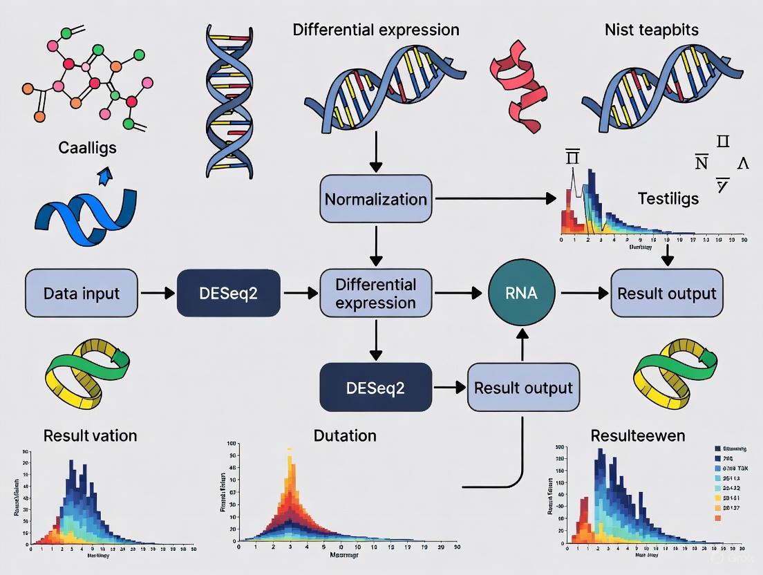
The following diagram illustrates the complete DESeq2 analysis workflow:
Key Experimental Protocols and Parameters
Input Data and Constructing a DESeqDataSet
The analysis begins with the creation of a DESeqDataSet object, which stores the count data, sample information, and model formula. DESeq2 can import data from various upstream quantification tools [2] [9].
Table: Methods for Creating a DESeqDataSet Object
| Method/Function | Input Data Type | Common Upstream Tools | Key Advantage |
|---|---|---|---|
DESeqDataSetFromTximport |
Transcript abundance estimates | Salmon [2], kallisto [2], Sailfish [2], RSEM [2] | Corrects for potential changes in gene length; faster than alignment-based methods [2]. |
DESeqDataSetFromMatrix |
Count matrix | featureCounts [3], HTSeq [10] | Use when a count matrix has already been generated. |
DESeqDataSet |
SummarizedExperiment object | tximeta [2] | Automatically populates annotation metadata for common transcriptomes. |
A critical step is defining the design formula using the tilde (~) operator. This formula expresses the variables in the column data that will be used to model the counts. To benefit from default settings, the variable of interest should be the last term in the formula, and the control level should be set as the first factor level [2] [3]. For example, to test for the effect of treatment while controlling for batch effects, the design would be ~ batch + treatment.
Running the Analysis and Extracting Results
The core analysis is performed with a single command:
This function executes the entire workflow: estimating size factors, estimating dispersions, fitting the dispersion trend, shrinking estimates, and fitting the models [7] [5].
Results for a specific comparison are extracted using the results() function. For factors with more than two levels, or for complex designs, the comparison must be specified with the contrast argument [10].
DESeq2 also offers log2 fold change shrinkage methods like apeglm to improve the accuracy and interpretability of fold change estimates, particularly for low-count genes [9] [1]. This is done with the lfcShrink() function.
Table: Key Parameters in a DESeq2 Analysis
| Parameter/Function | Description | Options / Notes |
|---|---|---|
fitType |
Type of fitting for dispersions to the mean intensity. | "parametric" (default), "local", "mean" [8]. |
testType |
The statistical test used for hypothesis testing. | "Wald" (default) or "LRT" (Likelihood Ratio Test) [8]. |
alpha |
The significance threshold for adjusted p-values. | Default is 0.1 [2]. |
lfcThreshold |
A non-zero log2 fold change threshold for testing. | Enables testing against a biologically meaningful threshold [1]. |
independentFiltering |
Automatically filter low-count genes to improve power. | Enabled by default [2]. |
The Scientist's Toolkit: Essential Reagents and Materials
Successful differential expression analysis requires both computational tools and well-annotated biological materials. The following table details key components for a typical DESeq2-based RNA-seq study.
Table: Research Reagent Solutions for RNA-seq and DESeq2 Analysis
| Item / Resource | Function / Role | Example / Specification |
|---|---|---|
| Reference Genome & Annotation | Provides the coordinate and feature system for read alignment and quantification. | GENCODE, Ensembl, or RefSeq human/mouse transcriptomes. Required for tools like Salmon [2] [6]. |
| Quantification Software (Pseudo-aligners) | Fast and accurate estimation of transcript/gene abundance from raw sequencing reads. | Salmon (recommended with --gcBias flag [2]), kallisto, or RSEM. |
| Alignment Software | Maps sequencing reads to a reference genome (traditional approach). | STAR (splice-aware aligner [6]), HISAT2. |
| DESeq2 R Package | Performs statistical testing for differential expression from count data. | Available via Bioconductor. Requires R [2]. |
| tximport / tximeta R Packages | Imports transcript abundance estimates and summarizes to gene-level for DESeq2. | tximport creates a list; tximeta creates a SummarizedExperiment with automatic metadata [2]. |
| BiocParallel R Package | Enables parallel computing to speed up the DESeq() and results() functions. |
Register multiple cores to reduce computation time [10]. |
| Sample Metadata (colData) | A data frame linking sample IDs to experimental conditions and covariates. | Critical: Must be accurate and match the columns of the count matrix. Used to define the design formula [6]. |
DESeq2 provides a statistically robust and computationally efficient framework for identifying differentially expressed genes in RNA-seq data. Its core innovation lies in the use of a negative binomial generalized linear model coupled with empirical Bayes shrinkage for dispersion and fold change estimates. This approach effectively handles the challenges of overdispersion and limited replication typical of sequencing experiments, leading to improved stability, interpretability, and power in differential expression analysis [1]. The standardized workflow, from raw count input to results extraction, along with its flexibility in handling complex designs, has cemented DESeq2's role as an indispensable tool for researchers, scientists, and drug development professionals in the field of genomics.
RNA sequencing (RNA-seq) has revolutionized transcriptomics by enabling comprehensive profiling of gene expression. However, two significant statistical challenges inherent to RNA-seq count data are the frequent use of low replicate numbers and the discrete nature of the data. These characteristics can compromise the power and reliability of differential expression analysis if not properly addressed. The DESeq2 package provides a robust statistical framework specifically designed to overcome these obstacles, making it an indispensable tool for researchers, scientists, and drug development professionals. This application note details the methodologies and advantages of using DESeq2 within a typical differential gene expression workflow, focusing on its handling of low replication and data discreteness.
The Statistical Challenges of RNA-Seq Data
Data Discreteness and Over-Dispersion
RNA-seq data is fundamentally composed of integer counts of sequencing reads mapped to genomic features. These counts are non-normally distributed and exhibit a mean-variance relationship, where the variance typically exceeds the mean—a property known as over-dispersion. Standard linear models assume normally distributed, continuous data with constant variance, making them unsuitable for raw RNA-seq counts [1]. DESeq2 employs a negative binomial generalized linear model (GLM) that accurately captures this over-dispersed count data structure, providing a more appropriate statistical foundation for inference [1] [10].
The Problem of Low Replicate Numbers
Controlled experiments in RNA-seq, particularly those involving human tissues or complex model organisms, often face practical constraints on sample size, resulting in low biological replication (often as few as 2-3 replicates per condition) [1]. With limited replicates, traditional per-gene variance estimates become highly unstable and lack statistical power. One study demonstrated that with only three biological replicates, common differential expression tools identified just 20-40% of the significantly differentially expressed (SDE) genes detected when using 42 replicates. Performance improved substantially for genes with large expression changes (>4-fold), where >85% were detected even with low replication [11]. This highlights the critical need for methods that enhance power in small-sample scenarios.
DESeq2's Statistical Framework for Enhanced Inference
Empirical Bayes Shrinkage for Dispersion Estimation
DESeq2's primary strategy for handling low replication is information sharing across genes through empirical Bayes shrinkage. Rather than estimating dispersion for each gene in isolation, DESeq2 assumes genes with similar average expression levels share similar dispersion. The method:
- Calculates initial gene-wise dispersion estimates using maximum likelihood.
- Fits a smooth curve modeling the relationship between dispersion and mean expression across all genes (Figure 1).
- Shrinks gene-wise dispersion estimates towards the curve-predicted values, generating final maximum a posteriori (MAP) estimates [1].
The strength of shrinkage is data-driven, automatically adjusting based on the dispersion variability around the fit and the available degrees of freedom (i.e., sample size). With fewer replicates, shrinkage is stronger, borrowing more information from the gene ensemble to produce stable, reliable estimates. As replication increases, shrinkage decreases, allowing gene-specific estimates to dominate [1].
Shrinkage Estimation of Logarithmic Fold Changes
For genes with low counts, logarithmic fold change (LFC) estimates are inherently noisy. DESeq2 incorporates a second empirical Bayes step that shrinks LFC estimates toward zero, using a prior distribution that models the expected effect sizes in the dataset. This shrinkage:
- Reduces the false positive rate from genes with high variance.
- Improves the power to detect true differential expression.
- Enables more accurate ranking and visualization of genes by effect size [1].
Table 1: Impact of Biological Replicates on Differential Expression Detection
| Number of Biological Replicates | Proportion of Significantly Differentially Expressed Genes Detected | Recommended Differential Expression Tool |
|---|---|---|
| 3 | 20%-40% | edgeR, DESeq2 |
| 6 (minimum general recommendation) | ~50% (depending on effect size) | edgeR, DESeq2 |
| 12 (for all fold changes) | >85% | DESeq2 |
| >20 | >85% for all SDE genes | DESeq (marginally outperforms others) |
Experimental Protocol: A Standard DESeq2 Workflow
Input Data Preparation
DESeq2 requires a matrix of un-normalized integer counts (e.g., from HTSeq-count or featureCounts) and a sample information table [2] [10]. Do not supply pre-normalized data, as DESeq2 internally corrects for library size and other factors.
Protocol 4.1.1: Constructing a DESeqDataSet from a Count Matrix
Note: The design formula should reflect the experimental structure. For multi-factor designs, include all relevant variables (e.g., ~ batch + condition).
Protocol 4.1.2: Importing Transcript Abundance Quantifications with tximport
For tools like Salmon or kallisto that output transcript-level abundance, use tximport to generate gene-level count matrices while correcting for potential transcript length changes [2].
Differential Expression Analysis
The core analysis is executed with a single function, which performs size factor estimation, dispersion estimation, model fitting, and hypothesis testing.
Protocol 4.2: Running the DESeq2 Analysis Pipeline
Results Interpretation and Visualization
Protocol 4.3: Generating Diagnostic and Results Plots
Visualization of the DESeq2 Statistical Workflow
The following diagram illustrates DESeq2's core statistical workflow for handling discrete count data and low replication through information sharing across genes.
Figure 1: DESeq2 Statistical Workflow for Handling Discrete Data and Low Replication
The Scientist's Toolkit: Essential Research Reagents and Computational Tools
Table 2: Key Research Reagent Solutions for RNA-seq Analysis with DESeq2
| Item | Type | Function in Workflow |
|---|---|---|
| DESeq2 R/Bioconductor Package | Software | Primary tool for differential expression analysis implementing negative binomial GLM with shrinkage. |
| Salmon / kallisto | Software | Fast transcript quantification for raw read alignment and count estimation. |
| tximport / tximeta R Packages | Software | Import and summarize transcript-level abundance to gene-level counts for DESeq2. |
| HTSeq / featureCounts | Software | Generate count matrices from aligned BAM files for input to DESeq2. |
| Bioconductor | Software Platform | Provides dependency management and genomic context for DESeq2 and related packages. |
| Illumina Sequencing Platform | Laboratory Instrument | Generates short-read RNA-seq data; common source of input data. |
| Truseq RNA Library Prep Kit | Laboratory Reagent | Prepares RNA-seq libraries for sequencing on Illumina platforms. |
| RNase Inhibitors | Laboratory Reagent | Preserves RNA integrity during sample preparation and library construction. |
DESeq2 provides a statistically rigorous solution to the twin challenges of data discreteness and low replicate numbers in RNA-seq experiments. Through its use of empirical Bayes shrinkage for both dispersion and fold change estimation, it enables robust and powerful differential expression analysis even with limited samples. The standardized protocols and workflows outlined in this application note provide researchers with a reliable framework for unlocking biological insights from their transcriptomic studies, accelerating discovery in basic research and drug development.
Proper experimental design forms the foundation of robust differential gene expression (DGE) analysis using RNA sequencing (RNA-seq) with DESeq2. Two particularly critical considerations that directly impact statistical power, result reliability, and biological validity are biological replicates and sequencing depth. This document outlines evidence-based recommendations for researchers designing RNA-seq experiments within the context of pharmacogenomics, drug development, and basic biological research. Optimal design choices at this stage prevent irreversible limitations in data interpretation and ensure meaningful biological conclusions from transcriptomic studies.
The Role of Biological Replicates
Definition and Importance
Biological replicates are defined as multiple, independent measurements of biological material collected from different specimens or sources under the same experimental condition. They are distinct from technical replicates, which involve repeated measurements of the same biological sample. Biological replicates are essential because they allow researchers to estimate the natural biological variability present in a population, which is separate from technical variability introduced during library preparation or sequencing [1].
In the context of DESeq2 analysis, biological replicates provide the necessary data for accurately estimating the dispersion of gene counts—a key parameter in the negative binomial generalized linear model that DESeq2 employs. Without adequate replication, dispersion estimates become unreliable, compromising the accuracy of statistical tests for differential expression [12] [1].
Consequences of Inadequate Replication
The DESeq2 package explicitly does not support analysis without biological replicates (1 vs. 1 comparison) [13]. Attempting such analysis is strongly discouraged because:
- No measures of statistical significance can be calculated for differential expression [13]
- Dispersion estimates become unstable or impossible to compute [1]
- High false positive and false negative rates are likely
- Biological conclusions cannot be generalized beyond the specific samples tested
When biological replicates are unavailable, the only option is to analyze log fold changes without significance testing, which provides limited biological insight [13].
Recommended Number of Replicates
While the optimal number of biological replicates depends on experimental constraints and expected effect sizes, general guidelines have emerged from statistical theory and practical experience:
Table 1: Recommended Biological Replicates for RNA-seq Experiments
| Experimental Scenario | Minimum Replicates per Condition | Ideal Replicates per Condition | Rationale |
|---|---|---|---|
| Standard experiments with moderate effect sizes | 3 | 5-6 | Balances practical constraints with reasonable power to detect 2-fold changes [13] |
| Experiments with subtle expression changes (<1.5-fold) | 5-6 | 10-12 | Increased power needed for detecting small effect sizes [1] |
| Pilot studies | 2-3 | - | Minimal level for preliminary data; limited statistical power |
| Clinical cohorts with high heterogeneity | 10+ | 20+ | Accounts for substantial biological variability in human populations |
For most experimental contexts, a minimum of three biological replicates per condition provides a reasonable balance between practical constraints and statistical needs [13]. However, power increases substantially with additional replicates, particularly for detecting subtle expression changes or working with heterogeneous populations.
Sequencing Depth Considerations
Definition and Impact on Detection Power
Sequencing depth refers to the number of sequenced reads obtained per sample, typically measured in millions of reads. Appropriate sequencing depth ensures sufficient coverage to detect expressed transcripts across the dynamic range of expression levels while maintaining cost-effectiveness.
Insufficient sequencing depth reduces the power to detect differentially expressed genes, particularly those with low expression levels. Excessive depth provides diminishing scientific returns for increased cost and may necessitate stricter multiple testing corrections due to increased detection of very low-abundance transcripts.
Recommended Sequencing Depth
Based on typical RNA-seq experiments, the following depth recommendations apply for standard bulk RNA-seq studies:
Table 2: RNA-seq Sequencing Depth Guidelines
| Application Context | Recommended Depth (Millions of Reads) | Coverage Considerations |
|---|---|---|
| Standard differential expression analysis | 10-30 million reads per library [13] | Adequate for detecting medium- to high-abundance transcripts |
| Studies focusing on low-abundance transcripts | 30-50+ million reads | Improved detection of transcription factors and regulatory RNAs |
| Complex genomes with high alternative splicing | 30-60 million reads | Enables more accurate isoform-level quantification |
| Single-cell RNA-seq | Varies by protocol | Typically lower depth per cell but many more cells |
For most standard DGE analyses using DESeq2, targeting 20-25 million reads per library provides a robust balance between cost and detection power, as this depth typically captures most medium- to high-abundance transcripts while allowing for accurate gene-level quantification [13].
Relationship Between Replicates and Depth
When designing experiments with fixed resources, researchers must balance the number of biological replicates against sequencing depth. In general, prioritizing more biological replicates over greater sequencing depth provides better statistical power for differential expression analysis [1]. For a fixed sequencing budget, the optimal design typically includes more replicates at moderate depth rather than few replicates at very high depth.
The following diagram illustrates the key decision points in experimental design and how they interrelate:
DESeq2-Specific Technical Requirements
Input Data Specifications
DESeq2 operates on raw, un-normalized count data rather than normalized values such as counts per million (CPM) or fragments per kilobase per million (FPKM) [2] [14]. The package expects a matrix of integer values representing the number of sequencing reads or fragments assigned to each gene in each sample. These raw counts allow DESeq2 to correctly assess measurement precision, as the variance of count data depends on the mean count value [2] [14].
DESeq2 internally corrects for differences in library size (sequencing depth) using its median-of-ratios method for size factor estimation [1]. The package does not require gene length normalization during this process, as gene length remains constant across samples and therefore does not affect differential expression comparisons [13].
Experimental Design Formula
Proper specification of the design formula is critical for DESeq2 analysis. The formula should include all major known sources of variation in the experiment, with the variable of interest specified last [2] [12]. For example, if studying treatment effects while accounting for sex differences, the design formula would be: ~ sex + treatment.
For paired experimental designs (e.g., before and after treatment in the same subjects), the design must include both subject and treatment information: ~ subject + condition [13]. This approach estimates treatment effect while accounting for inherent differences between subjects.
The following workflow diagram illustrates the complete DESeq2 analysis process with emphasis on the critical design considerations:
The Scientist's Toolkit
Essential Research Reagent Solutions
Table 3: Key Reagents and Materials for RNA-seq Experiments
| Reagent/Material | Function/Purpose | Considerations |
|---|---|---|
| RNA extraction kit (e.g., column-based) | Isolation of high-quality RNA from biological samples | Select based on sample type (cells, tissues, FFPE); prioritize RNA integrity |
| DNase treatment reagents | Removal of genomic DNA contamination | Critical for accurate RNA quantification; reduces background noise |
| Ribosomal RNA depletion kits | Enrichment for mRNA by removing abundant rRNA | Essential for non-polyA transcripts (e.g., bacterial RNA) |
| Poly(A) selection beads | Enrichment for eukaryotic mRNA | Standard for eukaryotic mRNA sequencing; may introduce 3' bias |
| Reverse transcriptase | cDNA synthesis from RNA template | High processivity important for full-length cDNA representation |
| DNA library prep kit | Preparation of sequencing-ready libraries | Compatibility with sequencing platform; efficiency for low-input samples |
| Size selection beads | Fragment size selection for libraries | Critical for insert size optimization; affects sequencing uniformity |
| Quality control reagents (Bioanalyzer, Qubit) | Assessment of RNA and library quality | Essential for troubleshooting; prevents sequencing failures |
Table 4: Essential Software Tools for RNA-seq Analysis with DESeq2
| Tool/Resource | Purpose | DESeq2 Integration |
|---|---|---|
| FastQC | Quality control of raw sequencing data | Preliminary step before alignment |
| HISAT2/STAR | Read alignment to reference genome | Generates BAM files for read counting |
| featureCounts/HTSeq | Generation of count matrices from aligned reads | Direct input to DESeq2 |
| tximport | Import of transcript-level quantifications | Creates gene-level count matrices for DESeq2 [2] |
| IGV | Visualization of aligned reads | Validation and exploration of specific genes |
| R/Bioconductor | Statistical analysis environment | Required platform for DESeq2 implementation |
Careful attention to biological replicates and sequencing depth during experimental design substantially enhances the reliability and interpretability of RNA-seq studies using DESeq2. Researchers should prioritize adequate biological replication (minimum 3, ideally 5-6 replicates per condition) and appropriate sequencing depth (10-30 million reads per library for standard applications) to ensure statistically robust differential expression analysis. These foundational elements, combined with proper specification of the experimental design formula and use of raw count data as input, form the basis for successful transcriptomic investigations in both basic research and drug development contexts.
Differential gene expression analysis with DESeq2 requires two primary components: a raw count matrix and a sample metadata table [5] [10]. These inputs form the foundation for the statistical model that identifies expression differences between experimental conditions. The raw count matrix contains the unnormalized sequencing read counts assigned to each gene across all samples, while the sample metadata describes the experimental design and biological conditions for each sample [15] [16]. Proper preparation and formatting of these inputs is critical for generating biologically meaningful and statistically valid results, as DESeq2 uses the raw counts and the information in the metadata to model the data using a negative binomial distribution and test for differential expression [5] [17].
Raw count matrix specifications
Definition and purpose
The raw count matrix is a tabular data structure where rows represent genes (or other genomic features) and columns represent samples [10] [16]. Each value in the matrix contains the number of sequencing reads (or UMIs for UMI-based protocols) that have been uniquely assigned to a specific gene in a specific sample [17]. These counts are derived from alignment tools (like STAR/HTSeq) or pseudo-alignment methods (like Salmon or kallisto) [18] [19]. DESeq2 uses these raw counts because its statistical model internally accounts for library size differences and other technical factors during the normalization process [5] [17].
Essential characteristics
The table below outlines the critical characteristics of a properly formatted raw count matrix for DESeq2 analysis:
Table 1: Key characteristics of DESeq2 raw count matrices
| Characteristic | Requirement | Rationale |
|---|---|---|
| Data Type | Integer values only | Counts represent discrete sequencing reads/fragments [17] |
| Pre-normalization | No transformation or normalization | DESeq2 performs internal normalization using size factors [5] [17] |
| Missing Values | Not allowed | All genes should have counts for all samples; zeros are acceptable [10] |
| Matrix Format | Genes as rows, samples as columns | Standard format for DESeq2 input functions [15] [10] |
| Gene Identifiers | Stable identifiers (e.g., Ensembl ID, Entrez) | Avoids ambiguity from gene symbols that may change over time [19] |
Generation methods
Count matrices can be generated through various bioinformatics pipelines depending on the experimental platform. For bulk RNA-seq data, common approaches include:
- Alignment-based methods: Tools like STAR align reads to a reference genome, followed by feature counting with HTSeq-count or featureCounts [18] [17].
- Pseudo-alignment methods: Tools like Salmon, kallisto, or alevin that provide fast transcript quantification without full alignment [19] [16].
- NCBI-generated counts: For human and mouse data in public repositories, NCBI's standardized pipeline uses HISAT2 for alignment and featureCounts for quantification [20].
Sample metadata requirements
Purpose and importance
The sample metadata table (also called colData) provides the experimental context for each sample, enabling DESeq2 to model the relationship between sample characteristics and gene expression patterns [5] [16]. This table connects the technical sample identifiers (matching the count matrix column names) to the biological and technical variables of interest, such as treatment conditions, time points, or batch information [10]. In complex experimental designs, proper metadata documentation is essential for specifying the statistical model that will test the hypotheses of interest [5].
Critical components
The sample metadata must include specific information to ensure proper analysis:
Table 2: Essential components of sample metadata for DESeq2
| Component | Description | Example |
|---|---|---|
| Sample IDs | Must exactly match column names in count matrix [15] | "SRR3383696", "treated1", "control2" |
| Condition of Interest | Primary experimental factor being tested [5] | "untreated", "treated", "infected", "healthy" |
| Batch Covariates | Technical factors to control for [5] | "sequencingbatch", "libraryprep_date" |
| Biological Covariates | Biological factors that may affect expression [5] [17] | "sex", "age", "genotype" |
| Factor Levels | Proper ordering of factor levels with control as reference [15] [10] | condition: levels = c("untreated", "treated") |
Design formula specification
The metadata directly informs the design formula, which specifies how DESeq2 models the data [5] [16]. The design formula uses tilde notation (~) followed by the variables that account for major sources of variation, with the factor of interest typically specified last [5]. For example:
- Simple design:
~ condition(compares two groups) - Controlled design:
~ batch + condition(accounts for batch effects) - Paired design:
~ individual + treatment(for paired experiments) - Interaction design:
~ genotype + treatment + genotype:treatment(tests interaction effects)
Experimental workflow integration
From sequencing to count matrix
The complete workflow for generating DESeq2 inputs begins with raw sequencing data and proceeds through multiple quality control and processing steps:
Diagram 1: Workflow from raw sequences to DESeq2 analysis
Data import into DESeq2
Once the count matrix and metadata are prepared, they are imported into R using the DESeqDataSetFromMatrix() function, which requires that the column names of the count matrix exactly match the row names of the metadata table [15] [10]. Proper ordering is critical, as DESeq2 will not guess which column of the count matrix belongs to which row of the metadata [15]. The following example demonstrates this process:
The scientist's toolkit: Research reagent solutions
Table 3: Essential reagents, tools, and resources for RNA-seq analysis
| Resource Category | Specific Tools/Reagents | Function in Analysis |
|---|---|---|
| Alignment Tools | STAR, HISAT2, Rsubread [20] [19] | Map sequencing reads to reference genome |
| Quantification Tools | HTSeq-count, featureCounts, Salmon [20] [19] | Generate raw counts for each gene |
| Quality Control | FastQC, RSeQC, Qualimap, Trimmomatic [18] | Assess read quality, alignment metrics |
| Reference Genomes | ENSEMBL, UCSC, GENCODE [20] [17] | Provide standardized genome sequences |
| Annotation Sources | Ensembl, Entrez, TAIR (for Arabidopsis) [21] [19] | Provide stable gene identifiers |
| Data Repositories | GEO, SRA, ArrayExpress [22] [20] | Source of public datasets and metadata |
Quality assessment and troubleshooting
Pre-analysis quality checks
Before performing differential expression analysis, several quality checks should be performed on both the count matrix and metadata:
- Count matrix checks: Verify that the matrix contains only integer values, check for excessive zeros, and confirm that row sums are reasonably distributed [10] [18].
- Metadata checks: Ensure factor levels are properly ordered with control as reference level, confirm no missing values, and verify that sample IDs exactly match count matrix column names [15] [10].
- Sample-level QC: Examine library sizes, gene detection rates, and outlier samples using PCA or other multivariate methods [18] [17].
Common issues and solutions
- Issue: "Error in DESeqDataSetFromMatrix: countData and colData have different numbers of columns"
- Solution: Verify that all samples in colData are present in countData and vice versa [15].
- Issue: Inflated false positives due to batch effects
- Solution: Include batch as a covariate in the design formula [5].
- Issue: Overabundance of zeros leading to inflated fold changes
- Solution: Use lfcShrink for effect size estimation [17].
- Issue: Low power to detect differential expression
- Solution: Ensure sufficient biological replicates and check for appropriate sequencing depth [18].
The importance of proper controls and minimizing batch effects in experimental design
In high-throughput genomic studies, such as differential gene expression analysis with RNA sequencing (RNA-seq), batch effects represent a significant challenge to data integrity and biological interpretation. Batch effects are unwanted technical variations introduced into data due to factors unrelated to the biological question under investigation [23] [24]. These non-biological fluctuations can arise from differences in sample collection, inconsistencies in pre-experimental processing, reagent lot changes, instrument drift over time, operator variability, or environmental conditions such as humidity and temperature [23]. In the context of DESeq2 research, which relies on accurate count data from RNA-seq experiments, batch effects can distort true biological signals and lead to both false positives and false negatives in differential expression testing [24].
The critical impact of batch effects stems from their potential to confound biological signals of interest. When technical variation correlates with experimental conditions, it becomes challenging to distinguish true differential expression from technical artifacts. This is particularly problematic in longitudinal studies or multi-batch experiments where biological variation may be masked by artificial technical noise [23]. Research has demonstrated that batch effects can affect a substantial proportion of features in a dataset, with one study noting that up to 99.5% of features showed significant correlation with technical variables [25]. For researchers using DESeq2 for differential expression analysis, understanding, detecting, and correcting for batch effects is therefore not merely optional but essential for producing robust, reproducible results.
Batch effects in RNA-seq experiments can originate from multiple technical sources throughout the experimental workflow. Sample preparation inconsistencies represent a major source, including variations in extraction duration, solvents used, or processing times by different technicians [23]. Instrumental factors also contribute significantly, with LC-MS systems exhibiting drift over time due to calibration changes or performance degradation. Additionally, environmental conditions such as room temperature and humidity fluctuations can introduce systematic biases, while reagent lot changes may alter background signals or reaction efficiencies [23]. Even the injection order of samples in large sets can create detectable patterns of technical variation that correlate with run sequence rather than biological groups.
The confounding nature of batch effects becomes particularly problematic when the batch variable correlates with the biological variable of interest. For instance, if all control samples are processed in one batch and all treatment samples in another, any observed differences become inextricably linked to the technical processing differences [24]. This confounding can lead to spurious findings where technical artifacts are misinterpreted as biological signals, or can mask true biological effects by introducing additional variance that reduces statistical power [24] [25]. The consequences extend beyond individual studies, as failure to account for batch effects can decrease reproducibility in meta-analyses and lead to inefficient resource utilization in follow-up studies [25].
Impact on Differential Expression Analysis with DESeq2
DESeq2 relies on accurate modeling of count data using negative binomial distributions to identify differentially expressed genes [2] [5]. When batch effects are present but unaccounted for, they violate key assumptions of the statistical model. The extra technical variation introduced by batch effects can inflate variance estimates, leading to reduced power to detect true differential expression. Conversely, when batch effects correlate with experimental conditions, they can produce artificially inflated fold changes that result in false positives [24].
The normalization process in DESeq2, which uses a median of ratios method to estimate size factors, assumes that most genes are not differentially expressed [5]. Batch effects that systematically alter the expression levels of large numbers of genes can disrupt this normalization, leading to improper adjustment of library size differences. Similarly, the dispersion estimation process, which models within-group variability, can be significantly impacted by batch effects, particularly when samples from the same biological group are processed in different batches with different technical artifacts [5].
Strategies for Minimizing Batch Effects Through Experimental Design
Proactive Experimental Design
The most effective approach to managing batch effects begins with proactive experimental design that anticipates and minimizes technical variation before data generation. When possible, processing all samples in a single batch represents the most straightforward solution, as it eliminates inter-batch variation entirely [23]. However, for large studies where multiple batches are unavoidable, strategic sample allocation across batches becomes critical to prevent confounding of technical and biological variables.
Randomization of sample order across batches ensures that no experimental group is disproportionately affected by technical variation. This approach distributes batch effects randomly across biological groups, making it easier to statistically separate technical from biological variation during analysis [23]. For complex studies with multiple potentially confounding variables, more sophisticated allocation methods have been developed, including anticlustering algorithms that explicitly avoid forming unwanted clusters of similar elements when dividing data into groups [26]. These methods actively maximize the similarity between batches with respect to known covariates, creating more balanced experimental designs.
Table 1: Batch Effect Minimization Strategies in Experimental Design
| Strategy | Implementation | Advantages | Limitations |
|---|---|---|---|
| Single Batch Processing | Process all samples in one continuous run | Eliminates inter-batch variation | Often impractical for large studies |
| Randomization | Randomly assign samples to batches | Simple to implement; breaks systematic confounding | May not balance multiple covariates |
| Stratified Randomization | Randomize within strata defined by key covariates | Balances important known variables | Requires prior knowledge of relevant covariates |
| Anticlustering | Use algorithms to maximize similarity between batches | Optimal balance of multiple covariates; prevents grouping by known variables | Computational complexity for large sample sizes |
| Propensity Score Matching | Allocate samples to minimize differences in propensity scores between batches | Handles multiple confounding variables simultaneously; dimension reduction | Requires complete covariate data before allocation |
Advanced Allocation Methods
Recent methodological advances have introduced sophisticated approaches to sample allocation that explicitly minimize batch effects. The anticlustering method developed by researchers at Heinrich Heine University Düsseldorf provides a systematic way to partition samples into batches while maximizing between-batch similarity [26]. This approach has been extended with a "Must-Link Method" that ensures related samples (such as multiple tissue samples from the same patient) are grouped in the same batch, enabling meaningful within-subject comparisons while maintaining balance across batches [26].
Another innovative approach utilizes propensity scores to guide sample allocation [25]. Propensity scores, which represent the probability of group membership conditional on a set of covariates, provide a dimension reduction technique that captures the overall balance in covariate distribution between groups. By selecting the batch allocation that minimizes differences in average propensity score between batches, researchers can create optimally balanced designs that minimize potential confounding [25]. Studies comparing this optimal allocation strategy to randomization and stratified randomization have demonstrated reduced bias in both null and alternative hypothesis conditions, particularly prior to batch correction [25].
Integrating Batch Effect Considerations into DESeq2 Workflow
Experimental Design Phase
Successful differential expression analysis with DESeq2 begins with thoughtful experimental planning that incorporates batch effect considerations from the earliest stages. Researchers should carefully consider all potential sources of technical variation and document them systematically. This includes recording metadata such as processing dates, technician identifiers, reagent lot numbers, and instrument calibration records. This metadata will prove essential for both statistical adjustment and troubleshooting potential issues that arise during analysis.
When designing a multi-batch experiment, researchers should utilize balanced allocation methods to ensure that biological groups of interest are proportionally represented across all batches. Additionally, including technical replicates across batches provides valuable data for assessing batch-to-batch variation and validating correction methods [23]. For RNA-seq experiments specifically, the use of quality control (QC) samples is highly recommended. These pooled QC samples, inserted at regular intervals throughout the run, allow for monitoring and correction of instrumental drift over time [23].
Sample Processing and Quality Control
During sample processing, several specific practices can help minimize batch effects. Standardizing protocols across all samples reduces introduction of technical variation, while randomizing processing order prevents systematic correlations between experimental conditions and run sequence. For large studies that necessarily span multiple batches, including internal reference samples that are processed in every batch enables direct quantification of batch effects and facilitates more effective correction [23].
The inclusion of controls specifically designed for batch effect assessment is crucial. Pooled quality control (QC) samples, created by combining equal aliquots from all samples or a representative subset, provide a technical baseline that should remain constant across batches [23]. Significant deviations in QC sample measurements between batches indicate technical variation that needs addressing. For studies expecting subtle biological effects, positive control samples with known expected differences can help verify that batch effect correction methods are not removing genuine biological signals.
Computational Approaches for Batch Effect Detection and Correction
Batch Effect Detection Methods
Before applying batch correction methods, it is essential to detect and quantify the presence and magnitude of batch effects in the data. Several visualization and statistical approaches are commonly used for this purpose. Principal Component Analysis (PCA) is particularly valuable, as it can reveal clustering patterns driven by batch rather than biological variables [23] [24]. When samples group by processing batch rather than experimental condition in PCA space, batch effects are likely present.
Hierarchical clustering and heatmaps provide complementary approaches for visualizing batch-related patterns [24]. These methods can reveal systematic differences in expression profiles between batches, particularly when combined with annotation tracks that color-code samples by batch and biological group. Additionally, correlation analysis of technical replicates across batches can quantitatively assess the impact of batch effects, with decreased correlation indicating stronger batch effects [23].
Table 2: Computational Methods for Batch Effect Correction
| Method | Underlying Strategy | Best Applied When | Key Considerations |
|---|---|---|---|
| RemoveBatchEffect (Limma) | Linear models | Batches are known and balanced; mild to moderate effects | Can be applied to normalized counts; may not handle complex nonlinear patterns |
| ComBat | Empirical Bayes | Batch effects are severe; small sample sizes | Shrinks batch effects toward overall mean; handles known batches only |
| SVA (Surrogate Variable Analysis) | Factor analysis | Unknown batches or unmodeled factors; complex confounding | Identifies unknown technical factors; risk of removing biological signal |
| RUV (Remove Unwanted Variation) | Factor analysis using control genes | Housekeeping or negative control genes are available | Requires appropriate control genes; choice of controls is critical |
| SVR (Support Vector Regression) | QC-based non-linear correction | QC samples are available at regular intervals | Models signal drift with flexibility; requires sufficient QC samples |
Batch Effect Correction Strategies
When batch effects are detected, several computational approaches can correct for them, falling into three main categories. Internal standard-based correction relies on spiked-in standards to adjust for technical variation, but requires the internal standard and target to behave similarly, limiting its general application [23]. Sample-based correction methods assume the total metabolite content is similar across samples, using approaches like Total Ion Count (TIC) normalization, where metabolite content is divided by the sum of all metabolite contents in each sample [23].
QC-based correction methods utilize regularly interspersed quality control samples to model and remove technical variation. These include Support Vector Regression (SVR) in the metaX R package, Robust Spline Correction (RSC) also in metaX, and the Random Forest-based QC-RFSC method in statTarget [23]. These approaches use the trend observed in QC samples to correct the entire dataset, effectively removing technical drift while preserving biological signals.
Implementing Batch Effect Correction in DESeq2 Analysis
Design Formula Specification
DESeq2 provides flexible modeling capabilities that can incorporate batch information directly into the statistical model. The key mechanism for this is the design formula, which specifies the variables to be included in the differential expression model [5]. When batch effects are known and recorded, they can be included in the design formula to control for their influence while testing for the biological effect of interest.
For example, if a researcher has samples processed across three batches and wants to test for differences between treatment and control groups, while controlling for batch effects, the design formula would be:
In this formula, batch represents the batch identifier and condition represents the biological groups of interest. The order of terms is important, as DESeq2 sequentially fits the model, first estimating batch effects and then estimating condition effects after accounting for batch [5]. This approach explicitly models batch as a separate factor, effectively adjusting the condition comparisons for batch differences.
Complex Experimental Designs
For more complex experimental designs with multiple factors and potential interactions, DESeq2 can accommodate extended design formulas. For instance, if researchers suspect that the effect of treatment might differ by batch (an interaction effect), this can be modeled by including an interaction term:
However, interpretation of such models becomes more complex, and the DESeq2 vignette often recommends creating a combined factor that represents the interaction of interest [5]. For example, combining batch and condition into a single factor (e.g., batch1control, batch1treatment, batch2_control, etc.) and then using a likelihood ratio test to examine specific contrasts.
When using the combined factor approach, the design formula would be:
With this approach, specific contrasts of interest can be tested using the contrast argument in the results() function. This provides flexibility in testing particular comparisons while controlling for batch effects.
Validation and Quality Assessment Post-Correction
Assessing Correction Effectiveness
After applying batch correction methods, it is crucial to validate their effectiveness and ensure that biological signals have not been unintentionally removed. Several approaches can be used for this validation. Principal Component Analysis should be repeated post-correction to verify that batch-driven clustering has been reduced or eliminated while biological groupings remain intact [23] [24]. Similarly, correlation analysis of technical replicates should show improved correlation after correction [23].
The negative control approach utilizes genes or samples known not to differ between biological groups to verify that correction hasn't introduced spurious differences. Conversely, positive controls (genes known to be differentially expressed) should maintain their significant differences after correction. When available, validation by orthogonal methods such as qPCR on a subset of genes provides the strongest evidence that correction has preserved biological truth while removing technical artifacts [27].
Managing Overcorrection and Signal Loss
A significant risk in batch effect correction is overcorrection, where biological signal is inadvertently removed along with technical variation. This occurs particularly when batch effects are partially confounded with biological effects, or when correction methods are too aggressive. Studies have reported instances where methods like Robust Spline Correction (RSC) and QC-RFSC actually decreased replicate correlation, indicating potential overcorrection or model mismatch [23].
To minimize this risk, researchers should apply multiple correction strategies and compare results, using visualization techniques to ensure biological patterns remain intact. Additionally, differential analysis consistency across methods can indicate robust biological findings [23]. When possible, biological validation of key findings through independent methods remains the gold standard for confirming that correction has preserved rather than removed true signals.
Successfully managing batch effects in DESeq2-based differential expression analysis requires an integrated approach spanning experimental design, processing, computational correction, and validation. No single batch correction method universally outperforms others across all datasets [23], making methodological flexibility and validation essential. Researchers should prioritize preventive measures through balanced experimental design whenever possible, as well-designed experiments with minimal confounding are more amenable to subsequent computational correction than severely confounded designs.
The most robust approach combines multiple complementary methods with careful validation to ensure that technical artifacts are removed while biological signals are preserved. By implementing these comprehensive strategies for batch effect management, researchers can significantly enhance the reliability, reproducibility, and biological validity of their DESeq2 differential expression analyses.
Integrated Batch Effect Management Workflow: This diagram illustrates the comprehensive approach to managing batch effects throughout the differential expression analysis pipeline, from experimental design to biological interpretation.
Table 3: Research Reagent Solutions for Batch Effect Management
| Reagent/Material | Function in Batch Effect Management | Application Notes |
|---|---|---|
| Pooled QC Samples | Monitoring technical variation across batches | Create by combining equal aliquots from all samples; analyze at regular intervals |
| Internal Standards | Correction of technical variation per sample | Use isotopically labeled compounds; limited to same compound type |
| Reference RNA Samples | Assessment of cross-batch technical performance | Commercial reference materials; process in each batch for comparison |
| Spike-in Controls | Normalization for technical variation | Add known quantities of foreign RNA to each sample |
| Multiple Reagent Lots | Assessing lot-to-lot variability | Intentionally include multiple lots when possible to model this effect |
Differential expression (DE) analysis represents a fundamental step in understanding how genes respond to different biological conditions using RNA sequencing (RNA-seq) data. This analytical process identifies systematic changes in gene expression patterns across tens of thousands of genes simultaneously, while accounting for biological variability and technical noise inherent in RNA-seq experiments [28]. The field has developed several sophisticated tools to address specific challenges in RNA-seq data, including count data overdispersion, small sample sizes, complex experimental designs, and varying levels of biological and technical noise [28]. Among the most widely used tools are DESeq2, edgeR, and NOISeq, each employing distinct statistical approaches with unique strengths and limitations. Understanding these differences is crucial for researchers, scientists, and drug development professionals to select the most appropriate methodology for their specific research context and experimental design. This comparative overview examines the statistical foundations, practical implementations, and performance characteristics of these three prominent methods, providing a framework for their application in differential gene expression analysis.
Statistical Foundations and Algorithmic Approaches
Core Methodologies
The three methods employ fundamentally different statistical frameworks for identifying differentially expressed genes:
DESeq2 utilizes a negative binomial modeling approach with empirical Bayes shrinkage for dispersion estimates and fold changes. It models the observed relationship between the mean and variance when estimating dispersion, allowing a more general, data-driven parameter estimation [28] [29]. This approach aims for a balanced selection of differentially expressed genes throughout the dynamic range of the data. DESeq2 incorporates automatic outlier detection and independent filtering to improve the reliability of its results [28].
edgeR also employs a negative binomial model but with a more flexible dispersion estimation approach. It uses an empirical Bayes procedure to moderate the degree of overdispersion across transcripts by borrowing information between genes, improving the reliability of inference [30] [29]. edgeR offers multiple testing strategies, including exact tests analogous to Fisher's exact test but adapted for overdispersed data, as well as quasi-likelihood options [30] [28].
NOISeq represents a non-parametric approach that contrasts fold changes and absolute expression differences within conditions to determine a null distribution, then compares observed differences to this null [31] [29]. Unlike the model-based approaches, NOISeq does not assume specific data distributions, making it less restrictive but requiring larger sample sizes to achieve good statistical power [32] [31].
Data Handling and Normalization
Each method employs distinct strategies for data normalization and variance handling:
DESeq2 performs internal normalization based on the geometric mean and uses adaptive shrinkage for dispersion estimates and fold changes [28] [15]. It calculates size factors to account for differences in sequencing depth across samples [33].
edgeR typically uses TMM normalization (Trimmed Mean of M-values) to correct for composition biases between samples, which adjusts to minimize differences in expression levels between samples when most genes are not expected to be differentially expressed [34] [29].
NOISeq offers multiple normalization options including RPKM, TMM, and upper quartile normalization, providing flexibility to address different technical biases [31] [29]. The package includes comprehensive quality control tools to guide normalization choices based on detected biases.
Table 1: Statistical Foundations of DESeq2, edgeR, and NOISeq
| Aspect | DESeq2 | edgeR | NOISeq |
|---|---|---|---|
| Core Statistical Approach | Negative binomial modeling with empirical Bayes shrinkage | Negative binomial modeling with flexible dispersion estimation | Non-parametric method using fold changes and absolute differences |
| Data Distribution Assumption | Negative binomial distribution | Negative binomial distribution | No specific distribution assumption |
| Differential Expression Test | Wald statistical test | Exact test or quasi-likelihood F-tests | Comparison to empirically derived null distribution |
| Normalization Method | Internal normalization based on geometric mean | TMM normalization by default | RPKM, TMM, or upper quartile normalization |
| Variance Handling | Adaptive shrinkage for dispersion estimates and fold changes | Empirical Bayes moderation of overdispersion across genes | No explicit variance modeling; relies on empirical distributions |
Performance Characteristics and Method Comparison
False Discovery Rate Control and Power
Recent evaluations have revealed crucial differences in how these methods control false discoveries, particularly in studies with large sample sizes:
A 2022 study published in Genome Biology demonstrated that when analyzing human population RNA-seq samples with large sample sizes (ranging from 100 to 1376), DESeq2 and edgeR exhibited exaggerated false positive rates [32]. In permutation analyses where any identified differentially expressed genes should theoretically be false positives, DESeq2 and edgeR had 84.88% and 78.89% chances, respectively, to identify more DEGs from permuted datasets than from the original dataset [32]. The actual false discovery rates of DESeq2 and edgeR sometimes exceeded 20% when the target FDR was 5% [32].
In contrast, the Wilcoxon rank-sum test (a non-parametric method similar in spirit to NOISeq) consistently controlled the FDR under a range of thresholds from 0.001% to 5% in the same study [32]. NOISeq, as a non-parametric method, has been reported to efficiently control false discoveries in experiments with biological replication [31]. This suggests that for population-level RNA-seq studies with large sample sizes, non-parametric methods like NOISeq may provide more reliable FDR control.
Sample Size Considerations and Robustness
The performance of these methods varies significantly with sample size:
DESeq2 generally performs well with moderate to large sample sizes (≥3 replicates, performs better with more) and can handle high biological variability and subtle expression changes effectively [28]. However, its performance deteriorates with very large sample sizes in population studies, showing inflated false positive rates [32].
edgeR is particularly efficient with very small sample sizes (≥2 replicates) and large datasets, making it valuable for experiments with limited replication [30] [28]. It shows strengths in analyzing genes with low expression counts, where its flexible dispersion estimation can better capture inherent variability in sparse count data [28].
NOISeq requires larger sample sizes to achieve good power due to its non-parametric nature [32] [31]. At FDR thresholds of 1%, non-parametric methods like NOISeq had almost no power when the per-condition sample size was smaller than 8, though this improves significantly with adequate replication [32].
Table 2: Performance Characteristics Under Different Experimental Conditions
| Performance Aspect | DESeq2 | edgeR | NOISeq |
|---|---|---|---|
| Ideal Sample Size | ≥3 replicates, performs well with more | ≥2 replicates, efficient with small samples | Requires larger sample sizes (≥8 per condition) |
| FDR Control in Large Samples | Problematic (actual FDR can exceed 20%) | Problematic (actual FDR can exceed 20%) | Robust FDR control |
| Power with Small Samples | Moderate | Good | Limited |
| Robustness to Outliers | Moderate | Moderate | High (due to non-parametric nature) |
| Handling Low-Count Genes | Conservative | Good with flexible dispersion | Requires careful filtering |
| Computational Efficiency | Can be intensive for large datasets | Highly efficient, fast processing | Moderate |
Practical Implementation and Workflow
DESeq2 Implementation Protocol
DESeq2 operates through a structured workflow that can be implemented in R:
Step 1: Data Preparation and Pre-filtering
- Read the count matrix into R, ensuring row names are gene identifiers and column names are samples
- Create a sample information data frame (colData) that specifies experimental conditions
- Ensure that columns of the count matrix and rows of the column data are in the same order [15]
- Perform minimal pre-filtering to remove genes with very few reads (e.g., fewer than 10 reads total across all samples) to reduce memory usage and improve speed [15] [10]
Step 2: DESeqDataSet Construction
- Construct a DESeqDataSet object using the
DESeqDataSetFromMatrix()function, specifying the count data, column data, and design formula [15] [10] - The design formula should reflect the experimental design, typically starting with "~" followed by the condition of interest
- Explicitly set factor levels using
factor()orrelevel()to control the reference level for comparisons [15]
Step 3: Differential Expression Analysis
- Run the core analysis using
DESeq()function, which performs estimation of size factors, dispersion estimation, and negative binomial GLM fitting [10] [33] - Extract results using the
results()function, specifying contrast if the design is multi-factorial [10] - Apply log-fold change shrinkage using
lfcShrink()to improve accuracy for visualization and ranking of genes [33]
Step 4: Results Interpretation
- Filter results based on adjusted p-values and log-fold change thresholds
- Perform visualization using MA-plots, PCA plots, or heatmaps to explore results
- Export significant differentially expressed genes for downstream analysis
edgeR Implementation Protocol
edgeR provides alternative approaches for differential expression analysis:
Step 1: Data Object Creation
- Read count data into R and create a DGEList object using
DGEList()function, combining counts and group information [34] - The group factor should specify the experimental condition for each sample
Step 2: Filtering and Normalization
- Filter lowly expressed genes using
filterByExpr()to retain genes with sufficient counts across samples [34] - Perform TMM normalization using
calcNormFactors()to correct for composition biases between samples [34]
Step 3: Dispersion Estimation and Testing
- Create a design matrix using
model.matrix()based on the experimental design - Estimate dispersions using
estimateDisp()to model biological variability [34] - For simple designs, use exact tests; for complex designs, use GLM approaches
- Perform quasi-likelihood F-tests using
glmQLFit()andglmQLFTest()for more rigorous statistical testing [34]
Step 4: Results Extraction
- Extract top differentially expressed genes using
topTags()with specified FDR threshold [34] - Consider adjusting results based on log-fold change thresholds in addition to statistical significance
NOISeq Implementation Protocol
NOISeq offers a non-parametric alternative with integrated quality control:
Step 1: Data Input and Quality Control
- Read count data into R and create a NOISeq data object
- Perform comprehensive quality control using NOISeq's diagnostic tools, including:
- Biotype detection plots to visualize percentage of genes detected per biotype
- Saturation plots to assess sequencing depth adequacy
- GC content and length bias plots to identify technical biases [31]
Step 2: Data Filtering and Normalization
- Apply low-count filtering using one of NOISeq's methods (CPM, proportion test, or Wilcoxon test) that consider the experimental design [31]
- Choose appropriate normalization based on quality control results (RPKM, TMM, or upper quartile)
Step 3: Differential Expression Analysis
- For experiments with biological replicates, use NOISeqBIO which implements an empirical Bayes approach to handle biological variability [31]
- Specify the comparison of interest and run the core analysis
- The method computes statistics based on fold changes and absolute expression differences compared to empirically derived null distributions
Step 4: Results Exploration
- Extract differentially expressed genes based on probability thresholds
- Utilize NOISeq's visualization capabilities to explore results, including expression plots, MD plots, and chromosomal distribution of DEGs [31]
Experimental Design Considerations and Recommendations
Method Selection Guide
Choosing between DESeq2, edgeR, and NOISeq depends on multiple factors:
For experiments with small sample sizes (n < 5 per group), edgeR is often preferable due to its efficient handling of limited replication and robust performance with minimal replicates [28]. Its flexible dispersion estimation provides good power even with few samples.
For moderate-sized experiments (5 ≤ n ≤ 20) with complex designs involving multiple factors, DESeq2 offers excellent capabilities for modeling complex relationships and provides reliable results with good FDR control [28]. Its sophisticated shrinkage estimation improves accuracy for low-count genes.
For large population studies (n > 50), non-parametric methods like NOISeq become advantageous due to their robust false discovery rate control [32]. As sample size increases, the power limitations of non-parametric methods diminish while their robustness to model assumptions becomes increasingly valuable.
For data with suspected outliers or severe violations of distributional assumptions, NOISeq provides a safer alternative as it doesn't rely on specific parametric assumptions [31]. This is particularly relevant when analyzing data from novel organisms or experimental conditions where distributional properties may be unknown.
For routine analyses with standard experimental designs, both DESeq2 and edgeR perform well and often show substantial concordance in their results [28]. The choice between them may depend on specific analytical needs or personal preference.
Integrated Analysis Strategy
Given the complementary strengths of these methods, an integrated approach can provide more robust results:
Primary analysis with multiple methods: Run both parametric (DESeq2 or edgeR) and non-parametric (NOISeq) methods, focusing on genes identified as significant by multiple approaches. This conservative strategy reduces false positives at the potential cost of some false negatives.
Method-specific validation: For genes identified by only one method, perform additional scrutiny based on effect size, biological plausibility, and experimental validation potential.
Quality assessment: Use NOISeq's comprehensive quality control metrics to inform data preprocessing decisions regardless of the primary analysis method chosen.
Power considerations: When designing experiments, consider the methodological requirements – non-parametric methods typically require larger sample sizes to achieve equivalent power to parametric approaches.
Diagram 1: Method Selection Workflow for Differential Expression Analysis
Computational Tools and Software Packages
Successful differential expression analysis requires specific computational tools and resources:
R Programming Environment: The foundational platform for all three methods, providing the computational environment for statistical analysis and visualization [15] [34]. R serves as the common interface for package installation, data manipulation, and analysis execution.
Bioconductor Project: A repository for bioinformatics packages including DESeq2, edgeR, and NOISeq [30] [31]. Bioconductor provides standardized installation and maintenance of these specialized tools along with extensive documentation.
DESeq2 Package: Specialized software for differential analysis of count-based RNA-seq data, implementing negative binomial generalized linear models with empirical Bayes shrinkage [15] [10]. Key functions include DESeqDataSetFromMatrix(), DESeq(), and results() for core analysis workflow.
edgeR Package: Software for examining differential expression of replicated count data using an overdispersed Poisson model to account for biological and technical variability [30] [34]. Essential functions include DGEList(), calcNormFactors(), and glmQLFTest().
NOISeq Package: Comprehensive resource for quality control and non-parametric analysis of count data, featuring both NOISeq and NOISeqBIO methods [31]. Provides extensive diagnostic plots and normalization options.
Additional Utility Packages: Supporting packages such as pheatmap for visualization, data.table for efficient data handling, and BiocParallel for parallel processing to reduce computation time [15] [10].
Proper analysis requires specific data formats and resources:
Raw Read Counts: Table of non-normalized sequence read counts at gene or transcript level, typically generated by tools like HTseq-count or featureCounts [10] [33]. Must be in matrix format with genes as rows and samples as columns.
Sample Metadata: Data frame specifying the experimental design, including sample identifiers, experimental conditions, and any batch information [10]. Critical for proper experimental design specification in all three methods.
Gene Annotation Data: Optional but recommended feature data containing gene identifiers, symbols, and genomic coordinates to enhance biological interpretation of results [15].
Quality Control Metrics: Pre-computed quality assessments from sequencing pipelines, including mapping statistics, insert size distributions, and sequencing depth information to inform analytical decisions.
Table 3: Essential Research Reagents and Computational Resources
| Resource Category | Specific Tools/Formats | Function in Analysis | Method Applicability |
|---|---|---|---|
| Analysis Software | DESeq2 R Package | Negative binomial GLM with empirical Bayes moderation | Primary analysis tool |
| edgeR R Package | Negative binomial modeling with flexible dispersion | Primary analysis tool | |
| NOISeq R Package | Non-parametric differential expression analysis | Primary analysis tool | |
| Input Data | Raw Count Matrix | Unnormalized read counts for genes across samples | Required for all methods |
| Sample Metadata | Experimental design specification | Required for all methods | |
| Gene Annotations | Gene identifiers, symbols, and genomic coordinates | Enhanced interpretation | |
| Quality Control | NOISeq Diagnostic Plots | Comprehensive data quality assessment | Particularly useful for NOISeq |
| Alignment Statistics | Mapping quality and coverage metrics | Informative for all methods | |
| Supporting Packages | BiocParallel | Parallel processing to reduce computation time | DESeq2 and edgeR |
| pheatmap, ggplot2 | Visualization of results | All methods | |
| data.table, tidyverse | Data manipulation and organization | All methods |
DESeq2, edgeR, and NOISeq represent complementary approaches to differential expression analysis with RNA-seq data, each with distinct statistical foundations and performance characteristics. DESeq2 and edgeR share a parametric foundation in negative binomial models but differ in their normalization approaches and dispersion estimation methods. NOISeq offers a non-parametric alternative that eliminates distributional assumptions at the cost of requiring larger sample sizes for equivalent power. Recent research has revealed important considerations for method selection, particularly the exaggerated false positive rates of parametric methods in large sample size studies and the robust FDR control of non-parametric approaches under these conditions. For researchers performing differential gene expression analysis, the choice between these methods should be guided by sample size, experimental design complexity, data quality, and specific research questions. An integrated approach leveraging the complementary strengths of multiple methods often provides the most robust and biologically meaningful results, particularly for novel discoveries where validation resources may be limited. As RNA-seq technologies continue to evolve and sample sizes increase in population-level studies, understanding these methodological distinctions becomes increasingly critical for generating reliable biological insights.
Step-by-Step DESeq2 Workflow: From Data Import to Differential Expression Results
In differential gene expression analysis with DESeq2, the initial creation of the DESeqDataSet object is a critical first step that establishes the foundation for all subsequent statistical testing. This object serves as the central container for raw count data, sample metadata, and the experimental design formula, thereby informing the statistical model about the structure of the experiment. Proper construction of this object is essential for generating biologically meaningful and statistically valid results. This protocol outlines the precise data structures and specification methods required to correctly initialize the DESeqDataSet within the context of a comprehensive differential expression analysis workflow.
Data Requirements and Preparation
Input Data Specifications
DESeq2 requires specific input data formats to function correctly. The package operates on raw, un-normalized count data rather than normalized values such as FPKM or TPM [35]. This requirement stems from the statistical model's reliance on raw counts to accurately assess measurement precision and account for library size differences internally [36].
Table 1: Essential Components for DESeqDataSet Creation
| Component | Description | Format Requirements | Source |
|---|---|---|---|
| Count Matrix | Raw gene-level counts | Integer values; genes as rows, samples as columns | HTSeq, featureCounts, or transcript quantifiers + tximport [37] [36] |
| Sample Metadata | Experimental design information | Data frame with sample IDs as row names | Experimentally defined |
| Design Formula | Model specification | Formula starting with tilde (~) | Statistical design |
The count data should be obtained from reliable quantification methods such as HTSeq [33], featureCounts, or via transcript abundance quantifiers like Salmon or kallisto followed by tximport for gene-level summarization [35]. The tximport approach offers advantages including correction for changes in gene length across samples and increased sensitivity for fragments aligning to multiple genes with homologous sequence [36].
Data Pre-filtering
While not strictly mandatory, pre-filtering of low-count genes is recommended to reduce memory usage and computational time [38]. A common approach is to remove genes with very low counts across all samples, as these provide little statistical power for detection of differential expression.
DESeq2 additionally performs independent filtering during results generation to further optimize detection power, but preliminary filtering helps streamline initial computational steps [38].
Constructing the DESeqDataSet Object
Creation Methods
DESeq2 provides four primary methods for creating a DESeqDataSet object, depending on data source [35]. For most users starting with a count matrix and sample metadata, DESeqDataSetFromMatrix() is the appropriate choice.
Critical requirements for successful object creation:
- Column names of the count matrix must exactly match row names of the sample metadata [33]
- Count values must be integers (raw counts, not normalized) [35]
- The design formula must reference columns present in the sample metadata
Verification Steps
After creating the DESeqDataSet, verification of proper construction is essential:
Successful creation should yield a DESeqDataSet object with dimensions matching your count matrix (genes × samples) and colData containing your experimental factors.
Design Formula Specification
Formula Basics
The design formula encapsulates the experimental design and informs DESeq2 which variables to account for during dispersion estimation and differential expression testing. The formula uses R's formula notation, beginning with a tilde (~) followed by the variables of interest [35].
Table 2: Common Design Formula Structures
| Experimental Design | Formula Structure | Interpretation |
|---|---|---|
| Simple comparison | ~ condition |
Tests effect of condition (2 groups) |
| Multiple factors | ~ batch + condition |
Tests condition effect while accounting for batch |
| Paired design | ~ patient + treatment |
Tests treatment effect within patients |
| Interaction | ~ genotype + treatment + genotype:treatment |
Tests if treatment effect depends on genotype |
| Complex design | ~ batch + time_point + treatment |
Tests treatment effect accounting for multiple covariates |
Factor Level Ordering
Proper ordering of factor levels is crucial for interpreting the direction of log2 fold changes. By default, R orders factors alphabetically, which may not place the reference level first [38]. Explicitly setting the reference level ensures the comparison direction matches your biological question.
In the resulting analysis, positive log2 fold changes indicate higher expression in the non-reference level (e.g., "treated") compared to the reference (e.g., "untreated").
Complex Designs
For experiments with multiple factors, the order of variables in the design formula matters. Variables accounting for major sources of variation should be included first, with the primary condition of interest specified last [5]:
When using complex designs with more than two levels in a factor, explicit contrasts must be specified during results extraction, as the default results will only show one comparison [10].
Complete Workflow Integration
End-to-End Protocol
The following protocol outlines the complete process from data preparation through DESeqDataSet creation:
- Prepare count data: Obtain raw counts from alignment-based counting (HTSeq, featureCounts) or transcript quantification (Salmon, kallisto) with tximport
- Prepare sample metadata: Create data frame with experimental factors, ensuring row names match count matrix column names
- Filter low-count genes: Remove genes with minimal expression across all samples
- Set factor levels: Explicitly define reference levels for all categorical variables
- Construct DESeqDataSet: Use appropriate constructor function for your data source
- Verify object: Confirm dimensions, sample matching, and design formula are correct
Workflow Visualization
The following diagram illustrates the complete DESeqDataSet creation workflow, highlighting critical decision points and verification steps:
Design Formula Relationships
Understanding how the design formula translates to the underlying model matrix is essential for proper experimental design interpretation:
The Scientist's Toolkit
Table 3: Essential Research Reagent Solutions for DESeq2 Analysis
| Reagent/Resource | Function in Analysis | Implementation Example |
|---|---|---|
| Raw Count Matrix | Primary input data containing gene-level counts | counts_matrix <- as.matrix(rawCounts[,sampleColumns]) |
| Sample Metadata | Links samples to experimental conditions | colData <- data.frame(condition=factor(c("A","A","B","B"))) |
| DESeq2 Package | Core differential expression analysis | library(DESeq2) |
| tximport Package | Import transcript-level estimates | txi <- tximport(files, type="salmon", tx2gene=tx2gene) |
| BiocParallel Package | Parallel processing for faster computation | register(MulticoreParam(4)) |
| Factor Variables | Categorical experimental groupings | condition <- factor(condition, levels=c("control","treatment")) |
| Design Formula | Specifies statistical model | design <- ~ batch + condition |
| Contrast Specification | Defines specific comparisons of interest | results(dds, contrast=c("condition","B","A")) |
Troubleshooting Common Issues
Data Structure Problems
Error: "column names of count matrix do not match row names of sample metadata"
- Solution: Verify and align sample identifiers between count matrix and metadata [33]
Error: "count data is not integer mode"
- Solution: Ensure count values are integers, not normalized values [35]
Design Formula Errors
Error: "terms in design formula must be columns of colData"
- Solution: Check that all formula variables exist in colData with correct spelling [35]
Issue: Unexpected direction of log2 fold changes
- Solution: Explicitly set factor levels with reference level first [38]
Memory and Performance Optimization
- For large datasets (>50 samples), allocate sufficient computational resources (8-16 cores, 64GB RAM) [39]
- Enable parallel processing where available using BiocParallel [10]
- Implement appropriate pre-filtering to reduce object size and improve performance [33]
By following this comprehensive protocol for creating the DESeqDataSet with proper data structures and formula specification, researchers establish a solid foundation for rigorous differential expression analysis, ensuring both computational efficiency and biological relevance in their results.
Within the comprehensive workflow of differential gene expression analysis using DESeq2, pre-filtering of low-count genes constitutes a critical preparatory step that significantly enhances analytical efficiency and computational performance. This protocol outlines systematic strategies for identifying and removing genes with low expression levels prior to conducting formal differential expression testing. While DESeq2 incorporates built-in independent filtering during results generation [40], strategic pre-filtering offers complementary benefits that optimize the entire analytical pipeline. Researchers implementing these methods will achieve reduced memory requirements, accelerated computational speed, and more focused downstream analyses, all while maintaining statistical integrity in their gene expression studies.
Theoretical Framework
The Nature of Low-Count Genes in RNA-seq Data
RNA-seq datasets characteristically contain a substantial proportion of genes with minimal read counts, often originating from various sources of biological and technical noise. These low-count genes typically exhibit:
- Technical artifacts: Sequencing errors, adapter contamination, or PCR duplicates [40]
- Biological noise: Stochastic transcription of very low-expressed genes [41]
- Mapping ambiguities: Reads aligning to multiple genomic locations or non-functional pseudogenes
The distribution of RNA-seq counts generally follows a pattern where a large number of genes display near-zero counts while a smaller subset shows moderate to high expression [41]. This distribution can be mathematically described as a mixture of negative binomial distribution (representing true biological signal) and exponential decay (representing technical noise) [41].
DESeq2's Internal Filtering Mechanisms
DESeq2 implements automated independent filtering within its results() function to remove genes with low mean normalized counts, thereby increasing detection power for differentially expressed genes while controlling the false discovery rate [40]. This data-driven approach:
- Automatically determines an optimal filtering threshold that maximizes the number of significant results after multiple testing correction
- Uses the mean of normalized counts as the filter criterion
- Is applied by default with an alpha level of 0.1 [40]
Despite this built-in functionality, strategic pre-filtering remains valuable for optimizing earlier stages of the DESeq2 workflow.
Pre-filtering Methodologies
Threshold-Based Filtering Approaches
Absolute Count Thresholds
The most straightforward pre-filtering method applies absolute count thresholds across samples:
Table 1: Common Absolute Count Thresholds
| Threshold Strategy | Typical Values | Implementation | Considerations |
|---|---|---|---|
| Total read sum | 5-10 counts [10] | rowSums(counts(dds)) > X |
Simple but sensitive to sample size |
| Counts in all samples | ≥10 counts [42] | all(counts(dds) >= X) |
Very conservative, may over-filter |
| Minimum average | 1-2 counts per sample [43] | rowMeans(counts(dds)) >= X |
Accounts for sample number |
Implementation example:
Sample-Based Thresholds
More flexible approaches require minimum counts in a subset of samples:
Table 2: Sample-Based Filtering Approaches
| Strategy | Implementation | Advantages |
|---|---|---|
| Minimum in any condition | Require >10 counts in at least one condition [42] | Retains condition-specific expression |
| Proportion of samples | Require minimum in ≥50% of samples per group [41] | Adapts to group size differences |
| Condition-specific sums | Require minimum total per experimental condition | Maintains biological replicates |
Implementation example:
Statistical Modeling Approaches
Data-Driven Noise Modeling
Advanced pre-filtering employs statistical modeling to distinguish technical noise from biological signal. The RNAdeNoise method models count distributions as a mixture of negative binomial (true signal) and exponential distributions (technical noise) [41]:
The model formulation:
N_f,i,r = N_f,i,r^NegBinom + N_f,r^Exponential
Where observed counts (N_f,i,r) are decomposed into real signal (NegBinom) and random noise (Exponential) components [41].
Implementation workflow:
- Fit exponential distribution to low-count region of distribution
- Identify threshold where exponential component becomes negligible
- Subtract estimated noise component from all counts
- Set negative values to zero
This approach automatically determines sample-specific filtering thresholds ranging typically from 12-21 counts based on noise characteristics [41].
Independent Filtering with HTSFilter
HTSFilter implements a data-driven threshold based on the Jaccard index to filter low-count genes, particularly effective for moderately to highly expressed genes [41]. This method:
- Assesses similarity between replicate samples
- Automatically determines optimal filtering threshold
- Can be applied before DESeq2 analysis as complementary filtering
Experimental Protocol
Minimal Pre-filtering for DESeq2
Purpose: Remove genes with negligible counts to reduce computational burden while preserving biological signal.
Materials:
- DESeq2 package installed in R [15]
- Raw count matrix (genes as rows, samples as columns)
- Sample metadata table
Procedure:
- Load required packages and data:
- Create DESeqDataSet object:
- Apply minimal pre-filtering (recommended threshold):
- Proceed with standard DESeq2 workflow:
Condition-Specific Pre-filtering
Purpose: Retain genes expressed in any experimental condition while removing universally low-count genes.
Procedure:
- Create DESeqDataSet as in Protocol 4.1
- Apply condition-aware filtering:
- Validate filtering by comparing condition-specific expression patterns
RNAdeNoise Implementation
Purpose: Implement data-driven noise reduction for improved detection of differentially expressed genes, particularly for low to moderately expressed genes [41].
Procedure:
- Install and load RNAdeNoise:
- Apply noise modeling and filtering:
Workflow Integration
Comprehensive DESeq2 Analysis with Pre-filtering
The following workflow diagram illustrates the integration of pre-filtering strategies within the complete DESeq2 analytical pipeline:
Filtering Strategy Decision Framework
Researchers should select appropriate pre-filtering strategies based on their experimental goals and data characteristics:
Performance Considerations
Computational Efficiency Metrics
Pre-filtering significantly impacts computational performance throughout the DESeq2 workflow:
Table 3: Computational Benefits of Pre-filtering
| Filtering Strategy | Memory Reduction | Speed Improvement | Typical Gene Retention |
|---|---|---|---|
| No pre-filtering | Baseline | Baseline | 100% |
| Minimal (≥10 total) | 30-50% [15] | 20-40% faster [15] | 50-70% |
| Stringent (≥1 count/sample) | 50-70% | 40-60% faster | 30-50% |
| Condition-aware | 40-60% | 30-50% faster | 40-60% |
Analytical Impact Assessment
The choice of pre-filtering strategy affects downstream results:
Table 4: Analytical Outcomes of Different Filtering Approaches
| Method | DE Detection Power | Low-count Gene Bias | False Discovery Control |
|---|---|---|---|
| No pre-filtering | Reference | Minimal | Optimal [40] |
| Minimal filtering | Comparable | Minimal | Maintained |
| Stringent filtering | Reduced for low-count genes | Substantial | Potentially altered |
| RNAdeNoise | Increased for low-moderate genes [41] | Reduced | Maintained |
Research Reagent Solutions
Table 5: Essential Tools for RNA-seq Pre-filtering Analysis
| Tool/Resource | Function | Application Context |
|---|---|---|
| DESeq2 R package [2] | Differential expression analysis | Primary analytical framework |
| tximport/tximeta [2] | Import transcript abundances | Quantification data import |
| RNAdeNoise [41] | Data-driven noise reduction | Enhanced low-count detection |
| HTSFilter [41] | Data-driven filtering | Replicate consistency filtering |
| genefilter package [40] | Independent filtering | Automated threshold optimization |
| Salmon/kallisto [2] | Transcript quantification | Rapid abundance estimation |
Validation and Quality Control
Filtering Impact Assessment
After applying pre-filtering strategies, researchers should validate:
- Expression distribution: Compare distribution of counts before and after filtering
- Biological signal preservation: Verify known housekeeping genes are retained
- Condition-specific patterns: Ensure filtering doesn't disproportionately affect experimental conditions
Comparative Analysis
For rigorous studies, implement parallel analyses with different filtering thresholds to confirm robustness of primary findings. This approach identifies potential filtering-induced artifacts and validates result stability.
Strategic pre-filtering of low-count genes represents an essential optimization step in RNA-seq analysis with DESeq2. While DESeq2's built-in independent filtering ensures statistical validity in final results, thoughtful pre-filtering significantly enhances computational efficiency without compromising biological discovery. The protocols outlined herein provide researchers with a structured framework for selecting and implementing appropriate pre-filtering strategies tailored to specific experimental designs and analytical priorities.
In high-throughput RNA sequencing (RNA-seq) experiments, library size normalization represents a critical computational step that ensures accurate comparison of gene expression levels between samples. Technical variations during library preparation and sequencing, particularly differences in sequencing depth, can create substantial biases in downstream analyses if not properly corrected. Without appropriate normalization, observed differences in read counts may reflect technical artifacts rather than true biological variation, leading to erroneous conclusions in differential expression studies.
The median-of-ratios method, implemented within the DESeq2 package, provides a robust solution to this challenge. This normalization approach operates under the principle that most genes are not differentially expressed, allowing it to estimate size factors that correct for library size differences while maintaining biological sensitivity. Unlike simple normalization methods like counts per million (CPM), which can be skewed by highly expressed genes, the median-of-ratios method uses a geometric mean-based approach that is more resistant to outliers and extreme values. This technical note explores the theoretical foundation, practical implementation, and experimental considerations of DESeq2's internal normalization method within the broader context of differential gene expression analysis.
Theoretical Foundation of the Median-of-Ratios Method
Core Mathematical Principles
The median-of-ratios method in DESeq2 employs a geometric mean approach to estimate size factors that account for library size differences. For each gene, the method calculates the geometric mean across all samples, then computes the ratio of each sample's count to this geometric mean. The size factor for each sample is derived as the median of these ratios across all genes, effectively normalizing for differences in sequencing depth [44] [45].
The mathematical procedure follows these specific steps:
- Calculate the geometric mean for each gene: For gene i, the geometric mean across all samples is computed as the nth root of the product of its counts across n samples.
- Compute ratios for each gene-sample pair: Each count is divided by its corresponding gene's geometric mean, generating a ratio matrix.
- Determine sample-specific size factors: The size factor for each sample is obtained as the median of all gene ratios for that sample, excluding genes with zero counts in any sample.
This method operates under the key biological assumption that the majority of genes are not differentially expressed across compared conditions. This ensures that the median ratio primarily captures technical variations rather than biological differences, providing a stable normalization factor [46] [44].
Comparison with Alternative Normalization Methods
DESeq2's median-of-ratios method differs significantly from other commonly used normalization approaches in both implementation and theoretical foundation:
Table 1: Comparison of RNA-seq Normalization Methods
| Method | Basis of Calculation | Handling of Highly Expressed Genes | Recommended Application |
|---|---|---|---|
| DESeq2 Median-of-Ratios | Geometric mean and median ratios | Robust resistance to outliers | Differential expression analysis between conditions |
| CPM/RPM | Total count scaling | Highly sensitive to extreme values | Within-sample comparisons only |
| TPM | Length-normalized total count | Moderate sensitivity | Gene expression quantification |
| TMM | Weighted trimmed mean of log ratios | Robust resistance to outliers | Differential expression analysis |
| RPKM/FPKM | Length-normalized total count | Moderate sensitivity | Single-sample transcript abundance |
Unlike CPM (Counts Per Million) which simply divides counts by total library size, the median-of-ratios method is not disproportionately influenced by highly expressed genes that consume a substantial portion of the sequencing reads [47]. Similarly, while RPKM/FPKM and TPM incorporate gene length normalization, they are primarily designed for within-sample comparisons rather than cross-sample differential expression analysis [47]. The TMM (Trimmed Mean of M-values) method used in edgeR shares similar robustness properties with DESeq2's approach but employs a different statistical framework based on log-fold changes rather than geometric means [48] [47].
DESeq2 Normalization Protocol and Implementation
Experimental Workflow Integration
The median-of-ratios normalization is automatically implemented within DESeq2's comprehensive differential expression analysis workflow. The diagram below illustrates the complete process, with the normalization step highlighted within the broader context:
Detailed Normalization Procedure
The median-of-ratios method follows a specific computational procedure within DESeq2. The algorithm diagram below illustrates the sequence of operations performed during size factor estimation:
The practical implementation in R requires specific data preparation and function calls:
The DESeq() function automatically executes the median-of-ratios method during its initial estimateSizeFactors step, which occurs before dispersion estimation and statistical testing [44] [45]. It is critical to provide raw, unnormalized integer counts as input, as pre-normalized data (e.g., FPKM, TPM) would disrupt DESeq2's statistical model that explicitly accounts for count-based variance [44] [49] [50].
Verification and Quality Control
After normalization, researchers should verify the effectiveness of the procedure through several quality control measures:
- Size factor inspection: Examine the calculated size factors using
sizeFactors(dds)to ensure they range approximately between 0.1 and 10, with extreme values warranting investigation. - PCA visualization: Perform principal component analysis on normalized counts to assess batch effects and sample clustering.
- Count distribution comparison: Plot distributions of log-transformed counts before and after normalization to confirm reduced technical variability.
Research Reagent Solutions for DESeq2 Analysis
Table 2: Essential Research Reagents and Computational Tools for DESeq2 Analysis
| Reagent/Tool | Function in Analysis | Implementation Details |
|---|---|---|
| DESeq2 R Package | Primary differential analysis platform | Implements median-of-ratios normalization and negative binomial generalized linear models [44] [45] |
| Raw Count Matrix | Input data for analysis | Unnormalized integer counts from alignment tools (HTSeq, featureCounts) [49] [51] |
| Sample Metadata | Experimental design specification | Data frame defining sample conditions, batches, and other covariates [44] [51] |
| Bioconductor Installer | Package management | Enables installation of DESeq2 and dependencies via BiocManager::install("DESeq2") [51] [52] |
| Visualization Packages | Results exploration and QC | ggplot2, pheatmap, and DESeq2's built-in plotting functions for diagnostic graphics [44] |
Troubleshooting and Technical Considerations
Addressing Common Challenges
Several scenarios require special attention when applying DESeq2's median-of-ratios normalization:
- Low-count genes: The presence of numerous low-count genes can impact size factor estimation. Pre-filtering to remove genes with very low counts across all samples (e.g.,
rowSums(counts(dds) >= 10) < 3) often improves normalization stability [44]. - Extreme differential expression: When a substantial proportion of genes are genuinely differentially expressed, the core assumption of the method may be violated. In such cases, using a set of housekeeping genes or alternative normalization methods may be considered.
- Batch effects: The median-of-ratios method corrects for library size but not for batch effects. When batch effects are present, they should be incorporated into the design formula (e.g.,
design = ~ batch + condition) [44]. - Outlier samples: Samples with extreme size factors may indicate technical issues. These should be investigated for RNA quality, contamination, or other technical problems before proceeding with analysis.
Integration with Downstream Applications
Proper normalization via the median-of-ratios method enables more accurate downstream analyses including:
- Differential expression testing: Wald tests or likelihood ratio tests on normalized counts with appropriate dispersion estimates [45].
- Exploratory data analysis: PCA and clustering analyses on variance-stabilized or regularized log-transformed counts [44] [50].
- Pathway enrichment analysis: Input of properly normalized fold changes to gene set enrichment tools.
- Data visualization: Generation of heatmaps, volcano plots, and expression profile plots based on normalized expression values.
DESeq2's median-of-ratios method represents a sophisticated approach to library size normalization that effectively addresses technical variations in RNA-seq data while preserving biological signals. Its integration within the comprehensive DESeq2 framework provides researchers with a robust, statistically sound method for differential expression analysis that has been extensively validated across diverse biological contexts. By implementing this normalization approach as part of a complete analytical workflow, researchers can generate more reliable and interpretable results in transcriptomic studies, particularly in therapeutic development contexts where accurate identification of differentially expressed genes can inform target discovery and biomarker development.
In the analysis of RNA-seq data, accurate identification of differentially expressed genes depends on reliable estimates of within-group variation. Dispersion estimation represents a fundamental challenge in this process, particularly given the typical constraints of biological experiments with small sample sizes. DESeq2 addresses this limitation through sophisticated information-sharing techniques that stabilize estimates across the genomic landscape.
Dispersion (α) in DESeq2 quantifies the variance in gene counts beyond what would be expected from Poisson sampling, using the relationship: Var = μ + αμ² [1]. For genes with moderate to high counts, the square root of dispersion approximates the coefficient of variation, making 0.01 dispersion equivalent to approximately 10% variation around the mean across biological replicates [5]. This parameter is inversely related to mean expression and directly proportional to variance, creating a characteristic pattern where dispersion estimates are higher for lowly expressed genes and lower for highly expressed genes [5].
The Statistical Foundation of DESeq2's Approach
DESeq2 employs negative binomial generalized linear models to account for overdispersion in count data [1] [14]. The core model represents read counts Kij with mean μij = sijqij, where sij are normalization factors and qij represents the proportional abundance of cDNA fragments. The logarithmic link function connects these parameters to the linear component of the model: log₂(qij) = ∑xjrβir [1].
The fundamental challenge DESeq2 addresses stems from the high variability of dispersion estimates when calculated independently for each gene, particularly with small sample sizes (typically 2-6 replicates) common in controlled experiments [1]. Without information sharing, these noisy estimates compromise the accuracy of differential expression testing, potentially leading to both false positives and false negatives.
Three-Step Dispersion Estimation Workflow
DESeq2 implements a carefully engineered three-step procedure for dispersion estimation that progressively incorporates information across genes:
Step 1: Gene-Wise Dispersion Estimates
The process begins with maximum likelihood estimation of dispersion values for each gene individually, using only data from that specific gene [5] [1]. These initial estimates (denoted as αᵢ^GW) provide a raw measure of within-group variability but suffer from high variance, especially for genes with low counts or few replicates.
Step 2: Fitting a Dispersion Trend Curve
DESeq2 next determines the relationship between expression strength and dispersion by fitting a smooth curve through the gene-wise estimates [1]. This curve (represented as αᵢ^TREND) captures the overall trend where dispersion decreases as mean expression increases, providing an expected dispersion value for genes of any given expression strength.
Step 3: Empirical Bayes Shrinkage
The final step applies Bayesian shrinkage to combine the gene-wise estimates with the fitted trend [1]. DESeq2 calculates a posterior dispersion value (αᵢ^SHRUNK) that represents a weighted compromise between the gene-specific estimate and the trend curve. The strength of shrinkage depends on:
- The precision of gene-wise estimates (more shrinkage for less precise estimates)
- The number of samples (more shrinkage for smaller experiments)
- The distance from the trend (genes with extremely high dispersion may use gene-wise estimates)
Table 1: Key Parameters in DESeq2's Dispersion Estimation Pipeline
| Parameter | Symbol | Description | Impact on Results |
|---|---|---|---|
| Gene-wise dispersion | αᵢ^GW | Raw estimate from individual gene data | Noisy, especially for low counts |
| Fitted trend | αᵢ^TREND | Expected dispersion based on expression level | Captures mean-dispersion relationship |
| Shrunken dispersion | αᵢ^SHRUNK | Final estimate after information sharing | Balanced, stable, reduced false positives |
| Prior degrees of freedom | - | Effective strength of prior distribution | Automatic in DESeq2, manual in edgeR |
Visualizing the Dispersion Estimation Workflow
Diagram 1: DESeq2 dispersion estimation workflow - This flowchart illustrates the three-step process for generating stable dispersion estimates, from initial gene-wise calculations through curve fitting to empirical Bayes shrinkage.
Information Sharing Mechanism
DESeq2's information sharing operates under the fundamental assumption that genes with similar expression levels exhibit similar dispersion [1]. This biologically reasonable premise allows the method to leverage data from all genes to improve estimates for each individual gene.
The empirical Bayes approach implemented in DESeq2 differs from earlier methods in several key aspects:
Automatic prior determination: The width of the prior distribution is estimated directly from the data, automatically controlling shrinkage strength based on observed data properties [1].
Sample size adaptation: As the number of replicates increases, the strength of shrinkage decreases, allowing gene-specific patterns to emerge when supported by sufficient data [1].
Outlier protection: When a gene's gene-wise dispersion estimate falls more than two residual standard deviations above the curve, DESeq2 uses the gene-wise estimate instead of the shrunken value to avoid false positives from genes with genuinely unusual variability [1].
Table 2: Comparison of Dispersion Estimation Methods Across RNA-seq Tools
| Method | Information Sharing Approach | Prior Specification | Handling of Outliers |
|---|---|---|---|
| DESeq2 | Empirical Bayes shrinkage toward trend | Data-driven automatic | Uses gene-wise estimate if >2 SD above curve |
| edgeR | Weighted conditional likelihood | User-adjustable prior degrees of freedom | Quasi-likelihood methods |
| DSS | Bayesian approach with known priors | Fixed prior distributions | Built into Bayesian framework |
| Original DESeq | Maximum of fitted curve and gene-wise estimate | N/A | Tended to overestimate dispersions |
Practical Implementation Protocol
Experimental Setup and Data Preparation
Materials and Reagents:
- Raw count matrix: Unnormalized integer counts from HTSeq, featureCounts, or similar tools [14] [33]
- Sample metadata: Data frame specifying experimental design and covariates
- DESeq2 package: Version 1.16.1 or newer [14]
- Bioconductor environment: R version 3.4 or higher
Procedure:
- Construct DESeqDataSet: Create the primary data object using
DESeqDataSetFromMatrix()with appropriate design formula [14] [15] - Pre-filter low counts: Remove genes with very few reads (
rowSums(counts(dds)) >= 10) to reduce memory and computational overhead [10] [15] - Specify design formula: Include major sources of variation, with the condition of interest as the last term (e.g.,
~ batch + condition) [5] [14]
Executing Dispersion Estimation
The DESeq() function automatically executes the complete three-step dispersion estimation workflow, generating:
- Size factors for normalization
- Gene-wise dispersion estimates
- Fitted dispersion trend
- Final shrunken dispersion estimates
Monitoring and Quality Control
Diagram 2: Quality assessment of dispersion estimates - This workflow illustrates the process for evaluating dispersion estimation quality, including visualization of the mean-dispersion relationship and diagnostic plots.
Troubleshooting Common Issues
Low Variability Data
When biological variability is extremely low (as in some simulations), DESeq2 may produce the error: "all gene-wise dispersion estimates are within 2 orders of magnitude" [53]. This indicates insufficient variation for standard curve fitting.
Solution: Use gene-wise estimates directly instead of fitted trends:
Insufficient Replicates
With very few samples (n < 3-4 per group), dispersion estimates have high uncertainty regardless of statistical methods [1]. While DESeq2's shrinkage helps, biological interpretation requires caution.
Heterogeneous Populations
When samples contain unrecognized subgroups or different cell types, dispersion estimates may be inflated, reducing power to detect true differences. Incorporate known covariates in the design formula or perform subset analysis.
Advanced Applications in Drug Development
For pharmaceutical researchers, DESeq2's stable dispersion estimation enables several critical applications:
- Biomarker identification: Reliable detection of differentially expressed genes in clinical samples, even with limited material
- Compound profiling: Accurate characterization of transcriptional responses to drug candidates across multiple doses and time points
- Toxicogenomics: Identification of safety biomarkers with controlled false discovery rates
- Clinical stratification: Patient subgroup identification based on expression patterns
The shrinkage approach provides particularly valuable effect size stabilization for ranking genes by biological significance rather than mere statistical significance, supporting prioritization decisions in drug development pipelines [1].
The Scientist's Toolkit
Table 3: Essential Research Reagents and Computational Tools for DESeq2 Analysis
| Resource | Type | Function | Implementation |
|---|---|---|---|
| DESeq2 R Package | Software | Differential expression analysis | Bioconductor installation |
| HTSeq-count | Software | Generate raw count matrices | Python package |
| featureCounts | Software | Alternative counting method | Rsubread package |
| Salmon | Software | Pseudo-alignment for quantification | Standalone application |
| tximport | Software | Import transcript-level estimates | R package |
| Negative Binomial Model | Statistical Model | Account for overdispersion in counts | DESeq2 default |
DESeq2's approach to dispersion estimation represents a sophisticated solution to a fundamental challenge in RNA-seq analysis. By sharing information across genes through empirical Bayes shrinkage, the method achieves stable, reliable variance estimates that enhance the detection of differentially expressed genes while controlling error rates. This methodology has proven particularly valuable in typical biological scenarios with limited replicates, enabling robust transcriptional analysis across diverse applications from basic research to drug development.
In the analysis of RNA-seq data for differential gene expression, selecting an appropriate statistical test is paramount to drawing valid biological conclusions. DESeq2, a widely used package for this purpose, primarily employs two distinct hypothesis testing frameworks: the Wald test and the Likelihood Ratio Test (LRT) [54] [55]. These tests operate under different principles and are suited to different experimental designs. The Wald test serves as the default method for pairwise comparisons between sample groups, evaluating whether the log2 fold change (LFC) for each gene is significantly different from zero [54] [17]. In contrast, the LRT is a more generalized approach that compares the goodness-of-fit between a full model and a reduced model to determine if the terms removed in the reduced model contribute significantly to explaining the observed data [55] [56]. Understanding the mathematical foundations, implementation details, and appropriate applications of each test enables researchers to optimize their analytical strategy for various experimental scenarios, from simple two-group comparisons to complex time-course studies or multi-factor designs.
Theoretical foundations
The Negative Binomial model foundation
DESeq2 operates on the fundamental principle that RNA-seq count data can be effectively modeled using a Negative Binomial distribution [54] [7]. This distribution is particularly suitable for count data because it accounts for overdispersion (variance > mean), a characteristic commonly observed in sequencing data [54] [7]. The model is formalized through a generalized linear model (GLM) framework, where the count data for each gene is described using the following parameters: size factors (to control for differences in library depth), dispersion estimates (to quantify gene-wise variability), and coefficients representing the effect of different experimental conditions [7].
The dispersion parameter (α) is central to this model, describing the relationship between the mean (μ) and variance (Var) of the counts through the equation: Var(Yij) = μij + αi × μij^2 [7]. DESeq2 employs a sophisticated approach to dispersion estimation, beginning with gene-wise estimates, fitting a curve to model the relationship between dispersion and mean expression, and finally shrinking gene-wise estimates toward the curve to improve reliability, particularly for genes with low counts [7]. This shrinkage approach reduces false positives while maintaining sensitivity for detecting truly differentially expressed genes.
Wald test principles and implementation
The Wald test in DESeq2 is a parameter-centric test that evaluates whether the estimated log2 fold change for a gene is statistically significantly different from zero [54] [57]. The test statistic is computed by dividing the LFC estimate by its standard error, resulting in a z-statistic: z = LFC / SE(LFC) [54]. This z-statistic is then compared to a standard normal distribution to compute a p-value [54].
The Wald test operates under the null hypothesis that there is no differential expression across two sample groups (LFC = 0) [54]. A key advantage of the Wald test is its computational efficiency, particularly when testing individual coefficients in the model. However, it relies on asymptotic normality assumptions, which may be less reliable with very small sample sizes, though DESeq2's implementation has been shown to control Type-I error reasonably well even in these scenarios [57]. The test is conducted after the model fitting and dispersion estimation steps, using the shrunken LFC estimates that incorporate information from the entire dataset to provide more stable results [17].
Likelihood Ratio Test principles and implementation
The Likelihood Ratio Test (LRT) in DESeq2 employs a different approach, comparing the goodness-of-fit between two nested models: a full model containing all factors of interest, and a reduced model with one or more factors removed [55] [56]. The test evaluates whether the increased likelihood of the data under the full model is more than would be expected if the extra terms were truly zero [55].
The LRT statistic is calculated as -2 × (log-likelihoodreduced - log-likelihoodfull), which asymptotically follows a chi-squared distribution with degrees of freedom equal to the difference in parameters between the two models [55] [58]. In the context of DESeq2, the LRT is implemented as an analysis of deviance (ANODEV) for the Negative Binomial GLM, where the deviance captures the difference in likelihood between the full and reduced models [55] [56]. This test is particularly valuable when examining the collective effect of multiple factors or factor levels, as it can test several parameters simultaneously without the need for multiple testing corrections across separate tests [55] [59].
Comparative analysis of Wald test and LRT
Statistical properties and performance
The Wald test and LRT exhibit different statistical properties that influence their performance under various experimental conditions. Simulation studies comparing these methods have revealed important differences in their power characteristics and error control. In scenarios with limited sample sizes, the LRT often demonstrates superior power for detecting differential expression, particularly when testing multiple factor levels simultaneously [55] [60]. However, one study specifically comparing tests for Poisson-distributed count data found that the Wald test with log transformation (Wald-Log) showed higher power compared to LRT and other methods, especially for genes with low expression levels [60].
When considering small sample sizes, the theoretical reliance of the Wald test on asymptotic normality through the Central Limit Theorem raises concerns about its performance [57]. However, empirical evidence suggests that while both tests may experience some power loss with small samples, they generally maintain reasonable control over Type-I error rates [57]. The LRT's performance in small-sample scenarios may benefit from its direct comparison of model likelihoods rather than reliance on parameter standard errors [58].
Practical considerations for experimental design
The choice between Wald test and LRT significantly impacts experimental design considerations and analysis strategy. The following table summarizes the key practical distinctions:
Table 1: Practical comparison of Wald test and LRT applications
| Aspect | Wald Test | Likelihood Ratio Test (LRT) |
|---|---|---|
| Primary Use Case | Pairwise comparisons between two sample groups [54] [59] | Testing multiple groups or complex terms simultaneously [55] [59] |
| Experimental Design | Simple two-group comparisons [59] | Time courses, multi-group designs, interaction effects [55] [61] |
| Results Interpretation | Direct assessment of individual LFC values [54] | Tests significance of terms collectively; may require follow-up [55] [56] |
| Multiple Testing Burden | Requires separate tests for each pairwise comparison [59] | Single test for overall effect across multiple groups [55] [59] |
| Reported Fold Changes | Specific to the contrast being tested [54] | May show a single representative LFC while p-value tests overall pattern [55] [56] |
For studies involving three or more sample groups, the LRT offers distinct advantages by testing for differences across any of the groups in a single test, thereby reducing multiple testing burden compared to conducting multiple Wald tests [55] [59]. Similarly, in time-course experiments, the LRT can test whether gene expression patterns over time differ between conditions through interaction terms, specifically evaluating whether the condition induces a change in gene expression at any time point after the reference time point [55] [61].
Application protocols
Protocol for Wald test implementation
The Wald test implementation in DESeq2 follows a standardized workflow. Begin by creating a DESeqDataSet object containing the raw count data and sample metadata, specifying the design formula that reflects the experimental design [7]. The design formula should include all major sources of variation, with the factor of interest positioned last [7]. For example, if investigating treatment effects while controlling for sex differences, the formula would be ~ sex + treatment.
Proceed to execute the differential expression analysis using the DESeq() function, which performs estimation of size factors, dispersion estimation, model fitting, and statistical testing in a comprehensive workflow [7]. By default, DESeq2 employs the Wald test when only two groups are present in the factor of interest [54] [17]. Following the analysis, extract results for specific comparisons using the results() function, which can be called without specifying a contrast for simple designs, or with explicitly defined contrasts for complex designs [54]. For instance, to compare "MOV10_overexpression" against "control" groups, use:
The resulting table contains baseMean, log2FoldChange, lfcSE, stat, pvalue, and padj (Benjamini-Hochberg adjusted p-values) columns for each gene [54]. The log2FoldChange represents the change in expression between the comparison group and the reference group, with negative values indicating lower expression in the comparison group [54].
Protocol for LRT implementation
Implementing the Likelihood Ratio Test in DESeq2 requires specific parameterization to define both full and reduced models. Begin similarly by creating a DESeqDataSet with a design formula that captures the full experimental design [55]. For multi-group comparisons, this might be a simple single-factor design (e.g., ~ condition), while for time-course experiments, a more complex design including interaction terms may be necessary (e.g., ~ genotype + treatment + time + treatment:time) [55] [61].
The key differentiation from the Wald test approach occurs when calling the DESeq() function, where you must specify test = "LRT" and provide a reduced model through the reduced argument [55]. The reduced model should contain a subset of the terms in the full model. For example, to test for any differences across multiple levels of a "condition" factor:
In this case, the reduced model contains only the intercept (~ 1), testing whether the condition factor explains a significant amount of variability in the data [55]. For time-course analyses testing condition-specific changes over time, the reduced model would typically exclude the interaction term [55] [61].
After obtaining LRT results, it is important to note that while the p-values test the overall significance of the removed terms, the log2 fold changes displayed in the results table may represent only one of possibly many comparisons [55] [56]. Therefore, significant genes from LRT should often be followed by additional analysis, such as clustering to identify patterns or targeted pairwise comparisons using Wald tests to characterize specific differences [55].
Decision framework for test selection
The choice between Wald test and LRT should be guided by the experimental design and research questions. The following diagram illustrates the decision process:
Figure 1: Decision framework for selecting between Wald test and LRT
This decision framework emphasizes that the Wald test is most appropriate for targeted pairwise comparisons, while the LRT is better suited for omnibus testing of factors with multiple levels or for evaluating interaction effects [55] [59]. In time-course experiments specifically, the LRT provides a powerful approach for identifying genes that exhibit condition-specific responses over time [55] [61].
Research reagents and computational solutions
Successful implementation of differential expression analysis with DESeq2 requires both appropriate statistical approaches and proper computational resources. The following table outlines key research reagents and computational solutions:
Table 2: Essential research reagents and computational solutions for DESeq2 analysis
| Resource Type | Specific Solution | Function in Analysis |
|---|---|---|
| Raw Data | FASTQ files [17] | Raw sequencing reads for alignment and quantification |
| Metadata | Experimental design spreadsheet [17] | Documents sample groups, covariates, and relationships |
| Alignment Tool | STAR aligner [17] | Maps sequencing reads to reference genome |
| Quantification Tool | HTSeq-count [17] | Generates count matrix from aligned reads |
| Reference Genome | Organism-specific annotations [17] | Provides genomic coordinates for genes/transcripts |
| Statistical Environment | R Programming Language [54] [55] | Platform for statistical analysis and visualization |
| Differential Expression Package | DESeq2 [54] [17] | Performs normalization, modeling, and hypothesis testing |
| Visualization Package | DEGreport [55] | Facilitates clustering and visualization of results |
These computational tools collectively enable the transformation of raw sequencing data into biologically interpretable results. The metadata spreadsheet is particularly critical as it must comprehensively describe the experimental design, including all relevant factors and covariates that will be incorporated into the statistical model [7] [17]. Proper documentation of the experimental design at this stage ensures that the statistical testing strategy implemented in DESeq2 appropriately reflects the biological questions being investigated.
Advanced applications and case studies
Time-course experimental designs
Time-course RNA-seq experiments present unique analytical challenges that often favor the application of LRT over Wald testing. In these designs, researchers typically aim to identify genes that exhibit condition-specific changes in expression patterns over time [55] [61]. The appropriate implementation involves specifying a full model that includes the main effects of condition, time, and their interaction, with the reduced model containing only the main effects [55] [61].
For example, to test whether treatment induces changes in gene expression at any time point after a reference time point (e.g., time zero), the following DESeq2 code would be implemented:
In this implementation, the interaction term (strain:minute) captures condition-specific changes over time, and the LRT evaluates whether these interaction terms collectively explain significant variation in the data [61]. Significant genes identified through this approach can then be further investigated through clustering analysis to group genes with similar temporal patterns [55].
Complex multi-factor designs
Experimental designs incorporating multiple factors and covariates require careful consideration of testing strategies. In such scenarios, the LRT offers advantages when testing the collective contribution of multiple related terms [55] [56]. For instance, in an experiment examining the effects of genotype, treatment, and their interaction, researchers might employ a full model (~ genotype + treatment + genotype:treatment) and test the significance of the interaction term using a reduced model without interactions (~ genotype + treatment) [55].
A key consideration in complex designs is that LRT p-values represent a test of all variables and levels that differ between the full and reduced models [56]. However, the results table can only display one column of log fold change, which typically shows a single comparison from among the potentially multiple log fold changes tested [56]. This characteristic underscores the importance of complementary analysis, including post-hoc testing and visualization, to fully interpret LRT results in complex experimental designs.
Power analysis and experimental planning
While the search results do not provide specific power calculation formulas for DESeq2, some general principles emerge for optimizing experimental designs for Wald and LRT testing. Biological replication is critical for both approaches, with a minimum of two replicates required for each condition being compared [56]. However, practical experience suggests that larger sample sizes (typically 5+ per group) substantially improve the detection power for both Wald and LRT methods, particularly for genes with modest fold changes or low expression levels [57].
The LRT may offer power advantages in studies with limited replication when testing multi-level factors or interaction effects, as it consolidates evidence across multiple comparisons into a single test [55] [59]. Conversely, when specific pairwise comparisons are of primary interest and sample sizes are adequate, the Wald test provides direct, interpretable results for each contrast [54] [17]. Researchers should consider their specific biological questions and prioritize replication accordingly, with more complex hypotheses generally benefiting from increased sample sizes to ensure reliable detection of differentially expressed genes.
Differential gene expression analysis with DESeq2 provides a powerful statistical framework for identifying genes that show significant changes in expression levels across experimental conditions. The process involves several key steps: estimating logarithmic fold changes (LFCs), conducting hypothesis testing with p-values, and correcting for multiple testing to control false discoveries. DESeq2 employs shrinkage estimation for fold changes and dispersion parameters, which is particularly valuable for dealing with the limited replicate numbers common in high-throughput sequencing experiments [1]. This methodology enables a more quantitative analysis focused on the strength of differential expression rather than merely its presence.
The core functionality revolves around the DESeq2 model, which uses a negative binomial distribution to model read counts Kij with mean μij and dispersion αi. The mean is parameterized as μij = sijqij, where sij represents normalization factors and qij represents the concentration of cDNA fragments. The logarithmic link then connects this to the linear model: log₂qij = ∑r xjrβir, where xjr are design matrix elements and βir are coefficients indicating expression strength and log₂ fold changes between conditions [1].
Statistical Foundations of Results Interpretation
Log2 Fold Change Estimation and Shrinkage
DESeq2 implements empirical Bayes shrinkage for fold change estimation to address the inherent noisiness of LFC estimates for genes with low counts. As visualized in Figure 2A of the DESeq2 publication, weakly expressed genes often appear to show much stronger differences between conditions than strongly expressed genes, which is a direct consequence of dealing with count data where ratios are inherently noisier when counts are low [1]. This heteroskedasticity (variance of LFCs depending on mean count) complicates downstream analysis and data interpretation.
The shrinkage approach implemented in DESeq2 provides three significant benefits:
- Improved stability: Reduces the variability of LFC estimates across genes
- Enhanced interpretability: Enables meaningful comparison of effect sizes across the dynamic range
- Better ranking: Facilitates more reliable gene ranking based on biological significance
Table 1: Key Parameters in DESeq2 Results Extraction
| Parameter | Description | Interpretation | Impact on Results |
|---|---|---|---|
| baseMean | Mean normalized count | Average expression level across all samples | Genes with very low baseMean often filtered |
| log2FoldChange | Logarithmic fold change | Effect size (shrunken in DESeq2) | Magnitude indicates strength of differential expression |
| lfcSE | Standard error of LFC | Uncertainty in effect size estimate | Affects Wald statistic calculation |
| stat | Wald statistic | Ratio: log2FoldChange / lfcSE | Used for p-value calculation |
| pvalue | Wald test p-value | Probability of null hypothesis | Unadjusted significance measure |
| padj | Adjusted p-value | Multiple testing corrected value | Primary metric for significance calling |
Hypothesis Testing and P-values
DESeq2 performs hypothesis testing using Wald tests for individual coefficients or likelihood ratio tests for nested models. The default approach tests the null hypothesis that the logarithmic fold change between treatment and control for a gene's expression is exactly zero [1]. The Wald statistic is computed as the ratio of the log₂ fold change to its standard error (log2FoldChange / lfcSE), which follows approximately a standard normal distribution under the null hypothesis.
The resulting p-values represent the probability of observing a test statistic as extreme as, or more extreme than, the one observed if the null hypothesis were true. However, it is crucial to recognize that well-powered RNA-seq experiments often generate an overwhelmingly long list of hits with statistically significant p-values, making effect size estimation and interpretation equally important as significance testing [1].
Multiple Testing Correction
In differential expression analysis, thousands of hypothesis tests are performed simultaneously (one per gene), creating a substantial multiple testing problem. Without correction, this would lead to an unacceptably high number of false positives. DESeq2 implements the Benjamini-Hochberg procedure to control the false discovery rate (FDR), which results in adjusted p-values (padj) [38].
The FDR represents the expected proportion of false discoveries among all genes called significant. DESeq2 also performs independent filtering automatically, which removes genes with low mean counts that have little chance of being detected as significant, thereby increasing detection power without inflating the FDR [38].
Experimental Protocol: Results Extraction and Interpretation
Generating Results Tables
The standard differential expression analysis in DESeq2 is performed using a wrapped function that executes multiple estimation steps simultaneously. Results tables are then generated using the results() function, which extracts a comprehensive table with log₂ fold changes, p-values, and adjusted p-values [38].
Protocol Steps:
- Execute the DESeq analysis pipeline on the DESeqDataSet object:
- Extract results using the results function:
- Specify comparisons explicitly when design contains multiple factors:
- Apply LFC shrinkage for more accurate fold change estimates:
By default, the results() function returns the comparison for the last variable in the design formula, comparing the last level to the reference level. The comparison details are printed to the console above the results table (e.g., "condition treated vs untreated"), indicating that the estimates represent the logarithmic fold change log₂(treated/untreated) [38].
Factor Level Management
Proper management of factor levels is critical for correct interpretation of results. By default, R chooses reference levels based on alphabetical order, which may not correspond to the desired control group. Two approaches can address this:
Method 1: Using factor()
Method 2: Using relevel()
After re-leveling, it is necessary to run DESeq(), nbinomWaldTest(), or nbinomLRT() for the changes to be reflected in the results names [38].
Results Filtering and Interpretation
Pre-filtering, though not strictly necessary, provides practical benefits by reducing memory usage and increasing computational speed. A minimal pre-filtering approach retains only rows with at least 10 reads total [38]:
More sophisticated filtering is automatically applied via independent filtering within the results() function based on the mean of normalized counts.
Table 2: Interpretation Guidelines for DESeq2 Results
| Result Pattern | Biological Interpretation | Recommended Action |
|---|---|---|
| padj < 0.05 & log2FC > 1 | Significant up-regulation | Consider for validation & functional analysis |
| padj < 0.05 & log2FC < -1 | Significant down-regulation | Consider for validation & functional analysis |
| padj < 0.05 & small ⎸log2FC⎸ | Statistically significant but small effect | Evaluate biological relevance carefully |
| padj > 0.05 & large ⎸log2FC⎸ | Large effect but not statistically significant | Check power limitations; consider as candidates |
| padj > 0.05 & small ⎸log2FC⎸ | Not significant | Typically filtered out from results |
Visualization and Downstream Analysis
Diagnostic Plots for Quality Assessment
Several visualization techniques assist in evaluating the quality and interpretation of DESeq2 results:
MA Plot: Displays log₂ fold changes versus mean normalized counts, highlighting the effect of LFC shrinkage where estimates for low-count genes are pulled toward zero.
Volcano Plot: Shows the relationship between statistical significance (-log₁₀ p-value) and effect size (log₂ fold change), enabling identification of genes with both large fold changes and high significance.
P-value Histogram: Reveals the distribution of p-values, which should show enrichment near zero for truly differential genes while following a uniform distribution for null genes.
A comprehensive summary of results can be obtained using:
This provides counts of genes with adjusted p-values below the threshold (default 0.1) for both up- and down-regulated categories. For results export:
Ordering results by adjusted p-value or fold change facilitates downstream analysis:
The Scientist's Toolkit: Research Reagent Solutions
Table 3: Essential Research Reagents and Computational Tools for DESeq2 Analysis
| Reagent/Tool | Function | Application Notes |
|---|---|---|
| DESeq2 R Package | Differential expression analysis | Core analytical framework for RNA-seq count data [1] |
| EnhancedVolcano | Visualization of results | Creates publication-quality volcano plots [62] |
| vsn Package | Variance stabilization | Normalization for downstream analyses like clustering [62] |
| apeglm Shrinkage | LFC shrinkage method | Provides improved effect size estimates [1] |
| Bioconductor | Repository of packages | Installation source for DESeq2 and related packages [62] |
| DESeqDataSet | Data container | Object class storing count data and experimental design [38] |
| pheatmap/ggplot2 | Visualization | Creation of heatmaps and custom plots for results presentation [62] |
Workflow Diagram: DESeq2 Results Extraction Process
Diagram 1: DESeq2 results extraction and interpretation workflow
Advanced Applications and Considerations
Threshold-Based Testing
DESeq2 facilitates testing against thresholds of biological significance, moving beyond the standard null hypothesis of exactly zero fold change. This approach enables researchers to focus on genes that show both statistical significance and biologically meaningful effect sizes [1]. The lfcThreshold parameter in the results function allows testing against specific fold change thresholds:
Independent Filtering and Power Optimization
DESeq2 automatically performs independent filtering to remove genes with low counts that have little power for detection of differential expression. This procedure increases detection power while controlling the false discovery rate. The filtering threshold is automatically chosen to maximize the number of genes passing the adjusted p-value threshold [38].
Complex Experimental Designs
For studies with multiple factors, DESeq2 supports complex designs through its model formula interface. Results for specific interactions or main effects can be extracted using appropriate contrasts:
This protocol provides a comprehensive framework for extracting, interpreting, and validating DESeq2 results, enabling researchers to confidently identify differentially expressed genes while understanding the statistical nuances of high-throughput sequencing data analysis.
Differential gene expression (DGE) analysis with DESeq2 represents a fundamental methodology in transcriptomics research, enabling researchers to identify genes showing significant expression changes between experimental conditions. Within this analytical framework, quality assessment through visualization forms a critical component that ensures the reliability and biological validity of findings. This protocol focuses on three essential visualization techniques—MA-plots, dispersion plots, and Principal Component Analysis (PCA)—that provide researchers with powerful diagnostic tools for evaluating data quality, model assumptions, and experimental outcomes. These visualizations serve as indispensable checkpoints throughout the DGE analysis workflow, allowing researchers to verify that normalization procedures have been effective, assess the fit of the statistical model, identify potential outliers, and confirm that biological replicates exhibit expected clustering patterns. For drug development professionals and research scientists, implementing these visualization techniques provides crucial insights into data quality before proceeding with biological interpretation of results, thereby reducing the risk of false discoveries and enhancing the robustness of conclusions drawn from RNA-seq experiments.
Theoretical Foundation
The Role of Visualization in DGE Analysis
Visualization techniques in DGE analysis serve multiple critical functions that extend beyond mere data representation. MA-plots provide a symmetric visualization of expression data by plotting log2 fold changes against mean expression levels, allowing researchers to detect biases in differential expression results and assess the magnitude and direction of expression changes across the dynamic range of gene expression [63] [64]. Dispersion plots illustrate the relationship between gene expression variance and mean expression, enabling verification that the data conforms to the negative binomial distribution assumptions underlying DESeq2's statistical model [5] [65]. PCA utilizes dimensionality reduction to visualize sample-to-sample distances, revealing overall data structure, identifying batch effects, detecting outliers, and confirming that biological replicates cluster together appropriately [66] [67]. Together, these visualization techniques form an interconnected quality assessment framework that helps researchers identify technical artifacts, validate statistical assumptions, and ensure that observed patterns reflect biological reality rather than analytical artifacts or technical confounding.
Mathematical Principles
The visualization methods discussed in this protocol are grounded in specific mathematical principles that transform raw count data into interpretable graphical representations. MA-plots display the log2 fold change (M) against the average expression level (A), where M = log2(G2/G1) and A = (1/2)log2(G1G2) for genes G1 and G2 [63]. DESeq2 employs a regularized log transformation (rlog) or variance stabilizing transformation (vst) for PCA plots to mitigate the dependence of variance on mean expression, which is essential for accurate sample distance calculation [66] [67]. Dispersion estimates in DESeq2 follow the formula α = (Var - μ)/μ^2, where α represents dispersion, Var represents variance, and μ represents mean expression [65]. DESeq2 improves upon raw dispersion estimates by applying shrinkage, which borrows information across genes to generate more accurate estimates of variation, particularly for genes with low counts [5] [65]. Understanding these underlying mathematical principles enables researchers to correctly interpret visualization outputs and make appropriate analytical decisions when anomalies are detected.
Experimental Protocols
Data Preparation and DESeq2 Workflow
Prior to generating quality assessment visualizations, proper data preparation and execution of the DESeq2 workflow are essential. Begin by creating a DESeqDataSet object from raw count data and sample metadata, specifying the experimental design formula that reflects your biological question [5]. The design formula should control for major known sources of variation, with the factor of interest specified last [10]. Execute the DESeq2 pipeline using the DESeq() function, which performs size factor estimation, dispersion estimation, and statistical testing in a comprehensive workflow [5]. For large datasets, improve computational efficiency by implementing pre-filtering to remove genes with low counts (e.g., deseq2Data <- deseq2Data[rowSums(counts(deseq2Data)) > 5, ]) [10]. To enhance processing speed for large experiments, enable parallel processing using the BiocParallel package before running DESeq(deseq2Data, parallel=TRUE) [10]. The resulting DESeqDataSet object contains all necessary components for generating the quality assessment visualizations described in the following sections.
Creating MA-Plots
MA-plots serve as crucial diagnostic tools for visualizing differential expression results and identifying potential biases. To generate an MA-plot from a DESeqResults object, use the plotMA() function with specified parameters: plotMA(res, alpha=0.1, main="MA-plot", ylim=c(-2,2)) [63]. The alpha parameter defines the significance threshold for highlighting differentially expressed genes, while ylim sets the bounds for the y-axis to focus on biologically relevant fold changes [63]. To create a custom MA-plot using ggplot2 for enhanced flexibility, first convert the results object to a data frame and add a significance column:
Interpret MA-plots by examining the distribution of points around the horizontal line at y=0 [63]. Ideally, the cloud of points should form a symmetric trumpet shape with most non-significant genes (typically shown in gray) centered around y=0, while significant genes (typically shown in blue) should be distributed both above and below the line without systematic biases at high or low expression levels [64]. A well-behaved MA-plot indicates proper normalization and absence of technical biases, while upward or downward skewing at high expression levels may suggest issues requiring further investigation [64].
Generating Dispersion Plots
Dispersion plots are essential for verifying the appropriateness of the negative binomial model fit to the RNA-seq count data. To generate a dispersion plot, use the plotDispEsts(dds) function on your DESeqDataSet object after running the DESeq() function [65]. This command produces a plot displaying the gene-wise dispersion estimates (black dots), the fitted curve of dispersion versus mean expression (red line), and the final shrunken dispersion estimates used in testing (blue dots) [65]. Interpret the dispersion plot by examining how closely the gene-wise estimates follow the fitted curve [65]. The dispersion should generally decrease with increasing mean expression, following the expected mean-variance relationship for RNA-seq data [5] [65]. Worrisome patterns include a cloud of points that does not follow the curve or dispersions that do not decrease with increasing mean, which may indicate sample outliers, contamination, or other data quality issues [65]. The shrinkage of dispersion estimates toward the fitted curve is particularly important for genes with low counts, as it reduces false positives by providing more accurate variance estimates [65].
Performing PCA for Quality Assessment
Principal Component Analysis (PCA) provides a powerful method for visualizing sample-to-sample relationships and identifying major sources of variation in the dataset. To perform PCA on DESeq2 data, first apply a transformation to the normalized counts to stabilize variance across the mean expression range: rld <- rlog(dds, blind=TRUE) [67]. For datasets with many samples, use the variance-stabilizing transformation instead for computational efficiency: vsd <- vst(dds, blind=TRUE) [67]. Generate the PCA plot using the plotPCA() function: plotPCA(rld, intgroup="condition") [66]. The intgroup parameter specifies the metadata column(s) to use for coloring sample points [66]. To extract the PCA data for custom visualizations, set returnData=TRUE: pcaData <- plotPCA(rld, intgroup="condition", returnData=TRUE) [66]. Interpret PCA plots by examining how samples cluster according to experimental conditions and other metadata factors [67]. Biological replicates should cluster closely together, while samples from different experimental conditions should separate along one or more principal components [67]. Unexpected clustering patterns may indicate batch effects, sample mishandling, or other technical artifacts that should be addressed before proceeding with differential expression analysis [67].
Table 1: Key Parameters for DESeq2 Quality Assessment Visualizations
| Visualization | Function | Critical Parameters | Interpretation Focus |
|---|---|---|---|
| MA-plot | plotMA() |
alpha (significance threshold), ylim (y-axis limits) |
Symmetry around y=0, absence of bias at high expression |
| Dispersion Plot | plotDispEsts() |
None required | Decreasing trend with mean, points following fitted curve |
| PCA | plotPCA() |
intgroup (grouping variable), ntop (number of variable genes) |
Replicate clustering, condition separation, outlier detection |
The Scientist's Toolkit
Research Reagent Solutions
Table 2: Essential Computational Tools for DESeq2 Quality Assessment Visualizations
| Tool/Resource | Function | Application Context |
|---|---|---|
| DESeq2 R Package | Statistical analysis and visualization | Primary toolbox for DGE analysis and generation of MA-plots, dispersion plots |
| ggplot2 R Package | Custom visualization | Flexible creation of enhanced graphics beyond default DESeq2 plots |
| pheatmap R Package | Heatmap generation | Visualization of sample-to-sample distances and gene expression patterns |
| BiocParallel R Package | Parallel processing | Acceleration of computationally intensive steps for large datasets |
| rlog Transformation | Data transformation for clustering | Stabilization of variance across mean for PCA and clustering analyses |
| vst Transformation | Data transformation for clustering | Faster alternative to rlog for large datasets with many samples |
Workflow Integration
Comprehensive Quality Assessment Framework
Integrating MA-plots, dispersion plots, and PCA into a comprehensive quality assessment framework provides researchers with complementary perspectives on data quality throughout the DGE analysis workflow. These visualizations should be employed at specific checkpoints: PCA after data transformation to assess sample relationships and identify potential outliers [67], dispersion plots after model fitting to verify appropriate mean-variance relationship [65], and MA-plots after statistical testing to evaluate differential expression results and detect potential biases [63]. This sequential application of visualization techniques creates a quality control pipeline that systematically addresses different aspects of data quality, from global sample relationships to gene-specific expression patterns. The insights gained from these visualizations may inform necessary adjustments to the analysis, such as incorporating additional covariates in the design formula to account for identified batch effects, removing outlier samples that demonstrate poor clustering with their biological replicates, or applying more stringent filtering criteria to eliminate genes with problematic dispersion profiles [67]. Documenting both the visualizations and any subsequent analytical adjustments ensures full reproducibility and transparency in the research process.
Figure 1: Integrated workflow for quality assessment visualization in DESeq2 analysis
Troubleshooting and Interpretation
Common Issues and Solutions
Even well-executed DGE analyses may exhibit unusual patterns in quality assessment visualizations that require interpretation and potential intervention. When dispersion plots show points that do not follow the expected decreasing trend, this may indicate the presence of outlier samples or unaccounted technical variation; in such cases, examine PCA plots to identify potential outlier samples and consider whether additional covariates should be included in the design formula [65]. If MA-plots display asymmetry or systematic biases at high expression levels, investigate whether this reflects biological reality or technical artifacts by examining the expression patterns of individual genes using the plotCounts(dds, gene="gene_id", intgroup="condition") function [68]. When PCA reveals unexpected clustering patterns, such as separation by batch rather than experimental condition, apply the vst() or rlog() transformation with blind=FALSE to account for the experimental design in the transformation process, or include the batch variable in the design formula when recreating the DESeqDataSet [67]. For MA-plots showing excessive numbers of significant genes with small fold changes, consider applying additional filtering criteria or adjusting the significance threshold to focus on biologically meaningful changes [64]. Document all troubleshooting steps and analytical adjustments to ensure methodological transparency and reproducibility.
Advanced Applications
Beyond basic quality assessment, the visualization techniques described in this protocol support advanced analytical applications that enhance the depth and biological relevance of DGE studies. For time-course experiments or complex experimental designs, combine PCA with batch-aware transformations and custom visualization approaches to disentangle multiple sources of variation [67]. To examine specific gene sets of biological interest, create focused MA-plots that highlight particular pathways or functional categories using color coding and interactive visualization tools [69]. When working with shrunken log fold changes, generate comparative MA-plots showing both unshrunken and shrunken estimates to visualize the impact of regularization on effect sizes [68]. For integrative analyses, combine dispersion plots with external sample metadata to investigate whether dispersion patterns correlate with specific sample characteristics or technical parameters [65]. These advanced applications transform basic quality assessment visualizations into powerful exploratory tools that generate novel biological hypotheses and provide deeper insights into transcriptomic regulation across diverse experimental conditions.
Table 3: Troubleshooting Guide for Quality Assessment Visualizations
| Problem | Potential Causes | Solution Approaches |
|---|---|---|
| Dispersion points not following curve | Sample outliers, unaccounted technical variation | Identify outliers via PCA, include covariates in design |
| Asymmetry in MA-plot | Technical bias, true biological signal | Verify with plotCounts(), check normalization factors |
| Poor replicate clustering in PCA | Batch effects, sample mishandling | Include batch in design, check sample metadata |
| Excessive significant genes in MA-plot | Overly liberal thresholds, insufficient filtering | Adjust FDR threshold, apply independent filtering |
| Horizontal stripe pattern in MA-plot | Low-count genes with inflated fold changes | Apply independent filtering, use shrunken LFC |
MA-plots, dispersion plots, and PCA represent three foundational visualization techniques that together form a comprehensive quality assessment framework for DGE analysis with DESeq2. When properly implemented and interpreted, these visualizations provide critical insights into data quality, model appropriateness, and experimental outcomes that significantly enhance the reliability and biological validity of research findings. The protocols outlined in this document provide researchers with standardized methodologies for generating and interpreting these essential visualizations, while the troubleshooting guidance supports appropriate responses to common analytical challenges. As transcriptomic technologies continue to evolve and experimental designs grow in complexity, the rigorous application of these quality assessment visualizations will remain essential for ensuring that differential expression findings reflect genuine biological signals rather than technical artifacts or analytical shortcomings. By integrating these visualization techniques as mandatory components of the DGE analysis workflow, researchers and drug development professionals can enhance the robustness of their conclusions and advance the discovery of biologically meaningful insights from RNA-seq data.
Solving Common DESeq2 Challenges: Optimization Strategies and Pitfall Avoidance
Addressing Convergence Warnings and Model Fitting Issues in DESeq2
Within the broader context of performing robust differential gene expression analysis with DESeq2, addressing convergence warnings represents a critical step in ensuring the statistical reliability of research findings. DESeq2 employs a negative binomial generalized linear model (GLM) to test for differential expression, and this iterative model fitting process can sometimes encounter convergence issues, particularly with complex experimental designs or datasets with specific characteristics [10] [70]. These warnings should not be ignored, as they indicate that the model parameters may not have stabilized to their optimal values, potentially compromising the validity of p-values and fold change estimates used in downstream analyses and biological interpretations.
For researchers, scientists, and drug development professionals, properly addressing these technical challenges is essential for generating trustworthy results that can inform experimental validation, biomarker discovery, and therapeutic development decisions. This protocol provides comprehensive guidance for systematically diagnosing and resolving the most common convergence issues encountered when using DESeq2 for RNA-seq analysis.
Understanding DESeq2's Statistical Model and Convergence
The DESeq2 Workflow and Potential Failure Points
DESeq2 performs differential expression analysis through a multi-step process that includes estimation of size factors, dispersion estimation, and GLM fitting using a Wald test or likelihood ratio test [10] [71]. Convergence warnings typically arise during the model fitting stage, particularly when the nbinomWaldTest function fails to converge for all genes within the default maximum number of iterations [72].
The diagram below illustrates the standard DESeq2 workflow with key checkpoints for convergence monitoring:
Common Causes of Convergence Failures
Several data characteristics and analysis decisions can contribute to convergence problems:
- Extreme outliers: Genes with unusual expression patterns that don't fit the expected distribution [73]
- Low-count genes: Genes with very few reads across samples lack sufficient statistical information for stable parameter estimation [72]
- Numerical covariates: Continuous variables in the design matrix with large means or standard deviations can create numerical instability [72]
- Complex designs: Models with multiple factors and interactions increase the parameter space and computational complexity [10]
- Large size factor variation: Substantial differences in sequencing depth between samples (dynamic range ≳4) can create technical artifacts [72]
Comprehensive Troubleshooting Protocol
Initial Diagnostic Steps
When convergence warnings appear, begin with these diagnostic procedures:
Examine warning messages carefully: DESeq2 typically provides specific information about the number of genes that failed to converge and suggests potential remedies [72].
Check for numerical variables in design: The most common trigger for convergence warnings is the presence of numeric covariates with large means or standard deviations (>5) in the design formula [72].
Assess data quality metrics:
- Calculate and visualize size factors across samples
- Examine the distribution of mean normalized counts
- Check for samples with extreme library sizes or unusual expression profiles
Systematic Resolution Strategies
The table below summarizes the primary convergence issues and their corresponding solutions:
Table 1: Comprehensive Guide to Resolving DESeq2 Convergence Warnings
| Warning/Issue | Primary Solution | Alternative Approaches | Implementation Code |
|---|---|---|---|
| Numeric variables with large mean/SD | Scale continuous covariates | Transform or categorize variables | dds$variable <- dds$variable / sd(dds$variable) |
| Genes not converging in beta | Increase maxit parameter | Remove non-converging genes | dds <- nbinomWaldTest(dds, maxit=5000) |
| General convergence problems | Pre-filter low-count genes | Adjust fitType parameter | keep <- rowSums(counts(dds) >= 10) >= minSamplesdds <- dds[keep,] |
| Large size factor variation | Use rlog instead of VST | Apply additional filtering | rld <- rlog(dds, blind=FALSE) |
Addressing Numerical Covariate Issues
For numeric variables in the design formula (e.g., RIN scores, age, dose concentration), scale them to improve GLM convergence:
Increasing Iterations for Problematic Genes
For genes that don't converge with default settings (maxit=100), progressively increase the maximum iterations:
Pre-filtering Low-Count Genes
Remove genes with insufficient counts across samples to improve model stability and computational efficiency:
Choosing Appropriate Transformation Methods
For datasets with large variation in sequencing depth (size factor range >4), select transformation methods carefully:
Experimental Design Considerations to Prevent Convergence Issues
Proactive Experimental Planning
Proper experimental design can prevent many convergence problems before data collection:
- Include sufficient biological replicates: Small sample sizes substantially reduce power and increase convergence problems [10]
- Balance experimental groups: Unequal group sizes can create instability in parameter estimation
- Limit technical covariates: Batch effects and other technical factors should be minimized through randomization
- Plan for blocking factors: Known sources of variation should be included in the initial design formula
Quality Control Checkpoints
Implement these QC measures during data processing:
Table 2: Essential Quality Control Metrics for Stable DESeq2 Analysis
| QC Metric | Target Range | Assessment Method | Corrective Action |
|---|---|---|---|
| Library size variation | < 4-fold difference | colSums(counts(dds)) |
Downsample or exclude outliers |
| Sample-level clustering | Clear group separation | plotPCA(vsd) |
Check for batch effects |
| Size factor distribution | 0.5 - 2.0 | plot(sizeFactors(dds)) |
Recount or quality filter |
| Dispersion estimates | Decreasing trend | plotDispEsts(dds) |
Adjust sharingMode |
The Scientist's Toolkit: Essential Research Reagent Solutions
Table 3: Critical Computational Tools and Packages for Robust DESeq2 Analysis
| Tool/Resource | Primary Function | Application in Convergence Issues | Installation Source |
|---|---|---|---|
| DESeq2 | Differential expression analysis | Core functionality for GLM fitting | Bioconductor |
| tximport/tximeta | Import transcript abundances | Improved count estimation from abundance quantifiers | Bioconductor |
| IHW | Independent hypothesis weighting | Multiple testing correction for low-power scenarios | Bioconductor |
| apeglm | Adaptive shrinkage | Improved log-fold change estimation | Bioconductor |
| BiocParallel | Parallel computing | Speed up computationally intensive steps | Bioconductor |
| DEGreport | Report generation | Quality assessment and visualization | Bioconductor |
Advanced Techniques for Persistent Problems
Alternative Parameterization Strategies
For datasets with persistent convergence issues despite standard approaches:
Diagnostic Visualization Methods
Implement comprehensive diagnostic visualizations to identify problem sources:
Validation and Reporting Standards
Confirming Resolution
After applying corrective measures, verify that convergence issues have been fully resolved:
- Confirm no remaining warning messages in the DESeq2 log
- Check that all genes have
betaConv = TRUE - Validate results stability through bootstrap or subsampling approaches
- Compare results with alternative methods (e.g., edgeR, limma-voom) for consistency
Transparent Reporting
When publishing results, include complete documentation of convergence issues and resolutions:
- Report any genes excluded due to non-convergence
- Document all parameter modifications (maxit, filtering thresholds)
- Describe scaling factors applied to continuous variables
- Provide code for reproducibility of the final analysis
By systematically implementing this protocol, researchers can effectively address convergence warnings in DESeq2, ensuring the production of statistically valid and biologically meaningful results in differential gene expression studies.
Differential gene expression analysis with DESeq2 is a powerful but computationally intensive process, particularly for large-scale RNA-seq studies involving hundreds of samples. The computational burden arises from the multiple steps involved: estimation of size factors, dispersion estimation, fitting negative binomial generalized linear models, and Wald statistics or likelihood ratio tests. As dataset size increases, processing time can become prohibitive in single-threaded execution. The BiocParallel package provides a standardized framework for parallel execution across multiple cores and computing environments, offering a potential solution to this computational challenge. When properly configured, parallel processing can significantly reduce analysis runtime for large datasets by distributing computational workload across available processing units [74] [75].
Implementation Protocol
System Configuration and Setup
The initial setup requires installation and loading of necessary packages, followed by registration of the parallel backend:
The workers parameter should be set according to available system resources, typically one less than the total number of available cores to maintain system stability [75]. For Windows systems, SnowParam should be used instead of MulticoreParam due to technical limitations in R's parallel processing capabilities on that platform [76].
Integration with DESeq2 Workflow
Once the parallel backend is registered, the DESeq() function can execute in parallel by setting the parallel parameter to TRUE:
This parallelization applies to both the main DESeq() function and the results() function for extracting differential expression statistics [74] [75]. The same parallel backend registration applies to both functions, ensuring consistent parallel execution throughout the analysis workflow.
Complete Workflow Example
The following exemplifies a complete parallelized DESeq2 workflow:
This workflow demonstrates the integration of parallel processing into standard DESeq2 analysis while maintaining all the essential steps including object construction, pre-filtering, statistical testing, and results extraction [10] [5].
Performance Considerations and Optimization
When Parallelization Provides Benefit
Parallel processing efficiency depends on several factors. The performance gains are most substantial with large sample sizes (typically >50 samples) and large feature numbers (thousands of genes). For smaller datasets, the overhead of distributing tasks across cores and combining results may outweigh benefits, potentially resulting in longer runtimes [76].
Experimental benchmarks demonstrate this variable performance. One user reported that with a 500×417 gene expression matrix, parallel execution with 28 workers took approximately 406 seconds compared to 137 seconds in serial mode— nearly three times slower. However, in controlled tests with sleep functions, the parallel implementation showed expected speedups, suggesting the performance impact is highly dependent on the specific computation being performed [76].
Table 1: Performance Comparison of Serial vs. Parallel Execution
| Dataset Dimensions | Serial Time | Parallel Workers | Parallel Time | Speedup Factor |
|---|---|---|---|---|
| 500 genes × 417 samples | 137 seconds | 28 | 406 seconds | 0.34× (slower) |
| Sleep test (4 tasks) | 20.0 seconds | 4 | 6.2 seconds | 3.23× (faster) |
| Sleep test (28 tasks) | 140.1 seconds | 28 | 17.4 seconds | 8.05× (faster) |
Optimization Guidelines
To maximize parallel efficiency:
Optimize worker count: Test different worker numbers rather than defaulting to the maximum available. The optimal number depends on dataset characteristics and system architecture [76].
Reduce memory footprint: Remove large, unnecessary objects from the R environment before parallel execution, as R's garbage collection may copy these files to worker nodes, increasing memory usage and communication overhead [74].
Consider alternative backends: For cluster computing environments,
BatchtoolsParamorSnowParammay provide better performance thanMulticoreParamdepending on network latency and filesystem configuration [75] [76].Evaluate problem size: For studies with extremely large sample sizes (>100), consider using
limma-voomas an alternative, as even DESeq2 developers have recommended it in such cases for potentially better performance characteristics [77].
Troubleshooting Common Issues
Performance Validation
Users should validate their parallel setup using simple tests before applying it to full analyses:
Significant discrepancies from expected speedups may indicate configuration issues [76].
Alternative Differential Expression Methods
When DESeq2 performance remains unsatisfactory despite parallel optimization, several alternatives exist:
limma-voom: Particularly suitable for studies with large sample sizes, offering robust performance with similar statistical rigor [77].
edgeR: Another negative binomial-based method that may offer different performance characteristics for certain dataset types [2] [1].
Python-based solutions: For users preferring Python, options exist though they may lack the extensive validation and community support of established R packages [77].
Visualizing the Parallel DESeq2 Workflow
Table 2: Key Resources for Parallel Differential Expression Analysis
| Resource Name | Category | Function/Purpose | Implementation Notes |
|---|---|---|---|
| BiocParallel | R Package | Provides parallel execution backend for Bioconductor packages | Required for parallel DESeq2 execution; supports multiple backends |
| MulticoreParam | Parallel Backend | Enables parallel processing on Unix-based systems | Not available on Windows systems |
| SnowParam | Parallel Backend | Enables parallel processing on all platforms including Windows | Slower than MulticoreParam due to communication overhead |
| DESeq2 | R Package | Differential gene expression analysis | Core analytical functionality; version 1.51.3 or later recommended |
| HTSeq-count | Python Tool | Generate raw count matrices from aligned BAM files | Alternative: fast transcript quantifiers (Salmon, kallisto) with tximport [2] |
| limma-voom | R Package | Alternative differential expression method | Recommended for very large sample sizes as alternative to DESeq2 [77] |
| tximport | R Package | Import transcript-level abundance for gene-level analysis | Recommended pipeline for use with Salmon, kallisto, or other quantifiers [2] |
Differential gene expression analysis with RNA-seq data is a fundamental tool in genomic research and drug development. A core challenge in this analysis is handling the inherent technical variability and biological heterogeneity that manifest as outliers and genes with high dispersion in count data. DESeq2 employs a sophisticated statistical framework that automatically addresses these issues to improve the stability and reliability of its results. This application note details these automated processes, providing context for researchers interpreting DESeq2 outputs and designing robust differential expression experiments.
Core Concepts: Outliers and Dispersion in RNA-seq Data
The Nature of the Problem
RNA-seq data analysis begins with a matrix of integer read counts, where each entry represents the number of reads mapped to a particular gene in a specific sample. This count data exhibits specific properties that must be accounted for:
- Overdispersion: The variance of count data typically exceeds the mean, a phenomenon known as overdispersion. The Negative Binomial distribution is used to model this characteristic [78].
- Mean-Variance Relationship: Dispersion (αi) describes the variance of counts via
VarK_ij = μ_ij + α_i * μ_ij², where μij is the mean count for gene i in sample j [1]. - Low Replication: Experimental designs often have small sample sizes (as few as 2-3 replicates per condition), resulting in high uncertainty when estimating gene-wise dispersions [1].
Impact on Differential Expression Analysis
Outliers and high dispersion genes can severely compromise differential expression analysis:
- False Positives: Genes with inaccurately low dispersion estimates may be called significantly differentially expressed when they are not [1] [79].
- Unstable Effect Sizes: Log2 fold change (LFC) estimates for low-count genes are inherently noisy, complicating the biological interpretation of results [1].
- Reduced Power: Failure to properly handle dispersion can decrease the ability to detect truly differentially expressed genes.
DESeq2's Automatic Filtering and Regularization Framework
DESeq2 implements a multi-layered approach to address these challenges through information sharing across genes and statistical regularization.
Dispersion Estimation and Shrinkage
DESeq2's dispersion estimation procedure uses empirical Bayes shrinkage to overcome the limitations of individual gene estimates:
Procedure:
- Gene-wise Dispersion Estimates: Calculate initial dispersion values using maximum likelihood for each gene individually (black dots in Figure 1) [1].
- Trend Fitting: Fit a smooth curve representing the expected dispersion values as a function of mean expression strength (red line in Figure 1) [1].
- Shrinkage: Shrink gene-wise dispersion estimates toward the values predicted by the curve to obtain final dispersion values (blue arrowheads in Figure 1) [1].
Mathematical Foundation: The strength of shrinkage depends on [1]:
- How close true dispersion values tend to be to the fitted curve
- The degrees of freedom (sample size); shrinkage decreases as sample size increases
Exception Handling: When a gene's dispersion is more than 2 residual standard deviations above the curve, DESeq2 uses the gene-wise estimate instead of the shrunken estimate to avoid false positives from genes that genuinely violate modeling assumptions [1].
Log2 Fold Change Shrinkage
DESeq2 applies a similar shrinkage approach to LFC estimates:
- Purpose: Shrink noisy LFC estimates toward zero when information is limited (low counts or high dispersion) [78].
- Benefit: Produces more stable, interpretable effect sizes for gene ranking and visualization [1] [78].
- Implementation: The
lfcShrink()function applies this shrinkage, which is particularly beneficial for low-count genes where LFC estimates are inherently noisy [78].
Table 1: DESeq2's Automatic Filtering Mechanisms
| Filtering Mechanism | Purpose | Key Parameters | Effect on Results |
|---|---|---|---|
| Dispersion Shrinkage | Improve stability of variance estimates | Dispersion trend curve | Reduces false positives from dispersion underestimation |
| LFC Shrinkage | Stabilize effect size estimates | Prior distribution | Improves gene ranking and interpretation |
| Independent Filtering | Increase detection power | alpha (FDR threshold) |
Automatically filters low-count genes with little power |
| Cook's Distance | Detect individual outliers | cooksCutoff |
Flags genes with influential outliers |
Independent Filtering
DESeq2 automatically performs independent filtering to remove low-count genes:
- Purpose: Genes with very low counts have little power for detection of differential expression. Removing them reduces the multiple testing burden [80].
- Implementation: The
results()function includes this step, which is optimized using thealphaparameter (FDR threshold) to maximize the number of discoveries [80]. - Effect: Genes that are filtered receive
NAin thepadjcolumn of results tables [81].
Outlier Detection via Cook's Distance
DESeq2 identifies count outliers using Cook's distance:
- Purpose: Detect individual counts that overly influence the model fit [81].
- Default Behavior: When
refitCooks = TRUE(default), samples with Cook's distance above a threshold are flagged, and these genes are excluded from significance testing [81].
Experimental Protocols
Standard Differential Expression Workflow
Figure 1: Standard DESeq2 differential expression analysis workflow incorporating automatic filtering steps.
Protocol: Basic DESeq2 Analysis with Automatic Filtering
Input Requirements:
- Count matrix: Integer counts, genes as rows, samples as columns
- Metadata: Sample information with experimental conditions
Procedure:
- Data Import and Preprocessing
Run DESeq2 with Default Filtering
Apply LFC Shrinkage
Identify Significant Genes
Protocol: Evaluating and Addressing Problematic Genes
Detecting High Dispersion Genes:
Investigating Outliers:
Advanced Applications and Customization
Winsorization for Enhanced False Positive Control
Recent research demonstrates that winsorization can substantially reduce false positives in DESeq2 when analyzing population-level RNA-seq datasets:
Procedure:
- Normalize counts using DESeq2 size factors (
estimateSizeFactors) - For each gene, replace normalized counts exceeding the α percentile with the α percentile value (α = 93, 95, 97)
- Multiply winsorized normalized counts by size factors and round to integers
- Use winsorized counts as DESeq2 input
Performance: 93rd percentile winsorization reduced false positive findings by 98.2% on average in permuted datasets while retaining most true positives [79].
Handling Complex Experimental Designs
DESeq2's filtering framework extends to complex designs:
- Multi-factor Designs: Include additional terms in the design formula (e.g.,
~ batch + condition) - Longitudinal Designs: Use within-subject correlation structures
- Interaction Terms: Test for condition-specific effects over time
The Scientist's Toolkit
Table 2: Essential Research Reagent Solutions for DESeq2 Analysis
| Tool/Resource | Function | Implementation |
|---|---|---|
| DESeq2 R Package | Primary analysis platform | Available via Bioconductor |
| Count Matrix | Raw input data | From alignment tools (HTSeq, featureCounts) |
| PyDESeq2 | Python implementation | Alternative for Python workflows |
| Winsorization Scripts | Outlier reduction | Custom implementation based on [79] |
| Metadata Table | Sample information | Must match count matrix column order |
Performance Benchmarks and Quantitative Outcomes
Table 3: Impact of DESeq2's Automatic Filtering on Analysis Quality
| Metric | Without Filtering/Shrinkage | With DESeq2 Defaults | With Winsorization (95th %) |
|---|---|---|---|
| False Positive Rate | Highly inflated [79] | Controlled | Near target 5% FDR [79] |
| LFC Stability | Noisy, especially for low counts [1] | Stable, interpretable estimates | Similar stability |
| Detection Power | Suboptimal due to multiple testing burden | Optimized via independent filtering | Comparable to Wilcoxon test [79] |
| Biological Interpretation | Challenging due to unstable effect sizes | Facilitated by shrunken LFCs | Similar interpretability |
Troubleshooting Guide
Common Issues and Solutions:
- Overly Conservative Results: Check if Cook's distance filtering is removing too many genes; consider
cooksCutoff = FALSE - Persistent High Dispersion Genes: Evaluate whether these represent true biological variability or technical artifacts
- Low Power: Ensure sufficient replication and verify independent filtering threshold
- Unexpected LFC Patterns: Confirm that reference levels are set correctly for factors
DESeq2's comprehensive framework for handling outliers and high dispersion genes through automatic filtering and shrinkage estimation provides researchers with robust, interpretable results for differential expression analysis. The combination of dispersion shrinkage, LFC stabilization, independent filtering, and outlier detection creates a balanced approach that controls false positives while maintaining sensitivity. Recent enhancements such as winsorization offer additional options for challenging datasets, particularly in population-level studies. By understanding and appropriately applying these automated features, researchers can generate more reliable biological insights from their RNA-seq experiments.
Differential gene expression (DGE) analysis is a cornerstone of modern transcriptomics, enabling researchers to identify genes with significant expression changes between experimental conditions. However, RNA-Seq experiments are frequently constrained by practical and financial limitations, leading to small cohort sizes that challenge robust statistical analysis. Despite recommendations from methodological studies, surveys indicate that approximately 50% of RNA-Seq experiments with human samples utilize six or fewer replicates per condition, with this percentage rising to 90% for non-human samples [82]. This widespread use of underpowered experimental designs creates a critical need to understand the limitations of standard analysis tools like DESeq2 under these conditions and to provide practical frameworks for obtaining biologically meaningful results.
The core challenge lies in the high-dimensional nature of transcriptomics data combined with inherent biological variability. When sample sizes are small, statistical power is substantially reduced, increasing the risk of both false positives and false negatives. Furthermore, recent studies on the replicability of preclinical research highlight how the combination of population heterogeneity and underpowered cohort sizes adversely affects the reliability of RNA-Seq findings [82]. This application note examines the performance of DESeq2 with small sample sizes, provides protocols for robust analysis under these constraints, and guides researchers on when to consider alternative methodological approaches.
How DESeq2 Handles Small Sample Sizes: Statistical Foundations and Limitations
Core Statistical Methodology
DESeq2 employs a sophisticated statistical framework specifically designed to address challenges inherent in RNA-Seq count data. At its foundation, the package uses a negative binomial distribution to model gene counts, thereby accounting for overdispersion (variance exceeding the mean) commonly observed in sequencing data [70] [83]. This approach provides a more flexible fit to biological variability than simpler Poisson models. For small sample sizes, DESeq2 implements several key features to enhance stability:
Size factor normalization: This procedure adjusts for differences in sequencing depth between samples by calculating scaling factors using the geometric mean of gene counts [70] [83]. The method assumes most genes are not differentially expressed, providing a robust normalization strategy even with limited replicates.
Empirical Bayes shrinkage: DESeq2 applies Bayesian shrinkage to both dispersion estimates and log2 fold changes, borrowing information across genes to stabilize estimates [70]. This approach is particularly valuable for small sample sizes where gene-specific estimates would otherwise be highly variable.
Adaptive shrinkage estimation: The "apeglm" method available in DESeq2 provides enhanced shrinkage for effect sizes, effectively reducing the impact of extreme values that commonly occur with limited replicates [70].
Specific Limitations with Small Samples
While DESeq2 incorporates specific features to address small sample challenges, important limitations persist:
Reduced statistical power: With fewer replicates, the ability to detect truly differentially expressed genes diminishes, particularly for genes with modest fold changes or low expression levels [82] [84].
Increased false discovery rate (FDR) variability: Although DESeq2 aims to control FDR, the actual false positive rate may deviate from the nominal level when sample sizes are very small [85].
Dispersion estimation instability: Accurate dispersion estimation is challenging with limited data points, potentially leading to inflated or deflated estimates of variability [70].
Limited ability to model complex designs: With small samples, incorporating multiple covariates or batch effects becomes statistically challenging, potentially introducing confounding [70].
Table 1: Impact of Sample Size on DESeq2 Performance Characteristics
| Sample Size (per condition) | Statistical Power | FDR Control | Dispersion Estimation | Overall Replicability |
|---|---|---|---|---|
| 2-3 | Very Low | Unreliable | Highly Variable | Poor |
| 4-5 | Low | Moderate | Variable | Moderate |
| 6-8 | Moderate | Generally Good | Reasonable | Good |
| 10+ | Good to High | Good | Stable | High |
Practical Protocols for Small Sample Analysis with DESeq2
Experimental Design Considerations
Optimal experimental design is crucial for maximizing information yield from limited samples:
Incorporate paired designs when possible: For matched experimental conditions (e.g., treated and untreated cells from the same donor), paired designs substantially increase statistical power by accounting for inherent biological correlations [84].
Balance experimental groups: Ensure equal sample sizes across comparison groups to optimize statistical power for a given total sample size.
Maximize sequencing depth strategically: While increasing sample size generally provides greater power gains than increasing sequencing depth, adequate depth (typically 20-30 million reads per sample for standard mRNA-Seq) remains important for detecting low-abundance transcripts [84].
Control for batch effects: When batches are unavoidable, incorporate batch information in the experimental design to enable statistical correction during analysis.
Step-by-Step DESeq2 Analysis Protocol for Small Samples
The following protocol outlines a robust analytical workflow for small sample sizes:
Table 2: Critical Parameter Adjustments for Small Sample DESeq2 Analysis
| Parameter | Standard Setting | Small Sample Adjustment | Rationale |
|---|---|---|---|
fitType |
"parametric" | "local" or "mean" | More flexible dispersion trend fitting |
sfType |
"ratio" | "poscounts" | Handles genes with zero counts robustly |
alpha (FDR threshold) |
0.05 | 0.1 | Compensates for reduced power |
lfcThreshold |
0 | log2(1.5) or higher | Focuses on biologically relevant effects |
altHypothesis |
"greaterAbs" | "greater" or "less" | Reduces multiple testing burden |
Quality Assessment and Diagnostic Framework
Rigorous quality assessment is particularly critical with small sample sizes:
With small sample sizes, individual outliers can disproportionately influence results. The diagnostic workflow above helps identify potential problems requiring attention before drawing biological conclusions.
Quantitative Evidence: How Sample Size Impacts Detection Power
Replicability Assessment Across Sample Sizes
Recent large-scale assessments provide quantitative evidence of how sample size affects analytical outcomes. A 2025 study conducted 18,000 subsampled RNA-Seq experiments based on 18 different datasets to systematically evaluate replicability across cohort sizes [82]. The findings revealed that differential expression and enrichment analysis results from underpowered experiments show poor replicability, with limited overlap in identified gene sets across subsampled cohorts of the same size.
Despite these replicability challenges, the same study found that precision (proportion of identified DEGs that are true positives) can remain high even with small samples. Specifically, 10 of 18 datasets achieved high median precision despite low recall and replicability for cohorts with more than five replicates [82]. This suggests that while small samples may miss many true positives, the genes identified as significant are often correct, though this varies substantially across datasets.
Power Calculations for Experimental Planning
Power analysis enables researchers to make informed decisions about sample sizes during experimental design:
Table 3: Recommended Minimum Sample Sizes for Different Experimental Goals
| Experimental Goal | Minimum Sample Size | Key Considerations |
|---|---|---|
| Pilot study / hypothesis generation | 3-4 per group | Focus on large effect sizes; interpret results cautiously |
| Confirming specific hypotheses (large effects expected) | 5-6 per group | Moderate power for fold changes >2 |
| Comprehensive profiling (including modest effects) | 8-10 per group | Reasonable power for fold changes >1.5 |
| Regulatory or clinical applications | 12+ per group | High replicability requirements; detect subtle effects |
When to Consider Alternative Methods: Decision Framework
Sample Size-Based Decision Guidelines
Different statistical approaches show varying performance characteristics across sample size ranges:
Very small samples (n < 5 per group): DESeq2 remains a reasonable choice due to its stabilization features, but results require stringent validation. Consider intersection approaches with edgeR or focus only on strong effects.
Moderate samples (n = 5-8 per group): DESeq2 performs well, particularly with the protocol adjustments described in Section 3. This represents the "sweet spot" for DESeq2's specialized small-sample features.
Large samples (n > 8 per group): Recent evidence suggests that nonparametric methods like the Wilcoxon rank-sum test may outperform DESeq2 in terms of false discovery rate control [86]. With large samples, the distributional assumptions of DESeq2 become less critical, and rank-based methods show advantages in controlling false positives, particularly in the presence of outliers.
Very large population studies (n > 50 per group): Nonparametric methods generally provide superior FDR control and computational efficiency [86]. DESeq2 may identify an exaggerated number of false positives in these contexts due to model misspecification with complex population heterogeneity.
Figure 1: Method Selection Framework Based on Sample Size and Experimental Context
Alternative Methods and Their Applications
When DESeq2 is not appropriate for a given sample size or data structure, several alternative approaches warrant consideration:
edgeR: Similar negative binomial framework but with different normalization (TMM instead of size factors) and dispersion estimation approaches. May offer complementary results to DESeq2, particularly for experiments with strong assumptions about the proportion of differentially expressed genes [83].
Limma-voom: Applies linear modeling to precision-weighted log-counts, combining the sophistication of linear models with appropriate variance modeling for counts. Particularly effective for complex experimental designs with multiple factors [83].
Nonparametric methods (Wilcoxon rank-sum test): As noted in Section 5.1, these methods become increasingly attractive with larger sample sizes, offering robust FDR control and reduced sensitivity to outliers [86].
Specialized single-cell methods (MAST, SCDE): For single-cell RNA-Seq data with characteristic zero-inflation, methods like MAST generally outperform DESeq2 across sample sizes [87] [88].
Table 4: Key Computational Tools for Small Sample Differential Expression Analysis
| Tool/Resource | Primary Function | Application Context | Key Reference |
|---|---|---|---|
| DESeq2 | Differential expression analysis | Bulk RNA-Seq, especially small samples | [70] |
| edgeR | Differential expression analysis | Bulk RNA-Seq, alternative approach | [83] |
| Limma-voom | Differential expression analysis | Complex designs, large samples | [83] |
| MAST | Differential expression analysis | Single-cell RNA-Seq data | [87] [88] |
| IGW | Power analysis and sample size planning | Experimental design | [84] |
| BootstrapSeq | Replicability assessment | Results validation | [82] |
Working with small sample sizes in differential expression analysis requires careful methodological consideration and interpretive caution. DESeq2 provides specialized features that make it particularly well-suited for studies with limited replication, especially in the range of 4-8 samples per condition. Through appropriate parameter adjustments, rigorous quality control, and careful results interpretation, researchers can extract meaningful biological insights even from constrained experimental designs.
The decision framework presented in this application note emphasizes that method selection should be guided by sample size considerations, with DESeq2 representing an optimal choice for moderate sample sizes but potentially being superseded by nonparametric approaches in large-sample contexts. Regardless of methodological approach, researchers should maintain realistic expectations about detection power, focus on effect sizes rather than statistical significance alone, and employ orthogonal validation for key findings.
Figure 2: Comprehensive RNA-Seq Analysis Workflow for Studies with Sample Size Constraints
Addressing Sample Swap and Batch Effects Through Experimental Design and Covariate Inclusion
In high-throughput RNA sequencing (RNA-seq) studies, technical artifacts such as batch effects and sample swaps represent significant challenges that can compromise data integrity and lead to spurious scientific conclusions. Batch effects are systematic non-biological variations introduced when samples are processed in different experimental batches, while sample swaps involve misidentification of samples during processing. These issues are particularly critical in differential gene expression analysis, where they can mask true biological signals or create false positives. Within the framework of DESeq2 analysis for differential expression, proper handling of these technical artifacts is essential for generating biologically meaningful results. This Application Note provides comprehensive protocols for preventing, detecting, and correcting these issues through robust experimental design and statistical adjustment, ensuring the reliability of transcriptomic studies in research and drug development contexts.
Background and Significance
The Impact of Technical Artifacts on RNA-seq Data
Batch effects arise from various technical sources including different reagent lots, personnel, sequencing runs, or processing dates. These systematic variations can introduce substantial noise into gene expression data, potentially overshadowing biological signals of interest. In severe cases, batch effects can account for a greater magnitude of differential expression than the primary biological variables under investigation [25]. Sample swaps, where sample identities become mislabeled during experimental processing, present an even more fundamental problem that can completely invalidate study conclusions if undetected.
The consequences of these technical artifacts are particularly pronounced in differential expression analysis using tools like DESeq2. When unaddressed, they can lead to increased false discovery rates, reduced statistical power, and compromised reproducibility. Research indicates that failure to account for batch effects can confound not only individual studies but also meta-analyses, representing an inefficient use of valuable research resources [25].
The Role of Covariates in Differential Expression Analysis
Covariates are additional variables beyond the primary factor of interest that may influence gene expression levels. These can include biological factors (age, sex, genetic background), technical factors (batch, sequencing lane), or environmental factors (growth conditions, handling). In RNA-seq analysis, properly accounting for covariates is essential for accurate differential expression testing [89].
Within the DESeq2 framework, covariates are incorporated through the design formula, which specifies how sources of variation should be controlled during statistical modeling. Appropriate specification of this formula ensures that technical artifacts do not confound biological signals, while preserving the ability to detect true differential expression [12].
Experimental Design Strategies for Batch Effect Prevention
Propensity Score-Based Sample Allocation
Recent methodological advances have introduced propensity scores as a novel approach to minimize batch effects during experimental design. This algorithm selects batch allocations that minimize differences in average propensity scores between batches, effectively balancing potential confounding variables across experimental batches [25].
Table 1: Comparison of Sample Allocation Strategies for Batch Effect Minimization
| Allocation Strategy | Maximum Absolute Bias (Null Hypothesis) | RMS of Maximum Absolute Bias | Performance After Batch Correction |
|---|---|---|---|
| Randomization | 0.145 | 0.158 | Moderate improvement |
| Stratified Randomization | 0.098 | 0.112 | Good improvement |
| Optimal Allocation (Propensity Score) | 0.032 | 0.045 | Best improvement |
Protocol 1: Propensity Score-Based Sample Allocation for Batch Design
- Identify Key Covariates: Determine biologically relevant covariates that may influence gene expression (e.g., age, sex, clinical status, RNA quality metrics) [25].
- Calculate Propensity Scores: For each sample, compute the propensity score representing the probability of group membership (e.g., case vs. control) conditional on the identified covariates.
- Generate Allocation Options: Systematically generate multiple possible ways to assign samples to batches.
- Evaluate Batch Balance: For each allocation option, calculate the difference in average propensity scores between batches.
- Select Optimal Allocation: Choose the allocation that minimizes differences in propensity scores between batches.
- Validate Allocation: Confirm that the selected allocation balances both individual covariates and overall propensity scores across batches.
Quality-Aware Experimental Design
Emerging approaches leverage sample quality metrics to inform experimental design. Machine learning classifiers can predict sample quality scores (Plow) that correlate with batch effects, enabling quality-balanced allocation of samples across batches [90].
Figure 1: Workflow for quality-aware experimental design integrating propensity scores and quality metrics to minimize batch effects before sample processing.
Detection and Diagnosis of Batch Effects and Sample Swaps
Batch Effect Detection Methods
Several computational approaches exist for detecting batch effects in RNA-seq data, ranging from visual inspection to quantitative metrics.
Table 2: Batch Effect Detection Methods and Their Applications
| Detection Method | Description | Use Case | Implementation |
|---|---|---|---|
| Principal Component Analysis (PCA) | Visual inspection of sample clustering by batch | Initial screening | DESeq2 transformation + plotPCA() |
| Machine Learning Quality Classification | Automated quality prediction (Plow) correlated with batches | Large datasets, automated pipelines | seqQscorer tool [90] |
| Surrogate Variable Analysis (SVA) | Data-driven estimation of hidden factors | Unknown batch effects | sva R package [91] |
| Differential Expression Analysis | Testing for significant expression differences between batches | Quantitative assessment | DESeq2 with batch as factor |
Protocol 2: Comprehensive Batch Effect Detection Workflow
Perform PCA Visualization
- Transform normalized counts using DESeq2's
vst()orrlog()function - Generate PCA plot coloring samples by known batch variables
- Examine clustering patterns: samples from the same batch should not cluster together
- Repeat coloring by biological variables to ensure biological signals dominate
- Transform normalized counts using DESeq2's
Calculate Quality Metrics
- Process FASTQ files through seqQscorer or similar quality assessment tool
- Compare quality scores (Plow) between batches using Kruskal-Wallis test
- Significant differences (p < 0.05) indicate quality-associated batch effects [90]
Conduct Surrogate Variable Analysis
- Install and load the sva package in R
- Create null and full model matrices:
- Estimate surrogate variables:
- Examine surrogate variables for correlation with known or hidden batches
Quantify Batch-Associated Differential Expression
- Perform differential expression analysis with batch as the primary factor
- Elevated numbers of differentially expressed genes indicate substantial batch effects
- Compare with expected biological effect sizes
Sample Swap Detection Strategies
Sample swaps represent a more fundamental identity problem that must be addressed before batch effect correction.
Protocol 3: Sample Swap Detection and Verification
Genotype-Based Verification
- Compare genotype data (SNP arrays or RNA-seq SNPs) with reference genotypes
- Implement identity checks using tools like PLINK or VerifyBamID
- Establish discrepancy thresholds for flagging potential swaps
Expression-Based Verification
- Utilize expression of sex-specific genes (XIST, RPS4Y1) to verify documented sex
- Compare with expected patterns based on sample metadata
- Investigate discrepancies between documented sex and expression patterns
Sample Tracking Systems
- Implement barcoding systems throughout sample processing
- Maintain chain-of-custody documentation
- Use unique dual-indexing in library preparation to prevent cross-contamination
Covariate Adjustment Methods for Batch Effect Correction
Integrated Covariate Selection and Hidden Factor Adjustment
Recent methodological developments address the joint challenges of covariate selection and hidden factor adjustment. Two integrated strategies have shown particular promise: FSRsva and SVAallFSR [91].
Figure 2: Two integrated strategies for addressing covariate selection and hidden factors in differential expression analysis, selected based on covariate relationships with the main factor of interest [91].
ComBat-Based Batch Effect Correction
The ComBat method, available through the sva package, uses empirical Bayes frameworks to adjust for batch effects while preserving biological signals of interest.
Protocol 4: ComBat Batch Effect Correction with Covariate Preservation
Prepare Data and Model
- Ensure data is properly normalized using DESeq2's median of ratios or similar method
- Identify known batches and biological covariates to preserve
- Create appropriate design matrix:
Execute ComBat Correction
- Load sva package in R
- Run ComBat with appropriate parameters:
- Use parametric empirical Bayes (par.prior=TRUE) unless data strongly violates assumptions
- Examine prior plots to assess distributional assumptions
Validate Correction Effectiveness
- Repeat PCA visualization on corrected data
- Verify that batch-associated clustering is reduced
- Confirm that biological-condition-associated clustering is preserved
- Check that known biological controls show expected patterns
DESeq2-Specific Implementation
Proper implementation of covariate adjustment within the DESeq2 framework requires careful specification of the design formula.
Protocol 5: DESeq2 Design Formula Specification for Covariate Adjustment
Basic Design Formula Construction
- Include all major sources of variation known to affect expression
- Place the condition of interest as the last term in the formula
- Example for condition of interest with batch and sex covariates:
Complex Design Scenarios
- For interaction effects (e.g., sex-specific treatment effects):
- For multiple batch variables:
- For continuous covariates:
DESeq2 Analysis Execution
- Create DESeqDataSet with specified design formula
- Run DESeq2 pipeline:
- Extract results for the contrast of interest:
The Scientist's Toolkit: Essential Research Reagents and Computational Tools
Table 3: Essential Research Reagent Solutions and Computational Tools for Batch Effect Management
| Tool/Reagent | Type | Primary Function | Implementation Considerations |
|---|---|---|---|
| DESeq2 | R/Bioconductor package | Differential expression analysis with covariate adjustment | Use ~30+ samples for stable results; specify design formula correctly |
| sva | R/Bioconductor package | Surrogate variable analysis and ComBat adjustment | Select parametric or non-parametric based on data distribution |
| seqQscorer | Python package | Machine learning-based sample quality prediction | Requires FASTQ files; useful for quality-aware allocation |
| Unique Dual Indexes | Wet-bench reagent | Sample multiplexing and swap prevention | Essential for preventing cross-contamination in library prep |
| RNA Integrity Number (RIN) | Quality metric | RNA sample quality assessment | Correlates with expression data quality; use as covariate |
| Propensity Score Algorithms | R/Python scripts | Optimal sample allocation to batches | Implement before sample processing; requires covariate data |
| VerifyBamID | Bioinformatics tool | Sample identity verification | Uses genotype data to detect sample swaps |
Effective management of batch effects and sample swaps requires integrated strategies spanning experimental design, detection methods, and statistical correction. By implementing propensity score-based sample allocation, comprehensive quality control, and appropriate covariate adjustment in DESeq2 analysis, researchers can significantly enhance the reliability and reproducibility of RNA-seq studies. The protocols presented herein provide a systematic framework for addressing these technical challenges throughout the research pipeline, from initial experimental design to final statistical analysis. As RNA-seq technologies continue to evolve and find expanded applications in both basic research and drug development, rigorous attention to these methodological considerations will remain essential for generating biologically valid insights from transcriptomic data.
In the field of transcriptomics, researchers conducting differential gene expression (DGE) analysis consistently face a fundamental experimental design dilemma: how to allocate limited resources between increasing biological replication and increasing sequencing depth. This strategic decision profoundly impacts the statistical power, reliability, and cost-effectiveness of RNA-sequencing (RNA-Seq) studies. Within the framework of DESeq2 analysis, proper experimental design is paramount for generating biologically meaningful results that accurately detect true expression changes.
Next-generation sequencing projects represent significant investments of time and budget, leading researchers to explore various strategies to conserve resources [92]. However, some common cost-saving approaches, such as pooling biological replicates or reducing replication, have proven counterproductive and can yield unusable data despite substantial resource investment. This application note synthesizes current evidence to provide detailed protocols and evidence-based recommendations for optimizing detection power in DESeq2-based differential expression studies through strategic allocation of sequencing resources.
Quantitative Evidence: The Statistical Power Imperative
The Impact of Replicate Number on Detection Sensitivity
Comprehensive studies have quantitatively demonstrated the critical importance of biological replication for detecting differentially expressed genes (DEGs). A benchmark RNA-seq experiment with 48 biological replicates in each of two conditions revealed striking limitations of low-replication designs [93]. With only three biological replicates, nine of eleven evaluated differential expression tools identified just 20-40% of the significantly differentially expressed (SDE) genes detected using the full set of 42 clean replicates. This detection rate rose to >85% for the subset of SDE genes changing in expression by more than fourfold, but to achieve >85% detection sensitivity for all SDE genes regardless of fold change required more than 20 biological replicates [93].
Table 1: Detection Sensitivity as a Function of Biological Replicates
| Number of Biological Replicates | Detection Sensitivity (% of True DEGs Identified) | Fold Change Dependency |
|---|---|---|
| 3 | 20-40% | High |
| 6 | ~50-70% | Moderate |
| 12 | >85% | Low |
| 20+ | >85% | Minimal |
These findings establish that while a minimum of six biological replicates is necessary for basic differential expression analysis, twelve or more replicates are required for comprehensive detection of differentially expressed genes across all fold changes [93]. The same study found that most tools, including DESeq2, successfully control their false discovery rate at ≤5% with adequate replication, but some tools fail to control FDR adequately with low numbers of replicates.
Comparative Power of Replicates Versus Sequencing Depth
Multiple investigations have explicitly addressed the trade-off between biological replication and sequencing depth. A seminal study comparing these factors demonstrated that adding more sequencing depth beyond 10 million reads provides diminishing returns for power to detect DE genes, whereas adding biological replicates improves power significantly regardless of sequencing depth [94]. This research proposed a cost-effectiveness metric that strongly favors sequencing fewer reads while performing more biological replication as the optimal strategy for large-scale RNA-Seq differential expression studies.
Empirical assessment of workflow performance confirmed that read depth has little effect on performance when maintained above 2 million reads per sample, while performance heterogeneity increases substantially below seven samples per group [95]. Among high-performing workflows, the recall/precision balance remains relatively stable across a range of read depths, but performance is more greatly impacted by the number of biological replicates than by read depth at ranges typically recommended for biological studies [95].
Table 2: Comparative Impact of Experimental Design Choices on Detection Power
| Design Factor | Impact on Statistical Power | Cost Implications | Recommended Minimum |
|---|---|---|---|
| Biological Replicates | High impact; directly estimates biological variation | Higher per additional sample | 6-12 per condition [93] |
| Sequencing Depth | Diminishing returns beyond threshold | Linear cost increase | 10-20 million reads [94] [95] |
| Experimental Design | Multifactor designs enhance power | Planning-dependent | Include pairing when possible [92] |
| Tool Selection | Varies by replicate number | None | DESeq2 for higher replicates [93] |
Protocol: Implementation Strategies for DESeq2 Experiments
Experimental Design Workflow
The following diagram outlines the decision process for optimizing an RNA-Seq experiment for differential expression analysis with DESeq2:
Sample Size and Power Calculation Protocol
Prior to initiating an RNA-Seq experiment, researchers should perform formal power analysis to determine the appropriate sample size. The following protocol describes this process:
Estimate Expected Effect Sizes: Based on pilot data or previous literature, establish the expected fold changes for genes of interest. For novel explorations, use conservative estimates (e.g., 1.5-2 fold changes).
Utilize Power Calculation Tools: Access the RNA-Seq Power Calculator available at https://bioinformaticshome.com/tools/rna-seq/descriptions/RNASeqPowerCalculator.html [92] or the SSPA package available through Bioconductor (http://www.bioconductor.org/packages/2.4/bioc/html/SSPA.html) [92].
Input Parameters:
- Base mean expression levels for genes of interest
- Expected fold changes between conditions
- Desired statistical power (typically 80%)
- Target false discovery rate (typically 5%)
- Estimated biological coefficient of variation
Iterative Calculation: Adjust replicate numbers until target power is achieved within budget constraints.
DESeq2-Specific Considerations: For experiments with limited replication potential, apply more stringent independent filtering and set the
fitTypeparameter to "local" for better dispersion estimation.
DESeq2 Analysis Protocol for Optimal Detection
The following step-by-step protocol ensures maximum detection power when analyzing RNA-Seq data with DESeq2:
Data Import and Preprocessing
Experimental Design Considerations in DESeq2
Dispersion Estimation and Differential Expression
Table 3: Key Research Reagent Solutions for RNA-Seq Experimental Design
| Reagent/Resource | Function | Implementation Notes |
|---|---|---|
| DESeq2 R Package | Differential expression analysis | Use version 1.30+ with improved stability and shrinkage estimators [15] |
| RNA Extraction Kits | High-quality RNA isolation | Ensure RIN > 8 for optimal library preparation |
| Library Prep Kits | cDNA library construction | Select kit compatible with desired sequencing depth |
| RNASeqPowerCalculator | Power analysis | Online tool for sample size calculation [92] |
| SSPA Package | Sample size analysis | Bioconductor package for complex designs [92] |
| Alignment Software (STAR, HISAT2) | Read alignment | Generate count matrices for DESeq2 input |
| tximport R Package | Transcript-to-gene summarization | For use with pseudoalignment tools [95] |
Advanced Strategies for Challenging Scenarios
Handling Technical Replicates and Batch Effects
A critical distinction must be made between biological and technical replicates in DESeq2 analysis. Technical replicates (multiple sequencing runs of the same library) should be collapsed using the collapseReplicates() function, as they do not contribute to the estimation of biological variation [96]. Failure to collapse technical replicates artificially inflates sample numbers and increases false positive rates.
For experiments with significant batch effects, include batch terms in the DESeq2 design formula:
This approach typically improves power by accounting for technical variability [96].
Low-Replication Workarounds and Their Limitations
While some experimental scenarios inevitably involve limited biological replication, researchers should understand the substantial limitations of these designs. When no biological replicates are available, DESeq2 will estimate dispersion by treating samples as replicates, providing a warning about this suboptimal approach [97]. In such cases, fold changes can be examined without reliable p-values using the rlog transformation:
However, this approach does not produce valid statistical significance measures and should not be used for definitive conclusions [97].
The evidence consistently demonstrates that biological replication provides substantially greater improvements in detection power for RNA-Seq differential expression studies compared to increases in sequencing depth. Researchers designing DESeq2 experiments should prioritize allocating resources to maximize biological replicates, with a minimum of 6 replicates per condition for basic detection and 12 or more replicates for comprehensive gene discovery. Sequencing depth can be optimized in the 10-20 million reads per sample range for most applications, with limited returns beyond this threshold.
By implementing the protocols and strategies outlined in this application note, researchers can significantly enhance the detection power and reliability of their DESeq2-based differential expression analyses while making efficient use of available resources.
In the analysis of RNA-sequencing data using DESeq2, independent filtering represents a crucial statistical procedure designed to enhance the detection power of differential expression analysis. This process automatically filters out low-count genes that have little chance of showing statistical significance, thereby reducing the severity of multiple testing correction and increasing the number of genes deemed differentially expressed at a given false discovery rate threshold. The procedure operates under the fundamental assumption that genes with very low counts across samples are unlikely to contain sufficient biological information to detect expression changes reliably. By removing these genes from multiple testing considerations, independent filtering improves the overall sensitivity of the analysis without substantially increasing the false discovery rate.
Independent filtering specifically addresses the unique characteristics of RNA-seq count data, where a substantial proportion of genes typically exhibit low expression levels. The statistical foundation of this approach recognizes that testing such genes consumes valuable degrees of freedom in multiple testing corrections while offering minimal potential for meaningful biological insights. The filtering mechanism implemented in DESeq2 leverages the relationship between a gene's mean normalized count and its statistical significance, systematically determining an optimal threshold that maximizes the number of significant results after multiple testing correction. Understanding this process is essential for proper interpretation of differential expression results, as it directly influences which genes appear in final results and how their adjusted p-values should be interpreted.
Statistical Foundation and Implementation
Theoretical Basis for Filtering
The statistical rationale for independent filtering stems from the inherent properties of RNA-seq count data and the challenges of multiple hypothesis testing. RNA-seq datasets typically contain measurements for tens of thousands of genes, with the majority exhibiting low to moderate expression levels. The negative binomial distribution used by DESeq2 to model count data demonstrates that low-count genes generally show higher relative variability, reducing the statistical power to detect genuine differential expression [4] [98]. This technical characteristic means that these genes rarely achieve statistical significance even when genuine biological differences exist.
Independent filtering capitalizes on the relationship between a gene's mean expression level and its statistical significance potential. The method operates by testing a range of possible filters based on the mean normalized counts, selecting the threshold that maximizes the number of significant results after multiple testing correction at a specified alpha level [80]. This approach is considered "independent" because the filtering criterion (mean normalized count) is statistically independent of the actual test statistic under the null hypothesis. This independence is crucial for maintaining the validity of the multiple testing correction while substantially improving detection power for moderately to highly expressed genes that possess sufficient statistical information for reliable inference.
DESeq2 Implementation Mechanics
In practical implementation, DESeq2 performs independent filtering automatically within the results() function. The algorithm systematically evaluates filters of increasing stringency, where each filter removes genes with mean normalized counts below a specific threshold. For each candidate filter threshold, the procedure calculates how many genes would remain significant after multiple testing correction using the Benjamini-Hochberg procedure. The final filter threshold is selected to maximize the number of genes with adjusted p-values below the user-specified alpha cutoff [80].
The alpha parameter supplied to the results() function plays a dual role in this process. Primarily, it determines the significance threshold for adjusted p-values in the final results. Additionally, it guides the independent filtering algorithm by defining what constitutes a "significant" result during the optimization process. Consequently, changing the alpha parameter can alter the filtering threshold selected, which in turn affects the resulting adjusted p-values for all genes. This interplay explains why modifying the alpha parameter in the results() function may change the adjusted p-values of genes, even though these values are typically considered fixed properties of the statistical test [80].
Table 1: Key Parameters Influencing Independent Filtering in DESeq2
| Parameter | Default Value | Impact on Filtering | Interpretation Consideration |
|---|---|---|---|
alpha |
0.1 | Determines significance threshold for filter optimization | Lower values may increase filtering stringency |
minReplicatesForFilter |
7 | Minimum replicates required for filtering | With smaller sample sizes, filtering may be less aggressive |
| Filter threshold | Automated | Cutoff based on mean normalized counts | Varies with dataset characteristics |
| Independent filtering | TRUE | Enables/disables the procedure | Disabling may reduce detection power |
Interpretation Challenges and Solutions
Impact on Results Interpretation
The implementation of independent filtering in DESeq2 creates several interpretation challenges that researchers must recognize to avoid misinterpreting their results. A common point of confusion arises when users observe that the same gene can display different adjusted p-values when tested with different alpha parameters in the results() function. This occurs because the independent filtering threshold is optimized specifically for each alpha value, potentially altering which genes are removed prior to multiple testing correction and thereby affecting the resulting adjusted p-values for all remaining genes [80].
Another significant challenge involves distinguishing between statistical significance and biological importance. Independent filtering systematically removes low-count genes regardless of their potential biological relevance, potentially discarding meaningful but weakly expressed transcriptional regulators or critical low-abundance transcripts. Researchers studying specific gene families or pathways containing lowly expressed members must recognize this limitation and consider supplemental analyses to ensure comprehensive assessment of their genes of interest. The filtering may also create an apparent discontinuity in results, where genes with similar raw p-values but different mean expression levels may receive dramatically different adjusted p-values based on whether they fall above or below the automatic filtering threshold.
Best Practices for Results Validation
To address these interpretation challenges, researchers should adopt several validation strategies. First, always document the alpha parameter used when extracting results, as this value directly influences the filtering process and resulting adjusted p-values. When analyzing specific low-expression genes of interest, consider performing a supplemental analysis with independent filtering disabled to verify whether these genes show consistent patterns, though this approach requires more stringent multiple testing correction.
Second, utilize the results() function with the independentFiltering=FALSE parameter to compare outcomes and assess the impact of filtering on specific genes of interest. This is particularly important when studying low-abundance transcripts or when preparing results for publication where complete transparency about analytical decisions is required. Additionally, researchers should report both filtered and unfiltered numbers of detected genes in methods sections to provide context for the stringency applied in their analysis.
Third, employ visualization techniques to understand the relationship between mean expression and significance in your dataset. Plotting mean normalized counts against p-values can reveal where the automatic filtering threshold was applied and help identify genes that might have been marginally excluded from the final results. This approach facilitates more informed biological interpretations and helps prevent overreliance on arbitrary statistical thresholds.
Table 2: Troubleshooting Independent Filtering Interpretation Issues
| Interpretation Challenge | Potential Misconception | Recommended Solution |
|---|---|---|
| Changing adjusted p-values with different alpha | Adjusted p-values should be fixed properties | Understand alpha's dual role in filtering and significance |
| Missing low-expression genes of interest | All biologically important genes should be detectable | Supplement with unfiltered analysis for specific genes |
| Discrepancies between expected and detected DEGs | Filtering removes unpromising tests to increase power | Report filtering parameters alongside results |
| Comparing results across studies with different filtering | Direct comparison of adjusted p-values | Compare effect sizes and raw counts for critical genes |
Experimental Protocols and Workflows
Standard Differential Expression Protocol
The following protocol outlines a comprehensive differential expression analysis workflow incorporating proper handling of independent filtering:
Step 1: Data Preparation and Preprocessing
Begin by creating a DESeqDataSet object from count data, either using DESeqDataSetFromMatrix() for count matrices or DESeqDataSetFromTximport() for transcript abundance quantifiers [3] [2]. Perform minimal pre-filtering to remove genes with extremely low counts across all samples (e.g., fewer than 10 total reads) to reduce computational burden, recognizing that more sophisticated filtering will occur later. This step primarily improves computational efficiency without substantially affecting biological conclusions.
Step 2: Model Fitting and Dispersion Estimation
Execute the core DESeq2 analysis pipeline using the DESeq() function, which performs size factor estimation, dispersion estimation, and negative binomial generalized linear model fitting in a single command [12]. This function automates the key statistical procedures that underlie both the differential expression testing and the subsequent independent filtering:
Step 3: Results Extraction with Filtering
Extract results using the results() function with explicit parameter specification to ensure reproducibility. The alpha parameter should be set according to the desired false discovery rate threshold, typically 0.05 for conventional significance:
To understand the impact of filtering, compare with unfiltered results:
Step 4: Results Interpretation and Validation Generate diagnostic plots to visualize the relationship between mean normalized counts and statistical significance. The following code creates a visualization that helps identify the filtering threshold:
Identify the specific filtering threshold applied by DESeq2 using metadata:
Specialized Protocol for Low-Abundance Transcripts
When investigating specific low-expression genes or when analyzing datasets where low-count genes may be biologically relevant, implement this supplemental protocol:
Step 1: Targeted Analysis of Genes of Interest Extract results for specific genes regardless of filtering status using their gene identifiers:
Step 2: Effect Size Assessment Evaluate log2 fold changes for critical genes independent of statistical significance, recognizing that large effect sizes in low-count genes may warrant further investigation despite filtering:
Step 3: Visualization of Low-Count Genes Create specialized visualizations to contextualize the expression patterns of low-abundance genes of interest:
Research Reagent Solutions
Table 3: Essential Computational Tools for DESeq2 Analysis with Independent Filtering
| Tool/Resource | Function | Application Context |
|---|---|---|
| DESeq2 R package | Differential expression analysis | Primary statistical testing and independent filtering implementation |
| tximport R package | Import transcript abundances | Preprocessing for count estimation from quantification tools |
| apeglm R package | Log-fold change shrinkage | Improved effect size estimation for visualization and interpretation |
| pheatmap R package | Heatmap generation | Visualization of expression patterns for significant genes |
| Salmon | Transcript quantification | Generation of count estimates for genes and transcripts |
| STAR | Read alignment | Genome alignment for count matrix generation |
| IHW package | Covariate-aware filtering | Alternative filtering approach using informative covariates |
Workflow Visualization
The following diagram illustrates the position and function of independent filtering within the comprehensive DESeq2 differential expression analysis workflow:
DESeq2 Analysis Workflow with Filtering
Independent filtering represents an integral component of the DESeq2 differential expression pipeline, systematically enhancing detection power by removing low-count genes with limited potential for statistical significance. Proper interpretation of results requires understanding that this filtering process is optimized based on the specified alpha parameter, which explains why adjusted p-values may change when different significance thresholds are used. Researchers should recognize both the statistical benefits and potential limitations of this approach, particularly when investigating low-abundance transcripts of biological interest. By implementing the protocols and validation strategies outlined in this article, researchers can more effectively navigate the interpretation challenges posed by independent filtering, leading to more robust and biologically meaningful conclusions from their RNA-seq experiments. Transparent reporting of analytical parameters and appropriate supplemental analyses for genes of interest ensure that the advantages of increased detection power do not come at the cost of missing biologically relevant findings in low-expression genes.
Validating DESeq2 Results and Comparative Method Performance in Real-World Applications
Differential gene expression (DGE) analysis represents a fundamental step in understanding how genes respond to different biological conditions, with RNA sequencing (RNA-seq) serving as a primary tool for transcriptome-wide analysis. The selection of an appropriate statistical method is crucial for generating robust, reproducible results in research and drug development contexts. Among the numerous tools available, DESeq2, edgeR, and EBSeq have emerged as widely-used solutions, each with distinct statistical foundations and performance characteristics, particularly across varying sample sizes. This evaluation examines the relative performance of these methods, providing researchers with evidence-based guidance for method selection within the broader context of DESeq2-focused research workflows.
The critical importance of method selection stems from the fact that DGE tools provide disparate results, as broadly acknowledged in the RNA-seq literature [99]. This variability poses significant challenges for study reproducibility and interpretation, especially in clinical and preclinical drug development settings where false discoveries can have substantial scientific and financial implications. Extensive benchmark studies have revealed that performance differences become particularly pronounced when dealing with the small sample sizes typical of preliminary studies, highlighting the need for careful consideration of experimental context when choosing an analytical approach [28] [100].
Statistical Foundations of DGE Methods
Core Algorithmic Approaches
DGE methods employ distinct statistical frameworks for modeling count data and testing for significant expression changes:
DESeq2 utilizes a negative binomial modeling approach with empirical Bayes shrinkage for both dispersion estimates and fold changes. The method incorporates internal normalization based on geometric means and features adaptive shrinkage for dispersion estimates [28]. DESeq2 implements automatic outlier detection and independent filtering to enhance result reliability.
edgeR also employs negative binomial modeling but offers more flexible dispersion estimation options. The method provides multiple testing strategies, including exact tests and quasi-likelihood (QL) F-tests, with Trimmed Mean of M-values (TMM) normalization applied by default [28] [48]. edgeR's robust options reduce the effect of outlier counts on parameter estimation.
EBSeq implements an empirical Bayesian approach that assumes counts follow a negative binomial distribution. The method applies median or quantile normalization and is particularly noted for its performance in multi-group comparisons [99] [101].
limma-voom takes a distinct approach by transforming counts to log-CPM values and using precision weights in a linear modeling framework with empirical Bayes moderation. This method excels at handling complex experimental designs and integrates well with other omics data types [28] [99].
Normalization Methods
Normalization addresses technical variations in RNA-seq data, with different methods employed across tools:
- DESeq2 uses the Relative Log Expression (RLE) method, which calculates size factors based on the geometric mean of counts [48] [16].
- edgeR employs the Trimmed Mean of M-values (TMM), which minimizes the log-fold changes between samples [48] [16].
- Between-sample normalization methods (TMM, RLE) differ from within-sample methods (RPKM, FPKM, TPM), with the former generally preferred for DGE analysis [47].
Table 1: Statistical Foundations of Major DGE Methods
| Method | Core Statistical Approach | Normalization Method | Variance Handling | Key Components |
|---|---|---|---|---|
| DESeq2 | Negative binomial with empirical Bayes shrinkage | RLE (size factors) | Adaptive shrinkage for dispersion estimates | Normalization, dispersion estimation, GLM fitting, hypothesis testing |
| edgeR | Negative binomial with flexible dispersion estimation | TMM by default | Common, trended, or tagged dispersion options | Normalization, dispersion modeling, GLM/QLF testing, exact testing |
| EBSeq | Empirical Bayesian with negative binomial | Median or quantile | Bayesian hierarchical modeling | Posterior probabilities for expression categories |
| limma-voom | Linear modeling with empirical Bayes moderation | voom transformation converts counts to log-CPM | Precision weights and empirical Bayes moderation | voom transformation, linear modeling, empirical Bayes, precision weights |
Performance Comparison Across Sample Sizes
Small Sample Sizes (n < 5 per group)
Small sample sizes present particular challenges for DGE analysis due to increased variability in variance estimation:
edgeR demonstrates strong performance with very small sample sizes, efficiently handling datasets with as few as 2 replicates per condition. The exact test combined with UQ-pgQ2 normalization has shown better performance in terms of power and specificity for small sample replicates [100] [48]. edgeR's flexible dispersion estimation is particularly advantageous for genes with low expression counts [28].
DESeq2 requires a minimum of 3 replicates per condition for reliable variance estimation [28]. While the method can be applied to smaller sample sizes, performance may be suboptimal, with one study reporting that DESeq2 had poor false discovery rate (FDR) control with only 2 samples [100].
limma-voom shows good FDR control but reduced power (sensitivity) with small sample sizes [102]. In 3 vs 3 sample comparisons, limma-voom typically identifies fewer differentially expressed genes (∼400) compared to DESeq2 and edgeR (∼700) [102].
NOISeq, a non-parametric method, has demonstrated particular robustness with small sample sizes, showing superior performance in controlled analyses of clinical datasets [99] [101].
Moderate to Large Sample Sizes (n ≥ 10 per group)
As sample sizes increase, performance characteristics shift considerably:
DESeq2 exhibits improved FDR control with larger sample sizes and performs well with moderate to high biological variability [28]. The method shows particular strength in detecting subtle expression changes and maintains strong FDR control [28].
edgeR remains highly efficient with large datasets, with the quasi-likelihood F-test performing best for sample sizes of 5, 10, and 15 across various normalizations [100] [48]. The method maintains good sensitivity while controlling false positives in well-powered experiments.
limma-voom becomes increasingly advantageous with larger sample sizes (n > 100), offering substantial computational efficiency improvements [102]. An additional benefit for large datasets is the ability to model sample correlations using the duplicateCorrelation function, particularly valuable for complex experimental designs with repeated measures [102].
EBSeq shows stable performance across sample sizes but is generally considered less robust than edgeR or DESeq2 according to comparative studies [99].
Table 2: Performance Characteristics Across Sample Sizes
| Method | Ideal Sample Size | Small Sample Performance | Large Sample Performance | Strengths | Limitations |
|---|---|---|---|---|---|
| DESeq2 | ≥3 replicates, better with more | Requires ≥3; conservative with high FDR control at n=2 | Excellent with moderate to large samples; good for subtle changes | Strong FDR control, automatic outlier detection, handles high variability | Computationally intensive for large datasets, conservative fold changes |
| edgeR | ≥2 replicates, efficient with small samples | Best with exact test/UQ-pgQ2; good power at n=2 | Highly efficient; QL F-test best for n=5-15 | Flexible dispersion estimation, good for low-count genes, fast processing | Requires parameter tuning, common dispersion may miss gene-specific patterns |
| limma-voom | ≥3 replicates | Good FDR control but reduced power | Excellent computational efficiency; ideal for n>100 | Handles complex designs, fast with large samples, models sample correlations | Lower sensitivity with small samples, requires careful QC of transformation |
| EBSeq | Not specified | Moderate performance | Stable but less robust than alternatives | Good for multi-group comparisons | Generally outperformed by other methods |
| NOISeq | Not specified | Most robust for small samples in clinical data | Good performance maintained | Non-parametric, robust to distribution assumptions | Less commonly used, fewer validation studies |
Quantitative Performance Metrics
Comparative studies have quantified performance differences across methods:
A self-consistency analysis using the Bottomly et al. mouse RNA-seq dataset showed that in 3 vs 3 sample comparisons, DESeq2 and edgeR identified approximately 700 differentially expressed genes, compared to ∼400 for edgeR-QL and limma-voom [102].
In a comprehensive robustness evaluation using breast cancer datasets, the overall ranking of methods from most to least robust was: NOISeq > edgeR > voom > EBSeq > DESeq2 [99] [101].
Analysis of overlap between methods typically shows 60-100% concordance, with lower overlap (∼60%) observed between DESeq2/edgeR and limma-voom in small sample comparisons. When overlap was only 60%, this typically indicated that 100% of the DEGs identified by one method were included in the other, suggesting one method was calling more genes than the other while both maintained FDR control [102].
Experimental Protocols for Method Evaluation
Standardized DGE Analysis Workflow
(Figure 1: Standard RNA-seq Differential Expression Analysis Workflow)
Specific Protocol for Cross-Method Performance Comparison
Objective: To systematically compare the performance of DESeq2, edgeR, EBSeq, and limma-voom across different sample sizes.
Materials and Reagents:
- RNA-seq count data (from public repositories or newly generated)
- Computational resources with R/Bioconductor installed
- Reference genome annotation files
Software Requirements:
- R (version 4.0 or higher)
- Bioconductor packages: DESeq2, edgeR, limma, EBSeq, NOISeq
- Additional packages for visualization and data manipulation
Procedure:
Data Preparation and Quality Control
- Obtain or generate RNA-seq count data with known experimental conditions
- Perform quality control using appropriate packages (e.g., FastQC, MultiQC)
- Filter low-expressed genes: retain genes expressed in at least 80% of samples [28]
Subsampling Experimental Design
- For large datasets (n > 20 per group), create subsets of varying sizes (n = 3, 5, 10, 15 per group)
- Ensure balanced group representation in each subset
- Repeat subsampling multiple times (e.g., 10 iterations) to assess variability
Method Application
- Apply each DGE method to all dataset sizes using default parameters
- For DESeq2: Follow standard workflow of DESeqDataSet creation, estimation of size factors, dispersion estimation, and Wald testing [16]
- For edgeR: Implement both exact tests and quasi-likelihood F-tests with TMM normalization [48]
- For limma-voom: Apply voom transformation followed by lmFit and eBayes [28]
- For EBSeq: Run with median normalization and default parameters [99]
Performance Metrics Calculation
- Calculate false discovery rates using known truths (simulated data) or consistent call sets (real data)
- Assess sensitivity by comparing calls on small subsets to calls in the larger complete dataset
- Evaluate specificity through intra-group analyses where no differential expression is expected
- Measure computational efficiency including memory usage and processing time
Results Integration and Visualization
- Create overlap diagrams (Venn diagrams) for DEG lists across methods
- Generate precision-recall curves and ROC curves where true status is known
- Plot consistency metrics across subsampling iterations
Table 3: Essential Research Reagents and Computational Solutions for DGE Analysis
| Category | Item | Function/Purpose | Examples/Alternatives |
|---|---|---|---|
| RNA-seq Preparation | Library Preparation Kits | Convert RNA to sequencing-ready libraries | Illumina TruSeq, NEBNext Ultra |
| Sequencing Reagents | Flow Cells & Sequencing Kits | Generate raw sequence data | Illumina SBS chemistry, PacBio SMRT cells |
| Alignment Tools | Read Alignment Software | Map sequencing reads to reference genome | STAR, HISAT2, Bowtie2 [48] |
| Quantification Tools | Count Generation Software | Assign reads to genomic features | HTSeq, featureCounts [99] |
| DGE Software | Statistical Analysis Packages | Identify differentially expressed genes | DESeq2, edgeR, limma-voom, EBSeq [28] [99] |
| Normalization Methods | Normalization Algorithms | Correct for technical biases | RLE, TMM, UQ-pgQ2, TPM [100] [47] |
| Visualization Tools | Data Exploration Packages | Visualize results and quality metrics | ggplot2, pheatmap, VennDiagram [28] |
| Validation Methods | Experimental Validation | Confirm key findings orthogonally | qRT-PCR, nanostring, RNA spike-ins |
Implementation Protocols
DESeq2 Standard Analysis Protocol
(Figure 2: DESeq2 Standard Analysis Protocol)
Detailed Steps:
Create DESeqDataSet:
- Import count data from matrix, SummarizedExperiment object, or HTSeq-count files
- Define experimental design using formula syntax (e.g.,
~ condition + batch) - Filter obviously problematic genes (e.g., zero counts across all samples)
Estimate Size Factors:
- Compute normalization factors using the relative log expression (RLE) method
- These factors account for differences in sequencing depth between samples
Estimate Dispersions:
- Calculate gene-wise dispersion estimates
- Fit dispersion trend across mean expression levels
- Shrink gene-wise estimates toward the trend using empirical Bayes
Model Fitting and Testing:
- Fit negative binomial generalized linear model
- Perform Wald tests for significance of coefficients
- Apply independent filtering to remove low-count genes
Results Extraction and Interpretation:
- Extract results with log2 fold changes, p-values, and adjusted p-values
- Apply multiple testing correction (Benjamini-Hochberg by default)
- Summarize and visualize significant results
edgeR Standard Analysis Protocol
Detailed Steps:
Create DGEList Object:
- Import counts and sample information
- Filter lowly expressed genes based on counts-per-million threshold
Normalization:
- Calculate normalization factors using TMM method
- Adjust for composition biases in the samples
Dispersion Estimation:
- Estimate common, trended, and tagwise dispersions
- For robust analysis, use
estimateGLMRobustDisp()to reduce outlier effects
Differential Expression Testing:
- For exact tests:
exactTest(dge) - For quasi-likelihood F-tests:
glmQLFit(dge, design)followed byglmQLFTest() - For generalized linear models:
glmFit(dge, design)followed byglmLRT()
- For exact tests:
Based on comprehensive performance evaluations across multiple studies, we recommend:
For very small sample sizes (n = 2-3): edgeR with exact tests and UQ-pgQ2 normalization provides the best balance of power and specificity [100] [48]. NOISeq represents a robust non-parametric alternative, particularly for clinical datasets where distributional assumptions may be violated [99].
For moderate sample sizes (n = 5-15): DESeq2 and edgeR with quasi-likelihood F-tests perform similarly well, with overlapping gene sets and comparable error control [102]. DESeq2 may be preferred for its conservative fold change estimates and automatic filtering, while edgeR shows advantages for low-count genes.
For large sample sizes (n > 20): limma-voom offers significant computational advantages while maintaining good FDR control [102]. The method efficiently handles complex experimental designs and can model sample correlations for repeated measures.
For method selection in critical applications: Consider running multiple methods and focusing on the consensus set of differentially expressed genes, as different tools may complement each other by capturing distinct aspects of the biology.
The performance characteristics outlined in this evaluation provide researchers with a framework for selecting appropriate DGE methods based on their specific experimental context, sample size constraints, and analytical priorities. As the field continues to evolve, ongoing method development and benchmarking will further refine these recommendations, ultimately enhancing the reliability and reproducibility of RNA-seq-based research and drug development programs.
Following differential gene expression analysis, researchers face the critical challenge of biological interpretation—transforming statistically significant gene lists into meaningful biological insights. Functional enrichment and pathway analysis provide powerful frameworks for this interpretation by identifying overrepresented biological themes within gene sets. These methods connect statistical findings with established biological knowledge, enabling hypothesis generation about underlying mechanisms. This protocol details a comprehensive workflow for bridging differential expression results from DESeq2 with downstream functional analysis, providing researchers with a standardized approach for biological validation [103].
The integration of differential expression with functional enrichment represents a fundamental step in transcriptomic studies, moving beyond mere gene-level statistics to pathway- and systems-level understanding. As large-scale RNA-seq studies become increasingly common, robust and reproducible methods for functional interpretation are essential for extracting biologically meaningful patterns from high-throughput data [104]. This guide covers both theoretical foundations and practical implementation, enabling researchers to effectively connect their DESeq2 results with biological context.
Theoretical Foundations: Enrichment Analysis Approaches
Three principal methodologies dominate functional enrichment analysis, each with distinct statistical foundations and application scenarios.
Over-Representation Analysis (ORA)
ORA employs hypergeometric testing or Fisher's exact test to determine whether genes associated with a specific biological pathway appear more frequently in a differentially expressed gene list than expected by chance [105]. This method requires pre-defined significance thresholds to select differentially expressed genes and an appropriate background gene set for comparison. While straightforward to implement and interpret, ORA depends on arbitrary significance cutoffs and assumes statistical independence between genes.
Functional Class Scoring (FCS)
FCS methods, including Gene Set Enrichment Analysis (GSEA), utilize expression changes across all measured genes rather than relying on arbitrary significance thresholds [105]. Genes are ranked by their strength of association with a phenotype (typically by log2 fold change or statistical significance), and specialized statistics determine whether members of a gene set appear non-randomly distributed throughout this ranked list. This approach increases sensitivity for detecting subtle but coordinated expression changes across biologically related genes.
Pathway Topology (PT) Methods
PT methods incorporate additional biological context by considering known pathway structures, including gene product interactions, regulatory relationships, and positional information within pathways [105]. These network-based approaches can provide more biologically realistic models but require well-annotated pathway structures with documented interactions, which may be limited for less-studied organisms or processes.
Table 1: Comparison of Functional Enrichment Methodologies
| Method Type | Key Features | Advantages | Limitations |
|---|---|---|---|
| ORA | Uses predefined gene sets; threshold-based | Simple implementation and interpretation; widely available | Depends on arbitrary cutoffs; ignores expression magnitudes |
| FCS (GSEA) | Uses ranked gene lists; no pre-selection | Detects subtle coordinated changes; uses all expression data | Computationally intensive; requires expression data |
| Pathway Topology | Incorporates pathway structure and interactions | More biologically realistic models | Limited by incomplete pathway annotations |
Experimental Protocol: From DESeq2 to Biological Interpretation
Preparing DESeq2 Results for Functional Analysis
Materials
- R installation (version 4.0 or higher)
- DESeq2 results object
- Bioconductor packages: DESeq2, clusterProfiler, org.Hs.eg.db (or species-specific annotation)
Procedure
Execute Differential Expression Analysis: Complete standard DESeq2 analysis, generating results using the
results()function. Ensure proper filtering has been applied to remove low-count genes [106].Extract and Format Results: Filter results based on adjusted p-value and log2 fold change thresholds appropriate for your biological question. Remove missing values to ensure clean input for downstream analysis [107].
Generate Ranked Gene Lists for GSEA: Create a metric that combines statistical significance and direction of expression change. Multiple ranking approaches are available:
Table 2: Gene Ranking Metrics for GSEA
Ranking Metric Calculation Use Case Signed p-value -log10(pvalue) * sign(log2FoldChange)Balances significance and direction DESeq2 Stat statcolumn from DESeq2 resultsIncorporates fold change and standard error Fold Change log2FoldChangealoneWhen directionality is primary concern
Performing Over-Representation Analysis
Materials
- R package clusterProfiler
- Gene ontology annotations (org.Hs.eg.db for human)
- Significant gene list from DESeq2
Procedure
Select Annotation Database: Choose the appropriate organism-specific annotation package (e.g., org.Hs.eg.db for human, org.Mm.eg.db for mouse).
Execute Enrichment Analysis: Perform GO enrichment using the
enrichGO()function, specifying the ontology subset (BP, MF, or CC) and appropriate keyType for your gene identifiers.Visualize and Interpret Results: Generate dotplots, enrichment maps, or barplots to visualize significantly enriched terms.
Conducting Gene Set Enrichment Analysis (GSEA)
Materials
- Ranked gene list from DESeq2
- Molecular Signatures Database (MSigDB) gene sets
- GSEA software or R package (clusterProfiler, fgsea)
Procedure
Acquire Gene Sets: Download appropriate gene set collections from MSigDB based on your biological focus. The Hallmark collection provides well-defined, reduced-redundancy gene sets ideal for initial discovery [105].
Execute GSEA: Perform analysis using the ranked gene list and selected gene sets.
Interpret Leading Edge Analysis: Identify genes contributing most significantly to enriched gene sets for follow-up validation.
Multi-Omics Integration with ActivePathways
Materials
- P-values from multiple omics datasets
- Pathway databases (GO, Reactome, KEGG)
- ActivePathways R package
Procedure
Prepare Input Matrix: Create a table with genes as rows and omics datasets as columns, containing statistical significance values (p-values) for each gene in each dataset [104].
Execute Integrative Analysis: Run ActivePathways using Brown's method for data fusion to identify pathways enriched across multiple datasets.
Identify Contributing Evidence: Determine which omics datasets support each significantly enriched pathway, highlighting complementary biological evidence.
Table 3: Key Research Reagents and Computational Resources
| Resource Category | Specific Tools/Databases | Primary Function |
|---|---|---|
| Differential Expression | DESeq2, edgeR, Limma | Identify statistically significant expression changes |
| Functional Databases | Gene Ontology, KEGG, Reactome, MSigDB | Provide curated gene sets and pathway definitions |
| Enrichment Tools | clusterProfiler, GSEA, DAVID, Enrichr | Perform statistical enrichment analysis |
| Annotation Resources | org.Hs.eg.db, AnnotationDbi, biomaRt | Map gene identifiers to functional annotations |
| Visualization | ggplot2, EnrichmentMap, Cytoscape | Visualize enrichment results and pathway networks |
Workflow Visualization
Analysis Framework and Decision Matrix
Troubleshooting and Quality Control
Common Challenges and Solutions
- Insufficient Significant Results: Consider less stringent thresholds or GSEA to detect subtle coordinated changes.
- Redundant GO Terms: Apply simplification algorithms (e.g.,
simplifyin clusterProfiler) or focus on GO Slim terms for broader overview [105]. - Identifier Mapping Issues: Use consistent gene identifiers throughout the workflow and verify mapping with annotation databases.
- Background Set Selection: Ensure appropriate background reflects genes actually measured in your experiment rather than all genome-wide genes.
Validation Strategies
- Experimental Validation: Select key genes from leading edge of enriched pathways for orthogonal validation (qPCR, Western blot).
- Cross-Database Validation: Confirm enrichment patterns across multiple pathway databases (GO, KEGG, Reactome).
- Literature Validation: Verify biological plausibility of enriched pathways in your experimental context.
This protocol provides a comprehensive framework for connecting differential expression results from DESeq2 with biological context through functional enrichment analysis. By following these standardized procedures, researchers can systematically interpret gene expression changes in relation to known biological pathways, generating testable hypotheses about underlying mechanisms. The integration of multiple analysis approaches—ORA, GSEA, and pathway topology—offers complementary perspectives on functional enrichment, while multi-omics integration enables more comprehensive biological insights. Proper implementation of these methods, coupled with appropriate validation strategies, transforms statistical gene lists into meaningful biological understanding, advancing the interpretation of transcriptomic studies in both basic research and drug development contexts.
Differential gene expression (DGE) analysis remains a cornerstone of transcriptomic studies, with DESeq2 emerging as one of the most widely used statistical methods for identifying expression changes between experimental conditions. As researchers increasingly rely on DESeq2 for critical discoveries in basic research and drug development, rigorous assessment of its performance characteristics—particularly false discovery rate (FDR) control, statistical power, and stability across experimental designs—becomes essential. This protocol provides comprehensive methodologies for benchmarking DESeq2 performance across diverse scenarios, enabling researchers to make informed decisions about experimental design and analytical parameters. The guidelines presented here stem from extensive benchmarking studies and practical experience with the method, framed within the broader context of establishing robust differential expression analysis workflows for biological discovery and translational applications.
DESeq2 employs a negative binomial generalized linear model to account for overdispersed count data, with shrinkage estimators for both dispersion and fold change parameters to improve stability and interpretability [1] [108]. These characteristics must be evaluated systematically to understand their impact on inference across the range of experimental designs commonly encountered in practice. This document details standardized approaches for such evaluations, with particular emphasis on FDR control under limited replication scenarios, power assessment across effect sizes, and stability analysis under varying data characteristics.
Experimental Design for Benchmarking Studies
Replication Requirements and Design Considerations
Proper experimental design forms the foundation of meaningful benchmarking. For comparative assessments of DESeq2 performance, studies must include appropriate replication levels across biologically relevant conditions.
Sample Size Considerations: Benchmarking experiments should systematically vary the number of biological replicates per condition (typically n = 3-12) to evaluate how replication affects FDR control and power. Extremely small sample sizes (n < 3) provide limited information for variance estimation and may compromise reliability, while very large sample sizes (n > 12) may demonstrate asymptotic performance less relevant to typical research budgets [109].
Experimental Design Formulation: The design formula must accurately reflect the experimental structure. For basic two-group comparisons, the formula
~ conditionsuffices, where the first level of the factor serves as the reference group. For more complex designs involving paired samples or multiple factors, the formula should account for these structures (e.g.,~ subject + conditionfor paired designs) [10] [13]. The factor of interest should always appear last in the design formula to ensure proper interpretation of results.Reference Level Specification: For controlled comparisons, explicitly set the reference level using the
relevel()function to ensure log2 fold changes are calculated in the intended direction (e.g., treated vs. control rather than alphabetical ordering) [13].
Data Simulation Strategies
Benchmarking requires datasets with known ground truth to calculate error rates accurately. Several simulation approaches exist, each with distinct advantages.
Negative Binomial Simulators: Generate synthetic count data using negative binomial distributions with parameters estimated from real RNA-seq datasets. This approach preserves the mean-variance relationship characteristic of transcriptomic data while allowing precise control over differentially expressed genes [110] [109].
Multi-Subject, Multi-Condition Simulators: For complex experimental designs, specialized simulators like MSMC-Sim model multiple sources of variation, including cell-to-cell variation within subjects, variation across subjects, variability across cell types, mean/variance relationships, library size effects, group effects, and covariate effects [110].
Spike-In Based Validation: In addition to fully simulated data, experimental validation using RNA mixtures with known concentration ratios provides empirical assessment of performance characteristics.
Table 1: Key Parameters for Simulation Studies
| Parameter | Typical Values | Description |
|---|---|---|
| Number of Genes | 10,000-60,000 | Should reflect the complexity of the transcriptome under study |
| Fraction of DE Genes | 10-30% | Proportion of genes with true differential expression |
| Effect Sizes | 1.5-4 fold | Log2 fold changes for differentially expressed genes |
| Replicates per Condition | 3-12 | Biological replicates for power assessment |
| Mean Expression Levels | Varies by simulator | Should match empirical distribution from real data |
| Dispersion Values | Varies by simulator | Critical for accurate Type I error control |
Benchmarking Methodologies
False Discovery Rate Control Assessment
Accurate FDR control is paramount for reliable inference. DESeq2 implements the Benjamini-Hochberg procedure to adjust p-values for multiple testing, producing adjusted p-values (padj) that estimate the false discovery rate [111] [108].
Null Scenario Analysis: Generate datasets with no true differential expression (all genes satisfy the null hypothesis) and apply DESeq2. The empirical FDR should approximately match the nominal FDR threshold across the range of possible values (e.g., 0.01-0.10).
Positive Control Experiments: Create datasets with known differentially expressed genes at various fold changes and proportions. Calculate the observed FDR as the proportion of identified genes that are false positives relative to the known ground truth.
Comparison with Alternative Methods: Compare FDR control against other commonly used methods such as edgeR, limma-voom, and QuasiSeq under identical simulation conditions [109]. Such comparisons reveal methodological strengths and limitations across data characteristics.
Influence of Replication: Assess how replication levels affect FDR control. With very small sample sizes (n < 3), FDR estimates may become unstable, potentially leading to conservative or liberal behavior depending on the data characteristics [112] [109].
Statistical Power Assessment
Statistical power, defined as the probability of detecting truly differentially expressed genes, depends on effect size, replication, and expression level.
Power Curve Generation: For a fixed replication level and significance threshold, calculate the proportion of true positives detected across a range of effect sizes. This generates characteristic power curves that inform experimental design decisions.
Expression Level Effects: Stratify power analysis by expression level categories (low, medium, high) to determine how power varies across the dynamic range of expression. Lowly expressed genes typically require larger effect sizes or higher replication for detection at the same significance threshold [1].
Replication Requirements: Establish replication guidelines by determining the number of biological replicates needed to achieve 80% power for various effect sizes and baseline expression levels.
Table 2: Exemplary Power Analysis for DESeq2 (α = 0.05)
| Fold Change | n=3 | n=6 | n=9 | n=12 |
|---|---|---|---|---|
| 1.5x | 0.18 | 0.42 | 0.65 | 0.82 |
| 2x | 0.35 | 0.75 | 0.92 | 0.98 |
| 3x | 0.62 | 0.96 | 0.99 | 1.00 |
| 4x | 0.82 | 0.99 | 1.00 | 1.00 |
Stability and Robustness Assessments
Method stability across diverse data characteristics ensures reliable performance in real-world applications.
Dispersion Estimation Stability: Evaluate how dispersion estimates vary across different replication levels and data characteristics. DESeq2's shrinkage of dispersion estimates toward a trended mean should improve stability, particularly for genes with low counts [1].
Fold Change Estimation Accuracy: Assess the accuracy and precision of log2 fold change estimates, particularly for lowly expressed genes where shrinkage through the apeglm or normal shrinkage methods can reduce variance at the cost of potential bias [1] [108].
Library Size Robustness: Test performance under varying library sizes and depth to ensure proper normalization via the median-of-ratios method, which should correct for technical variability without introducing artifacts [5] [108].
Model Misspecification Resilience: Evaluate how the method performs when data characteristics deviate from modeling assumptions, such as in the presence of extreme outliers, zero inflation, or batch effects not accounted for in the design.
Implementation Protocols
Standard Differential Expression Workflow
The following protocol outlines the standard DESeq2 workflow for differential expression analysis, which serves as the foundation for benchmarking assessments.
DESeq2 Analysis Workflow
- Data Import and Object Construction: Begin with raw, un-normalized count data and associated sample metadata. Construct a DESeqDataSet object, specifying the experimental design formula that reflects the biological question and accounting for potential confounding factors [108] [13].
Pre-filtering: Remove genes with very low counts across all samples to reduce multiple testing burden and computational requirements. A typical threshold requires at least 10 reads total across all samples, though this can be adjusted based on experimental context [10] [13].
Differential Expression Analysis: Execute the core DESeq2 analysis using the
DESeq()function, which performs size factor estimation, dispersion estimation, and negative binomial generalized linear model fitting in a single step [5] [108].Results Extraction: Extract results for specific comparisons using the
results()function, specifying appropriate contrasts for complex designs. Apply independent filtering by default to remove low-count genes that offer little statistical power, and employ the Benjamini-Hochberg procedure for FDR control [108] [13].
FDR Control Assessment Protocol
This specialized protocol details the assessment of FDR control using simulated data with known ground truth.
FDR Assessment Methodology
Data Simulation: Generate multiple synthetic datasets under the complete null hypothesis (no differentially expressed genes) using parameters derived from real RNA-seq studies. Systematically vary sample sizes, sequencing depths, and other relevant parameters.
Analysis Execution: Apply DESeq2 to each simulated dataset using standard parameters, extracting both nominal p-values and adjusted p-values (padj) for all genes.
Empirical FDR Calculation: For a given nominal FDR threshold α, calculate the empirical FDR as the proportion of significant findings that are false positives. Under the complete null, all significant findings are false positives by definition.
Performance Comparison: Repeat the process for competing methods such as edgeR, limma-voom, and QuasiSeq to establish comparative performance [109].
Scenario Testing: Evaluate FDR control under more realistic scenarios where most genes are null but a subset are truly differentially expressed, providing a more comprehensive assessment of error rate control.
Performance Benchmarking Code Framework
The following code provides a framework for comprehensive DESeq2 benchmarking:
Benchmarking Results and Interpretation
FDR Control Performance
Comprehensive benchmarking reveals that DESeq2 generally provides conservative FDR control, particularly under limited replication scenarios. In systematic comparisons, DESeq2 typically achieves empirical FDR rates slightly below the nominal threshold, indicating a conservative bias that reduces false positives at the potential cost of power [109]. This characteristic makes it particularly suitable for applications where false positive control is paramount, such as candidate biomarker identification or validation studies.
When compared with alternative methods, DESeq2 and edgeR generally demonstrate similar FDR control characteristics, both outperforming methods that fail to adequately account for the mean-variance relationship in RNA-seq data. The table below summarizes typical performance patterns observed in benchmarking studies:
Table 3: Comparative FDR Control Across Methods (Nominal FDR = 0.05)
| Method | n=3 | n=6 | n=9 | n=12 |
|---|---|---|---|---|
| DESeq2 | 0.038 | 0.045 | 0.048 | 0.049 |
| edgeR | 0.042 | 0.048 | 0.050 | 0.051 |
| limma-voom | 0.036 | 0.046 | 0.049 | 0.050 |
| QuasiSeq | 0.045 | 0.049 | 0.050 | 0.051 |
Power and Sensitivity Analysis
DESeq2's power characteristics strongly depend on replication and effect size. With sufficient biological replication (n ≥ 6), DESeq2 achieves good power (≥80%) for detecting moderate fold changes (≥2x) in moderately to highly expressed genes. For subtle expression differences (1.5x) or lowly expressed genes, substantial replication (n ≥ 9) may be necessary to achieve adequate power [109].
The method's dispersion shrinkage strategy generally improves power for low-count genes by borrowing information from genes with similar expression levels, though this comes at the cost of potential bias in dispersion estimates. When compared with alternative approaches, DESeq2 typically demonstrates intermediate power—less than the potentially anti-conservative edgeR but greater than more conservative methods like the original DESeq [109].
Stability Across Data Characteristics
DESeq2 demonstrates generally stable performance across diverse data characteristics, though several patterns merit consideration:
Library Size Dependence: The median-of-ratios normalization effectively corrects for varying library sizes across samples, making results robust to substantial differences in sequencing depth [5] [108].
Dispersion Estimation: The empirical Bayes shrinkage of dispersion estimates provides particularly significant benefits in small sample settings (n < 6), where direct estimation of per-gene dispersion is unstable. As sample size increases, the influence of shrinkage decreases appropriately [1].
Fold Change Stability: The implementation of fold change shrinkage in DESeq2 reduces the variance of estimates for lowly expressed genes, preventing dramatic but unreliable large fold changes that can occur with standard maximum likelihood estimation [1] [108].
The Scientist's Toolkit
Table 4: Essential Research Reagents and Computational Tools
| Resource | Function | Implementation Notes |
|---|---|---|
| DESeq2 R Package | Core differential expression analysis | Available via Bioconductor; requires R ≥ 4.1.0 |
| tximport/tximeta | Import transcript-level abundances | Enables utilization of Salmon/kallisto outputs |
| Negative Binomial Simulators | Generate synthetic data for benchmarking | splatter, polyester, or custom implementations |
| Benchmarking Frameworks | Compare multiple methods systematically | Custom implementations based on described protocols |
| High-Performance Computing | Parallelize intensive benchmarking | BiocParallel for multi-core processing |
Comprehensive benchmarking of DESeq2 reveals a method with generally conservative FDR control, good power characteristics with adequate replication, and stable performance across diverse data characteristics. These properties make it particularly well-suited for exploratory studies where false positive control is valued and sufficient biological replication is feasible. The methodologies outlined in this protocol provide researchers with standardized approaches for evaluating DESeq2 performance specific to their experimental contexts and analytical requirements. As transcriptomic technologies continue to evolve, with single-cell sequencing and other innovations becoming increasingly prominent, these benchmarking principles will remain essential for ensuring robust and reproducible differential expression analysis in both basic research and drug development applications.
Colorectal cancer (CRC) remains a major global health challenge, ranking as the third most common cancer and the second leading cause of cancer-related mortality worldwide, with approximately 1.93 million new cases and 940,000 deaths annually [113] [114]. The disease exhibits considerable molecular heterogeneity, with different molecular subtypes demonstrating varying clinical behaviors and treatment responses. Understanding the transcriptomic alterations that drive CRC progression from normal epithelium to primary tumor and potentially to metastatic disease is crucial for identifying diagnostic biomarkers and therapeutic targets [115].
This application note presents a comprehensive framework for analyzing CRC transcriptomes using high-throughput RNA sequencing (RNA-seq) data, with particular emphasis on differential gene expression (DGE) analysis using DESeq2. We demonstrate this approach through a real-data case study investigating CRC patients with synchronous polyps, integrating transcriptomic findings with clinical parameters to provide actionable biological insights. The protocols outlined herein are applicable to similar transcriptomic studies across various cancer types.
Key Findings from CRC Transcriptome Studies
Clinically Relevant Differentially Expressed Genes
Recent transcriptomic profiling of CRC tissues has identified numerous differentially expressed genes (DEGs) with potential clinical significance. The table below summarizes key DEGs identified in multiple studies:
Table 1: Key Differentially Expressed Genes in Colorectal Cancer
| Gene Symbol | Expression Pattern | Functional Role | Clinical Relevance |
|---|---|---|---|
| TIMP1 | Upregulated | Tissue inhibitor of metalloproteinases | Correlated with pathogenic bacteria; potential therapeutic target [113] |
| BCAT1 | Upregulated | Branched-chain amino acid transaminase | Associated with Fusobacterium nucleatum presence [113] |
| TRPM4 | Upregulated | Calcium-activated ion channel | Tumor progression [113] |
| MYBL2 | Upregulated | Transcription factor | Cell cycle regulation [113] |
| CDKN2A | Upregulated | Cyclin-dependent kinase inhibitor | Cell cycle control [113] [116] |
| PTPRC | Downregulated | Protein tyrosine phosphatase | Immune response regulation; hub gene in PPI networks [116] |
| PPARG | Upregulated | Peroxisome proliferator-activated receptor | KRAS mutation association [116] |
| PTGS2 | Upregulated | Prostaglandin-endoperoxide synthase | Inflammation and carcinogenesis [116] |
| ZG16 | Downregulated | Zymogen granule protein | Potential prognostic implications [117] |
| DPEP1 | Upregulated | Dipeptidase | Transition from low-grade to high-grade neoplasia [117] |
Pathway Alterations in CRC Progression
Functional enrichment analyses of DEGs consistently identify several crucial pathways in colorectal carcinogenesis:
- RAS signaling pathway: Frequently altered in CRC, particularly with KRAS mutations [116]
- PI3K-Akt signaling pathway: Important for cell survival and proliferation [116]
- Transcriptional misregulation in cancer: Common across multiple molecular subtypes [117]
- PPAR signaling pathway: Associated with metabolic reprogramming [117]
- Epithelial to mesenchymal transition (EMT): Critical for invasion and metastasis [116]
Experimental Design and Workflow
Sample Collection and Ethical Considerations
The foundational case study analyzed tumor tissues (CC), adjacent normal mucosa (NM), and synchronous colorectal polyp tissues (PP) from 10 patients diagnosed with both CRC and synchronous polyps [113] [118]. Key inclusion criteria comprised:
- Age between 18-80 years
- CRC diagnosis confirmed by colonoscopy and pathology
- Voluntary participation with signed informed consent
Exclusion criteria included:
- Familial colorectal cancer or familial polyposis
- History of diabetes
- Use of antibiotics or probiotics within past three months
- Symptoms of infection within the last week
- Presence of other intestinal diseases
The study protocol received approval from the appropriate Ethics Committee (Quanzhou First Hospital, Approval Number: [2024] K189) [113].
Comprehensive Transcriptomic Analysis Workflow
The following diagram illustrates the complete RNA-seq analysis workflow from sample collection to biological interpretation:
Wet-Lab Protocols
RNA Extraction from CRC Tissues
Principle: High-quality, intact RNA is essential for reliable transcriptome sequencing.
Reagents and Equipment:
- TRIzol reagent or equivalent
- Chloroform
- Isopropyl alcohol
- 75% ethanol (prepared with DEPC-treated water)
- RNase-free water
- Spectrophotometer (NanoDrop or equivalent)
- Bioanalyzer (Agilent 2100 or equivalent)
Procedure:
- Tissue Homogenization: Homogenize 30 mg of flash-frozen tissue in 1 mL TRIzol reagent using a mechanical homogenizer.
- Phase Separation: Incubate homogenized samples for 5 minutes at room temperature, add 0.2 mL chloroform, shake vigorously for 15 seconds, and incubate for 3 minutes.
- RNA Precipitation: Centrifuge at 12,000 × g for 15 minutes at 4°C. Transfer aqueous phase to new tube, add 0.5 mL isopropyl alcohol, and incubate for 10 minutes.
- RNA Wash: Centrifuge at 12,000 × g for 10 minutes at 4°C. Remove supernatant, wash pellet with 1 mL 75% ethanol, and centrifuge at 7,500 × g for 5 minutes.
- RNA Resuspension: Air-dry RNA pellet for 10 minutes, then dissolve in 30-50 μL RNase-free water.
- Quality Assessment: Measure RNA concentration using spectrophotometry and assess integrity with Bioanalyzer (RIN > 7.0 required).
RNA-seq Library Preparation
Principle: Convert purified RNA into sequencing-ready libraries with appropriate adapters.
Reagents:
- AHTS Universal V8 RNA-seq Library Prep Kit for Illumina (Vazyme) or equivalent
- SPRIselect beads (Beckman Coulter) or similar magnetic beads
- Appropriate size selection buffers
Procedure:
- RNA Fragmentation: Fragment 1 μg total RNA to approximately 200-300 bp fragments.
- cDNA Synthesis: Synthesize first-strand cDNA using random hexamers and reverse transcriptase, followed by second-strand synthesis.
- End Repair and A-tailing: Repair fragment ends and add adenine nucleotides to 3' ends.
- Adapter Ligation: Ligate Illumina sequencing adapters to cDNA fragments.
- Library Amplification: Amplify library using 10-12 cycles of PCR with index primers.
- Library Quality Control: Assess library size distribution using Bioanalyzer and quantify by qPCR.
Bioinformatics Analysis Protocol
Quality Control and Read Quantification
Software Requirements:
- FastQC (v0.11.9) for quality control
- Trimmomatic (v0.39) for adapter trimming
- Salmon (v1.8.0) or kallisto (v0.48.0) for transcript quantification
- tximport (R/Bioconductor) for gene-level summarization
Procedure:
- Quality Assessment: Run FastQC on raw FASTQ files to evaluate sequence quality, GC content, and adapter contamination.
- Read Trimming: Remove adapters and low-quality bases using Trimmomatic with parameters:
- Transcript Quantification: Quantify transcript abundances using Salmon in mapping-based mode with GC bias correction:
- Gene-level Summarization: Import transcript-level estimates to gene-level counts using tximport R package.
DESeq2 Differential Expression Analysis
Principle: Identify statistically significant differences in gene expression between sample groups using a negative binomial generalized linear model.
R Packages:
- DESeq2 (v1.38.3)
- ggplot2 (v3.4.4) for visualization
- pheatmap (v1.0.12) for heatmaps
- EnhancedVolcano (v1.18.0) for volcano plots
Procedure:
- Create DESeq2 Dataset:
Pre-filtering:
Differential Expression Analysis:
Results Summary:
Visualization:
Functional Enrichment Analysis
Principle: Identify biological pathways, molecular functions, and cellular components overrepresented among DEGs.
Tools and Databases:
- clusterProfiler (v4.8.2) for GO and KEGG enrichment
- STRING database for protein-protein interactions
- Cytoscape (v3.9.1) for network visualization
Procedure:
- Gene Ontology Enrichment:
KEGG Pathway Analysis:
Protein-Protein Interaction Networks:
- Input DEGs into STRING database
- Import network into Cytoscape
- Identify hub genes using CytoHubba plugin
The Scientist's Toolkit
Table 2: Essential Research Reagents and Computational Tools for CRC Transcriptome Analysis
| Category | Item | Specification/Version | Purpose |
|---|---|---|---|
| Wet-Lab Reagents | TRIzol Reagent | Invitrogen | RNA extraction from tissue samples |
| AHTS Universal V8 RNA-seq Library Prep Kit | Vazyme | Library preparation for Illumina platforms | |
| NEBNext Poly(A) mRNA Magnetic Isolation Module | New England Biolabs | mRNA enrichment for polyA-selection protocols | |
| SPRIselect Beads | Beckman Coulter | Size selection and clean-up | |
| Agilent RNA 6000 Nano Kit | Agilent Technologies | RNA integrity assessment | |
| Computational Tools | DESeq2 | Bioconductor v1.38.3 | Differential expression analysis |
| Salmon | v1.8.0 | Transcript quantification | |
| FastQC | v0.11.9 | Quality control of sequencing data | |
| Trimmomatic | v0.39 | Adapter trimming and quality filtering | |
| clusterProfiler | Bioconductor v4.8.2 | Functional enrichment analysis | |
| Cytoscape | v3.9.1 | Biological network visualization | |
| Reference Databases | GENCODE | Release 42 | Comprehensive gene annotation |
| Silva | Release 132 | 16S rRNA reference database | |
| STRING | v11.5 | Protein-protein interaction data | |
| MSigDB | v2023.1 | Molecular signatures database |
Data Integration and Validation Framework
Multi-Omics Integration
Advanced CRC transcriptome studies increasingly integrate multiple data types for comprehensive analysis. The following diagram illustrates the data integration framework:
Experimental Validation
Principle: Confirm bioinformatics predictions using orthogonal experimental methods.
qPCR Validation Protocol:
- cDNA Synthesis: Convert 1 μg total RNA to cDNA using High-Capacity cDNA Reverse Transcription Kit.
- Primer Design: Design exon-spanning primers for target genes with melting temperature ~60°C.
- qPCR Reaction: Prepare reactions in triplicate with SYBR Green Master Mix.
- Data Analysis: Calculate relative expression using 2^(-ΔΔCt) method with GAPDH as reference.
Statistical Analysis:
- Perform Pearson correlation between RNA-seq and qPCR fold-changes
- Assess significance with p-value < 0.05
Troubleshooting Guide
Table 3: Common Issues and Solutions in CRC Transcriptome Analysis
| Problem | Potential Cause | Solution |
|---|---|---|
| Low RNA quality | Improper tissue preservation or RNA extraction | Ensure immediate flash-freezing in liquid nitrogen; use fresh reagents |
| High adapter content in sequences | Incomplete adapter trimming | Optimize Trimmomatic parameters; verify adapter sequences |
| Low correlation between replicates | Biological or technical variability | Increase sample size; ensure consistent processing |
| Excessive number of DEGs | Inappropriate filtering or normalization | Apply independent filtering in DESeq2; verify experimental design |
| Poor enrichment in functional analysis | Incorrect gene identifier mapping | Use consistent gene symbols throughout analysis pipeline |
| Discrepancy between RNA-seq and qPCR | Different sensitivity or specificity | Validate with multiple reference genes; ensure primer specificity |
This application note provides a comprehensive framework for analyzing colorectal cancer transcriptomes using DESeq2, from experimental design through bioinformatics analysis to validation. The integrated approach demonstrated through the case study of CRC with synchronous polyps reveals how transcriptomic analyses can identify clinically relevant biomarkers and potential therapeutic targets.
The robust methodologies outlined here, particularly the DESeq2-based differential expression pipeline, provide researchers with a standardized approach for extracting biologically meaningful insights from RNA-seq data. As transcriptomic technologies continue to evolve, with emerging approaches like single-cell RNA-seq and spatial transcriptomics offering unprecedented resolution, the fundamental principles of rigorous experimental design, appropriate statistical analysis, and orthogonal validation remain paramount for generating reliable, actionable findings in colorectal cancer research.
In the field of transcriptomics, differential expression (DE) analysis serves as a fundamental approach for identifying genes that show significant expression changes between experimental conditions. While numerous statistical methods have been developed for this purpose, researchers often find that different tools yield surprisingly different sets of differentially expressed genes. This variability poses challenges for biological interpretation and validation, particularly in critical applications like drug development.
The core issue stems from fundamental methodological differences in how tools model RNA-seq data, handle variability, and address the unique characteristics of count-based sequencing data. DESeq2, edgeR, and limma-voom represent three widely used approaches that employ distinct statistical frameworks despite operating on the same raw count data [119]. Understanding these differences is essential for proper interpretation of results and selection of appropriate methodologies for specific experimental contexts.
# Statistical Foundations of Differential Expression Tools
Underlying Probability Models
The majority of differential expression tools for RNA-seq data utilize the negative binomial distribution, which effectively models count-based data where the variance typically exceeds the mean (a characteristic known as overdispersion). Both DESeq2 and edgeR implement negative binomial generalized linear models (GLMs), while limma employs a linear modeling approach with precision weights that are adapted for RNA-seq data via the voom transformation [120] [1].
DESeq2 incorporates additional stabilization through empirical Bayes shrinkage methods that moderate both dispersion estimates and log fold changes across genes. This approach effectively borrows information from the entire dataset to improve estimates for individual genes, particularly beneficial in studies with small sample sizes [1]. The shrinkage of fold changes helps prevent inflation of effect sizes for genes with low counts and reduces false positive rates, though it may potentially miss some true effects with minimal magnitude.
Variance Estimation Strategies
A critical differentiator among DE methods lies in their approach to variance estimation:
- DESeq2 models the dependence of dispersion on expression strength using a parametric curve and shrinks gene-wise dispersion estimates toward this curve [1]
- edgeR moderates dispersion estimates through weighted conditional likelihood toward a common or trended value across genes [1]
- limma-voom transforms count data to log-counts-per-million values with precision weights that account for the mean-variance relationship [119]
These distinct approaches to handling variability contribute significantly to the differing gene lists produced by each method, particularly for genes with low expression levels or high variability.
# Comparative Analysis of Differential Expression Methods
Methodological Differences in Practice
When applied to the same dataset, DESeq2, edgeR, and limma-voom demonstrate both convergence and divergence in their outputs. A comparison of results from analyzing cholangiocarcinoma (CHOL) versus normal tissues revealed partial overlap in identified differentially expressed genes, with each method producing unique gene sets alongside a common core [119].
Table 1: Key Characteristics of Major Differential Expression Tools
| Method | Underlying Model | Dispersion Estimation | Normalization Requirement | Shrinkage Approach |
|---|---|---|---|---|
| DESeq2 | Negative binomial GLM | Trended prior with empirical Bayes shrinkage | Not required (internal size factors) | Dispersion and LFC shrinkage |
| edgeR | Negative binomial GLM | Common, trended, or tagwise dispersions | Required (TMM recommended) | Dispersion shrinkage only |
| limma-voom | Linear model with precision weights | Mean-variance trend modeling | Required (TMM or quantile) | Precision weights and empirical Bayes moderation |
The observed discrepancies stem from several factors including different normalization techniques, variance estimation strategies, p-value calculation methods, and approaches to multiple testing correction. edgeR and limma typically require normalized count data, while DESeq2 operates directly on raw counts using internal size factors [119].
Impact of Experimental Design on Tool Performance
Experimental factors significantly influence the agreement between differential expression tools:
- Sample size: Studies with larger sample sizes typically show greater concordance between methods due to more stable variance estimates
- Effect size: Genes with large fold changes are more consistently identified across methods
- Expression level: Highly expressed genes show better agreement than lowly expressed genes
- Data quality: Datasets with stronger technical artifacts or batch effects exacerbate methodological differences
Time-course experiments present particular challenges, with a comprehensive comparison revealing that classical pairwise approaches often outperform specialized time-course methods on short series (<8 time points), with the exception of ImpulseDE2 [120]. This surprising finding underscores the importance of matching analytical tools to specific experimental designs.
# Experimental Protocol for Method Comparison
Study Design Considerations
When planning a comparative analysis of differential expression methods, careful experimental design is essential:
- Biological replication: Include sufficient replicates (minimum 3-5 per condition) to enable reliable variance estimation
- Randomization: Account for potential batch effects through randomized processing of samples
- Blocking: Incorporate known sources of variability (e.g., sequencing batch, library preparation date) into the design formula
- Sample size: Balance practical constraints with statistical power requirements
The design formula should specify all known major sources of variation, with the factor of interest placed last in the formula [12]. For example: design = ~ batch + sex + treatment_status.
Data Preprocessing Workflow
Diagram 1: Differential Expression Analysis Workflow. This flowchart illustrates the shared preprocessing steps and parallel analysis pathways when comparing multiple DE methods.
Step-by-Step Analysis Protocol
Data Input and Quality Control
- Obtain raw count data from alignment tools (HTSeq-count, featureCounts) or transcript quantifiers (Salmon, kallisto)
- For transcript quantifiers, use tximport to generate gene-level count estimates [2]
- Perform initial quality assessment using principal component analysis and sample clustering
- Identify and address potential outliers or failed samples
Data Filtering
- Apply minimal pre-filtering to remove genes with very low counts
- DESeq2 recommends removing genes with fewer than 10 reads total across all samples [15]
- This reduces memory usage and improves performance without compromising results
Running Differential Expression Analyses
- Execute each method according to its specific requirements:
DESeq2 Implementation:
edgeR Implementation:
limma-voom Implementation:
Results Comparison
- Extract lists of significant differentially expressed genes from each method using consistent thresholds (e.g., FDR < 0.05, |logFC| > 1)
- Compare the overlap using Venn diagrams or Upset plots
- Assess consistency in effect size estimates across methods
- Perform functional enrichment analysis on both consensus and method-specific gene sets
Table 2: Key Research Reagent Solutions for Differential Expression Analysis
| Resource Category | Specific Tools/Packages | Primary Function | Application Notes |
|---|---|---|---|
| Differential Expression Packages | DESeq2, edgeR, limma | Statistical detection of differentially expressed genes | DESeq2 recommended for studies with small sample sizes or expected outliers [1] |
| Data Import Tools | tximport, tximeta | Import transcript-level quantifications | Preserves length correction information, improves sensitivity for homologous genes [2] |
| Visualization Packages | ggplot2, pheatmap, EnhancedVolcano | Results visualization and exploration | Facilitate data quality assessment and interpretation of results |
| Annotation Resources | org.Hs.eg.db, AnnotationDbi | Gene identifier mapping and functional annotation | Critical for translating results to biological insight |
| Quality Control Tools | FastQC, MultiQC, DESeq2 transformation methods | Data quality assessment | Identify technical artifacts and batch effects |
# Consensus Building and Results Interpretation
Strategies for Reconciling Differing Results
When differential expression tools yield conflicting gene lists, several approaches can enhance confidence in the results:
- Overlap Analysis: Focus on genes identified by multiple methods, as these represent higher-confidence candidates [120]
- Effect Size Consistency: Prioritize genes with consistent direction and magnitude of effect across methods
- False Discovery Rate Integration: Apply more stringent significance thresholds or combine p-values across methods
- Biological Validation: Use pathway enrichment analysis to assess whether method-specific gene sets show coherent biological themes
The observation that overlapping candidate lists between tools reduces false positives while retaining true positives provides a powerful strategy for increasing confidence in results [120]. This approach acknowledges that each method has unique strengths while leveraging consensus to improve reliability.
Practical Guidance for Method Selection
The choice of differential expression method should consider specific experimental characteristics:
- For studies with small sample sizes (n < 5 per group): DESeq2's strong shrinkage properties provide more stable results
- For complex experimental designs with multiple factors: limma-voom offers flexible model specification
- For data with expected abundance of weakly expressed genes: edgeR's robust approach to low-count genes may be advantageous
- For time-course experiments: Consider specialized methods like ImpulseDE2 or splineTC for longer series (>8 time points) [120]
# Advanced Applications and Future Directions
Complex Experimental Designs
Modern differential expression analysis often involves sophisticated experimental designs that go beyond simple two-group comparisons. These include:
- Multi-factor designs: Controlling for batch effects, sex, age, or other covariates
- Interaction terms: Testing whether condition effects differ across groups
- Time series analyses: Modeling temporal expression patterns
- Paired designs: Accounting for natural pairing of samples
For complex designs involving multiple factors, the design formula should include all known major sources of variation with the factor of interest specified last [12]. For example, to test the effect of treatment while controlling for sex and batch: design = ~ sex + batch + treatment.
When investigating interaction effects (e.g., whether treatment effect differs by sex), the design would include an interaction term: design = ~ sex + treatment + sex:treatment. In this case, the results for the interaction term would indicate genes where the treatment effect depends on sex [12].
Methodological Integration Framework
Diagram 2: Integrated Framework for Multi-method Differential Expression Analysis. This workflow illustrates how combining wet-lab and computational approaches with cross-method validation strengthens differential expression findings.
The variability in gene sets identified by different differential expression methods reflects fundamental differences in their statistical approaches rather than methodological deficiencies. DESeq2, edgeR, and limma-voom each bring distinct strengths to differential expression analysis, with performance influenced by specific experimental contexts and data characteristics.
By understanding the statistical principles underlying these tools, researchers can make informed decisions about method selection and interpretation. Employing consensus approaches that leverage multiple methods provides a robust strategy for identifying high-confidence candidate genes while acknowledging the inherent uncertainty in statistical inference from high-dimensional genomic data.
As transcriptomic technologies continue to evolve and experimental designs grow more complex, the strategic application and integration of these powerful differential expression tools will remain essential for extracting biologically meaningful insights from RNA sequencing data.
Differential gene expression analysis with DESeq2 represents a fundamental methodology in modern genomic research, particularly in the context of drug development and biomarker discovery. The interpretation of results from such analyses requires careful consideration of both statistical rigor and biological meaning. DESeq2 employs a negative binomial distribution to model RNA-seq count data, addressing the characteristic mean-variance relationship in high-throughput sequencing experiments [4] [121]. This statistical foundation enables researchers to identify genes with significant expression changes across experimental conditions while controlling for technical variability and biological noise.
The challenge in interpreting DESeq2 results lies in balancing the statistical metrics provided by the software with the biological context of the research question. While adjusted p-values and log2 fold changes provide objective measures of differential expression, these statistical findings must be evaluated within the framework of experimental design, sample size, and biological effect size. This protocol provides comprehensive guidance for researchers navigating this complex interpretive landscape, with particular emphasis on applications in pharmaceutical development and translational research.
Statistical Foundations of DESeq2
Negative Binomial Modeling and Dispersion Estimation
DESeq2 utilizes a negative binomial generalized linear model (GLM) to account for the overdispersion typically observed in RNA-seq count data, where variance exceeds the mean [121]. The model parameterizes counts Kij for gene i and sample j with mean μij = sijqij and dispersion αi, where sij represents normalization factors and qij represents the normalized expression value [121]. A critical innovation in DESeq2 is its empirical Bayes approach to dispersion estimation, which shrinks gene-wise dispersion estimates toward a fitted trend curve based on mean expression levels [121]. This shrinkage mitigates the unreliability of variance estimates from limited replicates while preserving true biological variability.
The dispersion shrinkage process involves three key steps: (1) calculation of gene-wise dispersion estimates using maximum likelihood, (2) fitting a smooth curve to represent the mean-dispersion relationship across all genes, and (3) shrinking gene-wise estimates toward the predicted values from this curve [121]. This approach effectively borrows information across the entire dataset to produce more stable estimates, particularly important for studies with small sample sizes. The strength of shrinkage depends on both the proximity of true dispersion values to the fitted curve and the degrees of freedom available, with less shrinkage applied as sample size increases [121].
Log Fold Change Shrinkage and Normalization
DESeq2 incorporates a second shrinkage step for log2 fold change (LFC) estimates to address the inflation of effect sizes for low-count genes [121]. This shrinkage improves the stability and interpretability of results, particularly for visualization and ranking of genes. The method uses a zero-centered normal prior for LFCs, with the width of the prior estimated from the data [121]. For normalization, DESeq2 employs the median-of-ratios method to account for differences in sequencing depth and RNA composition between samples [4] [121]. This generates size factors that are incorporated into the model, effectively correcting for library size differences without requiring pre-normalized counts.
Table 1: Key Statistical Parameters in DESeq2 Analysis
| Parameter | Description | Interpretation | Impact on Results |
|---|---|---|---|
| baseMean | Average normalized count across all samples | Measure of expression level | Genes with very low baseMean often filtered |
| log2FoldChange | Logarithmic fold change between conditions | Effect size of differential expression | Values >1 or <-1 typically indicate substantial change |
| lfcSE | Standard error of log2 fold change | Precision of effect size estimate | Larger values indicate greater uncertainty |
| pvalue | Uncorrected p-value from Wald test or LRT | Probability of observed effect under null hypothesis | Prone to false positives without multiple testing correction |
| padj | Multiple testing adjusted p-value (Benjamini-Hochberg) | False discovery rate (FDR) controlled value | Primary metric for statistical significance |
Experimental Design Considerations
Design Formulas and Contrast Specification
Proper experimental design is paramount for meaningful interpretation of DESeq2 results. The design formula specifies the variables that account for major sources of variation in the data, with the factor of interest typically placed last [12]. For example, in a study examining treatment effects while controlling for sex and age differences, the design formula would be: ~ sex + age + treatment [12]. This formula structure informs DESeq2 how to model the counts and partition variance components appropriately.
For extracting results of specific comparisons, DESeq2 requires proper contrast specification. The contrast is a character vector with three elements: the factor name, the test condition, and the reference condition [12]. For instance, to compare "treatment" to "control" groups: contrast <- c("condition", "treatment", "control"). The choice of reference level determines the direction of reported log2 fold changes, with positive values indicating higher expression in the test condition relative to the reference [12].
Complex Experimental Designs
DESeq2 supports sophisticated experimental designs including interaction terms, which test whether the effect of one factor depends on another factor [12]. For example, to investigate whether a treatment effect differs by sex, the design formula would include an interaction term: ~ sex + treatment + sex:treatment [12]. In this case, results for the interaction term would indicate genes where the treatment effect is modified by sex. The interpretation of main effects becomes more nuanced in the presence of significant interactions, requiring careful examination of the specific contrasts.
Table 2: Common Experimental Designs and Appropriate Model Formulas
| Design Type | Example | Design Formula | Contrast Specification |
|---|---|---|---|
| Simple comparison | Treatment vs. Control | ~ condition |
c("condition", "treatment", "control") |
| Blocked design | Accounting for batch effects | ~ batch + condition |
c("condition", "treatment", "control") |
| Paired design | Same subjects pre/post treatment | ~ subject + condition |
c("condition", "post", "pre") |
| Factorial design | Two factors with interaction | ~ factor1 + factor2 + factor1:factor2 |
Multiple contrasts possible |
| Time course | Multiple time points | ~ time + condition + time:condition |
Specific time point comparisons |
Interpretation of Primary Results
Statistical Significance Thresholds
The establishment of appropriate significance thresholds requires consideration of both statistical stringency and biological discovery goals. The adjusted p-value (padj) represents the primary metric for statistical significance, controlling the false discovery rate (FDR) across multiple comparisons [4]. While a conventional threshold of padj < 0.05 is common, more stringent thresholds (e.g., padj < 0.01) may be appropriate in contexts requiring high confidence, such as biomarker validation for clinical applications [4].
DESeq2 performs independent filtering by default, which removes genes with low counts prior to multiple testing correction, thereby increasing detection power [122]. The results() function includes parameters to control this behavior, with the alpha argument setting the FDR cutoff for significance [123]. If not specified, this defaults to the alpha used in independent filtering or 0.1 if filtering was not performed [123]. The summary output indicates the number of genes with adjusted p-values below the threshold, as well as those filtered as outliers or low counts [122].
Effect Size Considerations
The log2 fold change represents the biological effect size, with values of 1 or -1 corresponding to twofold increases or decreases in expression, respectively [4]. However, the interpretation of fold change magnitudes depends on biological context, with some fields establishing standard thresholds (e.g., |log2FC| > 1) while others prioritize statistical significance over effect size. For genes with low expression, the shrunken LFC estimates provided by lfcShrink() are preferable for ranking and visualization, as they reduce noise from sampling variability [121].
The relationship between statistical significance and effect size can be visualized in volcano plots, which display -log10(padj) against log2FC [4]. Genes in the upper-right and upper-left quadrants represent both statistically significant and biologically relevant changes. However, researchers should note that extremely large fold changes with marginal significance may indicate problems with count normalization or the presence of outliers.
Quality Control and Validation
Diagnostic Visualizations
Quality control of DESeq2 results involves multiple visualization strategies to assess normalization effectiveness and identify potential artifacts. Principal component analysis (PCA) plots should show clustering of biological replicates and separation between conditions, indicating that biological effects dominate technical variation [4]. Dispersion plots illustrate the relationship between mean expression and variability, with the fitted curve representing the expected dispersion for a given expression level [121]. Genes with dispersion estimates far from the curve may represent true biological outliers or technical artifacts requiring further investigation.
Other essential diagnostic plots include heatmaps of normalized counts for significantly differentially expressed genes, which should show consistent patterns within conditions [4]. Boxplots or density plots of normalized counts across samples help verify that normalization has successfully aligned distributions across samples [4]. For time course or dose-response experiments, line plots of expression trends can reveal coherent patterns supporting biological relevance.
Addressing Common Artifacts
Several common artifacts can compromise DESeq2 result interpretation. Batch effects may manifest as unexpected clustering in PCA plots, potentially confounding biological interpretations. When detected, batch should be included in the design formula and the analysis rerun [12]. Outliers identified by DESeq2's Cook's distance calculations are automatically filtered from results, with this information reported in the summary output [122]. Extreme count values in individual samples may indicate sample-specific artifacts rather than true biological signal.
The summary() function of DESeq2 results provides a quick overview of the analysis, including the number of genes with adjusted p-values below the threshold, and those filtered as outliers or low counts [123]. This summary should be examined to ensure the filtering behavior aligns with experimental expectations. For genes of particular interest that have been filtered, examination of normalized counts may reveal whether the filtering was appropriate or whether the gene warrants further investigation despite not meeting formal significance thresholds.
Advanced Interpretation Strategies
Threshold-Based Testing
DESeq2 supports testing against non-zero fold change thresholds through the lfcThreshold argument in the results() function [121]. This approach, known as threshold-based testing, focuses on genes that show both statistical significance and biologically meaningful effect sizes. For example, setting lfcThreshold=1 would test the null hypothesis that |log2FC| ≤ 1, effectively requiring genes to show at least a twofold change to be considered significant. This strategy is particularly valuable in applications where modest fold changes are unlikely to be biologically impactful, such as in biomarker development or dose-response studies.
The implementation of threshold-based testing in DESeq2 uses a modified statistic that accounts for the alternative hypothesis boundary, providing proper error control [121]. When applying this approach, researchers should select thresholds based on biological knowledge rather than arbitrary cutoffs. For instance, in knock-down experiments, the expected fold change might inform threshold selection, while in clinical contexts, the threshold might reflect the minimal change likely to affect phenotype or therapeutic response.
Independent Filtering and Power Optimization
DESeq2 employs independent filtering by default to increase detection power, automatically removing genes with low mean normalized counts prior to multiple testing correction [122]. This procedure leverages the relationship between statistical power and mean count level, recognizing that low-count genes are unlikely to yield significant results even when truly differentially expressed. The filtering threshold is determined automatically to maximize the number of discoveries, though users can control this behavior through the independentFiltering argument in the results() function.
The summary output indicates the number of genes filtered due to low counts, which researchers should note when interpreting results [122]. For specialized applications where low-count genes may be of particular interest, such as transcription factor analysis or non-coding RNA studies, disabling independent filtering may be appropriate, though this comes at the cost of reduced power for higher-count genes. In such cases, more stringent multiple testing correction or fold change thresholds may be necessary to control false discoveries.
The Scientist's Toolkit: Research Reagent Solutions
Table 3: Essential Computational Tools for DESeq2 Analysis
| Tool/Resource | Function | Application Context | Key Features |
|---|---|---|---|
| DESeq2 R Package | Differential expression analysis | Primary statistical testing | Negative binomial GLM, dispersion shrinkage, LFC shrinkage |
| tximport/tximeta | Import transcript abundance | Quantification from Salmon/kallisto | Corrects for length biases, integrates with DESeq2 |
| FigureYa Framework | Visualization of results | Publication-quality figures | 317 modular scripts, standardized outputs [124] |
| EnhancedVolcano | Volcano plot creation | Result visualization and exploration | Customizable thresholds, gene labeling options |
| clusterProfiler | Functional enrichment analysis | Biological interpretation | Gene ontology, pathway analysis, comparison visualization |
| ComplexHeatmap | Heatmap generation | Pattern visualization across samples | Row/column annotations, multiple data integration |
| IGV (Integrative Genomics Viewer) | Genome browser visualization | Individual gene examination | Alignment with genomic context, isoform-level resolution |
Integration with Biological Context
Functional Enrichment Analysis
Statistically significant differentially expressed genes should be interpreted within biological context through functional enrichment analysis. Gene Ontology (GO) terms, Kyoto Encyclopedia of Genes and Genomes (KEGG) pathways, and domain-specific gene sets help determine whether observed expression changes converge on coherent biological processes. Enrichment analysis should be performed separately for up-regulated and down-regulated genes, as these often implicate distinct biological mechanisms.
The interpretation of enrichment results requires consideration of both statistical significance and biological plausibility. While FDR-corrected p-values identify statistically overrepresented functions, the biological relevance must be assessed in light of the experimental context. For example, in a cancer drug treatment study, enrichment of apoptosis pathways in up-regulated genes would align with expected mechanisms of action, while enrichment of metabolic processes might indicate secondary effects or toxicity concerns.
Validation Strategies
Computational findings from DESeq2 analysis should be validated through orthogonal methods to confirm both technical reproducibility and biological relevance. Quantitative PCR (qPCR) provides targeted validation of expression changes for key genes, while Western blotting can confirm corresponding protein-level changes for candidates with available antibodies. For larger gene sets, validation might involve independent sample sets or different technological platforms such as nanostring or RNA-seq from different laboratories.
In drug development contexts, functional validation through siRNA knock-down, CRISPR inhibition, or pharmacological manipulation may be necessary to establish causal relationships between gene expression changes and phenotypic outcomes. The selection of genes for validation should prioritize those with both statistical significance and potential biological importance, considering the role of the gene in relevant pathways, previous literature associations, and magnitude of expression change.
Effective interpretation of DESeq2 results requires integration of statistical evidence with biological knowledge, consideration of experimental design limitations, and appropriate validation strategies. By applying the principles and methods outlined in this protocol, researchers can maximize the biological insights gained from RNA-seq experiments while maintaining statistical rigor. The balanced approach described here facilitates the transition from statistical lists of differentially expressed genes to biologically meaningful conclusions with potential impact in basic research and therapeutic development.
Differential gene expression analysis with DESeq2 has become a fundamental methodology in clinical research and drug discovery pipelines. This powerful computational tool enables researchers to identify statistically significant changes in gene expression from RNA-seq data, providing critical insights for biomarker discovery and mechanism of action studies. As a specialized R package available through Bioconductor, DESeq2 employs statistical methods based on negative binomial generalized linear models to normalize and analyze RNA-seq count data, making it particularly valuable for detecting subtle transcriptional changes in complex biological systems [70] [2].
The application of DESeq2 spans the entire drug development continuum, from early target identification through clinical validation. In pharmaceutical research, it enables the identification of drug-responsive genes, pathway alterations, and predictive biomarkers that can guide therapeutic decisions. The robust statistical framework of DESeq2 accounts for technical variability while preserving biological signals, ensuring that findings are both reproducible and biologically relevant [106] [10]. This technical reliability makes it particularly suitable for the rigorous standards required in clinical and regulatory contexts.
Key Applications in Clinical and Pharmaceutical Research
Biomarker Identification and Validation
DESeq2 provides a robust statistical framework for discovering diagnostic biomarkers, prognostic indicators, and predictive biomarkers for treatment response. In cancer research, for example, DESeq2 can identify gene expression signatures that distinguish tumor subtypes with different clinical outcomes, as demonstrated in TCGA (The Cancer Genome Atlas) studies [125] [106]. The package's ability to model complex experimental designs while controlling for confounding factors makes it ideal for analyzing clinical cohorts with diverse patient characteristics.
A representative application comes from a TCGA-KICH (Kidney Chromophobe) analysis, where DESeq2 identified 11,232 significantly upregulated and 11,227 downregulated genes when comparing primary tumor tissues to solid tissue normal controls [106]. This extensive transcriptional profiling provides a rich resource for identifying candidate biomarkers with potential clinical utility. The statistical rigor of DESeq2 ensures that such biomarkers meet the stringent reproducibility standards required for clinical implementation.
Drug Mechanism of Action Studies
In pharmaceutical development, DESeq2 enables comprehensive characterization of drug-induced transcriptional changes that reveal mechanisms of action. By comparing gene expression profiles between treated and untreated samples, researchers can identify pathway perturbations, upstream regulators, and biological processes affected by compound treatment [10]. This approach is particularly valuable for characterizing novel therapeutic agents and repurposing existing drugs.
The package's support for complex experimental designs, including time-course studies and multi-factor comparisons, allows researchers to model sophisticated treatment regimens. For example, studies may incorporate factors such as dose concentration, treatment duration, and combination therapies to build comprehensive models of drug activity [2] [106]. Such detailed characterization facilitates the understanding of both primary and secondary drug effects, contributing to more predictive toxicology and efficacy assessments.
Companion Diagnostic Development
DESeq2 supports the development of companion diagnostics by identifying gene expression signatures that predict response to specific therapies. Using pre-treatment transcriptomic profiles, researchers can define molecular classifiers that stratify patients into responders and non-responders, enabling personalized treatment approaches [10]. This application is particularly important in oncology, where targeted therapies often benefit specific molecular subgroups.
The package's implementation of statistical shrinkage methods (e.g., apeglm) for log fold change estimation improves the stability of gene effect sizes, which is critical when developing multi-gene classifiers [70] [106]. This feature ensures that biomarker signatures remain robust across different patient cohorts and technical platforms, enhancing their clinical applicability.
Experimental Design Considerations
Sample Size and Replication
Adequate biological replication is essential for robust differential expression analysis in clinical and drug discovery applications. While DESeq2 can technically analyze studies with very small sample sizes (as few as 2-3 samples per group), such underpowered designs have limited ability to detect subtle but biologically important expression changes [106]. The following table summarizes recommended sample sizes for different application scenarios:
Table 1: Sample Size Recommendations for DESeq2 Studies
| Application Type | Minimum Samples per Group | Recommended Samples per Group | Key Considerations |
|---|---|---|---|
| Exploratory biomarker discovery | 3-5 | 10-15 | High variability in clinical samples necessitates larger n |
| Mechanism of action studies | 4-6 | 8-12 | Controlled experimental systems may require fewer replicates |
| Biomarker validation | 50+ | 100+ | Large cohorts needed for clinical validation and stratification |
| Dose-response studies | 3-4 per dose | 6-8 per dose | Multiple doses increase overall power through shared dispersion |
Controlling for Technical and Biological Confounders
Clinical RNA-seq datasets often contain technical artifacts and biological confounders that can obscure true biological signals if not properly accounted for. DESeq2's flexible model specification allows researchers to incorporate various covariates into the design formula, effectively controlling for sources of variation such as batch effects, patient demographics, and sample processing variables [106] [10].
In a demonstrated TCGA analysis, researchers effectively controlled for gender and tobacco smoking status while identifying differentially expressed genes associated with sample type [106]. This approach increases the specificity of differential expression detection by ensuring that identified changes are more likely attributable to the condition of interest rather than confounding factors. The likelihood ratio test (LRT) implementation in DESeq2 provides a formal statistical framework for comparing nested models with and without additional covariates, facilitating objective assessment of confounding effects.
Detailed Protocols for Key Applications
Protocol 1: Biomarker Discovery from Clinical Cohorts
This protocol outlines the identification of diagnostic biomarkers from clinically annotated RNA-seq data, using TCGA data as an exemplar [125] [106].
Step 1: Data Acquisition and Preprocessing
- Query and download HTSeq count data from TCGA using TCGAbiolinks package
- Extract sample metadata including disease status and clinical variables
- Filter low-count genes (recommended: keep genes with >10 reads total across all samples)
Step 2: DESeq2 Object Creation and Processing
- Create DESeqDataSet with appropriate design formula
- Incorporate relevant clinical covariates in design
- Perform standard DESeq2 analysis pipeline
Step 3: Differential Expression Analysis
- Run DESeq() function to estimate size factors, dispersions, and perform statistical testing
- Extract results using appropriate contrasts
- Apply independent filtering and multiple testing correction
Step 4: Biomarker Candidate Selection
- Filter results based on adjusted p-value and log2 fold change thresholds
- Annotate genes with biological information
- Generate visualizations for candidate evaluation
Protocol 2: Drug Mechanism of Action Studies
This protocol describes the analysis of drug treatment experiments to elucidate mechanisms of action through transcriptomic profiling [10].
Step 1: Experimental Design and Data Collection
- Design experiment with appropriate controls and multiple time points/doses
- Process RNA-seq data to obtain count matrices
- Prepare comprehensive sample metadata including treatment details
Step 2: Data Import and Processing
- Import count data using tximport for transcript-level quantifiers (Salmon, kallisto)
- Create DESeqDataSet with factorial design capturing treatment and time factors
- Implement pre-filtering to remove uninformative genes
Step 3: Multi-Factor Differential Expression Analysis
- Run DESeq2 with complex design accounting for multiple factors
- Extract results for specific contrasts of interest
- Use likelihood ratio tests for testing the effect of specific factors
Step 4: Pathway and Enrichment Analysis
- Perform gene set enrichment analysis on differential expression results
- Identify affected biological processes and pathways
- Integrate with upstream regulator analysis to predict activated/inhibited regulators
Protocol 3: Development of Gene Expression Signatures
This protocol details the creation of multi-gene expression signatures for patient stratification or treatment response prediction [126] [10].
Step 1: Signature Discovery in Training Cohort
- Identify coordinately expressed gene sets using DESeq2 results
- Apply machine learning approaches for signature refinement
- Evaluate signature performance using cross-validation
Step 2: Signature Validation in Independent Cohorts
- Apply signature to independent validation datasets
- Assess predictive performance using appropriate metrics
- Establish clinical utility through association with relevant endpoints
Step 3: Implementation-Ready Signature Development
- Normalize expression values for clinical application
- Establish scoring algorithms and classification thresholds
- Develop quality control metrics for clinical implementation
Data Analysis and Visualization Workflow
The DESeq2 analysis workflow involves multiple steps from raw data processing through biological interpretation. The following diagram illustrates the complete analytical pathway for clinical RNA-seq studies:
DESeq2 Clinical Analysis Workflow
Key Statistical Concepts and Parameters
DESeq2 employs several statistical approaches that are particularly relevant to clinical and pharmaceutical applications. Understanding these concepts is essential for appropriate interpretation of results:
Size Factor Normalization: DESeq2 corrects for library size differences using the median ratio method, which is more robust than total count normalization, especially when a small number of genes are highly expressed [127]. This approach ensures that technical variability does not obscure biological signals.
Dispersion Estimation: DESeq2 estimates the relationship between the variance and mean of count data, accounting for overdispersion common in RNA-seq datasets. The package uses empirical Bayes shrinkage to stabilize dispersion estimates across genes, improving reliability for studies with small sample sizes [2] [10].
Statistical Testing: DESeq2 employs Wald tests or likelihood ratio tests to assess statistical significance. For clinical applications with limited samples, the use of apeglm shrinkage for log2 fold change estimates provides more stable effect sizes without compromising false discovery rate control [70] [106].
Visualization Techniques for Clinical Applications
Effective visualization of DESeq2 results facilitates biological interpretation and clinical decision-making. The following approaches are particularly valuable:
Volcano Plots: Display the relationship between statistical significance (-log10 p-value) and effect size (log2 fold change), allowing rapid identification of the most promising biomarker candidates [126].
MA Plots: Visualize the relationship between average expression level and log2 fold change, helping to identify potential biases and assess the overall distribution of differential expression [126] [10].
Heatmaps: Display expression patterns of significant genes across samples, facilitating the identification of patient subgroups and confirmation of treatment effects [126].
Interactive Visualization Tools: Packages like Rvisdiff provide interactive interfaces for exploring differential expression results, enabling researchers to dynamically filter results and examine individual gene expression patterns across sample groups [126].
Research Reagent Solutions
Successful DESeq2 analysis requires appropriate experimental reagents and computational resources. The following table outlines essential components for clinical and drug discovery applications:
Table 2: Essential Research Reagents and Resources for DESeq2 Studies
| Category | Specific Solution | Application in DESeq2 Workflow |
|---|---|---|
| RNA-seq Library Prep | Illumina TruSeq Stranded mRNA | Generation of sequenceable libraries for gene expression quantification |
| Quantification Tools | Salmon, kallisto, HTSeq | Generation of count data from raw sequences for DESeq2 input |
| Reference Annotations | GENCODE, Ensembl | Gene model definitions for accurate read assignment and quantification |
| Clinical Data Management | REDCap, clinical databases | Integration of patient metadata with expression data for covariate control |
| Bioinformatics Packages | TCGAbiolinks, tximport, tximeta | Data import and preprocessing specialized for clinical and transcriptomic data |
| Visualization Tools | Rvisdiff, ggplot2, pheatmap | Interactive and static visualization of differential expression results |
| Functional Analysis | clusterProfiler, Enrichr | Biological interpretation of differential expression results through pathway analysis |
| High-Performance Computing | BiocParallel, Linux clusters | Acceleration of computationally intensive DESeq2 steps through parallelization |
Troubleshooting and Quality Assurance
Common Challenges and Solutions
Clinical RNA-seq studies present unique challenges that require specific quality assurance approaches:
Batch Effects: Technical batch effects are common in clinical datasets where samples are processed across multiple batches or sequencing runs. DESeq2 can model these effects when included in the design formula, but proactive experimental design with randomization is preferable [106]. The removeBatchEffect() function from limma or the sva package can be used for visualizations, though the statistical model should include batch terms.
Sample Quality Issues: Degraded RNA from clinical specimens can introduce biases in gene expression measurements. Quality metrics such as RNA integrity numbers (RIN) should be incorporated as covariates in the DESeq2 model when appropriate [10].
Confounding Clinical Variables: Patient demographics, comorbidities, and medications can confound expression analyses. Including these as factors in the DESeq2 design formula helps isolate the specific effects of interest [106].
Quality Control Metrics
The following quality control checks should be performed prior to DESeq2 analysis:
- Library Size: Total reads per sample should be within comparable ranges
- Gene Detection: Number of detected genes per sample should be consistent across groups
- Sample Similarity: PCA and clustering should show grouping by biological factors rather than technical artifacts
- Dispersion Estimates: The dispersion plot should show the characteristic decreasing trend with increasing mean
DESeq2 provides a robust, statistically sound framework for differential expression analysis that meets the rigorous requirements of clinical and drug discovery research. Through appropriate experimental design, careful data processing, and comprehensive interpretation of results, researchers can leverage DESeq2 to identify clinically actionable biomarkers and elucidate drug mechanisms of action. The protocols and guidelines presented here offer a pathway to generating biologically meaningful and clinically relevant insights from transcriptomic data.
The continuing development of DESeq2 and associated Bioconductor packages ensures that methodologies will evolve to address emerging challenges in clinical transcriptomics, including single-cell applications, multi-omics integration, and real-world evidence generation. By adhering to established best practices while embracing methodological innovations, researchers can maximize the value of gene expression data in clinical decision-making and therapeutic development.
Conclusion
DESeq2 remains a powerful and reliable method for differential gene expression analysis, particularly well-suited for studies with moderate to large sample sizes where its shrinkage estimation provides stable fold change and dispersion estimates. By understanding both the statistical foundations and practical implementation details, researchers can effectively leverage DESeq2 to generate biologically meaningful insights from RNA-seq data. Future directions include enhanced integration with single-cell RNA-seq workflows, improved handling of extremely small sample sizes, and development of more sophisticated approaches for analyzing complex time-course and drug-response experiments. As transcriptomic technologies continue to evolve, DESeq2's robust statistical framework provides a solid foundation for advancing biomedical discovery and therapeutic development.
References
- 1. Moderated estimation of fold change and dispersion for RNA-seq data with DESeq2 | Genome Biology | Full Text [https://genomebiology.biomedcentral.com/articles/10.1186/s13059-014-0550-8]
- 2. Analyzing RNA-seq data with DESeq2 [https://bioconductor.org/packages/devel/bioc/vignettes/DESeq2/inst/doc/DESeq2.html]
- 3. RNA-seq data with Analyzing DESeq 2 [https://bioconductor.statistik.tu-dortmund.de/packages/3.12/bioc/vignettes/DESeq2/inst/doc/DESeq2.html]
- 4. Lesson 14: Differential Expression Analysis for Bulk RNA Sequencing: The Actual Analysis - Bioinformatics for Beginners, January 2025 [https://bioinformatics.ccr.cancer.gov/docs/bioinformatics-for-beginners-2025/Module2_RNA_Sequencing/Lesson14/]
- 5. Gene-level differential expression analysis with DESeq2 | Introduction to DGE - ARCHIVED [https://hbctraining.github.io/DGE_workshop/lessons/04_DGE_DESeq2_analysis.html]
- 6. rnaseq: Usage [https://nf-co.re/rnaseq/dev/docs/usage/differential_expression_analysis/theory]
- 7. Chapter 4 Differential expression analysis with DESeq2 | PHAR520: Applied Omics Science for Drug Discovery and Development [https://bookdown.org/ggiaever/2025_RNA-Seq-Analysis/differential-expression-analysis-with-deseq2.html]
- 8. DE Analysis — Dprofiler 1.0.0 documentation [https://dprofiler-docs.readthedocs.io/en/latest/deseq/deseq.html]
- 9. RNA-seq data with Analyzing DESeq 2 [https://rdrr.io/bioc/DESeq2/f/vignettes/DESeq2.Rmd]
- 10. with Differential | Griffith Lab expression DEseq 2 [https://genviz.org/module-04-expression/0004/02/01/DifferentialExpression/]
- 11. How many biological replicates are needed in an RNA - seq experiment... [https://pubmed.ncbi.nlm.nih.gov/27022035/]
- 12. Identifying differentially expressed genes [https://ciri.gitbiopages.ens-lyon.fr/bibs/carine/rnaseq_analysis_training/Session_6.html]
- 13. RNA-seq Data Analysis with DESeq 2 [https://www.reneshbedre.com/blog/deseq2.html]
- 14. Analyzing RNA-seq data with DESeq 2 [https://bioconductor.statistik.tu-dortmund.de/packages/3.5/bioc/vignettes/DESeq2/inst/doc/DESeq2.html]
- 15. Tutorial DEseq 2 [https://introtogenomics.readthedocs.io/en/latest/2021.11.11.DeseqTutorial.html]
- 16. RNA-seq analysis with Bioconductor: Differential expression analysis [https://carpentries-incubator.github.io/bioc-rnaseq/instructor/05-differential-expression.html]
- 17. Bulk RNAseq Methodology and Analysis Guide [https://www.cores.emory.edu/eicc/_includes/documents/sections/resources/RNAseq_Methodology.html]
- 18. A survey of best practices for RNA-seq data analysis | Genome Biology | Full Text [https://genomebiology.biomedcentral.com/articles/10.1186/s13059-016-0881-8]
- 19. Chapter 3 Getting scRNA- seq datasets | Introduction to Single-Cell... [https://www.bioconductor.org/books/3.13/OSCA.intro/getting-scrna-seq-datasets.html]
- 20. NCBI-generated RNA - seq dataBETA - GEO - NCBI count [https://www.ncbi.nlm.nih.gov/geo/info/rnaseqcounts.html]
- 21. GitHub - cyntsc/ RNA - Seq - raw -integration: This repository contains... [https://github.com/cyntsc/RNA-Seq-raw-integration]
- 22. Cell-level metadata are indispensable for documenting single-cell sequencing datasets | PLOS Biology [https://journals.plos.org/plosbiology/article?id=10.1371/journal.pbio.3001077]
- 23. Metabolomics Batch : Causes, Detection, and Correction... Effects [https://www.metwarebio.com/metabolomics-batch-effects/]
- 24. Correction: What are Batch in Omics Data and How to... Batch Effects [https://bigomics.ch/blog/the-tricky-problem-of-batch-effects-in-biological-data/]
- 25. Propensity scores as a novel method to guide sample allocation and... [https://bmcbioinformatics.biomedcentral.com/articles/10.1186/s12859-023-05202-6]
- 26. Ensuring appropriate allocation to minimize batch effects [https://www.rna-seqblog.com/ensuring-appropriate-allocation-to-minimize-batch-effects/]
- 27. | Bioinformatics... | Fiveable Differential gene expression analysis [https://fiveable.me/bioinformatics/unit-7/differential-gene-expression-analysis/study-guide/grZ6LKCghZxrNIpP]
- 28. How to Analyze RNAseq Data for Absolute Beginners Part 20... [https://ngs101.com/how-to-analyze-rnaseq-data-for-absolute-beginners-part-20-comparing-limma-deseq2-and-edger-in-differential-expression-analysis/]
- 29. of software packages for detecting differential expression... Comparison [https://pmc.ncbi.nlm.nih.gov/articles/PMC4293378/]
- 30. edgeR: a Bioconductor package for differential expression analysis of digital gene expression data - PMC [https://pmc.ncbi.nlm.nih.gov/articles/PMC2796818/]
- 31. Data quality aware analysis of differential expression in RNA-seq with NOISeq R/Bioc package - PMC [https://pmc.ncbi.nlm.nih.gov/articles/PMC4666377/]
- 32. Exaggerated false positives by popular differential expression methods... [https://genomebiology.biomedcentral.com/articles/10.1186/s13059-022-02648-4]
- 33. with Differential | Griffith Lab Expression DESeq 2 [https://rnabio.org/module-03-expression/0003/03/03/Differential_Expression-DESeq2/]
- 34. edgeR: A Tutorial for Differential Expression Analysis in RNA-Seq Data [https://olvtools.com/en/documents/edger]
- 35. Analyzing RNA-seq data with DESeq2 [http://bioconductor.unipi.it/packages/3.19/bioc/vignettes/DESeq2/inst/doc/DESeq2.html]
- 36. RNA-Seq workflow: gene-level exploratory analysis... [https://f1000research.com/articles/4-1070]
- 37. RNA-Seq differential expression work flow using DESeq2 [https://www.sthda.com/english/wiki/rna-seq-differential-expression-work-flow-using-deseq2]
- 38. DEseq2 Tutorial [http://ecogenomics.biodiv.tw/exercises/2021_DeseqTutorial.html]
- 39. A Beginner's Guide to Differential Expression Analysis with ... [https://rtguides.it.tufts.edu/bio/tutorials/de-seq2-in-r-ood.html]
- 40. Gene filtering for RNA-seq data [https://support.bioconductor.org/p/56085/]
- 41. Modeling and cleaning RNA-seq data significantly improve detection of differentially expressed genes | BMC Bioinformatics | Full Text [https://bmcbioinformatics.biomedcentral.com/articles/10.1186/s12859-022-05023-z]
- 42. WM2 - Bioinformatics [https://icbi.i-med.ac.at/courses/bioinformatics_ex_2024/wm2_lfu_DGEA.html]
- 43. with very Remove for all samples or let... genes low counts [https://support.bioconductor.org/p/79855/]
- 44. DESeq2做差异基因分析 - Sam' Note [https://qinqianshan.com/bioinformatics/rna_seq/deseq2/]
- 45. — bioconductor v3.9.0 DESeq 2 [https://homolog.us/Bioconductor/DESeq2.html]
- 46. RNA-seq, why normalize for library ? size [https://www.biostars.org/p/349881/]
- 47. Gene expression units explained: RPM, RPKM, FPKM, TPM, DESeq... [https://www.reneshbedre.com/blog/expression_units.html]
- 48. Choice of library and statistical methods for... size normalization [https://bmcgenomics.biomedcentral.com/articles/10.1186/s12864-020-6502-7]
- 49. OmicShare Tools - 基迪奥生信云工具 [https://www.omicshare.com/tools/Home/Soft/diffanalysis]
- 50. DE Analysis — DEBrowser 1.14.0 documentation [https://debrowser.readthedocs.io/en/master/deseq/deseq.html]
- 51. 2.3.Differential Expression with DEseq2 and edgeR | Bioinformatics Tutorial [https://book.ncrnalab.org/teaching/part-iii.-ngs-data-analyses/2.rna-seq/2.3.differential_expression_with_deseq2-edger]
- 52. DESeq2包做差异分析,简单易懂,有手就行,你还不学? – 云生信 [http://www.biocloudservice.com/wordpress/?p=57709]
- 53. - "all DESeq -wise 2 are within 2 orders of..." gene dispersion estimates [https://www.biostars.org/p/149165/]
- 54. Gene-level differential expression analysis with DESeq2 [https://hbctraining.github.io/DGE_workshop_salmon/lessons/05_DGE_DESeq2_analysis2.html]
- 55. DGE analysis using LRT in DESeq2 - GitHub Pages [https://hbctraining.github.io/DGE_workshop_salmon/lessons/08_DGE_LRT.html]
- 56. DESeq2 (LRT) - differential gene expression analysis ... [https://view.commonwl.org/git/581156366f91861bd4dbb5bcb59f67d468b32af3/workflows/deseq-lrt.cwl?format=html]
- 57. Wald's test in DESeq2 [https://www.biostars.org/p/9562047/]
- 58. Likelihood calculations to evaluate experimental designs ... [https://www.nature.com/articles/hdy200846]
- 59. What is the difference between the Wald test and ... [https://support.basepairtech.com/support/solutions/articles/14000118984-what-is-the-difference-between-the-wald-test-and-the-likelihood-ratio-test-lrt-and-which-is-prefer]
- 60. Statistical methods on detecting differentially expressed ... [https://pmc.ncbi.nlm.nih.gov/articles/PMC3287564/]
- 61. Note for DEseq2 time course analysis [https://www.r-bloggers.com/2021/04/note-for-deseq2-time-course-analysis/]
- 62. DESeq2: tips and tricks | IGFL BioInfo Old Shack [https://igfl.gitbiopages.ens-lyon.fr/websites/OldShack/docs/DESeq2/]
- 63. plotMA: MA - plot from base means and log fold changes in... [https://rdrr.io/bioc/DESeq2/man/plotMA.html]
- 64. What problem does the MA diagnose? And how do you solve it? plot [https://www.biostars.org/p/239253/]
- 65. Here is an example of DESeq model - 2 : dispersion [https://campus.datacamp.com/courses/rna-seq-with-bioconductor-in-r/differential-expression-analysis-with-deseq2?ex=3]
- 66. plotPCA: Sample PCA plot for transformed data in DESeq2: Differential gene expression analysis based on the negative binomial distribution [https://rdrr.io/bioc/DESeq2/man/plotPCA.html]
- 67. QC methods for DE analysis using DESeq2 | Introduction to DGE - ARCHIVED [https://hbctraining.github.io/DGE_workshop/lessons/03_DGE_QC_analysis.html]
- 68. DE Visualization with DESeq | Griffith Lab 2 [https://rnabio.org/module-03-expression/0003/04/02/DE_Visualization-DESeq2/]
- 69. Visualization methods for differential expression analysis | BMC Bioinformatics | Full Text [https://bmcbioinformatics.biomedcentral.com/articles/10.1186/s12859-019-2968-1]
- 70. DESeq2 - Wikipedia [https://en.wikipedia.org/wiki/DESeq2]
- 71. Analyzing RNA-seq data with DESeq 2 [https://bioconductor.statistik.tu-dortmund.de/packages/3.18/bioc/vignettes/DESeq2/inst/doc/DESeq2.html]
- 72. DESeq2 doubts about transformations + nbinomWaldtest warning [https://support.bioconductor.org/p/132110/]
- 73. Count normalization with DESeq2 | Introduction to DGE - ARCHIVED [https://hbctraining.github.io/DGE_workshop/lessons/02_DGE_count_normalization.html]
- 74. DESeq: Differential based on the Negative... expression analysis [https://rdrr.io/bioc/DESeq2/man/DESeq.html]
- 75. Perform DGE analysis — run_deseq2 • DTUrtle [https://tobitekath.github.io/DTUrtle/reference/run_deseq2.html]
- 76. Why DESeq2 in parallel mode is slower than normal?! [https://www.biostars.org/p/255604/]
- 77. using non-R package/tool Differential Gene Expression analysis [https://www.biostars.org/p/427568/]
- 78. Gene-level differential expression analysis with DESeq2 [https://hbctraining.github.io/DGE_workshop/lessons/05_DGE_DESeq2_analysis2.html]
- 79. Winsorization greatly reduces false positives by popular ... [https://genomebiology.biomedcentral.com/articles/10.1186/s13059-024-03230-w]
- 80. Question on how to filter DESeq2 results correctly and use ... [https://www.biostars.org/p/9551104/]
- 81. DESeq2 in Python: Complete Beginner's Tutorial with ... [https://www.runcell.dev/blog/deseq2-python-tutorial]
- 82. Replicability of bulk RNA-Seq differential expression and enrichment analysis results for small cohort sizes | PLOS Computational Biology [https://journals.plos.org/ploscompbiol/article?id=10.1371/journal.pcbi.1011630]
- 83. Step-by-Step Guide: Differences Between edgeR... - Omics tutorials [https://omicstutorials.com/step-by-step-guide-differences-between-edger-limma-and-deseq2-for-differential-expression-analysis/]
- 84. Power analysis and sample size estimation for RNA-Seq differential expression - PMC [https://pmc.ncbi.nlm.nih.gov/articles/PMC4201821/]
- 85. Power analysis for RNA-Seq differential expression studies | BMC Bioinformatics | Full Text [https://bmcbioinformatics.biomedcentral.com/articles/10.1186/s12859-017-1648-2]
- 86. DESeq2 and edgeR should no longer be the default choices for large-sample differential gene... | Towards Data Science [https://towardsdatascience.com/deseq2-and-edger-should-no-longer-be-the-default-choice-for-large-sample-differential-gene-8fdf008deae9/]
- 87. Comparative analysis of differential gene expression analysis tools for single-cell RNA sequencing data - PMC [https://pmc.ncbi.nlm.nih.gov/articles/PMC6339299/]
- 88. An evaluation of statistical differential analysis methods in single-cell RNA-seq data - PubMed [https://pubmed.ncbi.nlm.nih.gov/36993457/]
- 89. Pluto Bio • The importance of considering covariates in RN... | Pluto Bio [https://pluto.bio/resources/Learning%20Series/the-importance-of-covariates-in-rna-seq-and-other-omics-data-analysis]
- 90. Batch effect detection and correction in RNA-seq data using machine-learning-based automated assessment of quality | BMC Bioinformatics | Full Text [https://bmcbioinformatics.biomedcentral.com/articles/10.1186/s12859-022-04775-y]
- 91. Covariate Selection for RNA-Seq Differential Expression Analysis with Hidden Factor Adjustment [https://www.mdpi.com/2227-7390/13/18/3047]
- 92. Considerations - Biological ... Replicate Differential Expression [https://lcsciences.com/technical-bulletin-biological-replicate-considerations-differential-expression-analysis/]
- 93. How many biological replicates are needed in an RNA-seq experiment and which differential expression tool should you use? - PMC [https://www.ncbi.nlm.nih.gov/pmc/articles/PMC4878611/]
- 94. RNA-seq differential studies: more expression or more... sequence [https://pubmed.ncbi.nlm.nih.gov/24319002/]
- 95. Empirical assessment of the impact of sample number and read depth on RNA-Seq analysis workflow performance | BMC Bioinformatics | Full Text [https://bmcbioinformatics.biomedcentral.com/articles/10.1186/s12859-018-2445-2]
- 96. Justification for collapsing technical replicates in DESeq2 [https://support.bioconductor.org/p/85536/]
- 97. DESeq2 without biol replicates - SEQanswers [https://www.seqanswers.com/forum/bioinformatics/bioinformatics-aa/26388-deseq2-without-biol-replicates]
- 98. Chapter 4 Differential expression analysis | Omics Data Analysis [https://uclouvain-cbio.github.io/WSBIM2122/sec-rnaseq.html]
- 99. Robustness of differential gene expression analysis of RNA-seq - PMC [https://www.ncbi.nlm.nih.gov/pmc/articles/PMC8214188/]
- 100. Choice of library size normalization and statistical methods for... [https://pmc.ncbi.nlm.nih.gov/articles/PMC6986029/]
- 101. Robustness of differential gene expression analysis of RNA-seq - ScienceDirect [https://www.sciencedirect.com/science/article/pii/S200103702100221X]
- 102. DESeq2 or edgeR [https://mikelove.wordpress.com/2016/09/28/deseq2-or-edger/]
- 103. Endothelial Cell RNA-Seq Data: Differential Expression and Functional Enrichment Analyses to Study Phenotypic Switching - PubMed [https://pubmed.ncbi.nlm.nih.gov/35099752/]
- 104. Integrative pathway enrichment analysis of multivariate omics data | Nature Communications [https://www.nature.com/articles/s41467-019-13983-9]
- 105. Gene ontology and pathway analysis - Bioinformatics for Beginners, January 2025 [https://bioinformatics.ccr.cancer.gov/docs/bioinformatics-for-beginners-2025/Module3_Pathway_Analysis/Lesson15/]
- 106. Bulk RNA-seq analysis with DESeq2 - Science in motion [https://hugochenel.github.io/blog/bulk-rna-seq-analysis/]
- 107. Gene Set Enrichment Analysis after DESeq2 [https://www.biostars.org/p/279097/]
- 108. Analyzing RNA-seq data with DESeq2 [https://bioconductor.org/packages/release/bioc/vignettes/DESeq2/inst/doc/DESeq2.html]
- 109. Error estimates for the analysis of differential expression from RNA-seq count data - PMC [https://pmc.ncbi.nlm.nih.gov/articles/PMC4179614/]
- 110. Recommendations of scRNA-seq Differential Gene Expression Analysis Based on Comprehensive Benchmarking - PMC [https://pmc.ncbi.nlm.nih.gov/articles/PMC9225332/]
- 111. FDR and padj in deseq and edger [https://support.bioconductor.org/p/74097/]
- 112. FDR in DESeq2 [https://support.bioconductor.org/p/52899/]
- 113. Frontiers | Comprehensive analysis of transcriptome and microbiome... [https://www.frontiersin.org/journals/cellular-and-infection-microbiology/articles/10.3389/fcimb.2025.1547057/full]
- 114. Identification of Differentially in Human Expressed ... Genes Colorectal [https://pmc.ncbi.nlm.nih.gov/articles/PMC11289010/]
- 115. Decoding genomic & transcriptomic signatures in colorectal cancer [https://dailyreporter.esmo.org/esmo-gastrointestinal-cancers-congress-2025/opinions/decoding-genomic-and-transcriptomic-signatures-in-colorectal-cancer-next-steps]
- 116. of Differential and long non-coding RNAs... expression analysis genes [https://www.nature.com/articles/s41598-022-11697-5?error=cookies_not_supported]
- 117. The identification of a common different gene signature in... expression [https://www.aimspress.com/article/id/3575]
- 118. Comprehensive analysis of transcriptome and microbiome in... [https://pmc.ncbi.nlm.nih.gov/articles/PMC12043645/]
- 119. Three Differential for RNA... Expression Analysis Methods [https://app.jove.com/t/62528/three-differential-expression-analysis-methods-for-rna-sequencing]
- 120. Comparative analysis of differential gene expression tools for RNA sequencing time course data - PMC [https://pmc.ncbi.nlm.nih.gov/articles/PMC6357553/]
- 121. Moderated estimation of fold change and dispersion for RNA-seq data... [https://pmc.ncbi.nlm.nih.gov/articles/PMC4302049/]
- 122. Summary results interpretation in DeSeq2 [https://support.bioconductor.org/p/117975/]
- 123. summary: Summarize DESeq results in DESeq2: Differential gene expression analysis based on the negative binomial distribution [https://rdrr.io/bioc/DESeq2/man/summary.html]
- 124. FigureYa标准化可视化框架:提升生物医学数据解读与科研效率的创新解决方案 - 生物通 [https://www.ebiotrade.com/newsf/2025-9/20250918001911415.htm]
- 125. Three Differential Methods for RNA Sequencing... Expression Analysis [https://www.jove.com/t/62528/three-differential-expression-analysis-methods-for-rna-sequencing]
- 126. Visualize Differential Expression results [https://bioconductor.org/packages/devel/bioc/vignettes/Rvisdiff/inst/doc/Rvisdiff.html]
- 127. RNA sequencing data analysis - Counting, normalization ... - STHDA [https://www.sthda.com/english/wiki/rna-sequencing-data-analysis-counting-normalization-and-differential-expression]