A Comprehensive Guide to Differentially Methylated Cytosine (DMC) Calling Tools: Benchmarking, Application, and Best Practices
This article provides a comprehensive overview of computational methods for identifying Differentially Methylated Cytosines (DMCs) from bisulfite sequencing (BS-seq) data.
A Comprehensive Guide to Differentially Methylated Cytosine (DMC) Calling Tools: Benchmarking, Application, and Best Practices
Abstract
This article provides a comprehensive overview of computational methods for identifying Differentially Methylated Cytosines (DMCs) from bisulfite sequencing (BS-seq) data. It covers foundational concepts of DNA methylation, details the statistical models underpinning major DMC detection tools, and offers a comparative analysis of their performance based on benchmarking studies. Aimed at researchers and bioinformaticians, the guide also delivers practical advice on parameter optimization, troubleshooting for common challenges like limited replicates, and validation strategies to ensure biologically meaningful results in disease research.
Understanding DMCs: The Role of DNA Methylation in Biology and Disease
Biological Foundations of DNA Methylation
DNA methylation is a fundamental epigenetic modification involving the addition of a methyl group to the 5' position of cytosine, primarily at CpG dinucleotides, resulting in 5-methylcytosine (5mC) [1]. This modification regulates gene expression and chromatin structure without altering the underlying DNA sequence [1]. DNA methylation is essential for numerous biological processes including genomic imprinting, X-chromosome inactivation, transposon silencing, cellular development, and differentiation [1].
The functional consequence of DNA methylation depends critically on its genomic context. Methylation within promoter regions typically suppresses gene expression by promoting chromatin densification [2]. In contrast, gene body methylation involves complex regulatory mechanisms that influence gene expression and maintain genomic stability, potentially increasing transcription by regulating splicing processes [2]. During development, the overall pattern of DNA methylation established in the early embryo serves as a sophisticated mechanism for maintaining a genome-wide network of gene regulatory elements in an inaccessible chromatin structure [3]. As development progresses, programmed demethylation in each cell type then provides specificity for maintaining select elements in an open structure, allowing these regulatory elements to interact with transcription factors and define cell identity [3].
Analytical Methods for DNA Methylation Profiling
Accurate assessment of DNA methylation patterns is essential for understanding their role in biological processes and disease mechanisms. The following table compares the principal technologies used for genome-wide DNA methylation analysis:
Table 1: Comparison of DNA Methylation Detection Methods
| Method | Resolution | Genomic Coverage | Key Advantages | Key Limitations | Best Applications |
|---|---|---|---|---|---|
| Whole-Genome Bisulfite Sequencing (WGBS) | Single-base | ~80% of CpGs; entire genome | Gold standard; high coverage; base-pair resolution | DNA degradation; requires deep sequencing; resource-intensive computational processing | Whole-genome methylation in high-quality DNA samples [4] |
| Enzymatic Methyl-Sequencing (EM-seq) | Single-base | Comprehensive genome-wide | Reduced DNA damage; better with low-input samples; distinguishes 5mC and 5hmC | Relatively new with fewer comparative studies | High-precision profiling in low-input or degraded samples [2] [4] |
| Oxford Nanopore Technologies (ONT) | Single-base (direct detection) | Long-read capability enables access to repetitive regions | Direct methylation detection; long-read phasing; minimal sample processing | Higher error rates; more DNA required; fewer established pipelines | Phasing methylation with genetic variants; repetitive regions [2] [4] |
| Illumina MethylationEPIC Microarray | Predefined CpG sites | >935,000 CpG sites | Cost-effective for large samples; well-established; high reproducibility | Limited to predefined sites; favors CpG islands; no sequencing data | Large-scale studies where predefined coverage is sufficient [2] [4] |
| Reduced Representation Bisulfite Sequencing (RRBS) | Single-base | ~5-10% of CpGs | Cost-effective; focused on CpG islands and promoters | Limited genome coverage; biased for high CpG density | Cost-sensitive studies focusing on CpG islands and promoters [4] |
| meCUT&RUN | Regional (base-pair optional) | Captures 80% of methylation with 20-50M reads | 20-fold less sequencing than WGBS; low-cost; robust with 10,000 cells | Nonquantitative; no percent methylation analysis | Cost-sensitive studies where base-pair resolution isn't required [4] |
Recent comparative evaluations demonstrate that EM-seq shows the highest concordance with WGBS, indicating strong reliability due to similar sequencing chemistry, while ONT sequencing captures certain loci uniquely and enables methylation detection in challenging genomic regions [2]. Despite substantial overlap in CpG detection among methods, each technique identifies unique CpG sites, emphasizing their complementary nature in comprehensive methylome analysis [2].
DNA Methylation in Development and Disease
During embryonic development, DNA methylation patterns undergo dynamic reprogramming [3]. The establishment and maintenance of cell-type specific methylation patterns are critical for cellular differentiation and tissue-specific gene expression [3]. This programmed demethylation allows regulatory elements to interact with transcription factors to establish the gene expression profiles that define cellular identity [3].
In cancer, DNA methylation patterns are frequently altered, with tumors typically displaying both genome-wide hypomethylation and hypermethylation of CpG-rich gene promoters [1]. Promoter hypermethylation of key tumor suppressor genes is commonly associated with gene silencing, while global hypomethylation can induce chromosomal instability [1]. These alterations often emerge early in tumorigenesis and remain stable throughout tumor evolution, making cancer-specific DNA methylation patterns highly relevant as biomarkers [1].
The stability of the DNA double helix provides additional protection compared to single-stranded nucleic acid-based biomarkers, and methylated DNA fragments appear enriched within circulating cell-free DNA (cfDNA) due to nucleosome interactions that protect them from nuclease degradation [1]. These features contribute to the high potential of DNA methylation-based biomarkers for liquid biopsy applications in cancer detection and monitoring [1].
Experimental Protocols for Methylation Analysis
Whole-Genome Bisulfite Sequencing Protocol
Principle: Sodium bisulfite converts unmethylated cytosines to uracil, while methylated cytosines remain unchanged, allowing base-pair resolution DNA methylation sequencing across the entire genome [4].
Procedure:
- DNA Quality Control: Assess DNA purity using NanoDrop (260/280 and 260/230 ratios) and quantify using fluorometric methods (e.g., Qubit Fluorometer) [2].
- Bisulfite Conversion: Treat DNA (500ng recommended) with sodium bisulfite using commercial kits (e.g., EZ DNA Methylation Kit, Zymo Research) [2].
- Library Preparation: Prepare sequencing libraries following manufacturer protocols with bisulfite-converted DNA.
- Sequencing: Perform deep sequencing on appropriate platforms (Illumina recommended) to achieve sufficient coverage.
- Data Analysis: Process data using established bioinformatics pipelines (e.g., Bismark, MethylKit) to determine methylation status reported as β-values (ratio of methylated probe intensity to total intensity) [2].
Technical Notes: Bisulfite treatment causes DNA fragmentation; input DNA quality is critical. Include unmethylated and methylated controls to assess conversion efficiency [4].
Enzymatic Methyl-Sequencing Protocol
Principle: Enzymatic conversion using TET2 and T4-BGT proteins protects modified cytosines while APOBEC deaminates unmodified cytosines, providing gentler alternative to bisulfite treatment [4].
Procedure:
- DNA Input: Use 1-100ng of DNA as starting material.
- Enzymatic Conversion: Treat DNA with TET2 enzyme to oxidize 5mC to 5caC and include T4-BGT to glucosylate 5hmC, protecting both from deamination.
- APOBEC Treatment: Apply APOBEC to deaminate unmodified cytosines to uracils.
- Library Preparation and Sequencing: Proceed with standard library prep and sequencing.
- Data Analysis: Use modified pipelines accounting for enzymatic conversion chemistry.
Technical Notes: EM-seq better preserves DNA integrity, reduces sequencing bias, improves CpG detection, and can handle lower DNA inputs compared to WGBS [2] [4].
Research Reagent Solutions
Table 2: Essential Research Reagents for DNA Methylation Analysis
| Reagent/Category | Specific Examples | Function/Application |
|---|---|---|
| DNA Methylation Conversion Kits | EZ DNA Methylation Kit (Zymo Research) | Bisulfite conversion of unmethylated cytosines for downstream analysis [2] |
| Enzymatic Conversion Modules | EM-seq Kit (New England Biolabs) | Enzymatic conversion of unmodified cytosines preserving DNA integrity [4] |
| DNA Extraction Kits | Nanobind Tissue Big DNA Kit; DNeasy Blood & Tissue Kit | High-quality DNA extraction from various sample types [2] |
| Methylation Arrays | Infinium MethylationEPIC BeadChip | High-throughput analysis of >935,000 CpG sites [2] |
| Targeted Methylation Analysis | Methylation-Specific PCR; Digital PCR | Locus-specific methylation validation with high sensitivity [1] |
| Antibodies for Enrichment | 5-Methylcytosine Antibodies | Immunoprecipitation of methylated DNA (MeDIP) [4] |
| Long-read Sequencing | Oxford Nanopore; PacBio | Direct detection without conversion; long-range phasing [4] |
Regulatory Pathways and Workflows
DNA Methylation Analysis Workflow
Developmental Gene Regulation by DNA Methylation
Defining Differentially Methylated Cytosines (DMCs) and Differentially Methylated Regions (DMRs)
DNA methylation is a fundamental epigenetic mechanism involving the covalent addition of a methyl group to the 5-carbon position of cytosine residues, predominantly at cytosine-phospho-guanine (CpG) dinucleotides [5]. This chemical modification regulates gene expression without altering the underlying DNA sequence, influencing chromatin structure, DNA conformation, and DNA-protein interactions [6]. In somatic cells, approximately 60-80% of CpG cytosines are methylated, with patterns varying dramatically between cell types, functional states, and disease conditions [5].
Differential methylation analysis identifies specific genomic locations where methylation patterns differ between biological conditions—such as disease versus healthy states, treated versus untreated samples, or different developmental time points. This analysis occurs at three primary levels of resolution [6]:
- DMCs (Differentially Methylated Cytosines): Individual cytosine sites showing statistically significant methylation differences between experimental groups.
- DMRs (Differentially Methylated Regions): Genomic regions containing multiple adjacent DMCs that exhibit coordinated methylation changes.
- DMGs (Differentially Methylated Genes): Genes functionally associated with DMRs in their promoter or gene body regions.
The identification of DMCs and DMRs represents a crucial initial step in understanding the functional consequences of epigenetic regulation in development, homeostasis, and disease pathogenesis [7].
Measurement Technologies for Genome-Wide Methylation Analysis
Several technologies enable genome-wide measurement of DNA methylation, each with distinct advantages, limitations, and appropriate applications [5].
Table 1: Comparison of Genome-Wide DNA Methylation Measurement Technologies
| Technique | Principle | Advantages | Disadvantages | Resolution |
|---|---|---|---|---|
| Whole Genome Bisulfite Sequencing (WGBS) | Bisulfite conversion followed by whole-genome sequencing | Single-nucleotide resolution; comprehensive genome coverage; identifies non-CpG methylation | High cost; computationally intensive; requires high sequencing depth | Single base |
| Reduced Representation Bisulfite Sequencing (RRBS) | Bisulfite conversion of restriction enzyme-digested fragments | Cost-effective; focuses on CpG-rich regions; lower computational demands | Limited to ~10% of genomic CpGs; bias toward CpG islands | Single base |
| Methylation Arrays (e.g., Illumina Infinium) | Bead-based probes hybridizing to bisulfite-converted DNA | High-throughput; cost-effective for large cohorts; well-established analysis pipelines | Limited to pre-defined CpG sites (~850,000 sites); genome coverage gaps | Single base (pre-defined) |
| Affinity Enrichment Methods (MeDIP/MBD) | Antibody-based enrichment of methylated DNA | Lower cost than WGBS; familiar protocol for ChIP-seq labs | Lower resolution; bias from copy number variation and CpG density | 100-500 bp |
Bisulfite conversion-based methods, particularly WGBS, represent the current gold standard for DNA methylation assessment due to their single-nucleotide resolution and comprehensive coverage [5]. The fundamental principle involves treating genomic DNA with sodium bisulfite, which converts unmethylated cytosines to uracils (read as thymines in sequencing data), while methylated cytosines remain protected from conversion [5]. After PCR amplification and sequencing, methylation status is determined by comparing the ratio of cytosines to thymines at each reference cytosine position, typically calculated as C/(T+C) [8].
Computational Identification of DMCs and DMRs
Statistical Frameworks for DMC Detection
Identifying DMCs involves applying statistical tests to individual CpG sites to assess whether methylation differences between experimental groups are significant. Common approaches include [8] [7]:
- Fisher's exact test: Used when single replicates are available per group, assessing independence between methylation counts and group assignment.
- Beta-binomial regression: Accounts for biological variability and read coverage across multiple replicates.
- Logistic regression: Models methylation status as a function of experimental group and other covariates.
- Welch's t-test: Applied to methylation percentages when multiple replicates are available.
A critical challenge in DMC detection involves addressing the spatial correlation of methylation levels across neighboring CpG sites, which violates the independence assumption of many statistical tests [8]. Advanced methods like the Wavelet-Based Functional Mixed Model (WFMM) address this limitation by incorporating correlation structures across the genome, resulting in higher sensitivity and specificity—particularly for detecting weak methylation effects (<1% average difference) [8].
Algorithms for DMR Detection
DMR detection algorithms identify genomic regions with statistically significant coordinated methylation changes. These methods generally outperform single-CpG analyses by increasing statistical power and reducing multiple testing burden.
Table 2: Computational Tools for DMR Identification
| Tool | Statistical Approach | Key Features | Input Formats | Requirements |
|---|---|---|---|---|
| Defiant | Weighted Welch Expansion (WWE) | Fast execution; accepts 10 input formats; standalone program | BS Seeker, Bismark, BED, etc. | GNU99 standard system |
| metilene | Binary segmentation + 2D KS-test | Fast for whole genomes; minimal resource requirements | BED-like custom format | Pre-installed libraries |
| MethylKit | Logistic/Fisher's exact test | Flexible; handles multiple experimental designs | Bismark, generic tabular | R programming language |
| BSmooth | Local likelihood smoothing + t-test | Robust for low-coverage data; smooths methylation profiles | BS Seeker, Bismark | R/Bioconductor |
| BSDMR | Non-homogeneous Hidden Markov Model | Models spatial correlation; optimized for paired samples | Bismark coverage files | R programming language |
| MethylSig | Beta-binomial test | Incorporates coverage and biological variation | Bismark, BS Seeker | R/Bioconductor |
DMR definition criteria vary between tools but generally include [7] [6]:
- Minimum number of CpGs per region (typically ≥5)
- Minimum methylation difference between groups (often ≥10-20%)
- Statistical significance threshold (p-value < 0.05, adjusted for multiple testing)
- Maximum distance between adjacent CpGs (commonly 200-300 bp)
- Minimum read coverage per CpG (usually ≥5-10x)
The Defiant tool exemplifies a modern approach, employing a Weighted Welch Expansion method that considers coverage-weighted methylation percentages and allows for a limited number of non-differentially methylated CpGs within DMRs [7]. Meanwhile, BSDMR represents a Bayesian approach using a non-homogeneous hidden Markov model that explicitly accounts for genomic distance effects on correlation between neighboring CpGs, showing superior performance particularly under low read depth conditions [9].
Workflow for DMC and DMR Analysis
The following diagram illustrates the comprehensive workflow for identifying and analyzing DMCs and DMRs from raw sequencing data:
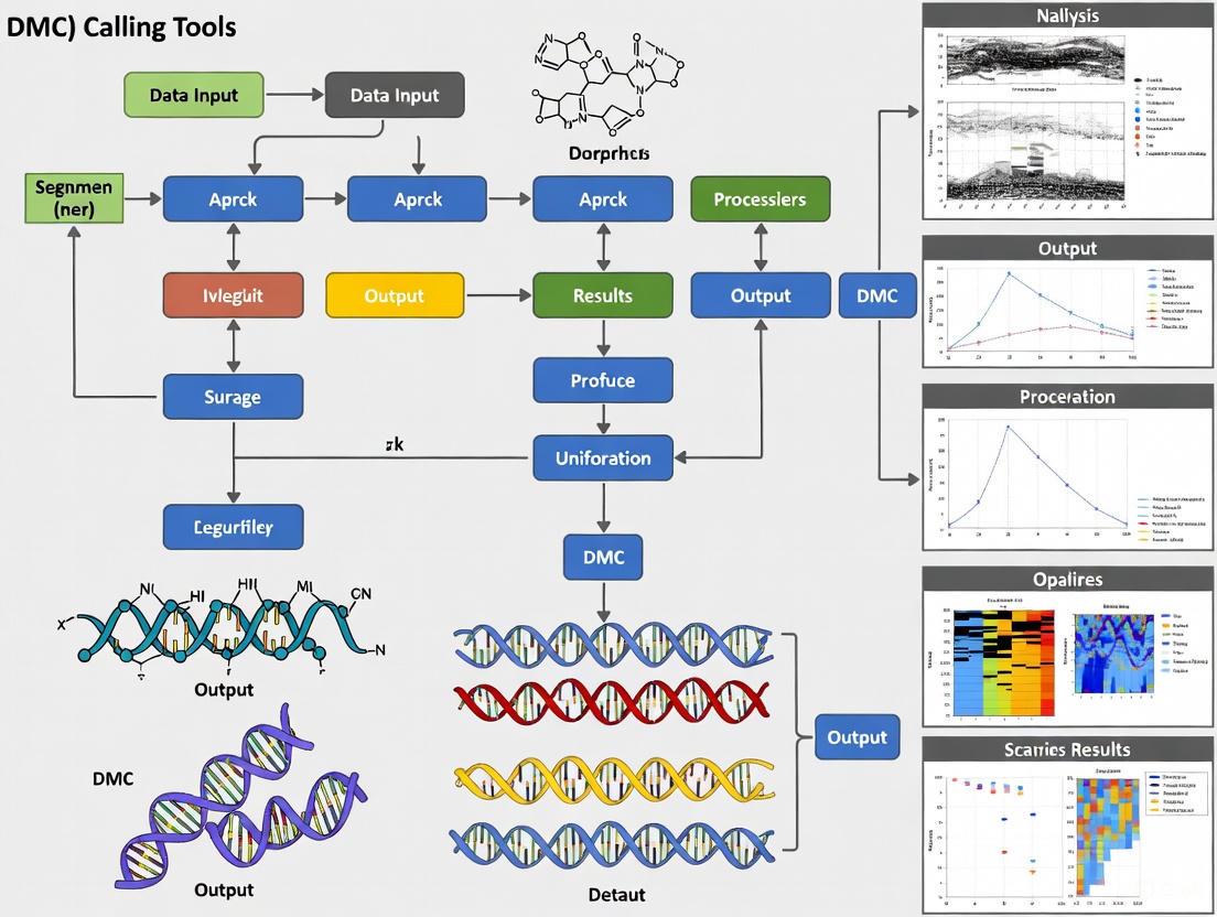
Figure 1: Comprehensive Workflow for DMC and DMR Analysis
Experimental Protocol for WGBS-Based DMR Identification
Library Preparation and Sequencing
This protocol outlines the steps for identifying DMRs from biological samples using whole-genome bisulfite sequencing, based on established methodologies [5] [7].
Materials:
- DNA Extraction Kit (e.g., Allprep DNA/RNA mini kit, Qiagen)
- Covaris M220 Ultrasonicator or equivalent mechanical shearing system
- NEBNext DNA Library Prep Kit (New England Biolabs)
- Methylated Adapters (Illumina)
- Imprint DNA Modification Kit (Sigma) or equivalent bisulfite conversion kit
- Size Selection System (e.g., Pippin Prep, Sage Science)
- PfuTurbo Cx Hotstart DNA Polymerase (Agilent Technologies)
- Illumina Sequencing Platform (HiSeq/NovaSeq)
Procedure:
DNA Extraction and Quality Control
- Extract genomic DNA from tissues or cells using appropriate methods.
- Assess DNA quality and quantity using spectrophotometry (A260/280 ratio ~1.8-2.0) and fluorometry.
- Use 1μg of high-molecular-weight genomic DNA as input material.
DNA Fragmentation and Library Preparation
- Fragment DNA to 300bp using Covaris M220 ultrasonicator.
- Repair ends and adenylate 3' ends using NEBNext library preparation kit.
- Ligate methylated Illumina adapters to fragments.
Bisulfite Conversion
- Treat adapter-ligated DNA with sodium bisulfite using Imprint DNA Modification Kit.
- Conduct conversion with the following cycling conditions:
- Denaturation: 95°C for 5 minutes
- Incubation: 60°C for 20-45 minutes
- Hold: 20°C indefinitely
- Purify converted DNA using provided columns or beads.
Library Amplification and Size Selection
- Amplify converted libraries using PfuTurbo Cx Hotstart DNA polymerase with the following program:
- Initial denaturation: 95°C for 2 minutes
- Cycling (12-15 cycles): 95°C for 30s, 60°C for 30s, 72°C for 45s
- Final extension: 72°C for 5 minutes
- Select 300-600bp fragments using Pippin Prep system.
- Validate library quality and quantity using Bioanalyzer and qPCR.
- Amplify converted libraries using PfuTurbo Cx Hotstart DNA polymerase with the following program:
Sequencing
- Sequence libraries on Illumina platform (HiSeq2000 or equivalent) to generate 100bp single-end or paired-end reads.
- Aim for minimum 30x genome-wide coverage for mammalian genomes.
Bioinformatics Analysis
Data Processing and Alignment:
- Quality Control and Adapter Trimming
- Assess read quality using FastQC.
- Trim adapters and low-quality bases using Trim Galore! or similar tools.
Alignment to Reference Genome
- Align bisulfite-converted reads using specialized aligners (BS Seeker, Bismark, or BSMAP).
- Command example for BS Seeker:
- Calculate and verify bisulfite conversion efficiency (>99%) using spike-in controls (e.g., λ-bacteriophage genome) [5].
Methylation Calling
- Extract methylation information at each cytosine using the aligned BAM files.
- Generate coverage files containing counts of methylated and unmethylated reads per cytosine.
DMR Identification Using Defiant:
- Installation
- Download Defiant from official repository and compile:
Execution
- Run Defiant with appropriate parameters:
- Essential parameters:
- Minimum coverage: 10x per CpG
- Minimum methylation difference: 10%
- p-value threshold: 0.05
- Minimum CpGs per DMR: 5
- Maximum distance between CpGs: 300bp
Output Interpretation
- Defiant generates BED files containing genomic coordinates of significant DMRs.
- Columns include: chromosome, start, end, methylation difference, p-value, and number of CpGs.
Annotation and Functional Interpretation
Genomic Annotation of DMRs/DMCs
Annotation associates identified DMRs/DMCs with genomic features to generate biological hypotheses. Common approaches include [10]:
Gene-Based Annotation
- Associate DMRs with nearby genes using tools like genomation or ChIPseeker.
- Categorize by genomic context:
- Promoter regions: Typically defined as ±2kb from transcription start site (TSS)
- Gene bodies: Introns and exons
- Intergenic regions: Distal to annotated genes
Regulatory Element Annotation
- Overlap with ENCODE regulatory elements (enhancers, promoters, DNase hypersensitive sites).
- Integration with chromatin state segmentation maps.
Sequence Context Analysis
- CpG island annotation: Shore (0-2kb from island), shelf (2-4kb from island), open sea (>4kb from island).
- Transcription factor binding site motif analysis.
Table 3: Genomic Distribution of DMRs in a Representative Analysis
| Genomic Feature | Percentage of DMRs | Potential Functional Impact |
|---|---|---|
| Promoter | 28.24% | Direct transcriptional regulation |
| Exon | 15.27% | Alternative splicing, gene expression |
| Intron | 33.59% | Enhancer activity, splicing regulation |
| Intergenic | 58.02% | Long-range regulation, unknown function |
Functional Enrichment Analysis
Functional analysis interprets the potential biological consequences of methylation changes [6] [11]:
Gene Ontology (GO) Enrichment
- Identify overrepresented biological processes, molecular functions, and cellular components among genes associated with DMRs.
- Use hypergeometric test with multiple testing correction (Benjamini-Hochberg FDR).
Pathway Analysis
- KEGG Pathway Enrichment: Detect signaling and metabolic pathways enriched for differentially methylated genes.
- Reactome Pathway Analysis: Identify molecular pathways and cascades affected.
Disease Association
- DisGeNET/Disease Ontology: Link DMGs to specific diseases and pathological processes.
- Integration with GWAS catalog to identify co-localization with disease-associated genetic variants.
Research Reagent Solutions
Table 4: Essential Materials for DMC/DMR Analysis
| Category | Specific Product/Kit | Application Note |
|---|---|---|
| DNA Extraction | Allprep DNA/RNA Mini Kit (Qiagen) | Simultaneous DNA/RNA preservation for multi-omics |
| Bisulfite Conversion | Imprint DNA Modification Kit (Sigma) | High conversion efficiency (>99%) with minimal DNA degradation |
| Library Preparation | NEBNext DNA Methylation Kit (NEB) | Optimized for bisulfite-converted DNA |
| Targeted Methylation | EpiTYPER MassARRAY (Agena) | Validation of DMRs without sequencing |
| Data Analysis | R/Bioconductor Packages (genomation, DSS, methylKit) | Comprehensive statistical analysis and visualization |
| Genome Annotation | genomation Package | Integration of DMRs with genomic features |
| Functional Analysis | clusterProfiler R package | GO and KEGG enrichment of DMGs |
The precise identification and interpretation of DMCs and DMRs is essential for understanding the functional role of DNA methylation in biological processes and disease. While bisulfite sequencing remains the gold standard measurement technology, appropriate statistical methods that account for spatial correlation and biological variability are crucial for accurate detection. The integration of DMR/DMC data with other genomic annotations and functional genomics datasets enables researchers to move beyond mere identification to mechanistic insights, ultimately advancing our understanding of epigenetic regulation in health and disease.
Bisulfite Sequencing (BS-seq) is a cornerstone technique in epigenetics for detecting DNA methylation at single-base resolution. The method leverages the differential sensitivity of cytosine bases to sodium bisulfite conversion, allowing researchers to precisely map 5-methylcytosine (5mC) across the genome. This protocol is fundamental for studies investigating differential methylation patterns in development, disease, and drug discovery. Within the context of Differentially Methylated Cytosines (DMC) calling tools research, a robust and well-optimized BS-seq workflow is critical, as the quality of the initial data directly impacts the reliability of all downstream bioinformatic analyses [12]. This application note provides a detailed, step-by-step protocol for the entire BS-seq workflow, from library preparation to methylation quantification.
The complete BS-seq workflow, from sample to analysis, involves a series of critical wet-lab and computational steps, culminating in data ready for DMC calling. The following diagram illustrates this integrated process:
Experimental Protocol
Bisulfite Conversion of Genomic DNA
Principle: Sodium bisulfite deaminates unmethylated cytosine to uracil, while methylated cytosine (5mC) remains unconverted. Subsequent PCR amplification treats uracil as thymine, creating sequence differences that reflect the original methylation status [13] [14] [12].
Materials:
- Source DNA: High-quality genomic DNA (1–10 µg) from cells, tissues, or paraffin-embedded sections [12].
- Key Reagents: Sodium bisulfite solution (3-5 M), NaOH (3 M), ammonium acetate, hydroquinone (optional), ethanol, and isopropanol [12]. Commercial kits (e.g., EpiTect Bisulfite Kit [Qiagen], EZ DNA Methylation Kit [Zymo Research]) are recommended for efficiency and consistency [12] [15].
Detailed Procedure:
- DNA Denaturation: Dilute 1-10 µg of genomic DNA in deionized water to a final volume of 18 µL. Denature the DNA by adding 2 µL of 3 M fresh NaOH and incubating at 37°C for 15 minutes [12].
- Bisulfite Treatment: Add 380 µL of a freshly prepared 5 M sodium bisulfite solution (pH 5.0) to the denatured DNA. Mix thoroughly. To prevent evaporation, overlay the reaction with 500 µL of heavy mineral oil [12].
- Incubation: Incubate the reaction mixture in the dark at 50°C for 12–16 hours. This long incubation is crucial for complete conversion [12].
- Purification and Desulfonation: Purify the bisulfite-treated DNA using a commercial clean-up kit (e.g., Wizard DNA Clean-Up System). Elute the DNA and add NaOH to a final concentration of 0.3 M to desulphonate the DNA. Incubate at 37°C for 15 minutes [12].
- DNA Precipitation: Precipitate the DNA by adding 5 M ammonium acetate, absolute ethanol, and isopropanol. Incubate at -20°C for 2-4 hours. Pellet the DNA by centrifugation at maximum speed for 10 minutes, wash with 70% ethanol, and air-dry the pellet [12].
- Resuspension: Resuspend the purified, bisulfite-converted DNA in 10-20 µL of TE buffer or deionized water. The DNA is now ready for library preparation [12].
Troubleshooting Notes:
- Incomplete Conversion: Ensure reagents are fresh, the pH of the bisulfite solution is correct (pH 5.0), and the incubation time is sufficient.
- Low DNA Yield: Bisulfite treatment causes significant DNA degradation (up to 90%); using carrier RNA or commercial kits designed to minimize loss is advised [13].
- PCR Amplification Issues: Design bisulfite-specific primers that do not contain CpG sites and optimize PCR conditions, as the converted DNA has reduced sequence complexity [12].
Library Preparation and Sequencing
Following bisulfite conversion, standard library preparation protocols are used, incorporating specific considerations. For Whole-Genome Bisulfite Sequencing (WGBS), fragmented DNA is used to construct libraries for whole-genome coverage. For Reduced-Representation Bisulfite Sequencing (RRBS), genomic DNA is digested with a restriction enzyme (e.g., MspI) to enrich for CpG-rich regions, thereby reducing sequencing costs [13]. The constructed libraries are then sequenced on an appropriate NGS platform.
Bioinformatics Analysis
Pre-mapping Quality Control and Read Trimming
Before mapping, raw sequencing reads must be assessed for quality using tools like FastQC. Adapter sequences and low-quality bases should be trimmed with tools such as Trimmomatic or Cutadapt to ensure high-quality data for alignment.
Bisulfite-Aware Read Mapping
Principle: Mapping bisulfite-treated reads is computationally challenging because the reference genome must be converted in silico to account for C-to-T (and G-to-A) changes. Specialized aligners simultaneously map reads to original and converted reference genomes [16].
Protocol using CLC Genomics Workbench:
- Tool Selection: Navigate to
Toolbox | Epigenomics Analysis | Bisulfite Sequencing | Map Bisulfite Reads to Reference[16]. - Input and Directionality: Select the trimmed read files. Critically, specify whether your library preparation protocol was directional (e.g., Illumina TruSeq DNA Methylation Kit) or non-directional (e.g., Zymo Pico Methyl-Seq). This informs the mapper which strands to expect reads from and is essential for accuracy [16].
- Reference Selection: Select the appropriate reference genome(s). Note that RAM requirements are doubled because the mapper indexes both the original and bisulfite-converted versions of the reference [16].
- Mapping Parameters:
- Cost Scheme: Adjust the match score (default 1), mismatch cost, insertion cost, and deletion cost. An affine gap cost is often preferred as it favors longer contiguous gaps over multiple short indels, which can improve mapping accuracy [16].
- Filtering Thresholds: Set the length fraction (minimum portion of the read that must align) and similarity fraction (minimum percent identity in the aligned region). The defaults (0.5 and 0.8, respectively) are a good starting point [16].
Table 1: Key Parameters for Bisulfite Read Mapping [16]
| Parameter | Description | Recommended Setting |
|---|---|---|
| Directionality | Specifies the library protocol type. | Must be determined from the experimental protocol. |
| Length Fraction | Minimum fraction of the read length that must align. | 0.5 - 0.9 |
| Similarity Fraction | Minimum percent identity of the aligned region. | 0.8 - 0.9 |
| Gap Cost | Cost model for insertions/deletions. | Affine (favors longer contiguous gaps) |
| Mismatch Cost | Penalty for a nucleotide mismatch. | Typically 2-3 |
Methylation Calling and Quantification
After mapping, the methylation status of each cytosine is determined. The number of reads supporting a methylated C (unconverted) versus an unmethylated C (converted to T) is counted. The methylation level (or beta-value) for a cytosine is calculated as:
[ \text{Methylation Level} = \frac{\text{Number of reads with methylated C}}{\text{Total reads covering the position}} ]
This generates a genome-wide methylation profile at single-base resolution, which is the direct input for DMC calling tools.
The Scientist's Toolkit
Table 2: Essential Reagents and Kits for the BS-seq Workflow
| Item | Function | Example Products / Notes |
|---|---|---|
| Bisulfite Conversion Kit | Chemically converts unmethylated C to U, critical for distinguishing methylation status. | EpiTect Bisulfite Kit (Qiagen), EZ DNA Methylation Kit (Zymo Research) [12] [15]. |
| DNA Purification Kits | For cleanup and concentration of DNA after bisulfite conversion and desulphonation. | Wizard DNA Clean-Up System (Promega), QIAquick PCR Purification Kit (Qiagen) [12]. |
| Bisulfite-Specific PCR Primers | Amplify target regions from bisulfite-converted DNA; must be designed for sequence after conversion. | Designed to be bisulfite-specific (avoiding CpGs) and strand-specific [12]. |
| Cloning & Sequencing Vectors | Required for cloning PCR products to analyze methylation patterns of single DNA molecules. | pGEM-T Easy Vector System (Promega) [12]. |
| Bisulfite Read Mapper | Software to align sequencing reads to a reference genome, accounting for C-to-T conversions. | CLC Genomics Workbench, Bismark, BS-Seeker [16]. |
Integration with DMC Calling Tools
The final output of the BS-seq workflow is a table of methylation levels for cytosines across the genome. This data serves as the primary input for computational tools designed to identify Differentially Methylated Cytosines (DMCs) and Regions (DMRs). The choice of DMC tool (e.g., MethylKit, DSS, MethylSig, bsseq, or newer tools like DiffMethylTools) depends on the experimental design and statistical considerations [17]. The reliability of the DMC results is inherently dependent on the precision and quality control applied at every stage of the BS-seq protocol detailed in this document.
Application Notes
The reliable identification of differentially methylated cytosines (DMCs) is fundamental to advancing our understanding of epigenetic regulation in development, disease, and therapeutic intervention. However, researchers face three interconnected challenges that can compromise the validity and reproducibility of findings: technical variation introduced by profiling platforms, biological heterogeneity stemming from tissue composition and disease states, and insufficient statistical power in study design and analysis. This document outlines these challenges and provides detailed protocols to enhance the rigor of DMC detection in epigenetic research.
Navigating Technical Variation in Methylation Profiling
Technical variation arises from differences in methylation detection technologies, each with distinct strengths and limitations regarding coverage, resolution, and data structure. Inconsistent results often stem from this technical landscape rather than true biological differences.
Table 1: Comparison of DNA Methylation Detection Methods
| Method | Resolution | Genomic Coverage | Key Advantages | Key Limitations | Best Suited For |
|---|---|---|---|---|---|
| Whole-Genome Bisulfite Sequencing (WGBS) | Single-base | ~80% of CpGs (unbiased) | Gold standard for comprehensive profiling; detects non-CpG methylation [2] | DNA degradation from bisulfite treatment; high cost and data complexity [2] | Discovery studies requiring full genome coverage |
| Enzymatic Methyl-Seq (EM-seq) | Single-base | Comparable to WGBS | Preserves DNA integrity; high concordance with WGBS [2] | Relatively new method; less established protocols [2] | Studies where DNA integrity is a primary concern |
| Illumina EPIC Array | Pre-defined CpG sites | ~850,000 CpG sites | Cost-effective; standardized processing; large sample capacity [2] | Limited to pre-designed probes; cannot discover novel CpGs [2] | Large-scale epidemiological or validation studies |
| Oxford Nanopore (ONT) | Single-base (Long-read) | High, including challenging regions | Long reads for haplotype resolution; no bisulfite conversion [2] | Higher DNA input required; lower agreement with WGBS/EM-seq [2] | Detecting methylation in complex, repetitive regions |
Accounting for Biological Heterogeneity
Biological heterogeneity manifests at multiple levels, from the cellular composition of bulk tissue samples to the molecular subtypes of diseases. Ignoring this heterogeneity can obscure genuine DMCs or create false positives.
A critical source of heterogeneity is the cellular composition of bulk tissue samples. A bulk tissue sample is a mixture of different cell types, each with its unique methylome. An observed methylation difference between case and control groups could be driven by a difference in the proportion of a specific cell type rather than a uniform change across all cells [18]. Furthermore, within a single disease category, such as breast cancer, significant inter-patient molecular heterogeneity exists. HER2-low breast cancers, for instance, comprise distinct subgroups that molecularly resemble either HER2-0 or HER2-positive disease, requiring separate analysis to uncover true DMCs [19].
Ensuring Statistical Power and Robust Analysis
The analysis of methylation data demands specialized statistical methods that account for its unique properties. Using inappropriate models or failing to account for data structure is a major source of low statistical power and irreproducible results.
A key consideration is model directionality. Statistical models for differential methylation implicitly assume a direction of effect. Modeling methylation (X) as dependent on a condition (Y), denoted X\|Y, is natural when the condition may affect methylation (e.g., smoking). The reverse direction, Y\|X, is more appropriate when methylation might affect the condition (e.g., mediating genetic risk for disease) [18]. Using a model with an incorrect directionality can drastically reduce performance and increase false positives [18].
Furthermore, standard methods like t-tests or ANOVA, which ignore the correlation between adjacent CpGs and the beta-binomial distribution of sequencing count data, are prone to higher false positive rates [20]. They also often discard positions with missing data, leading to substantial information loss and bias [20]. Powerful statistical methods like Hidden Markov Models (HMMs) and penalized trans-dimensional approaches are better suited for DMC detection as they can smooth methylation levels by leveraging local autocorrelation and handle missing data effectively [20].
Detailed Experimental Protocols
Protocol 1: A Robust Workflow for DMC Detection from Sequencing Data
This protocol utilizes the DMCTHM R package, a method based on a penalized trans-dimensional Hidden Markov Model, which is designed to address several key statistical challenges in DMC detection [20].
1. Sample Preparation and Sequencing
- Input Material: High-quality genomic DNA.
- Platform Recommendation: For discovery studies, use WGBS or EM-seq. EM-seq is advantageous when DNA degradation is a concern [2].
- Sequencing Depth: Aim for a minimum of 10-30x coverage per CpG site to ensure reliable methylation level quantification.
2. Data Preprocessing
- Quality Control: Use FastQC to assess raw read quality.
- Adapter Trimming and Read Alignment: Trim adapters (e.g., with Trim Galore!) and align reads to a reference genome using a bisulfite-aware aligner such as
BismarkorBS-Seeker2. - Methylation Calling: Extract methylation counts (methylated and unmethylated reads per cytosine) using the alignment tool's built-in functions.
3. DMC Detection with DMCTHM
- Rationale: This method models methylated counts with a beta-binomial distribution and uses an HMM to capture correlation among adjacent CpGs. It simultaneously estimates model parameters and smooths methylation levels, which is robust to outliers and missing data [20].
- Software Installation: Install the
DMCTHMR package (Version 0.1 or higher) from a relevant repository. - Input Data Preparation: Prepare a data object containing methylated and total read counts for each CpG site across all samples.
- Model Fitting:
- Output: The function returns a list of DMCs with their genomic coordinates, methylation differences, and statistical significance.
4. Result Interpretation and Annotation
- Annotation: Annotate significant DMCs with genomic features (e.g., promoters, gene bodies, CpG islands) using packages like
GenomicRangesand annotation databases (e.g.,AnnotationHub). - Visualization: Create Manhattan plots, volcano plots, and heatmaps to visualize the distribution and effect size of DMCs.
DMC Detection Workflow: A step-by-step process from sample preparation to final result reporting.
Protocol 2: A Framework for Cell-Type-Specific DMC Analysis from Bulk Tissue
This protocol outlines the use of TCA for deconvoluting bulk methylation data to identify DMCs at the cell-type-specific level, which is crucial for understanding the true cellular origin of epigenetic signals [18].
1. Data Requirements
- Bulk Methylation Data: Matrix of methylation values (β-values or M-values) from your bulk tissue samples.
- Cell-Type Proportions: A matrix of estimated cell-type proportions for each sample. These can be derived:
- Experimentally: Using cell sorting (e.g., FACS) and profiling of pure cell populations.
- Computationally: Using reference-based deconvolution tools (e.g.,
EpiDISH,minfi) if a reference methylome is available.
2. Choosing Model Directionality with TCA
- Decision Point: The choice between X\|Y (condition affects methylation) and Y\|X (methylation affects condition) is critical and should be based on biological plausibility [18].
- X\|Y Direction: Use for most case-control studies (e.g., disease state, environmental exposure).
- Y\|X Direction: Use when investigating methylation as a potential mediator, for example, between a genetic variant and a phenotype.
3. Running TCA Analysis
- Software Installation: Install the
TCAR package. - Input Data: Prepare your bulk methylation matrix, design matrix (condition of interest), and matrix of cell-type proportions.
- Execution:
4. Validation
- Where possible, validate key findings using independent methods, such as pyrosequencing on sorted cell populations or in situ techniques.
Tissue Deconvolution Logic: Illustrates how bulk tissue methylation data is a mixture of signals from constituent cell types, which statistical deconvolution aims to resolve.
The Scientist's Toolkit: Research Reagent Solutions
Table 2: Essential Reagents and Tools for DMC Research
| Item Name | Function/Application | Key Characteristics | Example/Reference |
|---|---|---|---|
| Zymo Research EZ DNA Methylation Kit | Bisulfite conversion of genomic DNA for WGBS or EPIC array | Efficient conversion while minimizing DNA degradation | Used in EPIC array protocol [2] |
| Nanobind Tissue Big DNA Kit | High-molecular-weight DNA extraction from tissue | Preserves long DNA fragments for long-read sequencing (ONT) or EM-seq | [2] |
| DMCTHM R Package | Statistical detection of DMCs from sequencing data | Handles missing data, autocorrelation; uses beta-binomial HMM | [20] |
| TCA R Package | Cell-type-specific analysis of bulk tissue methylation data | Deconvolutes bulk signals; allows for different model directionalities | [18] |
| Limma R Package | Differential analysis for microarray data (e.g., EPIC array) | Fits linear models with empirical Bayes moderation for stable inference | Used for DMC identification from arrays [21] |
| BSgenome.Hsapiens.UCSC.hg19 | Reference genome for annotation and coordinate mapping | Provides standardized genomic coordinates for CpG site annotation | Used for validating genomic regions [21] |
A Practical Toolkit: Statistical Models and Operational Workflows of Popular DMC Tools
The precise identification of differentially methylated cytosines (DMCs) represents a fundamental analytical procedure in epigenetics research, enabling investigators to locate individual cytosine bases exhibiting significant methylation variation between biological conditions. DMCs serve as crucial epigenetic markers associated with gene expression regulation, cellular differentiation, and disease pathogenesis [6]. The detection of DMCs typically constitutes the initial discovery phase in DNA methylation analysis, often preceding the investigation of larger differentially methylated regions (DMRs) which comprise clustered DMCs with synergistic effects [17]. The accuracy of DMC detection profoundly influences subsequent biological interpretations, making the selection of appropriate computational tools with understood assumptions and requirements a critical consideration for researchers.
DNA methylation, predominantly occurring as 5-methylcytosine (5mC) at cytosine-phospho-guanine (CpG) dinucleotides in mammalian genomes, represents a chemically stable yet biologically dynamic epigenetic mark [5]. DMC detection tools statistically compare methylation measurements between experimental groups (e.g., disease versus control, treated versus untreated) to identify CpG sites with significant methylation differences [6]. These computational approaches must account for biological variability, technical noise, and the unique statistical challenges posed by methylation data, which often consists of count-based measurements (methylated versus unmethylated reads) exhibiting binomial distributions [17] [5].
This application note provides a comprehensive comparative overview of DMC detection methodologies, emphasizing their underlying model assumptions, input requirements, and practical implementation protocols. By synthesizing this information within a structured framework, we aim to equip researchers with the necessary knowledge to select and implement appropriate DMC detection strategies for their specific experimental contexts.
Computational Tools for DMC Detection: A Comparative Analysis
Methodological Categories and Statistical Foundations
DMC detection tools employ diverse statistical frameworks that can be broadly categorized into five methodological approaches, each with distinct assumptions and performance characteristics [17]. Understanding these foundational statistical approaches is essential for selecting tools appropriate for specific experimental designs and data characteristics.
Generalized Linear Model (GLM) Based Methods represent the most prevalent category, modeling methylation counts using binomial or beta-binomial distributions to account for both biological variability and technical noise [17]. Tools in this category, including methylSig, DSS, and methylKit, implement sophisticated approaches for overdispersion estimation, which is critical for controlling false positive rates in experiments with biological replicates. These methods generally perform well with high-coverage sequencing datasets but may suffer from variance estimation instability with low-coverage data [17].
Statistical Testing-Based Methods utilize fundamental statistical tests such as Fisher's exact test, t-tests, or non-parametric alternatives to compare methylation frequencies between conditions [17]. While computationally efficient and straightforward to implement, these approaches often fail to adequately account for biological variability between replicates, potentially leading to inflated false discovery rates in heterogeneous sample populations. Methods like DMRfinder and eDMR fall into this category [17].
Smoothing-Based Methods, including BSmooth and BiSeq, employ local smoothing algorithms to aggregate information from neighboring CpG sites, reducing random noise and improving detection power for low-coverage data [17]. These methods assume correlation in methylation status between adjacent cytosines, which generally holds true in biological systems but may vary across genomic contexts.
Hidden Markov Model (HMM) Based Methods model methylation states and transitions along the genome using hidden Markov models, effectively capturing spatial dependencies in methylation patterns [17]. Tools like HMM-DM and Bisulfighter can handle low-coverage data more effectively than binomial-based methods but are computationally intensive and may be difficult to generalize across diverse experimental designs.
Linear Regression-Based Methods apply linear models to methylation levels assuming normal distribution of residuals, with implementations found in DMAP, RnBeads, and COHCAP [17]. These approaches perform optimally with high-quality, high-coverage data from array-based platforms or deeply sequenced bisulfite experiments but may lack robustness with count-based sequencing data exhibiting limited dynamic range.
Table 1: Methodological Categories of DMC Detection Tools
| Method Category | Representative Tools | Core Statistical Model | Optimal Use Case |
|---|---|---|---|
| GLM-Based | methylSig, DSS, methylKit | Binomial/Beta-binomial | High-coverage data with biological replicates |
| Statistical Testing | DMRfinder, eDMR | Fisher's exact test, t-tests | Preliminary screening of high-coverage data |
| Smooth-Based | BSmooth, BiSeq | Local likelihood smoothing | Low-coverage WGBS data |
| HMM-Based | HMM-DM, Bisulfighter | Hidden Markov Model | Data with strong spatial correlation |
| Linear Model | RnBeads, COHCAP | Linear regression | Array data or high-coverage BS-seq |
Tool-Specific Implementations and Requirements
DiffMethylTools represents a recently developed end-to-end solution that addresses analytical and computational difficulties in differential methylation dissection [17]. This tool supports versatile input formats for seamless transition from upstream methylation detection tools and offers diverse annotations and visualizations to facilitate downstream investigations. Comparative evaluations on multiple datasets, including both short-read and long-read methylomes, demonstrated that DiffMethylTools achieved overall better performance in detecting differential methylation than existing tools like MethylKit, DSS, MethylSig, and bsseq [17]. The tool implements robust statistical models specifically optimized for handling the increasing volume of long-read methylation data from third-generation sequencing technologies.
SMART2 provides a comprehensive toolkit for deep analysis of DNA methylation data from bisulfite sequencing platforms, with specific functionality for DMC identification [22]. The software supports analysis of data from whole-genome bisulfite sequencing (WGBS), reduced representation bisulfite sequencing (RRBS), and targeted bisulfite sequencing techniques including TruSeqEPIC, SureSelect and CpGiant. SMART2 incorporates specialized handling for replicate samples and includes functionality for missing value replacement using the median methylation value of other available samples from the same group [22]. For DMC detection, the tool employs entropy-based procedures to quantify methylation specificity for each CpG site, followed by statistical testing to identify significant differences across groups.
MethylKit represents a widely adopted GLM-based tool that models methylation counts using a binomial distribution and conducts statistical testing with likelihood ratio tests [17]. The tool accommodates multiple experimental designs through its flexible model specification but requires careful parameter tuning for overdispersion correction. MethylKit operates on methylation count data or preprocessed methylation percentage files and includes basic functionality for annotation and visualization, though these features are less comprehensive than those in integrated platforms like DiffMethylTools [17].
DSS (Dispersion Shrinkage for Sequencing) employs a Bayesian hierarchical framework to model methylation counts with a beta-binomial distribution, incorporating shrinkage estimation for overdispersion parameters to enhance stability with limited replicates [17]. This approach improves detection reliability in studies with small sample sizes, though it may exhibit conservative behavior in highly heterogeneous systems. DSS requires input data in BSseq object format or tab-delimited text files containing methylation counts.
Table 2: Input Requirements and Key Assumptions of DMC Detection Tools
| Tool | Input Data Format | Minimum Input Requirements | Key Statistical Assumptions |
|---|---|---|---|
| DiffMethylTools | BED, BedGraph, BSseq objects | ≥ 2 samples per group | Models biological variability and technical noise |
| SMART2 | Methylation matrix (chrome, start, end, beta values) | CpG sites with coverage data | Assumes methylation specificity follows entropy patterns |
| methylKit | TAB-delimited, BSseq objects | ≥ 3 replicates recommended for each group | Binomial distribution of methylation counts |
| DSS | BSseq objects, custom text formats | ≥ 2 samples per condition | Beta-binomial distribution with shrinkage |
| BSmooth | BSseq objects | Low-coverage across many samples | Spatial correlation of nearby CpG sites |
Experimental Protocols for DMC Detection
Benchmarking Framework for Tool Performance Assessment
Robust evaluation of DMC detection tools requires implementation of a standardized benchmarking framework that assesses both sensitivity and specificity across diverse data types. The following protocol outlines a comprehensive approach for comparative tool evaluation:
Experimental Design and Data Preparation: Begin by selecting appropriate benchmark datasets representing various sequencing technologies (e.g., WGBS, RRBS, targeted panels) and biological contexts [17]. Ideally, include both real datasets with validated DMCs and synthetic datasets with known ground truth. For synthetic data generation, utilize real methylation data as a starting methylome to preserve biological characteristics. Cluster CpG sites into regions with maximum gap of 100 base pairs, retaining only regions containing at least 20 CpG sites. Apply smoothing algorithms such as LOWESS (locally weighted scatterplot smoothing) to obtain baseline methylation profiles [17].
Simulation of Control and Case Conditions: Generate control methylomes by sampling methylation levels at individual CpG sites from a truncated normal distribution with mean equal to the local smoothed methylation level and standard deviation independently generated from a uniform distribution (e.g., ranging from 2 to 15) [17]. For case conditions, introduce structured methylation changes across the genome, with predetermined proportions of CpG sites assigned to different effect size categories (e.g., 40% minimal change, 30% ±5% difference, 10% ±10% difference, 10% ±15% difference, and remaining sites with larger differences of ±20%, ±30%, ±40%, and ±50%) [17].
Tool Execution and Parameter Optimization: Execute each DMC detection tool using multiple parameter configurations to assess sensitivity to settings. For tools employing statistical testing, apply false discovery rate (FDR) correction with standard thresholds (e.g., FDR < 0.05). Include coverage-based filtering appropriate for each dataset (e.g., minimum 5-10x coverage per CpG site) [6]. For tools supporting replication, utilize the built-in methods for handling biological replicates rather than pre-averaging methylation values.
Performance Metrics Calculation: Evaluate detection performance using standard metrics including sensitivity (recall), precision, F1-score, and area under the precision-recall curve (AUPRC). Calculate these metrics across a range of methylation difference thresholds (e.g., absolute methylation difference ≥ 0.1, 0.2, 0.3) and statistical significance thresholds (e.g., FDR < 0.05, 0.01, 0.001) [17]. Assess computational efficiency through runtime and memory consumption measurements on standardized computing infrastructure.
Standardized Workflow for DMC Detection with SMART2
The following protocol details a standardized approach for DMC identification using SMART2, which supports comprehensive methylation analysis across multiple sample groups:
Input Data Preparation: Format input data as a methylation matrix where each row represents a CpG site and columns contain chromosomal coordinates (chrome, start, end) followed by methylation values (ranging from 0 for unmethylated to 1 for fully methylated) for all samples [22]. The methylation matrix can be generated from individual BED files using BEDTools unionbedg command with appropriate parameters. Sample names should follow the convention G11, G12, G21, G22, etc., where Gi represents group i. Missing values should be denoted with "-" characters.
Command Line Execution: Execute SMART2 with the DMC project type using the following command structure:
Where the parameters specify: -t DMC for DMC detection; -MR for missing value replacement threshold (0.5 indicates replacement when ≥50% of group samples have data); -AG for percentage of available groups required for CpG inclusion (0.8 = 80%); -PD for p-value threshold for DMC significance (0.05); and -AD for absolute mean methylation difference threshold (0.1 = 10%) [22].
Result Interpretation and Validation: The primary output file (7_DMC.txt) contains identified differentially methylated CpG sites with statistical metrics including methylation specificity, Euclidean distance, entropy values, and ANOVA p-values [22]. Perform validation through integration with orthogonal data sources such as gene expression profiles or functional enrichment analysis of genes associated with identified DMCs. For technical validation, select a subset of DMCs for confirmation using targeted bisulfite sequencing or methylation-sensitive PCR.
Figure 1: Standardized workflow for DMC detection analysis illustrating key stages from data preparation through experimental validation.
Critical Considerations for Robust DMC Detection
Impact of Sequencing Technologies on Detection Accuracy
The choice of sequencing technology significantly influences DMC detection accuracy due to fundamental differences in detection principles, coverage distributions, and technical artifacts. Bisulfite sequencing-based methods, including WGBS and RRBS, represent the current gold standard for DNA methylation assessment, providing single-nucleotide resolution with conversion rates typically exceeding 99% [5]. These methods chemically convert unmethylated cytosines to uracils while methylated cytosines remain protected, effectively transforming epigenetic information into sequence information [5]. However, bisulfite conversion introduces DNA fragmentation and reduces sequence complexity, complicating alignment and increasing potential for PCR artifacts.
Emerging long-read technologies from Oxford Nanopore Technologies (ONT) offer bisulfite-free methylation detection while overcoming limitations in repetitive regions that challenge short-read approaches [23]. Comparative analyses demonstrate high concordance between ONT and bisulfite sequencing data, with Pearson correlation coefficients of 0.868 for R10.4.1 chemistry and 0.839 for R9.4.1 chemistry relative to bisulfite sequencing [23]. However, chemistry-specific biases exist between ONT platforms, with cross-chemistry comparisons showing reduced correlation values (e.g., R9 WT against R10 KO correlates at 0.8432 versus 0.8612 for R9 WT against R9 KO) [23]. These technology-specific biases must be considered when designing studies and analyzing data, particularly for meta-analyses combining datasets from multiple platforms.
Quality Control and Preprocessing Requirements
Robust DMC detection necessitates implementation of rigorous quality control measures throughout the data generation and processing pipeline. The following QC checkpoints are essential for reliable results:
Sequencing Data Quality Assessment: Evaluate raw sequencing quality metrics including per-base quality scores, adapter contamination, and bisulfite conversion efficiency (using spike-in controls such as λ-bacteriophage DNA) [5]. For ONT data, assess raw current signal quality and basecalling accuracy. Filter low-quality reads and trim adapter sequences using specialized bisulfite-aware preprocessing tools.
Alignment and Methylation Calling: Align processed reads to reference genomes using bisulfite-aware aligners such as Bismark or BSMAP, optimizing parameters for specific sequencing technologies [5]. Calculate methylation percentages at each CpG site using count-based approaches (methylated versus unmethylated reads). Apply coverage-based filtering, typically requiring minimum 5-10x coverage per CpG site for confident methylation assessment [6].
Sample-Level QC and Normalization: Examine sample-level metrics including coverage distribution, average methylation levels, and correlation between replicates. Identify potential outliers through multidimensional scaling or principal component analysis. For large-scale studies, apply normalization procedures to correct for technical variation while preserving biological signals, selecting methods appropriate for specific technologies (e.g., subset quantile normalization for array data, global scaling for sequencing data).
Figure 2: Essential quality control workflow for DMC detection analysis from raw sequencing data through preprocessing stages.
Table 3: Essential Research Reagent Solutions for DMC Analysis
| Reagent/Resource | Function | Implementation Example |
|---|---|---|
| Sodium Bisulfite | Chemical conversion of unmethylated cytosine to uracil | Bisulfite treatment of genomic DNA for WGBS and RRBS |
| Methylated Lambda Phage DNA | Bisulfite conversion efficiency control | Spike-in control for quantification of conversion rates |
| DNA Methyltransferases | Enzymatic methylation for control experiments | Generation of positive controls for methylation detection |
| Targeted Bisulfite Sequencing Kits | Validation of identified DMCs | Amplification and sequencing of specific genomic regions |
| Oxford Nanopore Flow Cells | Long-read methylation detection | R10.4.1 flow cells for native DNA methylation detection |
| Quality Control Software | Assessment of data quality | FastQC, MultiQC for sequencing quality metrics |
| Alignment Tools | Mapping of bisulfite-converted reads | Bismark, BSMAP for BS-seq data; minimap2 for ONT data |
| Statistical Computing Environment | Data analysis and visualization | R/Bioconductor with packages like DSS, methylSig |
The rapidly evolving landscape of DMC detection tools offers researchers diverse methodological approaches, each with distinctive assumptions and requirements that significantly influence detection performance. This comparative overview highlights the critical importance of matching tool selection to experimental design, data characteristics, and biological questions. As methylation profiling technologies continue to advance, particularly with the maturation of long-read sequencing platforms, computational methods must similarly evolve to address new challenges and opportunities.
Future methodology development will likely focus on improved integration of multi-omics data, enhanced normalization approaches for cross-platform analyses, and machine learning approaches for prioritizing functionally relevant methylation alterations. Additionally, as single-cell methylation profiling becomes more accessible, specialized tools for handling the unique characteristics of sparse single-cell data will become increasingly important. By maintaining awareness of both the capabilities and limitations of available DMC detection tools, researchers can maximize the biological insights gained from their epigenomic investigations while ensuring rigorous and reproducible analysis.
Table 1: Core Beta-Binomial Tools for Differential Methylation Analysis
| Tool Name | Primary Function | Key Model/Feature | Handles Biological Replicates |
|---|---|---|---|
| DSS (Dispersion Shrinkage for Sequencing data) [24] [25] | DMC & DMR detection | Beta-binomial with genome-wide dispersion prior | Yes |
| MethylSig [25] | DMC & DMR detection | Beta-binomial with local smoothing | Yes |
| RADMeth (Regression Analysis of Differential Methylation) [24] [25] | DMC & DMR detection | Beta-binomial regression | Yes |
| MACAU (Binomial Mixed Model) [24] | DMC detection | Binomial Mixed Model (accounts for population structure) | Yes, including genetic relatedness |
The quest to understand the epigenetic basis of cellular regulation and disease has made the identification of differentially methylated cytosines (DMCs) a critical bioinformatics challenge. Cytosine methylation, particularly in CpG contexts, is a fundamental epigenetic mark involved in gene regulation, genomic imprinting, and cellular differentiation [26] [5]. Bisulfite sequencing (BS-seq), including whole-genome bisulfite sequencing (WGBS) and reduced representation bisulfite sequencing (RRBS), provides quantitative methylation information at single-nucleotide resolution throughout the genome [27] [5]. These techniques exploit the differential sensitivity of methylated and unmethylated cytosines to sodium bisulfite conversion, which transforms unmethylated cytosines to uracils (read as thymines during sequencing) while leaving methylated cytosines unchanged [26] [5].
A fundamental characteristic of bisulfite sequencing data is its binomial nature: at each cytosine site, the data consists of the number of reads showing methylation (successes) and the total read coverage (trials) [27] [24]. However, biological and technical variability introduce over-dispersion—where the observed variation exceeds what the binomial distribution expects. This occurs because true methylation levels (pi) vary between biological replicates due to natural epigenetic variation and technical artifacts [27] [28]. Standard statistical tests that ignore this between-sample variability, or that convert counts to proportions, lack power and can produce misleading results [24] [25]. Beta-binomial models address this limitation by combining a binomial distribution (for sampling variability) with a beta distribution (for between-sample variability of methylation levels), providing a robust statistical framework for differential methylation analysis in complex experimental designs [27] [24].
Statistical Foundations of the Beta-Binomial Model
The beta-binomial model is a compound probability distribution that naturally handles over-dispersed count data. In the context of bisulfite sequencing, for a given cytosine site, the number of methylated reads (Mi) from sample *i* with total coverage *ni* is modeled in two stages [27]:
- Binomial Sampling: Conditional on the methylation level p_i, Mi ∼ Binomial(*pi, *n_i).
- Beta Distribution: The methylation level p_i itself varies across biological replicates following a Beta distribution, p_i ∼ Beta(α, β).
The parameters α and β of the Beta distribution are re-parameterized in terms of a mean methylation level π = α/(α+β) and a dispersion parameter γ = 1/(α+β+1). This leads to the following properties for the beta-binomial distribution [27]:
- E(Mi) = *niπ*
- Var(Mi) = *niπ(1-π)(1 + (n_i - 1)γ)*
The dispersion parameter γ quantifies the extra-binomial variation. When γ=0, the variance reduces to that of a binomial distribution. The larger the γ, the greater the over-dispersion between replicates. Beta-binomial regression extends this core model by allowing the mean methylation level π to be linked to a linear predictor of covariates (e.g., cell type, treatment) through a link function (e.g., logit), enabling the analysis of multifactor experiments [27] [29].
Tool-Specific Methodologies and Protocols
DSS (Dispersion Shrinkage for Sequencing Data)
Experimental Protocol for DMR Detection with DSS:
- Data Input Preparation: Prepare data as a matrix of methylated read counts and total read coverage counts for each CpG site and each sample. Data should be organized by experimental condition (e.g., case vs. control).
- Model Fitting: Use the
DMLfitfunction to estimate mean methylation levels and dispersion parameters for each condition. DSS employs a Bayesian shrinkage estimator for the dispersion parameter, borrowing information from across the entire genome to produce stable estimates even with small sample sizes [24] [25]. - Statistical Testing: Perform hypothesis testing for differential methylation between conditions using the
DMLtestfunction. This function computes test statistics and p-values for each CpG site based on the fitted beta-binomial models and the dispersion estimates. - DMR Calling: Utilize the
callDMRfunction to identify differentially methylated regions (DMRs). This function groups adjacent significant CpG sites into regions, using a threshold for the p-value difference and a minimum length requirement [25].
DSS is particularly noted for its ability to handle experiments with a small number of replicates through its robust dispersion shrinkage approach [25].
MethylSig
Experimental Protocol for DMR Detection with MethylSig:
- Data Input and Filtering: Input sorted BAM files or base-level coverage files. Filter CpG sites based on a minimum coverage threshold (e.g., at least 5 reads per site in each sample) to reduce false positives caused by low coverage [25].
- Local Information Smoothing: MethylSig improves parameter estimation for low-coverage sites by borrowing information from neighboring CpG sites. This local smoothing technique assumes that methylation levels are correlated within small genomic windows [25].
- Beta-Binomial Model Fitting: Fit a beta-binomial model at each CpG site. MethylSig estimates a separate dispersion parameter for each site or group of sites, in contrast to DSS's genome-wide shrinkage.
- Statistical Testing and Multiple Testing Correction: Test for significant differential methylation using a likelihood ratio test or a Wald test against the null hypothesis of no difference between groups. Apply multiple testing correction (e.g., Benjamini-Hochberg) to control the False Discovery Rate (FDR).
MethylSig's use of local smoothing makes it powerful for detecting DMRs, especially in regions with sparse but coordinated methylation changes [25].
RADMeth (Regression Analysis of Differential Methylation)
Experimental Protocol for DMR Detection with RADMeth:
- Data Input: Prepare data as a matrix of methylated and total read counts, along with a design matrix specifying the experimental conditions and any additional covariates (e.g., batch effects, age).
- Beta-Binomial Regression: The core of RADMeth is a beta-binomial regression model that can directly incorporate continuous or categorical covariates into the differential methylation test [24] [25]. This is represented as: g(π) = Xβ, where g is a link function (e.g., logit), X is the design matrix, and β are the coefficients.
- Dispersion Modeling: RADMeth accounts for over-dispersion by including a dispersion parameter in the regression model.
- Significance Testing and DMR Calling: Perform regression-based hypothesis tests on the coefficients of interest. RADMeth then combines evidence from adjacent CpG sites using a weighted Z-test to call DMRs, where the weights are based on the coverage at each site [25].
RADMeth's regression framework is its key strength, allowing for the analysis of complex experimental designs beyond simple two-group comparisons [24].
Diagram 1: A simplified workflow illustrating the core analytical pathways for DSS, MethylSig, and RADMeth, highlighting their key methodological distinctions.
The Scientist's Toolkit: Research Reagent Solutions
Table 2: Essential Reagents and Computational Tools for Beta-Binomial Analysis
| Item Name | Type | Function/Description | Example/Tool |
|---|---|---|---|
| Sodium Bisulfite | Chemical | Converts unmethylated cytosines to uracils, enabling methylation state discrimination [5]. | Sigma-Aldrich, Thermo Fisher |
| Unmethylated λ-DNA | Control | Spiked into reactions to empirically measure and confirm bisulfite conversion efficiency [5]. | Promega |
| CpG Island Reference | Bioinformatics | Genomic coordinates of CpG-rich regions for annotation and targeted analysis [5]. | UCSC Genome Browser |
| Bisulfite-Aware Aligner | Software | Aligns BS-seq reads to a reference genome, accounting for C-to-T conversions [26] [30]. | Bismark [26], BatMeth2 [30], BSMAP [26] |
| Methylation Caller | Software | Estimates methylation levels per cytosine from aligned BAM files [30]. | Bismark Methylation Extractor, BatMeth2 |
| Statistical Environment | Software | Platform for statistical analysis, visualization, and running specialized packages [25]. | R/Bioconductor |
| DSS / MethylSig / RADMeth | R Package | Implements the beta-binomial model for differential methylation testing [24] [25]. | Bioconductor |
Critical Analysis and Practical Considerations
Performance and Comparative Evaluation
Table 3: Comparative Analysis of Beta-Binomial Tools
| Feature | DSS | MethylSig | RADMeth | MACAU |
|---|---|---|---|---|
| Core Strength | Stability with few replicates | Power for coordinated sparse changes | Complex designs & covariates | Accounts for population structure |
| Dispersion Handling | Genome-wide shrinkage | Local estimation & smoothing | Regression-based | Mixed model component |
| Covariate Adjustment | Limited | Limited | Yes (in regression model) | Yes (as fixed effects) |
| Smoothing Approach | No | Yes (local CpGs) | No (but uses weighted Z-test for DMRs) | No |
| Optimal Use Case | Two-group comparisons with low replication | Defining precise DMR boundaries | Multi-factor or batch-adjusted designs | Structured populations (e.g., EWAS) |
Independent evaluations of beta-binomial methods have shown that they provide a major advantage over non-replicate methods like Fisher's exact test by properly controlling for between-sample variability [25]. However, the performance of each tool can vary depending on the data structure. A key finding is that while these methods are powerful, no single tool uniformly dominates all others across all scenarios [25]. For instance, DSS's shrinkage estimator provides robustness with small sample sizes, while RADMeth's regression framework is unparalleled in studies requiring adjustment for confounders [24] [25].
Limitations and Advanced Modeling
A significant limitation of standard beta-binomial models is their inability to account for genetic relatedness or population structure among samples [24]. In studies of structured populations or where kinship exists, this can lead to spurious associations, as DNA methylation levels are often heritable [24]. To address this, the binomial mixed model (BMM) implemented in MACAU incorporates a random effect to model the covariance between individuals due to population structure [24]. MACAU has been demonstrated to provide well-calibrated p-values in the presence of population structure and can detect more true positive associations than beta-binomial models in such scenarios [24].
Furthermore, beta-binomial models face challenges with extremely low coverage sites and very small sample sizes (e.g., fewer than 3 per group), where parameter estimation becomes unreliable. Methods like MethylSig's local smoothing and DSS's global shrinkage are attempts to mitigate these issues, but careful data filtering and cautious interpretation remain essential [25].
Within the framework of a broader thesis on Differentially Methylated Cytosines (DMC) calling tools, this document delves into three sophisticated computational approaches designed for identifying Differentially Methylated Regions (DMRs): metilene, BSmooth, and BiSeq. The accurate detection of DMRs is a critical prerequisite for characterizing epigenetic states linked to development, tumorigenesis, and systems biology [31]. While DMC callers focus on single cytosines, DMR tools like these aggregate information across multiple nearby CpG sites to identify broader, often more biologically meaningful, epigenetic patterns. These tools were developed to address the significant bottleneck in methylome analysis posed by the complexity and scale of data from whole-genome bisulfite sequencing (WGBS) and reduced representation bisulfite sequencing (RRBS) [31] [26]. This note details their underlying statistical models, provides structured performance comparisons, and outlines explicit protocols for their application in research and drug development.
The selection of an appropriate DMR detection tool depends heavily on the experimental design, data type, and specific biological question. The table below summarizes the core methodologies, key features, and optimal use cases for metilene, BSmooth, and BiSeq.
Table 1: Overview and Comparison of DMR Detection Tools
| Feature | metilene | BSmooth | BiSeq |
|---|---|---|---|
| Core Algorithm | Binary segmentation (CBS) combined with a two-dimensional Kolmogorov-Smirnov test [31]. | Local likelihood smoothing followed by a t-test accounting for biological variability [32]. | Beta-binomial regression model that smooths methylation levels within predefined regions [31] [26]. |
| Primary Approach | Non-parametric, model-free segmentation. | Smoothing-based, empirical Bayes. | Parametric, regression-based smoothing. |
| Key Strength | High speed and sensitivity; precise boundary detection; handles low-coverage data [31]. | Effectively accounts for biological variability; superior for low-coverage designs [32]. | Models biological variability explicitly through a random effect; provides well-calibrated error rates [31] [26]. |
| Optimal Data Type | WGBS, RRBS [31]. | WGBS, especially with biological replicates [32]. | RRBS, targeted bisulfite sequencing [31]. |
| Typical Output | Genomic coordinates of DMRs with p-values and methylation differences. | Smoothed methylation levels per sample and called DMRs. | DMRs with associated posterior probabilities or p-values. |
Performance benchmarks on synthetic and real data further illuminate the practical differences between these tools. Evaluations consistently show that metilene offers an exceptional balance of sensitivity, positive predictive value (PPV), and computational efficiency.
Table 2: Performance Benchmarks on Simulated and Real Data
| Performance Metric | metilene | BSmooth | BiSeq | Notes |
|---|---|---|---|---|
| True Positive Rate (TPR) | ~99.8% (Challenging DMRs) [31] | < 50% (Simulated DMRs) [31] | Comparable to metilene in some RRBS tests [31] | On DMRs with small methylation differences. |
| Positive Predictive Value (PPV) | ≥ 0.989 [31] | Information Missing | Lower than metilene [31] | Across all tested simulation scenarios. |
| Runtime (Chromosome 10) | ~4 min (single core) [31] | ~2.3 hours [31] | Information Missing | 2 x 10 samples; off-the-shelf hardware. |
| Memory Usage | < 1 GB [31] | 10.7 GB [31] | Information Missing | On the same dataset as runtime. |
| Performance on Low Coverage | High (Top PPV & TPR) [31] | Information Missing | Information Missing | Robust to missing data. |
Detailed Methodologies and Protocols
metilene: Protocol for DMR Calling
metilene's protocol involves a segmentation algorithm that scans the genome for regions with homogeneous methylation differences between groups, delimited by pairs of change points [31].
Input Data Preparation:
- Input Format: The input is a text file where each row represents a CpG site. The first three columns must be
chromosome,start, andendcoordinates. Subsequent columns contain methylation ratios (values between 0 and 1) for each sample, with columns named to reflect their group (e.g.,control_1,control_2,case_1,case_2). Missing values should be denoted by-[22]. - Data Generation: This matrix can be generated from individual BedGraph files using
bedtools unionbedg[22].
Command Line Execution:
Parameter Explanation:
--maxdist 300: Maximum allowed distance (in bp) between two adjacent CpGs within a DMR.--mincpgs 5: Minimum number of CpGs constituting a DMR.--minMethDiff 0.2: Minimum absolute average methylation difference between groups to be considered a DMR.--minNo 5: Minimum per-group sample number for which a CpG must have data.--threads 10: Number of threads for parallel computation.-a&-b: Sample group labels as they appear in the input file header [6].
Output Interpretation: The output is a tab-separated file with columns including chromosome, start and end of the DMR, p-value, methylation level in group A, methylation level in group B, and the absolute difference between them.
BSmooth: Protocol for DMR Analysis
BSmooth's workflow involves alignment, quality control, smoothing of methylation levels, and finally, DMR calling that incorporates biological variation [32].
Workflow Execution:
- Alignment & Preprocessing: BSmooth includes an alignment pipeline using
Bowtie 2or its custom alignerMermanfor colorspace data. This step generates BAM files [32]. - Quality Control (QC): A critical BSmooth-specific QC step is the assessment of "M-bias" by plotting the percentage of cytosines found at each read position. This identifies and allows filtering of systematic base-calling errors towards read ends [32].
- Smoothing and DMR Calling:
Key Analytical Steps:
- Smoothing: For each sample, local likelihood smoothing is applied to the single-CpG methylation estimates (
M_j / N_j). This leverages the spatial correlation of methylation levels along the genome to improve precision, especially beneficial for low-coverage data [32]. - DMR Calling: A t-test is performed at each CpG site, comparing the smoothed methylation values between groups. DMRs are defined as regions where the difference in smoothed methylation exceeds a threshold (e.g., 0.1) and the p-values from the t-tests are sufficiently small, after accounting for multiple testing. This step explicitly uses the variation between biological replicates [32].
BiSeq: Protocol for DMR Detection
BiSeq is tailored for RRBS and targeted data. It clusters CpG sites into regions and uses a beta-binomial model to account for variation between replicates [31] [26].
Workflow in R:
- Data Loading and Clustering: Load CpG methylation data and cluster them into potential regions of interest based on genomic proximity.
- Smoothing and Prediction: Smooth methylation levels within each cluster. This step borrows information from nearby CpG sites to obtain more stable methylation estimates for each region and sample.
- Differential Methylation Testing: Fit a beta-binomial regression model to test for significant methylation differences between groups, which can include a random effect for biological replicates.
Visual Workflows
The following diagrams illustrate the core logical workflows for each DMR calling tool, highlighting their distinct approaches to processing methylation data.
metilene DMR Calling Logic
BSmooth Analysis Pipeline
BiSeq DMR Detection Flow
The Scientist's Toolkit: Research Reagent Solutions
The following table lists key computational tools and resources essential for conducting the DMR analyses described in this document.
Table 3: Essential Research Reagents and Computational Tools
| Item Name | Function/Benefit | Usage in Protocol |
|---|---|---|
| Bismark | A widely used aligner and methylation caller for bisulfite sequencing data. It provides unbiased alignment and outputs coverage files suitable for downstream analysis [33]. | Used in preprocessing to generate per-CpG methylation counts (e.g., bismark_methylation_extractor), which can be formatted into input for metilene or methylKit. |
| Bowtie 2 | A fast and memory-efficient alignment tool for sequencing reads. It is often used in conjunction with in-silico bisulfite conversion pipelines [32]. | Serves as the core aligner in one of BSmooth's alignment pipelines for WGBS data. |
| bedtools | A powerful toolset for genome arithmetic. It is indispensable for intersecting, merging, and comparing genomic features. | Used to create a unified methylation matrix from multiple sample-specific BedGraph files, which is the required input format for metilene [22]. |
| R/Bioconductor | An open-source software environment for statistical computing and graphics. Bioconductor provides specialized packages for high-throughput genomic data analysis. | The primary platform for running BSmooth (via the bsseq package) and BiSeq. Also used for extensive downstream analysis, visualization, and annotation of results. |
| SAM/BAM Files | Standard file formats for storing aligned nucleotide sequence data. The binary BAM format is compact and indexed for efficient access. | The standard output of alignment tools like Bismark and Bowtie 2. These files are the starting point for BSmooth's pipeline and for generating methylation counts. |
The comprehensive analysis of DNA methylation data, particularly the identification of differentially methylated cytosines (DMCs), relies on robust statistical methods implemented within specialized software packages. methylKit is an R package that provides a comprehensive toolkit for analyzing DNA methylation data from high-throughput bisulfite sequencing experiments [34]. This package integrates multiple statistical approaches for DMC detection, primarily offering Fisher's Exact Test and logistic regression models to accommodate varied experimental designs and data characteristics [35] [36]. The selection between these tests is not arbitrary but depends on specific experimental parameters, including sample size, replication, and the underlying biological question. methylKit models methylation levels using logistic regression and tests differences in log odds between treatment and control groups to determine DMCs, while also providing Fisher's exact test as a robust alternative for specific scenarios [36]. This application note explores the theoretical foundations, practical implementation, and comparative performance of these two statistical approaches within the context of DMC calling, providing researchers with a framework for selecting and applying the optimal method for their experimental data.
Theoretical Foundations and Statistical Principles
Fisher's Exact Test
Fisher's Exact Test is a non-parametric statistical significance test used to determine if there are nonrandom associations between two categorical variables [37]. In the context of DNA methylation analysis, it assesses the independence of CpG methylation between two groups.
Mathematical Basis: The test calculates the exact probability of observing the data contingency table, or a more extreme arrangement, under the null hypothesis that methylation status is independent of the sample group [37] [38]. The probability (p) for a 2×2 table is calculated using the hypergeometric distribution with the formula:
p = (a+b)Ca * (c+d)Cc / (a+b+c+d)Ca+c
where the symbols represent counts in the contingency table: a (methylated in group1), b (unmethylated in group1), c (methylated in group2), and d (unmethylated in group2) [37].
Application Context: In methylKit, Fisher's Exact Test is automatically applied when there is only one sample per group after pooling replicates [35]. This method is particularly valuable for small sample sizes or when dealing with low numbers of methylated CpGs, as it does not rely on asymptotic approximations [37].
Logistic Regression Models
Logistic regression in methylKit provides a more flexible framework for differential methylation analysis, particularly suited for studies with multiple replicates or when incorporating covariates.
Mathematical Basis: The method models the log-odds of methylation status as a linear function of predictor variables. The fundamental model is represented as:
log(pi/(1-pi)) = θ0 + θ1xi1 + ... + θpxip
where pi represents the probability that a CpG site is methylated, θ0 is the intercept, and θ1,...,θp are regression coefficients for predictors x1,...,xp [39].
Application Context: methylKit implements logistic regression to test the difference in log-odds between treatment and control groups [36]. This approach can accommodate complex experimental designs, including the incorporation of covariates such as age, batch effects, or other clinical variables that might influence methylation patterns [35].
Key Statistical Differences
Table 1: Fundamental Differences Between Fisher's Exact Test and Logistic Regression in methylKit
| Feature | Fisher's Exact Test | Logistic Regression |
|---|---|---|
| Statistical Paradigm | Non-parametric, exact test | Parametric, model-based |
| Data Requirements | Single 2×2 contingency table | Multiple observations, covariates possible |
| Sample Size | Ideal for small samples (<1000) or when replicates are pooled | Requires adequate replicates for reliable parameter estimation |
| Effect Measurement | Odds Ratio (conditional MLE) | Log-Odds Ratio (unconditional MLE) |
| Covariate Adjustment | Not supported | Supported through model expansion |
| Output | Association significance and odds ratio | Coefficient estimates, significance, and model fit |
Practical Implementation in methylKit
Experimental Workflow for Differential Methylation Analysis
The following diagram illustrates the comprehensive workflow for differential methylation analysis in methylKit, highlighting decision points for test selection:
methylKit Protocol for Differential Methylation Analysis
Data Preparation and Quality Control
- Data Input: Read methylation data from text files or SAM/BAM format alignment files from Bismark aligner [34].
- Quality Filtering: Apply default filters requiring at least 10 reads covering a base, with each base having a minimum PHRED quality score of 20 [34].
- Data Normalization: Optionally apply normalization to correct for technical variations between samples.
Method Selection and Application
Table 2: methylKit Function Parameters for calculateDiffMeth()
| Parameter | Fisher's Exact Test Scenario | Logistic Regression Scenario | Description |
|---|---|---|---|
| test | "fast.fisher" or "midPval" | "F" or "Chisq" | Statistical test to use |
| overdispersion | "none" | "MN" or "shrinkMN" | Overdispersion correction for logistic regression |
| covariates | NULL | data.frame | Covariates to include in logistic model |
| adjust | "SLIM", "BH", "fdr" | "SLIM", "BH", "fdr" | Multiple testing correction method |
| mc.cores | 1 or more | 1 or more | Number of cores for parallel processing |
Code Implementation Examples
Scenario 1: Fisher's Exact Test with Pooled Samples
Scenario 2: Logistic Regression with Covariates
Performance Comparison and Benchmarking
Method Performance Under Different Experimental Conditions
Empirical evaluations of differential methylation methods reveal distinct performance characteristics for Fisher's Exact Test and logistic regression approaches.
Table 3: Performance Benchmarking of Statistical Methods in methylKit
| Performance Metric | Fisher's Exact Test | Logistic Regression |
|---|---|---|
| Small Sample Performance | Excellent (exact test) | May suffer from low power |
| Large Sample Performance | Computationally intensive | Optimal (asymptotic properties) |
| Type I Error Control | Conservative | Appropriate with adequate samples |
| Covariate Adjustment | Not applicable | Effective when properly specified |
| Computational Efficiency | Fast for small datasets | Efficient for large datasets with multiple replicates |
| DMR Detection Accuracy | Good for strong effects | Superior for subtle, coordinated effects |
Practical Considerations for Method Selection
The following decision diagram outlines the method selection process based on experimental design and research objectives:
Advanced Applications and Integration
Research Reagent Solutions for DNA Methylation Studies
Table 4: Essential Research Reagents and Platforms for Methylation Analysis
| Reagent/Platform | Function | Application in methylKit Workflow |
|---|---|---|
| Illumina Infinium Methylation BeadChip | Genome-wide methylation profiling | Data input source for array-based methylation levels |
| Bismark Bisulfite Read Mapper | Alignment of bisulfite-seq reads | Preprocessing step before methylKit analysis |
| Sodium Bisulfite Conversion Kits | Conversion of unmethylated cytosines to uracils | Sample preparation for BS-seq experiments |
| Methylated DNA Immunoprecipitation (MeDIP) Kits | Enrichment of methylated DNA regions | Alternative input for methylKit regional analysis |
| Whole-Genome Bisulfite Sequencing (WGBS) Kits | Comprehensive methylation profiling | Gold standard input for base-resolution analysis |
Integration with Downstream Bioinformatics Analyses
methylKit's differential methylation output serves as input for various downstream analyses, including:
- Functional Enrichment Analysis: DMRs can be tested for enrichment in functional genomic elements using Fisher's Exact Test in gene ontology analysis [40].
- Pathway Analysis: Significant DMCs can be mapped to genes and pathways to identify biological processes affected by methylation changes.
- Multi-Omics Integration: Correlation of methylation patterns with gene expression data to identify potential regulatory relationships.
- Visualization: methylKit provides built-in functions for clustering, principal component analysis, and visualization of methylation patterns [34] [36].
The selection between Fisher's Exact Test and logistic regression in methylKit should be guided by experimental design, sample size, and research objectives. Fisher's Exact Test provides a robust, non-parametric option for studies with limited replication or when samples are pooled, while logistic regression offers greater flexibility for complex experimental designs requiring covariate adjustment.
Based on comprehensive benchmarking studies, no single method consistently outperforms all others across all scenarios [36]. Researchers should consider the following evidence-based recommendations:
- For studies with pooled samples or minimal replication, Fisher's Exact Test provides reliable Type I error control.
- For complex experimental designs with multiple covariates, logistic regression implemented in methylKit offers appropriate flexibility.
- For large-scale studies with adequate replication, logistic regression typically demonstrates superior power for detecting subtle methylation differences.
- Regardless of method choice, careful multiple testing correction and independent validation remain essential components of rigorous differential methylation analysis.
The integration of these statistical approaches within methylKit provides epigenetics researchers with a comprehensive toolkit for identifying biologically meaningful methylation changes across diverse experimental contexts.
DNA methylation represents a fundamental epigenetic mark predominantly involving the addition of a methyl group to the fifth carbon of cytosine bases in CpG dinucleotides, forming 5-methylcytosine (5mC) [41] [5]. This modification plays crucial roles in transcriptional regulation, genome stability, and cellular differentiation, with aberrant patterns contributing to various diseases including cancer, neurological disorders, and autoimmune conditions [41] [26]. The oxidation of 5mC by ten-eleven translocation (TET) proteins generates additional cytosine modifications including 5-hydroxymethylcytosine (5hmC), 5-formylcytosine (5fC), and 5-carboxylcytosine (5caC), which may serve distinct biological functions beyond mere demethylation intermediates [41].
Advanced sequencing technologies now enable genome-wide methylation profiling at single-base resolution, with bisulfite sequencing representing the current gold standard [5] [26]. This technique chemically converts unmethylated cytosines to uracils (read as thymines during sequencing) while leaving methylated cytosines unchanged, allowing quantitative methylation assessment through the ratio of C/(C+T) reads at each cytosine position [26]. While initial analyses often focus on identifying differentially methylated cytosines (DMCs), biological interpretation typically requires aggregating these signals into differentially methylated regions (DMRs)—genomic intervals showing consistent methylation differences between conditions [26] [42]. This article focuses on segmentation-based approaches for DMR identification, which directly partition the genome into regions with homogeneous methylation patterns rather than first identifying individual DMCs.
Segmentation Approaches for DMR Calling
Segmentation methods represent a powerful paradigm for DMR detection that directly identifies genomic intervals with homogeneous methylation patterns, often outperforming approaches that first identify individual DMCs and subsequently merge them [42]. These methods typically leverage statistical frameworks that partition the genome based on changes in methylation patterns, effectively grouping CpGs that show similar methylation profiles across sample groups.
Core Segmentation Algorithms
Table 1: Key Segmentation Tools for DMR Calling
| Tool | Underlying Algorithm | Statistical Test | Key Features | Reference |
|---|---|---|---|---|
| metilene | Binary segmentation | Two-dimensional Kolmogorov-Smirnov test | Fast processing; suitable for low-coverage data; minimal memory requirements | [31] |
| MethyLasso | Fused lasso regression | Generalized additive model | No binning required; identifies constant methylation regions; versatile application | [42] |
| BSmooth | Local likelihood smoothing | Welch's t-test | Handles low coverage through smoothing; requires replicates | [42] [7] |
| Dmrseq | Beta-binomial model with smoothing | Autoregressive correlation | Controls FDR; replaces BSmooth; uses Bayesian approach | [42] |
| Defiant | Weighted Welch Expansion (WWE) | Welch's t-test or Fisher's exact test | Accepts 10 input formats; fast execution; handles multiple replicates | [7] |
The metilene algorithm employs a circular binary segmentation approach combined with a two-dimensional Kolmogorov-Smirnov test to identify regions with maximal intergroup methylation differences [31]. This method recursively segments the genome until regions contain fewer than a user-defined number of CpGs or statistical significance no longer improves, effectively identifying short methylation valleys embedded within longer differentially methylated regions [31]. Benchmarking analyses have demonstrated metilene's superior performance in both true positive rate (TPR) and positive predictive value (PPV), particularly for challenging scenarios with small methylation differences or complex background methylation patterns [31].
MethyLasso represents a recent innovation that models DNA methylation data using a generalized additive framework with fused lasso regularization [42]. This approach directly estimates regions with constant methylation levels without requiring pre-binning of data, thus avoiding the limitations of window-based methods. The strength of this statistical framework lies in its ability to handle the low signal-to-noise ratio in methylation data while preventing the agglomeration of unrelated genomic features [42]. MethyLasso serves as an all-in-one tool capable of identifying hypomethylated regions (LMRs, UMRs, DNA methylation valleys) in single conditions as well as DMRs between conditions.
Performance Considerations
Performance evaluations indicate that segmentation methods generally outperform alternative approaches in both sensitivity and computational efficiency. In comparative analyses, metilene correctly identified 99.8% of DMRs with smaller methylation differences in complex backgrounds where competing tools like MOABS detected less than 40% [31]. Regarding computational requirements, metilene processed a simulated chromosome 10 dataset with 2×10 samples in approximately 4 minutes using a single core, while MOABS required over 65 hours for the same task [31]. Memory consumption showed similar advantages, with metilene using <1 GB compared to 5.4 GB for MOABS and 10.7 GB for BSmooth [31].
Experimental Protocol for DMR Calling Using Segmentation Methods
Sample Preparation and Sequencing
Materials and Reagents:
- High-quality genomic DNA (≥1 μg for WGBS)
- Sodium bisulfite conversion kit (e.g., Imprint DNA modification kit)
- DNA fragmentation system (e.g., Covaris ultrasonicator)
- Library preparation kit (e.g., NEBNext genomic sequencing kit)
- Methylated adaptors for Illumina platforms
- Size selection system (e.g., Pippin Prep)
- High-fidelity DNA polymerase (e.g., PfuTurbo Cx Hotstart)
Procedure:
- DNA Extraction and Quality Control: Isolate genomic DNA using appropriate methods (e.g., Allprep DNA/RNA mini kit). Verify DNA integrity and quantity using spectrophotometry and fluorometry [7].
- DNA Fragmentation: Fragment DNA to 300 bp using focused ultrasonication [7].
- Bisulfite Conversion: Treat fragmented DNA with sodium bisulfite using optimized conversion kits. Include unmethylated λ-bacteriophage DNA as a conversion control to verify conversion rates >99% [5].
- Library Preparation: Prepare sequencing libraries using kits compatible with bisulfite-converted DNA. Ligate methylated paired-end adaptors to preserve methylation information [7].
- Size Selection and Amplification: Select fragments of 300-600 bp using agarose gel electrophoresis or automated systems. Amplify libraries using bisulfite-compatible polymerases [7].
- Sequencing: Sequence libraries on Illumina platforms (HiSeq, NovaSeq) with 100-150 bp reads, either paired-end or single-read based on project requirements [7].
Bioinformatic Processing Workflow
The following workflow diagram illustrates the complete process from raw sequencing data to DMR identification:
Bioinformatic Protocol:
Quality Control and Adapter Trimming
- Utilize Trim Galore! or Trimmomatic to remove low-quality bases and adaptor sequences [26].
- Set quality threshold (Phred score ≥20) and minimum read length (≥50 bp).
Alignment to Reference Genome
- Perform alignment using bisulfite-aware aligners such as Bismark (three-letter aligner) or BSMAP (wildcard aligner) [26].
- Convert all Cs to Ts in the forward strand and Gs to As in the reverse strand for three-letter alignment.
- Use default parameters with alignment rate threshold ≥70%.
Post-Alignment Processing
- Extract methylation calls using Bismark methylation extractor or similar tools.
- Calculate methylation percentages at each CpG site using the formula: % methylation = (number of C reads / (number of C reads + number of T reads)) × 100.
- Merge technical replicates and filter CpGs with coverage <10× [7].
DMR Calling with Segmentation Tools
For metilene:
Parameters: -t (threads), -m (minimum CpGs per DMR), -M (minimum methylation difference) [31].
For MethyLasso:
Include replicates for each condition when available [42].
Statistical Filtering and Annotation
- Apply multiple testing correction (Benjamini-Hochberg FDR ≤0.05) [7].
- Filter DMRs by minimum methylation difference (Δβ ≥0.1) and minimum number of CpGs (≥5).
- Annotate DMRs to genomic features using tools like HOMER or ChIPseeker.
Analysis and Interpretation of DMRs
Visualization and Validation
Effective visualization is crucial for interpreting DMR calling results. The following approaches are recommended:
- Circular Plots: Display chromosomal distribution of significant CpGs using tools like the SMART App [43].
- Regional Plots: Visualize methylation patterns across genomic regions with gene models and CpG islands.
- Heatmaps: Cluster samples based on methylation patterns in identified DMRs.
- Correlation Analysis: Integrate with gene expression data to identify potential regulatory relationships [43].
Validation of DMRs can be performed through:
- Bisulfite pyrosequencing of top candidate regions
- Correlation with independent datasets (e.g., methylation arrays)
- Integration with chromatin accessibility data (ATAC-seq) and histone modification data (ChIP-seq)
Integration with Multi-Omics Data
Advanced analysis involves integrating DMRs with complementary data types:
- Expression Correlation: Identify inverse correlations between promoter methylation and gene expression [43].
- Clinical Correlation: Associate DMRs with patient survival, disease stage, or treatment response [43].
- Mutation Integration: Examine methylation changes associated with specific mutations (e.g., IDH1 mutations in glioma) [43].
- Pathway Analysis: Perform enrichment analysis of genes associated with DMRs to identify affected biological processes.
Table 2: Key Research Reagents and Computational Tools for DMR Analysis
| Category | Item | Function/Application | Examples/Alternatives |
|---|---|---|---|
| Wet-Bench Reagents | Sodium bisulfite conversion kit | Converts unmethylated C to U | Imprint DNA Modification Kit, EZ DNA Methylation Kit |
| Methylated adaptors | Maintain methylation information during library prep | Illumina methylated adaptors | |
| High-fidelity polymerase | Amplifies bisulfite-converted DNA | PfuTurbo Cx Hotstart, Kapa HiFi Uracil+ | |
| Computational Tools | Bisulfite aligners | Maps bisulfite-converted reads | Bismark, BSMAP, BS Seeker [26] |
| DMR callers | Identifies differentially methylated regions | metilene, MethyLasso, Dmrseq [31] [42] | |
| Visualization tools | Interprets and visualizes results | SMART App, IGV, MethylAction [43] | |
| Reference Resources | CpG island annotations | Identifies promoter-associated regions | UCSC CpG Island Track |
| Validation datasets | Benchmarking and verification | TCGA methylation data, ENCODE [43] |
Segmentation methods for DMR calling represent powerful approaches that directly identify genomic regions with homogeneous methylation patterns, offering advantages in sensitivity, boundary precision, and computational efficiency compared to single-CpG approaches. Tools such as metilene and MethyLasso leverage sophisticated statistical frameworks including binary segmentation and fused lasso regression to accurately partition the methylome into biologically meaningful units. The experimental protocol outlined here provides a comprehensive roadmap from sample preparation through bioinformatic analysis, emphasizing quality control steps essential for robust DMR identification. As methylation profiling continues to advance through single-cell methods and long-read sequencing, segmentation approaches will remain fundamental for translating methylation data into biological insights across diverse research and clinical applications.
Differentially Methylated Cytosines (DMCs) are genomic loci that exhibit statistically significant differences in methylation status between biological sample groups. The identification of DMCs is a fundamental step in epigenomic research, enabling the discovery of regulatory mechanisms linked to development, disease progression, and drug response [5]. Whole-genome bisulfite sequencing (WGBS) has emerged as the gold standard for profiling DNA methylation at single-nucleotide resolution, as it allows for an unbiasedinterrogation of the entire methylome across all cytosine contexts (CG, CHG, and CHH, where H is A, T, or C) [5] [44].
This guide provides a standardized computational workflow for DMC analysis, from raw sequencing data to functional interpretation. The protocol is designed for researchers investigating epigenetic patterns in any species, with specific considerations for model organisms and non-model systems. We present executable code snippets, tool comparisons, and visualization strategies to ensure robust, reproducible identification of DMCs.
Essential Tools and Reagents
A successful DMC analysis requires a coordinated suite of bioinformatics tools and genomic resources. The table below details the essential components of the research toolkit.
Table 1: Research Reagent Solutions for DMC Analysis
| Item Name | Function/Description | Key Features |
|---|---|---|
| FastQC [44] | Quality control of raw sequencing reads. | Assesses per-base sequence quality, adapter contamination, and GC content. |
| TrimGalore! [44] | Adapter trimming and quality filtering. | Wrapper for Cutadapt and FastQC; automatically detects adapter sequences. |
| Bismark [44] | Alignment of BS-seq reads and methylation calling. | Aligns reads to a bisulfite-converted reference genome and extracts methylation calls for CX contexts. |
| BS Seeker2 [45] [44] | Alternative aligner for BS-seq reads. | Provides another robust option for mapping and initial methylation calling. |
| MethylKit [44] | Differential methylation analysis. | Identifies DMCs and DMRs with statistical rigor; provides multiple normalization options. |
| MethGET [45] | Correlation of methylation with gene expression. | Web-based tool that integrates DNA methylation data with RNA-seq data for any species. |
| msPIPE [44] | End-to-end WGBS analysis pipeline. | Dockerized workflow from pre-processing to DMC analysis and visualization. |
| Reference Genome [44] | Genomic sequence for read alignment. | UCSC assembly name (e.g., hg38, mm10) or user-provided FASTA file. |
| Gene Annotation (GTF) [45] [44] | Genomic feature coordinates. | Defines gene bodies, promoters, exons, and introns for annotation of DMCs. |
The DMC Analysis Workflow
The standard workflow for DMC analysis consists of four major stages: Data Pre-processing, Read Alignment & Methylation Calling, Differential Methylation Analysis, and Downstream Interpretation. The following diagram illustrates the logical flow and key decision points.
Data Pre-processing
The initial stage ensures the integrity of the input data for reliable downstream analysis. Quality control and adapter trimming are critical due to the harsh bisulfite conversion process, which can fragment DNA and degrade sequence quality [5].
Step 1: Quality Control with FastQC Run FastQC on raw FASTQ files to generate a quality report. This helps identify issues like low-quality bases, overrepresented sequences, and adapter contamination.
Step 2: Adapter Trimming and Quality Filtering with TrimGalore!
TrimGalore! is a wrapper around Cutadapt and FastQC that automatically detects and removes common adapter sequences and performs quality trimming. The --paired flag is used for paired-end sequencing data.
Read Alignment and Methylation Calling
This stage involves mapping the pre-processed reads to a reference genome and calculating methylation levels for each cytosine. Specialized aligners that handle bisulfite-converted sequences are required [44].
Step 3: Genome Preparation Prior to alignment, the reference genome must be converted in silico to its bisulfite-converted forms (C-to-T and G-to-A). This is a one-time preparation step for a given genome.
Step 4: Read Alignment with Bismark Align the trimmed reads to the prepared genome. Bismark simultaneously aligns to the C-to-T and G-to-A converted genomes to determine the original methylation state.
Step 5: Methylation Calling with Bismark
Extract the methylation information for every cytosine from the alignment file. The --comprehensive option outputs all cytosine contexts, and --CX_context provides a consolidated report.
The key output file, sample_bismark_bt2.CX_report.txt, contains columns for chromosome, position, strand, context (e.g., CG, CHG, CHH), methylation count, and non-methylation count.
Differential Methylation Analysis
With methylation calls generated for all samples, statistical testing identifies cytosines with significant methylation differences between experimental groups (e.g., case vs. control). This is typically performed in R using packages like methylKit [44].
Step 6: Read Methylation Data into R
methylKit reads the Bismark output files to create a list of objects storing methylation information for each sample.
Step 7: Filter, Normalize, and Compare Samples Filter based on coverage, merge samples to a unified base-pair space, and normalize coverage. Then, calculate differential methylation.
Step 8: Annotate DMCs with Genomic Features Annotate the identified DMCs with genomic features such as promoters, gene bodies, and CpG islands using annotation packages or a custom GTF file.
Downstream Interpretation and Integration
The final stage focuses on interpreting the biological significance of the DMC list through functional analysis and data integration.
Step 9: Integration with Gene Expression Tools like MethGET can correlate DNA methylation changes with alterations in gene expression from RNA-seq data [45]. MethGET accepts user-provided methylation and gene expression data for any species and performs both single-methylome and comparative multi-methylome analyses. It calculates Pearson correlations and generates scatterplots and 2D kernel density plots to visualize the relationship between promoter methylation and gene expression, which is typically negative [45].
Step 10: Functional Enrichment Analysis
Perform Gene Ontology (GO) or Kyoto Encyclopedia of Genes and Genomes (KEGG) pathway enrichment analysis on genes associated with DMCs (e.g., those with DMCs in their promoters) using packages like clusterProfiler.
Visualization and Reporting
Effective visualization is key to communicating findings from a DMC analysis. The following diagram outlines a standard visualization workflow.
Key Visualization Scripts:
This guide provides a comprehensive, code-driven protocol for identifying and interpreting Differentially Methylated Cytosines using WGBS data. The modular workflow allows researchers to adapt the pipeline to their specific experimental designs and biological questions. By following these standardized steps—from rigorous quality control and alignment to statistical testing and functional annotation—scientists can ensure the production of robust, reproducible results that advance our understanding of epigenetic regulation in health and disease.
Navigating Analytical Pitfalls: Optimizing Parameters and Handling Real-World Data Challenges
The reliable identification of differentially methylated cytosines (DMCs) represents a fundamental challenge in modern epigenetics research. For researchers and drug development professionals investigating disease mechanisms, the ability to accurately detect these epigenetic changes directly impacts study validity and translational potential. The dynamic nature of the epigenome, combined with technical variations in sequencing platforms, creates a complex landscape where experimental design parameters critically influence detection power. Understanding the interplay between sequencing depth, sample size, and effect size provides researchers with a framework for optimizing resource allocation while maintaining statistical rigor. This application note synthesizes current evidence to establish formal protocols for designing powerful and efficient DMC studies, framed within the broader context of methodological research on DMC calling tools.
Within the field of epigenetic epidemiology, DNA methylation studies primarily measure the proportion of cells methylated at a specific genomic position, producing data bounded between 0 and 1 [46]. This characteristic distribution, often non-normal and heteroskedastic, violates standard assumptions of Gaussian linear regression yet remains analytically tractable with appropriate statistical modifications [46]. The multiple testing burden in epigenome-wide association studies (EWAS) further complicates analysis, with the Illumina EPIC array requiring a significance threshold of P < 9 × 10⁻⁸ to adequately control the family-wise error rate [46]. These statistical particularities underscore why the critical parameters discussed herein must be carefully considered during experimental design.
The Interplay of Key Parameters in Detection Power
Quantitative Relationships Between Experimental Parameters
Statistical power in DMC detection refers to the probability that a study will correctly reject the null hypothesis when a true difference in methylation exists. This capability depends on a balanced interplay of several factors, with sequencing depth, number of replicates, and effect size forming the foundational triad. The relationship between these parameters exhibits both compensatory and synergistic characteristics, which can be leveraged during experimental design.
Table 1: Impact of Individual Parameters on DMC Detection Power
| Parameter | Impact on Statistical Power | Practical Considerations |
|---|---|---|
| Sequencing Depth | Increased depth improves coverage at CpG sites, enhancing detection of small effects but with diminishing returns [47] [48]. | Low coverage (<10x) severely limits accuracy; ultra-deep sequencing may be inefficient for large sample sizes [47]. |
| Number of Replicates | Higher replicates reduce biological variability, dramatically improving power, especially for subtle methylation differences [48]. | Small sample sizes (n < 3) create substantial analytical challenges that cannot be fully compensated by increased sequencing depth [48]. |
| Effect Size | Larger methylation differences (e.g., >10%) require fewer replicates and lower depth for detection at equivalent power [49]. | Effect sizes below 5% demand substantially increased resources; biologically relevant effects should guide design [49]. |
| Methylation Variance | Higher variability at intermediate methylation levels reduces power for equivalent effect sizes [49]. | CpG sites with extreme methylation levels (hypo/hyper-methylated) exhibit compressed variance [46]. |
The integrated relationship between these parameters follows established statistical principles adapted to bisulfite sequencing data. Power increases with larger effect sizes, greater sample sizes, higher significance thresholds (α), and reduced variance in methylation levels [49]. However, these factors interact in complex ways specific to methylation studies. For example, a small number of replicates introduces more difficulties in computational analysis of BS-seq data than low sequencing depth [48], highlighting the primacy of adequate biological replication in experimental design.
Formal Power Calculations for DMC Studies
Power and sample size estimation for epigenome-wide association scans must account for the unique characteristics of DNA methylation data. For case-control designs aiming to detect a 10% mean methylation difference at a genome-wide significance threshold of P = 1 × 10⁻⁶, approximately 112 cases and 112 controls are required to reach 80% power [49]. Disease-discordant monozygotic twin designs offer increased efficiency for this same effect size, requiring only 98 twin pairs to achieve equivalent power [49].
The MethylSeqDesign framework provides a specialized power calculation methodology for bisulfite sequencing experiments (Methyl-Seq) that accommodates the binomial nature of methylation data [47]. This approach utilizes a beta-binomial model to account for both biological variation and sampling variability, with power calculation achieved through mixture model fitting of p-values from pilot data and parametric bootstrap procedures [47]. The method focuses inference on pre-specified targeted regions to circumvent the analytical challenges posed by approximately 28 million CpG sites in the human genome [47].
Table 2: Sample Size Requirements for Different Experimental Scenarios
| Study Design | Effect Size | Target Power | Required Samples | Significance Threshold |
|---|---|---|---|---|
| Case-Control | 10% mean difference | 80% | 112 cases + 112 controls | P = 1 × 10⁻⁶ [49] |
| Discordant MZ Twins | 10% mean difference | 80% | 98 twin pairs | P = 1 × 10⁻⁶ [49] |
| EPIC Array | Small differences | 80% | ~1000 samples | P < 9 × 10⁻⁸ [46] |
| RRBS | Varies by region | 80% | Highly dependent on coverage and region length [50] | FDR < 5% |
For microarray-based approaches, studies with data on approximately 1000 samples demonstrate adequate power to detect small differences at the majority of sites on the EPIC array [46]. The correlation structure of DNA methylation across the genome means that increasing genomic coverage has diminishing returns in terms of novel association tests, as measured sites provide information about neighboring unmeasured loci [46].
Experimental Protocols for Power-Optimized DMC Studies
Protocol 1: Power and Sample Size Calculation Using MethylSeqDesign
Purpose: To determine the appropriate sample size and sequencing depth for a Methyl-Seq experiment with specified statistical power.
Materials:
MethylSeqDesignR package (available from https://github.com/liupeng2117/MethylSeqDesign)- Pilot Methyl-Seq dataset (optional but recommended)
- R statistical environment (version 3.6 or higher)
Procedure:
- Parameter Specification: If pilot data are available, proceed to step 3. Without pilot data, define the target difference in methylation levels (effect size), desired statistical power (typically 80%), and significance threshold (α = 0.05 with FDR correction).
Pilot Data Analysis (if available): Input pilot data consisting of methylated and total read counts for CpG regions across subjects. The dataset should include both case and control samples.
Statistical Modeling: The
MethylSeqDesignpackage employs a beta-binomial model to account for biological and technical variability in methylation data. The method uses the DSS-general approach for differential methylation analysis based on beta-binomial models with arcsine link function [47].Power Curve Generation: Execute the power calculation algorithm which:
- Obtains p-values and effect size distribution from pilot data
- Performs beta-uniform mixture (BUM) model fitting
- Conducts parametric bootstrap procedures to estimate power [47]
Sample Size Determination: Based on the power curves, identify the required number of biological replicates for your target effect size and power. The tool can evaluate trade-offs between sample size and sequencing depth.
Sequencing Depth Optimization: Use the package to assess whether increasing sequencing depth or sample size would provide better power for your specific experimental conditions and budget constraints.
Validation: For EPIC array studies, verify that your sample size provides adequate power for detecting small differences given the experiment-wide significance threshold of P < 9 × 10⁻⁸ [46].
Protocol 2: Technology Selection for Specific Research Objectives
Purpose: To select the appropriate methylation profiling technology based on study objectives, considering the impact of different platforms on parameter optimization.
Materials:
- DNA samples of adequate quantity and quality
- Budget allocation for genomic services
- Bioinformatics support for data analysis
Procedure:
- Research Objective Definition: Clearly articulate the primary research question, specifying whether the study requires:
- Single-base resolution across the entire genome
- Targeted analysis of specific genomic regions
- Discovery-based approach for novel methylation loci
Technology Evaluation: Compare the key characteristics of available platforms:
- Whole-Genome Bisulfite Sequencing (WGBS): Provides single-base resolution of nearly every CpG site but requires high sequencing depth and suffers from bisulfite-induced DNA degradation [2]
- Enzymatic Methyl-Sequencing (EM-seq): Offers comparable coverage to WGBS with better DNA preservation and lower input requirements through enzymatic conversion [2]
- Illumina EPIC Array: Interrogates ~935,000 pre-selected CpG sites at lower cost but with limited genome coverage [2] [46]
- Reduced Representation Bisulfite Sequencing (RRBS): Targets CpG-rich regions, providing a balance between coverage and cost [47] [50]
- Oxford Nanopore Technologies (ONT): Enables long-read sequencing for methylation detection in challenging genomic regions without bisulfite conversion [2]
Parameter Mapping: Align technology capabilities with your power calculations:
- For WGBS/EM-seq: Focus on sequencing depth (typically 20-30x) and number of replicates
- For EPIC array: Prioritize sample size over technical replication
- For RRBS: Optimize for region-specific coverage and read distribution
Experimental Design Finalization: Based on the selected technology, refine your power calculations and allocate budget accordingly, considering that each method identifies unique CpG sites, emphasizing their complementary nature [2].
Validation: Ensure your selected approach can adequately cover your genomic regions of interest, considering that CpG islands, shores, and enhancer regions may have different coverage characteristics across platforms.
Table 3: Critical Reagents and Resources for Powerful DMC Studies
| Category | Item | Specification/Function | Considerations |
|---|---|---|---|
| Wet Lab | DNA Extraction Kit | High-molecular-weight DNA preservation | Nanobind Tissue Big DNA Kit recommended for tissue; DNeasy Blood & Tissue Kit for cell lines [2] |
| Bisulfite Conversion | EZ DNA Methylation Kit | Complete conversion of unmethylated cytosines | Standard for Illumina microarrays; alternative enzymatic conversion for EM-seq [2] |
| Quality Control | Qubit Fluorometer | Accurate DNA quantification | Prefer over spectrophotometric methods for better accuracy [2] |
| Computational | Bismark | Bisulfite read alignment | Handles C-to-T conversions during mapping [48] |
| DMC Detection | DSS, methylKit, methylSig | Differential methylation analysis | DSS shows superior performance for limited replicates [48] |
| Power Analysis | MethylSeqDesign R package | Sample size and power calculation | Specifically designed for Methyl-Seq data [47] |
Decision Framework for Parameter Optimization
The optimization of critical parameters for DMC detection follows a sequential decision process that balances statistical requirements with practical constraints. The following workflow provides a systematic approach to experimental design:
This decision framework emphasizes that parameter optimization cannot occur in isolation from the research objective and practical constraints. The selection of technology platform creates fundamental boundaries for what can be achieved in terms of genomic coverage and resolution, which subsequently influences how sequencing depth and sample size should be balanced. Throughout this process, the biologically relevant effect size should guide decisions rather than arbitrary statistical thresholds, ensuring that findings will have practical significance in downstream applications.
The detection of differentially methylated cytosines with statistical confidence requires meticulous attention to the interplay between sequencing depth, number of replicates, and effect size. Through the systematic application of the protocols and frameworks presented herein, researchers can design epigenome-wide association studies that maximize detection power while efficiently utilizing resources. The continued development of specialized power calculation tools like MethylSeqDesign represents significant progress in addressing the unique statistical challenges of DNA methylation data. As technologies evolve toward long-read sequencing and enzymatic conversion methods, the fundamental relationships between these critical parameters will continue to inform robust experimental design in epigenetic research.
This application note addresses a critical finding in the design of bisulfite sequencing (BS-seq) experiments for differentially methylated cytosine (DMC) and region (DMR) discovery. Comprehensive benchmarking studies demonstrate that limited biological replication introduces greater analytical challenges than reduced sequencing depth [36] [51]. This insight has profound implications for resource allocation in epigenetic studies, suggesting that prioritizing replicate number over deep sequencing provides more statistically robust detection of methylation differences. The following sections detail the experimental evidence supporting this conclusion, provide optimized protocols for DMC calling, and present strategic recommendations for researchers investigating differential DNA methylation.
Bisulfite sequencing followed by high-throughput sequencing has emerged as the predominant technique for quantifying genome-wide DNA methylation at single-base resolution [36] [52]. The treatment of DNA with sodium bisulfite converts unmethylated cytosines to uracils (read as thymines), while methylated cytosines remain unchanged, allowing methylation levels to be quantified by calculating the frequency of Cs in the total reads (Cs + Ts) mapped to each cytosine locus [36].
The computational identification of differentially methylated cytosines (DMCs) and regions (DMRs) presents multiple challenges due to: (i) limitations in replicate numbers and sequencing depth, (ii) both technical and biological variations, and (iii) the substantial computational resources required for processing whole-genome BS-seq data [36]. Understanding how to optimally balance experimental resources between replication and sequencing depth is therefore essential for robust differential methylation analysis.
Quantitative Evidence: Replication Outweighs Depth
Direct Evidence from Methylation Studies
A comprehensive evaluation of eight commonly used differential methylation analysis methods revealed that "a small number of replicates created more difficulties in the computational analysis of BS-seq data than low sequencing depth" [36] [51]. The study, which evaluated methods including Fisher's exact test, BSmooth, methylKit, methylSig, DSS, metilene, RADMeth, and Biseq, further concluded that data analysis and interpretation should be performed with great care, especially when the number of replicates or sequencing depth is limited [36].
Table 1: Impact of Experimental Parameters on Differential Methylation Analysis
| Experimental Parameter | Impact on DMC/DMR Detection | Performance Trade-offs |
|---|---|---|
| Limited Replicates (n < 3) | High variability in DMC identification; reduced statistical power for detecting true differences | Greater compromise to analysis reliability; high false positive/negative rates |
| Low Sequencing Depth | Reduced coverage at CpG sites; lower confidence in methylation estimates | More manageable impact; can be partially offset by increasing replicates |
| Adequate Replication (n ≥ 4) | Substantially improved reproducibility; consistent identification of DMRs across methodologies | Enhanced statistical power even at moderate sequencing depths |
Supporting Evidence from Transcriptomics
The principle that biological replication outweighs sequencing depth extends to other functional genomics domains. In toxicogenomics dose-response RNA-seq studies, replication had a greater influence than depth for optimizing detection power of differentially expressed genes [53]. With only 2 replicates, over 80% of differentially expressed genes were unique to specific sequencing depths, indicating high variability. Increasing to 4 replicates substantially improved reproducibility, with over 550 genes consistently identified across most depths [53].
Experimental Protocols for Robust DMC Detection
Recommended DMC Calling Workflow
Diagram 1: Experimental workflow for DMC detection highlighting critical design factors.
Detailed Methodology for DMC Calling
Protocol 1: DMC Detection with Multiple Replicates
Step 1: Experimental Design Considerations
- Prioritize biological replication: Aim for at least 4 biological replicates per condition when possible [53]
- Allocate sequencing resources: Distribute sequencing depth across replicates rather than deeply sequencing fewer samples
- Include controls: Implement both positive and negative controls for methylation detection
Step 2: Library Preparation and Sequencing
- Follow standardized BS-seq protocols for bisulfite conversion [52]
- Utilize unique molecular identifiers (UMIs) to mitigate PCR duplicates [54]
- Sequence with sufficient depth: 20-30× coverage provides reasonable CpG coverage while allowing resource allocation to replication [36]
Step 3: Bioinformatics Processing
- Quality control: Assess base quality scores (Q20 > 94%) and bisulfite conversion efficiency [54]
- Alignment: Use BS-specific aligners (Bismark, BSMAP, or BatMeth) with appropriate reference genomes [36]
- Methylation extraction: Calculate methylation percentages at each CpG site using methylated vs. unmethylated read counts
Step 4: Statistical Analysis for DMC Detection
- Apply multiple testing correction: Use Benjamini-Hochberg FDR control at 5% [36]
- Utilize appropriate statistical models: Beta-binomial models in tools like DSS or methylSig account for biological variability [36]
- Implement segmentation methods: Identify DMRs by grouping neighboring significant CpGs
Protocol 2: Mitigation Strategies for Limited Replicates
When sample availability restricts replication, these approaches can enhance reliability:
Leverage Cross-Sample Information
- Use methods like methylSig that borrow information from nearby CpGs to improve parameter estimation [36]
- Implement smoothing approaches like BSmooth that assume neighboring CpGs change methylation gradually [36]
Increase Sequencing Depth Strategically
- When limited to 2 replicates, increase sequencing depth to 50-60× to improve per-site methylation quantification [36]
- Combine with sensitive statistical methods (DSS, metilene) designed for small sample sizes [36]
Validation Approaches
- Perform technical validation of top DMRs using orthogonal methods (pyrosequencing, MSP)
- Utilize functional validation through correlation with adjacent gene expression data
The Scientist's Toolkit: Essential Research Reagents
Table 2: Key Research Reagents and Computational Tools for DMC Analysis
| Category | Specific Tools/Reagents | Application Note |
|---|---|---|
| BS-seq Aligners | Bismark [36], BSMAP [36], BatMeth [36] | Specialized algorithms for mapping bisulfite-converted reads |
| DMC Calling Tools | DSS [36], methylSig [36], metilene [36] | Implement beta-binomial models; handle biological variation |
| DMR Detection | BSmooth [36], methylKit [36] | Identify genomic regions with coordinated methylation changes |
| Bisulfite Conversion Kits | EZ DNA Methylation-Gold, Epitect Bisulfite Kits | High-efficiency conversion with minimal DNA degradation |
| Validation Methods | Pyrosequencing, Methylation-Specific PCR | Orthogonal validation of candidate DMRs |
Based on comprehensive benchmarking evidence, we recommend the following strategic approaches for differential methylation studies:
Prioritize replication over depth: Allocate resources to achieve at least 4 biological replicates per condition, even if this necessitates moderate reductions in sequencing depth [36] [53].
Select appropriate computational tools: Choose DMC calling methods that explicitly model biological variability using beta-binomial distributions (DSS, methylSig) rather than those relying solely on Fisher's exact tests [36].
Implement careful experimental design: Balance study groups, randomize processing batches, and include control samples to mitigate technical artifacts that can compound the challenges of limited replication.
Adopt multi-tool validation: When replication is unavoidably limited, enhance confidence by using multiple DMC calling algorithms and orthogonal validation of key findings.
These recommendations provide a framework for optimizing BS-seq experimental designs to maximize the reliability of differential methylation discoveries while making efficient use of available resources.
The identification of Differentially Methylated Regions (DMRs) is a fundamental process in epigenetic research, particularly for understanding the etiology of rare diseases, cancer, and neurodevelopmental disorders. Traditional statistical methods for DMR detection often rely on the problematic assumption that individual CpG sites within a region are independent. This application note demonstrates how closely located CpGs exhibit significant spatial co-methylation, invalidating this assumption and leading to reduced statistical power and increased false positives in DMR calling. We present robust covariance-aggregation methodologies that explicitly account for inter-CpG correlation, along with detailed protocols for their implementation in rare disease research and drug development contexts.
The Challenge of CpG Correlation in DMR Analysis
DNA methylation (DNAm) plays crucial roles in cell biology, including tissue-specific gene regulation, X-chromosome inactivation, and genomic imprinting [55]. Differential methylation can occur at single cytosines (Differentially Methylated Cytosines, DMCs) or span multiple loci within genomic regions (DMRs). While detecting these patterns is vital for diagnosing rare neurodevelopmental disorders and imprinting diseases, the statistical approaches used present significant challenges.
A fundamental issue in DMR detection is the spatial correlation of methylation states between proximal CpG sites. Research has conclusively demonstrated that closely located CpGs tend to be co-methylated, with correlation strength inversely related to distance [55]. One analysis of a control population of 521 samples found a substantially higher proportion of highly correlated CpG pairs in the 0–200 bp range, with this proportion decreasing as distance increases [55]. The mean correlation levels vary significantly across functional genomic elements, being highest in imprinted regions (Mean r = 0.33) and enhancers (Mean r = 0.31), and lower in CpG islands (Mean r = 0.19) [55].
This correlation structure presents a serious methodological challenge for conventional DMR detection pipelines that employ statistical methods assuming probe independence. When correlation is present but ignored, aggregation methods like Fisher's method produce biased p-values and inflate type I error rates (false positives) [55]. This is particularly problematic in the context of rare diseases, where small cohort sizes and inter-patient heterogeneity already complicate statistical analysis.
Quantitative Analysis of CpG Correlation Patterns
Correlation Across Genomic Regions
Table 1: CpG Correlation Patterns Across Functional Genomic Elements
| Genomic Region | Mean Correlation Coefficient (r) | Correlation Pattern vs. Distance | Biological Significance |
|---|---|---|---|
| Imprinted Regions | 0.33 | Sharp decrease with distance | High stability, crucial for parent-of-origin expression |
| FANTOM5 Enhancers | 0.31 | Sharp decrease + increase at ~1800bp | Long-range regulatory interactions |
| CpG Islands | 0.19 | Gradual decrease with distance | Regulatory stability in promoter regions |
| Orphanet Genes | 0.05 | Minimal correlation | Tissue-specific regulation patterns |
Impact on DMR Detection Method Performance
Table 2: Method Comparison for Correlated CpG Data in DMR Detection
| Methodological Approach | Handling of CpG Correlation | Statistical Limitations | Optimal Use Context |
|---|---|---|---|
| Fisher's Aggregation | Assumes independence | Inflated type I error under correlation | Independent CpG sites only |
| Empirical Brown's Method | Explicitly models covariance | Requires sufficient sample size for covariance estimation | Correlated datasets, small sample sizes |
| Z-score + Brown Aggregation | Accounts for covariance in aggregation | Performance depends on control population size | Single-patient analysis, rare diseases |
| Empirical Rule-Based Methods | No formal correlation modeling | No confidence scoring, limited flexibility | Screening applications |
Covariance-Aggregation Methodology: Experimental Protocols
Protocol: Z-score with Empirical Brown Aggregation for Single-Patient DMR Detection
Purpose: To detect DMRs in single patients against a large control population while properly accounting for CpG correlation structure.
Materials and Reagents:
- DNA methylation data (EPIC array or bisulfite sequencing)
- Control population data (minimum recommended: n=50-100)
- Computational environment (R/Python with appropriate packages)
Procedure:
Data Preparation and Quality Control
- Process raw methylation data using standard normalization pipelines (e.g., BMIQ, Noob)
- Annotate probes to genomic coordinates and functional regions
- Filter probes with low detection p-values or high missing rates
- Match genomic coordinates between case and control datasets
Single CpG Site Analysis
- For each CpG site i, calculate Z-score comparing patient to controls:
- Zᵢ = (βᵢ,patient - μᵢ,control) / σᵢ,control
- Where β is methylation value, μ is control mean, σ is control standard deviation
- Convert Z-scores to p-values assuming standard normal distribution
- For each CpG site i, calculate Z-score comparing patient to controls:
Region Definition and Correlation Estimation
- Define genomic regions of interest based on annotation or sliding windows
- Calculate correlation matrix Σ for all CpG pairs within each region using control data
- For region with k CpGs, Σ is a k×k matrix where Σᵢⱼ = cor(CpGᵢ, CpGⱼ)
Empirical Brown's Aggregation
- For each region, combine p-values using Brown's method:
- Test statistic: T = -2∑ln(pᵢ)
- Under independence, T ∼ χ²(2k)
- Under correlation, T ∼ cχ²(2f) where c and f are calculated from Σ
- Calculate effective degrees of freedom:
- c = var(T) / (2k)
- f = k / c
- Obtain regional p-value from scaled chi-square distribution
- For each region, combine p-values using Brown's method:
Multiple Testing Correction
- Apply False Discovery Rate (FDR) correction to regional p-values
- Set significance threshold based on FDR < 0.05 or stricter
Validation:
- Apply method to positive control samples with known DMRs
- Compare detection rates with and without correlation adjustment
- Validate findings using orthogonal methods (e.g., bisulfite pyrosequencing)
Protocol: Covariate-Adjusted DMR Detection in Heterogeneous Samples
Purpose: To identify DMRs while adjusting for cell type heterogeneity and other covariates, accounting for CpG correlation.
Materials and Reagents:
- Methylation data from mixed tissue samples
- Cell type composition estimates (e.g., from reference-based deconvolution)
- Covariate information (age, sex, batch effects)
Procedure:
Model Specification
- Implement hierarchical regression model with smooth covariate effects
- Account for read depth variability and experimental errors
- Include cell type proportions as covariates in the model
Parameter Estimation
- Use specialized EM algorithm for parameter estimation
- Incorporate penalized regression splines for smooth positional effects
- Share information across samples with similar covariate profiles
Regional Testing with Correlation Adjustment
- Extract residuals after covariate adjustment
- Apply covariance-aggregation methods to residual methylation values
- Account for remaining correlation structure in significance testing
Visualization of Method Workflows
DMR Detection with Covariance Aggregation
CpG Correlation Patterns by Genomic Context
Table 3: Essential Resources for Covariance-Aware DMR Analysis
| Resource Category | Specific Tools/Methods | Function in DMR Analysis | Implementation Considerations |
|---|---|---|---|
| Statistical Packages | SOMNiBUS R package [56], Empirical Brown's method | Covariate-adjusted DMR detection, P-value aggregation | Handles bisulfite sequencing errors, cell type mixture adjustment |
| Methylation Arrays | Illumina EPIC Array [57] | Genome-wide methylation profiling | Covers >850,000 CpG sites; requires normalization for cell type effects |
| Sequencing Methods | Whole-genome bisulfite sequencing, Targeted Capture Bisulfite Sequencing | Single-base resolution methylation data | Higher cost but comprehensive coverage; requires specialized statistical handling |
| Control Resources | Public control populations (e.g., n=521) [55] | Normative reference for single-patient analysis | Larger control sizes improve DMR detection performance |
| Correlation Estimation | Pearson correlation across samples [55] | Quantifying inter-CpG dependence | Should be calculated within specific genomic contexts and cell types |
| Cell Type Deconvolution | Reference-based estimation methods | Adjusting for cellular heterogeneity | Critical for whole blood and heterogeneous tissue samples |
Application in Rare Disease and Drug Development Contexts
The implementation of covariance-aggregation methods is particularly crucial in pharmaceutical and clinical research settings for several applications:
Diagnostic Development for Rare Diseases: Single-patient DMR detection enables diagnosis of rare neurodevelopmental disorders and multilocus imprinting disturbances (MLIDs) where assembling large case cohorts is impossible [55]. The Z-score with Empirical Brown aggregation method has demonstrated diagnostic utility in these contexts.
Biomarker Discovery: In clinical trials, detecting subtle methylation changes in response to therapeutic interventions requires methods robust to correlation structure. Covariance-aware approaches increase power to detect true treatment effects while controlling false discovery rates.
Toxicology and Safety Assessment: Evaluating epigenetic changes in response to drug candidates benefits from accurate DMR detection that distinguishes true biological signals from technical artifacts arising from correlated CpG measurements.
Ignoring the inherent correlation structure between proximal CpG sites represents a significant methodological flaw in conventional DMR detection pipelines. The implementation of covariance-aggregation methods, particularly the Z-score with Empirical Brown's aggregation, provides a statistically robust framework for DMR identification that maintains appropriate type I error control while maximizing detection power. These methods are especially valuable in the context of rare disease research and drug development, where small sample sizes and heterogeneous samples present particular challenges. The protocols and resources provided herein offer researchers a comprehensive toolkit for implementing these advanced statistical approaches in their epigenetic research programs.
Strategies for Analyzing Data from Rare Diseases and Single-Patient Perspectives
Rare diseases present a significant challenge to healthcare systems, patients, and caregivers due to their low prevalence and unique characteristics. The fundamental obstacle in rare disease research is the limited number of patients available for study, which constrains statistical power and complicates the evaluation of interventions [58]. Collectively, rare diseases affect 3.5–5.9% of the global population, yet patients often endure a diagnostic odyssey averaging 5–6 years, navigating multiple specialties and referrals before receiving an accurate diagnosis [59]. This diagnostic delay not only prolongs uncertainty and emotional distress but also postpones the initiation of appropriate care, with only approximately 5% of rare diseases having an approved treatment or therapy [59].
The inherent scarcity of patients, coupled with often fragmented clinical data across multiple providers and institutions, creates substantial methodological challenges for researchers [59]. In traditional clinical trials with large sample sizes, conventional statistical methods provide reliable results, but these approaches often fail when applied to small sample sizes or single-patient perspectives. Furthermore, disease heterogeneity means that patients presenting with the same rare condition may exhibit considerable variation in symptoms, severity, and progression, complicating both diagnosis and analysis [58]. These challenges necessitate the development and application of specialized statistical methodologies, innovative study designs, and advanced data visualization techniques tailored to the rare disease context.
Statistical and Methodological Approaches for Small Sample Sizes
Advanced Statistical Methods for Longitudinal Data
The analysis of longitudinal data in rare disease studies requires specialized statistical approaches that maximize information extraction from limited observations. The "EBStatMax" project, part of the European Joint Programme on Rare Diseases' Demonstration Projects, systematically compared innovative statistical methods for analyzing longitudinally collected data from rare disease clinical trials [58]. This project focused on a single cross-over clinical trial assessing treatment efficacy in Epidermolysis bullosa simplex (EBS) patients, providing practical recommendations for rare disease cross-over trials with missing data.
Table 1: Comparison of Statistical Methods for Rare Disease Longitudinal Data
| Method | Key Features | Applications | Advantages |
|---|---|---|---|
| Non-parametric Marginal Models | Makes minimal assumptions about data distribution | Analysis of blister count outcomes; ordinal data | Robust to non-normal data distributions; focuses on ranks rather than absolute values |
| Generalized Pairwise Comparisons (GPC) | Uses prioritized outcomes and pairwise comparisons | Composite endpoints; combining diverse outcomes | Handles multiple outcome types; incorporates threshold for natural variability |
| Generalized Estimating Equations (GEE) | Accounts for correlation between repeated measures | Longitudinal analysis of pain and pruritus VAS scores | Accommodates various correlation structures; semi-parametric approach |
| Model Averaging | Combines multiple statistical models | Leveraging all available longitudinal data | Reduces model selection bias; improves prediction accuracy |
For continuous outcomes like blister counts, the project demonstrated significant information loss when dichotomizing data. A simulation study using the unmatched prioritized GPC showed a dramatic decrease in power to detect treatment effects from 53% to 10% when shifting from raw to dichotomized blister counts [58]. To optimize information use while accounting for measurement variability, researchers recommend incorporating a reference baseline period where blister dynamics are monitored before treatment administration. The average blister count during this baseline period can then serve as a covariate in modeling, a standardization factor, or part of longitudinal modeling approaches [58].
Handling Measurement Challenges in Patient-Reported Outcomes
Patient-reported outcomes such as pain and pruritus, typically measured via visual analog scale (VAS), present additional analytical challenges in rare disease contexts. While VAS scores appear metric (0-10 cm with 0.5 cm increments), they are recommended to be treated as ordinal data because differences between scores cannot be consistently interpreted in a meaningful way [58]. For instance, the difference between "3" and "1" may have clinically and intra-individually different connotations than the difference between "8" and "6," despite the identical absolute difference.
The EBStatMax project identified particular challenges in analyzing VAS data longitudinally [58]:
- Absolute values assume long-term reliability of scores, which is questionable beyond 24 hours
- Baseline-corrected absolute differences assume differences carry the same significance throughout the value range
- Relative differences compared to baseline assume long-term reliability and require nonzero baseline scores
- Dichotomizing VAS scores (e.g., 30% decrease) increases robustness but loses data granularity
Non-parametric methods that rely on the order of values rather than their absolute differences are often preferred for VAS data, though researchers must remain cognizant of substantial intra- and inter-subject variability that complicates between-group comparisons, particularly in small sample contexts [58].
Information-Theoretic Approaches for Patient Identification
Novel information-theoretic approaches show promise for identifying rare disease patients within electronic health record (EHR) systems. One recent study demonstrated the use of information content (IC) as a screening tool to identify rare disease candidates using SNOMED CT, the clinical terminology standard for EHRs [59]. This approach leverages the specificity of clinical terms within a given dataset to distinguish rare disease patient profiles from the first clinical encounter.
The study analyzed a longitudinal dataset of 1,274,199 patients containing 35,898 unique SNOMED terms, identifying rare disease patients through validated SNOMED-to-Orphanet mappings [59]. By applying outcome-driven IC thresholds, researchers achieved approximately 95% sensitivity while maintaining potential follow-up burden at a reasonable level (20% precision starting from 3 encounters, with an IC threshold of 8.17). The proportion of patients that could be statistically stratified based on significantly higher IC in their profile increased with each additional encounter—from approximately 20.65% of cases by the 3rd encounter to approximately 68.25% by the 20th encounter [59].
EHR Screening Workflow: Diagram illustrating the process of identifying rare disease (RD) patients from electronic health records using information content (IC) and SNOMED-CT terminology.
DNA Methylation Analysis in Rare Diseases
Methodological Comparisons for Methylation Analysis
DNA methylation analysis provides powerful insights into rare disease mechanisms, particularly for conditions with epigenetic components. A comprehensive 2025 study compared four DNA methylation detection approaches: whole-genome bisulfite sequencing (WGBS), Illumina methylation microarray (EPIC), enzymatic methyl-sequencing (EM-seq), and third-generation sequencing by Oxford Nanopore Technologies (ONT) [2]. The evaluation assessed these methods across multiple dimensions including resolution, genomic coverage, methylation calling accuracy, cost, time, and practical implementation using human genome samples derived from tissue, cell lines, and whole blood.
Table 2: Comparison of DNA Methylation Analysis Methods
| Method | Resolution | Coverage | Key Advantages | Key Limitations | Rare Disease Applications |
|---|---|---|---|---|---|
| WGBS | Single-base | ~80% of CpG sites | Gold standard; absolute methylation levels | DNA degradation; high cost | Comprehensive epigenome mapping |
| EPIC Array | Single-base (targeted) | >850,000 sites | Cost-effective; standardized processing | Limited to predefined sites | Large cohort screening |
| EM-seq | Single-base | Uniform coverage | Preserves DNA integrity; low input requirement | Newer method with less established protocols | Samples with limited DNA |
| ONT | Single-base | Challenging regions | Long-reads; direct detection | High DNA input; lower agreement with WGBS | Structural variant association |
EM-seq showed the highest concordance with WGBS, indicating strong reliability due to their similar sequencing chemistry [2]. EM-seq uses the TET2 enzyme for conversion and protection of 5-methylcytosine (5mC) to 5-carboxylcytosine (5caC), with T4 β-glucosyltransferase (T4-BGT) included to specifically glucosylate any 5-hydroxymethylcytosine (5hmC), protecting it from further oxidation and deamination. APOBEC then selectively deaminates unmodified cytosines while all modified cytosines are protected from deamination [2]. This enzymatic approach preserves DNA integrity and reduces sequencing bias compared to bisulfite treatment, which introduces single-strand breaks and substantial DNA fragmentation through harsh conditions involving extreme temperatures and strong basic conditions [2].
ONT sequencing, while showing lower agreement with WGBS and EM-seq, captured certain loci uniquely and enabled methylation detection in challenging genomic regions [2]. The method distinguishes nucleotides based on electrical signal deviations as DNA passes through protein nanopores, allowing direct detection of 5C, 5mC, and 5hmC without chemical conversion. Despite substantial overlap in CpG detection among methods, each technique identified unique CpG sites, emphasizing their complementary nature for comprehensive methylation analysis in rare diseases [2].
DMC Calling and Analysis Workflow
Differentially methylated cytosine (DMC) calling requires specialized bioinformatic workflows tailored to the specific methylation detection method employed. The following protocol outlines a generalized approach for DMC analysis from rare disease samples:
Protocol 1: DMC Identification from Rare Disease Samples
Sample Preparation and Quality Control
- Extract DNA using appropriate kits (e.g., Nanobind Tissue Big DNA Kit for tissue, DNeasy Blood & Tissue Kit for cell lines)
- Assess DNA purity using NanoDrop 260/280 and 260/230 ratios
- Quantify DNA using fluorometric methods (e.g., Qubit fluorometer)
- Ensure minimum input requirements: 500ng for EPIC array, lower inputs possible with EM-seq
Methylation Profiling
- For EPIC array: Bisulfite conversion using EZ DNA Methylation Kit followed by hybridization to BeadChip
- For WGBS/EM-seq: Library preparation followed by sequencing on appropriate platform
- For ONT: Direct sequencing without conversion steps
Data Processing and Normalization
- EPIC array: Process using minfi package with beta-mixture quantile normalization
- WGBS/EM-seq: Alignment to reference genome, methylation extraction using tools like Bismark or MethylDackel
- ONT: Basecalling, alignment, and methylation calling using Nanopolish or similar tools
Differential Methylation Analysis
- Identify DMCs using statistical packages (e.g., methylSig, DSS, Limma)
- Apply multiple testing correction (Benjamini-Hochberg FDR control)
- Set significance thresholds (e.g., FDR < 0.05, methylation difference > 10%)
Functional Interpretation
- Annotate DMCs to genomic features (promoters, enhancers, gene bodies)
- Perform pathway enrichment analysis
- Integrate with transcriptomic data where available
Methylation Analysis Pipeline: Workflow for identifying differentially methylated cytosines (DMCs) from rare disease samples using various profiling technologies.
Single-Patient Study Designs and N-of-1 Trials
Methodological Considerations for Single-Patient Research
Single-patient studies, particularly N-of-1 trials, represent a crucial methodology for rare disease research where traditional large-scale trials are infeasible. These designs involve repeated, systematic measurements on a single patient under different intervention conditions, often using cross-over designs to compare treatment effects within the same individual [58]. The EBStatMax project specifically addressed the analysis of longitudinal cross-over data in rare diseases, developing methodologies that leverage all available time points rather than focusing solely on end-of-treatment assessment [58].
Key design elements for single-patient studies in rare diseases include:
- Baseline monitoring period: Establishing stable pre-intervention measurements to account for natural disease variability
- Multiple cross-over periods: When ethically feasible, incorporating AB/BA or more complex sequences to establish effect reproducibility
- Systematic outcome measurement: Using validated, patient-relevant outcomes measured at consistent intervals
- Handling missing data: Implementing statistical methods robust to intermittent missing observations
For single-patient genomic analyses, such as methylation studies, researchers must distinguish true biological signals from technical variability through appropriate replicate strategies and careful normalization to reference samples or internal controls.
Data Monitoring Committees for Small-Scale Studies
Even in single-patient or small cohort studies, independent oversight can preserve study integrity and credibility. An innovative data monitoring committee (DMC) model has been established for joint industry-sponsored, observational, retrospective safety studies, which could be adapted for single-patient research contexts [60]. Unlike traditional DMCs focused primarily on participant safety in large clinical trials, this model emphasizes preserving study integrity and credibility through expert knowledge contribution and independent oversight [60].
For rare disease studies, adapted DMC functions include:
- Interim analysis review: Assessing accumulating evidence while maintaining blinding where appropriate
- Methodological guidance: Providing expertise on specialized statistical approaches for small samples
- Ethical oversight: Ensuring appropriate risk-benefit balance in vulnerable populations
- Stakeholder representation: Including patient advocacy perspectives when relevant
Visualization Techniques for Rare Disease Data
Principles of Effective Data Visualization
Effective data visualization is particularly crucial in rare disease research, where conveying complex relationships from limited data requires careful design. Ten key principles guide the creation of effective scientific visuals [61]:
- Diagram First: Prioritize the information to share before engaging with software
- Use the Right Software: Select tools capable of producing technically complex figures
- Use an Effective Geometry and Show Data: Match visual representations to data type (amounts, compositions, distributions, relationships)
- Use Color Effectively: Ensure color contrast and avoid relying solely on color to convey meaning
- Use Text and Annotations Strategically: Label significant elements clearly and directly
- Maximize Data-Ink Ratio: Remove non-data ink and redundant elements
- Avoid Chart Junk: Eliminate decorative elements that don't convey information
- Consider Your Audience: Tailor complexity to viewer expertise
- Show Uncertainty: Visualize variability and confidence intervals
- Provide Access to Data: Offer supplemental formats for data access
For rare disease contexts, principles 3, 4, 6, and 9 are particularly important. Maximizing the data-ink ratio ensures that every visual element conveys meaningful information from precious, hard-won data points. Showing uncertainty appropriately prevents overinterpretation of findings from small samples.
Specialized Visualization Tools for Network Data
For molecular data from rare disease studies, specialized visualization tools like Cytoscape enable researchers to encode table data as visual properties of biological networks [62]. Cytoscape's Style interface allows customization of node color, size, transparency, font type, and other properties based on underlying data values such as methylation status, gene expression, or protein interactions [62].
Cytoscape supports three primary palette types for data visualization [63]:
- Sequential palettes: Gradients for positive or negative values (e.g., methylation levels)
- Divergent palettes: Gradients for both positive and negative values (e.g., hyper/hypomethylation)
- Qualitative palettes: Discrete colors for categorical data (e.g., disease subtypes)
These tools enable integration of methylation data with other molecular information, creating comprehensive visual representations of rare disease pathophysiology that facilitate hypothesis generation despite limited sample sizes.
The Scientist's Toolkit: Research Reagent Solutions
Table 3: Essential Research Reagents and Tools for Rare Disease Methylation Analysis
| Category | Specific Tool/Reagent | Function | Application Context |
|---|---|---|---|
| DNA Extraction | Nanobind Tissue Big DNA Kit | High-quality DNA extraction from tissue | Fresh frozen clinical specimens |
| DNA Extraction | DNeasy Blood & Tissue Kit | DNA extraction from cell lines | Cultured cell models of rare diseases |
| Bisulfite Conversion | EZ DNA Methylation Kit | Bisulfite conversion for EPIC array | Targeted methylation analysis |
| Methylation Array | Infinium MethylationEPIC BeadChip | Genome-wide methylation profiling | Large-scale rare disease cohort screening |
| Sequencing Library | EM-seq Kit | Enzymatic conversion for sequencing | Samples with limited DNA input or quality |
| Data Analysis | minfi R package | Preprocessing and normalization of array data | Initial processing of EPIC array data |
| Data Analysis | Bismark | Alignment and methylation extraction from WGBS/EM-seq data | Comprehensive methylome analysis |
| Data Analysis | Nanopolish | Methylation calling from ONT data | Long-read methylation analysis |
| Visualization | Cytoscape | Network visualization and integration | Multi-omics data integration |
| Statistical Analysis | Custom R scripts for GPC/GEE | Specialized analyses for small sample sizes | Longitudinal rare disease trial data |
The analysis of rare disease data, particularly from single-patient perspectives or small cohorts, requires specialized methodologies that differ substantially from conventional approaches. Advanced statistical methods such as generalized pairwise comparisons, non-parametric marginal models, and model averaging maximize information extraction from limited observations. Novel bioinformatic approaches for DNA methylation analysis, including emerging technologies like EM-seq and nanopore sequencing, provide complementary insights into rare disease mechanisms. Furthermore, innovative patient identification strategies using information-theoretic measures of electronic health records offer promising approaches for overcoming the challenges of rare disease diagnosis and cohort identification. Together, these specialized methodologies create a robust framework for advancing rare disease research despite the inherent challenges of small sample sizes and disease heterogeneity.
Optimizing Computational Efficiency for Large-Scale Whole-Genome BS-seq Datasets
Within the broader scope of Differentially Methylated Cytosines (DMC) calling tools research, handling large-scale Whole-Genome Bisulfite Sequencing (WGBS) data presents significant computational challenges. The bisulfite conversion process reduces sequence complexity and introduces mismatches against reference genomes, complicating alignment and increasing processing demands [5] [64]. As studies scale to encompass hundreds of samples, efficient computational workflows become essential for accurate DMC identification. This Application Note provides detailed protocols and optimization strategies to enhance computational efficiency throughout the WGBS data analysis pipeline, from library preparation to methylation calling.
Experimental Protocols & Workflow Optimization
Library Preparation and Sequencing Strategies
Advances in library preparation methods can significantly impact downstream computational efficiency and data quality:
- BS-tagging Protocol: This transposase-based approach reduces costs and improves suitability for high-output sequencing platforms like Illumina HiSeq X. By incorporating methylated cytosines during fragment end repair, it generates libraries with larger insert sizes optimal for 150-bp paired-end sequencing, minimizing overlapping read pairs and improving coverage efficiency [65].
- Ultrafast BS-seq (UBS-seq): Utilizing highly concentrated ammonium bisulfite reagents at high reaction temperatures (98°C) accelerates bisulfite conversion by approximately 13-fold. This method reduces DNA degradation, lowers background noise, and provides more accurate methylation level estimates with higher genome coverage [66].
- Sequencing Spike-ins: For balanced base composition during sequencing, a high (G + C) content spike-in (e.g., Kineococcus radiotolerans at 74% GC) outperforms traditional PhiX (44% GC) when processing (A + T)-rich bisulfite-converted DNA, improving overall sequencing quality [65].
Computational Workflow Architecture
Table 1: Key Optimization Strategies for Large-Scale BS-seq Analysis
| Processing Stage | Optimization Strategy | Efficiency Gain | Implementation Notes |
|---|---|---|---|
| Quality Control & Trimming | Parallel execution with TrimGalore! and FastQC | >2x speedup | Split FASTQ files into 30 chunks; distribute across 30 CPUs [64] |
| Read Alignment | Multi-instance scheduling with Bismark/BatMeth2 | ~26% cost reduction | Use 6 c4.8xlarge EC2 instances (36 CPUs, 60GB memory each) [64] |
| Methylation Extraction | Chromosome-level parallelization | 60-70% time reduction | Process individual reference contigs simultaneously [64] |
| Resource Management | Amazon EC2 Spot Instances | Up to 90% cost savings | Monitor Spot Instance Advisor for price trends [64] |
A highly parallelized workflow architecture is essential for managing large-scale WGBS datasets. The following diagram illustrates the optimized processing workflow:
Figure 1: Optimized parallel workflow for large-scale BS-seq data analysis. The process begins with splitting input files for parallel quality control, followed by distributed alignment and chromosome-level methylation extraction to maximize computational efficiency.
Alignment Tool Selection and Configuration
Choosing appropriate alignment tools significantly impacts mapping efficiency and computational resource utilization:
- Bismark: Employing a three-letter alignment algorithm with Bowtie2, this widely-used tool provides comprehensive methylation extraction but may exhibit lower mapping efficiency (approximately 45% lower than BWA-meth in some evaluations) [67].
- BWA-meth: Combining the BWA mem algorithm with bisulfite-aware mapping, this approach demonstrates approximately 50% higher mapping efficiency than Bismark. It requires MethylDackel for subsequent methylation extraction but offers faster processing times due to simplified in silico conversion steps [67].
- BatMeth2: Implementing "Reverse-alignment" and "Deep-scan" algorithms, this tool provides indel-sensitive mapping critical for genetically diverse populations. It supports gapped alignment with affine-gap scoring and paired-end read optimization, improving accuracy in regions with structural variations [30].
For DMC calling in genetically variable populations, BatMeth2's ability to handle indels and structural variations prevents misalignment that could otherwise lead to erroneous methylation calls near variant sites [30].
The Scientist's Toolkit
Research Reagent Solutions
Table 2: Essential Reagents and Materials for Optimized WGBS Workflows
| Reagent/Material | Function | Optimization Benefit |
|---|---|---|
| Ammonium Bisulfite/Sulfite Mix (UBS-1) | Ultrafast bisulfite conversion | 13x faster conversion, reduced DNA damage, lower background [66] |
| Kineococcus radiotolerans DNA | High GC-content spike-in | Improved sequencing quality for (A+T)-rich bisulfite-converted DNA [65] |
| Methylated Adapter Oligos | Library preparation for BS-tagging | Enables transposase-based library prep with larger insert sizes [65] |
| λ-bacteriophage DNA | Conversion efficiency control | Monitors bisulfite conversion rates (>99% required) [5] |
| QIAseq Targeted Methyl Panel | Custom target enrichment | Enables focused analysis of specific CpG sites with reduced sequencing depth [68] |
Computational Resource Specifications
For large-scale WGBS analysis, the following computational resources are recommended:
- Instance Configuration: c4.8xlarge Amazon EC2 instances (36 CPUs, 60GB memory each) provide optimal performance-cost balance for parallel alignment tasks [64].
- Storage Requirements: Allow approximately 8.4GB for UCSC hg19 reference genome indexing, with additional space for temporary BAM files during processing [64].
- Spot Instance Strategy: Careful use of Amazon EC2 Spot Instances can reduce computational costs by up to 90%, though monitoring Spot Instance Advisor for price trends is recommended to avoid termination [64].
DMC Calling Considerations for Large Datasets
Depth Filtering and Coverage Optimization
Appropriate read depth thresholds are critical for reliable DMC detection:
- Minimum Coverage: Implement a minimum depth filter (typically 5x-10x) to reduce sequencing error impact on methylation proportion calculations [67] [30].
- Depth Optimization: For genetically diverse populations, deeply sequence initial individuals to determine coverage levels where mean methylation estimates stabilize, as this value varies by species and population structure [67].
- CpG Recovery: Depth filters significantly impact CpG site recovery across multiple individuals, particularly in WGBS where coverage is distributed across the entire genome rather than focused on CpG islands as in Reduced Representation Bisulfite Sequencing (RRBS) [67].
SNP-Aware Methylation Calling
Accurate DMC calling requires distinguishing true methylation from single nucleotide polymorphisms (SNPs):
- bsgenova: This Bayesian multinomial model-based SNP caller demonstrates superior sensitivity and precision for bisulfite-converted data, particularly for chromosome X and in the presence of low-quality reads. It integrates with bsextractor for comprehensive methylation analysis [69].
- MethylDackel: When using BWA-meth for alignment, MethylDackel provides methylation extraction with SNP discrimination capabilities, leveraging paired-end sequencing data to distinguish unconverted cytosines from genuine polymorphisms [67].
- BatMeth2 SNP Detection: Incorporates methods to differentiate C-to-T bisulfite conversions from C-to-T SNPs during methylation level calculation, preventing misinterpretation of genetic variants as epigenetic modifications [30].
Performance Benchmarks for Alignment Tools
Table 3: Comparative Performance of Bisulfite Read Mappers
| Tool | Mapping Algorithm | Relative Efficiency | Strengths | DMC Calling Considerations |
|---|---|---|---|---|
| Bismark | Bowtie2-based, 3-letter alignment | Baseline (45% lower than BWA-meth) [67] | Integrated workflow, extensive documentation | Systematic discard of ambiguous reads may bias methylation estimates |
| BWA-meth | BWA mem with bisulfite conversion | 50% higher than Bismark [67] | Faster computation, higher mapping efficiency | Requires MethylDackel for methylation extraction |
| BatMeth2 | Reverse-alignment with deep-scan | High accuracy near indels [30] | Indel-sensitive mapping, structural variant awareness | Critical for populations with high genetic diversity |
Optimizing computational efficiency for large-scale WGBS datasets requires integrated strategies spanning wet-lab protocols and bioinformatic processing. The BS-tagging and UBS-seq methods reduce library preparation costs and improve data quality, while parallelized computational workflows significantly decrease processing time and resource requirements. For DMC calling applications, tool selection should balance mapping efficiency with sensitivity to genetic variants, particularly when analyzing genetically diverse populations. Implementing these optimized protocols enables researchers to scale epigenetic studies to appropriately sized cohorts while maintaining data quality and analytical precision essential for identifying biologically significant methylation differences.
Benchmarking DMC Tools: Performance Evaluation, Validation, and Best-Practice Selection
Benchmarking frameworks are essential for validating the performance of computational tools in bioinformatics, providing objective measures of accuracy, efficiency, and reliability. In the specific context of Differentially Methylated Cytosines (DMC) calling tools, rigorous benchmarking enables researchers to select optimal methodologies for identifying methylation variations critical to gene regulation and disease mechanisms. The complexity of genomic data necessitates standardized evaluation protocols that can distinguish between true biological signals and technical artifacts, particularly as new algorithms and sequencing technologies continue to emerge.
Well-designed benchmarking studies assess tools across multiple performance dimensions, including sensitivity (ability to detect true positives), specificity (ability to avoid false positives), computational resource requirements, and usability. For DMC calling, this involves testing on both simulated datasets with known ground truth and biological datasets with validated results. The establishment of robust benchmarking frameworks has become increasingly important as the number of available tools grows, allowing researchers to make informed decisions about which tools best suit their specific experimental designs and research questions.
Core Principles of Benchmarking Structural Variants
While focusing on DMC calling tools, valuable insights can be drawn from established benchmarking practices in the related field of structural variant (SV) calling. A comprehensive benchmarking approach involves multiple stages from data preparation through final evaluation, with careful attention to experimental design throughout the process.
Experimental Design and Dataset Selection
Benchmarking studies should incorporate multiple datasets representing different biological contexts and technical characteristics. The use of well-established cell lines with validated truth sets, such as the COLO829 melanoma cell line for somatic SVs, provides a reliable foundation for performance assessment [70]. These validated datasets serve as reference points against which tool performance can be objectively measured.
For comprehensive evaluation, benchmarking should include:
- Synthetic datasets: Simulated data with precisely known variant positions and types
- Cell line data: Well-characterized biological samples with established reference sets
- Clinical samples: Real-world datasets representing actual use cases
- Multi-platform data: Sequencing data from different technologies (e.g., Nanopore, PacBio)
The tumor/normal pair approach used in SV calling, where variants are identified by comparing tumor samples to matched normal tissues, provides a robust framework that can be adapted for DMC calling studies [70]. This paired design helps distinguish somatic mutations from germline variants, a consideration equally relevant in methylation studies where tissue-specific methylation patterns must be distinguished from constitutive methylation.
Performance Metrics and Statistical Evaluation
Tool performance should be quantified using multiple complementary metrics that capture different aspects of detection accuracy:
- Precision and Recall: Fundamental measures of detection accuracy
- F1-score: Harmonic mean of precision and recall
- Receiver Operating Characteristic (ROC) curves: Visualization of true positive vs. false positive rates
- Area Under Curve (AUC): Overall performance summary statistic
In SV calling studies, performance varies significantly across different variant types and sizes [70]. This observation underscores the importance of stratified analysis in DMC calling benchmarking, where performance should be evaluated across different genomic contexts (e.g., CpG islands, shores, shelves), methylation densities, and chromosomal regions.
Benchmarking Framework for DMC Calling Tools
Tool Selection and Experimental Setup
A rigorous benchmarking study begins with careful selection of representative tools from different methodological approaches. For SV calling, studies often include 8 or more tools such as Sniffles, cuteSV, Delly, DeBreak, Dysgu, NanoVar, SVIM, and Severus [70]. Similarly, DMC calling benchmarks should include diverse algorithms encompassing statistical, machine learning, and hybrid approaches.
The experimental setup must ensure fair comparison through standardized processing of raw data through a unified pipeline. The following workflow illustrates the key stages in a benchmarking pipeline for genomic tools:
Figure 1. Workflow for benchmarking genomic tools, illustrating the stages from raw data processing through performance evaluation against a validated truth set.
Implementation of Multi-Tool Combination Strategies
Evidence from SV calling studies demonstrates that combining multiple tools can significantly enhance detection accuracy compared to individual tools. In one comprehensive analysis, researchers explored different combinations of SV callers to improve the detection of true somatic variants [70]. This approach can be adapted for DMC calling through:
- Union strategies: Combining predictions from multiple tools to maximize sensitivity
- Intersection strategies: Requiring agreement between tools to maximize precision
- Consensus approaches: Using voting systems or machine learning to integrate predictions
- Tool-specific weighting: Assigning weights based on known performance characteristics
The implementation of combination strategies typically employs specialized bioinformatics tools like SURVIVOR, which provides methods for comparing, combining, and evaluating variant calls across different tools [70]. For DMC calling, similar frameworks would need to be developed or adapted to handle methylation-specific data formats and characteristics.
Protocols for Benchmarking DMC Calling Tools
Protocol 1: Benchmarking on Simulated Data
Simulated datasets provide the advantage of known ground truth, enabling precise measurement of detection performance. The following protocol outlines a comprehensive approach for evaluating DMC calling tools on simulated data:
Step 1: Data Simulation
- Use established bisulfite sequencing simulators (e.g., BiSulfite FastQ Simulator, PBS) to generate synthetic reads with predetermined methylation patterns
- Incorporate realistic cytosine methylation contexts (CpG, CHG, CHH)
- Introduce experimental artifacts including bisulfite conversion errors, sequencing errors, and coverage variations
- Generate differential methylation scenarios with varying effect sizes and spatial distributions
Step 2: Tool Execution
- Process simulated datasets through each DMC calling tool using recommended parameters
- Ensure consistent preprocessing (adapter trimming, quality filtering) across all tools
- Execute each tool in its optimal configuration while maintaining comparable computational resources
Step 3: Performance Assessment
- Compare tool predictions to known DMCs from the simulation parameters
- Calculate precision, recall, F1-score, and AUC for each tool
- Perform stratified analysis by methylation context, effect size, and coverage depth
- Evaluate statistical calibration using reliability diagrams and calibration curves
Step 4: Results Integration
- Compile performance metrics across all simulation scenarios
- Identify tool-specific strengths and weaknesses in different contexts
- Generate comprehensive performance summaries for cross-tool comparison
Protocol 2: Benchmarking on Biological Datasets
Biological datasets provide complementary information about performance in real-world scenarios, though establishing reliable ground truth presents greater challenges:
Step 1: Reference Dataset Selection
- Identify well-characterized biological samples with orthogonal validation (e.g., targeted bisulfite sequencing, pyrosequencing)
- Utilize public resources such as the ENCODE project, BLUEPRINT Epigenome, or similar consortia providing validated methylation data
- Select datasets representing relevant biological contexts (e.g., tissue-specific methylation, cancer-normal pairs)
Step 2: Experimental Processing
- Process raw sequencing data through a standardized alignment pipeline appropriate for bisulfite-converted reads
- Apply consistent quality control metrics across all samples
- Execute each DMC calling tool using comparable computational resources and parameter settings optimized for biological data
Step 3: Orthogonal Validation
- Compare DMC calls to validation data from alternative technologies
- Perform gene set enrichment analysis to assess biological relevance of identified DMCs
- Evaluate replication in technical and biological replicates where available
Step 4: Comparative Analysis
- Assess concordance between tools in DMC identification
- Evaluate functional consistency of results through integration with complementary functional genomics data
- Analyze robustness to sequencing depth through downsampling experiments
Performance Metrics and Data Analysis
Quantitative Performance Comparison
Comprehensive benchmarking requires the calculation of multiple performance metrics across different experimental conditions. The following table summarizes key metrics that should be reported in DMC calling tool evaluations:
Table 1. Essential performance metrics for DMC calling tool evaluation
| Metric Category | Specific Metrics | Interpretation |
|---|---|---|
| Detection Accuracy | Precision, Recall, F1-score, AUC-ROC, AUC-PR | Overall detection performance |
| Effect Size Estimation | Correlation with validation data, Mean squared error | Accuracy of methylation difference quantification |
| Genomic Context Performance | Stratified performance by CpG density, genomic region | Context-specific detection capabilities |
| Statistical Calibration | P-value distribution under null, False discovery rate | Reliability of statistical significance measures |
| Computational Efficiency | CPU time, Memory usage, Storage requirements | Practical implementation considerations |
Advanced Analytical Approaches
Beyond basic performance metrics, several advanced analytical approaches provide deeper insights into tool characteristics:
- Cost-benefit analysis: Evaluating the trade-offs between sequencing depth, computational requirements, and detection power
- Robustness analysis: Assessing performance sensitivity to parameter variations and data quality issues
- Batch effect susceptibility: Measuring sensitivity to technical artifacts and experimental variations
- Reproducibility assessment: Quantifying consistency across replicates and experimental batches
Data from SV calling studies reveal that performance varies substantially across tools, with different tools exhibiting complementary strengths [70]. This pattern likely extends to DMC calling, highlighting the value of multi-tool approaches and ensemble methods.
Research Reagent Solutions for Benchmarking Studies
Successful implementation of benchmarking studies requires careful selection of computational tools and reference datasets. The following table outlines essential resources for conducting comprehensive evaluations of DMC calling tools:
Table 2. Essential research reagents and computational resources for DMC calling benchmarking
| Resource Category | Specific Tools/Datasets | Primary Function |
|---|---|---|
| Simulation Tools | BiSulfite FastQ Simulator, PBS, WGBS-Suite | Generation of synthetic datasets with known DMCs |
| Alignment Pipelines | Bismark, BSMAP, MethylStar | Processing of bisulfite sequencing data |
| DMC Calling Tools | metilene, MethylKit, DMReate, DSS | Detection of differentially methylated cytosines |
| Validation Resources | ENCODE, BLUEPRINT, TCGA | Experimentally validated reference datasets |
| Analysis Frameworks | R/Bioconductor, methylSig, methylSuite | Statistical analysis and visualization |
| Benchmarking Utilities | SURVIVOR (adapted), MultiQC, custom scripts | Performance assessment and metric calculation |
Implementation of a Standardized Benchmarking Pipeline
Integrated Workflow for Tool Assessment
A robust benchmarking pipeline integrates multiple stages from data preparation through final assessment. The following workflow illustrates the complete process for conducting a comprehensive evaluation of DMC calling tools:
Figure 2. Complete benchmarking workflow integrating multiple dataset types, processing stages, and analytical approaches for comprehensive tool evaluation.
Quality Control and Data Management
Robust benchmarking requires implementation of comprehensive quality control measures throughout the analytical pipeline:
- Sequencing data quality: Assessment of base quality scores, bisulfite conversion efficiency, and coverage uniformity
- Alignment metrics: Evaluation of mapping rates, duplicate levels, and genome coverage
- Metadata standardization: Consistent annotation of experimental conditions and processing parameters
- Version control: Detailed tracking of tool versions, parameters, and reference genomes
Data management should follow FAIR principles (Findable, Accessible, Interoperable, Reusable), ensuring that benchmarking results can be verified, compared, and extended by other researchers. This includes publishing complete workflow descriptions, parameter settings, and processing scripts alongside performance results.
Benchmarking frameworks for DMC calling tools require careful experimental design, implementation of standardized protocols, and comprehensive performance assessment. Drawing lessons from related fields like structural variant calling provides valuable insights into effective benchmarking strategies, while adaptation to methylation-specific considerations ensures biological relevance.
The rapid evolution of sequencing technologies and analytical methods necessitates ongoing benchmarking efforts to maintain current evaluations of tool performance. Future developments will likely include more sophisticated simulation approaches, standardized reference datasets, and automated benchmarking pipelines that can keep pace with methodological innovations. By establishing and maintaining rigorous benchmarking frameworks, the research community can ensure continued advancement in the accurate detection of differentially methylated cytosines and their biological interpretation.
The reliable detection of Differentially Methylated Cytosines (DMCs) is fundamental to understanding epigenetic regulation in development, disease, and drug response. The analytical validity of any DMC study hinges on the performance of the computational tools used, which is quantified by three core metrics: True Positive Rate (TPR), which measures sensitivity to detect true biological signals; False Discovery Control, typically the False Discovery Rate (FDR), which ensures the reliability of reported findings; and Boundary Estimation Accuracy, which assesses the precision in defining the genomic boundaries of differentially methylated regions (DMRs). These metrics form a trifecta that balances comprehensiveness, reliability, and precision in DMC identification. In the context of a growing number of DMC calling tools and sequencing technologies, rigorous benchmarking using these metrics is not merely beneficial—it is essential for producing biologically meaningful and reproducible results [17] [71].
The evaluation landscape is complicated by the existence of diverse tool categories, each with inherent strengths and weaknesses. Methods range from those using simple statistical models like Fisher's exact test, which are fast but ignore biological variability, to more sophisticated generalized linear models (GLM) that account for this variability using binomial or beta-binomial distributions [17]. More complex approaches include smoothing methods that reduce random noise by aggregating nearby CpG sites, and Hidden Markov Models (HMM) that model methylation states along the genome [17]. The choice of method directly impacts the trade-offs between TPR, FDR, and boundary accuracy, making a standardized evaluation framework critical for the research community.
Benchmarking DMC Calling Tools: Performance and Metrics
Tool Performance Comparison
Benchmarking studies reveal significant performance variations across DMC calling tools, with no single method dominating across all metrics. A comprehensive evaluation of tools like DiffMethylTools, MethylKit, DSS, MethylSig, and bsseq demonstrates these differences, particularly when applied to data from various sequencing platforms including Oxford Nanopore Technologies (ONT) and bisulfite sequencing [17].
Table 1: Performance Comparison of DMC Calling Tools on Various Datasets
| Tool | Algorithm Type | Performance on ONT Data | Performance on Bisulfite Data | Key Strengths |
|---|---|---|---|---|
| DiffMethylTools | End-to-end solution | Overall better performance | Robust performance | Reliable detection, versatile input, integrated annotation |
| MethylKit | GLM-based | Varying performance | Well-suited for high coverage | Established method, works well on high-coverage data |
| DSS | GLM-based | Varying performance | Well-suited for high coverage | Accounts for biological variability |
| MethylSig | GLM-based | Varying performance | Well-suited for high coverage | Models biological variability |
| bsseq | Smoothing-based | Varying performance | Effective on low coverage data | Noise reduction via smoothing |
The performance of these tools is highly dependent on data quality and sequencing technology. For example, Nanopore methylation data shows high correlation with bisulfite sequencing data (0.868 for R10 chemistry, 0.839 for R9 chemistry), confirming its reliability for methylation studies [23]. This correlation is an important baseline for evaluating tool-specific TPR and FDR.
Quantitative Metrics in Practice
In practical applications, TPR and FDR must be interpreted in the context of data quality and biological objectives. The True Positive Rate (TPR), or sensitivity, is calculated as the proportion of true DMCs correctly identified by a tool. The False Discovery Rate (FDR) represents the expected proportion of false positives among all discoveries. Modern FDR-controlling methods can increase power by incorporating informative covariates—data independent of p-values that are informative about each test's power or prior probability of being non-null [72].
Table 2: Key Metric Benchmarks for DMC Calling Tool Evaluation
| Metric | Calculation | Benchmark Target | Impact on Results |
|---|---|---|---|
| True Positive Rate (TPR) | TP / (TP + FN) | Maximize (> 0.8) | Higher TPR ensures fewer missed true DMCs |
| False Discovery Rate (FDR) | FP / (FP + TP) | Control at 0.05 | Lower FDR increases result reliability |
| Boundary Accuracy | Distance to true boundary | Minimize (bp) | Precise DMR boundaries aid biological interpretation |
| Concordance | Percentage agreement between tools | Maximize | Higher concordance increases confidence in calls |
Studies show that methods incorporating informative covariates are modestly more powerful than classic FDR approaches like the Benjamini-Hochberg procedure, and do not underperform classic approaches even when the covariate is uninformative [72]. The improvement of these modern FDR methods increases with the informativeness of the covariate, total number of hypothesis tests, and proportion of truly non-null hypotheses.
Experimental Protocols for DMC Tool Benchmarking
In Silico Benchmarking with Spike-in Data
A robust approach for evaluating TPR and FDR involves using in silico spike-in datasets where true positives are known. This protocol creates a controlled environment to precisely measure a tool's accuracy.
Protocol: In Silico Benchmarking with Spike-in Data
- Data Preparation: Obtain a dataset of biological replicates from a single condition (e.g., 48 yeast replicates) [72].
- Spike-in Signal Introduction: Randomly select sample sets (e.g., 2 sets of 5 and 10 samples each) and add a differential methylation signal to a predefined subset of cytosines or regions to establish "true positives" [72].
- Tool Execution: Run the DMC calling tools on the spike-in dataset. It is critical to run covariate-aware methods both with an informative covariate and with an uninformative random covariate to assess the covariate's impact [72].
- Performance Calculation:
- TPR Calculation: Calculate for each tool as: TPR = (True Positives Detected) / (Total True Positives Spiked-in).
- FDR Calculation: Calculate for each tool as: FDR = (False Positives Detected) / (Total Positives Reported by Tool).
- Replication: Repeat the experiment across multiple replications (e.g., 100 times) to obtain mean and standard error for each performance metric [72].
Cross-Technology Concordance Analysis
This protocol assesses the consistency of DMC calls across different sequencing technologies, providing insight into boundary estimation accuracy and general tool robustness.
Protocol: Cross-Technology Concordance Analysis
- Sample Sequencing: Sequence the same biological sample(s) using multiple technologies. For example, use both Oxford Nanopore Technologies (R9.4.1 and R10.4.1 flow cells) and bisulfite sequencing [23] [71].
- Data Processing:
- For ONT data: Use Dorado basecaller (version 7.2.13+fba8e8925) for basecalling, minimap2 for alignment, and modbam2bed for whole-genome methylation profiling [23].
- For bisulfite data: Follow standard processing pipelines for the specific technology used (e.g., WGBS or EM-seq).
- Methylation Calling: Generate methylation percentages for CpG sites, filtering out non-CpG or low-coverage sites (e.g., coverage < 10x) [23].
- Concordance Assessment:
- Boundary Accuracy Assessment: For known DMRs, measure the distance between the boundaries identified by each tool and the validated boundaries, reporting the average and standard deviation of these distances.
Chemistry-Specific Bias Evaluation for ONT Data
With the transition from R9.4.1 to R10.4.1 flow cells, evaluating technology-specific biases is crucial. This protocol identifies chemistry-preferential methylation sites that could lead to false positives.
Protocol: Chemistry-Specific Bias Evaluation
- Sample Preparation: Sequence pairs of wild-type and knockout samples using both R9.4.1 and R10.4.1 ONT flow cells with adequate coverage (>30x) [23].
- Methylation Detection: Process data through a standardized pipeline: Dorado basecalling → minimap2 alignment → modbam2bed methylation profiling [23].
- Bias Identification:
- Compare methylation percentages between chemistries for the same biological sample.
- Identify "R10-prefered" sites (high methylation in R10, low in R9) and "R9-prefered" sites (high methylation in R9, low in R10) using scatter plots and thresholding (e.g., >30% difference) [23].
- Cross-reference these sites with known biological features and validate a subset via bisulfite sequencing.
- Impact Quantification: Perform differential methylation analysis separately on R9 and R10 data, then compare results to quantify how many differential calls are likely technology-driven rather than biological.
Diagram 1: Experimental workflow for DMC tool benchmarking, showing three specialized protocols that feed into a standardized analysis pipeline.
The Scientist's Toolkit: Essential Research Reagents and Materials
Successful DMC identification requires careful selection of laboratory methods, analytical tools, and validation strategies. This toolkit details essential components for rigorous DMC analysis.
Table 3: Research Reagent Solutions for DMC Analysis
| Category | Item | Specification/Version | Function in DMC Analysis |
|---|---|---|---|
| Wet-Lab Technologies | Whole-Genome Bisulfite Seq (WGBS) | N/A | Gold standard for genome-wide methylation detection with single-base resolution [71]. |
| Enzymatic Methyl-Seq (EM-seq) | N/A | Bisulfite-free alternative; preserves DNA integrity, improves CpG detection [71]. | |
| Oxford Nanopore Tech (ONT) | R9.4.1 & R10.4.1 flow cells | Long-read sequencing enabling methylation detection in repetitive regions [23] [71]. | |
| Bioinformatics Tools | DiffMethylTools | Latest GitHub version | End-to-end differential methylation analysis with detection, annotation, visualization [17]. |
| MethylKit | v1.8.0+ | GLM-based DMC detection using binomial/beta-binomial distributions [17]. | |
| DSS | v1.8.0+ | GLM-based DMC detection accounting for biological variability [17]. | |
| modbam2bed | Latest version | Summarizes whole-genome methylation profiling from ONT data [23]. | |
| Reference Data | GIAB Consortium Benchmark Sets | N/A | Gold standard variant sets for tool evaluation (human) [73]. |
| In silico spike-in datasets | Custom-generated | Datasets with known true positives for controlled accuracy assessment [72]. |
Diagram 2: The DMC analysis pipeline showing how raw sequencing data progresses through processing stages, with key performance metrics applied to generate validated results.
Analysis and Interpretation of Benchmarking Results
Interpreting Metric Trade-offs
When evaluating DMC calling tools, researchers must recognize the inherent trade-offs between TPR, FDR, and boundary accuracy. A tool with an excessively high TPR may achieve this at the cost of an inflated FDR, introducing numerous false positives that complicate validation and biological interpretation. Conversely, an overly conservative approach that maintains a low FDR might miss genuine biological signals (low TPR). The optimal balance depends on the research objective: exploratory studies might prioritize TPR to generate hypotheses, while clinical validation studies would prioritize low FDR to ensure reliability [72].
Boundary estimation accuracy is particularly important for DMRs, where synergistic effects of adjacent DMCs define functional regions. Inaccurate boundaries can lead to misinterpretation of regulatory elements and their association with genes. Studies have shown that tool performance is not uniform across the genome; some methods excel in gene-rich regions while others perform better in repetitive elements or regions with complex methylation patterns [17] [71]. This variability underscores the importance of selecting tools matched to genomic contexts of interest.
Technology-Specific Considerations
The choice of sequencing technology significantly impacts all three performance metrics. While bisulfite sequencing (WGBS) remains the gold standard, enzymatic methods (EM-seq) show higher concordance with WGBS and offer advantages in DNA preservation [71]. Oxford Nanopore Technologies (ONT) enables long-read methylation detection, excelling in challenging repetitive regions but exhibiting chemistry-specific biases that must be accounted for [23].
Recent comparisons reveal that R10.4.1 ONT chemistry shows higher correlation with bisulfite sequencing (0.868) than R9.4.1 chemistry (0.839), demonstrating continuous improvement [23]. However, cross-chemistry comparisons between WT and KO samples show lower correlation values (e.g., R9 WT vs R10 KO correlates at 0.8432 vs R9 WT vs R9 KO at 0.8612), indicating that methylation differences across ONT chemistries can substantially affect differential methylation investigations [23]. These technology-specific effects must be considered when setting expectations for TPR and boundary accuracy.
Based on current benchmarking evidence, the following best practices are recommended for DMC studies:
- Employ Multiple Tools: Given that no single tool dominates all performance metrics, using a consensus approach from multiple tools (e.g., DiffMethylTools, MethylKit, and DSS) increases confidence in results.
- Leverage Modern FDR Methods: Implement FDR-controlling methods that use informative covariates (e.g., IHW, BL, AdaPT) to increase power without sacrificing false discovery control [72].
- Validate with Orthogonal Methods: Particularly for novel or high-impact findings, confirm DMCs using different technologies (e.g., validate ONT findings with bisulfite sequencing or EM-seq).
- Account for Technology Biases: When using ONT sequencing, be aware of chemistry-specific biases and consider filtering out "chemistry-prefered" methylation sites that may represent technical artifacts rather than biological signals [23].
- Contextualize Performance Metrics: Interpret TPR, FDR, and boundary accuracy in the context of your specific biological system, genomic regions of interest, and sequencing technology.
The field of DMC calling continues to evolve with advancements in both sequencing technologies and computational methods. Future benchmarking efforts would benefit from standardized, technology-agnostic evaluation frameworks and expanded reference sets that cover diverse genomic contexts and biological conditions. By adhering to rigorous benchmarking practices and maintaining awareness of the trade-offs between key performance metrics, researchers can maximize the reliability and biological relevance of their DMC findings.
The accurate identification of differentially methylated cytosines (DMCs) and regions (DMRs) is a cornerstone of modern epigenetic research, with critical implications for understanding gene regulation, cellular differentiation, and disease mechanisms [17]. Despite the proliferation of computational tools designed for this purpose, researchers face a significant challenge: the performance and output of these tools vary considerably across different experimental conditions, sequencing technologies, and biological contexts [17] [71]. This application note synthesizes current evidence demonstrating that no single DMC calling tool performs optimally across all scenarios. We provide a structured comparative analysis, detailed experimental protocols, and practical guidance to assist researchers in selecting appropriate methodologies and interpreting results within a framework that acknowledges the inherent limitations and complementary strengths of existing approaches.
Performance Landscape of DMC Calling Tools
Tool Categories and Methodological Approaches
DMC and DMR calling tools employ diverse statistical models and computational techniques, which directly influence their performance characteristics. These can be broadly categorized as follows:
- Generalized Linear Model (GLM)-based Methods: Tools like
MethylKit,DSS, andMethylSigmodel methylation counts using binomial or beta-binomial distributions to account for biological variability and technical noise. They are generally well-suited for high-coverage sequencing datasets but may underperform on low-coverage data where variance estimates become unreliable [17]. - Smoothing-based Methods: Tools such as
BSmoothandBiSeqreduce random noise by aggregating information from nearby CpG sites. This approach can be particularly effective for analyzing low-coverage data [17]. - Hidden Markov Model (HMM)-based Methods: Tools including
HMM-DMandBisulfightermodel methylation states and transitions along the genome. They are capable of handling low-coverage data and short-range dependencies but are computationally intensive and can be difficult to generalize [17]. - Integrated Solutions: Newer toolkits like
DiffMethylToolsaim to provide end-to-end solutions that integrate detection, annotation, and visualization. Benchmarking studies suggest such integrated tools can achieve robust performance across both short- and long-read sequencing technologies [17].
Comparative Benchmarking Results
Evaluations on diverse datasets, including whole-genome bisulfite sequencing (WGBS) and long-read sequencing data, consistently reveal performance variability. A benchmark study comparing DiffMethylTools against established tools (MethylKit, DSS, MethylSig, and bsseq) on six datasets demonstrated that while DiffMethylTools achieved overall better performance, the relative effectiveness of each tool was context-dependent [17]. This underscores the principle that the optimal tool is often determined by the specific data type and biological question.
Table 1: Overview of DMC Calling Tool Categories and Characteristics
| Tool Category | Representative Tools | Core Methodology | Strengths | Ideal Use Cases |
|---|---|---|---|---|
| GLM-based | MethylKit, DSS, MethylSig |
Binomial/Beta-binomial models | Robust for high-coverage data; accounts for biological variation | High-coverage WGBS/RRBS with biological replicates |
| Smoothing-based | BSmooth, BiSeq |
Local smoothing of nearby CpGs | Reduces noise; effective on low-coverage data | Low-coverage BS-seq; identifying broad DMRs |
| HMM-based | HMM-DM, Bisulfighter |
Hidden Markov Models | Handles low-coverage data; models spatial dependencies | Analyzing methylome states with defined transitions |
| Integrated Toolkits | DiffMethylTools |
Multiple integrated methods | Streamlined workflow; annotation & visualization | End-to-end analysis requiring robust detection and interpretation |
Experimental Protocols for Methylation Profiling
The choice of wet-lab methodology for generating methylation data profoundly impacts downstream bioinformatic analysis, including the selection of a DMC caller. Below are detailed protocols for the primary genome-wide methylation profiling techniques.
Protocol: Whole-Genome Bisulfite Sequencing (WGBS)
Principle: Bisulfite conversion of DNA, where unmethylated cytosines are deaminated to uracils (and read as thymines in sequencing), while methylated cytosines remain unchanged [71].
Procedure:
- DNA Input: Use 1 µg of high-molecular-weight DNA.
- Bisulfite Conversion: Treat DNA using a commercial kit (e.g., EZ DNA Methylation Kit, Zymo Research). This involves:
- Denaturation in a alkaline environment.
- Treatment with sodium bisulfite.
- Desulfonation and purification.
- Library Preparation: Prepare sequencing libraries from the bisulfite-converted DNA using protocols compatible with your sequencing platform (e.g., Illumina). Include appropriate adapters and indices.
- Sequencing: Perform sequencing on an Illumina platform to achieve sufficient coverage (typically 20-30x).
Considerations:
- Advantages: Single-base resolution; comprehensive genome-wide coverage.
- Disadvantages: DNA degradation due to harsh bisulfite treatment; false positives from incomplete cytosine conversion; high cost for deep sequencing [71].
Protocol: Enzymatic Methyl-Sequencing (EM-seq)
Principle: Uses enzymes (TET2 and APOBEC) to protect methylated cytosines and deaminate unmodified cytosines, thereby avoiding the DNA fragmentation associated with bisulfite treatment [71].
Procedure:
- DNA Input: Can handle lower DNA input than WGBS.
- Enzymatic Conversion:
- Oxidation: TET2 enzyme oxidizes 5-methylcytosine (5mC) and 5-hydroxymethylcytosine (5hmC) to 5-carboxylcytosine (5caC).
- Protection: T4-BGT glucosylates 5hmC, protecting it.
- Deamination: APOBEC deaminates unmodified cytosines to uracils. Modified cytosines (5mC, 5hmC, 5caC) are protected.
- Library Preparation & Sequencing: Proceed with standard library preparation and sequencing.
Considerations:
- Advantages: Preserves DNA integrity; more uniform genome coverage; high concordance with WGBS [71].
- Disadvantages: newer method with less established protocols in some labs.
Protocol: Oxford Nanopore Technologies (ONT) Sequencing
Principle: Directly detects DNA methylation during real-time sequencing by measuring changes in electrical current as DNA strands pass through protein nanopores. Modified bases cause distinct current deviations [17] [71].
Procedure:
- DNA Input: Requires relatively high DNA input (~1 µg of long fragments).
- Library Preparation: Prepare libraries using the ONT kit without bisulfite or enzymatic conversion. The process involves adapter ligation.
- Sequencing: Load the library onto a flow cell (e.g., R9.4) and run on a MinION, GridION, or PromethION device.
- Basecalling & Methylation Calling: Use integrated tools like
Doradofor basecalling andmodbam2bedto summarize methylation calls. Methylation percentage is calculated as100 * (N_mod / (N_mod + N_canon + N_alt_mod)), where N is the count of reads [17].
Considerations:
- Advantages: No conversion needed; detects various modifications; provides long reads for phasing and accessing repetitive regions [71].
- Disadvantages: Higher raw error rate; requires specialized bioinformatic pipelines for analysis.
Table 2: Comparison of DNA Methylation Profiling Methods
| Parameter | WGBS | EPIC Array | EM-seq | ONT Sequencing |
|---|---|---|---|---|
| Resolution | Single-base | Pre-defined CpG sites | Single-base | Single-base |
| Genome Coverage | ~80% of CpGs | ~935,000 CpG sites [71] | Comprehensive, uniform | Comprehensive |
| DNA Integrity Impact | High (fragmentation) | Moderate | Low | None |
| Throughput | High | Very High | High | Medium (raply improving) |
| Cost per Sample | $$$ | $ | $$ | $$ |
| Key Advantage | Gold standard resolution | Cost-effective for large cohorts | Superior DNA preservation & uniformity | Long reads, direct detection |
Experimental Workflow and Decision Pathways
The following diagram outlines a logical workflow for selecting a methylation profiling method and corresponding analysis tools based on experimental goals and constraints.
Successful DMC analysis relies on a combination of wet-lab reagents, computational tools, and data resources. The following table details key components of the methylation researcher's toolkit.
Table 3: Essential Reagents and Resources for DMC Research
| Category | Item | Function / Description | Example / Source |
|---|---|---|---|
| Wet-Lab Reagents | High-Quality Genomic DNA | The starting material for all methylation profiling protocols. Purity is critical. | Nanobind Tissue Big DNA Kit, DNeasy Blood & Tissue Kit [71] |
| Bisulfite Conversion Kit | Chemically converts unmethylated cytosines to uracils for WGBS and EPIC arrays. | EZ DNA Methylation Kit (Zymo Research) [71] | |
| Enzymatic Conversion Kit | Enzyme-based alternative to bisulfite for conversion, preserving DNA integrity. | EM-seq Kit (e.g., from New England Biolabs) | |
| Methylation Array | Pre-designed microarray for cost-effective, high-throughput CpG interrigation. | Illumina Infinium MethylationEPIC BeadChip [74] [71] | |
| Computational Tools | DMC/DMR Callers | Software for statistical identification of differential methylation from sequence data. | DiffMethylTools, MethylKit, DSS, bsseq [17] |
| Preprocessing & QC Tools | Packages for initial data quality control, normalization, and probe filtering. | minfi, ChAMP for array data [74] [71] |
|
| Alignment Tools | Aligns bisulfite or long-read sequencing data to a reference genome. | minimap2 for ONT data [17] [70] |
|
| Data Resources | Reference Methylomes | Public datasets for method validation, comparison, and as controls. | Gene Expression Omnibus (GEO) [74] |
| Specialized Databases | Curated collections of methylation data and age-related changes. | MethAgingDB [74] | |
| Genome Browsers | Visualization tools for contextualizing DMCs/DMRs in genomic regions. | Integrative Genomics Viewer (IGV) [70] |
The landscape of DMC tool analysis is inherently pluralistic. Robust epigenetic research requires an informed strategy that matches the analytical tool to the data generation method and the biological question. There is no universal "best" tool; rather, the most effective approach involves understanding the strengths and limitations of each method and, where possible, leveraging complementary tools or integrated pipelines like DiffMethylTools to enhance confidence in the results. As sequencing technologies continue to evolve, particularly with the increased adoption of long-read and enzymatic methods, benchmarking studies and flexible, well-annotated toolkits will become ever more critical for advancing our understanding of DNA methylation in biology and disease.
Within the framework of a broader thesis on Differentially Methylated Cytosines (DMC) calling tools, a critical phase involves the functional validation of computational predictions. Moving beyond mere statistical identification of DMCs or Differentially Methylated Regions (DMRs), integrative validation establishes their biological significance by correlating methylation changes with transcriptional outputs and other functional genomic data [75] [76]. This protocol details a comprehensive methodology for this essential process, enabling researchers to determine whether identified methylation alterations are associated with meaningful gene expression changes and are situated within functionally relevant genomic contexts, thereby strengthening conclusions in epigenetic studies related to drug development and disease mechanisms.
Experimental Design and Planning
Key Considerations for Study Design
Successful integration requires meticulous planning to ensure data compatibility and robustness.
- Biological Replication: Power analyses for epigenome-wide association studies (EWAS) suggest that studies with data on ~1000 samples are adequately powered to detect small differences at most sites. However, for controlled experiments, a minimum of three to five biological replicates per condition is strongly recommended to account for biological variability [77].
- Sample Matching: For comparative studies (e.g., disease vs. control), DNA and RNA must be extracted from the same biological sample or from meticulously matched samples to ensure that observed correlations are biologically meaningful and not due to sample heterogeneity.
- Platform Selection: The choice of technology for methylation assessment depends on the research goals, balancing cost, coverage, and resolution (Table 1).
Table 1: Comparison of DNA Methylation Profiling Technologies
| Technology | Resolution | Genomic Coverage | Key Advantages | Key Limitations | Best Suited For |
|---|---|---|---|---|---|
| Illumina EPIC Array | Single CpG site | ~850,000 - 935,000 pre-selected CpG sites [2] | Cost-effective; standardized processing; ideal for large cohorts [2] | Limited to pre-designed probes; cannot discover novel CpG sites | Large-scale EWAS and biomarker discovery |
| Whole-Genome Bisulfite Sequencing (WGBS) | Single-base | ~80% of ~28 million CpG sites in the human genome [2] | Comprehensive, genome-wide coverage; discovery of novel sites [17] | High cost; DNA degradation from bisulfite treatment [2] | Discovery-driven studies where no prior CpG site information exists |
| Enzymatic Methyl-Seq (EM-seq) | Single-base | Comparable to WGBS [2] | Reduced DNA damage; improved CpG detection [2] | Higher cost than arrays; more complex data analysis | Studies requiring high data quality and integrity of DNA |
| Oxford Nanopore (ONT) | Single-base | Varies with sequencing depth | Long reads enable haplotype phasing; no bisulfite conversion [78] | Higher error rate; requires specialized analysis tools like pycoMeth [78] | Detecting methylation in complex genomic regions and haplotype-specific methylation |
The Scientist's Toolkit: Essential Research Reagents and Materials
Table 2: Key Research Reagent Solutions for Integrative Methylation Studies
| Item | Function/Description | Example Products/Assays |
|---|---|---|
| DNA Extraction Kit | High-quality, high-molecular-weight DNA extraction for sequencing or array-based methods. | Nanobind Tissue Big DNA Kit (Circulomics), DNeasy Blood & Tissue Kit (Qiagen) [2] |
| RNA Extraction Kit | Isolation of total RNA with high integrity, free from genomic DNA contamination. | Trizol reagent (Thermo Fisher) [79] |
| Bisulfite Conversion Kit | Chemical treatment that converts unmethylated cytosines to uracils for bisulfite-based methods. | EZ DNA Methylation Kit (Zymo Research) [2] |
| Methylation Array | Pre-designed bead chips for profiling methylation at hundreds of thousands of CpG sites. | Illumina Infinium MethylationEPIC v1.0 or v2.0 BeadChip [2] |
| Library Prep Kit for RNA-seq | Preparation of sequencing libraries from mRNA for transcriptome analysis. | TruSeq Stranded mRNA LT Sample Prep Kit (Illumina) [79] |
| Methylation Caller (ONT) | Software to infer methylation status from raw Nanopore electrical signals. | Nanopolish, Megalodon, DeepSignal [78] |
Detailed Methodologies
Workflow for Integrative Analysis
The following diagram outlines the overarching workflow for correlating methylation findings with RNA-seq data, from experimental wet-lab work to integrative computational analysis.
Protocol 1: Generating DNA Methylation Data
A. Illumina MethylationEPIC Array
- DNA Quantification and Quality Control: Quantify 500 ng of genomic DNA using a fluorometer. Assess purity via NanoDrop, ensuring 260/280 and 260/230 ratios are within acceptable ranges [2].
- Bisulfite Conversion: Perform bisulfite conversion on the DNA sample using the EZ DNA Methylation Kit, following the manufacturer's protocol for Infinium assays. This step deaminates unmethylated cytosines to uracils while leaving methylated cytosines unchanged [2].
- Microarray Processing: Amplify the converted DNA, fragment it, and hybridize it to the Infinium MethylationEPIC BeadChip. The hybridization volume for the processed sample is 26 µl. Wash the array to remove non-specific binding [2].
- Data Extraction and Normalization: Scan the array and extract fluorescence intensities using GenomeStudio or modern alternatives like the R package
SeSAMe, which corrects for artifacts and improves detection calling [80]. Calculate beta-values (β = IntensityMethylated / (IntensityMethylated + Intensity_Unmethylated)) and normalize using a method like Beta-Mixture Quantile (BMIQ) normalization [2].
B. Whole-Genome Bisulfite Sequencing (WGBS)
- Library Preparation: Fragment genomic DNA, followed by end-repair, adenylation, and ligation of methylated adapters. Subject the adapter-ligated DNA to bisulfite conversion using a kit such as the EZ DNA Methylation-Gold Kit.
- Sequencing: Amplify the converted libraries via PCR and sequence on an Illumina platform (e.g., HiSeq X Ten) to achieve sufficient coverage (typically 30x).
C. Oxford Nanopore Technologies (ONT) Sequencing
- Library Preparation and Sequencing: Prepare a library from high-molecular-weight DNA without bisulfite conversion using the Ligation Sequencing Kit. Load the library onto a PromethION or MinION flow cell and perform sequencing. Basecalling is typically performed using Dorado basecaller [78].
- Methylation Calling: Process the raw signal data (
fast5orpod5files) with a methylation caller likeNanopolishorMegalodonto generate log-likelihood ratios (LLRs) or probabilities of methylation for each CpG site. Store results efficiently using a format likeMetH5from thepycoMethtoolbox [78].
Protocol 2: Generating RNA-Sequencing Data
- RNA Extraction and QC: Extract total RNA using Trizol reagent. Assess RNA integrity using an Agilent 2100 Bioanalyzer, accepting only samples with an RNA Integrity Number (RIN) ≥ 7.0 and 28S/18S ratio ≥ 0.7 [79].
- Library Preparation: Deplete ribosomal RNA or enrich for messenger RNA (mRNA) using poly-A selection. Construct sequencing libraries with a kit such as the TruSeq Stranded mRNA LT Sample Prep Kit, which preserves strand information [79] [81].
- Sequencing: Sequence the libraries on an Illumina platform (e.g., HiSeq X Ten) to generate a minimum of 20-30 million paired-end reads per sample to ensure robust quantification of transcripts.
Protocol 3: Computational Data Integration and Analysis
The core of integrative validation lies in the computational workflow below, which details the steps from raw data to biological insight.
A. Identification of DMCs/DMRs
- For Array Data: Use the
ChAMPR package to load beta-values, perform quality control, normalize data, and identify Differentially Methylated Positions (DMPs) and DMRs. A standard threshold is FDR < 0.05 and |Δβ| ≥ 0.1 [79]. - For Sequencing Data: Use tools like
methylKitorDSSwhich model methylation counts using binomial or beta-binomial distributions. For ONT data, usepycoMeth Meth_Segfor segmentation andpycoMeth Meth_Compfor differential testing, which leverages the probabilistic nature of the calls [75] [17] [78]. Multiple testing correction is critical; a significance threshold ofP < 9 × 10⁻⁸is proposed for EPIC arrays to control the family-wise error rate [77].
B. Identification of Differentially Expressed Genes (DEGs)
- Read Quantification: Map RNA-seq reads to a reference genome (e.g., GRCh38) using a splice-aware aligner like STAR. Quantify reads overlapping gene features using HTSeq or featureCounts.
- Differential Expression: Use the
DESeq2R package to normalize count data and identify DEGs. A common threshold is adjusted p-value (FDR) < 0.05 and |log2(fold change)| ≥ 1.5 [79].
C. Integrative Correlation Analysis
- Data Association: Overlap the genomic coordinates of significant DMRs (particularly in promoters, 5'UTRs, and gene bodies) with the transcription start sites of differentially expressed genes.
- Statistical Correlation: For each gene with a nearby DMR, perform a correlation test (e.g., Spearman rank correlation) between the methylation beta-value (or average methylation of the DMR) and the normalized gene expression value across all samples [79] [76]. A negative correlation is typically expected for promoter DMRs, while gene body methylation can show positive or context-dependent correlations.
D. Functional Enrichment and Pathway Analysis
- Gene List Compilation: Generate a list of genes that show both significant methylation changes and correlated expression changes.
- Enrichment Analysis: Perform Gene Ontology (GO) and Kyoto Encyclopedia of Genes and Genomes (KEGG) pathway enrichment analysis using tools like the
clusterProfilerR package [79]. This identifies biological processes, molecular functions, and pathways significantly enriched among the validated genes, such as the "NF-kappa B signaling pathway" or "T cell receptor signaling pathway" [79]. - Protein-Protein Interaction (PPI) Networks: Input the gene list into the STRING database to construct a PPI network. Visualize and analyze the network in Cytoscape to identify densely connected hub genes that may be critical regulators [79].
Validation and Follow-up Experiments
Protocol 4: Functional Genomic Validation
To establish a direct regulatory mechanism, integrate additional functional genomic data.
- Transcription Factor (TF) Binding Analysis (ChIP-seq): If a specific TF is implicated by the expression data, perform Chromatin Immunoprecipitation followed by sequencing (ChIP-seq) for that TF. The integration of ChIP-seq and RNA-seq data can help unravel transcriptional regulatory networks [81]. Overlap the TF binding peaks from ChIP-seq with the hypomethylated DMRs identified in your analysis. Co-localization of TF binding and hypomethylation strongly supports a direct regulatory role for the DMR.
- Procedure:
- Cross-link proteins to DNA in cells.
- Sonicate chromatin to fragment DNA.
- Immunoprecipitate DNA-protein complexes using an antibody specific to the TF of interest.
- Reverse cross-links, purify DNA, and prepare sequencing libraries.
- Map sequencing reads and call significant peaks using tools like MACS2.
- Overlap peak locations with DMR coordinates using BEDTools.
Protocol 5: Experimental Validation of Key Findings
- Pyrosequencing: Validate DMRs identified by computational tools using bisulfite pyrosequencing, a quantitative method.
- Design PCR primers flanking the DMR of interest.
- Amplify bisulfite-converted DNA.
- Sequence the PCR product by pyrosequencing and quantify the percentage methylation at each CpG site within the amplicon. This provides an orthogonal validation of the methylation status, as demonstrated in studies validating cell type-specific methylation [75].
- Functional Validation via CRISPR:
- Use CRISPR/Cas9-based epigenome editing (e.g., dCas9-DNMT3A for methylation or dCas9-TET1 for demethylation) to specifically target the validated DMR in a cell model.
- Measure the subsequent changes in expression of the linked gene using qRT-PCR.
- This experiment provides causal evidence that methylation at the specific locus directly regulates gene expression.
Troubleshooting and Data Interpretation
- Lack of Correlation: Not all DMRs will have a correlated change in expression. Methylation in some genomic contexts (e.g., repetitive elements, introns) may have minimal impact on transcription. Furthermore, methylation may work in concert with other epigenetic marks.
- Inflation of Test Statistics: Always check for and correct for confounding factors such as batch effects, cell type composition (especially in whole blood or tissue studies), and age. Methods like Reference-Free Adjustments or including surrogate variables in the model can mitigate this [77].
- Direction of Correlation: While promoter hypermethylation is typically associated with gene silencing, be aware that methylation in gene bodies or intergenic enhancers can sometimes be positively correlated with expression, reflecting complex regulatory mechanisms [75] [2].
The accurate identification of differentially methylated cytosines (DMCs) and regions (DMRs) is a cornerstone of modern epigenetics research, with implications for understanding development, disease mechanisms, and biomarker discovery [28]. The landscape of technologies for DNA methylation analysis has expanded significantly, moving beyond traditional bisulfite sequencing to include microarray platforms and long-read sequencing technologies. Each method possesses distinct strengths and limitations in terms of resolution, genomic coverage, cost, and data processing requirements [2]. This framework provides a structured approach for researchers to navigate this complex toolset, enabling selection of the most appropriate technology and analytical workflow based on the specific biological question, experimental constraints, and data type. The decision process must balance practical considerations such as budget, sample throughput, and required resolution with the overarching goal of generating biologically meaningful and statistically robust methylation data.
Technology Comparison and Selection Framework
Multiple high-throughput technologies are available for genome-wide DNA methylation analysis, each with different underlying chemistries and output characteristics. Illumina Infinium Methylation BeadChip microarrays (e.g., EPIC v2.0) are a proven approach that enables quantitative interrogation of selected methylation sites (over 935,000 CpGs in the latest version) across the genome [2] [82]. This technology offers high-throughput capabilities that minimize cost per sample and features a streamlined, standardized workflow validated for various sample types including FFPE tissues [82]. The platform uses a combination of Infinium I and II assay designs, where probes query the C/T polymorphism created by bisulfite conversion and methylation status is determined either by two different bead types (Infinium I) or single-base extension (Infinium II) [83].
Bisulfite sequencing methods, particularly Whole-Genome Bisulfite Sequencing (WGBS), represent the historical gold standard, offering single-base resolution and the ability to assess nearly every CpG site across the genome [2]. However, WGBS has limitations including substantial DNA fragmentation from the harsh bisulfite treatment conditions, which involve extreme temperatures and strong alkaline environments that can introduce single-strand breaks and lead to incomplete cytosine conversion, potentially resulting in false positives [2].
Emerging technologies aim to overcome these limitations. Enzymatic Methyl-Sequencing (EM-seq) uses the TET2 enzyme and T4-BGT for conversion and protection of methylated cytosines, with APOBEC selectively deaminating unmodified cytosines [2]. This enzymatic approach preserves DNA integrity, reduces sequencing bias, improves CpG detection, and can handle lower DNA input amounts compared to WGBS [2]. Oxford Nanopore Technologies (ONT) and PacBio platforms represent third-generation sequencing technologies that enable direct detection of DNA modifications without chemical conversion [2] [84]. ONT sequencing identifies methylation status through deviations in electrical signals as DNA passes through protein nanopores, offering long-read capabilities that facilitate haplotyping and access to challenging genomic regions [2] [84].
Table 1: Comparison of DNA Methylation Profiling Technologies
| Technology | Resolution | Coverage | Key Advantages | Main Limitations |
|---|---|---|---|---|
| Illumina Methylation Array | Single CpG site | ~935,000 pre-selected CpGs [2] | High throughput, low cost per sample, standardized analysis [2] [82] | Limited to pre-designed content, cannot discover novel sites |
| WGBS | Single-base | ~80% of all CpGs [2] | Comprehensive genome-wide coverage, discovery power [2] | DNA degradation, high cost, computational complexity [2] |
| EM-seq | Single-base | Comparable to WGBS [2] | Preserves DNA integrity, more uniform coverage, low input [2] | Still requires conversion step, newer method with less established protocols |
| ONT/PacBio | Single-base | Varies with sequencing depth | Long reads enable haplotype resolution, no conversion needed [2] [84] | Higher DNA input requirements, modification calling algorithm bias [2] [84] |
Decision Framework Workflow
The following diagram outlines the key decision points for selecting the appropriate DNA methylation analysis technology based on research goals and practical constraints:
Technology Selection Workflow
This decision pathway illustrates how to match research requirements with appropriate technologies. For large-scale epigenome-wide association studies (EWAS) with thousands of samples, methylation arrays provide the most cost-effective solution with streamlined data processing [2] [82]. When discovery of novel methylation sites outside predefined panels is required or single-base resolution is essential, WGBS remains the comprehensive choice despite higher computational demands [2]. In scenarios with limited DNA quantity or concerns about DNA degradation from bisulfite treatment, EM-seq offers a robust alternative with comparable performance to WGBS [2]. For research questions requiring haplotype-resolution methylation or analysis of structurally complex genomic regions, long-read sequencing technologies like ONT provide unique advantages [84].
Experimental Protocols and Data Analysis Workflows
Methylation Array Protocol (Infinium MethylationEPIC)
The Infinium MethylationEPIC array workflow provides a standardized approach for quantitative methylation profiling at single-CpG resolution across >935,000 sites [2] [82].
Sample Preparation and Hybridization:
- DNA Input and Bisulfite Conversion: Use 500 ng of genomic DNA for bisulfite conversion using the EZ DNA Methylation Kit (Zymo Research) or equivalent, following manufacturer's instructions for Infinium assays [2].
- Whole-Genome Amplification: Amplify bisulfite-converted DNA followed by enzymatic fragmentation.
- Array Hybridization: Hybridize the processed sample (26 µl volume) to the Infinium MethylationEPIC BeadChip [2].
- Single-Base Extension and Staining: Perform single-base extension with labeled nucleotides to incorporate fluorescent signals.
- Scanning: Image the arrays using the iScan System or equivalent microarray scanner [82].
Data Processing and Quality Control using R/Bioconductor:
- Load Packages and Data: Initialize the analysis environment in R using minfi and related packages [83]:
- Quality Control and Normalization:
- Probe Filtering: Filter out probes with detection p-value > 0.01 in more than 1% of samples, cross-reactive probes, and probes overlapping SNPs [83].
- β-value Calculation: Calculate β-values representing methylation levels using the formula: β = M/(M + U + 100), where M is methylated intensity and U is unmethylated intensity [83].
Long-Read Methylation Sequencing Protocol (ONT)
The Nanopore sequencing workflow enables direct detection of DNA methylation without bisulfite conversion, preserving DNA integrity and providing long-read information for haplotype-resolution analysis [84].
Library Preparation and Sequencing:
- DNA Quality Assessment: Verify DNA integrity using genomic DNA ScreenTape or pulse-field electrophoresis. High molecular weight DNA (>20 kb) is ideal for ultra-long reads.
- Library Construction: Prepare sequencing library using the Ligation Sequencing Kit, avoiding PCR amplification to preserve native methylation states.
- Sequencing: Load the library to a PromethION or MinION flow cell and perform sequencing for up to 72 hours, depending on desired coverage.
Data Processing and Methylation Calling with NanoMethViz:
- Basecalling and Alignment: Perform basecalling with Dorado to generate modified BAM (modBAM) files containing both sequence and methylation calls [84]:
- Data Loading in R:
- Exploratory Visualization: Generate genome-wide methylation plots and regional views to assess data quality and identify patterns [84]:
DMC Calling and Differential Methylation Analysis
Array-based DMC Detection with limma:
- Model Specification: Create design matrix specifying experimental groups:
- Model Fitting: Apply linear modeling to M-values (log2 ratio of methylated/unmethylated intensities) for better statistical properties [83]:
- Result Extraction: Identify significantly differentially methylated CpGs using false discovery rate correction:
Long-read DMR Detection with dmrseq:
- Data Preparation: Convert modBAM data to BSseq object for compatibility with DMR detection tools [84]:
- DMR Calling: Identify differentially methylated regions using the dmrseq algorithm:
Essential Research Reagents and Materials
The following table details key reagents and materials required for DNA methylation analysis across different platforms:
Table 2: Essential Research Reagents and Materials for DNA Methylation Analysis
| Item | Function/Application | Example Products/ Kits |
|---|---|---|
| DNA Bisulfite Conversion Kit | Converts unmethylated cytosines to uracils while preserving methylated cytosines | EZ DNA Methylation Kit (Zymo Research) [2] |
| Methylation-Specific Microarray | Multiplexed analysis of predefined CpG sites across the genome | Infinium MethylationEPIC v2.0 BeadChip [82] |
| Whole-Genome Amplification Kit | Amplifies bisulfite-converted DNA for microarray analysis | Infinium HD Methylation Kit components [82] |
| Enzymatic Methyl Conversion Kit | Converts unmethylated cytosines using enzymes rather than bisulfite | EM-seq Kit (New England Biolabs) [2] |
| Long-read Sequencing Kit | Prepares DNA libraries for native methylation detection | Ligation Sequencing Kit (Oxford Nanopore) [84] |
| DNA Integrity Assessment | Verifies sample quality and molecular weight | Genomic DNA ScreenTape (Agilent) |
| Methylation Analysis Software | Processes raw data, performs normalization, and identifies DMCs | minfi [83], NanoMethViz [84], dmrseq [84] |
Integrated Analysis and Visualization
Data Integration and Multi-Omic Approaches
Advanced methylation analysis often requires integration with other data types to derive biological meaning. The NanoMethViz package provides specialized functions for visualizing long-read methylation data, enabling researchers to explore patterns at single-read resolution [84]:
For array-based data, integration with gene expression datasets enables correlation of methylation changes with transcriptional outcomes, providing mechanistic insights into epigenetic regulation [28]. The missMethyl package in R offers specialized functions for gene set enrichment analysis that account for the number of probes per gene on the array [83].
Quality Control and Validation
Rigorous quality assessment is essential for robust DMC identification. For array data, this includes:
- Detection p-value filtering: Remove probes with poor signal in each sample [83]
- Sex chromosome validation: Confirm predicted gender matches sample metadata
- Intensity distribution checks: Identify outliers using multidimensional scaling plots
- Batch effect correction: Implement ComBat or similar methods when multiple processing batches are present
For sequencing-based approaches, key quality metrics include:
- Coverage uniformity: Assess evenness of coverage across genomic regions
- Bisulfite conversion efficiency (for WGBS/EM-seq): Verify >99% conversion rate
- Read length distribution: Monitor for excessive fragmentation
- Methylation level distribution: Check for expected bimodal distribution in mammalian genomes
The following diagram illustrates the complete analytical workflow from raw data to biological interpretation:
Analytical Workflow for Methylation Data
This comprehensive workflow guides researchers through the essential steps of methylation data analysis, from initial quality assessment to final biological interpretation, ensuring robust and reproducible results regardless of the chosen technology platform.
Conclusion
The accurate identification of DMCs is crucial for uncovering the epigenetic mechanisms underlying disease and development. This guide synthesizes key insights: the choice of computational tool is critical and should be informed by the specific experimental design, as no single method outperforms all others in every scenario. Benchmarking consistently shows that statistical power is more severely limited by a small number of replicates than by low sequencing depth. Future directions will involve developing more robust methods for heterogeneous data and single-case analyses, particularly for rare diseases, and tighter integration of methylation data with other multi-omics layers for functional validation. Careful tool selection and parameter optimization, as outlined, are paramount for biologically valid and reproducible discoveries.
References
- 1. a roadmap for DNA methylation biomarkers in liquid biopsies [https://www.nature.com/articles/s41388-025-03624-5]
- 2. Comparison of current methods for genome-wide DNA ... [https://pmc.ncbi.nlm.nih.gov/articles/PMC12376410/]
- 3. The role of DNA in methylation -wide genome during... gene regulation [https://pubmed.ncbi.nlm.nih.gov/35051273/]
- 4. A Comprehensive Guide to DNA Methylation Sequencing ... [https://www.epicypher.com/resources/blog/a-comprehensive-guide-to-dna-methylation-sequencing-methods/]
- 5. A Practical Guide to the Measurement and Analysis of DNA ... [https://pmc.ncbi.nlm.nih.gov/articles/PMC6775954/]
- 6. Identification of DMC, DMR, and DMG in DNA Methylation ... [https://www.cd-genomics.com/resource-dmc-dmr-dmg-in-dna-methylation-analysis.html]
- 7. Defiant: (DMRs: easy, fast, identification and ANnoTation ... [https://bmcbioinformatics.biomedcentral.com/articles/10.1186/s12859-018-2037-1]
- 8. Identification of Differentially Methylated Sites with Weak ... [https://pmc.ncbi.nlm.nih.gov/articles/PMC5852571/]
- 9. Detect differentially methylated regions using non- ... [https://www.sciencedirect.com/science/article/pii/S1046202320302048]
- 10. 10.6 Annotation of DMRs/DMCs and segments [https://compgenomr.github.io/book/annotation-of-dmrsdmcs-and-segments.html]
- 11. scMethBank-DMR Annotation [https://ngdc.cncb.ac.cn/methbank/scm/tool/annotation]
- 12. DNA methylation detection: Bisulfite genomic sequencing ... [https://pmc.ncbi.nlm.nih.gov/articles/PMC3233226/]
- 13. Bisulfite Sequencing (BS-Seq)/WGBS [https://www.illumina.com/techniques/sequencing/methylation-sequencing/bisulfite-sequencing.html]
- 14. Bisulfite sequencing [https://en.wikipedia.org/wiki/Bisulfite_sequencing]
- 15. EZ DNA Methylation Kits - Bisulfite Conversion Kits [https://www.zymoresearch.com/collections/ez-dna-methylation-kits?srsltid=AfmBOorAI4ySQ3ELLii6SOghEWR89DoIpSp96mxI7yNoKC5hObJvevmi]
- 16. Map Bisulfite Reads to Reference [https://resources.qiagenbioinformatics.com/manuals/clcgenomicsworkbench/2104/index.php?manual=Map_Bisulfite_Reads_Reference.html]
- 17. DiffMethylTools: a toolbox of the detection, annotation and ... [https://pmc.ncbi.nlm.nih.gov/articles/PMC12236622/]
- 18. The Effect of Model Directionality on Cell-Type-Specific ... [https://www.frontiersin.org/journals/bioinformatics/articles/10.3389/fbinf.2021.792605/full]
- 19. Molecular features and clinical implications of the ... [https://www.nature.com/articles/s41467-023-40715-x]
- 20. Data-Driven Identification of Early Cancer-Associated Genes ... [https://pmc.ncbi.nlm.nih.gov/articles/PMC11853217/]
- 21. Colorectal Cancer Biomarker Identification via Joint DNA ... [https://pmc.ncbi.nlm.nih.gov/articles/PMC12191883/]
- 22. SMART2 is a tool for deep analysis of DNA methylation ... [https://github.com/hbliu/SMART2]
- 23. Reliable investigation of DNA methylation using Oxford ... [https://www.nature.com/articles/s41598-025-99882-0]
- 24. A Flexible, Efficient Binomial Mixed Model for Identifying ... [https://pmc.ncbi.nlm.nih.gov/articles/PMC4657956/]
- 25. Evaluation of Recent Statistical Methods for Detecting ... [https://www.lidsen.com/journals/genetics/genetics-02-04-041]
- 26. A survey of the approaches for identifying differential ... [https://pmc.ncbi.nlm.nih.gov/articles/PMC6171488/]
- 27. Using beta-binomial regression for high-precision differential ... [https://bmcbioinformatics.biomedcentral.com/articles/10.1186/1471-2105-15-215]
- 28. Methods in DNA methylation array dataset analysis: A review [https://www.sciencedirect.com/science/article/pii/S2001037024001624]
- 29. A varying-coefficient model for the analysis of methylation ... [https://www.sciencedirect.com/science/article/pii/S1476927124000823]
- 30. An integrated package for bisulfite DNA methylation data ... [https://bmcbioinformatics.biomedcentral.com/articles/10.1186/s12859-018-2593-4]
- 31. metilene: fast and sensitive calling of differentially ... [https://pmc.ncbi.nlm.nih.gov/articles/PMC4728377/]
- 32. BSmooth: from whole genome bisulfite sequencing reads to ... [https://pmc.ncbi.nlm.nih.gov/articles/PMC3491411/]
- 33. DNA Methylation: Bisulfite Sequencing Workflow — Epigenomics... [https://nbis-workshop-epigenomics.readthedocs.io/en/latest/content/tutorials/methylationSeq/Seq_Tutorial.html]
- 34. : a comprehensive R package for the analysis of... methylKit [https://genomebiology.biomedcentral.com/articles/10.1186/gb-2012-13-10-r87]
- 35. calculateDiffMeth-methods: Calculate differential methylation statistics... [https://rdrr.io/bioc/methylKit/man/calculateDiffMeth-methods.html]
- 36. Comprehensive Evaluation of Differential Methylation Analysis ... [https://pmc.ncbi.nlm.nih.gov/articles/PMC8345583/]
- 37. Fisher's exact test [http://quma.cdb.riken.jp/help/fisherHelp.html]
- 38. 2.7: Fisher's Exact Test [https://stats.libretexts.org/Bookshelves/Applied_Statistics/Biological_Statistics_(McDonald)/02%3A_Tests_for_Nominal_Variables/2.07%3A_Fisher's_Exact_Test]
- 39. Penalized logistic regression for high-dimensional DNA ... [https://pmc.ncbi.nlm.nih.gov/articles/PMC3348559/]
- 40. Fisher's Exact Test vs. GSEA for Functional Enrichment [https://www.biobam.com/differences-between-fishers-exact-test-and-gsea-for-functional-enrichment/]
- 41. Methods for Detection and Mapping of Methylated and ... [https://pmc.ncbi.nlm.nih.gov/articles/PMC11591845/]
- 42. MethyLasso: a segmentation approach to analyze DNA ... [https://pmc.ncbi.nlm.nih.gov/articles/PMC11602171/]
- 43. The SMART App: an interactive web application for ... [https://epigeneticsandchromatin.biomedcentral.com/articles/10.1186/s13072-019-0316-3]
- 44. msPIPE: a pipeline for the analysis and visualization of whole ... [https://bmcbioinformatics.biomedcentral.com/articles/10.1186/s12859-022-04925-2]
- 45. MethGET: web-based bioinformatics software for correlating ... [https://bmcgenomics.biomedcentral.com/articles/10.1186/s12864-020-6722-x]
- 46. Guidance for DNA methylation studies: statistical insights from the... [https://bmcgenomics.biomedcentral.com/articles/10.1186/s12864-019-5761-7]
- 47. MethylSeqDesign: a framework for Methyl-Seq genome- ... [https://pmc.ncbi.nlm.nih.gov/articles/PMC7846147/]
- 48. Comprehensive Evaluation of Differential Methylation ... [https://www.mdpi.com/1660-4601/18/15/7975]
- 49. and sample Power estimation for epigenome-wide association... size [https://pubmed.ncbi.nlm.nih.gov/25972603/]
- 50. A comprehensive evaluation of computational tools to ... [https://www.sciencedirect.com/science/article/pii/S0888754320310594]
- 51. Comprehensive Evaluation of Differential Methylation ... [https://pubmed.ncbi.nlm.nih.gov/34360271/]
- 52. DNA methylation data by sequencing - Clinical Epigenetics [https://clinicalepigeneticsjournal.biomedcentral.com/articles/10.1186/s13148-019-0795-x]
- 53. Evaluation of Replicate Number and Sequencing Depth in ... [https://www.sciencedirect.com/science/article/pii/S2468111324000094]
- 54. Systematic comparison of variant calling pipelines of target ... [https://pmc.ncbi.nlm.nih.gov/articles/PMC10794554/]
- 55. Identification of differentially methylated regions in rare ... [https://pmc.ncbi.nlm.nih.gov/articles/PMC9758859/]
- 56. A novel statistical method for modeling covariate effects in ... [https://pmc.ncbi.nlm.nih.gov/articles/PMC8359306/]
- 57. Assessing the co-variability of DNA methylation across ... [https://pmc.ncbi.nlm.nih.gov/articles/PMC8011804/]
- 58. Reflection on clinical and methodological issues in rare ... [https://pmc.ncbi.nlm.nih.gov/articles/PMC12139083/]
- 59. Information content as a health system screening tool for ... [https://www.nature.com/articles/s41746-025-02096-x]
- 60. A joint industry‐sponsored data monitoring committee model ... [https://pmc.ncbi.nlm.nih.gov/articles/PMC8247341/]
- 61. Principles of Effective Data Visualization - PMC [https://pmc.ncbi.nlm.nih.gov/articles/PMC7733875/]
- 62. 12. Styles — Cytoscape User Manual 3.10.4 documentation [https://manual.cytoscape.org/en/stable/Styles.html]
- 63. Advanced Visualization: Working with Palettes [https://cytoscape.org/cytoscape-tutorials/protocols/advanced-palettes/]
- 64. Optimized Workflow for Bisulfite Sequencing Data Analysis [https://www.sevenbridges.com/optimized-workflow-for-bisulfite-sequencing-data-analysis/]
- 65. Whole-genome bisulfite sequencing with improved accuracy ... [https://pmc.ncbi.nlm.nih.gov/articles/PMC6120621/]
- 66. Ultrafast bisulfite sequencing detection of 5-methylcytosine ... [https://www.nature.com/articles/s41587-023-02034-w]
- 67. Variable performance of widely used bisulfite sequencing ... [https://pmc.ncbi.nlm.nih.gov/articles/PMC11957057/]
- 68. Comparative Performance of Methylation Array and ... [https://pmc.ncbi.nlm.nih.gov/articles/PMC12589796/]
- 69. bsgenova: an accurate, robust, and fast genotype caller for ... [https://bmcbioinformatics.biomedcentral.com/articles/10.1186/s12859-024-05821-7]
- 70. Benchmarking long-read structural variant calling tools and ... [https://www.nature.com/articles/s41598-025-92750-x]
- 71. Comparison of current methods for genome-wide DNA ... [https://epigeneticsandchromatin.biomedcentral.com/articles/10.1186/s13072-025-00616-3]
- 72. A practical guide to methods controlling false discoveries in ... [https://genomebiology.biomedcentral.com/articles/10.1186/s13059-019-1716-1]
- 73. Evaluation of variant calling tools for large plant genome re ... [https://pmc.ncbi.nlm.nih.gov/articles/PMC7430858/]
- 74. a comprehensive DNA methylation database for aging ... [https://www.nature.com/articles/s41597-025-05538-z]
- 75. Integration of DNA methylation and gene transcription ... [https://www.nature.com/articles/s41598-017-03837-z]
- 76. An Integrative Analysis of DNA Methylation and RNA-Seq ... [https://pubmed.ncbi.nlm.nih.gov/22784623/]
- 77. Guidance for DNA methylation studies: statistical insights ... [https://pmc.ncbi.nlm.nih.gov/articles/PMC6518823/]
- 78. pycoMeth: a toolbox for differential methylation testing from ... [https://genomebiology.biomedcentral.com/articles/10.1186/s13059-023-02917-w]
- 79. Identification of DNA methylation-regulated differentially ... [https://translational-medicine.biomedcentral.com/articles/10.1186/s12967-022-03664-5]
- 80. Methylation Analysis Pipeline - GDC Docs [https://docs.gdc.cancer.gov/Data/Bioinformatics_Pipelines/Methylation_Pipeline/]
- 81. RNA-seq and ChIP-seq as Complementary Approaches for ... [https://pmc.ncbi.nlm.nih.gov/articles/PMC6981605/]
- 82. DNA Methylation Analysis | Genome-wide ... [https://www.illumina.com/techniques/multiomics/epigenetics/dna-methylation-analysis.html]
- 83. DNA Methylation: Array Workflow [https://nbis-workshop-epigenomics.readthedocs.io/en/latest/content/tutorials/methylationArray/Array_Tutorial.html]
- 84. A streamlined workflow for long-read DNA methylation ... [https://pmc.ncbi.nlm.nih.gov/articles/PMC11907187/]