A Practical Guide to Filtering Low-Quality Cells in scRNA-seq Data: From Foundational QC to Advanced Optimization
This article provides a comprehensive guide for researchers and drug development professionals on filtering low-quality cells in single-cell RNA sequencing (scRNA-seq) data.
A Practical Guide to Filtering Low-Quality Cells in scRNA-seq Data: From Foundational QC to Advanced Optimization
Abstract
This article provides a comprehensive guide for researchers and drug development professionals on filtering low-quality cells in single-cell RNA sequencing (scRNA-seq) data. It covers the foundational principles of quality control, including the sources of technical noise and the biological meaning behind key QC metrics. The guide details methodological best practices for applying filters related to library size, mitochondrial content, and doublet detection, while also addressing critical troubleshooting scenarios like sample-specific thresholds and the unique challenges of cancer data. Finally, it explores validation strategies and comparative analyses of computational tools, offering a holistic framework to ensure data integrity and robust biological discovery in scRNA-seq studies.
Understanding the Why: Foundational Principles of scRNA-seq Quality Control
Core Concepts and Impact on Data Quality
What are the key technical artifacts that affect scRNA-seq data quality? The three primary technical artifacts in scRNA-seq data are ambient RNA, doublets, and cell stress signatures. Ambient RNA consists of cell-free mRNAs released from lysed cells that contaminate droplet contents, distorting transcriptome profiles by adding background expression noise [1] [2]. Doublets occur when two or more cells are captured within a single droplet, creating artificial hybrid expression profiles that can be misinterpreted as novel cell states [3] [4]. Cell stress signatures represent transcriptional changes induced by sample processing, which can obscure genuine biological signals and be mistaken for biological stress responses [4].
How do these artifacts impact downstream biological interpretations? These artifacts significantly compromise data integrity and can lead to incorrect biological conclusions. Ambient RNA contamination causes misidentification of cell types and false detection of differentially expressed genes, particularly impacting rare cell populations [1] [2]. Doublets create artificial cell types that don't exist biologically and obscure true cellular heterogeneity by blending expression profiles [3] [4]. Cell stress signatures mask true biological variation and can be misinterpreted as disease-related pathways, potentially leading to incorrect mechanistic insights [4].
Table 1: Characteristic Features of Major Technical Artifacts in scRNA-seq
| Artifact Type | Primary Causes | Key Indicators | Impact on Downstream Analysis |
|---|---|---|---|
| Ambient RNA | Cell lysis during tissue dissociation, extracellular RNA, RNA degradation [1] | Expression of cell-type-specific markers in inappropriate cell types, particularly markers from abundant cell populations [4] [5] | Misclassification of cell types, false positive DEGs, obscured cellular heterogeneity [1] [2] |
| Doublets | Overloading cells during library preparation, incomplete tissue dissociation [4] [6] | Co-expression of marker genes from distinct cell types, unusually high UMI counts/number of genes [4] [6] | Artificial hybrid cell types, obscured true heterogeneity, incorrect trajectory inference [3] [4] |
| Cell Stress | Sample processing delays, enzymatic digestion, mechanical stress [1] [4] | Elevated mitochondrial gene percentage (>5-15%), expression of dissociation-induced stress genes [4] [6] | Masked biological variation, misinterpretation of stress pathways, incorrect cell state identification [4] |
Detection and Troubleshooting Methodologies
How can I detect and quantify ambient RNA contamination in my dataset? Effective detection of ambient RNA utilizes both computational tools and biological indicators. SoupX provides a profile of the ambient RNA content by analyzing empty droplets and estimates contamination levels using known marker genes that shouldn't be expressed in certain cell types [1] [5]. CellBender employs deep learning to distinguish true cell expression from background noise, offering an end-to-end solution for large datasets [1] [2]. Biologically, a key indicator is detecting hemoglobin genes in non-erythroid cells or other cell-type-specific markers appearing in inappropriate contexts, suggesting contamination from the ambient pool [5].
What methods reliably identify doublets in scRNA-seq data? Doublet detection combines computational scoring and expression-based filtering. Scrublet creates artificial doublets and compares them to real cells to predict doublet scores, demonstrating good scalability for large datasets [1] [7]. DoubletFinder employs a neighborhood-based approach that has shown superior accuracy in benchmarking studies and effectively preserves downstream analyses [1] [4]. Additionally, cells exhibiting simultaneous expression of established marker genes for distinct cell types (e.g., immune and epithelial markers) should be carefully scrutinized as potential doublets [4].
Which experimental and computational strategies effectively mitigate cell stress effects? Addressing cell stress requires both protocol optimization and computational correction. Experimentally, reducing tissue processing time, optimizing dissociation protocols, and implementing rapid sample fixation can minimize stress induction [4]. Computationally, regressing out mitochondrial percentage and stress gene signatures during data scaling helps remove these confounding technical effects [4]. Filtering cells with mitochondrial percentages exceeding 5-15% (tissue-dependent) and removing cells with very low gene counts further cleanses the data of stress-affected cells [4] [6].
Table 2: Computational Tools for Artifact Identification and Removal
| Tool Name | Primary Application | Methodology | Key Strengths |
|---|---|---|---|
| SoupX | Ambient RNA removal | Estimates contamination from empty droplets; uses marker genes for decomposition [1] [5] | Does not require precise pre-annotation; effective with single-nucleus data [4] |
| CellBender | Ambient RNA and background noise removal | Deep learning model to distinguish biological signal from technical noise [1] [2] | End-to-end strategy; accurate background estimation; handles large datasets well [1] [4] |
| DecontX | Ambient RNA decontamination | Bayesian method to estimate and remove contamination [1] | Integrated with Celda pipeline; effective for diverse sample types [1] |
| Scrublet | Doublet detection | Creates synthetic doublets for comparison to real cells [1] [7] | Scalable for large datasets; widely adopted in community [7] [4] |
| DoubletFinder | Doublet detection | Neighborhood-based classification; uses artificial nearest neighbors [1] [4] | High accuracy in benchmarking; minimal impact on downstream analyses [4] |
Advanced Experimental Design Considerations
Can doublets ever provide biologically meaningful information? In specific contexts, doublets can indeed offer valuable biological insights. The CIcADA pipeline identifies biologically meaningful doublets representing cells engaged in juxtacrine interactions that maintained physical contact through processing [3]. These preserved doublets consistently upregulated immune response genes in tumor microenvironments, providing direct evidence of cell-cell communication events that would be invisible in singlet-based analyses [3]. To distinguish biological doublets from artifacts, CIcADA compares potential doublets against synthetic doublets created from high-confidence singlets, with differential expression analysis revealing interaction-specific signatures [3].
How should quality control thresholds be adapted for different biological systems? Quality control thresholds must be tailored to specific biological contexts as rigid universal standards can eliminate valid cell populations. Mitochondrial percentage thresholds should account for species and tissue differences, with human tissues typically exhibiting higher baseline mitochondrial gene expression than murine tissues [4]. Highly metabolically active tissues like kidney and heart naturally exhibit elevated mitochondrial content, necessitating adjusted thresholds to avoid filtering biologically relevant cells [4]. For heterogeneous samples, consider implementing cluster-specific QC metrics rather than global thresholds, as different cell types may have distinct technical characteristics [6].
Table 3: Essential Computational Tools for scRNA-seq Artifact Management
| Resource Category | Specific Tools | Primary Function | Implementation Platform |
|---|---|---|---|
| Ambient RNA Correction | SoupX, CellBender, DecontX [1] [4] [2] | Estimate and remove background RNA contamination | R (SoupX), Python (CellBender) |
| Doublet Detection | Scrublet, DoubletFinder, Solo [1] [4] [6] | Identify and filter multiplets from single-cell data | Python (Scrublet), R (DoubletFinder) |
| Quality Control & Filtering | Seurat, Scanpy, Scarf [7] [4] | Comprehensive QC metrics calculation and data filtering | R (Seurat), Python (Scanpy) |
| Data Integration & Batch Correction | Harmony, BBKNN, scVI [7] [4] | Remove technical batch effects while preserving biology | R/Python (Harmony), Python (BBKNN, scVI) |
Frequently Asked Questions (FAQs)
What percentage of mitochondrial genes should trigger cell filtering? While commonly used thresholds range from 5-15%, this must be adapted to your specific biological system [4]. Human samples typically exhibit higher mitochondrial percentages than mouse tissues, and highly metabolic tissues like kidney and heart naturally have elevated mitochondrial content [4] [6]. Cardiomyocytes, for instance, normally show high mitochondrial gene expression, and applying standard thresholds would inappropriately remove these biologically valid cells [6].
How does the multiplet rate change with the number of loaded cells? Multiplet rates increase substantially with higher cell loading concentrations. 10x Genomics reports that loading 7,000 target cells yields 378 multiplets (5.4%), while increasing to 10,000 cells raises the multiplet rate to 7.6% [4]. This non-linear relationship necessitates careful experimental planning to balance cell recovery against data quality, with consideration of downstream doublet detection tools to manage the resulting artifacts [4] [6].
Should I always remove cells co-expressing markers of different lineages? Not necessarily - while such co-expression often indicates doublets, it may also represent legitimate transitional states or hybrid cell identities [4]. Carefully examine these cells using tools like CIcADA that distinguish biological interactions from technical artifacts [3]. The experimental context is crucial; partially dissociated tissues may preserve biologically meaningful cell pairs that provide valuable interaction information [3].
Frequently Asked Questions (FAQs)
1. What are the key QC metrics for filtering low-quality cells in scRNA-seq data? The three fundamental QC metrics are UMI counts, genes detected, and mitochondrial read percentage. These metrics help distinguish high-quality cells from those compromised by technical issues like failed reverse transcription, cell damage, or apoptosis. Proper filtering is crucial as low-quality libraries can form misleading clusters, interfere with population heterogeneity characterization, and create false "upregulation" of genes [8] [9].
2. Why is the number of genes detected per cell an important metric? The number of genes detected (also called nFeature) indicates the complexity of a cell's transcriptome. Cells with an unusually low number of genes often represent empty droplets or severely damaged cells, while those with an extremely high number may be multiplets (droplets containing more than one cell) [10] [6]. This metric is closely related to the total UMI count.
3. How do I interpret and set a threshold for mitochondrial percentage? A high percentage of reads mapping to mitochondrial genes is a strong indicator of poor cell quality, often resulting from broken cells where cytoplasmic RNA has leaked out, leaving behind mitochondrial RNA [9] [6]. While a fixed threshold of 10% is sometimes used, the appropriate cutoff can vary by organism, cell type, and protocol. Some cell types, like cardiomyocytes, naturally have high mitochondrial activity, so applying a universal threshold may introduce bias [6]. It is often better to identify outliers statistically [9].
4. My dataset has cells with very low UMI counts. Should I filter them? Yes, barcodes with very low UMI counts (e.g., below 500) often do not represent true cells but instead contain only ambient RNA [6]. The lower limit can be data-dependent. For example, in the Seurat guided clustering tutorial, cells with fewer than 200 genes detected are filtered out. The distribution of UMI counts should be visualized to set an appropriate, dataset-specific threshold [8] [6].
5. Are fixed thresholds for these QC metrics applicable to all experiments? No, fixed thresholds are not universally applicable. The expected values for QC metrics can vary substantially based on the experimental protocol, sample type, and biological system [11] [6]. Using data-driven, adaptive thresholds—such as identifying outliers based on the median absolute deviation (MAD)—is a more robust approach, especially for heterogeneous samples [9] [6].
Troubleshooting Common QC Metric Issues
Problem 1: Unexpectedly Low UMI Counts or Genes Detected Across Most Cells
- Potential Cause: Inefficient cDNA capture or amplification during library preparation, or low sequencing depth.
- Solution:
- Bioinformatic: Be cautious when setting a lower threshold to avoid losing genuine small cell types (e.g., neutrophils). Use data-driven thresholds (e.g., 3 MADs below the median) instead of arbitrary ones [6].
- Experimental: Optimize cell viability and ensure accurate cell concentration quantification using a hemocytometer or automated cell counter—not a FACS machine or Bioanalyzer [8].
Problem 2: High Mitochondrial Percent in a Subset of Cells
- Potential Cause: This is typically a sign of apoptotic or physically damaged cells, which can occur during tissue dissociation [9] [6].
- Solution:
- Bioinformatic: Filter out cells that are outliers for mitochondrial percentage. Visually inspect the distribution per sample and apply a filter (e.g., 10% for PBMCs) [10]. For heterogeneous samples, consider cluster-specific QC [6].
- Experimental: Improve tissue dissociation techniques to reduce cell stress. Use a dead cell removal kit during sample preparation to enrich for live cells [12].
Problem 3: A Shoulder or Bimodal Distribution in Genes Detected per Cell
- Potential Cause: The presence of a distinct group of low-quality cells, or biologically distinct cell populations with inherently lower RNA content [8].
- Solution:
- Bioinformatic: Calculate and visualize the distribution of genes detected per cell. Correlate this metric with the mitochondrial percentage. Cells that are outliers for both are likely low-quality and should be removed [8] [11].
- Biological: Prior knowledge of the biological system is crucial. If your sample is expected to contain less complex cell types (e.g., quiescent cells), avoid filtering them out based on this metric alone [8].
Problem 4: Persistent Technical Noise After Standard Filtering
- Potential Cause: Ambient RNA contamination from lysed cells in the solution, which can be captured in droplets and distort gene expression counts [6].
- Solution:
Table 1: Common Thresholds and Considerations for Key QC Metrics
| QC Metric | Common Thresholds (Starting Points) | Biological/Technical Meaning | Caveats and Considerations |
|---|---|---|---|
| UMI Counts | • Lower limit: 500-1000 [8] [6]• Seurat example: > 500 [8] | • Low: Empty droplets, ambient RNA.• High: Multiplets. | Highly heterogeneous samples may contain real cells with naturally low (e.g., neutrophils) or high RNA content. Use data-driven thresholds [6]. |
| Genes Detected | • Lower limit: 200-500 genes [6]• Seurat example: 200-2500 [6] | Correlates with library complexity. Low values indicate poor-quality cells or empty droplets. | Often correlates strongly with UMI counts. Can be cell-type specific. |
| Mitochondrial Percent | • Upper limit: ~5-10% [10] [6]• Can use 3-5 MADs from median [9] | High values indicate cellular stress, apoptosis, or physical damage. | Varies by organism and cell type. Cardiomyocytes naturally have high mtRNA; applying a standard threshold can be misleading [6]. |
Table 2: Key Bioinformatics Tools for QC and Filtering
| Tool Name | Primary Function | Application Context |
|---|---|---|
| Seurat [8] [13] | Comprehensive analysis toolkit (R) | Calculates QC metrics, visualization, and filtering. |
| scater [9] [11] | Single-cell QC and visualization (R/Bioconductor) | Computes per-cell QC statistics and diagnostic plots. |
| Cell Ranger [10] [13] | Raw data processing (10x Genomics) | Initial processing, alignment, and cell calling from FASTQ files. |
| DoubletFinder / Scrublet [6] | Doublet detection | Identifies potential multiplets post-cell-calling. |
| SoupX / CellBender [6] [13] | Ambient RNA removal | Corrects for background noise in droplet-based data. |
Experimental Protocol: Calculating QC Metrics with Seurat
This protocol details the steps for calculating key QC metrics from a merged Seurat object, as outlined in the HCBR training materials [8].
1. Explore Initial Metadata:
- After creating or merging a Seurat object, begin by examining the automatically generated metadata with
View(merged_seurat@meta.data). This includesnCount_RNA(number of UMIs per cell) andnFeature_RNA(number of genes detected per cell) [8].
2. Calculate Genes per UMI:
- Compute the number of genes detected per UMI for each cell, which reflects the complexity of the RNA species in the cell. Log-transform the result for better comparison across samples.
- Code:
merged_seurat$log10GenesPerUMI <- log10(merged_seurat$nFeature_RNA) / log10(merged_seurat$nCount_RNA)[8].
3. Compute Mitochondrial Ratio:
- Use the
PercentageFeatureSet()function to calculate the percentage of transcripts mapping to mitochondrial genes. The pattern"^MT-"is used for human gene names. Adjust this pattern for your organism of interest (e.g.,"^mt-"for mouse). - Code:
r merged_seurat$mitoRatio <- PercentageFeatureSet(object = merged_seurat, pattern = "^MT-") merged_seurat$mitoRatio <- merged_seurat@meta.data$mitoRatio / 100[8].
4. Create and Augment Metadata Dataframe:
- Extract the metadata into a separate dataframe for safer manipulation.
- Code:
r metadata <- merged_seurat@meta.data metadata$cells <- rownames(metadata) # Add cell IDs metadata <- metadata %>% dplyr::rename(seq_folder = orig.ident, nUMI = nCount_RNA, nGene = nFeature_RNA) # Create a sample column based on cell IDs metadata$sample <- NA metadata$sample[which(str_detect(metadata$cells, "^ctrl_"))] <- "ctrl" metadata$sample[which(str_detect(metadata$cells, "^stim_"))] <- "stim"[8].
5. Integrate Metadata Back to Seurat Object:
- Save the updated metadata back into the Seurat object to complete the process.
- Code:
merged_seurat@meta.data <- metadata[8].
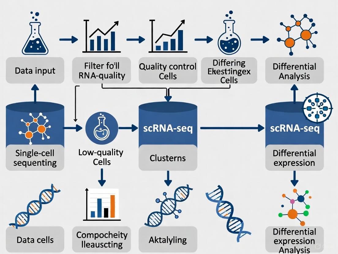
scRNA-seq QC Workflow
The following diagram illustrates the logical workflow for quality control in single-cell RNA sequencing analysis, from raw data to a filtered cell matrix.
A Researcher's Toolkit for scRNA-seq QC
Table 3: Essential Research Reagent Solutions for scRNA-seq QC
| Item | Function in QC Context |
|---|---|
| Dead Cell Removal Kit [12] | Improves initial sample quality by enriching for live cells, which reduces background signal from lysed cells and leads to a lower mitochondrial percentage in the final data. |
| Cell Strainer | Removes cell aggregates and large debris to prevent clogs in microfluidic chips and reduce multiplet rates, leading to more accurate UMI and gene counts per cell. |
| Hemocytometer / Automated Cell Counter [8] [12] | Provides an accurate count of cell concentration and viability. Inaccurate counting is a common source of poor cell recovery and can affect the interpretation of UMI counts per cell. |
| Trypan Blue or Fluorescent Viability Dyes [12] | Allows discrimination between live and dead cells during counting. Fluorescent dyes are more accurate for complex samples like nuclei suspensions or those with debris. |
| Cryopreservation Media (with DMSO) [12] | Enables freezing of high-quality cell suspensions for later processing, preserving cell viability and RNA integrity to prevent degradation that inflates mitochondrial metrics. |
| Nuclei Isolation Kit [12] | Provides a standardized method for nuclei extraction from difficult tissues, ensuring nuclear integrity and reducing contamination from cytoplasmic RNA, which affects gene and UMI counts. |
Within the framework of a broader thesis on filtering low-quality cells in single-cell RNA sequencing (scRNA-seq) research, this guide addresses a fundamental challenge: reliably distinguishing intact, high-quality cells from empty droplets and other artifacts. scRNA-seq data is inherently sparse and dropout-prone, making initial quality assessment critical for all downstream analyses [14] [15]. Proper identification of real cells ensures that subsequent discoveries in cellular heterogeneity, disease mechanisms, and drug development are built upon a solid biological foundation.
Frequently Asked Questions
1. What are the primary quantitative metrics used to distinguish real cells from empty droplets, and what are their typical thresholds?
The initial quality control (QC) step typically relies on three core metrics calculated for each cellular barcode. The following table summarizes these key indicators and their generally accepted thresholds for identifying high-quality cells [15] [8] [4].
Table 1: Key Quality Control Metrics for scRNA-seq Data
| Metric | Biological/Technical Meaning | Typical Threshold (for high-quality cells) | Rationale |
|---|---|---|---|
| Number of Counts per Cell (nUMI) | Total number of transcripts (UMIs) detected. Represents the "library size." | > 500 - 1000 [8] | Values that are too low suggest an empty droplet or a cell with little RNA content (e.g., a dead cell). |
| Number of Genes per Cell (nGene) | The diversity of expressed genes. | > 250 - 300 [8] | Low numbers indicate a poor-quality cell or empty droplet. Excessively high numbers may indicate a doublet. |
| Mitochondrial Count Fraction | Percentage of transcripts originating from mitochondrial genes. | Varies by species and sample; often 5% - 15% [4] | A high percentage suggests cell stress, apoptosis, or broken cytoplasm where mitochondrial RNA has leaked out. |
2. Beyond the standard metrics, what additional quality indicators can reveal low-quality cells?
Recent research advocates for incorporating the nuclear fraction, based on intronic read content, as a crucial quality metric [16]. In droplet-based scRNA-seq, all nucleated cells should have a significant fraction of reads mapped to introns. Cells with a very low intronic fraction likely represent empty droplets, cytoplasmic debris, or nuclei-free cytoplasmic remnants. Conversely, cells with an extremely high intronic fraction may represent lysed cells that have lost their cytosol [16]. The expression of the long non-coding RNA MALAT1 can also serve as a nuclear marker; its absence can flag cells lacking a nucleus [16].
3. What are doublets/multiplets, and why are they problematic?
A doublet or multiplet is a droplet that contains more than one cell. This occurs at a non-ignorable rate during library preparation, especially with higher cell loading concentrations [4] [17]. Multiplets are problematic because they create hybrid expression profiles, which can be misinterpreted as novel or transitional cell types, leading to spurious biological conclusions [17]. The multiplet rate for a platform like 10x Genomics can be around 5.4% when loading 7,000 target cells [4].
4. How can I differentiate a true rare cell population from a technical artifact like a doublet?
This is a significant challenge. True rare cell types will have coherent gene expression programs, including the expression of established marker genes for a known lineage. In contrast, heterotypic doublets (formed from two different cell types) may co-express marker genes from two distinct lineages, which is biologically implausible for a single cell [4]. Computational tools like DoubletFinder and Scrublet are designed to detect these anomalous cells by comparing observed expression profiles to simulated doublets [4]. However, caution is needed, as some true cells in transitional states might also co-express markers; therefore, a combination of automated tools and manual inspection is recommended [4].
5. My dataset has a high level of ambient RNA. How does this affect cell calling, and how can I correct for it?
Ambient RNA consists of transcripts from lysed cells that exist in the solution and are subsequently encapsulated into droplets along with intact cells [4]. This contamination can lead to the misidentification of empty droplets as cells and can blur the distinct expression profiles of real cell types, complicating annotation. Tools like SoupX and CellBender have been developed to estimate and subtract this background contamination [4]. SoupX is noted for its performance with single-nucleus data, while CellBender provides accurate estimation of background noise in diverse datasets [4].
Experimental Protocols for Robust Quality Control
The following workflow provides a detailed methodology for a comprehensive QC analysis of scRNA-seq data, integrating both standard and advanced metrics.
Protocol: A Comprehensive Workflow for scRNA-seq Quality Control
Step 1: Environment Setup and Data Input
Begin by loading your count matrix (e.g., from Cell Ranger) into a standard analysis environment like Scanpy (Python) or Seurat (R). For example, in Scanpy, you would use sc.read_10x_h5() to import the data [15].
Step 2: Calculation of QC Metrics Compute the standard QC metrics for each barcode:
- Total counts (nUMI) and number of genes detected (nGene).
- Mitochondrial fraction: Identify mitochondrial genes (e.g., those starting with "MT-" in humans, "mt-" in mice) and calculate their percentage of total counts [15] [8].
- Additional gene sets: Calculate the proportion of ribosomal protein genes (RPS, RPL) and hemoglobin genes (HB) as they can also indicate specific biological or technical states [15].
- Nuclear fraction: Use specialized packages (e.g., the
DropletQCR package) to calculate the proportion of intronic reads for each barcode, a key metric for identifying nucleus-free debris [16].
Step 3: Automated and Manual Thresholding for Filtering
- Strategy: It is advised to be as permissive as possible initially to avoid filtering out viable cell populations, especially rare subtypes [15].
- Manual Inspection: Visualize the distribution of QC metrics (nUMI, nGene, mitochondrial fraction) using histograms, violin plots, and scatter plots to identify outliers [8].
- Automated Thresholding: For larger datasets, apply an automatic outlier detection method. A robust method is to use the Median Absolute Deviation (MAD). Cells that deviate by more than 5 MADs from the median for a given metric can be flagged as potential low-quality cells [15].
Step 4: Doublet Detection
- Apply a computational doublet detection tool such as DoubletFinder, Scrublet, or COMPOSITE (the latter is designed for multi-omics data) [4] [17].
- These tools work by creating artificial doublets and then comparing all cells to these simulated profiles to assign a doublet score. Remove cells classified as doublets with high probability.
Step 5: Ambient RNA Correction
- Apply an ambient RNA removal tool like SoupX or CellBender to clean the count matrix. This step helps to correct the expression values of the remaining cells, improving downstream analysis [4].
Step 6: Iterative Re-assessment
- QC is not always a linear process. Re-assess your filtering strategy after initial clustering and annotation, as some biological populations may naturally have higher mitochondrial or lower gene content [15].
The following diagram illustrates the logical workflow and decision points in this protocol:
The following table details key computational tools and resources essential for implementing the quality control procedures described above.
Table 2: Research Reagent Solutions: Key Computational Tools for scRNA-seq QC
| Tool Name | Function | Brief Description of Role |
|---|---|---|
| Scanpy [15] | Data Analysis & QC | A comprehensive Python-based toolkit for analyzing single-cell gene expression data. Used for calculating QC metrics, filtering, and visualization. |
| Seurat [8] | Data Analysis & QC | A widely-used R toolkit for single-cell genomics. Provides functions for QC, normalization, and clustering. |
| DoubletFinder [4] | Doublet Detection | A tool that uses artificial nearest-neighbor networks to classify doublets in scRNA-seq data. |
| Scrublet [4] | Doublet Detection | A scalable tool for predicting doublets in scRNA-seq data by simulating doublets and identifying neighbors. |
| COMPOSITE [17] | Multiplet Detection | A statistical model-based framework for detecting multiplets, particularly effective in single-cell multi-omics data. |
| SoupX [4] | Ambient RNA Removal | A tool for estimating and removing the ambient RNA contamination profile from droplet-based scRNA-seq data. |
| CellBender [4] | Ambient RNA Removal | A tool that uses a deep generative model to remove technical artifacts, including ambient RNA, from count data. |
| DropletQC [16] | Nuclear Fraction | An R package for identifying empty droplets and low-quality cells based on nuclear fraction (intronic content). |
Frequently Asked Questions (FAQs)
Q1: I am working with fragile cells, like those from epithelial tissues. Which platform is gentler and might prevent cell stress? A1: For fragile cells, such as gastrointestinal tract epithelial cells, picowell-based (well-based) platforms are often the gentler option. They utilize processes that are less mechanically and enzymatically stressful compared to droplet-based methods, which helps preserve more natural gene expression profiles and reduces sample degradation [18].
Q2: My project requires profiling thousands of cells. Which platform is better for high-throughput studies? A2: Droplet-based technologies, like the 10x Genomics Chromium system, are generally superior for high-throughput applications. They are designed to process thousands to millions of cells in a single experiment, offering a lower cost per cell when working at a large scale [19] [14].
Q3: I am concerned about technical artifacts like doublets, where two cells are mistakenly sequenced as one. How do these platforms compare? A3: Both platforms generate doublets, but the causes and rates differ.
- Droplet-based: Doublets occur primarily from co-encapsulation of multiple cells in a single droplet. The rate follows a Poisson distribution and is typically kept below 5% with optimal cell loading concentrations [19] [20].
- Well-based: Doublets can occur but some systems, like Well-TEMP-seq, achieve a high single cell-barcoded bead pairing rate of ~80%, which is significantly higher than the Poisson-dependent pairing in some droplet methods [21].
Q4: What about background noise from ambient RNA? Is one platform less prone to this? A4: Picowell-based platforms can have an advantage in reducing ambient RNA contamination. The workflow for some well-based systems allows for the removal of cell-free RNAs by washing the wells before the barcoding step, which is not possible in standard droplet workflows [21]. In droplet-based systems, ambient RNA is a known challenge that often requires computational tools for correction [19] [22].
Technical Comparison Tables
Table 1: Key Performance Metrics for Droplet vs. Well-Based Platforms
| Performance Metric | Droplet-Based (e.g., 10x Genomics, inDrop) | Well-Based (e.g., Picowell platforms) |
|---|---|---|
| Typical Cell Throughput | Very High (Thousands to millions of cells) [19] | Variable, but generally lower than droplet-based systems [18] |
| Cell Capture Efficiency | 30% - 75% [19] | Information not specified in search results |
| mRNA Capture Efficiency | 10% - 50% of cellular transcripts [19] | Information not specified in search results |
| Typical Multiplet Rate | < 5% (with optimal loading) [19] | Information not specified in search results |
| Single Cell-Bead Pairing Efficiency | Low in some systems (e.g., <1% for Drop-seq based methods) due to Poisson distribution [21] | Can be very high (e.g., ~80% for Well-TEMP-seq) [21] |
| Gentleness on Fragile Cells | Lower; enzymatic/mechanical processes can stress delicate cells [18] | Higher; gentler capture process better preserves cell integrity [18] |
| Ambient RNA Control | Challenging; requires computational cleanup [19] [22] | Better; allows physical washing to remove cell-free RNA [21] |
| Cost per Cell | Lower at very high throughput [19] [14] | Can be a cost-effective alternative [18] |
Table 2: Essential Experimental Protocols for Platform Validation
| Experiment Name | Purpose | Key Steps | Interpretation & Role in Filtering Low-Quality Cells |
|---|---|---|---|
| Species-Mixing Experiment [20] | To quantify the cell doublet rate. | 1. Mix cells from different species (e.g., human & mouse).2. Process the mixed sample through the scRNA-seq platform.3. Sequence and analyze the data. | Identifies doublets: Cells expressing genes from both species are technical doublets. The measured heterotypic doublet rate is used to estimate the overall (including homotypic) doublet rate, allowing for the computational removal of these artifacts. |
| Cell Hashing / Multiplexing (e.g., MULTI-seq) [20] | To label cells from different samples with unique barcodes before pooling, enabling sample multiplexing and doublet detection. | 1. Label individual cell samples with unique lipid-conjugated or antibody-conjugated oligonucleotide barcodes.2. Pool the labeled samples.3. Process the pooled sample through the scRNA-seq platform. | Identifies sample multiplets: After sequencing, cells with more than one hashtag barcode are identified as doublets or multiplets and filtered out. This allows for intentional overloading of cells to increase throughput while controlling the final doublet rate. |
Workflow Diagrams
Diagram 1: Key Steps for scRNA-seq Quality Control
Diagram 2: Droplet vs. Well-Based Cell Capture
The Scientist's Toolkit: Key Research Reagent Solutions
Table 3: Essential Reagents for scRNA-seq Quality Control
| Reagent / Material | Function | Considerations for Platform Choice |
|---|---|---|
| Viability Stains (e.g., Calcein-AM) [22] | Fluorescent dye used to identify live cells based on esterase activity. | Can be used with advanced droplet systems (e.g., spinDrop) for fluorescence-activated droplet sorting (FADS) to enrich for live cells before lysis and barcoding. |
| Cell Hashing Oligonucleotides (e.g., MULTI-seq) [20] | Sample-specific barcodes (antibody- or lipid-conjugated) that label cells prior to pooling. | Enables sample multiplexing and doublet detection on both droplet and well-based platforms. Crucial for increasing throughput while maintaining data quality. |
| Barcoded Beads (Gel Beads) [19] [23] | Microbeads containing millions of oligonucleotides with cell barcodes and UMIs for capturing mRNA. | A core component for both platforms. Hydrogel beads (used in 10x, inDrop) allow for sub-Poisson loading, while hard resin beads are used in Drop-seq. |
| Unique Molecular Identifiers (UMIs) [24] [25] | Short random nucleotide sequences added to each transcript during reverse transcription. | Essential for both platforms to digitally count individual mRNA molecules and correct for PCR amplification bias, ensuring quantitative data. |
| Surfactant/Oil Emulsion [23] | Creates stable, nanoliter-scale water-in-oil droplets that act as isolated reaction chambers. | Critical for the stability of droplet-based systems. The quality and formulation directly impact droplet integrity and prevent cross-contamination. |
The How-To Guide: Methodologies and Tools for Effective Cell Filtering
This guide details the critical process of filtering low-quality cells in single-cell RNA sequencing (scRNA-seq) data analysis. The quality of your initial cell matrix profoundly impacts all downstream biological interpretations, from identifying cell types to understanding cellular communication. This workflow provides researchers with a structured, troubleshooting-oriented approach to ensure that the cellular data underlying their research is robust and reliable.
Why is Filtering Low-Quality Cells So Critical?
In scRNA-seq data, low-quality cells can arise from several sources, including damaged cells during dissociation, empty droplets, or droplets containing cell doublets/multiplets. If not removed, these cells introduce significant technical noise that can obscure true biological variation. For instance, transcripts from ruptured cells can become "ambient RNA," contaminating nearby cells and leading to misclassification. Furthermore, dying cells often exhibit aberrantly high mitochondrial gene expression, which can be mistaken for a genuine biological state. Effective filtering is the first and most crucial defense against these artifacts, ensuring that subsequent clustering and differential expression analysis reflect biology, not technical artifacts [26] [14] [10].
Frequently Asked Questions (FAQs)
1. What are the key metrics used to identify a low-quality cell? Three primary metrics are commonly used:
- Unique Gene Count (
nFeature_RNA): The number of genes detected in a cell. Low counts may indicate empty droplets or broken cells, while very high counts can suggest doublets (multiple cells captured together) [27] [10]. - UMI Count (
nCount_RNA): The total number of transcripts detected. This often correlates strongly with the gene count and is used similarly to identify outliers [27]. - Mitochondrial Gene Percentage (
percent.mt): The proportion of transcripts derived from mitochondrial genes. A high percentage is a hallmark of stressed, apoptotic, or low-quality cells due to compromised cell membranes [27] [28].
2. How do I set specific filtering thresholds for my dataset? Thresholds are not universal and must be determined empirically from the data distribution of your own experiment. The following steps are recommended:
- Visualize Distributions: Use violin plots or scatter plots to inspect the distributions of the three key metrics across all cells [27] [29].
- Identify Outliers: Manually set thresholds to exclude clear outliers. A common strategy is to filter cells with a mitochondrial percentage significantly above the majority of the population (e.g., >5-10% for PBMCs) and remove cells at the extreme low ends of the gene and UMI count distributions [27] [10].
- Iterate and Re-cluster: Filtering is an iterative process. After applying initial thresholds, re-visualize the data to ensure that low-quality clusters are removed while biologically relevant populations are retained [28].
3. My dataset has a high overall mitochondrial percentage. What should I do? A uniformly high mitochondrial percentage can indicate a problem with the sample viability itself. Before filtering, consider:
- Biology vs. Artifact: Certain cell types, like cardiomyocytes, naturally have high respiratory activity and thus high mitochondrial RNA content. Filtering these could introduce bias [26] [10].
- Experimental Review: If the biology does not explain the high percentage, it may point to an issue with the sample preparation protocol, such as excessive cell stress during dissociation. If possible, troubleshoot the wet-lab process.
4. What tools can I use to perform this filtering?
- Seurat (R): A widely used toolkit that provides functions for calculating QC metrics (
PercentageFeatureSet), visualization (VlnPlot,FeatureScatter), and filtering (subset) [27] [29]. - Loupe Browser (10x Genomics): A graphical interface that allows for interactive visualization and filtering of cells based on QC metrics, providing real-time feedback on how filtering affects cell clusters [10] [28].
- Command-Line Tools: Packages like
Cell Ranger(10x Genomics) andSTARsoloperform initial quantification and can generate summary reports used for QC [26] [10].
Troubleshooting Common Issues
Problem: Loss of a rare cell population after filtering.
- Potential Cause: Overly stringent thresholds on gene or UMI counts may have removed small cells with naturally low RNA content.
- Solution: Relax the lower bounds on UMI and gene counts. Visually inspect the pre-filtering plots to see if the potential rare population forms a distinct cluster at the lower end of the counts and adjust thresholds to preserve it [26] [28].
Problem: A distinct cluster of cells has a high mitochondrial percentage.
- Potential Cause: This could be a genuine population of stressed or dying cells, or a technical artifact.
- Solution: Do not automatically filter this entire cluster. First, check for the expression of marker genes for known cell types. If the cluster expresses markers for a real cell type but also has high stress signatures, you may choose to retain it and regress out the mitochondrial signal as a source of unwanted variation in downstream steps, rather than removing the cells entirely [27] [28].
Problem: Persistent technical batch effects after filtering.
- Potential Cause: Filtering alone cannot always correct for strong technical differences between samples processed in different batches.
- Solution: After performing quality control on each sample individually, use data integration tools like those provided in Seurat (
FindIntegrationAnchors,IntegrateData) to harmonize the datasets before proceeding to clustering [27] [30].
Standard Operating Procedure: Cell Quality Control and Filtering
Methodology Using Seurat in R
This protocol outlines the standard pre-processing workflow for scRNA-seq data in Seurat, focusing on quality control and filtering of cells.
1. Load Data and Calculate QC Metrics
2. Visualize QC Metrics to Inform Thresholds
3. Filter Cells Based on Visualized Distributions
Workflow Visualization
Reference Table: Common QC Threshold Guidelines
Table 1: Example quality control thresholds for different sample types. These are starting points and must be validated for each dataset.
| Sample Type | nFeature_RNA (Low) | nFeature_RNA (High) | percent.mt | Notes |
|---|---|---|---|---|
| PBMCs (10x) [27] | > 200 | < 2500 | < 5% | Standard immune cells; adjust high threshold for activated cells. |
| Complex Tissue [26] | > 500-1000 | < 5000-10000 | < 10-20% | More diverse cell sizes and types; thresholds are wider. |
| Cardiomyocytes [26] [10] | Cell-specific | Cell-specific | Use with caution | Naturally high mitochondrial content; do not filter based on this metric alone. |
| Nuclei (snRNA-seq) [26] | > 200 | < 5000 | < 5% | Generally lower gene counts per nucleus. |
The Scientist's Toolkit: Essential Research Reagents & Software
Table 2: Key tools and resources for scRNA-seq data quality control and analysis.
| Tool / Resource | Category | Primary Function | Reference |
|---|---|---|---|
| Cell Ranger | Processing Pipeline | Processes 10x Genomics FASTQ files into a count matrix; provides initial QC web summary. | [10] |
| Seurat | R Analysis Toolkit | Comprehensive toolkit for scRNA-seq analysis, including QC, filtering, normalization, and clustering. | [27] [29] |
| Loupe Browser | Visualization Software | Interactive visualization of 10x Genomics data; allows manual filtering with real-time cluster feedback. | [10] [28] |
| FastQC | Read QC Tool | Assesses the quality of raw sequencing reads from FASTQ files. | [26] [31] |
| DoubletFinder | R Package | Computational prediction of doublets in the data based on artificial nearest neighbors. | [26] |
| SoupX | R Package | Corrects for ambient RNA contamination in droplet-based data. | [10] |
## Frequently Asked Questions (FAQs)
Q1: Why should I use a data-driven method instead of fixed thresholds for filtering cells by UMI and gene counts? Fixed thresholds (e.g., keeping cells with gene counts between 200 and 2,500) are often borrowed from tutorials but are not suitable for all datasets. Using arbitrary cutoffs can inadvertently eliminate valid biological cells, especially in highly heterogeneous samples where some cell types naturally have very high or low RNA content. A data-driven approach identifies outliers specific to your dataset, which helps preserve biological heterogeneity and leads to more reliable downstream results [6] [9].
Q2: What are the common data-driven methods for identifying outlier cells? A common and robust method is to use the Median Absolute Deviation (MAD). This method calculates a threshold based on the median value of a metric (like UMI counts) across all cells and how much each cell deviates from that median. Cells that fall beyond a certain number of MADs from the median are considered outliers. This approach is more resistant to the influence of extreme values than methods based on the mean and standard deviation [6] [9].
Q3: How do I handle samples with different cell types that have naturally different UMI counts? When your sample contains cell types with vastly different RNA contents (e.g., neutrophils versus lymphocytes), applying one global threshold to the entire dataset can be harmful. In such cases, a more advanced strategy is cluster-specific QC. This involves performing an initial, permissive clustering of the cells and then applying data-driven QC thresholds within each cluster separately. This protects rare or biologically distinct cell populations from being filtered out [6].
Q4: My data is very large. Are there automated tools for this process?
Yes, many widely used single-cell analysis toolkits have built-in functions for data-driven QC. For example, the perCellQCFilters() function in the scater R package (part of the Bioconductor ecosystem) can automatically identify outliers for multiple QC metrics using the MAD method [9]. The Seurat and Scanpy packages also provide extensive functionality for calculating and visualizing these metrics to guide threshold setting.
Q5: What other metrics should I consider alongside UMI and gene counts? A comprehensive QC workflow always includes assessing the percentage of reads mapping to the mitochondrial genome. A high percentage often indicates broken or dying cells, as cytoplasmic RNA leaks out while mitochondrial RNAs are retained. However, this threshold is also biology-dependent; some active cell types, like cardiomyocytes, naturally have high mitochondrial activity [6] [10].
Q6: Is quality control a one-time step? No, quality control is often an iterative process. It is good practice to start with permissive filtering parameters. After performing initial clustering and cell type annotation, you should re-inspect the QC metrics across the clusters. You may discover that some low-quality cells were missed or that some valid cells were incorrectly filtered, requiring you to revisit your thresholds [6].
## Troubleshooting Guide
### Problem: Loss of a known cell population after filtering
Potential Cause: The filtering thresholds for UMI counts, gene counts, or mitochondrial percentage were too stringent and did not account for the natural biological variation of that specific cell type.
Solution:
- Re-visit your distributions: Before filtering, plot the distribution of your QC metrics (library size, number of genes, mitochondrial percentage) colored by the cluster or cell type identity from an initial, lightly filtered analysis.
- Implement cluster-specific filtering: If you observe that one cluster has systematically lower UMI counts or higher mitochondrial content, apply your data-driven thresholding method (e.g., MAD-based filtering) separately to each cluster. This ensures that thresholds are tailored to the biological properties of each cell group [6].
- Use diagnostic plots: The following diagnostic plot helps visualize the relationship between key QC metrics and can reveal if certain cell populations are driving specific metric distributions.
### Problem: Ambiguous boundary between cells and empty droplets in UMI/gene count distribution
Potential Cause: The data does not have a clear inflection point ("knee") in the barcode rank plot, making it difficult to distinguish true cells from background noise using simple thresholds.
Solution:
- Use advanced cell-calling algorithms: Move beyond a simple UMI cutoff. Tools like emptyDrops use a statistical framework to test whether each barcode's expression profile is significantly different from the ambient RNA profile. Barcodes that are significantly different are classified as cells [6] [10].
- Leverage ambient RNA removal: Consider using tools like SoupX, CellBender, or DecontX to estimate and subtract the background ambient RNA signal from your count matrix. This can "clean" the data and make the separation between cells and empty droplets more distinct, simplifying subsequent filtering [6] [10].
## Experimental Protocols & Data Presentation
### Protocol: A Data-Driven Workflow for Setting UMI and Gene Count Thresholds
This protocol outlines the steps for using the Median Absolute Deviation (MAD) method to set robust, dataset-specific filtering thresholds.
Step 1: Calculate QC Metrics
Using your analysis toolkit (e.g., Seurat's PercentageFeatureSet and CreateSeuratObject or Scanpy's pp.calculate_qc_metrics), compute for every cell barcode:
nCount_RNA: Total number of UMIs (library size).nFeature_RNA: Total number of unique genes detected.percent.mt: Percentage of UMIs mapping to mitochondrial genes.
Step 2: Visualize Metric Distributions Plot the distributions of these three metrics using violin plots or histograms. This provides an initial overview of data quality and helps identify obvious issues.
Step 3: Calculate MAD-Based Thresholds For each metric, calculate the lower and upper bounds. The following logic is typically applied:
- For UMI Counts and Gene Counts: Filter out outliers on both the lower end (likely empty droplets) and the upper end (likely multiplets).
- For Mitochondrial Percentage: Filter out outliers only on the upper end (likely dead/dying cells).
The standard formula for a MAD-based threshold for a metric ( x ) is: [ \text{Lower Bound} = \text{Median}(x) - 3 \times \text{MAD} ] [ \text{Upper Bound} = \text{Median}(x) + 3 \times \text{MAD} ] Where ( \text{MAD} = \text{median}( | x_i - \text{median}(x) | ) ).
Note: For library size and gene counts, calculations are often performed on log-transformed values to mitigate the influence of extreme high outliers on the MAD [9].
Step 4: Apply Filters and Document Remove all cell barcodes that fall outside the calculated bounds for any of the key metrics. It is critical to record the final thresholds and the number of cells filtered for each metric to ensure reproducibility.
The table below summarizes the key QC metrics, their biological interpretations, and the recommended data-driven approach for setting thresholds.
Table 1: Key Quality Control Metrics for scRNA-seq Data Filtering
| QC Metric | Description | What Low Values May Indicate | What High Values May Indicate | Data-Driven Thresholding Method |
|---|---|---|---|---|
| UMI Counts (Library Size) | Total number of mRNA molecules detected per cell. | Empty droplet, ambient RNA, or very small cell (e.g., platelet). | Multiplet (multiple cells in one droplet) or a large, transcriptionally active cell. | Median Absolute Deviation (MAD), typically applied to log-transformed values. Common cutoff: 3 MADs from the median [6] [9]. |
| Gene Counts (Number of Features) | Number of unique genes detected per cell. | Empty droplet, poor-quality cell, or a cell type with low transcriptional complexity. | Multiplet or a cell with very broad transcriptional activity. | Median Absolute Deviation (MAD), typically applied to log-transformed values. Common cutoff: 3 MADs from the median [6] [9]. |
| Mitochondrial Percent | Percentage of a cell's UMIs that map to mitochondrial genes. | Not typically used as a lower filter. | Cell stress, apoptosis, or broken cell where cytoplasmic RNA has been lost. | Median Absolute Deviation (MAD). Common cutoff: 3 MADs above the median [6] [9]. |
### The Scientist's Toolkit
Table 2: Essential Research Reagent Solutions for scRNA-seq QC
| Item | Function in scRNA-seq QC |
|---|---|
| Cell Ranger | A set of official pipelines from 10x Genomics that processes raw sequencing data (FASTQ) into aligned reads, generates the feature-barcode count matrix, and performs initial cell calling. It is the foundational step before applying further QC filters [6] [10]. |
| Unique Molecular Identifiers (UMIs) | Short nucleotide barcodes added to each mRNA molecule during library preparation. UMIs allow for the accurate counting of original transcript molecules and correction for amplification bias, making UMI count a core QC metric [14]. |
| ERCC Spike-in RNAs | A set of synthetic, external RNA controls added to the cell lysate in known concentrations. They can be used to monitor technical variability and assess the sensitivity of the assay, providing an alternative metric for identifying low-quality cells [9]. |
| Mitochondrial Read Proportion | Not a reagent, but a critical computational metric. The proportion of reads from mitochondrial genes serves as a natural internal control for cell health, as it increases in compromised cells [6] [32]. |
## Workflow Visualization
The following diagram illustrates the complete, iterative workflow for quality control and filtering of single-cell RNA-seq data, integrating both data-driven thresholding and biological inspection.
Addressing Ambient RNA Contamination with Tools like SoupX and CellBender
Ambient RNA contamination is a pervasive technical challenge in droplet-based single-cell RNA sequencing (scRNA-seq). It occurs when freely floating mRNAs from the input solution are captured along with cell-specific mRNAs, leading to a contaminated gene expression profile that can confound downstream biological interpretation [33] [34]. This background contamination, often originating from lysed or dead cells, varies significantly between experiments (typically 2-50%), with around 10% being common [33]. The consequences are particularly severe for rare cell type identification and can lead to biological misinterpretation, such as misannotation of glial cell types in brain samples due to neuronal ambient RNA [35]. This guide provides comprehensive troubleshooting and FAQs for addressing ambient RNA contamination within the broader context of filtering low-quality cells in scRNA-seq research.
FAQs and Troubleshooting for Ambient RNA Correction Tools
SoupX
Q: The autoEstCont function fails with an "Extremely high contamination estimated" error. What should I do?
A: This error occurs when SoupX estimates an unrealistically high contamination fraction (e.g., >0.5), often in problematic samples. You have several options:
- Manual Estimation: Use
setContaminationFractionto manually set a reasonable value based on the distribution from the failedautoEstContcall. For example, if the distribution shows a peak around 0.1, setcontFrac = 0.1[36]. - Range Limitation: Re-run
autoEstContwith thecontaminationRangeparameter to restrict the estimation range (e.g.,c(0.01, 0.5)) [36]. - Sample Evaluation: Consider if the sample quality is too poor. Extremely high contamination may indicate fundamental issues with sample preparation that cannot be fully resolved computationally [36].
Q: My data still appears contaminated after running SoupX. Why didn't it work?
A: Several factors could cause this:
- Missing Cluster Information: SoupX requires clustering information to identify contamination accurately. Ensure you have provided clustering information either via
setClustersor by loading 10X data withload10X, which automatically imports cellranger clusters [33]. - Insufficient Marker Diversity: The automatic estimation relies on diverse marker genes. For homogeneous data (e.g., cell lines) or very few cells (< few hundred), manual estimation may be necessary [33].
- Conservative Defaults: SoupX is designed to err on the side of not removing true counts. For situations where aggressive removal is preferred, try manually increasing the contamination fraction with
setContaminationFraction[33].
Q: I cannot find appropriate genes to estimate the contamination fraction. What should I try?
A: Ideal genes are highly specific to certain cell types and show bimodal expression patterns:
- Common Markers: First try commonly successful gene sets like hemoglobin (HB) genes for erythrocytes, immunoglobulin genes for B-cells, or TPSB2/TPSAB1 for mast cells [33].
- Visual Inspection: Use
plotMarkerDistributionto identify genes with appropriate expression patterns [33]. - Alternative Approach: If no clear markers emerge, test a range of contamination fractions (e.g., 2-10%) to evaluate their impact on your downstream analysis [33].
CellBender
Q: How do I know if CellBender worked correctly after it completes?
A: Several diagnostic approaches can verify success:
- Learning Curve Inspection: Check the ELBO (Evidence Lower Bound) versus epoch plot in the output
_report.html. A good run shows the ELBO increasing and plateauing, not spiking or decreasing [37]. - HTML Report: Review the automatic diagnostics in the output report, which provides warnings and recommendations [37].
- Downstream Analysis: Load the output with
load_anndata_from_input_and_outputand compare raw vs. corrected data in downstream analyses like clustering and marker gene expression [37].
Q: The learning curve (ELBO vs. epoch) looks strange with spikes or dips. What does this indicate?
A: Spikes or downward trends in the learning curve typically indicate training instability:
- Solution: Reduce the
--learning-rateby a factor of two and rerun CellBender. Training should proceed for at least 150 epochs until the ELBO plateaus [37]. - Expected Pattern: Ideally, the ELBO should increase monotonically and stabilize. Examples of good and bad learning curves are provided in the CellBender documentation [37].
Q: CellBender seems to have called too many or too few cells. How can I adjust this?
A: Cell calling depends on several parameters:
- Too Many Cells: Increase
--total-droplets-includedor decrease--expected-cells. Remember that CellBender identifies "non-empty" droplets, which may include low-quality cells requiring downstream filtering [37]. - Too Few Cells: Increase
--expected-cellsand ensure--total-droplets-includedis large enough to include all potentially non-empty droplets [37]. - Downstream Filtering: Apply additional filters post-CellBender based on mitochondrial read percentage and genes expressed per cell [37].
Q: Do I need a GPU to run CellBender, and how can I work around resource limitations?
A: While not absolutely necessary, GPU usage significantly speeds up processing:
- CPU Alternative: For CPU-only runs, use fewer
--total-droplets-included, increase--projected-ambient-count-thresholdto analyze fewer features, and decrease--empty-drop-training-fraction[37]. - Cloud Solutions: Consider Terra or Google Colab for free GPU access. CellBender produces checkpoint files, allowing you to resume interrupted runs [37].
Quantitative Data Comparison
Table 1: Key Characteristics of Ambient RNA Removal Tools
| Feature | SoupX | CellBender |
|---|---|---|
| Primary Approach | Estimates contamination from empty droplets; subtracts counts using cluster information [33] [34] | Deep generative model that distinguishes cell-containing from cell-free droplets; learns background profile [37] [34] |
| Typical Contamination Reduction | 2-50% (depending on initial contamination) [33] | Varies by dataset; metrics provided in output [37] |
| Computational Demand | Moderate | High (GPU recommended) [37] [34] |
| Key Parameters | Contamination fraction (rho), cluster labels [33] [38] | Expected cells, FPR, total droplets included [37] |
| Integration with scRNA-seq Pipelines | Compatible with Seurat; outputs corrected count matrix [33] [38] | Outputs Anndata object compatible with Scanpy and Seurat [37] |
| Best Suited For | Standard 10X data; cases where cluster information is available [33] | Complex datasets requiring joint cell calling and background removal [37] [34] |
Table 2: Typical Parameter Settings for Common Scenarios
| Scenario | SoupX Parameters | CellBender Parameters |
|---|---|---|
| Standard 10X Data | autoEstCont with default parameters [33] |
--expected-cells 10000 --fpr 0.01 [37] |
| High Contamination Samples | Manual setContaminationFraction at 0.1-0.2 or contaminationRange = c(0.01, 0.5) [36] |
--fpr 0.05 --total-droplets-included 20000 [37] |
| Low Cell Number (<1000) | Manual contamination fraction setting; cluster information critical [33] | Reduce --total-droplets-included; consider CPU parameters [37] |
| Complex Tissues (e.g., Brain) | Ensure clustering resolution sufficient to distinguish cell types [33] | Standard parameters typically adequate; check for neuronal contamination in glia [35] |
Workflow Integration Diagrams
Diagram 1: Ambient RNA Correction Workflow Integration. This diagram outlines the decision process for incorporating ambient RNA correction into scRNA-seq quality control, showing tool selection criteria and iterative evaluation.
Table 3: Key Resources for Ambient RNA Correction Experiments
| Resource | Function/Purpose | Implementation Notes |
|---|---|---|
| 10X Genomics Cell Ranger | Initial processing of 10X data; provides filtered and raw matrices for SoupX input [34] | Use raw matrix for SoupX; filtered matrix for CellBender (depending on workflow) |
| Clustering Information | Essential for SoupX to distinguish true expression from contamination [33] | Can be from Cell Ranger or custom clustering (e.g., Seurat, Scanpy) |
| Marker Gene Sets | Genes with cell-type specific expression used to estimate contamination [33] [35] | Hemoglobin genes, immunoglobulin genes, cell-type specific markers |
| Seurat/Scanpy | Downstream analysis platforms for evaluating correction effectiveness [33] [37] | Compare clustering and marker expression before/after correction |
| Mitochondrial Gene List | Quality control metric to identify compromised cells [39] | High expression may indicate cell stress/death contributing to ambient RNA |
| Reference Datasets | Positive controls for expected cell-type specific expression patterns [39] [35] | e.g., Allen Brain Atlas for neuronal markers; PanglaoDB for general cell markers |
Effective management of ambient RNA contamination is an essential component of comprehensive scRNA-seq quality control. Both SoupX and CellBender offer powerful solutions, with complementary strengths: SoupX provides a cluster-aware approach that integrates well with standard workflows, while CellBender offers a more comprehensive joint modeling of cells and background. Success requires careful parameter optimization, thorough diagnostic checks, and integration with other quality control measures. By addressing ambient RNA contamination appropriately, researchers can significantly improve cell type identification accuracy, enhance differential expression detection, and draw more reliable biological conclusions from their single-cell transcriptomic studies.
Identifying and Removing Doublets with Scrublet and DoubletFinder
Within the broader context of filtering low-quality cells in single-cell RNA sequencing (scRNA-seq) research, the identification and removal of doublets—artifacts formed when two or more cells are sequenced as a single entity—is a critical preprocessing step. Doublets can lead to spurious cell type identification, obscure genuine biological signals, and compromise the integrity of downstream analyses [40]. This guide focuses on two widely used computational tools for doublet detection, Scrublet and DoubletFinder, providing a technical support center to address common implementation challenges and frequently asked questions.
Scrublet: Core Principle and Workflow
Scrublet is a Python-based framework designed to predict the impact of multiplets and identify problematic doublets in scRNA-seq data. Its method operates on two key assumptions: multiplets are relatively rare events, and all cell states contributing to doublets are also present as single cells elsewhere in the data [40].
The algorithm works by:
- Simulating Doublets: Generating artificial doublets from the observed data by combining random pairs of observed transcriptomes.
- Building a Classifier: Constructing a k-nearest neighbor (KNN) classifier to calculate a continuous
doublet_scorefor each observed transcriptome based on the relative densities of simulated doublets and observed transcriptomes in its vicinity [40].
DoubletFinder: Core Principle and Workflow
DoubletFinder is an R package that interfaces with Seurat objects to predict doublets. Its performance is largely invariant to the proportion of artificial doublets generated (pN) but is sensitive to the neighborhood size (pK), which must be optimized for each dataset [41].
The process involves four main steps:
- Generate Artificial Doublets: Create artificial doublets from existing scRNA-seq data.
- Pre-process Merged Data: Merge real and artificial data and pre-process.
- Compute pANN: Perform PCA and use the PC distance matrix to find each cell's proportion of artificial k nearest neighbors (pANN).
- Threshold pANN Values: Rank order and threshold pANN values according to the expected number of doublets [41].
Classifying Doublet-Associated Errors
Understanding the types of errors doublets introduce helps in appreciating the tools' utility:
- Neotypic Errors: Generate new features in the data, such as spurious cell clusters, "branches" from an existing cluster, or "bridges" between clusters. These can lead to qualitatively incorrect biological inferences and are typically caused by heterotypic doublets (formed from transcriptionally distinct cell types) [40].
- Embedded Errors: Cause quantitative changes in gene expression when a doublet is grouped with a large population of similar singlets. Their impact is generally smaller if doublets are rare and are often caused by homotypic doublets (formed from transcriptionally similar cells) [40].
The following diagram illustrates the logical workflow and key decision points for both tools:
Troubleshooting Guides
Scrublet Troubleshooting
| Problem | Possible Cause | Solution |
|---|---|---|
| Poorly defined bimodal histogram [42] | Suboptimal choice of min_gene_variability_pctl, which controls the set of highly variable genes used for classification. |
Re-run Scrublet trying multiple percentile values (e.g., 80, 85, 90, 95). Choose the value that produces the best bimodal distribution in the doublet_score_histogram.png. |
| Predicted doublets do not co-localize in UMAP [43] | The doublet score threshold was set incorrectly, or pre-processing parameters do not adequately resolve the underlying cell states. | Manually adjust the scrublet_doublet_threshold parameter and/or re-process the data to better resolve cell states before running Scrublet. |
| Low doublet detection rate on merged data | Running Scrublet on an aggregated dataset from multiple samples (e.g., different 10X lanes) where artificial cell states are created. | Run Scrublet on each sample separately. The tool is designed to detect technical doublets within a single sample [43]. |
DoubletFinder Troubleshooting
| Problem | Possible Cause | Solution |
|---|---|---|
| Multiple potential pK values when visualizing BCmvn [41] | The mean-variance normalized bimodality coefficient (BCmvn) plot shows several local maxima, making pK selection ambiguous. | Spot-check the results in gene expression (GEX) space for the top candidate pK values. Select the pK that makes the most sense given your biological understanding of the data [41]. |
| Inaccurate doublet number estimation | Using the Poisson statistic alone, which overestimates detectable doublets by ignoring homotypic doublets (transcriptionally similar cells). | Use literature-supported cell type annotations to model the proportion of homotypic doublets. The Poisson estimate (without homotypic adjustment) and the adjusted estimate can 'bookend' the real detectable doublet rate [41]. |
| Poor performance on aggregated data | Running DoubletFinder on data merged from multiple distinct samples (e.g., WT and mutant cell lines). Artificial doublets generated from biologically distinct samples cannot exist in the real data and skew results. | Only run DoubletFinder on data from a single sample or from splitting a single sample across multiple lanes. Do not run on integrated Seurat objects representing biologically distinct conditions [41]. |
Frequently Asked Questions (FAQs)
Q1: What anticipated doublet rate should I use for my experiment?
The expected doublet rate is dependent on your platform (10x, Parse, etc.) and the number of cells loaded. It is not a fixed value. You should consult the user guide for your specific technology to determine the expected rate based on your cell loading density. For example, one common calculation for 10x data is to use (number of recovered cells / 1000) * 0.008 [42] [41].
Q2: Can Scrublet and DoubletFinder detect homotypic doublets? Both tools are primarily sensitive to heterotypic doublets (formed from different cell types). They are largely insensitive to homotypic doublets (formed from the same or very similar cell types) because the resulting transcriptome closely resembles a singlet [41] [40]. This is a fundamental limitation of most computational doublet detection methods.
Q3: Should I run doublet detection before or after data integration and normalization? You should run doublet detection on individual samples prior to data integration. Running these tools on aggregated or integrated data can create artificial cell states that do not biologically exist, leading to inaccurate doublet predictions [41] [43]. The analysis should be performed on normalized data, typically after initial quality control to remove low-quality cells but before batch correction or integration of multiple samples.
Q4: How do I determine the optimal pK parameter for DoubletFinder?
DoubletFinder does not set a default pK. The optimal pK must be determined for each dataset using the parameter sweep function (paramSweep_v3) and the mean-variance normalized bimodality coefficient (BCmvn) metric. The pK value corresponding to the maximum BCmvn is typically selected for downstream analysis [41].
Q5: My data is very homogeneous. Will these tools work? Performance of both tools suffers when applied to transcriptionally homogeneous data because the simulated doublets will be embedded within the main cell population, making them difficult to distinguish from singlets [41] [40]. In such cases, it is even more critical to use an accurate prior expectation for the doublet rate and to be aware that many true doublets may go undetected.
The Scientist's Toolkit: Essential Materials and Reagents
The following table details key reagents and computational tools essential for preparing samples for scRNA-seq and subsequent doublet detection analysis.
| Item | Function/Description | Relevance to Doublet Detection |
|---|---|---|
| 10x Genomics Chromium Platform | A droplet-based system for high-throughput single-cell partitioning and barcoding. | The platform's user guide provides expected doublet rates based on cell loading density, which is a critical input parameter for both Scrublet and DoubletFinder [10]. |
| Parse Biosciences Evercode Combinatorial Barcoding | A wafer-based technology that uses combinatorial barcoding for single-cell analysis. | Known for lower doublet rates, which can be used to inform the expecteddoubletrate parameter in Scrublet [44]. |
| Cell Hashing Antibodies [17] | Sample-specific antibody tags that allow experimental multiplexing. | Provides a ground-truth method for identifying multiplets formed from cells of different samples, enabling validation of computational predictions. |
| Cell Ranger Software | 10x Genomics' pipeline for processing raw sequencing data into a count matrix. | Generates the filtered_feature_bc_matrix.h5 file that serves as the primary input for Scrublet and is used to create the Seurat object for DoubletFinder [10]. |
| Seurat R Toolkit | A comprehensive R package for single-cell genomics. | DoubletFinder is implemented to interface directly with processed Seurat objects, making it a dependency for using this tool [41]. |
Beyond the Basics: Troubleshooting Common Pitfalls and Optimizing Filters
Core Concepts: Why Mitochondrial Thresholds Are Not One-Size-Fits-All
What is the biochemical threshold effect in mitochondrial biology?
The biochemical threshold effect refers to the minimum percentage of mutant mitochondrial DNA (mtDNA) copies, known as the Variant Allele Frequency (VAF), required before a measurable defect in oxidative phosphorylation (OXPHOS) complex activity occurs [45]. It is widely accepted that the mere presence of a pathogenic mtDNA variant is not sufficient to alter mitochondrial function and result in disease; the proportion of mutant mtDNAs must reach a critical level to cause a biochemical defect [45].
Why can't I use a single mitochondrial read percentage cutoff for all my scRNA-seq experiments?
Using a single cutoff is not recommended because the expression level of mitochondrial genes and the sensitivity to mitochondrial dysfunction vary significantly among different cell types [6]. For example:
- High-Energy Tissues: Cells from tissues with high energy demands, such as cardiomyocytes, neurons, and skeletal muscle, naturally have higher levels of mitochondrial gene expression. Applying a standard, stringent cutoff might incorrectly filter out these viable, biologically distinct cells [46] [6].
- Disease States: In the context of certain diseases, such as spinocerebellar ataxia type 1 (SCA1), mitochondrial processes are significantly affected in specific cell types like Purkinje cells. A generic filter could remove the very cells central to the study [47].
- Cell Death Indicator: An increased percentage of mitochondrial reads is often associated with broken or dying cells, as cytoplasmic mRNA leaks out while mitochondrial RNAs remain trapped and are captured in the assay [15] [6]. However, this is not the only biological interpretation.
The following table summarizes key factors that necessitate tailored thresholds:
Table 1: Factors Influencing Mitochondrial Thresholds in scRNA-seq
| Factor | Impact on Mitochondrial Read Percentage | Example Cell Types or Conditions |
|---|---|---|
| Inherent Cell Metabolism | Cells with high metabolic rates naturally have higher mitochondrial content. | Cardiomyocytes, neurons, skeletal muscle cells [46] [6] |
| Pathological Cell Stress | Loss of cytoplasmic RNA due to cell rupture artificially inflates the mitochondrial fraction. | Apoptotic or necrotic cells in any sample [15] [6] |
| Specific Disease Pathways | Disease mechanisms may directly alter mitochondrial gene expression or mass. | Neurodegenerative diseases, metabolic syndromes [47] [48] |
| Species and Gene Annotation | Mitochondrial gene prefixes differ by species (e.g., MT- human vs. mt- mouse) [15] [49]. |
Human (MT-ND1, MT-CO1), Mouse (mt-Nd1, mt-Co1) |
Experimental Protocols & Methodologies
How do I implement a data-driven threshold for mitochondrial read filtering?
A robust method for setting a flexible threshold is using the Median Absolute Deviation (MAD), a robust statistic of variability. This is preferable to arbitrary cutoffs, especially for heterogeneous samples [15] [6].
Protocol: Data-Driven Thresholding with Scanpy
This protocol assumes you have an AnnData object named adata containing your raw count matrix.
What is the recommended workflow for mitochondrial QC in scRNA-seq analysis?
A rigorous QC workflow involves multiple steps and iterative assessment. The diagram below outlines the logical sequence for making filtering decisions, emphasizing the importance of context.
Diagram Title: Iterative scRNA-seq Mitochondrial QC Workflow
Troubleshooting FAQs
I am studying a disease with known mitochondrial involvement. Should I relax my mitochondrial filters?
Not necessarily. Instead, adopt a more nuanced strategy. Filtering should be performed with the specific biological context in mind [47] [48].
- Recommended Action: Perform an initial, permissive round of filtering and clustering to annotate your cell types. Then, examine the distribution of
pct_counts_mtwithin each cell type, particularly the populations known to be affected by the disease (e.g., Purkinje cells in spinocerebellar ataxia) [47] [6]. You may choose to apply cluster-specific thresholds or forgo filtering on mitochondrial percentage for that specific, biologically relevant population to avoid masking the disease phenotype.
My dataset includes cardiomyocytes, and standard mitochondrial thresholds are removing them. What should I do?
This is a common issue. Cardiomyocytes have exceptionally high mitochondrial content, and their valid biology should not be mistaken for a technical artifact [6].
- Recommended Action:
- Use the MAD-based method described in Section 2.1, which is more adaptable to heterogeneous samples.
- Consider performing quality control separately on different cell types after initial clustering and annotation. This allows you to use a more lenient mitochondrial threshold for cardiomyocytes while potentially applying stricter thresholds to other cell types where high mitochondrial content indicates low quality [6].
How does the biochemical threshold of mtDNA variants relate to the computational threshold of mitochondrial reads?
These are distinct but related concepts. The biochemical threshold (e.g., >60% VAF for a specific mutation) refers to the level of mutant mtDNA required to cause a functional defect in the OXPHOS pathway in a cell [45]. The computational threshold in scRNA-seq (e.g., <20% mitochondrial reads) is a proxy for identifying individual cells that are low-quality or dying.
- The Connection: In a population of cells, a pathogenic mtDNA mutation with a high heteroplasmy level can lead to OXPHOS dysfunction. This cellular stress might make those cells more prone to death. In an scRNA-seq experiment, these stressed or dying cells would then be detected as having a high percentage of mitochondrial reads due to the loss of cytoplasmic RNA and would be filtered out. Therefore, while the thresholds measure different things, they can be sequentially linked in a disease process [46] [45].
The Scientist's Toolkit: Research Reagent Solutions
Table 2: Essential Tools for Mitochondrial Analysis in scRNA-seq
| Tool or Resource Name | Type | Primary Function |
|---|---|---|
| MitoCarta 2.0/3.0 [49] | Gene Inventory | A curated catalog of genes with strong evidence of mitochondrial localization. Provides a more accurate gene set for calculating mitochondrial percentage than simple prefix matching. |
| mitoXplorer 3.0 [47] | Web Tool | A specialized tool for mitochondria-centric analysis of bulk- and single-cell omics data. It helps identify cell subpopulations based on mitochondrial gene expression and analyze affected mitochondrial processes. |
| Scanpy [15] | Python Package | A scalable toolkit for analyzing single-cell gene expression data. It is used for the entire workflow, including calculating QC metrics, filtering, clustering, and visualization. |
| Seurat [49] | R Package | A comprehensive R package for single-cell genomics. Similar to Scanpy, it provides functions for QC, including the calculation of mitochondrial percentage and data filtering. |
| DoubletFinder / Scrublet [6] | Software Tools | Computational doublet-detection methods. Important because multiplets can have aberrantly high UMI and gene counts, which can confound mitochondrial QC. |
The following table synthesizes key quantitative findings on biochemical thresholds from a systematic review of the literature, highlighting that the often-cited 60% threshold is not universal [45].
Table 3: Evidence on Biochemical Thresholds for Pathogenic mtDNA Variants
| Variant (Gene) | Tissue / Cell Type | Correlation between VAF and OXPHOS Activity | Key Finding on Threshold |
|---|---|---|---|
| m.8993T>G (MT-ATP6) | Skeletal Muscle | Strong negative correlation (τ = -0.58, P=0.01) [45] | Supports a dose-dependent relationship in this tissue. |
| m.8993T>G (MT-ATP6) | Dermal Fibroblasts | No significant correlation (P=0.7) [45] | Suggests biochemical threshold is tissue-specific. |
| Various Complex I Variants | Multiple Tissues | Cases with VAF <60% showed reduced complex activity [45] | Indicates a biochemical threshold can be below 60% for some variants/tissues. |
The take-home message is that the biochemical threshold is variant-specific and tissue-specific. Relying on a single universal VAF threshold (like 60%) is not sufficiently precise, necessitating investigation of the specific threshold for a given pathogenic mtDNA variant in disease-relevant cell types [45].
In single-cell RNA-sequencing (scRNA-seq) analysis, quality control (QC) is a critical first step to ensure that downstream results reflect true biology. A standard QC practice involves filtering out cells with a high percentage of mitochondrial RNA counts (pctMT), as this metric is traditionally associated with cell stress, broken membranes, or low viability [15] [8]. However, emerging evidence from cancer research challenges this convention, revealing that elevated pctMT in malignant cells may not merely indicate poor quality but can instead represent a viable, metabolically active phenotype with significant clinical implications [50]. This technical guide addresses this dilemma, providing troubleshooting advice and frameworks to help researchers make informed decisions when analyzing tumor scRNA-seq data.
Frequently Asked Questions (FAQs)
1. Why is high mitochondrial content traditionally used as a QC metric? The presence of a high fraction of reads mapping to mitochondrial genes is historically linked to cells undergoing apoptosis or suffering from physical damage during tissue dissociation. When a cell's membrane is compromised, cytoplasmic mRNA leaks out, while mitochondrial transcripts remain, leading to an inflated pctMT measurement. Filtering these cells aims to remove low-quality data [15] [8].
2. Why is this practice particularly problematic in cancer studies? Malignant cells often undergo metabolic reprogramming, a well-established hallmark of cancer. This can lead to genuinely elevated levels of mitochondrial gene expression and mitochondrial DNA (mtDNA) copy number, independent of cell stress or death. Applying standard pctMT filters, often derived from studies of healthy tissues, can therefore inadvertently remove viable and biologically critical populations of cancer cells [51] [50].
3. What is the evidence that high-pctMT cancer cells are viable? A 2025 study analyzing 441,445 cells from 134 patients across nine cancer types found that malignant cells consistently showed higher pctMT than non-malignant cells in the tumor microenvironment. Crucially, these high-pctMT malignant cells did not show strong expression of dissociation-induced stress markers. Spatial transcriptomics data further confirmed the existence of subregions in breast and lung tumors with viable malignant cells expressing high levels of mitochondrial genes [50].
4. What biological traits are associated with high-pctMT malignant cells? These cells often exhibit a metabolic state driven by oxidative phosphorylation (OXPHOS) [51]. They can show metabolic dysregulation, including increased xenobiotic metabolism, which may be relevant to therapeutic response and drug resistance [50]. In Acute Myeloid Leukemia (AML), for instance, high mtDNA content is linked to chemoresistance but also to a therapeutic vulnerability that can be targeted with drugs like metformin [51].
5. How should I set a pctMT threshold for my cancer dataset? The evidence recommends against using a universal, pre-defined threshold. Instead, researchers should adopt a data-driven approach. This involves visualizing pctMT distributions across all cells, comparing pctMT between annotated cell types (especially malignant vs. non-malignant), and correlating pctMT with other QC metrics. The goal is to identify and remove clear outliers without systematically depleting entire metabolic phenotypes [50].
Troubleshooting Guides
Issue 1: Differentiating Stressed Cells from Metabolically Active Phenotypes
A critical challenge is distinguishing between genuine low-quality cells and viable, metabolically active tumor cells, both of which may exhibit high pctMT.
Investigation & Resolution Protocol:
- Step 1: Calculate Multiple QC Metrics. Do not rely on pctMT alone. For each cell, compute:
- Step 2: Assess Dissociation-Induced Stress. Calculate a dissociation stress score using gene signatures from established studies [50]. Compare this score between high-pctMT and low-pctMT cells within the malignant population. A weak correlation suggests that high pctMT is not primarily driven by stress.
- Step 3: Leverage Spatial Data (if available). When possible, validate findings with spatial transcriptomics data. This can directly show whether tissue regions with high mitochondrial gene expression contain morphologically intact or necrotic cells [50].
- Step 4: Make Data-Driven Filtering Decisions. Based on the multi-metric analysis, remove cells that are clear outliers (e.g., very low totalcounts and ngenes, and very high pctcountsmt). Be more permissive with cells that have high pctMT but otherwise appear healthy (e.g., high total_counts and normal gene complexity).
Issue 2: Setting Data-Driven pctMT Thresholds in Heterogeneous Tumors
Tumors are highly heterogeneous, and applying a single, stringent pctMT filter can bias your analysis.
Investigation & Resolution Protocol:
- Step 1: Annotate Cell Types Pre-Filtering. Perform a preliminary, broad cell type annotation (e.g., malignant, T-cell, macrophage, fibroblast) before applying stringent pctMT filters.
- Step 2: Visualize pctMT by Cell Type. Create violin or box plots to visualize the distribution of pctMT for each preliminary cell type.
- Step 3: Establish Cell Type-Specific Thresholds. If certain cell types (like malignant cells) show a naturally higher baseline pctMT, consider applying a more lenient, cell type-specific threshold or using robust statistical methods like Median Absolute Deviation (MAD) to identify outliers within each group [15] [50].
Data and Protocol Summaries
Table 1: Mitochondrial Content and Clinical Associations in Cancer
The following table summarizes key findings from recent studies on mitochondrial content in cancer biology.
| Cancer Type | Key Finding | Method Used | Clinical/Biological Association |
|---|---|---|---|
| Pan-Cancer (9 types) [50] | Malignant cells have significantly higher pctMT than TME cells. | scRNA-seq analysis of 441,445 cells. | High-pctMT cells are metabolically dysregulated and linked to drug response. |
| Acute Myeloid Leukemia (AML) [51] | High mtDNA content stratifies OXPHOS-driven AML. | qPCR measurement of mtDNA content (mtDNAc). | Inferior relapse-free survival with cytarabine-based therapy; targetable with metformin. |
| Clear Cell Renal Cell Carcinoma (ccRCC) [52] | Mitochondrial metabolism-related genes (MMRGs) are differentially expressed. | Bioinformatics analysis of TCGA & GEO datasets. | DEMMRGs are potential diagnostic and prognostic markers. |
| Various Cancers [53] | mtDNA mutations are common in primary human cancers. | Next-generation sequencing of mtDNA. | mtDNA mutations can serve as early detection markers; homoplasmy/heteroplasmy dynamics are key. |
Table 2: Key Research Reagent Solutions
A selection of crucial reagents, tools, and databases for investigating mitochondrial phenotypes in cancer.
| Item Name | Type | Primary Function in Research |
|---|---|---|
| MITOCARTA3.0 [52] | Database | Curated inventory of mammalian mitochondrial proteins and pathways for defining MMRGs. |
| Unique Molecular Identifiers (UMIs) [14] | Molecular Barcode | Attached to each mRNA molecule during library prep to correct for amplification bias and accurately quantify transcripts. |
| Metformin [51] | Small Molecule Drug | FDA-approved drug that inhibits mitochondrial complex I; used experimentally to target OXPHOS-dependent cancer cells. |
| GEPIA2 [52] | Web Tool | Database for differential gene expression analysis, including TCGA and GTEx data, to identify dysregulated MMRGs. |
| Scanorama/Combat [54] | Computational Tool | Algorithms for integrating multiple scRNA-seq datasets and correcting for technical batch effects. |
Experimental Protocol: Evaluating Metabolic Phenotype in High-pctMT Cells
Purpose: To functionally validate whether cells with high pctMT represent a metabolically active, OXPHOS-driven phenotype.
Methodology:
- Cell Sorting and Isolation:
- Use FACS to sort live cells from a tumor dissociation based on pctMT (HighMT vs. LowMT). pctMT can be proxied for sorting by staining with mitochondrial dyes (e.g., MitoTracker) or calculated post-hoc from scRNA-seq and used for sorting index-based populations [51].
- Functional Metabolic Analysis:
- Analyze the sorted populations using a Seahorse Analyzer to measure key parameters of mitochondrial function:
- Basal Respiration: Baseline OCR.
- ATP Production: OCR linked to ATP synthesis.
- Maximal Respiration: The cell's maximum respiratory capacity.
- Spare Respiratory Capacity: A measure of the cell's ability to respond to energetic stress [51].
- Analyze the sorted populations using a Seahorse Analyzer to measure key parameters of mitochondrial function:
- Molecular Validation:
- Perform proteomic analysis (e.g., LC-MS) on sorted cells. Conduct Gene Set Enrichment Analysis (GSEA) to test for enrichment of pathways like "OXIDATIVEPHOSPHORYLATION" and "MITOCHONDRIALGENE_EXPRESSION" [51].
- Validate protein expression of key metabolic markers (e.g., components of ETC complexes) and resistance markers (e.g., BCL2) via Western blot [51].
- Therapeutic Intervention:
- Treat sorted cell populations with standard chemotherapy (e.g., cytarabine for AML models) and/or mitochondrial inhibitors (e.g., metformin). Assess apoptosis and cell viability to determine differential therapeutic susceptibility [51].
The Scientist's Toolkit: Diagrams and Workflows
Diagram 1: Metabolic Pathways in Cancer Cells
This diagram illustrates the core metabolic pathways that are frequently reprogrammed in cancer cells, explaining the potential for elevated mitochondrial gene expression.
- Metabolic Pathways in Cancer Cells
Diagram 2: scRNA-seq QC Decision Workflow
A practical workflow to guide researchers through the process of handling high mitochondrial content in cancer scRNA-seq data.
- scRNA-seq QC Decision Workflow
Handling Batch Effects and Data Integration Without Losing Biological Signal
Frequently Asked Questions (FAQs)
FAQ 1: What is the fundamental difference between a batch effect and biological signal? A batch effect is non-biological, technical variation caused by differences in experiments, such as sequencing protocols, laboratories, or handling personnel [55] [56]. Biological signal, in contrast, represents the true transcriptional differences between cell types or states. The core challenge of data integration is to remove the former while preserving the latter [57].
FAQ 2: Why is my integrated data showing poor cell type separation? Is this overcorrection? Poor cell type separation after integration can indeed be a sign of overcorrection, where a batch effect method has erroneously removed biological variation [57]. This is often observed when methods are too aggressively tuned. To diagnose this, you can use metrics like RBET (Reference-informed Batch Effect Testing), which is specifically designed to be sensitive to overcorrection. RBET values will increase if biological signal is being degraded, while other metrics like LISI may not capture this phenomenon [57].
FAQ 3: How does the initial filtering of low-quality cells impact my ability to integrate data later? Filtering low-quality cells is a critical prerequisite for successful integration. Low-quality cells (e.g., dying cells with high mitochondrial counts) can exhibit aberrant expression profiles that confound both technical and biological variation [15] [32]. If not removed, these cells can be misidentified as a unique cell population during integration or can distort the alignment of matching cell types across batches, leading to a failure to correct batch effects properly [15].
FAQ 4: I am building a reference atlas. Should I use a reference-based or a non-reference-based integration method? For building a reference atlas, a reference-based method is often preferable. These methods project one or more "query" batches onto an untouched "reference" batch, which helps preserve the biological structure of the reference (e.g., a well-annotated standard like the Human Cell Atlas) [56]. This approach minimizes distortion of the reference data and ensures new data is mapped to a stable biological framework.
Troubleshooting Guides
Problem 1: Persistent Batch Clustering After Integration
Symptoms: In a UMAP/t-SNE plot, cells still cluster primarily by batch instead of by known cell type labels.
Solutions:
- Re-evaluate Feature Selection: The features (genes) used for integration may be inadequate. Instead of using all genes or a random set, use Highly Variable Genes (HVGs). Benchmarking shows that selecting 2,000 HVGs using a batch-aware method is an effective practice for producing high-quality integrations [58].
- Try a Different Integration Algorithm: If one method fails, try another with a different underlying approach. The following table summarizes common tools and their characteristics:
Table 1: Common Batch Effect Correction Tools and Their Characteristics
| Method Name | Category | Key Principle | Output | Considerations |
|---|---|---|---|---|
| SCIBER [56] | Anchor-based | Matches cell clusters by overlapping differentially expressed genes. | Full expression matrix | Simple, interpretable, and reference-based. |
| Scanorama [55] | Anchor-based | Uses mutual nearest neighbors (MNNs) in a low-dimensional embedding. | Low-dimensional embedding | Efficient for large datasets. |
| ComBat [55] | Model-based | Uses an empirical Bayes framework to adjust for batch effects. | Full expression matrix | Can be powerful but may assume data follows a normal distribution. |
| MNN Correct [55] | Anchor-based | Identifies mutual nearest neighbors across batches for correction. | Low-dimensional embedding | A foundational MNN method. |
| Harmony [57] | Model-based | Uses soft clustering to gradually integrate datasets. | Low-dimensional embedding | Noted as a top performer in some benchmarks, but output is not a full matrix. |
- Check Your Quality Control: Ensure that low-quality cells from each batch have been rigorously filtered using QC metrics (count depth, detected genes, mitochondrial fraction) before integration [15]. Inconsistent QC across batches can itself cause a batch effect.
Problem 2: Loss of Biologically Meaningful Cell Populations
Symptoms: A rare or distinct cell subpopulation present in one batch disappears or merges with another population after integration.
Solutions:
- Check for Overcorrection: This is a classic sign of overcorrection. Use a reference-based evaluation metric like RBET to see if your integration method is overly aggressive [57]. Try reducing the method's tuning parameters (e.g., the number of anchors or neighbors used for correction).
- Use Batch-Aware Feature Selection: When selecting highly variable genes, use a method that accounts for batch information. This helps ensure that genes variable due to biology, not batch, are prioritized for integration [58].
- Inspect the Data Before Integration: Visually compare the UMAPs of the unintegrated batches. If a cell population is only present in one batch, it may be more challenging to integrate without special methods designed to handle "unseen" cell types [58].
Problem 3: Inability to Map a New Query Dataset to an Existing Reference
Symptoms: When projecting a new dataset onto a pre-integrated reference, the query cells map poorly or to the wrong locations.
Solutions:
- Ensure Consistent Preprocessing: The query dataset must be preprocessed (normalized, scaled, and transformed) in the exact same way as the reference. Even minor differences can prevent successful mapping.
- Use a Compatible Integration Method: Employ a reference-based method like SCIBER or the reference-based mode in Seurat, which are explicitly designed for this task and keep the reference dataset fixed [56].
- Verify Feature Overlap: Ensure the same set of features (genes) used to build the reference is available in the query dataset. Missing features will severely impact mapping quality [58].
Experimental Protocols & Data
Protocol 1: A Standard Workflow for scRNA-seq Data Integration
This workflow outlines the key steps for integrating multiple scRNA-seq datasets, emphasizing the connection to initial quality control.
Diagram 1: scRNA-seq Data Integration Workflow.
Protocol 2: Diagnosing Overcorrection with RBET
RBET is a novel statistical framework that uses stably expressed Reference Genes (RGs) to evaluate batch effect correction with sensitivity to overcorrection [57].
- Select Reference Genes (RGs): Obtain a set of genes that are stably expressed across cell types and conditions. These can be:
- Tissue-specific housekeeping genes from published literature [57].
- Genes selected directly from the data that show stable expression within and across phenotypically different clusters.
- Perform Batch Effect Correction: Run one or more BEC methods on your dataset.
- Apply RBET Metric: For each corrected dataset, the RBET framework maps the data into a low-dimensional space (e.g., using UMAP) and uses maximum adjusted chi-squared (MAC) statistics to test for residual batch effects on the RGs.
- Interpret Results: A lower RBET score indicates better batch mixing. Crucially, if a BEC method is overcorrecting, it will degrade the natural variation in RGs, leading to an increase in the RBET score, thus signaling the loss of biological signal [57].
Table 2: Summary of Key Metrics for Evaluating Data Integration Performance [58] [57] [55]
| Metric Category | Metric Name | What It Measures | Interpretation |
|---|---|---|---|
| Batch Effect Removal | Batch ASW | How well batches are mixed within cell type clusters. | Values closer to 1 indicate better mixing. |
| kBET | Tests if local batch label distribution matches the global distribution. | Lower rejection rates indicate better mixing. | |
| RBET | Tests for residual batch effect on reference genes; sensitive to overcorrection. | Lower values indicate better correction; values increase upon overcorrection. | |
| Biological Conservation | cLISI | How well cell types from different batches mix. | Values closer to 1 indicate better biological preservation. |
| Graph Connectivity | Whether cells of the same type from different batches form a connected graph. | Higher scores (closer to 1) are better. | |
| Query Mapping | Cell Distance | The distance between query cells and their nearest reference neighbors. | Lower scores indicate more confident mapping. |
The Scientist's Toolkit
Table 3: Essential Computational Tools for Handling Batch Effects
| Tool / Resource | Function | Key Feature |
|---|---|---|
| Scanpy [15] [59] | A comprehensive Python-based toolkit for scRNA-seq analysis. | Provides a unified environment for QC, normalization, integration, and visualization. |
| Seurat [58] [55] | A powerful R package for single-cell genomics. | Widely used for its anchor-based integration method and reference mapping. |
| Harmony [57] | Algorithm for data integration. | Excels at integrating datasets with strong batch effects while preserving global structure. |
| SCIBER [56] | Batch effect remover (R package). | Outputs a corrected full gene expression matrix, is simple and reference-based. |
| Housekeeping Gene Lists | Curated lists of stably expressed genes. | Serves as candidate Reference Genes (RGs) for methods like RBET to evaluate overcorrection [57]. |
| HVG Selection Methods | Algorithms to select highly variable genes. | Using batch-aware HVG selection is a best practice for effective integration [58]. |
How do I statistically adjust for confounding factors in my scRNA-seq analysis?
Confounding factors are variables that can distort the true relationship between your variables of interest, potentially leading to false conclusions. In scRNA-seq, this could be technical artifacts like batch effects or biological variables like age that correlate with both your experimental groups and outcomes.
Detailed Methodology for Statistical Control:
Statistical control uses analytical techniques to adjust for the effect of confounding variables during the analysis phase. The core principle is to include the confounder as a covariate in a statistical model.
- Multivariate Regression Models: These models can handle numerous confounders simultaneously. By including confounders as covariates, you isolate the relationship between your independent and dependent variables.
- Linear Regression: Used when your outcome is a continuous numeric variable. It examines how the dependent variable varies with the independent variable after accounting for confounders [60].
- Logistic Regression: Used for binary outcomes and produces an odds ratio controlled for multiple confounders, known as the adjusted odds ratio [60].
- Analysis of Covariance (ANCOVA): A combination of ANOVA and linear regression, ANCOVA tests whether factors affect the outcome after removing variance accounted for by continuous confounders. This increases statistical power [60].
- Stratification: This method involves splitting your data into groups (strata) based on the level of the confounder. The exposure-outcome association is evaluated within each stratum, where the confounder does not vary. The Mantel-Haenszel estimator can then provide an adjusted result across all strata [60].
Table 1: Statistical Methods for Confounding Adjustment
| Method | Best For | Key Advantage | Implementation Example |
|---|---|---|---|
| Stratification | A small number of confounders with few levels [60] | Simple intuition; fixes the level of the confounder [60] | Mantel-Haenszel estimator [60] |
| Multivariate Regression | Adjusting for a large number of confounders [60] | Flexibility to simultaneously control for many covariates [60] | lm() (linear) or glm() (logistic) in R |
| ANCOVA | Models with a mix of categorical factors and continuous covariates [60] | Increases statistical power by removing nuisance variance [60] | aov() in R |
What are data-driven methods for setting thresholds to filter low-quality cells?
Setting thresholds for quality control (QC) metrics like UMI counts or mitochondrial gene percentage is critical. Data-driven thresholding uses the data itself to determine an objective and optimized cutoff, reducing subjective bias.
Detailed Methodology for Data-Driven Thresholding:
- Optimization-Based Thresholding: This technique involves calculating an optimized parameter for thresholding based on various metrics from the data itself. The general principle is to use a script or algorithm that tests a range of possible thresholds and selects the one that maximizes a specific criterion.
- Example: A PyScro script can be used with OpenCV and matplotlib to automatically compute a threshold level. The automated optimized parameter is found at the maximum of a curve plotting the metric value against the threshold [61].
- Otsu's Method: A specific, widely-used auto-thresholding method that maximizes the separability between classes (e.g., high-quality vs. low-quality cells). This methodology can be extrapolated to other binarization algorithms like top-hat or watershed [61].
- Model-Based Risk Estimation: For advanced users, the Stein's Unbiased Risk Estimator (SURE) can be used. SURE optimizes thresholding parameters by minimizing the estimated Mean Square Error (MSE) in a denoising context, which can be adapted for filtering noise in biological data [62].
Table 2: Data-Driven Thresholding Methods for scRNA-seq QC
| Method | Principle | Application in scRNA-seq | Tools/Packages |
|---|---|---|---|
| Otsu's Method | Maximizes variance between two classes [61] | Distinguishing cells from empty droplets; separating high-/low-quality cells [61] | OpenCV, scikit-image |
| Optimization-Based | Finds threshold that maximizes a defined metric (e.g., entropy) [61] | Objectively selecting a threshold for mitochondrial percentage or UMI counts [61] | Custom PyScro scripts [61] |
| SURE | Minimizes the estimated Mean Square Error [62] | Advanced signal denoising; can be applied to filter technical noise [62] | Custom implementation in signal processing |
How do I implement regression of confounding factors and data-driven thresholding in a Seurat workflow?
Here is a practical, step-by-step protocol integrating both concepts for filtering low-quality cells in scRNA-seq data using Seurat.
Experimental Protocol: Integrated QC Workflow
Calculate QC Metrics: After creating your Seurat object, compute standard cell-level metrics.
Visualize Metrics and Hypothesize Confounders: Generate plots to explore relationships. A strong correlation between
nCount_RNA(total UMIs) andpercent.mtis a classic confounder, as dying cells often have both low RNA and high mitochondrial contamination.Apply Data-Driven Thresholding: Use the distribution of your QC metrics to set thresholds. Instead of arbitrary cutoffs, use data-driven methods.
Subset Cells Based on Thresholds: Filter the Seurat object. Note that the old
FilterCellsfunction is deprecated; usesubset.Statistically Adjust for Remaining Confounders in Downstream Analysis: After basic filtering, confounders like batch effect or subtle technical biases may remain. Use integration or statistical models.
The Scientist's Toolkit: Research Reagent Solutions
Table 3: Essential Reagents and Tools for Robust scRNA-seq QC
| Item | Function | Consideration for QC |
|---|---|---|
| Parse Biosciences Evercode | Combinatorial barcoding for scRNA-seq [63] [64] | Fixation-based; shows low mitochondrial gene expression levels, beneficial for sensitive cells like neutrophils [64]. |
| 10x Genomics Chromium Flex | Probe-based hybridization for scRNA-seq [64] | Works with fixed cells; captures smaller RNA fragments, can help with challenging samples [64]. |
| HIVE scRNA-seq Device | Nano-well based single-cell profiling [64] | Allows cell stabilization and storage; useful for clinical site collection [64]. |
| Miltenyi gentleMACS | Semi-automated tissue dissociator [63] | Ensures high-quality cell suspensions by optimizing dissociation, which is crucial for initial cell integrity [63]. |
| RNase Inhibitors | Protects RNA from degradation [64] | Critical for preserving RNA in sensitive cell types (e.g., neutrophils) during processing [64]. |
Ensuring Rigor: Validating Your Filtering Strategy and Comparing Tool Performance
Frequently Asked Questions (FAQs)
General QC Concepts and Importance
Q1: Why is quality control (QC) and filtering so critical in single-cell RNA-seq analysis?
QC is a foundational step because scRNA-seq data has two inherent properties that can confound analysis: a high number of "drop-out" zeros due to limiting mRNA, and the potential for technical artifacts to be confounded with true biological signals [15]. The primary goals of QC filtering are to generate metrics that assess sample quality and to remove poor-quality data and noise that may distort downstream analysis and interpretation [6]. Including compromised cells, such as dying cells, multiplets, or empty droplets, inevitably affects data interpretation and can lead to incorrect biological conclusions [32]. Effective QC ensures that only data from single, live cells proceed to downstream steps, safeguarding the integrity of your findings.
Q2: What are the standard metrics used to identify low-quality cells?
The three most common QC metrics for filtering cell barcodes are listed in the table below.
Table 1: Standard QC Metrics for Filtering Cells
| Metric | Rationale | Common Thresholds |
|---|---|---|
| UMI Counts per Cell | Represents the absolute number of observed transcripts. Barcodes with unusually high counts may be multiplets; those with low counts may contain only ambient RNA. | Data-driven (e.g., 3-5 median absolute deviations from the median) or arbitrary cutoffs [6]. |
| Number of Genes per Cell | Barcodes with a very high number of genes may be multiplets, while those with very few may be empty droplets or low-quality cells. | Data-driven (e.g., 2-5 median absolute deviations from the median) or arbitrary cutoffs [6]. |
| Percent Mitochondrial Reads | An increased level of mitochondrial transcripts is associated with stressed, broken, or dying cells that have lost cytoplasmic RNA. | Data-driven or arbitrary cutoffs (e.g., >5-20%). Varies by cell type and sample [6] [15]. |
It is crucial to consider these covariates jointly, as filtering on a single metric in isolation can lead to the unintentional removal of viable cell populations, such as quiescent cells (low RNA content) or metabolically active cells like cardiomyocytes (high mitochondrial RNA) [6] [15].
Comparing QC and Preprocessing Methods
Q3: How do different preprocessing workflows for generating count matrices compare?
A comprehensive benchmark evaluated 10 end-to-end preprocessing workflows (e.g., Cell Ranger, Optimus, salmon alevin, kallisto bustools) [65]. While the workflows varied in their specific detection and quantification of genes, a key finding was that the choice of preprocessing method was generally less important than other downstream analysis steps (like normalization and clustering). When combined with performant downstream methods, almost all preprocessing workflows produced clustering results that agreed well with known cell type labels. This suggests that analysts can choose a well-documented, efficient workflow without excessive worry that it will be the primary driver of analytical outcomes.
Q4: How does feature selection impact downstream integration and analysis?
Feature selection—the process of selecting a subset of informative genes for downstream tasks like data integration—has a significant effect on performance. A registered report in Nature Methods benchmarked over 20 feature selection methods and found that the common practice of using highly variable genes is indeed effective for producing high-quality integrations [58]. The study further provides guidance on the number of features to select and recommends using batch-aware feature selection methods to improve integration quality and the ability to map new query samples to a reference atlas [58].
Q5: What advanced or automated methods exist for QC beyond standard metric filtering?
Beyond simple thresholding, several advanced approaches can improve QC:
- Machine Learning-based Classification: One method uses a curated set of over 20 biological (e.g., expression of genes related to metabolism, mitochondrion) and technical features to train a support vector machine (SVM) model that classifies low-quality cells. This approach has been shown to improve classification accuracy by over 30% compared to traditional methods [32].
- Automated Thresholding with MAD: For large datasets, manual thresholding becomes impractical. An automated and robust method is to use the Median Absolute Deviation (MAD). Cells are marked as outliers if a QC metric deviates by more than a certain number of MADs (e.g., 5 MADs) from the median, which is a more permissive strategy that helps avoid removing rare cell populations [15].
- Specialized Tools for Specific Artifacts:
- Doublet Detection: Tools like DoubletFinder and Scrublet generate artificial doublets and calculate a doublet score for each barcode to identify multiplets [6].
- Ambient RNA Removal: Tools such as SoupX, DecontX, and CellBender are designed to estimate and subtract the background signal of ambient RNA that can contaminate cell barcodes [6].
Experimental Design and Troubleshooting
Q6: Are there universal threshold values for standard QC metrics?
No, there are not. The choice of thresholds is highly dependent on the biological sample, the cell types present, the scRNA-seq protocol, and the specific biological questions. Tutorials and publications often list thresholds used for their specific dataset, but these should be treated as starting points, not universal rules [6]. It is a best practice to visualize the distribution of your QC metrics (e.g., using violin plots or histograms) before deciding on thresholds. The filtering process can be iterative—begin with permissive filters and revisit the parameters if downstream results are difficult to interpret [6] [15].
Q7: My dataset contains sensitive cell types, like neutrophils. Are there special QC considerations?
Yes, certain cell types have unique characteristics that require tailored QC. For example, neutrophils are known to have naturally low levels of mRNA and high levels of RNases. Standard thresholds on UMI counts or the number of genes per cell that work for other blood cells might be too stringent for neutrophils and could lead to their complete removal from your dataset [6] [66]. Before applying aggressive filters, it is beneficial to consult the literature for single-cell experiments with similar samples or cell types and, if possible, perform rough cell type annotation to guide cluster-specific QC [6] [15].
Troubleshooting Guides
Problem 1: Over-filtering and Loss of Biologically Relevant Cell Populations
Symptoms: After QC, a known or expected rare cell population is missing. The overall diversity of the dataset appears reduced.
Solutions:
- Use More Permissive, Data-Driven Thresholds: Switch from arbitrary thresholds to a robust method like the Median Absolute Deviation (MAD). A threshold of 5 MADs is a good starting point for a more inclusive filter [15].
- Perform Cluster-Specific QC: Instead of applying one set of thresholds to the entire dataset, first perform a preliminary clustering. Then, inspect and filter low-quality cells within each cluster independently. This can help preserve cell types that have naturally low RNA content (e.g., neutrophils, some neurons) or high metabolic activity [6].
- Iterate and Validate: QC is not always a one-time linear step. If a population is missing, go back and relax your filters. Use biological knowledge, such as known marker genes, to track populations of interest through the QC process.
Problem 2: Ineffective Removal of Multiplets and Ambient RNA
Symptoms: Clusters appear that co-express marker genes from distinct, unrelated cell lineages. There is a diffuse background of gene expression that makes it hard to distinguish clear cluster boundaries.
Solutions:
- Employ Specialized Doublet Detection Tools: Integrate a tool like DoubletFinder or Scrublet into your pipeline. These tools computationally predict multiplets based on the expression profile, which can be more accurate than relying solely on high UMI or gene counts [6].
- Use Ambient RNA Removal Algorithms: If you suspect significant ambient RNA contamination (common in droplet-based protocols), apply a tool like SoupX or CellBender. These methods estimate the "soup" of background RNA and correct the count matrix for each cell [6].
- Leverage Empty Droplet Identification: For droplet-based data, use methods like
emptyDropsto statistically distinguish cell-containing droplets from empty ones based on their significant deviation from the ambient RNA profile, which can improve the initial cell calling [6].
Table 2: Essential Tools and Resources for scRNA-seq QC
| Tool / Resource Name | Type | Primary Function in QC |
|---|---|---|
| Scanpy [15] | Software Package | A comprehensive Python-based toolkit for scRNA-seq analysis. Used to calculate QC metrics (e.g., sc.pp.calculate_qc_metrics), visualize distributions, and perform filtering. |
| Seurat [6] | Software Package | A widely-used R toolkit for single-cell genomics. Facilitates the calculation and visualization of QC metrics and filtering of cells. |
| DoubletFinder / Scrublet [6] | Computational Tool | Specialized algorithms for predicting and removing doublets (multiplets) from scRNA-seq data. |
| SoupX / CellBender [6] | Computational Tool | Algorithms designed to identify and remove the background signal of ambient RNA contamination. |
| emptyDrops [6] | Computational Algorithm | A statistical method to distinguish real cells from empty droplets in droplet-based scRNA-seq data. |
| MAD (Median Absolute Deviation) [15] | Statistical Method | A robust, data-driven method for setting QC thresholds to automatically identify and filter outlier cells without relying on arbitrary cutoffs. |
| SVM Classifier [32] | Machine Learning Model | A trained model that uses multiple technical and biological features to automatically classify and filter out low-quality cells. |
Experimental Protocols from Benchmarking Studies
Protocol 1: Benchmarking the Impact of Feature Selection on Data Integration
This protocol is derived from the registered report "Feature selection methods affect the performance of scRNA-seq data integration and querying" [58].
Objective: To systematically evaluate how different feature selection methods influence the quality of scRNA-seq data integration and mapping of query datasets.
Methodology:
- Dataset Preparation: Collect multiple scRNA-seq datasets with known batch effects and biological cell type labels.
- Feature Selection: Apply a wide range of feature selection methods (e.g., highly variable genes, random genes, stably expressed genes) to each dataset. Vary the number of features selected and test batch-aware methods.
- Data Integration: Integrate the datasets using a common integration model (e.g., scVI) for each feature set.
- Performance Evaluation: Calculate a wide array of metrics spanning five categories to assess integration quality:
- Batch Effect Removal: Metrics like Batch ASW and iLISI.
- Biological Conservation: Metrics like cLISI and graph connectivity.
- Query Mapping: Metrics like cell distance and mLISI.
- Result Scaling and Summary: Scale the metric scores using baseline methods (e.g., all features, 2000 HVGs) to enable fair comparison across methods and datasets.
Protocol 2: A Machine Learning Workflow for Classifying Low-Quality Cells
This protocol is based on the study "Classification of low quality cells from single-cell RNA-seq data" [32].
Objective: To accurately identify and remove low-quality cells using a supervised machine learning model trained on both technical and biological features.
Methodology:
- Training Set Creation: Obtain a dataset where each cell has been visually annotated via microscopy as high-quality, broken, empty, or a multiplet.
- Feature Extraction: Use a preprocessing pipeline to map and quantify gene expression. Then, calculate a curated set of over 20 features for each cell, including:
- Technical Features: Total read counts, alignment rates, duplication rates, ratio of spike-in RNAs.
- Biological Features: Proportion of reads mapping to gene categories related to cytoplasm, metabolism, mitochondrion, and membrane.
- Model Training: Train a Support Vector Machine (SVM) classifier using the visual annotations as ground truth and the extracted features as input.
- Model Application: Apply the trained model to new, unseen datasets to predict and filter out low-quality cells.
Workflow Diagrams
Diagram 1: High-Level scRNA-seq QC and Benchmarking Workflow
High-Level scRNA-seq QC and Benchmarking Workflow
Diagram 2: Machine Learning Approach to Cell QC
Machine Learning Approach to Cell QC
Leveraging Control Samples and Synthetic Mixtures for Validation
Frequently Asked Questions
FAQ 1: Why is validation specifically important for filtering low-quality cells in scRNA-seq? Filtering is a critical, yet challenging, step in scRNA-seq analysis. Overly aggressive filtering can remove rare but biologically genuine cell populations, while being too permissive allows low-quality cells to confound downstream results. These low-quality cells, characterized by low unique molecular identifier (UMI) counts, few detected genes, and high mitochondrial read fractions, can form their own distinct clusters or create artificial intermediate states, misleading biological interpretation [9]. Validation using control samples and synthetic mixtures provides a ground truth to objectively assess whether a chosen filtering strategy successfully removes technical artifacts while preserving biological signal.
FAQ 2: What are the primary metrics used to identify low-quality cells, and how can they be validated? The three primary QC metrics for identifying low-quality cells are [15] [9] [6]:
- The total number of counts per barcode (library size): Low counts can indicate empty droplets or cells where RNA was lost.
- The number of detected genes per barcode: A low number suggests poor cDNA capture.
- The fraction of mitochondrial reads per barcode: A high fraction is associated with cell damage or stress.
Validating thresholds for these metrics often involves using synthetic datasets where the true cell quality is known [67], or employing data-driven outlier detection methods like Median Absolute Deviation (MAD) to set sample-specific thresholds objectively [15] [9].
FAQ 3: How can I validate that my analysis is not misidentifying rare cell types as doublets? A key challenge in doublet detection is that algorithms might mistake rare, transitional, or complex cell states for technical artifacts [4]. To validate your results:
- Leverage ground-truth singlets: Frameworks like
singletCodeuse synthetic DNA barcodes to identify true single cells with high fidelity. These ground-truth singlets can then be used to benchmark the performance of standard doublet detection tools (e.g., Scrublet, DoubletFinder) on your specific dataset [68]. - Manual inspection: Carefully scrutinize cells identified as doublets that co-express established markers of distinct cell types. In some cases, these may be genuine transitional states rather than artifacts [4].
FAQ 4: What is an effective experimental protocol for validating my scRNA-seq filtering strategy? The following protocol leverages synthetic mixtures to create a controlled validation environment.
- Objective: To quantitatively assess the performance of a cell filtering strategy in preserving true cell types and removing technical artifacts.
- Materials:
- A well-annotated, high-quality scRNA-seq dataset (e.g., from a cell line or sorted cells).
- Computational doublet simulation tool (e.g., as used in Scrublet or DoubletFinder).
- Access to a synthetic barcoding technology (optional, for highest fidelity).
- Procedure:
- Create a Ground-Truth Dataset: Start with your high-quality scRNA-seq data. This will serve as your known "singlet" population.
- Spike-in Synthetic Artifacts: Artificially generate doublets in silico by combining gene expression profiles from two random cells in your ground-truth dataset. This creates a "synthetic mixture" where the identity of the doublets is known [68] [6].
- Apply Your Filtering Pipeline: Run your standard quality control and doublet detection pipeline on the combined dataset (true singlets + synthetic doublets).
- Quantify Performance: Calculate precision and recall.
- Recall (Sensitivity): What percentage of the spiked-in synthetic doublets were correctly identified and removed by your pipeline?
- Precision: Of all the cells your pipeline labeled as doublets, what percentage were actually the synthetic doublets?
- Expected Outcome: This protocol provides quantitative metrics to tune your filtering parameters, helping you achieve a balance where true biological variation is retained and technical noise is minimized.
FAQ 5: How do I handle cell types with naturally high mitochondrial gene expression? Some metabolically active cell types, like cardiomyocytes, naturally have high mitochondrial RNA content [6]. Applying a global mitochondrial threshold could wrongly filter these viable cells. The solution is cluster-specific QC.
- First, perform an initial, permissive round of filtering and clustering.
- Then, visualize the fraction of mitochondrial reads per cluster. If a specific cluster shows consistently high mitochondrial reads across all cells, this may be a biological feature rather than a quality issue.
- You can then choose to relax the mitochondrial filter specifically for that cluster in a subsequent re-analysis [6].
Data Presentation: Comparison of Validation Methods
The table below summarizes the core characteristics of different approaches to validating your cell filtering strategy.
| Validation Method | Core Principle | Key Advantage | Key Limitation |
|---|---|---|---|
| Synthetic Doublet Spike-in [68] [6] | Computational creation of artificial doublets added to the dataset. | Provides a known ground truth for doublets; highly flexible and accessible. | Does not model all technical complexities (e.g., homotypic doublets). |
| DNA Barcoding (e.g., singletCode) [68] | Using synthetic DNA barcodes to label individual cells before sequencing. | Provides experimental, high-fidelity ground truth for singlets. | Requires specialized experimental workflow; not applicable to existing datasets. |
| Data-Driven Outlier Detection [15] [9] | Using robust statistics (MAD) to identify outliers on QC metrics for each dataset. | No need for a prior ground truth; adapts to the specific sample. | May not perform well if the majority of cells are low-quality. |
| Cluster-Specific QC [6] | Re-assessing QC metrics within identified cell clusters post-clustering. | Prevents the removal of valid cell types with unusual expression profiles. | Requires an initial clustering step, which itself can be affected by low-quality cells. |
Experimental Protocols for Validation
Protocol 1: Benchmarking with DNA Barcode-Derived Ground Truth
This protocol uses the singletCode framework [68] to obtain the most reliable validation data.
- Experimental Design: Integrate a synthetic DNA barcode system (e.g., FateMap, Watermelon) into your scRNA-seq workflow. These barcodes uniquely label each cell before single-cell encapsulation.
- Singlet Identification: After sequencing, apply the
singletCodepipeline to identify "true singlets" based on barcode profiles. Criteria include:- Cells with only a single barcode.
- Cells where one barcode has a significantly higher UMI count than others within the same cell.
- Cells where a barcode combination is found in other cells, suggesting a real biological doublet [68].
- Performance Assessment: Use the identified ground-truth singlets to calculate the accuracy, false positive rate, and false negative rate of any computational doublet detection tool.
Protocol 2: Using Synthetic Data Simulators for Pipeline Stress-Testing
- Simulator Selection: Choose a reference-based scRNA-seq simulator (e.g., splatter). Be aware that simulators have varying abilities to mimic complex data structures [67].
- Data Generation: Use a high-quality, well-annotated real dataset as a reference to simulate new data. Introduce known quantities of low-quality cells and doublets into the simulation.
- Pipeline Evaluation: Run your complete filtering and analysis pipeline on the simulated data. Since you know the ground truth, you can precisely measure how well your pipeline recovers true cell types and removes introduced artifacts [67].
The Scientist's Toolkit
| Tool Category | Examples | Function in Validation |
|---|---|---|
| Doublet Detection Software | Scrublet, DoubletFinder, Solo [6] | Generates artificial doublets and scores cells to identify potential multiplets; essential for the synthetic doublet spike-in protocol. |
| DNA Barcoding Technologies | FateMap, Watermelon, LARRY, CellTag-multi [68] | Provides unique, heritable DNA identifiers for cells, enabling the extraction of ground-truth singlets for rigorous benchmarking. |
| Data Simulators | splatter, muscat, scDesign [67] | Generates synthetic scRNA-seq data with a known ground truth, allowing for controlled stress-testing of analysis pipelines. |
| Quality Control Packages | Scater (for R), Scanpy (for Python) [15] [9] | Computes essential QC metrics (library size, detected genes, mitochondrial fraction) and facilitates data-driven outlier detection. |
Workflow Diagram for Validation
The diagram below outlines a logical workflow for developing and validating a robust cell filtering strategy.
Logical Workflow for scRNA-seq Filtering Validation
DNA Barcode Validation Process
This diagram details the experimental process of using DNA barcodes to establish a ground truth for validation.
DNA Barcode Ground-Truth Generation
Frequently Asked Questions
Q1: What are the most common quality control (QC) metrics for filtering cells in scRNA-seq data, and what are their typical thresholds? The most common QC metrics are the number of unique genes detected per cell (nFeature), the total UMI counts per cell (nCount), and the percentage of reads mapping to the mitochondrial genome (percent.mt). While thresholds are dataset-specific, common starting points are detailed in the table below [6] [27].
| QC Metric | Rationale for Filtering | Common Starting Thresholds (e.g., in PBMCs) | Potential Caveats |
|---|---|---|---|
| Number of Features (Genes) | Low: Empty droplets or poor-quality cells. High: Multiplets (doublets). | 200 < nFeature_RNA < 2500 [27] | May eliminate real cells with naturally high or low RNA complexity (e.g., neutrophils) [6]. |
| UMI Counts | Low: Ambient RNA. High: Multiplets. | Data-driven; often 3-5x Median Absolute Deviation from the median [6]. | A single threshold may not be suitable for highly heterogeneous samples [6]. |
| Mitochondrial Read Percentage | High: Suggests cell stress or broken cells. | <5% [27] | Can vary by cell type; filtering may introduce bias in cells with high mitochondrial activity (e.g., cardiomyocytes) [6]. |
Q2: How can improper filtering of cells negatively impact my downstream clustering and differential expression (DE) analysis? Improper filtering directly confounds downstream biological interpretation. The table below summarizes the potential impacts [6].
| Filtering Error | Impact on Clustering | Impact on Differential Expression |
|---|---|---|
| Insufficient Filtering (Leaving in low-quality cells) | - Clusters dominated by technical artifacts (e.g., low-quality or dying cells) [6].- Masking of rare, biologically relevant cell populations. | - Inflation of false positives in DE analysis [6].- Biological signals confounded by stress-related gene expression. |
| Over-Filtering (Removing high-quality cells) | - Loss of rare cell populations [6].- Distortion of true cellular heterogeneity. | - Reduced statistical power to detect DE genes.- Introduction of bias if specific biological cell types are systematically removed. |
| Using Inappropriate Thresholds | - Creation of batch effects if filtering is not consistent across samples.- Merging or splitting of distinct cell populations. | - Failure to identify cell-type-specific DE due to loss of that cell type.- Pseudoreplication if sample-level effects are not considered in multi-sample DE [69]. |
Q3: What is a best-practice workflow for performing QC and filtering? A robust workflow is iterative and involves visualizing metrics before applying filters. The diagram below outlines the key steps from raw data to a filtered count matrix ready for analysis.
Protocol: Standard Pre-processing and QC Workflow using Seurat
- Setup and Metric Calculation: Create a Seurat object and calculate mitochondrial percentage [27].
- Visualization: Plot QC metrics to inform threshold decisions [27].
- Filtering: Subset the object based on chosen thresholds [27].
Q4: My dataset has multiple biological replicates. How should I handle them during filtering and DE analysis to avoid false discoveries? For multi-sample experiments, the sample—not the individual cell—is the experimental unit. Treating cells as independent replicates leads to pseudoreplication and false positives [69]. The recommended strategies for DE analysis are summarized below.
| Analysis Approach | Description | Recommended Tools |
|---|---|---|
| Pseudobulk | Aggregate UMI counts for each cell type within each sample, then use bulk RNA-seq DE tools. | muscat, pseudobulkDGE in scran, aggregateBioVar [69]. |
| Mixed-Effects Models | Model the condition as a fixed effect and include a sample-specific random intercept to account for correlation. | NEBULA, glmmTMB, MAST (with random effects) [69] [70]. |
| Differential Distribution | Test for differences in the entire expression distribution between conditions, not just the mean. | distinct, IDEAS [69]. |
Q5: What advanced filtering methods should I consider beyond basic UMI and mitochondrial thresholds? Basic metrics are a starting point. For complex datasets, advanced methods are crucial.
- Doublet Detection: Use algorithms to identify and remove multiplets. Tools like
DoubletFinderorScrubletgenerate artificial doublets and calculate a doublet score for each barcode [6]. - Ambient RNA Removal: Signal from ambient RNA can distort UMI counts. Tools like
SoupX,DecontX, orCellBendercan model and subtract this contamination [6]. - Empty Droplet Identification: Distinguish cell-containing droplets from empty ones using tools like
emptyDrops, which tests if a barcode's expression profile is significantly different from the ambient RNA profile [6].
The Scientist's Toolkit
| Tool or Reagent | Function in Experiment | Key Considerations |
|---|---|---|
| Cell Ranger | Primary analysis pipeline for 10x Genomics data. Aligns reads, generates feature-barcode matrices. | Output matrix is the foundation for all subsequent QC and filtering [6]. |
| Seurat / Scanpy | Comprehensive R/Python packages for single-cell analysis. | Provide functions for calculating QC metrics, visualization, and filtering [6] [27]. |
| DoubletFinder / Scrublet | Computational detection of multiplets. | Threshold setting for doublet score is subjective and data-dependent; check score distribution [6]. |
| SoupX / CellBender | Removal of ambient RNA contamination. | Helps correct UMI counts and improves accuracy of downstream DE analysis [6]. |
| NEBULA / glmmTMB | Perform DE analysis using mixed-effects models to account for sample-level effects. | Can be computationally intensive but provide valid statistical inference for multi-sample studies [69] [70]. |
| Polly | A cloud-based platform for curating and harmonizing multi-omics data. | Ensures data is ML-ready and analysis-ready through verified curation processes [71]. |
Quality control (QC) is a critical first step in single-cell RNA sequencing (scRNA-seq) data analysis, directly impacting all downstream biological interpretations. Low-quality cells can arise from various technical artifacts including cell damage during dissociation, failures in library preparation, or the presence of empty droplets and doublets. If not properly addressed, these can lead to misleading results such as the formation of distinct but biologically meaningless clusters, distortion in population heterogeneity characterization, and false identification of differentially expressed genes [72] [73]. This guide provides technical support for implementing two comprehensive QC workflows—SCTK-QC and Seurat—within the context of filtering low-quality cells, offering troubleshooting advice and detailed methodologies to ensure robust and reproducible analysis.
# Troubleshooting Guides and FAQs
## General Quality Control Concepts
What are the primary QC metrics used to identify low-quality cells in scRNA-seq data? Several key metrics are commonly utilized to identify low-quality cells. The library size (total UMI counts per cell) indicates overall RNA content, with unusually low values suggesting failed library preparation or severely damaged cells. The number of expressed features (genes with non-zero counts) helps identify cells where diverse transcripts weren't successfully captured. The mitochondrial gene percentage serves as an indicator of cell stress or damage, as perforated cells lose cytoplasmic RNA while retaining larger mitochondria. The ribosomal RNA percentage can indicate technical bias toward highly abundant transcripts, though it also varies by cell type. Additionally, the proportion of reads mapping to spike-in transcripts (when available) provides a controlled measure of technical performance [27] [72] [73].
Why is mitochondrial percentage a crucial QC metric, and what thresholds should I use? Mitochondrial percentage is particularly important because it serves as a sensitive indicator of cell stress and poor sample quality. When cells undergo stress or damage during dissociation, their cytoplasmic membranes develop perforations that allow smaller transcript molecules to escape while retaining larger mitochondria. This creates relative enrichment of mitochondrial RNA in compromised cells [74] [73]. While threshold selection should be data-dependent, common cutoffs range from 5-20%, with researchers often examining violin plots of QC metrics to identify natural breakpoints in their data [74] [27].
How can I distinguish between biological variation and technical artifacts in QC metrics? This distinction requires careful consideration of biological context. True technical artifacts typically affect cells across multiple samples and often show correlation between metrics—for example, cells with low UMI counts frequently have high mitochondrial percentages. Biological variation, in contrast, may be cell-type specific. Some cell types naturally exhibit higher mitochondrial content due to their metabolic requirements, while ribosomal content varies according to cellular function and translation activity [74] [72]. Examining known cell type markers and comparing metrics across annotated populations can help distinguish genuine biological differences from technical artifacts.
## SCTK-QC Specific Issues
How does SCTK-QC handle data from different preprocessing tools? SCTK-QC is designed for interoperability across multiple preprocessing environments. It supports automatic data import from 11 different preprocessing tools and file formats including CellRanger, BUStools, STARSolo, SEQC, Optimus, Alevin, and dropEST [72]. Users typically need only to specify the top-level directories for one or more samples, and SCTK-QC will import and combine each sample into a single matrix. The pipeline stores data as a SingleCellExperiment object, with cell-level metrics stored in the colData slot and corrected count matrices in the assays slot for downstream compatibility [72] [75].
What empty droplet detection methods are available in SCTK-QC?
SCTK-QC incorporates two primary algorithms for empty droplet detection through the runDropletQC() wrapper function. The barcodeRanks method ranks all barcodes by total UMI counts and computes knee and inflection points from the log-log plot, flagging barcodes below these thresholds as empty droplets. The EmptyDrops method uses a more sophisticated statistical approach to distinguish cells from ambient RNA [72]. These are particularly crucial for droplet-based technologies where >90% of droplets may not contain actual cells but can still contain low levels of background ambient RNA that confound analysis [72].
Why does SCTK-QC use the terms "Droplet," "Cell," and "FilteredCell" matrices? This nomenclature was adopted to eliminate ambiguity common in single-cell analysis terminology. The "Droplet" matrix contains all barcodes including empty droplets. The "Cell" matrix excludes empty droplets but retains all putative cells regardless of quality. The "FilteredCell" matrix further removes poor-quality cells based on QC metrics [72]. This precise terminology helps researchers clearly communicate which processing stage their data represents, unlike ambiguous terms like "raw" and "filtered" which can refer to either sample filtering or count normalization states.
## Seurat Workflow Challenges
How do I calculate and interpret mitochondrial percentages in Seurat?
In Seurat, mitochondrial percentage is calculated using the PercentageFeatureSet() function with a pattern matching mitochondrial genes. For human data, the pattern "^MT-" is typically used, while for mouse data "^Mt-" would be appropriate [27] [76]. The resulting percentage is stored in the object metadata and can be visualized using VlnPlot() or FeatureScatter(). Interpreting these values requires understanding that high mitochondrial percentage (>5-20%, depending on cell type and protocol) often indicates compromised cell integrity, as mitochondrial RNAs are retained while cytoplasmic RNAs leak out through membrane perforations [27] [73].
What are appropriate filtering thresholds for UMI counts and detected genes in Seurat? Appropriate thresholds vary by protocol and cell type, but common starting points include filtering out cells with fewer than 200-500 detected genes and either very low or extremely high UMI counts [74] [27]. The Seurat tutorial on PBMC data uses thresholds of 200-2500 genes and <5% mitochondrial content [27]. However, these should be adjusted based on visual inspection of QC violin plots and scatter plots, as some cell types naturally express fewer genes. Extremely high gene counts may indicate doublets, where multiple cells are captured in a single droplet [74] [72].
How does Seurat handle the integration of multiple samples or conditions?
Seurat uses an "anchor-based" integration workflow to combine multiple datasets. In Seurat v5, all data is kept in a single object but split into different layers. The IntegrateLayers() function with CCAIntegration method identifies mutual nearest neighbors (anchors) between datasets to create a joint structure that enables downstream comparative analysis [77]. This approach allows cells from the same cell type to cluster together regardless of their sample of origin, facilitating the identification of conserved cell type markers and condition-specific responses [77].
Table 1: Common Quality Control Thresholds for scRNA-seq Data
| QC Metric | Typical Threshold Range | Interpretation | Considerations |
|---|---|---|---|
| Library Size (UMI counts) | 500 - 50,000 | Cells with very low counts may be empty droplets; very high counts may be doublets | Protocol and cell-type dependent |
| Detected Genes | 200 - 6,000 | Few genes suggests poor RNA capture; too many suggests multiplets | Varies by cell type and sequencing depth |
| Mitochondrial Percentage | 5% - 20% | High percentage indicates cell stress or damage | Higher thresholds for metabolically active cells |
| Ribosomal Percentage | 5% - 40% | Extreme values may indicate technical bias | Biological variation across cell types |
## Advanced QC Considerations
How can I address batch effects and sample integration in my QC process? Batch effects should be considered during QC, particularly when working with multiple samples. While strict filtering should be applied uniformly, some QC metrics may vary systematically between batches due to technical differences. Tools like Harmony can be integrated into both Seurat and Scanpy pipelines to correct for batch effects while preserving biological variation [13] [77]. For integrative analysis in Seurat, the anchoring method enables robust data integration across batches, tissues, and modalities, returning a dimensional reduction that captures shared sources of variance [77].
What methods are available for doublet detection and how reliable are they? Doublet detection typically involves in silico simulation of doublets by combining expression profiles of randomly selected cells, followed by scoring each actual cell against these simulated doublets [72]. Multiple algorithms exist, with SCTK-QC incorporating six different doublet detection methods [72]. In Seurat workflows, tools like DoubletFinder are commonly used [74]. The reliability of these methods depends on accurate estimation of the expected doublet rate, which is linearly related to cell loading concentration in droplet-based methods. For a typical 10x experiment loading 9,000 cells, the doublet rate is approximately 4% [74].
How do I handle sex-specific biases in scRNA-seq data? Sex-specific biases can be identified by examining reads from chromosome Y (in males) and XIST expression (mainly in females) [74]. When working with human or animal samples, constraining experiments to a single sex is ideal to avoid introducing sex bias, though this isn't always possible. Computational predictions should be compared with sample metadata to identify potential mislabeling [74]. If sex bias is present and cannot be avoided, it should be accounted for in downstream analyses to prevent confounding biological interpretations.
Table 2: Comparison of SCTK-QC and Seurat QC Workflows
| Feature | SCTK-QC Pipeline | Seurat Workflow |
|---|---|---|
| Primary Environment | R/Bioconductor, command line, graphical interface | R |
| Data Structure | SingleCellExperiment object | Seurat object |
| Empty Droplet Detection | barcodeRanks, EmptyDrops (via dropletUtils) | Not native, typically pre-filtered |
| Doublet Detection | 6 algorithms integrated | Via external packages (e.g., DoubletFinder) |
| Ambient RNA Estimation | DecontX | Not native |
| Batch Correction | Limited native support | Harmony integration, CCA anchoring |
| Visualization | HTML reports | Integrated plotting functions |
| Installation Options | Docker, Singularity, Bioconductor | CRAN, GitHub |
# Experimental Protocols
## Comprehensive QC Workflow Using SCTK-QC
Sample Processing and Data Import
- Begin with aligned sequencing data processed through tools like CellRanger, BUStools, or STARSolo
- Import data using SCTK-QC's automated import functions, specifying top-level directories for one or more samples
- The pipeline will create a SingleCellExperiment object containing both Droplet and Cell matrices [72]
Empty Droplet Detection
- Execute
runDropletQC()function to apply both barcodeRanks and EmptyDrops algorithms - barcodeRanks calculates knee and inflection points from the log-log plot of barcode ranks versus total counts
- EmptyDrops uses a statistical approach to distinguish cells from ambient RNA
- Create a Cell matrix by excluding barcodes flagged as empty droplets [72]
Quality Metric Calculation
- Calculate standard QC metrics including library sizes, detected genes per cell, and mitochondrial percentages
- Perform doublet detection using one of six integrated algorithms, specifying expected doublet rates based on cell loading concentrations
- Estimate ambient RNA contamination using DecontX to identify and potentially correct for background RNA [72]
Visualization and Export
- Generate comprehensive HTML reports containing visualizations of all QC metrics
- Export data in formats compatible with downstream analysis workflows, including filtered count matrices and quality annotations [72] [75]
## Standard QC Workflow Using Seurat
Data Loading and Initialization
- Load count data using
Read10X()for CellRanger output orRead10X_h5()for h5 files - Create Seurat object with
CreateSeuratObject(), setting minimum thresholds (e.g.,min.cells = 3,min.features = 200) [27]
Seurat QC and Analysis Workflow
QC Metric Calculation and Visualization
- Add mitochondrial percentage to metadata with
pbmc[["percent.mt"]] <- PercentageFeatureSet(pbmc, pattern = "^MT-")[27] - Similarly calculate ribosomal percentages using pattern "^RP[SL]" [76]
- Visualize QC metrics as violin plots using
VlnPlot(pbmc, features = c("nFeature_RNA", "nCount_RNA", "percent.mt"))[27] - Examine relationships between metrics using
FeatureScatter(pbmc, feature1 = "nCount_RNA", feature2 = "percent.mt")[27]
Cell Filtering
- Filter cells based on QC thresholds using
subset(pbmc, subset = nFeature_RNA > 200 & nFeature_RNA < 2500 & percent.mt < 5)[27] - Adjust thresholds based on visual inspection of QC plots and experimental context
Data Normalization and Scaling
- Normalize data using
NormalizeData(pbmc, normalization.method = "LogNormalize", scale.factor = 10000)[27] - Alternatively, use
SCTransform()which incorporates normalization, variance stabilization, and feature selection while allowing regression of confounding variables like mitochondrial percentage [27]
## Multi-Sample Integration Protocol
Data Preparation
- Load multiple samples and create individual Seurat objects for each, ensuring consistent gene annotations
- Merge objects while keeping expression information split into different layers in Seurat v5 [74] [77]
Integration Procedure
- Use
IntegrateLayers()with CCAIntegration method to find shared sources of variance across datasets - Specify the original reduction ("pca") and create a new integrated reduction ("integrated.cca") [77]
- Re-join layers after integration using
JoinLayers()[77]
Post-Integration Analysis
- Perform dimensional reduction on the integrated space using
RunUMAP(ifnb, dims = 1:30, reduction = "integrated.cca")[77] - Identify conserved cell type markers using
FindConservedMarkers()with grouping.var parameter to specify condition [77] - Compare conditions within cell types to identify context-specific responses
# The Scientist's Toolkit
Table 3: Essential Research Reagent Solutions for scRNA-seq QC
| Tool/Resource | Primary Function | Application in QC |
|---|---|---|
| Cell Ranger | Preprocessing of 10x Genomics data | Generates initial count matrices from raw sequencing data [13] |
| SingleCellTK (SCTK) | Comprehensive QC pipeline | Integrates multiple QC algorithms into unified workflow [72] [75] |
| Seurat | End-to-end scRNA-seq analysis | Performs QC, normalization, integration, and downstream analysis [27] [13] |
| DoubletFinder | Doublet detection | Identifies potential multiplets in droplet-based data [74] |
| Harmony | Batch correction | Integrates datasets across experiments while preserving biology [13] [77] |
| DropletUtils | Empty droplet identification | Distinguishes true cells from empty droplets in raw matrices [72] |
| SingleCellExperiment | Data container | Standardized object for storing single-cell data and metadata [72] [13] |
| DecontX | Ambient RNA removal | Estimates and corrects for background RNA contamination [72] |
Comprehensive QC Pipeline Overview
Implementing robust quality control procedures using either SCTK-QC or Seurat workflows is essential for ensuring the validity of scRNA-seq studies. While both approaches offer comprehensive solutions, they complement each other with SCTK-QC providing specialized algorithms for empty droplet detection, doublet prediction, and ambient RNA estimation, and Seurat excelling at data integration, visualization, and downstream analysis. The troubleshooting guidelines and experimental protocols provided here address common challenges researchers face when filtering low-quality cells, emphasizing the importance of context-specific threshold selection and appropriate handling of technical artifacts. By applying these standardized workflows and leveraging the integrated toolkit of QC resources, researchers can significantly enhance the reliability and reproducibility of their single-cell studies, particularly in complex contexts such as drug development and disease research where accurate cell population identification is paramount.
Conclusion
Effective filtering of low-quality cells is not a one-size-fits-all process but a critical, context-dependent step that lays the foundation for all subsequent scRNA-seq analysis. This guide synthesizes key takeaways: understanding the origin of QC metrics is essential for their correct interpretation; methodological application requires a balance between standardized practices and sample-specific adjustments, especially in complex diseases like cancer; and rigorous validation is paramount for biological reproducibility. Future directions will involve the development of more automated, yet intelligent, QC pipelines that can better distinguish technical artifacts from nuanced biological states, particularly in clinical samples. As single-cell technologies become integral to biomarker discovery and therapeutic development, robust and transparent quality control practices will be the cornerstone of reliable and impactful research.
References
- 1. Mitigating ambient RNA and doublets effects on single cell ... [https://www.sciencedirect.com/science/article/abs/pii/S0304383525002599]
- 2. Understanding and mitigating the impact of ambient mRNA ... [https://journals.plos.org/plosone/article?id=10.1371/journal.pone.0332440]
- 3. Cell type-specific interaction analysis using doublets in ... [https://pmc.ncbi.nlm.nih.gov/articles/PMC10516525/]
- 4. A practical handbook on single-cell RNA sequencing data ... [https://pmc.ncbi.nlm.nih.gov/articles/PMC11374959/]
- 5. Chapter 5 Problems with ambient RNA | Multi-Sample ... [https://bioconductor.org/books/3.13/OSCA.multisample/ambient-problems.html]
- 6. Common Considerations for Quality Control Filters ... [https://www.10xgenomics.com/analysis-guides/common-considerations-for-quality-control-filters-for-single-cell-rna-seq-data]
- 7. Navigating the Complexity of Single-Cell RNA-Seq Data ... [https://www.nygen.io/resources/blog/navigating-scrnaseq-data-analysis]
- 8. Single-cell RNA-seq: Quality Control Analysis - GitHub Pages [https://hbctraining.github.io/scRNA-seq/lessons/04_SC_quality_control.html]
- 9. Chapter 1 Quality Control | Basics of Single-Cell Analysis ... [https://bioconductor.org/books/3.13/OSCA.basic/quality-control.html]
- 10. Best Practices for Analysis of 10x Genomics Single Cell ... [https://www.10xgenomics.com/analysis-guides/best-practices-analysis-10x-single-cell-rnaseq-data]
- 11. 6 Basic Quality Control (QC) and Exploration of scRNA-seq ... [https://www.singlecellcourse.org/basic-quality-control-qc-and-exploration-of-scrna-seq-datasets.html]
- 12. FAQs about single cell sample preparation (covering the ... [https://www.10xgenomics.com/blog/faqs-about-single-cell-sample-preparation-covering-the-basics]
- 13. Top 10 Bioinformatics Tools for scRNA-seq in 2025 [https://neovarsity.org/blogs/top-bioinformatics-tools-for-scrna-seq]
- 14. Navigating single-cell RNA-sequencing: protocols, tools ... [https://pmc.ncbi.nlm.nih.gov/articles/PMC12085826/]
- 15. 6. Quality Control [https://www.sc-best-practices.org/preprocessing_visualization/quality_control.html]
- 16. High content of nuclei-free low-quality cells in reference single ... [https://bmcgenomics.biomedcentral.com/articles/10.1186/s12864-024-11015-5]
- 17. A unified model-based framework for doublet or multiplet ... [https://www.nature.com/articles/s41467-024-49448-x]
- 18. Droplet vs picowell – considerations for single-cell ... [https://www.rna-seqblog.com/droplet-vs-picowell-considerations-for-single-cell-transcriptomic-profiling-of-human-colon-biopsies/]
- 19. Droplet-based single-cell RNA sequencing: decoding cellular ... [https://translational-medicine.biomedcentral.com/articles/10.1186/s12967-025-06996-0]
- 20. Concepts and new developments in droplet-based single-cell ... [https://pmc.ncbi.nlm.nih.gov/articles/PMC11568944/]
- 21. Well-TEMP-seq as a microwell-based strategy for ... [https://www.nature.com/articles/s41467-023-36902-5]
- 22. spinDrop: a droplet microfluidic platform to ... [https://www.nature.com/articles/s41467-023-40322-w]
- 23. Droplet-Sequencing (Drop-Seq) [https://elveflow.com/microfluidic-reviews/drop-seq/]
- 24. Single‐cell RNA sequencing technologies and applications [https://pmc.ncbi.nlm.nih.gov/articles/PMC8964935/]
- 25. Chapter 1 Introduction to Single Cell Technology [https://holab-hku.github.io/Fundamental-scRNA/background.html]
- 26. bioinformatics Practical for single-cell RNA- pipelines ... seq data [https://pmc.ncbi.nlm.nih.gov/articles/PMC10189648/]
- 27. Seurat - Guided Clustering Tutorial [https://satijalab.org/seurat/articles/pbmc3k_tutorial.html]
- 28. Seurat function argument values in scRNA-seq data analysis [https://www.frontiersin.org/journals/bioinformatics/articles/10.3389/fbinf.2025.1519468/full]
- 29. Filter, plot, and explore single cell RNA-seq data with Seurat [https://training.galaxyproject.org/training-material/topics/single-cell/tutorials/scrna-case_FilterPlotandExplore_SeuratTools/tutorial.html]
- 30. Using R, Seurat, and CellChat to Analyze a Single-Cell ... [https://www.jove.com/t/67266/using-r-seurat-cellchat-to-analyze-single-cell-transcriptomics]
- 31. 3. Raw data processing — Single-cell best practices [https://www.sc-best-practices.org/introduction/raw_data_processing.html]
- 32. Classification of low quality cells from single-cell RNA-seq data [https://genomebiology.biomedcentral.com/articles/10.1186/s13059-016-0888-1]
- 33. SoupX [https://cran.r-project.org/web/packages/SoupX/readme/README.html]
- 34. Introduction to Ambient RNA Correction [https://www.10xgenomics.com/analysis-guides/introduction-to-ambient-rna-correction]
- 35. Neuronal ambient RNA contamination causes misinterpreted ... [https://pmc.ncbi.nlm.nih.gov/articles/PMC9789184/]
- 36. SoupX high contamination guideline · Issue #154 [https://github.com/constantAmateur/SoupX/issues/154]
- 37. Troubleshooting — CellBender 0.3.2 documentation [https://cellbender.readthedocs.io/en/latest/troubleshooting/]
- 38. Help for package SoupX [https://cran.r-project.org/web/packages/SoupX/refman/SoupX.html]
- 39. Unique challenges and best practices for single cell ... [https://pmc.ncbi.nlm.nih.gov/articles/PMC11027889/]
- 40. Scrublet: Computational Identification of Cell Doublets in ... [https://pmc.ncbi.nlm.nih.gov/articles/PMC6625319/]
- 41. chris-mcginnis-ucsf/DoubletFinder: R package for detecting ... [https://github.com/chris-mcginnis-ucsf/DoubletFinder]
- 42. Scrublet — Demuxafy 3.0.0 documentation - Read the Docs [https://demultiplexing-doublet-detecting-docs.readthedocs.io/en/latest/Scrublet.html]
- 43. swolock/scrublet: Detect doublets in single-cell RNA-seq data [https://github.com/swolock/scrublet]
- 44. Single Cell Library Prep Automation [https://www.parsebiosciences.com/single-cell-automation/]
- 45. A systematic review on the biochemical threshold of ... [https://pmc.ncbi.nlm.nih.gov/articles/PMC11067886/]
- 46. Mitochondrial diseases: from molecular mechanisms to ... [https://www.nature.com/articles/s41392-024-02044-3]
- 47. mitoXplorer 3.0, A Web Tool for Exploring Mitochondrial ... [https://www.sciencedirect.com/science/article/pii/S0022283625000701]
- 48. Preclinical models of mitochondrial dysfunction: mtDNA and ... [https://pmc.ncbi.nlm.nih.gov/articles/PMC12230096/]
- 49. Calculate % mitochondrial for mouse scRNA-seq [https://www.michaelchimenti.com/2019/03/calculate-mitochondrial-for-mouse-scrna-seq/]
- 50. Filtering cells with high mitochondrial content depletes viable ... [https://genomebiology.biomedcentral.com/articles/10.1186/s13059-025-03559-w]
- 51. High mtDNA content identifies oxidative phosphorylation ... [https://www.nature.com/articles/s41392-025-02303-x]
- 52. Bioinformatics analysis of mitochondrial metabolism-related ... [https://pmc.ncbi.nlm.nih.gov/articles/PMC11717778/]
- 53. Mitochondrial Subversion in Cancer - PMC [https://pmc.ncbi.nlm.nih.gov/articles/PMC3298745/]
- 54. Challenges in Single-Cell RNA Seq Data Analysis & ... [https://www.elucidata.io/blog/challenges-and-solutions-in-single-cell-rna-seq-data-analysis]
- 55. Comparison of Scanpy-based algorithms to remove the batch effect from single-cell RNA-seq data - PMC [https://pmc.ncbi.nlm.nih.gov/articles/PMC7338326/]
- 56. a simple method for removing batch effects from single-cell ... [https://pmc.ncbi.nlm.nih.gov/articles/PMC9848058/]
- 57. Reference-informed evaluation of batch correction for ... [https://www.nature.com/articles/s42003-025-07947-7]
- 58. Feature selection methods affect the performance of ... [https://www.nature.com/articles/s41592-025-02624-3]
- 59. 230 Free Single-cell RNA-seq (scRNA-seq) Tools [https://bioinformaticshome.com/tools/rna-seq/scRNA-seq.html]
- 60. How to control confounding effects by statistical analysis [https://pmc.ncbi.nlm.nih.gov/articles/PMC4017459/]
- 61. Parameter optimization for auto-thresholding a data [https://www.thermofisher.com/software-em-3d-vis/xtra-library/xtras/parameter-optimization-for-auto-thresholding-a-data]
- 62. Data-driven Thresholding in Denoising with Spectral Graph ... [https://arxiv.org/abs/1906.01882]
- 63. The Why and How of scRNA-Seq – A Guide for Beginners [https://www.parsebiosciences.com/blog/the-why-and-how-of-scrna-seq-a-guide-for-beginners/]
- 64. Comparison of single-cell RNA-seq methods to enable ... [https://pmc.ncbi.nlm.nih.gov/articles/PMC12539239/]
- 65. Benchmarking UMI-based single-cell RNA-seq preprocessing ... [https://genomebiology.biomedcentral.com/articles/10.1186/s13059-021-02552-3]
- 66. Comparison of single-cell RNA-seq methods to enable ... [https://www.sciencedirect.com/science/article/pii/S2667237525002097]
- 67. The shaky foundations of simulating single-cell RNA ... [https://genomebiology.biomedcentral.com/articles/10.1186/s13059-023-02904-1]
- 68. Synthetic DNA barcodes identify singlets in scRNA-seq ... [https://pmc.ncbi.nlm.nih.gov/articles/PMC11293576/]
- 69. Differential Gene Expression Analysis in scRNA-seq Data ... [https://www.10xgenomics.com/analysis-guides/differential-gene-expression-analysis-in-scrna-seq-data-between-conditions-with-biological-replicates]
- 70. Recommendations of scRNA-seq Differential Gene ... [https://pmc.ncbi.nlm.nih.gov/articles/PMC9225332/]
- 71. Enhancing Data Quality: QC Filters for Single Cell RNA- ... [https://www.elucidata.io/solution-briefs/enhancing-data-quality-qc-filters-for-single-cell-rna-seq-analysis]
- 72. Comprehensive generation, visualization, and reporting of ... [https://www.nature.com/articles/s41467-022-29212-9]
- 73. Chapter 6 Quality Control | Orchestrating Single-Cell ... [https://bioconductor.org/books/3.12/OSCA/quality-control.html]
- 74. Quality Control - GitHub Pages [https://nbisweden.github.io/workshop-scRNAseq/labs/seurat/seurat_01_qc.html]
- 75. Interactive analysis of single-cell data using flexible ... [https://www.sciencedirect.com/science/article/pii/S2666389923001824]
- 76. 8 Single cell RNA-seq analysis using Seurat [https://www.singlecellcourse.org/single-cell-rna-seq-analysis-using-seurat.html]
- 77. Introduction to scRNA-seq integration [https://satijalab.org/seurat/articles/integration_introduction]