Batch Effect Correction for PCA in Genomics: A Comprehensive Guide for Researchers and Clinicians
This article provides a comprehensive guide for researchers and drug development professionals on identifying, correcting, and validating batch effects in genomic studies using Principal Component Analysis (PCA) and advanced methods.
Batch Effect Correction for PCA in Genomics: A Comprehensive Guide for Researchers and Clinicians
Abstract
This article provides a comprehensive guide for researchers and drug development professionals on identifying, correcting, and validating batch effects in genomic studies using Principal Component Analysis (PCA) and advanced methods. It covers foundational concepts of non-biological technical variation, explores specialized methodologies like guided PCA (gPCA) and ratio-based correction, and offers practical troubleshooting for common challenges like over-correction and sample imbalance. Building on the latest benchmarking studies, the guide delivers evidence-based recommendations for method selection and robust validation strategies to ensure data integrity in downstream analyses, including differential expression and predictive modeling.
Understanding and Diagnosing Batch Effects in Genomic Data
What Are Batch Effects? Defining Non-Biological Technical Variation
In molecular biology, batch effects are systematic technical variations introduced into experimental data by non-biological factors. These unwanted variations occur when samples are processed and measured in different batches, creating differences that are unrelated to any genuine biological variation. Batch effects are notoriously common across various high-throughput technologies, including microarrays, mass spectrometry, and single-cell RNA-sequencing, and can lead to inaccurate conclusions when their causes correlate with experimental outcomes of interest [1].
The fundamental challenge with batch effects stems from their ability to confound analysis. When technical variations—arising from factors like different reagent lots, personnel, or instrument calibrations—become systematically linked to biological groups, they can create the illusion of biological signals where none exist or mask true biological signals. This is particularly problematic in large-scale genomics research where samples often must be processed across multiple batches due to practical limitations [2].
Batch effects originate from numerous technical sources throughout the experimental workflow. Understanding these sources is crucial for both prevention and effective correction.
- Laboratory conditions: Fluctuations in temperature, humidity, or atmospheric ozone levels can introduce systematic variations [1] [3]
- Reagent variability: Different lots or batches of reagents, enzymes, or kits may have varying efficiencies [1]
- Personnel differences: Variations in technique between different technicians handling samples [1]
- Instrumentation factors: Changes in machine calibration, performance drift over time, or using different instruments [1] [3]
- Temporal factors: Experiments conducted at different times of day, different days, or across longer periods [1] [4]
- Sample preparation inconsistencies: Variations in extraction protocols, incubation times, or solvent batches [3]
Experimental Scenarios Prone to Batch Effects
Batch effects are particularly problematic in specific experimental scenarios:
- Longitudinal studies where samples are collected and processed over extended periods
- Multi-center studies involving different laboratories or facilities
- Large-scale genomics studies requiring processing in multiple batches due to technical constraints
- Meta-analyses combining existing datasets from different sources [4] [5]
Impact of Batch Effects on Genomic Research
The consequences of uncorrected batch effects can severely impact research validity and reproducibility.
Analytical Consequences
- False discoveries in differential expression analysis: Batch-confounded features may be erroneously identified as significant [4] [5]
- Misleading clustering patterns: Samples may cluster by batch rather than biological similarity [6]
- Reduced statistical power: Technical variation dilutes true biological signals [5]
- Compromised prediction models: Batch effects can lead to overfitted models that fail to generalize [5]
Reproducibility Implications
Batch effects represent a paramount factor contributing to the reproducibility crisis in scientific research. A Nature survey found that 90% of researchers believe there is a reproducibility crisis, with batch effects from reagent variability and experimental bias identified as key contributing factors [5].
In one notable example, batch effects introduced by a change in RNA-extraction solution resulted in incorrect classification outcomes for 162 patients in a clinical trial, 28 of whom subsequently received incorrect or unnecessary chemotherapy regimens [5].
Table 1: Documented Impacts of Batch Effects in Biomedical Research
| Impact Category | Specific Consequences | Field |
|---|---|---|
| Clinical Implications | Incorrect patient classifications; Inappropriate treatment decisions | Clinical trials [5] |
| Scientific Integrity | Retracted publications; Irreproducible findings | Multiple fields [5] |
| Data Integration | Inability to combine datasets; Misleading cross-study comparisons | Multi-omics [5] |
| Biological Interpretation | False pathway identification; Incorrect biological conclusions | Genomics, transcriptomics [4] |
Detecting Batch Effects Using Principal Component Analysis
Principal Component Analysis (PCA) serves as a powerful unsupervised method for detecting batch effects by exploring the variance structure of high-dimensional data and reducing it to a few principal components (PCs) that explain the greatest variation [7].
PCA Workflow for Batch Effect Detection
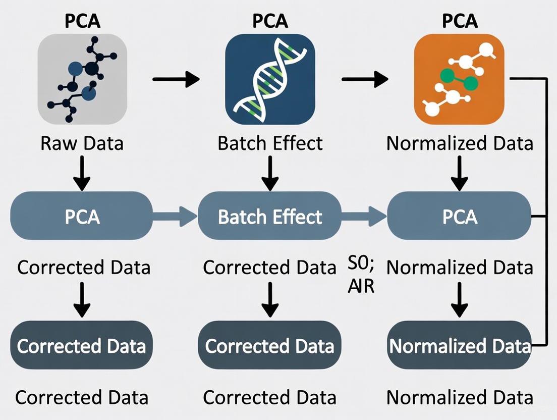
The following diagram illustrates the standard workflow for PCA-based batch effect detection:
Interpretation of PCA Results
In PCA, the first few principal components capture the largest sources of variation in the data. When batch effects represent a major source of variation:
- PC scatter plots show clear separation of samples by batch rather than biological condition [7] [6]
- Density plots for each principal component reveal different distributions across batches [7]
- Variance explanation shows batch-related components accounting for substantial proportion of total variance [7]
Practical Example from Sponge Dataset
Analysis of the sponge dataset demonstrates how PCA reveals batch effects:
In this example, PC1 captured biological variation between different tissues (the effect of interest), while PC2 displayed sample differences due to different gel batches. This clear separation in PCA space confirms the presence of batch effects that require correction [7].
Quantitative Assessment Methods
Beyond visual inspection, quantitative metrics can strengthen batch effect detection:
- Linear models testing batch coefficient significance for individual features [7]
- ANOVA assessing batch contribution to overall variance [7]
- kBET (k-nearest neighbor batch effect test) measuring local batch mixing [6] [8]
- Silhouette width quantifying separation strength between batches [8]
Table 2: PCA Interpretation Guide for Batch Effect Detection
| PCA Pattern | Interpretation | Recommended Action |
|---|---|---|
| Clear batch separation in top PCs | Strong batch effects present; may confound biological analysis | Batch effect correction required before downstream analysis |
| Biological grouping in PC1, batch effects in later PCs | Batch effects present but smaller than biological effects | Evaluate whether correction is needed based on effect size |
| Mixed patterns without clear batch separation | Minimal batch effects or complex interactions | Proceed with caution; consider covariate adjustment in models |
| Batch effects stronger than biological signals | Severe batch confounding | Major correction needed; may require re-analysis with different approach |
Experimental Design Strategies for Batch Effect Prevention
Proper experimental design represents the most effective approach to managing batch effects, as prevention is superior to correction.
Strategic Sample Allocation
The Optimal Sample Assignment Tool (OSAT) was specifically developed to facilitate proper allocation of collected samples to different batches in genomics studies. OSAT optimizes the even distribution of biological groups and confounding factors across batches, reducing the correlation between batches and biological variables of interest [2].
Key principles for effective sample allocation include:
- Balance biological groups across batches to avoid confounding
- Distribute confounding factors (e.g., age, sex, sample source) homogeneously
- Include replicate samples across batches to enable correction validation
- Randomize processing order where possible to avoid systematic biases [2]
Quality Control Measures
Incorporating appropriate quality control (QC) samples is essential for both detecting and correcting batch effects:
- Pooled QC samples: Inserted at regular intervals to monitor and correct instrumental drift [3]
- Technical replicates: Processed across different batches to assess cross-batch reproducibility [3]
- Reference materials: Commercial or internally standardized materials for normalization [9]
Batch Effect Correction Methods
When batch effects cannot be prevented through experimental design, numerous computational approaches exist for batch effect correction.
Classification of Correction Approaches
Genomics-Focused Correction Tools
Table 3: Batch Effect Correction Methods for Genomics Research
| Method | Underlying Approach | Best For | Considerations |
|---|---|---|---|
| ComBat-seq [4] | Empirical Bayes framework | RNA-seq count data | Preserves biological signals; handles small batch sizes |
| removeBatchEffect (limma) [4] | Linear model adjustment | Normalized expression data | Integrated with limma-voom workflow |
| Harmony [10] [6] | Iterative clustering with PCA | Single-cell and bulk data | Fast runtime; good scalability |
| Mutual Nearest Neighbors (MNN) [1] [6] | Matching mutual nearest neighbors | Single-cell RNA-seq data | Identifies shared cell populations across batches |
| Surrogate Variable Analysis (sva) [1] [4] | Estimation of unmodeled variation | Studies with unknown covariates | Handles incomplete batch information |
| Mixed Linear Models [4] | Random effects for batch | Complex experimental designs | Handles nested and hierarchical structures |
Correction Strategy Selection Guidelines
Choosing an appropriate correction method depends on multiple factors:
- Data type: Count-based (ComBat-seq) vs. continuous (ComBat) data
- Batch structure: Balanced vs. confounded designs [9]
- Sample size: Empirical Bayes methods advantageous for small batches [1]
- Biological complexity: Methods preserving subtle biological signals [6]
Recent benchmarking studies recommend:
- Harmony and Seurat CCA for single-cell data with preference for Harmony due to faster runtime [6]
- Protein-level correction for MS-based proteomics data [9]
- Ratio-based methods when batch effects are confounded with biological groups [9]
Research Reagent Solutions for Batch Effect Management
Table 4: Essential Research Reagents and Resources for Batch Effect Control
| Reagent/Resource | Function in Batch Effect Management | Application Context |
|---|---|---|
| Reference Materials (e.g., Quartet protein reference materials) [9] | Inter-batch calibration standards | Large-scale proteomics and multi-omics studies |
| Pooled QC Samples [3] | Monitoring technical variation across batches | Metabolomics, proteomics, and transcriptomics |
| Internal Standards (isotopically labeled) [3] | Normalization within batches | Mass spectrometry-based proteomics and metabolomics |
| Universal Reference Samples [9] | Cross-batch normalization | Multi-center studies and dataset integration |
| Standardized Reagent Lots [1] | Minimizing batch-to-batch reagent variation | All high-throughput genomics applications |
Batch effects represent a fundamental challenge in genomics research, introducing non-biological technical variation that can compromise data interpretation and research reproducibility. Through careful experimental design, vigilant detection using methods like PCA, and appropriate application of correction algorithms, researchers can effectively manage batch effects to ensure the reliability of their genomic findings. As genomic technologies continue to evolve and datasets grow in complexity, sophisticated batch effect management will remain essential for generating biologically meaningful and reproducible results.
In genomics research, Principal Component Analysis (PCA) is a cornerstone tool for the exploratory analysis of high-dimensional data. Its standard application involves projecting samples into a reduced-dimensional space defined by principal components (PCs) that sequentially capture the greatest variance in the dataset. A fundamental assumption in this process is that the largest sources of variation represent the most biologically significant signals. However, this assumption fails dramatically when technically introduced batch effects—systematic technical variations arising from different processing times, laboratories, protocols, or operators—constitute an intermediate source of variation, neither the largest nor the smallest in the dataset [11] [5].
This technical limitation of standard PCA has profound implications for genomic studies. When batch effects are not the primary drivers of variance, they often remain hidden within lower-order principal components, evading visual detection while still significantly confounding biological interpretation [11] [12]. Consequently, researchers may draw incorrect biological conclusions from data where technical artifacts masquerade as biological signals. This paper examines why standard PCA fails under these conditions, introduces enhanced methodologies for detecting and correcting hidden batch effects, and provides practical protocols for genomics researchers working toward robust batch effect correction.
The Limitation of Standard PCA in Batch Effect Detection
How Standard PCA Obscures Intermediate Batch Effects
Standard PCA operates on a straightforward variance-maximization principle: the first PC captures the direction of maximum variance in the data, with subsequent PCs capturing remaining orthogonal variance in descending order. This approach succeeds when batch effects either dominate the variance structure (appearing in early PCs) or represent minor noise (appearing in late PCs). However, when batch effects constitute an intermediate source of variation, they become embedded within middle-order PCs where they are rarely visualized and often overlooked [11].
The consequence is that biologically distinct sample types may cluster by batch rather than by biological condition in the latent space defined by these intermediate components. As noted in assessments of genomic consortia data, "batch effects are a considerable issue, but it is non-trivial to determine if batch adjustment leads to an improvement in data quality" [11]. Visual inspection of only the first two or three PCs—a common practice—provides a false sense of security when batch effects reside in higher-order components.
Specific Failure Scenarios in Genomic Research
- Heterogeneous Samples with Strong Biological Signals: In datasets with substantial legitimate biological variation (e.g., different tissue types, cancer subtypes), biological differences may dominate the first several PCs, pushing batch effects to intermediate components [11].
- Confounded Designs: When batch effects are correlated with biological groups of interest—a common occurrence in multi-center studies—standard PCA cannot distinguish technical artifacts from biological signals [13].
- Low Replicate Numbers: With few biological replicates per batch, the statistical power to detect batch effects through standard PCA diminishes significantly [11].
Table 1: Scenarios Where Standard PCA Fails to Detect Batch Effects
| Scenario | Impact on PCA | Potential Consequences |
|---|---|---|
| High sample heterogeneity | Biological variation dominates early PCs, pushing batch effects to middle PCs | False biological interpretations; batch-confounded results |
| Confounded batch and biological groups | Inability to distinguish technical from biological variance | Incorrect assignment of batch effects as biological signals |
| Longitudinal studies | Time effects entangled with batch effects | Misattribution of temporal changes to batch effects or vice versa |
| Multi-platform data integration | Platform-specific technical variations appear across multiple PCs | Failure to properly integrate datasets from different technologies |
Enhanced Methods for Detecting Hidden Batch Effects
PCA-Plus: Extensions to Standard PCA
To address the limitations of standard PCA, enhanced methods like PCA-Plus introduce algorithmic extensions that improve batch effect detection [12]. PCA-Plus incorporates several key enhancements:
- Group Centroids: Computes and visualizes the central point for each pre-defined batch or biological group
- Dispersion Rays: Shows the variation and distribution of samples within each group
- Trend Trajectories: Identifies and visualizes temporal or sequential patterns across batches
- Dispersion Separability Criterion (DSC): A novel metric that quantifies the separation between groups while accounting for within-group variation
The DSC metric is particularly valuable as it provides a quantitative measure of batch effect severity. It is defined as DSC = D~b~/D~w~, where D~b~ represents the trace of the between-group scatter matrix and D~w~ represents the trace of the within-group scatter matrix [12]. Higher DSC values indicate greater separation between groups relative to within-group variation, suggesting more pronounced batch effects.
Alternative Visualization and Quantification Approaches
Beyond PCA-Plus, several other methods have proven effective for detecting batch effects that evade standard PCA:
- t-Distributed Stochastic Neighbor Embedding (t-SNE): This nonlinear dimensionality reduction technique can reveal batch-associated patterns that remain hidden in PCA [14].
- k-Nearest Neighbor Batch Effect Test (kBET): Quantifies batch effects by measuring how well batches mix at the local level of each sample's nearest neighbors [15].
- Principal Variance Component Analysis (PVCA): Partitions total variation in the dataset into components attributable to batch, biological, and other known factors [9].
Table 2: Methods for Detecting Hidden Batch Effects
| Method | Underlying Principle | Advantages | Limitations |
|---|---|---|---|
| PCA-Plus | Enhanced PCA with group centroids and DSC metric | Quantifiable separation index; maintains PCA interpretability | Requires pre-defined group labels |
| t-SNE | Nonlinear dimensionality reduction | Can reveal complex batch patterns invisible to linear methods | Computational intensive; harder to interpret |
| kBET | Local neighborhood batch mixing | Quantifies batch effect at local scale | Requires batch labels; sensitive to parameters |
| PVCA | Variance partitioning | Quantifies contribution of known factors | Requires complete metadata |
Advanced Batch Effect Correction Strategies
Reference-Based Correction Methods
For scenarios where batch effects are confounded with biological groups, reference-based methods have demonstrated particular effectiveness:
Ratio-Based Scaling: This method transforms absolute feature values into ratios relative to concurrently profiled reference materials. The approach has shown superior performance in multi-omics studies, especially when batch effects are completely confounded with biological factors [13] [9].
Reference Material Design: The Quartet Project employs multi-omics reference materials from four related cell lines, enabling robust batch effect correction across diverse genomic platforms [13]. When implementing ratio-based correction, expression values of study samples are scaled relative to the reference material processed in the same batch:
Corrected Value = Original Value / Reference Value.
Algorithmic Correction Methods
Multiple batch effect correction algorithms (BECAs) have been developed with varying strengths for different scenarios:
Harmony: This method iteratively clusters cells by similarity and calculates cluster-specific correction factors, demonstrating strong performance across both single-cell and bulk genomic data [13] [16].
ComBat: Utilizing empirical Bayes frameworks, ComBat adjusts for batch effects by modeling them as additive and multiplicative noise. Its performance improves significantly when biological covariates are included in the model [17] [16].
Mutual Nearest Neighbors (MNN): This approach identifies pairs of cells across batches that are mutual nearest neighbors in expression space, using these "anchors" to correct batch effects while preserving biological variation [14].
Batch Effect Correction Workflow
Experimental Protocols for Robust Batch Effect Management
Protocol 1: Comprehensive Batch Effect Assessment
Purpose: Systematically evaluate batch effects in genomic data when standard PCA suggests minimal technical artifacts.
Materials:
- Normalized genomic data matrix (e.g., gene expression, methylation)
- Complete metadata including batch identifiers and biological covariates
- R or Python statistical environment
Procedure:
- Standard PCA Visualization
- Perform conventional PCA and plot PC1 vs. PC2, PC2 vs. PC3, and PC1 vs. PC3
- Color points by known batch variables and biological conditions
- Document apparent clustering patterns
Enhanced PCA Analysis
- Implement PCA-Plus with DSC calculation [12]
- Compute group centroids for each batch
- Calculate DSC metric with permutation testing for significance
- Retain components explaining >80% cumulative variance for full assessment
Alternative Visualization
- Apply t-SNE with multiple perplexity values
- Color resulting embeddings by batch and biological variables
- Compare patterns across visualizations
Quantitative Assessment
- Perform kBET analysis to quantify local batch mixing
- Conduct PVCA to partition variance components
- Calculate correlation between replicates within and across batches
Interpretation: Significant batch effects are indicated by DSC p-value <0.05, kBET rejection rate >0.2, or batch accounting for >15% variance in PVCA.
Protocol 2: Reference Material-Based Batch Correction
Purpose: Implement ratio-based batch effect correction using reference materials.
Materials:
- Study samples with reference materials processed in parallel batches
- Quantified feature-level data (e.g., gene expression values)
- Computing environment with statistical software
Procedure:
- Data Preparation
- Organize data by processing batch
- Verify reference material presence in each batch
- Log-transform expression data if necessary
Ratio Calculation
- For each feature in every study sample:
Ratio = Study Sample Value / Reference Material Value - Use median reference value if multiple reference replicates available
- Handle zero values with appropriate pseudo-counts
- For each feature in every study sample:
Batch Effect Assessment
- Apply PCA to ratio-transformed data
- Compare with pre-correction visualization
- Quantify improvement using DSC or similar metrics
Validation
- Assess biological signal preservation through known biological groups
- Evaluate technical artifact reduction through replicate correlation
Notes: This method is particularly effective for multi-omics studies and confounded batch-group scenarios [13].
Protocol 3: Algorithmic Batch Effect Correction for Complex Datasets
Purpose: Apply and compare computational batch correction methods.
Materials:
- Normalized genomic data with batch labels
- Biological covariates of interest
- High-performance computing resources for resource-intensive methods
Procedure:
- Data Preprocessing
- Select highly variable genes (top 5000 by default) [14]
- Scale data using multiBatchNorm or equivalent approach
- Split into discovery and validation sets if possible
Method Application
- Apply multiple correction methods (Harmony, ComBat, MNN, etc.)
- For ComBat, run with and without biological covariates
- For deep learning methods (e.g., scVI), ensure adequate computational resources
Performance Evaluation
- Visualize corrected data using PCA and t-SNE
- Quantify batch mixing using kBET or similar metrics
- Assess biological preservation through clustering of known cell types
- Evaluate method robustness via replicate correlation
Method Selection
- Choose method that optimally balances batch removal and biological signal preservation
- Consider computational efficiency for large datasets
Troubleshooting: If over-correction is suspected (loss of biological signal), prioritize methods that incorporate biological covariates or use more conservative parameters.
Table 3: Research Reagent Solutions for Batch Effect Management
| Reagent/Resource | Function | Application Notes |
|---|---|---|
| Quartet Reference Materials | Multi-omics quality control and ratio-based correction | Enables ratio-based scaling across DNA, RNA, protein, and metabolite profiling [13] |
| Cell Line Controls | Batch effect monitoring through consistent biological material | Include in every processing batch to track technical variation |
| Universal RNA References | Standardization of transcriptomic measurements | Particularly valuable for cross-laboratory studies |
| Synthetic Spike-in Controls | Technical variation assessment | Add known quantities of synthetic sequences to distinguish technical from biological variation |
Standard PCA represents a necessary but insufficient tool for comprehensive batch effect detection in genomics research. Its fundamental limitation lies in the variance-maximization principle that inevitably misses batch effects when they constitute intermediate rather than dominant sources of variation. This oversight can lead to biologically misleading conclusions and compromised analytical outcomes.
The enhanced methodologies presented here—including PCA-Plus with its DSC metric, reference material-based ratio correction, and sophisticated algorithms like Harmony and ComBat—provide researchers with a robust toolkit for identifying and correcting these hidden technical artifacts. The experimental protocols offer practical guidance for implementation across diverse genomic research scenarios.
As genomic studies grow in scale and complexity, with increasing integration of multi-omics data from multiple centers, rigorous approaches to batch effect management become increasingly critical. By moving beyond standard PCA and adopting the comprehensive framework outlined here, researchers can significantly enhance the reliability and reproducibility of their genomic findings, ensuring that biological signals remain distinct from technical artifacts in even the most challenging research contexts.
In genomics research, batch effects are technical variations introduced during the experimental process that are unrelated to the biological signals of interest. These non-biological variations arise from differences in reagent lots, processing times, equipment calibration, laboratory personnel, or sequencing platforms [18]. In large-scale omics studies, such as those using single-cell RNA sequencing (scRNA-seq), batch effects can confound biological variation, reduce statistical power, and potentially lead to misleading conclusions if not properly addressed [18] [19]. The detection and correction of these effects are therefore crucial steps in ensuring data reliability and reproducibility.
Visual diagnostic tools play a fundamental role in the initial detection and assessment of batch effects. Dimensionality reduction techniques – including Principal Component Analysis (PCA), t-distributed Stochastic Neighbor Embedding (t-SNE), and Uniform Manifold Approximation and Projection (UMAP) – transform high-dimensional genomic data into two or three-dimensional spaces that can be visually inspected [20] [6] [21]. These methods allow researchers to observe systematic patterns in their data that may indicate the presence of batch effects before applying quantitative metrics or correction algorithms. When batches cluster separately rather than mixing according to biological conditions, it provides strong visual evidence of batch effects that require remediation [6].
Theoretical Foundations of Visualization Methods
Principal Component Analysis (PCA)
PCA is a linear dimensionality reduction technique that projects data onto the directions of maximum variance, called principal components [20] [19]. It operates by computing the eigenvectors of the covariance matrix of the data, with the first component capturing the greatest variance, the second component the second greatest, and so on. For batch effect detection, PCA is computationally efficient and effective when batch effects exhibit linear patterns [19]. However, its linear nature makes it less capable of capturing complex nonlinear batch effects that are common in genomic data [19].
t-Distributed Stochastic Neighbor Embedding (t-SNE)
t-SNE is a nonlinear probabilistic method that minimizes the Kullback-Leibler divergence between probability distributions in high and low dimensions [20]. It emphasizes the preservation of local data structures, making it particularly effective for visualizing distinct cell types or sample groups. However, t-SNE may not preserve global structures well, and its interpretation can be complicated by parameters such as perplexity that significantly affect the resulting visualization [20].
Uniform Manifold Approximation and Projection (UMAP)
UMAP is based on Riemannian geometry and fuzzy simplicial set theory [20]. It constructs a graphical representation of the data manifold and optimizes a low-dimensional layout that preserves both local and some global structures [20]. UMAP generally offers faster runtime than t-SNE and often provides better preservation of global data structure, making it increasingly popular for single-cell genomics visualization [20] [21].
Table 1: Comparative Characteristics of Dimensionality Reduction Methods
| Feature | PCA | t-SNE | UMAP |
|---|---|---|---|
| Type | Linear | Nonlinear | Nonlinear |
| Preservation | Global variance | Local structure | Local & global structure |
| Speed | Fast | Slow | Moderate to Fast |
| Deterministic | Yes | No | Yes |
| Parameters | Few | Perplexity, iterations | Neighbors, min distance |
| Batch Effect Detection | Effective for linear patterns | Effective for local patterns | Effective for complex patterns |
Experimental Protocols for Batch Effect Detection
Data Preprocessing Requirements
Prior to applying visualization techniques, proper data preprocessing is essential. For scRNA-seq data, this typically includes quality control filtering to remove low-quality cells, normalization to account for sequencing depth variations, logarithmic transformation to stabilize variance, and selection of highly variable genes that drive biological variation [20] [21]. These steps help ensure that technical artifacts do not dominate the visualization and that the resulting plots reflect true biological signals and batch effects rather than preprocessing artifacts.
Protocol for PCA-Based Batch Effect Detection
- Input Preparation: Begin with a normalized gene expression matrix (cells × genes) and associated batch metadata.
- Feature Selection: Use highly variable genes (typically 2,000-5,000) as input features to focus on biologically relevant signals [21].
- PCA Computation: Perform PCA on the standardized expression matrix using singular value decomposition.
- Variance Examination: Check the proportion of variance explained by each principal component, noting components that correlate with batch metadata.
- Visualization: Create scatter plots of the first few principal components (e.g., PC1 vs. PC2, PC2 vs. PC3) colored by batch labels.
- Interpretation: Look for clear separation of samples by batch rather than biological condition in the PCA plot, which indicates batch effects [6].
Protocol for t-SNE and UMAP-Based Batch Effect Detection
- Input Preparation: Use the same normalized expression matrix and batch metadata as for PCA.
- Initial Dimensionality Reduction: First reduce dimensions with PCA (e.g., 50 principal components) to denoise data and reduce computational burden [21].
- Parameter Optimization:
- Embedding Generation: Run t-SNE or UMAP on the PCA-reduced data to generate 2D coordinates for each cell.
- Visualization: Create scatter plots of the t-SNE or UMAP embeddings, coloring points by batch labels and optionally by cell type labels.
- Interpretation: Assess whether cells cluster primarily by batch rather than biological cell type, which indicates batch effects [6].
The following workflow diagram illustrates the complete batch effect detection process:
Quantitative Validation of Visual Findings
While visual inspection provides initial evidence of batch effects, quantitative metrics offer objective validation. The most commonly used metrics include:
- k-nearest neighbor Batch Effect Test (kBET): Measures batch mixing by comparing local versus global batch label distributions using a chi-squared test [19] [21]. Lower rejection rates indicate better batch mixing.
- Local Inverse Simpson's Index (LISI): Quantifies the diversity of batches in local neighborhoods, with higher scores indicating better integration [19] [21]. Integration LISI (iLISI) specifically measures batch mixing.
- Average Silhouette Width (ASW): Evaluates cluster compactness and separation, with separate calculations for batch and biological labels [20] [21]. Batch ASW should be low while cell type ASW should be high after successful correction.
- Adjusted Rand Index (ARI): Measures similarity between clustering results and known cell type annotations, with higher values indicating better preservation of biological signals [22] [21].
Table 2: Quantitative Metrics for Batch Effect Assessment
| Metric | Measurement Target | Ideal Value | Interpretation |
|---|---|---|---|
| kBET Rejection Rate | Batch mixing in local neighborhoods | < 0.2 | Lower = better mixing |
| iLISI Score | Diversity of batches in local neighborhoods | > 1.5 | Higher = better integration |
| Batch ASW | Separation by batch | Close to 0 | Lower = less batch effect |
| Cell Type ASW | Separation by cell type | > 0.5 | Higher = biological preservation |
| ARI | Agreement with cell type labels | > 0.7 | Higher = biological preservation |
Advanced Detection Methods and Limitations
Addressing Nonlinear Batch Effects with BEENE
Traditional PCA has limitations in detecting nonlinear batch effects, which are common in complex genomic datasets [19]. To address this challenge, Batch Effect Estimation using Nonlinear Embedding (BEENE) employs a deep autoencoder network that learns both batch and biological variables simultaneously [19]. BEENE generates embeddings that are more sensitive to both linear and nonlinear batch effects compared to PCA, providing enhanced detection capability for complex batch effects that might be missed by linear methods [19].
Limitations and Considerations
Each visualization method has limitations that researchers must consider. PCA may miss complex nonlinear batch effects [19]. t-SNE results can vary between runs due to stochasticity and are sensitive to parameter choices [20]. UMAP may create artificial connections between distinct clusters, potentially obscuring true biological separation [20]. Additionally, over-reliance on visual inspection without quantitative validation can lead to subjective interpretations [19]. Therefore, a combination of multiple visualization methods and quantitative metrics is recommended for comprehensive batch effect assessment [6] [21].
Research Reagent Solutions
Table 3: Essential Tools for Batch Effect Analysis in Genomics Research
| Tool/Resource | Function | Application Context |
|---|---|---|
| BEEx [23] | Open-source platform for qualitative and quantitative batch effect assessment in medical images | Digital pathology, radiology |
| BEENE [19] | Deep autoencoder for detecting nonlinear batch effects | scRNA-seq data with complex batch effects |
| Harmony [16] [21] | Batch effect correction using iterative clustering | scRNA-seq, image-based profiling |
| Seurat [16] [21] | Integration method using CCA or RPCA and mutual nearest neighbors | scRNA-seq, multi-modal genomics |
| Scanpy [20] | Python-based toolkit for single-cell data analysis | scRNA-seq preprocessing and visualization |
| TCGA Batch Effects Viewer [24] | Web-based platform for assessing batch effects in TCGA data | Cancer genomics, multi-institutional studies |
Effective batch effect detection using PCA, t-SNE, and UMAP visualization is a critical first step in ensuring the reliability of genomic analyses. Each method offers complementary strengths: PCA provides linear efficiency, t-SNE reveals local structure, and UMAP balances local and global patterns. When combined with quantitative metrics like kBET and LISI, these visual tools form an essential component of rigorous genomic data quality assessment. As batch effects grow more complex in large-scale multi-omics studies, advanced methods like BEENE that address nonlinear patterns will become increasingly important for maintaining data quality and biological validity in genomics research.
In genomics research, batch effects are a pervasive challenge, defined as systematic non-biological variations between groups of samples processed under different conditions, such as different times, laboratories, or technicians [25]. These technical artifacts can confound biological signals, leading to misleading conclusions in downstream analyses. Principal Component Analysis (PCA) is a common visual tool for initial batch effect detection; however, its utility is limited because it identifies directions of maximum variance, which may not always correspond to batch effects if they are not the largest source of variation [25] [26]. This limitation within a broader thesis on batch effect correction underscores the necessity for robust, quantitative statistical metrics to reliably identify and measure batch effects prior to applying correction methods such as ComBat or Harmony [27] [21] [28]. This document provides detailed application notes and protocols for three key metrics—Dispersion Separability Criterion (DSC), guided PCA (gPCA), and findBATCH—enabling researchers to make informed decisions about the presence and severity of batch effects in their genomic data.
The following table summarizes the core characteristics of the three quantitative batch effect assessment metrics discussed in this protocol.
Table 1: Overview of Quantitative Batch Effect Assessment Metrics
| Metric | Full Name | Underlying Principle | Primary Output | Key Reference |
|---|---|---|---|---|
| DSC | Dispersion Separability Criterion | Ratio of between-batch to within-batch dispersion | A continuous positive value (DSC) and an empirical p-value | [24] |
| gPCA | guided Principal Component Analysis | Modifies PCA to be guided by a batch indicator matrix, comparing variance to unguided PCA | Test statistic (δ) and a p-value from a permutation test | [25] [26] |
| findBATCH | finding Batch Effects | Evaluates batch effects based on Probabilistic Principal Component and Covariates Analysis (PPCCA) | A statistical measure for diagnosing and quantifying batch effects | [29] |
The Scientist's Toolkit: Essential Research Reagents and Software
Successful implementation of the assessment protocols requires specific computational tools and resources.
Table 2: Key Research Reagent Solutions for Batch Effect Assessment
| Item Name | Function/Application | Implementation |
|---|---|---|
| gPCA R Package | Provides functions to perform the gPCA method and compute the δ statistic. | Available via CRAN [25] |
| MBatch R Package | Contains algorithms (e.g., ANOVA, Empirical Bayes, Median Polish) for assessing and correcting batch effects, and is associated with the TCGA Batch Effects Viewer. | R package [24] [28] |
| TCGA Batch Effects Viewer | A web-based platform to quantitatively and visually assess batch effects in TCGA data, including DSC metric calculation. | Online tool [24] |
| Harman R Package | An alternative batch effect correction and diagnosis tool that maximizes batch noise removal while constraining the risk of signal loss. | Available on Bioconductor [30] |
| findBATCH Algorithm | A method to evaluate batch effect based on probabilistic principal component and covariates analysis (PPCCA). | Methodology described in literature [29] |
Detailed Methodologies and Experimental Protocols
Dispersion Separability Criterion (DSC)
The DSC metric quantifies batch effect by measuring the ratio of dispersion between batches to the dispersion within batches [24].
Mathematical Foundation
The DSC is calculated using the following formulas:
- Between-batch dispersion ((Db)): ( Db = \sqrt{trace(S_b)} )
- Within-batch dispersion ((Dw)): ( Dw = \sqrt{trace(S_w)} )
- DSC: ( DSC = Db / Dw )
Here, (Sb) is the between-batch scatter matrix and (Sw) is the within-batch scatter matrix, as defined in Dy et al., 2004 [24]. (Dw) represents the average distance between samples within a batch and the batch's centroid, while (Db) represents the average distance between batch centroids and the global mean.
Interpretation and Decision Guidelines
Table 3: Interpreting DSC Values and Associated Actions
| DSC Value | p-value | Interpretation | Recommended Action |
|---|---|---|---|
| < 0.5 | Any | Batch effects are not strong. | Proceed with analysis; correction may be unnecessary. |
| > 0.5 | < 0.05 | Significant batch effects are likely present. | Consider batch effect correction before analysis. |
| > 1 | < 0.05 | Strong batch effects are present. | Batch effect correction is strongly recommended. |
Note: The p-value is derived empirically via permutation tests (e.g., 1000 permutations). Both the DSC value and its p-value should be considered for a robust assessment [24].
Experimental Protocol
Procedure:
- Input Data Preparation: Start with a normalized data matrix (e.g., gene expression counts) and a metadata file specifying the batch identifier for each sample.
- DSC Calculation: a. Compute the global mean feature vector across all samples. b. For each batch, calculate the batch centroid (mean feature vector of samples within the batch). c. Compute (Sb), the between-batch scatter matrix. d. For each sample, calculate its deviation from its respective batch centroid. Compute (Sw), the within-batch scatter matrix. e. Calculate (Db) and (Dw) as the square root of the trace of (Sb) and (Sw), respectively. f. Compute the DSC statistic.
- Significance Testing via Permutation: a. Randomly permute the batch labels across all samples. b. Recalculate the DSC statistic with the permuted labels. c. Repeat this process a large number of times (e.g., M=1000) to build a null distribution of DSC under the hypothesis of no batch effects. d. The empirical p-value is the proportion of permuted DSC values that are greater than or equal to the observed DSC value.
- Decision: Use Table 3 to interpret the results and decide whether batch effect correction is needed.
guided Principal Component Analysis (gPCA)
gPCA is an extension of traditional PCA that incorporates a batch indicator matrix to directly guide the decomposition towards variance associated with batch [25].
Mathematical Foundation and Test Statistic
The core of gPCA involves performing singular value decomposition (SVD) not on the data matrix (X) itself, but on (Y'X), where (Y) is the batch indicator matrix. This guides the analysis to find components that separate batches [25].
The primary test statistic, δ, quantifies the proportion of variance attributable to batch effects: [ \delta = \frac{\text{Variance of 1st PC from gPCA}}{\text{Variance of 1st PC from unguided PCA}} = \frac{\lambdag}{\lambdau} ] where (\lambdag) and (\lambdau) are the first eigenvalues from the gPCA and unguided PCA, respectively [25]. A δ value near 1 implies a large batch effect.
The percentage of total variation explained by batch can be estimated as: [ \% \text{ Var} = \frac{\sum \lambda{g, i}}{\sum \lambda{u, i}} \times 100\% ] where the summation is over all principal components [25].
Experimental Protocol
Procedure:
- Data Preprocessing: Center the data matrix (X). Filter non-informative features (e.g., retain the 1000 most variable probes) to reduce noise [25].
- Unguided PCA: Perform standard SVD on the centered matrix (X) to obtain the eigenvalues ((\lambda_u)) for the unguided principal components.
- Guided PCA (gPCA): a. Construct the batch indicator matrix (Y). b. Perform SVD on the matrix (Y'X) to obtain the guided eigenvalues ((\lambda_g)).
- Calculate the δ Statistic: Compute δ using the first eigenvalues from steps 2 and 3.
- Significance Testing via Permutation: a. Permute the batch assignment vector. b. Recompute the δ statistic with the permuted batch labels. c. Repeat this process M times (e.g., M=1000) to create a permutation distribution for δ under the null hypothesis. d. Calculate a one-sided p-value as the proportion of permuted δ values that are greater than or equal to the observed δ.
- Interpretation: A significant p-value (e.g., < 0.05) indicates the presence of a statistically significant batch effect.
findBATCH
The findBATCH algorithm offers a novel approach to diagnosing and quantifying batch effects using a probabilistic framework [29].
findBATCH is based on Probabilistic Principal Component and Covariates Analysis (PPCCA). This method integrates the assessment of batch effects directly into a probabilistic model for dimensionality reduction, allowing for a more formal statistical assessment of the influence of batch covariates on the high-dimensional data structure.
The following diagram illustrates the logical workflow for applying and interpreting these three batch effect assessment metrics.
Diagram 1: Batch effect assessment workflow.
Experimental Protocol
Procedure:
- Input: A normalized genomic data matrix (e.g., from microarray or RNA-seq) and a covariate matrix that includes batch identifiers and potentially other biological factors.
- Model Fitting: Apply the PPCCA model to the data. This model jointly estimates the principal components and the effects of the provided covariates (like batch) on the data.
- Batch Effect Diagnosis: The model output provides a statistical measure quantifying the extent to which batch covariates explain the variance in the data. A stronger association indicates a more substantial batch effect.
- Correction (Optional): The same PPCCA framework underlying findBATCH can be extended to provide a correction method,
CorrectBATCH, which aims to remove the identified batch effects [29].
Integrated Application Workflow
For a comprehensive assessment, it is advisable to use these metrics in concert, as they probe different aspects of batch effects.
- Exploratory Analysis: Begin with a standard PCA plot colored by batch to visually inspect for obvious clustering by batch.
- Quantitative Assessment: a. Run the gPCA test to obtain a p-value for the presence of any batch effect. b. Calculate the DSC metric and its p-value to quantify the strength and significance of batch separation relative to within-batch variation. c. Apply findBATCH to leverage a probabilistic model for a complementary assessment.
- Holistic Decision Making: Synthesize results from all metrics. If multiple metrics indicate a significant batch effect (e.g., gPCA p-value < 0.05 and DSC > 0.5), proceed with batch effect correction using a method such as ComBat, Harmony, or a ratio-based method before any downstream biological analysis [31] [27] [21].
Within the broader objective of developing robust batch effect correction pipelines for genomics, reliable detection is the critical first step. The DSC, gPCA, and findBATCH metrics provide a powerful, statistically grounded toolkit that moves beyond visual PCA inspection. By implementing the detailed application notes and protocols outlined herein, researchers and drug development professionals can systematically diagnose batch effects, thereby ensuring the integrity and reproducibility of their genomic findings.
In the realm of genomics research, batch effects represent a formidable challenge, introducing non-biological technical variations that can compromise data integrity and lead to irreproducible findings. These effects are notoriously common in omics data and, if left uncorrected, can result in misleading outcomes and biased biological interpretation [18]. This application note presents a concrete case study demonstrating how uncorrected batch effects skewed analysis in a real genomic dataset and details the experimental protocols used to diagnose and correct these effects, framed within a broader thesis on batch effect correction for principal component analysis (PCA).
Case Study: Batch Effects in Breast Cancer Gene Expression Profiling
Background and Experimental Design
This case study examines the integration of gene expression data from three independent breast cancer studies profiled using the Affymetrix GeneChip Human Genome U133 Plus 2.0 Array [32]. The pooled dataset comprised 70 samples (30, 22, and 18 from studies GSE12763, GSE13787, and GSE23593, respectively) after standard microarray quality control procedures. The research aimed to identify conserved gene expression signatures across different breast cancer cohorts.
Table 1: Dataset Composition for Breast Cancer Case Study
| Dataset Identifier | Sample Size | Platform | Primary Tissue Source |
|---|---|---|---|
| GSE12763 | 30 | Affymetrix U133 Plus 2.0 | Primary human breast tumors |
| GSE13787 | 22 | Affymetrix U133 Plus 2.0 | Primary human breast tumors |
| GSE23593 | 18 | Affymetrix U133 Plus 2.0 | Primary human breast tumors |
Manifestation of Batch Effects
Initial PCA of the pooled dataset revealed a critical problem: sample clustering in the principal subspace was exclusively driven by batch effect rather than biological characteristics. As shown in Figure 1, samples clustered strictly by their study of origin (batch) in the principal component space, with the first two principal components capturing technical variations rather than biological signals [32].
Figure 1: Workflow demonstrating how batch effects manifested in the breast cancer gene expression case study. PCA visualization revealed clustering by study origin rather than biological characteristics.
Formal statistical testing using the findBATCH method (part of the exploBATCH framework based on Probabilistic Principal Component and Covariates Analysis - PPCCA) confirmed significant batch effects on three of the first five probabilistic principal components (pPCs) [32]. The 95% confidence intervals for the estimated batch effects on pPC1, pPC2, and pPC4 did not include zero, indicating statistically significant technical variation across the batches.
Consequences of Uncorrected Batch Effects
The profound impact of these uncorrected batch effects included:
Masked Biological Signals: True biological differences between breast cancer subtypes were obscured by stronger technical variations [32].
Risk of False Associations: Differential expression analysis conducted on uncorrected data risked identifying falsely significant genes correlated with batch rather than biology [18].
Irreproducible Findings: Any conclusions drawn from the uncorrected data would be specific to the individual studies rather than generalizable across breast cancer populations [18].
Quantitative Impact Assessment
The consequences of batch effects extend beyond this single case study. In a clinical trial context, batch effects introduced by a change in RNA-extraction solution resulted in incorrect classification outcomes for 162 patients, 28 of whom received incorrect or unnecessary chemotherapy regimens [18]. The table below summarizes documented impacts of uncorrected batch effects across various genomic studies.
Table 2: Documented Impacts of Uncorrected Batch Effects in Genomic Studies
| Research Context | Impact of Uncorrected Batch Effects | Consequence |
|---|---|---|
| Breast Cancer Gene Expression [32] | Samples clustered by study origin rather than biology | Masked true biological signals; risk of false conclusions |
| Clinical Trial Molecular Profiling [18] | Shift in gene-based risk calculation | 162 patients misclassified, 28 received incorrect chemotherapy |
| Cross-Species Comparison [18] | Apparent species differences greater than tissue differences | Misleading evolutionary conclusions; corrected to show tissue similarities |
| Ovarian Cancer Study [33] | False gene expression signatures identified | Retracted study and misdirected research directions |
Experimental Protocols for Batch Effect Diagnosis and Correction
Protocol 1: Statistical Diagnosis of Batch Effects
Principle: Implement formal statistical testing to diagnose batch effects before correction [32].
Reagents and Materials:
- R statistical environment (v4.0 or higher)
- exploBATCH R package (https://github.com/syspremed/exploBATCH)
- Normalized gene expression matrix (samples × features)
- Batch metadata (study/site/platform of origin for each sample)
Procedure:
- Data Preprocessing: Normalize each dataset separately according to platform-specific protocols, then pool based on common gene identifiers.
- PPCCA Modeling: Apply findBATCH function to select optimal number of probabilistic principal components using Bayesian Information Criterion.
- Batch Effect Quantification: Calculate 95% confidence intervals for estimated batch effects on each probabilistic principal component.
- Significance Determination: Identify components with 95% CIs not including zero as significantly affected by batch effects.
- Visualization: Generate forest plots to visualize effect sizes and confidence intervals across components.
Expected Results: Formal statistical testing will identify which principal components are significantly affected by batch effects, providing guidance for targeted correction approaches.
Protocol 2: Batch Effect Correction Using exploBATCH
Principle: Implement PPCCA-based correction to remove batch effects while preserving biological variation [32].
Reagents and Materials:
- R statistical environment
- exploBATCH R package
- Expression matrix with confirmed batch effects
- Biological covariates of interest (e.g., disease status)
Procedure:
- Model Fitting: Apply correctBATCH function to estimate batch effects on significant components identified in Protocol 1.
- Effect Subtraction: Subtract the estimated batch effect from each affected probabilistic principal component.
- Data Reconstruction: Reconstruct batch-corrected expression data using the adjusted components.
- Validation: Confirm removal of batch effects using PCA visualization and statistical testing.
- Biological Signal Verification: Verify preservation of biological effects using known biological covariates.
Expected Results: Batch-corrected data where samples cluster by biological characteristics rather than technical artifacts, enabling valid cross-study comparisons.
Protocol 3: ComBat-based Correction for RNA-seq Data
Principle: Implement reference-based batch correction using negative binomial models for count-based RNA-seq data [31].
Reagents and Materials:
- R statistical environment
- ComBat-ref implementation
- RNA-seq count matrix
- Batch metadata
- Reference batch identification
Procedure:
- Reference Batch Selection: Identify the batch with smallest dispersion as reference batch.
- Model Parameter Estimation: Fit negative binomial models to estimate batch-specific parameters.
- Data Adjustment: Adjust non-reference batches toward the reference batch while preserving count data nature.
- Quality Assessment: Evaluate correction using silhouette scores and PCA visualization.
- Downstream Analysis: Proceed with differential expression analysis on corrected data.
Expected Results: Effective removal of batch effects while maintaining the statistical properties of count data and improving sensitivity and specificity of differential expression analysis.
The Scientist's Toolkit: Essential Research Reagents and Computational Tools
Table 3: Essential Research Reagents and Computational Tools for Batch Effect Management
| Tool/Reagent | Function | Application Context |
|---|---|---|
| exploBATCH R Package [32] | Statistical diagnosis and correction of batch effects using PPCCA | General genomic studies (microarray, RNA-seq) |
| ComBat/ComBat-seq [31] [34] | Empirical Bayes framework for batch correction | Microarray (ComBat) and RNA-seq count data (ComBat-seq) |
| ComBat-met [34] | Beta regression framework for DNA methylation data | Methylation array or bisulfite sequencing data |
| findBATCH Function [32] | Formal statistical testing for batch effects | Pre-correction diagnosis in any high-throughput data |
| Reference Materials [9] | Quality control samples for batch effect monitoring | Large-scale multi-batch proteomics and genomics studies |
| Harmony Algorithm [10] | Iterative clustering-based batch correction | Single-cell RNA sequencing and spatial transcriptomics |
Batch Effect Correction Workflow
A robust batch effect management strategy requires a systematic approach from experimental design through data analysis, as illustrated below.
Figure 2: Comprehensive batch effect management workflow spanning experimental design through validation phases. A systematic approach is essential for generating reliable, reproducible genomic data.
This case study demonstrates that uncorrected batch effects can severely compromise genomic analyses, leading to misleading biological interpretations and potentially costly clinical misapplications. The breast cancer gene expression example illustrates how technical variations can dominate the principal components that should ideally capture biological signals. Through implementation of rigorous statistical diagnosis and appropriate correction methods such as those provided by the exploBATCH framework, researchers can effectively mitigate these technical artifacts while preserving biological signals of interest. As genomic technologies continue to evolve and multi-study integrations become increasingly common, robust batch effect management will remain essential for generating reliable, reproducible research findings.
A Practical Guide to Batch Correction Methods Integrating PCA
In genomic studies, batch effects are technical, non-biological variations introduced when samples are processed in different groups or "batches" due to factors like different time points, personnel, reagents, or sequencing platforms [10] [18]. These effects can confound biological signals, reduce statistical power, and if left uncorrected, lead to misleading scientific conclusions and irreproducible results [18] [5]. A survey in Nature found that 90% of researchers believe there is a reproducibility crisis, with batch effects being a paramount contributing factor [5]. Traditional diagnostic methods like Principal Component Analysis (PCA) rely on visual inspection to detect batch clustering, but this approach is subjective and can fail when batch effect is not the greatest source of variability [32].
Guided PCA (gPCA) is a statistical methodology designed specifically to address the limitations of conventional PCA in batch effect diagnosis. Unlike standard PCA, which identifies directions of maximal variance without considering their source, gPCA provides a formal, statistical framework to determine whether the observed patterns in high-dimensional genomic data are significantly associated with batch [32]. This targeted approach offers researchers an objective measure to diagnose batch effects before proceeding with correction, thereby reducing the risk of unnecessary data manipulation or failure to detect confounding technical variation.
gPCA in Context: Comparison with Other Batch Effect Evaluation Methods
Several methods exist for diagnosing and evaluating batch effects in genomic data, each with different strengths and limitations. The table below summarizes the key characteristics of gPCA against other common evaluation approaches.
Table 1: Comparison of Batch Effect Evaluation Methods
| Method | Underlying Principle | Key Output | Primary Use | Key Advantage | Key Limitation |
|---|---|---|---|---|---|
| Guided PCA (gPCA) | Extension of traditional PCA that incorporates batch labels to test for significant batch-associated variation [32]. | A formal statistical test p-value for the global presence of batch effect across all principal components [32]. | Formal statistical diagnosis of batch effect. | Provides an objective, global test for batch effect significance, reducing subjectivity [32]. | Does not assess the effect of batch on individual principal components [32]. |
| Principal Component Analysis (PCA) | Identifies directions of maximal variance in the data without using batch information. | Visualization (e.g., scatter plot) of samples in the space of the first few principal components. | Exploratory visual inspection for batch clustering. | Intuitive and widely used for an initial, quick check of data structure. | Subjective; relies on visual inspection. Can fail if batch effect is not the largest source of variance [32]. |
| findBATCH (via PPCCA) | Uses Probabilistic PCA and Covariates Analysis to model batch as a covariate [32]. | Forest plots with 95% confidence intervals for the estimated batch effect on each probabilistic PC [32]. | Formal statistical diagnosis and quantification of batch effect on individual components. | Identifies which specific principal components are significantly affected by batch [32]. | More complex multi-step procedure; requires selection of the optimal number of probabilistic PCs [32]. |
| Principal Variation Component Analysis (PVCA) | Combines PCA and a linear mixed model to quantify variance contributions from batch and other factors [32]. | Proportion of total variability in the data attributed to batch effect. | Quantifying the magnitude of batch effect relative to other sources of variation. | Provides an estimate of how much of the total data variance is due to batch. | Involves multiple steps, reducing statistical power; no formal statistical test for the presence of batch effect [32]. |
gPCA: Theoretical Foundation and Statistical Protocol
Core Principles of gPCA
Guided PCA is built upon an extension of the traditional PCA framework. While standard PCA identifies a set of orthogonal axes (principal components) that capture the greatest variance in the data matrix X, gPCA specifically tests whether the structure of this variance is significantly associated with a known batch variable b. The method determines if the data distribution in the principal subspace is statistically dependent on the batch labels, providing a formal test for the null hypothesis that no batch effect exists [32]. This makes it a more powerful and objective diagnostic tool than visual inspection of PCA plots, particularly in cases where batch effects are subtle or confounded with biological signals.
Detailed Statistical Protocol
The following workflow outlines the key steps for implementing a gPCA analysis to diagnose batch effects.
Diagram 1: gPCA analysis workflow for batch effect diagnosis.
Step 1: Data Preparation. Begin with a normalized genomic data matrix (e.g., gene expression counts) where rows represent features (genes) and columns represent samples. Ensure the data is properly normalized and filtered according to the standards for the specific omics technology (e.g., RNA-seq, microarrays) [32]. Simultaneously, define a batch covariate vector that assigns each sample to a specific batch (e.g., study, processing date, lab).
Step 2: Algorithm Execution. Run the gPCA algorithm, which compares the variance explained by the principal components in the context of the predefined batch labels. The core of gPCA involves a supervised decomposition of the data matrix that is "guided" by the batch covariate [32].
Step 3: Test Statistic Calculation. The gPCA algorithm computes a test statistic, often denoted as δ, which quantifies the degree to which the principal components are associated with the batch variable. A larger δ value suggests a stronger batch effect.
Step 4: Significance Assessment. To determine the statistical significance of the δ statistic, gPCA employs a permutation test [32]. This involves:
- a) Randomly shuffling the batch labels across the samples a large number of times (e.g., 1000 permutations).
- b) Recalculating the δ statistic for each permuted dataset.
- c) Generating a null distribution of δ values under the assumption of no true batch effect.
- d) Comparing the observed δ from the original data to this null distribution to calculate a p-value.
Step 5: Interpretation. A statistically significant p-value (e.g., p < 0.05) leads to the rejection of the null hypothesis and provides evidence of a significant batch effect in the dataset. This objective result should then inform the decision to apply a batch correction method before proceeding with further biological analysis.
A Practical Toolkit for gPCA and Batch Analysis
Implementing gPCA and related analyses requires specific computational tools and reagents. The following table lists essential components for a research pipeline focused on batch effect diagnosis and correction.
Table 2: Research Reagent Solutions for Batch Effect Analysis
| Item Name / Resource | Type / Category | Function in Analysis | Relevant Method(s) |
|---|---|---|---|
| R Statistical Software | Software Environment | Primary platform for statistical computing, implementing most batch effect diagnosis and correction algorithms. | gPCA [32], findBATCH [32], ComBat [32] [35] |
| exploBATCH R Package | Software Tool / R Package | Provides a framework for formal statistical testing of batch effects using findBATCH (PPCCA) and includes correctBATCH for correction [32]. | findBATCH, correctBATCH [32] |
| Normalized Count Matrix | Data Object | A pre-processed genomic data matrix (e.g., gene expression), normalized for sequencing depth and other technical factors, serving as the input for analysis. | gPCA [32], PCA, all correction methods |
| Batch Covariate File | Data Object | A text file or vector defining the batch membership (e.g., lab, date) for each sample in the study. | gPCA [32], all batch-aware methods |
| ComBat / ComBat-seq | Algorithm / R Function | Empirical Bayes methods for batch correction, often used as a standard against which new methods are compared [32] [35]. | ComBat (microarray), ComBat-seq (RNA-seq) [35] |
| Harmony | Algorithm / R/Python Function | Batch correction method that operates on a principal component embedding, often recommended for single-cell RNA-seq data [36]. | Harmony |
Comparative Performance and Application Protocols
Performance Evaluation: gPCA vs. findBATCH
In a study integrating three breast cancer gene expression datasets (GSE12763, GSE13787, GSE23593), both gPCA and findBATCH were applied to diagnose batch effects. Visual inspection via traditional PCA showed clear clustering by batch, suggesting a strong effect [32]. The findBATCH method, part of the exploBATCH package, identified significant batch effects on three out of the first five probabilistic principal components (pPCs) by using 95% confidence intervals that did not include zero [32]. In the same analysis, gPCA provided a global p-value of less than 0.001, confirming the presence of a significant batch effect across all components, a finding consistent with findBATCH but without identifying which specific PCs were affected [32].
Integrated Experimental Protocol for Batch Management
This protocol describes a complete workflow from batch effect diagnosis to correction and validation, positioning gPCA as a critical first diagnostic step.
Diagram 2: Integrated workflow for batch effect management.
Step 1: Cohort and Study Design. During the initial experimental design, implement strategies to minimize batch effects. This includes randomizing biological samples across processing batches, balancing biological groups of interest within batches, and using the same reagents and equipment where possible [10] [18].
Step 2: Data Generation and Collection. Generate the omics data (e.g., RNA-seq), meticulously recording all technical metadata that could define a batch, such as sequencing date, flow cell, library preparation kit lot, and personnel [18].
Step 3: Data Pre-processing. Normalize the raw data using standard methods for the specific technology (e.g., TPM for RNA-seq, RMA for microarrays). Perform quality control and filtering to remove low-quality features and samples.
Step 4: Batch Effect Diagnosis. This is the critical stage where gPCA is applied.
- 4a. Apply gPCA: Execute the gPCA protocol as described in Section 3.2, using the prepared batch covariate file.
- 4b. Visualize with PCA: Generate a standard PCA plot colored by batch for visual corroboration of the gPCA result.
Step 5: Decision Point. Interpret the gPCA result. A significant p-value (e.g., p < 0.05) indicates a statistically significant batch effect that requires correction. A non-significant result suggests that batch effects are minimal, and you may proceed to biological analysis, though visual inspection of the PCA plot should also be considered.
Step 6: Batch Effect Correction. If a significant batch effect is diagnosed, select and apply an appropriate correction algorithm. For single-cell RNA-seq data, Harmony has been shown to perform well without introducing significant artifacts [36]. For bulk RNA-seq count data, ComBat-seq or the newer ComBat-ref are suitable choices that preserve the integer nature of the data [35].
Step 7: Post-Correction Validation. Re-run gPCA and PCA visualization on the corrected data to confirm the removal of the batch effect. The gPCA p-value should now be non-significant, and samples should no longer cluster by batch in the PCA plot.
Step 8: Biological Analysis. Only after confirming the successful mitigation of batch effects should you proceed with downstream analyses such as differential expression, clustering, or biomarker discovery.
Guided PCA provides a crucial, statistically rigorous tool for the initial diagnosis of batch effects, addressing a key challenge in modern genomics: distinguishing technical artifacts from true biological signals. Its primary strength lies in its objectivity, replacing the subjective visual inspection of PCA plots with a formal hypothesis test [32]. This is particularly valuable in large-scale or multi-center studies where batch effects are almost inevitable and can have profound negative impacts, including false conclusions and irreproducible research [18] [5].
However, the utility of gPCA must be understood in the context of its limitations. As a global test, it indicates the presence of a batch effect but does not specify which principal components are affected, a detail offered by alternative methods like findBATCH [32]. Therefore, gPCA is best deployed as part of an integrated workflow, such as the one detailed in this protocol, where it serves as a gatekeeper to determine the necessity of batch correction.
The ultimate goal of any batch effect management strategy is to preserve biological truth while removing technical noise. Over-correction poses a real risk of distorting or removing meaningful biological variation [18]. By providing a statistically sound basis for the decision to correct, gPCA helps ensure that subsequent analytical steps—whether using established methods like ComBat-seq and Harmony or newer algorithms like ComBat-ref—are applied judiciously. This promotes the generation of reliable, reproducible genomic findings that can robustly inform drug development and scientific discovery.
Batch effects are notorious technical variations in genomic and multi-omics studies that are irrelevant to biological factors of interest but can profoundly skew analytical outcomes and lead to misleading conclusions [13]. These effects arise from differences in experimental conditions, reagent lots, operators, and other non-biological factors across batches [25]. When biological factors and batch factors are completely confounded—where distinct biological groups are processed in entirely separate batches—most conventional batch-effect correction algorithms (BECAs) struggle to distinguish true biological signals from technical artifacts [13].
Ratio-based correction methods provide a powerful alternative by scaling the absolute feature values of study samples relative to those of concurrently profiled reference materials [37] [38]. This approach fundamentally addresses the limitation of absolute feature quantification, which has been identified as a root cause of irreproducibility in multi-omics measurement and data integration [38]. By transforming data to a ratio scale, this method enhances comparability across batches, laboratories, and analytical platforms.
The Quartet Project has pioneered the development and characterization of multi-omics reference materials specifically designed to enable ratio-based correction approaches [38]. These publicly available reference materials, derived from immortalized cell lines from a family quartet, provide built-in biological truth defined by pedigree relationships and central dogma information flow, offering an objective foundation for assessing batch effect correction performance [38].
Core Principles and Advantages of Ratio-Based Scaling
Theoretical Foundation
Ratio-based batch effect correction operates on the principle of scaling absolute feature measurements from study samples relative to corresponding measurements from common reference materials analyzed within the same batch [37] [13]. This approach effectively converts absolute measurements to relative values, thereby canceling out batch-specific technical variations that affect both study samples and reference materials similarly.
The mathematical transformation can be represented as:
[ R{ij} = \frac{A{ij}}{R_{j}} ]
Where:
- (R_{ij}) = Ratio-scaled value for feature (i) in study sample (j)
- (A_{ij}) = Absolute value for feature (i) in study sample (j)
- (R_{j}) = Absolute value for feature (i) in the reference material analyzed in the same batch as sample (j)
This simple yet powerful transformation effectively mitigates batch effects when the technical variations systematically influence both study samples and reference materials within a batch [13].
Comparative Advantages
Table 1: Performance Comparison of Batch Effect Correction Methods in Confounded Scenarios
| Method | DEF Identification Accuracy | Predictive Model Robustness | Sample Classification Accuracy | Applicability in Confounded Designs |
|---|---|---|---|---|
| Ratio-Based Scaling | High | High | High | Excellent |
| ComBat | Moderate | Moderate | Moderate | Limited |
| Harmony | Moderate | Moderate | Moderate | Limited |
| BMC (Per Batch Mean-Centering) | Low | Low | Low | Poor |
| SVA | Variable | Variable | Variable | Limited |
| RUVseq | Moderate | Moderate | Moderate | Limited |
Ratio-based methods demonstrate particular superiority in confounded experimental scenarios where biological groups are completely aligned with batch groups, making biological signals technically inseparable through conventional methods [13]. In such challenging cases, ratio-based scaling maintains the ability to distinguish true biological differences while effectively removing technical artifacts.
Additionally, ratio-based approaches show broad applicability across diverse omics types, including transcriptomics, proteomics, and metabolomics data, making them particularly valuable for integrated multi-omics studies [37] [13].
Experimental Protocols for Ratio-Based Implementation
Reference Material Selection and Design
The foundation of effective ratio-based correction lies in appropriate reference material selection. The Quartet Project reference materials provide an exemplary model with the following characteristics:
- Biological Source: Derived from B-lymphoblastoid cell lines (LCLs) from a Chinese Quartet family including father (F7), mother (M8), and monozygotic twin daughters (D5 and D6) [38]
- Material Types: Matched DNA, RNA, protein, and metabolite reference materials prepared simultaneously from the same cell lines [38]
- Quality Standards: Approved as National Reference Materials (GBW 099000–GBW 099007) by China's State Administration for Market Regulation [38]
- Scalability: Stocked in more than 1,000 vials for each reference material, enabling large-scale studies [38]
For general study design, one of the Quartet references (typically D6) serves as the common reference material analyzed concurrently with study samples in every batch [13].
Experimental Workflow for Ratio-Based Batch Correction
The following workflow diagram illustrates the complete experimental protocol for implementing ratio-based batch effect correction:
Workflow for Ratio-Based Batch Effect Correction
Protocol Details
Batch Design and Sample Processing
Batch Structure Definition:
- Define batches based on processing date, laboratory, platform, or other technical factors
- Ensure each batch includes both study samples and common reference material aliquots
- For balanced designs: Distribute biological groups evenly across batches
- For confounded designs: Acknowledge limitations and prioritize reference material inclusion
Concurrent Processing:
- Process study samples and reference materials simultaneously within each batch
- Maintain identical experimental conditions, reagents, and protocols for all samples within a batch
- Include technical replicates (recommended: 3 replicates per sample) to assess technical variability [13]
Multi-Omics Data Generation:
- Apply appropriate profiling technologies (RNA-seq, LC-MS/MS proteomics, metabolomics)
- Maintain consistent platform-specific protocols across batches
- Implement technology-specific quality control measures
Ratio Transformation and Data Integration
Ratio Calculation:
- For each feature (gene, protein, metabolite) in every study sample, calculate the ratio relative to the reference material
- Use the formula: ( \text{Ratio} = \frac{\text{Study Sample Value}}{\text{Reference Material Value}} )
- Apply logarithmic transformation when appropriate for statistical stabilization
Data Integration:
- Combine ratio-scaled data from multiple batches
- Assess integration quality using appropriate metrics (see Section 4)
- Proceed to downstream analyses (differential expression, clustering, predictive modeling)
Performance Assessment and Quality Control
Quality Control Metrics
Table 2: Quality Control Metrics for Ratio-Based Batch Effect Correction
| Metric Category | Specific Metric | Target Performance | Application Context |
|---|---|---|---|
| Horizontal Integration (Within-Omics) | Signal-to-Noise Ratio (SNR) | > 5:1 | All omics types |
| Relative Correlation (RC) Coefficient | > 0.9 | Comparison to reference datasets | |
| Vertical Integration (Cross-Omics) | Sample Classification Accuracy | > 95% | Donor identification |
| Central Dogma Consistency | High correlation DNA→RNA→Protein | Feature relationship validation | |
| Batch Effect Removal | Proportion of Variance Due to Batch (δ) | < 0.1 | Guided PCA assessment |
Assessment Protocols
Signal-to-Noise Ratio (SNR) Calculation
- Data Requirements: Integrated ratio-scaled data from multiple batches with known biological groups
- Calculation Method:
- Perform PCA on the integrated dataset
- Calculate between-group variance (biological signal) and within-group variance (noise)
- Compute SNR as the ratio of between-group to within-group variance along principal components
- Interpretation: Higher SNR values indicate better separation of biological groups relative to technical noise [13]
Sample Classification Accuracy Assessment
- Protocol:
- Apply clustering algorithms (e.g., k-means, hierarchical clustering) to integrated ratio-scaled data
- Assess ability to correctly cluster samples by biological origin (e.g., Quartet donors)
- Calculate classification accuracy as proportion of correctly classified samples
- Quartet-Based Validation:
- Utilize built-in truth from family relationships
- Assess clustering into four individuals (D5, D6, F7, M8) and three genetic clusters (daughters, father, mother) [38]
Guided PCA for Batch Effect Detection
- Implementation:
- Apply guided PCA (gPCA) using batch indicator matrix [25]
- Compute test statistic δ quantifying proportion of variance due to batch effects: [ \delta = \frac{\text{Variance}{gPCA}}{\text{Variance}{unguided\ PCA}} ]
- Significance Testing:
- Generate permutation distribution (M = 1000 permutations) of batch labels
- Calculate p-value as proportion of permuted δ values exceeding observed δ [25]
- Interpretation: Small δ values and non-significant p-values indicate successful batch effect removal
Research Reagent Solutions
Table 3: Essential Research Reagents for Ratio-Based Batch Effect Correction
| Reagent/Material | Specifications | Function in Experimental Workflow | Example Source |
|---|---|---|---|
| DNA Reference Material | Quartet DNA (GBW 099000-099007); >1,000 vials | Genomic variant calling normalization; Mendelian consistency assessment | Quartet Project [38] |
| RNA Reference Material | Quartet RNA; integrity number (RIN) >9.0 | Transcriptomics data scaling; cross-batch mRNA expression comparability | Quartet Project [38] [13] |
| Protein Reference Material | Quartet protein extracts from LCLs | Proteomics data ratio scaling; LC-MS/MS signal normalization | Quartet Project [38] [13] |
| Metabolite Reference Material | Quartet metabolite extracts from LCLs | Metabolomics batch correction; spectral alignment reference | Quartet Project [38] [13] |
| Multi-Omics QC Reference Suite | Matched DNA, RNA, protein, metabolites from same LCLs | Vertical integration assessment; central dogma relationship validation | Quartet Project [38] |
Implementation Guidelines and Best Practices
Practical Implementation Framework
The following decision framework guides researchers in implementing ratio-based correction methods effectively:
Decision Framework for Ratio-Based Method Implementation
Scenario-Specific Recommendations
Completely Confounded Designs:
- Prioritize ratio-based methods over other BECAs
- Ensure reference materials represent diverse biological states
- Increase technical replication of reference materials
Balanced or Partially Confounded Designs:
- Consider ratio-based methods as primary approach
- Compare performance with other BECAs (ComBat, Harmony) using objective metrics
- Implement ratio-based scaling for enhanced cross-platform comparability
Large-Scale Multi-Omics Studies:
- Implement reference materials in every processing batch
- Utilize multiple reference materials covering biological diversity
- Apply ratio-based scaling consistently across all omics types
Troubleshooting Common Issues
Poor Signal Preservation:
- Verify reference material relevance to study biological context
- Assess reference material stability across batches
- Consider using multiple reference materials
Incomplete Batch Effect Removal:
- Confirm consistent processing of reference materials and study samples
- Check for batch-associated covariates beyond processing date/lab
- Consider hybrid approaches complementing ratio-based scaling with other BECAs
Cross-Platform Integration Challenges:
- Ensure reference materials are compatible with all platforms
- Validate ratio-based scaling on each platform separately before integration
- Assess platform-specific biases using technical replicates
Batch effects are systematic non-biological variations introduced into datasets due to technical differences in experimental conditions, sequencing protocols, or processing times. In genomics research, particularly in transcriptomics, these effects can confound true biological signals, compromise data reliability, and obscure meaningful differential expression analysis [31] [36]. The proliferation of single-cell RNA sequencing (scRNA-seq) technologies has exacerbated this challenge, as researchers increasingly seek to combine datasets from different studies, technologies, and institutions [39] [40]. The integration of these diverse datasets is essential for powerful cross-study comparisons, population-level analyses, and the construction of comprehensive cell atlases [40].
Principal Component Analysis (PCA) serves as a fundamental dimensionality reduction technique in genomics, producing embeddings that capture major sources of variation in high-dimensional data. However, when batch effects are present, they often dominate these principal components, making batch effect correction a critical preprocessing step for meaningful biological interpretation [41] [21]. Without proper correction, downstream analyses—including clustering, differential expression, and trajectory inference—can yield misleading results.
Among the numerous batch correction methods developed, three have gained prominent roles in genomic analysis workflows: ComBat, Harmony, and Seurat. Each employs distinct statistical and computational frameworks to address the batch effect challenge while preserving biological variation. This review provides a comprehensive technical overview of these three methods, focusing on their application to PCA embeddings in genomics research, with detailed protocols for implementation and comparative performance analysis.
Core Algorithmic Principles
ComBat: Empirical Bayes Framework
ComBat operates on an empirical Bayes framework that models batch effects as additive and multiplicative parameters. Originally developed for microarray data, it has been adapted for RNA-seq count data through ComBat-seq, which uses a negative binomial model [31] [42]. The algorithm estimates batch-specific parameters by pooling information across genes, making it particularly effective for studies with small sample sizes. A key advantage of ComBat is its order-preserving feature, which maintains the original relative rankings of gene expression levels within each batch after correction [22]. This property is crucial for preserving biologically meaningful patterns in downstream differential expression analysis.
The recent development of ComBat-ref enhances the original algorithm by selecting a reference batch with the smallest dispersion and adjusting other batches toward this reference, thereby improving sensitivity and specificity in differential expression analysis [31]. Python implementations such as pyComBat have emerged, offering computational efficiency while maintaining correction power equivalent to the original R implementation [42].
Harmony: Iterative Cluster-Based Integration
Harmony performs batch correction by iteratively clustering cells in a reduced dimension space (typically PCA embeddings) and correcting these embeddings based on cluster membership. The algorithm employs a soft k-means approach to assign cells to clusters, then calculates correction factors that maximize the diversity of batches within each cluster [39] [21]. This process iterates until convergence, effectively removing batch effects while preserving biological structure.
A significant advantage of Harmony is its operation on embeddings rather than the original count matrix, which preserves the count data for downstream expression analyses [36]. The method has demonstrated exceptional performance in benchmark studies, with one comprehensive evaluation finding it to be the only method that consistently performed well without introducing detectable artifacts [43] [36]. Furthermore, Harmony has been adapted to federated learning frameworks (Federated Harmony), enabling integration of decentralized data without sharing raw data, thus addressing privacy concerns in multi-institutional studies [39].
Seurat: Anchor-Based Integration
The Seurat integration method, particularly version 3 and later, employs a two-step anchor-based approach. First, it identifies mutual nearest neighbors (MNNs) between batches in a reduced dimensional space created by canonical correlation analysis (CCA). These MNNs serve as "anchors" that represent biologically corresponding cells across different batches. The algorithm then uses these anchors to learn a correction function that transforms the query dataset to align with the reference [44] [21].
Unlike Harmony, Seurat returns a corrected count matrix, which directly facilitates downstream differential expression analysis [36]. The method effectively handles datasets with partially overlapping cell types and has demonstrated strong performance in integrating diverse single-cell modalities, including scRNA-seq, scATAC-seq, and spatial transcriptomics [44] [21].
Table 1: Core Characteristics of Batch Correction Methods
| Feature | ComBat/ComBat-seq | Harmony | Seurat |
|---|---|---|---|
| Statistical Foundation | Empirical Bayes with negative binomial model | Iterative clustering with soft k-means | Mutual nearest neighbors (MNNs) with CCA |
| Input Data | Raw or normalized count matrix | PCA embeddings | Normalized count matrix |
| Correction Object | Count matrix | Embeddings | Count matrix and embeddings |
| Order-Preserving | Yes [22] | No (works on embeddings) | No |
| Key Advantage | Preserves expression rankings; handles small sample sizes | Fast; preserves count data; excellent benchmarking performance [43] [36] [21] | Handles partially overlapping cell types; returns corrected count matrix |
| Computational Scalability | Moderate | High [21] | Moderate to high |
Performance Comparison and Benchmarking
Multiple independent benchmarks have evaluated the performance of batch correction methods across diverse datasets and scenarios. These studies employ metrics such as Local Inverse Simpson's Index (LISI), which measures batch mixing within cell neighborhoods; Adjusted Rand Index (ARI), which assesses clustering accuracy against known cell labels; and Average Silhouette Width (ASW), which evaluates cluster compactness and separation [39] [21].
A comprehensive 2020 benchmark study comparing 14 methods across ten datasets with different characteristics identified Harmony, LIGER, and Seurat 3 as the top-performing methods. Due to its significantly shorter runtime, Harmony was recommended as the first method to try, with the other methods as viable alternatives [21]. This study evaluated methods in five scenarios: identical cell types with different technologies, non-identical cell types, multiple batches, large datasets, and simulated data.
A more recent 2025 evaluation took a novel approach to assess calibration by testing how methods perform when applied to data without true batch effects. This study found that many methods introduced detectable artifacts during correction, with Harmony being the only method that consistently performed well without altering the underlying data structure in the absence of true batch effects [43] [36]. Specifically, MNN, SCVI, and LIGER performed poorly in these tests, often altering the data considerably, while Combat, ComBat-seq, BBKNN, and Seurat introduced artifacts that could be detected in their setup [36].
Table 2: Performance Metrics Across Batch Correction Methods
| Method | Batch Mixing (iLISI) | Cell Type Preservation (cLISI) | Runtime Efficiency | Artifact Introduction |
|---|---|---|---|---|
| ComBat | Moderate | Moderate | Fast | Low to moderate [36] |
| Harmony | High [39] | High [36] | Fast [21] | Low [43] [36] |
| Seurat | High [21] | High [21] | Moderate | Moderate [36] |
| MNN Correct | Variable | Variable | Slow | High [36] |
| SCVI | Variable | Variable | Moderate (after training) | High [36] |
When considering specific data challenges, methods vary in their effectiveness. For integrating datasets with substantial batch effects (e.g., across species, between organoids and primary tissue, or different protocols like single-cell and single-nuclei RNA-seq), conditional variational autoencoder (cVAE)-based methods have shown promise, though they may require specific extensions to handle these challenging scenarios effectively [40]. For standard within-species and within-technology integrations, Harmony and Seurat consistently demonstrate robust performance.
Experimental Protocols
ComBat-seq Implementation for RNA-seq Data
Materials and Reagents:
- Raw RNA-seq count matrix
- Batch covariate information
- Biological covariate information (optional)
- R with sva package or Python with inmoose package
Procedure:
- Data Preparation: Compile raw count data from all batches into a single matrix with genes as rows and samples as columns. Ensure batch information is accurately recorded for each sample.
- Model Specification: Define the model matrix including biological covariates of interest. If no biological covariates are known, use a model with only an intercept term.
- Parameter Estimation: Run ComBat-seq with the specified model to estimate location and scale parameters for each batch using the empirical Bayes framework.
- Batch Adjustment: Apply the estimated parameters to adjust the count data toward the reference batch (in ComBat-ref) or overall mean (in standard ComBat).
- Output: The corrected count matrix for downstream differential expression analysis.
Technical Notes: ComBat-seq preserves the integer nature of count data and can be applied to both bulk and single-cell RNA-seq data. For large datasets, the Python implementation (pyComBat) offers significant speed improvements, being 4-5 times faster than the R implementation while producing equivalent results [42].
Harmony Integration with Seurat Workflow
Materials and Reagents:
- Seurat object containing normalized single-cell data
- Batch covariate metadata
- R with harmony and Seurat packages installed
Procedure:
- Standard Preprocessing: Perform standard Seurat preprocessing including normalization, variable feature identification, and scaling on the unintegrated data.
- PCA Calculation: Run PCA on the scaled data to obtain initial cellular embeddings.
- Harmony Integration: Apply Harmony to correct the PCA embeddings using batch metadata.
- Downstream Analysis: Use the Harmony-corrected embeddings for clustering and UMAP visualization.
- Data Joining: Rejoin layers if necessary for differential expression testing on the corrected data.
Technical Notes:
Harmony operates on the PCA embeddings rather than the original count matrix, preserving the integrity of the expression values for downstream differential expression analysis. For optimal performance with large datasets (>1M cells), adjust the ncores parameter gradually to assess potential parallelization benefits [41].
Seurat CCA Integration Workflow
Materials and Reagents:
- Multiple Seurat objects representing different batches
- Batch and biological metadata
- R with Seurat package (v3 or later)
Procedure:
- Independent Preprocessing: Normalize and identify variable features for each dataset independently.
- Integration Anchor Identification: Select integration features and find anchors between datasets.
- Data Integration: Integrate the datasets using the identified anchors.
- Integrated Analysis: Switch to the integrated data assay and run standard workflow.
- Joint Clustering: Perform clustering on the integrated data to identify cell types across batches.
Technical Notes: Seurat's integration method is particularly effective when dealing with datasets that have only partially overlapping cell types. The method can integrate multiple batches simultaneously and returns a corrected count matrix suitable for downstream differential expression analysis.
Visualization of Computational Workflows
Workflow Comparison of Three Batch Correction Methods
The Scientist's Toolkit
Table 3: Essential Research Reagents and Computational Tools
| Resource | Type | Function | Implementation |
|---|---|---|---|
| sva package | Software | Implements ComBat and ComBat-seq for batch correction | R |
| harmony package | Software | Fast, sensitive integration of single-cell data | R/Python |
| Seurat | Software | Comprehensive toolkit for single-cell analysis, including integration methods | R |
| inmoose/pyComBat | Software | Python implementation of ComBat and ComBat-seq | Python |
| Scanpy | Software | Single-cell analysis in Python, includes some batch correction methods | Python |
| PCA | Algorithm | Dimensionality reduction to obtain cellular embeddings | Various |
| UMAP | Algorithm | Visualization of high-dimensional data in 2D/3D | Various |
| LISI/iLISI | Metric | Evaluate batch mixing and cell type separation after integration | R/Python |
ComBat, Harmony, and Seurat represent three distinct approaches to the critical challenge of batch effect correction in genomics research. ComBat's empirical Bayes framework provides a robust statistical approach that preserves expression rankings and handles small sample sizes effectively. Harmony's iterative clustering method offers exceptional speed and performance, particularly for large-scale single-cell datasets, while operating on embeddings to preserve count data integrity. Seurat's anchor-based integration excels at handling complex integration scenarios with partially overlapping cell types and returns a corrected count matrix suitable for comprehensive downstream analysis.
The choice among these methods depends on specific research contexts, data characteristics, and analytical goals. For standard integrations with well-defined batches, Harmony provides an excellent balance of performance and computational efficiency. When preserving the exact ranking of gene expressions is critical, ComBat offers unique advantages. For complex integrations involving diverse technologies or partially overlapping cell populations, Seurat's anchor-based approach demonstrates particular strength.
As single-cell technologies continue to evolve and dataset scales expand, effective batch correction remains essential for extracting biologically meaningful insights from genomic data. The ongoing development of these methods, including federated implementations for privacy-preserving collaboration and enhanced algorithms for substantial batch effects, will continue to advance the field of genomic data integration.
This overview provides researchers with the theoretical foundation, practical protocols, and performance characteristics needed to select and implement appropriate batch correction strategies for their specific genomic research applications.
In the field of genomics, particularly with the advent of single-cell RNA sequencing (scRNA-seq) technologies, researchers are frequently faced with a choice between numerous computational methods for performing data analyses. Benchmarking studies aim to rigorously compare the performance of different methods using well-characterized datasets to determine their strengths and provide recommendations for method selection. The fundamental challenge in single-cell genomics is the presence of complex, nested batch effects in data originating from different samples, locations, laboratories, and conditions. Thus, joint analysis of atlas datasets requires reliable data integration to remove these unwanted technical variations while preserving crucial biological signals. This review synthesizes key findings from large-scale benchmarking studies focused on batch effect correction methods, with particular emphasis on their application in principal component analysis (PCA) for genomic data.
Key Benchmarking Studies and Their Findings
Large-Scale Integration Benchmarking (scIB)
A landmark study published in Nature Methods benchmarked 68 method and preprocessing combinations on 85 batches of gene expression, chromatin accessibility, and simulation data from 23 publications, representing >1.2 million cells distributed across 13 atlas-level integration tasks [45]. Methods were evaluated according to scalability, usability, and their ability to remove batch effects while retaining biological variation using 14 evaluation metrics. The study revealed that highly variable gene selection improves the performance of data integration methods, whereas scaling pushes methods to prioritize batch removal over conservation of biological variation. Overall, scANVI, Scanorama, scVI, and scGen performed well, particularly on complex integration tasks, while single-cell ATAC-sequencing integration performance was strongly affected by choice of feature space.
Table 1: Top-Performing Methods in Large-Scale Benchmarking
| Method | Performance Characteristics | Optimal Use Cases |
|---|---|---|
| scANVI | Best when cell annotations are available; extends scVI with semi-supervised learning | Complex integration tasks with partial cell-type labels |
| Scanorama | Fast, efficient; outputs both corrected matrices and embeddings | Large-scale datasets requiring rapid processing |
| scVI | Fully probabilistic framework; accounts for biological and technical noise | Datasets with significant technical variability |
| Harmony | Fast, linear method; effective for simpler tasks | Datasets with less complex batch effects |
| LIGER | Assumes biological differences between datasets; uses integrative NMF | When biological differences across batches are expected |
Deep Learning Method Benchmarking
A 2025 benchmarking study evaluated 16 deep-learning single-cell integration methods across three distinct levels within a unified variational autoencoder framework, comprehensively evaluating the impact of different loss function combinations on data integration [46]. The methods utilized batch information, cell-type information, or both jointly. The study identified that current benchmarking metrics and batch-correction methods fail to adequately capture intra-cell-type biological conservation. This finding was validated with multi-layered annotations from the Human Lung Cell Atlas (HLCA) and the Human Fetal Lung Cell Atlas. To address this gap, the authors introduced a correlation-based loss function to better preserve biological signals and refined existing benchmarking metrics by incorporating intra-cell-type biological conservation.
PCA-Specific Benchmarking
A specialized benchmark of PCA for large-scale scRNA-seq datasets reviewed existing fast and memory-efficient PCA algorithms and evaluated their practical application [47]. The benchmark showed that PCA algorithms based on Krylov subspace and randomized singular value decomposition are fast, memory-efficient, and more accurate than other algorithms for large-scale data. This is particularly relevant as PCA is commonly applied for multiple purposes in scRNA-seq analysis: data visualization, data quality control, feature selection, denoising, imputation, batch effect confirmation and removal, cell-cycle effect confirmation and estimation, rare cell type detection, and as input for other non-linear dimensionality reduction and clustering methods.
Table 2: Benchmarking Metrics for Evaluation of Batch Effect Correction Methods
| Metric Category | Specific Metrics | Measures |
|---|---|---|
| Batch Effect Removal | kBET, LISI, ASW, Graph iLISI, PCA Regression | Effectiveness in removing technical variations between batches |
| Biological Conservation | ARI, NMI, Cell-type ASW, Graph cLISI, Isolated Label Scores | Preservation of biological signals and cell-type separation |
| Label-Free Conservation | Cell-cycle variance, HVG overlap, Trajectory conservation | Preservation of biological structure beyond annotated cell types |
| Practical Considerations | Runtime, Memory usage, Scalability, Usability | Practical implementation aspects |
Experimental Protocols for Benchmarking Studies
Standardized Benchmarking Pipeline
Based on the analyzed benchmarking studies, a robust protocol for evaluating batch effect correction methods should include the following key steps:
Dataset Selection and Preparation: Curate diverse datasets representing various challenges, including identical cell types with different technologies, non-identical cell types, multiple batches (>2 batches), big data, and simulated data. Ensure datasets have predetermined ground truth through careful preprocessing and annotation [21] [45].
Method Selection and Implementation: Select methods representing different algorithmic approaches (neural networks, mutual nearest neighbors, matrix factorization, etc.). For comprehensive benchmarks, include all available methods meeting predefined inclusion criteria (freely available software, successful installation, compatibility) [48].
Preprocessing Considerations: Test methods with and without scaling and highly variable gene (HVG) selection, as these preprocessing decisions significantly impact performance [45].
Evaluation Metric Calculation: Compute a comprehensive set of metrics covering batch effect removal, biological conservation at label and label-free levels, and practical considerations like runtime and memory usage.
Result Aggregation and Visualization: Use overall accuracy scores computed by taking weighted means of metrics (typically with 40/60 weighting of batch effect removal to biological variance conservation) alongside visualization techniques like UMAP plots to assess performance qualitatively [45].
Benchmarking Guidelines
To ensure robust and unbiased benchmarking, studies should follow these essential guidelines [48]:
- Define clear purpose and scope at the beginning of the study
- Ensure methodological neutrality by approximately equal familiarity with all included methods
- Avoid parameter tuning bias by applying similar optimization efforts across all methods
- Include a variety of datasets representing different conditions and challenges
- Use multiple complementary metrics to provide a comprehensive performance assessment
- Ensure reproducibility through available code, containerization, and detailed documentation
Figure 1: Workflow for rigorous benchmarking of batch effect correction methods
Table 3: Essential Research Reagent Solutions for Genomic Benchmarking Studies
| Tool/Resource | Type | Function/Purpose |
|---|---|---|
| scIB Python Module [45] | Software | Comprehensive benchmarking pipeline for data integration methods |
| Single-cell Variational Inference (scVI) [46] | Algorithm | Probabilistic framework for scRNA-seq data analysis and integration |
| Harmony [21] | Algorithm | Fast integration method using iterative clustering and correction |
| Mutual Nearest Neighbors (MNN) [21] | Algorithm | Batch correction by identifying correspondences across datasets |
| Scanorama [45] | Algorithm | Panoramic stitching of heterogeneous single-cell datasets |
| LIGER [21] | Algorithm | Integrative non-negative matrix factorization for multiple datasets |
| Seurat v3 [21] | Software | Comprehensive scRNA-seq analysis with CCA-based integration |
| BBKNN [21] | Algorithm | Batch-balanced k-nearest neighbors for neighborhood graph construction |
| UCSC Cell Browser [45] | Resource | Visualization platform for exploring single-cell datasets |
| 10X Genomics Datasets [47] | Data | Standardized scRNA-seq datasets for benchmarking and validation |
Signaling Pathways and Computational Workflows in Data Integration
The computational process of data integration in single-cell genomics follows a logical pathway that can be conceptualized similarly to biological signaling pathways. The following diagram illustrates the key decision points and methodological approaches in batch effect correction:
Figure 2: Computational pathways for single-cell data integration
Large-scale benchmarking studies have provided critical insights into the performance characteristics of batch effect correction methods for PCA and other dimensionality reduction techniques in genomics research. The key findings consistently highlight that method performance is context-dependent, with different approaches excelling in different scenarios. Deep learning methods like scVI and scANVI generally perform well on complex integration tasks, while faster methods like Harmony and Scanorama remain competitive for standard applications. Future benchmarking efforts should continue to address emerging challenges in single-cell genomics, including multi-omic data integration, spatial transcriptomics, and increasingly complex experimental designs. As the field evolves, standardized benchmarking practices will remain essential for guiding methodological choices and advancing genomic research.
Batch effects are technical variations introduced into high-throughput omics data due to changes in experimental conditions over time, use of different laboratories or equipment, or variations between analysis pipelines [18]. In genomic studies, these non-biological variations can introduce noise that dilutes biological signals, reduces statistical power, or even leads to misleading and non-reproducible results [18]. The profound negative impact of batch effects has been demonstrated in clinical settings, where one study reported that batch effects from a change in RNA-extraction solution resulted in incorrect classification outcomes for 162 patients, 28 of whom received incorrect or unnecessary chemotherapy regimens [18].
The challenge of batch effects is particularly pronounced in principal component analysis (PCA), where technical variations can easily confound biological signals and dominate the principal components if not properly addressed. This can result in visualizations and interpretations that reflect technical artifacts rather than true biological relationships. In cross-species comparisons, for instance, batch effects have been shown to create apparent differences between human and mouse that disappeared after appropriate correction, after which the data clustered by tissue type rather than by species [18]. Therefore, implementing robust correction methods within genomic analysis pipelines is essential for ensuring the reliability and reproducibility of research findings.
Comprehensive Genomic Analysis Workflow
The following workflow outlines a complete genomic analysis pipeline with integrated batch correction procedures, suitable for both single-cell and bulk sequencing data. This workflow assumes starting data from high-throughput sequencing technologies, which generate enormous amounts of short reads that require sophisticated alignment and processing methods [49].
Raw Data Processing and Quality Control
The initial stages of genomic analysis focus on processing raw sequencing data and ensuring quality before downstream analysis:
Read Trimming: Utilize trimmers such as Trimmomatic or Skewer to remove adapter sequences and low-quality bases from raw sequencing reads [49]. This critical first step eliminates technical artifacts that could interfere with alignment and subsequent analysis.
Read Alignment: Process trimmed reads using the Burrows-Wheeler Aligner (BWA) mem algorithm to align reads to a reference genome [49]. BWA is approximately 10-20 times faster than previous tools like MAQ while achieving similar accuracy, making it suitable for large-scale genomic datasets.
Post-Alignment Processing: Perform critical refinement steps including indel realignment and base quality recalibration using the Genome Analysis Toolkit (GATK) to improve read quality after alignment [49]. Mark fragment duplicates using Picard MarkDuplicates to identify potential PCR artifacts.
Table 1: Essential Tools for Genomic Data Processing
| Processing Step | Recommended Tools | Key Function |
|---|---|---|
| Read Trimming | Trimmomatic, Skewer | Remove adapter sequences and low-quality bases |
| Read Alignment | BWA-mem | Align sequences to reference genome |
| Indel Realignment | GATK | Improve alignment around insertions/deletions |
| Duplicate Marking | Picard MarkDuplicates | Identify PCR duplicates |
| Variant Calling | GATK HaplotypeCaller, SAMtools mpileup | Identify genetic variants |
Variant Calling and Annotation
After processing the aligned reads, the pipeline proceeds to identify genetic variants and add functional annotations:
Variant Identification: Call single-nucleotide polymorphisms and small indels using either GATK haplotype caller or SAMtools mpileup based on your specific protocol requirements [49]. Each approach has distinct advantages, with GATK often preferred for its optimized best practices pipeline.
Variant Annotation: Incorporate additional functional annotations using databases such as dbNSFP and GEMINI [49]. These annotations provide crucial information about potential functional impacts of identified variants.
Quality Control Metrics: Collect QC metrics at various stages of the pipeline and visualize them using MultiQC for comprehensive quality assessment [49]. This enables researchers to identify potential issues and batch effects early in the analysis process.
The Variant Call Format (VCF) serves as the standardized format for storing genetic variation calls throughout this process [50]. Proper handling of VCF files, including compression with gzip or bgzip and indexing with tabix, is essential for managing the large file sizes typical in genomic studies [50].
Batch Effect Assessment and Correction
Implement systematic batch effect evaluation and correction before conducting PCA:
Batch Effect Diagnostics: Apply evaluation metrics such as the k-nearest neighbor batch-effect test (kBET) and local inverse Simpson's index (LISI) to quantify batch effects in your data [21]. These metrics provide objective measures of batch mixing and help determine whether correction is necessary.
Correction Method Selection: Choose appropriate batch effect correction algorithms (BECAs) based on your data characteristics. For large-scale single-cell RNA sequencing data, benchmarking studies have recommended Harmony, LIGER, and Seurat 3 as effective methods, with Harmony offering significantly shorter runtime [21].
Strategic Considerations: When selecting correction methods, be aware that some methods like Harmony and fastMNN operate on low-dimensional embeddings rather than the original expression matrix, which may limit their use for downstream analyses requiring the full expression matrix [51]. Other methods like BBKNN operate on the k-nearest neighbor graph, restricting output to analyses using only cell labels [51].
Table 2: Batch Effect Correction Methods for Genomic Data
| Method | Operating Space | Output Type | Best Use Cases |
|---|---|---|---|
| Harmony | Low-dimensional embedding | Corrected embedding | Large datasets, rapid processing |
| fastMNN | Low-dimensional embedding | Corrected embedding | scRNA-seq data integration |
| Seurat 3 | Expression matrix | Corrected expression matrix | Multi-technology dataset integration |
| ComBat | Expression matrix | Corrected expression matrix | Microarray-style batch correction |
| BBKNN | k-NN graph | Cell graph | When only cell labels are needed |
| limma | Expression matrix | Corrected expression matrix | Traditional RNA-seq data |
Principal Component Analysis on Corrected Data
After batch effect correction, perform PCA on the integrated dataset to explore biological patterns:
Data Preparation: Filter and normalize the batch-corrected data appropriately for PCA. For genomic data, this may include additional steps such as linkage disequilibrium pruning for population genetics studies [50].
PCA Implementation: Use established tools such as EIGENSOFT's smartpca for population genomic data or standard PCA implementations in R or Python for other data types [50]. These tools efficiently handle the high-dimensional nature of genomic data.
Result Interpretation: Carefully interpret the principal components in the context of your biological question, recognizing that successful batch correction should minimize the representation of batch variables in early components while preserving biological signal.
Implementation Protocols for Batch Effect Correction
Experimental Design Considerations
Proactive experimental design significantly reduces batch effect challenges in downstream analysis:
Sample Randomization: Distribute biological variables of interest evenly across batches and processing times to avoid confounding technical and biological effects [18]. This fundamental design principle facilitates more effective batch correction later in the pipeline.
Reference Standards: Include reference samples or control materials across batches when possible to provide anchors for batch correction algorithms [18]. These standards help distinguish technical variations from biological signals.
Batch Documentation: Meticulously record all potential batch variables including collection dates, personnel, reagent lots, and equipment [18]. Comprehensive metadata collection is essential for identifying batch structures during analysis.
Computational Implementation Framework
The following protocol provides a step-by-step implementation guide for batch effect correction in genomic analysis:
Step 1: Preprocessing and Quality Control
- Begin with quality-controlled genomic data that has undergone appropriate normalization for your data type
- For scRNA-seq data, follow established preprocessing workflows including normalization, scaling, and highly variable gene (HVG) selection using packages such as Seurat [21]
- Perform initial visualization using PCA or t-SNE to assess initial batch separation before correction
Step 2: Batch Effect Evaluation
- Apply quantitative assessment metrics including kBET to measure batch mixing on the local level and LISI to evaluate integration quality [21]
- Calculate average silhouette width (ASW) to assess both batch mixing and cell type separation [21]
- Use these metrics to establish a baseline understanding of batch effects in your data before proceeding with correction
Step 3: Method Selection and Application
- Select appropriate correction methods based on your data characteristics and computational constraints
- For most scRNA-seq applications, begin with Harmony due to its favorable balance of performance and computational efficiency [21]
- Apply the chosen method following established best practices and parameter settings recommended by the method developers
Step 4: Post-Correction Validation
- Re-run assessment metrics (kBET, LISI, ASW) on the corrected data to quantitatively evaluate correction efficacy
- Visualize corrected data using UMAP or t-SNE to qualitatively assess batch integration and biological structure preservation [21]
- Verify that biological signals of interest remain intact after correction through differential expression analysis or other relevant methods
Step 5: Downstream Analysis
- Proceed with PCA and other multivariate analyses on the batch-corrected data
- For population genomic data, use tools like EIGENSOFT's smartpca to explore population structure [50]
- Interpret results in the context of both biological questions and the batch correction approach applied
Table 3: Essential Research Reagents and Computational Tools for Genomic Analysis
| Resource Category | Specific Tools/Reagents | Function in Pipeline |
|---|---|---|
| Sequencing Technologies | Illumina, PacBio, Oxford Nanopore | Generate raw sequencing data with different read characteristics |
| Alignment Tools | BWA-mem, HISAT2, STAR | Map sequences to reference genomes |
| Variant Callers | GATK HaplotypeCaller, SAMtools mpileup | Identify genetic variants from aligned reads |
| Batch Correction Algorithms | Harmony, Seurat 3, LIGER, ComBat | Remove technical variations while preserving biological signals |
| Quality Control Tools | FastQC, MultiQC | Assess data quality throughout pipeline |
| Visualization Tools | t-SNE, UMAP, ggplot2 | Visualize high-dimensional data and correction results |
| Statistical Frameworks | R, Python with specialized packages | Implement analysis workflows and custom algorithms |
Workflow Visualization
Genomic Analysis Pipeline with Batch Correction Integration
Batch Effect Correction Method Selection Workflow
Solving Common Challenges and Optimizing Correction Strategies
In genomics research, the integration of multiple datasets through Principal Component Analysis (PCA) is fundamentally complicated by batch effects—systematic technical variations introduced by differences in experimental conditions, sequencing platforms, or laboratory processing. While batch-effect correction methods aim to remove these non-biological variations, aggressive correction often comes at a substantial cost: the loss of genuine biological signal. This over-correction phenomenon can distort subtle but biologically meaningful patterns, including gradient gene expressions, rare cell populations, and critical gene-gene correlations essential for understanding regulatory networks [22]. The challenge is particularly acute in single-cell RNA sequencing (scRNA-seq) studies, where preserving cellular heterogeneity while integrating datasets is paramount for accurate biological interpretation.
Recent methodological advancements have highlighted that many procedural batch correction approaches, particularly those utilizing deep learning or iterative alignment, frequently neglect the crucial aspect of order preservation—maintaining the relative rankings of gene expression levels within each batch after correction [22]. This oversight can fundamentally compromise downstream analyses, including differential expression testing and gene regulatory network inference. Within the context of PCA-based analyses, over-correction manifests as artificial clustering patterns, loss of biologically relevant principal components, and diminished power for detecting true differential expression across conditions. This application note establishes a framework for evaluating and implementing batch correction strategies that effectively mitigate technical artifacts while safeguarding biological fidelity, with specific emphasis on PCA-based genomics research.
Defining and Diagnosing Over-Correction
Key Metrics for Identifying Signal Loss
The diagnosis of over-correction requires monitoring specific analytical metrics before and after batch integration. Researchers should employ a multi-faceted evaluation strategy that assesses both technical artifact removal and biological signal preservation.
Table 1: Diagnostic Metrics for Over-Correction Assessment
| Metric Category | Specific Metric | Measures | Ideal Outcome |
|---|---|---|---|
| Batch Mixing | Local Inverse Simpson's Index (LISI) [16] | Diversity of batches within local neighborhoods | Increased integration while preserving biological structure |
| Cluster Integrity | Adjusted Rand Index (ARI) [22] | Concordance of cell type clustering before/after correction | High agreement with validated cell type labels |
| Average Silhouette Width (ASW) [22] | Compactness and separation of biological clusters | Maintained or improved cluster compactness | |
| Biological Structure | Inter-gene Correlation Preservation [22] | Consistency of gene-gene correlation patterns | High correlation with pre-correction patterns |
| Differential Expression Consistency [22] | Preservation of known differential expression signals | Retention of established biologically relevant DE | |
| Order Preservation | Spearman Correlation [22] | Maintenance of gene expression rank orders | High correlation between pre- and post-correction ranks |
Visual Manifestations of Over-Correction in PCA
In PCA visualizations, over-correction typically presents as excessive alignment of samples across batches, resulting in the loss of biologically meaningful separation. For instance, distinct cell types that properly separate in within-batch analyses may become artificially merged after correction. Conversely, under-correction appears as clear batch-specific clustering in the PCA plot. The optimal correction balances batch integration with biological separation, preserving known categorical distinctions while eliminating technical batch clusters.
Diagram 1: Data Structure Transformation During Batch Correction. The diagram contrasts the excessive merging characteristic of over-correction against the ideal preservation of biological groups (cell types) across batches.
Comparative Analysis of Batch Correction Methods
Performance Across Genomics Technologies
Recent benchmarking studies have systematically evaluated batch correction methods across diverse genomic applications. In image-based cell profiling, Harmony and Seurat RPCA consistently ranked among the top performers across multiple scenarios, effectively balancing batch removal with biological conservation [16]. For scRNA-seq data, methods incorporating order-preserving features demonstrate superior performance in maintaining inter-gene correlations and differential expression patterns [22]. In mass spectrometry-based proteomics, protein-level correction has emerged as more robust than precursor or peptide-level approaches for maintaining biological signals in large-scale studies [9].
Table 2: Method Performance Comparison Across Genomic Applications
| Method | Algorithm Type | scRNA-seq Performance | Proteomics Performance | Image-based Profiling | Order Preservation |
|---|---|---|---|---|---|
| ComBat [22] [31] | Linear model/Bayesian | Moderate (limited by sparsity) | Effective in proteomics [9] | Moderate [16] | High [22] |
| Harmony [16] | Mixture model/iterative | High [16] | Tested in proteomics [9] | Top performer [16] | Not specified |
| Seurat RPCA [16] | Nearest neighbor/linear | High [16] | Not specified | Top performer [16] | Not specified |
| Order-Preserving Method [22] | Monotonic deep learning | Superior for inter-gene correlation | Not specified | Not specified | High (by design) [22] |
| scVI [16] | Neural network/variational | High [16] | Not specified | Moderate [16] | Not specified |
| MMD-ResNet [22] | Deep learning | Moderate [22] | Not specified | Not specified | Low without modification [22] |
Quantitative Assessment of Biological Signal Preservation
The order-preserving batch correction method, which utilizes a monotonic deep learning network, demonstrates quantitatively superior performance in maintaining biological signals. When evaluated on inter-gene correlation preservation, this approach showed smaller root mean square error (RMSE) and higher Pearson and Kendall correlation coefficients compared to methods that neglect order preservation [22]. Specifically, it maintained significantly higher Spearman correlation coefficients for gene expression ranks before versus after correction, particularly for non-zero expression values that are critical for biological interpretation.
Experimental Protocols for Signal-Preserving Correction
Protocol 1: Implementing Order-Preserving Correction for scRNA-seq Data
Principle: This protocol utilizes a monotonic deep learning network with weighted Maximum Mean Discrepancy (MMD) to correct batch effects while preserving the intrinsic order of gene expression levels [22].
Step-by-Step Workflow:
Data Preprocessing and Quality Control
- Filter cells based on quality metrics (mitochondrial percentage, unique feature counts).
- Normalize counts using standard methods (e.g., SCTransform or log-normalization).
- Identify highly variable features for downstream analysis.
Initial Clustering and Probability Estimation
- Perform preliminary clustering using graph-based methods (e.g., Louvain, Leiden algorithm).
- Estimate probability of each cell belonging to each cluster.
- Calculate intra-batch and inter-batch nearest neighbor (NN) information.
Cluster Similarity Assessment and Matching
- Utilize NN information to evaluate similarity among obtained clusters.
- Perform intra-batch merging of similar cell populations.
- Execute inter-batch matching of biologically equivalent clusters.
Weighted MMD Calculation
- Select a reference batch with minimal dispersion.
- Calculate distribution distance between reference and query batches using weighted MMD.
- Address potential class imbalances between batches through weighted design.
Monotonic Network Correction
- Implement global or partial monotonic deep learning network.
- Minimize MMD loss while constraining the network to preserve expression ranks.
- Generate corrected gene expression matrix with maintained order properties.
Validation and Quality Assessment
- Visualize integration using UMAP/t-SNE, coloring by batch and cell type.
- Quantitatively assess performance using ARI, ASW, and LISI metrics.
- Verify preservation of inter-gene correlations and differential expression patterns.
Diagram 2: Order-Preserving Batch Correction Workflow. The protocol emphasizes cluster similarity assessment and monotonic network correction to preserve biological structure.
Protocol 2: Reference-Based Correction for Multi-Batch RNA-seq Count Data
Principle: ComBat-ref employs a negative binomial model specifically designed for count data, selecting a reference batch with minimal dispersion and adjusting other batches toward this reference while preserving count data integrity [31].
Step-by-Step Workflow:
Reference Batch Selection
- Calculate dispersion metrics for all candidate batches.
- Select the batch with minimal dispersion as the reference.
- Preserve all count data for the reference batch without modification.
Model Parameter Estimation
- Fit a negative binomial generalized linear model to the count data.
- Estimate batch-specific parameters (mean and variance) for each feature.
- Incorporate biological covariates of interest to prevent over-correction.
Empirical Bayes Adjustment
- Shrink batch effect parameter estimates toward common values.
- This shrinkage improves stability for features with limited information.
- Generate adjusted parameters for non-reference batches.
Batch Effect Removal
- Adjust non-reference batches toward the reference using estimated parameters.
- Maintain the count-based nature of the data throughout transformation.
- Preserve biological differences of interest specified in the model.
Validation in PCA Space
- Perform PCA on the corrected count matrix.
- Verify elimination of batch-driven separation in principal components.
- Confirm preservation of biologically relevant sample clustering.
The Scientist's Toolkit: Essential Research Reagents and Computational Solutions
Table 3: Key Research Solutions for Signal-Preserving Batch Correction
| Tool/Resource | Function | Application Context |
|---|---|---|
| Order-Preserving Framework [22] | Monotonic deep learning network for batch correction with order preservation | scRNA-seq data integration |
| ComBat-ref [31] | Reference-based batch correction using negative binomial model | RNA-seq count data |
| Harmony [16] | Iterative mixture model for dataset integration | Multi-platform genomics and image-based profiling |
| Seurat RPCA [16] | Reciprocal PCA with mutual nearest neighbors for cross-dataset alignment | scRNA-seq and image-based profiling |
| Weighted MMD [22] | Distribution distance metric addressing class imbalance | Batch correction in heterogeneous cell populations |
| LISI Metric [16] | Evaluation metric for batch mixing and biological separation | Performance assessment of correction methods |
| Spearman Correlation [22] | Validation metric for expression order preservation | Quality control post-correction |
Effectively avoiding over-correction in batch effect removal requires a nuanced approach that prioritizes both technical integration and biological fidelity. The methodologies and protocols presented herein emphasize several foundational principles: (1) the implementation of order-preserving constraints during correction, (2) strategic selection of reference batches with minimal technical variation, and (3) comprehensive multi-metric validation that assesses both batch mixing and biological signal preservation. As batch correction methodologies continue to evolve, researchers must maintain focus on this critical balance—ensuring that the removal of technical artifacts does not come at the cost of genuine biological discovery, particularly when utilizing PCA and other dimensionality reduction techniques for data exploration and hypothesis generation.
In genomics research, principal component analysis (PCA) is a fundamental tool for exploring high-dimensional data, such as RNA-sequencing (RNA-seq) and single-cell RNA-sequencing (scRNA-seq) data. However, the presence of batch effects—systematic technical variations arising from different experimental processing times, reagents, handlers, or locations—can severely confound these analyses. This problem becomes particularly acute in confounded designs, where batch effects are entangled with biological factors of interest. In such cases, standard correction methods risk removing genuine biological signal, leading to flawed interpretations and irreproducible results [52] [53]. This document provides application notes and protocols for detecting, evaluating, and correcting for batch effects in confounded designs, framed within a broader thesis on batch effect correction for PCA in genomics.
The Challenge of Confounded Designs
A design is considered confounded when a batch effect is systematically correlated with a biological condition. For example, if all control samples are processed in one batch and all treatment samples in another, any observed difference could be due to either the biology or the batch. Standard correction methods that rely on a priori batch information can inadvertently remove the biological signal, a phenomenon known as "over-correction" [52]. Furthermore, confounding can persist even in randomized controlled experiments if post-treatment variables are improperly adjusted for, as illustrated by causal directed acyclic graphs (DAGs) [53]. Therefore, a nuanced approach that combines quality metrics, rigorous statistical evaluation, and careful experimental design is required.
Detection and Evaluation of Batch Effects
The first step in handling confounded designs is to detect and quantify the presence and impact of batch effects.
Key Metrics for Evaluation
The following table summarizes the key metrics used to evaluate batch effect presence and correction efficacy. These metrics are calculated from the data before and after correction to assess improvement.
Table 1: Key Metrics for Evaluating Batch Effects and Correction Methods
| Metric Name | What It Measures | Interpretation |
|---|---|---|
| Differential Genes (DEGs) | Number of statistically significant differentially expressed genes between groups. | An increase after correction suggests biological signal recovery [52]. |
| Clustering Gamma | Quality of sample clustering in reduced dimensions (e.g., PCA). | Higher values indicate better, more separated clusters [52]. |
| Clustering Dunn1 | Ratio of the smallest distance between clusters to the largest within-cluster distance. | Higher values indicate compact, well-separated clusters [52]. |
| Within-Between Ratio (WbRatio) | Ratio of within-cluster to between-cluster distance. | Lower values (closer to 0) indicate better separation [52]. |
| Adjusted Rand Index (ARI) | Similarity between two data clusterings (e.g., before and after correction). | Values closer to 1 indicate higher agreement with true biological labels [22]. |
| Average Silhouette Width (ASW) | How well each sample lies within its cluster compared to other clusters. | Higher values (closer to 1) indicate better clustering compactness and separation [22]. |
| Local Inverse Simpson's Index (LISI) | Diversity of batches or cell types in a sample's local neighborhood. | Higher LISI scores for batch labels indicate better batch mixing; higher scores for cell type labels indicate biological purity is maintained [22]. |
| Design Bias | Correlation between a sample's quality score (e.g., P_low) and its experimental group. |
High correlation suggests a confounded design where quality and biology are entangled [52]. |
Workflow for Detection and Diagnosis
The following diagram outlines a logical workflow for detecting batch effects and diagnosing confounded designs, using a combination of quality scores and statistical tests.
Correction Strategies for Confounded Designs
When a confounded design is diagnosed, standard batch correction using known batch labels is risky. The following strategies, which do not rely solely on a priori batch knowledge, are recommended.
Quality-Score-Based Correction
This method uses machine learning-predicted sample quality scores to guide correction, which can be effective even when batch information is incomplete or confounded.
- Principle: Batch effects often manifest as systematic differences in sample quality. A machine learning model (e.g., seqQscorer) can be trained to predict a probability of a sample being low quality (
P_low). This quality score is then used as a surrogate for batch effect in the correction model [52]. - Protocol:
- Quality Score Prediction: For each sample's FASTQ file, derive quality features (e.g., using FastQC). Use a pre-trained classifier (e.g., seqQscorer) to predict the
P_lowscore for each sample [52]. - Outlier Identification: Identify samples with extreme
P_lowscores as potential outliers. These can be removed prior to correction to improve results [52]. - Model-Based Correction: Incorporate the
P_lowscore as a covariate in a normalization or batch-effect correction model. This can be done using:- Linear models: Include
P_lowas a covariate in a linear model when generating normalized expression values. - The
svapackage: Use theP_lowscore as a variable in theComBatfunction to adjust for this source of unwanted variation [52].
- Linear models: Include
- Quality Score Prediction: For each sample's FASTQ file, derive quality features (e.g., using FastQC). Use a pre-trained classifier (e.g., seqQscorer) to predict the
Order-Preserving Procedural Correction (for scRNA-seq)
For single-cell data, a key challenge is maintaining biological integrity during correction. Order-preserving methods ensure the relative rankings of gene expression levels within a cell are maintained post-correction.
- Principle: This procedural method uses a monotonic deep learning network to correct batch effects while constraining the model to preserve the original order of gene expression levels. This helps retain critical biological information, such as inter-gene correlations and differential expression patterns, which are often lost in other procedural methods like Seurat v3 or Harmony [22].
- Protocol:
- Preprocessing & Initial Clustering: Normalize the raw scRNA-seq count data (see Section 4.1) and perform initial clustering to identify putative cell types.
- Similarity Calculation: Use nearest-neighbor (NN) information within and between batches to construct similarities between the initial clusters.
- Weighted MMD Loss: Calculate the distribution distance between a reference batch and a query batch using a weighted Maximum Mean Discrepancy (MMD) function. The weighting accounts for potential class (cell type) imbalances.
- Monotonic Network Training: Train a monotonic deep learning network to minimize the weighted MMD loss. The "monotonic" constraint is crucial as it ensures the order of gene expression values is preserved from input to output.
- Output: The network generates a corrected, full gene expression matrix that is aligned across batches and maintains intra-genic order [22].
Essential Protocols for Data Preprocessing
Normalization Methods for scRNA-seq Data
Before batch correction, data must be normalized to remove technical artifacts like library size. The table below compares common methods.
Table 2: Common Normalization Methods for scRNA-seq Data
| Method | Principle | Use Case & Considerations |
|---|---|---|
| CPM | Converts counts to Counts Per Million. Simple but sensitive to highly expressed, differentially expressed genes. | Quick analysis; not recommended for complex differential expression [54]. |
| SCTransform | Uses a regularized negative binomial model to regress out library size effect. Outputs variance-stabilized residuals. | Recommended for UMI count data; effectively removes relationship between expression and library depth [54]. |
| scran | Pools cells to compute size factors, then deconvolves them to cell-specific factors. Robust to zero inflation. | Good for general use on sparse scRNA-seq data; handles high proportion of zeros well [54]. |
| RLE (SF) | Calculates a size factor as the median of ratios to a pseudo-reference sample (geometric mean across cells). | Requires genes with non-zero expression in all cells; less suitable for very sparse data [54]. |
The Scientist's Toolkit: Research Reagent Solutions
This table details key software tools and their functions for implementing the protocols described.
Table 3: Key Software Tools for Batch Effect Handling
| Tool / Resource | Function in Analysis |
|---|---|
| seqQscorer | Machine learning-based tool that predicts a quality score (P_low) for NGS samples, used for batch detection and as a covariate for correction [52]. |
| sva (incl. ComBat) | A Bioconductor package for identifying and correcting for batch effects and other unwanted variation using empirical Bayes methods [52]. |
| Order-Preserving Monotonic Network | A specialized deep learning method for scRNA-seq batch correction that maintains the order of gene expression levels, preserving biological signals [22]. |
| Scater/Scran | Bioconductor packages for pre-processing, quality control, and normalization of single-cell data, including the scran pooling-based size factor calculation [54]. |
| Seurat v3 | A comprehensive toolkit for single-cell analysis, which includes a canonical correlation analysis (CCA) and anchoring procedure for data integration [22]. |
| Harmony | An iterative method that integrates single-cell data by correcting embeddings (e.g., from PCA), improving cluster separation and batch mixing [22]. |
Integrated Workflow for Confounded Designs
The following diagram synthesizes the detection, correction, and validation steps into a single, cohesive experimental workflow.
In the realm of genomics research, particularly in single-cell RNA sequencing (scRNA-seq) studies, batch effect correction is a critical step for integrating data from multiple experiments. However, a frequently overlooked challenge in this process is sample imbalance, where the proportions of cell types differ significantly across batches. This imbalance is not merely a technical nuisance; it has profound implications for the integrity of biological discovery. Recent investigations reveal that cell-type imbalance during data integration can lead to a substantial loss of biological signal in the integrated space and alter the interpretation of downstream analyses [55]. Such loss can mask true biological differences or create artificial ones, ultimately compromising the validity of scientific conclusions drawn from integrated datasets.
The challenge is particularly acute in large-scale single-cell projects where logistical constraints necessitate data generation across multiple batches, each potentially subject to uncontrollable differences in operator, reagent quality, or processing conditions [56]. When these batches also contain different compositions of cell populations, standard batch correction methods that assume similar cell type distributions across batches may fail or introduce new artifacts. Therefore, developing robust strategies to manage sample imbalance is paramount for researchers, scientists, and drug development professionals working with integrated genomic datasets.
The Impact of Sample Imbalance on Data Integration
Sample imbalance refers to significant disparities in the number of cells or the proportion of cell types across different batches in a study. The Iniquitate pipeline has systematically assessed these impacts through perturbations to dataset balance, demonstrating that imbalance not only leads to loss of biological signal but can also fundamentally change how we interpret data after integration [55]. This problem is exacerbated by the fact that many computational methods assume uniform cell type distributions across batches, an assumption rarely met in real-world scenarios.
The consequences of ignoring sample imbalance are particularly evident in differential state analysis, where the goal is to detect predefined cell types with distinct transcriptomic profiles between conditions. Failure to account for individual-to-individual variability can lead to false positive findings, as demonstrated by benchmarking studies comparing different analytical approaches [57]. For example, when applied to negative control datasets where no cell type should be detected as different, some methods falsely identified cell types like red blood cells as perturbed in all trials, primarily due to high across-individual variability being misinterpreted as condition-specific differences [57].
Quantitative Evidence of Integration Challenges
Table 1: Documented Impacts of Sample Imbalance on Single-Cell Data Integration
| Impact Category | Specific Consequences | Experimental Evidence |
|---|---|---|
| Biological Signal Loss | Loss of meaningful biological variation in integrated space; masking of true cell-type-specific signals | Iniquitate pipeline perturbations showed systematic loss of biological information [55] |
| Analytical Interpretation | Altered downstream analysis results; changed biological conclusions post-integration | Re-analysis of integrated data showed different biological interpretations based on balance conditions [55] |
| False Positive Findings | Incorrect identification of differentially expressed genes or cell types; spurious differential abundance | Methods like Augur showed 93% false positive rates in negative controls due to individual variability [57] |
| Technical Artifacts | Introduction of computational artifacts during correction process; over-correction of biological signals | Batch correction methods like MNN, SCVI, and LIGER created measurable artifacts in data [36] |
Strategic Approaches for Managing Sample Imbalance
Method Selection for Balanced Integration
Choosing appropriate batch correction methods is the first line of defense against the negative impacts of sample imbalance. Comprehensive benchmarking studies have evaluated numerous algorithms under various conditions, providing evidence-based guidance for method selection. A key finding from these evaluations is that Harmony consistently performs well across multiple testing methodologies and is the only method that maintains proper calibration while effectively removing batch effects [36]. Unlike other methods, Harmony demonstrates a superior ability to integrate datasets with strong batch effects while retaining biological variation, making it particularly suitable for imbalanced datasets.
Other methods recommended in benchmarking studies include LIGER and Seurat 3, though these may require additional caution as they have been shown to sometimes alter data considerably or introduce artifacts [21] [36]. The selection criteria should prioritize methods that explicitly account for potential imbalances in cell type composition rather than assuming uniform distributions across batches.
Computational Frameworks for Imbalance-Aware Analysis
For differential state analysis in the context of sample imbalance, scDist provides a statistically rigorous approach based on a mixed-effects model that specifically accounts for individual-to-individual variability [57]. This method quantifies transcriptomic differences by estimating the distance in gene expression space between condition means while controlling for technical variability and individual effects. The model can be represented as:
Where zij represents normalized counts for cell i and sample j, α is baseline expression, xj is the condition indicator, β represents condition differences, ωj accounts for individual differences, and εij represents other variability sources [57]. By explicitly modeling these components, scDist controls false positives while maintaining sensitivity to true biological differences.
Another advanced approach is implemented in xCell 2.0, which introduces automated handling of cell type dependencies through ontological integration [58]. This method extracts cell type lineage information directly from the standardized Cell Ontology, enabling the entire pipeline to account for cell type relationships automatically. This is particularly valuable for managing imbalance because it prevents closely related cell types from being directly compared during signature generation, reducing lineage-related biases that can be exacerbated by uneven cell type distributions.
Practical Experimental Design Considerations
Proactive experimental design can significantly mitigate the challenges of sample imbalance. When planning studies that will involve batch integration, researchers should:
- Standardize cell sorting protocols across batches to minimize technical variation in cell type proportions
- Implement balanced sampling strategies where possible, ensuring each batch contains similar numbers of cells from each type
- Include reference samples in each batch to facilitate robust normalization and correction
- Document potential confounding factors thoroughly to inform appropriate statistical modeling
For studies where complete balance is impossible due to biological constraints or practical limitations, incorporating computational strategies that explicitly account for expected imbalances is essential.
Detailed Protocols for Managing Sample Imbalance
Protocol 1: Imbalance-Aware Data Integration with Harmony
This protocol provides a step-by-step workflow for integrating multi-batch scRNA-seq data using Harmony, with specific modifications to address sample imbalance.
Table 2: Reagents and Resources for Harmony Integration
| Category | Specific Tool/Resource | Purpose | Implementation Notes |
|---|---|---|---|
| Software | R package: Harmony | Batch effect correction | Version 1.2 or higher; compatible with Seurat objects |
| Data Structure | SingleCellExperiment or Seurat object | Container for single-cell data | Must include batch and preliminary cluster information |
| Preprocessing | multiBatchNorm (batchelor) | Scaling for sequencing depth differences | Adjusts size factors for systematic coverage differences |
| Feature Selection | combineVar (scran) | Identifying highly variable genes | Responsive to batch-specific HVGs while preserving within-batch ranking |
Step-by-Step Procedure:
Data Preparation and Subsetting
- Begin with quality-controlled datasets from individual batches
- Subset all batches to the common "universe" of features using gene identifier mapping if necessary
- Rescale each batch to adjust for differences in sequencing depth using
multiBatchNorm()from the batchelor package [56]
Feature Selection with Imbalance in Mind
- Perform feature selection by averaging variance components across all batches using
combineVar() - When integrating datasets of variable composition, err on including more highly variable genes (HVGs) than in single-dataset analysis
- Use a larger X (e.g., 5000 genes) for top-X selection to ensure markers for dataset-specific subpopulations are retained [56]
- Perform feature selection by averaging variance components across all batches using
Dimensionality Reduction and Integration
- Perform PCA on the log-expression values for selected HVGs
- Apply Harmony to the PCA embedding using batch information as the covariate
- Set appropriate parameters for theta (diversity clustering) to control the strength of integration
Quality Assessment of Integrated Data
- Examine clustering results to verify that similar cell types from different batches co-localize
- Check for the presence of batch-specific clusters that may indicate unique subpopulations or integration failures
- Validate with known marker genes to ensure biological signals are preserved
Harmony Integration Workflow for Imbalanced Data
Protocol 2: Differential Analysis with scDist for Imbalanced Designs
This protocol details the use of scDist for robust differential state analysis in the presence of sample imbalance and individual variability.
Preparatory Steps:
Data Normalization
- Normalize raw counts using Pearson residuals from a Poisson or negative binomial GLM, as implemented in the
scTransformfunction [57] - Alternatively, use other normalization methods appropriate for the experimental design
- Normalize raw counts using Pearson residuals from a Poisson or negative binomial GLM, as implemented in the
Cell Type Annotation
- Perform standard clustering and cell type annotation using established markers
- Ensure consistent annotation across batches by examining expression of key markers
scDist Analysis Procedure:
Model Specification
- For each cell type of interest, format the normalized count data
- Prepare the condition vector (reference vs. alternative condition)
- Include individual identifiers to account for sample-level variability
Distance Estimation
- Apply scDist to compute the approximate Euclidean distance between condition means
- Use the default unweighted approach unless prior knowledge suggests specific gene weighting
- The algorithm will automatically perform dimension reduction to improve computational efficiency
Result Interpretation
- Examine the posterior distribution of the distance metric DK
- Consider the statistical test for the null hypothesis that DK = 0
- Compare distances across cell types to identify the most perturbed populations
scDist Analytical Workflow for Robust Differential Analysis
Table 3: Research Reagent Solutions for Managing Sample Imbalance
| Resource Category | Specific Tool/Method | Function in Managing Imbalance | Key Features |
|---|---|---|---|
| Batch Correction Algorithms | Harmony [36] | Removes batch effects while preserving biological variation in imbalanced designs | Fast runtime; maintains calibration; handles multiple batches |
| Differential Analysis Tools | scDist [57] | Identifies perturbed cell types while controlling for individual variability | Mixed-effects model; accounts for pseudoreplication; Bayesian estimation |
| Cell Type Proportion Methods | xCell 2.0 [58] | Estimates cell type proportions from bulk data with improved imbalance handling | Automated cell type dependency handling; ontological integration |
| Data Integration Frameworks | batchelor package [56] | Provides multiple correction methods with proper data preparation | Includes rescaleBatches() and fastMNN; compatible with SingleCellExperiment |
| Quality Control Metrics | kBET, LISI, ASW [21] | Evaluates success of integration in imbalanced scenarios | Multiple complementary metrics; assesses both batch mixing and biological preservation |
Managing sample imbalance in genomic studies requires a multifaceted approach that spans experimental design, method selection, and analytical strategies. The evidence clearly demonstrates that failure to account for differing cell types and proportions across batches can lead to loss of biological information, altered interpretation of results, and false positive findings. By implementing the strategies and protocols outlined in this document—including careful method selection, use of robust statistical frameworks like scDist, and adherence to imbalance-aware integration protocols—researchers can significantly enhance the reliability and interpretability of their genomic analyses. As single-cell technologies continue to evolve and dataset sizes grow, these approaches will become increasingly critical for extracting meaningful biological insights from complex, multi-batch study designs.
In genomics research, batch effects are defined as systematic non-biological variations between groups of samples (batches) resulting from experimental artifacts not related to the biological question of interest. These technical variations can arise from multiple sources, including differences in processing times, laboratory personnel, reagent lots, sequencing platforms, and instrumentation [25]. Principal Component Analysis (PCA) serves as a fundamental tool for visualizing and identifying these batch effects, where separation of samples by batch in the principal component space indicates technical confounding. However, traditional "unguided" PCA identifies linear combinations of variables that contribute maximum variance and may fail to detect batch effects when they do not represent the largest source of variability in the dataset [25].
The critical challenge in batch effect correction lies in removing these technical artifacts while preserving biological signals of interest. This balance requires careful method selection based on specific data characteristics and experimental designs. Over-correction can remove meaningful biological variation, while under-correction leaves analyses vulnerable to technical confounding. This application note establishes a decision framework to guide researchers in selecting appropriate batch correction methods based on their specific data characteristics, with particular emphasis on methods compatible with PCA-based analytical workflows in genomics research [6].
Quantitative Comparison of Batch Effect Correction Methods
Method Performance Across Experimental Scenarios
Comprehensive benchmarking studies have evaluated batch correction method performance across diverse experimental scenarios. A landmark study by Tran et al. (2020) assessed 14 methods on ten datasets using multiple evaluation metrics, providing robust recommendations for method selection based on data characteristics [21]. The table below summarizes method performance across common experimental scenarios in genomic studies:
Table 1: Performance of Batch Correction Methods Across Experimental Scenarios
| Method | Identical Cell Types, Different Technologies | Non-Identical Cell Types | Multiple Batches (>2) | Large Datasets (>500k cells) | Computational Efficiency |
|---|---|---|---|---|---|
| Harmony | Excellent | Good | Excellent | Good | Fast |
| LIGER | Good | Excellent | Good | Good | Moderate |
| Seurat 3 | Good | Excellent | Good | Moderate | Moderate |
| fastMNN | Good | Good | Good | Moderate | Moderate |
| ComBat | Moderate* | Risk of over-correction | Moderate | Limited | Fast |
| Scanorama | Good | Good | Good | Good | Moderate |
| BBKNN | Good | Good | Good | Excellent | Fast |
| scGen | Good | Moderate | Moderate | Limited | Slow |
*Note: ComBat performs better with balanced designs and known batch effects [21] [59].
Quantitative Metrics for Method Evaluation
The performance of batch effect correction methods can be quantitatively assessed using multiple established metrics. These metrics evaluate different aspects of correction quality, including batch mixing and biological preservation:
Table 2: Key Metrics for Evaluating Batch Effect Correction
| Metric | Acronym | Measures | Ideal Value | Interpretation |
|---|---|---|---|---|
| k-nearest neighbor Batch-Effect Test | kBET | Batch mixing on local level | Lower rejection rate | Better batch mixing |
| Local Inverse Simpson's Index | LISI | Diversity of batches in local neighborhoods | Higher score | Better batch mixing |
| Average Silhouette Width | ASW | Cell type separation and batch mixing | Higher for cell type, lower for batch | Better biological preservation |
| Adjusted Rand Index | ARI | Similarity between clustering before and after correction | Higher score | Better biological preservation |
These metrics should be used in combination to provide a comprehensive assessment of correction quality, as each captures different aspects of performance [21] [59].
Method Selection Framework
Decision Framework for Method Selection
The following workflow diagram illustrates the decision process for selecting appropriate batch effect correction methods based on data characteristics:
Decision Workflow for Batch Effect Correction Method Selection
Technical Considerations for Method Implementation
Data Preprocessing Requirements
Different batch correction methods require specific data preprocessing steps. For most scRNA-seq methods, including Harmony, Seurat 3, and fastMNN, data preprocessing typically includes normalization, scaling, and highly variable gene (HVG) selection. The Seurat package provides standardized workflows for these preprocessing steps, while other methods may have specific requirements [21]. For proteomics data, the choice of quantification method (MaxLFQ, TopPep3, or iBAQ) interacts with batch-effect correction algorithms, requiring careful consideration of the entire preprocessing pipeline [60].
Handling Sample Imbalance
Sample imbalance, where differences exist in the number of cell types present, cells per cell type, and cell type proportions across samples, significantly impacts batch correction performance. Maan et al. (2024) benchmarked integration techniques across 2,600 integration experiments and found that "sample imbalance has substantial impacts on downstream analyses and the biological interpretation of integration results" [6]. In imbalanced scenarios, methods with clustering-based approaches (e.g., Harmony) generally outperform nearest-neighbor methods, as they are less sensitive to compositional differences between batches.
Experimental Design Considerations
The optimal approach to batch effects begins with experimental design rather than computational correction. Preventive measures include randomizing samples across batches so each condition is represented within each processing batch, balancing biological groups across time and operators, using consistent reagents and protocols, and incorporating pooled quality control samples and technical replicates across batches [59]. These design decisions significantly reduce reliance on post-hoc computational correction and improve the reliability of downstream analyses.
Detailed Experimental Protocols
Protocol 1: Guided PCA for Batch Effect Detection
Guided PCA (gPCA) provides a statistical framework for quantifying batch effects in high-dimensional genomic data [25].
Reagents and Equipment
Table 3: Research Reagent Solutions for gPCA Implementation
| Reagent/Software | Specification | Function | Source |
|---|---|---|---|
| gPCA R Package | Version available via CRAN | Implements guided PCA and statistical test for batch effects | Reese et al. [25] |
| R Statistical Environment | Version 4.0 or higher | Platform for statistical computing and visualization | R Project |
| High-dimensional Genomic Data | Matrix format (samples × features) | Input data for batch effect assessment | Experimental data |
| Batch Indicator Matrix | Binary matrix specifying batch membership | Guides PCA to identify batch-associated variation | Experimental design |
Step-by-Step Procedure
Data Preparation and Filtering
- Format data as centered matrix X (n samples × p genomic features)
- Create binary batch indicator matrix Y (n samples × b batches)
- Apply variance filtering to retain the most variable features (e.g., top 1000 features) to reduce noise [25]
Perform Guided PCA
- Compute SVD on XTY instead of X alone
- Extract U (batch loadings) and V (probe loadings) matrices
- Calculate singular values, where large values indicate batch importance
Calculate Test Statistic
- Compute δ = (VgTXTXVg) / (VuTXTXVu)
- Where g indicates gPCA and u indicates unguided PCA
- δ represents the proportion of variance due to batch effects
Significance Testing
- Generate permutation distribution (M=1000 permutations) by shuffling batch labels
- Calculate one-sided p-value as proportion of permuted δ exceeding observed δ
- Significant p-values (<0.05) indicate statistically significant batch effects
Interpretation
- Calculate percentage of total variation explained by batch: %Variation = (δ × 100) / (1 + δ)
- Values near 1 indicate substantial batch effects requiring correction
Protocol 2: Harmony Integration for Single-Cell RNA-seq Data
Harmony is an efficient batch integration method that iteratively clusters cells and corrects batch effects within clusters [21].
Reagents and Equipment
Table 4: Research Reagent Solutions for Harmony Implementation
| Reagent/Software | Specification | Function | Source |
|---|---|---|---|
| Harmony Package | R or Python version | Batch integration using iterative clustering | Korsunsky et al. |
| Single-cell Expression Data | Normalized count matrix | Input data for integration | scRNA-seq experiment |
| PCA Results | Reduced dimension space | Input for Harmony algorithm | Preprocessing output |
| Batch Covariates | Vector specifying batch membership | Guides integration process | Experimental metadata |
Step-by-Step Procedure
Data Preprocessing
- Normalize raw counts using standard scRNA-seq workflow (e.g., Seurat)
- Select highly variable genes (HVGs) to focus on biologically relevant features
- Scale data to equalize gene expression variances
- Perform PCA to obtain reduced-dimensional representation
Harmony Integration
- Initialize Harmony with PCA embedding and batch covariates
- Iterate until convergence: a. Cluster cells in PCA space using soft k-means clustering b. Calculate cluster-specific linear correction factors c. Apply corrections to minimize batch diversity within clusters
- Return integrated PCA embedding
Downstream Analysis
- Generate UMAP/t-SNE visualizations from integrated embedding
- Perform clustering on integrated space
- Conduct differential expression analysis
Quality Control
- Apply quantitative metrics (kBET, LISI) to assess batch mixing
- Verify biological preservation using ASW for cell types
- Compare with pre-correction visualization to evaluate improvement
Protocol 3: Protein-Level Batch Effect Correction in Proteomics
For MS-based proteomics data, batch-effect correction at the protein level demonstrates superior robustness compared to precursor or peptide-level correction [60].
Reagents and Equipment
Table 5: Research Reagent Solutions for Proteomics Batch Correction
| Reagent/Software | Specification | Function | Source |
|---|---|---|---|
| MaxLFQ Algorithm | Implemented in MaxQuant | Protein quantification from peptide intensities | Cox et al. |
| Reference Materials | Quartet protein reference materials | Quality control for batch correction | Quartet Project |
| Ratio Correction | Custom implementation | Intensity ratio-based batch correction | Yu et al. |
| ComBat Algorithm | R package (sva) | Empirical Bayes batch effect correction | Johnson et al. |
Step-by-Step Procedure
Protein Quantification
- Process raw MS data using standard proteomics pipeline
- Extract precursor and peptide intensities
- Generate protein-level quantities using quantification method (MaxLFQ, TopPep3, or iBAQ)
Batch Effect Correction at Protein Level
- Apply selected batch-effect correction algorithm (Ratio method recommended)
- For Ratio method: Calculate intensity ratios of study samples to reference materials
- For ComBat: Adjust for known batch variables using empirical Bayes framework
- Return corrected protein quantification matrix
Quality Assessment
- Calculate coefficient of variation (CV) within technical replicates
- Assess differential protein expression using Matthews correlation coefficient (MCC)
- Evaluate sample separation using signal-to-noise ratio (SNR) in PCA space
- Quantify biological vs. batch variance using PVCA
Validation and Quality Control
Detecting and Validating Batch Effect Correction
The following workflow illustrates the comprehensive validation process for batch effect correction:
Comprehensive Validation Workflow for Batch Effect Correction
Identifying Over-correction
Over-correction represents a significant risk in batch effect correction, where biological signals are inadvertently removed along with technical variation. Key indicators of over-correction include:
- Distinct cell types clustering together in dimensionality reduction plots when they should separate
- Complete overlap of samples from very different biological conditions
- Loss of established cell type markers in differential expression analysis
- High proportions of ubiquitous genes (e.g., ribosomal genes) in cluster-specific markers [6]
To mitigate over-correction, researchers should compare results across multiple correction methods, validate with known biological truths, and carefully interpret quantitative metrics in biological context. Methods like LIGER that explicitly model both shared and dataset-specific factors may reduce over-correction risk by not assuming all inter-dataset differences are technical [21].
Selecting appropriate batch effect correction methods requires careful consideration of data characteristics, including data type, sample size, batch structure, and biological complexity. This framework provides researchers with a structured approach to method selection, implementation, and validation. Guided PCA offers a robust approach for quantifying batch effects, while method selection should be guided by comprehensive benchmarking studies that evaluate performance across multiple metrics and scenarios. Through careful application of these principles and protocols, researchers can effectively address batch effects while preserving biological signals, ensuring the reliability and interpretability of their genomic analyses.
Batch effects are notoriously common technical variations in multi-omics data that can lead to misleading outcomes if not properly addressed [61]. In mass spectrometry (MS)-based proteomics, where protein quantities are inferred from precursor- and peptide-level intensities, a critical question remains: at which data level should batch-effect correction be applied for optimal results? [9] The choice between precursor, peptide, or protein-level correction significantly impacts downstream analyses, including the principal component analysis (PCA) central to genomics research. Emerging evidence from rigorous benchmarking studies indicates that protein-level correction provides the most robust strategy for mitigating batch effects while preserving biological signals in large-scale multi-omics studies [9]. This protocol outlines detailed methodologies for implementing and evaluating batch-effect correction at each level, with particular emphasis on integration with PCA-based analytical frameworks.
Key Concepts and Definitions
Data Levels in MS-Based Proteomics
- Precursor-Level Data: Represent the most fundamental data level, defined as peptides with specific charge states or modifications identified during initial MS detection [9].
- Peptide-Level Data: Generated by aggregating precursor-level data, representing the complete peptide sequences without charge state information.
- Protein-Level Data: The most aggregated form, where protein expression quantities are inferred by quantification methods from the extracted ion current intensities of multiple peptides assigned to a protein group [9].
Batch Effect Scenarios
- Balanced Scenario: Samples across biological groups of interest are evenly distributed across batch factors, allowing many batch-effect correction algorithms to perform effectively [61].
- Confounded Scenario: Biological factors and batch factors are completely mixed and difficult to distinguish, commonly seen in longitudinal and multi-center cohort studies where most batch-effect correction algorithms may fail [61].
Comparative Performance Analysis
Quantitative Assessment of Correction Levels
Table 1: Performance comparison of batch-effect correction levels across evaluation metrics
| Correction Level | Signal-to-Noise Ratio | Coefficient of Variation | Differential Expression Accuracy | Robustness in Confounded Scenarios |
|---|---|---|---|---|
| Precursor-Level | Variable | Higher than protein-level | Moderate | Low |
| Peptide-Level | Moderate | Moderate | Moderate | Moderate |
| Protein-Level | Highest | Lowest | Highest | Highest |
Table 2: Recommended batch-effect correction algorithms by data level
| Correction Level | Recommended Algorithms | Compatible Quantification Methods |
|---|---|---|
| Precursor-Level | NormAE, WaveICA2.0 | N/A |
| Peptide-Level | ComBat, Median Centering, RUV-III-C | N/A |
| Protein-Level | Ratio, ComBat, Harmony, RUV-III-C, Median Centering | MaxLFQ, TopPep3, iBAQ |
Experimental Findings
Benchmarking analyses utilizing the Quartet reference materials demonstrate that protein-level correction consistently outperforms earlier-stage corrections across multiple metrics and scenarios [9]. The superiority of protein-level correction is particularly evident in:
- Enhanced Signal-to-Noise Ratio: Protein-level correction achieves the highest separation between biological groups in PCA projections [9].
- Reduced Technical Variation: Lowest coefficients of variation within technical replicates across different batches [9].
- Improved Differential Expression Analysis: Highest accuracy in identifying truly differentially expressed proteins, particularly when using the Ratio method combined with MaxLFQ quantification [9].
- Superior Performance in Confounded Scenarios: Protein-level correction, especially ratio-based methods, maintains robustness even when batch effects are completely confounded with biological factors of interest [61].
Experimental Protocols
Protocol 1: Protein-Level Batch-Effect Correction Using Ratio-Based Method
Purpose: To implement ratio-based batch-effect correction at the protein level using concurrently profiled reference materials.
Materials:
- Multi-batch protein quantification data
- Universal reference materials (e.g., Quartet reference materials)
- Statistical software (R/Python)
Procedure:
- Data Preparation: Organize protein intensity matrices with samples as columns and protein features as rows.
- Reference Material Integration: Identify intensity values for universal reference materials profiled concurrently with study samples in each batch.
- Ratio Calculation: For each protein feature in every study sample, calculate ratio values using the formula:
Ratio = Study Sample Intensity / Reference Material Intensity. - Batch Adjustment: Replace absolute intensity values with ratio values to generate batch-corrected protein expression matrix.
- Quality Assessment: Evaluate correction effectiveness using signal-to-noise ratio in PCA projections and coefficient of variation analysis.
Technical Notes: The ratio-based method is particularly effective in completely confounded scenarios where biological factors of interest are aligned with batch factors [61]. This method requires profiling of reference materials alongside study samples in each batch.
Protocol 2: Multi-Level Batch-Effect Correction Workflow
Purpose: To compare batch-effect correction effectiveness across precursor, peptide, and protein levels.
Materials:
- Raw MS data files
- Database search software (MaxQuant, Proteome Discoverer)
- Batch-effect correction algorithms (ComBat, RUV-III-C, Harmony, etc.)
Procedure:
- Data Preprocessing:
- Process raw files through database search software
- Export data matrices at precursor, peptide, and protein levels
- Log-transform intensity values for normalization
Precursor-Level Correction:
- Apply compatible algorithms (NormAE, WaveICA2.0)
- Aggregate corrected precursors to peptide and protein levels
- Generate final protein-level matrix
Peptide-Level Correction:
- Aggregate precursor-level data to peptide-level
- Apply batch-effect correction (ComBat, Median Centering, RUV-III-C)
- Generate protein-level matrix using quantification methods
Protein-Level Correction:
- Generate protein-level matrix using standard quantification
- Apply batch-effect correction algorithms (Ratio, ComBat, Harmony)
- Export final corrected protein matrix
Performance Evaluation:
- Calculate coefficients of variation for technical replicates
- Perform PCA and calculate signal-to-noise ratio
- Assess differential expression accuracy using known standards
Technical Notes: This comprehensive workflow enables direct comparison of correction strategies. Protein quantification methods (MaxLFQ, TopPep3, iBAQ) interact with batch-effect correction algorithms, influencing final outcomes [9].
Visualization of Workflows and Relationships
Batch effect correction workflow showing the sequential nature of MS data processing and the application of different batch effect correction algorithms (BECAs) at each level, with protein-level correction demonstrating superior performance.
Performance outcomes of different batch effect correction methods in balanced versus confounded scenarios, highlighting the particular effectiveness of ratio-based methods in challenging confounded conditions.
The Scientist's Toolkit: Essential Research Reagents and Materials
Table 3: Essential research reagents and reference materials for batch-effect correction studies
| Reagent/Material | Function | Application Notes |
|---|---|---|
| Quartet Reference Materials | Multi-omics reference materials derived from B-lymphoblastoid cell lines for objective performance assessment | Enables batch effect correction algorithm evaluation; includes matched DNA, RNA, protein, and metabolite reference materials [61] |
| Universal Protein Reference Materials | Provides benchmark for ratio-based correction in proteomics studies | Profiled concurrently with study samples in each batch; enables calculation of ratio values for batch correction [9] |
| QC Samples (Plasma) | Quality control samples for monitoring batch effects in large-scale studies | Includes samples from healthy male donors; profiled alongside study samples for batch effect tracking [9] |
| Multi-Batch Datasets | Real-world datasets with known batch effects for algorithm validation | Quartet Project provides transcriptomics, proteomics, and metabolomics datasets from multiple labs, platforms, and batches [61] |
The strategic implementation of batch-effect correction at the appropriate data level is crucial for meaningful PCA and downstream analysis in multi-omics studies. Protein-level correction emerges as the most robust and effective strategy, particularly when combined with ratio-based methods using universal reference materials. This approach maintains biological signal integrity while effectively removing technical artifacts, even in challenging confounded scenarios commonly encountered in longitudinal and multi-center studies. The protocols and analyses presented herein provide researchers with a comprehensive framework for optimizing batch-effect correction strategies in genomics and multi-omics research.
Validating Corrected Data and Comparing Method Efficacy
In genomics research, particularly in the analysis of single-cell RNA sequencing (scRNA-seq) data, batch effects are technical variations that can obscure biological signals. These effects arise from differences in sample collection, processing protocols, sequencing platforms, and other non-biological factors. Effective batch effect correction is crucial for integrating datasets from multiple sources to enable robust comparative analyses. The evaluation of correction methods relies on specialized metrics that quantify two key aspects: batch mixing (the removal of technical biases) and bio-conservation (the preservation of biological variance). This article focuses on four principal metrics—kBET, LISI, ASW, and ARI—used for benchmarking batch effect correction methods in genomics, providing detailed protocols for their application and interpretation within a PCA-based analytical framework.
Metric Summaries and Comparative Analysis
The following table summarizes the core attributes, interpretations, and applications of the four key benchmarking metrics.
Table 1: Summary of Key Batch Effect Evaluation Metrics
| Metric | Full Name | Primary Evaluation Goal | Core Principle | Interpretation Range | Key Strengths | Key Limitations |
|---|---|---|---|---|---|---|
| kBET | k-nearest neighbour Batch Effect Test [62] [63] | Batch Mixing | Pearson’s χ² test for batch label distribution in local neighbourhoods vs. global distribution [62]. | 0 (well-mixed) to 1 (strong batch effect) [62]. | High sensitivity to technical bias; provides a binary test result per sample [62]. | Sensitive to dataset size and cell type composition; may require subsampling for large datasets [62]. |
| LISI | Local Inverse Simpson's Index [64] [65] | Batch Mixing & Bio-conservation | Computes the effective number of labels (batches or cell types) in a cell's local neighbourhood [65]. | iLISI: 1 (poor mixing) to >1 (good mixing). cLISI: 1 (good separation) to 2 (poor separation) [65]. | Computationally efficient; provides a cell-specific score [65]. | Sensitive to dataset size imbalance; standard iLISI can be inflated by loss of biological variance [64]. |
| ASW | Average Silhouette Width [63] [66] | Bio-conservation (primarily) | Measures the relationship between within-cluster cohesion and between-cluster separation for a given label [66]. | -1 (poor) to 1 (highly separated); often rescaled to (0,1) [66]. | Intuitive measure of cluster compactness and separation [66]. | Assumes convex clusters; can be unreliable for evaluating batch mixing due to "nearest-cluster issue" [67]. |
| ARI | Adjusted Rand Index [68] | Bio-conservation | Measures the similarity between two clusterings (e.g., before/after correction or vs. ground truth), adjusted for chance [68]. | -1 (disagreement) to 1 (perfect agreement); 0 indicates random chance [68]. | Chance-corrected; robust and interpretable; does not assume cluster structure [68] [66]. | Requires ground truth labels, which may not always be available (extrinsic measure) [66]. |
To address the limitations of the standard LISI metric, a cell type-aware variant, CiLISI, has been proposed. Unlike iLISI, which can be inflated by the loss of biological variance, CiLISI measures batch mixing on a per-cell-type basis, providing a more reliable assessment of integration quality [64].
Detailed Experimental Protocols
Protocol for kBET Calculation
The kBET algorithm tests whether the local batch label distribution in a cell's neighbourhood is consistent with the global distribution [62].
- Input Preparation: Format your data matrix so that rows represent cells (or other observations) and columns represent features (e.g., genes). Prepare a batch label vector where the length equals the number of cells [62].
- Neighbourhood Size Definition: Define the neighbourhood size,
k0. A common heuristic is to set it to the mean batch size:k0 = floor(mean(table(batch)))[62]. - k-Nearest Neighbour (kNN) Graph Construction: Perform a kNN search using the data matrix and
k0. This can be done using theget.knnfunction from theFNNR package [62]. - kBET Execution: Run the kBET function using the precomputed kNN graph to reduce computation time.
- Result Interpretation: The primary output,
batch.estimate$summary, provides the average rejection rate. A lower rate indicates better batch mixing. The function also returns a boxplot visualizing observed versus expected rejection rates by default [62].
Notes on Subsampling: For very large datasets where n * k0 > 2^31, the kNN search may fail. In such cases, subsample the data to 10% of cells irrespective of substructure, or use stratified sampling per cluster to preserve smaller batches [62].
Protocol for LISI Calculation
LISI computes the effective number of batches or cell types in a local neighbourhood [65].
- Input Preparation: Obtain a low-dimensional embedding of your data (e.g., PCA, UMAP) where cells are represented as points. Have vectors for batch and cell type labels ready.
- Neighbourhood Definition: For each cell, calculate the distances to all other cells in the embedding space. The neighbourhood is defined based on these distances, typically using a kernel function to assign weights to neighbouring cells.
- Score Calculation:
- iLISI (Integration LISI): Apply the Inverse Simpson's Index using batch labels. A higher average iLISI score (closer to the number of batches) indicates better batch mixing [65].
- cLISI (Cell-type LISI): Apply the same index using cell type labels. A higher average cLISI score (closer to 1) indicates better separation of cell types, signifying preserved biological variance [65].
- CiLISI (Cell-type aware iLISI): To avoid the pitfall where iLISI is inflated by loss of biological variance, compute iLISI separately for each cell type and then average the scores. This ensures mixing is evaluated only among biologically similar cells [64].
- Implementation: LISI is implemented in the
harmonyR package and thescibPython package.
Protocol for ASW Calculation
The Silhouette Width measures how well each cell fits into its own cluster compared to neighbouring clusters [66].
- Label Assignment: For bio-conservation, use cell type labels as cluster assignments. For batch effect removal (a use case now discouraged [67]), batch labels were historically used.
- Distance Calculation: For a cell
iin clusterC_i, calculate:a(i): The mean distance between celliand all other cells inC_i(within-cluster cohesion).b(i): The mean distance between celliand all cells in the nearest cluster not containingi(between-cluster separation) [66].
- Silhouette Calculation: Compute the Silhouette Width for each cell,
s(i):s(i) = (b(i) - a(i)) / max(a(i), b(i))[66] This value ranges from -1 to 1. - Averaging: The Average Silhouette Width (ASW) is the mean of
s(i)across all cells. For bio-conservation, the ASW is often rescaled to(ASW + 1)/2so that it ranges from 0 to 1 [67]. - Implementation: The
silhouette_scorefunction insklearn.metricscan be used for this calculation.
Protocol for ARI Calculation
The Adjusted Rand Index compares the similarity between two clusterings, adjusting for chance agreement [68].
- Define Clusterings: Let
C1be the ground truth cell type labeling (or a stable clustering from uncorrected data) andC2be the clustering obtained after batch correction and re-clustering. - Pair Counting: For all pairs of cells, count:
a: Pairs in the same cluster in bothC1andC2.b: Pairs in different clusters in bothC1andC2.c: Pairs in the same cluster inC1but different inC2.d: Pairs in different clusters inC1but the same inC2[68].
- ARI Computation: Use the contingency table formulation for efficient calculation [68]:
ARI = (Sum_ij(n_ij choose 2) - [Sum_i(a_i choose 2) * Sum_j(b_j choose 2)] / (N choose 2)) / ( 0.5 * [Sum_i(a_i choose 2) + Sum_j(b_j choose 2)] - [Sum_i(a_i choose 2) * Sum_j(b_j choose 2)] / (N choose 2) )wheren_ijis the contingency table,a_iare row sums,b_jare column sums, andNis the total cell count. - Implementation: Use the
adjusted_rand_scorefunction insklearn.metrics.
Workflow Visualization
The following diagram illustrates the logical workflow for applying these metrics to benchmark a batch correction method, highlighting the parallel assessment of batch mixing and bio-conservation.
Diagram 1: Benchmarking workflow for batch effect correction.
Table 2: Key Software Tools and Packages for Metric Implementation
| Tool Name | Language | Primary Function | Relevance to Metrics |
|---|---|---|---|
| kBET Package [62] | R | Batch effect testing | Direct implementation of the kBET metric. |
| Harmony Package [65] | R | Data integration | Provides implementation of the LISI metric. |
| scIntegrationMetrics [64] | R | Integration quality assessment | Contains the cell type-aware CiLISI metric. |
| Scikit-learn (sklearn) [66] | Python | Machine learning | Provides functions for ARI and ASW. |
| FNN Package [62] | R | Fast nearest neighbour search | Required for efficient kBET computation. |
| Seurat [36] | R | Single-cell analysis | A comprehensive toolkit that includes integration and analysis functions. |
| Scanpy [36] | Python | Single-cell analysis | A Python-based toolkit for analyzing single-cell data. |
Benchmarking batch effect correction methods requires a multi-faceted approach. Relying on a single metric is insufficient, as each captures different aspects of integration quality. Based on current research, the following best practices are recommended:
- Use a Multi-Metric Approach: Always combine metrics for batch mixing (e.g., kBET, CiLISI) and bio-conservation (e.g., ARI, cell-type ASW, cLISI) for a balanced evaluation [64] [67].
- Prefer Cell Type-Aware Metrics: For batch mixing, use metrics like CiLISI or the per-cluster kBET approach, which account for cell type imbalance and prevent misleading scores [64].
- Be Cautious with Silhouette for Batch Mixing: The use of ASW with batch labels (batch ASW) is not recommended due to its "nearest-cluster issue" and sensitivity to irregular cluster geometries [67].
- Validate with Biological Ground Truth: Whenever possible, use extrinsic metrics like ARI that compare against known biological labels to ensure that correction methods preserve meaningful biological variation [68].
Batch effects represent a formidable challenge in genomics research, introducing non-biological technical variations that can severely compromise the integrity of downstream differential expression (DE) analysis. These unwanted variations arise from multiple sources throughout the experimental workflow, including different sequencing runs, reagent lots, personnel, library preparation protocols, and processing times [52] [4]. Within the broader context of batch effect correction methods for principal component analysis (PCA) in genomics, it is crucial to recognize that PCA serves not only as a visualization tool for detecting these artifacts but also as a foundational element in correction methodologies that precede statistical testing for DE. The critical relationship between effective batch correction and accurate DE findings necessitates rigorous assessment protocols to ensure that technical artifacts do not confound biological interpretations, particularly in biomarker discovery and therapeutic development pipelines [69] [70].
This document provides detailed application notes and experimental protocols for assessing how batch effect correction methodologies impact downstream DE analysis, enabling researchers to make informed decisions about correction strategies while maintaining biological signal integrity.
Key Concepts and Theoretical Foundation
Batch Effect Origins and Implications
Batch effects constitute systematic technical variations introduced during experimental processes that are unrelated to biological variables of interest. In RNA-seq data analysis, these effects manifest as distributional differences between batches that can profoundly impact downstream DE analysis [4]. The primary sources of batch effects include different sequencing instruments, reagent batches, library preparation protocols, personnel, and temporal separation of processing [52] [4]. When uncorrected, these artifacts increase false positive and false negative rates in DE detection, potentially leading to erroneous biological conclusions and misdirected research trajectories [69].
The fundamental challenge in batch effect correction lies in distinguishing technical artifacts from genuine biological signals, particularly when batch effects are confounded with experimental conditions [52]. This distinction becomes especially critical in clinical and drug development settings, where accurate biomarker identification can directly impact diagnostic applications and therapeutic target discovery [70].
PCA's Dual Role in Batch Effect Assessment
Principal component analysis serves a dual purpose in genomic batch effect management. As a diagnostic tool, PCA visualizations reveal batch-related clustering patterns that indicate technical artifacts [71]. As a corrective component, PCA forms the mathematical foundation for advanced batch correction methods such as Harmony and PCA-Plus, which operate in reduced-dimensional spaces to align datasets across batches [12] [21].
The enhanced PCA-Plus algorithm incorporates group centroids and dispersion separability criterion (DSC) to quantify batch effects objectively, addressing limitations of conventional PCA in handling moderate inter-batch differences compared to intra-batch variations [12]. This quantitative approach enables more rigorous assessment of correction efficacy before proceeding to DE analysis.
Quantitative Comparison of Batch Effect Correction Performance
Table 1: Performance Metrics of Batch Effect Correction Methods Across Benchmarking Studies
| Correction Method | Data Type | Preservation of Biological Signal (ARI/ASW) | Batch Mixing (kBET/LISI) | Impact on DEG Detection | Computational Efficiency |
|---|---|---|---|---|---|
| Harmony [21] | scRNA-seq | High (ARI: 0.7-0.9) | Excellent (kBET: 0.1-0.3) | Improved F-score in DEG detection | Fast (Minutes for large datasets) |
| ComBat-seq [31] | Bulk RNA-seq | Moderate to High | Good | Reduces false positives | Moderate |
| ComBat-ref [31] | Bulk RNA-seq | High | Excellent | Superior sensitivity/specificity | Moderate |
| LIGER [21] | scRNA-seq | High (ARI: 0.7-0.85) | Good | Maintains biological variation | Moderate to Slow |
| Seurat 3 [21] | scRNA-seq | High (ARI: 0.65-0.8) | Good to Excellent | Good DEG recovery | Moderate |
| limma removeBatchEffect [4] | Bulk RNA-seq | Moderate | Good | Must be used in model, not pre-correction | Fast |
| Protein-level correction [9] | Proteomics | High (SNR improvement: 15-25%) | N/A | Improved differential protein detection | Varies by algorithm |
Table 2: Impact of Pipeline Components on Gene Expression Accuracy and Precision [69]
| Pipeline Component | Options | Impact on Accuracy (Deviation from qPCR) | Impact on Precision (Coefficient of Variation) | Recommended Choices |
|---|---|---|---|---|
| Normalization | Median normalization | Lowest deviation (0.27-0.63) | Moderate (6.30-7.96%) | Median normalization |
| Other methods | Higher deviation | Similar range | ||
| Mapping Strategy | Unspliced alignment (GSNAP) | Moderate deviation | Higher CoV with RSEM | Select based on quantification |
| Spliced alignment | Moderate deviation | Lower CoV with count-based | ||
| Quantification | Count-based | Lower accuracy with Bowtie2 multi-hit | Higher precision except Bowtie2 multi-hit | Count-based or Cufflinks |
| RSEM | Moderate accuracy | Lower precision | ||
| Expression Level | All genes | Lower deviation (0.27-0.63) | Lower CoV (6.30-7.96%) | |
| Low-expression genes | Higher deviation (0.45-0.69) | Higher CoV (11.0-15.6%) | Careful filtering needed |
Experimental Protocols
Comprehensive Workflow for Assessing Batch Effect Correction Impact
Diagram 1: Batch Effect Correction Impact Assessment Workflow. This workflow outlines the comprehensive process for evaluating how batch effect correction influences downstream differential expression analysis.
Protocol 1: Batch Effect Detection and Visualization Using PCA-Plus
Purpose: To detect and quantify batch effects in RNA-seq data prior to differential expression analysis using enhanced PCA methodologies.
Materials:
- Normalized gene expression matrix (counts or TPM)
- Sample metadata with batch and condition information
- R statistical environment with PCA-Plus package [12]
Procedure:
Data Preparation
- Load normalized expression matrix and metadata
- Filter low-expressed genes (keep genes expressed in at least 80% of samples) [4]
- Log-transform count data if using normalized counts
PCA-Plus Implementation
Interpretation
- Examine clustering patterns in PCA plot
- Batch effects manifest as separation of samples by batch rather than biological condition
- Calculate DSC metric: values >1 indicate distinct batch separation [12]
- Compare within-condition vs. between-batch variations
Troubleshooting:
- If PCA shows strong batch effects, proceed to correction protocols
- If biological and technical variations are confounded, consider reference-based methods [31]
Protocol 2: Batch Effect Correction Using ComBat-ref
Purpose: To correct batch effects in RNA-seq count data while preserving biological signals using a reference-based approach.
Materials:
- Raw count matrix from RNA-seq experiment
- Batch information for all samples
- R environment with ComBat-ref implementation [31]
Procedure:
Reference Batch Selection
ComBat-ref Implementation
Quality Assessment
- Perform PCA on corrected data
- Verify reduced batch clustering while maintaining biological separation
- Compare differential expression results before and after correction
Validation:
- Apply positive and negative control gene sets with known expression patterns
- Check that correction doesn't remove genuine biological signals [31]
Protocol 3: Differential Expression Analysis with Batch Adjustment
Purpose: To perform differential expression analysis while accounting for batch effects through statistical modeling.
Materials:
- Normalized expression data (corrected or uncorrected)
- Experimental design metadata
- R packages: DESeq2, edgeR, limma [70] [4]
Procedure:
Model Specification with Batch Covariates
Model Comparison and Selection
- Compare models with and batch terms using likelihood ratio tests
- Evaluate residual distributions for model adequacy
- Assess consistency of known positive controls across models
Result Interpretation
- Identify significantly differentially expressed genes (FDR < 0.05)
- Compare DEG lists between batch-adjusted and unadjusted models
- Note genes that appear only when ignoring batch effects (potential false positives)
Critical Considerations:
- Avoid applying removeBatchEffect from limma directly before DE analysis; instead include batch in the design matrix [4]
- For complex designs with multiple batch variables, consider mixed models [4]
The Scientist's Toolkit: Essential Research Reagents and Computational Tools
Table 3: Key Research Reagent Solutions and Computational Tools for Batch Effect Management
| Category | Item/Software | Specific Function | Application Context |
|---|---|---|---|
| Reference Materials | Quartet protein reference materials [9] | Inter-laboratory standardization and batch effect monitoring | Multi-site proteomics studies |
| SEQC benchmark samples [69] | RNA-seq pipeline performance validation | Cross-platform transcriptomics | |
| Normalization Reagents | External RNA Controls Consortium (ERCC) spikes | Technical variation assessment | RNA-seq normalization control |
| UMIs (Unique Molecular Identifiers) | PCR amplification bias correction | Single-cell and low-input RNA-seq | |
| Computational Tools | ComBat-seq/ComBat-ref [31] | Batch effect correction for count data | Bulk RNA-seq studies |
| Harmony [21] | Fast integration of single-cell data | scRNA-seq batch correction | |
| PCA-Plus [12] | Enhanced batch effect visualization and quantification | Any high-dimensional genomic data | |
| FLOP workflow [72] | End-to-end pipeline impact assessment | Transcriptomics method selection | |
| Quality Assessment Packages | kBET [21] | Local batch mixing quantification | Single-cell data integration |
| LISI [21] | Batch and cell type mixing metrics | Method benchmarking | |
| DSC metric [12] | Global group separability measure | PCA-based assessment |
Impact on Downstream Functional Analysis
The consequences of batch effect correction decisions extend beyond differential expression lists to functional enrichment analysis, which typically provides biological interpretation of results. Studies demonstrate that pipeline selection, particularly filtering strategies for low-expressed genes, significantly impacts the consistency of functional results across analytical workflows [72].
The FLOP (FunctionaL Omics Processing) workflow enables systematic assessment of how methodological choices in preprocessing, normalization, and DE analysis affect downstream functional interpretation [72]. Benchmarking analyses reveal that not filtering low-expression genes has the highest impact on correlation between pipelines in gene set space, potentially altering biological conclusions drawn from enrichment analyses [72].
Furthermore, the choice of batch correction method influences pathway enrichment results, with different methods potentially highlighting distinct biological processes from the same underlying data. This emphasizes the importance of validating key findings using multiple correction approaches or orthogonal experimental methods when possible.
Decision Framework and Best Practices
Diagram 2: Batch Effect Correction Decision Framework. This framework guides researchers in selecting appropriate correction strategies based on experimental design and data structure.
Recommended Best Practices
Experimental Design
- Whenever possible, ensure each biological condition is represented in each batch
- Randomize sample processing order across conditions
- Include technical replicates across batches to assess technical variation
Correction Method Selection
- For balanced designs: Standard methods like ComBat-seq or limma with batch covariates
- For confounded designs: Reference-based methods like ComBat-ref or ratio-based scaling
- For single-cell data: Harmony, LIGER, or Seurat 3 based on benchmarking [21]
Validation and Quality Control
- Always visualize data before and after correction using PCA or similar methods
- Check positive control genes with known expression patterns are maintained
- Assess negative controls that should not show differential expression
- Use multiple metrics (DSC, kBET, LISI) for comprehensive evaluation [12] [21]
Documentation and Reporting
- Clearly document all batch correction procedures in methods sections
- Report both corrected and uncorrected results for critical findings when possible
- Acknowledge limitations when batch effects are severe or confounded with biology
By implementing these protocols and following the decision framework, researchers can significantly improve the reliability and reproducibility of their differential expression analyses, leading to more robust biological conclusions and more successful translational outcomes.
Within genomics research, particularly in the analysis of single-cell and spatial transcriptomics data, Principal Component Analysis (PCA) is a fundamental tool for dimensional reduction and exploratory data analysis. The reliability of PCA, however, is heavily dependent on the quality and integrity of the input data. Batch effects—systematic non-biological variations introduced by different experimental batches, platforms, or handling procedures—can severely compromise PCA results by obscuring true biological signals and artificially clustering data based on technical artifacts [31]. For drug development professionals and researchers, this poses a significant challenge in distinguishing genuine cellular subpopulations from technical noise. The preservation of cell type separation and cluster integrity is thus paramount, serving as a critical benchmark for successful batch effect correction and subsequent biological interpretation. This application note provides a structured framework for evaluating biological preservation in the context of batch effect correction methods for PCA, with a focus on practical protocols and quantitative assessments.
Evaluating the efficacy of batch effect correction methods requires a multi-faceted approach, quantifying both the removal of technical artifacts and the preservation of biological truth. The following metrics are essential for a comprehensive assessment.
Table 1: Key Metrics for Evaluating Batch Effect Correction and Biological Preservation
| Metric Category | Specific Metric | Description | Interpretation |
|---|---|---|---|
| Batch Mixing | Principal Component Analysis (PCA) | Visual inspection of PCA plots for batch-specific clustering. | Effective correction merges batches into a unified cloud without distinct, batch-driven clusters [31]. |
| Local Label Homogeneity | Measures the purity of batch labels within local neighborhoods of the data manifold. | Higher homogeneity after correction indicates persistent batch effects. | |
| Biological Preservation | Cell Type Cluster Integrity (Silhouette Score) | Measures how similar individual cells are to their assigned cell type cluster compared to other clusters. | A high score indicates clear separation between distinct cell types is maintained post-correction [73]. |
| Cell Type Annotation Accuracy (F1 Score) | The harmonic mean of precision and recall for assigning cells to known types. | Correction should not degrade accuracy; an F1 score >0.7 is typically considered strong [73]. | |
| Rare Cell Type Detection | The correlation between annotated and expected counts of rare cell types. | A high correlation (e.g., R > 0.7) indicates the method protects rare, biologically critical populations [73]. | |
| Method-Specific Performance | Weighted Recall & Precision | Overall accuracy metrics for cell type identification across all cell types. | Superior methods maintain high recall and precision (e.g., ~0.75-0.79) post-correction [73]. |
Table 2: Comparative Performance of Batch Effect Correction Algorithms
| Algorithm | Mechanism of Action | Performance on Dominant Cell Types (Correlation with Reference) | Performance on Rare Cell Types (Correlation with Reference) | Key Strengths |
|---|---|---|---|---|
| ComBat-ref [31] | Negative binomial model; adjusts batches towards a low-dispersion reference batch. | Data Not Available (High consistency expected) | Data Not Available (Superior performance claimed) | Preserves count data integrity; improves sensitivity/specificity in differential expression. |
| TACIT [73] | Unsupervised, threshold-based assignment using Cell Type Relevance (CTR) scores and microclusters. | ~1.00 | ~0.76 | Excels in spatial multiomics; identifies rare cell types; agnostic to organ and disease. |
| CELESTA [73] | Probabilistic modeling based on marker expression. | ~0.99 | ~0.24 | Effective for dominant cell types with clear markers. |
| Louvain [73] | Graph-based clustering on overall marker similarity. | ~0.95 | ~0.62 | Standard for unsupervised clustering; struggles with sparse marker panels. |
| SCINA [73] | Semi-supervised model using known marker genes. | ~0.99 | Failed to identify many rare types | Good for predefined signatures; limited by panel design. |
Experimental Protocols
Protocol 1: Pre-processing and Batch Effect Correction with ComBat-ref
This protocol details the application of a refined batch effect correction method to RNA-seq count data prior to PCA.
I. Essential Research Reagent Solutions
Table 3: Key Research Reagents and Materials
| Item Name | Function / Description | Application Note |
|---|---|---|
| RNA-seq Count Data | Matrix of raw gene counts per sample/cell. | The starting material for analysis. Data should be from multiple batches [31]. |
| ComBat-ref Software | R/Python package for batch effect correction. | Implements a negative binomial model. Selects the batch with the smallest dispersion as a reference [31]. |
| Reference Batch | A single batch from the dataset characterized by minimal dispersion. | Serves as the adjustment target for all other batches, helping to preserve biological signal [31]. |
| High-Performance Computing (HPC) Cluster | Infrastructure for computationally intensive analyses. | Necessary for processing large datasets (e.g., millions of cells) in a reasonable time frame [73]. |
II. Step-by-Step Methodology
- Data Input and Validation: Load your RNA-seq count matrix, ensuring that batch identifiers are accurately recorded for every sample.
- Reference Batch Selection: The ComBat-ref algorithm automatically identifies and selects the batch with the smallest dispersion as the reference batch. This batch's data structure is preserved throughout the correction process [31].
- Model Fitting and Adjustment: ComBat-ref fits a negative binomial generalized linear model to the data. It then adjusts the gene counts in non-reference batches towards the statistical distribution of the reference batch, effectively removing systematic non-biological variation [31].
- Output: The procedure yields a corrected count matrix, which can then be log-transformed and normalized using standard pipelines (e.g., TPM, FPKM) in preparation for PCA.
Protocol 2: Evaluating Cluster Integrity with TACIT for Spatial Multiomics
This protocol leverages the TACIT algorithm to validate cell type separation in spatially-resolved data, providing a ground truth for assessing PCA outputs.
I. Step-by-Step Methodology
- Data Preparation and Segmentation: Begin with spatially resolved transcriptomics or proteomics data (e.g., from Akoya Phenocycler-Fusion). Segment the images to define precise cell boundaries and extract a CELLxFEATURE matrix of expression values [73].
- Define Cell Type Signatures: Create a TYPExMARKER matrix based on expert biological knowledge. This matrix assigns a relevance score (between 0 and 1) for each marker in defining specific cell types [73].
- Microcluster Formation and CTR Calculation:
- Microclustering: Use a graph-based algorithm to cluster cells into highly homogeneous MicroClusters (MCs), representing 0.1–0.5% of the total cell population.
- Cell Type Relevance (CTR) Score: For every cell, calculate a CTR score for each predefined cell type by multiplying its normalized marker expression vector with the cell type's signature vector [73].
- Threshold Learning and Cell Assignment:
- Gather median CTRs for each cell type across all MCs.
- Use a segmental regression model to fit the ranked CTRs and establish a positivity threshold that minimizes misclassification.
- Label cells as positive for a cell type if their CTR exceeds this threshold [73].
- Deconvolution of Ambiguous Labels: Resolve instances where a single cell is labeled as multiple types. TACIT employs a k-Nearest Neighbors (k-NN) algorithm on a feature subspace relevant to the ambiguous types to assign a single, definitive label [73].
- Quality Assessment and Visualization:
- Assess annotation quality using p-value and fold-change calculations to quantify marker enrichment for each final cell type.
- Visualize results via UMAP plots, spatial mapping, and heatmaps to confirm that annotated cell types form distinct clusters and reside in biologically plausible tissue locations [73].
The Scientist's Toolkit
A successful evaluation of biological preservation relies on a combination of advanced computational tools and rigorous experimental design.
Table 4: Essential Toolkit for Batch Effect Evaluation
| Tool / Resource | Category | Primary Function | Relevance to Cluster Integrity |
|---|---|---|---|
| ComBat-ref [31] | Computational Algorithm | Batch effect correction for RNA-seq count data. | Foundationally removes technical variance that obscures true cell type separation in PCA. |
| TACIT [73] | Computational Algorithm | Unsupervised cell type annotation for spatial multiomics. | Provides a robust, benchmarked ground truth for validating cell type clusters post-correction. |
| 10x Genomics Chromium X [74] | Platform | Single-cell RNA sequencing. | Generates high-resolution single-cell data which is often subject to batch effects. |
| Akoya Phenocycler-Fusion [73] | Platform | Multiplexed spatial proteomics. | Provides spatially resolved data for validating the anatomical context of clusters. |
| AI-Enhanced FACS [74] | Technology | Cell sorting with adaptive gating. | Can be used to physically isolate rare populations for validation of computationally identified clusters. |
| NASA GeneLab Datasets [31] | Data Resource | Publicly available transcriptomic data. | Serves as a real-world, complex dataset for testing and benchmarking correction methods. |
Within the field of genomics research, particularly in the analysis of single-cell RNA sequencing (scRNA-seq) data, the integration of multiple datasets is a fundamental task. Such integration is invariably confounded by batch effects—technical sources of variation arising from differences in sequencing technologies, handling personnel, reagent lots, or equipment [21] [10]. These non-biological variations can obscure true biological signals, complicating downstream analyses such as cell type identification, clustering, and differential expression. Consequently, robust computational methods for batch-effect correction are indispensable for ensuring the validity and reproducibility of scientific findings. This application note provides a detailed comparative analysis of three prominent batch correction tools—Harmony, LIGER, and Seurat—framed within the context of a broader thesis on managing technical variation in principal component analysis (PCA) and other dimensional reduction spaces in genomics. We summarize performance metrics across independent benchmarking studies, outline detailed experimental protocols for implementation, and provide practical guidance for researchers and drug development professionals.
The three methods examined herein employ distinct algorithmic strategies to achieve batch integration, each with unique strengths and considerations.
Harmony operates on a precomputed PCA embedding of the data. It employs an iterative process of soft k-means clustering and mixture-based correction. In each iteration, it identifies clusters of cells with high diversity across batches and applies a linear correction factor within these clusters to minimize batch-specific effects. This iterative process successfully aligns datasets in the low-dimensional space without altering the original count matrix, making it both fast and memory-efficient [21] [41].
LIGER (Linked Inference of Genomic Experimental Relationships) utilizes integrative non-negative matrix factorization (iNMF) to decompose the expression matrices of multiple datasets into shared and dataset-specific factors. This approach explicitly models both biological and technical sources of variation. Following factorization, LIGER employs a normalization step—originally quantile alignment and more recently a centroid-based alignment method (centroidAlign)—to align the cells across datasets based on their factor loadings. A key philosophical advantage of LIGER is its intention to remove only technical variation while preserving biologically meaningful differences between datasets [21] [75] [76].
Seurat (specifically its integration method from v3 onwards) operates by identifying "anchors" between pairs of datasets. These anchors are pairs of cells—Mutual Nearest Neighbors (MNNs)—identified within a shared low-dimensional space computed via Canonical Correlation Analysis (CCA). A correction vector is calculated for each anchor pair and smoothed across all cells to transform the datasets into a shared, batch-corrected space. Unlike Harmony, Seurat's integration returns a corrected expression matrix, which can be used for downstream analyses [21] [10].
Table 1: Core Algorithmic Characteristics of Harmony, LIGER, and Seurat
| Method | Core Algorithm | Dimensionality Reduction | Correction Object | Returns |
|---|---|---|---|---|
| Harmony | Iterative mixture modeling & linear correction | PCA | Low-dimensional embedding | Corrected embedding |
| LIGER | Integrative Non-negative Matrix Factorization (iNMF) | iNMF factors | Factor loadings | Corrected embedding |
| Seurat | Mutual Nearest Neighbors (MNN) & Anchor-based correction | CCA or RPCA | Count matrix | Corrected count matrix |
The following workflow diagram illustrates the high-level processes common to these batch correction methods and their integration into a standard scRNA-seq analysis pipeline.
Performance Benchmarking Across Studies
Independent benchmarking studies have evaluated these methods on various datasets, providing critical insights into their performance across multiple metrics. A large-scale benchmark study evaluating 14 methods on ten datasets with different characteristics highlighted Harmony, LIGER, and Seurat 3 as recommended methods for batch integration. This study emphasized Harmony's significantly shorter runtime, making it a recommended first choice [21]. Performance was assessed using metrics such as:
- kBET & LISI: Measure the degree of batch mixing.
- ASW (Average Silhouette Width): Evaluates cell type separation.
- ARI (Adjusted Rand Index): Measures clustering accuracy against known cell type labels.
Table 2: Comparative Performance Summary from Key Benchmarking Studies
| Study & Context | Harmony | LIGER | Seurat | Key Findings Summary |
|---|---|---|---|---|
| Tran et al. 2020 (scRNA-seq) [21] | Top performer, fast runtime | Top performer, conserves biology | Top performer | Harmony, LIGER, and Seurat 3 are top recommendations. Harmony is notably faster. |
| scDML Study 2023 (scRNA-seq) [77] | Accurately presents true cell types | Fails to recover true cell types in some sims | Outperformed by scDML | In simulation, LIGER and INSCT showed high batch mixing but corrupted biological structure. |
| Tyler et al. 2025 (scRNA-seq) [36] | Only method consistently performing well | Introduces measurable artifacts | Introduces artifacts | Harmony was the only method recommended due to superior calibration and minimal artifact introduction. |
| Image-Based Profiling 2024 [16] | Top 3 rank, efficient | Not among top performers | Top 3 rank, efficient | Harmony and Seurat RPCA were consistently top-ranked across all scenarios in non-transcriptomic data. |
A notable finding from a 2025 study was that many methods, including MNN, LIGER, ComBat, and Seurat, were found to be poorly calibrated, introducing measurable artifacts into the data even in the absence of batch effects. In this stringent testing framework, Harmony was the only method that consistently performed well without introducing such artifacts [36]. This suggests that while all three are powerful, their application requires careful consideration of the potential for over-correction.
Detailed Experimental Protocols
To ensure reproducibility and facilitate adoption, we provide step-by-step protocols for implementing each batch correction method. These protocols assume basic familiarity with R and the respective software packages.
Protocol for Harmony Integration with Seurat
Application Note: Harmony is designed for fast, sensitive, and accurate integration within a standard Seurat workflow, directly correcting the PCA embeddings [41].
Materials:
- Software: R, Seurat, harmony packages.
- Input: A Seurat object containing multiple datasets, with normalized and scaled data.
Procedure:
- Preprocessing & PCA: Perform standard Seurat preprocessing (normalization, identification of highly variable genes) and run PCA on the combined data to generate the initial cell embeddings.
- Run Harmony: Execute the
RunHarmonyfunction, specifying the PCA reduction to use and the batch variable (group.by.vars). - Downstream Analysis: Use the Harmony embeddings (accessed via
Embeddings(seurat_object, 'harmony')) for downstream clustering and UMAP visualization instead of the original PCA embeddings.
Protocol for LIGER Integration
Application Note: LIGER is particularly suited for integrating datasets across different modalities or species, as it aims to distinguish shared and dataset-specific biological signals from technical noise [75] [76].
Materials:
- Software: R, rliger package.
- Input: A list of raw or normalized count matrices from multiple batches.
Procedure:
- Object Creation and Preprocessing: Create a LIGER object and perform necessary preprocessing, including normalization and selecting highly variable genes.
- Factorization and Alignment: Perform integrative NMF and align the datasets. The new
centroidAlignmethod is recommended for its improved performance. - Dimensionality Reduction and Clustering: Generate a UMAP embedding from the integrated factors and perform clustering.
Protocol for Seurat Integration
Application Note: Seurat's anchor-based integration is a robust and widely adopted method for correcting strong batch effects and is effective when datasets share a significant proportion of cell populations [21] [10].
Materials:
- Software: R, Seurat package.
- Input: A list of Seurat objects, each representing a separate batch.
Procedure:
- Independent Preprocessing: Normalize and identify variable features for each dataset independently.
- Find Integration Anchors: Identify anchors across the datasets using the
FindIntegrationAnchorsfunction. - Integrate Data: Use the anchors to integrate the datasets into a single, corrected expression matrix.
- Downstream Analysis: Switch to the integrated data assay and proceed with scaling, PCA, and clustering on the batch-corrected matrix.
This section outlines the key computational "reagents" required to perform batch correction analysis, mirroring the materials section of a wet-lab protocol.
Table 3: Essential Computational Tools for Batch Correction
| Item Name | Function/Application | Availability |
|---|---|---|
| Seurat | A comprehensive R toolkit for single-cell genomics, providing the framework for data handling, preprocessing, and its own integration method. | R package: https://satijalab.org/seurat/ |
| Harmony | An R package that integrates directly into the Seurat workflow to rapidly remove batch effects from PCA embeddings. | R package: https://github.com/immunogenomics/harmony |
| rliger | The R implementation of LIGER for integrating single-cell datasets across batches, modalities, and species. | R package: https://github.com/welch-lab/liger |
| SCIB Metrics | A standardized set of Python-based benchmarking metrics (e.g., ARI, ASW, LISI) to quantitatively evaluate integration performance. | Python package: https://github.com/theislab/scib |
| Annotated scRNA-seq Datasets | Benchmark datasets with known cell types and batches (e.g., human pancreas data) for method validation and training. | Public repositories like the Single-Cell Portal or curated benchmark collections [21] [76] |
The comparative analysis of Harmony, LIGER, and Seurat reveals that there is no single best method for all scenarios; the choice depends on the specific experimental context and analytical priorities.
Harmony is distinguished by its computational speed and robust performance across diverse benchmarking studies, including those outside transcriptomics [21] [16]. Its ability to integrate data without altering the original count matrix and its strong calibration profile [36] make it an excellent first choice for most standard integration tasks, especially when dealing with large datasets or when a rapid, reliable result is needed.
LIGER is a powerful alternative when the research goal involves comparing and contrasting biological states across batches, such as in cross-species analysis or when integrating across different experimental modalities. Its philosophy of preserving biological variation and its unique factorization approach can be advantageous, though users should be aware of its potential to introduce artifacts in some null scenarios [36] [76].
Seurat remains a highly robust and versatile method, deeply embedded in the single-cell analysis ecosystem. Its anchor-based approach is particularly effective for integrating datasets with shared cell types, and it provides a corrected count matrix that can be used for a wide array of downstream analyses. Its performance is consistently high, though it may be computationally more intensive than Harmony for very large datasets [21] [16].
The following decision diagram synthesizes the evidence from this analysis to guide researchers in selecting an appropriate method.
In conclusion, as single-cell and other genomic technologies continue to generate increasingly complex and large-scale datasets, effective batch effect management will only grow in importance. Researchers are encouraged to leverage the provided protocols and decision framework to make informed choices, always validating the results of any batch correction method with biological knowledge and the benchmarking metrics outlined in this note.
Batch effects are systematic technical variations introduced during the processing of omics samples in different batches, laboratories, or using different platforms. These non-biological variations can profoundly impact the reliability and reproducibility of large-scale studies by obscuring true biological signals and leading to spurious findings [78]. In mass spectrometry (MS)-based proteomics, protein quantities are inferred from precursor- and peptide-level intensities, making the data particularly susceptible to these technical variations across multiple runs [9]. Similarly, in transcriptomics, technical inconsistencies during sample collection, library preparation, or sequencing can introduce batch effects that distort gene expression data [59].
The challenge is particularly acute in large-scale cohort studies where data generation may span several days, months, or even years, involving multiple reagent batches, instrument types, operators, and collaborating laboratories [9]. The complexity of experimental and analytical procedures in MS-based large-scale proteomics data may lead to batch effects confounded with various factors of interest, thus challenging the reproducibility and reliability of proteomics studies. When biological factors and batch factors are strongly confounded—a common scenario in longitudinal and multi-center cohort studies—most batch-effect correction algorithms (BECAs) may struggle to distinguish true biological signals from technical noise [13] [61].
Performance Comparison of Batch Effect Correction Strategies
Comprehensive Benchmarking Insights
Recent large-scale benchmarking studies have provided critical insights into the performance of various batch effect correction methods across different omics types and experimental scenarios. Leveraging real-world multi-batch data from the Quartet protein reference materials and simulated data, researchers have systematically evaluated correction strategies at precursor, peptide, and protein levels combined across balanced and confounded scenarios [9].
The findings reveal that protein-level correction consistently emerges as the most robust strategy for proteomics data, with the quantification process significantly interacting with batch-effect correction algorithms [9]. In proteomics, the MaxLFQ-Ratio combination demonstrated superior prediction performance when extended to large-scale data from 1,431 plasma samples of type 2 diabetes patients in Phase 3 clinical trials [9].
For multi-omics applications, the ratio-based method—scaling absolute feature values of study samples relative to those of concurrently profiled reference materials—proved substantially more effective and broadly applicable than other methods, especially when batch effects are completely confounded with biological factors of interest [13] [61]. This approach consistently outperformed other algorithms in terms of clinical relevance metrics, including the accuracy of identifying differentially expressed features, robustness of predictive models, and ability to accurately cluster cross-batch samples [13].
Table 1: Performance Comparison of Batch Effect Correction Algorithms
| Algorithm | Omics Applicability | Balanced Scenario Performance | Confounded Scenario Performance | Key Strengths |
|---|---|---|---|---|
| Ratio-based Scaling | Multi-omics (Proteomics, Transcriptomics, Metabolomics) | Excellent | Superior | Effective even with complete confounding; uses reference materials |
| ComBat | Transcriptomics, Proteomics | Good | Limited with complete confounding | Empirical Bayes framework; handles known batch variables |
| Harmony | Single-cell RNA-seq, Proteomics | Good | Moderate | Iterative clustering with PCA; preserves biological variation |
| SVA | Transcriptomics | Good | Limited with complete confounding | Captures hidden batch effects; suitable for unknown batch labels |
| RUV variants | Transcriptomics, Proteomics | Good | Moderate | Removes unwanted variation using control features or samples |
| limma removeBatchEffect | Transcriptomics, Proteomics | Good | Limited with complete confounding | Linear modeling; integrates with differential expression workflows |
| Protein-level Correction | Proteomics | Excellent | Good | Most robust for MS-based proteomics; works with multiple quantification methods |
Quantitative Performance Metrics
Evaluation of batch effect correction methods employs both feature-based and sample-based metrics. For feature-based quality assessment, the coefficient of variation (CV) within technical replicates across different batches provides a fundamental measure of precision [9]. In simulated data matrices with known feature expression patterns, the Matthews correlation coefficient (MCC) and Pearson correlation coefficient (RC) assess the accuracy of identified differentially expressed proteins (DEPs) or features [9].
Sample-based quality assessment includes the signal-to-noise ratio (SNR) to evaluate the resolution in differentiating known sample groups based on Principal Component Analysis (PCA) [9] [13]. Additionally, principal variance component analysis (PVCA) quantifies the contributions of biological versus batch factors to overall data variance, providing a comprehensive view of correction effectiveness [9].
For transcriptomics data specifically, quantitative metrics include Average Silhouette Width (ASW), Adjusted Rand Index (ARI), Local Inverse Simpson's Index (LISI), and the k-nearest neighbor Batch Effect Test (kBET), each evaluating different aspects of correction quality such as clustering tightness, batch mixing, and preservation of cell identity [59].
Table 2: Key Metrics for Evaluating Batch Effect Correction Performance
| Metric Category | Specific Metric | Application | Interpretation |
|---|---|---|---|
| Feature-based | Coefficient of Variation (CV) | Proteomics, Transcriptomics | Lower values indicate better precision across batches |
| Feature-based | Matthews Correlation Coefficient (MCC) | All omics (with known truth) | Values closer to 1 indicate better differential expression detection |
| Feature-based | Pearson Correlation Coefficient (RC) | All omics (with known truth) | Values closer to 1 indicate better correlation with expected fold changes |
| Sample-based | Signal-to-Noise Ratio (SNR) | All omics | Higher values indicate better separation of biological groups |
| Sample-based | Principal Variance Component Analysis (PVCA) | All omics | Quantifies percentage variance explained by biological vs. batch factors |
| Sample-based | Average Silhouette Width (ASW) | Single-cell RNA-seq | Higher values indicate better clustering and batch mixing |
| Sample-based | k-nearest neighbor Batch Effect Test (kBET) | Single-cell RNA-seq | Higher acceptance rates indicate successful batch mixing |
Experimental Protocols for Batch Effect Correction
Reference Material-Based Ratio Method Protocol
The ratio-based method has demonstrated exceptional performance in challenging confounded scenarios. Below is a detailed protocol for implementing this approach:
Materials Required:
- Quartet multi-omics reference materials (or study-specific reference samples)
- MS or sequencing platforms for proteomics or transcriptomics profiling
- Data processing pipelines (MaxLFQ, TopPep3, or iBAQ for proteomics; appropriate transcript quantification for transcriptomics)
Procedure:
- Reference Material Selection: Select appropriate reference materials for your study. The Quartet Project provides well-characterized multi-omics reference materials derived from B-lymphoblastoid cell lines from four members of a monozygotic twin family [13] [61]. Alternatively, establish study-specific reference samples that are representative of your experimental conditions.
Experimental Design: Concurrently profile one or more reference materials alongside study samples in each batch. For proteomics studies using Quartet reference materials, process triplicates of each donor (D5, D6, F7, M8) in each batch [9]. For large-scale studies, include multiple technical replicates of reference materials across all batches.
Data Generation: Process samples using standardized protocols. For proteomics, use liquid chromatography coupled to tandem mass spectrometry (LC-MS/MS) systems [9]. For transcriptomics, follow consistent library preparation and sequencing protocols across batches [59].
Ratio Calculation: Transform expression profiles of each sample to ratio-based values using expression data of the reference sample(s) as the denominator. Calculate the ratio for each feature (protein, peptide, or gene) as:
Ratio = Feature_StudySample / Feature_ReferenceSample[13] [61].Data Integration: Combine ratio-scaled data from multiple batches for downstream analysis. The transformed data should now have reduced batch-specific technical variations while preserving biological signals.
Quality Assessment: Evaluate correction effectiveness using metrics outlined in Table 2. Successful correction should show samples clustering by biological group rather than batch in dimensionality reduction plots [13].
Protein-Level Batch Effect Correction Protocol for Proteomics
For MS-based proteomics data, protein-level correction has emerged as the most robust strategy:
Materials Required:
- Raw or processed proteomics data from multiple batches
- Protein quantification software (MaxQuant for MaxLFQ, etc.)
- Batch effect correction algorithms (ComBat, Ratio, RUV-III-C, Harmony, etc.)
Procedure:
- Data Preprocessing: Process raw MS data using standard pipelines. Perform initial quality control to identify outliers and technical artifacts.
Protein Quantification: Aggregate precursor and peptide-level intensities to protein-level abundances using your chosen quantification method (MaxLFQ, TopPep3, or iBAQ) [9]. The selection of quantification method interacts with batch-effect correction performance, so maintain consistency across batches.
Batch Effect Correction at Protein Level: Apply selected batch effect correction algorithms to the protein-level abundance data. Based on benchmarking results, the Ratio method, ComBat, and RUV-III-C generally show strong performance [9].
Scenario-Specific Considerations: For balanced scenarios (biological groups evenly distributed across batches), most BECAs perform adequately. For confounded scenarios (biological groups completely separated by batch), prioritize ratio-based methods using reference materials [9] [13].
Validation: Validate correction using the positive control samples with known biological differences. Assess whether known biological variations are preserved while technical batch effects are minimized.
Transcriptomics Batch Effect Correction Protocol
For transcriptomics data, both bulk and single-cell RNA sequencing require specialized approaches:
Materials Required:
- Gene expression count matrices from multiple batches
- Quality control metrics for each sample
- Batch metadata documenting processing groups
Procedure:
- Data Preprocessing: Perform standard RNA-seq preprocessing including quality control, adapter trimming, alignment, and gene quantification. Remove low-quality samples based on standard QC metrics.
Batch Effect Assessment: Before correction, visualize data using PCA or t-SNE to assess the magnitude of batch effects. Samples colored by batch should show clear separation if batch effects are substantial [59].
Algorithm Selection: Choose appropriate correction methods based on your experimental design:
- For known batch variables: ComBat, limma removeBatchEffect
- For unknown batch variables: SVA, RUV variants
- For single-cell data: Harmony, fastMNN, Scanorama [59]
Application of Correction: Implement selected methods following package-specific protocols. For ComBat, specify known batch variables and optionally include biological covariates to preserve. For Harmony, iteratively cluster cells by similarity and calculate cluster-specific correction factors [13].
Post-Correction Validation: Verify that batch effects are reduced while biological signals are preserved. Use quantitative metrics (ASW, ARI, LISI, kBET) alongside visual inspection of dimensionality reduction plots [59].
Implementation Workflows and Visualization
Integrated Workflow for Multi-Batch Omics Studies
The following diagram illustrates the comprehensive workflow for batch effect correction in multi-batch proteomics and transcriptomics studies, integrating the most effective strategies identified through benchmarking studies:
Reference Material-Based Ratio Method Workflow
The ratio-based method has demonstrated superior performance, particularly in challenging confounded scenarios. The following diagram details this specialized workflow:
Essential Research Reagent Solutions
Successful batch effect correction in multi-omics studies relies on both computational methods and well-characterized research reagents. The following table details essential materials and their functions:
Table 3: Essential Research Reagents for Robust Multi-Batch Studies
| Reagent/Material | Function in Batch Effect Management | Application Notes |
|---|---|---|
| Quartet Multi-Omics Reference Materials | Provides benchmark samples for cross-batch normalization | Derived from four-family B-lymphoblastoid cell lines; enables ratio-based correction [13] [61] |
| Quality Control (QC) Samples | Monitors technical performance across batches | Should be representative of study samples; processed alongside experimental samples |
| Internal Standard Proteins (Proteomics) | Enables signal calibration in mass spectrometry | Should cover dynamic range of protein abundances; added before digestion |
| Spike-in RNA Controls (Transcriptomics) | Monitors technical variation in RNA sequencing | Added before library preparation; enables detection of batch effects |
| Consistent Reagent Lots | Minimizes introduction of batch variations | Use same lot numbers for critical reagents across all batches when possible |
| Standardized Protocol Kits | Ensures processing consistency across batches | Reduces operator-specific variations in sample preparation |
| Reference Material D6 (Quartet) | Common reference for ratio-based normalization | Arbitrarily selected as denominator in ratio calculations [13] |
Robust batch effect correction is fundamental for generating reliable and reproducible results in large-scale multi-omics studies. The integration of thoughtful experimental design with computational correction strategies—particularly protein-level correction for proteomics and reference material-based ratio methods for confounded scenarios—provides a powerful framework for handling technical variations. By implementing the protocols and workflows outlined in this application note, researchers can significantly enhance the validity of their biological findings in multi-batch proteomics and transcriptomics studies, ultimately supporting more confident conclusions in genomics research and drug development.
Conclusion
Effective batch effect correction is not a one-size-fits-all process but requires a careful, methodical approach tailored to specific experimental designs and data types. The integration of PCA with advanced methods like gPCA for diagnosis and ratio-based scaling or Harmony for correction provides a powerful toolkit for mitigating technical noise. Successful implementation hinges on rigorous validation using multiple metrics to confirm that batch effects are removed without sacrificing biological relevance. As genomic studies grow in scale and complexity, particularly in clinical and drug development settings, robust batch correction will be paramount for ensuring reproducible, reliable results. Future directions will likely involve more automated correction pipelines, improved methods for highly confounded designs, and standardized reporting frameworks to enhance cross-study comparability and translational impact.
References
- 1. Batch effect [https://en.wikipedia.org/wiki/Batch_effect]
- 2. a tool for sample-to-batch allocations in genomics experiments [https://pmc.ncbi.nlm.nih.gov/articles/PMC3548766/]
- 3. Metabolomics Batch Effects [https://www.metwarebio.com/metabolomics-batch-effects/]
- 4. How to Analyze RNAseq Data for Absolute Beginners 21 [https://ngs101.com/how-to-analyze-rnaseq-data-for-absolute-beginners-21-a-comprehensive-guide-to-batch-effects-covariates-adjustment/]
- 5. Assessing and mitigating batch effects in large-scale omics ... [https://pmc.ncbi.nlm.nih.gov/articles/PMC11447944/]
- 6. 4 Handy Tips For Correcting Batch Effects | ScRNA-seq ... [https://www.pythiabio.com/post/4-handy-tips-for-correcting-batch-effects-scrna-seq-analysis-101]
- 7. Chapter 2 Batch effect detection [https://evayiwenwang.github.io/Managing_batch_effects/detect.html]
- 8. Opinion Are batch effects still relevant in the age of big data? [https://www.sciencedirect.com/science/article/abs/pii/S0167779922000361]
- 9. Protein-level batch-effect correction enhances robustness ... [https://www.nature.com/articles/s41467-025-64718-y]
- 10. Batch Effect Correction [https://www.10xgenomics.com/analysis-guides/introduction-batch-effect-correction]
- 11. An ontology-based method for assessing batch effect ... [https://pmc.ncbi.nlm.nih.gov/articles/PMC6129283/]
- 12. PCA-Plus: Enhanced principal component analysis with ... [https://pmc.ncbi.nlm.nih.gov/articles/PMC10802324/]
- 13. Correcting batch effects in large-scale multiomics studies ... [https://genomebiology.biomedcentral.com/articles/10.1186/s13059-023-03047-z]
- 14. Chapter 1 Correcting batch effects | Multi-Sample Single ... [https://bioconductor.org/books/3.23/OSCA.multisample/integrating-datasets.html]
- 15. Are batch effects still relevant in the age of big data? [https://www.sciencedirect.com/science/article/pii/S0167779922000361]
- 16. Evaluating batch correction methods for image-based cell ... [https://www.nature.com/articles/s41467-024-50613-5]
- 17. Comparative evaluation of imputation and batch-effect ... [https://www.medrxiv.org/content/10.1101/2025.08.14.25333694v1.full-text]
- 18. Assessing and mitigating batch effects in large-scale omics ... [https://genomebiology.biomedcentral.com/articles/10.1186/s13059-024-03401-9]
- 19. deep learning-based nonlinear embedding improves batch ... [https://pmc.ncbi.nlm.nih.gov/articles/PMC10448987/]
- 20. A comparative study of manifold learning methods for ... [https://www.nature.com/articles/s41598-025-14301-8]
- 21. A benchmark of batch-effect correction methods for single-cell ... [https://genomebiology.biomedcentral.com/articles/10.1186/s13059-019-1850-9]
- 22. An order-preserving batch-effect correction method based ... [https://pmc.ncbi.nlm.nih.gov/articles/PMC12207412/]
- 23. BEEx is An Open-Source Tool that Evaluates Batch Effects in ... [https://pmc.ncbi.nlm.nih.gov/articles/PMC11735318/]
- 24. TCGA Batch Effects Viewer [https://bioinformatics.mdanderson.org/public-software/tcga-batch-effects/]
- 25. A new statistic for identifying batch effects in high- ... [https://pmc.ncbi.nlm.nih.gov/articles/PMC3810845/]
- 26. A new statistic for identifying batch effects in high ... - PubMed [https://pubmed.ncbi.nlm.nih.gov/23958724/]
- 27. Evaluating batch correction methods for image-based cell ... [https://pmc.ncbi.nlm.nih.gov/articles/PMC11297288/]
- 28. Batch effect correction methods for NASA GeneLab ... [https://www.frontiersin.org/journals/astronomy-and-space-sciences/articles/10.3389/fspas.2023.1200132/full]
- 29. A new statistic for identifying batch effects in high- ... [https://www.semanticscholar.org/paper/A-new-statistic-for-identifying-batch-effects-in-Reese-Archer/ddc2f9a08d98dcdbe16d50b592f0690255f0c5f3]
- 30. Risk-conscious correction of batch effects - BMC Bioinformatics [https://bmcbioinformatics.biomedcentral.com/articles/10.1186/s12859-016-1212-5]
- 31. Highly effective batch effect correction method for RNA-seq ... [https://www.sciencedirect.com/science/article/pii/S200103702400432X]
- 32. A Novel Statistical Method to Diagnose, Quantify and ... [https://www.nature.com/articles/s41598-017-11110-6]
- 33. Thinking points for effective batch correction on biomedical ... [https://pmc.ncbi.nlm.nih.gov/articles/PMC11471903/]
- 34. ComBat-met: adjusting batch effects in DNA methylation data [https://pmc.ncbi.nlm.nih.gov/articles/PMC12086544/]
- 35. Highly effective batch effect correction method for RNA-seq ... [https://pmc.ncbi.nlm.nih.gov/articles/PMC11718288/]
- 36. Batch correction methods used in single-cell RNA sequencing ... [https://pmc.ncbi.nlm.nih.gov/articles/PMC12315870/]
- 37. Correcting batch effects in large-scale multiomics studies ... [https://pubmed.ncbi.nlm.nih.gov/37674217/]
- 38. Multi-omics data integration using ratio-based quantitative ... [https://www.nature.com/articles/s41587-023-01934-1]
- 39. Harmony-based data integration for distributed single-cell ... [https://pmc.ncbi.nlm.nih.gov/articles/PMC12513639/]
- 40. Integrating single-cell RNA-seq datasets with substantial ... [https://bmcgenomics.biomedcentral.com/articles/10.1186/s12864-025-12126-3]
- 41. immunogenomics/harmony: Fast, sensitive and accurate ... [https://github.com/immunogenomics/harmony]
- 42. pyComBat, a Python tool for batch effects correction in high ... [https://bmcbioinformatics.biomedcentral.com/articles/10.1186/s12859-023-05578-5]
- 43. Batch correction methods used in single-cell RNA ... [https://pubmed.ncbi.nlm.nih.gov/40623818/]
- 44. Data Integration + Batch Effect Removal - satijalab/seurat [https://github.com/satijalab/seurat/issues/9002]
- 45. Benchmarking atlas-level data integration in single-cell ... [https://www.nature.com/articles/s41592-021-01336-8]
- 46. Benchmarking deep learning methods for ... - Genome Biology [https://genomebiology.biomedcentral.com/articles/10.1186/s13059-025-03869-z]
- 47. Benchmarking principal component analysis for large-scale ... [https://genomebiology.biomedcentral.com/articles/10.1186/s13059-019-1900-3]
- 48. Essential guidelines for computational method benchmarking [https://pmc.ncbi.nlm.nih.gov/articles/PMC6584985/]
- 49. DNA Sequencing Pipeline - GenPipes - Read the Docs [https://genpipes.readthedocs.io/en/genpipes-v-3.6.2/user_guide/pipelines/gp_dnaseq.html]
- 50. First steps in genomic data analysis [https://evomics.org/learning/population-and-speciation-genomics/2020-population-and-speciation-genomics/first-steps-in-genomic-data-analysis/]
- 51. Flexible comparison of batch correction methods for single ... [https://pmc.ncbi.nlm.nih.gov/articles/PMC8053088/]
- 52. Batch effect detection and correction in RNA-seq data ... [https://pmc.ncbi.nlm.nih.gov/articles/PMC9284682/]
- 53. Methods in causal inference. Part 4: confounding ... [https://pmc.ncbi.nlm.nih.gov/articles/PMC11658928/]
- 54. 7 Normalization, confounders and batch correction [https://biocellgen-public.svi.edu.au/mig_2019_scrnaseq-workshop/normalization-confounders-and-batch-correction.html]
- 55. Imbalanced single-cell data integration leads to loss of ... [https://www.nature.com/articles/s41587-023-02114-x]
- 56. Chapter 1 Correcting batch effects | Multi-Sample Single- ... [https://bioconductor.org/books/3.22/OSCA.multisample/integrating-datasets.html]
- 57. Robust identification of perturbed cell types in single- ... [https://www.nature.com/articles/s41467-024-51649-3]
- 58. xCell 2.0: robust algorithm for cell type proportion estimation ... [https://pmc.ncbi.nlm.nih.gov/articles/PMC12492622/]
- 59. Why You Must Correct Batch Effects in Transcriptomics Data? [https://www.metwarebio.com/transcriptomics-batch-effect-correction/]
- 60. Protein-level batch-effect correction enhances robustness ... [https://pmc.ncbi.nlm.nih.gov/articles/PMC12586625/]
- 61. Correcting batch effects in large-scale multiomics studies ... [https://pmc.ncbi.nlm.nih.gov/articles/PMC10483871/]
- 62. theislab/kBET: An R package to test for batch effects in high ... [https://github.com/theislab/kBET]
- 63. CellMixS: quantifying and visualizing batch effects in single ... [https://pmc.ncbi.nlm.nih.gov/articles/PMC7994321/]
- 64. Semi-supervised integration of single-cell transcriptomics ... [https://www.nature.com/articles/s41467-024-45240-z]
- 65. Fast, sensitive, and accurate integration of single cell data ... [https://pmc.ncbi.nlm.nih.gov/articles/PMC6884693/]
- 66. 7 Evaluation Metrics for Clustering Algorithms [https://towardsdatascience.com/7-evaluation-metrics-for-clustering-algorithms-bdc537ff54d2/]
- 67. Shortcomings of silhouette in single-cell integration ... [https://www.nature.com/articles/s41587-025-02743-4]
- 68. Adjusted Rand Index (ARI) - Metrics [https://oecd.ai/en/catalogue/metrics/adjusted-rand-index-ari]
- 69. Impact of RNA-seq data analysis algorithms on gene ... [https://www.nature.com/articles/s41598-020-74567-y]
- 70. Differential gene expression analysis pipelines and ... [https://www.sciencedirect.com/science/article/pii/S2001037024000424]
- 71. Batch Correction Analysis [https://rnabio.org/module-03-expression/0003/06/02/Batch-Correction/]
- 72. Assessing the impact of transcriptomics data analysis ... [https://pmc.ncbi.nlm.nih.gov/articles/PMC11317128/]
- 73. Deconvolution of cell types and states in spatial multiomics ... [https://www.nature.com/articles/s41467-025-58874-4]
- 74. Cell Isolation Methods 2025: Single-Cell Technologies Guide [https://semionics.com/cell-isolation-methods-2025-guide/]
- 75. welch-lab/liger: R package for integrating and analyzing ... [https://github.com/welch-lab/liger]
- 76. Benchmarking • rliger - Welch Lab [https://welch-lab.github.io/liger/articles/benchmark.html]
- 77. Batch alignment of single-cell transcriptomics data using ... [https://www.nature.com/articles/s41467-023-36635-5]
- 78. What are Batch Effects in Omics Data and How to Correct ... [https://bigomics.ch/blog/the-tricky-problem-of-batch-effects-in-biological-data/]