Benchmarking scRNA-seq Pipelines: A Comprehensive Guide for Robust and Reproducible Single-Cell Analysis
The rapid proliferation of single-cell RNA sequencing technologies and analytical methods presents a major challenge for researchers and drug development professionals: how to select the optimal pipeline for accurate biological...
Benchmarking scRNA-seq Pipelines: A Comprehensive Guide for Robust and Reproducible Single-Cell Analysis
Abstract
The rapid proliferation of single-cell RNA sequencing technologies and analytical methods presents a major challenge for researchers and drug development professionals: how to select the optimal pipeline for accurate biological interpretation. This article synthesizes findings from major benchmarking studies to provide a structured guide for navigating scRNA-seq analysis. We explore the critical impact of platform selection, data preprocessing, and normalization methods on downstream results. The content systematically addresses foundational concepts, methodological comparisons, troubleshooting of common pitfalls, and validation frameworks. By highlighting how dataset characteristics dictate optimal bioinformatic choices, this guide empowers scientists to design robust studies, improve reproducibility, and generate reliable insights into cellular heterogeneity for biomedical and clinical applications.
Laying the Groundwork: Understanding scRNA-seq Technologies and Core Analysis Challenges
Single-cell RNA sequencing (scRNA-seq) has revolutionized biological research by allowing scientists to investigate gene expression at the resolution of individual cells. Since its emergence in 2009, this technology has progressed from a novel method to a standard approach across diverse fields including cancer research, immunology, neuroscience, and developmental biology [1]. As the field has matured, numerous high-throughput commercial platforms have become available, each with distinct technical approaches, performance characteristics, and applications. This review provides a comprehensive comparison of major scRNA-seq platforms and protocols, framing the discussion within the broader context of benchmarking scRNA-seq analysis pipelines to guide researchers in selecting the most appropriate technology for their specific experimental needs.
Section 1: Comparison of Major scRNA-seq Platforms
The selection of an appropriate scRNA-seq platform is critical for experimental success, as each technology offers different advantages in throughput, sensitivity, sample compatibility, and cost. The table below summarizes the key specifications of four mainstream platforms frequently used in research settings.
Table 1: Technical Specifications of Major scRNA-seq Platforms
| Platform | Technology | Throughput (cells/run) | Capture Efficiency | Key Strengths | Sample Compatibility | Viability Requirement |
|---|---|---|---|---|---|---|
| 10x Genomics Chromium [1] | Droplet Microfluidics | ~80,000 (8 channels) | Up to ~65% | High throughput, strong reproducibility, broad species compatibility | Fresh, frozen, gradient-frozen, FFPE tissue | Standard viability requirements |
| 10x Genomics FLEX [1] | Droplet Microfluidics | Up to 1 million (with multiplexing) | Similar to Chromium | FFPE compatibility, sample multiplexing (up to 128 samples), fixation stability | FFPE, 4% PFA fixed samples | Suitable for fixed samples |
| BD Rhapsody [1] | Microwell with Magnetic Beads | Variable | Up to ~70% | Combined RNA & protein profiling, tolerance for lower viability samples | Fresh, frozen; lower viability samples | ~65% viability |
| MobiDrop [1] | Droplet-based | Adjustable | Not specified | Cost-effectiveness, automated workflow, scalable for large projects | Fresh, frozen, and FFPE samples | Standard viability requirements |
Platform Strengths and Optimal Applications
Each platform's unique design lends itself to particular research scenarios:
- The 10x Genomics Chromium system remains the most widely adopted platform globally, often chosen by more than 80% of researchers for its robust performance and reproducibility [1]. Its droplet-based approach provides consistent results for standard fresh or frozen samples across a broad range of eukaryotic species.
- The 10x Genomics FLEX chemistry addresses specific challenges related to sample preservation and complex study designs. Its ability to handle formalin-fixed, paraffin-embedded (FFPE) tissue unlocks vast archives of clinical specimens for single-cell analysis [1]. The platform's powerful multiplexing capability (up to 16 samples per channel) enables million-cell scale experiments, making it suitable for multi-center and multi-timepoint projects.
- The BD Rhapsody platform employs a microwell-based capture system with 200,000 wells (50µm diameter) paired with 35µm magnetic barcoded beads [1]. This technology provides the highest capture efficiency among the platforms compared and offers unique advantages for immunology studies through its compatibility with CITE-seq, Cell Hashing, and AbSeq kits for simultaneous transcriptome and surface protein profiling.
- The MobiDrop system emphasizes flexibility and cost control, featuring lower per-cell reagent costs compared to most droplet-based systems and a streamlined workflow that integrates capture, library preparation, and nucleic acid extraction in a single step [1].
Section 2: Experimental Design and Methodologies
Key Elements of scRNA-seq Experimental Workflows
A typical scRNA-seq experiment involves multiple critical steps from sample preparation to sequencing. Understanding these steps is essential for proper experimental design and interpretation of results.
Diagram: scRNA-seq Experimental Workflow
The process begins with single-cell dissociation, where tissue samples are digested to create a single-cell suspension [2]. The method of single-cell isolation varies by platform, with plate-based techniques isolating cells into individual wells and droplet-based methods capturing cells in microfluidic droplets [2]. During library construction, intracellular mRNA is captured, reverse-transcribed to cDNA, and amplified. Critical to this process is the labeling of mRNA from each cell with a cellular barcode and, in many protocols, unique molecular identifiers (UMIs) that distinguish between amplified copies of the same mRNA molecule and reads from separate mRNA molecules [2]. Protocol choices significantly impact experimental outcomes, with full-length protocols like Smart-seq2 providing coverage across the entire transcript, while 3' or 5' tag-based methods like those used by 10x Genomics focus on either end of the transcripts [3].
Minimum Information Standards and Experimental Design
To ensure reproducibility and robust interpretation of scRNA-seq data, the minSCe (Minimum Information about a Single-Cell Experiment) guidelines provide a framework for reporting critical metadata [3]. Key experimental design considerations include:
- Species specification: Determines appropriate reference genomes and data resources for analysis [4].
- Sample origin: Affects analysis strategies, with common sources including tumor biopsies, peripheral blood mononuclear cells (PBMCs), and patient-derived organoids [4].
- Experimental design: Case-control studies, cohort designs with nested case-control approaches, and sample multiplexing each require tailored analysis strategies [4].
Section 3: Data Processing and Quality Control Framework
Raw Data Processing and Quality Control
The initial processing of scRNA-seq data transforms raw sequencing reads into gene expression matrices. Standardized pipelines are typically provided by platform vendors, such as Cell Ranger for 10x Genomics Chromium and CeleScope for Singleron's systems [4]. Alternative tools include UMI-tools, scPipe, zUMIs, and kallisto bustools [4]. The outputs of these pipelines are count matrices that represent molecular counts per gene per cell, forming the foundation for all downstream analyses.
Quality control (QC) is a critical step to ensure that only high-quality cells are included in subsequent analyses. The table below outlines the key QC metrics and their interpretation.
Table 2: Essential Quality Control Metrics and Interpretation
| QC Metric | Description | Indication of Low Quality | Indication of Doublets |
|---|---|---|---|
| Count Depth [4] [2] | Total UMI counts per barcode | Low count depth | Unexpectedly high counts |
| Genes Detected [4] [2] | Number of genes detected per barcode | Few detected genes | Large number of detected genes |
| Mitochondrial Fraction [4] [2] | Fraction of counts from mitochondrial genes | High fraction (>10-20%) | Not typically indicative |
| Hemoglobin Genes [4] | Expression of hemoglobin genes (e.g., HBB) | High in RBC contamination | Not applicable |
Quality control involves examining the distributions of these QC metrics and applying appropriate thresholds to filter out low-quality cells [2]. Cells with low numbers of detected genes and low count depth typically indicate damaged cells, while a high proportion of mitochondrial counts (often >10-20%) suggests dying cells [4] [2]. Conversely, barcodes with unexpectedly high counts and large numbers of detected genes may represent doublets (multiple cells captured together) [2]. These QC covariates should be considered jointly rather than in isolation to avoid inadvertently filtering out biologically distinct cell populations [2].
Benchmarking Computational Frameworks for scRNA-seq Analysis
The computational analysis of scRNA-seq data presents significant challenges, particularly with increasingly large datasets. A recent benchmarking study compared five widely used analysis frameworks—Seurat, OSCA, scrapper, Scanpy, and rapids-singlecell—focusing on their scalability, efficiency, and accuracy [5]. Key findings include:
- GPU acceleration: The rapids-singlecell pipeline, which utilizes GPU-based computation, provided a 15× speed-up over the best CPU methods with moderate memory usage [5].
- Clustering accuracy: OSCA and scrapper achieved the highest clustering accuracy (Adjusted Rand Index up to 0.97) in datasets with known cell identities [5].
- PCA performance: All principal component analysis (PCA) methods showed high concordance, with truncated approaches (ARPACK and IRLBA for sparse matrices; randomized SVD for HDF5-backed data) providing optimal efficiency without significant accuracy loss [5].
- Performance determinants: Differences in overall pipeline performance were largely driven by the choice of highly variable genes (HVGs) and PCA implementation rather than other analysis steps [5].
Section 4: The Scientist's Toolkit
Essential Research Reagent Solutions
Table 3: Key Reagents and Their Functions in scRNA-seq Workflows
| Reagent/Kit | Function | Application Context |
|---|---|---|
| Cellular Barcodes [2] | Labels mRNA from individual cells during library construction | All scRNA-seq protocols; enables multiplexing |
| Unique Molecular Identifiers (UMIs) [2] | Distinguishes between amplified mRNA molecules | UMI-based protocols; enables accurate transcript counting |
| CITE-seq Kits [1] | Enables simultaneous measurement of surface proteins and transcriptome | BD Rhapsody platform; immunology applications |
| Cell Hashing [1] | Allows sample multiplexing by labeling cells from different samples | 10x Genomics FLEX and BD Rhapsody; large cohort studies |
| AbSeq Kits [1] | Combines antibody-based protein detection with transcriptome profiling | BD Rhapsody platform; multi-omics studies |
| Spike-in RNAs [3] | Added to samples for quality control and normalization | Experimental quality assessment |
Platform Selection Framework
Selecting the appropriate scRNA-seq platform requires careful consideration of multiple experimental factors. The decision framework below illustrates key considerations in this process.
Diagram: Platform Selection Decision Framework
Section 5: Advanced Analytical Applications
Beyond basic cell type identification, scRNA-seq data enables several advanced analytical applications that provide deeper biological insights:
- Trajectory Inference: Reconstructs cellular differentiation pathways and developmental trajectories by ordering cells along pseudotemporal axes [4].
- Cell-Cell Communication (CCC) Analysis: Infers potential interactions between different cell types by analyzing ligand-receptor expression patterns [4].
- Transcription Factor Activity Prediction: Uses tools like regulon inference to predict transcription factor activity from gene expression data [4].
- Metabolic Analysis: Estimates metabolic flux and pathway activity at single-cell resolution [4].
Each of these advanced applications requires specialized computational tools and careful interpretation within the context of specific biological questions and experimental designs.
The scRNA-seq landscape offers multiple mature platform options, each with distinct strengths that make them suitable for different research scenarios. The 10x Genomics Chromium platform provides robust, high-throughput analysis for standard sample types, while the FLEX system enables unique applications with archived specimens. The BD Rhapsody platform offers advantages for integrated RNA-protein profiling and challenging clinical samples, and MobiDrop provides a cost-effective solution for large-scale studies. As dataset sizes continue to grow, computational considerations including GPU acceleration and efficient algorithm implementation become increasingly important. By matching platform capabilities to experimental requirements and implementing rigorous quality control and analysis frameworks, researchers can maximize the biological insights gained from single-cell RNA sequencing studies.
Single-cell RNA sequencing (scRNA-seq) has revolutionized biological research by enabling the profiling of gene expression at the individual cell level, uncovering cellular heterogeneity, identifying novel cell types, and illuminating developmental trajectories. However, the analytical pathway from raw data to biological insight is fraught with technical challenges that can compromise interpretation if not properly addressed. Two predominant technical issues—dropout events and batch effects—represent significant hurdles in scRNA-seq data analysis. Dropouts, where genes expressed in a cell are incorrectly measured as zero due to technical limitations, create sparse data matrices that obscure true biological signals [6]. Batch effects, systematic technical variations introduced when datasets are generated under different conditions, can confound biological variation and lead to spurious results [7] [8]. This guide provides a comprehensive benchmarking framework for computational strategies addressing these challenges, offering researchers evidence-based recommendations for optimizing their scRNA-seq analysis pipelines.
Understanding and Addressing Dropout Events
The Nature and Impact of Dropouts
Dropout events constitute a fundamental characteristic of scRNA-seq data, arising from the stochastic nature of gene expression combined with technical limitations in mRNA capture and amplification efficiency. These events result in excessive zero values in the gene expression matrix, where a gene actively expressed in a cell may fail to be detected. The implications for downstream analysis are profound: dropouts can break the assumption that similar cells remain proximate in high-dimensional space, thereby compromising clustering stability and making subpopulation identification increasingly difficult [9]. As datasets grow in size and complexity, the reliable detection of local cell neighborhoods becomes challenging under increasing dropout rates, potentially leading to inconsistent biological conclusions.
Benchmarking Dropout Correction Strategies
Table 1: Comparative Analysis of scRNA-seq Dropout Handling Methods
| Method | Underlying Approach | Key Advantages | Documented Limitations |
|---|---|---|---|
| scDoc | Cell-to-cell similarity-based imputation | Directly incorporates dropout information in similarity estimation; Superior performance in visualization and cell identification [10] | Requires definition of similar cells; Performance dependent on accurate similarity estimation |
| Co-occurrence Clustering | Binary dropout pattern analysis | Utilizes dropout patterns as biological signals; Identifies cell types without highly variable genes [6] | Discards quantitative expression information; Limited for detecting subtle expression differences |
| DCA | Denoising autoencoder with ZINB loss | Global model-based approach; Avoids parametric assumptions [11] | May oversmooth rare cell populations; Computationally intensive |
| M3Drop | Statistical modeling of dropout rates | Identifies genes with higher-than-expected dropouts; Useful for cross-experiment mapping [6] | Limited to specific patterns of differential expression |
| DropDAE | Contrastive learning-enhanced denoising autoencoder | Improves cluster separation while imputing; Balances reconstruction and cluster differentiation [11] | Requires careful hyperparameter tuning; Complex training process |
Experimental Protocols for Dropout Method Evaluation
Benchmarking Framework for Dropout Correction Performance
To objectively evaluate dropout correction methods, researchers should implement the following experimental protocol:
Data Preparation and Simulation:
- Utilize both synthetic datasets with known ground truth (simulated using tools like Splatter) and real-world datasets with orthogonal validation [11] [12].
- Systematically introduce additional dropout events using controlled parameters (e.g.,
dropout.midin Splatter) to test method robustness under varying noise levels [11].
Performance Metrics Calculation:
- Cluster Quality: Assess using Adjusted Rand Index (ARI), Normalized Mutual Information (NMI), and Silhouette Coefficient (SC) against known cell labels [13].
- Biological Signal Preservation: Evaluate through differential expression analysis precision/recall, measuring false positive and false negative rates against established marker genes [12].
- Computational Efficiency: Measure runtime and memory usage across increasing dataset sizes.
Visualization Assessment:
- Employ t-SNE and UMAP projections to qualitatively examine cluster separation and cell type mixing after correction [12].
Figure 1: Computational Workflow for Addressing Dropout Events in scRNA-seq Data
Tackling Batch Effects in scRNA-seq Integration
The Batch Effect Challenge
Batch effects represent systematic technical variations introduced when datasets are generated across different experiments, sequencing platforms, or processing conditions. These non-biological variations can obscure true biological signals and lead to incorrect inferences in downstream analyses [7]. In scRNA-seq data, batch effects arise from multiple sources including differences in sample preparation, reagent batches, sequencing platforms, and laboratory personnel. The amplification process essential for scRNA-seq particularly amplifies these technical variations alongside biological signals, making batch effect correction especially crucial in single-cell studies [12].
Benchmarking Batch Effect Correction Methods
Table 2: Performance Comparison of scRNA-seq Batch Effect Correction Methods
| Method | Algorithmic Approach | Batch Mixing Performance | Biological Preservation | Computational Scaling |
|---|---|---|---|---|
| Harmony | Iterative clustering in PCA space | High (LISI: 0.78) [8] | High (cLISI: 0.82) [8] | Fast, suitable for large datasets [7] |
| Seurat Integration | CCA + MNN anchoring | High (LISI: 0.75) [8] | High (cLISI: 0.85) [8] | Memory-intensive for large datasets [7] |
| BBKNN | Batch-balanced k-nearest neighbors | Moderate (LISI: 0.71) [7] | Moderate (cLISI: 0.76) [7] | Fast, lightweight [7] |
| LIGER | Integrative non-negative matrix factorization | Moderate (LISI: 0.72) [8] | High (cLISI: 0.83) [8] | Moderate, suitable for large datasets [8] |
| scANVI | Deep generative modeling | High (LISI: 0.79) [7] | High (cLISI: 0.84) [7] | Requires GPU acceleration [7] |
| ComBat | Empirical Bayes framework | Moderate (LISI: 0.69) [8] | Low (cLISI: 0.65) [14] | Fast, but limited by model assumptions [14] |
Experimental Framework for Batch Correction Evaluation
Comprehensive Benchmarking Protocol for Integration Methods
Dataset Selection and Preprocessing:
- Curate datasets with known batch structure and established cell type annotations, ensuring representation of various scenarios (identical cell types across technologies, partially overlapping cell types, multiple batches) [8].
- Apply consistent preprocessing including normalization, highly variable gene selection, and scaling according to method-specific recommendations.
Integration Execution:
- Apply batch correction methods using default parameters as specified in original publications.
- For methods requiring reference-based approaches (e.g., Seurat), designate the largest batch as reference.
Multi-metric Assessment:
- Batch Mixing Metrics: Calculate Local Inverse Simpson's Index (LISI) [8], kBET rejection rate [8], and batch ASW (Average Silhouette Width) [8] to quantify technical effect removal.
- Biological Preservation Metrics: Evaluate using cell-type LISI (cLISI) [8], ARI (Adjusted Rand Index) [8], and label ASW to assess biological structure retention.
- Overcorrection Detection: Implement Reference-informed Batch Effect Testing (RBET) which utilizes reference gene expression patterns to detect overcorrection sensitivity [13].
Downstream Analysis Validation:
- Assess performance in real analytical tasks including differential expression analysis, trajectory inference, and cell-cell communication prediction [13].
- Compare results with biological ground truth where available.
Figure 2: Comprehensive Evaluation Workflow for scRNA-seq Batch Effect Correction Methods
Table 3: Essential Computational Tools for scRNA-seq Analysis
| Tool Category | Specific Tools | Primary Function | Key Applications |
|---|---|---|---|
| Comprehensive Analysis Suites | Seurat, Scanpy | End-to-end scRNA-seq analysis | Data preprocessing, normalization, clustering, visualization, differential expression |
| Batch Correction Methods | Harmony, BBKNN, Seurat Integration, LIGER | Multi-dataset integration | Atlas building, cross-study comparisons, meta-analyses |
| Dropout Imputation | scDoc, DCA, DropDAE | Handling zero-inflated data | Data denoising, improving cluster separation, enhancing downstream analysis |
| Normalization Algorithms | SCTransform, Scran, LogNormalize | Technical bias removal | Correcting sequencing depth differences, RNA content variability |
| Evaluation Metrics | LISI, kBET, RBET, ARI | Method performance assessment | Benchmarking tool efficacy, pipeline optimization, quality control |
| Visualization Packages | ggplot2, plotly, UMAP, t-SNE | Data exploration and presentation | Cluster visualization, batch effect diagnosis, result communication |
Integrated Analysis: Navigating Trade-offs in Pipeline Design
Method Selection Guidelines
Building robust scRNA-seq analysis pipelines requires careful consideration of the trade-offs between different computational approaches. For dropout correction, researchers must choose between imputation methods that borrow information from similar cells (e.g., scDoc) and global denoising approaches (e.g., DCA, DropDAE). The former excels at recovering subtle biological signals but depends on accurate cell similarity estimation, while the latter provides more stable performance across diverse cell types but may oversmooth rare populations [10] [11]. For batch effect correction, the choice often involves balancing computational efficiency against biological preservation. Methods like Harmony offer fast processing suitable for large-scale atlas projects, while Seurat provides superior biological fidelity at the cost of greater computational resources [7] [8].
The interdependence between preprocessing steps necessitates integrated benchmarking rather than isolated method evaluation. Feature selection strategies significantly impact downstream integration success, with highly variable genes generally outperforming random gene sets [15]. Similarly, normalization choices affect both dropout imputation and batch correction efficacy, with SCTransform generally providing superior variance stabilization compared to standard log normalization [7].
Emerging Approaches and Future Directions
Recent methodological advances highlight promising new directions for addressing scRNA-seq technical challenges. The concept of leveraging dropout patterns as biological signals rather than noise represents a paradigm shift in the field [6]. Similarly, order-preserving batch correction methods that maintain gene expression rankings while removing technical artifacts offer improved preservation of biological relationships [14]. Deep learning approaches continue to evolve, with architectures like DropDAE integrating contrastive learning to simultaneously address dropouts and enhance cluster separation [11].
Evaluation frameworks are also advancing, with reference-informed metrics like RBET addressing the critical challenge of overcorrection detection that was largely overlooked in earlier benchmarking studies [13]. As single-cell technologies continue to scale, developing computationally efficient yet biologically sensitive evaluation metrics will remain essential for method development and pipeline optimization.
This benchmarking guide demonstrates that addressing technical artifacts in scRNA-seq data requires method selection tailored to specific biological questions and experimental designs. For dropout correction, methods like scDoc and DropDAE show particular promise in balancing imputation accuracy with biological structure preservation. For batch effect correction, Harmony and Seurat emerge as consistently strong performers, though optimal choice depends on dataset size and complexity. Critically, researchers should implement comprehensive evaluation frameworks that assess both technical artifact removal and biological signal preservation, with particular attention to overcorrection risks. By applying these evidence-based recommendations and maintaining awareness of methodological trade-offs, researchers can significantly enhance the reliability and biological relevance of their single-cell transcriptomic studies.
Single-cell RNA sequencing (scRNA-seq) has revolutionized biological research by enabling the characterization of gene expression at unprecedented resolution. However, the rapid development of scRNA-seq technologies and analysis methods has created a pressing challenge: the current lack of gold-standard benchmark datasets makes it difficult for researchers to systematically compare the performance of the many methods available [16]. Establishing robust benchmarks through reference samples and controlled experiments has therefore become a critical foundation for ensuring the reliability and reproducibility of single-cell genomics research.
This guide examines the experimental designs and computational frameworks that enable objective performance assessment of scRNA-seq analysis pipelines. By providing structured comparisons of benchmarking methodologies and their associated outcomes, we aim to equip researchers with the knowledge needed to select appropriate benchmarking strategies for their specific research contexts and to critically evaluate the growing array of computational tools in the field.
Experimental Designs for scRNA-seq Benchmarking
Mixture Control Experiments
A powerful approach for creating ground truth in scRNA-seq benchmarking involves the use of mixture control experiments. Researchers from The Walter and Eliza Hall Institute of Medical Research generated a realistic benchmark experiment that included single cells and admixtures of cells or RNA to create 'pseudo cells' from up to five distinct cancer cell lines [16] [17].
Table 1: Key Characteristics of Mixture Control Experiments
| Experimental Feature | Description | Utility in Benchmarking |
|---|---|---|
| Sample Types | Single cells and admixtures of cells/RNA | Creates known cellular composition for validation |
| Cell Lines | Up to five distinct cancer cell lines | Provides biological diversity while maintaining control |
| Protocols | 14 datasets using droplet and plate-based scRNA-seq | Tests protocol-specific performance |
| Analysis Combinations | 3,913 method combinations evaluated | Comprehensive pipeline assessment |
The experimental design involves creating controlled mixtures where the proportions of different cell types are known in advance, enabling researchers to determine how accurately computational pipelines can recover these known biological truths [17]. This approach has been instrumental in benchmarking tasks ranging from normalization and imputation to clustering, trajectory analysis, and data integration.
Figure 1: Workflow of mixture control experiments for scRNA-seq benchmarking. Known cell lines are used to create defined mixtures, which are processed through multiple scRNA-seq protocols to generate datasets with known ground truth for objective pipeline evaluation.
In Silico Simulation Methods
When experimental controls are difficult or impossible to generate, in silico simulation methods provide a valuable alternative for benchmarking. Simulation methods employ statistical models to estimate characteristics of real experimental single-cell data and use this information as a template to generate synthetic datasets with known ground truth [18].
Table 2: Categories of scRNA-seq Simulation Methods
| Simulation Approach | Underlying Framework | Representative Methods | Key Characteristics |
|---|---|---|---|
| Parametric Models | Negative Binomial / ZINB | Splat, powsimR, zingeR | Strong distributional assumptions |
| Semi-parametric Models | Density Estimation | SPsimSeq | Fewer distributional assumptions |
| Kinetic Models | Markov Chain Monte Carlo | SymSim | Models transcriptional kinetics |
| Deep Learning | Generative Adversarial Networks | cscGAN | Learns data distribution without explicit assumptions |
A comprehensive evaluation of 12 simulation methods through the SimBench framework revealed significant performance differences in their ability to capture properties of experimental data [18]. The benchmark assessed methods on data property estimation, biological signal retention, computational scalability, and general applicability across 35 diverse experimental datasets.
Performance Comparison Across Analysis Pipelines
Impact of Library Preparation and Normalization
Systematic evaluations of scRNA-seq analysis pipelines have revealed that choices of normalisation and library preparation protocols have the biggest impact on analytical outcomes [19] [20]. These factors substantially influence the ability to detect differential expression, particularly in challenging scenarios with asymmetric expression changes between cell types.
Library preparation protocol determines the ability to detect symmetric expression differences, while normalisation dominates pipeline performance in asymmetric differential expression setups [20]. In extreme scenarios with 60% differentially expressed genes and complete asymmetry, most normalization methods lose false discovery rate control, with only SCnorm and scran maintaining performance when cells are grouped or clustered prior to normalisation [20].
Figure 2: Impact of different analysis steps on scRNA-seq pipeline performance. Library preparation protocols most strongly affect symmetric differential expression (DE) detection, while normalization methods dominate performance in asymmetric DE setups.
Platform-Specific Performance Characteristics
Benchmarking studies have also revealed platform-specific performance characteristics in complex tissues. A systematic comparison of 10× Chromium and BD Rhapsody platforms using tumors with high cellular diversity showed distinct performance metrics including gene sensitivity, mitochondrial content, reproducibility, clustering capabilities, cell type representation, and ambient RNA contamination [21].
Table 3: Platform Performance Comparison in Complex Tissues
| Performance Metric | 10× Chromium | BD Rhapsody | Biological Implications |
|---|---|---|---|
| Gene Sensitivity | Similar between platforms | Similar between platforms | Comparable transcript detection |
| Mitochondrial Content | Lower | Higher | Affects quality control metrics |
| Cell Type Detection | Lower sensitivity for granulocytes | Lower proportion of endothelial/myofibroblast cells | Platform-specific cell type biases |
| Ambient RNA Source | Droplet-specific background | Plate-based specific background | Different decontamination strategies needed |
| Data Reproducibility | High | High | Both platforms produce robust data |
These platform-specific differential performances should be carefully considered during experimental design, as they can significantly impact cell type detection and downstream biological interpretations [21].
Benchmarking Frameworks and Computational Tools
The CellBench Framework
The CellBench R package was developed specifically for benchmarking single-cell analysis methods and provides a comprehensive framework for evaluating most common scRNA-seq analysis steps [16] [17]. This package enables systematic performance assessment of various computational methods using controlled experimental data, allowing researchers to identify optimal pipelines for their specific data types and analytical tasks.
CellBench facilitates the comparison of multiple analysis methods across different data modalities and provides standardized evaluation metrics to ensure fair comparisons. The availability of this framework in Bioconductor ensures accessibility to the broader research community and promotes reproducible benchmarking practices.
The scCompare Pipeline
For comparing biological similarities and differences between scRNA-seq samples, the scCompare computational pipeline provides a specialized approach. This method transfers phenotypic identities from a known dataset to another dataset using correlation-based mapping to average transcriptomic signatures from each cluster of cells' annotated phenotype [22].
A key feature of scCompare is its use of statistically derived lower cutoffs for phenotype inclusivity, allowing cells to be unmapped if they are distinct from known phenotypes, thereby facilitating novel cell type detection [22]. In comparisons using scRNA-seq datasets from human peripheral blood mononuclear cells (PBMCs), scCompare outperformed single-cell variational inference (scVI) in higher precision and sensitivity for most cell types [22].
Essential Research Reagents and Tools
Table 4: Key Research Reagent Solutions for scRNA-seq Benchmarking
| Reagent/Tool | Function | Utility in Benchmarking |
|---|---|---|
| CellBench R Package | Computational framework for method comparison | Standardized evaluation of analysis pipelines |
| Cell Line Mixtures | Reference samples with known composition | Ground truth for validation studies |
| scCompare Pipeline | Computational tool for dataset comparison | Objective assessment of biological reproducibility |
| Spike-in RNA Standards | External RNA controls | Normalization quality assessment |
| Human Protein Atlas Data | Reference transcriptome data | Annotation quality benchmarking |
| Tabula Sapiens Dataset | Multicellular reference atlas | Cross-study reproducibility assessment |
Benchmarking using reference samples and controlled experiments provides an essential foundation for advancing single-cell genomics. The development of mixture control experiments, sophisticated simulation frameworks, and standardized computational benchmarking tools has created a robust ecosystem for objective performance evaluation of scRNA-seq analysis methods.
As the field continues to evolve, these benchmarking approaches will play an increasingly critical role in ensuring analytical validity and reproducibility. Researchers should select benchmarking strategies that align with their specific experimental designs and analytical questions, leveraging the growing collection of reference datasets and computational frameworks now available to the community.
The systematic evaluation of analysis pipelines has demonstrated that informed choices can have the same impact on detecting biological signals as quadrupling the sample size [20], highlighting the tremendous value of rigorous benchmarking in maximizing the scientific return from scRNA-seq studies.
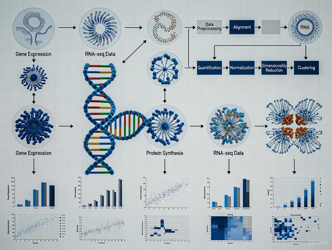
Impact of Pre-processing Pipelines on Cell Identification and Gene Detection
The analysis of single-cell RNA-sequencing (scRNA-seq) data is a multi-step process, with the initial pre-processing stage forming the critical foundation for all downstream biological interpretations. This stage encompasses tasks such as quality control (QC), empty droplet detection, normalization, and feature selection. The choice of methods at this juncture significantly influences the ability to accurately identify cell populations and detect genes that define cellular identity and state [23] [24] [25]. While numerous benchmarking studies have evaluated downstream tasks like clustering and differential expression, the impact of the initial pre-processing pipeline has received less systematic attention. This guide synthesizes current benchmarking research to objectively compare pre-processing methodologies, providing experimental data and protocols to inform pipeline selection for researchers and drug development professionals.
Critical Pre-processing Steps and Their Impact on Downstream Analysis
The scRNA-seq pre-processing workflow involves several interdependent steps, each addressing specific technical artifacts. The following diagram illustrates the logical sequence of these steps and their potential impacts on the final analysis.
The choices made at each step of the pre-processing pipeline can introduce distinct analytical artifacts. For instance, inappropriate normalization can fail to stabilize variance across the gene expression dynamic range, while overzealous quality control can remove rare but biologically critical cell populations [23] [26] [27]. The following sections provide a detailed, evidence-based comparison of methods for each step.
Benchmarking Quality Control and Empty Droplet Detection
Experimental Protocols for QC Assessment
Benchmarking QC pipelines requires datasets with known cell population labels or spike-in controls to establish ground truth. A standard protocol involves:
- Data Acquisition: Utilize datasets with validated cell type labels, such as the human pancreas dataset (CEL-Seq2, SMART-seq2) or the scmixology cell line mixture [13] [25].
- Pipeline Application: Apply different QC workflows (e.g., SCTK-QC, Scanpy, Seurat) to the same raw data. The SCTK-QC pipeline, for example, integrates multiple algorithms:
barcodeRanksandEmptyDropsfrom theDropletUtilspackage for empty droplet detection;scdsandScrubletfor doublet detection; anddecontXfor ambient RNA estimation [23]. - Metric Calculation: Calculate clustering accuracy metrics after downstream analysis (e.g., Adjusted Rand Index (ARI), Normalized Mutual Information (NMI), and cell type annotation accuracy) to compare how well each QC pipeline preserves biological signal while removing technical noise [13] [28].
- Overcorrection Evaluation: Use negative controls where batches are randomly assigned to measure the pipeline's tendency to introduce artifacts in the absence of true batch effects [29].
Performance Comparison of QC Metrics
Systematic benchmarking reveals that the choice of QC metrics significantly impacts the fairness of method evaluation. The following table summarizes key metrics and their properties, as identified in recent benchmarking studies.
Table 1: Performance Metrics for Evaluating Quality Control and Batch Correction
| Metric Category | Metric Name | What It Measures | Performance Insights |
|---|---|---|---|
| Batch Correction | RBET (Reference-informed Batch Effect Testing) | Batch effect removal using stable reference genes | More sensitive to overcorrection and robust to large batch effect sizes compared to other metrics [13]. |
| LISI (Local Inverse Simpson's Index) | Local batch mixing and cell type separation | Can lose discrimination power with large batch effects; may not detect overcorrection [13]. | |
| kBET (k-Nearest Neighbour Batch Effect Test) | Overall batch mixing in k-nearest neighbour graph | Prone to loss of type I error control; variation collapses with large batch effects [13] [15]. | |
| Biological Conservation | ARI (Adjusted Rand Index) / NMI (Normalized Mutual Information) | Similarity between clustering results and known cell type labels | Highly correlated metrics; selecting a subset is sufficient for benchmarking [15]. |
| cLISI (Cell-type LISI) | Separation of known cell types | A value of 1 indicates perfect separation of cell types [15]. | |
| Graph Connectivity | Whether cells of the same type form a connected graph | Measures preservation of continuous biological trajectories [15]. | |
| Cluster Purity | Silhouette Coefficient (SIL) | How similar a cell is to its own cluster vs. other clusters | Requires correction for dependency on the number of clusters [28]. |
| Calinski-Harabasz Index (CH) | Ratio of between-cluster to within-cluster dispersion | Also requires correction for number of clusters [28]. |
Benchmarking Normalization and Transformation Methods
Experimental Protocols for Normalization Benchmarking
To evaluate normalization methods, benchmarking studies typically employ a standardized workflow:
- Dataset Curation: Select datasets with strong ground truth (e.g., cell lines or FACS-sorted populations) and include a dilution series or datasets with varying library sizes to test robustness [26] [25].
- Method Application: Apply a range of normalization methods, from simple global scaling (e.g., log(CPM)) to more sophisticated approaches like Pearson residuals (
sctransform) or latent expression inference (Sanity, Dino) [26]. - Downstream Analysis: Perform standard dimensionality reduction (PCA, UMAP) and clustering on the normalized data.
- Performance Quantification: Assess performance using metrics that evaluate the removal of technical artifacts (e.g., correlation between principal components and batch) and the preservation of biological variation (e.g., cluster purity metrics from Table 1) [26] [28].
Performance Comparison of Normalization Methods
A comprehensive comparison of transformation methods for scRNA-seq data revealed that their performance is context-dependent, with simple methods often rivaling more complex ones.
Table 2: Comparison of scRNA-seq Normalization and Transformation Methods
| Method Class | Example Methods | Key Principle | Impact on Cell ID & Gene Detection |
|---|---|---|---|
| Delta Method | Log(CPM), acosh |
Applies a non-linear function to stabilize variance. | Simple and effective, but performance depends heavily on the chosen pseudo-count. Log(CPM) can fail to mix cells with different size factors [26]. |
| Residuals-based | Pearson Residuals (sctransform) |
Fits a GLM and uses Pearson residuals for normalization. | Better handles the mean-variance relationship and can more effectively mix cells with different size factors compared to delta methods [26]. |
| Latent Expression | Sanity, Dino, Normalisr | Infers a latent "true" expression state from the observed counts. | Has appealing theoretical properties, but in benchmarks, does not consistently outperform simpler approaches [26]. |
| Factor Analysis | GLM-PCA, NewWave | Directly models counts using a factor analysis framework. | A powerful alternative to transformations, directly producing a low-dimensional representation for downstream analysis [26]. |
The Scientist's Toolkit: Essential Research Reagents and Software
This section details key computational tools and resources used in benchmarking scRNA-seq pre-processing pipelines, which are essential for reproducing and extending the findings discussed in this guide.
Table 3: Key Research Reagents and Computational Tools for scRNA-seq Pre-processing
| Tool or Resource Name | Type | Primary Function in Pre-processing |
|---|---|---|
| SCTK-QC Pipeline | R/Python Software | An integrated pipeline for comprehensive QC, including empty droplet detection, doublet prediction, and ambient RNA estimation [23]. |
| Scanpy | Python Toolkit | A widely used Python-based toolkit for single-cell analysis that provides standard QC, normalization, and clustering workflows [27]. |
| Seurat | R Toolkit | A comprehensive R package for single-cell genomics, offering a full suite of pre-processing and analysis functions [30]. |
| scRNA-seq Benchmarking Datasets | Data Resource | Publicly available datasets with known ground truth (e.g., cell line mixtures, annotated pancreas data) essential for validating pipelines [13] [25]. |
| Harmony | R/Python Software | A high-performing batch integration tool that corrects for batch effects without severely altering the underlying data structure, as recommended in benchmarks [29]. |
| scIB | R/Python Software | A curated set of benchmarking metrics and tools for evaluating data integration, including LISI and other metrics [15]. |
End-to-End Pre-processing Workflow
The following diagram synthesizes the key steps, common tool choices, and critical decision points in a standard scRNA-seq pre-processing workflow, based on the aggregated benchmarking evidence.
The collective evidence from benchmarking studies indicates that the pre-processing pipeline has a non-negligible impact on cell identification and gene detection, though this impact can be context-dependent.
- Pre-processing vs. Downstream Analysis: One major benchmarking study of 10 pre-processing workflows found that while quantification properties varied, the choice of pre-processing method was generally less influential on final clustering results than the choice of downstream normalization and clustering methods [25]. This suggests that a well-validated downstream analysis pipeline can be robust to moderate variations in pre-processing.
- Interaction Between Steps: The effect of a pre-processing step can be modulated by other steps in the pipeline. For example, feature selection has been shown to significantly affect the performance of data integration and query mapping, with Highly Variable Gene (HVG) selection generally producing high-quality integrations [15].
- Risk of Overcorrection: A critical finding across multiple studies is the risk of overcorrection, where batch effect correction or normalization methods erase genuine biological variation. Metrics like RBET have been developed specifically to detect this issue, which can lead to false biological discoveries [13] [29].
- No One-Size-Fits-All Solution: No single pipeline performs best across all datasets and biological questions. The optimal choice depends on dataset-specific characteristics, such as the number of cells, batch effect size, and the complexity of the biological system [28]. Consequently, a rigorous, metric-driven evaluation of the pre-processing pipeline is recommended for every new study.
In single-cell RNA sequencing (scRNA-seq), the strategic allocation of a finite sequencing budget presents a fundamental experimental design challenge: should one sequence fewer cells more deeply or more cells at a shallower depth? [31] The resolution of this trade-off directly influences the accuracy of gene expression estimation, the ability to resolve rare cell populations, and the power to detect subtle transcriptional variations. The concepts of sequencing depth (number of reads per cell) and library complexity (the diversity of represented transcripts) are intrinsically linked to the phenomenon of saturation—the point at which additional sequencing yields diminishing returns in transcript detection [32]. Within the broader context of benchmarking scRNA-seq analysis pipelines [17], understanding how these parameters interact across different technological platforms is essential for designing biologically informative experiments, optimizing costs, and ensuring that downstream analytical pipelines operate on high-quality data. This guide objectively compares performance across major scRNA-seq platforms, providing the experimental data and frameworks needed to make evidence-based decisions.
Key Concepts and Experimental Designs for Saturation Analysis
Foundational Concepts and the Sequencing Budget
- Sequencing Depth: Defined as the number of reads allocated per cell. Deeper sequencing reduces technical noise for more accurate estimation of a cell's true transcriptional state [31].
- Library Complexity: Refers to the number of unique transcripts detected in a sample. Higher complexity libraries provide a more complete picture of the transcriptome [33].
- Saturation: In scRNA-seq, this describes the point at which additional sequencing reads fail to detect a significant number of new genes or unique transcripts. It is a key metric for determining the optimal stopping point for sequencing [32].
- The Sequencing Budget Constraint: The core trade-off is framed by the fixed total sequencing budget, expressed as ( B = n{\text{cells}} \times n{\text{reads}} ), where ( B ) is the total number of reads, ( n{\text{cells}} ) is the number of cells, and ( n{\text{reads}} ) is the mean read depth per cell [31]. Allocating this budget effectively is the central problem of experimental design.
Mathematical Frameworks for Determining Optimal Depth
A mathematical framework for scRNA-seq posits that for estimating fundamental gene properties, the optimal allocation is to sequence at a depth of around one read per cell per gene [31]. This framework uses a hierarchical model:
- The true gene expression of a cell, ( \mathbf{X}c ), is a sample from a biological distribution ( P{\mathbf{X}} ).
- The observed read counts, ( \mathbf{Y}c ), are generated through Poisson sampling from ( \mathbf{X}c ), given a sequencing depth ( n_{\text{reads}} ).
The optimal estimator derived from this model is not the standard plug-in estimator but one developed via empirical Bayes, suggesting that significantly shallower sequencing than traditionally practiced may be sufficient for many tasks [31]. For instance, an analysis of a 10x Genomics pbmc_4k dataset suggested that the optimal trade-off would have been achieved by sequencing 10 times shallower with 10 times more cells, potentially reducing the estimation error by half [31].
Table 1: Key Metrics for Saturation Analysis
| Metric | Description | Application in Saturation Analysis |
|---|---|---|
| Sequencing Saturation | The fraction of reads that originate from an already-observed unique molecular identifier (UMI). | A high saturation value (>80-90%) often indicates that additional sequencing will yield few new transcripts. |
| Gene Detection Saturation Curve | A plot of the number of genes detected per cell as a function of sequencing depth. | Used to identify the point where the curve plateaus, indicating optimal depth for gene discovery. |
| Cells vs. Reads Trade-off | The analytical framework for balancing ( n{\text{cells}} ) and ( n{\text{reads}} ) under a fixed budget ( B ) [31]. | Determines the allocation that minimizes the estimation error for a target gene property. |
| Jaccard Index (JI) | A statistic measuring the similarity between enhancer calls or cell type identifications from different datasets [34]. | Assesses the consistency of biological discovery as a function of sequencing depth and platform. |
Experimental Designs for Benchmarking
Benchmarking studies rely on controlled experimental designs to disentangle technical effects from biological signals.
- Mixture Control Experiments: These involve creating 'pseudo cells' from admixtures of cells or RNA from distinct cancer cell lines. The known composition provides a ground truth for evaluating how well analysis pipelines recover expected proportions and expression profiles [17]. For example, one study generated 14 datasets using both droplet and plate-based protocols from up to five cell lines to compare 3,913 method combinations [17] [35].
- Physical Resampling for Validation: Techniques like transcriptome resampling can physically recover targeted cDNA subsets from scRNA-seq libraries for deeper re-sequencing [33]. This allows direct validation of whether low-depth missing transcripts are detectable with higher depth, as demonstrated by increasing the median genes detected per megakaryocyte from 1,313 to 2,002 [33].
- Cross-Platform Consistency Checks: Systematic evaluation of different platforms (e.g., droplet-based, plate-based, combinatorial barcoding) on the same cell line reveals substantial inconsistencies in outputs like enhancer calls, which can be mitigated through uniform data processing pipelines [34].
Comparative Performance Across Platforms and Protocols
The optimal sequencing depth and the resulting library complexity are highly dependent on the scRNA-seq platform and library preparation method.
Table 2: Platform Comparison and Recommended Sequencing Depth
| Platform / Technology | Typical Recommended Reads/Cell | Key Strengths | Saturation Characteristics & Evidence |
|---|---|---|---|
| Droplet-Based (e.g., 10x Genomics) | 20,000 - 50,000 reads [32] | High cell throughput, commercial standardization. | A mathematical analysis suggests that for specific gene estimation, optimal depth may be much shallower (~1 UMI/cell/gene), favoring more cells over deeper sequencing [31]. |
| Combinatorial Barcoding (e.g., Parse Biosciences) | Flexible; determined via sub-sampling [32] | Low multiplet rate, flexible scaling, no specialized equipment. | A key advantage is the ability to use one sublibrary to empirically determine the saturation point, then apply this optimal depth to all other sublibraries, ensuring cost-effectiveness [32]. |
| Plate-Based (Smart-seq2) | >1,000,000 reads | High sensitivity for gene detection, full-length transcript coverage. | Designed for deep sequencing to maximize library complexity from individual cells. Saturation of isoform detection may require extreme depths, as seen in ultra-deep RNA-seq studies [36]. |
| Ultra-High-Depth RNA-seq (Bulk) | Up to 1 billion reads [36] | Detection of very low-abundance transcripts and rare splicing events. | In Mendelian disease diagnostics, gene detection nears saturation at ~1 billion reads, but isoform detection continues to improve with further depth, revealing pathologies invisible at 50 million reads [36]. |
The relationship between experimental goals, platform choice, and data quality is structured as follows:
Figure 1: Logical workflow for designing a sequencing experiment. The experimental goal drives the choice of platform, which in turn influences key sequencing design parameters like depth. The resulting data quality and saturation metrics should inform future experimental designs.
Detailed Experimental Protocols for Saturation Analysis
Protocol for Saturation Analysis Using Combinatorial Barcoding
Purpose: To empirically determine the optimal sequencing depth for a given sample using combinatorial barcoding technology. Steps:
- Library Preparation: Prepare the scRNA-seq library according to the combinatorial barcoding protocol (e.g., fixed and permeabilized cells undergo multiple rounds of barcoding in 96-well plates) [32].
- Sub-sampling: Take one or more completed sublibraries for a sequencing depth test [32].
- Sequencing: Sequence the test sublibrary(s) to a high depth.
- Bioinformatic Analysis: Use the provider's software or custom pipelines (e.g.,
CellBench[17]) to generate saturation curves.- Downsample the sequencing data to various fractions (e.g., 10%, 25%, 50% of reads).
- For each downsampled dataset, count the number of genes detected per cell and the total transcripts (UMIs) detected.
- Determine Optimal Depth: Identify the point on the saturation curve where the rate of new gene discovery sharply declines. This is the cost-effective optimal depth [32].
- Sequence Remaining Libraries: Apply the determined optimal depth to sequence all remaining sublibraries.
Protocol for Benchmarking Pipeline Performance with Mixture Controls
Purpose: To evaluate how different analysis pipelines (normalization, clustering, etc.) perform under varying sequencing depths using a known ground truth. Steps:
- Generate Control Data: Create a benchmark dataset, such as a mixture of single cells and 'pseudo cells' from distinct cell lines. Generate data using multiple scRNA-seq protocols (e.g., droplet and plate-based) [17].
- Variable Depth Simulation: Use the raw data (available under GEO SuperSeries GSE118767 [17]) and computationally subsample it to simulate different sequencing depths (e.g., from 10,000 to 100,000 reads per cell).
- Run Multiple Pipelines: Analyze each depth-simulated dataset with a wide array of analysis pipelines. The landmark study compared 3,913 method combinations for tasks like normalization, imputation, clustering, and trajectory analysis [17].
- Evaluate Performance: Compare the pipeline outputs against the known ground truth. Metrics include:
- Clustering Accuracy: Using Adjusted Rand Index (ARI) to measure concordance with known cell line identities [17].
- Differential Expression Power: The ability to recover known differentially expressed genes between cell lines.
- Trajectory Inference Accuracy: How well the inferred trajectory matches the known lineage relationships in the mixture.
The Scientist's Toolkit: Essential Reagents and Computational Tools
Table 3: Key Research Reagent Solutions and Computational Tools
| Item Name | Type | Function in Saturation Analysis | |
|---|---|---|---|
| CellBench [17] | R/Bioconductor Package | Provides data and framework for benchmarking scRNA-seq analysis methods, enabling direct comparison of how pipelines perform at different depths. | |
| CellRanger (10x Genomics) | Commercial Pipeline | Processes FASTQ files from droplet-based platforms into count matrices, and includes metrics like "Sequencing Saturation" in its summary. | |
| Split-pipe [37] | Computational Pipeline | An example of a commercial provider's pipeline (Parse Biosciences) for processing FASTQ files from combinatorial barcoding data, generating initial count matrices for QC. | |
| STAR [37] | Open-Source Aligner | A widely used spliced transcript aligner for reference genomes, a critical step in generating count data from FASTQ files. | |
| Kallisto | Bustools [37] | Open-Source Pseudoaligner | A fast, lightweight alternative for transcriptome alignment and count matrix generation, useful for large-scale studies. |
| FastQC [32] | Quality Control Tool | Assesses raw sequencing data quality from FASTQ files, a prerequisite for any meaningful saturation analysis. | |
| SC3 [17] | Clustering Tool | A consensus clustering method for single-cell data; its performance can be benchmarked at different depths using mixture controls. | |
| Slingshot [17] | Trajectory Analysis Tool | Infers cell lineages and pseudotime; its accuracy can be evaluated on benchmark datasets with known trajectories at varying depths. | |
| Unique Molecular Identifiers (UMIs) [38] | Molecular Barcode | Short random sequences added to each mRNA molecule during library prep to correct for PCR amplification bias and allow accurate transcript counting. | |
| Phi-X Control [32] | Sequencing Control | Spiked into Illumina sequencing runs to increase base diversity, which is crucial for maintaining sequencing quality on low-diversity libraries. |
The pursuit of optimal sequencing depth is not a quest for a single universal number, but rather a strategic balance dictated by the biological question, the chosen technology, and the constraints of the sequencing budget. Evidence from rigorous benchmarking studies demonstrates that shallow sequencing around one read per cell per gene can be optimal for estimating many gene properties, favoring the sequencing of more cells [31]. However, for applications requiring the detection of low-abundance transcripts or rare splicing events, significantly deeper sequencing remains necessary [36].
Platform choice directly influences this calculus. While droplet-based systems offer standardized workflows, combinatorial barcoding technologies provide a unique empirical method to determine sample-specific saturation, potentially leading to significant cost savings [32]. Ultimately, effective scRNA-seq experimental design requires researchers to clearly define their biological objectives, understand the performance characteristics of their chosen platform, and leverage the growing body of benchmarking data and mathematical frameworks to allocate their sequencing budget in a way that maximizes the discovery potential of their research.
Method Selection in Practice: Normalization, Batch Correction, and Differential Expression
In the analysis of high-throughput sequencing data, such as single-cell RNA-sequencing (scRNA-seq) and Chromatin Immunoprecipitation sequencing (ChIP-seq), normalization is a critical preprocessing step that accounts for technical variability to enable accurate biological comparisons. The performance of normalization methods is highly dependent on whether key technical assumptions about the data are met. Recent research has highlighted that the symmetry of differentially expressed (DE) features—whether the number of up-regulated and down-regulated features is approximately balanced—is a crucial factor influencing normalization efficacy [39] [40]. This comparative guide examines the performance of various normalization methods under symmetric versus asymmetric differential expression (DE) setups, providing researchers with evidence-based recommendations for selecting appropriate methods based on their experimental conditions.
The fundamental challenge in normalization stems from the compositional nature of sequencing data, where an increase in one transcript's abundance can technically lead to decreases in others due to library size constraints [41]. This property means that normalization methods relying on different statistical assumptions will perform variably when the underlying data characteristics match or violate these assumptions. Understanding these relationships is essential for accurate differential expression analysis, clustering, and trajectory inference in scRNA-seq studies [24] [41].
Conceptual Framework: Technical Conditions and Assumptions
Normalization methods operate based on specific technical assumptions about the data structure. When these assumptions are violated, normalization performance deteriorates, leading to increased false discovery rates or reduced power to detect true biological signals [39] [40]. Three key technical conditions have been identified as particularly important for normalization methods:
- Balanced Differential Signal: The number of genomic features with increased expression (or binding) between conditions is approximately equal to the number with decreased expression [39] [40]. This is also referred to as "symmetric differential DNA occupancy" in ChIP-seq contexts [40].
- Equal Total Signal: The total amount of signal (e.g., total RNA expression or DNA binding) remains constant across experimental states [39].
- Equal Background Signal: The level of non-specific background signal is consistent across samples and experimental conditions [40].
The balanced differential signal condition is particularly crucial for many widely-used normalization methods. Methods such as Library Size normalization, Trimmed Mean of M-values (TMM), and Relative Log Expression (RLE) assume that most features are not differentially expressed and that up- and down-regulation are approximately balanced [39] [40]. When this symmetry assumption is violated—such as in experiments with strong transcriptional activation or repression—these methods may produce biased results.
Table 1: Technical Conditions Underlying Major Normalization Methods
| Normalization Method | Balanced DE Assumption | Equal Total Signal Assumption | Primary Application |
|---|---|---|---|
| Library Size/TC | Required | Required | scRNA-seq, ChIP-seq |
| TMM | Required | Not Required | scRNA-seq, ChIP-seq |
| RLE | Required | Not Required | scRNA-seq, ChIP-seq |
| Med-pgQ2/UQ-pgQ2 | Less Stringent | Not Required | RNA-seq (low expression) |
| SCTransform | Less Stringent | Not Required | scRNA-seq |
| CoDA-CLR | Not Required | Required (compositional) | scRNA-seq |
Normalization Methods: Categories and Mechanisms
Global Scaling Methods
Global scaling methods apply a single scaling factor to all features in a sample to adjust for technical variations in sequencing depth or library size. These include:
- Total Count (TC): Normalizes by the total number of reads or counts per sample [42] [43].
- Reads Per Million (RPM): Similar to TC but scales to a fixed number of reads [42].
- Trimmed Mean of M-values (TMM): Removes extreme log fold-changes and library sizes before calculating scaling factors, assuming most genes are not differentially expressed [42] [43].
- Relative Log Expression (RLE): Uses a pseudo-reference sample based on geometric means of gene counts across samples [40].
These methods are computationally efficient but sensitive to violations of the balanced DE assumption, particularly when large-scale differential expression exists between conditions [39] [40].
Per-Gene Normalization Methods
Per-gene normalization approaches apply different normalization factors to individual genes based on their expression characteristics:
- Med-pgQ2 and UQ-pgQ2: These methods perform per-gene normalization after per-sample median or upper-quartile global scaling, making them more robust for data skewed towards lowly expressed genes with high variation [43].
- SCTransform: Applies regularized negative binomial regression to normalize data and is particularly effective for handling technical noise in scRNA-seq data [41].
These methods demonstrate advantages when analyzing datasets with asymmetric differential expression or low-expression skewness, maintaining better specificity while controlling false discovery rates [43].
Compositional Data Analysis (CoDA) Methods
Compositional data analysis methods explicitly treat sequencing data as relative abundances rather than absolute counts:
Centered Log-Ratio (CLR) Transformation: Transforms data using the logarithm of the ratio between each component and the geometric mean of all components [41]. The CLR transformation for a gene i in cell j is calculated as:
(CLR(x{ij}) = \log \left( \frac{x{ij}}{g(\mathbf{x}_j)} \right))
where (g(\mathbf{x}_j)) represents the geometric mean of all gene counts in cell j.
CoDA methods inherently address the compositional nature of sequencing data and can be more robust to asymmetric differential expression patterns, though they require careful handling of zeros in the data [41].
Figure 1: A decision framework for selecting normalization methods based on differential expression symmetry. Methods should be chosen based on whether the data exhibits symmetric or asymmetric differential expression patterns.
Performance Benchmarking in Symmetric vs. Asymmetric Setups
Simulation Studies
Simulation studies where ground truth is known provide the most reliable assessment of normalization method performance. In systematically designed simulations, researchers can control the symmetry of differential expression and directly measure false discovery rates (FDR) and statistical power.
In ChIP-seq simulations where technical conditions were violated, normalization methods showed markedly different performances [39] [40]. When the symmetric differential DNA occupancy assumption was violated, methods like Library Size normalization, TMM, and RLE demonstrated elevated false discovery rates in downstream differential binding analysis. Under these asymmetric conditions, the high-confidence peakset approach—taking the intersection of peaks identified by multiple normalization methods—proved more robust than relying on any single method [39].
For scRNA-seq data, the scone framework provides a comprehensive evaluation approach using multiple data-driven metrics to assess normalization performance [42]. This framework evaluates normalization methods based on their ability to remove unwanted technical variation while preserving biological signal, with performance assessment including:
- Clustering accuracy using silhouette widths
- Batch effect removal using K-nearest neighbor batch-effect test
- Preservation of biological variation using highly variable genes detection [24]
Table 2: Performance Comparison of Normalization Methods Under Different DE Setups
| Normalization Method | Symmetric DE Setup | Asymmetric DE Setup | Key Strengths | Notable Limitations |
|---|---|---|---|---|
| TMM | High accuracy, Controlled FDR | Elevated FDR, Bias | Computational efficiency, Established use | Sensitive to DE symmetry |
| RLE | High accuracy, Controlled FDR | Elevated FDR, Bias | Robust for small sample sizes | Sensitive to DE symmetry |
| Med-pgQ2/UQ-pgQ2 | Moderate accuracy | Maintained specificity, Controlled FDR | Handles low-expression skewness | Less established in scRNA-seq |
| SCTransform | High accuracy | Maintained accuracy, Controlled FDR | Handles technical noise, Zero inflation | Computational intensity |
| CoDA-CLR | Moderate accuracy | High accuracy, Robust performance | Compositional nature, Cluster separation | Zero-handling challenges |
Experimental Validations
Experimental benchmarks using mixture control experiments—where cells or RNA from different cell lines are mixed in known proportions—provide validation for simulation findings. In one comprehensive benchmark involving 14 datasets and 3,913 analysis pipelines, normalization methods performed variably depending on the data structure and technology platform [16].
For symmetric differential expression setups, global scaling methods like TMM and RLE performed well when the balanced DE assumption held. However, under asymmetric conditions—such as when one condition had widespread transcriptional activation—these methods introduced systematic biases, while per-gene methods and CoDA approaches maintained better performance [43] [41].
In the scRNA-seq context, the OSCA and scrapper pipelines achieved the highest clustering accuracy (ARI up to 0.97) in datasets with known cell identities when appropriate normalization was applied [5]. Performance differences were largely driven by the choice of highly variable genes and PCA implementation, both of which are influenced by prior normalization steps [5].
Experimental Protocols for Performance Assessment
Benchmarking Framework Implementation
Comprehensive benchmarking of normalization methods should follow established protocols to ensure reproducible and biologically meaningful assessments:
Data Preprocessing and Quality Control
- Perform initial QC assessment including alignment rates, count distributions, and detection of technical artifacts [42]
- Filter cells and genes based on quality metrics (mitochondrial content, library size, gene detection) [42] [24]
- The scone framework provides standardized approaches for these initial steps [42]
Normalization Implementation
- Apply multiple normalization methods including global scaling (TMM, RLE), per-gene approaches (Med-pgQ2, SCTransform), and compositional methods (CoDA-CLR)
- For each method, use recommended parameter settings and address method-specific requirements (e.g., zero handling for CoDA) [41]
Performance Metric Calculation
- Evaluate clustering accuracy using adjusted Rand index (ARI) when ground truth cell labels are available [5] [16]
- Assess batch effect correction using the K-nearest neighbor batch-effect test [24]
- Measure detection of highly variable genes using established benchmarks [24]
- For differential expression analysis, calculate precision-recall curves and false discovery rates when spike-ins or synthetic datasets are available [43] [16]
Figure 2: Workflow for systematic benchmarking of normalization methods. The process begins with quality control, applies multiple normalization approaches, performs downstream analyses, and assesses performance using multiple metrics before making method recommendations.
High-Confidence Peakset Strategy
For analyses where the appropriate normalization method is uncertain, a high-confidence peakset (or gene set) strategy can be employed:
- Conduct parallel analyses using multiple normalization methods with different technical assumptions [39] [40]
- Identify differentially expressed genes or bound peaks for each normalization method
- Take the intersection of these gene/peak sets as a high-confidence set for biological interpretation [39]
- In experimental analyses, roughly half of called peaks were identified as differentially bound across all normalization methods, providing a robust set for downstream investigation [39]
This approach reduces sensitivity to the choice of a specific normalization method and provides more robust biological conclusions when there is uncertainty about which technical conditions are satisfied.
Table 3: Key Computational Tools for Normalization Benchmarking
| Tool/Resource | Primary Function | Application Context | Key Features |
|---|---|---|---|
| SCONE [42] | Normalization implementation and evaluation | scRNA-seq | Comprehensive metric panel, Ranking methods, Modular framework |
| CellBench [16] | Pipeline benchmarking | scRNA-seq | Mixture control data, Multi-method comparison, Reproducible workflows |
| CoDAhd [41] | Compositional data normalization | scRNA-seq | CLR transformation, Zero-handling, High-dimensional data |
| SCTransform [41] | Regularized negative binomial regression | scRNA-seq | Handles technical noise, Addresses zero inflation |
| Seurat [41] | Integrated scRNA-seq analysis | scRNA-seq | Log-normalization, SCTransform implementation, Clustering |
| OSCA [44] [5] | Single-cell analysis workflow | scRNA-seq | Quality control, Normalization, Clustering, Bioconductor-based |
Based on comprehensive benchmarking studies, we provide the following evidence-based recommendations for selecting normalization methods:
For symmetric DE setups where most genes are not differentially expressed and up-/down-regulation is balanced, global scaling methods (TMM, RLE) provide excellent performance with computational efficiency [39] [43].
For asymmetric DE setups with widespread transcriptional changes, per-gene normalization methods (Med-pgQ2, UQ-pgQ2) and model-based approaches (SCTransform) demonstrate superior performance by maintaining specificity while controlling false discovery rates [43] [41].
For exploratory analyses or when the DE structure is unknown, compositional data analysis methods (CoDA-CLR) offer robustness to various data structures and should be included in method comparisons [41].
When uncertainty exists about which technical conditions are satisfied, employing a high-confidence set approach that intersects results from multiple normalization methods provides the most robust biological conclusions [39].
For large-scale datasets, consider computational efficiency and scalability, with GPU-accelerated implementations providing up to 15× speed-ups over CPU-based methods [5].
Normalization method selection should be guided by both the technical conditions of the experiment and the biological context. Researchers should assess the likely symmetry of differential expression based on their experimental system and employ benchmarking frameworks like scone to evaluate multiple normalization approaches when analyzing novel datasets [42]. As scRNA-seq technologies continue to evolve and dataset sizes increase, ongoing methodology development and benchmarking will remain essential for ensuring accurate biological interpretation of sequencing data.
Batch effects, defined as unwanted technical variations introduced by differences in laboratories, experimental protocols, sequencing platforms, or reagent batches, present a significant challenge in the analysis of single-cell RNA sequencing (scRNA-seq) data [45]. These non-biological variations can obscure genuine biological signals, reduce statistical power, and potentially lead to misleading scientific conclusions if not properly addressed [45]. The proliferation of large-scale scRNA-seq studies, often combining datasets from multiple sources, has intensified the need for effective batch-effect correction algorithms (BECAs) to ensure data integration reliability and analytical reproducibility.
The fundamental challenge in batch-effect correction lies in distinguishing technical artifacts from true biological variation, particularly when batch effects are confounded with biological factors of interest [46]. This challenge is especially pronounced in scRNA-seq data due to its unique characteristics, including high dimensionality, sparsity, dropout events, and substantial technical noise [8]. As the field continues to generate increasingly massive datasets, the computational efficiency of correction methods becomes equally important as their statistical efficacy.
This benchmark review evaluates seven prominent batch-effect correction methods, assessing their performance across multiple metrics including computational efficiency, batch mixing quality, biological conservation, and scalability. By providing a structured comparison of these algorithms, we aim to offer researchers in genomics and drug development a evidence-based framework for selecting appropriate batch correction strategies in scRNA-seq analysis pipelines.
Table 1: Summary of Seven Batch-Effect Correction Algorithms
| Algorithm | Underlying Methodology | Key Features | Input Data Requirements | Output Type |
|---|---|---|---|---|
| ComBat | Empirical Bayes framework | Adjusts for mean and variance shifts; can incorporate covariates | Normalized gene expression matrix | Corrected expression matrix |
| Median Centering | Location adjustment | Centers median expression per feature to zero; simple and fast | Normalized expression values | Median-normalized matrix |
| Ratio | Scaling-based normalization | Uses reference samples for intensity ratio calculation | Raw intensities with reference measurements | Scaled expression matrix |
| RUV-III-C | Linear regression with controls | Utilizes control genes/samples to estimate unwanted variation | Raw data with specified controls | Batch-effect removed data |
| Harmony | Iterative clustering and correction | Maximizes batch diversity within clusters; PCA-based | PCA-reduced dimensions or original features | Integrated low-dimensional embedding |
| WaveICA2.0 | Multi-scale decomposition | Removes batch effects using injection order time trends | Feature intensities with injection order | Corrected expression matrix |
| NormAE | Deep learning (Autoencoder) | Neural network learns to remove non-linear batch effects | Raw data with m/z and RT for MS-based | Batch-effect corrected data |
The seven benchmarked algorithms represent diverse methodological approaches to the batch-effect correction problem. ComBat, originally developed for microarray data, employs an empirical Bayesian framework to model and adjust for location and scale shifts between batches [46] [8]. Its ability to incorporate biological covariates in the design matrix makes it particularly useful for preserving known biological variation during correction.
Harmony implements an iterative clustering approach that projects cells into a shared embedding where cells are grouped by cell type rather than batch of origin [8]. By maximizing the diversity of batches within each cluster, Harmony effectively disentangles technical artifacts from biological signals without requiring explicit batch information for all correction steps.
Deep learning approaches like NormAE utilize autoencoder architectures to learn non-linear batch effects and remove them while preserving biological variation [46]. These methods typically require larger datasets for effective training but can capture complex batch effects that linear methods might miss.
Benchmarking Methodology
Experimental Design and Data Sets
The benchmarking analysis employed multiple experimental scenarios to evaluate algorithm performance under different conditions. Two primary study designs were implemented: balanced (B) and confounded (C) scenarios [46]. In balanced designs, biological groups are equally distributed across batches, allowing clearer separation of biological and technical effects. In confounded designs, batch effects are correlated with biological groups, representing a more challenging but realistic scenario commonly encountered in real-world studies.
The evaluation utilized both simulated and real-world datasets. Simulation approaches generated data with known ground truth, enabling precise quantification of batch correction efficacy and biological conservation [46]. Real-world data from the Quartet multi-omics reference materials provided biologically relevant benchmarking contexts with technical replicates across multiple batches [46]. Additionally, large-scale scRNA-seq datasets encompassing diverse cell types (dendritic cells, pancreatic cells, retinal cells, and PBMCs) across different sequencing technologies (10X, SMART-seq, Drop-seq) were included to assess performance across various biological contexts [8].
Performance Metrics and Evaluation Framework
Table 2: Performance Metrics for Batch-Effect Correction Evaluation
| Metric Category | Specific Metric | Measurement Focus | Ideal Value |
|---|---|---|---|
| Batch Mixing Quality | k-nearest neighbor Batch-Effect Test (kBET) | Local batch label distribution | Lower rejection rate ≈ 0 |
| Local Inverse Simpson's Index (LISI) | Batch diversity in local neighborhoods | Higher score ≈ batch count | |
| Average Silhouette Width (ASW) Batch | Cluster cohesion vs separation by batch | Closer to 0 (no batch structure) | |
| Biological Conservation | Average Silhouette Width (ASW) Label/Cell Type | Cell type separation after correction | Higher score ≈ 1 |
| Adjusted Rand Index (ARI) | Similarity between clustering and known labels | Higher score ≈ 1 | |
| Feature-level Quality | Coefficient of Variation (CV) | Technical variation in replicates | Lower values preferred |
| Matthews Correlation Coefficient (MCC) | Differential expression identification accuracy | Higher score ≈ 1 | |
| Pearson Correlation Coefficient (RC) | Expression pattern correlation with truth | Higher score ≈ 1 | |
| Sample-level Quality | Signal-to-Noise Ratio (SNR) | Biological group differentiation in PCA | Higher values preferred |
| Principal Variance Component Analysis (PVCA) | Variance attribution to biological vs batch factors | Higher biological variance |
Algorithm performance was assessed using multiple complementary metrics spanning four categories: batch mixing quality, biological conservation, feature-level quality, and sample-level quality [46] [8]. This multi-faceted approach ensured comprehensive evaluation of both technical correction efficacy and biological preservation.
Batch mixing quality was primarily evaluated using kBET, which tests whether local neighborhoods show balanced representation from all batches, and LISI, which quantifies the effective number of batches in local neighborhoods [8]. Biological conservation was assessed through cell type purity metrics including ASW for cell labels and ARI, which measures similarity between clustering results and known cell type annotations [8].
For differential expression analysis, Matthews Correlation Coefficient (MCC) and Pearson Correlation Coefficient (RC) were used to evaluate the accuracy in identifying differentially expressed proteins/genes compared to known ground truth in simulated data [46]. Coefficient of variation (CV) across technical replicates provided insight into the reduction of technical noise post-correction.
Experimental Workflow
The benchmarking workflow followed a standardized procedure to ensure fair comparison across methods. Raw data underwent initial quality control and normalization specific to each dataset. Batch-effect correction was then applied using each algorithm with default or recommended parameters. Corrected data were evaluated using the comprehensive metric suite, with computational resources and runtime recorded for efficiency assessment.
Figure 1: Benchmarking workflow for evaluating seven batch-effect correction algorithms. The process begins with raw data collection, followed by quality control and normalization. Algorithms are applied to corrected data, with performance evaluated across multiple metric categories before final ranking.
Performance Results and Comparative Analysis
Batch Correction Efficacy and Biological Preservation
Table 3: Comparative Performance of Batch-Effect Correction Algorithms
| Algorithm | Batch Mixing (kBET ↓) | Biological Conservation (ASW Label ↑) | Runtime Efficiency | Scalability to Large Datasets | Handling of Confounded Designs |
|---|---|---|---|---|---|
| ComBat | Moderate | Moderate | Fast | Good | Moderate |
| Median Centering | Low | Low | Very Fast | Excellent | Poor |
| Ratio | Moderate | Moderate-High | Fast | Good | Good |
| RUV-III-C | High | High | Moderate | Moderate | Good |
| Harmony | High | High | Fast | Excellent | Good |
| WaveICA2.0 | Moderate-High | Moderate | Moderate | Good | Moderate |
| NormAE | High | Moderate | Slow (requires GPU) | Moderate (memory-intensive) | Good |
Across multiple benchmarking scenarios, Harmony consistently demonstrated superior performance in both batch mixing quality and biological conservation [8]. Harmony's iterative clustering approach effectively integrated batches while maintaining clear separation of cell types, achieving excellent scores in both kBET (lower rejection rate) and ASW label (higher values near 1) metrics [8]. Its significantly shorter runtime compared to other high-performing methods made it particularly suitable for large-scale scRNA-seq datasets.
RUV-III-C also performed well, especially in scenarios where reliable control features or samples were available [46]. By explicitly modeling unwanted variation using control genes, RUV-III-C effectively removed batch effects while preserving biological signals, though its performance was dependent on appropriate control selection.
ComBat showed solid performance across multiple metrics, with its empirical Bayes approach providing robust correction for mean and variance shifts between batches [46] [8]. However, in severely confounded designs where batch effects strongly correlated with biological groups, ComBat occasionally removed biological variation along with technical artifacts.
The deep learning approach NormAE demonstrated strong batch mixing capabilities, particularly for complex, non-linear batch effects [46]. However, its computational demands and longer runtimes presented practical challenges for very large datasets, though GPU acceleration could mitigate these issues.
Impact of Data Level and Quantification Methods
Recent benchmarking evidence suggests that the level at which batch-effect correction is applied significantly impacts performance outcomes. In proteomics data, protein-level correction consistently demonstrated superior robustness compared to precursor or peptide-level correction across multiple quantification methods (MaxLFQ, TopPep3, iBAQ) and BECAs [46]. This finding has important implications for scRNA-seq analysis, suggesting that correction at appropriately aggregated levels may yield more reliable results.
The interaction between quantification methods and batch-effect correction algorithms was also evident across studies [46]. Specifically, the MaxLFQ quantification method combined with Ratio-based batch correction demonstrated particularly strong performance in large-scale proteomics datasets, highlighting the importance of considering the complete data processing pipeline rather than BECAs in isolation.
Practical Implementation Guide
Recommended Research Reagent Solutions
Table 4: Essential Research Reagents and Computational Tools for Batch-Effect Correction Studies
| Category | Specific Resource | Function/Purpose | Example Applications |
|---|---|---|---|
| Reference Materials | Quartet multi-omics reference materials | Provides biologically relevant benchmarking with technical replicates | Method validation and performance assessment [46] |
| Quantification Methods | MaxLFQ, TopPep3, iBAQ | Protein quantification from MS data | Assessing BECA performance across different quantification approaches [46] |
| Data Simulation Tools | Splatter package | Generates synthetic scRNA-seq data with known ground truth | Controlled algorithm evaluation [8] |
| Evaluation Frameworks | kBET, LISI, ASW, ARI metrics | Quantifies batch mixing and biological conservation | Comprehensive algorithm assessment [8] |
| Computational Platforms | R/Python environments | Provides implementation of various BECAs | Flexible algorithm application and customization |
Implementation Considerations and Best Practices
Based on comprehensive benchmarking results, we recommend Harmony as a primary choice for most scRNA-seq batch integration tasks due to its balanced performance across correction efficacy, biological conservation, and computational efficiency [8]. For studies with clearly defined control features or samples, RUV-III-C provides a robust alternative, particularly in challenging confounded designs.
When implementing batch-effect correction, researchers should:
Apply correction at appropriate biological aggregation levels - Evidence suggests protein-level correction outperforms earlier-stage correction in proteomics, indicating that correction after appropriate aggregation may be beneficial in scRNA-seq [46].
Validate results using multiple metrics - No single metric captures all aspects of batch correction quality. Employ complementary metrics assessing both batch mixing and biological conservation [8].
Consider the study design context - Algorithm performance varies between balanced and confounded designs. Test multiple methods in pilot studies when biological and technical effects are potentially correlated [46].
Account for computational requirements - For large-scale datasets, runtime and memory requirements become practical constraints. Harmony offers favorable scalability, while deep learning methods like NormAE provide advanced capability at higher computational cost [8].
Figure 2: Decision framework for selecting appropriate batch-effect correction algorithms based on study design, data characteristics, and computational resources.
This comprehensive benchmark of seven batch-effect correction algorithms demonstrates that method selection should be guided by specific study characteristics, including experimental design, data scale, and analytical priorities. Harmony emerges as a robust first choice for most scRNA-seq integration tasks due to its balanced performance profile and computational efficiency [8]. RUV-III-C provides a strong alternative for studies with appropriate controls, particularly in challenging confounded designs [46].
The interaction between batch-effect correction and other data processing steps highlights the importance of considering the complete analytical pipeline rather than algorithms in isolation. Evidence from proteomics suggesting superior performance of protein-level correction indicates that further research is needed to determine optimal correction levels in scRNA-seq data [46].
Future directions in batch-effect correction development should address remaining challenges including improved handling of severely confounded designs, better preservation of subtle biological signals, and enhanced computational efficiency for exponentially growing single-cell datasets. As single-cell technologies continue to evolve, robust batch-effect correction remains essential for ensuring the reliability and reproducibility of integrative genomic analyses.
Accurate gene expression quantification is a cornerstone of modern transcriptomics, influencing everything from basic biological discovery to clinical diagnostics. The evolution of RNA sequencing (RNA-seq) from bulk to single-cell analyses has introduced new dimensions of complexity, making the choice of mapping and quantification strategies more critical than ever. Within the broader context of benchmarking single-cell RNA-seq (scRNA-seq) analysis pipelines, understanding the performance characteristics of these foundational tools is paramount. This guide objectively compares the accuracy of prevalent quantification methods, drawing on experimental data from controlled benchmark studies to provide researchers, scientists, and drug development professionals with evidence-based recommendations for their analytical workflows.
Performance Comparison of Quantification Tools
Bulk RNA-seq Quantification Tools
Early benchmarking studies for bulk RNA-seq laid the groundwork for evaluating quantification accuracy. A seminal study compared four early quantification tools—Cufflinks, IsoEM, HTSeq, and RSEM—using RNA-seq data from the MAQC project with matched TaqMan RT-qPCR measurements as ground truth [47].
Table 1: Performance Comparison of Bulk RNA-seq Quantification Tools
| Tool | Correlation with RT-qPCR (Pearson) | Root-Mean-Square Deviation | Quantification Approach | Normalization |
|---|---|---|---|---|
| HTSeq | 0.89 (Highest) | Greatest deviation | Naive count-based | FPKM |
| RSEM | 0.85-0.89 | Lower deviation | Bayesian estimation | FPKM/TPM |
| Cufflinks | 0.85-0.89 | Lower deviation | Statistical model | FPKM |
| IsoEM | 0.85-0.89 | Lower deviation | Expectation-Maximization | FPKM |
The study revealed a crucial trade-off: while HTSeq exhibited the highest correlation with RT-qPCR measurements (0.89), it also produced the greatest root-mean-square deviation from these same ground truth measurements [47]. This suggests that correlation coefficients alone are insufficient for evaluating tool performance, a point later reinforced by additional research showing that correlation does not adequately measure precision or reproducibility [48].
Later, a more comprehensive benchmark of seven competing pipelines (including STAR, TopHat2, Bowtie2, Cufflinks, eXpress, Flux Capacitor, kallisto, RSEM, Sailfish, and Salmon) introduced more sophisticated assessment metrics focused on differential expression analysis [48]. Using predefined differentially expressed genes from microarray data and simulated datasets, this evaluation found that performance was generally poor across methods, with RSEM slightly outperforming the others [48].
Single-Cell Specific Quantification Challenges
The transition to single-cell RNA-seq introduced additional challenges for accurate quantification, including sparsity (high dropout rates), technical noise, and the need to resolve isoform-level expression in individual cells. A 2025 study introduced SCALPEL, a tool designed for quantifying transcript isoforms from standard 3' scRNA-seq data [49].
Table 2: Benchmarking Isoform Quantification Tools for scRNA-seq
| Tool | Type | Sensitivity | Specificity | Runtime | Memory Efficiency |
|---|---|---|---|---|---|
| SCALPEL | Isoform quantification | Highest | High | Medium | Medium |
| scUTRquant* | Isoform quantification | High | High | Fast (with 3' UTRome) | High (with 3' UTRome) |
| Sierra | Peak-calling | Lower | High | Varies | Varies |
| scAPA | Peak-calling | Lower | High | Varies | Varies |
| scAPAtrap | Peak-calling | Lower | High | Varies | Varies |
| SCAPTURE | Peak-calling | Lower | High | Varies | Varies |
| scDaPars | Peak-calling | Lowest | High | Varies | Varies |
When benchmarked using synthetic data with known ground truth, SCALPEL demonstrated higher sensitivity (correctly identifying 57% of differentially used isoform genes in low-expression quartile) compared to other tools (19-22% for scUTRquant), while maintaining high specificity [49]. The benchmark also revealed clear differences between peak-calling and isoform-based methods, with the latter generally showing higher sensitivity [49].
Experimental Protocols for Benchmarking
Bulk RNA-seq Benchmarking Methodology
The foundational bulk RNA-seq benchmarking study utilized a standardized workflow to ensure fair comparison [47]:
Dataset Preparation: Downloaded single-read Illumina data from NCBI SRA (accession: SRX003926, SRX003927) from the MAQC project, with corresponding TaqMan RT-qPCR measurements (GSE5350). These datasets included mixed human brain samples and mixed human cell lines with technical replicates.
Sequence Alignment: Processed all reads through a consistent alignment step using TopHat (v2.0.6) with Bowtie, aligning approximately 68% of reads to the Ensembl GRCh37 reference genome.
Expression Quantification: Applied each quantification tool (Cufflinks v2.0.2, HTSeq v0.5.3p9, RSEM v1.2.1, IsoEM v1.1.1) to the same alignment outputs with consistent settings. All tools were configured to output FPKM values for comparison.
Performance Evaluation: Compared relative expression estimates (log2 ratio of sample means) between RNA-seq tools and RT-qPCR measurements for 531 commonly expressed genes. Calculated both Pearson correlation coefficients and root-mean-square deviation (RMSD).
Large-Scale Multi-Center Benchmarking
A 2024 study established a more comprehensive benchmarking framework through the Quartet project, involving 45 laboratories that each used their in-house experimental protocols and analysis pipelines [50]. This approach provided real-world performance data across diverse conditions:
Reference Materials: Used Quartet RNA reference materials (derived from B-lymphoblastoid cell lines) with small biological differences and MAQC samples with larger differences. Included ERCC spike-in controls and defined mixture samples with known ratios.
Data Generation: Each laboratory processed 24 RNA samples (including technical replicates) using their standard RNA-seq workflows, generating 1080 libraries totaling ~120 billion reads.
Performance Assessment: Evaluated multiple metrics including:
- Signal-to-noise ratio based on principal component analysis
- Accuracy of absolute and relative expression using Quartet reference datasets
- Performance with ERCC spike-ins and sample mixtures
- Accuracy in detecting differentially expressed genes
Factor Analysis: Systematically investigated 26 experimental processes and 140 bioinformatics pipelines to identify key sources of variation.
The study found significantly greater inter-laboratory variations in detecting subtle differential expression compared to large differences, highlighting the particular challenge of accurately quantifying clinically relevant small expression changes [50].
Single-Cell Isoform Quantification Benchmarking
The SCALPEL evaluation employed synthetic data with known ground truth to overcome the limitations of real datasets [49]:
Data Simulation: Generated synthetic single-cell isoform expression datasets for 6,000 cells across two populations using Splatter (v1.28.0), simulating 6,560 genes and 12,320 isoforms with different dropout rates.
Read Simulation: Developed scr4eam to generate isoform-aware realistic scRNA-seq reads from the synthetic expression matrices.
Tool Comparison: Compared SCALPEL against five peak-calling based tools (Sierra, scAPA, scAPAtrap, SCAPTURE, scDaPars) and one isoform quantification tool (scUTRquant) using both default and common annotations.
Performance Metrics: Assessed sensitivity (number of detected genes/isoforms), specificity, differential isoform usage detection accuracy, and computational efficiency.
Table 3: Key Reagents and Resources for RNA-seq Quantification Studies
| Resource | Type | Function in Benchmarking | Example Sources |
|---|---|---|---|
| Reference Materials | Biological samples | Provide ground truth for expression measurements | Quartet project, MAQC samples [50] |
| ERCC Spike-Ins | Synthetic RNA controls | Enable assessment of technical accuracy | External RNA Controls Consortium [50] |
| Alignment Tools | Software | Map sequencing reads to reference genome | TopHat, STAR, Bowtie2 [47] [48] |
| Quantification Tools | Software | Estimate gene/isoform expression levels | Cufflinks, HTSeq, RSEM, kallisto [47] [48] |
| Synthetic Datasets | Computational | Provide known ground truth for validation | Splatter-simulated data, Polyester [49] [48] |
| Validation Technologies | Experimental | Independent measurement of expression | RT-qPCR, TaqMan assays [47] |
Emerging Trends and Future Directions
The field of RNA-seq quantification continues to evolve with several emerging trends. For single-cell analyses, benchmarking studies have expanded beyond quantification to encompass clustering algorithms, with a 2025 evaluation of 28 methods revealing that scDCC, scAIDE, and FlowSOM perform best for both transcriptomic and proteomic data [51]. The integration of single-cell isoform sequencing with large language models for cell type annotation represents another frontier [52], potentially enabling more precise cellular characterization beyond gene-level expression.
Recent large-scale benchmarking efforts highlight the profound influence of experimental execution on quantification accuracy [50]. Best practice recommendations emphasize that experimental factors including mRNA enrichment and strandedness, combined with each bioinformatics step, emerge as primary sources of variations in gene expression measurements. For clinical applications, quality control at subtle differential expression levels using appropriate reference materials like the Quartet samples is particularly crucial.
As RNA-seq moves toward clinical applications, ensuring reliability and cross-laboratory consistency becomes increasingly important. Future quantification strategies will need to address the unique challenges of detecting subtle differential expressions between disease subtypes or stages while maintaining robustness across diverse experimental and computational environments.
Differential expression (DE) analysis is a cornerstone of transcriptomics, enabling researchers to identify genes whose expression levels change significantly across different biological conditions. With the advent of single-cell RNA sequencing (scRNA-seq), this analysis can now be performed at unprecedented resolution, revealing cellular heterogeneity and nuanced transcriptional responses not detectable in bulk RNA-seq studies [53] [54]. However, this technological advancement introduces new analytical challenges, including handling within-sample correlation, addressing zero-inflation, and distinguishing technical artifacts from biological variation [54] [55].
The field has responded with a proliferation of computational methods specifically designed for scRNA-seq DE analysis. This creates a critical need for comprehensive benchmarking studies that objectively evaluate method performance across diverse cellular scenarios. Such benchmarks provide researchers, scientists, and drug development professionals with evidence-based guidance for selecting appropriate analytical tools, ultimately ensuring the biological validity of their findings [56] [15]. This guide synthesizes current benchmarking evidence to compare the performance of leading DE methods, detailing their operational characteristics, relative strengths, and optimal applications within scRNA-seq pipelines.
Key Methodologies and Statistical Approaches
Differential expression methods for single-cell data employ diverse statistical frameworks to address the unique characteristics of scRNA-seq data. Understanding these foundational approaches is crucial for selecting an appropriate method and interpreting its results.
Pseudobulk Approaches: These methods aggregate cell-level counts to the sample level, creating a single expression profile per biological replicate. This strategy effectively addresses the hierarchical correlation structure inherent in multi-sample scRNA-seq experiments, where cells from the same individual are more similar to each other than to cells from different individuals [53] [55]. Once aggregated, established bulk RNA-seq tools like edgeR and limma can be applied. Benchmarking studies consistently show that pseudobulk approaches provide excellent type I error control and reliability, making them a robust choice for many experimental designs [55].
Model-Based Single-Cell Approaches: In contrast, methods like MAST (Model-based Analysis of Single-cell Transcriptomics) operate directly on the single-cell count matrix. MAST uses a two-component hurdle model, combining a logistic regression component to model the probability of a gene being detected (differential detection) with a Gaussian linear model for the expression level conditional on detection (differential expression) [55]. While powerful for capturing zero-inflation, these methods face computational scalability challenges with large datasets and must carefully account for within-sample correlation to avoid inflated false positive rates.
Emerging Frameworks: Recent innovations extend DE analysis beyond mean expression. Memento employs a method of moments framework with efficient resampling to enable robust differential analysis of not only mean expression but also variability and gene correlation from scRNA-seq data [54]. DiSC extracts multiple distributional characteristics from gene expression data and tests their joint association with variables of interest using a permutation-based framework to control the false discovery rate (FDR) [53]. These approaches can detect more complex patterns of transcriptional regulation than traditional methods focused solely on average expression changes.
Table 1: Foundational Statistical Approaches in scRNA-seq Differential Expression Analysis
| Statistical Approach | Representative Methods | Core Model | Handling of Zeros |
|---|---|---|---|
| Pseudobulk Aggregation | edgeR on pseudobulk, limma-voom on pseudobulk | Negative Binomial GLM, Linear Models | Through aggregation |
| Hurdle Models | MAST | Logistic + Gaussian Regression | Explicit two-part model |
| Distributional Characteristic Testing | DiSC | Omnibus F-statistic with permutation | Incorporated in distributional features |
| Method of Moments | Memento | Moment-based with bootstrap | Integrated in variance decomposition |
Comprehensive Performance Benchmarking
Performance Across Experimental Designs
Rigorous benchmarking studies have evaluated DE methods across diverse experimental scenarios to provide guidance for method selection. Performance varies considerably based on experimental parameters such as sample size, effect size, and the nature of the biological signal.
For studies with very small sample sizes (n < 5 per group), all methods struggle with reliability, and results should be interpreted with caution [56]. As sample sizes increase, methods combining variance-stabilizing transformations with the limma method for differential expression analysis generally perform well across diverse conditions [56]. The nonparametric SAMseq method also demonstrates robust performance with larger sample sizes [56].
In multi-sample, multi-condition scRNA-seq experiments, pseudobulk approaches consistently outperform methods that ignore the hierarchical data structure. A recent benchmark of differential detection (DD) workflows found that pseudobulk aggregation of binarized counts followed by analysis with edgeR (edgeRNBoptim) provided the best balance of type I error control and sensitivity [55]. This approach effectively addresses within-sample correlation while maintaining computational feasibility.
DiSC demonstrates particular advantages in scalability, being approximately 100 times faster than other state-of-the-art distribution-based methods like IDEAS and BSDE while maintaining comparable statistical power and FDR control [53]. This makes it particularly suitable for large-scale atlas-level studies with thousands of cells across many individuals.
Memento has shown capability to identify more significant and reproducible differences in mean expression compared to existing methods while also detecting differences in variability and gene correlation that suggest distinct transcriptional regulation mechanisms [54]. Its efficiency allows application to millions of cells, facilitating discovery in increasingly large scRNA-seq datasets.
Quantitative Performance Comparisons
Table 2: Performance Comparison of scRNA-seq Differential Expression Methods
| Method | Primary Use Case | Sample Size Recommendation | Strengths | Limitations |
|---|---|---|---|---|
| edgeR/limma (Pseudobulk) | Multi-sample group comparisons | > 3 samples/group [55] | Excellent type I error control, handles within-sample correlation | Loss of single-cell resolution |
| MAST | Single-cell level analysis | Larger cell numbers per group [55] | Models zero-inflation, captures detection changes | Computationally intensive, requires random effects for sample correlation |
| DiSC | Large multi-individual studies | Larger sample sizes [53] | High computational efficiency, detects distribution shifts | Requires sufficient cells per individual |
| Memento | Multi-condition perturbation studies | Scalable to millions of cells [54] | Detects changes in mean, variance, and co-expression | Complex implementation |
| SAMseq | Bulk or pseudobulk RNA-seq | Larger sample sizes [56] | Nonparametric, robust to outliers | Less efficient with very small samples |
Experimental Protocols and Workflows
Standardized Benchmarking Framework
To ensure fair and interpretable comparisons between DE methods, benchmarking studies employ standardized evaluation frameworks. These typically involve both simulated data, where the true differential expression status is known, and real datasets with orthogonal validation [56] [57].
For simulation studies, counts are typically generated using Negative Binomial distributions, with mean and dispersion parameters estimated from real data to preserve biological realism [56]. This approach allows precise quantification of false discovery rates (FDR), sensitivity, and specificity under controlled conditions.
Real data benchmarks often utilize datasets with accompanying gold-standard measurements, such as quantitative RT-PCR validated genes [58] or CNV calls validated by whole-genome sequencing [57]. For example, one benchmarking study used 32 genes validated by qRT-PCR across 18 samples from two human cell lines under different treatment conditions to assess the accuracy of 192 different analytical pipelines [58].
Performance metrics commonly include:
- Type I Error Control: The ability to maintain the nominal false positive rate (e.g., α=0.05) when no true differences exist [55]
- Sensitivity and Specificity: The power to detect true differential expression while correctly identifying non-DE genes
- Area Under the Curve (AUC): Threshold-independent measure of classification performance [57]
- Computational Efficiency: Runtime and memory requirements, particularly important for large datasets [53]
Differential Expression Analysis Workflow
The following diagram illustrates a standardized workflow for conducting and benchmarking differential expression analysis in single-cell RNA-seq studies:
Visualization and Interpretation
Effective visualization is crucial for verifying DE analysis results and detecting potential problems not apparent from statistical models alone [59]. Modern RNA-seq analysis should incorporate interactive graphics as an integral component rather than relying solely on model outputs.
Parallel Coordinate Plots are essential for visualizing relationships between variables in multivariate expression data [59]. Each gene is represented as a line connecting its expression values across samples. Ideal datasets show flat connections between replicates but crossed connections between treatment groups, indicating higher between-treatment than between-replicate variability. These plots can reveal cluster-specific expression patterns and identify samples with unexpected behavior that might indicate technical artifacts or batch effects.
Scatterplot Matrices enable rapid assessment of expression distribution across all genes and samples [59]. Each gene is represented as a point in scatterplots comparing all sample pairs. In clean data, scatterplots between replicates should show points tightly clustered along the x=y line, while treatment comparisons should show greater spread. Interactive versions allow investigators to identify outlier genes that may represent genuine differential expression or technical artifacts.
Volcano Plots (combining statistical significance with magnitude of change) and MA Plots (visualizing log-ratio versus average expression) remain standard for summarizing DE results, helping researchers balance fold-change against statistical evidence when selecting genes for follow-up validation.
The R package bigPint provides specialized visualization tools for RNA-seq data that can detect normalization issues, differential expression designation problems, and common analysis errors that might otherwise go unnoticed [59].
The Scientist's Toolkit
Essential Research Reagent Solutions
Table 3: Essential Reagents and Computational Resources for scRNA-seq DE Analysis
| Resource Type | Specific Examples | Function/Purpose |
|---|---|---|
| Library Prep Kits | 10x Genomics Chromium Single Cell Gene Expression, SMART-Seq | Generate barcoded scRNA-seq libraries |
| Validation Reagents | TaqMan qRT-PCR Assays, PrimeTime qPCR Probes | Experimental validation of DE genes |
| Reference Data | Human Cell Atlas, CELLxGENE Discover, Tabula Sapiens | Reference cell atlases for normalization and annotation |
| Computational Resources | High-performance computing clusters, Cloud computing platforms | Handle computationally intensive DE analyses |
Software and Implementation
For implementation, the R/Bioconductor ecosystem provides the most comprehensive set of tools for differential expression analysis. The muscat package implements state-of-the-art pseudobulk approaches for multi-sample multi-condition scRNA-seq data [55]. DiSC is available through the "SingleCellStat" R package on CRAN [53], while Memento provides resources for differential analysis of mean expression, variability, and gene correlation [54].
When implementing these methods, careful attention to data preprocessing is essential. Normalization approaches should be selected based on the specific characteristics of the dataset, with methods like log(1+10^4 * x/column_sum) providing effective library size normalization for single-cell data [60]. Feature selection using highly variable genes (HVGs) generally improves integration performance and downstream analysis quality [15].
Differential expression analysis for scRNA-seq data continues to evolve rapidly, with method performance highly dependent on experimental design and biological context. Pseudobulk approaches currently provide the most robust performance for standard multi-sample comparisons, while emerging methods like DiSC and Memento offer advantages for specific applications including large-scale studies and detection of complex distributional changes.
Researchers should select methods based on their specific experimental design, sample size, and biological questions rather than relying on a single approach for all scenarios. Future method development will likely focus on improving computational efficiency for ever-larger datasets, better integration of multi-omics data, and more sophisticated modeling of complex transcriptional responses to perturbations. Through careful method selection and appropriate validation, differential expression analysis remains a powerful approach for unlocking biological insights from single-cell transcriptomic data.
Single-cell RNA sequencing (scRNA-seq) has revolutionized biological research by enabling high-resolution analysis of cellular heterogeneity in complex biological systems, providing unprecedented insights into diverse areas including tumor microenvironments, developmental pathways, and drug discovery [61]. As the technology has rapidly evolved, an explosion of computational methods and tailored data analysis pipelines has emerged, creating a critical challenge for researchers: selecting the most appropriate, efficient, and accurate workflows for their specific research contexts [16]. The complexity of scRNA-seq data, which is often noisy, high-dimensional, and sparsely populated, further complicates this selection process and underscores the necessity for specialized computational tools [61].
The absence of gold-standard benchmark datasets has historically made systematic comparisons of analytical performance difficult [16]. This gap in standardized evaluation has driven numerous research groups to conduct comprehensive benchmarking studies that rigorously test the scalability, efficiency, and accuracy of various computational frameworks. These benchmarking efforts provide invaluable practical guidance for building effective end-to-end analysis pipelines, ensuring that researchers can transform raw sequencing data into reliable biological insights. This review synthesizes findings from recent large-scale benchmarking studies to offer evidence-based recommendations for pipeline construction, focusing on the performance of specific tools at key analytical stages from preprocessing to biological interpretation.
Performance Benchmarking of Major Analysis Frameworks
Comprehensive Evaluation of Scalability and Accuracy
A large-scale benchmarking study systematically evaluated the performance of five widely used scRNA-seq analysis frameworks—Seurat, OSCA, scrapper, Scanpy, and rapids-singlecell—focusing on their scalability, efficiency, and accuracy when processing datasets of varying sizes [5]. The researchers employed a systematic comparison approach using representative datasets, including a massive 1.3-million-cell mouse brain dataset for scalability assessment and three smaller datasets (BE1, scMixology, and cord blood CITE-seq) with established ground truth labels for evaluating clustering accuracy [5].
Table 1: Overall Performance of Major scRNA-seq Analysis Frameworks
| Framework | Scalability | Clustering Accuracy (ARI) | Speed | Memory Efficiency | Best Use Cases |
|---|---|---|---|---|---|
| rapids-singlecell | Excellent | High | Fastest (15× GPU speed-up) | Moderate | Large-scale datasets, time-sensitive analyses |
| OSCA | Good | Highest (up to 0.97) | Moderate | Good | Studies prioritizing accuracy, Bioconductor workflows |
| scrapper | Good | Highest (up to 0.97) | Moderate | Good | Accurate cell type identification |
| Seurat | Good | High | Moderate | Good | General-purpose analyses, integrative workflows |
| Scanpy | Good | High | Moderate | Good | Python-based workflows, integration with Python ecosystems |
The performance differences between these frameworks were largely driven by two key factors: the selection method for highly variable genes (HVGs) and the implementation of Principal Component Analysis (PCA) [5]. In terms of computational performance, the study revealed that GPU-based computation using rapids-singlecell provided a remarkable 15× speed-up over the best CPU methods, with only moderate memory usage [5]. For researchers working with extremely large datasets, this acceleration factor could transform project timelines from weeks to days. On CPU systems, ARPACK and IRLBA algorithms demonstrated the highest efficiency for sparse matrices, while randomized SVD performed best for HDF5-backed data [5].
Among the full pipelines assessed, rapids-singlecell emerged as the fastest option, making it particularly valuable for analyzing very large datasets where computational time is a limiting factor [5]. In contrast, OSCA and scrapper achieved the highest clustering accuracy, with Adjusted Rand Index (ARI) scores reaching up to 0.97 on datasets with known cell identities [5]. This superior accuracy makes these frameworks particularly suitable for studies where precise cell type identification is paramount, such as in characterizing novel cell populations or defining subtle disease subtypes.
Benchmarking Single-Cell Clustering Algorithms
Clustering represents a fundamental step in scRNA-seq analysis, enabling researchers to delineate cellular heterogeneity and identify distinct cell populations [51]. A comprehensive benchmarking study evaluated 28 computational clustering algorithms across 10 paired transcriptomic and proteomic datasets, assessing their performance using multiple metrics including Adjusted Rand Index (ARI), Normalized Mutual Information (NMI), clustering accuracy, purity, peak memory usage, and running time [51].
Table 2: Top-Performing Single-Cell Clustering Algorithms Across Omics Types
| Algorithm | Overall Rank (Transcriptomics) | Overall Rank (Proteomics) | Key Strengths | Computational Efficiency |
|---|---|---|---|---|
| scAIDE | 2 | 1 | Top performance, strong generalization | Moderate |
| scDCC | 1 | 2 | Excellent accuracy, memory efficient | High memory efficiency |
| FlowSOM | 3 | 3 | Robustness, balanced performance | Excellent time efficiency |
| TSCAN | 6 | 8 | Time efficiency | Fastest execution |
| SHARP | 7 | 10 | Time efficiency | Fast execution |
| MarkovHC | 8 | 6 | Time efficiency | Fast execution |
The benchmarking revealed that scDCC, scAIDE, and FlowSOM consistently delivered top-tier performance across both transcriptomic and proteomic data types, demonstrating their strong generalization capabilities [51]. Interestingly, while these three methods excelled in both domains, some algorithms exhibited significant performance variations between data types. For instance, CarDEC and PARC ranked 4th and 5th respectively in transcriptomics but experienced substantial ranking drops in proteomic applications [51]. This modality-specific performance highlights the importance of selecting clustering methods aligned with data types.
For users with specific computational constraints, the study provided tailored recommendations: scDCC and scDeepCluster are suggested for memory-efficient operations, while TSCAN, SHARP, and MarkovHC are recommended when time efficiency is prioritized [51]. Community detection-based methods generally offered a balanced compromise between performance and resource consumption [51]. These findings are particularly valuable for researchers working with large datasets or with limited computational resources, enabling informed method selection based on project requirements and infrastructure limitations.
Experimental Protocols for Rigorous Benchmarking
Mixture Control Experimental Design
To address the challenge of lacking gold-standard benchmarks, researchers developed an innovative experimental approach using mixture control experiments [16]. This methodology involves generating 'pseudo cells' through controlled admixtures of cells or RNA from up to five distinct cancer cell lines, creating datasets with known composition that serve as ground truth for benchmarking purposes [16]. In total, 14 datasets were generated using both droplet and plate-based scRNA-seq protocols, enabling comprehensive evaluation of 3,913 methodological combinations across essential analytical tasks including normalization, imputation, clustering, trajectory analysis, and data integration [16].
The CellBench R package was specifically developed for benchmarking single-cell analysis methods and incorporates these mixture control datasets [16]. This resource provides researchers with a standardized framework for comparative method assessment, facilitating more reproducible and objective evaluations of analytical performance across different tools and workflows. The package is available through both GitHub and Bioconductor, making it accessible to the broader research community [16].
Multi-Platform Spatial Transcriptomics Benchmarking
With the rapid advancement of spatial transcriptomics technologies, which integrate high-throughput transcriptomic profiling with spatially contextualized tissue architecture, there is a growing need for systematic platform evaluation [62]. A recent study established a comprehensive benchmarking approach using serial tissue sections from colon adenocarcinoma, hepatocellular carcinoma, and ovarian cancer samples to systematically evaluate four high-throughput platforms with subcellular resolution: Stereo-seq v1.3, Visium HD FFPE, CosMx 6K, and Xenium 5K [62].
To establish reliable ground truth datasets for robust evaluation, the researchers profiled proteins on tissue sections adjacent to all platforms using CODEX and performed single-cell RNA sequencing on the same samples [62]. This multi-omics design enabled integrative cross-modal comparisons across diverse platforms. Leveraging manual nuclear segmentation and detailed annotations, the study systematically assessed each platform's performance across multiple critical metrics: capture sensitivity, specificity, diffusion control, cell segmentation, cell annotation, spatial clustering, and concordance with adjacent CODEX data [62].
The evaluation of molecular capture efficiency revealed important performance characteristics across platforms. Xenium 5K demonstrated superior sensitivity for multiple marker genes, while Stereo-seq v1.3, Visium HD FFPE, and Xenium 5K showed high correlations with matched scRNA-seq profiles [62]. Although CosMx 6K detected a higher total number of transcripts than Xenium 5K, its gene-wise transcript counts showed substantial deviation from matched scRNA-seq references, a discrepancy that persisted even when applying more stringent quality control thresholds [62]. These findings provide crucial guidance for researchers selecting spatial transcriptomics platforms based on their specific experimental needs and accuracy requirements.
Essential Tools and Reagents for scRNA-seq Analysis
Research Reagent Solutions and Computational Tools
Table 3: Essential Reagents and Computational Tools for scRNA-seq Analysis
| Item | Function | Application Context |
|---|---|---|
| UMIs (Unique Molecular Identifiers) | Corrects for PCR amplification bias; enables accurate transcript quantification | All droplet-based protocols (10x Genomics, Drop-Seq, inDrop) |
| Poly[T]-primers | Selectively captures polyadenylated mRNA; minimizes ribosomal RNA contamination | Library preparation across most protocols |
| Hydrogel beads (inDrop) | Encapsulates single cells with barcoded primers | inDrop platform specifically |
| Oligonucleotide-labeled antibodies | Enables simultaneous protein and RNA detection | CITE-seq, REAP-Seq, ECCITE-seq, Abseq |
| CellBench R package | Standardized framework for method benchmarking | Performance evaluation of analysis pipelines |
| Seurat R package | Comprehensive toolkit for scRNA-seq analysis | Quality control, integration, clustering, differential expression |
| SPATCH web server | Data visualization, exploration, and download | Spatial transcriptomics data analysis |
The table above outlines key reagents and computational resources that form the foundation of reliable scRNA-seq experiments and analyses. Unique Molecular Identifiers (UMIs) are particularly crucial as they correct for PCR amplification biases, enabling accurate transcript quantification [61]. Poly[T]-primers serve the essential function of selectively capturing polyadenylated mRNA while minimizing ribosomal RNA contamination during library preparation [61]. For multi-omics applications integrating transcriptomic and proteomic data, oligonucleotide-labeled antibodies enable simultaneous protein and RNA detection within the same cells, as utilized in CITE-seq, REAP-Seq, ECCITE-seq, and Abseq protocols [51].
On the computational side, the CellBench R package provides a standardized framework for method benchmarking, facilitating objective performance comparisons [16]. The Seurat R package offers a comprehensive toolkit for scRNA-seq analysis, covering everything from quality control to advanced analytical techniques [63]. For spatial transcriptomics applications, the SPATCH web server enables user-friendly data visualization, exploration, and download of benchmarking datasets [62].
scRNA-seq Protocol Selection Guide
The selection of an appropriate scRNA-seq protocol represents a critical initial decision point that significantly impacts downstream analytical possibilities and limitations. Different scRNA-seq technologies offer distinct advantages and trade-offs in terms of transcript coverage, cell throughput, and analytical applications [61].
Full-length transcript methods such as Smart-Seq2, MATQ-Seq, and Quartz-Seq2 excel in detecting low-abundance genes and enable comprehensive isoform usage analysis, allelic expression detection, and identification of RNA editing events due to their complete coverage of transcripts [61]. These protocols are particularly valuable for studies focusing on alternative splicing, mutation detection, or comprehensive transcriptome characterization.
In contrast, 3' end counting methods like Drop-Seq, inDrop, 10x Genomics Chromium, and Seq-Well typically offer higher cell throughput and lower sequencing costs per cell, making them particularly advantageous for detecting diverse cell subpopulations within complex tissues or tumor samples [61]. The choice between these methodological approaches should be guided by specific research questions, with full-length protocols preferred for deep molecular characterization of limited cell numbers, and droplet-based methods better suited for large-scale cellular census projects.
Visualization of scRNA-seq Benchmarking Workflows
Experimental Benchmarking Design
Single-Cell Analysis Pipeline Structure
Specialized Analytical Applications
Copy Number Variation Detection from scRNA-seq Data
Beyond conventional transcriptomic analysis, scRNA-seq data can be leveraged to infer genomic alterations such as copy number variations (CNVs), which are particularly valuable in cancer research for understanding tumor evolution and heterogeneity [64]. A comprehensive benchmarking study evaluated five computational tools for CNV inference from scRNA-seq data: HoneyBADGER, inferCNV, sciCNV, CaSpER, and CopyKAT [64].
The evaluation utilized diverse scRNA-seq platforms and tumor models, including a newly generated clinical dataset from a small cell lung cancer patient [64]. Performance was assessed based on sensitivity, specificity, and accuracy in identifying tumor subpopulations. The results revealed dramatic differences in method performance depending on data type and experimental design [64]. CaSpER and CopyKAT consistently delivered the most balanced CNV inference results across platforms, though their effectiveness varied with sequencing depth [64]. inferCNV and CopyKAT excelled in distinguishing tumor subclones when analyzing data from a single platform, while inferCNV showed particularly strong sensitivity for detecting rare tumor populations when sufficient cells were sequenced [64].
When combining datasets across different platforms, batch effects severely impacted most methods unless corrected using specialized tools like ComBat [64]. This finding highlights the importance of considering batch effect correction strategies in studies integrating data from multiple sources. Validation using clinical small cell lung cancer samples confirmed that CaSpER and CopyKAT yielded the most accurate CNV calls, while inferCNV and CopyKAT best identified relapsed subclones, demonstrating the value of selecting method-specific strengths aligned with particular research objectives [64].
Interpretable Cell Annotation with PCLDA
Cell type annotation represents another critical challenge in scRNA-seq analysis, with numerous automated tools available but often suffering from complex modeling assumptions that can hinder reliability across varied datasets and protocols [65]. To address this limitation, researchers developed PCLDA, an interpretable cell annotation pipeline based on simple statistical methods including t-test-based gene screening, principal component analysis (PCA), and linear discriminant analysis (LDA) [65].
When benchmarked against nine state-of-the-art methods across 22 public scRNA-seq datasets and 35 distinct evaluation scenarios, PCLDA consistently achieved top-tier accuracy under both intra-dataset (cross-validation) and inter-dataset (cross-platform) conditions [65]. Notably, PCLDA remained stable and often outperformed more complex machine learning approaches when reference and query data were generated via different protocols [65]. Furthermore, the method offers strong interpretability due to the linear nature of its PCA and LDA modules, with final decision boundaries representing linear combinations of the original gene expression values that directly reflect the contribution of each gene to the classification [65].
The top-weighted genes identified by PCLDA better capture biologically meaningful signals in enrichment analyses than those selected via marginal screening alone, offering deeper functional insights into cell-type specificity [65]. This combination of performance, robustness, and interpretability makes PCLDA a valuable alternative to more complex annotation pipelines, particularly in settings where understanding the biological basis of classification is as important as the classification itself.
Based on the comprehensive benchmarking evidence, several key recommendations emerge for constructing efficient and accurate end-to-end scRNA-seq analysis pipelines:
For large-scale datasets where computational time is a primary concern, rapids-singlecell provides unmatched speed, particularly when leveraging GPU acceleration [5]. When clustering accuracy is the priority, particularly for cell type identification, OSCA and scrapper frameworks deliver superior performance [5]. For clustering algorithm selection, scDCC, scAIDE, and FlowSOM consistently perform well across both transcriptomic and proteomic data types, demonstrating strong generalization capabilities [51].
The benchmarking studies collectively demonstrate that scalability in scRNA-seq analysis depends critically on both algorithmic choices and computational infrastructure [5]. GPU acceleration and optimized numerical libraries markedly enhance performance for large datasets, while Bioconductor-based pipelines remain robust in accuracy [5]. As single-cell technologies continue to evolve toward multi-omics integration and clinical applications, these evidence-based guidelines will help researchers navigate the complex analytical landscape and build pipelines that effectively transform raw data into biologically meaningful insights.
Solving Real-World Problems: Quality Control, Data Integration, and Rare Cell Population Analysis
Comprehensive Quality Control Metrics and Filtering Strategies
In the rapidly evolving field of single-cell RNA sequencing (scRNA-seq), comprehensive quality control (QC) forms the foundational step upon which all subsequent analyses depend. The immense sensitivity of scRNA-seq technologies enables unprecedented resolution of cellular heterogeneity, but this same sensitivity makes the data particularly vulnerable to technical artifacts that can obscure biological signals if not properly addressed [61]. Within the broader context of benchmarking scRNA-seq analysis pipelines, standardized QC metrics and filtering strategies are paramount for ensuring that performance comparisons between computational methods reflect true analytical capabilities rather than differential sensitivity to data quality issues. The reliability of methodological evaluations hinges on the quality of the underlying data, making robust QC practices an essential component of any benchmarking framework.
Technical artifacts in scRNA-seq data arise from multiple sources throughout the experimental workflow, including cell dissociation stress, inefficient cell encapsulation, library preparation biases, and sequencing limitations [23]. These artifacts manifest in various forms, such as poor-quality cells with compromised RNA content, empty droplets containing only ambient RNA, doublets/multiplets where two or more cells are misidentified as one, and contamination from ambient RNA that blur true cell-type-specific expression profiles. Without systematic QC approaches to identify and address these issues, downstream analyses—including differential expression testing, cell type identification, and trajectory inference—can produce misleading results. Thus, establishing comprehensive QC metrics and filtering strategies represents the critical first step in creating reliable benchmarking frameworks for the increasingly complex ecosystem of scRNA-seq analytical tools.
Core Quality Control Metrics for scRNA-seq Data
Standard Cell-Level QC Metrics
Quality control for scRNA-seq data begins with the assessment of cell-level metrics that help distinguish high-quality cells from technical artifacts. The most fundamental of these metrics include UMI counts, gene detection rates, and mitochondrial RNA proportions [23]. Cells with unusually low UMI counts or few detected genes often indicate poor-quality cells or empty droplets, while those with exceptionally high counts may represent multiplets. The proportion of mitochondrial RNA serves as a sensitive indicator of cell stress, as increased mitochondrial transcription frequently occurs during apoptosis or in response to dissociation-induced stress.
The interpretation of these metrics requires the establishment of appropriate threshold values, which may vary depending on the biological system and experimental protocol. While exact thresholds are study-dependent, common filtering practices include removing cells with library sizes (total UMI counts) below a certain percentile or those exceeding expected values for single cells. Similarly, cells with mitochondrial proportions significantly above the typical range (often 5-10% for many mammalian cells) warrant careful inspection and potential exclusion [23]. These standard metrics provide the first line of defense against including technically compromised cells in downstream analyses.
Advanced QC Challenges: Empty Droplets, Doublets, and Ambient RNA
Beyond standard cell-level metrics, scRNA-seq data from droplet-based platforms present unique QC challenges that require specialized detection algorithms. Empty droplet detection addresses the fact that the majority of droplets in droplet-based scRNA-seq experiments (>90%) do not contain an actual cell [23]. These empty droplets can contain low levels of background ambient RNA that was present in the cell suspension, creating a technical background that must be distinguished from true cell-containing droplets. Methods like barcodeRanks and EmptyDrops from the dropletUtils package help identify these empty droplets by analyzing the distribution of UMI counts across all barcodes and identifying the inflection point where true cells separate from background [23].
Doublet detection represents another critical QC step, as doublets (two or more cells encapsulated in a single droplet) create hybrid expression profiles that can be misinterpreted as novel cell types or intermediate states. Multiple computational approaches have been developed to identify these artifacts by creating in silico doublets and comparing their expression profiles to real cells [23]. Ambient RNA contamination affects both empty droplets and true cells, where highly expressed genes from one cell type can contaminate the expression profiles of other cells. Tools like DecontX estimate contamination levels and deconvolute each cell's counts into native versus contaminating RNA components [23]. Together, these advanced QC measures address protocol-specific artifacts that could otherwise severely compromise downstream interpretations.
Table 1: Comprehensive scRNA-seq QC Metrics and Their Interpretations
| QC Metric Category | Specific Metrics | Technical/Biological Interpretation | Common Threshold Guidelines |
|---|---|---|---|
| Library Quality | Total UMI counts | Indicates sequencing depth; low values suggest empty droplets or poor-quality cells | Study-dependent; often minimum 500-1000 UMIs |
| Number of genes detected | Reflects transcriptome complexity; low values suggest compromised cells | Typically correlates with UMI counts | |
| Cell Viability | Mitochondrial RNA percentage | Indicates cellular stress; high values suggest apoptosis or dissociation damage | Often 5-10% for mammalian cells; system-dependent |
| Ribosomal RNA percentage | Varies by cell type; extreme values may indicate bias | Usually not filtered but monitored | |
| Protocol-Specific Artifacts | Empty droplet probability | Identifies barcodes corresponding to empty droplets | p < 0.05 for empty droplet classification |
| Doublet score | Probability a cell is a multiplet | Study-dependent; often top 5-10% of scores | |
| Ambient RNA contamination | Estimate of transcript contamination from other cells | Varies; high values require correction |
QC Tools and Integrated Pipelines
Standalone QC Tools and Their Specialized Functions
The scRNA-seq community has developed numerous specialized tools targeting specific QC challenges, each with distinct methodological approaches and strengths. For empty droplet detection, the barcodeRanks method identifies the "knee point" in the log-log plot of barcode ranks against total counts, assuming that cells will have higher UMI counts than empty droplets [23]. The more sophisticated EmptyDrops method uses a Monte Carlo approach to test whether each barcode's expression profile significantly differs from the ambient RNA background, offering improved sensitivity for detecting cells with lower RNA content [23].
For doublet detection, multiple algorithms have been developed that operate on different principles. Some tools create artificial doublets by combining expression profiles from randomly selected cells and then compare real cells to these simulated doublets to identify hybrids [23]. Other approaches leverage the expectation that doublets will appear as intermediate between distinct cell types in reduced-dimensional spaces. For ambient RNA correction, DecontX employs a Bayesian model to estimate the contribution of ambient RNA to each cell's expression profile and deconvolutes the counts into native and contaminating components [23]. Each of these specialized tools addresses specific artifacts, but their scattered implementation across different programming environments has historically complicated comprehensive QC workflows.
Integrated QC Pipelines: Standardizing the Workflow
To address the fragmentation of QC tools across programming environments, integrated pipelines like the SCTK-QC pipeline within the singleCellTK R package have been developed [23]. These pipelines streamline the QC process by providing unified frameworks that incorporate multiple QC tools and generate comprehensive assessment reports. The SCTK-QC workflow follows a logical progression from data import through empty droplet detection, standard metric calculation, doublet prediction, and ambient RNA estimation [23].
A key advantage of integrated pipelines is their ability to handle data from diverse preprocessing tools and platforms. The SCTK-QC pipeline supports importing data from 11 different preprocessing tools or file formats, including CellRanger, BUStools, STARSolo, SEQC, Optimus, Alevin, and dropEST [23]. This interoperability ensures that researchers can apply consistent QC standards regardless of their initial data processing choices. The pipeline produces organized outputs including HTML reports for visualization and various export formats compatible with downstream analysis workflows, significantly reducing the computational burden and expertise required for comprehensive scRNA-seq quality assessment.
Diagram 1: scRNA-seq Quality Control Workflow. The SCTK-QC pipeline follows a sequential process from raw data import through filtered matrix generation, with comprehensive reporting at each stage.
Impact of QC on Downstream Analyses and Benchmarking
QC Influences on Differential Expression Analysis
Quality control decisions profoundly impact the performance of differential expression (DE) analysis in scRNA-seq data. Systematic evaluations have demonstrated that choices in normalization methods—a key component of QC—dominate pipeline performance in asymmetric DE setups where cell types contain differing amounts of total mRNA [20]. Methods like scran and SCnorm maintain better false discovery rate (FDR) control compared to bulk RNA-seq normalization methods as the asymmetry of expression differences increases [20]. This is particularly relevant when comparing diverse cell types, where studies have found up to 60% differentially expressed genes and differing total mRNA levels between cell types [20].
The interaction between library preparation protocols and normalization methods further highlights the importance of QC choices in benchmarking contexts. Research has shown that library preparation protocol determines the ability to detect symmetric expression differences, while normalization dominates pipeline performance in asymmetric DE-setups [20]. The impact of these choices can be substantial—a well-optimized scRNA-seq pipeline can have the same effect on detecting biological signals as quadrupling the sample size [20]. These findings underscore why QC decisions must be carefully considered and reported in methodological comparisons to ensure fair benchmarking.
Implications for Cell Type Identification and Data Integration
Quality control practices directly affect downstream cell type identification and data integration, particularly as studies scale to encompass hundreds of samples. Population-scale scRNA-seq studies present unique QC challenges, as artifacts can manifest at the sample level rather than just the cell level [66]. Approaches like GloScope have been developed to represent each sample as a probability distribution of cells, enabling sample-level QC assessment and visualization [66]. This global perspective helps identify outliers and batch effects that might be missed when focusing exclusively on cell-level metrics.
The ability to detect rare cell populations—a frequently cited advantage of scRNA-seq—is particularly sensitive to QC decisions. Overly stringent filtering may eliminate biologically relevant rare cells, while insufficient QC allows technical artifacts to be misinterpreted as rare populations. Doublets, for instance, can create apparent intermediate cell states that don't exist biologically. Similarly, ambient RNA contamination can blur the distinctions between closely related cell types. As benchmarking studies evaluate methods for rare cell type detection, the QC strategies employed become critical factors in determining methodological performance, highlighting the need for standardized QC approaches in comparative analyses.
Table 2: Performance Comparison of scRNA-seq Normalization Methods Under Different DE Scenarios
| Normalization Method | Small % DE Genes Performance | High Asymmetry Performance | Spike-in Requirement | Recommended Use Cases |
|---|---|---|---|---|
| scran | Good TPR, controlled FDR | Maintains FDR control better than most | Not required but beneficial | General purpose; most experimental setups |
| SCnorm | Good TPR, controlled FDR | Maintains FDR control with grouped cells | Required for extreme asymmetry | Smart-seq2 data without spike-ins |
| Census | Constant deviation of 0.1 | Only method maintaining FDR control at 60% DE | Not required | Smart-seq2 data without spike-ins |
| TMM | Good TPR, controlled FDR | Loses FDR control with increasing asymmetry | Not required but beneficial | Symmetric DE or small % DE genes |
| Linnorm | Consistently worse performance | Poor FDR control | Not required | Limited recommendation |
Benchmarking Frameworks and Quality Control
Simulation-Based Benchmarking Approaches
Simulation frameworks play a crucial role in benchmarking scRNA-seq analysis pipelines by providing data with known ground truth, enabling objective performance evaluation. Several specialized simulation tools have been developed, each with different strengths and limitations. Comprehensive evaluations of 12 simulation methods have revealed that methods like ZINB-WaVE, SPARSim, and SymSim perform well across multiple data properties, while others excel in specific areas like maintaining biological signals or computational scalability [67].
The design of simulation benchmarks must carefully balance realism with computational tractability. Methods like SPsimSeq that estimate correlation structures using Gaussian-copula models score well in maintaining gene- and cell-wise correlations but suffer from poor scalability, taking nearly 6 hours to simulate 5000 cells [67]. In contrast, SPARSim demonstrates better scalability while maintaining good parameter estimation, making it more suitable for large-scale benchmarking studies [67]. These trade-offs highlight how benchmark design decisions, including the choice of simulation method, can influence the evaluation of analytical pipelines and reinforce the importance of selecting simulation approaches that align with benchmarking goals.
Experimental Benchmark Designs
While simulation-based benchmarks provide valuable controlled comparisons, experimental benchmarks using designed mixtures offer complementary insights by incorporating real-world technical variability. Approaches like the CellBench framework utilize mixtures of cells or RNA from distinct cell lines to create pseudo-cells with known composition, generating benchmark datasets that capture realistic technical artifacts [16]. These experimental designs enable systematic evaluation of analysis pipelines for tasks including normalization, imputation, clustering, trajectory analysis, and data integration.
The performance rankings of analytical methods can differ substantially between simulated and experimental benchmarks, underscoring the value of both approaches. For example, while some methods perform excellently on simulated data, they may struggle with unexpected technical artifacts present in experimental data. This discrepancy highlights why comprehensive benchmarking should incorporate multiple assessment strategies to provide a complete picture of methodological performance. As the field progresses, integrating both simulation-based and experimental benchmarking approaches will provide the most reliable guidance for method selection and development.
Table 3: Essential Computational Tools for scRNA-seq Quality Control
| Tool/Package Name | Primary Function | Key Features | Implementation |
|---|---|---|---|
| SingleCellTK (SCTK-QC) | Integrated QC pipeline | Combines multiple QC tools; HTML reporting | R/Bioconductor |
| DropletUtils | Empty droplet detection | barcodeRanks, EmptyDrops algorithms | R/Bioconductor |
| Scran | Normalization | Pool-based size factors; handles composition bias | R/Bioconductor |
| SCnorm | Normalization | Quantile regression for count-depth relationship | R/Bioconductor |
| DecontX | Ambient RNA correction | Bayesian estimation of contamination | R/Python |
| DoubletFinder | Doublet detection | Artificial nearest-neighbor formation | R |
| Scrublet | Doublet detection | Simulated doublet comparison | Python |
| GloScope | Sample-level QC | Represents samples as probability distributions | R |
As scRNA-seq technologies continue to evolve and application spaces expand from basic research to clinical translation, robust quality control metrics and filtering strategies remain foundational to generating reliable biological insights. The benchmarking studies reviewed here consistently demonstrate that QC decisions profoundly impact downstream analytical outcomes, with method choices in normalization and artifact detection potentially outweighing even substantial increases in sample size. For researchers engaged in method development or comparison, standardized QC approaches are not merely preliminary data cleaning steps but essential components of fair performance evaluation. Future directions in scRNA-seq QC will likely focus on developing more sophisticated sample-level quality assessment tools, improving automated threshold selection, and creating adaptive frameworks that account for protocol-specific artifacts. As single-cell technologies continue to scale toward population-level studies and clinical applications, the principles of comprehensive quality control will remain essential for distinguishing true biological signals from technical artifacts across the increasingly diverse landscape of scRNA-seq applications.
Addressing Amplification Bias and Ambient RNA Contamination
Single-cell RNA sequencing (scRNA-seq) has revolutionized transcriptomic profiling by enabling the measurement of gene expression at the level of individual cells, facilitating the study of cellular heterogeneity and the identification of rare cell populations [61] [68]. However, the transformative potential of this technology is constrained by significant technical challenges, principally amplification bias and ambient RNA contamination, which can distort biological interpretations if not properly addressed [69] [70]. Within a broader thesis on benchmarking scRNA-seq analysis pipelines, this guide objectively compares the performance of various computational strategies designed to mitigate these specific artifacts. The evaluation is grounded in experimental data from controlled studies, providing researchers and drug development professionals with evidence-based recommendations for optimizing their analytical workflows. The subsequent sections detail the nature of these challenges, systematically compare the tools available for their correction, and present quantitative performance data to guide pipeline selection.
Understanding the Technical Challenges
The Problem of Ambient RNA Contamination
In droplet-based scRNA-seq platforms, which are widely adopted for their high throughput and cost-effectiveness, ambient RNA contamination presents a formidable challenge [69] [71]. This artifact arises when cell-free mRNA molecules, released from ruptured cells during tissue dissociation, are co-encapsulated with intact cells within droplets and subsequently sequenced alongside genuine cellular transcripts [71]. The resulting contamination profile reflects the transcriptome of the most prevalent or fragile cell types in the sample and can substantially confound data interpretation.
The impact of ambient RNA is not trivial; it leads to the misclassification of cell types and the false detection of gene expression in cell populations where such genes are not biologically active. For instance, a 2025 study demonstrated that before correction, ambient mRNA transcripts appeared among differentially expressed genes (DEGs), subsequently leading to the identification of significant but biologically implausible pathways in unexpected cell subpopulations [69] [72]. In brain single-nuclei RNA sequencing, ambient mRNA contamination was found to obscure the detection of committed oligodendrocyte progenitor cells, a rare population that was only revealed after computational decontamination [72].
The Complexities of Amplification and Normalization
Amplification bias is another core issue rooted in the early technical steps of library preparation. During scRNA-seq, the minute quantities of RNA from a single cell must be amplified via polymerase chain reaction (PCR) before sequencing. This process is not perfectly uniform; some transcripts are amplified more efficiently than others, introducing technical noise that can mask true biological variation [61].
The use of Unique Molecular Identifiers (UMIs) has been a critical advancement for quantifying absolute RNA abundances and mitigating the effects of PCR duplication [61]. However, a more subtle challenge, termed the "curse of normalization," emerges in downstream analysis. Many conventional analysis workflows apply size-factor-based normalization methods (e.g., Counts Per Million - CPM) to single-cell UMI count data. This approach, borrowed from bulk RNA-seq, converts absolute UMI counts into relative abundances, thereby erasing the very quantitative information that UMIs provide [70]. As demonstrated in a 2025 analysis, such normalization can equalize library sizes across fundamentally different cell types, such as highly active macrophages and dormant mast cells, thereby obscuring biologically meaningful variation [70].
Table 1: Key Technical Artifacts in scRNA-seq Data
| Artifact | Primary Cause | Impact on Data | Common in These Protocols |
|---|---|---|---|
| Ambient RNA Contamination | Co-encapsulation of cell-free mRNA from lysed cells [69] | False gene expression signals; misassignment of cell identity [71] | Droplet-based (10x Genomics, Drop-seq, inDrop) [69] |
| Amplification Bias | Non-uniform PCR amplification during library prep [61] | Technical noise in gene expression measurements [61] | All protocols, but mitigated by UMIs [61] |
| Normalization-Induced Bias | Application of relative abundance normalization (e.g., CPM) to absolute UMI counts [70] | Obscures true differences in RNA content between cell types [70] | Analysis workflows for UMI-based data |
Comparative Analysis of Mitigation Tools
Tools for Ambient RNA Correction
Several computational tools have been developed to model and subtract the ambient RNA signal from scRNA-seq expression matrices. The following table summarizes the core methodologies and applications of three widely used tools.
Table 2: Comparison of Ambient RNA Correction Tools
| Tool | Underlying Methodology | Input Requirements | Key Features | Output |
|---|---|---|---|---|
| SoupX [69] [72] | Statistical estimation of the ambient "soup" profile from the dataset itself [71] | Raw and filtered count matrices; can incorporate a user-defined set of non-expressed genes [72] | Fast, flexible, allows for manual curation to improve estimation [69] | A corrected count matrix with ambient RNA subtracted |
| CellBender [69] [71] | Deep generative model (autoencoder) to separate true cell signal from background [71] | Raw count matrix (requires UMI data) | End-to-end approach; simultaneously models and removes ambient RNA and other background noise [72] | A high-quality, corrected count matrix and an estimate of the ambient profile |
| DecontX [71] | Bayesian model to decontaminate count data [71] | Count matrix | Models the contamination as a mixture of background and cell-type-specific expression [71] | A decontaminated count matrix |
A rigorous 2025 benchmark study employed two independent scRNA-seq datasets—peripheral blood mononuclear cells (PBMCs) from dengue-infected patients and human fetal liver tissues—to evaluate SoupX and CellBender [69] [72]. The study used a predefined set of genes (e.g., immunoglobulins for PBMCs, hemoglobins for liver) to guide SoupX, while CellBender performed automated correction. Both tools effectively reduced the expression levels of ambient mRNAs, which in turn refined the list of differentially expressed genes (DEGs) and led to the identification of biologically relevant pathways specific to correct cell subpopulations [73]. For example, after correction, B cell-related genes were no longer falsely detected in non-B cell populations [73].
Addressing Normalization and Amplification Biases
To combat the pitfalls of normalization, new statistical frameworks are emerging. GLIMES, a method introduced in 2025, directly models raw UMI counts and zero proportions within a generalized Poisson/Binomial mixed-effects model [70]. This approach uses absolute RNA expression rather than relative abundance, thereby avoiding the information loss inherent in size-factor normalization. The developers rigorously benchmarked GLIMES against six existing differential expression methods and demonstrated that it improves sensitivity and reduces false discoveries by better accounting for challenges like excessive zeros and donor effects [70].
Furthermore, for analyses extending beyond the transcriptome, such as inferring copy number variations (CNVs) from scRNA-seq data, the choice of normalization and reference is paramount. A 2025 benchmarking study of six scRNA-seq CNV callers found that methods incorporating allelic information (e.g., Numbat, CaSpER) performed more robustly for large droplet-based datasets, as they could leverage an additional layer of biological data beyond just gene expression [57]. The study also highlighted that the performance of all methods is highly dependent on the choice of a high-quality reference dataset of euploid cells for normalization [57].
Experimental Protocols for Benchmarking
Protocol 1: Evaluating Ambient RNA Correction
Objective: To assess the efficacy of ambient RNA correction tools (SoupX, CellBender) on a droplet-based scRNA-seq dataset.
Data Acquisition and Preprocessing:
- Obtain a publicly available dataset with known or potential contamination (e.g., PBMC datasets from 10x Genomics or ArrayExpress E-MTAB-9467) [69] [72].
- Align raw FASTQ files and quantify gene expression using
Cell Ranger(v8.0.1) with a reference genome (e.g., GRCh38). - Perform initial quality control and filtering using
Seurat(v5.2.1) orScanpy. Filter out cells with high mitochondrial gene expression (>10%) and remove doublets using a tool likeDoubletFinder[72].
Ambient Correction with SoupX:
- Input the raw and filtered gene-barcode matrices into
SoupX(v1.6.2). - To enhance accuracy, provide a curated set of genes that should not be expressed in specific cell types (e.g., immunoglobulin genes for T cells in a PBMC sample).
- Run
autoEstContto estimate the global contamination fraction and generate the corrected count matrix.
- Input the raw and filtered gene-barcode matrices into
Ambient Correction with CellBender:
- Input the raw UMI count matrix into
CellBender(v0.3.0) using theremove-backgroundcommand with default parameters. - The tool will output a corrected HDF5 file containing the decontaminated count matrix.
- Input the raw UMI count matrix into
Downstream Analysis and Evaluation:
- Process the corrected matrices (from both tools) and the uncorrected matrix through an identical pipeline: normalization, scaling, PCA, clustering, and UMAP projection.
- Annotate cell types using a reference-based method like
Azimuthor manual annotation with canonical markers. - Key Metrics for Comparison: Visually inspect UMAPs for the resolution of cell clusters. Quantify the expression level of known marker genes in their expected versus unexpected cell types before and after correction. Perform differential expression analysis between subpopulations and compare the lists of DEGs, noting the removal of implausible genes [69] [73].
Protocol 2: Benchmarking Differential Expression Methods
Objective: To compare the performance of differential expression (DE) methods, including those designed to handle amplification/normalization bias, using a controlled dataset.
Controlled Data Generation:
- Utilize a "mixology" experiment, which involves creating controlled mixtures of distinct cell lines (e.g., cancer cell lines) to generate pseudo-cells with known RNA compositions [16]. This provides a ground truth for expected differential expression.
Pipeline Application:
- Process the raw data from the mixture experiment through multiple DE analysis pipelines. This should include traditional methods and newer frameworks like GLIMES [70].
- Ensure that for UMI-based data, some pipelines use raw counts (as with GLIMES) while others use normalized data (e.g., CPM, sctransform) for comparison.
Performance Evaluation:
- Sensitivity and False Discovery Rate (FDR): Assess how well each method recovers the known differentially expressed genes between cell lines and quantify the number of false positives.
- Biological Interpretability: Perform pathway enrichment analysis on the DEG lists from each method. A superior method should yield pathways that are more consistent with the known biology of the cell lines used [70].
- Robustness to Donor Effects: If the dataset includes biological replicates, evaluate the method's ability to account for this variation without over-fitting.
The following workflow diagram illustrates the key decision points and steps involved in a comprehensive benchmarking pipeline designed to address these artifacts.
The Scientist's Toolkit: Essential Research Reagents & Tools
Table 3: Key Reagents and Computational Tools for scRNA-seq Artifact Mitigation
| Item Name | Type | Function in Mitigation | Example Use Case |
|---|---|---|---|
| Unique Molecular Identifiers (UMIs) [61] | Molecular Barcode | Enables absolute quantification of RNA molecules and corrects for PCR amplification bias [61]. | Fundamental to all UMI-based scRNA-seq protocols (e.g., 10x Genomics). |
| SoupX [69] [71] | R Software Package | Estimates and subtracts a global profile of ambient RNA contamination from the expression matrix. | Correcting PBMC data where platelet mRNA contaminates other immune cells. |
| CellBender [72] [71] | Python Software Package | Uses a deep learning model to remove ambient RNA and background noise from the raw count matrix. | Processing complex tumor microenvironment data with high levels of cell lysis. |
| GLIMES [70] | Statistical Framework | Performs differential expression analysis on raw UMI counts, avoiding biases from relative normalization. | Identifying subtle, cell-type-specific responses to a drug treatment in a heterogeneous sample. |
| CellBench [16] | R Software Package & Data | Provides a framework and controlled mixture datasets for benchmarking scRNA-seq analysis methods. | Systematically comparing the performance of multiple normalization and imputation pipelines. |
| Reference Diploid Cells [57] | Biological Sample | A set of normal cells used as a baseline for normalizing gene expression in CNV analysis. | Inferring copy number variations in a tumor sample by comparing to matched healthy cells. |
The benchmarking data from recent studies allows for the formulation of evidence-based recommendations. For addressing ambient RNA contamination, the choice between SoupX and CellBender may depend on the specific context. SoupX offers flexibility and is effective when the researcher has a priori knowledge of genes that should not be expressed in certain populations, while CellBender provides a powerful, automated end-to-end solution [69] [71]. For tackling the challenges of amplification bias and flawed normalization, a paradigm shift towards methods that utilize absolute UMI counts, such as GLIMES, is recommended over those relying on relative normalization, as they provide improved sensitivity and biological interpretability [70].
Ultimately, the optimal scRNA-seq analysis pipeline is context-dependent. The integration of rigorous experimental designs, like mixture control experiments [16], with robust computational corrections for ambient RNA and amplification artifacts, is paramount for ensuring that biological discoveries are driven by true signal, not technical noise.
Optimizing Cell Type Identification and Classification Accuracy
The rapid evolution of single-cell RNA sequencing (scRNA-seq) technologies has revolutionized our ability to profile cellular heterogeneity in complex biological systems. However, this advancement has introduced major computational challenges in cell type identification and classification. With over 270 methods developed for cell clustering alone and thousands of possible analysis pipelines, researchers face a critical selection problem: which computational approaches will yield the most accurate and biologically meaningful results for their specific dataset [74]. This comparison guide synthesizes recent large-scale benchmarking studies to provide evidence-based recommendations for optimizing cell type identification and classification accuracy in scRNA-seq analysis.
The fundamental challenge lies in the combinatorial number of possible pipelines resulting from different method combinations for filtering, normalization, feature selection, dimensionality reduction, and clustering. When parameter choices are considered, the number of sensible pipelines can reach into the millions, creating an impossible evaluation burden for individual research groups [74]. This guide systematically evaluates computational frameworks, clustering algorithms, and emerging artificial intelligence approaches to provide clear, data-driven guidance for researchers navigating this complex landscape.
Benchmarking Computational Frameworks and Algorithms
Performance Comparison of Major Analysis Frameworks
Recent systematic benchmarking of five widely used scRNA-seq analysis frameworks reveals significant variation in their scalability, efficiency, and accuracy. A study evaluating Seurat, OSCA, Scrapper, Scanpy, and RAPIDS single-cell using representative datasets including a 1.3 million mouse brain cell dataset found notable performance differences driven by algorithmic and infrastructural choices [5].
Table 1: Performance Metrics of scRNA-seq Analysis Frameworks
| Framework | Clustering Accuracy (ARI) | Scalability | Computational Speed | Memory Efficiency |
|---|---|---|---|---|
| OSCA | Up to 0.97 | Moderate | Moderate | High |
| Scrapper | Up to 0.97 | Moderate | Moderate | High |
| RAPIDS single-cell | 0.89-0.94 | High | 15× faster than best CPU | Moderate |
| Seurat | 0.85-0.92 | Moderate | Moderate | Moderate |
| Scanpy | 0.86-0.93 | Moderate | Moderate | Moderate |
Principal Component Analysis (PCA) implementation emerged as a critical differentiator, with GPU-based computation using RAPIDS single-cell providing a 15× speed-up over the best CPU methods with moderate memory usage [5]. For CPU-based computation, ARPACK and IRLBA were most efficient for sparse matrices, while randomized SVD performed best for HDF5-backed data. Performance differences were largely driven by the choice of highly variable genes (HVGs) and PCA implementation rather than the core algorithms themselves.
Evaluation of Clustering Algorithms Across Modalities
A comprehensive benchmark of 28 clustering algorithms across 10 paired transcriptomic and proteomic datasets revealed significant modality-specific performance patterns. The evaluation considered classical machine learning-based methods, community detection-based approaches, and deep learning-based algorithms using multiple metrics including Adjusted Rand Index (ARI), Normalized Mutual Information (NMI), clustering accuracy, purity, peak memory, and running time [51].
Table 2: Top-Performing Clustering Algorithms by Data Modality
| Rank | Transcriptomic Data | ARI Score | Proteomic Data | ARI Score | Multi-omics Integration | Performance Notes |
|---|---|---|---|---|---|---|
| 1 | scDCC | 0.91 | scAIDE | 0.89 | scAIDE | Best overall performance |
| 2 | scAIDE | 0.89 | scDCC | 0.87 | scDCC | Excellent generalization |
| 3 | FlowSOM | 0.88 | FlowSOM | 0.85 | FlowSOM | Excellent robustness |
| 4 | CarDEC | 0.86 | PARC | 0.82 | moETM + clustering | Good balance of metrics |
| 5 | PARC | 0.85 | Leiden | 0.80 | sciPENN + clustering | Fast execution |
The analysis demonstrated that scAIDE, scDCC, and FlowSOM consistently achieved top performance across both transcriptomic and proteomic data, suggesting strong generalization capabilities across modalities [51]. FlowSOM additionally exhibited excellent robustness in validation studies using 30 simulated datasets with varying noise levels and dataset sizes. For memory-efficient applications, scDCC and scDeepCluster were recommended, while TSCAN, SHARP, and MarkovHC provided the best time efficiency.
Impact of Feature Selection on Integration Performance
Feature selection methods significantly impact the performance of scRNA-seq data integration and query mapping, with Highly Variable Gene (HVG) selection emerging as a critical determinant of success. A registered report published in Nature Methods systematically evaluated over 20 feature selection methods using metrics beyond batch correction to assess query mapping, label transfer, and detection of unseen populations [15].
The benchmark revealed that highly variable feature selection remains effective for producing high-quality integrations, with batch-aware feature selection approaches particularly beneficial for complex datasets. The number of selected features showed strong correlation with integration performance, with most integration metrics positively correlated with feature set size while mapping metrics were generally negatively correlated [15]. This highlights the trade-off between capturing biological variation and maintaining mapping precision.
The study further provided guidance on lineage-specific feature selection and the interaction between feature selection and integration models. These findings are particularly valuable for researchers working with large-scale tissue atlases or integrating their own data to address specific biological questions, as optimal feature selection can dramatically improve both integration quality and downstream analysis accuracy.
Emerging Approaches and Integrated Solutions
Hybrid Cell Type Annotation with ScInfeR
Cell type annotation remains a major challenge in single-cell analysis, with most existing methods relying exclusively on either single-cell RNA sequencing references or predefined marker sets. To address the limitations of both approaches, ScInfeR introduces a hybrid graph-based method that combines information from both scRNA-seq references and marker sets [75].
ScInfeR employs a hierarchical framework inspired by message-passing layers in graph neural networks to accurately identify cell subtypes. The tool implements a two-round annotation strategy: first annotating cell clusters by correlating cluster-specific markers with cell-type-specific markers in a cell-cell similarity graph, then annotating subtypes and clusters containing multiple cell types in a hierarchical manner [75]. This approach supports weighted positive and negative markers, allowing users to specify marker importance in classification.
In comprehensive benchmarking across multiple atlas-scale scRNA-seq, single-cell ATAC-seq (scATAC-seq), and spatial datasets evaluating 10 existing tools in over 100 cell-type prediction tasks, ScInfeR demonstrated superior performance and robustness against batch effects [75]. The method's versatility across technologies and its ability to leverage both reference data and marker knowledge makes it particularly valuable for complex annotation tasks where either approach alone would be insufficient.
Large Language Models for Automated Annotation
The emergence of large language models (LLMs) has created new opportunities for automating cell type annotation based on marker genes. AnnDictionary, an open-source package built on top of LangChain and AnnData, enables parallel, independent analysis of multiple anndata objects with support for all common LLM providers [76].
Benchmarking studies using AnnDictionary revealed that LLMs vary greatly in absolute agreement with manual annotation based on model size, with Claude 3.5 Sonnet achieving the highest agreement [76]. The study found LLM annotation of most major cell types to be more than 80-90% accurate, demonstrating the feasibility of automated annotation approaches. Inter-LLM agreement also varied with model size, suggesting that larger, more capable models provide more consistent biological interpretations.
AnnDictionary includes numerous multithreading optimizations to support atlas-scale data analysis and requires only one line of code to configure or switch the LLM backend [76]. This flexibility allows researchers to leverage the most advanced models available while maintaining consistent analysis workflows, significantly reducing the annotation burden in large-scale single-cell studies.
Experimental Protocols and Methodologies
Standardized Benchmarking Workflow
To ensure fair and reproducible comparisons of scRNA-seq analysis methods, recent benchmarking studies have adopted standardized evaluation workflows. The following experimental protocol represents a consensus approach derived from multiple high-quality benchmarks [5] [15] [51]:
Dataset Collection and Preprocessing: Benchmarks typically utilize diverse datasets spanning multiple tissues, conditions, and technologies. The SCIPIO-86 dataset comprises 86 human scRNA-seq datasets from EMBL-EBI's Single Cell Expression Atlas totaling 1,271,052 cells [74]. Quality control includes filtering based on detected genes, mitochondrial content, and other quality metrics.
Normalization: Three predominant normalization methods are commonly compared: Seurat's log-normalization, scran's pooling-based normalization, and sctransform's variance-stabilizing normalization [74]. The choice significantly impacts downstream clustering performance.
Feature Selection: Highly variable gene selection using batch-aware methods improves integration quality [15]. The number of selected features (typically 500-5,000) requires optimization for each dataset.
Dimensionality Reduction: PCA implementations are systematically compared using different SVD algorithms (exact, ARPACK, IRLBA, randomized, Jacobi, and incremental PCA) across data representations [5].
Clustering and Annotation: Multiple clustering algorithms are evaluated against ground truth labels using metrics like ARI, NMI, and clustering accuracy [51]. Cell type annotation employs reference-based, marker-based, or hybrid approaches.
Performance Evaluation Metrics
Comprehensive benchmarking requires multiple evaluation metrics capturing different aspects of performance:
Integration Quality Metrics:
- Batch correction: Batch ASW, iLISI, Batch PCR
- Biological conservation: cLISI, ARI, NMI, graph connectivity
- Query mapping: Cell distance, Label distance, mLISI, qLISI
Classification Metrics:
- F1 (Macro), F1 (Micro), F1 (Rarity) for label transfer quality [15]
Unseen Population Detection:
- Milo, Unseen cell distance, Unseen label distance
Computational Efficiency:
Metric selection is critical, as some metrics show little variation across feature sets while others are strongly correlated with technical factors like the number of selected features [15]. Proper metric selection ensures fair method comparisons and identifies complementary aspects of performance.
The Scientist's Toolkit: Essential Research Reagents and Computational Solutions
Table 3: Essential Research Reagents and Computational Tools for scRNA-seq Analysis
| Category | Item/Resource | Function/Application | Performance Notes |
|---|---|---|---|
| Computational Frameworks | Seurat, Scanpy, OSCA | End-to-end scRNA-seq analysis | OSCA and Scrapper achieve highest clustering accuracy (ARI up to 0.97) [5] |
| GPU-Accelerated Tools | RAPIDS single-cell | Large-scale data processing | 15× speed-up over best CPU methods [5] |
| Clustering Algorithms | scAIDE, scDCC, FlowSOM | Cell population identification | Top performance across transcriptomic and proteomic data [51] |
| Feature Selection Methods | HVG selection, batch-aware selection | Gene selection for integration | Critical for integration quality and query mapping [15] |
| Cell Type Annotation | ScInfeR, AnnDictionary | Automated cell labeling | ScInfeR: hybrid approach superior in benchmarking [75]; AnnDictionary: LLM-based annotation >80% accurate [76] |
| Reference Databases | ScInfeRDB, CellMarker | Cell type marker information | ScInfeRDB contains 329 cell-types, 2497 markers across 28 tissues [75] |
| Integration Methods | Harmony, scVI | Multi-sample dataset integration | Batch effect removal while preserving biological variation [15] |
The comprehensive benchmarking of scRNA-seq analysis methods reveals that optimal cell type identification and classification requires careful consideration of multiple computational components. No single pipeline performs best across all datasets, emphasizing the need for dataset-specific optimization [74]. However, consistent patterns emerge: OSCA and Scrapper frameworks achieve the highest clustering accuracy, scAIDE and scDCC excel across data modalities, and hybrid annotation approaches like ScInfeR outperform methods relying on single information sources.
Emerging approaches including GPU acceleration [5], large language models for annotation [76], and predictive models of pipeline performance [74] represent promising directions for addressing computational challenges in single-cell analysis. As dataset sizes continue growing and multi-modal technologies become standard, these efficient, automated approaches will become increasingly essential for extracting biological insights from complex single-cell data.
For researchers seeking to optimize their scRNA-seq analysis pipelines, the evidence supports a strategy combining batch-aware feature selection [15], optimized dimensionality reduction implementations [5], robust clustering algorithms like scAIDE or FlowSOM [51], and hybrid annotation approaches leveraging both reference data and marker knowledge [75]. This combination addresses the major computational bottlenecks while maintaining biological accuracy across diverse application scenarios.
Strategies for Analyzing Rare Cell Populations and Managing Cell-to-Cell Variability
Single-cell RNA sequencing (scRNA-seq) has revolutionized biomedical research by enabling transcriptome-wide quantification of gene expression at the individual cell level. This technological advancement is particularly transformative for studying rare cell populations and quantifying cell-to-cell variability, biological areas where bulk analysis approaches fundamentally fall short. However, the analytical pathway from raw sequencing data to biological insights involves numerous critical decision points regarding platforms, preprocessing tools, normalization methods, and specialized algorithms for assessing variability.
The field faces a combinatorial challenge—with over 270 tools developed for cell clustering alone and thousands of possible analytical pipelines, researchers need evidence-based guidance for optimal strategy selection [74]. This comparison guide synthesizes recent benchmarking studies to provide objective, data-driven recommendations for analyzing rare cell populations and managing cell-to-cell variability, empowering researchers to make informed decisions tailored to their specific experimental contexts and biological questions.
Platform and Technology Selection for Rare Cell Analysis
Comparative Performance of scRNA-seq Platforms
Selecting an appropriate scRNA-seq platform is the foundational first step in any study of rare cells. Benchmarking studies have systematically compared platform performance using complex tissues and well-characterized reference samples. The performance differentials between platforms directly impact rare cell detection capability.
Table 1: Performance Comparison of High-Throughput scRNA-seq Platforms
| Platform | Gene Sensitivity | Cell Type Detection Biases | Mitochondrial Content | Ambient RNA Contamination |
|---|---|---|---|---|
| 10X Chromium | Moderate to High | Lower sensitivity for granulocytes | Lower | Source: droplet-based systems |
| BD Rhapsody | Moderate to High | Lower proportion of endothelial and myofibroblast cells | Higher | Source: plate-based systems |
| Full-length (C1, ICELL8) | Higher sensitivity per cell | Dependent on cell type | Variable | Generally lower |
| 3'-end (10X) | Lower sensitivity per cell | Dependent on cell type | Variable | Generally higher |
Platform selection involves important trade-offs. Full-length transcript technologies (e.g., Fluidigm C1, Takara Bio ICELL8) demonstrate higher library complexity and provide better representations of captured transcripts with lower sequencing depth compared to 3'-end technologies (e.g., 10X Genomics) [77]. However, full-length platforms typically profile fewer cells, which may impact the ability to capture extremely rare populations. 10X Chromium and BD Rhapsody show similar gene sensitivity, but exhibit distinct cell type detection biases—BD Rhapsody shows lower proportions of endothelial and myofibroblast cells, while 10X Chromium has lower gene sensitivity in granulocytes [21].
Experimental Design Strategies for Rare Cell Populations
Successfully capturing and analyzing rare cell populations requires specialized experimental design considerations that differ from standard scRNA-seq workflows:
Cell Sorting Strategy: Researchers must decide between a strict a priori approach (isolating only specific cells of interest) versus a more agnostic approach (isolating a mixed population containing the target cells). The strict approach decreases heterogeneity and may require fewer cells, while the agnostic approach enables de novo discovery of new cell subtypes but increases sequencing requirements [78].
Sequencing Depth and Coverage: For rare cell populations, sufficient sequencing depth is critical. As a general guideline, half a million reads per cell detects most genes, but greater depth may be required for genes with low expression [78]. Statistical packages like powsimR can perform power calculations to estimate the total number of cells needed for robust rare population detection [78].
Minimizing Technical Variability: Technical bias can be minimized through randomization of samples across library preparation plates and sequencing lanes. When possible, batching of experiments should be avoided as computational correction cannot completely eliminate batch effects [78].
The following workflow outlines the key decision points when designing a rare cell analysis study:
Analytical Frameworks for Cell-to-Cell Variability
Benchmarking Variability Metrics
Cell-to-cell gene expression variability is increasingly recognized as a crucial dimension of cellular biology, with implications for differentiation, aging, and disease processes. Systematic evaluation of variability metrics has identified optimal statistical approaches for quantifying this heterogeneity.
Table 2: Performance Comparison of Cell-to-Cell Variability Metrics
| Metric Category | Representative Methods | Optimal Use Cases | Key Strengths | Performance Notes |
|---|---|---|---|---|
| Generic metrics | CV, Fano factor | Preliminary exploration | Simple interpretation | Strong mean-variance relationship |
| Local normalization | scran [79] | General purpose applications | Handles mean-variance relationship | Strongest all-round performance [79] |
| Regression-based | Various specialized tools | Condition-specific variability | Models technical noise | Variable performance |
| Bayesian-based | Custom implementations | Complex hierarchical data | Incorporates prior knowledge | Computationally intensive |
| Differential variability | spline-DV [80] | Identifying changes in variability between conditions | Model-free, accounts for dropout | Identifies functionally relevant genes |
A comprehensive benchmarking study evaluating 14 different variability metrics revealed that scran demonstrated the strongest all-round performance for measuring cell-to-cell variability [79]. This finding is particularly significant because scRNA-seq data presents challenging structures like zero inflation and strong mean-variance relationships that complicate variability estimation. The performance of variability metrics is influenced by dataset-specific features including sparsity and sequencing platform, with platform-specific differences in gene expression variability often larger than differences due to metric choice [79].
Differential Variability Analysis in Practice
While traditional differential expression (DE) analysis focuses on mean expression changes between conditions, differential variability (DV) analysis identifies genes with significantly increased or decreased expression variability among cells from different experimental conditions. The recently introduced spline-DV method provides a nonparametric, model-free framework for DV analysis that uses three gene-level metrics—mean expression, coefficient of variation (CV), and dropout rate—to create a 3D model for estimating gene expression variability [80].
In application studies, spline-DV has successfully identified biologically relevant DV genes. In adipocytes from diet-induced obese mice, spline-DV identified 249 DV genes, including Plpp1 (increased variability in high-fat diet) and Thrsp (decreased variability in high-fat diet), both known contributors to metabolic dysfunction [80]. This demonstrates how DV analysis can reveal regulatory changes that may be overlooked by solely focusing on mean expression changes.
The following diagram illustrates the conceptual workflow for differential variability analysis:
Integration and Batch Correction Strategies
The Critical Role of Feature Selection
Effective integration of multiple scRNA-seq datasets is essential for studying rare cell populations across conditions, timepoints, or individuals. Benchmarking studies have revealed that feature selection—the process of selecting informative genes for integration—dramatically affects integration quality and subsequent analysis.
Highly variable feature selection is established as an effective practice for producing high-quality integrations [15]. However, the number of features selected, batch-aware feature selection methods, and lineage-specific feature selection all significantly impact integration outcomes. Studies show that most integration metrics are positively correlated with the number of selected features, with a mean correlation of approximately 0.5, though mapping metrics are generally negatively correlated with feature number [15].
Batch Correction Performance
Batch-effect correction has been identified as the most important factor in correctly classifying cells from diverse datasets, outweighing the contributions of preprocessing and normalization methods [77]. When combining datasets across platforms, batch effects severely impact most analytical methods unless corrected using specialized tools like ComBat [77].
Performance evaluations of seven batch-correction algorithms (Seurat v3, fastMNN, Scanorama, BBKNN, Harmony, limma, and ComBat) revealed that the optimal method depends on dataset characteristics including sample/cellular heterogeneity and platform used [77]. Nevertheless, reproducibility across centers and platforms remains high when appropriate bioinformatic methods are applied.
Copy Number Variation Analysis in Single Cells
Benchmarking scRNA-seq CNV Callers
Inferring copy number variations (CNVs) from scRNA-seq data enables researchers to identify genetic subclones and understand tumor heterogeneity without additional sequencing. A comprehensive benchmarking study evaluated six popular CNV calling methods across 21 scRNA-seq datasets with orthogonal validation [57].
Table 3: Performance Comparison of scRNA-seq CNV Callers
| CNV Method | Underlying Approach | Resolution | Reference Requirement | Performance Notes |
|---|---|---|---|---|
| InferCNV | HMM on expression levels | Gene or segment | User-provided | Excellent tumor subpopulation identification [64] |
| CopyKAT | Segmentation approach | Gene or segment | User-provided or automatic | Top performer for CNV inference [64] |
| SCEVAN | Segmentation approach | Gene or segment | User-provided or automatic | Excellent with single-platform data [64] |
| CONICSmat | Mixture model | Chromosome arm | User-provided | Lower resolution |
| CaSpER | HMM with allelic information | Gene or segment | User-provided | Top performer for CNV inference [64] |
| Numbat | HMM with allelic information | Gene or segment | Automatic | Robust for large droplet datasets |
Methods that incorporate allelic information (CaSpER and Numbat) generally perform more robustly for large droplet-based datasets but require higher computational runtime [57]. The performance of all methods is significantly influenced by dataset-specific factors including dataset size, the number and type of CNVs in the sample, and critically, the choice of reference dataset [57].
Experimental Protocol for CNV Analysis
For researchers implementing CNV analysis, the following protocol synthesizes best practices from benchmarking studies:
Reference Selection: Select matched euploid reference cells from the same or similar cell types. When analyzing primary tissues, use annotated normal cells from the same sample as reference when possible.
Method Selection: Choose methods based on dataset characteristics. For large droplet-based datasets with allelic information, CaSpER or Numbat are recommended. For standard droplet data without allelic information, CopyKAT or InferCNV perform well.
Parameter Optimization: Follow method-specific tutorials for parameter optimization. For InferCNV, use HMM-based prediction for subclone identification.
Validation: When possible, validate key CNV calls using orthogonal methods such as fluorescence in situ hybridization (FISH) or bulk DNA sequencing.
Visualization: Utilize method-specific visualization tools to inspect CNV profiles across the genome and identify subclonal patterns.
Essential Research Reagent Solutions
Table 4: Key Research Reagents for scRNA-seq Studies
| Reagent/Category | Specific Examples | Function | Considerations |
|---|---|---|---|
| Reference Materials | ERCC spike-ins [78], Sequin standards [78] | Calibrate measurements, account for technical variability | Sequin standards better represent eukaryotic gene complexity |
| Dissociation Enzymes | Cold active proteases from Bacillus licheniformis [78] | Tissue dissociation while minimizing transcriptional changes | Reduces heat stress-induced artifacts |
| Cell Viability Markers | Propidium iodide, DAPI, LIVE/DEAD kits | Dead cell exclusion during sorting | Critical for data quality |
| Feature Selection Tools | Scanpy [15], Seurat [15] | Identify informative genes for analysis | Batch-aware methods improve integration |
| Fluorescent Reporters | Photoactivatable-GFP [78], Kikume [78], Kaede [78] | Optical marking of rare cells in niches | Enables spatial context preservation |
| Normalization Algorithms | scran [79], SCTransform [74], Linnorm [77] | Remove technical variability from data | Choice significantly impacts variability analysis |
The rapidly evolving landscape of scRNA-seq technologies and analytical methods presents both challenges and opportunities for researchers studying rare cell populations and cell-to-cell variability. Current benchmarking evidence indicates that optimal strategy selection depends heavily on specific dataset characteristics and research questions.
Looking forward, supervised machine learning approaches show promise for recommending optimal analysis pipelines for specific datasets, potentially alleviating the burden of choosing from thousands of possible combinations [74]. As the field continues to mature, the development of standardized benchmarking frameworks and shared resources will further enhance our ability to extract biologically meaningful insights from complex single-cell data.
For researchers designing studies of rare cell populations, an agnostic cell isolation approach combined with sufficient sequencing depth and appropriate batch correction provides the most robust foundation for discovery. For investigations of cell-to-cell variability, methods like scran and spline-DV offer powerful approaches to quantify and interpret expression heterogeneity. By applying these evidence-based strategies, researchers can maximize the biological insights gained from their scRNA-seq studies while ensuring analytical rigor and reproducibility.
The advent of single-cell RNA sequencing (scRNA-seq) has revolutionized biomedical research by enabling transcriptome-wide quantification of gene expression at single-cell resolution. As the field progresses, single-cell atlases often incorporate samples spanning multiple locations, laboratories, and experimental conditions, leading to complex, nested batch effects within the data [81]. Batch effects represent unwanted technical variations resulting from handling cells in distinct groups, which can arise from differences in sequencing depth, protocols, reagents, laboratories, or even biological factors like donor variation [82]. These effects can obscure biological signals and complicate joint analysis, making reliable data integration a critical prerequisite for meaningful biological insights [81] [83].
Data integration methods aim to combine multiple scRNA-seq datasets to produce a self-consistent representation that removes technical artifacts while preserving biologically relevant variation [81]. The challenge lies in the delicate balance between removing batch effects and conserving biological variance, as over-correction can eliminate meaningful biological signals along with technical noise [82]. With at least 49 integration methods available for scRNA-seq data, researchers face the complex task of selecting appropriate methods for their specific integration scenarios [81]. This guide provides an evidence-based framework for navigating this complex landscape, drawing from comprehensive benchmarking studies and best practices established by the scientific community.
Understanding Data Integration Methods
Categories of Integration Approaches
Batch effect removal methods for scRNA-seq data can be conceptually classified into four main categories, which have evolved in sophistication over time [82]:
Global Models: Originating from bulk transcriptomics, these methods model batch effect as a consistent (additive and/or multiplicative) effect across all cells. A common example is ComBat, which uses linear decomposition and empirical Bayesian estimation to remove batch effects [82] [83].
Linear Embedding Models: These single-cell-specific methods use variants of singular value decomposition to embed data, then identify local neighborhoods of similar cells across batches to correct batch effects in a locally adaptive manner. Prominent examples include Mutual Nearest Neighbors (MNN), Seurat integration, Scanorama, FastMNN, and Harmony [82].
Graph-Based Methods: Typically the fastest approaches, these methods use nearest-neighbor graphs to represent data from each batch and correct effects by forcing connections between cells from different batches. The most prominent example is Batch-Balanced k-Nearest Neighbors (BBKNN) [82].
Deep Learning Approaches: The most recent and complex methods, predominantly based on autoencoder networks, either condition dimensionality reduction on batch covariates in conditional variational autoencoders (CVAE) or fit locally linear corrections in embedded space. Notable examples include scVI, scANVI, and scGen [82].
Integration Complexity: Batch Correction vs. Data Integration
The removal of batch effects in scRNA-seq data addresses two distinct subtasks with differing complexity levels [82]:
Batch Correction: Handles effects between samples in the same experiment where cell identity compositions are consistent, and effects are often quasi-linear.
Data Integration: Deals with complex, often nested batch effects between datasets generated with different protocols, where cell identities may not be shared across batches.
This distinction is crucial because methods optimized for simpler batch correction tasks may perform poorly on complex data integration challenges, and vice versa [82].
Benchmarking Frameworks and Performance Metrics
Evaluation Metrics for Data Integration
Comprehensive benchmarking of integration methods requires multiple metrics assessing both batch effect removal and biological conservation. The scIB pipeline, introduced in a landmark Nature Methods study, employs 14 performance metrics categorized as follows [81]:
Batch Effect Removal Metrics:
- kBET (k-nearest-neighbor Batch Effect Test): Measures whether the local label composition matches the global expected distribution
- Graph Connectivity: Assesses whether cells from different batches form a connected graph
- ASW (Average Silhouette Width): Evaluates separation between batches
- iLISI (Graph Integration Local Inverse Simpson's Index): Quantifies batch mixing in local neighborhoods
- PCA Regression: Measures the variance explained by batch after integration
Biological Conservation Metrics:
- cLISI (Graph Conservation Local Inverse Simpson's Index): Assesses cell-type mixing
- ARI (Adjusted Rand Index): Measures similarity between clustering before and after integration
- NMI (Normalized Mutual Information): Quantifies clustering concordance
- Cell-type ASW: Evaluates cell-type separation
- Isolated Label Scores: Assesses conservation of rare cell populations
- Trajectory Conservation: Measures preservation of developmental trajectories
- Cell-cycle Conservation: Evaluates conservation of cell-cycle variation
- HVG Conservation: Assesses overlap of highly variable genes before and after integration
Overall accuracy scores are computed by taking a weighted mean of all metrics, typically with a 40/60 weighting of batch effect removal to biological conservation [81].
Experimental Design for Benchmarking Studies
Robust benchmarking requires diverse datasets representing various integration challenges. The scIB benchmark employed 13 integration tasks including simulation data, scRNA-seq tasks, and scATAC-seq tasks, representing over 1.2 million cells from 23 publications [81]. Key aspects of experimental design include:
Dataset Selection:
- Inclusion of both biologically similar and distinct samples
- Variation in batch effect complexity (protocols, laboratories, species)
- Diversity in tissue types and cellular compositions
- Range of dataset sizes to assess scalability
Method Evaluation:
- Testing multiple preprocessing combinations (scaling, highly variable gene selection)
- Comparing different output formats (corrected matrices, embeddings)
- Assessing usability and computational efficiency
- Evaluating performance across different integration scenarios
Standardized benchmarking pipelines like scIB provide reproducible workflows for objective method comparison, enabling researchers to identify optimal integration strategies for their specific data characteristics [81].
Performance Comparison of Integration Methods
Quantitative Benchmarking Results
Table 1: Performance of Selected Integration Methods on Complex Atlas-Level Tasks
| Method | Type | Requires Labels | Batch Removal | Bio Conservation | Scalability | Best Use Case |
|---|---|---|---|---|---|---|
| scANVI | Deep Learning | Yes | High | High | High | Complex tasks with partial labels |
| Scanorama | Linear Embedding | No | High | High | Medium | Complex atlas integration |
| scVI | Deep Learning | No | High | High | High | Large-scale integration |
| Harmony | Linear Embedding | No | Medium | Medium | High | Simple batch correction |
| Seurat v3 | Linear Embedding | No | Medium | Medium | Medium | Simple batch correction |
| BBKNN | Graph-based | No | Medium | Medium | High | Fast preprocessing |
| ComBat | Global Model | No | Low | Low | High | Mild batch effects |
Table 2: Performance Across Data Modalities and Task Complexities
| Method | Simple Tasks | Complex Atlas | scATAC-seq | Cross-Species | Cross-Platform |
|---|---|---|---|---|---|
| scANVI | Good | Excellent | Good | Good | Good |
| Scanorama | Good | Excellent | Variable | Good | Good |
| scVI | Good | Excellent | Variable | Good | Good |
| Harmony | Excellent | Good | Good | Fair | Good |
| Seurat v3 | Excellent | Fair | Good | Fair | Good |
| FastMNN | Good | Good | Fair | Fair | Good |
| LIGER | Fair | Good | Good | Fair | Fair |
Based on comprehensive benchmarking, method performance varies significantly based on task complexity. For simpler integration tasks with consistent cell-type compositions and quasi-linear batch effects, methods like Harmony and Seurat v3 perform well [82]. However, for more complex atlas-level integration tasks with nested batch effects and partially overlapping cell-type compositions, scANVI, Scanorama, and scVI consistently outperform other approaches [81] [82].
The performance of methods for single-cell ATAC-seq integration is strongly influenced by feature space selection, with Harmony and LIGER performing well on window and peak feature spaces [81]. For cross-system integration with substantial batch effects (e.g., across species, between organoids and primary tissue, or different protocols), recent methods like sysVI (incorporating VampPrior and cycle-consistency constraints) show improved performance over standard cVAE-based approaches [84].
Impact of Preprocessing on Integration Performance
Preprocessing decisions significantly impact integration outcomes across all methods [81]:
- Highly Variable Gene Selection: Improves performance of most data integration methods by focusing on biologically relevant features
- Scaling: Can push methods to prioritize batch removal over conservation of biological variation
- Normalization: Methods like Scran perform well for batch correction tasks, while analytical Pearson residuals are better suited for biological variance preservation [85]
Benchmarking results indicate that no single method performs best across all scenarios, highlighting the importance of dataset-specific method selection [81] [74].
Experimental Protocols for Data Integration
Standardized Workflow for Dataset Integration
A robust workflow for integrating multiple scRNA-seq datasets consists of several critical steps [83]:
1. Batch Definition:
- Identify major factors causing batch effects (donors, protocols, platforms)
- Define batches as sets of samples with similar characteristics
- For multiple significant factors, consider sequential correction
2. Data Preprocessing:
- Merge datasets by concatenating expression counts for the same genes
- Normalize counts by dividing by total counts per cell, multiplying by a scale factor (e.g., 10,000), and log-transformation
- Select highly variable genes (e.g., 2,000 genes) using consistent methods across batches
3. Integration Execution:
- Choose appropriate method based on integration complexity
- Optimize parameters using benchmarked recommendations
- Run integration on selected highly variable genes
4. Quality Assessment:
- Evaluate batch mixing using metrics like kBET or iLISI
- Assess biological conservation using cell-type clustering metrics
- Validate with known biological patterns (trajectories, rare populations)
Data Integration Workflow: Standardized pipeline for scRNA-seq dataset integration.
Method-Specific Protocols
- Input: Normalized and log-transformed count matrices
- Select highly variable genes (2,000-5,000 genes)
- Apply Scanorama with default parameters
- Output: Integrated embedding or corrected gene expression matrix
scVI/scANVI Protocol [81] [82] [85]:
- Input: Raw count matrices (not scaled)
- Select highly variable genes (2,000-5,000 genes)
- For scANVI: Provide cell-type labels when available
- Train model for 200-500 epochs with early stopping
- Output: Latent representation and corrected expression estimates
- Input: PCA reduction of normalized data
- Set appropriate batch covariates
- Run Harmony integration with default parameters
- Output: Integrated embedding for downstream analysis
Computational Tools and Packages
Table 3: Essential Computational Tools for scRNA-seq Data Integration
| Tool/Package | Function | Application | Reference |
|---|---|---|---|
| scIB | Benchmarking pipeline | Integration method evaluation | [81] |
| Scanpy | Single-cell analysis | Preprocessing and visualization | [85] |
| Seurat | Single-cell analysis | Preprocessing and integration | [86] [85] |
| Scanorama | Data integration | Complex atlas integration | [81] [82] |
| scVI/scANVI | Deep learning integration | Large-scale complex integration | [81] [85] |
| Harmony | Linear embedding integration | Simple to moderate batch correction | [81] [82] |
Standardized reference datasets are crucial for method development and validation:
Cell Line Mixtures [86]:
- HCC1395 (breast cancer) and HCC1395BL (B lymphocyte) cell lines
- Processed across multiple platforms (10X Genomics, Fluidigm C1, ICELL8)
- Generated at multiple centers (LLU, NCI, FDA, TBU)
- Includes both individual and mixed samples
Complex Tissue References [21] [87]:
- Fresh and artificially damaged tumor samples
- Comparisons across platforms (10X Chromium, BD Rhapsody)
- Assessment of cell type detection biases
Cross-System Challenges [84]:
- Organoid vs. primary tissue comparisons
- Cross-species integrations
- Single-cell vs. single-nuclei protocol comparisons
Emerging Trends and Future Directions
The field of scRNA-seq data integration continues to evolve rapidly, with several emerging trends shaping future development:
Automated Pipeline Selection: Machine learning approaches are being developed to recommend optimal analysis pipelines for specific dataset characteristics, potentially alleviating the burden of choosing from numerous possible combinations [74].
Enhanced Deep Learning Models: New architectures like sysVI incorporate advanced constraints (VampPrior, cycle-consistency) to better handle substantial batch effects while preserving biological information [84].
Multimodal Integration: Methods capable of integrating data across different modalities (scRNA-seq, scATAC-seq, protein expression) are becoming increasingly important for comprehensive cellular characterization [85].
Foundation Models: Large-scale pretrained models that can be adapted to new datasets show promise for reducing computational burdens and improving integration performance [84].
As these developments mature, they are expected to further improve the reliability and efficiency of cross-platform and cross-laboratory data integration, enabling more comprehensive and reproducible single-cell research.
Evolution of scRNA-seq Data Integration: From current method-specific approaches toward unified frameworks.
Ensuring Reliability: Validation Frameworks, Simulation Methods, and Performance Metrics
Benchmarking Frameworks for scRNA-seq Analysis Pipelines
The rapid expansion of single-cell RNA sequencing (scRNA-seq) technologies has revolutionized our understanding of cellular heterogeneity, driving discoveries across developmental biology, oncology, and drug development. This technological revolution has been accompanied by an explosion of computational methods designed to extract meaningful biological signals from the sparse and high-dimensional data generated by scRNA-seq platforms. With over 250 tools available for single-cell data integration alone [15], researchers face significant challenges in selecting appropriate analysis strategies. The absence of gold-standard benchmark datasets and the complex interactions between pipeline components further complicate method selection [88]. This landscape has created an urgent need for comprehensive benchmarking frameworks that objectively evaluate analytical performance across diverse biological contexts. Such frameworks are essential for establishing best practices, improving reproducibility, and ensuring that biological conclusions reflect true signals rather than computational artifacts.
The development of robust benchmarking frameworks requires careful consideration of multiple factors, including the selection of appropriate metrics, dataset diversity, and experimental design. Previous evaluations have often focused on individual analysis steps in isolation, overlooking critical interactions between pipeline components [20]. Moreover, benchmarking studies must account for the specific goals of scRNA-seq analyses, whether identifying cell types through clustering, reconstructing developmental trajectories, or integrating datasets across experimental conditions. This review synthesizes recent advances in scRNA-seq benchmarking, providing researchers with practical guidance for navigating the complex analytical ecosystem and highlighting emerging standards that promote biological fidelity in computational analysis.
Key Benchmarking Studies and Frameworks
Comprehensive Pipeline-Level Benchmarks
Several large-scale studies have evaluated complete scRNA-seq analysis pipelines, examining how different combinations of tools perform across multiple analytical tasks. One foundational benchmark used mixture control experiments with known proportions of cell lines to evaluate 3,913 combinations of data analysis methods spanning normalization, imputation, clustering, trajectory analysis, and data integration [88]. This approach provided ground-truth validation across multiple experimental protocols, including CEL-seq2, SORT-seq, 10X Chromium, and Drop-seq. The study revealed that library preparation protocols and normalization choices have the most substantial impact on analytical outcomes, while imputation methods showed more variable effects dependent on other pipeline steps [88] [20].
Another systematic evaluation used realistic simulations based on five scRNA-seq library protocols to assess approximately 3,000 analytical pipelines [20]. This investigation highlighted critical interactions between pipeline steps, demonstrating that optimal method selection depends heavily on the specific biological question and data characteristics. Notably, both studies found that the best-performing pipelines could increase detection power equivalent to quadrupling sample size, underscoring the tremendous impact of computational choices on research efficiency and cost [20].
Table 1: Key Large-Scale scRNA-seq Benchmarking Studies
| Study | Methods Evaluated | Key Findings | Primary Metrics |
|---|---|---|---|
| scRNA-seq mixology [88] | 3,913 method combinations across normalization, imputation, clustering, trajectory, integration | Library preparation and normalization have largest impact; Linnorm, scran, Seurat show strong performance | Silhouette width, correlations, kBET, entropy, ARI |
| powsimR simulation framework [20] | ~3,000 pipelines across mapping, imputation, normalization, DE analysis | Pipeline choices can impact power as much as 4x sample size; scran robust for normalization | TPR, FDR, pAUC |
| scIMC platform [89] | 11 scRNA-seq imputation methods | DCA excels at recovery of expression and clustering; scGNN best for differential expression | RMSE, PCC, NMI, ARI, POS, KOR |
| Feature selection benchmark [15] | 20+ feature selection methods for data integration | Highly variable genes effective; number of features significantly impacts integration quality | Batch ASW, iLISI, cLISI, graph connectivity |
Benchmarking Frameworks for Specific Analytical Tasks
Data Imputation Methods
The scIMC platform provides a specialized benchmark for scRNA-seq data imputation methods, evaluating 11 algorithms across four critical tasks: recovery of true gene expression distribution, cell clustering, differential expression analysis, and cell trajectory reconstruction [89]. This comprehensive evaluation revealed that deep learning-based approaches generally outperform model-based methods, with DCA (Deep Count Autoencoder) showing superior performance in recovering gene expression and facilitating cell clustering, while scGNN excelled in differential expression analysis [89]. The benchmark introduced a multi-metric assessment strategy, employing root mean square error (RMSE) and Pearson correlation coefficient (PCC) for expression recovery, normalized mutual information (NMI) and adjusted Rand index (ARI) for clustering performance, and trajectory accuracy measures (POS, KOR) for developmental reconstruction.
Table 2: Top-Performing scRNA-seq Imputation Methods Across Different Tasks
| Analytical Task | Best-Performing Methods | Key Metrics | Performance Notes |
|---|---|---|---|
| Recovery of gene expression | DCA, DeepImpute | RMSE, PCC | Deep learning methods outperform model-based approaches |
| Cell clustering | DCA, DrImpute, DeepImpute | NMI, ARI, Silhouette score, Purity | DCA consistently superior across multiple metrics |
| Differential expression | scGNN, DrImpute, scTSSR | EdgeR comparison, marker detection | scGNN shows exceptional performance |
| Trajectory reconstruction | scImpute | POS, KOR | Highest correspondence with true cell ordering |
Clustering Algorithms
A recent benchmark of 28 clustering algorithms across 10 paired transcriptomic and proteomic datasets revealed that methods like scDCC, scAIDE, and FlowSOM demonstrate strong performance across both omics modalities [51]. This evaluation employed multiple metrics including Adjusted Rand Index (ARI), Normalized Mutual Information (NMI), Clustering Accuracy (CA), and Purity, while also assessing computational efficiency through running time and memory consumption. The study highlighted that optimal clustering method selection depends on data modality, with some algorithms showing strong transcriptomic performance but diminished capability on proteomic data. Additionally, the benchmark examined how factors like highly variable gene selection and cell type granularity influence clustering outcomes, providing practical guidance for researchers working with diverse single-cell data types [51].
The emergence of novel clustering approaches continues to address persistent challenges in scRNA-seq analysis. The recently developed scSiameseClu framework employs a siamese clustering architecture with dual augmentation and optimal transport clustering to address representation collapse issues common in graph neural networks [90]. Evaluated across seven diverse biological datasets, this method demonstrated state-of-the-art performance in clustering accuracy (ACC), NMI, and ARI, particularly excelling at preserving fine-grained cellular heterogeneity [90].
Data Integration and Feature Selection
As single-cell atlases expand, robust data integration has become increasingly critical for combining datasets across experiments, technologies, and laboratories. A comprehensive benchmark of feature selection methods for scRNA-seq integration evaluated over 20 approaches using metrics spanning five categories: batch effect removal, biological variation conservation, query mapping, label transfer, and detection of unseen populations [15]. This study reinforced that highly variable gene selection effectively produces high-quality integrations but provided nuanced guidance on the number of features to select, batch-aware selection strategies, and interactions between feature selection and integration models.
For deep learning-based integration, a recent benchmark of 16 methods within a unified variational autoencoder framework revealed limitations in existing evaluation metrics [91]. The authors introduced an enhanced benchmarking approach (scIB-E) that better captures biological conservation at both inter-cell-type and intra-cell-type levels, addressing a critical gap in traditional integration assessment [91]. This work highlights the importance of metric selection in benchmarking studies, as different metrics can favor distinct methodological approaches depending on their sensitivity to specific data characteristics.
Spatial Transcriptomics Integration
The integration of spatial transcriptomics with scRNA-seq data presents unique benchmarking challenges due to technological differences in resolution and sensitivity. A recent Nature Methods study evaluated 16 integration methods using 45 paired datasets and 32 simulated datasets [92]. This comprehensive benchmark identified Tangram, gimVI, and SpaGE as top performers for transcript distribution prediction, while Cell2location, SpatialDWLS, and RCTD excelled at cell type deconvolution [92]. The study also highlighted substantial differences in computational resource requirements, with Seurat and LIGER offering the fastest processing times for transcript distribution prediction.
Experimental Design in Benchmarking Studies
Dataset Selection and Ground Truth Establishment
Effective benchmarking requires diverse datasets with established ground truth, a particular challenge in scRNA-seq where true biological states are often unknown. Benchmarking studies employ several strategies to address this limitation:
Mixture control experiments use predefined proportions of cell lines to create known cellular mixtures, providing unambiguous ground truth for evaluating clustering and classification methods [88]. These controlled setups enable precise measurement of method accuracy in identifying expected cell populations and proportions.
Synthetic data generation tools like Splatter simulate scRNA-seq data with known properties, allowing researchers to systematically vary parameters like dropout rates, cell population sizes, and differential expression patterns [89] [20]. Simulations provide complete ground truth but may not fully capture the complexity of real biological data.
Annotated biological datasets with well-established cell type labels through extensive marker analysis or orthogonal validation serve as proxy ground truth in many benchmarks [91] [51]. The Human Lung Cell Atlas (HLCA) and paired transcriptomic-proteomic datasets exemplify this approach, leveraging community consensus on cell type identification [91] [51].
Metric Selection and Evaluation Framework
Benchmarking studies employ diverse metric portfolios to capture different aspects of analytical performance:
Batch correction metrics including Batch ASW (Average Silhouette Width), iLISI (Integration Local Inverse Simpson's Index), and Batch PCR (Principal Component Regression) quantify the removal of technical artifacts while preserving biological variation [15] [91].
Biological conservation metrics such as cLISI (Cell-type LISI), ARI (Adjusted Rand Index), and isolated label F1 score measure how well methods preserve true biological structure [15] [91].
Mapping and label transfer metrics including mLISI (Mapping LISI) and classification F1 scores evaluate the ability to accurately project new data into existing references [15].
Recent benchmarks have highlighted critical considerations in metric selection, noting that some metrics show limited sensitivity to method differences or strong correlations with technical factors like feature number [15]. The development of refined metric sets like scIB-E addresses limitations in traditional benchmarking by better capturing intra-cell-type variation, an important aspect of biological fidelity [91].
Diagram 1: Experimental Workflow for scRNA-seq Benchmarking Studies. This diagram illustrates the key components and sequence of steps in comprehensive benchmarking studies, from initial design through to practical outputs.
Signaling Pathways in Benchmarking Frameworks
The conceptual "signaling pathways" of benchmarking frameworks represent the logical flow from experimental design to biological insights. These pathways connect methodological choices to their impact on analytical outcomes and ultimately to biological interpretability.
The primary pathway begins with input data characteristics including technical factors (sequencing platform, batch effects) and biological factors (cell type complexity, differentiation states). These inputs undergo computational transformation through the selected analytical method, whose performance is measured using evaluation metrics that balance technical and biological considerations. The pathway culminates in biological interpretations that range from cell type identification to developmental trajectories, with benchmarking providing quality control throughout this process.
A critical signaling node in this framework is the metric selection step, where decisions about evaluation criteria fundamentally influence benchmarking outcomes. Recent studies have demonstrated that many metrics are strongly correlated with technical factors like the number of features selected, while others show limited sensitivity to method performance [15]. This has led to the development of more sophisticated metric frameworks that explicitly balance batch correction against biological conservation, with specialized metrics for specific tasks like query mapping [15] [91].
Diagram 2: Signaling Pathways in scRNA-seq Benchmarking. This diagram illustrates the logical framework connecting methodological choices to biological insights through performance evaluation, highlighting key considerations at each step.
Benchmarking Platforms and Web Servers
Dedicated platforms have emerged to make benchmarking insights accessible to researchers without computational expertise:
The scIMC platform provides a web-based interface for comparing scRNA-seq data imputation methods, integrating multiple algorithms and downstream analysis capabilities [89]. This server allows users to upload their data, apply various imputation methods, and evaluate results through standardized visualizations and metrics, significantly lowering the barrier to method selection.
The CAS-MCS scoring toolkit offers a reproducible framework for quantifying the biological fidelity of scRNA-seq pipelines through a dual-metric approach [93]. This toolkit implements the Cluster Annotation Score (CAS), measuring concordance between cell-level and cluster-level labels, and the Marker Concordance Score (MCS), evaluating marker gene cohesion within cell types.
CytoTRACE 2 provides both R and Python implementations for developmental potential analysis, demonstrating how benchmarking insights can be translated into accessible tools [94]. This framework supports cross-dataset analysis through a deep learning approach that predicts absolute developmental potential, addressing limitations of dataset-specific trajectory inference.
SpatialBenchmarking (https://github.com/QuKunLab/SpatialBenchmarking) provides a complete workflow for evaluating spatial transcriptomics integration methods, including data preprocessing, method implementation, and metric calculation [92]. This resource enables researchers to reproduce published benchmarks and extend them to new methods or datasets.
The scIB package implements the single-cell integration benchmarking metrics used in multiple major studies, providing standardized evaluation across batch correction and biological conservation dimensions [15] [91]. This package facilitates method comparison and supports the development of new integration approaches.
Table 3: Essential Research Reagent Solutions for scRNA-seq Benchmarking
| Tool/Resource | Function | Access | Key Applications |
|---|---|---|---|
| scIMC Platform [89] | Imputation method comparison and visualization | Web server https://server.wei-group.net/scIMC/ | Data quality control, method selection for specific datasets |
| CAS-MCS Toolkit [93] | Biological fidelity assessment of pipelines | Open-source code | Pipeline optimization, quality assurance |
| CytoTRACE 2 [94] | Developmental potential prediction | R/Python packages https://github.com/digitalcytometry/cytotrace2 | Trajectory inference, stem cell identification |
| SpatialBenchmarking [92] | Spatial transcriptomics integration evaluation | GitHub repository https://github.com/QuKunLab/SpatialBenchmarking | Spatial data analysis, integration method selection |
| scIB Metrics [15] [91] | Standardized integration benchmarking | Python package | Method development, performance evaluation |
The expanding ecosystem of scRNA-seq benchmarking studies has dramatically improved our understanding of analytical method performance, revealing both general principles and context-specific considerations. Several consistent findings emerge across multiple benchmarks: deep learning approaches generally excel at data imputation and integration [89] [91]; highly variable gene selection provides a robust foundation for feature selection [15]; and method performance shows significant dependence on data characteristics and analytical goals [88] [51] [20].
Future benchmarking efforts face several important challenges and opportunities. As single-cell technologies evolve to encompass multimodal assays (simultaneous measurement of transcriptome, epigenome, and proteome), spatial resolution, and increasingly large sample sizes, benchmarking frameworks must adapt accordingly [91] [51]. The development of context-specific benchmarks tailored to particular biological questions or experimental designs represents another important frontier, moving beyond one-size-fits-all recommendations. Additionally, there is growing recognition of the need for more sophisticated metrics that better capture biological fidelity, particularly for subtle phenotypes like cellular plasticity, rare cell populations, and continuous differentiation processes [94] [91].
For researchers and drug development professionals, the current benchmarking landscape offers both clear guidance and important caveats. Method selection should be guided by the specific analytical task, data characteristics, and biological question, leveraging the comprehensive insights provided by recent benchmarks while recognizing that optimal performance may require task-specific optimization. As the field continues to mature, the development of more accessible benchmarking platforms and standardized evaluation frameworks will further empower researchers to make informed analytical choices, ultimately accelerating biological discovery and therapeutic development through more robust and reproducible single-cell data analysis.
The rapid proliferation of single-cell RNA sequencing (scRNA-seq) technologies has catalyzed an extraordinary burst of computational method development, with over 1,500 analysis tools available as of 2023 [95]. This methodological explosion presents researchers with a combinatorial challenge—thousands of potential analysis pipelines comprising different combinations of normalization, clustering, and differential expression methods. Such diversity necessitates rigorous benchmarking to guide methodological selection, but real biological datasets rarely contain complete ground truth, making performance evaluation inherently difficult. This fundamental limitation has established simulated data as an indispensable resource for method development and validation [96] [97].
Simulated scRNA-seq data serves multiple critical functions in the benchmarking ecosystem: it provides known ground truth for validating method accuracy, enables controlled exploration of method performance across diverse data characteristics, and facilitates the identification of methodological limitations under specific conditions. As Ziegenhain et al. demonstrated in their seminal comparison of six scRNA-seq protocols, systematic evaluations using well-characterized data are essential for informed methodological selection [98]. Similarly, Tian et al. underscored this point when they benchmarked 3,913 analysis pipelines using mixture control experiments, highlighting the necessity of known truth for meaningful comparison [17] [35].
Within this context, this review comprehensively evaluates the landscape of scRNA-seq simulation methods, assessing their realism, utility, and performance characteristics to guide researchers in selecting appropriate simulation approaches for benchmarking studies.
Fundamental Simulation Approaches
scRNA-seq simulation methods employ diverse statistical frameworks to replicate the complex characteristics of real single-cell transcriptome data. These approaches range from simple parametric models to sophisticated hierarchical frameworks that capture multiple technical and biological sources of variation.
The Splatter framework has emerged as a comprehensive solution, providing implementations of multiple simulation models through a consistent interface [96] [97]. Within Splatter, the Splat model utilizes a gamma-Poisson hierarchical structure to simulate key features of scRNA-seq data, including highly expressed outlier genes, variable library sizes between cells, mean-variance relationships, and expression-dependent dropout events [96]. This model can generate diverse experimental scenarios, including multiple cell groups, batch effects, and continuous trajectories.
Alternative approaches include ZINB-WaVE, which employs a sophisticated zero-inflated negative binomial model incorporating cell- and gene-level covariates, and BASiCS, which uses Bayesian hierarchical models that can incorporate spike-in information [97]. Simple simulations based on basic negative binomial distributions provide baseline references but lack the technical realism of more advanced methods.
The SplatPop Model for Population-Scale Simulations
As scRNA-seq studies expand to population scales, simulation methods have evolved accordingly. The splatPop model extends Splatter to simulate population-scale scRNA-seq data with known expression quantitative trait loci (eQTL) [99]. This model incorporates three critical elements: single-cell parameters (estimated from homogeneous cell populations), population-scale parameters (estimated from bulk RNA-seq or aggregated scRNA-seq), and eQTL effect sizes (estimated from real eQTL mapping results).
splatPop enables simulation of complex experimental designs featuring batch effects, multiple cell groups, and conditional effects between individuals from different cohorts, making it particularly valuable for benchmarking methods designed for population-scale single-cell analyses [99].
Table 1: Key scRNA-seq Simulation Tools and Their Applications
| Simulation Tool | Underlying Model | Key Features | Primary Applications |
|---|---|---|---|
| Splat (Splatter) | Gamma-Poisson | Mean-variance relationship, library size variation, expression-dependent dropout | General method development, batch effect studies, trajectory inference |
| splatPop (Splatter) | Extended Gamma-Poisson | Population structure, genetic effects (eQTL), conditional effects | Population-scale methods, eQTL mapping, cohort studies |
| ZINB-WaVE | Zero-inflated negative binomial | Cell and gene-level covariates, sophisticated zero-inflation | Differential expression, normalization, dimensionality reduction |
| BASiCS | Bayesian hierarchical | Spike-in incorporation, technical noise modeling | Technical noise characterization, differential expression |
| Simple (Splatter) | Negative binomial | Basic count structure, minimal parameters | Method validation, baseline comparisons |
Experimental Protocols for Simulation Evaluation
Parameter Estimation from Real Data
The realism of simulated data fundamentally depends on appropriate parameter estimation from empirical datasets. The standard evaluation protocol involves:
Dataset Selection: Curating representative scRNA-seq datasets encompassing diverse protocols (e.g., SMART-seq2, 10x Genomics), tissue types, and experimental conditions [96] [97]. For example, the Splatter framework has been evaluated using data from induced pluripotent stem cells (iPSCs), neural lineages, and disease models such as idiopathic pulmonary fibrosis [99].
Parameter Estimation: Applying simulation-specific estimation procedures to extract key parameters from reference data. These typically include: (1) mean expression parameters; (2) dispersion parameters; (3) library size distributions; (4) dropout characteristics; and (5) for population-scale simulations, eQTL effect sizes and population variance [99] [97].
Simulation Generation: Producing synthetic datasets using the estimated parameters, typically matching the gene and cell numbers of the reference data.
Comparison Metrics: Evaluating similarity between simulated and empirical data using multiple quantitative measures, including distributions of means, variances, zeros per cell, and mean-variance relationships [97].
Benchmarking Experimental Designs
Comprehensive simulation evaluation employs multiple experimental designs to assess different aspects of performance:
Mixture control experiments, as implemented by Tian et al., create pseudo-cells from mixtures of distinct cell lines, providing known cellular compositions for validating clustering and classification methods [17] [35]. These designs enable quantitative assessment of a method's ability to recover known cellular identities and expression patterns.
Differential expression benchmarks introduce controlled fold-changes between cell populations in simulated data, enabling evaluation of differential expression methods using known true positives and negatives [95]. Similarly, trajectory benchmarking simulates continuous differentiation processes to assess pseudotemporal ordering methods [96].
Population genetics benchmarks utilize splatPop to simulate individuals with known genetic architectures, enabling evaluation of sc-eQTL mapping approaches and context-specific genetic effect detection [99].
The following diagram illustrates the comprehensive workflow for evaluating simulation methods:
Comparative Performance of Simulation Methods
Realism and Fidelity to Empirical Data
Systematic comparisons reveal significant variability in how well different simulation methods reproduce key characteristics of real scRNA-seq data. In comprehensive evaluations using multiple empirical datasets as references, the ZINB-WaVE model consistently produces the closest match to real data across multiple metrics, including mean expression, variance, and zero inflation [97]. This performance advantage stems from its sophisticated parameter estimation procedure and flexible zero-inflated model structure.
The BASiCS model also demonstrates excellent performance, particularly after incorporating improved estimation procedures using non-linear regression to capture relationships between gene mean and over-dispersion [97]. The Splat model provides intermediate performance—less accurate than ZINB-WaVE and BASiCS but superior to simpler approaches—while offering greater flexibility for simulating diverse experimental designs [97].
Notably, method performance is dataset-dependent, with different simulations excelling for data from different protocols or biological contexts. As Zappi et al. observed, "no single simulation model performs best across all datasets and all characteristics" [97], highlighting the importance of selecting simulation approaches matched to specific data types and benchmarking goals.
Table 2: Performance Comparison of scRNA-seq Simulation Methods
| Simulation Method | Mean Expression Accuracy | Variance Structure | Dropout Characterization | Computational Efficiency | Population Genetics |
|---|---|---|---|---|---|
| Splat | Moderate | Moderate | Moderate | High | Not supported |
| splatPop | Moderate | Moderate | Moderate | High | Supported |
| ZINB-WaVE | High | High | High | Low | Not supported |
| BASiCS | High | High | Moderate | Low | Not supported |
| Simple | Low | Low | Low | Very high | Not supported |
| Lun | Moderate | Low | Low | High | Not supported |
| Lun 2 | Moderate | Moderate | Low | High | Not supported |
Utility for Method Benchmarking
Beyond statistical realism, simulation methods must provide practical utility for benchmarking analytical pipelines. The Splat model excels in this dimension due to its flexibility in generating diverse experimental designs, including multiple cell types, batch effects, and differentiation trajectories [96]. This flexibility enables comprehensive benchmarking across biologically relevant scenarios.
For clustering method evaluation, simulations that generate discrete cell populations with known identities are essential. Both Splat and simple group-based simulations serve this purpose effectively. For trajectory inference benchmarking, specialized simulations like mfa (bifurcating trajectories) and PhenoPath (complex pseudotemporal patterns) provide more appropriate test cases [97].
Recent advances in population-scale simulation through splatPop have enabled benchmarking of genetic association methods, addressing the critical need for evaluating single-cell eQTL mapping approaches [99]. These simulations incorporate realistic population structure and genetic effects, allowing researchers to assess method performance under controlled genetic architectures.
Practical Implementation Guidance
Selection Criteria for Simulation Methods
Choosing an appropriate simulation method requires balancing multiple considerations:
Benchmarking Goal Alignment: Select simulations that generate data characteristics relevant to specific benchmarking questions. For clustering evaluation, use simulations with discrete cell types; for trajectory methods, employ continuous differentiation models; for normalization assessment, incorporate technical artifacts like batch effects.
Protocol Matching: Ensure simulation parameters are estimated from data generated with similar technologies to the methods being benchmarked. UMI-based protocols (e.g., 10x Genomics) exhibit different statistical characteristics than full-length protocols (e.g., SMART-seq2) [100] [97].
Complexity Considerations: Balance biological realism against computational requirements. While ZINB-WaVE produces highly realistic data, its computational demands may be prohibitive for large-scale benchmarking studies, making Splat a practical alternative [97].
Ground Truth Availability: Verify that simulations provide appropriate ground truth annotations for the benchmarking task. For differential expression evaluation, simulated datasets must include known differentially expressed genes with specified effect sizes.
The Researcher's Toolkit for Simulation-Based Benchmarking
Table 3: Essential Tools for Simulation-Based scRNA-seq Method Evaluation
| Tool Category | Specific Solutions | Function | Implementation |
|---|---|---|---|
| Simulation Framework | Splatter | Consolidated interface for multiple simulation models | Bioconductor R package |
| Population Simulation | splatPop | Population-scale simulations with genetic effects | Extension in Splatter |
| Data Handling | SingleCellExperiment | Standardized object for scRNA-seq data | Bioconductor R package |
| Quality Control | countsimQC | Comprehensive simulation evaluation | R package |
| Pipeline Benchmarking | CellBench | Framework for comparing analysis pipelines | R package with predefined workflows |
| Performance Metrics | SCIPIO-86 | Standardized pipeline performance assessment | Custom evaluation framework |
Emerging Directions and Future Developments
The landscape of scRNA-seq simulation continues to evolve with several promising directions:
Pipeline performance prediction represents a frontier where supervised machine learning models leverage benchmarking results to recommend optimal analysis pipelines for specific datasets [74]. These approaches aim to transcend traditional benchmarking by predicting pipeline success based on dataset characteristics, potentially alleviating the combinatorial challenge of pipeline selection.
Enhanced biological realism through incorporation of more sophisticated molecular processes remains an active development area. Future simulations may more accurately represent spatial relationships, cellular communication, and multi-omic integration.
Specialized simulations for particular cell types or experimental conditions are also emerging. For example, recent work has developed simulations optimized for challenging cell types like neutrophils, which exhibit unique technical characteristics [100].
The following diagram illustrates the emerging paradigm of predictive pipeline recommendation:
Simulation methods for scRNA-seq data have evolved from simple generative models to sophisticated frameworks that capture complex technical and biological variations. The Splatter ecosystem, particularly through its Splat and splatPop models, provides flexible, well-documented tools for generating realistic synthetic data across diverse experimental scenarios. Meanwhile, specialized approaches like ZINB-WaVE and BASiCS offer enhanced statistical fidelity for specific applications.
Evaluation studies consistently demonstrate that simulation performance is context-dependent, with different approaches excelling for different data types and benchmarking goals. This variability underscores the importance of selecting simulation methods aligned with specific research questions and data characteristics.
As scRNA-seq applications continue to expand into new biological domains and increase in scale, simulation methods will play an increasingly critical role in validating analytical approaches. Emerging paradigms, including predictive pipeline recommendation and enhanced biological realism, promise to further strengthen the utility of simulations for powering robust, reproducible single-cell research. Through continued development and rigorous evaluation, simulation methods will remain indispensable tools for navigating the complex landscape of scRNA-seq analysis.
The rapid expansion of single-cell RNA sequencing (scRNA-seq) technologies has led to an explosion of computational methods and pipelines for data analysis. This diversity presents a significant challenge for researchers and drug development professionals who must select optimal strategies for their specific biological questions and data types. Benchmarking studies have emerged as critical resources for guiding these choices by providing systematic, evidence-based comparisons of computational performance. The fundamental goal of pipeline assessment is to balance multiple considerations, including technical accuracy, biological fidelity, computational efficiency, and robustness to data characteristics such as batch effects, sequencing depth, and cellular heterogeneity.
Benchmarking frameworks typically employ carefully designed experiments with known ground truth, such as cell lines with predefined identities, synthetic mixtures, or simulated data. These controlled setups enable objective evaluation of how different computational choices affect a pipeline's ability to recover biological signals accurately. Performance assessment spans the entire analytical workflow, from initial read processing and expression quantification to higher-order analyses like clustering and differential expression. Recent research has highlighted that decisions at early analysis stages can have profound and persistent effects on downstream results, emphasizing the need for integrated pipeline evaluation rather than isolated method comparisons.
Evaluating Expression Quantification and Normalization
Performance of Mapping and Quantification Tools
The initial steps of scRNA-seq analysis, including read mapping, UMI processing, and expression quantification, establish the foundation for all downstream interpretations. These steps determine the accuracy and completeness of the gene expression matrix that serves as input for subsequent analytical stages. Systematic evaluations have revealed significant differences in performance across popular quantification approaches, with important implications for downstream analytical power.
A comprehensive assessment of mapping strategies examined three popular approaches: genome alignment with STAR, transcriptome alignment with BWA, and pseudoalignment with Kallisto. When evaluated based on read assignment rates and power to detect differentially expressed genes, STAR with GENCODE annotation consistently demonstrated superior performance for UMI-based protocols, aligning 82-86% of reads and assigning 37-63% to genes [20]. Kallisto showed lower mapping rates (20-40%) but performed slightly better than STAR for full-length protocols like Smart-seq2 when used with RefSeq annotation. BWA exhibited concerning characteristics, with the same UMI sequences often associated with multiple genes, indicating a high false mapping rate that introduced noise and reduced power for differential expression detection [20]. These findings highlight that mapping strategy should be considered in the context of the specific library preparation protocol employed.
Table 1: Performance Comparison of Mapping and Normalization Methods
| Method Category | Tool | Key Performance Characteristics | Optimal Use Case |
|---|---|---|---|
| Mapping/Alignment | STAR (with GENCODE) | High read alignment (82-86%) and assignment rates (37-63%); best for UMI methods [20] | UMI-based protocols (e.g., 10X, Drop-seq) |
| Kallisto | Lower mapping rates (20-40%); better for full-length protocols [20] | Smart-seq2 data | |
| BWA | High false mapping rate; reduces DE detection power [20] | Not recommended | |
| Normalization | scran | Maintains FDR control across symmetric and asymmetric DE; robust [20] | Most experimental setups |
| SCnorm | Good FDR control with grouped/clustered cells [20] | Data with clear cell groups | |
| Census | Better for Smart-seq2 without spike-ins [20] | Full-length protocols lacking spikes | |
| Linnorm | Consistently worse FDR control [20] | Not recommended |
Normalization Method Performance
Normalization represents a critical analytical step that accounts for technical variations in sequencing depth and efficiency, with profound implications for downstream differential expression analysis. Benchmarking studies have evaluated normalization performance across diverse scenarios, particularly focusing on the challenge of asymmetric expression changes where the numbers of up- and down-regulated genes are unbalanced—a common scenario when comparing different cell types.
Most normalization methods perform adequately when differential expression is limited to a small fraction of genes or when changes are symmetric. However, with increasing asymmetry (e.g., 60% differentially expressed genes), only SCnorm and scran maintain proper false discovery rate (FDR) control, provided cells are grouped or clustered prior to normalization [20]. Linnorm performs consistently worse across scenarios, while Census behaves uniquely, maintaining a constant deviation. For the most extreme asymmetric scenarios, only Census retains FDR control without spike-ins, though spike-in controls can help SCnorm, scran, and other methods regain FDR control in challenging conditions [20]. These findings underscore the importance of selecting normalization strategies that match the expected biological context, particularly regarding the anticipated proportion and directionality of expression changes.
Clustering and Cell Type Identification
Benchmarking Clustering Algorithms
Clustering represents a fundamental analytical step in scRNA-seq analysis, enabling the identification of distinct cell populations and states. Comprehensive benchmarking studies have evaluated clustering performance across diverse algorithms, datasets, and modalities. A recent large-scale assessment of 28 clustering algorithms on 10 paired transcriptomic and proteomic datasets revealed that scDCC, scAIDE, and FlowSOM consistently achieved top performance for both transcriptomic and proteomic data [51]. This cross-modal consistency suggests these methods capture fundamental biological structures effectively regardless of the specific molecular modality.
Performance evaluation using metrics including Adjusted Rand Index (ARI), Normalized Mutual Information (NMI), and clustering accuracy revealed distinct performance patterns across method categories. Deep learning approaches generally demonstrated superior accuracy but varied in their computational demands. The study also found that the choice of highly variable genes (HVGs) significantly influenced clustering outcomes, with optimal feature selection depending on the specific dataset characteristics [51]. For researchers prioritizing specific practical considerations, the benchmarking recommended scDCC and scDeepCluster for memory efficiency, TSCAN, SHARP, and MarkovHC for time efficiency, and community detection-based methods for balanced performance [51].
Table 2: Top Performing Clustering Algorithms Across Modalities
| Algorithm | Type | Transcriptome Ranking | Proteome Ranking | Key Strengths |
|---|---|---|---|---|
| scAIDE | Deep Learning | 2nd | 1st | Top performance for proteomic data [51] |
| scDCC | Deep Learning | 1st | 2nd | Best for transcriptomic data; memory efficient [51] |
| FlowSOM | Classical ML | 3rd | 3rd | Excellent robustness; fast [51] |
| PARC | Community Detection | 5th | >15th | Strong for transcriptomics only [51] |
| CarDEC | Deep Learning | 4th | >15th | Good for transcriptomics but not proteomics [51] |
Feature Selection for Integration and Mapping
As single-cell research evolves toward larger reference atlases and multi-sample studies, feature selection has emerged as a critical factor influencing integration quality and query mapping accuracy. A systematic benchmark evaluated over 20 feature selection methods using metrics spanning five categories: batch effect removal, biological variation conservation, query mapping quality, label transfer accuracy, and detection of unseen populations [15].
The study reinforced that highly variable gene selection effectively produces high-quality integrations, but provided additional nuanced guidance. The number of selected features significantly impacts performance, with most integration metrics showing positive correlation with feature number, while mapping metrics generally show negative correlation [15]. Batch-aware feature selection strategies outperformed batch-naive approaches when integrating data from different sources. The research also highlighted strong correlations between certain metric types, prompting recommendations for focused metric selection in future benchmarking efforts [15].
Differential Expression and Specialized Analyses
Differential Expression Detection
Differential expression analysis represents one of the most common applications of scRNA-seq data, enabling the identification of genes that vary between conditions, cell types, or states. Benchmarking studies have evaluated DE detection performance using mixture experiments with known composition, providing ground truth for assessment. These evaluations have revealed that pipeline choices, particularly normalization methods and library preparation protocols, significantly impact the ability to detect true differential expression while controlling false positives.
The performance of DE detection methods strongly depends on the symmetry of expression changes. When the numbers of up- and down-regulated genes are approximately equal, most methods perform adequately. However, in scenarios with highly asymmetric changes (e.g., 60% of genes differentially expressed), only specific normalization approaches like scran and SCnorm maintain proper false discovery rate control [20]. The library preparation protocol also significantly influences DE detection capability, with UMI protocols generally demonstrating higher power than full-length methods like Smart-seq2 for detecting symmetric expression differences [20]. Informed pipeline selection can have substantial practical impact, with benchmarking results indicating that an optimal scRNA-seq pipeline can provide the same improvement in biological signal detection as quadrupling the sample size [20].
Specialized Analytical Tasks
Beyond standard clustering and differential expression, benchmarking studies have addressed performance in more specialized analytical contexts, including multi-timepoint analyses and copy number variation detection. For longitudinal single-cell studies, the CASi framework provides specialized functionality for cross-timepoint cell annotation, novel cell type detection, visualization of population evolution, and identification of temporal differentially expressed genes (tDEGs) [101]. This approach uses artificial neural networks for supervised annotation and can identify new cell types emerging over time, addressing challenges specific to time-course experimental designs.
For copy number variation (CNV) detection from scRNA-seq data, a comprehensive benchmark of six methods (InferCNV, copyKat, SCEVAN, CONICSmat, CaSpER, and Numbat) revealed dataset-specific performance factors, including dataset size, the number and type of CNVs, and reference selection [57]. Methods incorporating allelic information (CaSpER and Numbat) performed more robustly for large droplet-based datasets but required higher computational resources [57]. The evaluation also found that methods differed significantly in their ability to correctly identify euploid cells and subclonal structures, with important implications for cancer single-cell studies.
Experimental Design and Metrics Framework
Benchmarking Experimental Designs
Rigorous benchmarking requires carefully controlled experimental designs that enable accurate performance assessment. Two primary approaches have emerged: mixture experiments using cells or RNA from known cell lines, and computational simulations based on real data characteristics. The CellBench framework employed mixture control experiments involving single cells and admixed 'pseudo cells' from up to five distinct cancer cell lines, generating 14 datasets using both droplet and plate-based protocols [16]. This design enabled systematic evaluation of 3,913 pipeline combinations across normalization, imputation, clustering, trajectory analysis, and data integration tasks.
Simulation-based approaches offer complementary advantages, particularly the precise knowledge of ground truth for all aspects of the data. The powsimR framework simulates realistic data based on raw count matrices from real experiments, preserving the mean-variance relationship of gene expression measures [20]. This approach has been used to evaluate ~3,000 pipeline combinations, examining interactions between mapping, imputation, normalization, and differential expression testing methods [20]. Simulation designs can specifically challenge methods with extreme but biologically relevant scenarios, such as completely asymmetric differential expression where 60% of genes are differentially expressed.
Metric Selection and Evaluation Framework
Comprehensive pipeline assessment requires carefully selected metrics that capture multiple performance dimensions. A recent benchmark of feature selection methods for data integration employed a rigorous metric selection process, evaluating measures across five categories: batch effect removal, biological variation conservation, query mapping quality, label transfer accuracy, and detection of unseen populations [15]. This process revealed that many metrics show strong correlations with technical factors like the number of features or cells, and some metrics exhibit high mutual correlation, guiding the selection of a non-redundant metric set.
For evaluating biological fidelity beyond technical performance, a dual-metric framework has been proposed incorporating the Cluster Annotation Score (CAS) and Marker Concordance Score (MCS) [93]. CAS assesses concordance between direct cell-level and cluster-level consensus labels, while MCS measures the cohesion of de novo identified marker genes per cell type. This approach has demonstrated that alignment-based pipelines (STARsolo, Cell Ranger) yield higher CAS/MCS values and more biologically faithful annotations compared to alignment-free methods, despite faster runtimes of the latter [93]. These differences persist through batch correction and integration, potentially affecting disease-associated cell type detection.
Table 3: Key Metric Categories for Pipeline Assessment
| Category | Specific Metrics | What They Measure | Interpretation |
|---|---|---|---|
| Batch Correction | Batch ASW, iLISI, Batch PCR | Effectiveness of technical batch removal [15] | Higher values indicate better batch mixing |
| Biology Conservation | cLISI, ARI, NMI, Label ASW | Preservation of real biological variation [15] | Higher values indicate better cell type separation |
| Mapping Quality | Cell distance, Label distance, mLISI | Accuracy of query to reference mapping [15] | Lower distance scores indicate better mapping |
| Classification Accuracy | F1 (Macro/Micro/Rarity) | Correctness of transferred labels [15] | Higher values indicate more accurate annotation |
| Biological Fidelity | CAS, MCS | Concordance of labels and marker coherence [93] | Higher values indicate more biologically plausible results |
Practical Implementation Guidelines
Integrated Workflow for Pipeline Assessment
Implementing a rigorous pipeline assessment strategy requires careful planning and execution across multiple stages. The following workflow integrates recommendations from multiple benchmarking studies to provide a practical approach for researchers evaluating scRNA-seq analysis pipelines:
First, define analysis objectives and requirements based on the specific biological question, experimental design, and data characteristics. Consider whether the study requires standard cell type identification, differential expression analysis, trajectory inference, or specialized applications like multi-timepoint analysis or CNV detection. Different analytical tasks may benefit from distinct pipeline configurations, as benchmarking results demonstrate method performance varies across tasks [16].
Second, select candidate pipelines based on benchmarking evidence matched to your data type and analytical goals. For standard droplet-based data like 10x Genomics, evidence supports alignment-based quantification (STARsolo or Cell Ranger) coupled with scran normalization [20] [93]. For clustering, scDCC, scAIDE, or FlowSOM provide strong performance across modalities [51]. When integrating multiple datasets, employ batch-aware feature selection methods [15].
Third, implement quality control and preprocessing following best practices. The 10x Genomics recommended workflow includes filtering cells by UMI counts, detected genes, and mitochondrial percentage [102]. For PBMC data, thresholds might include removing cells with >10% mitochondrial reads, while this threshold may need adjustment for other cell types with naturally higher mitochondrial gene expression [102].
Fourth, execute and evaluate multiple pipelines using appropriate metrics for each analytical stage. For clustering, use ARI and NMI against known cell labels when available [51]. For differential expression, assess positive controls and negative controls to estimate true positive and false positive rates [20]. For biological fidelity, implement CAS and MCS scoring [93].
Finally, select and validate the optimal pipeline based on comprehensive performance assessment. Differences introduced during early processing persist through downstream analysis, making initial pipeline selection critical [93]. Where possible, validate findings using orthogonal methods or biological replicates to confirm that computational choices enhance rather than obscure biological signals.
Table 4: Key Benchmarking Datasets and Computational Tools
| Resource | Type | Description | Application |
|---|---|---|---|
| CellBench | R package & data | Framework for benchmarking using mixture control experiments [16] | Method evaluation across multiple analysis tasks |
| powsimR | R package | Simulation framework for scRNA-seq data with known differential expression [20] | Power analysis and pipeline performance testing |
| 10x PBMC Datasets | Biological data | Standardized peripheral blood mononuclear cell datasets [102] | Pipeline testing and validation |
| CAS-MCS Toolkit | Computational tool | Implements Cluster Annotation Score and Marker Concordance Score [93] | Assessing biological fidelity of pipelines |
| scRNA-seq CNV Benchmark | Pipeline & data | Reproducible Snakemake pipeline for CNV caller evaluation [57] | Comparing CNV detection methods |
Cross-Platform and Multi-Center Reproducibility Analysis
Single-cell RNA sequencing (scRNA-seq) has revolutionized biological research by enabling transcriptomic profiling at unprecedented resolution, revealing cellular heterogeneity in complex biological systems [77] [61]. However, the rapid proliferation of diverse scRNA-seq technologies, experimental protocols, and bioinformatics methods has created a challenging landscape for researchers and drug development professionals seeking reliable, reproducible results. The bewildering choice of analytical platforms and bioinformatics methods, each with distinct capabilities, limitations, and costs, complicates study design and interpretation [77].
Multi-center reproducibility studies address this critical need by providing evidence-based guidance for technology selection and analytical optimization. As scRNA-seq transitions toward clinical applications—including drug discovery, biomarker identification, and personalized medicine—ensuring consistency across laboratories and platforms becomes paramount [50] [61]. This analysis synthesizes findings from major benchmarking initiatives to objectively compare platform performance, identify optimal analytical pipelines, and provide practical recommendations for robust experimental design.
Major Benchmarking Studies and Experimental Designs
The SEQC-2 Consortium Multi-Center Study
The Sequencing Quality Control (SEQC-2) consortium conducted a comprehensive multi-center study to evaluate the influence of technology platform, sample composition, and bioinformatic methods on scRNA-seq results [77]. This rigorous investigation utilized two well-characterized, biologically distinct commercially available reference cell lines: a human breast cancer cell line (HCC1395) and a matched control 'normal' B lymphocyte line (HCC1395BL) derived from the same donor [77] [103].
Table 1: Key Experimental Parameters in the SEQC-2 Study
| Parameter | Specifications |
|---|---|
| Reference Samples | Breast cancer cell line (HCC1395) and matched B-lymphocyte line (HCC1395BL) |
| Platforms Compared | 10X Genomics Chromium, Fluidigm C1, Fluidigm C1 HT, Takara Bio ICELL8 |
| Testing Sites | Loma Linda University (LLU), National Cancer Institute (NCI), US Food and Drug Administration (FDA), Takara Bio USA (TBU) |
| Datasets Generated | 20 scRNA-seq datasets including 3′-transcript and full-length transcript methods |
| Bioinformatic Methods | 6 preprocessing pipelines, 8 normalization methods, 7 batch-effect correction algorithms |
| Total Cells Sequenced | 30,693 single cells |
The experimental design included analyzing the two cell lines both independently and as defined mixtures, including spike-in experiments with 5% or 10% cancer cells mixed into B lymphocytes [77] [103]. This approach enabled researchers to distinguish biological variability from technical artifacts and identify factors affecting accurate cell classification.
The Quartet Project for Transcriptomic Quality Control
The Quartet project established a different approach to quality assessment, focusing on the challenging task of detecting subtle differential expression relevant to clinical diagnostics [50]. This study utilized multi-omics reference materials derived from immortalized B-lymphoblastoid cell lines from a Chinese quartet family, providing well-characterized, homogeneous, and stable RNA reference materials with small inter-sample biological differences.
In a massive undertaking spanning 45 independent laboratories, the study generated approximately 120 billion reads of RNA-seq data from 1080 libraries, each laboratory employing distinct RNA-seq workflows with different processing methods, library preparation protocols, sequencing platforms, and bioinformatics pipelines [50]. This design accurately reflected real-world research practices and identified significant variations in detecting subtle differential expression among laboratories.
Figure 1: Experimental design framework for major scRNA-seq benchmarking studies
Performance Comparison Across scRNA-seq Platforms
Technology-Specific Strengths and Limitations
Different scRNA-seq technologies offer distinct advantages depending on research objectives. Full-length transcript methods (e.g., Smart-Seq2, Fluidigm C1) excel in detecting more expressed genes per cell, with superior performance for isoform usage analysis, allelic expression detection, and identifying RNA editing due to comprehensive transcript coverage [61]. These protocols demonstrate higher library complexity and provide better representations of captured transcripts with lower sequencing depth compared to 3′-based technologies [77].
Droplet-based 3′-end counting methods (e.g., 10X Genomics Chromium, Drop-Seq, inDrop) enable higher throughput at lower cost per cell, making them particularly valuable for detecting cell subpopulations in complex tissues or tumor samples [61]. However, these methods typically detect fewer genes per cell and may require greater sequencing depth to achieve similar gene detection sensitivity.
Table 2: Platform Performance Characteristics in Benchmarking Studies
| Platform/Technology | Gene Detection Efficiency | Cell Throughput | Cost per Cell | Optimal Application |
|---|---|---|---|---|
| 10X Genomics Chromium | Moderate (3′ bias) | High (thousands to tens of thousands) | Low | Large-scale atlas projects, complex tissues |
| Fluidigm C1 | High (full-length) | Low to moderate (hundreds) | High | Targeted studies requiring isoform information |
| Fluidigm C1 HT | High (full-length) | Moderate (thousands) | Moderate | Studies balancing depth and throughput |
| Takara Bio ICELL8 | High (full-length) | Moderate (thousands) | Moderate | Full-transcript coverage applications |
| Smart-Seq2 | Highest (full-length) | Low (96-384) | Highest | Maximum gene detection, rare cell characterization |
Impact of Sequencing Depth on Gene Detection
Benchmarking studies systematically evaluated the relationship between sequencing depth and gene detection across platforms. The number of genes detected per cell increases rapidly with sequencing depth up to approximately 100,000 reads per cell for both cancer cells and B-lymphocytes [77]. However, the rate of saturation varies significantly between technologies.
Full-length transcript technologies (C1_LLU and ICELL8) demonstrate slower saturation rates, continuing to detect additional genes with increasing sequencing depth beyond 100,000 reads, while 3′-based technologies (10X Chromium) plateau more rapidly [77]. This fundamental difference reflects the higher complexity of full-length libraries, which sample fragments across a gene's entire transcript compared with 3′-based technologies that primarily sample transcript ends.
Critical Bioinformatics Factors Influencing Reproducibility
Preprocessing and Normalization Methods
Data preprocessing approaches contribute significantly to variability in scRNA-seq results. For UMI-based scRNA-seq data, benchmarking compared three preprocessing pipelines: Cell Ranger 3.1 (10X Genomics), UMI-tools, and zUMIs [77]. Substantial variations emerged in both the number of cells identified and genes detected per cell, with Cell Ranger v3 proving most sensitive for cell barcode identification, while UMI-tools and zUMIs filtered more low gene/transcript expressing cells but detected more genes per cell [77].
For non-UMI based scRNA-seq data, even larger variations occurred across preprocessing pipelines (FeatureCounts, Kallisto, and RSEM), with Kallisto identifying significantly higher numbers of genes per cell in full-length transcript datasets [77]. These findings highlight the substantial impact of preprocessing choices on downstream results.
Figure 2: Bioinformatics workflow for scRNA-seq data analysis with key methodological choices
Batch Effect Correction: The Most Critical Factor
While preprocessing and normalization contributed to variability, batch-effect correction emerged as the most important factor in correctly classifying cells in multi-center studies [77]. The SEQC-2 consortium evaluated seven batch-effect correction algorithms: Seurat v3, fastMNN, Scanorama, BBKNN, Harmony, limma, and ComBat, finding that appropriate correction was essential for accurate biological interpretation.
Cross-species integration benchmarking further emphasized the importance of selecting appropriate batch correction methods [104]. The BENGAL pipeline evaluated 28 integration strategies across 16 biological scenarios, examining species-mixing capability and biological heterogeneity preservation. Methods including scANVI, scVI, and SeuratV4 achieved the best balance between species-mixing and biology conservation for evolutionarily distant species [104].
Benchmarking Outcomes and Best Practice Recommendations
Key Findings on Reproducibility and Data Quality
Multi-center studies consistently demonstrate that reproducibility across centers and platforms remains high when appropriate bioinformatic methods are applied [77]. However, significant inter-laboratory variations emerge in detecting subtle differential expression, with greater variability observed for samples with smaller biological differences (Quartet samples) compared to those with large differences (MAQC samples) [50].
Experimental factors including mRNA enrichment strategies, library strandedness, and each bioinformatics step (normalization, gene annotation, quantification) constitute primary sources of variation in gene expression measurements [50]. The characteristics of scRNA-seq datasets—including sample/cellular heterogeneity and platform used—critically determine the optimal bioinformatic method [77].
Recommendations for Experimental Design
Based on comprehensive benchmarking evidence, the following recommendations emerge for robust scRNA-seq study design:
Platform Selection: Choose full-length transcript technologies (Smart-Seq2, Fluidigm C1) when maximizing gene detection per cell is prioritized, particularly for characterizing rare cell populations or analyzing isoform expression. Select droplet-based methods (10X Genomics) for large-scale atlas projects requiring high cell throughput [77] [61].
Sequencing Depth: Target approximately 100,000 reads per cell as a cost-effective saturation point for most applications, with additional depth providing diminishing returns for 3′-based methods but continued benefits for full-length protocols [77].
Batch Effect Management: Implement appropriate batch correction methods (Seurat, Harmony, or scVI) particularly in multi-center designs, as this represents the most critical bioinformatic factor for accurate cell classification [77] [104].
Reference Materials: Incorporate well-characterized reference samples like the Quartet materials or defined cell line mixtures to monitor technical performance, particularly when seeking to detect subtle expression differences with clinical relevance [50].
Bioinformatic Pipelines: Select preprocessing and normalization methods aligned with platform technology (UMI vs. non-UMI based) and experimental objectives, recognizing that these choices significantly impact gene detection and cell identification [77].
The Scientist's Toolkit: Essential Research Reagents and Materials
Table 3: Key Research Reagents and Reference Materials for scRNA-seq Benchmarking
| Resource | Type | Function in Benchmarking | Key Characteristics |
|---|---|---|---|
| HCC1395 & HCC1395BL Cell Lines | Paired cell lines | Reference samples for SEQC-2 study | Breast cancer and matched B-lymphocyte lines from same donor; well-characterized genomes |
| Quartet Reference Materials | B-lymphoblastoid cell lines | Multi-omics reference standards | Derived from Chinese quartet family; stable, homogeneous with small biological differences |
| ERCC RNA Spike-In Controls | Synthetic RNA mixtures | External RNA controls | 92 artificial RNA sequences at defined concentrations; enable quantification accuracy assessment |
| MAQC Reference Samples | RNA from cell lines and tissues | Large differential expression benchmark | MAQC A (cancer cell lines) and MAQC B (brain tissues) with large biological differences |
| Cell Ranger | Software pipeline | Preprocessing for 10X Genomics data | Processing, demultiplexing, barcode counting, gene quantification |
| Seurat Suite | R toolkit | Comprehensive scRNA-seq analysis | Integration, normalization, clustering, differential expression, visualization |
| Scanpy | Python toolkit | Scalable scRNA-seq analysis | Preprocessing, visualization, clustering, trajectory inference for large datasets |
Cross-platform and multi-center reproducibility analyses provide essential guidance for navigating the complex landscape of single-cell RNA sequencing technologies and analytical methods. Benchmarking evidence consistently demonstrates that while technical variations exist across platforms and laboratories, reproducible biological interpretations are achievable through appropriate experimental design and bioinformatic processing.
The critical importance of batch effect correction emerges across studies, highlighting this as the most influential factor in multi-center designs. As scRNA-seq advances toward clinical applications, continued benchmarking using well-characterized reference materials will be essential for establishing standards that ensure reliable detection of biologically and clinically meaningful signals amid technical variability.
Future developments should focus on standardized benchmarking pipelines, enhanced batch correction methods capable of handling increasingly complex multi-omic data, and reference materials that better recapitulate the subtle differential expression patterns relevant to disease diagnostics and therapeutic development.
Cell type identification represents a critical step in single-cell RNA sequencing (scRNA-seq) analysis, enabling researchers to decipher cellular heterogeneity and its implications in development, health, and disease. The choice between supervised and unsupervised methodologies significantly impacts annotation accuracy, reliability, and biological interpretability. This comprehensive guide objectively compares the performance of these approaches through systematic benchmarking studies, experimental data, and empirical evaluations. By synthesizing evidence from multiple large-scale benchmarks, we provide a structured framework for selecting appropriate cell annotation strategies based on specific research contexts, data characteristics, and biological questions.
Single-cell RNA sequencing has revolutionized biological research by enabling the characterization of gene expression patterns at unprecedented resolution. Within this analytical landscape, cell type identification stands as a fundamental prerequisite for interpreting scRNA-seq data and deriving meaningful biological insights. Computational methods for this task generally fall into two philosophical paradigms: supervised approaches that leverage pre-existing knowledge from annotated reference datasets, and unsupervised approaches that identify intrinsic cellular groupings without prior labeling [105] [106].
The fundamental distinction between these paradigms lies in their use of existing biological knowledge. Supervised methods, including classifiers like support vector machines (SVM), random forests, and neural networks, transfer labels from well-characterized reference datasets to new query data [107]. These methods excel when reference data comprehensively represents the cellular diversity expected in query samples. In contrast, unsupervised methods such as clustering algorithms group cells based on transcriptional similarity, requiring subsequent annotation using marker genes from databases like CellMarker or PanglaoDB [105] [108]. This approach becomes indispensable when analyzing cellular populations containing novel or uncharacterized cell types not present in existing references.
Understanding the relative performance characteristics, accuracy trade-offs, and optimal application domains for each paradigm is essential for robust scRNA-seq analysis. This guide synthesizes empirical evidence from benchmarking studies to equip researchers with practical insights for method selection within the broader context of benchmarking scRNA-seq analysis pipelines.
Performance Comparison: Quantitative Benchmarks
Systematic evaluations across diverse biological contexts reveal consistent performance patterns between supervised and unsupervised cell identification methods. The table below summarizes key quantitative findings from major benchmarking studies:
| Evaluation Metric | Supervised Methods | Unsupervised Methods | Contextual Notes | Source |
|---|---|---|---|---|
| Overall Accuracy | Superior in most scenarios | Comparable only when reference quality is poor | Performance depends on reference quality and similarity to target | [106] |
| Unknown Cell Identification | Limited capability | Primary strength | Unsupervised methods naturally reveal novel populations | [106] |
| Rare Cell Detection | Variable performance | Effective with appropriate clustering resolution | Highly sensitive to parameter tuning in unsupervised approaches | [106] |
| Computational Efficiency | Generally faster | Often more computationally intensive | Supervised methods accelerate analysis once trained | [108] |
| Batch Effect Robustness | Vulnerable to technical variance | More resilient to technical artifacts | Batch effects significantly impact supervised performance | [106] |
| Scalability | Excellent for large datasets | Challenging with massive cell numbers | Supervised classification scales more efficiently | [109] |
A comprehensive comparison of 8 supervised and 10 unsupervised methods across 14 public scRNA-seq datasets demonstrated that supervised approaches generally achieve higher accuracy when reference data exhibits high informational sufficiency, low complexity, and strong similarity to query data [106]. However, this performance advantage diminishes when reference datasets suffer from issues like batch effects, compositional differences, or limited representation of relevant cell types, scenarios where unsupervised methods become competitive.
For specific cell types, particularly rare populations or closely related subtypes, method performance varies considerably. The ScType algorithm, which utilizes a comprehensive marker database and specificity scoring, achieved 98.6% accuracy (72 out of 73 cell types correctly annotated) across six human and mouse tissue datasets, outperforming other methods in five out of six datasets while being more than 30 times faster than the next best performer, scSorter [108].
Experimental Protocols and Benchmarking Methodologies
Benchmarking Study Designs
Rigorous benchmarking of cell identification methods requires carefully controlled experimental designs that isolate methodological performance from confounding technical variables. The CellBench framework employs mixture control experiments using single cells and admixtures of cells or RNA from distinct cancer cell lines to create 'pseudo cells' with known composition [16]. This approach generates datasets with predefined cellular identities across multiple scRNA-seq protocols (both droplet and plate-based), enabling objective accuracy assessment against experimentally known ground truth.
For evaluating supervised methods specifically, studies typically employ cross-validation strategies where labeled datasets are partitioned into reference and query sets, with method performance quantified by label transfer accuracy [106]. The TORC (Target-Oriented Reference Construction) study further advanced this paradigm by systematically investigating how reference data construction impacts classification outcomes, using datasets with established cell identities from fluorescence-activated cell sorting (FACS) as gold standards [110].
Data Preprocessing Workflows
Standardized preprocessing pipelines are critical for meaningful method comparisons. Benchmarking studies consistently implement the following key steps before method evaluation:
- Quality Control: Filtering cells based on detected gene counts, total molecule counts, and mitochondrial gene expression percentages to remove low-quality cells and technical artifacts [105]
- Normalization: Adjusting for sequencing depth variation between cells using methods implemented in established frameworks like Seurat, Scanpy, or OSCA [109]
- Feature Selection: Identifying highly variable genes (HVGs) that drive biological heterogeneity while reducing technical noise [109]
- Batch Correction: Addressing technical variance between datasets using integration methods like Harmony or MNNCorrect, particularly important for supervised learning [107]
These standardized protocols ensure fair comparisons by minimizing confounding factors from data quality differences. The computational performance of these preprocessing steps has been systematically benchmarked across frameworks, with GPU-accelerated implementations like rapids-singlecell providing up to 15× speed-ups over CPU-based methods [109].
Evaluation Metrics
Quantitative assessment of cell type identification accuracy employs multiple complementary metrics:
- Accuracy: The overall percentage of correct cell-type assignments, particularly useful for balanced datasets [110]
- F1 Score: The harmonic mean of precision and recall, especially valuable for evaluating performance on rare cell populations [107]
- Adjusted Rand Index (ARI): Measures clustering similarity against ground truth, with values up to 0.97 reported for high-performing pipelines [109]
- Area Under the Curve (AUC): Assesses method performance across classification thresholds, useful for evaluating probabilistic outputs [57]
These metrics collectively provide a comprehensive view of method performance across different aspects of cell type identification, from broad classification to rare cell detection.
Visualization of Method Selection and Workflow
The following diagram illustrates the key decision factors and recommended workflows for choosing between supervised and unsupervised approaches based on benchmarking evidence:
Successful cell type identification requires both computational tools and biological reference databases. The table below catalogues essential resources based on their appearance in benchmarking studies:
| Resource Name | Type | Primary Function | Relevance to Method Type |
|---|---|---|---|
| CellMarker | Marker Database | Curated cell-specific marker genes | Foundation for unsupervised annotation |
| PanglaoDB | Marker Database | Annotated scRNA-seq data and markers | Supports both manual and automated annotation |
| ScType Database | Marker Database | Comprehensive positive/negative markers | Enables specific automated annotation |
| Seurat | Analysis Framework | End-to-end scRNA-seq analysis | Implements both supervised and unsupervised methods |
| Scanpy | Analysis Framework | Python-based scalable analysis | Supports large-scale unsupervised analysis |
| TORC | Reference Construction | Target-oriented reference building | Enhances supervised method performance |
| CellBench | Benchmarking Platform | Pipeline evaluation using mixture experiments | Method validation and selection guidance |
| Human Cell Atlas | Reference Data | Multi-organ single-cell data | Comprehensive reference for supervised learning |
Integration of these resources creates a robust foundation for cell type identification. Marker databases like CellMarker and PanglaoDB provide the biological knowledge base for both manual cluster annotation and automated tools like ScType [105] [108]. Analysis frameworks including Seurat and Scanpy offer implemented workflows for both methodological paradigms, while reference datasets like the Human Cell Atlas enable the construction of comprehensive reference data for supervised approaches [105] [109].
Discussion and Future Directions
The empirical evidence from benchmarking studies clearly indicates that neither supervised nor unsupervised methods universally outperform the other; rather, their effectiveness depends on specific research contexts and data characteristics. Supervised methods excel in standardized cell typing tasks with well-established reference data, while unsupervised approaches remain essential for discovering novel cell types and states [106].
Emerging methodologies are increasingly blurring the boundaries between these paradigms. Hybrid approaches that combine supervised and unsupervised elements show promise for addressing complex annotation challenges. Tools like scClassify employ ensemble learning with k-nearest neighbors to build hierarchical classification trees that can assign "unassigned" labels when reference mismatches occur, effectively balancing the discovery potential of unsupervised methods with the accuracy of supervised approaches [107].
Deep learning techniques represent another frontier in cell type identification. Transformer-based models like scBERT and scGPT leverage large-scale pretraining to capture deep relationships between cell types and gene expression patterns, showing particular potential for addressing the long-tail distribution problem arising from data imbalance in rare cell types [105] [107]. These models may eventually bridge the gap between supervised performance and unsupervised flexibility through their ability to learn generalized representations of cellular identity.
As single-cell technologies continue to evolve, producing increasingly massive datasets, method scalability will become increasingly important. GPU-accelerated frameworks like rapids-singlecell already demonstrate substantial efficiency gains, with 15× speed-ups reported over CPU-based methods [109]. Future methodological developments will likely focus on improving scalability while maintaining analytical precision, particularly for integrating multi-omics data and modeling dynamic biological processes.
This comprehensive comparison demonstrates that the choice between supervised and unsupervised methods for cell type identification involves nuanced trade-offs between accuracy, discovery potential, and computational efficiency. Supervised methods generally provide superior accuracy when high-quality, biologically relevant reference data exists, while unsupervised approaches remain essential for detecting novel cell types and states not represented in existing references.
Researchers should select their analytical approach based on specific experimental goals, data quality, and biological context. For standardized cell typing tasks with established references, supervised methods like SVM-based classifiers or ScType typically offer optimal performance. When exploring uncharted cellular landscapes or when reference data is limited, unsupervised clustering followed by careful marker-based annotation represents a more appropriate strategy. Emerging hybrid approaches and deep learning methodologies offer promising avenues for transcending these traditional trade-offs, potentially enabling both accurate classification and novel cell type discovery within unified computational frameworks.
Conclusion
Benchmarking studies consistently demonstrate that successful scRNA-seq analysis requires careful pipeline selection tailored to specific dataset characteristics and biological questions. Batch-effect correction emerges as the most critical factor for accurate cell classification, while normalization methods and library preparation protocols significantly impact the ability to detect differential expression, especially in asymmetric scenarios. The field is moving toward standardized benchmarking frameworks and realistic simulation methods that enable robust pipeline validation. Future directions include developing more integrated analysis suites, improving methods for multi-omics data integration, and establishing community-wide standards for reproducibility. For biomedical and clinical research, these advancements will enhance the reliability of identifying disease-specific cell states, biomarkers, and therapeutic targets, ultimately accelerating translational applications in personalized medicine and drug development.
References
- 1. Comparing Single-Cell Sequencing Platforms [https://www.omicsempower.com/blog/single-cell-sequencing-platforms-comparison/]
- 2. Current best practices in single‐cell RNA‐seq analysis [https://pmc.ncbi.nlm.nih.gov/articles/PMC6582955/]
- 3. Guidelines for reporting single-cell RNA-seq experiments [https://pmc.ncbi.nlm.nih.gov/articles/PMC9302581/]
- 4. Data analysis guidelines for single-cell RNA-seq in ... [https://mmrjournal.biomedcentral.com/articles/10.1186/s40779-022-00434-8]
- 5. Benchmarking large-scale single-cell RNA-seq analysis [https://pubmed.ncbi.nlm.nih.gov/41279840/]
- 6. Embracing the dropouts in single-cell RNA-seq analysis [https://www.nature.com/articles/s41467-020-14976-9]
- 7. Understanding Batch Effect and Normalization in scRNA-Seq Data [https://www.nygen.io/resources/blog/batch-effect-normalization-techniques-scrna-seq]
- 8. A benchmark of batch-effect correction methods for single-cell ... [https://genomebiology.biomedcentral.com/articles/10.1186/s13059-019-1850-9]
- 9. The impact of dropouts in scRNAseq dense neighborhood ... [https://www.sciencedirect.com/science/article/pii/S2001037025001023]
- 10. scDoc: correcting drop-out events in single-cell RNA-seq data - PubMed [https://pubmed.ncbi.nlm.nih.gov/32365169/]
- 11. DropDAE: Denosing Autoencoder with Contrastive Learning ... [https://pmc.ncbi.nlm.nih.gov/articles/PMC12383624/]
- 12. Looking at batch effect through scRNA-seq data [https://dataxight.com/blogs/looking-at-batch-effect-through-scrna-seq-data]
- 13. Reference-informed evaluation of batch correction for ... [https://www.nature.com/articles/s42003-025-07947-7]
- 14. An order-preserving batch-effect correction method based ... [https://pmc.ncbi.nlm.nih.gov/articles/PMC12207412/]
- 15. Feature selection methods affect the performance of ... [https://www.nature.com/articles/s41592-025-02624-3]
- 16. Benchmarking single cell RNA-sequencing analysis ... [https://www.rna-seqblog.com/benchmarking-single-cell-rna-sequencing-analysis-pipelines-using-mixture-control-experiments/]
- 17. Benchmarking single cell RNA-sequencing analysis ... [https://www.nature.com/articles/s41592-019-0425-8]
- 18. A benchmark study of simulation methods for single-cell ... [https://pmc.ncbi.nlm.nih.gov/articles/PMC8617278/]
- 19. A systematic evaluation of single cell RNA-seq analysis ... [https://www.nature.com/articles/s41467-019-12266-7]
- 20. A systematic evaluation of single cell RNA-seq analysis ... [https://pmc.ncbi.nlm.nih.gov/articles/PMC6789098/]
- 21. Performance comparison of high throughput single-cell ... [https://www.sciencedirect.com/science/article/pii/S2405844024132161]
- 22. A Strategy to Compare Single-Cell RNA Sequencing Data ... [https://pmc.ncbi.nlm.nih.gov/articles/PMC11457179/]
- 23. Comprehensive generation, visualization, and reporting of ... [https://www.nature.com/articles/s41467-022-29212-9]
- 24. Data normalization for addressing the challenges in the ... [https://bmcgenomics.biomedcentral.com/articles/10.1186/s12864-024-10364-5]
- 25. Benchmarking UMI-based single-cell RNA-seq preprocessing ... [https://genomebiology.biomedcentral.com/articles/10.1186/s13059-021-02552-3]
- 26. Comparison of transformations for single-cell RNA-seq data [https://www.nature.com/articles/s41592-023-01814-1]
- 27. 6. Quality Control [https://www.sc-best-practices.org/preprocessing_visualization/quality_control.html]
- 28. Beyond benchmarking and towards predictive models of ... [https://pmc.ncbi.nlm.nih.gov/articles/PMC11184819/]
- 29. Batch correction methods used in single-cell RNA sequencing ... [https://pmc.ncbi.nlm.nih.gov/articles/PMC12315870/]
- 30. Powerful scRNA-seq Data Analysis Tools in 2025 [https://www.nygen.io/resources/blog/single-cell-scrna-seq-multi-omics-data-analysis-tools]
- 31. Determining sequencing depth in a single-cell RNA-seq ... [https://www.nature.com/articles/s41467-020-14482-y]
- 32. Getting Started with scRNA-seq: Library Preparation, QC ... [https://www.parsebiosciences.com/blog/getting-started-with-scrna-seq-library-preparation-qc-and-sequencing/]
- 33. Recovery and analysis of transcriptome subsets from ... [https://pmc.ncbi.nlm.nih.gov/articles/PMC6393243/]
- 34. Comprehensive evaluation of diverse massively parallel ... [https://pmc.ncbi.nlm.nih.gov/articles/PMC12581240/]
- 35. Benchmarking single cell RNA-sequencing analysis ... [https://pubmed.ncbi.nlm.nih.gov/31133762/]
- 36. The utility of ultra-deep RNA sequencing in Mendelian ... [https://www.sciencedirect.com/science/article/pii/S0002929725003696]
- 37. Getting Started with scRNA-seq: Post-Sequencing Data ... [https://www.parsebiosciences.com/blog/getting-started-with-scrna-seq-post-sequencing-data-analysis/]
- 38. A practical guide to single-cell RNA-sequencing for ... [https://genomemedicine.biomedcentral.com/articles/10.1186/s13073-017-0467-4]
- 39. Selecting ChIP-seq normalization methods from the ... [https://pubmed.ncbi.nlm.nih.gov/40838784/]
- 40. Selecting ChIP-Seq Normalization Methods from the ... [https://arxiv.org/html/2501.02028v1]
- 41. Compositional data modeling of high-dimensional single cell ... [https://translational-medicine.biomedcentral.com/articles/10.1186/s12967-025-07157-z]
- 42. Performance Assessment and Selection of Normalization ... [https://pmc.ncbi.nlm.nih.gov/articles/PMC6544759/]
- 43. A comparison of normalization methods for differential ... [https://www.rna-seqblog.com/a-comparison-of-normalization-methods-for-differential-expression-analysis-of-rna-seq-data/]
- 44. Basics of Single-Cell Analysis with Bioconductor [https://bioconductor.org/books/3.21/OSCA.basic/]
- 45. Assessing and mitigating batch effects in large-scale omics ... [https://genomebiology.biomedcentral.com/articles/10.1186/s13059-024-03401-9]
- 46. Protein-level batch-effect correction enhances robustness ... [https://pmc.ncbi.nlm.nih.gov/articles/PMC12586625/]
- 47. Benchmarking RNA-Seq quantification tools - PMC [https://pmc.ncbi.nlm.nih.gov/articles/PMC5003039/]
- 48. A benchmark for RNA-seq quantification pipelines [https://genomebiology.biomedcentral.com/articles/10.1186/s13059-016-0940-1]
- 49. Quantification of transcript isoforms at the single-cell level ... [https://www.nature.com/articles/s41467-025-61118-0]
- 50. A real-world multi-center RNA-seq benchmarking study ... [https://www.nature.com/articles/s41467-024-50420-y]
- 51. Comparative benchmarking of single-cell clustering ... [https://genomebiology.biomedcentral.com/articles/10.1186/s13059-025-03719-y]
- 52. Advancing Automated Cell Type Annotation with Large ... [https://www.sciencedirect.com/science/article/pii/S2001037025004775]
- 53. DiSC: a statistical tool for fast differential expression analysis ... [https://pmc.ncbi.nlm.nih.gov/articles/PMC12203077/]
- 54. Method of moments framework for differential expression ... [https://www.sciencedirect.com/science/article/pii/S0092867424011449]
- 55. Differential detection workflows for multi-sample single-cell ... [https://bmcgenomics.biomedcentral.com/articles/10.1186/s12864-025-12102-x]
- 56. A comparison of methods for differential expression analysis ... [https://pmc.ncbi.nlm.nih.gov/articles/PMC3608160/]
- 57. Benchmarking scRNA-seq copy number variation callers [https://www.nature.com/articles/s41467-025-62359-9]
- 58. Systematic comparison and assessment of RNA-seq ... [https://www.nature.com/articles/s41598-020-76881-x]
- 59. Visualization methods for differential expression analysis [https://bmcbioinformatics.biomedcentral.com/articles/10.1186/s12859-019-2968-1]
- 60. PCLDA: An interpretable cell annotation tool for single- ... [https://pmc.ncbi.nlm.nih.gov/articles/PMC12329077/]
- 61. Navigating single-cell RNA-sequencing: protocols, tools ... [https://pmc.ncbi.nlm.nih.gov/articles/PMC12085826/]
- 62. Systematic benchmarking of high-throughput subcellular ... [https://www.nature.com/articles/s41467-025-64292-3]
- 63. A practical guide for single-cell transcriptome data analysis ... [https://www.sciencedirect.com/science/article/pii/S0168010225000653]
- 64. Which tool tells the truth? A head-to-head benchmarking of ... [https://www.rna-seqblog.com/which-tool-tells-the-truth-a-head-to-head-benchmarking-of-scrna-seq-cnv-methods/]
- 65. PCLDA: An interpretable cell annotation tool for single- ... [https://www.sciencedirect.com/science/article/pii/S2001037025002818]
- 66. Visualizing scRNA-Seq data at population scale with GloScope [https://genomebiology.biomedcentral.com/articles/10.1186/s13059-024-03398-1]
- 67. A benchmark study of simulation methods for single-cell ... [https://www.nature.com/articles/s41467-021-27130-w]
- 68. A comparative study of manifold learning methods for ... [https://www.nature.com/articles/s41598-025-14301-8]
- 69. Understanding and mitigating the impact of ambient mRNA ... [https://journals.plos.org/plosone/article?id=10.1371/journal.pone.0332440]
- 70. Exploring and mitigating shortcomings in single-cell ... [https://genomebiology.biomedcentral.com/articles/10.1186/s13059-025-03525-6]
- 71. Mitigating ambient RNA and doublets effects on single cell ... [https://www.sciencedirect.com/science/article/abs/pii/S0304383525002599]
- 72. Understanding and mitigating the impact of ambient mRNA ... [https://pmc.ncbi.nlm.nih.gov/articles/PMC12459771/]
- 73. Understanding and mitigating the impact of ambient mRNA ... [https://www.rna-seqblog.com/understanding-and-mitigating-the-impact-of-ambient-mrna-contamination-in-single-cell-rna-sequencing-analysis/]
- 74. Beyond benchmarking and towards predictive models of ... [https://genomebiology.biomedcentral.com/articles/10.1186/s13059-024-03304-9]
- 75. ScInfeR: an efficient method for annotating cell types and ... [https://pmc.ncbi.nlm.nih.gov/articles/PMC12140018/]
- 76. Benchmarking cell type and gene set annotation by large ... [https://www.nature.com/articles/s41467-025-64511-x]
- 77. A multi-center study benchmarking single-cell RNA ... [https://pmc.ncbi.nlm.nih.gov/articles/PMC11245320/]
- 78. Single Cell RNA Sequencing of Rare Immune ... [https://www.frontiersin.org/journals/immunology/articles/10.3389/fimmu.2018.01553/full]
- 79. Measuring cell-to-cell expression variability in single-cell ... [https://pmc.ncbi.nlm.nih.gov/articles/PMC10588274/]
- 80. Exploring cell-to-cell variability and functional insights ... [https://www.nature.com/articles/s41540-025-00507-z]
- 81. Benchmarking atlas-level data integration in single-cell ... [https://www.nature.com/articles/s41592-021-01336-8]
- 82. 12. Data integration [https://www.sc-best-practices.org/cellular_structure/integration.html]
- 83. Integration of Single-Cell RNA-Seq Datasets [https://pmc.ncbi.nlm.nih.gov/articles/PMC9982060/]
- 84. Integrating single-cell RNA-seq datasets with substantial ... [https://bmcgenomics.biomedcentral.com/articles/10.1186/s12864-025-12126-3]
- 85. Best practices for single-cell analysis across modalities [https://pmc.ncbi.nlm.nih.gov/articles/PMC10066026/]
- 86. A multi-center cross-platform single-cell RNA sequencing ... [https://www.nature.com/articles/s41597-021-00809-x]
- 87. Performance comparison of high throughput single-cell ... [https://pubmed.ncbi.nlm.nih.gov/39296129]
- 88. 文献阅读| Benchmarking single cell RNA-sequencing ... [https://www.cnblogs.com/leezx/p/11098799.html]
- 89. scIMC: 单细胞RNA测序数据插补方法的基准比较和可视化 ... [https://cloud.tencent.com/developer/article/2134954]
- 90. scSiameseClu 在无监督单细胞聚类任务中达到SOTA 性能 [https://blog.csdn.net/HyperAI/article/details/151719658]
- 91. Benchmarking deep learning methods for biologically ... [https://pmc.ncbi.nlm.nih.gov/articles/PMC12636214/]
- 92. Benchmarking spatial and single-cell transcriptomics ... [https://blog.csdn.net/victory0431/article/details/149230682]
- 93. A dual-metric framework for quantifying the biological fidelity ... [https://sciety-labs.elifesciences.org/articles/by?article_doi=10.1101/2025.11.12.688141]
- 94. CytoTRACE也更新到v2版本啦:2025年10月27发表 ... [http://www.360doc.com/content/25/1121/23/76149697_1165319516.shtml]
- 95. A comparison of marker gene selection methods for single - cell ... RNA [https://link.springer.com/article/10.1186/s13059-024-03183-0]
- 96. Splatter: simulation of single-cell RNA sequencing data [https://genomebiology.biomedcentral.com/articles/10.1186/s13059-017-1305-0]
- 97. Chapter 3: Simulating scRNA-seq data [https://lazappi.github.io/phd-thesis/3-simulation.html]
- 98. Analysis of Comparative - Single Sequencing Cell RNA Methods [https://pubmed.ncbi.nlm.nih.gov/28212749/]
- 99. simulating population scale single-cell RNA sequencing data [https://pmc.ncbi.nlm.nih.gov/articles/PMC8672480/]
- 100. A comparison of single - cell - RNA to... | seq - methods Blog RNA Seq [https://www.rna-seqblog.com/a-comparison-of-single-cell-rna-seq-methods-to-enable-transcriptome-profiling-of-neutrophils-in-clinical-samples/]
- 101. CASi: A framework for cross-timepoint analysis of single- ... [https://www.nature.com/articles/s41598-024-58566-x]
- 102. Best Practices for Analysis of 10x Genomics Single Cell ... [https://www.10xgenomics.com/analysis-guides/best-practices-analysis-10x-single-cell-rnaseq-data]
- 103. A multicenter study benchmarking single-cell ... [https://www.rna-seqblog.com/a-multicenter-study-benchmarking-single-cell-rna-sequencing-technologies-using-reference-samples/]
- 104. Benchmarking strategies for cross-species integration of ... [https://www.nature.com/articles/s41467-023-41855-w]
- 105. An overview of computational methods in single-cell ... [https://pmc.ncbi.nlm.nih.gov/articles/PMC12065632/]
- 106. A comprehensive comparison of supervised and ... - PubMed [https://pubmed.ncbi.nlm.nih.gov/35021202/]
- 107. A Comparative Study of Machine Learning Techniques for ... [https://www.mdpi.com/1999-4893/18/4/232]
- 108. Fully-automated and ultra-fast cell-type identification using ... [https://www.nature.com/articles/s41467-022-28803-w]
- 109. Benchmarking large-scale single-cell RNA-seq analysis [https://pmc.ncbi.nlm.nih.gov/articles/PMC12636554/]
- 110. TORC: Target-Oriented Reference Construction for supervised ... [https://genomebiology.biomedcentral.com/articles/10.1186/s13059-025-03614-6]