Biological vs. Technical Replicates: A Strategic Guide for qPCR and RNA-Seq in Biomedical Research
This article provides a comprehensive guide for researchers and drug development professionals on the critical distinction between biological and technical replicates in qPCR and RNA-Seq experiments.
Biological vs. Technical Replicates: A Strategic Guide for qPCR and RNA-Seq in Biomedical Research
Abstract
This article provides a comprehensive guide for researchers and drug development professionals on the critical distinction between biological and technical replicates in qPCR and RNA-Seq experiments. It covers foundational concepts, methodological applications, and advanced optimization strategies to ensure data integrity. The content addresses common pitfalls, offers troubleshooting advice, and explores validation techniques, including the use of RNA-seq data to inform qPCR normalization. By synthesizing current best practices, this guide empowers scientists to design robust, reproducible experiments that yield statistically sound and biologically meaningful results, ultimately accelerating discovery in biomedical and clinical research.
What Are Biological and Technical Replicates? Defining the Core Concepts for Robust Science
Core Definitions and Purpose
In molecular biology research, particularly in quantitative techniques like qPCR and RNA-Seq, a clear understanding of replication is fundamental to generating statistically sound and biologically relevant data. The two primary types of replication, biological and technical, serve distinct and complementary purposes in experimental design.
Biological Replicates are defined as measurements taken from multiple, distinct biological sources or entities within the same experimental group. Their primary purpose is to capture the natural biological variation present in a population, thereby ensuring that the findings are generalizable and not specific to a single individual or sample [1] [2]. For instance, in a study investigating gene expression in response to a drug treatment, biological replicates would involve using cells or tissues derived from different animals or human donors [1]. This approach accounts for the inherent genetic and physiological diversity that exists between individuals. The variation observed among biological replicates is the true biological variation, and it is this variance that statistical tests use to determine if observed effects are significant and likely to be real, rather than mere chance occurrences [3].
Technical Replicates, in contrast, involve repeated measurements of the same biological sample. They are designed to assess and minimize the variation introduced by the experimental methodology itself [1] [2]. This includes variability from procedures such as pipetting, RNA extraction, reverse transcription, and instrument measurement. In a qPCR experiment, technical replicates would be multiple reaction wells loaded with cDNA from the same RNA extraction [1]. The primary value of technical replicates lies in providing an estimate of the precision of the experimental system, improving the reliability of the measurement for that specific sample, and allowing for the detection of potential outliers [1]. However, they do not provide any information about biological variability within a population.
Table 1: Fundamental Comparison of Biological and Technical Replicates
| Feature | Biological Replicates | Technical Replicates |
|---|---|---|
| Definition | Different biological samples or entities (e.g., individuals, animals, cells) [2] | The same biological sample, measured multiple times [2] |
| Source of Material | Multiple independent biological sources | A single, shared biological source |
| Primary Purpose | To assess biological variability and ensure findings are reliable and generalizable [2] | To assess and minimize technical variation from workflows and measurement [2] |
| Accounts for Variation In | Genetics, physiology, environment, and other inter-individual differences | Pipetting, instrument noise, reagent efficiency, and operator error |
| Example | 3 different animals or cell samples in each experimental group (treatment vs. control) [2] | 3 separate qPCR reactions or RNA-Seq libraries from the same RNA sample [2] |
Experimental Design and Protocols
The strategic implementation of both biological and technical replicates is critical for robust experimental design in both qPCR and RNA-Seq workflows. The optimal number and priority of each replicate type depend on the research question, the technique being used, and practical constraints.
qPCR Replication Protocol
In qPCR experiments, a nested replication strategy is widely recommended to ensure both accuracy and precision [4].
- Biological Replication: A minimum of three independent biological replicates per experimental condition is considered essential for statistical rigor [4]. This allows for a reasonable estimation of the biological variance, which forms the denominator in statistical tests comparing groups. For studies with high inherent variability, such as those involving outbred animal models or human tissues, increasing the number of biological replicates (e.g., 5-8) is highly advisable to achieve sufficient statistical power [1].
- Technical Replication: For each biological replicate, it is standard practice to run at least two or three technical replicates for each PCR reaction [4]. These are typically run on the same qPCR plate to control for inter-well variation. The primary role of these replicates is to provide a precise measurement for that specific biological sample and to flag any potential reaction failures or outliers. The mean Cq value of the technical replicates is typically used for subsequent calculations of gene expression [4].
This design means that for one gene and one sample, a researcher would use 3 biological replicates × 3 technical replicates = 9 reaction wells [5]. This structure ensures that statistical analysis is performed on the biological replicates, which are the independent data points, thereby allowing for valid inference about the population.
RNA-Seq Replication Protocol
In RNA-Seq, the principles of replication are similar, but the cost and workflow scale shift the priorities.
- Biological Replication is Paramount: For RNA-Seq experiments designed to detect differential gene expression, biological replicates are critically important and are required, not technical replicates [6]. Technical replicates (e.g., sequencing the same library multiple times) are generally discouraged as they consume resources without providing new information about biological variability.
- Recommended Numbers: A minimum of three biological replicates per condition is considered the absolute minimum, but four is the optimum minimum for reliable results [6]. For more complex studies or those with higher expected variability, between 4 and 8 replicates per sample group are recommended to cover most experimental requirements and provide adequate statistical power [2].
- Batch Effects: When processing a large number of samples, it is inevitable that they will be processed in batches. The experimental design must account for this by ensuring that replicates for each condition are distributed across different processing batches. This allows for batch effects to be measured and removed bioinformatically during data analysis [2] [6].
Table 2: Summary of Replication Best Practices in qPCR and RNA-Seq
| Aspect | qPCR | RNA-Seq |
|---|---|---|
| Minimum Biological Replicates | 3 [4] | 3 (absolute minimum), 4 (optimum minimum) [6] |
| Typical Technical Replicates | 2-3 per biological sample [4] | Not generally recommended; biological replicates are preferred [6] |
| Primary Goal of Replication | Improve precision of measurement for each sample and estimate biological variance | Ensure statistical power for differential expression detection and generalizability |
| Statistical Unit | The biological replicate (e.g., mean value from one individual's technical replicates) | The biological replicate (e.g., one sequencing library from one individual) |
| Key Consideration | Plate layout and randomization to control for well effects [1] | Batch effect correction by distributing samples across processing runs [2] |
Statistical Rationale and Data Analysis
The mathematical and statistical principles underlying replication provide a clear rationale for prioritizing biological over technical replicates, especially when resources are limited.
The total variance in an experiment ((σ{TOT}^2)) can be decomposed into contributions from different levels of replication. A model for this is: (σ{TOT}^2 = σ{A}^2 + σ{C}^2 + σ{M}^2), where (σ{A}^2) is the variance between animals (biological replicates), (σ{C}^2) is the variance between cell samples from the same animal, and (σ{M}^2) is the variance from the measurement technique (technical replicates) [3]. The precision of the estimated mean expression depends on how these variances are weighted by the number of replicates at each level: (Var(\overline{X}) = \frac{σ{A}^2}{n{A}} + \frac{σ{C}^2}{n{A}n{C}} + \frac{σ{M}^2}{n{A}n{C}n_{M}}) [3].
This formula reveals a critical insight: increasing the number of biological replicates ((nA)) reduces the contribution of all variance components, including the technical variance ((σ{M}^2)). In contrast, increasing only technical replicates ((n_M)) only reduces the measurement error. Consequently, investing in more biological replicates is a more efficient way to improve the precision and reliability of the overall experiment [3].
In RNA-Seq, this principle is powerfully demonstrated in the context of sequencing depth. When total sequencing throughput is fixed, allocating the data to more biological replicates provides a greater boost to the detection of differentially expressed genes (True Positive Rate) than sequencing each of a few samples at a greater depth [7]. For example, splitting a fixed total data量 across 6 biological replicates yields a much higher true positive rate than sequencing 2 biological replicates at three times the depth [7].
For statistical testing in qPCR, after technical replicates have been averaged, the normalized relative quantities (NRQs) from the biological replicates are typically log-transformed to stabilize variance [4]. Statistical comparisons between groups, such as with a t-test or ANOVA, are then performed using these transformed values from the biological replicates, which represent the independent data points [4].
The Scientist's Toolkit: Research Reagent Solutions
Table 3: Essential Reagents and Materials for Replicate-Based Studies
| Reagent / Material | Function in Experimental Replication |
|---|---|
| Passive Reference Dye (qPCR) | A dye included in the qPCR master mix at a fixed concentration to normalize for variations in reaction volume and optical anomalies across wells, thereby improving the precision of technical replicates [1]. |
| Spike-in Controls (RNA-Seq) | Artificial RNA or DNA sequences added in known quantities to each sample during library preparation. They serve as an internal standard to control for technical variation across samples, allowing for normalization and assessment of technical performance in large-scale experiments [2]. |
| Multiplex Assays (qPCR) | The amplification and detection of multiple gene targets (e.g., a gene of interest and a reference gene) in the same reaction well. This setup allows for normalization within the well, creating a precision correction that improves the overall precision of the data [1]. |
| Ribosomal RNA Depletion Kits | Reagents used to remove abundant ribosomal RNA from total RNA samples prior to RNA-Seq library construction. This enhances the sequencing coverage of mRNA and other RNA species, improving the sensitivity and efficiency of data obtained from each biological replicate [8]. |
| Stranded Library Prep Kits | Kits for constructing RNA-Seq libraries that preserve the strand orientation of transcripts. This is crucial for accurate transcript annotation and quantification, especially in complex genomes, ensuring that data from different biological replicates are comparable and biologically meaningful [8]. |
To maximize the return on investment in research, scientists should adhere to several key best practices regarding replicates.
- Prioritize Biological Replicates: Always prioritize resources for an adequate number of biological replicates. This is the most critical factor for ensuring the statistical power and generalizability of your results [3] [7]. For both qPCR and RNA-Seq, a minimum of three biological replicates is a standard starting point, with more required for noisy systems or subtle expected effects [4] [6].
- Use Technical Replicates Judiciously: In qPCR, use technical replicates (typically 2-3) to improve the precision of the measurement for each biological sample. In RNA-Seq, technical replicates of sequencing runs are generally not cost-effective; the focus should be on biological replication [6].
- Plan for Batch Effects: For large studies, design your experiment so that biological replicates from all experimental groups are distributed across processing batches (e.g., different days, different library prep kits). This design enables the use of bioinformatic tools to correct for batch effects during data analysis, preventing them from being confounded with the biological effect of interest [2] [6].
- Validate with Pilot Studies: When working with a new model system or assay, conduct a small pilot study to estimate the levels of biological and technical variation. This data will inform a proper power analysis, helping you determine the optimal number of biological replicates needed for the definitive study [2].
In conclusion, a profound understanding of the distinct roles of biological and technical replicates is non-negotiable for rigorous scientific research. Biological replicates are the cornerstone of generalizable findings, as they capture the true variation of the system under study. Technical replicates are valuable tools for optimizing and monitoring the precision of laboratory measurements. By strategically implementing these principles in the experimental design of qPCR and RNA-Seq studies, researchers can produce data that is both statistically defensible and biologically relevant, ultimately accelerating discovery in drug development and basic research.
In molecular biology research, particularly in gene expression analysis using techniques like qPCR and RNA-Seq, the concepts of biological and technical replication form the bedrock of statistically sound and biologically meaningful experimental design. A precise understanding of this distinction is not merely academic; it directly governs the validity, interpretation, and generalizability of research findings. Biological replicates are defined as measurements performed on distinct biological units (e.g., different animals, plants, or independently cultured cell lines) sampled from a population. They are essential for capturing the random biological variation inherent in the system under study [3] [9]. In contrast, technical replicates involve repeated measurements of the same biological sample (e.g., the same RNA extract aliquoted and measured multiple times) and primarily serve to quantify and reduce the noise introduced by the measurement technology itself [3] [9].
The fundamental distinction lies in what each type of replicate can conclude. Technical replicates provide high confidence in the measurement of a single individual but cannot infer anything about the population from which that individual was drawn. As one analogy aptly notes, "repeating multiple measurements of one man and one woman's height cannot support a conclusion about differences in height between men and women" [9]. For such a generalized conclusion, multiple different men and women—biological replicates—are required. This application note will delineate the profound impact of this distinction on data interpretation, provide robust experimental protocols, and establish a framework for optimal replicate design in qPCR and RNA-Seq studies.
The Statistical and Conceptual Foundation of Replicates
The core reason for the critical distinction between biological and technical replicates is their contribution to the total variance observed in an experiment. The total variance (σ²_TOT) in a dataset can be conceptually broken down into components originating from different levels of replication [3]:
σ²TOT = σ²A + σ²C + σ²M
In this model, σ²A represents the variance arising from differences between individual animals or primary biological units, σ²C denotes the variance from preparing multiple cell cultures from one animal, and σ²M signifies the variance introduced by the measurement technology itself [3]. Biological replicates account for σ²A and σ²C, while technical replicates only account for σ²M.
The implications for experimental design are profound. When the number of biological replicates (nA) is one, the experiment cannot estimate the biological variance (σ²A). Consequently, the total variance is underestimated, and any statistical tests performed are prone to false positives, as the analysis mistakenly interprets technical variation as a true biological effect [3]. The primary goal of increasing biological replicates is to obtain a more accurate estimate of the population variance, thereby enhancing the generalizability of the findings. The goal of technical replication is to increase the precision of the measurement for a specific sample, thereby improving the reliability of individual data points.
Table 1: Comparative Overview of Biological and Technical Replicates
| Feature | Biological Replicates | Technical Replicates |
|---|---|---|
| Definition | Measurements from different biological sources [9] | Repeated measurements on the same biological sample [9] |
| Primary Purpose | To capture inherent biological variation and allow generalization to a population [3] [9] | To quantify and reduce measurement error/technical noise [3] [9] |
| Controls For | Biological variability between individuals, sample preparation differences | Pipetting error, instrument noise, assay variability |
| Impact on Variance | Estimates σ²A (Animal/biological unit variance) and σ²C (Cell culture variance) [3] | Estimates σ²_M (Measurement technology variance) [3] |
| Impact on Conclusions | Enables inference to the broader population | Provides confidence in the measurement of a specific sample |
| Risk of Misuse | False positives and over-generalization if underpowered [3] [10] | False positives if used to infer population-level effects [3] |
Quantitative Impact on Data Interpretation: Evidence from RNA-Seq
Empirical studies have systematically evaluated the trade-offs between sequencing depth (which is related to technical measurement) and biological replication. The consensus from multiple high-citation studies is clear: once a minimum sequencing depth is achieved, increasing the number of biological replicates provides a substantially greater boost to statistical power and reliability than further increasing depth [10].
Research shows that for differential expression analysis in RNA-Seq, a sequencing depth of around 10 million reads per library often represents a practical sufficiency point. When reads increase from 2.5 million to 10 million, the ability to detect differentially expressed genes (sensitivity) and the precision of fold-change estimates improve markedly. However, beyond 10 million reads, these gains diminish significantly, and the curves for metrics like the Receiver Operating Characteristic (ROC) and the coefficient of variation of log-fold change flatten out [10].
In contrast, increasing the number of biological replicates continues to significantly improve detection power and reduce false discovery rates well beyond typical sample sizes. For instance, one analysis showed that with just one biological replicate, the true positive rate was approximately 55% at a false positive rate of 20%. Increasing to just two biological replicates raised the true positive rate to about 75% at the same false positive rate, and benefits were still evident up to 14 replicates [10]. This underscores that biological replication is the primary determinant of the ability to detect true biological effects, especially for low-abundance transcripts where technical noise is proportionally higher.
Table 2: Impact of Sequencing Depth vs. Biological Replication on Key RNA-Seq Analysis Metrics
| Metric | Impact of Increasing Sequencing Depth (from 2.5M to 10M reads) | Impact of Further Increasing Depth (>10M reads) | Impact of Increasing Biological Replicates |
|---|---|---|---|
| Detection Sensitivity (True Positive Rate) | Increases significantly [10] | Gains are minimal and plateau [10] | Increases significantly and consistently, even at high replicate numbers [10] |
| False Positive Rate (FPR) | For high-abundance genes: FPR decreases [10] | Effect plateaus [10] | For high-abundance genes: FPR decreases with more replicates [10] |
| Precision of Fold-Change (CV of logFC) | Coefficient of Variation decreases markedly [10] | Effect plateaus, curve flattens [10] | Superior reduction in CV compared to increasing depth; improves result reliability [10] |
| Recommendation | Essential to reach a minimum threshold (e.g., 10M reads) | Lower priority; yields diminishing returns | High priority after minimum depth; most effective use of resources [10] |
Experimental Protocols and Best Practices
Protocol for qPCR Experiment Design and Analysis
A. Experimental Design
- Biological Replicates: The number of biological replicates is the most critical factor for a robust experiment. A minimum of n=3 is required for any statistical comparison, but n=5-8 is strongly recommended to achieve adequate power, particularly for detecting small effect sizes [3].
- Technical Replicates: Technical replicates are used to control for pipetting and platform variance. Running samples in triplicate (n=3) is standard practice. It is crucial to understand that these are used to calculate a single, more precise measurement value (e.g., mean Ct) for that biological sample before statistical comparison across biological replicates.
B. Sample Processing and RNA Extraction
- Process biological samples independently throughout the entire workflow, from homogenization to RNA extraction. Pooling samples before RNA extraction should be avoided unless it is explicitly part of the experimental question, as it destroys information about inter-individual variation and effectively turns multiple biological replicates into a single one [11].
- Use a single, well-validated RNA extraction method for all samples to minimize technical variation introduced at this stage.
C. Data Analysis Workflow
- Calculate Technical Variation: For each biological sample, calculate the mean Ct value and standard deviation from its technical replicates. A high standard deviation may indicate a pipetting error or well-specific failure.
- Normalize to Endogenous Controls: Normalize the gene of interest's mean Ct value to the mean Ct value of one or more stable reference genes (∆Ct).
- Perform Statistical Analysis: Use the normalized values (∆Ct) from each biological replicate (not the technical replicates) as the input data for statistical tests (e.g., t-test, ANOVA) comparing experimental groups. The n-value for this analysis is the number of biological replicates.
Protocol for RNA-Seq Experiment Design and Analysis
A. Experimental Design and Power Analysis
- Prioritize Biological Replication: Allocate the majority of the budget to biological replication. For model organisms under controlled conditions, a minimum of n=4 is recommended, while for human cohorts or heterogeneous samples, n>10 may be necessary.
- Determine Sequencing Depth: Allocate sufficient depth to saturate gene discovery. For standard differential expression analyses in a well-annotated genome, 10-20 million reads per library is often adequate [10]. Deeper sequencing is required for novel isoform or splice variant discovery.
- Avoid Pooling: Avoid the practice of pooling RNA from multiple biological individuals into a single sequencing library. This confounds biological variance and prevents statistical assessment of inter-individual variability, severely limiting the generalizability of the results [11]. If faced with previously pooled data, analysis must shift to methods like Mfuzz for time-series patterns, as standard differential expression testing is invalidated [11].
B. Quality Control and Preprocessing
- Perform stringent quality control on individual samples. Assess RNA Integrity Number (RIN), and for the resulting sequencing data, use tools like FastQC. Filter cells or libraries based on metrics like counts per cell, number of genes detected, and mitochondrial read fraction to remove low-quality cells or libraries [12].
- Employ dedicated tools like Scrublet or DoubletFinder to identify and remove multiplets (technical artifacts where two cells are sequenced as one) in single-cell RNA-Seq data [12].
C. Data Analysis and Validation
- Assess Replicate Concordance: Before differential expression analysis, evaluate the quality of biological replicates by calculating inter-sample correlations (e.g., Spearman correlation) [13] or by performing Principal Component Analysis (PCA). High correlation between replicates within a group and clear separation between groups in PCA is indicative of a strong experiment.
- Differential Expression: Use statistical frameworks designed for RNA-Seq data that explicitly model biological variation, such as DESeq2, edgeR, or limma-voom. These tools use the variation between biological replicates to estimate gene-wise dispersions and test for significance.
- Independent Validation: Always validate key findings from RNA-Seq using an orthogonal method, such as qPCR, on independent biological replicates. This confirms the technical validity and biological relevance of the results.
The Scientist's Toolkit: Essential Reagent Solutions
Table 3: Key Research Reagents and Materials for Replicate-Based Studies
| Item | Function/Description | Consideration for Replicates |
|---|---|---|
| RNA Extraction Kit | Isolates high-quality RNA from biological material. | Use the same kit and lot number for all samples in a study to minimize technical variation. |
| DNase I (RNase-free) | Removes genomic DNA contamination from RNA preparations. | Essential for accurate qPCR results; must be applied consistently to all samples. |
| Reverse Transcription Kit | Synthesizes cDNA from RNA templates. | Using a master mix for reverse transcription of all samples controls for kit performance variability. |
| qPCR Master Mix | Contains polymerase, dNTPs, buffer, and fluorescent dye for real-time PCR. | A master mix is critical for technical replicates to ensure uniform reaction conditions. |
| Unique Molecular Identifiers (UMIs) | Short random nucleotide sequences that tag individual mRNA molecules during library prep [12]. | Allows bioinformatic correction for PCR amplification bias, improving technical accuracy of RNA-Seq counts. |
| Viability Stain (e.g., Trypan Blue) | Assesses cell viability prior to single-cell sequencing or culture. | Ensures consistency in starting material quality across biological replicates. |
The distinction between biological and technical replicates is a fundamental principle that directly dictates the scientific validity and broader impact of research. Biological replicates are non-negotiable for drawing conclusions that extend beyond the specific individuals measured in the study, as they account for the natural variation that defines biological systems. Technical replicates are necessary for ensuring measurement precision but are a poor substitute for biological replication.
To implement these principles, follow this decision guide:
- For Generalizability: Your experimental n-number must be the number of biological replicates. This is the primary determinant of statistical power [3] [10].
- For Resource Allocation: In RNA-Seq, after achieving a baseline sequencing depth (e.g., 10M reads), investing in more biological replicates yields a greater return on investment than further increasing sequencing depth [10].
- For Analysis: Always use the summary values from technical replicates (e.g., mean Ct) from each biological replicate as the input for statistical tests comparing groups.
- For Quality Control: Routinely assess the correlation and clustering of biological replicates as a first step in data analysis. Poor concordance often indicates underlying experimental or sample-quality issues [13].
The Critical Role of Replication in Statistical Power and Experimental Reproducibility
In quantitative life science research, the principles of replication form the cornerstone of experimental validity. Replication involves the repetition of experimental procedures to assess the variability, reliability, and significance of observed results. Within molecular techniques such as qPCR and RNA-Seq, understanding and implementing appropriate replication strategies is fundamental to distinguishing biological significance from technical artifacts. The credibility of scientific findings depends heavily on robust replication, as irreproducible research wastes an estimated $28 billion annually in preclinical studies alone [14]. This application note examines the critical distinction between biological and technical replicates, their differential impacts on statistical power, and provides detailed protocols for implementing replication strategies that enhance experimental reproducibility in genomics research.
Defining Replicate Types and Their Applications
Conceptual Framework
In experimental design, replicates are categorized based on what source of variability they aim to capture, which directly influences how results can be interpreted and generalized.
Biological replicates are defined as independent biological samples that represent the entire population of interest, each processed separately through the experimental workflow. In the context of qPCR and RNA-Seq research, biological replicates account for the natural variation occurring between subjects, cell cultures, or organisms [15] [1]. For example, when researching the effect of drug treatment on gene expression in mice, multiple mice receiving the same treatment constitute biological replicates. These replicates are essential for capturing biological variation and ensuring that study conclusions can be generalized to the population [1].
Technical replicates involve multiple measurements of the same biological sample. They are repetitions of the same sample using the same template preparation and PCR reagents, processed through the identical experimental workflow [1]. Technical replicates primarily estimate the variation inherent to the measuring system itself, including pipetting variation, instrument-derived variation, and other technical noise sources [1]. While they help improve measurement precision and identify technical outliers, technical replicates cannot account for biological variability and therefore do not strengthen inferences about population-level effects [15].
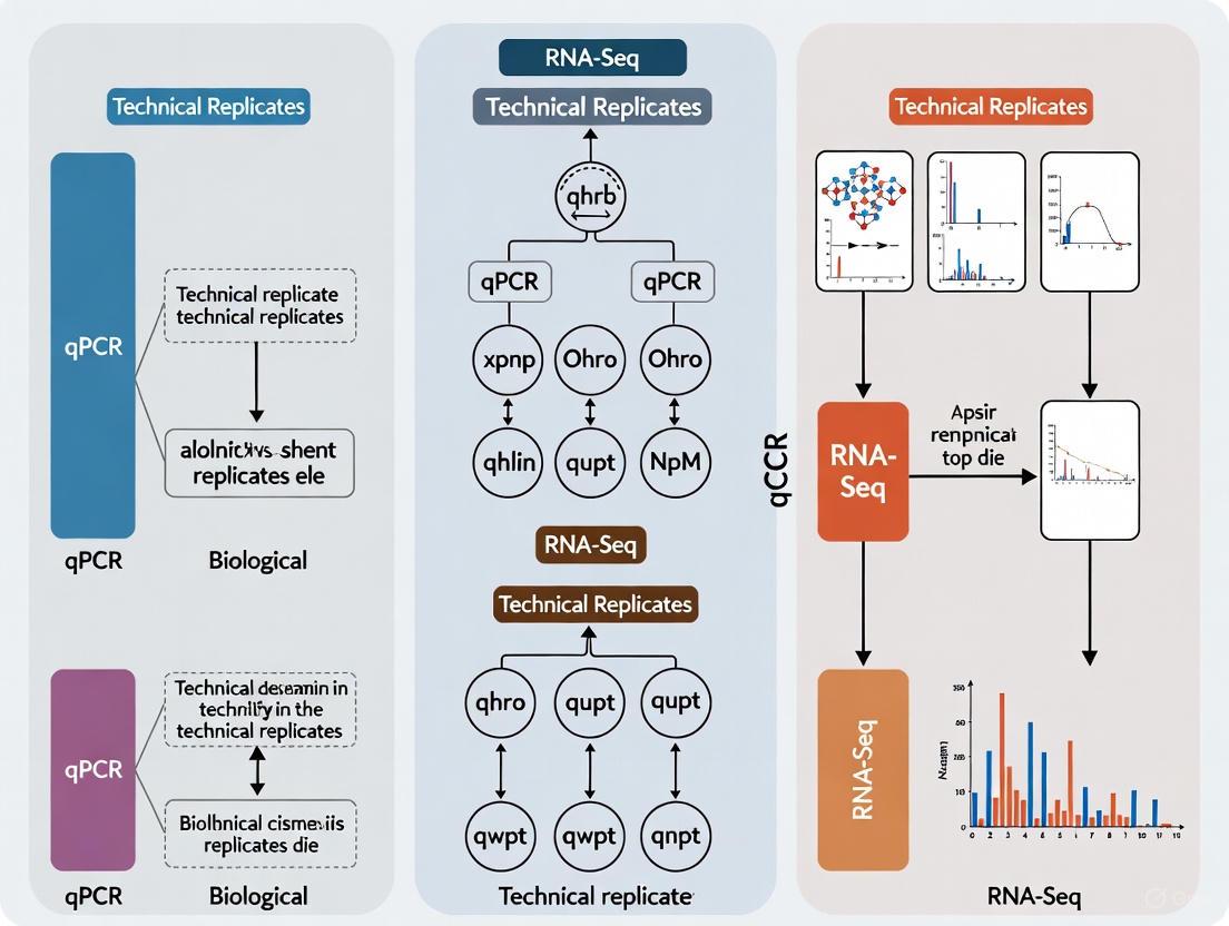
The relationship between these replicate types and their role in experimental design can be visualized as a hierarchical process:
The Critical Problem of Pseudoreplication
A fundamental error in experimental design occurs when researchers mistakenly treat technical replicates or pseudo-biological replicates as true biological replicates [15]. This problem, known as pseudoreplication, artificially inflates statistical significance and leads to hundreds of false positive differentially expressed genes in genomic studies [15]. A common example includes treating three cell-culture flasks of the same passage of a cell line as biological replicates when they actually originated from the same biological source [15]. This practice fails to capture true biological variation and results in spurious findings that cannot be reproduced in subsequent studies.
Quantitative Impact of Replication on Statistical Power
Empirical Evidence from RNA-Seq Studies
The relationship between biological replication and statistical power in genomics research has been quantitatively demonstrated through comprehensive RNA-Seq experiments. A landmark study performing RNA-seq with 48 biological replicates in each of two conditions revealed striking findings about replication requirements [16].
Table 1: Statistical Power for Detecting Differentially Expressed Genes in RNA-Seq Based on Replicate Number
| Biological Replicates | Percentage of All SDE Genes Detected | Percentage of >4-Fold Change SDE Genes Detected | Recommended Statistical Tools |
|---|---|---|---|
| 3 | 20%-40% | >85% | edgeR, DESeq2 |
| 6 | ~60% | >90% | edgeR, DESeq2 |
| 12 | >85% | >95% | DESeq2 |
| 20+ | >85% | >95% | DESeq |
With only three biological replicates—a common practice in many published studies—current statistical tools identified only 20-40% of the significantly differentially expressed (SDE) genes detected using the full set of 42 clean replicates [16]. This statistical power limitation is particularly pronounced for genes with subtle expression changes, though even with low replication, genes showing strong fold changes (>4-fold) can be detected with >85% power [16]. To achieve >85% detection power for all SDE genes regardless of fold change magnitude, more than 20 biological replicates are required [16].
Field-Specific Replication Guidelines
Best practices for replication vary across molecular techniques, reflecting differing technical variabilities and application requirements:
Table 2: Replication Guidelines by Experimental Method
| Method | Minimum Replicates | Optimal Replicates | Replicate Type Emphasis | Sequencing Depth |
|---|---|---|---|---|
| RNA-Seq | 3 | 4-6+ | Biological [6] | 10-60M PE reads |
| ChIP-Seq | 2 | 3 | Biological [6] | 10-30M reads |
| qPCR | 3 technical | 3 biological + 2-3 technical | Both [1] | N/A |
For RNA-Seq experiments, biological replicates are strongly recommended over technical replicates, with an absolute minimum of 3 replicates, though 4 replicates provides a more optimum minimum [6]. The CCBR Bioinformatics Core recommends processing RNA extractions simultaneously whenever possible, as extractions performed at different times introduce unwanted batch effects that compromise reproducibility [6]. When batch processing is unavoidable, researchers should ensure that replicates for each condition are represented in each batch so bioinformatic tools can measure and remove these effects during analysis [6].
In qPCR experiments, both replicate types serve distinct purposes. Technical replicates (typically triplicates) provide estimates of system precision, improve experimental variation measurements, and allow for outlier detection [1]. Biological replicates account for the true variation in target quantity among samples within the same group, enabling appropriate statistical generalization to the population [1].
Detailed Experimental Protocols
Protocol 1: Designing Replication for RNA-Seq Experiments
Principle: Biological replicates are essential for RNA-Seq because they capture the natural variation in gene expression between individuals, treatments, or conditions. Technical variation in sequencing is generally low compared to biological variation, making technical replicates less valuable than biological replicates [15].
Materials:
- RNA extraction kit (e.g., Qiagen RNeasy)
- DNase I digestion kit
- RNA integrity assessment system (e.g., Bioanalyzer)
- Library preparation kit
- Sequencing platform (Illumina recommended)
Procedure:
- Experimental Design Phase:
- Determine primary research question and key comparisons
- For animal studies: Plan for a minimum of 4 biological replicates per condition using distinct animals [6]
- For cell culture: Ensure biological replicates represent independent cultures from different passages or source flasks, not aliquots from the same culture [15]
- Calculate sample size using power analysis when possible
Sample Collection and Randomization:
- Process biological replicates independently throughout entire workflow
- Randomize sample processing order to avoid batch effects
- If processing in batches is unavoidable, ensure each batch contains samples from all experimental conditions
RNA Extraction and Quality Control:
- Extract RNA using standardized protocol
- Determine RNA Integrity Number (RIN) - require RIN >8 for mRNA sequencing [6]
- Quantify RNA using fluorometric method
Library Preparation and Sequencing:
- Use the same library preparation kit for all samples
- For mRNA analysis: Use mRNA library prep (10-20M paired-end reads) [6]
- For total RNA analysis (including non-coding RNA): Use total RNA method (25-60M paired-end reads) [6]
- Multiplex all samples together and run on the same sequencing lane when possible
Data Analysis:
Validation: Include positive control genes with known expression patterns when possible. Monitor internal consistency between replicates through correlation analysis.
Protocol 2: Implementing Replication in qPCR Experiments
Principle: qPCR experiments require both technical replicates (to measure system precision) and biological replicates (to capture population variation) for statistically valid conclusions [1].
Materials:
- qPCR instrument with calibrated temperature verification
- Pipettes with regular calibration
- Quality-controlled primers and probes
- Passive reference dye
- Plate centrifuge
Procedure:
- Experimental Design:
- Include 3-5 biological replicates per experimental group
- Plan for 3 technical replicates per biological sample
- Include no-template controls and positive controls
Sample Preparation:
- Process each biological sample independently through RNA extraction and cDNA synthesis
- Use the same reagent batches for all samples within an experiment
- Verify RNA quality and quantity before proceeding
Reaction Plate Setup:
- Master mix preparation: Prepare sufficient master mix for all technical replicates plus excess
- Aliquot master mix to plate first, then add template
- Include passive reference dye in reactions
- Ensure sample volume does not exceed 20% of PCR reaction volume to prevent optical mixing [1]
Plate Sealing and Centrifugation:
- Seal plate thoroughly to prevent evaporation
- Centrifuge plate briefly to bring liquids to well bottom and remove air bubbles
qPCR Run:
- Use manufacturer-recommended cycling conditions
- Verify temperature calibration of instrument blocks
- Include melt curve analysis for SYBR Green assays
Data Analysis:
- Calculate mean Cq values from technical replicates
- Identify and investigate outliers using coefficient of variation (CV >5% suggests technical issues) [1]
- Use appropriate normalization strategy (multiple reference genes recommended)
- Perform statistical tests (t-tests, ANOVA) on biological replicates, not technical replicates
Troubleshooting: High technical variation (CV >5%) may indicate pipetting errors, inadequate mixing, or instrument issues. Unusually low biological variation may indicate pseudoreplication or over-controlled conditions [1].
The Scientist's Toolkit: Essential Research Reagents and Materials
Table 3: Essential Research Reagents for Robust Replication Studies
| Reagent/Material | Function | Quality Control Requirements | Impact on Reproducibility |
|---|---|---|---|
| Authenticated Cell Lines | Biological replicate source | STR profiling, mycoplasma testing, low passage use [14] | Prevents false results from misidentified or contaminated lines |
| RNA Preservation Reagents | Stabilize RNA expression profiles | RNase-free certification, batch consistency | Maintains accurate transcriptome representation |
| Nucleic Acid Quantitation Kits | Accurate sample quantification | Fluorometric quantification standards | Ensures equal loading and reduces technical variation |
| Library Preparation Kits | cDNA library construction | Lot-to-lot consistency testing | Reduces batch effects in sequencing data |
| qPCR Master Mixes | Amplification reaction | Performance validation, inclusion of passive reference dye [1] | Improves precision and enables cross-plate comparison |
| Validated Antibodies (ChIP-Seq) | Target protein immunoprecipitation | ChIP-seq grade verification, lot number tracking [6] | Ensures specific binding and reproducible results |
Integrated Replication Strategy and Workflow
Implementing a comprehensive replication strategy requires understanding how different replicate types interact throughout the experimental process. The following workflow illustrates how biological and technical replicates integrate to deliver statistically powerful and reproducible results:
Proper experimental replication represents a fundamental pillar of scientific rigor in molecular biology research. The strategic implementation of both biological and technical replicates, following the detailed protocols outlined in this document, enables researchers to accurately distinguish biological effects from technical artifacts. By adhering to evidence-based replication standards—incorporating sufficient biological replicates to achieve appropriate statistical power, utilizing technical replicates to measure system precision, and avoiding the critical pitfall of pseudoreplication—scientists can significantly enhance the reproducibility and reliability of their genomic findings. These practices not only strengthen individual research outcomes but also contribute to the collective advancement of robust scientific knowledge.
In gene expression studies using qPCR and RNA-Seq, understanding and managing sources of variation is fundamental to generating reliable, reproducible data. The accuracy of biological conclusions depends on properly distinguishing between different types of noise inherent in these sensitive techniques. Variation in molecular experiments can be categorized into three primary types: system variation from technical measurement processes, biological variation from inherent differences between subjects, and experimental variation which represents the combined effect observed in data [1]. Each type has distinct characteristics, implications for data interpretation, and requires specific methodological approaches for mitigation. This article provides a comprehensive framework for identifying, quantifying, and controlling these variability sources within the context of replicate strategy decisions in qPCR and RNA-Seq workflows, enabling researchers to optimize experimental designs for robust scientific conclusions.
System Variation
System variation, also called technical variation, originates from the measurement system itself. This includes variability introduced by instrumentation, reagent efficiency, and operator technique [1]. In qPCR, contributors include pipetting inaccuracies, instrument calibration differences, well-position effects in thermal cyclers, and batch-to-batch variations in reagent kits [1]. For RNA-Seq, system variation encompasses library preparation efficiency, sequencing depth differences, and lane effects on flow cells [17]. System variation can be estimated by assaying multiple aliquots of the same biological sample, known as technical replicates [1]. This type of noise directly impacts measurement precision and can be reduced through protocol optimization and technical replication.
Biological Variation
Biological variation represents the true physiological differences in target quantity between individual organisms or samples within the same experimental group [1]. This variation arises from genetic heterogeneity, differential environmental exposures, stochastic cellular processes, and subtle variations in experimental treatments. For example, when researching drug treatment effects on gene expression in mice, biological variation exists between individual mice treated with the same drug [1]. Biological variation is accounted for by including multiple biological replicates in an experimental design – truly independent samples that represent the population being studied [1]. This variation determines the fundamental resolution limits for detecting biologically significant effects.
Experimental Variation
Experimental variation is the composite variability measured for samples belonging to the same biological group [1]. It serves as the practical estimate of true biological variation but is inevitably influenced by system variation. Due to this influence, experimental variation will typically not exactly equal the true biological variation [1]. The magnitude of system variation directly impacts how accurately experimental variation reflects biological reality – larger system variation increases its potential to distort experimental variation estimates [1]. Understanding this relationship is crucial for appropriate statistical interpretation of experimental data.
The diagram below illustrates the relationships and components of these three sources of variation:
Statistical Metrics for Quantifying Variation
| Metric | Calculation | Interpretation | Application Context |
|---|---|---|---|
| Coefficient of Variation (CV) | (Standard Deviation / Mean) × 100% | Measure of precision; lower CV indicates higher consistency | Assessing technical replicate consistency in qPCR [1] |
| Standard Deviation (SD) | √[Σ(xᵢ - μ)²/(N-1)] | Absolute measure of dispersion in data units | Describing population distribution; ±1 SD ≈ 68% of normally distributed population [1] |
| Standard Error (SE) | SD / √N | Measure of sampling error of the mean | Providing confidence boundaries for how close measured mean is to true mean [1] |
Replicate Strategy Impact on Variation
| Replicate Type | Definition | Controls For | Typical Number | Key Considerations |
|---|---|---|---|---|
| Technical Replicates | Repeated measurements of same sample aliquot [1] | System variation | 2-3 for qPCR [1] | Improves measurement precision; detects amplification failures; adds cost [1] |
| Biological Replicates | Measurements from different biological sources [1] | Biological variation | 3-6+ depending on effect size | Essential for statistical inference; captures population diversity [20] |
| Artificial Replicates (RNA-Seq) | Computationally generated replicates [17] | Assessment of reproducibility | Variable | FASTQ-bootstrapping shows best performance; computationally intensive [17] |
Experimental Protocols for Variance Analysis
Protocol: Assessing System Variation in qPCR
Principle: Quantify technical noise by repeatedly measuring identical sample aliquots to establish platform precision and identify optimal technical replicate strategy [1] [20].
Materials:
- Homogeneous cDNA sample (from bulk RNA extraction)
- Validated primer sets for high, medium, and low abundance targets
- qPCR instrument with calibrated block temperature
- Master mix prepared for full replicate set
Procedure:
- Sample Preparation: Create a single master mix containing all reaction components and aliquot into 12 wells of a qPCR plate for each target gene to be assessed [1].
- Plate Setup: Distribute replicates across different plate positions to identify positional effects.
- Amplification: Run qPCR with standardized cycling conditions.
- Data Collection: Record Ct values for all replicates.
- Variation Calculation:
Interpretation: Technical CV < 5% generally indicates acceptable precision. Recent large-scale evidence suggests duplicates often approximate triplicate means sufficiently, offering potential 33-66% savings in reagents and time [20].
Protocol: Evaluating Biological Variation in RNA-Seq
Principle: Distinguish biological variability from technical noise through appropriate replicate design and analysis to ensure adequate power for detecting differential expression [19] [17].
Materials:
- Appropriately preserved tissue or cell samples from multiple biological sources
- RNA extraction kit with DNase treatment
- RNA integrity assessment system (e.g., Bioanalyzer)
- Library preparation kit (whole transcriptome or 3' mRNA-Seq)
Procedure:
- Experimental Design:
- Include minimum of 4-6 biological replicates per condition for adequate power [17].
- Consider using 3' mRNA-Seq (e.g., QuantSeq) for large-scale gene expression studies with many samples, as it provides cost-effective quantification with lower sequencing depth requirements (1-5 million reads/sample) [19].
- Reserve whole transcriptome sequencing for studies requiring isoform resolution, splicing information, or non-polyadenylated RNA detection [19].
- Library Preparation and Sequencing:
- Process biological replicates simultaneously using same reagent batches.
- Include randomization during library preparation and sequencing runs.
- Data Analysis:
- Perform quality control (FastQC) and alignment (STAR).
- Generate read counts (featureCounts) and analyze with statistical software (DESeq2).
- Assess biological variation through:
- PCA plots to visualize sample clustering.
- Inter-replicate correlation analysis.
- Dispersion estimates across expression levels.
Interpretation: High concordance between biological replicates indicates robust results. Studies show that 3' mRNA-Seq and whole transcriptome approaches yield highly similar biological conclusions despite differences in numbers of detected differentially expressed genes [19].
Protocol: Artificial Replicate Generation for RNA-Seq
Principle: Generate computational replicates to assess analysis reproducibility when true technical replicates are unavailable [17].
Materials:
- Original FASTQ files from RNA-seq experiment
- Sufficient computational storage and processing resources
- Standard RNA-seq analysis pipeline (Trimmomatic, STAR, DESeq2)
Procedure for FASTQ Bootstrapping (FB):
- Read Resampling: For each original FASTQ file, draw π·k reads with replacement, where k is original read count and π is percentage (typically 100%) [17].
- File Generation: Create new FASTQ files containing resampled reads.
- Standard Analysis: Process bootstrapped FASTQ files through identical mapping and quantification pipeline as original data.
- Comparison: Analyze correlation of p-values and fold changes between original and bootstrapped datasets.
Alternative Methods:
- Column Bootstrapping (CB): Bootstrap samples from columns of expression matrix [17].
- Mixing Observations (MO): Generate new samples as weighted means of original expression columns [17].
Interpretation: FASTQ bootstrapping produces results most similar to true technical replicates, making it preferred for reproducibility assessment, despite higher computational requirements [17].
Research Reagent Solutions
| Reagent Category | Specific Examples | Function in Variance Control | Considerations for Selection |
|---|---|---|---|
| Reverse Transcriptases | Maxima H Minus, SuperScript IV [18] | Minimize RT efficiency variation; critical bottleneck in single-cell workflows | Select for high sensitivity, reproducibility, and ability to handle degraded samples [18] |
| qPCR Master Mixes | SYBR Green, TaqMan assays [20] | Provide consistent amplification efficiency; reduce technical variation | Probe-based chemistries show lower variability than dye-based [20] |
| RNA-Seq Library Prep Kits | QuantSeq (3' mRNA), Stranded mRNA (whole transcriptome) [19] | Control for library preparation bias and coverage variation | Choose based on research question: gene quantification (3') vs. isoform detection (whole) [19] |
| Normalization Reagents | Passive reference dyes (ROX) [1] | Correct for pipetting variation and optical anomalies | Essential for improving precision in qPCR; use according to instrument requirements [1] |
| RNA Stabilization Reagents | RNAlater, QIAzol [17] [18] | Preserve RNA integrity; minimize degradation-induced variation | Critical for working with challenging samples (FFPE, low input) [19] |
Methodological Decision Framework
The workflow below outlines key decision points for designing gene expression experiments that properly account for different sources of variation:
Proper management of variation sources requires strategic experimental design decisions that balance practical constraints with scientific rigor. System variation can be controlled through technical optimization and appropriate replication, but biological variation must be addressed through adequate biological replication [1] [20]. Method selection between qPCR and RNA-Seq—and within RNA-Seq between 3' focused and whole transcriptome approaches—significantly impacts the ability to resolve biological signals from technical noise [19]. When true replicates are limited, computational approaches like FASTQ-bootstrapping provide valuable alternatives for assessing reproducibility [17]. By systematically addressing each source of variation through the protocols and frameworks presented here, researchers can design more efficient experiments and draw more reliable biological conclusions from gene expression data.
Strategic Experimental Design: How to Implement Replicates in qPCR and RNA-Seq Workflows
In the realm of gene expression analysis, quantitative polymerase chain reaction (qPCR) remains a cornerstone technology for its sensitivity, specificity, and quantitative capabilities. The reliability of qPCR data, however, is profoundly influenced by experimental design, particularly the implementation of appropriate replication. Within the context of a broader thesis comparing qPCR and RNA-Seq methodologies, understanding the distinction and optimal application of biological versus technical replicates is paramount. Proper replication strategy not only controls for experimental variability but also ensures that observed differences reflect true biological phenomena rather than technical noise. This document outlines evidence-based best practices for determining the optimal number of replicates in qPCR experiments, providing detailed protocols to guide researchers, scientists, and drug development professionals in producing robust, reproducible, and statistically significant data.
Understanding Replicate Types: Biological vs. Technical
Replicates in qPCR experiments are broadly categorized into two types: technical and biological. Each serves a distinct purpose in controlling for different sources of variation and is fundamental to a sound experimental design.
Technical Replicates are multiple repetitions of the same biological sample. They are created by dividing a single nucleic acid extraction into multiple wells, using the same template preparation and PCR reagent master mix. The primary role of technical replicates is to assess the precision and variability inherent to the qPCR measurement system itself. This includes variation from pipetting, instrument noise, and reaction efficiency. Technical replicates help identify potential outliers and provide a more reliable measure (e.g., the mean) for that specific sample's Cq value. However, they do not provide information about the biological variation within a sample group [1].
Biological Replicates are measurements taken from multiple, independent biological samples within the same experimental group. For example, in a study investigating the effect of a drug treatment on gene expression in mice, each individually treated mouse represents a distinct biological replicate. Biological replicates are essential because they account for the natural variation that exists between individuals or primary samples in a population. The experimental variation measured across biological replicates is used as an estimate of this true biological variation and forms the basis for statistical comparisons between groups (e.g., Control vs. Treated) [1] [4].
The following table summarizes the key characteristics of each replicate type:
Table 1: Characteristics of Technical and Biological Replicates in qPCR
| Feature | Technical Replicates | Biological Replicates |
|---|---|---|
| Definition | Multiple measurements of the same sample aliquot [1] | Measurements from different individual biological sources within the same group [1] |
| Primary Purpose | Estimate system precision (pipetting, instrument variation) [1] | Estimate biological variation within a group [1] |
| Controls For | Experimental/analytical noise | Biological heterogeneity |
| Example | Same cDNA sample run in triplicate wells on a qPCR plate [1] | Three different mice from the same treatment group, each analyzed separately [1] |
| Informs | Reproducibility of the assay technique | Generalizability of the finding to the population |
The relationship and purpose of these replicates within a qPCR experimental workflow can be visualized as follows:
Determining the Optimal Number of Replicates
Determining the correct number of replicates is a critical decision that balances statistical power with practical constraints like cost, time, and sample availability.
The Gold Standard: Prioritizing Biological Replication
The consensus in the field is that biological replication is non-negotiable for making statistically valid inferences about a population [4]. An experiment should ideally encompass at least three independent biological replicates of each treatment or condition. Biological variation is often the largest source of variability in gene expression studies, and without sufficient biological replicates, it is impossible to determine if an observed effect is consistent or merely anecdotal. Increasing the number of biological replicates enhances the power of statistical tests to discriminate smaller, biologically relevant fold changes in gene expression [1] [21].
The Role of Technical Replication
For technical replicates, triplicates are a commonly selected and practical number in basic research [1]. Running technical replicates (e.g., duplicates or triplicates) for each biological sample provides confidence in the measurement for that specific sample. It allows for the detection of failed reactions or significant pipetting errors and provides a more precise mean Cq value for the biological replicate. However, it is generally recognized that investing resources in increasing the number of biological replicates provides a greater return in statistical power than running a large number of technical replicates for a few biological samples.
The Interplay Between Replicates and Precision
The precision of a qPCR experiment, measured by metrics like the Coefficient of Variation (CV), is directly impacted by replication. Improved precision allows researchers to discriminate smaller differences in nucleic acid copy numbers. Increasing the number of both technical and biological replicates tends to reduce the impact of random variation, leading to a more accurate estimate of the true mean [1]. The following table provides a summary of recommended replicate numbers based on different experimental goals:
Table 2: Replication Strategy Guidance for Different qPCR Applications
| Experimental Goal | Minimum Biological Replicates | Recommended Technical Replicates | Key Considerations |
|---|---|---|---|
| Gene Expression (General) | 3 per group [4] | 2-3 per sample [1] [4] | Balance between cost and reliability. |
| Detecting Small Fold Changes | >5 per group [1] [21] | 2-3 per sample | More biological replicates increase power to detect subtle differences. |
| Method Validation / Assay Precision | 1 (to start) | ≥3 to estimate CV [1] | Focus is on measuring system variation, not biological difference. |
| High-Throughput Screening | 3 per group | 2 (to conserve plates & reagents) | Requires rigorous assay validation beforehand. |
A Practical Experimental Protocol
This section provides a detailed, step-by-step protocol for a standard relative quantification qPCR experiment, incorporating best practices for replication.
Experimental Design and Sample Maximization
- Step 1: Define Biological Groups and Replicates. Determine the experimental groups (e.g., Control, Treated). Plan for a minimum of three independent biological replicates per group. This is the foundation of the experiment [4].
- Step 2: Plate Design. Ideally, all samples for a full experiment (all biological replicates, all target genes, and all reference genes) should be analyzed on a single qPCR plate to avoid inter-plate variation. If multiple plates are unavoidable, employ a randomized block design where each plate contains a complete set of one biological replicate from each group. Use Inter-Run Calibrators (IRCs) on each plate to correct for plate-to-plate variation [4].
- Step 3: Assign Technical Replicates. Within the plate design, assign two or three technical replicates for each combination of biological sample and assay (target gene and reference gene) [4].
Laboratory Workflow: From RNA to Cq
- Step 4: Nucleic Acid Extraction. Extract total RNA from each biological sample independently using a validated method. Treat samples with DNase to remove genomic DNA contamination. Assess RNA concentration, purity (A260/A280 ratio), and integrity (e.g., via agarose gel electrophoresis) [22] [23].
- Step 5: Reverse Transcription. Convert equal amounts of total RNA (e.g., 1 µg) from each sample to cDNA using a high-efficiency reverse transcriptase kit. Use a mixture of oligo-dT and random hexamer primers for comprehensive coverage. Keep all reaction conditions consistent across samples [23].
- Step 6: qPCR Setup.
- Precision Pipetting: Use calibrated pipettes and ensure tips fit snugly. Pipette master mixes first to minimize variation. For probe-based assays, ensure the final reaction volume is at least 20 µL to avoid optical mixing issues [1].
- Reaction Composition: Each reaction should contain: cDNA template, forward and reverse primers, probe (or intercalating dye), and PCR master mix.
- Plate Sealing and Centrifugation: After loading, seal the plate properly and centrifuge to collect all liquid at the bottom of the wells and remove air bubbles [1].
- Step 7: qPCR Run. Use the following standard cycling conditions, optimized for your instrument and reagent system:
- UDG incubation (if applicable): 50°C for 2 minutes
- Polymerase activation: 95°C for 2 minutes
- Amplification (40 cycles): 95°C for 15 seconds (denaturation) → 60°C for 1 minute (annealing/extension; acquire fluorescence)
Data Analysis and Statistical Procedures
- Step 8: Data Quality Control and Preprocessing.
- Baseline Correction: Manually set the baseline cycles to the early phases where no amplification is detected (e.g., cycles 5-15) to correct for background fluorescence variations [24].
- Threshold Setting: Set the fluorescence threshold within the exponential phase of all amplifications where the amplification curves are parallel. This ensures consistent Cq determination across samples [24].
- Efficiency Estimation: Calculate the amplification efficiency (E) for each assay. This can be done via a standard curve of serial dilutions or using algorithms like LinReg that analyze the raw amplification curves. The mean efficiency per assay is typically used for subsequent calculations [21] [4].
- Step 9: Calculation of Normalized Relative Quantities.
- For each biological replicate, calculate the mean Cq of its technical replicates for both the Gene of Interest (GOI) and Reference Gene(s).
- Calculate the normalized relative quantity (NRQ) using the efficiency-adjusted model, such as the Pfaffl method [24]: ( NRQ = \frac{(E{GOI})^{-\Delta Cq{GOI}}}{(E{Ref})^{-\Delta Cq{Ref}}} ) Where (\Delta Cq) is the difference in Cq between treated and control samples for each biological replicate.
- Step 10: Statistical Analysis of Biological Variation.
- Log Transformation: To stabilize variance and normalize the data, apply a log transformation (base 2 or natural) to the NRQ values. The transformed values are referred to as Cq' [4].
- Hypothesis Testing: Perform appropriate statistical tests on the Cq' values of the biological replicates. For two-group comparisons, use a t-test. For more complex designs (e.g., multiple groups or factors), use Analysis of Variance (ANOVA). The unit of replication for these tests is the biological replicate, not the technical replicate [4].
The Scientist's Toolkit: Essential Reagents and Materials
Table 3: Key Research Reagent Solutions for a Robust qPCR Experiment
| Item | Function / Description | Example / Note |
|---|---|---|
| qPCR Instrument | Instrument for thermal cycling and fluorescence detection. | Applied Biosystems ViiA 7, QuantStudio 7, Bio-Rad CFX [22] |
| Probe-based qPCR Master Mix | Optimized buffer, enzymes, and dNTPs for probe-based detection. | Provides superior specificity over dye-based methods [22] |
| TaqMan Assays | Pre-optimized primer and probe sets for specific targets. | Ideal for high-throughput and multiplexing applications [22] |
| RNA Extraction Kit | For isolation of high-quality, intact total RNA. | Qiagen RNeasy, TRIzol LS Reagent [25] [23] |
| Reverse Transcription Kit | For synthesis of first-strand cDNA from RNA templates. | Use kits with a mix of oligo-dT and random hexamers [23] |
| Validated Reference Genes | Genes with stable expression for data normalization. | Must be stability-tested for your specific sample set (e.g., using geNorm, NormFinder) [4] [23] |
| Nuclease-free Water | Water certified to be free of RNases and DNases. | Essential for preventing nucleic acid degradation. |
| Optical Plates & Seals | Plates and adhesive films designed for qPCR fluorescence reading. | Ensure compatibility with the qPCR instrument. |
Connecting to Broader Research: qPCR and RNA-Seq
The principles of robust experimental design, particularly adequate biological replication, are universally critical in genomics, whether using qPCR or RNA-Seq. RNA-Seq provides an unbiased, genome-wide view of the transcriptome but comes with its own set of computational and normalization challenges [26]. qPCR remains the gold standard for validating RNA-Seq findings due to its superior sensitivity, dynamic range, and precision for a limited number of targets [25] [26]. The relationship between these technologies is synergistic. A well-designed RNA-Seq study with sufficient biological replication (e.g., n≥3) can identify candidate differentially expressed genes, which are then confirmed with a rigorously designed qPCR experiment on independent samples, also with adequate biological replication. This combined approach leverages the strengths of both technologies to generate reliable and impactful conclusions in gene expression research.
In the context of a broader thesis on biological versus technical replicates in qPCR and RNA-Seq research, careful experimental design forms the cornerstone of reliable transcriptomic analysis. The fundamental challenge in any gene expression study lies in accurately distinguishing biological signal from experimental noise. While quantitative PCR (qPCR) has long provided a sensitive method for targeted gene expression analysis, RNA sequencing (RNA-Seq) offers an unbiased, genome-scale perspective on the transcriptome. However, this comprehensive view introduces additional complexity in experimental design, particularly regarding replication strategy and sequencing depth. The decision between biological replicates (measurements across different biological subjects) and technical replicates (repeated measurements of the same biological sample) carries profound implications for statistical power, generalizability, and cost-efficiency. This application note synthesizes current evidence and best practices to guide researchers in making informed design choices that ensure robust and interpretable RNA-Seq results, with particular relevance for drug development applications where accurate detection of differential expression directly impacts decision-making.
Core Principles: Biological vs. Technical Replication
Definitions and Distinct Purposes
In RNA-Seq experiments, understanding the distinction between biological and technical replicates is paramount, as each addresses different sources of variability. Biological replicates are measurements taken from distinct biological entities (e.g., different animals, independently cultured cells, or human subjects) that capture the natural variation present in the population under study [2]. They are essential for ensuring that findings are generalizable beyond the specific samples measured. In contrast, technical replicates involve multiple measurements of the same biological sample through the experimental workflow (e.g., sequencing the same library multiple times or preparing multiple libraries from the same RNA extraction) [2]. Technical replicates primarily assess variability introduced by laboratory procedures and sequencing platforms rather than biological variation.
Table 1: Comparison of Replicate Types in RNA-Seq Experiments
| Aspect | Biological Replicates | Technical Replicates |
|---|---|---|
| Definition | Different biological samples or entities | Same biological sample, measured multiple times |
| Purpose | Assess biological variability and ensure findings are generalizable | Assess technical variation from workflows and sequencing |
| Example | 3 different animals in each treatment group | 3 sequencing runs for the same RNA sample |
| Addresses | Natural variation between individuals/subjects | Measurement error, library prep, and sequencing variability |
| Priority | Essential for biological inference | Useful for quality control but secondary to biological replication |
Relative Importance in Experimental Design
Multiple studies consistently demonstrate that biological replication provides substantially greater value than technical replication for detecting differentially expressed genes. Biological replicates enable researchers to estimate the natural variation in gene expression within a population, which is crucial for statistical tests that identify expression changes between conditions [27] [28]. Technical reproducibility in RNA-Seq is generally considered excellent when using consistent laboratory protocols, making technical replicates less critical for most study designs [29]. In fact, for a fixed budget, prioritizing resources toward additional biological replicates rather than technical replicates or extreme sequencing depth typically yields more statistically powerful experiments [27] [28].
Quantitative Guidelines: Replicate Numbers and Sequencing Depth
Recommended Replicate Numbers
The number of biological replicates required depends on the expected effect size, biological variability, and desired statistical power. While more replicates always improve power, practical considerations often dictate a balance between statistical rigor and resource constraints.
Table 2: Recommended Replicate Numbers for RNA-Seq Experiments
| Scenario | Minimum Replicates | Optimal Replicates | Rationale |
|---|---|---|---|
| General research | 3 per condition [30] [6] | 4-8 per condition [2] [28] | 3 replicates enable basic variability estimation; 4+ substantially improve reproducibility and power |
| Pilot studies | 2-3 per condition | 3-4 per condition | Provides preliminary data for power calculations in subsequent larger studies |
| High-variability systems | 4 per condition | 6-8 per condition [2] | Compensates for substantial biological variation (e.g., human samples, complex tissues) |
| Cost-constrained screens | 3 per condition | 4 per condition | Balances statistical needs with throughput requirements |
| Toxicology/Drug discovery | 3 per dose | 4 per dose [28] | Enhances reproducibility of dose-response patterns and benchmark dose estimates |
Evidence strongly indicates that increasing biological replicates significantly enhances the detection of differentially expressed genes. One toxicogenomics study found that with only 2 replicates, over 80% of differentially expressed genes were unique to specific sequencing depths, indicating high variability. Increasing to 4 replicates substantially improved reproducibility, with over 550 genes consistently identified across most depths [28]. Similarly, research has demonstrated that power to detect differential expression improves markedly when the number of biological replicates increases from n = 2 to n = 5 [27].
Optimal Sequencing Depth Recommendations
Sequencing depth requirements vary based on the organism, transcriptome complexity, and specific research objectives. Adequate depth ensures sufficient coverage to detect and quantify transcripts of interest, particularly those expressed at low levels.
Table 3: Recommended Sequencing Depth Based on Experimental Goals
| Application | Recommended Depth | Key Considerations |
|---|---|---|
| Standard differential expression (3' mRNA-Seq) | 1-5 million reads/sample [19] | Sufficient for gene-level quantification when reads localize to 3' end |
| Standard differential expression (whole transcriptome) | 20-30 million reads/sample [30] [6] | Balances cost with detection sensitivity for most protein-coding genes |
| Total RNA-Seq (including non-coding RNA) | 25-60 million reads/sample [6] | Additional depth needed for comprehensive coverage of diverse RNA species |
| Isoform analysis & splice variants | 30+ million reads/sample | Higher depth required to resolve transcript structures |
| Large-scale screening studies | 10-20 million reads/sample [6] | Enables cost-effective profiling of many samples |
Strategic Balance Between Replicates and Depth
When facing budget constraints, researchers must often choose between sequencing more biological replicates at lower depth or fewer replicates at greater depth. Multiple lines of evidence indicate that biological replication generally provides better returns on investment than increased sequencing depth [27] [28]. One study found that sequencing depth could be reduced to as low as 15% without substantial impacts on false positive or true positive rates, whereas reducing replicate numbers significantly diminished statistical power [27]. Another toxicogenomics study concluded that replication had a greater influence than depth for optimizing detection power, with higher replicates increasing the rate of overlap of benchmark dose pathways and precision of median benchmark dose estimates [28].
Practical Protocols and Implementation
Workflow for RNA-Seq Experimental Design
The following diagram illustrates the key decision points in designing a robust RNA-Seq experiment, emphasizing replication and sequencing strategies:
Protocol for Replicate Planning and Sample Preparation
Step 1: Define Experimental Units and Conditions
- Clearly identify the biological unit of interest (individual organism, cell culture, tissue sample)
- Define all conditions and time points for comparison
- Ensure true biological independence for biological replicates
Step 2: Determine Replication Strategy
- Allocate resources for minimum of 3 biological replicates per condition [30] [6]
- Increase to 4-6 replicates for studies with anticipated high variability
- Consider including extra samples to account for potential quality control failures
Step 3: Randomization and Batch Effects Mitigation
- Randomly assign samples to processing batches to avoid confounding technical and biological effects
- Process samples in balanced batches that include representatives from all experimental conditions
- Include control samples in each batch to monitor technical variability
Step 4: Sample Collection and Storage
- Collect biological replicates following standardized protocols to minimize introduction of technical artifacts
- Process samples uniformly during RNA extraction and library preparation
- Use appropriate storage conditions (e.g., -80°C in RNase-free plates with proper sealing) [18]
Step 5: Library Preparation and Quality Control
- Select appropriate library preparation method based on research goals (3' mRNA-Seq vs. whole transcriptome)
- Perform rigorous RNA quality assessment (e.g., RIN > 8 for mRNA sequencing) [6]
- Use unique dual indices for multiplexing to enable sample pooling and sequencing
Protocol for Sequencing Depth Optimization
Step 1: Preliminary Depth Estimation
- For standard differential expression: target 20-30 million reads per sample for whole transcriptome [30]
- For 3' mRNA-Seq: 1-5 million reads may be sufficient [19]
- Adjust based on transcriptome complexity and expression dynamic range
Step 2: Pilot Studies for Depth Calibration
- When possible, conduct pilot sequencing with 1-2 samples at higher depth
- Use saturation analysis to determine optimal depth for full study
- Consider published datasets from similar biological systems as reference
Step 3: Multiplexing and Lane Allocation
- Multiplex samples using barcoding to maximize throughput and minimize batch effects [27]
- Balance samples across sequencing lanes to avoid confounding technical effects
- When multiple lanes are required, ensure all conditions are represented in each lane
Step 4: Quality Assessment and Potential Resequencing
- Monitor sequencing quality metrics (Q scores, base composition)
- If initial depth is insufficient, consider additional sequencing rather than technical replicates
- For critical samples, sequence additional lanes and combine data after quality checking [29]
The Scientist's Toolkit: Essential Research Reagents and Materials
Table 4: Key Reagent Solutions for RNA-Seq Experiments
| Reagent/Category | Function | Examples/Considerations |
|---|---|---|
| RNA Stabilization | Preserves RNA integrity post-collection | RNAlater, PAXgene Tissue systems |
| RNA Extraction Kits | Isolate high-quality RNA from samples | PicoPure (low input), miRNeasy (various species), column-based or TRIzol methods |
| rRNA Depletion Kits | Remove ribosomal RNA from total RNA | Ribozero, NEBNext rRNA Depletion; critical for total RNA-seq |
| Poly(A) Selection | Enrich for polyadenylated transcripts | Oligo(dT) beads; standard for 3' mRNA-Seq |
| Library Prep Kits | Prepare sequencing libraries from RNA | Illumina TruSeq, NEBNext Ultra, QuantSeq (3' specific) |
| Quality Control Assays | Assess RNA and library quality | Bioanalyzer/TapeStation (RIN), Qubit (quantification), qPCR assays |
| Spike-in Controls | Normalization and process controls | ERCC RNA Spike-In Mix, SIRVs; particularly valuable for single-cell or degraded samples |
| Unique Dual Indexes | Sample multiplexing | Enable pooling of multiple libraries while tracking samples |
Well-designed RNA-Seq experiments strategically balance biological replication, technical considerations, and sequencing depth to maximize statistical power and biological relevance. The evidence consistently demonstrates that biological replication should be prioritized over technical replication or extreme sequencing depth in most scenarios. A minimum of 3-4 biological replicates per condition provides a foundation for reliable differential expression analysis, while 20-30 million reads per sample generally suffices for standard whole transcriptome studies. By applying these guidelines within the context of specific research objectives and budget constraints, researchers can design RNA-Seq experiments that yield robust, reproducible, and biologically meaningful results, advancing discovery in basic research and drug development alike.
In the fields of drug discovery and development, the reliability of gene expression data from techniques like qPCR and RNA-Seq is paramount. This reliability is fundamentally governed by a sound experimental design that appropriately uses biological replicates and technical replicates. A biological replicate is defined as an independent biological sample or entity (e.g., different animals, individuals, or cell culture preparations), and its primary purpose is to assess biological variability and ensure findings are generalizable [2]. A technical replicate, in contrast, is a repetition of the measurement on the same biological sample, and its purpose is to assess and minimize variation introduced by the experimental workflow itself [1] [2]. The careful application of these replicates is critical for distinguishing true biological signals, such as genuine drug response biomarkers, from technical noise and natural biological variation, thereby enabling valid statistical inference and confident decision-making throughout the drug development pipeline.
Quantitative Comparison of Replicate Strategies
Statistical Power and Replicate Number
The number of replicates has a direct and quantifiable impact on the quality and reliability of results. The following table summarizes key findings from large-scale studies on the effect of replicate numbers in RNA-Seq and qPCR.
Table 1: Impact of Replicate Number on Data Quality in RNA-Seq and qPCR
| Technique | Number of Replicates | Outcome and Performance | Key Findings |
|---|---|---|---|
| RNA-Seq [16] | 3 biological replicates | Identified only 20%–40% of significantly differentially expressed (SDE) genes found with 42 replicates. | Low replicate numbers miss a majority of true positives, especially genes with small fold changes. |
| RNA-Seq [16] | 6 biological replicates | Recommended minimum for basic reliability; superior performance of tools like edgeR and DESeq2. | Rising to at least 12 replicates is recommended when identifying all SDE genes, regardless of fold change, is important. |
| RNA-Seq [16] | >20 biological replicates | Required to identify >85% of all SDE genes. | High numbers of biological replicates are necessary for comprehensive, robust gene discovery. |
| qPCR [20] | Technical triplicates vs. duplicates/singles | Duplicates or single replicates sufficiently approximated triplicate means. | Moving to fewer technical replicates can save 33–66% in reagents, time, and labor without major precision loss. |
Recommendations for Different Scenarios
Based on the quantitative data, the following table provides actionable recommendations for applying replicate strategies across different practical scenarios in drug discovery and development.
Table 2: Replicate Application Guide for Drug Development Scenarios
| Practical Scenario | Recommended Replicate Strategy | Rationale and Considerations |
|---|---|---|
| Large-Scale Drug Screening (e.g., using cell lines) [2] | Prioritize biological replicates (ideally 4-8 per treatment group). Technical replicates are less critical. | The primary goal is to capture reproducibility of the drug effect across independent cultures. Biological replicates account for this variability. High throughput and cost-efficiency are key. |
| Preclinical In Vivo Studies [1] | Focus on biological replicates (different animals). Use technical replicates sparingly, if at all. | The major source of variation is inter-indimal biological difference. Technical replicates from the same animal do not capture this and can lead to pseudoreplication and false positives [15]. |
| Biomarker Discovery & Validation from patient samples (e.g., FFPE, blood) [2] [31] | Maximize biological replicates (patients). A minimum of 3 is typical, but more are beneficial. Technical replicates may be used for low-abundance targets or assay QC. | Sample availability may be limited, making each biological replicate precious. The focus is on generalizing findings across a population. Technical variation is often minor compared to inter-patient biological variability. |
| Mode-of-Action / Dose-Response Studies [2] | Use both biological and technical replicates. 3-6 biological replicates per condition/time point. Technical replicates can guard against assay failure. | These studies require high precision to track changes over time or concentration. Technical replicates help ensure the reliability of each measured data point within a complex experimental design. |
| qPCR Assay Validation [1] [20] | Use technical replicates (traditionally triplicates) during initial assay setup and validation. For routine high-throughput use, duplicates or even singles may be sufficient. | Technical replicates are crucial for estimating the system's precision during development. Recent large-scale data suggests that for well-optimized assays, fewer replicates can maintain precision while greatly improving efficiency [20]. |
Experimental Protocols
Protocol for RNA-Seq in a Drug Treatment vs. Control Study
This protocol is designed for a study comparing treated and control cells, typical in early drug discovery.
1. Experimental Design and Sample Size Determination
- Define Hypothesis: Clearly state the expected differential expression (e.g., "Drug X alters the expression of genes in pathway Y").
- Determine Replicates: Based on power analysis (see Table 1), include a minimum of 4-6 biological replicates per condition (treatment vs. control). Crucially, each biological replicate must be an independently cultured and treated batch of cells to avoid pseudoreplication [15].
- Randomization: Randomly assign cells to treatment or control groups across multiple culture plates to correct for potential batch effects [2].
2. Wet Lab Workflow
- Cell Culture & Treatment: Culture cells and apply drug treatment or vehicle control to independently prepared biological replicates.
- RNA Extraction: At the designated time point, extract total RNA from each replicate using a kit suitable for your cell type (e.g., RNeasy Plus Mini kit). Include a DNase digestion step to remove genomic DNA contamination [32].
- RNA Quality Control: Assess RNA integrity using an Agilent 2100 Bioanalyzer. Only proceed with samples having high RNA Integrity Numbers (RIN > 8).
- Library Preparation: For large-scale gene expression studies, use a 3'-end focused method (e.g., QuantSeq) for cost-effectiveness and high-throughput. For isoform or fusion discovery, use whole transcriptome methods with rRNA depletion [2]. Consider using spike-in controls (e.g., SIRVs) to monitor technical performance.
- Sequencing: Sequence libraries on an Illumina platform (e.g., NextSeq 500). A common configuration is 75-100 bp paired-end reads, with a depth of 20-30 million reads per sample [17].
3. Bioinformatic Analysis
- Quality Control & Trimming: Use FastQC to assess raw read quality. Trim adapters and low-quality bases using Trimmomatic [17] or Cutadapt [32].
- Alignment: Map quality-trimmed reads to the appropriate reference genome (e.g., GRCm38 for mouse) using a splice-aware aligner like STAR [17].
- Quantification: Generate read counts per gene using featureCounts or a similar tool, based on a standard annotation file (e.g., Ensembl).
- Differential Expression Analysis: Import the count matrix into R and perform analysis using DESeq2 [17] or edgeR, which are robust tools for experiments with lower replicate numbers [16]. A false discovery rate (FDR) of 5% is typically used to identify significantly differentially expressed genes.
Protocol for qPCR Validation of RNA-Seq Hits
This protocol is for validating a subset of candidate genes identified from an RNA-Seq experiment.
1. Experimental Design
- Sample: Use the same cDNA samples that were used for RNA-Seq, or prepare new cDNA from the original RNA extracts.
- Replicates: The original RNA samples are your biological replicates. Run each biological sample in technical triplicates for the initial assay validation. For high-throughput validation of known assays, technical duplicates may be sufficient based on recent evidence [20].
2. Wet Lab Workflow
- Reverse Transcription: Convert 1 µg of total RNA to cDNA using a high-capacity cDNA reverse transcription kit with oligo(dT) and/or random primers.
- qPCR Reaction Setup:
- Use TaqMan probes for highest specificity or SYBR Green for flexibility.
- Prepare a master mix containing the qPCR reagents, primers, and probe to minimize pipetting error.
- Aliquot the master mix into a qPCR plate and add cDNA from each biological replicate.
- Centrifuge the sealed plate to remove bubbles and ensure all liquid is at the bottom of the wells [1].
- qPCR Run: Run the plate on a calibrated qPCR instrument (e.g., QuantStudio) using the manufacturer's recommended cycling conditions.
3. Data Analysis
- Quality Control: Check that the technical replicates have a low coefficient of variation (CV). A CV < 5% is generally considered acceptable [1].
- Normalization: Normalize the data using the ΔΔCt method. Select a stable normalizer gene (or genes) identified using a tool like RefFinder [32]. Avoid using genes like GAPDH or ACTB if their expression is affected by the drug treatment [32].
- Statistical Analysis: Perform a t-test or ANOVA on the ΔCt values between treatment and control groups across biological replicates to determine statistical significance.
Visualizing Workflows and Logical Relationships
Decision Framework for Replicate Strategy
The following diagram outlines the key decision points for choosing an appropriate replicate strategy in gene expression studies.
RNA-Seq Data Analysis Workflow with Replicate Consideration
This diagram illustrates the standard RNA-Seq data analysis pipeline, highlighting steps where proper replication is critical for robust results.
The Scientist's Toolkit: Research Reagent Solutions
Table 3: Essential Reagents and Kits for Gene Expression Studies in Drug Discovery
| Item Name | Function / Application | Example Use Case |
|---|---|---|
| RNeasy Plus Mini Kit (Qiagen) [32] | Total RNA extraction from cells and tissues; includes gDNA removal. | RNA extraction from cell culture treatments for RNA-Seq or qPCR. |
| TruSeq Stranded mRNA Library Prep Kit (Illumina) [17] | Preparation of strand-specific RNA-Seq libraries for whole transcriptome analysis. | Library construction for mode-of-action studies requiring isoform information. |
| QuantSeq 3' mRNA-Seq Library Prep Kit (Lexogen) [2] | 3'-end focused, cost-effective library prep for gene expression analysis. | Ideal for large-scale drug screens where hundreds of samples need profiling. |
| TruSight RNA Pan-Cancer Panel (Illumina) [31] | Targeted RNA sequencing panel for focused analysis of known cancer genes. | Profiling drug response in oncology-focused discovery programs. |
| High-capacity RNA-to-cDNA Kit (Thermo Fisher) [20] | Reverse transcription of total RNA into cDNA for qPCR analysis. | First-step cDNA synthesis for high-throughput qPCR validation. |
| TaqMan Advanced miRNA Assays (Thermo Fisher) [20] | Probe-based detection and quantification of specific microRNAs. | Biomarker discovery and validation for circulating miRNAs in biofluids. |
| SIRV Spike-in Control Kits (Lexogen) [2] | Artificial RNA controls for normalization and quality control in RNA-Seq. | Monitoring technical performance and enabling cross-study comparisons in large projects. |
A foundational principle of scientific research is replication, which allows researchers to measure variability, estimate experimental effects with greater precision, and draw meaningful statistical conclusions. However, a critical error that frequently undermines experimental integrity is pseudoreplication—the use of inferential statistics to test for treatment effects with data from experiments where either treatments are not replicated or replicates are not statistically independent [33]. In practical terms, pseudoreplication occurs when researchers mistakenly treat multiple measurements from the same biological entity as independent data points, artificially inflating sample size and leading to potentially invalid conclusions [33] [34].
The distinction between genuine replication and pseudoreplication is particularly crucial in molecular biology techniques such as qPCR and RNA-Seq, where the hierarchy of measurement (e.g., multiple wells measuring the same sample, multiple samples from the same animal, or multiple animals from the same treatment group) must be carefully considered in experimental design and statistical analysis. This guide examines the sources, consequences, and solutions for pseudoreplication in biomedical research, with specific application notes for designing methodologically sound qPCR and RNA-Seq experiments.
Defining Replication Types and Their Roles
Biological versus Technical Replicates
In molecular biology experiments, two fundamental types of replicates must be distinguished:
Biological replicates are measurements taken on different biological entities (e.g., cells, tissues, or animals) that have been subjected to the same experimental condition. These captures the natural biological variation within a population and allow for generalization of results beyond the specific samples used [35] [1]. In a mouse drug treatment study, for example, biological replicates would consist of multiple independently treated mice, each contributing one data point for statistical analysis [1].
Technical replicates are repeated measurements of the same biological sample. These help account for variability introduced by the measurement process itself (e.g., pipetting errors, instrument noise, or assay variability) but do not provide information about biological variation [35] [1]. Measuring the same cDNA sample in multiple qPCR wells constitutes technical replication [20].
Table 1: Characteristics of Biological and Technical Replicates
| Aspect | Biological Replicates | Technical Replicates |
|---|---|---|
| Definition | Different biological samples under same condition | Repeated measurements of same biological sample |
| Source of Variation Measured | Biological variability between individuals | Technical variability of measurement system |
| Question Answered | Is the effect consistent across biological units? | How precise is my measurement technique? |
| Example in qPCR | Different mice from same treatment group | Multiple wells containing same cDNA sample |
| Example in RNA-Seq | Cells from different animals or culture batches | Multiple sequencing runs of same RNA library |
| Primary Benefit | Enables statistical inference to population | Improves measurement precision |
The Concept and Forms of Pseudoreplication
Pseudoreplication arises when measurements that are not statistically independent are treated as independent data points in statistical analysis [33]. This typically occurs when researchers:
- Take multiple measurements from the same biological entity (e.g., measuring blood pressure three times in each rat) and treat these as independent samples [33]
- Use cells from the same culture flask in different wells and treat them as biological replicates [15]
- Pool samples from multiple subjects and treat the pool as a single independent observation [34]
The fundamental issue with pseudoreplication is that it violates the assumption of independence underlying most statistical tests. Measurements from the same biological unit are typically more similar to each other than measurements from different biological units, creating correlated errors that invalidate traditional statistical approaches [33].
Consequences of Pseudoreplication in Research
Statistical and Interpretative Problems
Pseudoreplication creates two serious problems for statistical analysis:
Incorrect hypothesis testing: The statistical analysis tests a different hypothesis than the researcher intends. When multiple measurements from the same subject are treated as independent, the analysis effectively tests whether measurements within subjects differ, rather than whether treatment effects exist between subjects [33].
False precision: Pseudoreplication artificially inflates sample size (n), which reduces standard errors and narrows confidence intervals incorrectly. This creates an illusion of precision that doesn't reflect true biological variability [33].
To illustrate, consider an experiment where 10 rats are randomly assigned to treatment or control groups, with performance tested on three consecutive days. An incorrect analysis that treats all 15 observations per group as independent would yield t₂₈ = 2.1, p = 0.045. The correct analysis, using rat averages with only 8 degrees of freedom, gives t₈ = 2.1, p = 0.069—a non-significant result [33]. This dramatic difference in p-values (0.045 vs. 0.069) demonstrates how pseudoreplication can lead to false positive findings.
Impact on Research Reproducibility
Pseudoreplication contributes to the reproducibility crisis in biomedical research by increasing both false positive and false negative rates [33] [35]. When statistical analyses are based on inflated sample sizes, they may detect effects that don't truly exist or fail to detect genuine effects due to improper modeling of variance structure.
The consequences extend beyond statistical significance to effect size estimation and biological interpretation. For example, in RNA-Seq experiments, using pseudoreplicates can identify hundreds of false positive differentially expressed genes [15]. Such errors waste research resources, bias the scientific literature, and may lead to fruitless exploration of non-existent phenomena or advancement of ineffectual therapies to clinical trials [33].
Experimental Design Principles to Avoid Pseudoreplication
Criteria for Genuine Replication
Genuine replication in experiments requires satisfying three key criteria [34]:
- Random and independent assignment: Biological entities of interest must be randomly and independently assigned to treatment groups.
- Independent treatment application: Treatments should be applied independently to each experimental unit.
- No interference between units: Experimental units should not influence each other, particularly on the measured outcome variables.
These criteria help ensure that the "N" in statistical analysis represents the number of independent observations that contribute to estimating biological variability.
Determining the Appropriate Experimental Unit
The experimental unit is defined as the smallest entity that can be randomly assigned to a different treatment condition [33] [34]. Identifying this unit correctly is essential for avoiding pseudoreplication:
- In animal studies, the experimental unit is typically the individual animal, not cells or tissues within that animal.
- In cell culture experiments, the experimental unit is often the culture well or flask, not individual cells within that culture [34].
- In multi-center clinical trials, the experimental unit may be the center, not individual patients, if treatments are assigned at the center level.
When the criteria for genuine replication cannot be met at the level of interest, the solution is to replicate one level up in the biological or technical hierarchy and use appropriate statistical models that account for the nested structure of the data [34].
The following diagram illustrates the decision process for determining the correct experimental unit and replication strategy:
Decision Framework for Determining Experimental Units
Application Notes: qPCR Experimental Design
Optimal Replication Strategy in qPCR
Quantitative PCR experiments require careful consideration of replication strategy to balance cost, throughput, and statistical validity. Recent large-scale studies challenge conventional assumptions about technical replication in qPCR:
- An analysis of 71,142 cycle threshold (Ct) values from 1,113 RT-qPCR runs found that duplicate or single replicates sufficiently approximated triplicate means across various conditions, instruments, and operator experience levels [20].
- Moving from technical triplicates to duplicates or singles can reduce reagent use, instrument time, and labor by 33-66%, offering substantial savings for high-throughput projects without compromising precision [20].
- While inexperienced operators exhibited slightly higher technical variability, their replicates remained within widely accepted precision limits [20].
Table 2: Replication Guidelines for qPCR Experiments Based on Recent Evidence
| Scenario | Recommended Technical Replicates | Recommended Biological Replicates | Rationale |
|---|---|---|---|
| High-throughput screening | 1-2 | 6-12 | Single replicates sufficiently approximate true values; biological replication provides power for detection of differential expression [20] [16] |
| Low template concentration | 2 | 8-15 | No correlation found between Ct values and coefficient of variation; biological replication remains priority [20] |
| Inexperienced operators | 2 | 6-12 | Slightly higher variability but still within acceptable precision limits [20] |
| Probe-based detection | 1-2 | 6-12 | Lower variability compared to dye-based methods [20] |
| Dye-based detection | 2 | 6-12 | Higher variability warrants additional technical replication [20] |
Practical Protocol for qPCR Experimental Design
Step 1: Prioritize Biological Replication
- Determine the number of biological replicates based on expected effect size and variability using power analysis [16]. For typical gene expression studies, at least 6 biological replicates per group are recommended, increasing to 12 or more for detecting small fold changes [16].
- Biological replicates must represent independent biological samples, not technical repetitions of the same sample [35].
Step 2: Optimize Technical Replication
- Consider using duplicates rather than triplicates to conserve resources while maintaining precision [20].
- Use technical replicates primarily to measure and control for technical variability, not as substitutes for biological replicates [1].
Step 3: Implement Appropriate Statistical Analysis
- Account for the nested structure of data (multiple technical replicates within each biological replicate) using mixed models or by averaging technical replicates before analysis [35].
- Monitor coefficients of variation (CV) for technical replicates; typical qPCR experiments should maintain CV < 5% for Ct values [1].
Step 4: Apply Proper Normalization and Quality Control
- Include appropriate reference genes for normalization [36].
- Implement rigorous quality control measures, including evaluation of RNA quality, reverse transcription efficiency, and amplification specificity [36].
Application Notes: RNA-Seq Experimental Design
Replication Requirements for RNA-Seq
RNA-Seq experiments have distinct replication requirements due to the complexity and cost of the methodology:
- With 3 biological replicates, most differential expression tools identify only 20-40% of significantly differentially expressed genes detected with 42 clean replicates [16].
- To achieve >85% detection for all significant genes regardless of fold change requires more than 20 biological replicates [16].
- For most studies, at least 6 biological replicates should be used, rising to at least 12 when identifying differentially expressed genes with small fold changes [16].
The following workflow outlines an optimized experimental design for RNA-Seq studies:
RNA-Seq Experimental Design Workflow
Practical Protocol for RNA-Seq Experimental Design
Step 1: Define Biological Question and Replication Needs
- Clearly articulate the primary biological question to determine appropriate replication level [8].
- For novel discovery or detection of small effect sizes, plan for higher biological replication (12+ per group) [16].
- For confirmation of large effect sizes, fewer replicates (6 per group) may suffice [16].
Step 2: Implement Appropriate Library Preparation Strategies
- Choose stranded library protocols for better preservation of transcript orientation information, particularly important for long non-coding RNAs [8].
- Consider ribosomal RNA depletion to increase coverage of non-ribosomal RNAs, but be aware of potential variability and off-target effects [8].
- Ensure RNA integrity (RIN > 7) for polyA-selection protocols; use rRNA depletion methods for degraded samples [8].
Step 3: Address Technical Variability
- If using technical replicates (e.g., sequencing the same library across multiple lanes), account for this in statistical models rather than treating them as biological replicates [17].
- Balance sequencing depth against the number of biological replicates; for most differential expression studies, more biological replicates provide better statistical power than deeper sequencing of fewer samples [16].
Step 4: Select Appropriate Analysis Methods
- Use statistical methods specifically designed for RNA-Seq data (e.g., DESeq2, edgeR) that properly model count-based distributions and biological variability [16].
- Account for batch effects in experimental design and statistical analysis [35].
The Scientist's Toolkit: Research Reagent Solutions
Table 3: Essential Reagents and Materials for qPCR and RNA-Seq Experiments
| Reagent/Material | Function | Considerations |
|---|---|---|
| RNA Stabilization Reagents (e.g., PAXgene) | Preserve RNA integrity during sample collection and storage | Critical for blood samples; should be selected based on sample type and storage conditions [8] |
| Quality Assessment Tools (e.g., Bioanalyzer, TapeStation) | Evaluate RNA integrity (RIN) and sample quality | Essential for determining sample eligibility for RNA-Seq; RIN >7 generally recommended [8] |
| Reverse Transcription Kits | Convert RNA to cDNA for qPCR or library preparation | Efficiency impacts quantification; should be optimized for specific RNA targets [20] [36] |
| qPCR Master Mixes | Provide enzymes, buffers, and nucleotides for amplification | Selection between dye-based vs. probe-based chemistry affects specificity and cost [20] [1] |
| RNA-Seq Library Preparation Kits | Prepare RNA samples for sequencing | Strandedness, ribosomal depletion, and input requirements vary between kits [8] |
| Normalization Reference Genes | Control for technical variability in qPCR | Should be validated for specific sample types and experimental conditions [36] |
| Passive Reference Dyes (e.g., ROX) | Normalize for non-PCR-related fluorescence fluctuations in qPCR | Improves precision by correcting for volume variations and optical anomalies [1] |
Proper experimental design that distinguishes between biological and technical replicates, and avoids pseudoreplication, is fundamental to producing valid, reproducible research findings in molecular biology. By applying the principles and protocols outlined in this guide, researchers can design qPCR and RNA-Seq experiments that efficiently use resources while generating statistically sound and biologically meaningful results. The critical first step remains clearly defining the research question and identifying the appropriate experimental units before commencing any study, as proper design decisions cannot be remedied by sophisticated statistical analysis of poorly collected data.
Optimizing Precision and Power: Advanced Strategies for Troubleshooting Replicate Data
In the context of a broader research framework integrating qPCR and RNA-Seq, precision in Quantitative PCR (qPCR) is paramount for ensuring reliable and reproducible gene expression data. Precision, defined as the random variation of repeated measurements, directly impacts the statistical power to discriminate biologically significant fold changes in gene expression [1]. High variation can obscure genuine treatment effects in drug discovery studies, potentially leading to false negatives or positives, and may necessitate increased replicate numbers, thereby elevating costs and reducing throughput [1]. This application note details a comprehensive set of techniques, from foundational pipetting practices to advanced multiplexing strategies, designed to maximize qPCR precision. By implementing these protocols, researchers can enhance the quality of their data, ensuring that findings from qPCR studies are robust, reliable, and seamlessly comparable with larger-scale transcriptomic analyses like RNA-Seq.
Understanding and Quantifying Precision in qPCR
Key Metrics for Assessing Precision
Precision in qPCR is quantitatively assessed using specific statistical measures that describe the variability in replicate measurements. Understanding these metrics is essential for evaluating data quality. The key values are summarized in the table below.
Table 1: Key Statistical Measures of Precision in qPCR
| Metric | Calculation | Interpretation and Role in Precision |
|---|---|---|
| Standard Deviation (SD) | Measures spread of data around the mean. | Describes a portion of a normally distributed population relative to the mean (e.g., mean ± 1 SD encompasses 68% of the population) [1]. |
| Coefficient of Variation (CV) | (Standard Deviation / Mean) × 100% | A key measure of precision; a lower CV percentage indicates higher consistency and lower random variation between replicates [1]. |
| Standard Error (SE) | Standard Deviation / √(number of replicates) | Measures sampling error, providing confidence boundaries for how the sample mean relates to the true population mean. Not interchangeable with SD [1]. |
Variation in a qPCR experiment arises from three primary sources, which must be understood and managed to improve precision [1]:
- System Variation: This is the inherent variation of the measuring system itself, stemming from pipetting variation, instrument noise, and other technical fluctuations. It is estimated by assaying multiple technical replicates of the same sample.
- Biological Variation: This is the true, natural variation in the target's quantity among different biological samples within the same experimental group (e.g., different cells, animals, or patients).
- Experimental Variation: This is the total variation measured for samples within the same group. It is used as an estimate of the true biological variation but is inevitably influenced by the system variation. High system variation can inflate the experimental variation, making it a less accurate reflection of the underlying biology.
Foundational Techniques: Pipetting and Workflow Optimization
Meticulous technique during reaction setup is one of the most critical factors for achieving high precision. The following protocols address common sources of pre-amplification error.
Protocol: Master Mix Preparation and Plate Loading
Objective: To minimize well-to-well variability by ensuring consistent delivery of reagents and samples.
Materials:
- Calibrated, well-maintained single- and multi-channel pipettes.
- Appropriate, high-quality pipette tips that fit snugly.
- qPCR master mix (e.g., Luna Universal qPCR Master Mix [37]).
- Template DNA/cDNA and nuclease-free water.
- Optical reaction plate and sealing film.
- Centrifuge with plate rotor.
Method:
- Master Mix Creation: Thaw all reagents completely and mix them gently by inversion or flicking the tube. Pulse-centrifuge to collect contents at the bottom of the tube.
- Prepare a Homogenous Master Mix: Calculate the required volume for all reactions plus an excess (typically 10%) to account for pipetting loss. Combine all common reagents (water, master mix, primers/probes) in a single tube. Vortex the master mix thoroughly and pulse-centrifuge.
- Plate Loading: Aliquot the homogeneous master mix into the reaction plate.
- Template Addition: Add the template (DNA, cDNA, or RNA) to each well. For multi-channel pipettes, ensure volume levels are consistent across all tips. Visually confirm consistent liquid volumes in all wells after dispensing [1].
- Sealing and Centrifugation: Apply a clear optical sealing film. Centrifuge the plate at a low speed (e.g., 1000 × g for 1 minute) to bring all liquid to the bottom of the wells and eliminate air bubbles [1].
- Pre-Run Check: Inspect the sealed plate to ensure no wells are empty and no bubbles are trapped.
Troubleshooting Notes:
- Viscous Liquids: Pay special attention when pipetting unusually viscous liquids or those containing detergents; use slow, consistent pipetting speeds.
- Optical Mixing: If the sample volume exceeds 20% of the total PCR reaction volume, vortex the sealed plate for a few seconds to prevent an optical anomaly called "optical mixing" that can harm precision [1].
Strategic Use of Replicates and Normalization
Biological vs. Technical Replicates: Purpose and Power
A robust experimental design strategically uses both biological and technical replicates to account for different sources of variation. Their distinct purposes are critical in the context of RNA-Seq research, where qPCR often serves to validate findings on a subset of targets.
Table 2: Comparison of Biological and Technical Replicates in qPCR
| Aspect | Biological Replicates | Technical Replicates |
|---|---|---|
| Definition | Independent biological samples or entities (e.g., different individuals, animals, cell cultures) [2] [1]. | Repeated measurements of the same biological sample [2] [1]. |
| Primary Purpose | To assess natural biological variability and ensure findings are generalizable to the population [2]. | To estimate and minimize technical variation from the workflow (pipetting, instrument) [1]. |
| Example | 3 different mice in a treatment group, or 3 independently cultured cell samples [2]. | 3 separate qPCR reactions loaded from the same cDNA preparation [2]. |
| Impact on Precision | Increases the power to detect statistically significant fold changes between experimental groups by accounting for biological variance [1]. | Improves the accuracy of the measurement for that specific sample and helps detect outliers [1]. |
| Recommended Number | A minimum of 3 is typical, but 4-8 are recommended for increased reliability, especially when biological variability is high [2]. | Triplicates are common in basic research, balancing benefits with cost and throughput [1]. |
The following workflow diagram illustrates the strategic integration of both replicate types into a qPCR experiment designed for validation within a larger research project.
Advanced Normalization: Moving Beyond Single Reference Genes
Accurate data normalization is crucial for precision. While traditional housekeeping genes are widely used, their expression can vary, introducing bias [38]. Advanced methods are now recommended:
- The Gene Combination Method: This approach uses a combination of multiple genes (e.g., a geometric mean) whose individual expression levels balance each other out across all experimental conditions, even if no single gene is perfectly stable. This combination can be identified in silico from comprehensive RNA-Seq databases, providing a more robust normalizing factor than single reference genes [38].
- Multiplexing for Precision Correction: Amplifying and detecting the target gene and the normalizer/reference gene in the same well creates a precision correction. Normalizing the target data with the normalizer data from the same well significantly improves precision by canceling out well-specific technical variations [1].
Reagent and Instrumentation Strategies for Enhanced Precision
Utilizing Passive Reference Dyes and Multiplexing
Passive Reference Dyes: These are dyes included in the qPCR reaction at a fixed concentration that do not participate in amplification. They function as an internal standard to normalize the reporter dye signals, correcting for variations in assay master mix volume and optical anomalies across the plate, thereby directly improving precision [1]. Many commercial master mixes, such as the Luna series, contain a universal passive reference dye for compatibility with instruments requiring No, Low, or High ROX concentrations [37].
Multiplex qPCR Design: For multiplex assays, careful dye selection is paramount. The following table outlines key reagents and their functions for achieving high precision in multiplexing.
Table 3: Research Reagent Solutions for Multiplex qPCR Precision
| Reagent / Tool | Function / Characteristic | Role in Improving Precision |
|---|---|---|
| Double-Quenched Probes (e.g., with ZEN/TAO quenchers) | Highly efficient dark quenchers reduce background fluorescence [39]. | Lower background minimizes signal cross-talk between dyes in a single tube, leading to clearer, more precise Cq values [39]. |
| PrimeTime Multiplex Dye Selection Tool (IDT) | Online tool for selecting dye combinations compatible with over 35 instrument models [39]. | Prevents spectral overlap by recommending dyes with distinct excitation/emission spectra, ensuring clean, discrete signals for each target [39]. |
| Luna Probe One-Step RT-qPCR 4X Mix with UDG (NEB #M3019) | A 4X master mix optimized for multiplex detection of up to 5 targets, includes dUTP/UDG for carryover prevention [37]. | Consolidated master mix format reduces pipetting steps and variability. Robust performance in multiplexing allows for internal control in the same well, enabling precision correction [1] [37]. |
| Instrument-Specific Dye Calibration | Process of calibrating the qPCR instrument's optical system for the specific fluorophores used. | Ensures the instrument accurately detects the emission spectrum of each dye, minimizing background and signal quantification errors [39]. |
When designing a multiplex assay, select dyes with minimal emission spectrum overlap and ensure they are compatible with your instrument's filter sets. For instance, use a high-intensity dye like FAM for low-copy targets and a dye with lower signal intensity for high-copy targets like housekeeping genes [39]. The relationship between careful dye selection, robust reagents, and the resulting high-precision output is illustrated below.
Protocol: Instrument Performance Verification
Objective: To ensure the qPCR instrument itself is not a major source of systematic variation.
Materials:
- qPCR instrument.
- Performance verification kit or a well-characterized, stable control template and assay.
- Optical plates and seals.
Method:
- Regular Maintenance: Adhere to the manufacturer's planned maintenance schedule, which may include temperature verification, optical calibration, and performance checks by a trained engineer [1].
- Block Inspection: Periodically inspect the thermal block for cleanliness, as writing residue or other contaminants on reaction plates can absorb light and affect results [1].
- Performance Verification Run: Regularly run a performance verification test according to the instrument manufacturer's protocol. This typically involves running a multiplicate assay of a control template across the entire block.
- Data Analysis: Calculate the CV% for the Cq values of the replicates. Compare this CV to the instrument's specified performance criteria. An elevated CV may indicate an instrument issue requiring service.
Data Analysis: Moving Beyond the 2^–ΔΔCq Method
The final step to ensuring precision lies in robust data analysis. The commonly used 2^–ΔΔCq method assumes 100% amplification efficiency for both target and reference genes, an assumption that is often violated and can introduce inaccuracy [40] [41]. More advanced statistical methods offer greater robustness:
- The Pfaffl Method: This method explicitly incorporates the amplification efficiencies (E) of the target and reference genes into the fold change calculation, providing a more accurate representation of relative gene expression levels, especially when efficiencies are not equal to 2 [40].
- ANCOVA (Analysis of Covariance): This flexible linear modeling approach uses the raw Cq values as the response variable and can account for factors like efficiency and experimental group. Studies show ANCOVA enhances statistical power compared to 2^–ΔΔCq and its P-values are not affected by variability in qPCR amplification efficiency [41]. The
rtpcrpackage in R is an example of a tool that can implement such analyses, offering t-tests, ANOVA, and ANCOVA for fold change calculation with efficiency correction [40].
The pathway below contrasts the standard analysis method with a more rigorous, efficiency-corrected approach.
RNA sequencing (RNA-seq) has become a cornerstone of transcriptomics, providing detailed insights into gene expression across diverse biological conditions and sample types [42]. However, the reliability of RNA-seq data is often undermined by technical variation—systematic, non-biological differences introduced during experimental workflows. These technical variations, often termed batch effects, can arise from multiple sources including sample processing, library preparation, and sequencing runs, potentially obscuring true biological signals and compromising data integrity [43] [44].
Understanding and mitigating these effects is particularly crucial when framed within the broader context of replicate design in transcriptomics. A fundamental distinction exists between biological replicates (samples representing different biological units under the same condition) and technical replicates (multiple measurements from the same biological unit). While biological replicates capture natural biological variation and are essential for inferring conclusions to a broader population, technical replicates primarily address measurement noise and are often omitted in RNA-seq experiments due to cost considerations [45] [15]. This protocol article provides comprehensive guidance and practical methodologies for researchers to identify, correct, and prevent technical variation, with a specific focus on library preparation artifacts and batch effects.
Definition and Impact
Batch effects are systematic non-biological variations that occur when samples are processed in different batches, potentially leading to misleading conclusions in differential expression analysis [42] [44]. These effects can be on a similar scale or even larger than biological differences of interest, significantly reducing statistical power to detect genuinely differentially expressed (DE) genes [42]. When uncontrolled, batch effects can cause samples to cluster by technical variables (e.g., processing date, sequencing lane) rather than biological condition, increasing false positive rates and masking true biological signals [44].
The following table summarizes major sources of batch effects throughout the RNA-seq workflow:
Table 1: Common Sources of Technical Variation in RNA-seq Experiments
| Category | Specific Examples | Applicability |
|---|---|---|
| Sample Preparation | Different protocols, technicians, enzyme efficiency | Bulk & single-cell RNA-seq |
| Library Preparation | Reverse transcription efficiency, amplification cycles, ligation bias | Mostly bulk RNA-seq |
| Sequencing Platform | Machine type, calibration, flow cell variation | Bulk & single-cell RNA-seq |
| Reagent Batches | Different lot numbers, chemical purity variations | All types |
| Environmental Conditions | Temperature, humidity, handling time | All types |
During library preparation specifically, critical steps such as reverse transcription, fragmentation, adapter ligation, and amplification can introduce significant technical variability. Even experienced technicians can introduce user-specific effects, and temporal factors (processing samples on different days) remain a predominant cause of batch effects [43].
Detection and Diagnostic Strategies
Visualization Methods
Effective detection begins with visualizing the data to identify unwanted clustering patterns driven by technical factors.
- Principal Component Analysis (PCA): This dimensionality reduction technique is a primary tool for batch effect detection. The first principal component (PC1) typically describes the most variation within the data. When samples cluster primarily by batch rather than biological condition along PC1 or PC2, a significant batch effect is likely present [43].
- UMAP Plots: Particularly valuable for single-cell RNA-seq data, UMAP visualizations can reveal batch-driven clustering. Successful correction should show cells mixing by batch while maintaining separation by cell type [44].
Quantitative Metrics
Beyond visualization, several numerical metrics help assess batch effect severity and correction quality:
- kBET (k-nearest neighbor Batch Effect Test): Measures batch mixing in local neighborhoods [44].
- ASI (Average Silhouette Width): Quantifies clustering quality and separation [44].
- ARI (Adjusted Rand Index): Compares clustering similarity before and after correction [44].
- LISI (Local Inverse Simpson's Index): Assesses diversity of batch labels in local neighborhoods [44].
Batch Effect Correction Methods
Several statistical methods have been developed to correct for batch effects in transcriptomic data. The choice of method depends on your data structure (bulk vs. single-cell), whether batch information is known, and the nature of the expected effects.
Table 2: Comparison of Popular Batch Effect Correction Methods
| Method | Underlying Principle | Strengths | Limitations |
|---|---|---|---|
| ComBat/ComBat-seq | Empirical Bayes framework with negative binomial model [42] | Widely used; preserves integer count data; adjusts known batch effects [42] [44] | Requires known batch information; may not handle nonlinear effects [44] |
| ComBat-ref | Negative binomial model with reference batch selection (minimum dispersion) [42] | Superior statistical power; high sensitivity and specificity [42] | Newer method with less established usage history |
| SVA | Surrogate Variable Analysis estimates hidden variation [44] | Captures unknown batch effects; no prior batch labels needed [44] | Risk of removing biological signal; requires careful modeling [44] |
| limma removeBatchEffect | Linear modeling-based correction [44] | Efficient; integrates well with DE analysis workflows [44] | Assumes known, additive batch effects; less flexible [44] |
| Harmony | Iterative clustering and integration [44] | Excellent for single-cell data; preserves biological variation [44] | Primarily designed for single-cell applications |
Detailed Protocol: ComBat-ref Implementation
ComBat-ref represents a recent advancement in batch correction that builds upon ComBat-seq but incorporates key improvements for enhanced performance [42].
Algorithm and Workflow
ComBat-ref employs a negative binomial model for RNA-seq count data. The key innovation is its selection of a reference batch with the smallest dispersion, preserving count data for this reference batch while adjusting other batches toward it [42].
The method models RNA-seq count data as:
nijg ~ NB(μijg, λig)
where nijg is the count for gene g in sample j from batch i, μijg is the expected expression level, and λig is the dispersion parameter for batch i [42].
The generalized linear model is defined as:
log(μijg) = αg + γig + βcjg + log(Nj)
where αg is the global expression of gene g, γig is the effect of batch i, βcjg represents biological condition effects, and Nj is the library size [42].
For adjustment, assuming batch 1 is the reference:
log(μ̃ijg) = log(μijg) + γ1g - γig
with adjusted dispersion λ̃i = λ1 [42].
Implementation Code
Experimental Design for Minimizing Batch Effects
Proactive Planning Strategies
The most effective approach to batch effects is prevention through careful experimental design [44].
- Randomization and Balancing: Distribute biological conditions evenly across processing batches. Avoid processing all samples from one condition together [44].
- Replication Design: Include at least two biological replicates per group per batch to enable robust statistical modeling of batch effects. True biological replicates (different biological units) are essential—treating technical replicates (e.g., multiple aliquots from the same biological sample) as biological replicates creates pseudoreplication and leads to spurious results [15].
- Consistent Materials: Use consistent reagent lots, protocols, and personnel throughout the study when possible [43].
- Control Samples: Include pooled quality control samples across batches for normalization and validation [44].
Protocol for Library Preparation Consistency
Minimizing variation during library preparation is crucial for data quality:
- Simultaneous Processing: Perform RNA isolation and library preparation for all samples in the same experiment on the same day using the same reagent lots [43].
- Technical Replicates: While biological replicates are essential for drawing population-level inferences, strategic technical replicates (e.g., splitting one sample across library preps or sequencing lanes) can help quantify technical variation, though they shouldn't be used as substitutes for biological replicates [45] [15].
- Quality Control: Implement rigorous QC checkpoints after RNA extraction (RIN > 7.0), after library preparation (fragment analysis), and before sequencing [43].
Table 3: Key Research Reagent Solutions for RNA-seq Experiments
| Reagent/Kit | Function | Considerations |
|---|---|---|
| Poly(A) Selection Kits | mRNA enrichment from total RNA | Preferred for mRNA sequencing; introduces 3' bias [43] |
| rRNA Depletion Kits | Remove ribosomal RNA | Essential for degraded samples or non-coding RNA analysis [43] |
| Stranded Library Prep Kits | Maintain strand orientation | Crucial for accurate transcript annotation and antisense detection [43] |
| RNA Stabilization Reagents | Preserve RNA integrity | Critical for clinical samples or prolonged storage [44] |
| UMI Adapters | Unique Molecular Identifiers | Correct for PCR amplification bias and quantify absolute molecule counts [44] |
| External RNA Controls | Spike-in RNAs | Monitor technical performance and normalize across batches [44] |
Validation and Quality Assessment
Post-Correction Evaluation
After applying batch correction methods, thorough validation is essential:
- Visual Inspection: Re-examine PCA and UMAP plots. Successful correction should show samples clustering by biological condition rather than batch, while maintaining biologically meaningful separation [44].
- Quantitative Metrics: Calculate batch mixing metrics (e.g., kBET acceptance rate, LISI scores) before and after correction. Effective methods should show improved mixing while preserving biological separation [44].
- Biological Validation: Confirm known biological relationships are maintained or enhanced after correction. Check expression patterns of housekeeping genes or previously validated differentially expressed genes [43].
Differential Expression Analysis
Finally, perform differential expression analysis using established tools like edgeR or DESeq2, including any remaining batch structure in the statistical model even after correction [42] [43].
Effective management of technical variation in RNA-seq requires a comprehensive approach spanning experimental design, computational correction, and rigorous validation. By implementing the protocols and methodologies outlined in this article, researchers can significantly enhance the reliability, reproducibility, and biological accuracy of their transcriptomic studies. The distinction between biological and technical replicates remains fundamental, with biological replicates being essential for inferring conclusions beyond the specific samples measured, while technical replicates serve primarily to quantify measurement noise. As RNA-seq technologies continue to evolve, maintaining vigilance against technical artifacts will remain crucial for extracting meaningful biological insights from transcriptomic data.
In quantitative biology, the conflict between data rigor and resource constraints is a fundamental challenge. The choice between biological and technical replicates directly influences the statistical power, reproducibility, and financial cost of gene expression studies using qPCR and RNA-Seq. Biological replicates capture the natural variation within a population and are essential for drawing generalizable conclusions, while technical replicates account for variability introduced by the experimental procedure itself [1]. A well-designed experiment strategically balances these replicate types to maximize scientific insight while operating within practical constraints of budget, time, and sample availability. This application note provides a structured framework for making these critical decisions, supported by quantitative data and actionable protocols.
Fundamental Concepts: Biological vs. Technical Replicates
Definitions and Purposes
- Biological Replicates are measurements taken from different biological sources or entities (e.g., different animals, individually processed cell cultures, or separate patient samples) that belong to the same experimental group. Their primary purpose is to assess biological variability and ensure findings are reliable and generalizable to the population from which the samples were drawn [1] [2].
- Technical Replicates are repeated measurements of the same biological sample. They are used to assess and minimize technical variation inherent to the experimental workflow, such as pipetting inaccuracies, instrument noise, or reagent efficiency [1] [2].
Consequences of Poor Replication Design
Inadequate replication can lead to two major problems:
- Pseudoreplication: Using the wrong unit of replication artificially inflates the sample size, leading to false positives and invalid conclusions. For example, treating multiple measurements from the same biological subject as independent data points is a common form of pseudoreplication [46].
- Reduced Statistical Power: An insufficient number of biological replicates limits the ability to detect genuine differential expression, especially for genes with small effect sizes or in systems with high biological variability [30] [46].
Table 1: Comparison of Replicate Types in Gene Expression Studies
| Feature | Biological Replicates | Technical Replicates |
|---|---|---|
| Definition | Different biological entities per experimental group [1] | Repeated measurements of the same biological sample [1] |
| Primary Purpose | Capture natural biological variation; enable statistical inference about a population [2] [46] | Measure and control for technical noise from instruments and procedures [1] |
| Example | 3 different animals in a control group [1] | 3 aliquots from the same RNA sample run on the same qPCR plate [1] |
| Impact on Generalizability | High (Essential) [46] | Low [1] |
| Major Cost Driver | Sample acquisition, animal husbandry, patient recruitment | Reagents, consumables, sequencing, instrument time |
qPCR Replication Strategy: A Data-Driven Approach
The Case for Reducing Technical Replicates
A landmark study analyzing 71,142 cycle threshold (Ct) values from 1,113 RT-qPCR runs provides compelling evidence for re-evaluating standard practices. The findings challenge several traditional assumptions [20]:
- No Link to Template Concentration: The data showed no correlation between Ct values (a proxy for initial template concentration) and the coefficient of variation (CV) between technical replicates, contradicting the common belief that low template concentration inherently increases variability [20].
- Operator Expertise Has Minor Impact: While inexperienced operators exhibited slightly higher variability, their technical replicates remained within widely accepted precision limits [20].
- Duplicates Can Suffice: The study concluded that moving from technical triplicates to duplicates or even single replicates can approximate the mean result from triplicates with minimal loss of precision. This reduction can cut reagent use, instrument time, and labor by 33–66% [20].
Recommended qPCR Replication Protocol
Based on current evidence, the following protocol is recommended for designing a cost-effective qPCR experiment:
- Prioritize Biological Replication: Allocate the majority of the budget to increasing the number of biological replicates (n), as this is the most critical factor for robust statistical inference [1] [46].
- Use Technical Duplicates as Standard: For most applications, especially with probe-based chemistry which shows lower variability, technical duplicates provide a prudent balance between error checking and resource use [20].
- Reserve Triplicates for Specific Cases: Consider technical triplicates only in specific scenarios:
- When using dye-based chemistry (e.g., SYBR Green), which demonstrated higher variability than probe-based assays [20].
- During initial assay validation and optimization.
- When absolutely required by stringent journal or regulatory guidelines.
- Implement a Dilution-Replicate Design: For absolute quantification, consider a dilution-replicate design. This involves running a single reaction on several dilutions of every sample. This method simultaneously estimates PCR efficiency and initial DNA quantity for each sample, potentially reducing the total number of reactions compared to traditional designs that require separate efficiency curves [47].
Figure 1: A decision workflow for cost-effective qPCR replication design, emphasizing biological replicates and context-dependent technical replication.
RNA-Seq Replication Strategy: Power and Depth
The Primacy of Biological Replicates
In RNA-Seq, the massive amount of data per sample (millions of reads) can create the illusion of a large dataset, but it is the number of biological replicates, not sequencing depth, that primarily enables valid population inferences [30] [46]. A sample size of one per condition is essentially useless for statistical comparison, regardless of sequencing depth, as it provides no information about population variability [46].
Recommended RNA-Seq Replication and Design Protocol
- Establish Minimum Replicates: For hypothesis-driven differential expression analysis, a minimum of three biological replicates per condition is often considered the standard. However, this number is not universally sufficient [30].
- Conduct a Power Analysis: Before starting a large study, use power analysis to optimize the sample size. This method calculates the number of biological replicates needed to detect a specific effect size (e.g., a 2-fold change) with a desired probability, given an estimated level of biological variation [46]. Pilot experiments or data from comparable published studies are invaluable for informing this analysis.
- Balance Depth and Replicates: For a fixed budget, prioritize increasing biological replicates over achieving extreme sequencing depth. The benefits of deeper sequencing plateau after a moderate depth is achieved, while adding more replicates continuously improves the power to detect differential expression [30] [46]. Table 2 provides general guidelines.
- Plan for Batch Effects: For large-scale studies where samples cannot be processed simultaneously, design the experiment to minimize and correct for batch effects. This can involve randomizing samples across processing batches and using a plate layout that allows for statistical batch correction during data analysis [2].
- Utilize Pilot Studies and Spike-Ins: Run a small pilot experiment with a representative subset of samples to test wet-lab and data analysis workflows. Artificial spike-in controls (e.g., SIRVs) are highly valuable for monitoring technical performance, normalization, and data quality across large experiments [2] [48].
Table 2: RNA-Seq Experimental Design Guidelines for Differential Gene Expression
| Parameter | Recommended Guideline | Rationale & Considerations |
|---|---|---|
| Biological Replicates | Minimum 3 per condition; 4-8 is ideal for most in vitro studies [30] [2] | Enables estimation of biological variance and robust statistical testing. More replicates are needed for heterogeneous samples (e.g., human tissues). |
| Technical Replicates | Typically not necessary for sequencing itself [2] | Library preparation and sequencing are major cost drivers; technical variability is minimal compared to biological variation. |
| Sequencing Depth | ~20-30 million reads per sample for standard DGE [30] | Provides sufficient sensitivity for most medium- to high-abundance transcripts. Deeper sequencing is needed for low-abundance targets or isoform-level analysis. |
| Power Analysis | Strongly recommended before finalizing design [46] | Prevents wasted resources on under-powered studies; uses pilot data or literature to estimate required sample size. |
Figure 2: A strategic workflow for planning a robust and efficient RNA-Seq experiment, highlighting power analysis and pilot studies.
The Scientist's Toolkit: Essential Research Reagent Solutions
Table 3: Key Reagents and Materials for qPCR and RNA-Seq Workflows
| Item | Function/Application | Example Uses & Notes |
|---|---|---|
| qPCR Plates & Seals | Secure containment of reactions to prevent contamination and evaporation [49] | High-throughput applications (384-well); ensure compatibility with instrument block. |
| Passive Reference Dye | Normalizes for variations in reaction volume and optical anomalies [1] | Critical for improving precision in multiplex qPCR and across different well positions. |
| Spike-In Controls (e.g., SIRVs, ERCCs) | Artificial RNA sequences added to samples as an internal standard [2] [48] | Enables assessment of technical performance, dynamic range, and normalization accuracy in RNA-Seq. |
| 3' mRNA-Seq Kits (e.g., QuantSeq) | Targeted library prep focusing on the 3' end of transcripts [2] [48] | Ideal for high-throughput gene expression profiling; cost-effective; allows direct lysis-to-library workflows. |
| rRNA Depletion Kits | Removes abundant ribosomal RNA from total RNA samples [48] | Essential for whole transcriptome analysis of non-polyadenylated RNA or degraded samples (e.g., FFPE). |
Strategic experimental design is not merely a preliminary step but a critical determinant of scientific and financial ROI. The core principle is unequivocal: invest first in biological replication. For qPCR, emerging data supports a shift from reflexive technical triplicates towards more efficient duplicate-based designs, potentially saving significant resources without compromising data integrity [20]. For RNA-Seq, power analysis and pilot studies are non-negotiable tools for determining the optimal balance of replicate number and sequencing depth, ensuring studies are adequately powered rather than merely data-rich [30] [46]. By adopting these evidence-based protocols, researchers in both academia and drug development can generate more reliable, reproducible, and generalizable data while making the most cost-effective use of their experimental budgets.
In high-throughput biological research, particularly in qPCR and RNA-Seq experiments, the proper identification and analysis of biological and technical replicates is fundamental to obtaining reliable, interpretable results. Biological replicates are measurements of biologically distinct samples that capture the random biological variation present in a population, such as different individuals, cultures, or samples processed independently. In contrast, technical replicates are repeated measurements of the same biological sample that primarily capture the variability introduced by the experimental technology and protocols [50] [1]. The confusion between these replicate types or their improper statistical treatment can lead to inflated false discovery rates, irreproducible findings, and incorrect biological conclusions [50] [51] [52].
The Coefficient of Variation (CV) serves as a key metric for quantifying precision and assessing replicate quality in molecular experiments. Calculated as the standard deviation divided by the mean, CV provides a standardized, dimensionless measure of variability that enables comparison across different genes, experiments, and measurement scales [1]. This application note details practical protocols for using CV and advanced statistical models to evaluate replicate quality, with direct application to qPCR and RNA-Seq research in drug development contexts.
Biological and technical replicates address fundamentally different sources of variability in experimental data. The total variability observed in any dataset can be partitioned into three primary components:
- Biological variation: The true variation in target quantity among samples within the same biological group, arising from genetic differences, environmental exposures, and physiological states [1].
- Technical variation: The variability introduced by the measurement system itself, including pipetting inaccuracies, instrument noise, reagent lot differences, and protocol inconsistencies [1].
- Sampling variation: The random variability due to measuring only a subset of the total population, particularly relevant in RNA-Seq where only a tiny fraction (typically <0.004%) of available molecules is actually sequenced [53].
Technical replicates primarily estimate technical variation, while biological replicates capture the combined effect of biological and technical variability. Proper experimental design requires both replicate types to disentangle these variance components and draw valid biological inferences [50] [1].
The Critical Distinction in Practice
In vascular biology research, for example, multiple arterial rings from the same animal represent biological replicates if they account for positional heterogeneity along the artery, but might be considered technical replicates if they merely assess measurement precision of the same homogenous tissue segment [50]. This distinction profoundly impacts statistical analysis: treating biological replicates as technical replicates artificially inflates sample size and increases the risk of false positive findings, as it ignores the natural clustering of data from the same biological source [50].
Quantitative Assessment: Coefficient of Variation (CV) for Replicate Quality
Calculating and Interpreting CV
The Coefficient of Variation (CV) is calculated as: [ CV = \frac{\text{Standard Deviation}}{\text{Mean}} \times 100\% ] This normalized measure of dispersion enables direct comparison of variability across different genes, experiments, and measurement scales [1].
In qPCR experiments, CV values are typically calculated from Cq (quantification cycle) values or from efficiency-corrected starting concentrations [1] [54]. System precision can be estimated by assaying multiple aliquots of the same sample (technical replicates), while biological variation is estimated from measurements of different biological samples within the same group [1].
Practical Guidelines for CV Interpretation
Table 1: Interpreting CV Values in qPCR and RNA-Seq Experiments
| CV Range | Precision Level | Interpretation | Recommended Action |
|---|---|---|---|
| <5% | Excellent | Low variation | Acceptable for publication with appropriate replication |
| 5-10% | Good | Moderate variation | Ensure adequate technical replication |
| 10-15% | Acceptable | Concerning variation | Investigate sources of variability |
| >15% | Unacceptable | High variation | Troubleshoot experimental protocol |
For qPCR data, improved precision enables discrimination of smaller differences in nucleic acid copy numbers or fold changes. If variation is low, results will be more consistent, and statistical tests will have improved ability to discriminate fold changes in gene quantities [1]. High variation may necessitate increasing replicate numbers to maintain discrimination power, though this increases experimental costs [1].
Workflow for Replicate Quality Assessment
The following diagram illustrates the decision process for assessing replicate quality using CV and selecting appropriate statistical models:
Experimental Protocols
Protocol 1: Assessing Technical Variability in qPCR
Purpose: To quantify technical precision and identify potential outliers in qPCR experiments.
Materials:
- qPCR instrument with calibrated photodetectors
- Precision pipettes and matched tips
- Optimized primer-probe sets [55]
- Passive reference dye [1]
- Multi-well reaction plates
Procedure:
- Prepare a master mix containing all reaction components except template.
- Aliquot identical template volumes into multiple reaction wells (typically 3-5 technical replicates).
- Run qPCR amplification using manufacturer-recommended cycling conditions.
- Record Cq values for each technical replicate.
- Calculate mean Cq, standard deviation, and CV for each set of technical replicates.
- Apply the 20% rule: if sample volume exceeds 20% of PCR reaction volume, vortex sealed plate to prevent optical mixing artifacts [1].
Quality Control:
- Monitor variation across technical replicates: unusually large variation may indicate pipetting errors, well position effects, or reagent problems [1].
- System CV should typically be <5% for validated assays [1].
- Exclude outliers only with rigorous statistical justification and documentation.
Protocol 2: Evaluating Biological Variability in RNA-Seq
Purpose: To estimate biological variation and determine appropriate replication for adequate statistical power.
Materials:
- High-quality RNA samples from distinct biological sources
- Library preparation kit
- Sequencing platform (Illumina, PacBio, or Oxford Nanopore)
- Bioinformatics pipeline for read alignment and quantification
Procedure:
- Isolve RNA from multiple biological units (e.g., different animals, patients, or cell culture passages).
- Prepare independent libraries for each biological replicate.
- Sequence libraries separately or pool with barcoding.
- Align reads to reference genome and generate count tables.
- Calculate mean expression and CV for each gene across biological replicates.
- For low-coverage exons (<5 reads per nucleotide), note that detection consistency may be poor between technical replicates [53].
Quality Control:
- Biological CV is typically larger than technical CV [53].
- Genes with exceptionally high biological CV (>100%) may represent poorly annotated isoforms or regulated genes [56].
- For RNA-Seq, Schurch et al. (2016) recommend at least 6 biological replicates per condition for robust differential expression detection, increasing to 12 replicates to identify most differentially expressed genes [52].
Protocol 3: Power Analysis for Replicate Planning
Purpose: To determine the number of biological and technical replicates needed for robust statistical power.
Procedure:
- Perform a pilot study with 3-5 biological replicates.
- Estimate biological and technical variance components from pilot data.
- Use statistical power analysis software (e.g., G*Power) to calculate required sample sizes [50].
- For RNA-Seq, consider using a bootstrapping procedure to estimate expected replicability and precision metrics [52].
- Optimize the trade-off between biological and technical replication given budget constraints [1].
Interpretation:
- When biological variation is high relative to technical variation, increasing biological replication provides greater power gains than technical replication.
- For highly variable systems, Lamarre et al. recommend 5-7 replicates per group for typical FDR thresholds of 0.05-0.01 [52].
Statistical Models for Analyzing Replicate Data
Choosing Appropriate Statistical Models
Table 2: Statistical Models for Analyzing Replicate Data in qPCR and RNA-Seq
| Model Type | Best For | Replicate Handling | Software Implementation |
|---|---|---|---|
| Mixed Effects Models | Data with hierarchical structure (e.g., multiple technical replicates per biological replicate) | Accounts for both fixed (treatment) and random (biological source) effects | R: lme4, nlme |
| Negative Binomial GLMs | RNA-Seq count data with overdispersion | Models biological variation separately from technical variation | edgeR, DESeq2 [57] [56] |
| Hierarchical Models | Clustered data with intraclass correlation | Explicitly models variance components at different levels | R: MCMCglmm |
| Linear Models (voom) | RNA-Seq data after transformation | Uses precision weights based on mean-variance relationship | limma-voom [57] |
| Non-parametric Methods | Data with unknown distribution or outliers | Makes minimal assumptions about data distribution | SAMseq, NOIseq [57] |
Implementing Mixed Effects Models for Hierarchical Data
For data with both biological and technical replicates, mixed effects modeling outperforms traditional ANOVA approaches by appropriately accounting for the hierarchical structure without making restrictive sphericity assumptions [51]. The model specification for a typical experiment with technical replicates nested within biological replicates is:
[ Y{ijk} = \mu + \alphai + \betaj + (\alpha\beta){ij} + \varepsilon_{ijk} ]
Where:
- (Y_{ijk}) is the measurement for technical replicate k from biological replicate j in treatment group i
- (\mu) is the overall mean
- (\alpha_i) is the fixed effect of treatment i
- (\betaj) is the random effect of biological replicate j, typically assumed (\betaj \sim N(0, \sigma^2_{\beta}))
- ((\alpha\beta)_{ij}) is the interaction effect
- (\varepsilon{ijk}) is the residual error, (\varepsilon{ijk} \sim N(0, \sigma^2_{\varepsilon}))
This approach prevents artificial inflation of significance that occurs when technical replicates are treated as independent biological observations [50] [51].
Research Reagent Solutions
Table 3: Essential Reagents and Materials for Replicate Quality Assessment
| Reagent/Material | Function | Quality Considerations |
|---|---|---|
| Validated Primer-Probe Sets | Specific target amplification | Design 3+ sets; verify specificity against host genome [55] |
| Passive Reference Dyes | Normalization for well-to-well variation | Corrects for pipetting volume differences and optical anomalies [1] |
| Standardized Reference RNA | Inter-experiment calibration | Enables normalization across batches and platforms |
| Digital PCR Mastermix | Absolute quantification | Required for transitioning qPCR assays to dPCR platform [55] |
| UMI Barcodes (RNA-Seq) | Technical variability reduction | Distinguishes PCR duplicates from original molecules |
| Stable Normalization Genes | Reference for qPCR | Should exhibit minimal biological variation under experimental conditions |
Proper assessment of replicate quality using Coefficient of Variation and appropriate statistical models is essential for generating reliable, reproducible data in qPCR and RNA-Seq experiments. Technical replicates should be used to measure and control technical variability, while biological replicates remain indispensable for making inferences about biological populations. By implementing the protocols and analytical frameworks described in this application note, researchers can optimize their experimental designs, improve statistical power, and draw more valid biological conclusions in drug development research.
The consistent application of these principles—particularly the careful distinction between biological and technical replicates in both experimental design and statistical analysis—addresses a critical source of irreproducibility in preclinical research and strengthens the foundation for translational applications.
Bridging Technologies: Validating RNA-seq Findings with qPCR and Cross-Platform Consistency
Next-generation RNA sequencing (RNA-seq) has become the predominant method for genome-wide expression profiling, offering an unbiased view of the entire transcriptome [58] [26]. This powerful technology enables researchers to detect both known and novel transcripts, identify alternative splicing events, and discover gene fusions without requiring prior sequence knowledge [59] [60]. Despite these advantages, quantitative PCR (qPCR) remains the established gold standard for validating RNA-seq findings due to its superior sensitivity, simplicity, and proven reliability [61] [62]. The persistence of qPCR validation stems from both historical precedent—where it was essential for confirming microarray results—and ongoing scientific rigor, as many journal reviewers and editors expect independent verification of key results through orthogonal methods [58] [62].
This application note details structured protocols for designing and executing effective qPCR validation studies for RNA-seq data. We frame this within the critical context of biological versus technical replication, providing researchers, scientists, and drug development professionals with practical frameworks to ensure the accuracy and reproducibility of their gene expression findings.
RNA-seq and qPCR: A Technical Comparison
RNA-seq and qPCR offer complementary strengths in gene expression analysis. Understanding their technical differences is essential for designing proper validation experiments.
Table 1: Comparison of RNA-seq and qPCR Technologies
| Feature | RNA-seq | qPCR |
|---|---|---|
| Throughput | Whole transcriptome (>10,000 genes) [63] | Low-plex (typically 1-30 genes) [63] [60] |
| Dynamic Range | Broad [59] [26] | Widest dynamic range, lowest quantification limits [63] |
| Discovery Power | High (can detect novel transcripts, isoforms, and variants) [59] [60] | Limited to known, pre-defined sequences [59] |
| Sensitivity | Can detect lowly expressed genes, though challenges exist with rare transcripts [59] | Highly sensitive for detecting low-abundance targets [61] |
| Workflow Complexity | High (library prep, bioinformatics, substantial computing resources) [63] [60] | Low (simple, fast, accessible to most labs) [63] [62] |
| Cost per Sample | High (especially for transcriptome-wide analysis) [63] | Low for small numbers of targets [63] [60] |
| Time to Results | Days to weeks (including data analysis) [60] | 1-3 days [60] |
While RNA-seq workflows have become increasingly robust, studies comparing RNA-seq to whole-transcriptome qPCR have revealed that approximately 85-90% of genes show consistent differential expression results between the two technologies [26]. However, a small but specific gene set—often characterized by shorter length, fewer exons, and lower expression levels—may show discrepancies [26]. These systematic differences highlight the continued importance of qPCR validation for key findings.
Figure 1: Decision workflow for determining when qPCR validation of RNA-seq data is appropriate.
Experimental Design for Effective Validation
Biological vs. Technical Replicates: A Critical Distinction
Proper experimental design hinges on understanding the fundamental difference between biological and technical replicates. Biological replicates involve independent biological samples (e.g., cells from different donors, tissues from different organisms) and are essential for accounting natural biological variation. Technical replicates involve multiple measurements of the same biological sample and primarily address measurement variability introduced by the experimental platform.
For both RNA-seq and qPCR experiments, biological replication is paramount for drawing statistically valid conclusions about biological differences [61]. Technical replication, while useful for assessing assay precision, cannot substitute for biological replication when making inferences about populations or treatment effects.
Sample Considerations and Power Analysis
When designing validation studies, using an independent set of samples for qPCR—distinct from those used in the initial RNA-seq experiment—provides the strongest confirmation of biological findings [62]. This approach validates both the technological accuracy and the biological reproducibility of the observed effects.
Power analysis should guide replicate number determination. While optimal replication depends on effect size and variability, general guidelines suggest:
- RNA-seq Discovery Phase: Minimum of 3-5 biological replicates per condition for adequate statistical power in detecting differentially expressed genes.
- qPCR Validation Phase: 3 or more biological replicates per condition, with technical replicates to assess assay precision.
Protocols for qPCR Validation of RNA-seq Results
Stage 1: Selection of Candidate Genes and Reference Controls
The initial step involves strategic selection of candidate genes for validation based on RNA-seq results and biological relevance.
Table 2: Criteria for Selecting Validation Candidates from RNA-seq Data
| Candidate Type | Selection Criteria | Purpose |
|---|---|---|
| Differentially Expressed Genes | Significant p-value (< 0.05) and fold-change (> 1.5-2) [64] | Confirm key RNA-seq findings |
| High Priority Biological Targets | Genes central to hypothesized mechanisms or pathways | Verify biologically relevant results |
| Reference Genes | Stable, high expression across all samples (see Section 4.2) [65] | Normalization controls |
| Variable Control Genes | Genes with highly variable expression between conditions [65] | Positive controls for assay sensitivity |
Tools like Gene Selector for Validation (GSV) software can systematically identify optimal reference genes from RNA-seq data by applying filters for expression stability, abundance, and low variation across conditions [65]. This approach moves beyond traditional "housekeeping" genes (e.g., GAPDH, ACTB), which may vary considerably under different experimental conditions [65].
Stage 2: Wet-Lab Validation Workflow
A rigorous, stepwise approach to qPCR experimentation is essential for producing publication-quality, reproducible data [61].
Figure 2: Comprehensive qPCR validation workflow encompassing six critical stages.
Step 1: RNA Extraction and Quality Control
- Extract high-quality RNA using reliable isolation kits (e.g., Aurum Total RNA Isolation Kits) to minimize contaminants that can inhibit downstream reactions [61].
- Assess RNA purity spectrophotometrically (A260/A280 ratio ~2.0, A260/A230 ratio >2.0).
- Evaluate RNA integrity using agarose gel electrophoresis or Bioanalyzer systems to ensure RNA Quality Numbers (RQN) >8.0 for optimal results.
Step 2: Reverse Transcription
- Use a robust reverse transcription kit containing:
- Mixed priming (random hexamers and oligo(dT)) for complete transcriptome coverage
- RNase H activity to minimize cDNA synthesis bias
- RNase inhibitor to prevent RNA degradation
- High-efficiency reverse transcriptase for broad dynamic range [61]
- Use consistent input RNA amounts across all samples (typically 100-1000 ng) to maintain reproducibility.
Step 3: Primer Validation
- Design primers with the following characteristics:
- Amplicon size: 70-150 bp for optimal amplification efficiency
- Span exon-exon junctions to minimize genomic DNA amplification
- Tm: 58-62°C with minimal primer-dimer formation potential
- Validate primers using:
- Temperature gradient to determine optimal annealing temperature
- Standard curve with serial dilutions of cDNA pool to calculate amplification efficiency [61]
- Acceptable validation parameters:
- Amplification efficiency: 90-110%
- R² value for standard curve: >0.985
- Single peak in melt curve analysis
Step 4: Reference Gene Selection and Validation
- Test a panel of 7-10 potential reference genes using stability assessment algorithms (e.g., GeNorm, NormFinder) [65] [61].
- Select 2-3 most stable reference genes for normalization [61].
- Avoid using traditional housekeeping genes without validation, as their expression may vary significantly across experimental conditions [65].
Stage 3: qPCR Data Analysis and Correlation with RNA-seq
Normalization and Relative Quantification
- Apply the ΔΔCq method using multiple validated reference genes for normalization.
- Use established analysis frameworks such as:
- Pfaffl method for efficiency-corrected relative quantification
- Vandesompele method for geometric averaging of multiple reference genes [61]
- Utilize specialized software (e.g., CFX Maestro) to minimize data manipulation errors and ensure reproducible calculations across multiple plates [61].
Correlation Analysis with RNA-seq Data
- Compare log₂ fold-changes between qPCR and RNA-seq for each validated gene.
- Expect strong correlation (typically R² > 0.70-0.95) for most genes [26] [64].
- Note that qPCR fold-change values may be less extreme than RNA-seq estimates for some genes, particularly those with lower expression levels [64].
Table 3: Troubleshooting Common Discrepancies Between RNA-seq and qPCR Results
| Issue | Potential Causes | Solutions |
|---|---|---|
| Systematic underestimation of fold-change in qPCR | Suboptimal primer efficiency, inappropriate reference genes, RNA quality issues [61] | Re-validate primers, test additional reference genes, check RNA integrity |
| Directional discordance (opposite fold-changes) | Sequence alignment issues in RNA-seq, genomic DNA contamination in qPCR, sample mix-ups [58] | Verify RNA-seq read alignment with IGV, include no-RT controls, confirm sample identities |
| Poor correlation across all genes | Fundamental problems with sample matching, major batch effects, different biological samples used [25] | Ensure same biological samples are compared, check for technical artifacts, repeat experiments |
| Specific genes showing inconsistent results | Genetic polymorphisms affecting primer binding, alternative isoforms detected differently [25] | Design new primers targeting different transcript regions, verify isoform-specific expression |
When is qPCR Validation Essential?
While RNA-seq technology has matured significantly, certain scenarios warrant qPCR validation:
- When a story relies on a limited number of genes: If biological conclusions depend heavily on differential expression of a small gene set, independent verification is prudent [58] [62].
- When RNA-seq uses minimal biological replication: Studies with few replicates benefit from qPCR validation on additional samples to strengthen statistical confidence [62].
- For low-expression genes or small fold-changes: These represent challenging scenarios for any transcriptomics technology and warrant orthogonal confirmation [58] [26].
- For publication requirements: Many journal reviewers expect qPCR validation of key RNA-seq findings [62].
Conversely, qPCR validation may be unnecessary when:
- RNA-seq serves primarily for hypothesis generation with extensive follow-up planned at protein or functional levels [62].
- Validation will be performed using a new, larger RNA-seq dataset [62].
- The study encompasses hundreds of significant differentially expressed genes and conclusions don't hinge on a small subset [58].
Table 4: Key Research Reagent Solutions for qPCR Validation
| Reagent/Resource | Function | Example Products |
|---|---|---|
| Total RNA Isolation Kit | High-quality RNA purification with minimal contaminants | Aurum Total RNA Isolation Kits [61] |
| Reverse Transcription Kit | Production of representative cDNA with complete transcriptome coverage | iScript Reverse Transcription Reagents [61] |
| SYBR Green Supermix | Sensitive detection of amplified DNA with inhibitor tolerance | SsoAdvanced Universal Inhibitor-Tolerant SYBR Green Supermix [61] |
| Validated Primer Assays | Sequence-verified, efficiency-tested primers for specific targets | PrimePCR Assays [61] |
| Reference Gene Panels | Pre-selected candidate reference genes for stability testing | PrimePCR Reference Gene Panels [61] |
| qPCR Analysis Software | Automated data analysis with proper normalization algorithms | CFX Maestro Software [61] |
| Gene Selection Software | Bioinformatics tool for identifying optimal reference genes from RNA-seq data | Gene Selector for Validation (GSV) [65] |
qPCR remains an indispensable tool for validating RNA-seq results, particularly when research conclusions critically depend on accurate gene expression measurements of key targets. By implementing the rigorous experimental design and detailed protocols outlined in this application note, researchers can effectively leverage the complementary strengths of both technologies. The strategic approach of selecting appropriate validation candidates, employing stringent wet-lab methodologies, and applying proper statistical analysis ensures that qPCR validation provides the confirmatory power needed to advance robust, reproducible scientific discoveries in drug development and basic research.
Adherence to these best practices—particularly proper biological replication and reference gene validation—addresses the broader thesis context of distinguishing biological variation from technical artifacts, ultimately strengthening the foundation for translational research applications.
The integration of quantitative PCR (qPCR) and RNA sequencing (RNA-seq) has become a cornerstone of modern transcriptomics, particularly in rigorous research and drug development environments. While RNA-seq provides an unbiased, genome-wide survey of the transcriptome, qPCR remains the gold standard for validating specific gene expression changes due to its superior sensitivity, dynamic range, and precision [26] [1]. Understanding the correlation and inherent discrepancies between these two technologies is paramount for accurate biological interpretation, especially when differentiating true biological variation from technical artifacts. This application note, framed within a broader thesis on research design, delineates the critical factors influencing agreement between qPCR and RNA-seq data. It provides detailed protocols and analytical frameworks to guide researchers in designing robust experiments, selecting appropriate normalization strategies, and correctly interpreting validation outcomes, with a constant emphasis on the distinct roles of biological and technical replicates.
Quantitative Correlation Between qPCR and RNA-seq
Comprehensive benchmarking studies reveal a generally high concordance between qPCR and RNA-seq, though the degree of correlation is influenced by the data processing workflows employed for RNA-seq analysis.
Expression and Fold Change Correlation
A landmark study comparing whole-transcriptome qPCR data with multiple RNA-seq workflows reported high Pearson correlations for both gene expression intensities (R² = 0.798 - 0.845) and gene expression fold changes (R² = 0.927 - 0.934) between sample types [26]. These findings underscore the overall reliability of RNA-seq for relative quantification. The table below summarizes the performance of different RNA-seq analysis workflows against qPCR benchmark data.
Table 1: Performance of RNA-seq Workflows Compared to qPCR
| Workflow | Expression Correlation (R² with qPCR) | Fold Change Correlation (R² with qPCR) | Type of Workflow |
|---|---|---|---|
| Salmon | 0.845 | 0.929 | Pseudoalignment |
| Kallisto | 0.839 | 0.930 | Pseudoalignment |
| Tophat-HTSeq | 0.827 | 0.934 | Alignment-based |
| STAR-HTSeq | 0.821 | 0.933 | Alignment-based |
| Tophat-Cufflinks | 0.798 | 0.927 | Alignment-based |
Identifying and Interpreting Discrepancies
Despite strong overall correlation, a subset of genes consistently shows discrepant results. One study found that while 85% of genes showed consistent fold changes between qPCR and RNA-seq, approximately 15% were non-concordant [26]. These discrepant genes are not random; they are often characterized by specific features:
- Low Expression Levels: Genes with low abundance, as indicated by high Cq values in qPCR, are more prone to quantification inaccuracies in both technologies [26].
- Specific Gene Architectures: Discrepant genes tend to be smaller and have fewer exons, which can complicate mapping and quantification in RNA-seq [26].
- Complex Loci: Analysis of polymorphic regions like the HLA genes shows only moderate correlation (rho = 0.2 - 0.53) between qPCR and RNA-seq due to mapping challenges with standard references [25].
These systematic discrepancies highlight the importance of technology-aware validation rather than treating qPCR as an infallible gold standard.
Experimental Protocols for Cross-Technology Validation
Protocol: Platelet RNA Extraction and qPCR Algorithm for Cancer Detection
This protocol, adapted from a study on ovarian cancer detection, outlines a method for using platelet RNA to develop a diagnostic qPCR assay from RNA-seq data [66].
Patient Recruitment and Blood Collection:
- Collect peripheral blood samples using EDTA-coated vacutainers.
- Store samples at 4°C and process within 48 hours of collection.
- Apply strict exclusion criteria (e.g., recent hormonal therapy, anticoagulants, or chemotherapy) to minimize confounding factors.
Platelet Isolation and RNA Extraction:
- Isolate platelets via a two-step centrifugation process.
- Suspend the platelet pellet in RNAlater and store overnight at 4°C, then transfer to -80°C for long-term storage.
- Extract total RNA within two months using a dedicated RNA isolation kit (e.g., mirVana RNA Isolation Kit).
RNA Sequencing and Biomarker Discovery:
- Assess RNA quality using an Agilent BioAnalyzer (RIN ≥ 6 required).
- For low-input RNA (500 pg), use the SMART-Seq v4 Ultra Low Input RNA Kit for cDNA synthesis and amplification.
- Prepare sequencing libraries (e.g., with Truseq Nano DNA Kit) and sequence on an Illumina NovaSeq6000 platform (150 bp paired-end).
- Process reads: trim adapters with Trimmomatic, align to the reference genome (e.g., GRCh38) using HISAT2, and quantify splice junction counts (CPM) and gene expression (FPKM/TPM) to identify cancer-specific biomarkers.
qPCR Assay Development and Validation:
- Select a panel of candidate biomarkers (e.g., 10 splice-junction markers) from RNA-seq analysis.
- Design qPCR assays and validate them against the RNA-seq data to ensure strong agreement (R² = 0.44 – 0.98).
- Develop a classification algorithm (e.g., achieving 94.1% sensitivity, 94.4% specificity) for diagnostic application.
Protocol: Reference Gene Validation for Normalization
This protocol provides a method for identifying and validating stable reference genes (RGs) for qPCR normalization in a target tissue, which is critical for accurate cross-technology comparisons [67].
Sample Selection and Gene Profiling:
- Obtain tissue samples from all relevant biological conditions (e.g., healthy, diseased).
- Profile a large set of genes (e.g., 96 genes), including classical and candidate RGs, using a high-throughput qPCR platform.
Data Curation:
- Remove technical replicates with Cq value differences exceeding two cycles.
- Exclude genes with poor amplification efficiency (<80%), non-specific melting curves, or low expression.
Stability Analysis:
- Input the Cq values of the candidate RGs into stability analysis algorithms such as GeNorm and NormFinder.
- Rank the genes based on their stability measures (M-value in GeNorm, stability value in NormFinder).
Evaluation of Normalization Strategies:
- Test normalization strategies using the top one to five most stable RGs.
- As an alternative, calculate the Global Mean (GM) of expression from all well-performing genes in the dataset.
- Compare the performance of all strategies by calculating the Coefficient of Variation (CV) of gene expression across sample groups. The method that minimizes CV is optimal.
Analytical Workflows and Data Interpretation
The following diagrams outline the logical workflow for cross-platform validation and the decision process for addressing discrepancies.
Diagram 1: Overall workflow for correlating qPCR and RNA-seq data, highlighting parallel processes for each technology and the critical point of integration at normalization.
Diagram 2: A decision tree for troubleshooting and interpreting discrepancies between qPCR and RNA-seq results.
The Scientist's Toolkit: Essential Reagents and Materials
Table 2: Key Research Reagent Solutions for qPCR and RNA-seq Studies
| Item | Function/Application | Key Considerations |
|---|---|---|
| RNAlater Stabilization Solution | Stabilizes RNA in cells and tissues immediately after collection, preserving gene expression profiles [66]. | Critical for preserving sample integrity, especially when processing occurs hours post-collection. |
| SMART-Seq v4 Ultra Low Input RNA Kit | cDNA synthesis and amplification from low quantities of RNA (e.g., 500 pg) for high-quality RNA-seq libraries [66]. | Essential for samples with limited starting material, such as platelet isolates or biopsy samples. |
| mirVana RNA Isolation Kit | Purification of total RNA, including small RNAs, from a variety of sample sources [66]. | Ensures high-quality, intact RNA suitable for both qPCR and sequencing. |
| Illumina Truseq Nano DNA Sample Prep Kit | Preparation of sequencing libraries for Illumina platforms from fragmented cDNA [66]. | A standard for generating high-complexity, strand-specific RNA-seq libraries. |
| Stable Reference Genes (e.g., RPS5, RPL8, HMBS) | Endogenous controls for normalizing qPCR data to correct for technical variation [67]. | Must be validated for stability in the specific tissue and experimental conditions under study. |
| Passive Reference Dye (e.g., ROX) | Normalizes for non-PCR related fluctuations in fluorescence across wells in a qPCR reaction [1]. | Improves well-to-well reproducibility and precision of qPCR data. |
Successfully correlating qPCR and RNA-seq data hinges on a foundation of rigorous experimental design, appropriate normalization, and a nuanced understanding of the strengths and limitations of each technology. Key takeaways include the necessity of validating reference genes for qPCR, the significant impact of RNA-seq analysis workflows on downstream results, and the importance of not dismissing discrepancies outright but investigating them as potential sources of novel biological insight or technical refinement. By adhering to the detailed protocols and frameworks provided herein, researchers can robustly validate transcriptomic findings, thereby enhancing the reliability and impact of their research in both basic science and drug development.
The reverse transcription quantitative polymerase chain reaction (RT-qPCR or qPCR) remains the gold standard for validating gene expression data due to its high sensitivity, specificity, and reproducibility [65]. A critical, yet often overlooked, step in qPCR analysis is the normalization of data using stably expressed reference genes, which corrects for variations in initial sample amount, nucleic acid quality, and enzymatic efficiency [68] [69]. The choice of inappropriate reference genes is a major source of error that can lead to the misinterpretation of results [68] [65].
While traditional housekeeping genes (HKGs) like ACTB (actin beta) and GAPDH (glyceraldehyde-3-phosphate dehydrogenase) are frequently used, their expression can vary significantly across different tissues, developmental stages, and experimental conditions [69] [70] [65]. To address this limitation, researchers are increasingly turning to public RNA-seq databases to identify more reliable, evidence-based reference genes tailored to their specific experimental contexts [71] [69]. Furthermore, a novel approach suggests that a stable combination of non-stable genes can outperform even the best single reference gene [69].
This protocol details methods for leveraging RNA-seq data to identify optimal single reference genes and gene combinations for qPCR normalization, framed within the critical distinction between biological and technical replication in experimental design [20] [4].
The Scientist's Toolkit: Research Reagent Solutions
The following table summarizes key reagents and tools essential for implementing the protocols described in this document.
Table 1: Essential Research Reagents and Tools
| Item | Function / Description | Examples / Key Features |
|---|---|---|
| RNA-seq Databases | Public repositories for mining stable gene expression data. | GEO/SRA, GTEx, TCGA, EMBL Expression Atlas, Recount3, TomExpress (for tomato) [71] [69]. |
| Analysis Software | Tools to identify stable genes from RNA-seq data. | "Gene Selector for Validation" (GSV), RefGenes (Genevestigator), ARCHS4 [69] [65]. |
| Stability Algorithms | Programs to rank candidate genes based on qPCR data. | GeNorm, NormFinder, BestKeeper [68] [69] [65]. |
| qPCR Reagents | Chemistry for fluorescence-based nucleic acid detection. | SYBR Green (dye-based), TaqMan (probe-based) assays [20] [70]. |
| High-Quality RNA | Starting material for cDNA synthesis and qPCR. | High RNA Integrity Number (RIN ≥ 8.8) is crucial [68]. |
Rationale: Why Use RNA-seq to Find Reference Genes?
The use of internal RNA-seq data or public repositories provides a hypothesis-free, data-driven method for selecting candidate reference genes. This approach moves beyond the assumption that traditional HKGs are always stable.
- Comprehensive Profiling: RNA-seq data allows for the examination of gene expression stability across thousands of genes and hundreds of biological conditions simultaneously, which is not feasible with qPCR alone [71] [69].
- Condition-Specific Stability: A gene's stability is context-dependent. A gene stable in one tissue or under one condition may be variable in another. RNA-seq databases enable the selection of candidates that are genuinely stable for the conditions of interest [69] [65].
- Overcoming Traditional Limitations: Studies have confirmed that with a robust statistical approach for reference gene selection, stable genes selected from RNA-seq data do not offer a significant advantage over a well-chosen set of conventional candidates [68]. However, RNA-seq is invaluable for discovering these well-chosen candidates in the first place, especially for non-model organisms or novel experimental conditions.
Protocol 1: Identifying Stable Single Reference Genes from RNA-seq Data
This protocol uses the GSV (Gene Selector for Validation) software, a tool specifically designed to select reference and variable candidate genes from transcriptome data for RT-qPCR validation [65].
Materials and Input Data
- RNA-seq Quantification Data: A table of gene expression values (e.g., in TPM - Transcripts Per Million or FPKM/RPKM) for all samples/conditions relevant to your study.
- Note: TPM is preferred for cross-sample comparison [65].
- GSV Software: The tool is implemented in Python and available for use as described in the original publication [65].
Step-by-Step Procedure
- Data Compilation: Compile a gene expression matrix from your RNA-seq data or a public database, ensuring the data covers the biological conditions you wish to validate with qPCR.
- Software Input: Load the gene expression matrix into the GSV software.
- Apply Filtration Criteria: GSV applies a sequential filter to identify the most stable and suitably expressed genes [65]:
- Filter I (Presence): Gene must have an expression value > 0 in all samples
(TPM_i) > 0. - Filter II (Low Variability): Standard deviation of
log2(TPM)across samples must be < 1σ(log2(TPMi)) < 1. - Filter III (No Outliers): No sample's
log2(TPM)value deviates from the mean by more than 2|log2(TPMi) - mean(log2TPM)| < 2. - Filter IV (High Expression): The average
log2(TPM)must be > 5mean(log2TPM) > 5. - Filter V (Low Coefficient of Variation): The coefficient of variation (CV) of
log2(TPM)must be < 0.2σ(log2(TPMi)) / mean(log2TPM) < 0.2.
- Filter I (Presence): Gene must have an expression value > 0 in all samples
- Output and Candidate Selection: GSV outputs a ranked list of reference candidate genes that pass all filters. The top-ranked genes are the most stable and highly expressed for your conditions.
Experimental Validation
Candidates identified in silico must be validated experimentally with qPCR.
- Primer Design: Design primers with high specificity. For plants or organisms with gene families, use single-nucleotide polymorphisms (SNPs) to distinguish between highly homologous sequences [70].
- qPCR Optimization: Optimize primer annealing temperature and concentration to achieve an amplification efficiency between 90–110% (ideally 100 ± 5%) and a correlation coefficient (R²) of ≥ 0.99 for the standard curve [70].
- Stability Assessment: Run qPCR on your candidate genes across biological replicates. Analyze the resulting Cq values using stability algorithms like GeNorm, NormFinder, and BestKeeper to confirm their stability [68] [69] [65].
Diagram 1: A workflow for identifying stable single reference genes from RNA-seq data using the GSV software filtration criteria.
Protocol 2: Identifying a Stable Combination of Non-Stable Genes
A groundbreaking study demonstrated that a fixed-number combination of genes, whose individual expressions balance each other out across conditions, can provide superior normalization compared to single genes, even if the individual genes are not perfectly stable [69].
Principles of the Gene Combination Method
The core idea is to find k genes (e.g., k=3) whose geometric mean of expression is stable across the experimental conditions. The arithmetic mean of their expression levels is used to calculate variance during the selection process to avoid bias from extreme values [69].
Step-by-Step Procedure
This protocol is performed on a comprehensive RNA-seq dataset (e.g., TomExpress for tomato) that encompasses the conditions of interest [69].
- Define Target Gene Mean: Calculate the mean expression level of your target gene from the RNA-seq dataset.
- Create Candidate Pool: From the RNA-seq dataset, extract a pool of N genes (e.g., N=500 was found to be effective) that have mean expressions greater than or equal to the target gene's mean expression [69].
- Generate Combination Profiles:
- For a chosen value of k (e.g., k=3), calculate all possible combinations of k genes from the pool.
- For each combination, calculate two profiles:
- The geometric mean of the k genes' expressions (used for final normalization and mean expression criteria).
- The arithmetic mean of the k genes' expressions (used for variance calculation).
- Select Optimal Combination: Apply two selection criteria to identify the best k-gene set [69]:
- The geometric mean of the k-gene set must be greater than or equal to the target gene's mean expression.
- The arithmetic mean of the k-gene set must have the lowest variance among all possible k-gene combinations from the pool.
- qPCR Validation: The selected gene combination is then validated using qPCR. The Cq values of the k genes are combined using the geometric mean to create a single normalization factor for the target gene.
Diagram 2: A workflow for identifying a stable combination of genes from RNA-seq data, where individual gene expressions balance each other.
Implementation and Best Practices
Accessing Public RNA-seq Databases
A wide array of public databases host RNA-seq data suitable for this analysis. The table below summarizes key resources.
Table 2: Key Public RNA-seq Databases for Candidate Gene Discovery
| Database | Description | Key Features / Organisms |
|---|---|---|
| GEO / SRA [71] | A broad NIH repository for high-throughput sequencing data. | Hosts raw data (FASTQ) and processed matrices from diverse organisms and experimental conditions. |
| EMBL Expression Atlas [71] | A curated resource providing baseline and differential expression data. | Allows filtering by organism, tissue, and disease. Provides processed, downloadable data. |
| GTEx [71] | Genotype-Tissue Expression project. | Focused on human tissue-specific gene expression. Includes bulk and single-cell data. |
| TCGA [71] | The Cancer Genome Atlas. | Repository for cancer-related RNA-seq data from human patients. |
| Recount3 [71] | A resource of uniformly processed RNA-seq data. | Provides easy access to data from GEO, GTEx, and TCGA via an R/Bioconductor package. |
| ARCHS4 [71] | A resource providing uniformly processed RNA-seq data from mouse and human samples. | Offers an interactive interface for sample selection and gene expression matrix download. |
Experimental Design: Biological vs. Technical Replicates
A critical consideration in any qPCR experiment is the proper use of replication, which directly impacts the statistical power and biological relevance of the results.
Biological Replicates: These are measurements from independently sourced biological materials (e.g., different animals, plants, or cell culture passages). They are non-negotiable for capturing true biological variation and enabling statistically sound inference about a population. Studies consistently show that increasing the number of biological replicates provides the greatest gain in statistical power [20] [4]. The MIQE guidelines recommend a minimum of three biological replicates [4].
Technical Replicates: These are repeated measurements of the same biological sample (e.g., the same cDNA run in multiple wells on a qPCR plate). Their primary purpose is to account for technical noise from pipetting, instrument performance, and reaction setup [1] [20]. Recent large-scale studies analyzing over 71,000 Ct values challenge the default use of technical triplicates, finding that duplicates or even single replicates often approximate the triplicate mean sufficiently well, especially with probe-based chemistry and experienced operators [20]. Moving from triplicates to duplicates can save 33% in reagents, time, and labor.
Recommendation: Prioritize resources for a sufficient number of biological replicates (n ≥ 3). The use of technical duplicates (rather than triplicates) can be a cost-effective strategy without compromising data quality, particularly in high-throughput settings [20].
The integration of RNA-seq data into the qPCR workflow represents a significant advancement in gene expression analysis. By moving beyond traditional housekeeping genes, researchers can leverage the power of public databases to identify evidence-based, condition-specific reference genes or innovative gene combinations. This approach, coupled with a rigorous experimental design that emphasizes biological over technical replication, ensures more reliable, reproducible, and biologically relevant qPCR results. The protocols outlined herein provide a clear roadmap for researchers to implement these strategies in their own work, thereby enhancing the accuracy of gene expression validation.
The selection of an appropriate gene expression profiling platform is a critical decision in molecular biology, impacting the validity, interpretability, and cost of research outcomes. This decision is intrinsically linked to a fundamental aspect of experimental design: the proper use and understanding of biological versus technical replicates. Within the context of qPCR and RNA-Seq research, failing to distinguish between these replicate types can lead to spurious results and inaccurate biological conclusions [15]. This case study provides a comparative analysis of major gene expression platforms—qPCR, RNA-Seq, and microarrays—framed within the essential principles of replicate design. We present standardized protocols, quantitative comparisons, and analytical workflows to guide researchers in selecting appropriate technologies and implementing robust experimental designs that accurately capture biological variation while controlling for technical artifacts.
Platform Comparison and Selection Guide
The choice between qPCR, RNA-Seq, and microarrays involves balancing multiple factors including sensitivity, throughput, cost, and data complexity. The table below provides a systematic comparison of these technologies to inform platform selection.
Table 1: Comparative Analysis of Gene Expression Profiling Platforms
| Feature | qPCR | RNA-Seq | Microarray |
|---|---|---|---|
| Detection Principle | Fluorescence-based amplification [1] | High-throughput sequencing [72] | Hybridization to probes [72] |
| Throughput | Low to medium (dozens to hundreds of genes) | High (entire transcriptome) [72] | Medium to high (pre-defined probe sets) [72] |
| Sensitivity | Very High (can detect single molecules) [18] | High [72] | Lower than RNA-Seq [72] |
| Dynamic Range | ~9 logs [1] | Unlimited (digital counts) [72] | Limited (fluorescence saturation) [72] |
| Fold Change Resolution | N/A (primary tool for validation) | Can accurately measure ~1.25 fold change [72] | Reliably detects ~2 fold change [72] |
| Ability to Detect Novel Transcripts | No | Yes [72] | No [72] |
| Sample Input Requirement | As little as 10 pg of RNA [72] | Similar to microarray (~1 μg typical) [72] | As little as 200 ng of RNA [72] |
| Cost per Sample | Low to Medium | High (up to $1000/sample) [72] | More cost-effective ($300/sample) [72] |
| Data Analysis Complexity | Low to Medium | High (requires bioinformatics skills) [72] | Low (user-friendly software) [72] |
The Critical Role of Replicates in Experimental Design
A foundational concept in gene expression analysis is the distinction between biological and technical replicates, as misapplication can lead to hundreds of false positives [15].
Definitions and Purpose
- Biological Replicates are measurements from different biological samples (e.g., cells from different animals, tissues from different human patients, or independently cultured cell lines) [1]. They are essential to account for the natural variation that exists within a population and are required to make inferential statements about the broader population. Using multiple flasks of the same passage of a cell line as biological replicates creates a "pseudoreplication" dilemma [15].
- Technical Replicates are repeated measurements of the same biological sample (e.g., the same RNA preparation aliquoted into multiple qPCR wells) [1]. Their primary purpose is to assess the variability and precision introduced by the experimental technique itself, such as pipetting errors or instrument noise.
Implications for qPCR and RNA-Seq
In qPCR, technical replicates (commonly triplicates) help measure system precision and allow for outlier detection [1]. However, they do not provide any information about biological variation. True biological replication is always required to draw meaningful conclusions about treatment effects.
For RNA-Seq, technical replicates are generally not helpful for assessing biological variability [15]. The primary focus should be on an adequate number of biological replicates to ensure the study has the statistical power to detect meaningful differential expression.
The following diagram illustrates the hierarchical relationship between biological and technical replicates in a typical experimental workflow.
Detailed Experimental Protocols
Protocol: qPCR for Targeted Gene Expression Validation
This protocol is adapted from established single-cell RT-qPCR guidelines, which emphasize precision and sensitivity [18].
1. Sample Collection and Lysis
- Cell Culture: Harvest cells and wash with PBS. For adherent cells, use a gentle dissociation method.
- Lysis: Lyse cells directly in a buffer containing 0.1% BSA in nuclease-free water. This simple solution effectively stabilizes RNA without the need for RNA extraction at this stage [18].
- Storage: Immediately freeze lysates at -80°C in RNase-free plates sealed with temperature-resistant foils.
2. Reverse Transcription (RT)
- Enzyme Selection: Use high-efficiency reverse transcriptases like Maxima H- or SuperScript IV for maximum cDNA yield [18].
- Reaction Setup: Combine lysate with RT mix. A typical 20 µL reaction includes:
- RNA lysate
- 1x RT Buffer
- 500 µM each dNTP
- 5 µM Oligo(dT) or gene-specific primers
- 2 U/µL Reverse Transcriptase
- RNase inhibitors
- Thermal Cycling: Incubate at 50-55°C for 30-60 min, followed by enzyme inactivation at 85°C for 5 min.
3. Quantitative PCR
- Assay Design: Design primers to span exon-exon junctions where possible to avoid genomic DNA amplification [18]. Amplicon length should be 50-150 bp for optimal efficiency.
- Reaction Setup: Prepare a master mix for technical triplicates. A typical 10-20 µL reaction contains:
- 1x SYBR Green or TaqMan Master Mix
- 200-500 nM Forward and Reverse Primer
- cDNA template (diluted as needed)
- Cycling Parameters:
- Initial Denaturation: 95°C for 10 min
- 40-45 Cycles of:
- 95°C for 15 sec (Denaturation)
- 60°C for 1 min (Annealing/Extension)
- Melt Curve Analysis (for SYBR Green): 65°C to 95°C, increment 0.5°C.
4. Data Analysis
- Calculate mean Cq values from technical replicates.
- Normalize Cq values to stable reference genes (e.g., GAPDH, ACTB) using the ∆Cq method.
- Calculate fold changes using the 2^(-∆∆Cq) method for treatment vs. control comparisons.
- Perform statistical testing (e.g., t-test) on ∆Cq values using biological replicates, not technical replicates.
Protocol: Cross-Platform Validation Workflow (RNA-Seq to qPCR)
This workflow is critical for confirming transcriptomic discoveries.
1. Candidate Gene Selection from RNA-Seq
- Select genes showing statistically significant differential expression with a range of fold changes (e.g., high (>4-fold), medium (2-4 fold), and low (<2-fold)).
- Include genes with varying expression levels (high, medium, low abundance).
2. Experimental Design
- Use the same original RNA samples that were used for RNA-Seq.
- Include a minimum of 3-6 biological replicates per condition to ensure statistical power.
- Perform qPCR analysis in technical triplicates for each biological sample.
3. Correlation Analysis
- Compare the log2(fold change) values obtained from RNA-Seq with those obtained from qPCR.
- A strong positive correlation (e.g., Pearson R > 0.85) validates the RNA-Seq results.
The following diagram outlines this validation workflow.
The Scientist's Toolkit: Essential Reagents and Materials
Successful gene expression profiling relies on a suite of reliable reagents and tools. The following table details key solutions for these experiments.
Table 2: Research Reagent Solutions for Gene Expression Profiling
| Reagent/Material | Function | Application Notes |
|---|---|---|
| High-Efficiency Reverse Transcriptase | Converts RNA into complementary DNA (cDNA); a critical bottleneck in the workflow [18]. | Enzymes like Maxima H- or SuperScript IV are recommended for high sensitivity and robustness to inhibitors in single-cell and bulk applications [18]. |
| Target-Specific Assays | Enable precise quantification of genes of interest during qPCR. | Includes validated primer pairs or TaqMan probes. Design primers to span exon-exon junctions to prevent genomic DNA amplification [18]. |
| Nuclease-Free Water | Serves as a pure solvent for preparing reaction mixes and lysis buffers. | Essential for preventing RNA degradation by environmental RNases. A 0.1% BSA solution in nuclease-free water can be an effective lysis/storage buffer [18]. |
| Passive Reference Dye | Normalizes for non-PCR-related fluctuations in fluorescence across the qPCR plate [1]. | Included in many master mixes. Corrects for variations in reaction volume and optical anomalies, thereby improving well-to-well precision [1]. |
| Multiplex qPCR Master Mix | Allows for amplification and detection of multiple gene targets in a single well. | Enables immediate normalization of a target gene to a reference gene in the same well, improving precision and throughput [1]. |
Data Analysis and Statistical Considerations
qPCR Data Analysis
Precision in qPCR is paramount, as variation impacts the significance of the results [1].
- Precision Metrics: Calculate the Coefficient of Variation (CV) for technical replicates. CV = (Standard Deviation / Mean) * 100%. A low CV indicates high precision [1].
- Statistical Testing: Use biological replicates (e.g., the normalized expression values from 5 different mice)—not technical replicates—in t-tests or ANOVA to determine statistical significance between groups [1]. Remember that a statistically significant result (e.g., p < 0.05) may not be biologically relevant; in eukaryotic gene expression, a two-fold change is often considered a minimum for physiological significance [1].
RNA-Seq and Microarray Data Analysis
- Microarrays: Data processing typically involves background correction, normalization, and summarization. Differential expression is often assessed using a t-test [72].
- RNA-Seq: Analysis involves aligning reads to a reference genome, counting reads per gene, and normalizing counts (e.g., TPM, DESeq2). Differential expression is typically determined using tests based on negative binomial distributions, such as those in DESeq2 or edgeR [72].
The landscape of gene expression analysis offers multiple powerful platforms, each with distinct strengths. qPCR remains the gold standard for sensitive, targeted validation, while RNA-Seq provides an unparalleled breadth of discovery for the entire transcriptome. Microarrays offer a cost-effective middle ground for well-annotated model organisms. Underpinning the successful application of any platform is a rigorous experimental design that prioritizes adequate biological replication to capture population-level variation and understands the role of technical replication in controlling for measurement noise. By adhering to the detailed protocols, selection guidelines, and statistical principles outlined in this application note, researchers can generate robust, reliable, and biologically meaningful gene expression data.
Conclusion
The strategic use of biological and technical replicates is non-negotiable for generating credible and reproducible data in both qPCR and RNA-seq experiments. Biological replicates are essential for capturing the true variation within a population and ensuring findings are generalizable, while technical replicates control for measurement noise. Best practices recommend a minimum of three biological replicates, with four being optimal for RNA-seq, to achieve sufficient statistical power. As technologies evolve, the synergy between them grows stronger; RNA-seq datasets now provide an invaluable resource for optimizing qPCR normalization, moving beyond traditional housekeeping genes. By adhering to these rigorous design principles, researchers in drug discovery and clinical development can confidently generate data that accurately reflects biological reality, reduces false discoveries, and accelerates the translation of scientific insights into tangible clinical applications.
References
- 1. Quantitative PCR (qPCR) Data Analysis [https://www.thermofisher.com/us/en/home/life-science/pcr/real-time-pcr/real-time-pcr-learning-center/gene-expression-analysis-real-time-pcr-information/precision-qpcr.html]
- 2. RNA-Seq Experimental Design Guide for Drug Discovery [https://www.lexogen.com/blog/planning-for-success-a-strategic-design-guide-for-rna-seq-experiments-in-drug-discovery/]
- 3. 【一文读懂生物学重复与技术重复】 原创 [https://blog.csdn.net/qq524730309/article/details/123194215]
- 4. Real-Time Quantitative RT-PCR: Design, Calculations, and ... [https://pmc.ncbi.nlm.nih.gov/articles/PMC2685626/]
- 5. qPCR实验重复性问题 [https://www.biomart.cn/lab-web/que/316nrk6go401g/32jmsl8go3u75.html]
- 6. Experimental Design: Best Practices - Bioinformatics [https://bioinformatics.ccr.cancer.gov/ccbr/project-support/experimental-design-best-practices/]
- 7. RNA-seq项目设计:生物学重复和单个样本测序量对结果的影响 [https://www.genedenovo.com/news/183.html]
- 8. Commentary: a review of technical considerations for ... [https://pmc.ncbi.nlm.nih.gov/articles/PMC12522984/]
- 9. 生物学重复与技术重复 [https://www.plob.org/article/2596.html]
- 10. 生物學重複與定序深度在RNAseq研究中何者重要?| [https://www.toolsbiotech.com/news_detail.php?id=261]
- 11. 这种作者只提供了每组多个重复样本合并后数据的怎么办 [https://cloud.tencent.com/developer/article/2488424]
- 12. 单细胞RNA 测序分析的当前最佳实践:教程-文献精读80 [https://blog.csdn.net/weixin_44874487/article/details/143635715]
- 13. 生物学重复好不好--看看样本相关性 [https://blog.csdn.net/qq_42491125/article/details/108685890]
- 14. Six factors affecting reproducibility in life science research ... [https://www.nature.com/articles/d42473-019-00004-y]
- 15. Why should I avoid technical replicates and pseudoreplicates? [https://dnatech.ucdavis.edu/faqs/why-should-i-avoid-technical-replicates-and-pseudoreplicates]
- 16. How many biological replicates are needed in an RNA-seq ... [https://pmc.ncbi.nlm.nih.gov/articles/PMC4878611/]
- 17. A comparison of strategies for generating artificial ... [https://www.nature.com/articles/s41598-022-11302-9]
- 18. Tutorial: Guidelines for Single-Cell RT-qPCR - PMC [https://pmc.ncbi.nlm.nih.gov/articles/PMC8534298/]
- 19. Whole Transcriptome vs 3' mRNA-Seq: How to Choose ... [https://www.lexogen.com/blog/choosing-between-whole-transcriptome-total-rna-seq-and-3-mrna-seq/]
- 20. Assessing the necessity of technical replicates in reverse ... [https://pmc.ncbi.nlm.nih.gov/articles/PMC12315840/]
- 21. Statistical significance of quantitative PCR - PMC [https://pmc.ncbi.nlm.nih.gov/articles/PMC1868764/]
- 22. Validation of qPCR Assays for Gene Therapy [https://www.bioagilytix.com/blog/validation-of-qpcr-assays-for-gene-therapy/]
- 23. Selection and Validation of Reference Genes for RT-qPCR ... [https://pmc.ncbi.nlm.nih.gov/articles/PMC7875621/]
- 24. PCR/qPCR Data Analysis [https://www.sigmaaldrich.com/US/en/technical-documents/technical-article/genomics/qpcr/data-analysis?srsltid=AfmBOoqtRWymeJ3teA0Yl0hl-HrdSHJPPrqPzb1muMz2oq4dPmGg4nae]
- 25. Comparison between qPCR and RNA-seq reveals challenges ... [https://pmc.ncbi.nlm.nih.gov/articles/PMC9883133/]
- 26. Benchmarking of RNA-sequencing analysis workflows ... [https://www.nature.com/articles/s41598-017-01617-3]
- 27. Efficient experimental design and analysis strategies for the ... [https://bmcgenomics.biomedcentral.com/articles/10.1186/1471-2164-13-484]
- 28. Evaluation of Replicate Number and Sequencing Depth in ... [https://www.sciencedirect.com/science/article/abs/pii/S2468111324000094]
- 29. Technical replicates in RNAseq [https://www.biostars.org/p/273421/]
- 30. a practical guide to RNA sequencing data analysis [https://pmc.ncbi.nlm.nih.gov/articles/PMC12599993/]
- 31. Drug Response RNA Biomarker Discovery and Profiling [https://www.illumina.com/areas-of-interest/genomics-in-drug-development/ngs-for-drug-development/rna-biomarker-discovery-profiling.html]
- 32. Systematic comparison and assessment of RNA-seq ... [https://www.nature.com/articles/s41598-020-76881-x]
- 33. The problem of pseudoreplication in neuroscientific studies [https://bmcneurosci.biomedcentral.com/articles/10.1186/1471-2202-11-5]
- 34. Genuine replication and pseudoreplication: what's the ... [https://blogs.bmj.com/openscience/2019/09/16/genuine-replication-and-pseudoreplication-whats-the-difference/]
- 35. Rigor and Reproducibility in Experimental Design [https://smcclatchy.github.io/exp-design/03-common-flaws/]
- 36. The Ultimate qPCR Experiment: Producing Publication ... [https://www.sciencedirect.com/science/article/pii/S0167779918303421]
- 37. Luna® Universal qPCR & RT-qPCR [https://www.neb.com/en-us/luna/luna-universal-qpcr-and-rt-qpcr?srsltid=AfmBOooRkyk6FX5wEOvIiFL0JKtXxqQ6rw5Ailz-w4FQvEGWhetLIJC_]
- 38. A stable combination of non-stable genes outperforms ... [https://pmc.ncbi.nlm.nih.gov/articles/PMC11682138/]
- 39. Recommended dye combinations for multiplex qPCR [https://eu.idtdna.com/page/support-and-education/decoded-plus/recommended-dye-combinations-for-multiplex-qpcr/]
- 40. rtpcr: a package for statistical analysis and graphical ... [https://pmc.ncbi.nlm.nih.gov/articles/PMC12530193/]
- 41. Analyzing qPCR data: Better practices to facilitate rigor and ... [https://www.sciencedirect.com/science/article/pii/S2405580825004431]
- 42. Highly effective batch effect correction method for RNA-seq ... [https://pmc.ncbi.nlm.nih.gov/articles/PMC11718288/]
- 43. A Beginner's Guide to Analysis of RNA Sequencing Data [https://pmc.ncbi.nlm.nih.gov/articles/PMC6096346/]
- 44. Why You Must Correct Batch Effects in Transcriptomics Data? [https://www.metwarebio.com/transcriptomics-batch-effect-correction/]
- 45. A comparison of strategies for generating artificial ... [https://pmc.ncbi.nlm.nih.gov/articles/PMC9065086/]
- 46. How thoughtful experimental design can empower ... [https://www.nature.com/articles/s41467-025-62616-x]
- 47. Efficient experimental design and analysis of real-time ... [https://pmc.ncbi.nlm.nih.gov/articles/PMC3710343/]
- 48. Experimental and Data Analysis Planning for RNA-Seq [https://www.lexogen.com/blog/rna-lexicon-experimental-and-data-analysis-planning-for-rna-sequencing/]
- 49. Providing both historical trends and future projections, the ... [https://www.linkedin.com/pulse/providing-both-historical-trends-future-projections-pcr-qpcr-wpthe]
- 50. Distinguishing Between Biological and Technical ... [https://pmc.ncbi.nlm.nih.gov/articles/PMC6595250/]
- 51. Biological vs technical replicates: Now from a data analysis ... [https://paasp.net/biological-vs-technical-replicates-now-from-a-data-analysis-perspective/]
- 52. Replicability of bulk RNA-Seq differential expression and ... [https://journals.plos.org/ploscompbiol/article?id=10.1371/journal.pcbi.1011630]
- 53. RNA-seq: technical variability and sampling - BMC Genomics [https://bmcgenomics.biomedcentral.com/articles/10.1186/1471-2164-12-293]
- 54. Use and Misuse of Cq in qPCR Data Analysis and Reporting [https://pmc.ncbi.nlm.nih.gov/articles/PMC8229287/]
- 55. Recommendations for Method Development and ... [https://link.springer.com/article/10.1208/s12248-023-00880-9]
- 56. Differential expression analysis of multifactor RNA-Seq ... [https://pmc.ncbi.nlm.nih.gov/articles/PMC3378882/]
- 57. Statistical Methods for RNA Sequencing Data Analysis - NCBI [https://www.ncbi.nlm.nih.gov/books/NBK550334/]
- 58. Do results obtained with RNA-sequencing require ... [https://pmc.ncbi.nlm.nih.gov/articles/PMC7823214/]
- 59. NGS vs qPCR [https://www.illumina.com/science/technology/next-generation-sequencing/beginners/advantages/ngs-vs-qpcr.html]
- 60. BLOG: Choosing the Right RNA Analysis Method [https://www.eurofins-viracorbiopharma.com/resource-library/posts/2025/june/blog-choosing-the-right-rna-analysis-method-rna-seq-nanostring-or-qpcr/]
- 61. Robust Field Guide to qPCR | Bio- ... [https://www.bioradiations.com/end-the-cycle-of-repeated-failed-experiments-with-a-robust-field-guide-to-qpcr/]
- 62. Do I Need to Validate My RNA-Seq Results With qPCR? [https://www.rna-seqblog.com/do-i-need-to-validate-my-rna-seq-results-with-qpcr/]
- 63. qPCR, Microarrays or RNA Sequencing - What to Choose? [https://biosistemika.com/blog/qpcr-microarrays-rna-sequencing-choose-one/]
- 64. What Do You Think About This Correlation Between Qpcr ... [https://www.biostars.org/p/91641/]
- 65. RNA-seq validation: software for selection of reference and ... [https://bmcgenomics.biomedcentral.com/articles/10.1186/s12864-024-10511-y]
- 66. Innovative qPCR Algorithm Using Platelet-Derived RNA for ... [https://pmc.ncbi.nlm.nih.gov/articles/PMC11988175/]
- 67. Evaluation of normalisation strategies for qPCR data ... [https://www.nature.com/articles/s41598-025-06657-8]
- 68. RNA-Seq is not required to determine stable reference genes ... [https://pmc.ncbi.nlm.nih.gov/articles/PMC8912902/]
- 69. A stable combination of non-stable genes outperforms ... [https://www.nature.com/articles/s41598-024-82651-w]
- 70. An optimized protocol for stepwise optimization of real-time ... [https://www.nature.com/articles/s41438-021-00616-w]
- 71. Public RNA-Seq and scRNA-Seq Databases [https://bigomics.ch/blog/ultimate-guide-to-public-rnaseq-and-sc-rna-seq-databases/]
- 72. Comparing Bioinformatic Gene Expression Profiling Methods [https://pmc.ncbi.nlm.nih.gov/articles/PMC4152252/]