Building a Robust Functional Annotation Pipeline: From Gene Calls to Actionable Biological Insights
This article provides a comprehensive guide for researchers and drug development professionals on constructing and executing a high-quality functional annotation pipeline following gene calling.
Building a Robust Functional Annotation Pipeline: From Gene Calls to Actionable Biological Insights
Abstract
This article provides a comprehensive guide for researchers and drug development professionals on constructing and executing a high-quality functional annotation pipeline following gene calling. It covers foundational principles, diverse methodological approaches including evidence-based and ab initio strategies, and best practices for troubleshooting and optimization. A strong emphasis is placed on validation techniques and comparative genomics to ensure annotation accuracy, completeness, and reproducibility, which are critical for downstream analyses in biomedical and clinical research.
Laying the Groundwork: Core Principles of Functional Genome Annotation
Following the structural annotation of a genome, where gene locations and coding sequences are identified, the crucial next step is functional annotation. This process assigns biologically relevant meaning to predicted genes, transforming a list of nucleotide coordinates and protein sequences into a functional catalog of the organism's genomic capabilities [1]. Functional annotation adds critical information such as putative protein function, presence of specific domains, cellular localization, assigned metabolic pathways, Gene Ontology (GO) terms, and comprehensive gene descriptions [1]. For predicted protein-coding genes, function is automatically assigned based on sequence and/or structural similarity to proteins in available databases, providing the essential link between genetic information and biological activity that drives hypothesis-driven research and drug discovery.
Core Components of Functional Annotation
The typical functional annotation workflow integrates multiple methods that analyze protein sequences from complementary angles [1]. The primary approaches include:
- Homology-Based Annotation: This method relies on performing similarity searches (e.g., using BLAST+ or DIAMOND) against representative sequence databases like UniProt or NCBI NR (non-redundant) [1] [2]. It assigns function based on evolutionary relationships and sequence conservation.
- Orthology-Based Annotation: Tools like EggNOG-mapper assign a predicted protein to a known orthologous group, inferring functional annotation from these evolutionarily related genes [1].
- Signature-Based Annotation: This approach identifies known protein signatures and domains using diagnostic models, such as hidden Markov models (HMM), against specialized databases including InterPro, which integrates PANTHER, Pfam, and SUPERFAMILY, among others [1] [2].
Quantitative Comparison of Functional Annotation Pipelines
Selecting an appropriate annotation pipeline requires careful consideration of their methodologies, dependencies, and outputs. The table below summarizes key features of several available tools.
Table 1: Comparative Analysis of Functional Annotation Pipelines
| Program/Pipeline | Installation | Core Software Used | Primary Databases | Key Characteristics |
|---|---|---|---|---|
| FA-nf [1] | Local Installation | Nextflow, BLAST+, DIAMOND, InterProScan | Custom (typically NCBI BLAST DBs, InterPro, UniProt-GOA) | Based on Nextflow for workflow management; uses software containers for reproducibility and ease of deployment. |
| MicrobeAnnotator [2] | Local Installation | DIAMOND, KOFAM | KEGG Orthology (KO), SwissProt/TrEMBL, RefSeq | Focused on microbial genomes; provides metabolic summary heatmaps; standard and light running modes. |
| Blast2GO [1] | Local & Web/Cloud | BLAST+, InterProScan | NCBI BLAST DBs, InterPro, GO | Subscription-based tool; includes a visualization dashboard and gene structural annotation options. |
| EggNOG Mapper [1] | Web Service & Local | DIAMOND, HMMER | EggNOGdb (consolidated from several sources), GO, PFAM, SMART, COG | Available as a command-line tool and via REST API; performs orthology assignments. |
| PANNZER2 [1] | Web Service | SANSparallel | UniProt, UniProt-GOA, GO, KEGG | Web-service based; also offers a command-line tool for querying the service. |
| DAVID [3] | Web Service | Not Specified | DAVID Knowledgebase (integrates multiple sources) | Specialized in functional enrichment analysis of gene lists; identifies enriched GO terms and pathways. |
Experimental Protocols for Functional Annotation
Protocol A: Comprehensive Annotation Using the FA-nf Pipeline
The FA-nf pipeline exemplifies a robust, containerized approach for comprehensive functional annotation [1].
I. Input Requirements
- Primary Input: A FASTA file containing predicted protein sequences.
- Optional Input: A structural annotation file in GFF3 format, where protein IDs must match those in the FASTA file [1].
II. Computational Configuration
- Software Deployment: The pipeline is implemented in Nextflow. All software dependencies are pre-packaged in Docker or Singularity containers, ensuring full reproducibility and eliminating manual installation steps [1].
- Resource Tuning: In the
nextflow.configfile, adjust CPU cores, memory, and maximum run time for each process based on protein sequence number and length, and available HPC infrastructure [1]. - Database Setup: Configure the pipeline to use relational database management systems (SQLite or MySQL/MariaDB) for storing intermediate results and speeding up report generation [1].
III. Execution Steps
- Preprocessing: If a GFF file is provided, it is automatically verified and cleaned using the AGAT Toolkit to ensure compatibility with downstream processes [1].
- Parallelized Analysis: The workflow executes multiple annotation processes in parallel, typically including:
- Homology searches using BLAST+ and DIAMOND against databases like UniRef90 and NCBI NR.
- Domain and family analysis using InterProScan.
- Motif identification via databases like CDD.
- Prediction of subcellular localization using SignalP and TargetP [1].
- Data Consolidation: Results from all processes are stored in the relational database. A consensus annotation is generated by gathering and merging the best-matching predictions for each protein entry [1].
- Output Generation: The pipeline produces several outputs, including GO assignments, summary files from the various programs, a final annotation report in GFF3 format, and a comprehensive HTML report [1].
Protocol B: Specialized Microbial Annotation Using MicrobeAnnotator
MicrobeAnnotator provides a user-friendly, comprehensive pipeline optimized for microbial genomes [2].
I. Input Requirements
- Input: One or multiple FASTA files containing predicted protein sequences from microbial genomes [2].
II. Pre-Run Setup
- Database Building: Execute the
microbeannotator_db_builderscript to download and format the required databases (KOfam, SwissProt, trEMBL, and RefSeq). The user selects the search tool (BLAST, DIAMOND, or Sword) and the number of threads to use [2].
III. Execution Steps
- Iterative Database Search:
- Step 1: All proteins are searched against the curated KEGG Ortholog (KO) database using KOfamscan; best matches are selected based on adaptive score thresholds [2].
- Step 2: Proteins without a KO match are searched against SwissProt (default: ≥40% amino acid identity, bitscore ≥80, alignment length ≥70%) [2].
- Step 3: Proteins still without annotation are searched against RefSeq [2].
- Step 4: Remaining proteins are searched against trEMBL [2].
- Annotation Compilation: All annotations are compiled into a single table per genome, including associated metadata and cross-database identifiers (KO, E.C., GO, Pfam, InterPro) [2].
- Metabolic Reconstruction: KO identifiers are used to calculate the completeness of KEGG modules, which are functional units linked to metabolic pathways. The results are summarized in a graphical heatmap to quickly compare metabolic capabilities across multiple genomes [2].
Successful functional annotation relies on a suite of computational "reagents" – databases and software tools.
Table 2: Key Research Reagent Solutions for Functional Annotation
| Resource Name | Type | Primary Function in Annotation |
|---|---|---|
| UniProt (SwissProt/TrEMBL) [2] | Protein Sequence Database | Provides high-quality, manually annotated (SwissProt) and automatically annotated (trEMBL) sequences for homology searches. |
| RefSeq [2] | Protein Sequence Database | A comprehensive, non-redundant database used as a reference for sequence similarity searches. |
| KEGG Orthology (KO) [2] | Orthology Database | A database of orthologous groups used to assign functional identifiers and reconstruct metabolic pathways. |
| InterPro [1] | Protein Signature Database | Integrates multiple protein signature databases (Pfam, PANTHER, etc.) to identify domains, families, and functional sites. |
| Gene Ontology (GO) [1] | Ontology Database | Provides a standardized set of terms (Molecular Function, Biological Process, Cellular Component) for functional characterization. |
| DIAMOND [1] [2] | Search Software | A high-throughput sequence aligner for protein searches, much faster than BLAST, used for large datasets. |
| InterProScan [1] | Search Software | Scans protein sequences against the InterPro database to classify them into families and predict domains. |
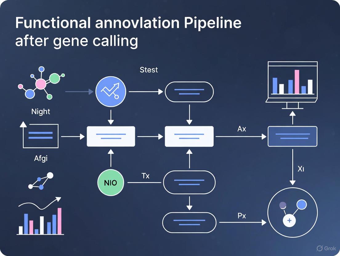
Workflow Visualization and Data Interpretation
The following diagram illustrates the generalized logical workflow of an integrated functional annotation pipeline, synthesizing the steps from the described protocols.
Functional Annotation Workflow Logic
Visualizing the MicrobeAnnotator Iterative Process
MicrobeAnnotator employs a specific sequential strategy to maximize annotation yield efficiently. The DOT script below models this iterative process.
Iterative Annotation Strategy
The Critical Role of Annotation in Translational Research and Drug Discovery
In the landscape of modern translational research, the path from genomic sequencing to actionable biological insights is paved by functional annotation. Following gene calling, which identifies the structural elements within a genome, functional annotation adds biologically relevant meaning to these predicted features, such as putative function, presence of specific domains, cellular localization, and involvement in metabolic pathways [1]. This process is indispensable for drug discovery, enabling researchers to prioritize targets, understand disease mechanisms, and predict potential side effects. The integration of diverse annotation approaches—from homology searches to orthology assignment and protein signature identification—provides a consensus view that is critical for validating findings and building reliable models for therapeutic development [1]. As stem cell research and single-cell genomics advance, sophisticated annotation methods further allow scientists to evaluate the congruence of stem cell-derived models with in vivo biology, ensuring these powerful tools accurately recapitulate human disease for therapy development [4].
Functional Annotation Pipeline: Components and Workflow
A typical functional annotation workflow integrates multiple methods to analyze a protein sequence from complementary angles. The FA-nf pipeline, implemented via the Nextflow workflow management engine, exemplifies a scalable framework that combines various tools into a cohesive, reproducible process [1]. This pipeline begins with a protein sequence FASTA file and an optional structural annotation file in GFF3 format, proceeding through several critical stages.
Key integrated approaches include:
- Homology Search: Using tools like BLAST+ or DIAMOND to search against reference databases such as UniProt or NCBI NR, identifying sequences with significant similarity [1].
- Orthology Assignment: Leveraging resources like eggNOG to assign proteins to known orthologous groups, inferring functional annotation from these evolutionarily related proteins [1].
- Protein Signature Identification: Utilizing InterProScan to identify known protein domains and motifs by searching against diagnostic models, including hidden Markov models (HMM) from databases such as PANTHER, Pfam, and SUPERFAMILY [1].
The following workflow diagram illustrates the integrated process of a functional annotation pipeline, from raw sequence data to biologically relevant insights:
Comparative Analysis of Annotation Pipelines and Tools
The bioinformatics community has developed numerous frameworks for functional annotation, each with distinct strengths, installation requirements, and specialized applications. These tools can be broadly categorized into web-based services with user-friendly interfaces and locally installed pipelines that offer greater control and scalability for large datasets [1].
Table 1: Comparative Analysis of Functional Annotation Pipelines
| Program/Pipeline | Installation | Key Software Components | Primary Databases | Specialized Applications |
|---|---|---|---|---|
| FA-nf | Local Installation | BLAST+, DIAMOND, InterProScan, KOFAM, SignalP, TargetP | Custom (typically NCBI BLAST DBs, InterPro, UniProt-GOA) | General-purpose annotation with containerization for reproducibility [1] |
| Blast2GO | Local installation & Web/Cloud services | BLAST+, InterProScan, Blast2GO specific software | NCBI BLAST DBs, InterPro, GO | Subscription tool with visualization dashboard and gene structural annotation options [1] |
| eggNOG mapper | Web service & Local installation | DIAMOND, HMMER | eggNOGdb (multiple sources), GO, PFAM, SMART, COG | Command-line tool and REST API; suitable for orthology-based annotation [1] |
| MicrobeAnnotator | Local Installation | BLAST+, DIAMOND, KOFAM | SwissProt/TrEMBL, RefSeq, KEGG | Specialized for microbiome datasets [1] |
| PANNZER2 | Web Service | SANSparallel | UniProt, UniProt-GOA, GO, KEGG | Command-line tool available for querying service [1] |
| GenSAS | Web Service | BLAST+, DIAMOND, InterProScan, SignalP, TargetP | SwissProt/TrEMBL, RefSeq, RepBase | Online platform with structural annotation and visualization capabilities [1] |
Web-accessible tools like GenSAS and PANNZER2 offer convenience but may limit the number of sequences that can be processed, while locally installed pipelines such as FA-nf and MicrobeAnnotator provide scalability for high-throughput analysis at the cost of requiring more computational expertise [1]. The FA-nf pipeline distinguishes itself through its use of software containerization, which mitigates installation challenges and ensures reproducibility across different computing environments [1].
Advanced Annotation Applications in Translational Research
LA3: Language-Based Automatic Annotation Augmentation
Recent advancements in artificial intelligence are addressing the critical bottleneck of annotation scarcity in biological research. The LA3 (Language-based Automatic Annotation Augmentation) framework leverages large language models to systematically rewrite and augment molecular annotations, creating more varied sentence structures and vocabulary while preserving essential information [5]. This approach has demonstrated remarkable efficacy in training AI models for tasks such as text-based de novo molecule generation and molecule captioning. Experimental results show that models trained on LA3-augmented datasets can outperform state-of-the-art architectures by up to 301%, highlighting the transformative potential of enriched annotations for AI-driven drug discovery [5].
Single-Cell Annotation in Stem Cell Research
Stem cell-derived models represent a powerful tool for studying biology and developing cell-based therapies. However, determining precisely which cell types are present and how closely they recapitulate in vivo cells remains challenging. Single-cell genomics coupled with advanced annotation methods provides a framework for evaluating this congruence [4]. These annotation approaches face particular challenges in stem cell-derived models, including heterogeneity, incomplete differentiation, and the dynamic nature of cellular identities. Emerging recommendations emphasize the application of cell manifolds and integrated annotation strategies to better capture the complexity of stem cell systems for therapeutic development [4].
The following diagram illustrates the experimental validation workflow for confirming annotated gene functions:
Research Reagent Solutions for Annotation Experiments
Table 2: Essential Research Reagents and Resources for Functional Annotation
| Reagent/Resource | Function/Application | Key Features |
|---|---|---|
| UniProt Knowledgebase | Protein sequence and functional information repository | Manually annotated records with experimental evidence; computational annotation with taxonomy-specific rules [1] |
| InterPro | Protein signature database integrating multiple resources | Combines protein domain, family, and functional site predictions from PANTHER, Pfam, SUPERFAMILY, and other databases [1] |
| eggNOG | Orthology database and functional annotation resource | Evolutionary genealogy of genes; broad taxonomic coverage with functional inference from orthologs [1] |
| KEGG (Kyoto Encyclopedia of Genes and Genomes) | Pathway mapping and functional assignment | Maps annotated genes to metabolic pathways; useful for understanding systemic biological functions [1] |
| NCBI NR (Non-Redundant) Database | Comprehensive protein sequence database | Large-scale reference for homology searches; contains sequences from multiple sources [1] |
| SignalP | Subcellular localization prediction | Predicts presence and location of signal peptide cleavage sites [1] |
| TargetP | Subcellular localization prediction | Predicts subcellular localization of eukaryotic proteins [1] |
Implementation Protocols for Functional Annotation
Protocol 1: Basic Functional Annotation Using FA-nf Pipeline
Objective: To perform comprehensive functional annotation of protein sequences from a de novo genome assembly.
Materials and Input Requirements:
- Protein sequences in FASTA format (required)
- Structural annotation file in GFF3 format (optional but recommended)
- Linux computational environment with Docker/Singularity support
- Access to reference databases (UniProt, InterPro, KEGG, etc.)
Methodology:
- Pipeline Setup: Configure the Nextflow workflow management engine and associated container images. Software containers are provided for each process to ensure reproducibility and avoid dependency conflicts [1].
- Input Validation and Preprocessing: Verify and clean input GFF files using the AGAT Toolkit to ensure compatibility with downstream processes, as GFF3 formats vary significantly between sources like ENSEMBL and NCBI [1].
- Parallelized Execution: Launch the FA-nf pipeline with appropriate computational resources. Adjust chunk size parameters based on sequence number and length to optimize performance on available HPC infrastructure [1].
- Multi-Method Annotation: The pipeline automatically executes in parallel:
- Homology searches using BLAST+ and DIAMOND against reference databases
- Domain and motif identification using InterProScan
- Orthology assignment using eggNOG mapper
- Pathway mapping using KEGG
- Subcellular localization prediction using SignalP and TargetP [1]
- Data Integration and Output Generation: Results from all processes are stored in a relational database (SQLite or MySQL/MariaDB) with final annotation reports generated in GFF3, plain text, and HTML formats [1].
Troubleshooting Tips:
- For shared file systems in HPC environments, use MySQL/MariaDB instead of SQLite for better performance [1].
- Adjust CPU cores, memory allocation, and maximum runtime in the nextflow.config file based on sequence characteristics [1].
- Test parameters with debug mode (debugSize parameter) before full execution to optimize resource allocation [1].
Protocol 2: Annotation Augmentation Using LA3 Framework
Objective: To enhance existing molecular annotations using large language models for improved AI training in drug discovery applications.
Materials:
- Established dataset with molecular annotations (e.g., ChEBI)
- Computational environment with access to large language models
- Benchmark architecture for model training (e.g., MolT5-based models)
Methodology:
- Dataset Preparation: Extract existing molecular annotations from established datasets, preserving essential molecular information while identifying areas for linguistic diversification [5].
- Systematic Rewriting: Apply the LA3 framework to rewrite annotations while maintaining scientific accuracy. This process creates more varied sentence structures and vocabulary, enhancing the linguistic diversity of the training data [5].
- Dataset Validation: Ensure augmented annotations preserve critical molecular information while improving linguistic patterns suitable for AI model training.
- Model Training: Train molecular translation models (e.g., LaMolT5) on the enhanced dataset (e.g., LaChEBI-20) to learn mappings between molecular representations and augmented annotations [5].
- Performance Evaluation: Assess model performance on text-based de novo molecule generation and molecule captioning tasks, comparing results against state-of-the-art benchmarks [5].
Functional annotation represents the critical bridge between genomic sequences and biologically meaningful insights that drive translational research and drug discovery. As the field advances, integrated pipelines that combine multiple annotation approaches, augmented datasets enhanced by natural language processing, and specialized methods for emerging technologies like single-cell genomics are collectively expanding the boundaries of what can be achieved. The implementation of robust, reproducible annotation workflows, coupled with appropriate experimental validation, provides the foundation for identifying novel therapeutic targets, understanding disease mechanisms, and ultimately accelerating the development of new treatments. As these methodologies continue to evolve, they will undoubtedly play an increasingly vital role in personalized medicine and the next generation of biomedical breakthroughs.
Following gene calling in genomic sequencing pipelines, functional annotation serves as the critical next step, translating raw variant calls into meaningful biological insights. This process involves predicting the potential impact of genetic variants on protein structure, gene expression, cellular functions, and biological processes [6]. The landscape of genetic variation encompasses both protein-coding regions and vast non-coding territories, each requiring specialized annotation strategies to elucidate their roles in health and disease [6].
As sequencing technologies advance, the scope of functional annotation has expanded dramatically beyond protein-coding genes to include diverse regulatory elements that orchestrate gene expression. The integration of multi-omics approaches—combining genomic, epigenomic, transcriptomic, and proteomic data—now provides a more comprehensive view of biological systems than genomic analysis alone [7]. This evolution is particularly crucial given that the majority of human genetic variation resides in non-protein coding regions of the genome, presenting both a challenge and opportunity for discovering novel therapeutic targets [6].
Key Genomic Feature Classes
Protein-Coding Genes
Protein-coding genes represent the best-understood class of genomic features, with functional annotation primarily focusing on variants that alter amino acid sequences and affect protein function or structure [6]. Traditional approaches for automatic functional annotation in non-model species or newly sequenced genomes rely on homology transfer based on sequence similarity using tools like Blast2GO, identification of conserved domains, and InterPro2GO [8].
Accurately determining orthologous relationships between genes allows for more robust functional assignment of predicted genes, often described as the "orthology-function conjecture" [8]. Guided by collinear genomic segments, researchers can infer orthologous protein-coding gene pairs (OPPs) between species, which serve as proxies for transferring functional terms from well-annotated genomes to newly sequenced ones [8].
Non-Coding Regulatory Elements
Beyond protein-coding genes, the genome contains numerous regulatory elements that control gene expression, including:
- Promoter and enhancer sequences that initiate and amplify transcription [6]
- Non-coding RNAs with diverse regulatory functions [6]
- Transcription factor binding sites (TFBS) that mediate protein-DNA interactions [6]
- DNA methylation sites that influence epigenetic regulation [6]
- Transposable elements that can reshape genomic architecture [6]
The functional annotation of variants lying outside protein-coding genomic regions (intronic or intergenic) and their potential co-localization with these regulatory elements is essential for understanding their impact on phenotype [6]. Advanced sequencing techniques such as Hi-C provide insights into the three-dimensional organization of the genome, mapping global physical interactions between different genomic regions and revealing long-range interactions between regulatory elements and gene promoters [6].
Table 1: Key Genomic Feature Classes and Their Functional Characteristics
| Feature Class | Genomic Location | Primary Function | Annotation Challenges |
|---|---|---|---|
| Protein-Coding Genes | Exonic regions | Encode protein sequences | Missense variant interpretation, splice site determination |
| Promoters | 5' upstream of genes | Transcription initiation | Identifying core vs. regulatory regions |
| Enhancers | Intergenic or intronic | Enhance transcription levels | Tissue-specific activity, target gene assignment |
| Non-coding RNAs | Various genomic locations | Gene regulation | Functional classification, target identification |
| TFBS | Throughout genome | Transcription factor binding | Cell-type specificity, binding affinity prediction |
| DNA Methylation Sites | CpG islands, gene bodies | Epigenetic regulation | Tissue-specific patterns, functional consequences |
The field of genomic annotation employs diverse computational tools and databases, each with specific strengths for different annotation tasks. Ensembl Variant Effect Predictor (VEP) and ANNOVAR represent widely-used tools that can directly handle raw VCF files and are well-suited for large-scale annotation tasks, such as whole-genome and whole-exome sequencing projects [6].
The landscape of variant annotation tools is complex, with different tools targeting different genomic regions and performing different types of analyses. Some tools specialize in annotating exonic regions, while others concentrate on non-exonic intragenic regions or comprehensive tools that annotate variants across all genomic regions [6]. A less represented category includes tools designed to assess the cumulative impact of multiple variants, analyzing their collective effect on genes, pathways, or biological processes, which is particularly important for understanding complex traits and polygenic diseases [6].
Table 2: Quantitative Comparison of Functional Annotation Approaches
| Annotation Method | Primary Application | Data Input Requirements | Output Metrics | Limitations |
|---|---|---|---|---|
| Sequence Homology (BLAST) | Gene function prediction | Protein or nucleotide sequences | E-value, percent identity | May miss distant homologs |
| Orthology Inference (OrthoMCL) | Cross-species function transfer | Multiple genome sequences | Orthologous groups | Sensitive to parameter settings |
| Collinearity Analysis (i-ADHoRe) | Positional orthology detection | Genome assemblies and annotations | Collinear segments | Requires multiple related genomes |
| Machine Learning (DeepVariant) | Variant calling | Sequencing read alignments | Variant quality scores | Computational intensity |
| Multi-omics Integration | Systems biology modeling | Diverse molecular datasets | Pathway enrichment scores | Data normalization challenges |
Experimental Protocols for Comprehensive Functional Annotation
Protocol 1: Genome-Wide Collinearity Detection for Orthology Inference
Objective: Identify positional orthologous regions across multiple genomes to facilitate reliable functional annotation transfer.
Materials and Reagents:
- Genome assemblies for target species and related organisms
- High-performance computing infrastructure
- Multiple genome alignment software (LASTZ/MULTIZ)
- Collinearity detection tools (i-ADHoRe v3.0)
Methodology:
Data Preparation: Obtain genome sequences for multiple related species meeting quality thresholds (preferably chromosome-level assemblies or scaffold N50 >260 kbp) [8].
Multiple Whole-Genome Alignment:
- Perform pairwise whole-genome alignments against the reference genome using LASTZ, optimized for whole-genome alignments [8].
- Process pairwise alignments through alignment "chaining" and "netting" pipelines to ensure each reference base aligns with at most one base in other genomes [8].
- Join resulting pairwise alignments using MULTIZ, guided by phylogenetic relationships [8].
Collinearity Detection:
- Use genomic markers (multiple alignment anchors or protein-coding gene families) with i-ADHoRe to identify homologous regions [8].
- Set parameters including alignmentmethod = gg4, anchorpoints = 3, gapsize = 30 for MAA-based or 10 for protein-based, probcutoff = 0.01 [8].
- Identify n-way collinear segments (n∈{3,4,…,15}) representing multiple species level of collinear segments [8].
Orthology Inference:
- Designate protein-coding genes as orthologous pairs if they share sequence similarity (BLASTP E-value = 1E-05) and at least 50% of their sequences locate in the same collinear segment [8].
- Apply additional optional conditions including bidirectional best hits and confirmation using different marker types [8].
Protocol 2: Integrated Coding and Non-Coding Variant Annotation
Objective: Perform comprehensive functional annotation of both coding and non-coding genetic variants from whole-genome sequencing data.
Materials and Reagents:
- Variant Calling Format (VCF) files from WGS/WES
- Functional annotation tools (Ensembl VEP, ANNOVAR)
- Regulatory element databases (ENCODE, Roadmap Epigenomics)
- High-performance computing resources
Methodology:
Variant Effect Prediction:
Coding Variant Analysis:
- Focus on variants that alter amino acid sequences (missense, nonsense).
- Predict potential impact on protein function using pathogenicity prediction scores.
- Assess effect on protein structure and functional domains.
Non-Coding Variant Analysis:
- Annotate variants overlapping known regulatory elements from specialized databases [6].
- Identify variants in promoter regions, enhancers, non-coding RNAs, DNA methylation sites, TFBS, and transposable elements [6].
- Prioritize variants based on evolutionary conservation and functional genomic evidence.
Regulatory Impact Assessment:
- Integrate chromatin interaction data (Hi-C) to connect non-coding variants with potential target genes [6].
- Correlate with transcriptomic data to assess effect on gene expression.
- Utilize machine learning approaches to predict variant disruptiveness.
Visualization of Functional Annotation Workflows
Comprehensive Functional Annotation Pipeline
Functional Annotation Pipeline: This workflow illustrates the comprehensive process from raw sequencing data to biological insights, integrating both coding and non-coding variant analysis with multi-omics data.
Orthology-Based Function Transfer Protocol
Orthology-Based Function Transfer: This diagram shows the protocol for transferring functional annotations from well-characterized genomes to newly sequenced ones using collinearity and orthology inference.
Research Reagent Solutions
Table 3: Essential Research Reagents and Computational Tools for Functional Annotation
| Reagent/Tool | Category | Primary Function | Application Context |
|---|---|---|---|
| Ensembl VEP | Bioinformatics Tool | Variant effect prediction | Annotates variants with functional consequences |
| ANNOVAR | Bioinformatics Tool | Variant annotation | Handles VCF files from WGS/WES projects |
| LASTZ/MULTIZ | Alignment Software | Multiple genome alignment | Identifies conserved genomic segments |
| i-ADHoRe | Computational Algorithm | Collinearity detection | Finds homologous regions across genomes |
| OrthoMCL | Orthology Tool | Gene family clustering | Identifies orthologous groups across species |
| Hi-C Data | Genomic Resource | 3D genome organization | Maps physical interactions between genomic regions |
| ENCODE Data | Regulatory Annotation | Regulatory element mapping | Provides reference regulatory element annotations |
The Impact of Assembly Quality and Evidence on Final Annotation Outcomes
The functional annotation of predicted genes represents a critical phase in genomic studies, bridging the gap between raw sequence data and biological insight. The quality and completeness of this annotation are not predetermined but are profoundly influenced by two interdependent factors: the quality of the genome assembly itself and the nature and breadth of the experimental evidence integrated into the annotation pipeline. High-quality assembly provides a solid architectural foundation, enabling the accurate placement and structural determination of gene models. Concurrently, multifaceted evidence, including transcriptomic and protein data, is essential for corroborating these models and elucidating functional elements. This application note details protocols and analytical measures for evaluating and optimizing these factors within eukaryotic genome annotation projects, providing a framework for generating biologically meaningful and reliable annotations to support downstream research and drug development.
Quantitative Measures for Annotation Assessment and Management
Effective annotation management requires moving beyond simple gene counts to quantitative measures that track changes and quality over time. The table below summarizes key metrics for intra- and inter-genome annotation analysis.
Table 1: Quantitative Measures for Genome Annotation Management
| Measure Name | Application Scope | Description | Interpretation |
|---|---|---|---|
| Annotation Edit Distance (AED) | Intra-genome | Quantifies the structural change to a specific gene annotation between two pipeline runs or releases [9]. | Ranges from 0 (identical) to 1 (completely different). A low average AED indicates stable annotations [9]. |
| Annotation Turnover | Intra-genome | Tracks the addition and deletion of gene annotations from one release to the next [9]. | Supplements gene counts; can detect "resurrection events" where annotations are deleted and later re-created [9]. |
| Splice Complexity | Inter-genome | Quantifies the transcriptional complexity of a gene based on its alternative splicing patterns, independent of sequence homology [9]. | Allows for novel, global comparisons of alternative splicing across different genomes [9]. |
| Assembly-Induced Changes | Intra-genome | Identifies annotations altered solely due to changes in the underlying genomic assembly [9]. | Helps separate artifacts of assembly improvement from genuine annotation refinements. |
These measures reveal significant variation in annotation stability across organisms. For instance, the D. melanogaster genome is highly stable, with 94% of genes in one release remaining unaltered since 2004, and only 0.3% altered more than once. In contrast, 58% of C. elegans annotations were modified over a similar period, with 32% altered more than once, despite gene counts changing by less than 3% [9]. Furthermore, the magnitude of changes can differ; while fewer D. melanogaster annotations were revised, the changes tended to be larger (average AED 0.092) compared to C. elegans (average AED 0.058) [9].
The NCBI Eukaryotic Genome Annotation Pipeline: A Reference Protocol
The NCBI Eukaryotic Genome Annotation Pipeline provides a robust, publicly available framework that exemplifies the integration of assembly and evidence. The following protocol describes its key steps, which can be adapted for custom annotation projects [10].
Inputs: Genome Assembly and Evidence Data
- Source of Genome Assemblies: The pipeline annotates RefSeq assemblies, which are copies of public genome assemblies from INSDC (DDBJ, ENA, and GenBank). Note that unplaced scaffolds shorter than 1000 bp may be excluded from the RefSeq copy under specific conditions [10].
- Evidence Data Curation:
- Transcripts: The evidence set includes RefSeq transcripts (NM, NR), GenBank transcripts from taxonomically relevant divisions, and ESTs from dbEST. Sequences with potential contamination or low quality are screened out [10].
- Proteins: The set includes known RefSeq proteins and GenBank proteins from cDNA. Highly repetitive sequences are removed [10].
- RNA-Seq Data: When available, RNA-Seq reads from SRA are aligned. Projects spanning wide ranges of tissues and developmental stages are preferred, with a focus on untreated/non-diseased samples [10].
Core Computational Workflow
- Masking: The genomic sequence is masked to identify and handle repetitive elements. Human and mouse genomes are masked with RepeatMasker using Dfam libraries, while other species are masked with WindowMasker [10].
- Sequence Alignment:
- Transcripts: Cleaned transcripts are aligned to the genome using BLAST to find locations, followed by global re-alignment with Splign to refine splice sites. Only the best-placed (rank 1) alignment is selected [10].
- Proteins: Proteins are aligned locally with BLAST and then re-aligned globally using ProSplign [10].
- RNA-Seq Reads: RNA-Seq reads are aligned with STAR. Alignments with identical splice structures are collapsed, and rare introns likely to be noise are filtered out [10].
- Long Reads: Transcriptomics long reads (PacBio, Oxford Nanopore) are aligned with Minimap2, and the best-placed alignment above 85% identity is selected [10].
- Gene Model Prediction: The alignment evidence is passed to the Gnomon gene prediction software. Gnomon first chains non-conflicting alignments into putative models. It then performs HMM-based ab initio prediction to extend predictions or create models in regions with no supporting evidence but with long open reading frames. This initial set is refined by alignment against a non-redundant protein database, and the prediction steps are repeated [10].
- Model Selection and Integration: The final annotation is built by combining pre-existing RefSeq sequences and well-supported Gnomon predictions.
- Priority is given to known and curated RefSeq models.
- Gnomon predictions are included if they meet quality thresholds, such as being fully or partially supported by evidence, or being pure ab initio predictions with high coverage hits to Swiss-Prot proteins.
- Conflicting or poorly supported models are excluded [10].
Outputs and Functional Annotation
- Annotation Products: The pipeline produces gene, RNA, and CDS features on the genome. Models are assigned accessions: known RefSeq transcripts (NM, NR), model RefSeq transcripts (XM, XR), and predicted proteins (XP_) [10].
- Functional Naming: Genes from known RefSeq inherit their existing names and locus types. Genes represented by predicted models are named based on homology to Swiss-Prot proteins. Models with indels or frameshifts may be labeled as pseudogenes or, if they have strong homology, as "PREDICTED: LOW QUALITY PROTEIN" [10].
A Case Study in Assembly and Annotation: The Taohongling Sika Deer
A recent high-quality chromosome-scale genome assembly of the Taohongling Sika deer (Cervus nippon kopschi) demonstrates the impact of modern sequencing and assembly methods on annotation outcomes [11].
Experimental Protocol for Genome Assembly
- Sequencing Technology: The assembly utilized a combination of PacBio Sequel II (HiFi long reads, 36.22x coverage), Illumina NovaSeq 6000 (short reads, 41.82x coverage), and Hi-C sequencing (82.66x coverage) from muscle tissue [11].
- Assembly Process: Contig-level assembly was performed with HIFIASM (v0.16.1) using PacBio HiFi reads, resulting in a primary contig assembly with an N50 of 61.59 Mb. Hi-C data was then used to scaffold and assign 97.23% of the assembled sequence onto 34 chromosomes, achieving a scaffold N50 of 85.86 Mb [11].
- Assembly Quality Assessment:
Annotation Results and Analysis
The high-quality assembly directly enabled a comprehensive annotation [11]:
- Repeat Content: 46.19% of the genome was identified as repetitive sequences.
- Protein-Coding Genes: 22,890 genes were predicted, with 97.16% (22,240 genes) receiving functional annotations.
- Non-Coding RNAs: 63,473 noncoding RNAs were identified.
Table 2: Impact of Assembly Quality on Annotation Metrics in Sika Deer
| Assembly Metric | Value | Impact on Annotation |
|---|---|---|
| Sequencing Coverage | 36.22x PacBio HiFi, 41.82x Illumina | Provides high accuracy for base calling and structural variant resolution. |
| Contig N50 | 61.59 Mb | Greatly reduces gene fragmentation, allowing complete genes to be assembled on single contigs. |
| Scaffold N50 / Chromosome Assignment | 85.86 Mb / 97.23% anchored | Enables accurate analysis of gene order, synteny, and regulatory contexts. |
| BUSCO Completeness | 98.0% | Indicates near-complete representation of universal single-copy orthologs, minimizing gene loss. |
| Mapping Rate of Short Reads | 99.52% | Suggests high base-level accuracy, reducing frameshifts and erroneous stop codons in gene models. |
Critical Considerations for Comparative Genomics
The choice of annotation method is a critical, often overlooked variable that directly impacts downstream comparative analyses. A key study demonstrated that using genomes annotated with different methods (i.e., "annotation heterogeneity") within a comparative analysis can dramatically inflate the apparent number of lineage-specific genes—by up to 15-fold in some case studies [12]. These genes, which appear unique to one species or clade, are often interpreted as sources of genetic novelty and unique adaptations. However, annotation heterogeneity can create artifacts where orthologous DNA sequences are annotated as a gene in one species but not in another, falsely appearing to be lineage-specific [12]. Therefore, for meaningful comparative genomics, it is crucial to either use annotations generated by a uniform pipeline or to account for methodological differences when interpreting results.
The Scientist's Toolkit: Research Reagent Solutions
Table 3: Essential Tools and Reagents for Modern Genome Assembly and Annotation
| Tool / Reagent | Function in Workflow | Example Use Case |
|---|---|---|
| PacBio HiFi Reads | Long-read sequencing providing high accuracy; ideal for resolving complex repeats and assembling complete contigs. | Generating the primary contig assembly for the Taohongling Sika deer [11]. |
| Hi-C Sequencing | Captures chromatin conformation data; used to scaffold contigs into chromosome-scale assemblies. | Anchoring 97.23% of the Sika deer assembly onto 34 chromosomes [11]. |
| Illumina Short Reads | High-accuracy short reads; used for error correction of long reads and base-level quality assessment. | Polishing the Sika deer assembly and calculating mapping rates/coverage [11]. |
| Iso-Seq (PacBio) | Full-length transcriptome sequencing without assembly; provides direct evidence for splice variants and UTRs. | Informing gene model prediction; part of the NCBI pipeline when data is available [10]. |
| RNA-Seq from SRA | Provides evidence for transcribed regions and splice junctions; crucial for supporting and refining gene models. | Aligned with STAR in the NCBI pipeline to support Gnomon predictions [10]. |
| BUSCO | Software to assess genome completeness based on evolutionarily informed expectations of gene content. | Benchmarking the completeness of the Sika deer assembly (98.0%) [11]. |
| Gnomon | NCBI's HMM-based gene prediction program that integrates evidence and performs ab initio prediction. | Core of the NCBI annotation pipeline for predicting gene models [10]. |
| Splign / ProSplign | Algorithms for computationally mapping transcripts and proteins to a genome to determine precise exon-intron structures. | Used in the NCBI pipeline for global alignment of transcripts and proteins [10]. |
In the final stages of a functional annotation pipeline, following the initial gene calling phase, researchers must systematically interpret and catalog predicted genomic elements. This process relies on standardized file formats to represent features such as genes, transcripts, exons, and regulatory elements in a computationally accessible manner. The General Feature Format (GFF) and Gene Transfer Format (GTF) are two cornerstone file specifications that enable this critical step, serving as the lingua franca for exchanging genomic annotations between visualization tools, databases, and analysis software [13] [14]. While they appear structurally similar at a glance, understanding their nuanced differences—rooted in historical development and specific use cases—is essential for selecting the appropriate format to ensure analytical accuracy and pipeline interoperability. Misapplication of these formats can lead to erroneous data interpretation, processing failures in downstream tools, and ultimately, compromised research conclusions [15] [16].
The evolution of these formats mirrors the increasing complexity of genomic science. The original GFF specification emerged from a 1997 meeting at the Isaac Newton Institute, designed as a common format for sharing information between gene-finding sensors and predictors [14]. This format has since undergone significant revisions, culminating in GFF3, while GTF developed as a more gene-centric derivative, often termed "GFF2.5," to meet the specific needs of collaborative genome annotation projects around the year 2000 [13] [17]. Today, these files are not merely passive containers of data but active components in sophisticated annotation pipelines, interfacing with tools for transcriptome assembly, functional prediction, and comparative genomics [18] [19].
Historical Development and Format Specification
The Evolution of GFF and GTF
The phylogenetic development of GFF and GTF formats reveals a trajectory of increasing specificity and structural complexity. The inaugural GFF version 0, used before November 1997, established the basic paradigm of a tab-delimited, nine-field format for describing genomic features, though it lacked the modern source field [14]. The subsequent GFF1 specification, formalized in 1998, introduced the source field and the foundational nine-column structure that persists in contemporary versions, albeit with an optional group attribute for feature grouping [14]. A significant evolutionary leap occurred with GFF2 in 2000, which standardized the attribute column formatting and enhanced the specification's robustness for increasingly complex genomic annotations [14].
The GTF format (GFF2.5) emerged around 2000 as a specialized branch from the GFF2 lineage, developed specifically to address the requirements of genome annotation projects like Ensembl [13] [17]. This format imposed stricter conventions on the attribute column, mandating specific tags such as gene_id and transcript_id to unambiguously define gene model hierarchies. The most recent major revision, GFF3, was developed to overcome GFF2's limitations in representing complex biological relationships, most notably through the introduction of explicit parent-child feature relationships using ID and Parent attributes, enabling true multi-level hierarchies of genomic features [13] [14]. This progression from a flat feature representation to a structured hierarchy mirrors the growing understanding of genomic complexity, from simple gene models to intricate networks of alternative transcripts, regulatory elements, and non-coding RNAs.
Structural Anatomy of GFF and GTF Files
Despite their historical divergence, GFF and GTF files share a common structural framework consisting of nine mandatory tab-delimited columns that provide a complete descriptor for each genomic feature [13] [20]. The table below delineates the function and content of each column:
Table: Column-by-Column Specification of GFF/GTF Format Structure
| Column Number | Column Name | Description | Format Notes |
|---|---|---|---|
| 1 | SeqID (seqname) | Reference sequence identifier (chromosome/scaffold) | Must match corresponding FASTA file [16] |
| 2 | Source | Algorithm/database that generated the feature | e.g., "AUGUSTUS," "Ensembl"; "." for unknown [20] |
| 3 | Feature (type) | Biological feature type | e.g., "gene," "exon," "CDS"; should use Sequence Ontology terms [16] |
| 4 | Start | Start coordinate of the feature | 1-based, inclusive [13] [20] |
| 5 | End | End coordinate of the feature | 1-based, inclusive [13] [20] |
| 6 | Score | Confidence measure or statistical score | Floating-point or "." if null/not applicable [13] |
| 7 | Strand | Genomic strand orientation | "+", "-", or "." for non-stranded features [13] |
| 8 | Phase (Frame) | Reading frame offset for CDS features | "0", "1", "2", or "." for non-CDS features [13] [20] |
| 9 | Attributes | Semicolon-separated key-value pairs | Meta-information (IDs, parents, names) [13] |
A critical distinction between the formats lies in their coordinate system. Both GFF and GTF use a 1-based, inclusive coordinate system, where the first nucleotide of a sequence is position 1, and an interval includes both the start and end positions [20]. This contrasts with other bioinformatics formats like BED, which use a 0-based, half-open coordinate system (where the start coordinate is zero-indexed and the end coordinate is exclusive) [13]. This fundamental difference necessitates careful handling during format conversion to prevent off-by-one errors that could misalign genomic features.
The attribute column (column 9) constitutes the most complex and format-divergent field, housing the semantic relationships between features. In GFF3, attributes follow a strict key=value syntax with multiple values comma-separated (e.g., ID=exon00001;Parent=mRNA00001;Name=EDEN) [20] [16]. In GTF, the attribute syntax is less standardized, often mixing key "value"; (with quotes and trailing semicolon) and key value; formats, though it mandates specific tags like gene_id and transcript_id for gene model representation [20] [21].
Key Differences Between GFF and GTF
Structural and Philosophical Divergences
While GFF and GTF share a common ancestry and structural framework, they diverge significantly in their philosophical approach to genomic annotation. GTF adopts a gene-centric perspective, with its specification deliberately optimized for representing eukaryotic gene models with precise exon-intron structures [13] [15]. This focus is evident in its requirement for specific attribute tags—gene_id, transcript_id—that explicitly define the relationships between transcripts and their parent genes, creating a rigid but unambiguous hierarchy necessary for transcriptome analysis and gene expression quantification [13] [17]. Consequently, GTF files are particularly valuable in RNA-seq workflows where tools like Cufflinks, StringTie, and TopHat rely on these consistent identifiers to assemble transcripts and quantify expression levels [13] [18].
In contrast, GFF3 embraces a genome-centric paradigm designed to annotate the full complexity of genomic features beyond protein-coding genes alone [13] [15]. Its flexibility allows for the representation of diverse elements including regulatory regions (promoters, enhancers), repetitive elements, non-coding RNAs, and epigenetic marks within a unified framework [22]. This comprehensive scope is facilitated by GFF3's implementation of explicit parent-child relationships through ID and Parent attributes, which can represent complex multi-level hierarchies such as gene → transcript → exon → CDS, or even more intricate relationships in the case of alternatively spliced transcripts or polycistronic genes [16] [14]. This makes GFF3 the preferred format for genome browsers, comparative genomics, and comprehensive genome annotation projects that require representing biological reality in its full complexity.
Practical Implications for Genomic Analyses
The philosophical differences between GFF and GTF translate directly to practical consequences in bioinformatics workflows. The stricter, more standardized nature of GTF ensures greater consistency and reliability when processed by different gene-centric tools, reducing the likelihood of parsing errors in pipelines focused on transcriptomics [15] [17]. However, this specificity comes at the cost of flexibility—GTF is less adept at representing the broad universe of genomic elements beyond genes and their subfeatures.
The richer hierarchical model of GFF3 comes with a computational cost; parsing and interpreting these relationships requires more sophisticated software capable of reconstructing the feature graph from the parent-child relationships [22] [14]. While this offers superior representational power, it can lead to compatibility issues with tools expecting the simpler, flatter structure of GTF files. Furthermore, the flexibility of GFF3's attribute column can result in "format flavors," where different annotation tools or communities implement semi-compatible variations, potentially causing interoperability challenges without careful validation [14] [21].
Table: Comparative Analysis of GFF3 and GTF Format Characteristics
| Characteristic | GFF3 | GTF |
|---|---|---|
| Primary Focus | Whole-genome annotation [13] [15] | Gene structure annotation [13] [15] |
| Attribute Syntax | key=value pairs [20] [16] |
Mixed: key "value"; and key value; [20] [21] |
| Hierarchy Representation | Explicit ID/Parent relationships [16] [14] |
Implicit via gene_id, transcript_id [13] [17] |
| Coordinate System | 1-based, inclusive [13] [20] | 1-based, inclusive [13] [20] |
| Flexibility | High (diverse feature types) [13] [15] | Low (gene-centric) [13] [15] |
| Ideal Use Cases | Genome browsing, comparative genomics, regulatory annotation [13] [22] | Transcriptome assembly, RNA-seq, expression quantification [13] [18] |
Application in Functional Annotation Pipelines
Integration with Annotation Tools and Workflows
In a complete functional annotation pipeline, GFF and GTF files serve as the critical junction between structural gene prediction and functional characterization. Modern annotation tools produce outputs in these formats, which then feed into downstream processes including functional term assignment, motif identification, and comparative genomic analyses [18] [19]. The decision between GFF3 and GTF often depends on the annotation methodology employed. Evidence-based approaches utilizing RNA-seq data, such as StringTie and Scallop, typically output GTF files that describe transcript structures assembled from RNA-seq read alignments [18]. Similarly, annotation transfer tools like Liftoff and TOGA, which map annotations from a reference genome to a target assembly, often use GFF3 as both input and output to preserve complex feature relationships during the transfer process [18].
Ab initio prediction tools demonstrate varied format preferences reflecting their methodological foundations. BRAKER3, which combines protein homology and RNA-seq evidence with GeneMark-ETP and AUGUSTUS, generates GFF3 output containing comprehensive gene models [19]. In contrast, newer approaches like Helixer, which employs deep learning models for cross-species gene prediction, also produces GFF3 format, underscoring its status as the current community standard for comprehensive genome annotation [19]. This convergence toward GFF3 reflects the growing recognition that high-quality genome annotations must capture biological complexity beyond simple coding sequences, including untranslated regions (UTRs), non-coding RNAs, and regulatory elements.
Protocol: Processing and Validating Annotation Files
A critical step in any annotation pipeline involves the quality control and processing of GFF/GTF files to ensure downstream compatibility. The following protocol outlines a standardized workflow for handling these files:
Step 1: File Validation and Syntax Checking
- For GFF3 files, use standalone validators such as
gff3-validatororAGATto confirm syntactic correctness and adherence to specification [16]. - For GTF files, leverage format-specific validators like
gtf2validatoror the parsing functions in Bioconductor/BioPython libraries to identify common formatting issues. - Check for critical attributes: GFF3 files require proper
IDandParentrelationships, while GTF files must containgene_idandtranscript_idfor all features [16] [17].
Step 2: Format Conversion (When Necessary)
- Utilize robust conversion tools like
AGATorGenomeToolsfor interconversion between GFF and GTF formats [13]. - Exercise caution during conversion, as the transformation is lossy—GTF to GFF3 conversion may infer hierarchical relationships, while GFF3 to GTF requires flattening of complex hierarchies [13] [14].
- Validate converted files to ensure critical information (gene boundaries, transcript structures) is preserved accurately.
Step 3: Feature Indexing for Rapid Access
- For large annotation files, implement indexing strategies to enable efficient querying. The Rust-based
GFFxtoolkit demonstrates performance benchmarks of 10-80× faster feature extraction compared to traditional tools [22]. - Alternative indexing approaches include loading annotations into SQLite databases (gffutils) or creating interval trees for rapid region-based queries [22].
- For whole-genome alignment workflows, ensure coordinate consistency between annotation files and the reference genome assembly [16].
Step 4: Quality Assessment Metrics
- Compute completeness metrics using tools like BUSCO to evaluate the annotation's coverage of evolutionarily conserved genes [18] [19].
- Perform internal consistency checks: validate coding sequence lengths (should be multiples of 3), check for premature stop codons within CDS features, and verify splice site consensus sequences [16].
- For comparative assessments, employ annotation editing tools like
Mikadoto integrate predictions from multiple methods and select highest-quality models [18].
Table: Essential Bioinformatics Tools for GFF/GTF File Processing
| Tool Name | Primary Function | Use Case | Citation |
|---|---|---|---|
| AGAT | Comprehensive annotation manipulation | Format conversion, statistics, filtering | [13] [14] |
| GFFx | High-performance indexing/querying | Ultra-large annotation files, rapid access | [22] |
| GenomeTools | Genome analysis toolkit | Format conversion, manipulation, visualization | [13] |
| gffutils | Python database interface | Programmatic access, SQL queries of annotations | [22] |
| BCBio-GFF | Python parser and validator | Validation, processing in custom pipelines | [22] |
| BedTools | Genomic interval operations | Intersection, comparison with other genomic files | [22] |
| SeqAn GffIO | C++ library for file I/O | High-performance parsing in C++ applications | [20] |
Within the framework of a comprehensive functional annotation pipeline, the selection between GFF and GTF formats represents more than a mere technical implementation detail—it constitutes a fundamental decision that shapes the scope, compatibility, and biological fidelity of the resulting annotation. The gene-centric specificity of GTF offers advantages for transcriptomics-focused workflows where consistent gene model representation is paramount, while the comprehensive flexibility of GFF3 better serves whole-genome annotation projects that aim to capture the full complexity of genomic architecture [13] [15]. As genomic datasets continue to expand in both scale and resolution, with emerging technologies revealing ever-more intricate regulatory networks and non-coding elements, the representational capacity of GFF3 positions it as the increasingly preferred community standard [22] [18].
The ongoing development of high-performance processing tools like GFFx, which demonstrates order-of-magnitude improvements in annotation indexing and query performance, underscores the growing importance of efficient GFF/GTF handling in contemporary genomics research [22]. For researchers engaged in drug development and precision medicine, where accurate genomic annotation forms the foundation for variant interpretation and target identification, adherence to format specifications and implementation of rigorous validation protocols is not merely good practice—it is an essential component of reproducible, clinically translatable genomics. As annotation methodologies continue to evolve, integrating deep learning approaches with multi-omic evidence, the role of standardized, computable annotation formats will only increase in importance for bridging the gap between genome sequence and biological function.
A Practical Toolkit: Evidence-Based and Ab Initio Annotation Strategies
Following gene calling, the selection of an appropriate functional annotation pipeline is a critical step that directly determines the quality and biological relevance of your genomic research outcomes. This process involves attaching biological information—such as Gene Ontology (GO) terms describing molecular functions, biological processes, and cellular components—to predicted protein-coding genes. Traditional methods have primarily relied on sequence similarity approaches using tools like BLAST to transfer annotations from characterized proteins in databases, a process with inherent limitations when annotating fast-evolving, divergent, or orphan genes without clear homologs [23]. The emergence of artificial intelligence, particularly protein Language Models, has introduced a paradigm shift, capturing functional and structural signals beyond traditional homology to illuminate the "dark proteome"—the substantial portion of protein-coding genes that remain uncharacterized in both model and non-model organisms [24].
This framework provides a structured decision process to navigate the expanding toolkit of annotation methodologies, balancing factors including research goals, data availability, and computational resources to optimize functional discovery.
Method Comparison and Selection Framework
The choice between traditional homology-based and novel embedding-based annotation strategies represents a fundamental decision point. The table below summarizes the core characteristics of these approaches to guide initial selection.
Table 1: Core Methodologies for Functional Annotation
| Method Type | Underlying Principle | Key Tools | Primary Input | Typical Output |
|---|---|---|---|---|
| Homology-Based | Transfer of function via sequence similarity to annotated proteins | BLAST, DIAMOND [23] | Protein/ Nucleotide FASTA | Annotations based on best database match |
| Embedding-Based (pLMs) | Function inference via embedding similarity in AI-learned space | FANTASIA [24], GOPredSim [24] | Protein FASTA | Gene Ontology (GO) terms |
A more detailed analysis of performance characteristics reveals critical trade-offs that influence method selection based on specific research objectives.
Table 2: Performance and Application Comparison of Annotation Methods
| Characteristic | Homology-Based Methods | Embedding-Based Methods (pLMs) |
|---|---|---|
| Annotation Coverage | Lower (fails on ~30-50% of genes, especially in non-models) [24] | Higher (can annotate a large portion of the dark proteome) [24] |
| Basis for Annotation | Sequence similarity (primary structure) | Functional/structural signals in embeddings [24] |
| Ideal for Non-Model Taxa | Limited by database representation | Strong zero-shot generalization [24] |
| Output Informativeness | Can be noisy with many non-specific terms | More precise and informative GO terms [24] |
| Computational Demand | Moderate (alignment-based) | High (requires GPU for embedding generation) |
The following decision workflow synthesizes this information into a logical selection pathway.
Detailed Experimental Protocols
Protocol 1: Large-Scale Annotation with FANTASIA
Purpose: To perform zero-shot functional annotation of a eukaryotic proteome using protein language models (pLMs), ideal for non-model organisms with high proportions of uncharacterized genes.
Principle: This protocol uses FANTASIA (Functional ANnoTAtion based on embedding space SImilArity) to generate numerical embeddings (dense vector representations) for query protein sequences using a pre-trained pLM. It then infers Gene Ontology terms by finding the closest embeddings in a reference database (e.g., GOA) and transferring their annotations based on embedding similarity, capturing functional signals beyond sequence homology [24].
Workflow:
Steps:
Input Preprocessing
- Provide a FASTA file containing the protein sequences from gene calling.
- Optional: Filter sequences by length or cluster to remove near-identical sequences (>90% identity) to reduce computational redundancy.
Compute Protein Embeddings
- Install FANTASIA v2 from GitHub (
https://github.com/CBBIO/FANTASIA). - Choose a pre-trained pLM. ProtT5 is recommended based on benchmarking [24].
- Execute the embedding generation. This step is computationally intensive and benefits significantly from GPU acceleration.
- Example Command:
- Install FANTASIA v2 from GitHub (
Embedding Similarity Search
- FANTASIA automatically accesses its on-the-fly generated database of reference embeddings (from GOA).
- The pipeline computes the cosine similarity between your query protein embeddings and all reference embeddings.
GO Term Transference
- For each query protein, the pipeline identifies the k-nearest reference embeddings (default k=50).
- GO terms associated with these nearest neighbors are transferred to the query protein.
- Distance-based filtering is applied to reduce noise from low-similarity hits.
Output and Interpretation
- The primary output is a tabular file listing each protein and its predicted GO terms.
- Analyze the results by performing GO enrichment analysis on newly annotated genes to reveal potential biological insights specific to your organism.
Protocol 2: Integrated Validation with AnnotaPipeline
Purpose: To provide automated functional annotation validated by experimental multi-omics data (RNA-Seq and MS/MS), enhancing confidence in predictions.
Principle: This protocol uses the AnnotaPipeline to perform classic homology-based annotation via BLAST against SwissProt and user-defined databases, then integrates transcriptomic and proteomic evidence to validate the in silico predictions [23]. This proteogenomic approach cross-validates predicted CDS, reducing the annotation of false-positive genes and providing expression-level support.
Workflow:
Steps:
Input and Configuration
- Inputs:
- Required: Genomic FASTA file OR protein FASTA file OR structural annotation GFF3 file.
- Optional: RNA-seq data (FASTQ) and/or MS/MS data (mzXML format).
- Configuration:
- Install AnnotaPipeline (
https://github.com/bioinformatics-ufsc/AnnotaPipeline). - Edit the
AnnotaPipeline.yamlfile to specify paths to software (AUGUSTUS, BLAST) and databases (SwissProt, custom databases). - Define classification keywords for "hypothetical proteins" (e.g., "uncharacterized," "hypothetical").
- Install AnnotaPipeline (
- Inputs:
Gene Prediction and Similarity Analysis
- If starting from a genomic FASTA, gene prediction is performed using AUGUSTUS (requires a pre-trained model for your organism).
- BLASTp is run against the SwissProt database and any user-specified databases.
- Proteins are classified as "annotated," "hypothetical," or "no-hit" based on search results and keyword filtering.
Experimental Data Integration
- RNA-seq Validation: Map RNA-seq reads to the genome to provide transcriptional evidence for predicted CDS.
- Proteomic Validation: Search MS/MS spectra against a custom database built from the predicted proteome to confirm protein expression.
- Predicted genes with supporting transcriptional and/or proteomic evidence are flagged as validated.
Output Analysis
- The pipeline produces an annotated GFF3 file and summary statistics.
- Focus downstream analyses on annotated proteins with multi-omics support for high-confidence results.
Successful implementation of functional annotation pipelines requires access to specific data resources and software tools. The following table details key components of the annotation toolkit.
Table 3: Essential Resources for Functional Annotation Pipelines
| Resource Name | Type | Primary Function | Key Features |
|---|---|---|---|
| SwissProt/UniProtKB [23] | Protein Database | Provides high-quality, manually curated protein sequences for homology-based annotation. | Low redundancy; high annotation reliability; standard for benchmark comparisons. |
| Gene Ontology (GO) [24] | Ontology/Vocabulary | Standardized framework for describing gene functions across species. | Three aspects: Molecular Function, Biological Process, Cellular Component; enables enrichment analysis. |
| Gene Ontology Annotation (GOA) [24] | Annotation Database | Source of pre-annotated proteins used as a reference database for tools like FANTASIA. | Comprehensive; regularly updated; links GO terms to proteins. |
| AUGUSTUS [23] | Gene Prediction Tool | Predicts the structure of protein-coding genes in genomic sequences. | Species-specific training; accurate ab initio prediction; often used in annotation pipelines. |
| BLAST/DIAMOND [23] | Sequence Similarity Tool | Performs rapid sequence alignment for homology detection and annotation transfer. | BLAST is standard; DIAMOND is faster for large datasets; core of traditional annotation. |
| ProtT5/ESM2 [24] | Protein Language Model | Converts protein sequences into numerical embeddings that encode functional features. | Enables annotation without sequence similarity; captures structural/functional signals. |
| FANTASIA [24] | Annotation Pipeline | Uses pLM embeddings for large-scale, zero-shot functional annotation of proteomes. | High coverage in non-model organisms; reduced annotation noise; GitHub available. |
| AnnotaPipeline [23] | Annotation Pipeline | Integrates multi-omics data (RNA-seq, MS/MS) to validate in silico functional predictions. | Proteogenomic approach; provides experimental evidence; reduces false positives. |
The advent of high-throughput sequencing has revolutionized transcriptomics, enabling comprehensive analysis of gene expression and RNA transcript diversity. A major challenge for researchers lies in selecting and effectively utilizing the appropriate sequencing technology—short-read or long-read—to answer specific biological questions within functional annotation pipelines. Short-read sequencing (e.g., Illumina) provides high-throughput, cost-effective data ideal for quantifying gene expression levels [25] [26]. In contrast, long-read sequencing (e.g., PacBio, Oxford Nanopore) captures full-length transcripts, enabling the precise identification of alternative splicing, novel isoforms, fusion transcripts, and RNA modifications [27]. This application note provides clear guidelines, detailed protocols, and resource tables to help researchers design robust transcriptomic studies that leverage the complementary strengths of both technologies for enhanced functional annotation after gene calling.
Technology Comparison and Selection Guidelines
The choice between short and long-read sequencing technologies involves trade-offs between throughput, read length, accuracy, and the specific biological features of interest. The following table summarizes the core characteristics of each approach to inform experimental design.
Table 1: Comparative analysis of short-read and long-read RNA sequencing technologies
| Feature | Short-Read Sequencing (Illumina) | Long-Read Sequencing (PacBio) | Long-Read Sequencing (Oxford Nanopore) |
|---|---|---|---|
| Typical Read Length | 50-300 bp [26] | 5,000-30,000 bp [26] | 5,000 to >1,000,000 bp [26] |
| Primary Accuracy | High (Q30+) [26] | High for HiFi (Q30-Q40+) [26] | Variable; improves with consensus [26] |
| Key Strengths | High sequencing depth, low cost per base, established analysis pipelines [25] [28] | Full-length isoform resolution, high single-read accuracy [25] [27] | Direct RNA sequencing, detection of base modifications, extreme read lengths [27] |
| Main Limitations | Limited resolution of complex isoforms and repetitive regions [27] [26] | Lower throughput, higher input requirements, higher cost per sample [25] | Historically higher error rates, though improving [26] |
| Ideal Applications | Gene-level differential expression, large cohort studies, single-cell RNA-seq [25] | Discovery of novel isoforms, fusion transcripts, and alternative splicing [25] [27] | Direct RNA modification detection (e.g., m6A), real-time sequencing, complex structural variation [27] |
Recent advancements are blurring the lines between these platforms. Pacific Biosciences (PacBio) has introduced Kinnex (formerly MAS-ISO-seq) to increase throughput by concatenating multiple transcripts into a single long read [25]. Conversely, Illumina now offers Complete Long-Reads kits to generate long-read data on their established short-read instruments [26]. For the most comprehensive functional annotation, an integrated approach using both technologies is often most powerful, using short reads for robust gene-level quantification and long reads to resolve underlying transcript isoform complexity [29].
Experimental Protocols for RNA-Seq Library Preparation
The following section outlines standard protocols for preparing RNA-seq libraries, highlighting critical steps where methodology influences downstream functional annotation.
Short-Read RNA-Seq Protocol (Illumina)
This protocol is based on standard procedures using the Illumina platform, which remains the workhorse for gene expression quantification [28] [30].
- STEP 1: RNA Extraction and Quality Control. Extract total RNA using a column-based or phenol-chloroform method. Assess RNA quality and integrity using an instrument such as an Agilent TapeStation or Bioanalyzer. An RNA Integrity Number (RIN) > 7.0 is generally recommended for high-quality libraries [28].
- STEP 2: RNA Enrichment. Select for mRNA using poly(A) tail capture beads or deplete ribosomal RNA (rRNA) using probe-based kits. Poly(A) selection is standard for eukaryotic mRNA sequencing [30].
- STEP 3: cDNA Synthesis and Library Construction. Reverse-transcribe RNA into double-stranded cDNA. Fragment the cDNA mechanically or enzymatically to a target size of 200-300 bp. Perform end repair, A-tailing, and ligation of Illumina-specific indexing adapters [25] [30].
- STEP 4: Library Amplification and Quality Control. Amplify the adapter-ligated DNA using a limited-cycle PCR to enrich for properly constructed fragments. Validate the final library size distribution using a Fragment Analyzer or TapeStation and quantify using a fluorescence-based method like Qubit [25].
- STEP 5: Sequencing. Pool multiplexed libraries and sequence on an Illumina platform (e.g., NovaSeq) with paired-end 150 bp reads being common for transcriptomics [28].
Long-Read RNA-Seq Protocol (PacBio Iso-Seq)
This protocol describes the Pacific Biosciences Iso-Seq method, which preserves full-length transcript information, crucial for annotating splice variants [25] [27].
- STEP 1: Full-Length cDNA Synthesis. Use the 10x Genomics Chromium platform or similar to partition cells and barcode RNA. Within each partition (GEM), reverse-transcribe RNA using an oligo-dT primer containing a cell barcode and a Unique Molecular Identifier (UMI) to generate full-length cDNA [25].
- STEP 2: cDNA Amplification and Size Selection. PCR-amplify the cDNA. Perform a clean-up and size selection using SPRI (Solid-Phase Reversible Immobilization) beads to remove short fragments and primers. This step helps retain transcripts shorter than 500 bp that might otherwise be lost [25].
- STEP 3: Removal of Artefacts (Critical for PacBio). To remove truncated cDNA contaminated by template-switching oligos (TSO), perform a PCR with a modified primer (e.g., MAS capture primer) to incorporate a biotin tag only into desired full-length cDNA products. Capture these using streptavidin-coated beads [25].
- STEP 4: SMRTbell Library Preparation and Sequencing. Repair the cDNA and ligate SMRTbell adapters to form circular sequencing templates. For higher throughput with Kinnex, cDNA is segmented and directionally assembled into long concatenated arrays (10-15 kb). Sequence the library on a PacBio Sequel IIe or Revio system [25].
Diagram: A generalized workflow for designing a transcriptome study integrating short and long reads.
Data Processing and Integration for Functional Annotation
After sequencing, raw data must be processed to generate accurate gene and transcript models for functional annotation. The workflows for short and long reads differ significantly but can be integrated.
Bioinformatic Processing Workflows
Short-Read Processing:
- Quality Control: Use
FastQCto assess read quality [30]. - Trimming and Adapter Removal: Use
Trimmomaticto remove low-quality bases and adapter sequences [30]. - Alignment: Map reads to a reference genome using splice-aware aligners like
HISAT2orSTAR[30]. - Quantification: Generate gene-level counts using
featureCountsor transcript-level estimates withSalmon/kallisto[30]. These counts are used for differential expression analysis with tools likeDESeq2oredgeR[28].
- Quality Control: Use
Long-Read Processing (PacBio Iso-Seq):
- CCS Generation: For HiFi data, generate highly accurate Circular Consensus Sequencing (CCS) reads from subread data [25].
- Read Classification and Refinement: Use the
Iso-Seqpipeline to identify full-length non-chimeric reads, cluster transcripts, and polish consensus sequences. Tools likeSQANTI3are then used to classify transcripts against a reference annotation, flagging novel isoforms, fusion transcripts, and other structural variants [25] [27]. - Integration with Short Reads: Long-read-derived transcriptomes can be used as a custom reference for short-read alignment, improving the accuracy of isoform quantification [29].
Functional Annotation Pipeline
Once a high-confidence transcriptome is assembled, functional annotation links these sequences to biological meaning. This is a critical step after gene calling in a genomics pipeline.
- Homology and Orthology: Tools like TransAnnot or Trinotate use fast sequence searches (e.g.,
MMseqs2) to find homologs in Swiss-Prot, assign Gene Ontology (GO) terms, and identify orthogroups from databases likeeggNOG[31]. - Domain and Motif Identification: Search for functional protein domains using profile hidden Markov models against databases such as Pfam [31] [32].
- Deep Learning for Specific Functions: For enzyme annotation, specialized tools like DeepECtransformer can predict Enzyme Commission (EC) numbers from amino acid sequences, helping to annotate metabolic pathways [32].
Diagram: A pipeline for functional annotation of a newly assembled transcriptome.
Successful transcriptomic studies rely on a suite of wet-lab and computational tools. The following table lists key resources for generating and analyzing short and long-read RNA-seq data.
Table 2: Key research reagents and computational resources for transcriptomic analysis
| Category | Item | Function and Application |
|---|---|---|
| Wet-Lab Reagents | 10x Genomics Chromium | Partitions single cells for barcoding RNA, enabling single-cell 3' cDNA synthesis for both short and long-read sequencing [25]. |
| SPRI Beads | Solid-phase reversible immobilization beads for DNA size selection and clean-up during library prep [25]. | |
| MAS-ISO-seq Kit (PacBio) | Facilitates the concatenation of cDNA into long arrays for high-throughput Iso-Seq on PacBio systems [25]. | |
| Spike-In Controls | Sequins, ERCC, SIRVs | Synthetic RNA spikes with known sequence and concentration, used to benchmark sequencing performance, accuracy, and quantification across platforms [27]. |
| Computational Tools | HISAT2 / STAR | Splice-aware aligners for mapping short RNA-seq reads to a reference genome [30]. |
| SQANTI3 | A comprehensive tool for quality control and classification of long-read transcripts against a reference annotation [25]. | |
| TransAnnot | A fast, automated pipeline for functional annotation of transcriptomes, including GO terms and protein domains [31]. | |
| DeepECtransformer | A deep learning model that predicts Enzyme Commission (EC) numbers from amino acid sequences [32]. | |
| Databases | Swiss-Prot | A high-quality, manually annotated protein sequence database for reliable homology searches [31]. |
| Pfam / eggNOG | Databases of protein families/domains and orthologous groups, respectively, for functional characterization [31]. |
In genomic research, the period following gene calling is critical, as researchers transition from identifying genes to understanding their biological functions. Homology—the inference of common evolutionary ancestry—serves as a cornerstone for this process, enabling the transfer of functional knowledge from well-characterized proteins to newly determined sequences [33]. This process of functional annotation adds biologically relevant information to predicted genomic features, transforming raw sequence data into actionable biological insights [1]. Modern annotation pipelines strategically combine trusted protein databases with cross-species evidence to maximize both accuracy and coverage. When sequence similarity produces statistically significant alignment scores, we can confidently infer homology, as common ancestry provides the simplest explanation for excess similarity beyond what would be expected by chance [33]. This application note details practical protocols for harnessing homology through trusted databases and cross-species integration, framed within the context of a comprehensive functional annotation pipeline.
Foundations of Homology-Based Annotation
Inferring Homology from Sequence Similarity
The inference of homology from sequence similarity relies on robust statistical frameworks. Local sequence alignment tools such as BLAST, FASTA, and HMMER calculate expectation values (E-values) that estimate the probability of an alignment score occurring by chance. For protein-protein alignments, expectation values < 0.001 generally provide reliable evidence for homology [33]. This statistical significance threshold helps minimize false positives while capturing true evolutionary relationships.
Several key principles govern this inference process. First, statistically significant similarity implies homology, but the converse is not always true—homologous sequences may not always share detectable similarity, especially across vast evolutionary distances [33]. Second, database size directly impacts statistical significance, as the same alignment score will be 100-fold less significant when searching a 10,000,000-entry database compared to a 100,000-entry database [33]. Third, search sensitivity varies dramatically between sequence types, with protein-protein searches offering 5-10-fold longer evolutionary "look-back time" compared to DNA-DNA searches [33].
The Functional Annotation Pipeline Architecture
Automated functional annotation pipelines integrate multiple complementary methods to analyze protein sequences from different perspectives [1]. The typical workflow incorporates homology searching against reference databases, domain and motif identification, ortholog assignment, and gene ontology term mapping. Modern implementations such as FA-nf and AnnotaPipeline leverage workflow management systems and software containerization to ensure reproducibility and simplify deployment [1] [23]. These pipelines typically accept protein sequences in FASTA format and optionally structural annotation files in GFF3 format, producing comprehensive annotation reports that include functional assignments, domain architecture, and metabolic pathway information [1].
Table 1: Core Components of a Functional Annotation Pipeline
| Component | Function | Example Tools |
|---|---|---|
| Homology Search | Identifies similar sequences in reference databases | BLAST, DIAMOND, MMseqs2 |
| Domain Analysis | Detects conserved protein domains and motifs | InterProScan, Pfam, CDD |
| Ortholog Assignment | Maps genes to orthologous groups | OrthoLoger, eggNOG-mapper |
| Pathway Mapping | Links proteins to metabolic pathways | KEGG, KOFAM |
| Subcellular Localization | Predicts protein localization signals | SignalP, TargetP |
Trusted Protein Databases for Homology Detection
Database Selection Strategy
Curated reference databases form the foundation of reliable homology-based annotation. SwissProt, the manually annotated component of UniProtKB, provides approximately 570,000 expertly curated protein sequences with high-quality functional information [23]. This database serves as a primary resource for annotation transfer due to its minimal misannotation risk. The UniRef (UniProt Reference Clusters) databases offer non-redundant sequence collections at different identity thresholds (UniRef100, UniRef90, UniRef50), balancing comprehensiveness with computational efficiency [34].
Specialized databases address specific annotation needs. Pfam and InterPro catalog protein domains and families, enabling identification of functional modules even when full-length sequence similarity is weak [1]. The OrthoDB database provides evolutionary annotations of orthologs across diverse species, facilitating cross-species functional inference [35]. For metabolic pathway annotation, KEGG (Kyoto Encyclopedia of Genes and Genomes) links gene products to biochemical pathways [1].
Homology Search Tools and Performance Characteristics
Selecting appropriate search algorithms is crucial for detecting homology across different evolutionary distances. Traditional tools like BLAST and DIAMOND provide fast sequence similarity searching, with DIAMOND offering accelerated performance for large datasets [1] [23]. For more sensitive detection of remote homologs, profile-based methods such as HMMER and HHblits leverage multiple sequence alignments to capture conserved patterns [34].
Recent advances in protein language models have dramatically improved remote homology detection. PLMSearch uses deep representations from pre-trained protein language models to capture remote homology information concealed behind sequences, achieving sensitivity comparable to structure-based search methods while maintaining the speed and convenience of sequence-based approaches [34]. In benchmark evaluations, PLMSearch demonstrated a threefold increase in sensitivity over MMseqs2 at the superfamily level, correctly identifying homologous relationships even when sequence similarity was minimal [34].
Table 2: Performance Comparison of Homology Search Tools
| Tool | Methodology | Best Use Case | Sensitivity to Remote Homologs |
|---|---|---|---|
| BLAST/DIAMOND | Sequence similarity | Rapid search against large databases | Moderate |
| HMMER/HHblits | Profile hidden Markov models | Detecting distant relationships | High |
| PLMSearch | Protein language model embeddings | Remote homology detection | Very high (comparable to structure) |
| Foldseek | Structural alignment | When structures are available | Highest (structure-based) |
Cross-Species Integration Strategies
Principles of Cross-Species Functional Inference
Cross-species comparison enables functional annotation through evolutionary conservation. Orthologs—genes in different species that evolved from a common ancestral gene by speciation—often retain similar functions, making them particularly valuable for annotation transfer [35]. The Hayai-Annotation tool implements a network-based approach that uses orthologs and gene ontology terms as nodes, creating a comprehensive view of gene distribution and function across plant species [35]. This method allows researchers to infer functions of uncharacterized genes by comparing edge patterns across species, highlighting both conserved biological processes and species-specific adaptations.
Effective cross-species integration requires addressing several challenges. Gene homology mapping must account for different types of evolutionary relationships, including one-to-one, one-to-many, and many-to-many orthologs [36]. Sequence divergence can obscure homologous relationships, necessitating sensitive detection methods. Species-specific gene expansions may complicate direct one-to-one mapping, requiring specialized approaches for accurate functional inference.
Single-Cell Transcriptomic Integration
Recent advances in single-cell RNA sequencing enable cross-species comparison at unprecedented resolution. The Icebear framework decomposes single-cell measurements into factors representing cell identity, species, and batch effects, enabling accurate prediction of single-cell gene expression profiles across species [37]. This approach provides high-resolution cell type and disease profiles in under-characterized contexts, facilitating knowledge transfer from model organisms to humans.
For robust integration of single-cell data across species, the BENGAL pipeline benchmarks multiple strategies, examining 28 combinations of gene homology mapping methods and data integration algorithms [36]. Their evaluation identified scANVI, scVI, and SeuratV4 as achieving an optimal balance between species-mixing and biological conservation. For evolutionarily distant species, including in-paralogs in the homology mapping proved beneficial, while SAMap outperformed other methods when integrating whole-body atlases between species with challenging gene homology annotation [36].
Experimental Protocols
Protocol 1: Basic Homology-Based Functional Annotation
This protocol outlines a standard workflow for transferring functional annotations from trusted databases to query protein sequences.
Materials:
- Protein sequences in FASTA format
- Computational resources (Linux environment)
- Reference databases (SwissProt, UniRef, Pfam)
- Software tools: DIAMOND, InterProScan
Procedure:
- Database Preparation: Download and format reference databases. For SwissProt:
Sequence Similarity Search: Run DIAMOND against SwissProt:
Domain Analysis: Identify protein domains and motifs:
Annotation Integration: Combine results from multiple sources, prioritizing annotations based on evidence strength. SwissProt matches with E-values < 1e-10 and identity >40% provide high-confidence annotations.
Validation: For scientifically unexpected annotations, validate statistical significance using sequence shuffling or database down-sampling approaches [33].
Protocol 2: Cross-Species Ortholog-Based Annotation
This protocol leverages orthologous relationships for functional inference, particularly valuable for poorly characterized species.
Materials:
- Protein sequences from target species
- Ortholog database (OrthoDB, eggNOG)
- Tools: OrthoLoger, eggNOG-mapper
Procedure:
- Ortholog Assignment: Map query proteins to orthologous groups:
Functional Transfer: Infer functions from orthologs:
Network Analysis (Hayai-Annotation): Create annotation networks using orthologs and GO terms as nodes:
Cross-Species Validation: Compare annotation patterns across multiple species to identify conserved functions versus species-specific innovations [35].
Protocol 3: Remote Homology Detection with PLMSearch
This protocol uses protein language models to detect remote homologous relationships that evade traditional sequence-based methods.
Materials:
- Protein sequences in FASTA format
- PLMSearch software (https://dmiip.sjtu.edu.cn/PLMSearch)
- Computational environment with GPU acceleration (recommended)
Procedure:
- Software Setup: Install PLMSearch following provided documentation:
Embedding Generation: Generate deep representations for query and target sequences:
Similarity Prediction: Predict structural similarity between query-target pairs:
Result Filtering: Filter pairs by Pfam clan domain sharing and predicted similarity threshold (default: 0.3):
Alignment Generation: For top-ranked pairs, generate detailed alignments:
The Scientist's Toolkit
Table 3: Essential Research Reagents and Computational Resources
| Resource | Function | Application Context |
|---|---|---|
| SwissProt/UniProtKB | Manually curated protein database | High-confidence annotation transfer |
| DIAMOND | Accelerated protein sequence similarity search | Rapid homology searching against large databases |
| InterProScan | Integrated database of protein domains/families | Domain architecture analysis |
| - OrthoDB | Database of orthologs across species | Evolutionary-based functional inference |
| - PLMSearch | Protein language model-based search tool | Remote homology detection |
| Icebear | Neural network for cross-species expression prediction | Single-cell transcriptomic comparison |
| BENGAL Pipeline | Benchmarking platform for integration strategies | Evaluating cross-species integration methods |
Workflow Visualization
Diagram 1: Functional annotation workflow integrating multiple homology evidence sources.
Diagram 2: Decision process for homology-based functional inference.
Homology-based methods, when properly implemented using trusted protein databases and cross-species evidence, provide a powerful framework for functional annotation in genomic research. By combining traditional sequence similarity approaches with emerging technologies like protein language models and single-cell cross-species integration, researchers can maximize annotation coverage even for evolutionarily distant sequences. The protocols and resources outlined in this application note offer practical guidance for implementing these strategies within a comprehensive functional annotation pipeline, ultimately accelerating the transformation of genomic data into biological insight.
The completion of a genome sequence presents the significant challenge of identifying the functional elements within it, a process known as genome annotation. For eukaryotic genomes, the accurate prediction of protein-coding gene structures remains particularly complex due to features such as introns, alternative splicing, and non-coding genes. In the context of a broader thesis on functional annotation pipelines, this protocol focuses on the implementation of BRAKER, a powerful tool for fully automated genome annotation that synergistically combines the self-training capacity of GeneMark-ES/ET/EP with the high prediction accuracy of AUGUSTUS [38] [39]. Unlike many annotation pipelines that require manually curated training data, BRAKER can generate species-specific gene models in a fully automated fashion, making it exceptionally valuable for annotating novel eukaryotic genomes where such resources are unavailable [40] [39].
The BRAKER pipeline has evolved through several versions, each expanding its capabilities. BRAKER1 was designed to work with genomic DNA and RNA-Seq data, while BRAKER2 introduced the ability to use protein homology information from even evolutionarily distant species [40] [41]. The most recent iteration, BRAKER3, enables the simultaneous use of both RNA-seq and protein data to generate highly reliable gene sets [38]. When integrating both types of evidence, the companion tool TSEBRA (Transcript Selector for BRAKER) can be employed to select the best-supported predictions from parallel runs of BRAKER1 and BRAKER2 [42]. This protocol will guide researchers through the key concepts, implementation details, and practical application of these tools to generate high-quality genome annotations for functional genomics studies.
Core Components of the BRAKER Pipeline
The BRAKER pipeline operates on the principle of combining two complementary gene prediction tools: GeneMark-ES/ET/EP/ETP and AUGUSTUS. GeneMark-ES/ET/EP/ETP excels at self-training on novel genomic sequences without requiring pre-existing training data, using either the genomic sequence alone (ES), incorporated RNA-Seq splice site information (ET), or protein homology data (EP/ETP) to generate initial gene structures [38] [39]. These initial predictions then serve as the training set for AUGUSTUS, one of the most accurate gene prediction tools available, which further refines its models using statistical pattern recognition and can integrate extrinsic evidence directly into the prediction process [39] [43].
A key advantage of this combination is that it eliminates the need for manually curated training data, which is often the most significant bottleneck in annotating novel genomes. The pipeline automatically selects an appropriate set of genes from GeneMark's output to train AUGUSTUS, prioritizing multi-exon genes while maintaining a representative sample of single-exon genes [39]. This automated training process enables researchers to achieve state-of-the-art prediction accuracy even for non-model organisms with limited prior genomic resources.
Modes of Operation Based on Available Data
BRAKER can be run in several distinct modes depending on the types of extrinsic evidence available for the target genome, each with particular strengths and data requirements [38] [39]:
- Genome-only mode: Uses only the genomic sequence, with GeneMark-ES training itself unsupervised and AUGUSTUS making ab initio predictions. This approach typically yields the lowest accuracy but can be used when no extrinsic evidence is available.
- RNA-Seq-supported mode: Incorporates spliced alignments of RNA-Seq reads (in BAM format) to guide both GeneMark-ET and AUGUSTUS. This is the standard BRAKER1 pipeline and is ideal when high-quality transcriptomic data from the same species is available.
- Protein-supported mode: Uses protein sequences from related species (e.g., from OrthoDB) to generate hints for GeneMark-EP+ and AUGUSTUS. This BRAKER2 approach is particularly valuable when no RNA-Seq data exists, as it can maintain high accuracy even with evolutionarily distant protein homologs.
- Combined evidence mode: Leverages both RNA-Seq and protein evidence, either through BRAKER3 or by running BRAKER1 and BRAKER2 separately then combining results with TSEBRA.
Table 1: Comparison of BRAKER Execution Modes Based on Available Input Data
| Mode | Required Input | BRAKER Version | Typical Use Case |
|---|---|---|---|
| Genome-only | Genome sequence (FASTA) | BRAKER (any) | No extrinsic evidence available |
| RNA-Seq-supported | Genome + RNA-Seq alignments (BAM) | BRAKER1 | Species-specific transcriptome data available |
| Protein-supported | Genome + protein database (FASTA) | BRAKER2 | Protein data available, RNA-Seq unavailable |
| Combined evidence | Genome + both RNA-Seq and protein data | BRAKER3 or BRAKER1+2 with TSEBRA | Maximizing accuracy with diverse evidence |
The following diagram illustrates the decision process for selecting the appropriate BRAKER workflow based on available data inputs:
Implementation Protocol
Prerequisites and Installation
BRAKER can be executed on a modern desktop computer with at least 8 GB RAM per core, though larger genomes will require more substantial resources. The pipeline efficiently parallelizes many subprocesses, with optimal performance on a workstation with 8-48 cores [39]. Note that excessive core counts (beyond 48) may be counterproductive for data-parallelization steps when processing smaller genomic sequences.
The following software dependencies must be installed before executing BRAKER [38] [39]:
- Linux environment with Bash and Perl
- Perl modules: File::Spec::Functions, Hash::Merge, List::Util, Logger::Simple, Module::Load::Conditional, Parallel::ForkManager, POSIX, Scalar::Util::Numeric, YAML
- AUGUSTUS (version 3.3.1 or newer) from https://github.com/Gaius-Augustus/Augustus
- GeneMark-ES/ET/EP (version 4.33 or newer)
- NCBI BLAST+ (version 2.2.31+ or newer)
- SAMtools and BAMtools (if using BAM format RNA-Seq data)
- GenomeThreader (if using protein data from closely related species)
BRAKER is available for download from the official GitHub repository at https://github.com/Gaius-Augustus/BRAKER [38] [41]. The installation can be completed by cloning the repository:
Additionally, for AUGUSTUS to function properly, the environment variable AUGUSTUS_CONFIG_PATH should be set to point to the config directory containing species-specific parameter files [44].
Input Data Preparation
Successful gene prediction critically depends on proper input data preparation. The genome assembly should be of high quality, with simple scaffold names (e.g., ">contig1") rather than complex identifiers with special characters [38]. For optimal results, the genome should be soft-masked for repeats, meaning repetitive regions are converted to lowercase while unique regions remain uppercase. This approach yields better results than hard-masking (replacing repetitive regions with Ns) and is essential for some RNA-Seq mappers while being properly handled by both GeneMark and AUGUSTUS [38].
For RNA-Seq-supported annotation, BRAKER requires spliced alignments in BAM format sorted by coordinate position. These alignments should be generated using splice-aware aligners such as HISAT2 or STAR, and the BAM file must contain the CIGAR string information for spliced alignments. For protein-supported annotation, a database of protein sequences in FASTA format is required. For optimal results with evolutionarily distant species, databases with broad phylogenetic coverage such as OrthoDB are recommended [38] [40].
Table 2: Research Reagent Solutions for BRAKER Implementation
| Reagent/Resource | Specifications | Function in Pipeline |
|---|---|---|
| Genome Assembly | Soft-masked FASTA format, simple sequence IDs | Primary substrate for gene prediction |
| RNA-Seq Alignments | BAM format, coordinate-sorted, with CIGAR strings | Provides splice site and exon evidence |
| Protein Database | FASTA format, comprehensive (e.g., OrthoDB) | Provides cross-species homology evidence |
| AUGUSTUS | Version 3.3.1+, configured with species parameters | Statistical gene prediction with evidence integration |
| GeneMark-ES/ET/EP | Version 4.33+, with valid license | Self-training gene finder for initial gene models |
| TSEBRA | Latest version from GitHub | Combines BRAKER1 and BRAKER2 predictions |
Execution Workflows
RNA-Seq-Supported Annotation (BRAKER1)
For annotation with RNA-Seq data, execute BRAKER with both genome and aligned RNA-Seq data. The basic command structure is:
This command executes the BRAKER1 pipeline where GeneMark-ET first performs RNA-Seq-supported iterative training to generate initial gene structures, followed by AUGUSTUS training and prediction incorporating the RNA-Seq alignment information as extrinsic evidence [41]. The workflow for this mode can be visualized as follows:
Protein-Supported Annotation (BRAKER2)
When using protein homology data instead of RNA-Seq, the command structure is:
In this BRAKER2 mode, GeneMark-EP+ first runs self-training then uses the ProtHint pipeline to generate protein splice hints by identifying homologous proteins in the reference database, performing spliced alignments with Spaln, and inferring high-confidence hints to introns and start/stop codons [40]. These hints are used to refine the GeneMark models and subsequently train AUGUSTUS, which also incorporates the protein evidence in its final predictions. The protein hints include special CDSpart chains that help AUGUSTUS combine exons into coherent transcripts [40].
Combining Evidence with TSEBRA
When both RNA-Seq and protein data are available, run both BRAKER1 and BRAKER2 separately, then combine the results using TSEBRA:
TSEBRA uses a rule-based approach to compare overlapping transcripts from both predictions based on their support from RNA-seq and protein evidence, selecting those with the strongest combined support while filtering out less reliable predictions [42]. This approach has been shown to achieve higher accuracy than either BRAKER1 or BRAKER2 running alone [42].
Accuracy Assessment and Validation
Performance Benchmarks
Extensive evaluations have demonstrated BRAKER's competitive accuracy compared to other annotation pipelines. In assessments across multiple species, BRAKER consistently outperforms or matches alternative approaches in both gene-level and exon-level accuracy metrics [40] [41]. The following tables summarize key performance comparisons:
Table 3: BRAKER1 Gene Prediction Accuracy with RNA-Seq Data Compared to MAKER2 [41]
| Species | Metric | BRAKER1 | MAKER2 |
|---|---|---|---|
| Arabidopsis thaliana | Gene Sensitivity | 64.4 | 51.3 |
| Gene Specificity | 52.0 | 52.2 | |
| Exon Sensitivity | 82.9 | 76.1 | |
| Exon Specificity | 79.0 | 76.1 | |
| Drosophila melanogaster | Gene Sensitivity | 64.9 | 55.2 |
| Gene Specificity | 59.4 | 46.3 | |
| Exon Sensitivity | 75.0 | 66.4 | |
| Exon Specificity | 81.7 | 66.9 |
Table 4: BRAKER2 Gene Prediction Accuracy with Protein Data Compared to MAKER2 [41]
| Species | Metric | BRAKER2 | MAKER2 |
|---|---|---|---|
| Arabidopsis thaliana | Gene Sensitivity | 70.6 | 53.9 |
| Gene Specificity | 65.8 | 55.6 | |
| Exon Sensitivity | 80.6 | 74.7 | |
| Exon Specificity | 85.8 | 83.0 | |
| Caenorhabditis elegans | Gene Sensitivity | 43.7 | 30.4 |
| Gene Specificity | 51.3 | 38.9 | |
| Exon Sensitivity | 71.9 | 62.6 | |
| Exon Specificity | 87.1 | 81.4 |
Validation and Quality Control
After completing annotation with BRAKER, several validation steps are recommended before using the gene models for downstream analyses:
- BUSCO Analysis: Use Benchmarking Universal Single-Copy Orthologs (BUSCO) to assess the completeness of your annotation by quantifying the representation of evolutionarily conserved genes [40].
- Visual Inspection in Genome Browser: Load the BRAKER predictions into a genome browser (e.g., UCSC Genome Browser, IGV) along with the extrinsic evidence (RNA-Seq alignments, protein homologies) to visually inspect gene models in genomic context [38]. BRAKER supports generating track data hubs for the UCSC Genome Browser using MakeHub for this purpose.
- Functional Annotation: Perform basic functional annotation using tools like BLAST against SwissProt/TrEMBL, InterProScan for domain analysis, and gene ontology (GO) term assignment to identify potential anomalies or missing functions.
Unexpected results, such as very low gene counts or poor BUSCO scores, may indicate issues with input data quality, incorrect software configuration, or biological peculiarities of the target genome that require parameter adjustments.
Advanced Applications and Integration
Integration in Broader Functional Annotation Pipelines
Within a comprehensive functional annotation pipeline, BRAKER serves as the core component for structural gene prediction. The output gene models can be integrated with additional analyses including:
- Non-coding RNA prediction using tools like Infernal or tRNAscan-SE
- Repeat annotation using RepeatModeler and RepeatMasker
- Functional assignment via homology searches against curated databases
- Gene ontology and pathway enrichment analysis
A modular pipeline approach, such as the SmedAnno pipeline described for Schmidtea mediterranea [45], demonstrates how BRAKER can be effectively combined with other tools for comprehensive genome annotation. Such pipelines typically proceed from genome assembly and repeat masking to structural gene prediction (with BRAKER), functional annotation, and manual curation interfaces.
Troubleshooting Common Issues
Several common challenges may arise when implementing BRAKER:
- Poor gene model accuracy: Ensure proper repeat masking of the genome and verify the quality of extrinsic evidence. Consider adjusting species-specific parameters or using TSEBRA to combine multiple evidence types.
- Long run times: For large genomes, ensure adequate computational resources. Use the
--coresparameter to leverage parallel processing, but avoid excessive core counts (>48) that may fragment data too severely. - Missing genes: Run BUSCO to identify missing conserved genes, then consider adjusting training parameters or adding additional evidence sources. The GALBA pipeline (a BRAKER spin-off) may improve sensitivity for certain missing genes.
- Memory issues: For large genomes, increase available RAM or consider running on a computational cluster with higher memory nodes.
For ongoing support and community knowledge, users are encouraged to consult the BRAKER GitHub repository issues page and the AUGUSTUS forum [38] [43].
BRAKER represents a significant advancement in automated eukaryotic genome annotation, effectively addressing the challenge of generating accurate gene predictions without requiring manually curated training data. By combining the self-training capability of GeneMark-ES/ET/EP with the high prediction accuracy of AUGUSTUS, BRAKER enables researchers to annotate novel genomes with state-of-the-art accuracy using various types of extrinsic evidence. The continued development of the pipeline, including the recent additions of BRAKER3 and TSEBRA for combining heterogeneous evidence, ensures that BRAKER remains at the forefront of genome annotation methodology.
When implemented as described in this protocol, BRAKER provides a robust foundation for structural gene prediction within a broader functional annotation pipeline. The generated gene models serve as the critical starting point for downstream analyses including functional assignment, comparative genomics, and evolutionary studies. As sequencing technologies continue to advance and more eukaryotic genomes are sequenced, tools like BRAKER will play an increasingly important role in extracting biological knowledge from genomic sequences.
In the contemporary genomics workflow, where obtaining a draft genome assembly is no longer the primary bottleneck, the challenge has shifted to the accurate structural and functional annotation of these assemblies [46] [47]. Annotation transfer, or 'liftover', represents a powerful strategy for this task. This approach involves mapping gene annotations from a high-quality, well-annotated reference genome to a new, unannotated target genome assembly of the same or a closely related species [48] [18]. Following the gene calling phase, which identifies the potential locations of genes, annotation transfer efficiently populates these loci with functional information, dramatically accelerating the annotation pipeline. This document provides a detailed protocol and application note for two leading annotation transfer tools: Liftoff and TOGA (Tool to Infer Orthologs from Genome Alignments). We focus on their operational methods, input requirements, and inherent limitations, providing a framework for researchers to integrate these tools effectively into a comprehensive functional annotation pipeline.
Foundational Concepts and Tool Comparison
Annotation transfer tools circumvent the need for ab initio gene prediction by leveraging existing knowledge. Their performance is intrinsically tied to the quality of the whole-genome alignment (WGA) between the reference and target genomes and the evolutionary distance between them [18]. Liftoff and TOGA represent two sophisticated but philosophically distinct approaches to this problem.
Liftoff is designed for the comprehensive transfer of both protein-coding and non-coding gene annotations [18]. It aligns the full exon-intron structure of each annotated transcript from the source to the target, making it capable of capturing a broader view of the transcriptome. In contrast, TOGA is specialized for the transfer of protein-coding genes in an exon-aware manner, with specific adjustments to maximize the retention of complete open reading frames (ORFs) [46] [18]. Its methodology is particularly focused on inferring orthologs.
The choice between these tools must be guided by the research objective. TOGA, BRAKER3, and StringTie have been identified as consistently top-performing methods across various metrics like BUSCO recovery [46] [47]. However, TOGA's performance can be less robust in some plant lineages, such as monocots, with high repeat content [46]. The following table summarizes the core characteristics of each tool.
Table 1: Core Characteristics of Liftoff and TOGA
| Feature | Liftoff | TOGA |
|---|---|---|
| Primary Approach | Exon-aware alignment and transfer of features [48]. | Exon-aware orthology inference via whole-genome alignment [18]. |
| Genomic Compartment | Full transcriptome (coding and non-coding) [18]. | Proteome (protein-coding genes) [18]. |
| Key Strength | Comprehensive feature transfer, including UTRs and ncRNAs. | High accuracy for protein-coding genes, optimized for ortholog mapping. |
| Typical Input Evidence | WGA & annotation from a related species [18]. | WGA & high-quality annotation from a related species [18]. |
Detailed Methodologies and Protocols
Input Data Requirements and Preparation
The foundation of a successful annotation transfer is high-quality input data. The requirements for both tools are similar, though their internal processing differs.
- Reference Genome Assembly & Annotation (GFF3): The source genome must be of high contiguity (preferably chromosome-level) and possess a high-quality annotation file in GFF3 or GTF format. This annotation serves as the direct source for the features to be transferred.
- Target Genome Assembly (FASTA): The new, unannotated genome assembly to which the annotations will be mapped.
- Whole-Genome Alignment (WGA): A whole-genome alignment between the reference and target assemblies is a critical prerequisite. Tools like LAST or Minimap2 are commonly used to generate this alignment. The quality of the WGA is a major determinant of the transfer's success [18].
Protocol for Running Liftoff
Liftoff's workflow involves aligning each gene feature from the reference to the target genome independently, followed by a polishing step to refine the annotations.
Table 2: Research Reagent Solutions for Annotation Transfer
| Item | Function/Explanation |
|---|---|
| Reference Annotation (GFF3/GTF) | Provides the curated set of gene models, transcripts, and other features to be transferred. |
| Reference & Target Genomes (FASTA) | The sequenced genomic DNA of the source and target organisms. |
| Whole-Genome Alignment (WGA) | Defines the homologous regions between the two genomes, serving as the map for transfer. |
| BUSCO Dataset | Benchmarking tool to assess the completeness of the annotation post-transfer by looking for universal single-copy orthologs [18]. |
The following diagram illustrates the core logical workflow of the Liftoff tool.
Step-by-Step Command Line Protocol:
- Prerequisite - Generate Whole-Genome Alignment: Use a tool like Minimap2 to create the alignment file.
- Run Liftoff: Execute Liftoff with the necessary input files. The
-copiesflag allows Liftoff to search for additional paralogous copies in the target genome, and-polishhelps in refining the annotations to fix splice sites and other minor issues. - Post-processing and Validation: After the transfer, it is critical to validate the results. Use BUSCO to assess the completeness of the annotated gene set.
Protocol for Running TOGA
TOGA's methodology is built on a foundation of whole-genome alignment and chain/net files, with a specific focus on inferring orthology relationships for protein-coding genes.
Step-by-Step Command Line Protocol:
- Prerequisite - Generate Whole-Genome Alignment in UCSC Format: TOGA typically requires alignments in the UCSC chain/net format, which can be generated using tools like
lastfollowed bylast-trainandlastal, and then converted usingaxtChainandchainNetfrom the UCSC Kent utilities. - Run TOGA: The execution of TOGA involves running its main script with the prepared inputs.
- Validation: As with Liftoff, running BUSCO on the resulting annotation is essential for quality control.
Performance Analysis and Limitations
Understanding the performance characteristics and limitations of Liftoff and TOGA is crucial for interpreting results and selecting the appropriate tool.
Quantitative Performance Benchmarks
A large-scale evaluation across 21 species spanning vertebrates, plants, and insects provides critical performance data [46]. The study measured metrics such as BUSCO recovery (completeness), CDS length, and false-positive rate.
Table 3: Performance Comparison of Annotation Methods, including TOGA and Liftoff
| Method | Approach | Reported Performance | Key Limitations |
|---|---|---|---|
| TOGA | Annotation Transfer (Proteome) | Consistently a top performer in BUSCO recovery across vertebrates and insects; performance can decline in some monocots [46]. | Specialized for protein-coding genes; performance declines with increasing evolutionary divergence and in complex plant genomes [46] [18]. |
| Liftoff | Annotation Transfer (Transcriptome) | Capable of high-quality transfer; performance is highly dependent on the quality of the WGA and reference annotation [48] [18]. | Struggles with fragmented assemblies or highly divergent genomes; may not perform as well as TOGA for core protein-coding gene recovery in some clades [46]. |
| BRAKER3 | HMM-based (Proteome) | A top performer, especially when RNA-seq data is incorporated [46] [18]. | Primarily predicts protein-coding features; requires appropriate protein evidence or RNA-seq data [18]. |
| StringTie | RNA-seq assembly (Transcriptome) | A top performer when RNA-seq data is available; captures the full transcriptome, including UTRs and ncRNAs [46] [18]. | Requires high-quality RNA-seq data from the target species; CDS annotation requires a downstream step with tools like TransDecoder [18]. |
Synteny and Copy Number Analysis with LiftoffTools
For a deeper analysis post-Liftoff, the LiftoffTools toolkit can be employed [48]. It automates the detection and analysis of gene sequence variants, synteny, and gene copy number changes. Running LiftoffTools after a Liftoff transfer can provide quantitative insights into the biological differences between the reference and target genomes.
Protocol for LiftoffTools:
Example Output Analysis: When comparing the GRCh38 human reference genome to the CHM13 assembly using LiftoffTools, the variants module can quantify the functional impact of sequence differences. For instance, it might report that out of ~130,000 protein-coding transcripts, over 77,000 were identical in CHM13, while ~53,000 had variants, with the vast majority being simple amino acid changes or in-frame indels, and only a small fraction (~932) having a major disruptive effect like a frameshift or start codon loss [48]. The clusters module can reveal significant copy number variations, such as a gain of over 3,000 gene copies in CHM13, largely attributable to the complete assembly of repetitive clusters like the ribosomal DNA genes [48].
Integrated Application within a Functional Annotation Pipeline
Annotation transfer is a single component of a robust functional annotation pipeline. The following diagram illustrates how Liftoff and TOGA integrate with other critical steps, from genome assembly to functional assignment.
Decision Framework for Pipeline Integration:
- Path A (Annotation Transfer): This is the optimal path when a high-quality reference genome and annotation from a closely related species are available. It provides a rapid and often very accurate annotation. TOGA should be selected for a focus on protein-coding orthologs, while Liftoff is preferable for a comprehensive transcriptome annotation [18].
- Path B (RNA-seq Evidence): When no close reference exists but RNA-seq data from the target species is available, a transcriptome assembly-based approach with StringTie is a top-performing alternative [46] [18]. This method uniquely captures species-specific transcripts and alternative splicing.
- Path C (Protein Evidence): In the absence of both a close reference and RNA-seq data, HMM-based tools like BRAKER2 (using protein evidence) become the method of choice [18].
- Caveat for Plant Genomes: For plant genomes, especially large ones with high repetitive content, obtaining a high-quality WGA is challenging. Therefore, annotation transfer tools may underperform, and paths B or C are often more reliable [46] [18].
Liftoff and TOGA are powerful and efficient tools for genome annotation, capable of producing high-quality gene models by leveraging existing curated annotations. TOGA excels in the accurate transfer of protein-coding orthologs, while Liftoff offers a broader transfer of transcriptomic features. Their performance is intrinsically limited by evolutionary divergence, genome quality, and complexity. Integrating these tools into a larger pipeline, complemented by rigorous validation using benchmarks like BUSCO and analytical toolkits like LiftoffTools, allows researchers to build robust and reliable genome annotations. This, in turn, provides a solid foundation for downstream comparative genomic analyses and drug discovery efforts.
Eukaryotic genome annotation represents a fundamental challenge in genomics, requiring the integration of diverse data types to accurately identify gene structures and functions. This protocol details the application of two major annotation pipelines—BRAKER and MAKER—that leverage combined evidence from transcriptomic and protein homology data to produce high-quality gene models. We provide step-by-step methodologies for implementing BRAKER3, which integrates RNA-seq and protein evidence through GeneMark-ETP and AUGUSTUS, and MAKER2, which synthesizes predictions from multiple gene finders with extrinsic evidence. The procedures include guidance on data preparation, pipeline execution, and quality assessment using tools like BUSCO. Benchmarking results demonstrate that BRAKER3 achieves approximately 20% higher transcript-level F1 scores compared to previous versions and outperforms other tools, particularly for large, complex genomes. These integrated workflows provide researchers with robust, automated solutions for structural genome annotation, forming a critical foundation for downstream functional analysis in genomic research.
The accurate annotation of protein-coding genes is essential for extracting biological knowledge from sequenced eukaryotic genomes. Despite advances in sequencing technologies, genome annotation remains challenging due to the variable quality of available extrinsic evidence and species-specific genomic features [49]. Ab initio gene prediction tools such as AUGUSTUS and GeneMark-ES rely solely on genomic sequence and statistical models, but their performance significantly improves when integrated with extrinsic evidence from transcriptomes and proteomes [50].
Integrated annotation pipelines address this need by combining multiple evidence sources with statistical gene models. The BRAKER suite has emerged as a leading approach for eukaryotic genome annotation, with BRAKER3 representing its most recent iteration that simultaneously utilizes RNA-seq and protein homology evidence through the GeneMark-ETP algorithm [49]. Similarly, MAKER2 provides a flexible framework for synthesizing evidence from diverse sources including ESTs, protein sequences, and ab initio predictions [51] [52]. These pipelines enable researchers to produce consistent, high-quality annotations even for newly sequenced genomes with limited experimental data.
This protocol details the implementation of both BRAKER and MAKER workflows, with particular emphasis on BRAKER3 as the currently best-performing automated pipeline [49]. We provide comprehensive methodologies for data preparation, pipeline execution, and validation, enabling researchers to select and implement the most appropriate approach for their specific genomic annotation projects.
Pipeline Architectures and Methodologies
BRAKER3 Workflow
BRAKER3 represents a significant advancement in automated genome annotation by integrating both RNA-seq and protein homology evidence within a unified pipeline [49]. The workflow begins with quality assessment and preprocessing of input data, followed by evidence integration and iterative model training.
Core Components:
- GeneMark-ETP: Integrates evidence from both RNA-seq and protein databases to generate high-confidence gene structures [49]
- AUGUSTUS: Predicts gene models using statistical approaches informed by extrinsic evidence [38]
- TSEBRA: Combines predictions from multiple evidence sources to produce a final annotation set [42]
The pipeline requires three input types: genome sequence in FASTA format, RNA-seq data (as BAM files, FASTQ files, or SRA accessions), and a protein database file containing sequences from the broad clade of the target genome [49] [19]. For optimal performance, the genome should be soft-masked for repeats, with repetitive regions in lowercase and other regions in uppercase [38].
Table 1: BRAKER3 Input Requirements and Specifications
| Input Type | Format Options | Preparation Requirements | Purpose |
|---|---|---|---|
| Genome Sequence | FASTA | Soft-masked; simple scaffold names | Primary sequence for annotation |
| RNA-seq Evidence | BAM, FASTQ, or SRA IDs | HISAT2 alignment with proper strand information | Transcript structure evidence |
| Protein Database | FASTA | OrthoDB subset or related species | Protein homology evidence |
The BRAKER3 workflow implements several innovative approaches to evidence integration. First, StringTie2 assembles transcripts from RNA-seq reads aligned by HISAT2 [49]. GeneMarkS-T then predicts protein-coding genes within these assembled transcripts, and the resulting proteins are searched against the provided database. GeneMark-ETP uses similarity scores to identify high-confidence gene structures, which subsequently train GeneMark.hmm parameters [49]. This iterative process of training, hint generation, and prediction continues until optimal models are generated, with TSEBRA ultimately combining the strongest predictions from both evidence types [42].
MAKER2 Workflow
MAKER2 provides an alternative framework for genome annotation that synthesizes evidence from diverse sources including ESTs, protein sequences, and ab initio predictions [51]. Unlike BRAKER3, MAKER2 does not automatically train ab initio gene models, though it can incorporate pre-trained models from AUGUSTUS and SNAP [51].
The MAKER2 workflow begins with repeat masking using tools like RepeatMasker and RepeatModeler [51]. Evidence from protein sequences and transcripts is then aligned to the genome to generate hints for gene structure. MAKER2 executes multiple gene predictors and synthesizes their outputs based on quality evidence values, making it particularly suitable for projects where researchers want to combine specific evidence types or custom gene predictors [52].
Table 2: Comparison of BRAKER3 and MAKER2 Approaches
| Feature | BRAKER3 | MAKER2 |
|---|---|---|
| Evidence Integration | Automated integration of RNA-seq and proteins | Configurable combination of diverse evidence |
| Training Requirements | Fully automated training | Requires pre-trained models for optimal performance |
| Core Algorithms | GeneMark-ETP, AUGUSTUS | Supports multiple gene finders |
| Execution Speed | Faster due to automation | Slower due to multiple evidence integration |
| Accuracy | Higher (F1 ~20% better than previous versions) [49] | Variable depending on evidence quality |
Experimental Protocols
BRAKER3 Implementation Protocol
Data Preparation
Genome Preparation:
RNA-seq Data Preparation:
Protein Database Preparation:
Pipeline Execution
Execute BRAKER3 with the following command structure:
Critical Parameters:
--genome: Soft-masked genome FASTA file--bam: Aligned RNA-seq reads in BAM format--prot_seq: Protein database in FASTA format--species: Unique identifier for the target species--cores: Number of CPU cores to utilize [19]
Output Processing and Quality Assessment
Generate BUSCO Assessment:
- Use the appropriate lineage dataset for your target organism [51]
- Compare BUSCO scores against reference annotations to assess completeness
Visualization in Genome Browsers:
MAKER2 Implementation Protocol
Data Preparation and Repeat Masking
Construct Species-Specific Repetitive Elements:
Mask Repetitive Elements:
Training Gene Prediction Models
Train AUGUSTUS Using BUSCO:
- The
--longparameter enables optimization mode for non-model organisms [51]
- The
Train SNAP Using MAKER2:
- Run MAKER2 with
est2genome=1orprotein2genome=1for initial predictions - Use output to train SNAP with three iterative rounds [51]
- Incorporate trained models into subsequent MAKER2 runs
- Run MAKER2 with
MAKER2 Execution
Configure MAKER2 Control Files:
Edit
maker_opts.ctlto specify:genome=genome_assembly.faest=transcript_evidence.faprotein=protein.famodel_org=(select appropriate model organism)rmlib=consensi.fa.classified
Execute MAKER2:
- Adjust processor count based on available resources [51]
Performance Benchmarking and Validation
Quantitative Assessment
BRAKER3 has demonstrated significant improvements in annotation accuracy compared to previous approaches. Benchmarking experiments across 11 eukaryotic species revealed that BRAKER3 outperforms both BRAKER1 and BRAKER2, with an average increase in transcript-level F1-score of approximately 20 percentage points [49]. The improvement is particularly pronounced for species with large and complex genomes.
Table 3: Performance Comparison of Annotation Tools
| Tool | Evidence Sources | Average F1 Score | Key Strengths |
|---|---|---|---|
| BRAKER3 | RNA-seq + Proteins | ~20% higher than BRAKER1/2 [49] | Best for large, complex genomes |
| BRAKER1 | RNA-seq only | Baseline | Good with high-quality transcriptomes |
| BRAKER2 | Proteins only | Lower than BRAKER3 [49] | Suitable without RNA-seq |
| MAKER2 | Multiple sources | Variable | Flexible evidence integration |
| Helixer | None (ab initio) | Competitive with HMM tools [50] | Fast, no extrinsic data needed |
The integration of both RNA-seq and protein evidence in BRAKER3 addresses limitations of single-evidence approaches. Protein evidence helps identify conserved coding regions that may be missed in RNA-seq data due to low expression or condition-specific regulation, while RNA-seq provides direct evidence of splicing patterns and untranslated regions [49].
Quality Control Measures
BUSCO Assessment:
- Evaluate completeness using lineage-specific benchmark universal single-copy orthologs [51]
- Compare against reference annotations when available
- Use results to identify potential systematic errors in annotation
Visual Inspection:
- Load annotations into genome browsers alongside evidence tracks [38]
- Verify splicing patterns, UTR regions, and gene boundaries
- Identify potential misannotations in complex genomic regions
Experimental Validation:
- Design PCR primers to amplify predicted splice variants
- Perform RT-PCR across different tissues or conditions
- Validate novel gene predictions through proteomic mass spectrometry [23]
Technical Considerations and Troubleshooting
Data Quality Requirements
Successful annotation with BRAKER3 requires high-quality input data. The genome assembly should have high continuity with minimal fragmentation, as excessive short scaffolds can dramatically increase runtime without improving accuracy [38]. RNA-seq data should provide sufficient coverage across tissues or developmental stages, with particular attention to intron coverage—each intron should be supported by multiple aligned reads [38]. Protein databases should represent diverse protein families rather than single representatives per family; OrthoDB partitions are specifically recommended for this purpose [38].
BRAKER3 is computationally intensive, particularly for large genomes. The pipeline benefits significantly from high-memory nodes and multiple processor cores. For mammalian-sized genomes (>3 GB), allocate a minimum of 64 GB RAM and 16 CPU cores. Execution time varies from days to weeks depending on genome size and complexity [51]. MAKER2 similarly requires substantial resources, particularly when integrating multiple evidence types and gene predictors.
Common Issues and Solutions
Poor Gene Model Quality:
- Verify proper repeat masking of the genome
- Check RNA-seq alignment quality and strand specificity
- Assess protein database relevance to target species
Pipeline Execution Failures:
- Ensure compatible software versions
- Verify sufficient disk space for intermediate files
- Check memory allocation for large genomes
Low BUSCO Scores:
- Reassess evidence quality and completeness
- Consider incorporating additional RNA-seq libraries
- Evaluate potential assembly errors in missing BUSCO regions
The Scientist's Toolkit
Table 4: Essential Research Reagents and Computational Tools
| Resource | Type | Function | Availability |
|---|---|---|---|
| BRAKER3 | Software Pipeline | Integrated genome annotation | https://github.com/Gaius-Augustus/BRAKER |
| MAKER2 | Software Pipeline | Evidence-integrated annotation | https://www.yandell-lab.org/software/maker.html |
| AUGUSTUS | Gene Prediction Tool | Ab initio gene finding | https://github.com/Gaius-Augustus/Augustus |
| BUSCO | Assessment Tool | Annotation completeness evaluation | https://busco.ezlab.org/ |
| RepeatMasker | Preprocessing Tool | Repeat element identification | http://www.repeatmasker.org/ |
| OrthoDB | Protein Database | Curated orthologous groups | https://www.orthodb.org/ |
| HISAT2 | Alignment Tool | RNA-seq read alignment | https://daehwankimlab.github.io/hisat2/ |
| TSEBRA | Combiner Tool | Transcript selection from BRAKER outputs | https://github.com/Gaius-Augustus/TSEBRA |
Workflow Diagrams
BRAKER3 Integrated Annotation Workflow
MAKER2 Evidence Integration Workflow
Integrated annotation pipelines that combine multiple evidence sources represent the current state-of-the-art in eukaryotic genome annotation. BRAKER3 provides a fully automated solution that leverages both RNA-seq and protein homology evidence through iterative model training, achieving superior accuracy compared to previous approaches. MAKER2 offers a flexible framework for combining diverse evidence types with configurable gene predictors. The protocols detailed in this article provide researchers with comprehensive methodologies for implementing these pipelines, from data preparation through quality assessment. As genomic sequencing continues to expand across diverse eukaryotic taxa, these integrated approaches will play an increasingly critical role in extracting biological knowledge from sequence data and forming foundations for functional genomic research.
Enhancing Accuracy and Efficiency: Overcoming Common Annotation Challenges
The characterization of complex genomes, particularly those with high repeat content and varied ploidy, presents significant challenges in genomics research. These complexities directly influence the design and effectiveness of functional annotation pipelines that follow initial gene calling. High levels of heterozygosity and extensive repetitive sequences can cause fragmentation in genome assemblies and introduce errors in gene model prediction, thereby compromising downstream functional analysis [53] [50]. This application note provides detailed methodologies and data analysis protocols for managing these challenges, establishing a robust foundation for accurate genome annotation.
Experimental Data and Analysis
Systematic ploidy analysis and genome surveying provide essential parameters for configuring genome sequencing and annotation strategies. The following tables summarize key experimental findings from analysis of complex genomes.
Table 1: Ploidy Distribution and Genome Size in Choerospondias axillaris Germplasm Resources. Based on flow cytometry analysis of 58 accessions, this data is critical for predicting genomic complexity and allocating sequencing resources [53].
| Ploidy Category | Number of Accessions | Percentage of Total | Ploidy Coefficient Range | Estimated Ploidy Value | Genome Size Range (Mb) | Average Genome Size (Mb) |
|---|---|---|---|---|---|---|
| Diploid | 47 | 81.03% | 0.91 - 1.15 | 1.81 - 2.29 | 402.85 - 563.89 | 450.36 ± 28.51 |
| Triploid | 11 | 18.97% | 1.27 - 1.66 | 2.53 - 3.32 | 571.01 - 746.64 | 678.51 ± 61.41 |
Table 2: Key Genomic Features of a Diploid C. axillaris Accession (No.22) from K-mer Analysis. These parameters, derived from Illumina sequencing data, directly inform expectations for assembly contiguity and gene model accuracy in a functional annotation pipeline [53].
| Genomic Feature | Estimated Value | Significance for Annotation |
|---|---|---|
| Estimated Genome Size | 365.25 Mb | Informs sequencing coverage requirements and assembly expectations. |
| Genome Heterozygosity | 0.91% | High value complicates assembly and can lead to fragmented gene models. |
| GC Content | 34.17% | Can affect sequencing library preparation and gene prediction algorithms. |
| Repeated Sequences | 47.74% | High level challenges assembly and can cause mis-assembly of gene regions. |
| Sequencing Depth | 224.44X | Indicates sufficient coverage for accurate variant calling and assembly. |
| Q30 Score Proportion | >95% | Reflects high-quality sequencing data suitable for reliable analysis. |
Experimental Protocols
Flow Cytometry for Ploidy Analysis and Genome Size Estimation
This optimized protocol enables rapid ploidy determination and genome size estimation, which are critical for planning appropriate sequencing strategies [53].
Materials and Reagents
- Plant Material: Fresh leaf tissues from C. axillaris at leaf expansion stage (leaves expanded, tender green color).
- Internal Reference Standards: Rice (Oryza sativa subsp. japonica 'Nipponbare', DNA 2C = 0.91 pg) or tomato (Solanum lycopersicum L. LA2683, DNA 2C = 0.92 pg).
- Nuclear Dissociation Solution: WPB lysis solution (Leagene Biotechnology Co., Ltd., Beijing, China).
- Staining Solution: Propidium iodide or DAPI.
- Equipment: Flow cytometer with appropriate laser and filter sets for the chosen dye.
Step-by-Step Procedure
Sample Preparation:
- Collect approximately 1 cm² of leaf tissue from the target specimen and reference standard.
- Place tissues together in a petri dish and rapidly chop with a sharp razor blade for 30-60 seconds.
Nuclear Dissociation:
- Immediately add 1 mL of pre-cooled WPB dissociation solution to the chopped tissue.
- Gently vortex the mixture for 3-5 seconds to facilitate nuclear release.
Staining and Analysis:
- Filter the nuclear suspension through a 30-50 μm nylon mesh to remove debris.
- Add an appropriate fluorescent dye (e.g., 50 μg/mL propidium iodide) and mix gently.
- Analyze the sample on the flow cytometer immediately (within 1-2 minutes of dissociation).
Data Interpretation:
- Collect fluorescence data for at least 5,000 nuclei per sample.
- Calculate the ploidy coefficient using the formula: Sample G1 peak mean / Reference Standard G1 peak mean.
- Estimate genome size using the formula: (Sample G1 peak mean / Reference G1 peak mean) × Genome size of reference (pg).
Critical Protocol Notes
- Timing: The dissociation-to-analysis interval should be minimized (<2 minutes) to reduce nuclear degradation and impurity peaks.
- Tissue Selection: Leaf expansion stage tissues yield optimal results; avoid fully expanded dark green leaves or floral organs.
- Quality Control: Coefficient of variation (CV) of G1 peaks should be <5% for reliable interpretation.
K-mer Analysis for Genome Survey Sequencing
This computational protocol estimates genome size, heterozygosity, and repeat content from Illumina short-read data, providing essential parameters for planning a de novo genome project [53].
Sequencing Requirements
- Library Type: Pa-end Illumina short-insert library (350-500 bp insert size).
- Sequencing Depth: Minimum 50X coverage; recommended 100X for complex genomes.
- Data Quality: Q20 > 98% and Q30 > 95% to ensure accurate K-mer analysis.
Bioinformatic Procedure
Data Preprocessing:
- Remove adapters and low-quality reads using Fastp or Trimmomatic.
- Assess quality of filtered data using FastQC.
K-mer Counting:
- Select an appropriate K-mer size (typically 17-21 for plant genomes).
- Count K-mers using Jellyfish or GCE:
jellyfish count -m 21 -s 100M -t 10 -C *.fastq
Genome Property Estimation:
- Generate a K-mer frequency histogram.
- Use GCE or GenomeScope to estimate:
- Genome size: Total K-mers / K-mer depth
- Heterozygosity rate
- Repeat sequence percentage
Data Interpretation:
- A single sharp peak in the K-mer profile indicates low heterozygosity.
- Two separate peaks with a ratio of 1:2 suggest high heterozygosity.
- A broad distribution at higher depths indicates high repetitive content.
Integrated Computational Workflow for Annotation
The following diagram illustrates a recommended computational workflow for handling complex genomes from sequencing through functional annotation, incorporating tools validated for challenging genomic contexts.
Figure 1: Computational Workflow for Complex Genomes. Integrated pipeline for assembling and annotating complex genomes, incorporating experimental data to guide computational steps. Dashed lines indicate informational inputs rather than direct data flow.
The Scientist's Toolkit: Research Reagent Solutions
Table 3: Essential Research Reagents and Computational Tools for Complex Genome Analysis. This table details key reagents and tools specifically validated for challenging genomic contexts, enabling researchers to select appropriate resources for their projects [53] [54] [50].
| Item Name | Specific Type/Version | Function in Workflow | Application Context |
|---|---|---|---|
| Nuclear Dissociation Solution | WPB Lysis Solution | Releases intact nuclei for ploidy analysis | Optimal for C. axillaris and related species; superior to mGb and LB01 for this application |
| Internal Reference Standard | Rice 'Nipponbare' (2C=0.91pg) | Provides calibration for genome size estimation | Well-characterized genome size; suitable for plants with small to medium genomes |
| Assembly Software | Verkko, hifiasm (ultra-long) | Generates haplotype-resolved assemblies | Essential for highly heterozygous or polyploid genomes; integrates multiple sequencing technologies |
| Gene Calling Tool | Helixer (v0.3 models) | Ab initio gene prediction without training data | Effective for diverse eukaryotic genomes; requires no RNA-seq or homology data |
| Phasing Data | Strand-seq, Hi-C | Resolves haplotypes in diploid assemblies | Critical for distinguishing alleles in heterozygous regions and complex structural variants |
| Sequence Similarity Tool | BLAST against NT database | Assesses contamination and evolutionary relationships | Identifies closest species matches; confirms sample identity before assembly |
Discussion and Implementation Notes
Integrating ploidy and genome complexity information early in the research design phase significantly enhances the success of functional annotation pipelines. The combination of flow cytometry and K-mer analysis provides complementary validation of genome characteristics, with flow cytometry offering a wet-bench assessment of ploidy and genome size, while K-mer analysis delivers additional parameters including heterozygosity and repeat content [53]. For gene calling in complex genomes, tools like Helixer show particular promise as they operate without requiring species-specific training data, which is often unavailable for non-model organisms with complex genomes [50].
For genomes with exceptionally high repeat content (>45%) or heterozygosity (>0.5%), the integration of multiple sequencing technologies—including PacBio HiFi for base-level accuracy and ONT ultra-long reads for spanning repetitive regions—significantly improves assembly continuity [54]. This enhanced assembly directly benefits downstream annotation by providing more complete gene models and better resolution of complex genomic regions, ultimately leading to more accurate functional predictions in the annotation pipeline.
The Critical Step of Repeat Masking and Its Impact on Downstream Analysis
Repeat masking is a fundamental preprocessing step in genome annotation pipelines that identifies and flags repetitive DNA sequences within a newly assembled genome. This process is crucial because eukaryotic genomes contain a substantial proportion of repetitive elements, ranging from 25% to 50% in mammalian genomes [55] and approximately 47% in the human genome [56]. These repetitive elements primarily consist of transposable elements (TEs) and tandem repeats (TRs), which if left unmasked, can severely compromise the accuracy of downstream gene annotation and functional analysis [56] [57].
The fundamental purpose of repeat masking is to signal downstream sequence alignment and gene prediction tools to treat repetitive regions appropriately. The masking process transforms every nucleotide identified as part of a repetitive element either to a lowercase letter (soft masking) or replaces them entirely with 'N's or 'X's (hard masking) [57]. This process prevents spurious alignments during homology searches and ensures gene prediction algorithms do not mistakenly identify transposon open reading frames (ORFs) as host genes, which could lead to completely corrupted final gene annotations [56]. Adequate repeat masking is thus not merely an optional optimization but a necessary step in every genome annotation project to ensure biological validity.
Biological Rationale: Why Repeat Masking is Essential
The Nature of Repetitive DNA
Repetitive DNA sequences are patterns of nucleic acids that occur in multiple copies throughout the genome and can be broadly classified into two categories based on their arrangement: tandem repeats (TRs) and interspersed repeats [55]. Tandem repeats are sequences where the basic repeating units are connected head-to-tail, while interspersed repeats (also known as transposons) are dispersed throughout the genome [55].
The classification of repetitive elements extends further based on their structure and mechanism of propagation. Transposons are categorized as either Class I (retrotransposons) that move via an RNA intermediate, or Class II (DNA transposons) that move directly as DNA [55]. In the human genome, the most prevalent active retrotransposon families are LINE1 (L1), Alu, and SINE-VNTR-Alu (SVA) elements [55]. These elements have significant biological impacts, with L1 elements potentially contributing to human cancers by mutating specific oncogenes or tumor suppressor genes in somatic cells [55].
Table 1: Major Categories of Repetitive Elements in Eukaryotic Genomes
| Category | Subcategory | Description | Examples | Approximate % of Human Genome |
|---|---|---|---|---|
| Tandem Repeats | Microsatellites | Short repeating units of 1-6 bp | (AC)n repeats | ~3% [55] |
| Minisatellites | Longer repeating units of 7-100 bp | VNTRs | ~3% [55] | |
| Satellites | Large arrays in heterochromatin | Alpha-satellite | ~8% [55] | |
| Interspersed Repeats | LINEs | Long interspersed nuclear elements | LINE1 (L1) | ~17% [55] |
| SINEs | Short interspersed nuclear elements | Alu elements | ~11% [55] | |
| LTR Elements | Long terminal repeat retrotransposons | Endogenous retroviruses | ~8% [55] | |
| DNA Transposons | Cut-and-paste transposable elements | Various inactive families | ~3% [55] |
Consequences of Unmasked Repeats in Downstream Analysis
Failure to properly mask repetitive elements before gene annotation can lead to catastrophic results in downstream analyses. The primary issues include:
Spurious Homology Searches: Repetitive sequences can seed millions of false positive BLAST alignments because they occur far more frequently than expected by random chance [58] [56]. This produces false evidence for gene annotations and complicates the identification of truly homologous sequences.
Corrupted Gene Predictions: Many transposon open reading frames (ORFs) resemble true host genes to gene prediction software such as FGENESH, Augustus, GENSCAN, and SNAP [59]. Unmasked repeats can cause portions of transposon ORFs to be added as additional exons to gene predictions, completely corrupting the final gene models [56] [59].
Inaccurate Gene Counts: Historical genome papers initially overestimated gene numbers (approximately 100,000 genes in both Arabidopsis and human) due to inadequate computational tools for identifying and masking transposons prior to gene prediction [59]. Proper masking is essential for obtaining accurate gene counts and annotations.
Beyond these technical issues, it is important to recognize that repetitive elements play significant biological roles, including influencing gene regulation, serving as developmental markers, and contributing to evolutionary innovation [60]. However, for the specific task of protein-coding gene annotation, masking remains essential to distinguish genuine host genes from repetitive elements.
Repeat Masking Methodologies and Protocols
There are two primary masking strategies employed in genomic analysis:
Soft-masking: Repeat elements are converted to lowercase letters while non-repetitive sequences remain in uppercase [57]. This approach is preferred for gene annotation as it allows downstream tools to utilize the sequence information while being aware of its repetitive nature.
Hard-masking: Repeat elements are replaced entirely by stretches of 'N' or 'X' [57]. This approach is more destructive but may be useful for certain applications like conservative contaminant removal or simplifying BLAST searches [61].
Most gene annotation pipelines recommend soft-masking as it preserves sequence information while signaling repetitive regions to downstream tools [57].
Integrated Repeat Masking Workflow
A comprehensive repeat masking strategy integrates both de novo repeat identification and similarity-based masking using existing libraries. The following workflow represents a best-practice protocol:
Diagram 1: Integrated repeat masking workflow showing parallel de novo and library-based approaches.
Protocol 1: De Novo Repeat Identification with RepeatModeler
De novo repeat identification is essential for discovering species-specific repetitive elements not present in existing databases. RepeatModeler has become the tool of choice for this purpose, with the following implementation protocol:
Step 1: Build RepeatModeler Database
This command creates a formatted BLAST database from your input genome assembly, where "IDGenusspecies" should be replaced with a relevant identifier for your organism [62].
Step 2: Run RepeatModeler
This executes the core RepeatModeler algorithm using 16 parallel processors, with the -pa parameter adjustable based on available computational resources [62]. The process typically takes 1-3 days for vertebrate genomes [62].
Step 3: Curate the Output Library
RepeatModeler generates a FASTA format library of repeat consensus sequences (reference-genome-families.fa). It is recommended to add a species-specific prefix to each consensus sequence header for clarity in comparative analyses:
The library can then be separated into classified and unclassified elements:
Quantify the number of known versus unknown elements using:
The unknown elements may require further curation through iterative masking approaches or manual inspection [62].
Protocol 2: Similarity-Based Masking with RepeatMasker
RepeatMasker is the most widely used tool for identifying and masking repeats based on similarity to known repetitive elements. The basic implementation protocol:
Basic Command Structure:
Advanced Implementation with Custom Libraries:
The -xsmall option produces soft-masked output (lowercase for repeats), which is preferred for gene annotation [61]. The -gff option generates annotation files in GFF format for downstream analysis [57].
Critical Configuration Parameters:
Repeat Library Selection: RepeatMasker can utilize several repeat database sources:
Search Engine Configuration: RepeatMasker supports multiple search engines with different sensitivity/speed trade-offs:
- RMBlast: RepeatMasker-compatible version of NCBI BLAST (default)
- HMMER: Uses nhmmer program to search against the Dfam database [56]
Sensitivity Settings: The search sensitivity can be adjusted based on project needs:
Table 2: Comparative Analysis of Repeat Masking Tools and Approaches
| Tool/Approach | Methodology | Advantages | Limitations | Ideal Use Case |
|---|---|---|---|---|
| RepeatMasker with Dfam/RepBase | Similarity-based using known repeat libraries | Fast, leverages curated knowledge, provides detailed classification | Limited to known repeats, may miss lineage-specific elements | Initial masking pass, organisms with good database representation |
| RepeatModeler + RepeatMasker | De novo discovery followed by similarity masking | Comprehensive, identifies novel repeats, species-specific | Computationally intensive (1-3 days for vertebrates), requires curation | Non-model organisms, comprehensive genome annotation |
| RED | Ab initio prediction using sequence patterns | No prior knowledge required, works on any genome | Limited repeat classification, may over-predict | Quick initial assessment, organisms with no reference data |
| ULTRA | HMM-based tandem repeat detection | Superior for degenerated tandem repeats, interpretable scores | Focused primarily on tandem repeats | Specialized analysis of satellite DNA and TRs |
Impact on Downstream Analysis and Best Practices
Validation and Quality Assessment
After performing repeat masking, it is essential to validate the results and assess quality metrics:
Quantitative Assessment: Calculate the percentage of the genome masked, which typically ranges from 30-50% for mammalian genomes but varies significantly across taxa [57].
Repeat Classification Analysis: Examine the distribution of repeat types (LINEs, SINEs, DNA transposons, etc.) as this provides biological insights into genome evolution and composition [55].
Comparison with Gene Annotation Results: Assess whether gene models incorporate repetitive sequences, which would indicate insufficient masking.
Integration with Functional Annotation Pipelines
Properly masked genomes significantly improve the accuracy of subsequent functional annotation steps:
Gene Prediction: Most ab initio gene predictors (e.g., AUGUSTUS, SNAP) perform better with soft-masked genomes as they can utilize sequence information while avoiding erroneous predictions in repetitive regions [57].
Homology-Based Annotation: BLAST and similar tools produce more accurate results when repetitive sequences are masked, reducing false positive matches [56].
Transcriptome Integration: RNA-seq read mapping and transcript assembly benefit from repeat masking by reducing multi-mapping reads to repetitive regions [60].
The Scientist's Toolkit: Essential Research Reagents
Table 3: Key Research Reagent Solutions for Repeat Masking and Analysis
| Category | Tool/Resource | Primary Function | Application Notes |
|---|---|---|---|
| Core Software | RepeatMasker | Similarity-based repeat identification and masking | Use with -xsmall for soft-masking; compatible with multiple search engines [57] [61] |
| RepeatModeler | De novo repeat family identification | Generates custom libraries; requires 8-12 cores for efficient operation [62] | |
| Repeat Databases | Dfam | Curated database of repetitive element families | Bundled with RepeatMasker; covers major model organisms [56] |
| RepBase | Reference database of repetitive sequences | Requires separate license; may provide improved annotation for some taxa [56] | |
| Auxiliary Tools | TRF (Tandem Repeats Finder) | Specialized tandem repeat detection | Integrated within RepeatMasker; useful for satellite DNA analysis [55] |
| ULTRA | Advanced tandem repeat annotation | Superior for degenerated tandem repeats; provides interpretable statistics [58] | |
| seqkit | FASTA/Q file manipulation | Useful for library curation and sequence header modification [62] |
Repeat masking represents a critical foundation step in genome annotation pipelines that directly impacts the quality and reliability of all subsequent analyses. By implementing a comprehensive strategy that integrates both de novo repeat discovery with RepeatModeler and similarity-based masking with RepeatMasker, researchers can significantly improve the accuracy of gene predictions, reduce spurious homology matches, and generate more biologically meaningful genome annotations. As genomic sequencing expands to encompass increasingly diverse organisms, proper repeat masking protocols will continue to play an essential role in extracting biologically valid insights from genomic data.
The advent of advanced sequencing technologies has fundamentally transformed genomic research, yet each technology presents distinct advantages and limitations. Short-read sequencing (e.g., Illumina) provides high base-level accuracy but struggles with repetitive regions and structural variant detection. Conversely, long-read sequencing (e.g., PacBio, Oxford Nanopore) excels in resolving complex genomic architectures but suffers from higher error rates. Integrating these complementary data types creates a powerful synergistic effect, producing a more accurate and comprehensive genomic landscape than either approach could achieve independently [63] [64]. For functional annotation pipelines, this integrated evidence is crucial for generating high-quality gene models, accurately determining splice variants, and ultimately assigning correct biological functions to genes, which forms the foundational step for downstream research in comparative genomics, disease association studies, and drug target identification [50].
Quantitative Comparison of Sequencing and Integration Approaches
Selecting the appropriate integration strategy requires a clear understanding of the performance characteristics and costs associated with different sequencing depths and technologies. The following tables summarize key quantitative data to guide experimental design and platform selection.
Table 1: Performance Characteristics of Sequencing Technologies
| Technology | Read Length | Strengths | Limitations | Ideal Use Cases |
|---|---|---|---|---|
| Short-Read (Illumina) | 50-300 bp | High base accuracy (Q30+), Cost-effective for high coverage [64] | Poor performance in repetitive regions, limited SV detection [63] [64] | Small variant (SNP/Indel) discovery, population studies |
| Long-Read (PacBio, Nanopore) | 10 kb - 2 Mb+ | Resolves complex regions, excellent for SVs and phasing [63] [64] | Higher error rates (5-15%), higher cost per base, lower throughput [63] [64] | De novo assembly, structural variant detection, resolving haplotypes |
Table 2: Hybrid Approach Performance and Cost Analysis
| Sequencing Strategy | Variant Type | Key Performance Metric | Result | Reference |
|---|---|---|---|---|
| DNAscope Hybrid (e.g., 30x Illumina + 5-10x PacBio) | SNPs & Indels | Error Reduction | >50% reduction vs. standalone 30x short or long-reads [63] | [63] |
| Shallow Hybrid (15x Illumina + 15x Nanopore) | Small Variants | Accuracy | Matches or surpasses deep single-technology methods [64] | [64] |
| Shallow Hybrid (15x Illumina + 15x Nanopore) | Cost | Cost per genome | Lower overall cost vs. deep single-tech sequencing [64] | [64] |
| Helixer (ab initio annotation) | Gene Models | Genic F1 Score | Outperforms GeneMark-ES and AUGUSTUS in plants/vertebrates [50] | [50] |
Experimental Protocols for Data Integration
This section provides detailed, actionable protocols for two common scenarios in genomic data integration: hybrid variant calling for genetic disease research and integrated gene calling for genome annotation.
Protocol 1: Hybrid Variant Calling for Germline Mutation Detection
This protocol leverages a harmonized pipeline for Illumina and Nanopore data to achieve high-accuracy variant detection, which is critical for diagnosing rare genetic diseases [64].
I. Sample Preparation & Sequencing
- DNA Source: High-molecular-weight DNA from patient blood or tissue.
- Short-Read Library: Prepare Illumina whole-genome sequencing library using a standardized kit (e.g., Illumina DNA Prep). Target a minimum coverage of 15x.
- Long-Read Library: Prepare Nanopore library (e.g., using Ligation Sequencing Kit V14 on R10.4.1 or later flow cells). Target a minimum coverage of 15x. Duplex reading is recommended for higher accuracy [64].
II. Data Processing & Harmonization
- Short-Read Processing:
- Perform base calling and demultiplexing with Illumina's
bcl2fastq. - Align reads to the reference genome (e.g., GRCh38) using
BWA-MEMorSTAR.
- Perform base calling and demultiplexing with Illumina's
- Long-Read Processing:
- Perform base calling of raw FAST5/POD5 files using
Dorado(for Nanopore data). - Align reads using
minimap2to the same reference genome.
- Perform base calling of raw FAST5/POD5 files using
- Data Harmonization: Process both datasets through a unified pipeline (e.g., the
honey_pipesworkflow available at https://github.com/gambalab/honey_pipes) to generate consistent, high-quality BAM files, minimizing batch effects [64].
III. Hybrid Variant Calling with a Retrained DeepVariant Model
- Model Training: Retrain a
DeepVariantmodel using a composite training set of both aligned Illumina and Nanopore BAM files from the same sample. Publicly available, harmonized datasets from consortia like GIAB are ideal for this step [64]. - Variant Calling: Execute the retrained hybrid
DeepVariantmodel on the patient's combined BAM files. This model learns to leverage the complementary strengths of both data types, improving call accuracy in challenging genomic regions [64]. - Output: A unified VCF file containing small variants (SNPs, Indels).
IV. Validation & Analysis
- Benchmarking: Compare the hybrid-derived call set against a high-confidence truth set (e.g., from GIAB) using
hap.pyto calculate precision and recall. - Annotation: Annotate the final VCF file using tools like
SNPEfforEnsembl VEPto predict variant functional impact.
Protocol 2: Integrated Gene Calling for Structural Annotation
This protocol utilizes deep learning to integrate genomic sequence data directly for ab initio gene prediction, producing high-quality structural annotations without requiring additional experimental evidence like RNA-seq [50].
I. Input Data Curation
- Genomic Sequence: Provide the assembled genome sequence in FASTA format. Soft-masking (lowercase for repetitive bases) is recommended but not required.
- Optional Evidence: While Helixer is designed for ab initio prediction, extrinsic evidence such as RNA-seq alignments or protein homology data can be integrated in downstream steps for validation or augmentation.
II. Execution of Helixer
- Model Selection: Choose the appropriate pre-trained Helixer model for your taxonomic group (
land_plant,vertebrate,invertebrate, orfungi). - Command Line Execution: Run the
Helixer.pyscript in a single command, specifying the input FASTA and output file. - Internal Workflow: The tool executes a base-wise deep learning classifier (
HelixerBW) using a convolutional and recurrent neural network to predict genic features, followed by a hidden Markov model-based post-processor (HelixerPost) to assemble these predictions into coherent, finalized gene models [50].
III. Model Output & Quality Control
- Primary Output: The pipeline outputs structural gene annotations in GFF3 format, including coordinates for genes, exons, introns, and UTRs.
- Quality Assessment:
- Run
BUSCOto assess the completeness of the predicted proteome against lineage-specific benchmark universal single-copy orthologs. - Compare the gene number and structure with existing annotations of related species, if available.
- Run
IV. Functional Annotation (Downstream)
- The generated GFF3 file is now ready for functional annotation, which can be performed by tools like
Hayai-Annotation, which uses sequence alignment and ortholog inference to assign Gene Ontology terms and other functional descriptors [35].
The Scientist's Toolkit: Essential Research Reagents & Software
Successful implementation of integrated data pipelines relies on a suite of specialized computational tools and resources. The following table catalogs key solutions referenced in these protocols.
Table 3: Essential Research Reagent Solutions for Data Integration
| Category | Tool / Resource | Primary Function | Application Note |
|---|---|---|---|
| Variant Calling | DNAscope Hybrid [63] | Integrated alignment and variant calling from hybrid short/long-read data. | Highly computationally efficient (<90 min runtime on a standard instance). Optimized for clinical-scale applications. |
| Variant Calling | DeepVariant [64] | Deep learning-based variant caller that can be retrained for hybrid data. | Converting variant discovery to an image classification problem; requires retraining on hybrid datasets for optimal performance. |
| Gene Calling | Helixer [50] | Ab initio eukaryotic gene prediction from sequence using deep learning. | Does not require species-specific training or extrinsic data, providing immediate, consistent annotations across diverse species. |
| Functional Annotation | Hayai-Annotation [35] | Functional annotation pipeline integrating orthologs and Gene Ontology terms. | Creates annotation networks to visualize conserved functions and species-specific adaptations, aiding functional inference. |
| Data Harmonization | honey_pipes [64] | Unified pipeline for processing raw Nanopore FAST5/POD5 data to aligned BAMs. | Critical for generating consistent, high-quality training and input data for AI models, minimizing technical batch effects. |
| Benchmarking | BUSCO [50] | Assessment of genomic annotation completeness using universal single-copy orthologs. | Provides a standardized metric to compare the quality of gene sets from different pipelines or species. |
| Benchmarking | GIAB Consortium Data [64] | High-confidence reference genomes and variant calls for benchmarkings. | Serves as a "ground truth" for training AI models (e.g., for DeepVariant) and validating entire workflows. |
Mitigating Annotation Heterogeneity to Prevent Spurious Gene Predictions
The identification of genuine genetic novelty, such as lineage-specific genes, is a fundamental objective in comparative genomics. However, this endeavor is critically dependent on the quality and consistency of the underlying gene annotations that serve as the starting point for analysis. It is now widely recognized that the use of differentially generated annotations across species—a phenomenon termed annotation heterogeneity—can be a substantial source of artifact, leading to the spurious prediction of genes that appear unique to a particular lineage. This application note examines the impact of annotation heterogeneity on gene prediction reliability and provides detailed protocols for implementing consistent, standardized annotation pipelines to ensure robust comparative genomic analyses.
The Impact of Annotation Heterogeneity: Quantitative Evidence
Comparative genomic studies typically identify lineage-specific genes by searching for genes in a focal lineage that lack detectable homologs in outgroup species. This process, however, is highly sensitive to inconsistencies in annotation methodology. When orthologous DNA sequences are annotated as genes in one species but not in another due to differing annotation criteria, they can erroneously appear as lineage-specific innovations [65].
To quantify this effect, a controlled analysis was performed on four clades of species (cichlids, primates, bats, and rodents) where each species had multiple, independent gene annotations available. The number of inferred lineage-specific genes was compared when using uniform annotation methods versus heterogeneous methods [65].
Table 1: Impact of Annotation Heterogeneity on Inferred Lineage-Specific Genes
| Annotation Pattern | Description | Impact on Apparent Lineage-Specific Genes |
|---|---|---|
| Phyletic Annotation | All ingroup species annotated with one method; all outgroups with a different method | Up to 15-fold increase vs. uniform annotation |
| Semi-Phyletic Annotation | Uniform method for ingroup; mixed methods for outgroup | Intermediate increase |
| Unpatterned Annotation | Mixed methods for both ingroup and outgroup | Smallest increase |
The most dramatic effects were observed under phyletic annotation scenarios, which commonly occur when a newly sequenced lineage is annotated with a novel pipeline and compared to existing annotations from a single external source (e.g., Ensembl or NCBI). This configuration maximizes methodological differences between the ingroup and outgroup, creating optimal conditions for spurious lineage-specific gene calls [65].
Even within a single genome, different annotation methods can produce dramatically different results. A comparison of two annotations for the cichlid Astatotilapia burtonii revealed that one method (Broad Institute) called 4,110 genes without significant homologs in the other annotation (NCBI), while the NCBI annotation contained 799 genes without matches in the Broad annotation. This highlights that annotation heterogeneity alone can generate thousands of seemingly unique genes, even with an identical underlying genome sequence [65].
Standardized Pipelines to Mitigate Annotation Heterogeneity
To prevent artifacts introduced by annotation heterogeneity, researchers should employ standardized, consistent annotation pipelines across all genomes in a comparative analysis. Below are detailed protocols for several available tools suitable for different research contexts.
Protocol: The multiPhATE Pipeline for Phage Genomics
The multiple-genome Phage Annotation Toolkit and Evaluator (multiPhATE) is a high-throughput pipeline designed for functional annotation of phage genomes, which can be adapted for bacterial and other microbial sequences [66].
Table 2: Key Research Reagent Solutions for Annotation Pipelines
| Tool / Resource | Primary Function | Key Features |
|---|---|---|
| multiPhATE | Phage genome annotation | Integrates multiple gene callers; uses phage-centric databases |
| MicrobeAnnotator | Microbial genome functional annotation | Combines multiple reference databases; generates KEGG module summaries |
| Helixer | Ab initio eukaryotic gene prediction | Deep learning-based; requires no extrinsic data or species-specific training |
| Hayai-Annotation | Plant gene annotation | Integrates orthologs and Gene Ontology terms for network analysis |
Workflow Overview:
- Input Preparation: Gather genome sequences in FASTA format.
- Configuration: Specify parameters via a configuration file, including:
- List of genome files and output directories
- Selection of gene callers (e.g., PHANOTATE, GeneMarkS, Glimmer, Prodigal)
- Choice of databases for homology searches (e.g., NCBI Virus, RefSeq, NR, SwissProt, pVOGs)
- BLAST and HMMER parameters
- Execution: The pipeline serially executes gene calling and functional annotation across all input genomes.
- Output Analysis: Results include:
- Predicted genes and translated peptides
- Functional annotations from homology searches
- Summary tables comparing gene calls from different algorithms
Key Advantage: multiPhATE's design ensures that all genomes in an analysis are processed with identical parameters and database versions, eliminating a major source of annotation heterogeneity [66].
Protocol: The MicrobeAnnotator Pipeline for Microbial Genomes
MicrobeAnnotator is a comprehensive functional annotation pipeline for microbial genomes that balances computational efficiency with annotation depth. It is implemented in Python 3 and can run on personal computers [2].
Workflow Overview:
- Input: Provide predicted protein sequences in FASTA format (one file per genome).
- Database Setup: Use the
microbeannotator_db_builderscript to download and format required databases (UniProt Swiss-Prot, TrEMBL, RefSeq, KOfam). - Iterative Annotation: The pipeline executes a four-step search strategy to assign functions and identifiers:
- Step 1: Search against the curated KEGG Ortholog (KO) database using KOfamscan.
- Step 2: Proteins without a KO are searched against SwissProt.
- Step 3: Remaining proteins are searched against RefSeq.
- Step 4: Final proteins without annotations are searched against TrEMBL.
- Output and Summarization: Results are compiled into a single table per genome, including:
- All functional annotations and associated metadata (e.g., KO, E.C., GO, Pfam IDs)
- A graphical heatmap summary of KEGG module completeness across multiple genomes
Key Advantage: By using multiple, well-curated databases in a tiered search strategy, MicrobeAnnotator maximizes annotation coverage while maintaining consistency across genomes, directly combating the heterogeneity common in aggregated database searches [2].
Protocol: Helixer forAb InitioEukaryotic Gene Prediction
Helixer is a deep learning-based tool for ab initio gene prediction in eukaryotic genomes. It offers a significant advantage for standardizing annotations across diverse species because it does not require RNA-seq data, homologous proteins, or species-specific training [50].
Workflow Overview:
- Input: Provide genomic DNA sequences in FASTA format.
- Model Selection: Choose a pre-trained model appropriate for the phylogenetic clade (e.g., land plants, vertebrates, invertebrates, fungi).
- Prediction: Execute the
Helixer.pyscript to generate base-wise predictions of genic classes (e.g., intergenic, coding exon, intron, UTR). - Post-processing: Use the integrated tool,
HelixerPost, to convert the base-wise predictions into coherent, finalized gene models in GFF3 format.
Key Advantage: Helixer produces consistent gene models across highly divergent species using the same underlying model, effectively eliminating annotation heterogeneity that arises from using taxon-specific pipelines or the variable availability of extrinsic evidence like RNA-seq [50].
Visualization of a Standardized Annotation Workflow
The following diagram illustrates a generalized, standardized workflow for comparative genomic analysis designed to mitigate annotation heterogeneity.
Figure 1: Standardized Pipeline for Robust Comparative Genomics. This workflow ensures methodological consistency across all analyzed genomes to prevent spurious predictions caused by annotation heterogeneity.
The risk of spurious gene predictions due to annotation heterogeneity is a significant and quantifiable problem in comparative genomics, with the potential to inflate apparent lineage-specific genes by over an order of magnitude. Mitigating this risk is paramount for studies investigating genetic novelty and adaptation. Researchers can effectively address this challenge by adopting standardized annotation pipelines—such as multiPhATE, MicrobeAnnotator, or Helixer—that apply consistent gene calling and functional annotation methods across all genomes in an analysis. The protocols outlined herein provide a practical roadmap for implementing such standardized workflows, thereby ensuring the biological validity of inferences drawn from comparative genomic data.
Post-Processing with Functional and Structural Filters to Reduce False Positives
Application Note: Enhancing Specificity in Genomic Annotation Pipelines
In genomic research, the initial step of gene calling often produces a substantial number of false positive predictions, where genomic regions are incorrectly annotated as protein-coding genes or functional elements. These inaccuracies can misdirect downstream analyses, functional assays, and therapeutic target identification. Applying a structured framework of post-processing filters is therefore critical for improving the reliability of genomic datasets [67] [68].
This document outlines a rigorous methodology for implementing functional and structural filters, drawing on principles from high-standard fields like clinical statistics [69] and computer vision [67]. The objective is to systematically reduce false positives (instances where a non-functional region is annotated as a gene) while minimizing the loss of true positives (correctly identified genes) [70].
Core Concepts: Error Types and Filtering Logic
- False Positive (FP): A genomic element is incorrectly annotated as a functional gene or significant variant when it is not. This is analogous to a Type I error in statistical hypothesis testing [70].
- False Negative (FN): A true functional gene or significant variant is missed by the annotation pipeline. This is analogous to a Type II error [70].
- Filtering Strategy: The application of post-processing thresholds, such as confidence scores or structural rules, to raw algorithmic outputs. This process aims to discard most erroneous predictions while retaining a high proportion of correct ones [67]. The chosen threshold directly influences the balance between false positive and false negative rates [71].
Experimental Protocols for Filter Implementation and Validation
Protocol 1: Establishing a Confidence Threshold for Functional Predictions
Objective: To determine an optimal confidence score threshold for discarding low-probability gene calls from a primary annotation algorithm.
Materials:
- Output from a primary gene-calling tool (e.g., output from a deep learning model or ab initio predictor).
- A curated benchmark dataset of known genes and non-genic regions for validation.
- Computational environment for statistical analysis (e.g., R or Python).
Methodology:
- Data Preparation: Compile the raw prediction scores for all gene calls from the primary tool.
- Threshold Sweep: Systematically test a range of confidence thresholds (e.g., from 0.5 to 0.99 in increments of 0.05).
- Performance Calculation: For each threshold, calculate the following metrics against the benchmark dataset:
- False Positive Rate (FPR): The proportion of non-genic regions incorrectly classified as genes.
FPR = FP / (FP + True Negatives)[70]. - False Negative Rate (FNR): The proportion of true genes that are incorrectly discarded.
FNR = FN / (FN + True Positives)[70]. - Precision: The proportion of predicted genes that are true genes.
Precision = True Positives / (True Positives + FP).
- False Positive Rate (FPR): The proportion of non-genic regions incorrectly classified as genes.
- Threshold Selection: Plot FPR and FNR against the confidence thresholds. The optimal operating point is where the FPR shows a sharp decline without a commensurate sharp increase in FNR, prioritizing high precision for target identification [67].
Protocol 2: Structural Filtering for Splice-Disruptive Variants
Objective: To implement a structural filter that removes variant predictions which violate fundamental rules of RNA splicing, a common source of false positives in functional annotation [68].
Materials:
- A list of predicted splicing variants (e.g., from a deep learning-based computational tool).
- Reference genome sequence and annotated splice sites.
- In silico splicing prediction tools (e.g., those assessing splice site strength, branch point sequences, or splicing regulatory elements).
Methodology:
- Canonical Splice Site Check: Filter out any predicted splice-altering variant that does not reside within a defined window of a known canonical splice site (5' GU...AG 3') or a predicted cryptic site, unless strong ancillary evidence exists [68].
- Exon Definition Model Assessment: For variants predicted to cause exon skipping or inclusion, evaluate the change in strength of the flanking 5' and 3' splice sites using a model that considers cross-exon communication. Predictions that result in a biologically non-viable exon length (e.g., not a multiple of 3, leading to a frameshift) should be flagged for manual review [68].
- Regulatory Element Analysis: Overlap variant positions with known splicing enhancer/silencer motifs (e.g., ESE, ESS). Variants that fall within these motifs but do not significantly alter the binding score for trans-acting factors like SRSF proteins or hnRNPs may be down-prioritized [68].
Protocol 3: Meta-Analytic Validation for Drug Target Candidates
Objective: To leverage a meta-analytic approach, aggregating evidence from multiple independent analyses or datasets to mitigate false positives arising from single-experiment noise, inspired by FDA endorsement criteria [69].
Materials:
- Results from multiple independent genomic studies or analytical replicates (e.g., from different cell lines, tissues, or statistical models).
- Effect size estimates and measures of statistical significance for each candidate gene/variant.
Methodology:
- Evidence Aggregation: Instead of relying on a single significant p-value, require that a candidate gene is supported by at least two independent analyses with statistically significant results (e.g., p < 0.05) and effect sizes in the same direction [69].
- Bayesian Framework Application: As an alternative to strict p-value counting, compute a Bayes Factor for the aggregated data. This provides a continuous measure of evidence for an effect over the null hypothesis. A Bayes Factor greater than 10 or 20 can be set as a threshold for "strong evidence," which may offer better performance in fields with a relatively large number of non-zero effects [69].
- Confidence Interval Evaluation: Perform a meta-analysis to generate a summary effect size estimate with a confidence interval. A candidate is considered robust if the 95% confidence interval from the meta-analysis excludes the null value and the point estimate is clinically meaningful [69].
Quantitative Performance Metrics
The following tables summarize key metrics and outcomes from simulated and empirical studies on post-processing filters.
Table 1: Impact of Near-Perfect Model Performance on Biodiversity Metrics (Simulation Data) [67]
| Biodiversity Metric | Impact of Near-Perfect Model (Recall=0.99, Precision=0.99) | Primary Cause of Error |
|---|---|---|
| Total Abundance | Overestimation | False Positives |
| Species Richness | Overestimation | False Positives |
| Species Diversity | Overestimation | False Positives |
Table 2: Comparison of Reliable Change Index (RCI) and Hageman-Arrindell (HA) Methods in Clinical Pre-Post Design [71]
| Statistical Method | False Positive Rate | False Negative Rate | Key Differentiating Feature |
|---|---|---|---|
| RCI (Jacobson-Truax) | Unacceptably High (5.0 - 34.3%) | Acceptable (with stringent effect size criteria) | Standard formula using pre-test SD and test reliability |
| HA (Hageman-Arrindell) | Acceptable | Acceptable (similar to RCI) | Incorporates reliability of pre-post differences |
Table 3: Statistical Method Performance for Drug Endorsement Based on 5 Clinical Trials (Simulation Data) [69]
| Evaluation Method | Key Performance Insight | Advantage |
|---|---|---|
| Two Significant Trials (p < 0.05) | Inconsistent strength of evidence; can endorse when most trials are non-significant | Traditional, simple rule |
| Bayes Factors | Higher true positive rates without increasing false positives when many trials are conducted | Quantifies evidence for an effect over the null |
| Meta-Analytic Confidence Intervals | Similar performance to Bayes factors in many scenarios | Uses all available data from all trials |
Workflow Visualization
Filter Workflow
The Scientist's Toolkit: Research Reagent Solutions
Table 4: Essential Computational Tools and Resources for Filter Implementation
| Tool / Resource | Function in the Filtering Pipeline | Example / Note |
|---|---|---|
| In Silico Splicing Predictors | Predicts the impact of genetic variants on RNA splicing, enabling structural filtering. | Deep learning-based models or motif-oriented tools for identifying splice-disruptive variants [68]. |
| Bayesian Statistical Packages | Quantifies evidence for a gene call or variant effect by calculating Bayes Factors, an alternative to p-values. | Software (e.g., in R or Python) to compute Bayes Factors for meta-validation [69]. |
| Meta-Analysis Software | Aggregates effect sizes and confidence intervals from multiple studies or analyses to validate findings. | Tools for fixed-effects or random-effects meta-analysis to reduce false positives from single studies [69]. |
| Benchmark Datasets | Provides a ground-truth set of known genes and non-genic regions for calibrating confidence thresholds and validating performance. | Curated datasets, such as those from GENCODE or clinical variant databases, essential for Protocols 1 and 2. |
| Excel Macro for HA Statistic | Calculates the complex Hageman-Arrindell reliable change index, a conservative method for assessing significant change. | Aids non-expert users in implementing a method with a lower false positive rate [71]. |
Workflow Automation with Snakemake and Nextflow for Reproducibility
In the field of genomics and bioinformatics, the analysis of genetic data often involves complex, multi-step computational processes. Following the crucial step of gene calling, in which potential genes are identified within sequenced DNA, researchers face the subsequent challenge of functional annotation. Functional annotation pipelines assign biological meaning to these predicted genes, characterizing their functions, pathways, and roles within the organism. The scalability and reproducibility of these pipelines are paramount, especially in critical areas like drug development, where consistent and verifiable results are essential. Workflow management systems such as Snakemake and Nextflow have emerged as powerful solutions to these challenges, enabling researchers to construct robust, scalable, and reproducible bioinformatics analyses [72] [73]. This article provides detailed application notes and protocols for employing these tools specifically within the context of functional annotation pipelines, offering a structured guide for researchers and scientists.
Comprehensive System Comparison
The choice between Snakemake and Nextflow depends on the specific requirements of the project, the computational environment, and the team's expertise. The table below summarizes their core characteristics for a direct comparison.
Table 1: Feature Comparison of Snakemake and Nextflow
| Feature | Snakemake | Nextflow |
|---|---|---|
| Underlying Language | Python-based syntax [72] | Groovy-based Domain-Specific Language (DSL) [72] |
| Programming Model | Rule-based, dependent on file modifications [72] [74] | Dataflow programming model using channels [73] [74] |
| Ease of Learning | Easier for users familiar with Python [72] | Steeper learning curve due to Groovy DSL [72] |
| Parallel Execution | Good, based on a dependency graph [72] | Excellent, inherent in its dataflow model [72] |
| Scalability & Distributed Computing | Moderate; limited native cloud support often requires additional tools [72] | High; built-in support for HPC, AWS, Google Cloud, and Azure [72] [73] |
| Containerization Support | Docker, Singularity, Conda [72] | Docker, Singularity, Conda [72] |
| Reproducibility Features | Strong, via containerized environments and workflow structure [72] [75] | Strong, via workflow versioning, caching, and containers [72] [73] |
| Ideal Use Cases | Python users, small-to-medium scale workflows, quick prototyping [72] [74] | Large-scale, distributed workflows in HPC and cloud environments, production-grade pipelines [72] [74] |
Decision Framework for Selection
- Choose Snakemake if: Your team is proficient in Python and you need a readable, flexible workflow structure for small-to-medium scale projects. It is ideal for prototyping and for workflows that do not require extensive, native distributed computing [72] [74].
- Choose Nextflow if: Your projects involve high-throughput data, require scaling across cluster or cloud environments, and demand strong portability and reproducibility guarantees. It is the preferred tool for large-scale bioinformatics projects and is widely adopted in industry [72] [74].
Protocols for Functional Annotation Workflows
This section provides detailed, step-by-step protocols for implementing a functional annotation pipeline using both Snakemake and Nextflow. The workflow assumes that gene calling has been completed, and the input is a set of predicted gene sequences in FASTA format.
Protocol 1: Implementing with Snakemake
Snakemake workflows are defined in a Snakefile,composed of rules that describe how to create output files from input files [76] [77].
Step 1: Establish Workflow Structure and Configuration Create a project repository with a standardized structure for reproducibility and sharing [75].
Step 2: Define Configuration (config/config.yaml)
Step 3: Develop the Snakefile (workflow/Snakefile)
Step 4: Define the Software Environment (workflow/envs/blast.yaml)
Step 5: Execute the Workflow Run the workflow on a local machine with 8 cores.
Protocol 2: Implementing with Nextflow
Nextflow uses a dataflow paradigm where processes communicate through channels [78].
Step 1: Create a Nextflow Script (main.nf)
Step 2: Define Configuration (nextflow.config)
Step 3: Execute the Pipeline Launch the workflow, which will handle parallel execution automatically.
Workflow Visualization
Visualizing a workflow's structure is critical for understanding, debugging, and communicating its logic. Below are Graphviz (DOT) diagrams for the functional annotation pipeline.
Snakemake Rule Graph
Diagram Title: Snakemake Functional Annotation Dataflow
Nextflow Process Graph
Diagram Title: Nextflow Functional Annotation Dataflow
The Scientist's Toolkit: Research Reagent Solutions
A functional annotation pipeline relies on various software "reagents" and databases. The table below details key components.
Table 2: Essential Research Reagents for Functional Annotation
| Reagent / Resource | Type | Primary Function in Pipeline |
|---|---|---|
| BLAST+ [66] | Software Suite | Performs sequence similarity searches against protein databases to infer homology and putative function. |
| DIAMOND | Software Tool | A high-throughput alternative to BLAST for protein sequence search, significantly faster for large datasets. |
| InterProScan [35] | Software Package | Scans protein sequences against multiple databases to identify protein domains, families, and functional sites. |
| UniProtKB/Swiss-Prot [66] [35] | Protein Database | A high-quality, manually annotated, and non-redundant protein sequence database. |
| pVOGs [66] | Protein Database | Database of Virus Orthologous Groups, essential for phage and viral gene annotation. |
| Gene Ontology (GO) [35] | Ontology/Vocabulary | Provides a standardized set of terms for describing gene product attributes across species. |
| Conda/Bioconda | Package Manager | Manages isolated software environments to ensure consistent tool versions and dependencies. |
| Docker/Singularity [72] | Containerization Platform | Packages the entire software environment into a portable, reproducible container. |
Snakemake and Nextflow are both powerful and capable workflow management systems that directly address the critical need for reproducibility and scalability in bioinformatics. The choice between them is not a matter of which is universally better, but which is better suited to a particular project's scale, computational environment, and the team's technical background. By adopting the protocols and principles outlined in these application notes, researchers involved in functional annotation and drug development can significantly enhance the reliability, efficiency, and shareability of their computational analyses, thereby accelerating scientific discovery.
Benchmarking and Quality Control: Ensuring Annotation Reliability
In the context of functional annotation pipelines, the step following gene calling is the comprehensive assessment of the generated gene models. Establishing robust benchmarks for the completeness of genome assemblies and annotations is a critical, biologically meaningful quality control step that directly impacts all downstream analyses. Unlike technical metrics such as N50, which evaluate assembly contiguity, completeness assessment evaluates whether the expected gene content of a species is present and correctly assembled. This is vital for ensuring the reliability of subsequent functional annotation, which assigns biological meaning to these gene models.
The core concept of gene-content-based assessment relies on sets of universal single-copy orthologs—genes that are expected to be present in a single copy in virtually all members of a specific phylogenetic clade due to their essential biological functions. The inability to identify these genes in an assembled genome or annotated gene set typically indicates failures in sequencing, assembly, or annotation rather than genuine biological absence. Integrating these assessments into annotation pipelines provides researchers with a quantitative measure of confidence, guiding decisions on whether to proceed with downstream analysis or reinvest in improved data generation. The most widely adopted tool for this purpose is BUSCO (Benchmarking Universal Single-Copy Orthologs), which provides a standardized metric complementary to technical assembly statistics [79] [80].
BUSCO: Core Principles and Metric Interpretation
The BUSCO methodology operates on an evolutionarily informed framework. It uses orthologous groups of genes curated from the OrthoDB database that are present in at least 90% of species within a defined lineage, expecting them to be found as single-copy orthologs [81]. The assessment involves searching the assembly or annotation against a lineage-specific BUSCO dataset to classify each of these core genes into one of four result categories [80] [81]:
- Complete (C): The BUSCO gene has been found in the assembly, with a length within the expected range for that ortholog. This category is further divided into Single-copy (S) and Duplicated (D).
- Fragmented (F): Only a portion of the BUSCO gene was identified, suggesting an incomplete gene model or transcript.
- Missing (M): No significant match was found for the BUSCO gene, indicating its apparent absence.
The results are presented as a percentage score for each category, based on the total number of BUSCO genes searched (n) [80]. A high proportion of complete, single-copy BUSCOs indicates a high-quality, complete assembly and annotation. Elevated duplicated BUSCOs can suggest haplotype duplication or recent whole-genome duplication, but may also indicate assembly artifacts. High fragmented or missing BUSCOs are a strong indicator of a poor-quality assembly, insufficient sequencing depth, or inadequate annotation strategy [81].
Table 1: Interpretation of BUSCO Assessment Results
| Result Category | Interpretation | Implied Quality of Assembly/Annotation |
|---|---|---|
| Complete & Single-copy (S) | The essential gene is present, in one copy. | High |
| Complete & Duplicated (D) | The essential gene is present in multiple copies. | Could be biologically accurate (e.g., polyploidy) or an assembly artifact. |
| Fragmented (F) | Only a part of the gene was found. | Low; suggests incomplete gene models or assembly. |
| Missing (M) | The essential gene was not found. | Very Low; indicates major gaps. |
BUSCO Analysis Protocol
This protocol details the steps for conducting a BUSCO analysis to assess the completeness of a genome assembly or annotated gene set, a crucial step after gene calling in a functional annotation pipeline.
Software Installation and Setup
BUSCO can be installed via several methods. The use of Conda is recommended for simplicity as it handles dependencies.
Installation with Conda: Ensure conda (v4.8.4+) is installed. Create and activate a new environment, then install BUSCO [82]:
Installation with Docker: A Docker image with all dependencies pre-installed is available [82]:
Manual Installation: This requires manually installing the BUSCO code and all third-party dependencies (Python, BioPython, HMMER, etc.), which is not recommended for most users [82].
After installation, you can view all available lineage datasets with the command busco --list-datasets [82].
Selecting the Appropriate Lineage Dataset
The choice of lineage dataset is critical for a meaningful assessment. The dataset should be the most closely related lineage to your species available in BUSCO.
- Available Clades: BUSCO provides datasets for major clades including eukaryota, bacteria, archaea, and viruses, with further subdivisions (e.g., embryophyta for land plants) [83] [82].
- Auto-Lineage: If the optimal lineage is uncertain, use the
--auto-lineageoption to allow BUSCO to automatically determine the most appropriate dataset [82].
Executing the BUSCO Analysis
The specific command varies based on the input data type (genome, protein, transcriptome) and the chosen gene predictor. The following workflow illustrates a typical BUSCO analysis for a genome assembly.
Figure 1: BUSCO Genome Assessment Workflow
For Genome Assembly Assessment (using the default Miniprot pipeline for eukaryotes) [82]:
Parameters:
-i: input FASTA;-m: analysis mode;-l: lineage dataset;-c: number of CPU threads;-o: output label.For Annotated Protein Set Assessment (as used by NCBI's annotation pipeline) [80]:
Results Interpretation
Upon completion, BUSCO creates an output directory containing the full results and a short_summary_{label}.txt file. The short summary provides a concise overview of the assessment, which should be integrated into the project's quality control documentation.
Complementary Quality Metrics
While BUSCO assesses gene content completeness, a robust benchmarking protocol incorporates additional metrics to evaluate other aspects of quality.
K-mer Based Assessment with Merqury
K-mer analysis evaluates assembly accuracy and base-level quality by comparing the frequency of all possible subsequences of length k (k-mers) in the raw sequencing reads to their presence in the final assembly [84].
- Protocol:
- Determine optimal k-mer size: Use a script like
best_k.shbased on genome size and tolerable collision rate [84]. - Count k-mers in reads: Use
merylto count k-mers in the original sequencing reads. - Run Merqury: Evaluate the assembly against the k-mer database.
- Determine optimal k-mer size: Use a script like
- Interpretation: A high rate of k-mers from the reads found in the assembly indicates high completeness, while a low rate of unique k-mers in the assembly suggests high base-level accuracy.
Assembly Contiguity and Technical Metrics
These metrics, while not direct measures of completeness, provide context for the BUSCO scores.
- N50/L50: The contig or scaffold length such that all sequences of this length or longer contain at least 50% of the total assembly length. A higher N50 generally indicates a more contiguous assembly.
- Number of Contigs/Scaffolds: Fewer contigs typically indicate a more complete assembly, though this is not always true if the assembly is overly merged.
- GC Content and Sequence Composition: Can be used to check for contamination or systematic biases.
Table 2: Summary of Key Genome Quality Assessment Metrics
| Metric | What It Measures | Tool Example | Ideal Outcome |
|---|---|---|---|
| BUSCO Score | Gene content completeness based on evolutionarily informed expectations. | BUSCO, Compleasm [84] | High % of Complete (Single-copy) genes. |
| K-mer Completeness | Base-level accuracy and presence of sequencing reads in the assembly. | Merqury [84] | High rate of k-mers from reads found in assembly. |
| N50 / L50 | Contiguity and fragmentation of the assembly. | BBTools (in BUSCO), Assemblathon [84] [82] | High N50, Low L50. |
| Gene Number | Total count of predicted protein-coding genes. | Annotation Pipeline | Comparable to closely related species. |
The Scientist's Toolkit: Research Reagent Solutions
A successful benchmarking workflow requires several key software tools and databases, each serving a specific function.
Table 3: Essential Tools and Resources for Genomic Completeness Assessment
| Tool/Resource | Primary Function | Role in Benchmarking |
|---|---|---|
| BUSCO | Benchmarking Universal Single-Copy Orthologs assessment. | Core tool for assessing gene-based completeness of genomes, transcriptomes, and protein sets [79] [80]. |
| OrthoDB | Database of orthologous genes across the tree of life. | Source of the evolutionarily informed gene sets used by BUSCO to define "universal" genes [83]. |
| Merqury | Reference-free genome evaluation using k-mer spectra. | Provides base-level accuracy and completeness metrics by comparing the assembly to the original reads [84]. |
| BBTools Suite | A suite of bioinformatics tools. | Used by BUSCO to calculate technical assembly metrics like N50 and GC content [82]. |
| HMMER | Profile hidden Markov model searches for sequence analysis. | Underlying engine for sensitive homology searches in the BUSCO pipeline [82]. |
| Conda/Docker | Package and environment management software. | Ensures reproducible installation of BUSCO and its complex dependencies [82]. |
Integration into a Functional Annotation Pipeline
The benchmarking of completeness is not a standalone activity but a critical quality gate within a larger functional annotation pipeline. The following workflow diagram integrates BUSCO and other metrics with the steps of gene calling and functional annotation.
Figure 2: Benchmarking Integrated in a Functional Annotation Pipeline
As shown in Figure 2, after the initial gene calling step—which can be performed by modern tools like Helixer [50] or traditional HMMs—the generated gene models are subjected to a completeness benchmark. If the BUSCO scores and other metrics meet a pre-defined quality threshold, the pipeline proceeds to detailed functional annotation using tools like Ensembl's VEP or ANNOVAR to assign biological function to the gene models [85] [6]. If the scores are unsatisfactory, the process loops back to refine the genome assembly or adjust gene calling parameters, preventing wasted effort on annotating an incomplete or erroneous gene set. This integrated approach ensures that the foundational data for all subsequent research, such as variant effect prediction and association studies, is robust and reliable.
Conducting Comparative Analyses to Identify Lineage-Specific Genes and Annotations
In the field of genomics, the identification of lineage-specific genes and their functional annotations provides crucial insights into evolutionary adaptation, species diversification, and specialized biological functions. While gene calling identifies putative gene models within a genome, functional annotation provides biological meaning to these predictions by assigning identities, functions, and ontological categories [35]. The challenge intensifies when studying non-model organisms or complex metagenomic samples where lineage-specific genetic variations can be substantial yet poorly characterized [86]. This application note details integrated bioinformatics methodologies for conducting comparative analyses to identify lineage-specific genetic elements, framed within a comprehensive functional annotation pipeline following gene calling procedures. These protocols address the critical research bottleneck in moving from sequence data to biological insight, enabling researchers to uncover evolutionarily significant markers and functionally specialized genes across diverse biological lineages.
Key Concepts and Biological Significance
Lineage-specific genes are genomic elements present in a particular evolutionary clade but absent from closely related taxa, often arising through gene duplication, horizontal transfer, or de novo formation. Their identification requires comparative genomics approaches that distinguish truly lineage-specific elements from those merely undetected due to technical limitations [87]. Similarly, lineage-specific annotations refer to functional assignments that exhibit phylogenetic conservation, indicating specialized biological roles within particular clades.
The biological significance of these elements spans multiple domains. In microbial systems, lineage-specific regions of difference (RDs) in the Mycobacterium tuberculosis complex correlate with variations in virulence, metabolic capacity, and antibiotic resistance profiles [87]. In plant species, specialized metabolic pathways for compounds like flavonoids and carotenoids demonstrate lineage-specific expansion of biosynthetic genes [88]. For human microbiome studies, understanding lineage-specific protein families illuminates host-microbe interactions and ecological adaptations within specific body sites [86].
Research Reagent Solutions
Table 1: Essential computational tools and databases for lineage-specific gene analysis
| Resource Name | Type | Primary Function | Applicable Lineages |
|---|---|---|---|
| Helixer [50] | Gene Prediction Tool | Ab initio eukaryotic gene model prediction using deep learning | Eukaryotes (plants, fungi, vertebrates, invertebrates) |
| PLSDB [89] | Database | Curated plasmid sequences with functional annotations | Bacteria, Archaea |
| OrthoDB [35] | Database | Hierarchical catalog of orthologs across evolutionary scales | Multiple taxa |
| CoExpPhylo [88] | Analysis Pipeline | Integrates coexpression and phylogenetics for gene discovery | Plants (specialized metabolism) |
| InvestiGUT [86] | Analysis Tool | Identifies associations between protein prevalence and host parameters | Human gut microbes |
| Hayai-Annotation [35] | Annotation Tool | Functional prediction integrating orthologs and gene ontology | Plants |
| UNITE [90] | Database | ITS sequences for fungal taxonomic classification | Fungi |
| MiProGut [86] | Protein Catalogue | Microbial protein catalogue of the human gut | Human gut microbes |
Quantitative Tool Performance Metrics
Table 2: Performance comparison of gene prediction tools across eukaryotic lineages
| Tool | Underlying Technology | Plant F1 Score | Vertebrate F1 Score | Fungal F1 Score | Invertebrate F1 Score |
|---|---|---|---|---|---|
| Helixer [50] | Deep Learning + HMM | 0.89 | 0.87 | 0.79 | 0.82 |
| AUGUSTUS [50] | HMM | 0.76 | 0.74 | 0.78 | 0.79 |
| GeneMark-ES [50] | HMM | 0.71 | 0.69 | 0.75 | 0.77 |
| Tiberius [50] | Deep Learning | N/A | 0.91 (Mammals) | N/A | N/A |
Experimental Protocols
Protocol 1: Lineage-Specific Gene Prediction from Metagenomic Assemblies
Purpose: To predict protein-coding genes from metagenomic contigs using lineage-aware approaches that account for taxonomic differences in genetic codes and gene structures.
Experimental Workflow:
Step-by-Step Methodology:
Input Preparation: Begin with quality-filtered and assembled metagenomic contigs in FASTA format. For the human gut microbiome example described in Nature Communications, researchers processed 9,634 metagenomes and 3,594 isolate genomes [86].
Taxonomic Assignment: Assign taxonomic labels to each contig using a tool such as Kraken 2. This step is crucial as it determines the appropriate genetic code and prediction parameters for downstream analysis. In human gut studies, approximately 58.4% of contigs typically classify as bacterial, while a substantial proportion (41.2%) may remain unclassified due to limited reference databases [86].
Tool Selection: Based on taxonomic assignment, select appropriate gene prediction tools:
- Bacterial contigs: Prodigal or Pyrodigal
- Archaeal contigs: Similar to bacteria but with potential code variations
- Eukaryotic contigs: Tools capable of predicting multi-exon genes (AUGUSTUS, SNAP)
- Viral contigs: Specific viral gene finders
Parameter Customization: Apply lineage-specific parameters including the correct genetic code (e.g., standard, bacterial, archaeal), minimum gene length thresholds, and handling of partial genes. For eukaryotic genes, ensure tools can predict intron-exon boundaries.
Gene Prediction Execution: Execute prediction tools on their respective contig sets. The combination of multiple tools per taxonomic group increases sensitivity, with the three-tool approach showing optimal balance between sensitivity and specificity [86].
Result Integration and Filtering: Merge predictions from all tools, removing spurious or incomplete predictions. Filter based on length, presence of start/stop codons, and domain integrity.
Output: Generate a comprehensive protein catalogue with taxonomic provenance for each prediction. Application of this protocol to human gut data increased captured microbial proteins by 78.9% compared to single-method approaches [86].
Validation: Confirm predictions through metatranscriptomic alignment, with one study reporting 39.1% of singleton protein clusters showing evidence of expression [86].
Protocol 2: Ortholog-Based Lineage-Specific Annotation
Purpose: To functionally annotate genes and identify lineage-specific innovations through orthologous group analysis and gene ontology enrichment.
Experimental Workflow:
Step-by-Step Methodology:
Input Preparation: Use protein sequences from gene calling as input. For plant genome analysis, Hayai-Annotation provides a specialized workflow [35].
Sequence Similarity Search: Perform alignment against comprehensive protein databases (e.g., UniProtKB) using fast tools like DIAMOND. Use conservative E-value thresholds (e.g., 1e-5) to balance sensitivity and specificity.
Ortholog Inference: Identify orthologous relationships using tools like OrthoLoger or similar approaches that differentiate paralogs from true orthologs. The CoExpPhylo pipeline systematically clusters candidate genes into Orthologous Coexpressed Groups (OCGs) [88].
Functional Annotation: Transfer Gene Ontology (GO) terms based on orthology, using evidence codes for homology-based transfer. Hayai-Annotation has demonstrated superior GO annotation accuracy compared to InterProScan in benchmark tests [35].
Network Analysis: Construct annotation networks where orthologs and GO terms form interconnected nodes. This approach visualizes conserved biological processes and highlights lineage-specific adaptations [35].
Phylogenetic Profiling: Map gene presence/absence patterns across the phylogenetic tree of interest to identify lineage-specific gains or losses.
Statistical Enrichment: Perform enrichment analysis for GO terms, protein domains, or pathways within lineage-specific gene sets compared to background expectations.
Validation: For plant specialized metabolism, CoExpPhylo successfully recovered known genes in anthocyanin, proanthocyanidin, and flavonol biosynthesis pathways while identifying novel candidates [88].
Protocol 3: Pangenome Analysis for Lineage-Specific Regions
Purpose: To identify lineage-specific genomic regions through pangenome construction and variation analysis.
Step-by-Step Methodology:
Dataset Curation: Collect high-quality genomes representing the phylogenetic diversity of the taxon of interest. The MTBC pangenome study utilized 339 whole-genome sequences covering all known lineages except L10 [87].
Genome Assembly and Quality Control: Use long-read technologies where possible to ensure completeness. The MTBC study required >30x coverage for >99% sequence recovery [87].
Gene Family Clustering: Cluster all coding sequences into homologous groups using tools such as OrthoMCL, PanX, or similar approaches. For MTBC, this revealed a small, closed pangenome of distinct genomic features [87].
Pangenome Classification: Categorize genes as:
- Core genome: Present in ≥95% of genomes
- Shell genome: Present in 15-95% of genomes
- Cloud genome: Present in <15% of genomes
Variant Calling: Identify single nucleotide polymorphisms (SNPs), insertions/deletions (indels), and structural variants relative to a reference genome.
Region of Difference (RD) Identification: Detect large deletions present in specific lineages or sub-lineages using read-depth analysis and split-read mapping. The MTBC study found these RDs are not just between lineages but also between sub-lineages [87].
Functional Profiling: Annotate lineage-specific regions with functional information and associate with phenotypic data.
Applications: In the MTBC complex, this approach revealed that the accessory genome is primarily a product of genome reduction, showing both divergent and convergent deletions with implications for virulence, drug resistance, and metabolism [87].
Data Analysis and Interpretation
Statistical Considerations for Lineage-Specificity
True lineage-specific genes must be distinguished from technical artifacts through rigorous statistical testing:
- Phylogenetic Distribution Testing: Apply maximum likelihood methods to test gene presence/absence patterns against the species tree.
- Horizontal Transfer Detection: Use composition-based methods (k-mer frequencies, codon usage) to identify potentially transferred elements.
- Significance Thresholds: Employ multiple testing correction for enrichment analyses, with false discovery rates (FDR) < 0.05 generally considered significant.
Visualization Strategies
Effective visualization enhances interpretation of lineage-specific elements:
- Phylogenetic Profiling Heatmaps: Display presence/absence patterns of genes across lineages alongside the phylogenetic tree.
- Functional Enrichment Networks: Visualize connections between lineage-specific genes and enriched functions using force-directed layouts.
- Variant Distribution Plots: Illustrate the density of lineage-specific variations across genomic coordinates.
Applications and Case Studies
Human Gut Microbiome Protein Catalogue
The lineage-specific gene prediction approach applied to 9,634 human gut metagenomes yielded the MiProGut catalogue containing 29,232,514 protein clusters after dereplication at 90% similarity - a 210.2% increase over previous catalogues [86]. This expanded resource captured previously hidden functional groups, including 3,772,658 small protein clusters often missed by standard approaches. The accompanying InvestiGUT tool enables ecological studies of protein prevalence and association with host parameters [86].
Mycobacterium tuberculosis Complex Pangenome
Analysis of 339 MTBC genomes revealed a small, closed pangenome shaped by sub-lineage-specific regions of difference [87]. This study provided the first closed genomes for L5.2, L9, La2, and M. pinnipedii, identifying lineage-specific deletions affecting virulence-associated regions like PE/PPE gene families and ESX secretion systems.
Plant Specialized Metabolism
The CoExpPhylo pipeline, which integrates coexpression data with phylogenetic clustering, successfully identified candidate genes involved in specialized biosynthesis pathways for anthocyanins, proanthocyanidins, flavonols, and carotenoids [88]. This approach facilitates discovery of both conserved and lineage-specific genes in plant metabolism.
Troubleshooting and Optimization
Common challenges in lineage-specific gene analysis include:
- Database Bias: Underrepresentation of non-model organisms in reference databases can lead to inflated lineage-specific calls. Mitigate by using custom databases where possible.
- Annotation Transfer Errors: Homology-based function transfer requires careful thresholds to balance precision and recall.
- Fragmented Assemblies: Particularly problematic for metagenomic data, leading to partial gene predictions. Implement rigorous quality filtering.
- Taxonomic Misassignment: Erroneous taxonomic labels propagate through the pipeline. Use robust classification approaches with confidence estimates.
Optimization strategies include leveraging multiple gene finders for each taxonomic group, integrating transcriptional evidence where available, and employing iterative annotation approaches that refine predictions based on accumulating evidence.
Detecting and Resolving Annotation Artifacts and Inconsistencies
In the context of functional annotation pipelines following gene calling, the accuracy and consistency of genomic annotations are paramount. Annotation artifacts and inconsistencies can arise from various sources, including limitations in sequencing technologies, algorithmic biases in prediction tools, and the inherent challenges of handling complex genomic regions. These inaccuracies can significantly impede downstream analyses, from functional genomics studies to drug target identification [91]. This document provides application notes and detailed protocols for researchers to detect, resolve, and validate genomic annotations, with a focus on leveraging modern computational approaches to enhance the reliability of annotation pipelines [92] [93].
Detection of Annotation Artifacts: Application Notes and Quantitative Framework
Effective detection is the first critical step in mitigating annotation artifacts. The following table summarizes key artifact types, their causes, and detection methodologies.
Table 1: Common Annotation Artifacts and Detection Strategies
| Artifact Type | Primary Causes | Recommended Detection Methods | Key Performance Metrics |
|---|---|---|---|
| Fragmented Gene Models | Incomplete genome assembly; low sequencing coverage [91] | Comparison of assembly continuity (e.g., N50); BUSCO analysis for completeness [91] | Contig N50, Scaffold N50, BUSCO completeness (%) [91] |
| Inconsistent Functional Predictions | Over-reliance on single evidence source; limited database homology | Integration of multiple evidence types (de novo, homology, transcriptomics) [91] | Number of genes with conflicting annotations from different tools |
| Missing Non-Coding/Regulatory Elements | Tool limitations; pipeline focus on protein-coding genes [92] | Application of specialized tools for ncRNAs, promoters, enhancers [92] [93] | Percentage of known regulatory elements recovered (e.g., from ENCODE) [92] |
| Species-Specific Generalization Errors | Models trained on limited species data [92] | Benchmarking against species-specific benchmarks; using multispecies models [92] | Matthews Correlation Coefficient (MCC), F1-score on held-out species data [92] |
Quantitative data from recent studies highlight the performance of modern annotation tools. For instance, the BASys2 pipeline demonstrates a significant improvement in annotation speed, reducing processing time from 24 hours to as little as 10 seconds for a bacterial genome, while doubling the depth of annotation fields compared to its predecessor [93]. In eukaryotes, foundation model-based approaches like SegmentNT achieve high accuracy in localizing genic elements, with Matthews Correlation Coefficient (MCC) values above 0.5 for exons, splice sites, and promoters when sufficient sequence context (10 kb) is provided [92].
Experimental Protocols for Validation and Resolution
Protocol for Computational Validation of Gene Model Consistency
This protocol is designed to identify and rectify inconsistencies in gene annotations, such as those arising from incomplete data or algorithmic errors [94].
Step-by-Step Procedure:
- Data Integration: Compile gene predictions from at least two independent methods (e.g., de novo predictors like AUGUSTUS, homology-based tools like BRAKER2, and transcriptome-assisted predictors) [92] [91].
- Evidence Comparison: Create a unified file, such as a GFF3 file, that collates all evidence tracks. Use a tool like
gffcompareto identify consensus and discrepant models. - Artifact Flagging: Systematically flag models that show:
- Major discrepancies in exon-intron structures.
- A lack of support from homology or transcriptomic data.
- Overlap with known repetitive regions (masked using tools like RepeatMasker).
- Manual Curation (Targeted): For critical genomic regions or genes of interest (e.g., candidate drug targets), manually inspect flagged models in a genome browser (e.g., JBrowse, Integrative Genomics Viewer) by overlaying all evidence tracks.
- Model Reconciliation: Resolve conflicts by giving higher weight to models supported by multiple lines of evidence, particularly experimental transcriptomic data.
Protocol for Resolving Annotations Using Foundation Models
This protocol leverages deep learning models pre-trained on vast genomic datasets to identify functional elements with single-nucleotide resolution, offering a powerful approach to annotate beyond protein-coding genes [92].
Step-by-Step Procedure:
- Model Selection: Choose a pretrained DNA foundation model suited to your task and sequence length. For example:
- Data Preparation: Format your DNA sequence of interest (e.g., a contig or a specific genomic locus) into a FASTA file. Ensure the sequence length is compatible with the chosen model.
- Model Inference: Run the model on the prepared sequence. The output will be a probability score for each nucleotide belonging to predefined element classes (e.g., exon, intron, promoter, enhancer).
- Post-Processing: Apply a probability threshold (e.g., 0.5) to generate a binary mask annotating the presence of each genomic element.
- Integration with Existing Annotations: Compare the foundation model's predictions with your current annotations. Use the model's outputs to:
- Validate existing gene models by checking for concordance in exon-intron boundaries.
- Identify Missing Elements such as promoters, enhancers, or non-coding RNAs that were not annotated by traditional pipelines.
- Resolve Ambiguities in overlapping or conflicting annotations by leveraging the model's nucleotide-level precision.
The Scientist's Toolkit: Research Reagent Solutions
The following table details key computational tools and databases essential for a modern, robust annotation pipeline.
Table 2: Essential Research Reagents and Tools for Annotation Pipelines
| Item Name | Type | Function in Pipeline |
|---|---|---|
| BASys2 | Web Server / Pipeline | Provides rapid, comprehensive annotation of bacterial genomes, including metabolite prediction and structural proteome generation [93]. |
| SegmentNT | Deep Learning Model | Annotates 14+ genic and regulatory elements at single-nucleotide resolution by fine-tuning DNA foundation models [92]. |
| HiCat | Software Package | A semi-supervised cell type annotation tool for single-cell RNA-seq data that improves classification of known types and identifies novel cell types [95]. |
| BUSCO | Software Tool | Benchmarks Universal Single-Copy Orthologs to assess the completeness of a genome assembly and its annotation [91]. |
| GENCODE/ENCODE | Database | Provides high-quality, manually curated reference annotations for human and model organisms, used for training and validation [92]. |
| AlphaFold Protein Structure Database (APSD) | Database | Provides predicted 3D protein structures, allowing for functional inference and validation of annotated gene products [93]. |
Mandatory Visualizations
Workflow for Annotation Pipeline
The following diagram illustrates a comprehensive workflow for a functional annotation pipeline that integrates the detection and resolution strategies discussed.
Artifact Detection and Resolution Logic
This diagram details the logical decision process within the Artifact Detection Module for identifying and handling different types of annotation artifacts.
The Role of Tools like MOSGA 2 in Genomic Data Quality Control and Validation
In the era of high-throughput sequencing, the generation of eukaryotic genome assemblies has become a routine task for many biologists [96]. However, the annotation of these assemblies—a crucial step toward unlocking the biology of organisms of interest—remains a complex challenge often requiring advanced bioinformatics expertise. The data quality of genome assemblies profoundly impacts the annotation quality, where low-quality assemblies introduce errors that propagate through subsequent analyses and may lead to incorrect biological conclusions [97]. Within functional annotation pipelines that follow gene calling, validation of genomic data represents a critical prerequisite for generating biologically meaningful results. MOSGA 2 (Modular Open-Source Genome Annotator) addresses these challenges by integrating comprehensive quality control and validation tools within an accessible framework, enabling researchers to ensure high-quality genomic data before proceeding with detailed functional annotation [98] [97].
MOSGA 2 extends the original MOSGA framework, which was designed to facilitate eukaryotic genome annotations through a user-friendly web interface that combines various prediction tools and generates submission-ready annotation files [96]. The development of MOSGA 2 specifically responded to the growing recognition that quality control of genomic data is as important as data availability for meaningful biological insights [97]. By incorporating multiple validation tools and comparative genomics methods, MOSGA 2 allows researchers to identify potential issues in genome assemblies, assess their completeness, detect contamination, and place individual genomes in a broader evolutionary context through phylogenetics [98].
Integrated Quality Control and Validation Modules in MOSGA 2
MOSGA 2 incorporates multiple specialized tools for genomic data validation, each targeting specific quality dimensions. These modules operate within a unified framework, providing researchers with a comprehensive assessment of genome assembly quality prior to functional annotation. The system is designed with a modular architecture that allows easy integration of new prediction tools or even whole third-party pipelines, enhancing its adaptability to evolving research needs [99].
Table 1: Quality Control Modules in MOSGA 2
| Validation Module | Function | Tools Integrated | Application in Functional Annotation |
|---|---|---|---|
| Genome Completeness Assessment | Estimates assembly completeness using single-copy orthologs | BUSCO, EukCC | Identifies missing genes; guides improvement of assemblies before annotation |
| Contamination Detection | Identifies sequences from other organisms or adapters | BlobTools, VecScreen (NCBI) | Prevents erroneous annotation of contaminant sequences |
| Organellar DNA Scanner | Identifies mitochondrial and plastid sequences in assemblies | barrnap, tRNAscan-SE 2.0 | Ensures nuclear genome annotation specificity |
| Annotation Quality Validation | Checks gene feature consistency and coding sequence integrity | tbl2asn (NCBI), custom filters | Verifies structural annotation quality before functional analysis |
Genome Completeness Assessment
The completeness of a genome assembly fundamentally affects the comprehensiveness of subsequent functional annotations. MOSGA 2 integrates two complementary tools for assessing genome completeness: BUSCO (Benchmarking Universal Single-Copy Orthologs) and EukCC (Eukaryotic Completeness Calculator) [97]. BUSCO utilizes conserved single-copy orthologs from OrthoDB to provide a quantitative measure of completeness based on evolutionary expectations [97]. Similarly, EukCC employs a set of conserved marker genes to estimate completeness, though it relies on the PANTHER database rather than OrthoDB [97]. The results from these assessments are visualized within both the annotation and comparative genomics workflows, enabling researchers to make informed decisions about whether an assembly meets quality thresholds before investing resources in functional annotation.
Contamination Detection
Contamination in genome assemblies can arise from various sources, including symbiotic organisms, laboratory contaminants, or residual sequencing adapters, potentially leading to erroneous functional annotations. MOSGA 2 incorporates two specialized tools for contamination detection: BlobTools and NCBI's VecScreen [97]. BlobTools provides taxonomic attribution of sequences, helping identify potential biological contaminants by estimating the taxonomical source of each scaffold. This is particularly valuable in gene prediction workflows that incorporate RNA-seq data, where cross-species contamination could significantly impact annotation accuracy. VecScreen specifically targets technical contaminants by screening sequences against the UniVec database of adapter sequences and other artificial elements [97].
Organellar DNA Scanner
Nuclear genome assemblies frequently contain sequences originating from organelles (mitochondria, plastids), which can complicate accurate functional annotation if not properly identified. MOSGA 2 includes a specialized organellar DNA scanner that combines evidence from GC content analysis, plastid and mitochondrial reference protein databases, and RNA prediction tools including barrnap and tRNAscan-SE 2.0 [97]. The system creates a relative ranking of scaffolds most likely to contain organellar DNA based on a scoring algorithm that considers the density of organellar-specific genes, tRNA and rRNA counts, and GC content variance [97]. This enables researchers to exclude or separately analyze organellar sequences during nuclear genome annotation.
Annotation Quality Validation
Beyond assembly quality, MOSGA 2 implements multiple validation checks specifically targeting annotation quality. The system incorporates filters that check feature sizes (exons, introns) and verify the structural integrity of protein-coding sequences, including the presence of internal stop codons and appropriate start/stop codons [97]. These validations follow approaches established by Pirovano et al. [97] and ensure that resulting annotations meet NCBI compatibility standards. Each finished MOSGA genome annotation is automatically validated using NCBI's tbl2asn tool, with additional filters applied to improve submission success [97].
Comparative Genomics for Quality Validation
MOSGA 2 extends its validation capabilities through comparative genomics workflows that enable quality assessment in an evolutionary context. By analyzing multiple genomes simultaneously, researchers can identify inconsistencies, assess conservation patterns, and validate annotations through cross-species comparisons [97].
Table 2: Comparative Genomics Validation Methods in MOSGA 2
| Method | Purpose | Technical Implementation | Quality Validation Application |
|---|---|---|---|
| Phylogenetic Analysis | Evolutionary context and lineage-specific validation | BUSCO/EukCC, MAFFT/ClustalW/MUSCLE, trimAl/ClipKIT, RAxML/FastME | Identifies anomalous evolutionary relationships suggesting assembly/annotation errors |
| Average Nucleotide Identity (ANI) | Whole-genome similarity assessment | FastANI | Detects mislabeled samples or unexpected relationships |
| Protein-Coding Gene Comparison | Consistency of gene predictions across genomes | Protein-coding sequence extraction and comparison | Evaluates consistency between gene prediction methods |
Phylogenetic Analysis
The phylogenetic workflow in MOSGA 2 enables validation of genome assemblies through evolutionary consistency checks. The pipeline incorporates nine established tools for phylogenetic reconstruction, beginning with marker gene identification using either BUSCO or EukCC [97]. These markers undergo multiple sequence alignment using MAFFT, ClustalW, or MUSCLE, followed by optional alignment trimming with trimAl or ClipKIT [97]. Phylogenetic relationships are then reconstructed using either maximum likelihood (RAxML) or distance-based (FastME) methods, with resulting trees visualized using ggtree and ape [97]. Anomalous phylogenetic placements can indicate issues with assembly quality, contamination, or misidentification, providing valuable contextual validation.
Average Nucleotide Identity (ANI)
For whole-genome similarity assessment, MOSGA 2 integrates FastANI to calculate Average Nucleotide Identity between genomes [97]. This measure provides a quantitative assessment of genomic similarity that can help validate taxonomic classifications and identify potentially mislabeled samples or unexpected relationships. The results are presented as a heatmap, facilitating rapid assessment of similarity patterns across multiple genomes [97].
Protein-Coding Gene Comparison
MOSGA 2 includes a specialized workflow for comparing protein-coding gene predictions across multiple genomes. This functionality requires prior gene annotations, which can be generated within MOSGA or imported as GBFF files [97]. The system extracts protein-coding sequences and compares them against each other, binning matches above a defined threshold back to their respective genomes [97]. The resulting average coding content similarities are displayed as a heatmap, enabling researchers to assess the consistency of gene predictions across different genomes or annotation methods. This approach allows for particularly valuable consistency checks when comparing different gene prediction tools or evaluating experimental annotations against reference datasets [97].
Experimental Protocols for Genomic Data Validation
Protocol: Comprehensive Genome Quality Assessment
This protocol describes the complete quality assessment workflow for eukaryotic genome assemblies using MOSGA 2, suitable for implementation prior to functional annotation pipelines.
Research Reagent Solutions:
- Input Genome Assembly: Eukaryotic genome in FASTA format (max 2 GiB)
- Reference Databases: OrthoDB for BUSCO, PANTHER for EukCC, UniVec for VecScreen
- Software Tools: MOSGA 2 framework with integrated tools (BUSCO, EukCC, BlobTools, VecScreen)
- Computational Resources: 16-thread processor, 32 GB RAM (recommended)
Procedure:
- Data Upload and Preparation
- Access MOSGA 2 through the web interface at https://mosga.mathematik.uni-marburg.de [99]
- Upload assembled genome in FASTA format
- Select appropriate taxonomic group for lineage-specific assessment
Completeness Analysis
- Activate both BUSCO and EukCC modules for comprehensive assessment
- For BUSCO: Select appropriate OrthoDB lineage based on target organism taxonomy
- For EukCC: Utilize default parameters with PANTHER database
- Execute completeness assessment and review results in visualization interface
Contamination Screening
- Enable BlobTools for taxonomic contamination detection
- Configure VecScreen with UniVec database for technical contaminant identification
- Review contamination reports and filter identified contaminants from assembly
Organellar DNA Identification
- Activate organellar DNA scanner module
- Configure reference protein databases for mitochondria and plastids
- Set thresholds for GC-content variance and organellar gene density
- Review scaffold rankings and separate organellar sequences
Result Interpretation and Decision
- Assess completeness scores (>90% recommended for high-quality assemblies)
- Review contamination sources and remove significantly affected scaffolds
- Determine if assembly meets quality thresholds for functional annotation
- Proceed to annotation or return to assembly improvement based on outcomes
Troubleshooting Tips:
- For low completeness scores, consider additional sequencing or assembly improvement
- For high contamination levels, implement additional purification steps
- For large genomes (>500 Mb), allow extended processing time
Protocol: Comparative Genomics Validation
This protocol utilizes MOSGA 2's comparative genomics capabilities to validate genome quality through multi-species analysis.
Research Reagent Solutions:
- Multiple Genome Assemblies: 3-10 related eukaryotic genomes in FASTA or GBFF format
- Alignment Tools: MAFFT, ClustalW, or MUSCLE for multiple sequence alignment
- Phylogenetic Tools: RAxML or FastME for tree construction
- Similarity Assessment: FastANI for whole-genome comparison
Procedure:
- Dataset Preparation
- Upload multiple genome assemblies (FASTA) or annotations (GBFF) to MOSGA 2
- Select representative taxa covering expected evolutionary relationships
- Include known outgroup species for phylogenetic rooting
Marker Gene Selection and Alignment
- Choose phylogenetic marker source (BUSCO recommended for eukaryotes)
- Select alignment tool (MAFFT recommended for large datasets)
- Choose alignment trimming method (trimAl with automated heuristic)
- Execute concatenated alignment pipeline
Phylogenetic Reconstruction
- Select reconstruction method (RAxML for maximum likelihood)
- Configure bootstrap parameters (minimum 100 replicates)
- Designate outgroup species for tree rooting
- Execute phylogenetic analysis and visualize resulting tree
Whole-Genome Similarity Assessment
- Activate FastANI module for all genome pairs
- Set minimum fragment length to 1000 bp for increased accuracy
- Generate similarity heatmap for visualization
Validation Assessment
- Compare phylogenetic placement with established taxonomy
- Identify anomalous relationships potentially indicating assembly issues
- Assess ANI values against expected evolutionary distances
- Use inconsistencies to flag potential assembly problems
Validation Criteria:
- Phylogenetic placement should match established taxonomy
- ANI values should decrease with increasing taxonomic distance
- Protein-coding gene content should show expected conservation patterns
Implementation in Functional Annotation Pipelines
The integration of MOSGA 2's validation modules within functional annotation pipelines creates a robust framework for ensuring annotation reliability. The system's modular architecture allows flexible implementation based on specific research needs and data characteristics [99].
Pre-annotation Quality Control
Implementing MOSGA 2's validation tools before initiating gene calling and functional annotation prevents the propagation of assembly errors into downstream analyses. The taxonomy search feature assists in selecting appropriate gene prediction parameters by identifying the most suitable species- or lineage-specific models for tools like Augustus, GlimmerHMM, and SNAP [97]. This functionality uses a trimmed version of the NCBI taxonomy database to calculate weighted distances between taxonomic nodes, guiding researchers toward optimal prediction parameters [97].
Integrated Validation Workflow
MOSGA 2's design enables both sequential and parallel execution of validation modules, allowing researchers to tailor the workflow to specific needs. The system's capacity to import existing annotations in GenBank Flat Format (GBFF) enables validation of previously annotated genomes and comparison of different annotation methodologies [97]. This functionality is valuable for benchmarking gene prediction tools or updating legacy annotations with newer evidence.
API Integrations for Enhanced Functional Annotation
Beyond core validation, MOSGA 2 incorporates API connections to external databases that enhance functional annotation capabilities. The system includes interfaces to g:Profiler for functional enrichment analysis, and both the Integrated Interactions Database and STRING database for protein-protein interactions analysis [97]. These connections enable MOSGA 2 to submit predicted protein identifiers to established functional databases and incorporate the results directly into the annotation output, bridging structural validation with functional interpretation.
MOSGA 2 represents a significant advancement in genomic data validation by integrating comprehensive quality control measures within an accessible, modular framework. By addressing critical quality dimensions including completeness, contamination, and evolutionary consistency, MOSGA 2 enables researchers to identify potential issues in genome assemblies before proceeding with resource-intensive functional annotation. The integration of these validation capabilities within a user-friendly web interface democratizes access to sophisticated quality assessment tools that were previously limited to bioinformatics specialists [99].
The role of tools like MOSGA 2 in functional annotation pipelines after gene calling is particularly crucial as the volume of genomic data continues to expand rapidly. With the Earth Biogenome Project and similar initiatives dramatically increasing the availability of eukaryotic genomes, robust validation methodologies become essential for ensuring the reliability of resulting biological insights [100]. MOSGA 2's capacity to combine multiple validation approaches—from technical checks for adapter contamination to evolutionary assessments through comparative genomics—provides a multifaceted solution to the complex challenge of genomic data quality.
As genomic technologies continue to evolve and datasets expand, the integration of comprehensive validation tools like those in MOSGA 2 will become increasingly essential for generating biologically meaningful annotations. The modular architecture of MOSGA 2 ensures its adaptability to emerging technologies and methodologies, positioning it as a valuable resource for the genomics community as we enter an era of unprecedented data availability and biological discovery [99] [97].
Assessing Gene Structures, Sequence Similarity, and Mono-exonic/Multi-exonic Ratios
Within functional annotation pipelines, the phase following automated gene calling (gene prediction) is critical for transforming raw gene models into biologically meaningful data. This process involves rigorous assessment of gene structural features and evolutionary relationships to refine initial predictions and generate a high-confidence annotation set [101]. Key to this refinement is the integrated analysis of sequence similarity against known proteins and the examination of intrinsic gene architecture, particularly the ratio of mono-exonic (single-exon) to multi-exonic genes. A careful evaluation of these parameters serves as a vital quality control step, helping to identify misannotated genes, validate novel gene models, and provide initial functional clues [102]. This application note details standardized protocols for conducting these assessments, enabling researchers to improve the reliability of genome annotations for downstream research and drug discovery applications.
Key Assessment Metrics and Quantitative Benchmarks
The quality of a gene annotation set can be quantified using several key metrics. The table below summarizes the primary dimensions of assessment, their quantitative measures, and the tools commonly used for their evaluation.
Table 1: Key Metrics for Assessing Gene Annotation Quality
| Assessment Dimension | Key Quantitative Measures | Typical Values/Benchmarks | Common Tools/Methods |
|---|---|---|---|
| Gene Structure | Mono-exonic to Multi-exonic Gene Ratio | ~0.27 (as in B. glabra) [102]; high ratios may indicate fragmentation | Calculated from GFF/GTF annotation files |
| Sequence Similarity & Homology | Sequence Identity, BUSCO Completeness Score | BUSCO scores >90% (high-quality) [102]; AED ≤ 0.5 [103] | BLAST, BUSCO, OrthoDB |
| Feature-Level Accuracy | Exon, Intron, and Gene F1 Scores | Varies by tool and clade (e.g., Helixer outperforms in plants/vertebrates) [50] | GFF Compare, Eval (from EvidenceModeler) |
| Proteome Completeness | Percentage of genes with functional annotation (e.g., Pfam, GO) | ~70% of predicted proteins with Pfam/PANTHER domain [103] | InterProScan, PfamScan, Blast2GO |
Protocol 1: Sequence Similarity and Homology Analysis
This protocol assesses the evolutionary conservation and potential function of predicted genes by comparing them to known protein sequences in curated databases.
Research Reagent Solutions
Table 2: Essential Reagents for Sequence Similarity Analysis
| Item | Function / Application |
|---|---|
| Swiss-Prot Database | Manually annotated and reviewed protein sequence database providing high-quality homology evidence [102]. |
| NCBI nr (non-redundant) Database | Comprehensive protein sequence database for broader functional annotation via BLAST [102]. |
| OrthoDB | Catalog of orthologs for evolutionary and functional analysis of gene content across species [104]. |
| BUSCO (Benchmarking Universal Single-Copy Orthologs) | Tool and lineage-specific datasets to assess annotation completeness based on evolutionarily informed single-copy orthologs [101] [102]. |
Experimental Procedure
- Database Preparation: Download and format protein databases such as Swiss-Prot and NCBI nr. Prepare the appropriate BUSCO dataset (e.g.,
eudicots_odb10for dicot plants) [102]. - Homology Search: Perform homology-based searches using the predicted protein sequences from your annotation as query.
- Use
blastp(E-value cutoff ≤ 1e-5) against the Swiss-Prot and NCBI nr databases to identify significant matches and assign putative functions [102]. - Use
miniprotor similar tools to align protein sequences directly to the genome for evidence of spliced alignment, which can support or refine gene models [101] [102].
- Use
- Orthology Assessment: Utilize
BUSCOwith the-m proteinsflag and the appropriate lineage dataset to quantify the completeness of your annotated gene set by identifying near-universal single-copy orthologs [102]. - Data Integration and Filtering: Integrate the homology evidence with other sources (e.g., de novo predictions, RNA-seq evidence) using tools like
EvidenceModeler(EVM) [101] [102]. Use metrics like the Annotation Edit Distance (AED) to filter models; an AED ≤ 0.5 indicates high confidence in the annotation's support from extrinsic evidence [103].
Figure 1: Sequence similarity and homology analysis workflow for gene annotation quality assessment.
Protocol 2: Gene Structure Assessment
This protocol focuses on evaluating the intrinsic structural properties of predicted gene models, with a specific emphasis on the mono-exonic to multi-exonic gene ratio, a key indicator of annotation quality.
Research Reagent Solutions
Table 3: Essential Reagents for Gene Structure Assessment
| Item | Function / Application |
|---|---|
| Annotation File (GFF3/GTF) | Standard file format containing the structural features (exons, introns, CDS) of predicted genes. |
| Scripting Language (Python/R) | For custom parsing of annotation files and calculation of metrics like the mono-exonic/multi-exonic ratio. |
| EVM (EvidenceModeler) | Software to combine ab initio predictions, homology evidence, and RNA-seq data into a consensus annotation [101] [102]. |
| Transcriptome Assembly (e.g., Trinity, PASA) | Provides RNA-seq evidence to validate and correct intron-exon boundaries of gene models [102]. |
Experimental Procedure
- Data Extraction: Parse the final annotation file (GFF3 format) to count the total number of genes. For each gene, determine the number of exons.
- Classification and Ratio Calculation:
- Classify genes as mono-exonic (containing a single exon) or multi-exonic (containing two or more exons).
- Calculate the ratio of mono-exonic to multi-exonic genes (e.g., Mono/Multi Ratio = Number of Mono-exonic Genes / Number of Multi-exonic Genes). A well-annotated genome from a species with typical intron-bearing genes typically has a low ratio. For example, the high-quality annotation of Bougainvillea glabra reported ratios of 0.27 and 0.26 for its two haplotypes, indicating a low proportion of potential fragmentation or misannotation [102].
- Contextual Validation: Use transcriptomic evidence (e.g., from RNA-seq assemblies via
TrinityandPASA) to validate the exon-intron structure of gene models, particularly for mono-exonic genes [102]. This helps distinguish genuine single-exon genes from fragmented or incomplete multi-exonic models. - Comparative Analysis: Compare the calculated ratio against values from well-annotated, closely related species, if available. Abnormally high ratios may indicate issues in the gene-calling step, such as excessive fragmentation of true multi-exonic genes, often due to missed splice sites [102].
Figure 2: Gene structure assessment workflow focusing on mono/multi-exonic ratio calculation.
Integrated Workflow for Annotation Refinement
For a comprehensive assessment, the analyses from Protocol 1 and Protocol 2 should be integrated into a single, iterative refinement pipeline. This systems biology approach allows for the identification of gene models that are problematic in both structure and evolutionary conservation, leading to a more robust final annotation.
The most powerful application of these metrics occurs when they are cross-referenced. A gene model that is flagged as structurally anomalous (e.g., mono-exonic) and also lacks any sequence similarity to known proteins (no BLAST hit, and not a BUSCO) should be treated with high skepticism. It could be a novel gene, but it is also a prime candidate for manual inspection to rule out artifacts like transposable elements or false-positive predictions from the gene caller. Conversely, a mono-exonic gene with a clear, high-confidence hit to a known single-exon protein family can be confidently retained. This integrated filtering strategy is a cornerstone of modern annotation pipelines, enabling a balance between the discovery of novel genes and the maintenance of high annotation quality [101] [102].
Utilizing Interactive Visualization for Feature Inspection and Validation
Following gene calling in genomic research, functional annotation adds biological meaning to predicted features, identifying functions, domains, and pathways [1]. This process generates complex, high-dimensional data, making robust inspection and validation critical. Interactive visualization transforms this validation from a static, descriptive task into a dynamic, exploratory process. It enables researchers to move beyond simple data observation to active hypothesis testing, allowing for the intuitive inspection of features like gene expression patterns, variant impacts, and functional enrichment results. These advanced techniques are essential for ensuring the accuracy and biological relevance of annotations, ultimately supporting downstream applications in fields like drug discovery and precision medicine [7] [105].
The Scientist's Toolkit: Key Research Reagents & Software
The table below summarizes essential reagents and computational tools for functional annotation and visualization.
Table 1: Key Research Reagents and Software Solutions for Functional Annotation and Visualization
| Item Name | Type | Primary Function | Application Context in Validation |
|---|---|---|---|
| exvar R Package [106] | Software Tool | Integrated gene expression analysis and genetic variant calling from RNA-seq data. | Provides Shiny apps for interactive visualization of differential expression (MA, PCA, volcano plots) and genetic variants (barplots of SNP types). |
| EnrichmentMap: RNASeq [107] | Web Application | Network-based visualization of gene-set enrichment analysis (GSEA) results. | Creates interpretable networks of enriched pathways; nodes are pathways, edges represent gene overlap, clusters show functional groups. |
| Temporal GeneTerrain [105] | Visualization Method | Dynamic visualization of gene expression changes over time. | Maps temporal gene expression onto a fixed protein-protein interaction network layout to reveal delayed responses and transient patterns. |
| FA-nf Pipeline [1] | Annotation Pipeline | Functional annotation of protein sequences using BLAST+, InterProScan, etc. | Generates consensus annotation; containerized for reproducibility; output is used for downstream visual validation. |
| multiPhATE [66] | Annotation Pipeline | High-throughput functional annotation of phage genomes. | Incorporates phage-specific gene caller (PHANOTATE) and multiple databases; outputs raw data for manual inspection and validation. |
| spVelo [108] | Computational Method | Calculates RNA velocity (rate of gene expression change) using spatial and batch data. | Infers future cell states and gene expression trajectories from single-cell RNA-seq data, aiding in cell fate validation. |
Experimental Protocols for Key Visualization Workflows
Protocol: Interactive Validation of Gene-Set Enrichment Using EnrichmentMap: RNASeq
This protocol details the use of the EnrichmentMap: RNASeq web app for the visual validation of pathway enrichment results following differential expression analysis [107].
I. Input Data Preparation
- Generate a Rank File (.rnk): Using differential expression analysis tools like edgeR (v4.4.1), create a ranked list of genes. The ranking metric is typically the inverse of the differential expression p-value, signed by the direction of change (e.g., positive for up-regulated, negative for down-regulated).
- Alternative: Provide Expression Data: Upload a normalized RNA count matrix (in TSV, CSV, or Excel format) where rows are genes (using HGNC or Ensembl IDs) and columns are samples. The application will internally perform normalization (TMM method) and differential testing using edgeR.
II. Web Application Execution
- Access the Tool: Navigate to
enrichmentmap.orgusing a standard web browser. No installation is required. - Upload Data: Use the interface to upload the prepared .rnk file or expression matrix.
- Specify Experimental Classes (if using expression data): Assign samples to respective condition groups (e.g., Control vs. Treatment) via the graphical interface.
- Initiate Analysis: Run the analysis with pre-configured parameters. The tool uses fGSEA for rapid enrichment analysis, typically completing in under one minute.
III. Visualization and Interpretation
- Explore the Network: The output is an interactive network where nodes represent enriched pathways. Node color indicates the direction of enrichment (e.g., red for up-regulated, blue for down-regulated), and node size often reflects the statistical significance.
- Identify Functional Clusters: Observe clusters of pathways, connected by edges that represent significant gene set overlap. These clusters indicate coherent biological themes.
- Validate Annotations: Cross-reference clustered pathways with the original gene calls and annotations from your pipeline (e.g., FA-nf). Coherent clustering strengthens validation, while outliers may warrant re-inspection of the underlying gene models or functional assignments.
Protocol: Temporal Visualization of Expression Dynamics with Temporal GeneTerrain
This protocol describes the application of the Temporal GeneTerrain method to validate dynamic gene expression responses in a time-course experiment, which can reveal the functional impact of perturbations [105].
I. Input Data Preprocessing
- Data Normalization: Normalize the gene expression matrix (e.g., FPKM, TPM) from a time-series experiment (time points t0, t1, ... tn) using Z-score normalization.
- Feature Selection: Select the top 1,000 most variably expressed genes across all time points and conditions.
- Filter for Co-expression: Calculate Pearson correlation coefficients among the variable genes. Retain only those genes with a correlation coefficient ( r \geq 0.5 ) to ensure strong co-expression dynamics.
II. Network Construction and Embedding
- Build Protein-Protein Interaction (PPI) Network: For the filtered gene set, construct a PPI network using a reference database (e.g., STRING).
- Create Fixed Layout: Embed the PPI network into a two-dimensional space using the Kamada-Kawai force-directed algorithm. This layout remains constant across all time points, enabling direct comparison.
III. Dynamic Terrain Generation
- Map Expression to Network: For each combination of time point and experimental condition, map the normalized expression values of the genes onto the fixed network layout.
- Generate Gaussian Density Fields: Represent the mapped expression values as a Gaussian density field (with a kernel bandwidth, σ, of 0.03). This creates a continuous "terrain" where peaks indicate regions of high gene activity within the network.
- Visual Inspection: Analyze the series of Temporal GeneTerrain maps to identify:
- Traveling Waves: Movement of high-expression areas across the network, indicating propagating regulatory signals.
- Delayed Responses: Pathways that activate later in the time series, which might be missed by static analysis.
- Treatment-Specific Effects: Differences in the terrain evolution between control and drug-treated conditions, validating the functional impact of the treatment.
Workflow Visualization and Data Presentation
Integrated Functional Annotation and Visualization Workflow
The following diagram illustrates the overarching pipeline from gene calling to the interactive visual validation of functional annotations.
Quantitative Comparison of Visualization Tools
The table below provides a structured comparison of key interactive visualization tools discussed, highlighting their specific applications in feature validation.
Table 2: Quantitative and Functional Comparison of Visualization Tools for Feature Validation
| Tool Name | Primary Data Input | Visualization Output | Key Validation Metric | Integration with Annotation Pipelines |
|---|---|---|---|---|
| EnrichmentMap: RNASeq [107] | Gene rank file (.rnk) or Expression matrix | Interactive network of pathways | Pathway clustering by gene overlap (Jaccard index) | Accepts output from differential expression tools (e.g., edgeR, DESeq2) used post-annotation. |
| exvar Package [106] | RNA-seq BAM files | Shiny apps: MA plots, PCA, Volcano plots, SNP barplots | Adjusted p-value, log2 Fold Change, variant allele frequency | Directly processes aligned reads; can use annotations from pipelines like FA-nf for context. |
| Temporal GeneTerrain [105] | Time-course gene expression matrix | Temporal series of 2D network terrain maps | Pearson correlation (r ≥ 0.5) of co-expressed genes | Validates dynamic functional responses predicted by annotated genes. |
| spVelo [108] | Single-cell RNA-seq (spliced/unspliced counts) | RNA velocity vector fields on spatial layouts | Confidence measures for cell fate trajectories | Infers future expression states, validating functional cell annotations. |
Conclusion
A robust functional annotation pipeline is not a one-size-fits-all solution but a carefully constructed workflow that integrates multiple evidence types and rigorous validation. The key to success lies in selecting methods aligned with research objectives, leveraging high-quality same-species evidence where possible, and proactively addressing challenges like annotation heterogeneity and computational resource limitations. As we move further into the era of precision medicine, the demand for accurate, splicing-aware annotations will only grow. Future developments will likely be driven by increased automation, the integration of real-time annotation during sequencing, and advanced AI models, ultimately enabling deeper insights into genetic mechanisms and accelerating the development of targeted therapies.
References
- 1. FA-nf: A Functional Annotation Pipeline for Proteins from Non ... [https://pmc.ncbi.nlm.nih.gov/articles/PMC8535801/]
- 2. MicrobeAnnotator: a user-friendly, comprehensive functional ... [https://bmcbioinformatics.biomedcentral.com/articles/10.1186/s12859-020-03940-5]
- 3. DAVID Functional Annotation Bioinformatics Microarray Analysis [https://davidbioinformatics.nih.gov/]
- 4. A novel paradigm for single-cell annotation in stem ... [https://pubmed.ncbi.nlm.nih.gov/41202819/]
- 5. Automatic Annotation Augmentation Boosts Translation ... [https://aclanthology.org/2025.findings-naacl.345/]
- 6. Genome-wide functional annotation of variants: a systematic ... [https://pmc.ncbi.nlm.nih.gov/articles/PMC11911558/]
- 7. 2025 and Beyond: The Future of Genomic Data Analysis ... [https://blog.crownbio.com/2025-and-beyond-the-future-of-genomic-data-analysis-and-innovations-in-genomics-services]
- 8. An Updated Functional Annotation of Protein-Coding ... [https://www.frontiersin.org/journals/plant-science/articles/10.3389/fpls.2018.00325/full]
- 9. Quantitative measures for the management and comparison of ... [https://pmc.ncbi.nlm.nih.gov/articles/PMC2653490/]
- 10. The NCBI Eukaryotic Genome Annotation Pipeline - NIH [https://www.ncbi.nlm.nih.gov/refseq/annotation_euk/process/]
- 11. High-quality chromosome-scale genome assembly and ... [https://www.nature.com/articles/s41597-025-06122-1]
- 12. Mixing genome annotation methods in a comparative ... [https://www.sciencedirect.com/science/article/pii/S0960982222007217]
- 13. Differences Between GTF and GFF Files in Genomic Data ... [https://www.biobam.com/differences-between-gtf-and-gff-files-in-genomic-data-analysis/]
- 14. The GTF/GFF formats - AGAT's documentation! [https://agat.readthedocs.io/en/latest/gxf.html]
- 15. What is the difference between GTF and GFF files? [https://synapse.patsnap.com/article/what-is-the-difference-between-gtf-and-gff-files]
- 16. Annotating Genomes with GFF3 or GTF files - NCBI - NIH [https://www.ncbi.nlm.nih.gov/genbank/genomes_gff]
- 17. What is the difference between GTF AND GFF format [https://www.biostars.org/p/99462/]
- 18. [Tutorial] Genome annotation [https://informatics.fas.harvard.edu/resources/tutorials/how-to-annotate-a-genome/]
- 19. Comparison of two annotation tools - Helixer and Braker3 [https://training.galaxyproject.org/training-material/topics/genome-annotation/tutorials/comparison-braker-helixer-annotation/tutorial.html]
- 20. GFF and GTF I/O - SeqAn Manual - Read the Docs [https://seqan.readthedocs.io/en/seqan-v2.0.2/Tutorial/GffAndGtfIO.html]
- 21. GFF and GTF file support [https://zenbu-wiki.gsc.riken.jp/zenbu/wiki/index.php/GFF_and_GTF_file_support]
- 22. GFFx: A Rust-based suite of utilities for ultra-fast genomic ... [https://pmc.ncbi.nlm.nih.gov/articles/PMC12548526/]
- 23. AnnotaPipeline: An integrated tool to annotate eukaryotic ... [https://www.frontiersin.org/journals/genetics/articles/10.3389/fgene.2022.1020100/full]
- 24. FANTASIA leverages language models to decode the ... [https://www.nature.com/articles/s42003-025-08651-2]
- 25. Comparison of single-cell long-read and short ... [https://pmc.ncbi.nlm.nih.gov/articles/PMC12231600/]
- 26. Long-read sequencing vs short-read sequencing [https://frontlinegenomics.com/long-read-sequencing-vs-short-read-sequencing/]
- 27. A systematic benchmark of Nanopore long-read RNA ... [https://www.nature.com/articles/s41592-025-02623-4]
- 28. A Beginner's Guide to Analysis of RNA Sequencing Data [https://pmc.ncbi.nlm.nih.gov/articles/PMC6096346/]
- 29. De novo annotation reveals transcriptomic complexity ... [https://www.nature.com/articles/s41467-025-64046-1]
- 30. A Guide to Basic RNA Sequencing Data Processing and ... [https://en.bio-protocol.org/en/bpdetail?id=5295&type=0]
- 31. TransAnnot—a fast transcriptome annotation pipeline - PMC [https://pmc.ncbi.nlm.nih.gov/articles/PMC11530227/]
- 32. Functional annotation of enzyme-encoding genes using ... [https://www.nature.com/articles/s41467-023-43216-z]
- 33. An Introduction to Sequence Similarity (“Homology”) Searching [https://pmc.ncbi.nlm.nih.gov/articles/PMC3820096/]
- 34. PLMSearch: Protein language model powers accurate and ... [https://www.nature.com/articles/s41467-024-46808-5]
- 35. Hayai-Annotation: A functional gene prediction tool that ... [https://www.sciencedirect.com/science/article/pii/S2001037024004331]
- 36. Benchmarking strategies for cross-species integration of ... [https://www.nature.com/articles/s41467-023-41855-w]
- 37. Cross-species imputation and comparison of single-cell ... [https://genomebiology.biomedcentral.com/articles/10.1186/s13059-025-03493-x]
- 38. Gaius-Augustus/BRAKER [https://github.com/Gaius-Augustus/BRAKER]
- 39. Whole-Genome Annotation with BRAKER - PMC [https://pmc.ncbi.nlm.nih.gov/articles/PMC6635606/]
- 40. BRAKER2: automatic eukaryotic genome annotation with ... [https://pmc.ncbi.nlm.nih.gov/articles/PMC7787252/]
- 41. Braker - Bioinformatics Web Server - University of Greifswald [https://bioinf.uni-greifswald.de/bioinf/braker/]
- 42. TSEBRA: transcript selector for BRAKER - BMC Bioinformatics [https://bmcbioinformatics.biomedcentral.com/articles/10.1186/s12859-021-04482-0]
- 43. Augustus: gene prediction [https://bioinf.uni-greifswald.de/augustus/]
- 44. Genome annotation with AUGUSTUS [https://github.com/Gaius-Augustus/Augustus]
- 45. A case study on Schmidtea mediterranea [https://www.sciencedirect.com/science/article/pii/S088875432500120X]
- 46. Building better genome annotations across the tree of life - PMC [https://pmc.ncbi.nlm.nih.gov/articles/PMC12047660/]
- 47. Building better genome annotations across the tree of life [https://genome.cshlp.org/content/35/5/1261.abstract]
- 48. LiftoffTools: a toolkit for comparing gene annotations ... [https://pmc.ncbi.nlm.nih.gov/articles/PMC11137477/]
- 49. BRAKER3: Fully automated genome annotation using RNA ... [https://pmc.ncbi.nlm.nih.gov/articles/PMC10312602/]
- 50. Helixer: ab initio prediction of primary eukaryotic gene ... [https://www.nature.com/articles/s41592-025-02939-1]
- 51. Protocol for gene annotation, prediction, and validation of ... [https://pmc.ncbi.nlm.nih.gov/articles/PMC9494284/]
- 52. Genome prediction and functional annotation [https://bio-protocol.org/exchange/minidetail?id=16560158&type=30]
- 53. First Insights into Ploidy and Genome Size Estimation in ... [https://pmc.ncbi.nlm.nih.gov/articles/PMC12527106/]
- 54. Complex genetic variation in nearly complete human ... [https://www.nature.com/articles/s41586-025-09140-6]
- 55. Repetitive DNA sequence detection and its role in the ... [https://www.nature.com/articles/s42003-023-05322-y]
- 56. Repeat Masking - OmicsBox User Manual [https://docs.omicsbox.biobam.com/latest/Repeat-Masking/]
- 57. Genome Annotation / Masking repeats with RepeatMasker ... [https://training.galaxyproject.org/training-material/topics/genome-annotation/tutorials/repeatmasker/tutorial.html]
- 58. ULTRA-Effective Labeling of Repetitive Genomic Sequence [https://pmc.ncbi.nlm.nih.gov/articles/PMC11185745/]
- 59. how do I run repeat masker [https://www.biostars.org/p/9584929/]
- 60. The case for not masking away repetitive DNA [https://mobilednajournal.biomedcentral.com/articles/10.1186/s13100-018-0120-9]
- 61. RepeatMasker Documentation [https://www.repeatmasker.org/webrepeatmaskerhelp.html]
- 62. Repeat Annotation using RepeatModeler and RepeatMasker [https://darencard.github.io/blog/2022-07-09-genome-repeat-annotation/]
- 63. A Novel and Accelerated Method for Integrated Alignment ... [https://www.frontiersin.org/journals/bioinformatics/articles/10.3389/fbinf.2025.1691056/full]
- 64. Joint processing of long- and short-read sequencing data ... [https://pmc.ncbi.nlm.nih.gov/articles/PMC12296420/]
- 65. Mixing genome annotation methods in a comparative analysis ... [https://pmc.ncbi.nlm.nih.gov/articles/PMC9346927/]
- 66. multiPhATE: bioinformatics pipeline for functional ... [https://pmc.ncbi.nlm.nih.gov/articles/PMC6821344/]
- 67. Impact of deep learning and post-processing algorithms ... [https://journals.plos.org/plosone/article?id=10.1371/journal.pone.0327577]
- 68. Genome-wide functional annotation and interpretation of ... [https://www.nature.com/articles/s10038-025-01424-z]
- 69. True and false positive rates for different criteria of ... [https://pmc.ncbi.nlm.nih.gov/articles/PMC6882054/]
- 70. False positives and false negatives [https://en.wikipedia.org/wiki/False_positives_and_false_negatives]
- 71. Comparison of false positive and false negative rates ... [https://www.frontiersin.org/journals/psychology/articles/10.3389/fpsyg.2023.1132128/full]
- 72. Nextflow vs Snakemake: A Comprehensive Comparison of ... [https://tasrieit.com/nextflow-vs-snakemake-comprehensive-comparison-of-workflow-management]
- 73. Nextflow v25.10.0 documentation [https://nextflow.io/docs/stable/index.html]
- 74. Battle of the Workflow Managers [https://bridgeinformatics.com/battle-of-the-workflow-managers/]
- 75. Distribution and Reproducibility - Snakemake - Read the Docs [https://snakemake.readthedocs.io/en/stable/snakefiles/deployment.html]
- 76. Snakefiles and Rules | Snakemake 9.13.7 documentation [https://snakemake.readthedocs.io/en/stable/snakefiles/rules.html]
- 77. Snakefiles and Rules - Snakemake 8.1.3 documentation [https://snakemake.readthedocs.io/en/v8.1.3/snakefiles/rules.html]
- 78. Workflows — Nextflow documentation [https://www.nextflow.io/docs/latest/workflow.html]
- 79. BUSCO: Assessing Genome Assembly and Annotation Completeness - PubMed [https://pubmed.ncbi.nlm.nih.gov/31020564/]
- 80. Benchmarking Universal Single-Copy Orthologs (BUSCO ... [https://www.ncbi.nlm.nih.gov/datasets/docs/v2/data-processing/policies-annotation/quality/busco/]
- 81. Exploring transcriptome completeness in OmicsBox with BUSCO [https://www.biobam.com/exploring-transcriptome-completeness-in-omicsbox-with-busco/]
- 82. User guide BUSCO v6.0.0 - EZlab [https://busco.ezlab.org/busco_userguide.html]
- 83. BUSCO - from QC to gene prediction and phylogenomics [https://busco.ezlab.org/]
- 84. Data Preparation and Quality Assessment in Genome Assembly | SCINet | USDA Scientific Computing Initiative [https://scinet.usda.gov/events/2025-04-29-data]
- 85. Genome-wide functional annotation of variants [https://www.frontiersin.org/journals/pharmacology/articles/10.3389/fphar.2025.1474026/full]
- 86. Lineage-specific microbial protein prediction enables large ... [https://www.nature.com/articles/s41467-025-58442-w]
- 87. The Mycobacterium tuberculosis complex pangenome is ... [https://elifesciences.org/articles/97870]
- 88. CoExpPhylo - a novel pipeline for biosynthesis gene ... [https://pubmed.ncbi.nlm.nih.gov/40983916/]
- 89. The PLSDB 2025 update: enhanced annotations and ... [https://pmc.ncbi.nlm.nih.gov/articles/PMC11701622/]
- 90. Multi-marker comparative analysis of 18S, ITS1, and ITS2 ... [https://www.frontiersin.org/journals/bioinformatics/articles/10.3389/fbinf.2025.1690766/full]
- 91. An improved chromosome-level genome assembly and ... [https://www.nature.com/articles/s41597-025-04523-w]
- 92. Annotating the genome at single-nucleotide resolution with ... [https://www.nature.com/articles/s41592-025-02881-2]
- 93. BASys2: a next-generation bacterial genome annotation ... [https://pmc.ncbi.nlm.nih.gov/articles/PMC12230659/]
- 94. Protocol for gene annotation, prediction, and validation of ... [https://www.sciencedirect.com/science/article/pii/S266616672200572X]
- 95. HiCat: a semi-supervised approach for cell type annotation [https://pmc.ncbi.nlm.nih.gov/articles/PMC12365967/]
- 96. MOSGA: Modular Open-Source Genome Annotator - PubMed [https://pubmed.ncbi.nlm.nih.gov/33258916/]
- 97. MOSGA 2: Comparative genomics and validation tools - PMC [https://pmc.ncbi.nlm.nih.gov/articles/PMC8517542/]
- 98. MOSGA 2: Comparative genomics and validation tools - PubMed [https://pubmed.ncbi.nlm.nih.gov/34712396/]
- 99. MOSGA [https://mosga.mathematik.uni-marburg.de/]
- 100. Galba: genome annotation with miniprot and AUGUSTUS [https://bmcbioinformatics.biomedcentral.com/articles/10.1186/s12859-023-05449-z]
- 101. Tips for improving genome annotation quality [https://www.maxapress.com/article/id/67e36fa6fa6c5866a7a3e810]
- 102. Chromosome-level and haplotype-resolved genome ... [https://www.nature.com/articles/s41597-024-04333-6]
- 103. Three reference genomes for freshwater diatom ecology ... [https://pmc.ncbi.nlm.nih.gov/articles/PMC12044402/]
- 104. Report on Analysis Standards [https://www.earthbiogenome.org/report-on-analysis-standards-copy]
- 105. Temporal GeneTerrain: advancing precision medicine ... [https://www.frontiersin.org/journals/bioinformatics/articles/10.3389/fbinf.2025.1602850/full]
- 106. Efficient and easy gene expression and genetic variation ... [https://www.nature.com/articles/s41598-025-93067-5]
- 107. Gene-set enrichment analysis and visualization on the web ... [https://pmc.ncbi.nlm.nih.gov/articles/PMC12373637/]
- 108. New method calculates rate of gene expression to ... [https://www.psu.edu/news/eberly-college-science/story/new-method-calculates-rate-gene-expression-understand-cell-fate]