Decoding Mutect2's Somatic Likelihoods Model: A Comprehensive Guide for Cancer Researchers
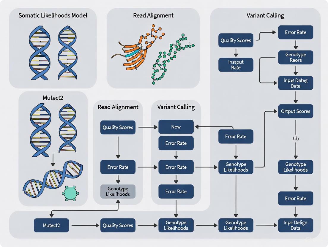
This article provides a comprehensive exploration of the somatic likelihoods model in GATK's Mutect2, a cornerstone tool for somatic variant discovery in cancer genomics.
Decoding Mutect2's Somatic Likelihoods Model: A Comprehensive Guide for Cancer Researchers
Abstract
This article provides a comprehensive exploration of the somatic likelihoods model in GATK's Mutect2, a cornerstone tool for somatic variant discovery in cancer genomics. Tailored for researchers and drug development professionals, it begins by deconstructing the foundational Bayesian probability framework that distinguishes somatic from germline variants and sequencing artifacts. The guide then details practical methodologies for applying Mutect2 across various sequencing contexts—including tumor-normal, tumor-only, and mitochondrial modes—and offers advanced strategies for troubleshooting and optimizing performance, particularly for challenging samples like FFPE or those with low allele fractions. Finally, it synthesizes evidence from independent benchmarking studies that validate Mutect2's performance against other callers and in novel applications like RNA-seq, empowering readers to implement robust, accurate somatic mutation detection pipelines for both research and clinical applications.
Core Principles: Deconstructing Mutect2's Bayesian Somatic Genotyping Model
In genomic analysis, the accurate identification of variants hinges on using specialized computational models tailored to distinct biological contexts. Somatic and germline variants represent fundamentally different biological phenomena, necessitating specialized calling approaches rather than a one-size-fits-all methodology. Germline variants are inherited polymorphisms present in all cells of an organism, while somatic variants are acquired mutations occurring in specific cell lineages, such as tumors, and are not present in the germline [1]. This distinction is not merely biological but extends to technical requirements for detection. Somatic variants in cancer often appear at low allele frequencies due to tumor heterogeneity, normal cell contamination, and complex copy number changes, presenting unique computational challenges [1]. The specialized nature of somatic calling is exemplified by Mutect2, part of the Broad Institute's Genome Analysis Toolkit (GATK), which employs a fundamentally different statistical model than germline callers like HaplotypeCaller, despite sharing some initial processing steps [1] [2]. Understanding why these specialized models are essential requires examining their differing biological contexts, technical implementations, and performance characteristics across varying genomic conditions.
Key Biological and Technical Differences Between Somatic and Germline Variants
The divergence between somatic and germline variant calling stems from fundamental biological differences that directly influence computational strategy. Germline variants follow Mendelian inheritance patterns, typically exhibit heterozygous or homozygous states in diploid organisms, and are present at predictable allele frequencies (approximately 50% for heterozygotes, 100% for homozygotes). In contrast, somatic variants in cancer display highly variable allele frequencies due to tumor purity, subclonal populations, and aneuploidy, violating the fixed ploidy assumptions of germline callers [1].
Variant Characteristics: Germline variants are straightforward changes against the reference genome, while somatic variants must satisfy two conditions: they must differ from the reference genome AND differ from the matched normal sample [1]. This dual requirement means that a site reverting to the reference allele in a tumor sample would not be considered a somatic variant, illustrating the nuanced logic required.
Ploidy Assumptions: Germline callers like HaplotypeCaller operate with a fixed ploidy assumption (typically diploid), which is appropriate for inherited variants but inadequate for tumors, where copy number alterations are common. Mutect2 allows for varying allele fractions without fixed ploidy constraints, accommodating the complex genomics of cancer [1].
Privacy Considerations: Somatic calling was originally designed for cancer research with specific privacy considerations. Germline variants in noncoding regions can potentially identify individuals, leading to stringent filtering practices in somatic callers to protect patient confidentiality by removing identifying germline variation [1].
The following table summarizes the core operational differences between germline and somatic calling approaches:
Table 1: Fundamental Differences Between Germline and Somatic Variant Calling Approaches
| Aspect | Germline Calling (e.g., HaplotypeCaller) | Somatic Calling (e.g., Mutect2) |
|---|---|---|
| Primary Design Purpose | Identify inherited polymorphisms | Detect acquired mutations in cancer/somatic tissues |
| Ploidy Assumption | Fixed ploidy (configurable) | Variable ploidy to account for tumor heterogeneity |
| Variant Frequency Expectations | Expected allele fractions ~50% (heterozygous) or 100% (homozygous) | Highly variable allele fractions due to tumor purity and subclonality |
| Reference Confidence Modeling | Supports reference confidence calculations and GVCF generation | Incapable of calculating reference confidence [1] |
| Genotyping Output | Full genotyping of samples | Focus on variant presence/absence without traditional genotyping |
| Annotation Approach | INFO field annotations derived from all observed alleles including reference | INFO field annotations refer only to ALT alleles [1] |
Computational Models: How Mutect2 Diverges from Germline Callers
Shared Initial Steps: Assembly-Based Haplotype Reconstruction
While somatic and germline callers diverge significantly in their statistical approaches, they share common initial processing steps. Both Mutect2 and HaplotypeCaller utilize active region-based processing, assembly-based haplotype reconstruction, and PairHMM alignment of reads to haplotypes [1]. This shared foundation in local assembly allows both callers to better handle indels and complex variants by reconstructing potential haplotypes in genomic regions showing evidence of variation.
Divergent Statistical Frameworks
The critical divergence occurs after haplotype assembly, where Mutect2 implements a fundamentally different statistical model specifically designed for somatic variant detection:
Bayesian Somatic Genotyping: Mutect2 employs a Bayesian model that calculates the probability that a variant is somatic by contrasting evidence between tumor and normal samples [2]. This differs fundamentally from HaplotypeCaller's germline genotyping model, which uses ploidy in its genotype likelihood calculations [1].
Specialized Parameters: Mutect2 incorporates several specialized parameters not available in germline callers, including:
--af-of-alleles-not-in-resource: Defines the germline variant prior for alleles not found in population databases [1]--log-somatic-prior: Defines the somatic variant prior used in likelihood calculations [1]--normal-lod: Threshold for filtering variants present in the normal sample [1]--tumor-lod-to-emit: Cutoff for emitting tumor variants [1]
Prefiltering Strategies: Mutect2 employs sophisticated prefilters using matched normals, panels of normals (PoN), and population germline resources to eliminate likely germline variants early in the process, conserving computational resources [1]. This is particularly important because the average human genome contains approximately one common germline variant per thousand bases, creating a substantial background against which to detect true somatic events [1].
The following diagram illustrates the core logical workflow of Mutect2 for distinguishing somatic from germline variants:
Experimental Evidence: Performance Benchmarking Across Conditions
Methodologies for Benchmarking Variant Callers
Robust evaluation of somatic variant callers requires carefully designed benchmarking approaches using datasets with known ground truth. Multiple studies have employed different methodologies to assess caller performance:
Synthetic Spike-in Experiments: Chen et al. (2020) created benchmark datasets by mixing whole-exome sequencing data from two normal human cell lines, introducing known somatic mutations at specific frequencies and depths [3]. This approach provides precise ground truth for calculating precision and recall metrics.
In Silico Mutation Introduction: BAMSurgeon software can introduce known mutations into existing BAM files with controlled variant allele frequencies (VAFs), typically ranging from 1% to 100% [4]. This method allows systematic testing across different mutation frequencies and sequencing depths while maintaining realistic sequencing artifacts.
Real Clinical Sample Analysis: Performance validation using real tumor-normal pairs from cancer patients, with verification through orthogonal methods or consensus approaches [4] [5]. These studies typically compare caller concordance and examine variant characteristics in exclusive calls.
Quantitative Performance Comparisons Across Mutation Frequencies
Multiple benchmarking studies have revealed critical performance differences between somatic callers across varying mutation frequencies and sequencing depths. The following table synthesizes key quantitative findings from these systematic evaluations:
Table 2: Performance Comparison of Somatic Variant Callers Across Mutation Frequencies
| Mutation Frequency | Sequencing Depth | Mutect2 Recall | Strelka2 Recall | VarScan Recall | VarDictJava Recall | FANSe Recall |
|---|---|---|---|---|---|---|
| 1% (Low) | 100-300X | 5-19% [5] | 6-19% [5] | 79% (800X) [3] | 81% (800X) [3] | 87% (800X) [3] |
| 5-10% (Medium) | 200X | 50-96% [5] | 48-93% [5] | Information Missing | Information Missing | Information Missing |
| ≥20% (High) | 200X | >90% [5] | >90% [5] | Information Missing | Information Missing | Information Missing |
| Precision | All conditions | >95% [3] [4] [5] | >95% [3] [4] [5] | >95% [3] | >95% [3] | >95% [3] |
The data reveal that all major somatic callers maintain high precision (>95%) across conditions, but show dramatic differences in sensitivity at low mutation frequencies. While Mutect2 and Strelka2 perform comparably at medium and high mutation frequencies, they struggle with low-frequency variants (1%) even at high sequencing depths [3] [5]. At 800X depth, Mutect2 achieves only 19% recall for 1% VAF mutations, while VarScan, VarDictJava, and FANSe show substantially higher sensitivity (79-87%) for these challenging low-frequency variants [3].
Tumor-Only Calling Challenges and Solutions
In clinical settings, matched normal samples are not always available, creating additional challenges for distinguishing somatic from germline variants. Tumor-only analysis requires specialized approaches to mitigate false positives from germline polymorphisms:
Panel of Normals (PoN): Mutect2 can utilize a PoN containing common artifacts and germline variants seen in normal samples, which helps filter systematic false positives [2] [6]. Creating a PoN involves running Mutect2 on multiple normal samples and identifying sites recurrent across samples [1].
Population Germline Resources: Databases like gnomAD provide allele frequencies in population cohorts, allowing Mutect2 to filter common germline variants [2] [7]. The
--germline-resourceparameter incorporates this information into the calling model.Statistical Priors: The
--af-of-alleles-not-in-resourceparameter sets the prior probability for alleles absent from germline resources, influencing whether such alleles are considered potential somatic mutations [7].
Recent evaluations of tumor-only methods like OncoTOP demonstrate that sophisticated approaches can achieve high accuracy (99.8% positive predictive value) in distinguishing somatic from germline variants without matched normals [8].
Practical Implementation: Workflows and Research Reagents
Recommended Somatic Calling Workflow with Mutect2
Implementing a robust somatic variant calling workflow requires careful attention to data preparation, parameter configuration, and filtering strategies:
Data Requirements: The basic Mutect2 workflow requires tumor BAM files (with matched normal recommended), a reference genome, and optionally a germline resource (e.g., gnomAD) and panel of normals [2]. For best practices, the Broad Institute recommends sequencing depths of at least 200X for tumor and 100X for normal samples in whole-exome studies [5].
Basic Tumor-Normal Command:
This command initiates Mutect2 calling with matched normal and germline resource filtering [2].
Post-calling Filtering: The initial Mutect2 output requires additional filtering using FilterMutectCalls, which incorporates orientation bias analysis, contamination checking, and statistical filtering based on multiple annotations [2] [9].
Essential Research Reagents and Computational Tools
Successful somatic variant identification requires a comprehensive toolkit of reference databases, software tools, and computational resources:
Table 3: Essential Research Reagents and Resources for Somatic Variant Calling
| Resource Category | Specific Examples | Purpose and Function |
|---|---|---|
| Reference Genomes | GRCh37 (hg19), GRCh38 | Standardized reference sequences for read alignment and variant calling [3] [4] |
| Germline Variant Databases | gnomAD, dbSNP, 1000 Genomes | Population allele frequency data for filtering common germline polymorphisms [1] [2] |
| Somatic Mutation Databases | COSMIC, TCGA | Catalog of known cancer-associated mutations for validation and annotation [4] |
| Alignment Tools | BWA-MEM, FANSe3 | Map sequencing reads to reference genome [3] [4] |
| Variant Callers | Mutect2, Strelka2, VarScan | Detect somatic mutations using specialized statistical models [3] [4] [5] |
| Variant Filtering/Annotation | FilterMutectCalls, VEP, Funcotator | Filter false positives and annotate functional consequences [2] [6] |
| Benchmarking Tools | BAMSurgeon, SomaticSeq | Generate synthetic datasets and evaluate caller performance [4] |
Ensemble Approaches for Enhanced Sensitivity
Given the complementary strengths of different callers, ensemble approaches often outperform individual tools. SomaticSeq can integrate calls from multiple tools (e.g., Mutect2 + Strelka2) through machine learning or consensus approaches, typically retaining variants with higher VAFs and coverage metrics [4]. This strategy helps overcome limitations of individual callers, particularly for low-frequency variants.
The distinction between somatic and germline variant calling is not merely academic but reflects fundamental differences in biological origin, technical detection requirements, and clinical application. Specialized tools like Mutect2 are essential because they incorporate specific statistical priors, filtering strategies, and computational models that address the unique challenges of somatic mutation detection, particularly low variant allele frequencies, tumor heterogeneity, and the need to distinguish somatic events from extensive germline background variation.
The performance characteristics of somatic callers vary significantly across mutation frequencies, with no single tool dominating across all scenarios. This underscores the importance of selecting calling strategies based on specific experimental conditions and considering ensemble approaches for comprehensive variant detection. As genomic medicine continues to advance, the development and refinement of specialized variant calling models will remain crucial for unlocking the full potential of cancer genomics in both research and clinical applications.
In cancer genomics, the precise identification of somatic mutations—genetic alterations present in tumor tissue but absent in the germline—is fundamental to understanding tumorigenesis, progression, and therapeutic targeting. This process hinges on a critical computational challenge: distinguishing true somatic mutations from sequencing errors and inherited germline variants using statistical evidence derived from next-generation sequencing data. The Bayesian statistical framework provides the mathematical foundation for this discrimination, enabling researchers to calculate the relative likelihoods of the somatic versus germline hypotheses for each observed genetic alteration.
Early methods for somatic mutation detection often relied on heuristic filters, but contemporary approaches like Mutect2 implement formal Bayesian models to evaluate evidence more systematically [10]. These models compute probabilities by considering multiple lines of evidence, including allele frequencies, sequencing qualities, and prior knowledge from population databases. Within this framework, the calculation of likelihoods for competing hypotheses—somatic mutation versus germline variant—forms the core decision mechanism that dictates variant classification accuracy. This technical guide examines the Bayesian foundations of these likelihood calculations, with specific focus on their implementation in modern tools like Mutect2, their performance characteristics across different experimental conditions, and the methodological protocols for proper application in cancer genomics research.
Mathematical Framework: Bayesian Probability Models for Hypothesis Testing
Core Bayesian Formulation for Somatic-Germline Discrimination
The fundamental statistical challenge in somatic variant calling involves comparing two competing hypotheses for each observed variant: the somatic hypothesis (S) suggesting the variant is tumor-specific, and the germline hypothesis (G) suggesting it is an inherited polymorphism. Using Bayesian inference, the posterior probability for each hypothesis given the observed sequencing data (D) is calculated as:
[ P(S|D) = \frac{P(D|S) \cdot P(S)}{P(D)} \quad \text{and} \quad P(G|D) = \frac{P(D|G) \cdot P(G)}{P(D)} ]
where (P(D|S)) and (P(D|G)) are the likelihoods of the data under each hypothesis, (P(S)) and (P(G)) are the prior probabilities for each hypothesis, and (P(D)) is a normalizing constant [10]. In practice, variant callers often compute the log-odds ratio (LOD) to make a decision:
[ \text{LOD} = \log \frac{P(S|D)}{P(G|D)} = \log \frac{P(D|S) \cdot P(S)}{P(D|G) \cdot P(G)} ]
The SGZ method exemplifies this approach by modeling the observed allele frequency (f) and read depth (n) at each variant position, calculating the probability of the data under both hypotheses using binomial distributions [11]. For a given variant, the method computes:
[ P(y|G; AF{germline}) = \text{Bin}(nf, n, AF{germline}) ] [ P(y|S; AF{somatic}) = \text{Bin}(nf, n, AF{somatic}) ]
where (AF{germline}) and (AF{somatic}) represent the expected allele frequencies under each hypothesis, which are derived from tumor purity (p) and local copy number (C) estimates [11]. A variant is classified as somatic if (P(y|S; AF{somatic}) > α) and (P(y|G; AF{germline}) ≤ α), where α is a significance threshold.
Mutect2's Implementation of Bayesian Genotyping
Mutect2 implements a specialized Bayesian genotyping model that differs from its predecessor MuTect, incorporating local assembly of haplotypes from the HaplotypeCaller framework [2]. The core of its approach involves calculating the tumor LOD (TLOD)—the log odds that a variant is present in the tumor—against the null hypothesis that the apparent variation is due to technical artifacts. The default threshold for this statistic is 3.0, corresponding to an odds ratio of approximately 1:1000 [12].
A key advancement in Mutect2's Bayesian framework is its handling of allele frequency distributions through a Dirichlet process binomial mixture model [13]. This approach recognizes that somatic mutations in a tumor sample do not occur at uniform allele frequencies but rather cluster around specific values corresponding to different clonal populations. The model learns these clusters during analysis, allowing it to be more sensitive to mutations whose allele frequencies match these patterns and more skeptical of apparent variants that deviate from the expected distribution.
Table 1: Key Statistical Parameters in Bayesian Somatic Variant Calling
| Parameter | Mathematical Representation | Biological Interpretation | Typical Default Values |
|---|---|---|---|
| Tumor LOD (TLOD) | (\log \frac{P(D \mid \text{Somatic})}{P(D \mid \text{Artifact})}) | Evidence for true somatic variant versus sequencing error | 3.0 (≈1:1000 odds) [12] |
| Germline Prior | (P(G)) | Prior probability of germline polymorphism | Population frequency-dependent [2] |
| Somatic Prior | (P(S)) | Prior probability of somatic mutation | Learned from data [13] |
| α (Significance threshold) | (P(y \mid G) ≤ α) | Type I error rate for germline hypothesis | 0.05-0.01 [11] |
Experimental Protocols: Methodologies for Benchmarking Bayesian Classifiers
Establishing Ground Truth for Validation Studies
Rigorous evaluation of somatic variant calling performance requires datasets with known somatic mutations. The ICGC-TCGA DREAM challenge addressed this need by creating synthetic tumor-normal pairs through computational manipulation of real sequencing data from the HCC1143 cell line [14]. The process involved: (1) sequencing the cell line at high depth (80x whole genome sequencing), (2) splitting the data into two subsets to simulate tumor and normal pairs, and (3) computationally adding mutations at different frequencies (50%, 33%, and 20%) to one subset to create synthetic tumor samples with known ground truth [14]. This approach generated a validated set of 474 true-positive somatic SNVs and 464 indels for benchmarking.
An alternative methodology employed by Chen et al. involved mixing sequencing data from two normal human cell lines with known germline differences [5] [15]. By combining data from NA12878 and YH-1 samples at specific proportions (1%, 5%, 10%, 20%, 30%, 40%), researchers created synthetic tumor samples with precisely known mutation frequencies corresponding to the mixing ratios [5]. This protocol enabled systematic evaluation of how mutation frequency and sequencing depth affect detection sensitivity, with true mutations defined as sites with completely different homozygous genotypes between the two cell lines.
Performance Metrics and Evaluation Framework
Studies consistently employ standard classification metrics to evaluate somatic variant callers: precision (also called positive predictive value), recall (sensitivity), and F-score (harmonic mean of precision and recall) [5] [15]. These are defined as:
- Precision = TP / (TP + FP)
- Recall = TP / (TP + FN)
- F-score = 2 × (Precision × Recall) / (Precision + Recall)
where TP represents true positives, FP false positives, and FN false negatives. To ensure robustness, benchmarking typically incorporates multiple replicates—for example, Chen et al. generated three independent replicates for each depth and mutation frequency combination, with results showing less than 2% variation in recall rate and 3.5% in precision across replicates [5].
Table 2: Performance Comparison of Somatic Variant Callers Across Mutation Frequencies
| Variant Caller | Mutation Frequency 1% | Mutation Frequency 5-10% | Mutation Frequency ≥20% | Computational Speed |
|---|---|---|---|---|
| Mutect2 | Low sensitivity (F-score: 0.05-0.50) [5] | Moderate sensitivity (F-score: 0.65-0.95) [5] | High sensitivity (F-score: 0.94-0.96) [5] | Slow (17-22x slower than Strelka2) [5] |
| Strelka2 | Low sensitivity (F-score: 0.06-0.37) [5] | Moderate sensitivity (F-score: 0.64-0.94) [5] | High sensitivity (F-score: >0.94) [5] | Fast [5] |
| FANSe | Highest sensitivity for low frequency [15] | High sensitivity [15] | High sensitivity [15] | Fastest (8.8-19x faster than others) [15] |
| VarScan/VarDict | Outperforms Mutect2 at low frequency [15] | Good performance [15] | Good performance [15] | Moderate [15] |
Parameter Optimization: Adjusting Bayesian Thresholds for Enhanced Sensitivity
Key Adjustable Parameters in Mutect2
While Mutect2 implements a sophisticated Bayesian model with generally effective default parameters, specific research scenarios may require adjustment of statistical thresholds to optimize sensitivity-precision tradeoffs. The primary parameter controlling sensitivity is the tumor LOD threshold (--tumor-lod-to-emit), which determines the minimum evidence required to emit a candidate variant [12]. Reducing this threshold from its default value of 3.0 to 2.0 increases sensitivity but may decrease precision by admitting more false positives.
For challenging detection scenarios such as low-frequency variants in ctDNA or heterogeneous tumors, additional parameters may require adjustment. The --pruning-lod-threshold parameter controls the minimum LOD required to keep a haplotype in the assembly graph, with lower values preserving more potential haplotypes during local assembly [16]. Similarly, --initial-lod and --emit-lod can be decreased to enhance sensitivity for variants with weak statistical support, though this must be balanced against increased false positive rates [16]. One study reported successfully detecting an insertion in a homopolymer region with TLOD of -4.74 by setting init-lod and emit-lod to -10, demonstrating how extreme parameter adjustments can recover marginal variants in high-depth data [16].
Addressing Technical Artifacts in Bayesian Frameworks
Sequencing artifacts present significant challenges to accurate somatic variant calling, and Bayesian frameworks must incorporate specialized models to address them. Mutect2 includes a specific workflow for read orientation artifacts, which are particularly problematic in FFPE samples and Illumina Novaseq data [13]. This three-step process involves: (1) collecting strand-specific artifact data during Mutect2 execution using the --f1r2-tar-gz argument, (2) learning the orientation bias model with LearnReadOrientationModel, and (3) incorporating these artifact priors into FilterMutectCalls with the --ob-priors argument [13].
Another critical adjustment involves the parameter --af-of-alleles-not-in-resource, which sets the imputed allele frequency for variants absent from germline resources [2]. This parameter dynamically adjusts based on analysis mode: tumor-only calling defaults to 5e-8, tumor-normal to 1e-6, and mitochondrial mode to 4e-3 [2]. Proper configuration of this parameter is essential for accurate prior specification in the Bayesian model, particularly for tumor-only analyses where matched normal sequencing is unavailable.
Research Reagent Solutions: Essential Materials for Experimental Implementation
Table 3: Key Research Reagents and Computational Resources for Somatic Variant Detection
| Resource Category | Specific Examples | Function in Bayesian Somatic Calling | Implementation Notes |
|---|---|---|---|
| Reference Datasets | ICGC-TCGA DREAM Challenge data [14] | Provides validated ground truth for benchmarking and parameter optimization | Synthetic tumor-normal pairs with known mutations |
| Germline Resources | gnomAD (af-only-gnomad.vcf) [2] [13] | Provides population allele frequencies for germline prior estimation | Critical for filtering common germline polymorphisms |
| Panel of Normals | CreateSomaticPanelOfNormals output [2] [13] | Identifies technical artifacts systematic across normal samples | Should be created using normals sequenced with same protocol |
| Validation Samples | SEQC2 consortium samples (HCC1395/HCC1395BL) [14] | Enables cross-platform validation of somatic calls | Orthogonal validation with multiple technologies |
| Software Containers | GATK Docker images [2] [13] | Ensures reproducible execution of Mutect2 workflow | Version control essential for consistent results |
Visualization: Bayesian Decision Framework for Somatic Mutation Calling
Diagram Title: Bayesian Decision Framework for Somatic Mutation Calling
Advanced Applications and Future Directions
Ensemble Approaches and Method Integration
While individual variant callers implementing Bayesian frameworks have demonstrated good performance, recent research indicates that ensemble approaches combining multiple callers can achieve superior accuracy. A comprehensive benchmarking study that evaluated 20 somatic variant callers found that ensemble methods outperformed even the best individual caller [14]. For somatic SNVs, an ensemble combining LoFreq, Muse, Mutect2, SomaticSniper, Strelka, and Lancet achieved a mean F-score of 0.927, exceeding the top-performing individual caller (Dragen) by over 3.6% [14]. Similarly, for indels, an ensemble of Mutect2, Strelka, Varscan2, and Pindel achieved a mean F-score of 0.867, outperforming the best individual caller (NeuSomatic) by more than 3.5% [14].
To balance accuracy with computational efficiency, the same study identified an optimal combination of four callers: Muse, Mutect2, and Strelka for SNVs, with Mutect2, Strelka, and Varscan2 for indels [14]. This approach leverages the complementary strengths of different Bayesian implementations while maintaining practical computational requirements for large-scale studies.
Tumor-Only Analysis and Computational Considerations
In clinical contexts where matched normal tissue is unavailable, specialized Bayesian methods have been developed to distinguish somatic from germline variants. The SGZ (somatic-germline-zygosity) method exemplifies this approach by modeling allele frequencies while accounting for tumor purity, ploidy, and local copy number [11]. This method demonstrates 95-99% accuracy when predictions can be made (covering approximately 85% of cases), significantly outperforming basic allele frequency-based approaches [11].
Computational efficiency remains an important consideration in somatic variant calling, particularly for large cohorts. Performance comparisons reveal substantial differences in processing time between callers, with Strelka2 being 17-22 times faster than Mutect2 on average [5], and FANSe demonstrating even greater speed advantages (8.8-19 times faster than other pipelines) [15]. These differences highlight the trade-offs between model sophistication and computational practicality, informing tool selection based on study scale and resource constraints.
Within the somatic likelihoods model of GATK's Mutect2, the accuracy of variant calling is fundamentally governed by the integration of prior biological knowledge through specific input resources. This technical guide details the function and implementation of two critical classes of model inputs: the germline resource, which provides population allele frequencies to inform a Bayesian classification of variants, and the panel of normals (PoN), which catalogs technical artifacts to suppress false positive calls. We elucidate the statistical framework that leverages these inputs, provide validated experimental protocols for their creation and use, and present quantitative performance data from benchmarking studies. Directed at researchers and scientists in oncology drug development, this whitepaper serves as a comprehensive reference for constructing robust and reliable somatic mutation detection pipelines.
Mutect2 employs a sophisticated Bayesian statistical model to distinguish true somatic mutations from germline variants and sequencing artifacts. The core of this model calculates the probability that the observed sequencing data (the likelihood) supports the hypothesis of a somatic mutation versus other scenarios. The model's discriminative power is significantly enhanced by incorporating prior probabilities derived from large-scale genomic datasets. These priors represent pre-existing knowledge about the probability of a genomic locus being a germline variant or a technical artifact, independent of the immediate sequencing data from the sample. The two primary inputs that provide this prior knowledge are the germline resource and the panel of normals (PoN). Their integration allows Mutect2 to "learn from the experience" of previously sequenced genomes, thereby achieving a level of accuracy that would be impossible from a single sample's data alone [17].
The Germline Resource: Informing the Germline Prior
Definition and Purpose
The germline resource is a population variant call format (VCF) file containing known germline variants and their population allele frequencies (AF). Its primary function within the Mutect2 model is to provide a prior probability that a variant observed in a tumor sample is actually a common germline polymorphism rather than a somatic mutation [2]. When Mutect2 encounters a locus with an alternate allele, it queries the germline resource. If the allele is present, the associated population AF is used as a prior in the statistical model. This makes it less likely that common germline variants will be erroneously classified as somatic events. The resource is crucial for tumor-only analysis, where a matched normal sample is unavailable, but it also substantially improves the accuracy of tumor-normal paired analyses [2] [17].
Implementation and Key Parameters
In practice, the germline resource is supplied to Mutect2 using the --germline-resource argument. A common resource used for human data is the gnomAD dataset, often in a processed form distributed by the GATK team as af-only-gnomad.vcf.gz [17]. A critical parameter that works in concert with the germline resource is --af-of-alleles-not-in-resource. This parameter sets the imputed allele frequency for any allele not found in the germline resource. The default value for this parameter is dynamically adjusted by Mutect2 based on the analysis mode, reflecting the different prior expectations for each scenario [2]:
- Tumor-only mode: Default AF = 5e-8
- Tumor-normal mode: Default AF = 1e-6
- Mitochondrial mode: Default AF = 4e-3
This mechanism ensures that alleles absent from the germline resource are treated as extremely rare germline variants, which the model can then more confidently classify as potential somatic mutations [17].
Table 1: Key Parameters for the Germline Resource in Mutect2
| Parameter | Function | Common Setting / Example |
|---|---|---|
--germline-resource |
Provides a VCF of known germline variants and their population allele frequencies. | af-only-gnomad.vcf.gz |
--af-of-alleles-not-in-resource |
Sets the imputed allele frequency for alleles not found in the germline resource. | Tumor-Only: 5e-8; Tumor-Normal: 1e-6; Mitochondrial: 4e-3 |
The Panel of Normals (PoN): The Artifact Prior
Definition and Purpose
The Panel of Normals (PoN) is a VCF file compiled from the analysis of multiple normal tissue samples. Its purpose is to catalog technical artifacts and common germline variants that recur across sequencing runs, which are not already captured by the germline resource. These artifacts can arise from systematic errors in sequencing, PCR amplification, or mapping biases [13]. In the Mutect2 model, sites listed in the PoN are considered "inactive by default," meaning they are heavily down-weighted during the initial variant discovery phase. This pre-filtering prevents the pipeline from spending computational resources on known artifact sites and dramatically reduces the false positive rate in the final callset [17].
Construction Protocol
Creating a robust PoN requires a cohort of normal samples processed with the same experimental and analytical protocols intended for the tumor samples. The following step-by-step protocol, adapted from the GATK Best Practices, details the process [13]:
Run Mutect2 in tumor-only mode on each normal sample.
- This step identifies potential variants (mostly germline) and, more importantly, technical artifacts specific to each normal sample.
- Command example:
Import the normal VCFs into a GenomicsDB.
- This step efficiently consolidates the variant calls from all normal samples into a single database for the next step.
- Command example:
Create the combined Panel of Normals.
- This tool analyzes the database of normal variants and outputs a final VCF containing sites that are found in multiple normals and are therefore likely to be artifacts.
- Command example:
Table 2: Research Reagent Solutions for Mutect2 Workflows
| Reagent / Resource | Type | Function in the Workflow |
|---|---|---|
| Germline Resource (e.g., af-only-gnomad.vcf.gz) | Data File | Provides population allele frequencies to calculate the prior probability that a variant is germline. |
| Panel of Normals (pon.vcf.gz) | Data File | Catalogs recurring technical artifacts and common germline variants from a set of normal samples to suppress false positives. |
| Reference Genome (e.g., GRCh38) | Data File | The standard reference sequence to which all sequencing reads are aligned and variants are called. |
| BAM/SAM/CRAM Files | Data File | The aligned sequencing data for tumor and normal samples, which serve as the primary input for variant calling. |
Quantitative Performance and Benchmarking
The impact of using a germline resource and PoN on somatic mutation calling has been quantitatively assessed in benchmarking studies. A comprehensive simulation study that created a "ground truth" dataset demonstrated that Mutect2's performance is highly dependent on these inputs, especially for detecting low-frequency variants [18]. The study simulated a uniform distribution of 10,000 somatic variants in each of 100 allele frequency bins (semi-centiles) and assessed Mutect2's detection rate.
Table 3: Mutect2 Performance on Simulated Somatic Variants (Adapted from [18])
| Variant Allele Frequency (VAF) Range | Approximate Detection Rate by Mutect2 | Primary Filtering Reasons for False Negatives |
|---|---|---|
| Very Low (e.g., <0.02) | Low | Insufficient read depth, tumor LOD, strand bias. |
| Low to Medium (0.02 - 0.1) | Moderate to High | Varies with depth and local sequence context. |
| High (>0.1) | Very High | Few false negatives; those that occur may be filtered as germline or due to mapping issues. |
Comparative studies have also evaluated Mutect2 against other callers. One assessment found that while VarScan2 may have a higher number of concordant mutations at high frequencies (above 40%), Mutect2 significantly outperforms it at variant allele frequencies below 20%, identifying up to four times as many mutations in this critical range [19]. This high sensitivity at lower frequencies is a direct result of its powerful statistical model, which is refined by the use of germline and artifact priors.
Advanced Workflow: Integrating the Read Orientation Filter
For specific sample types, such as formalin-fixed paraffin-embedded (FFPE) tumors or those sequenced on Illumina Novaseq machines, an additional prior for read orientation artifacts can be integrated. This advanced workflow generates a prior probability for oxidation and FFPE-associated artifacts [13] [17].
- Collect Raw Data with Mutect2: Run Mutect2 with the
--f1r2-tar-gzargument to generate a file containing counts of forward and reverse reads supporting reference and alternate alleles. - Learn the Artifact Model: Use
LearnReadOrientationModelto process the raw counts and generate an artifact prior table. - Apply the Prior during Filtering: Pass the model to
FilterMutectCallsusing the--ob-priorsargument.
The sophisticated statistical framework of Mutect2 derives its power and precision from the principled incorporation of prior knowledge. The germline resource and the panel of normals are not mere optional inputs but are fundamental components that shape the model's ability to discriminate true somatic mutations from the vast background of germline variation and technical noise. As demonstrated by benchmarking studies, their use is critical for achieving high sensitivity, particularly for low-allele-fraction variants, while maintaining high specificity. A proper understanding of these key model inputs—their creation, function, and integration into the broader somatic likelihoods model—is therefore indispensable for any research or clinical pipeline aimed at the reliable detection of somatic mutations in cancer.
Somatic variant discovery represents a distinct computational challenge from germline genotyping, necessitating specialized algorithms that operate without fixed ploidy assumptions and can detect low-frequency mutations amid tumor heterogeneity. This technical guide examines the fundamental architectural differences between GATK's Mutect2 and HaplotypeCaller engines, highlighting how Mutect2's Bayesian somatic genotyping model addresses the unique requirements of cancer genomics. We explore Mutect2's specialized handling of variable allele fractions, its sophisticated filtering infrastructure utilizing matched normals and panels of normals, and its departure from germline-centric statistical assumptions. Performance benchmarking data across varying sequencing depths and allele frequencies provide empirical validation of Mutect2's capabilities in somatic contexts, while implementation guidelines offer researchers practical frameworks for deploying Mutect2 in diverse cancer genomics scenarios.
Somatic mutation detection fundamentally differs from germline variant calling in both its biological context and technical requirements. The term "somatic" derives from the Greek word soma, referring to an organism's body cells other than reproductive cells [1]. In cancer genomics, somatic mutations accumulate in tissues like skin due to environmental exposures and are absent from the germline [1]. This biological reality creates specific computational challenges: somatic variants often appear at low allele frequencies due to tumor sample contamination with normal cells, heterogeneous tumor subpopulations, and aneuploid events that alter genomic copy number in patchwork patterns [1].
The Mutect2 algorithm represents a specialized solution to these challenges, combining the assembly-based machinery of HaplotypeCaller with a Bayesian somatic genotyping model adapted from the original MuTect tool [2] [20]. Unlike germline callers that assume fixed ploidy, Mutect2 operates without explicit ploidy constraints, instead modeling the continuous spectrum of allele fractions characteristic of tumor genomics [1]. This technical innovation enables detection of mutations that would be systematically overlooked by germline-oriented approaches.
Core Algorithmic Divergences: Shared Assembly, Divergent Genotyping
Shared Assembly Machinery
Both Mutect2 and HaplotypeCaller employ identical initial processing steps centered on local assembly-based haplotype reconstruction:
Active Region Determination: Both tools identify genomic regions showing evidence of variation using a smoothed activity profile based on probabilities of variation at each position [21]. The profile is thresholded to define ActiveRegions where assembly occurs.
Graph-Based Assembly: Both algorithms construct De Bruijn-like graphs from reads in ActiveRegions, selecting the most likely paths through the graph as candidate haplotypes [1] [22].
PairHMM Alignment: Both tools align individual reads to candidate haplotypes using the PairHMM algorithm, producing a likelihood matrix relating reads to haplotypes [22].
The following diagram illustrates this shared assembly workflow:
Divergent Genotyping Models
The critical algorithmic divergence occurs at the genotyping phase, where Mutect2 and HaplotypeCaller apply fundamentally different statistical models:
Table 1: Core Algorithmic Differences Between HaplotypeCaller and Mutect2
| Feature | HaplotypeCaller | Mutect2 |
|---|---|---|
| Primary Design Purpose | Germline variant discovery [1] | Somatic mutation detection [1] |
| Ploidy Assumption | Fixed ploidy (default diploid) [1] | Variable ploidy with continuous allele fractions [1] |
| Variant Likelihood Model | Ploidy-based genotype likelihood calculations [1] | Bayesian somatic genotyping model [2] |
| Reference Confidence Modeling | Supports GVCF output with reference confidence [1] | No reference confidence capability [1] |
| Joint Calling Capability | Multi-sample joint calling supported [1] | Single-sample calling (until v4.1.0.0) [1] |
| Variant Filtering Approach | Standard germline filtering | Matched normal, panel of normals, germline resource [2] |
HaplotypeCaller employs a ploidy-based model for germline variant discovery, calculating genotype likelihoods using a fixed ploidy assumption (typically diploid) [1]. This approach works well for germline variants but struggles with the allele frequency spectrum characteristic of somatic mutations in tumors. In contrast, Mutect2 utilizes a Bayesian somatic model that explicitly calculates the likelihood that a variant is somatic rather than germline, incorporating prior probabilities for both scenarios [1]. This model accommodates the varying allele fractions resulting from tumor purity, subclonality, and copy number alterations [1].
Specialized Features of the Mutect2 Somatic Model
Handling Variable Ploidy and Low VAF
Mutect2's statistical engine incorporates several specialized parameters to address tumor-specific biological phenomena:
Somatic Prior Probability: The
--log-somatic-priorparameter defines the prior probability that a variant is somatic, fundamentally shaping the likelihood calculations [1].Germline Resource Utilization: For alleles absent from population germline resources, Mutect2 uses the
--af-of-alleles-not-in-resourceparameter to impute an allele frequency, typically set to 1e-6 for tumor-normal mode [2]. This helps distinguish novel somatic events from rare germline variants.LOD Threshold Configuration: The
--normal-lodparameter sets the log-odds threshold for filtering variants present in the matched normal, while--tumor-lod-to-emitcontrols the cutoff for variant emission [1].
Comprehensive Filtering Strategy
Mutect2 employs a multi-layered filtering approach to maximize specificity in somatic calling:
Matched Normal Analysis: In tumor-normal mode, Mutect2 performs an early-stage filter to exclude variants clearly present in the germline, preserving computational resources [2].
Panel of Normals (PoN): A panel of normals constructed from germline samples identifies systematic artifacts present across multiple normal samples [1]. Sites appearing in the PoN are filtered from tumor samples.
Germline Resource Filtering: Population frequency databases (e.g., gnomAD) provide allele-specific frequencies to filter common germline polymorphisms [2].
Downstream Filtering with FilterMutectCalls: The secondary filtering tool applies additional filters including orientation bias, strand distribution, and contamination checks [1].
Experimental Validation and Performance Benchmarking
Performance Across VAF and Sequencing Depth
Recent benchmarking studies provide empirical validation of Mutect2's performance characteristics across varying experimental conditions:
Table 2: Mutect2 Performance Across VAF Ranges and Sequencing Depths
| VAF Range | Sequencing Depth | Recall Rate | Precision Rate | Optimal Use Case |
|---|---|---|---|---|
| 1% | 100-300X | 2.7-34.5% [5] | 68.9-100% [5] | Limited utility; require ultra-high depth |
| 5-10% | 200-800X | 48-96% [5] | >95% [5] | Subclonal mutation detection |
| ≥20% | ≥200X | 92-97% [5] | >95% [5] | High-purity tumor samples |
| ≥20% | 100X | ~90% [5] | >95% [5] | Cost-effective designs |
These data reveal that Mutect2 achieves optimal performance for variants with VAF ≥20% at sequencing depths ≥200X, while lower-frequency variants (5-10% VAF) require increased sequencing depth for reliable detection [5]. Variants at 1% VAF prove challenging even at high depths, with recall rates below 35% across all tested conditions [5].
Comparative Performance Against Other Callers
Benchmarking studies demonstrate Mutect2's position within the ecosystem of somatic variant callers:
Versus HaplotypeCaller: In mosaic variant detection, Mutect2 tumor-only mode (MT2-to) showed superior sensitivity for low-VAF variants (<10%) compared to HaplotypeCaller with modified ploidy settings [23].
Versus Strelka2: Mutect2 and Strelka2 show comparable performance for variants with VAF ≥20%, but Mutect2 demonstrates advantages for lower-frequency variants (5-10% VAF) while Strelka2 shows marginally better precision [5].
Runtime Considerations: Strelka2 demonstrates significantly faster processing times (17-22x faster than Mutect2 in some benchmarks), an important consideration for large-scale studies [5].
The following diagram illustrates a standardized benchmarking workflow for somatic caller evaluation:
Implementation Guidelines and Research Applications
Operational Modes and Recommended Usage
Mutect2 supports four primary operational modes, each optimized for specific research scenarios:
Tumor with Matched Normal: The recommended approach for somatic studies, utilizing the normal sample to filter germline variants [2].
Tumor-Only Mode: Acceptable when matched normal is unavailable but produces higher false positive rates due to unexplained germline variation [2] [24].
Mitochondrial Mode: Optimized for high-depth mitochondrial DNA analysis with adjusted parameters [2].
Force-Calling Mode: Targets specific alleles regardless of evidence, useful for clinical validation of known variants [2].
Essential Research Reagents and Computational Tools
Table 3: Essential Research Reagents and Computational Tools for Mutect2 Implementation
| Resource Type | Specific Examples | Function in Somatic Analysis |
|---|---|---|
| Germline Resource | af-only-gnomAD.vcf.gz [2] | Filters common population polymorphisms |
| Panel of Normals | Custom-generated from normals [2] | Identifies systematic technical artifacts |
| Reference Genome | GRCh38, hg19 | Standardized genomic coordinate system |
| Alignment Tools | BWA-MEM [25] | Maps sequencing reads to reference |
| Post-Calling Filters | FilterMutectCalls [1] | Applies orientation bias and contamination filters |
| Annotation Tools | Funcotator [2] | Adds biological and clinical context to variants |
Best Practices for Experimental Design
Based on performance benchmarking, the following guidelines ensure optimal Mutect2 performance:
Sequencing Depth: For variants with VAF ≥20%, 200X depth provides sufficient sensitivity (>95% recall); for lower VAF (5-10%), 500-800X depth improves recall [5].
Sample Quality: Address sample quality issues (e.g., FFPE degradation, low purity) through experimental rather than computational means when possible [5].
Validation: Orthogonal validation (e.g., digital PCR, amplicon sequencing) remains essential for clinical applications, particularly for low-VAF variants [25].
Mutect2 represents a specialized computational solution for somatic variant detection, fundamentally differentiated from HaplotypeCaller through its Bayesian genotyping model that accommodates variable ploidy and continuous allele fractions. While both tools share assembly-based haplotype reconstruction machinery, Mutect2's statistical framework, comprehensive filtering infrastructure, and tuned parameters address the specific challenges of cancer genomics—including tumor heterogeneity, normal contamination, and technical artifacts. Empirical benchmarking demonstrates Mutect2's robust performance across diverse VAF ranges and sequencing depths, though detection of very low-frequency variants (<5%) remains challenging even with optimized parameters. As somatic mutation analysis continues to evolve in both research and clinical domains, Mutect2's specialized architecture provides a foundation for accurate variant detection in cancer genomics.
Within the Mutect2 somatic likelihoods model, three core parameters—the Log Somatic Prior, the Tumor Log Odds (LOD), and the Allele Frequency of alleles not in the resource—govern the fundamental Bayesian probabilities that distinguish true somatic mutations from germline variants and sequencing artifacts. This whitepaper provides an in-depth technical analysis of these parameters, detailing their mathematical definitions, default values, and influence on variant calling sensitivity and specificity. Aimed at researchers and drug development professionals, this guide synthesizes experimental protocols and quantitative data to empower robust somatic variant detection in cancer genomics, framing the discussion within the broader thesis of understanding Mutect2's probabilistic framework.
Mutect2 employs a Bayesian statistical model to identify somatic short variants (SNVs and indels). Unlike germline callers that assume fixed ploidy, Mutect2 is designed to handle varying allele fractions, a critical feature in cancer genomes affected by tumor purity, subclonality, and copy number alterations [1]. The core of this model involves calculating the probability that a observed variant is a true somatic mutation, which requires carefully calibrated prior probabilities and likelihood ratios to separate true signals from the noise of sequencing errors and innate germline variation [26]. The parameters discussed herein are central to this calculation, shaping the prior beliefs and evidence thresholds that underpin the entire calling process.
In-Depth Parameter Analysis
AF-of-alleles-not-in-resource (--af-of-alleles-not-in-resource)
Function and Mathematical Role: This parameter (--af-of-alleles-not-in-resource or --default-af) defines the population allele fraction assigned to any allele not found in a provided germline resource (e.g., gnomAD) [27] [2]. In the Bayesian framework, this value serves as the prior probability that an unobserved allele is, in fact, a germline variant. Its primary function is to pre-filter alleles and provide evidence for the germline status of a variant [27] [28]. If an allele is absent from the germline resource, this imputed frequency is used to calculate the likelihood that the allele is not germline, thereby supporting its potential somatic nature [27].
Experimental Protocol and Defaults: The appropriate value for this parameter depends on the size of the population used to create the germline resource. For example, the default value is dynamically set based on the calling mode in recent GATK versions (post-4.0.4.0) [2]. The following table summarizes the default values and derivation logic.
Table 1: Default Values and Application of --af-of-alleles-not-in-resource
| Calling Mode | Default Value | Rationale & Derivation |
|---|---|---|
| Tumor-Normal | 1e-6 | For a resource like gnomAD with ~32,000 homologs, the probability of an allele being absent is ~1/32,000, which is approximately 3.125e-5 [27] [2]. |
| Tumor-Only | 5e-8 | Set to a more stringent value to account for the increased risk of false positives without a matched normal [2]. |
| Mitochondrial | 4e-3 | Adjusted for the different genetics and higher mutation rate of mitochondrial DNA [2]. |
Impact on Research Findings: Setting this parameter too high increases the risk of misclassifying novel germline variants as somatic, leading to reduced specificity. Conversely, setting it too low may cause the model to be overly skeptical of true, low-frequency somatic variants that are not in the resource, reducing sensitivity. For organisms without a comprehensive germline resource, this parameter should be set to 1/(ploidy * number of samples in resource) [2].
Tumor LOD (Log Odds)
Function and Mathematical Role: The Tumor LOD is a critical evidence metric calculated for each potential variant. It represents the log-odds ratio comparing the likelihood that the variant exists versus the likelihood that it does not [29]. The formula is essentially:
Tumor LOD = log10[ P(Data | Variant) / P(Data | No Variant) ]
A higher Tumor LOD provides stronger evidence for the presence of a variant. Mutect2 uses two key thresholds related to this score: --tumor-lod-to-emit, which determines if a variant is written to the output VCF, and the internal threshold used by FilterMutectCalls to decide if a variant passes filtering [27] [13].
Experimental Protocol and Defaults: In the initial discovery phase, a threshold is used to decide which candidate variants are emitted for further analysis. Historically, a --tumor-lod-to-emit of 0.0 and a filtering threshold of 5.3 were used [27] [28]. However, the filtering workflow has been significantly updated.
Table 2: Evolution of Tumor LOD Thresholds in Mutect2 Workflows
| GATK Version | Parameter | Default Value | Function |
|---|---|---|---|
| Pre-4.1.1.0 | --tumor-lod (in FilterMutectCalls) |
5.3 | Threshold for filtering variants; variants below this were filtered out [27]. |
| Pre-4.1.1.0 | --tumor-lod-to-emit (in Mutect2) |
0.0 | Threshold for emitting variant calls into the output VCF [2]. |
| 4.1.1.0+ | (In FilterMutectCalls) | N/A | The -tumor-lod parameter was removed. Filtering is now based on a unified posterior probability that optimizes the F-score [13]. |
Impact on Research Findings: The move away from a fixed Tumor LOD threshold in FilterMutectCalls to a model that optimizes the F-score automatically balances sensitivity and precision, adapting to the specific error profile and variant spectrum of the dataset [13]. This is particularly beneficial for detecting low-frequency variants in heterogeneous tumors or in off-target applications like mitochondrial variant calling.
Log Somatic Prior (--log-somatic-prior)
Function and Mathematical Role: The log somatic prior (--log-somatic-prior) is the Bayesian prior probability that any given genomic site harbors a somatic mutation [1]. This value is incorporated into the likelihood calculations to evaluate the possibility of a variant being somatic. Because the prior probability of a somatic mutation at any single base is very low, this parameter helps maintain a high specificity by requiring stronger evidence to call a variant.
Experimental Protocol and Defaults: While the exact default value is not explicitly listed in the results, its function is clearly defined within Mutect2's Bayesian somatic genotyping model [1]. This prior works in concert with the germline prior (informed by the germline resource and --af-of-alleles-not-in-resource) to calculate the overall probability of a variant being somatic.
Impact on Research Findings: The log somatic prior acts as a calibration parameter. A less informative (e.g., overly optimistic) prior can lead to a higher false positive rate, as the model becomes more willing to explain data patterns as somatic events. A well-chosen prior reflects the expected mutation burden of the sample type, improving the accuracy of the final callset.
The Scientist's Toolkit: Essential Research Reagents and Materials
Successful somatic variant calling with Mutect2 requires a suite of data resources and computational tools. The following table details the key components.
Table 3: Essential Research Reagents and Materials for Mutect2 Analysis
| Item | Function / Purpose | Usage Notes |
|---|---|---|
| Germline Resource (e.g., af-only-gnomad.vcf) | Provides population allele frequencies to distinguish common germline variants from somatic mutations [27] [2]. | A VCF with AF INFO field. Critical for informing the model about known germline variation. |
| Panel of Normals (PoN) | A VCF of sites discovered in normal samples; used to filter out recurrent technical artifacts [27] [13]. | Created using CreateSomaticPanelOfNormals. Filters sites shared across normal samples. |
| Reference Genome (-R) | The standard reference sequence for read alignment and variant calling [27]. | Must be the same reference used for read alignment and provided to both Mutect2 and FilterMutectCalls. |
| Sequence Alignment Map (-I) | The input BAM or CRAM file containing sequence reads aligned to the reference genome [27]. | Requires read groups and sample names to be defined. |
| LearnReadOrientationModel | Tool that creates an artifact prior table to filter strand-specific errors (common in FFPE data) [13]. | Used with --f1r2-tar-gz output from Mutect2. |
| CalculateContamination | Tool that estimates cross-sample contamination, which is used as a filter in FilterMutectCalls [13]. |
Requires a pileup summary table generated by GetPileupSummaries. |
Workflow and Parameter Interaction Diagrams
The following diagrams illustrate the logical relationships between the core parameters and their roles within the Mutect2 somatic calling workflow.
Diagram 1: High-level Mutect2 workflow showing the influence of critical parameters on the calling process.
Diagram 2: The Bayesian logic underpinning Mutect2, showing how parameters inform the prior and likelihood.
Advanced Experimental Protocols
Protocol: Building a Panel of Normals (PoN)
A Panel of Normals is crucial for filtering out systematic technical artifacts [13].
- Call Variants on Normal Samples: Run Mutect2 in tumor-only mode on each normal BAM file.
- Import into a GenomicsDB: Consolidate the VCFs from all normal samples.
- Create the Panel: Generate the final PoN VCF.
Protocol: Mitigating Read Orientation Artifacts
This protocol is essential for FFPE-derived samples or data from certain sequencing platforms [13].
- Generate F1R2 Tar during Mutect2 Calling: Use the
--f1r2-tar-gzargument. - Learn the Artifact Model: Pass the F1R2 data to
LearnReadOrientationModel. - Apply the Model in Filtering: Input the learned model to
FilterMutectCalls.
The parameters --af-of-alleles-not-in-resource, Tumor LOD, and --log-somatic-prior are not merely tunable knobs but are fundamental components of the Bayesian likelihood model that defines Mutect2's approach to somatic variant discovery. A deep understanding of their function—how they shape priors, evaluate evidence, and ultimately calculate the posterior probability of a somatic mutation—is essential for researchers aiming to push the boundaries of sensitivity and specificity. As Mutect2 continues to evolve, with recent updates moving towards more automated and adaptive filtering, the underlying principles governed by these parameters remain the bedrock of accurate somatic variant calling, directly impacting the reliability of downstream analyses in cancer research and therapeutic development.
From Theory to Practice: Implementing Mutect2 Workflows Across Sequencing Contexts
Mutect2 implements a sophisticated Bayesian somatic genotyping model to distinguish true somatic mutations from germline variants and sequencing artifacts. Unlike germline callers that assume a diploid genotype, Mutect2's core computational framework calculates the likelihood that observed data supports a somatic mutation versus other explanations. This somatic likelihoods model forms the theoretical foundation for the tool's accuracy, combining population allele frequency priors from germline resources with evidence from matched normal samples to calculate the probability that a variant is truly somatic [2] [17]. The model differs fundamentally from the original MuTect algorithm by incorporating local assembly of haplotypes, enabling more sensitive detection of complex variants and indels [7].
Essential Input Components for Tumor-Normal Analysis
Input Alignment Files and Sample Identification
The fundamental input data consists of BAM/SAM/CRAM files containing aligned sequencing reads:
- Tumor BAM: Contains sequencing data from the tumor tissue, expected to harbor somatic mutations.
- Matched Normal BAM: Derived from healthy tissue of the same individual, providing a germline reference to filter out inherited variants [2].
In current Mutect2 versions (4.1.0.0+), you need only specify the normal sample name(s) with -normal, as the tool automatically identifies tumor samples [2]. For multiple samples from the same individual, specify all BAM files with multiple -I arguments and corresponding normal samples with multiple -normal arguments [2].
Germline Resource (Population VCF)
The germline resource provides population allele frequencies that serve as informative priors for the somatic likelihoods model. This resource, typically derived from large population databases like gnomAD, enables Mutect2 to calculate the prior probability that a variant represents common germline variation rather than a somatic mutation [2] [17].
The germline resource is a VCF file containing population allele frequencies (AF) in the INFO field [2] [7]. When a variant is absent from this resource, Mutect2 uses a small imputed allele frequency provided by --af-of-alleles-not-in-resource [2] [17]. This parameter defaults to 1e-6 for tumor-normal analysis, reflecting the low prior probability of a novel germline variant [2].
Panel of Normals (PON)
The Panel of Normals (PON) is a critical artifact-filtering resource generated from variant calls across multiple normal samples. It systematically captures recurrent technical artifacts specific to your sequencing platform and protocols, allowing Mutect2 to exclude these common false positives during variant calling [30].
The PON is implemented as a sites-only VCF file containing loci identified as artifacts across multiple normal samples [30] [31]. Mutect2 performs early pre-filtering using the PON, avoiding computational resources on known artifact sites [2] [17]. The Broad Institute provides pre-built PONs for common reference genomes, though creating a study-specific PON is recommended when sufficient normal samples are available [30].
Table: Essential Input Components for Mutect2 Tumor-Normal Analysis
| Component | Format | Purpose in Somatic Likelihoods Model | Recommended Source |
|---|---|---|---|
| Tumor BAM | BAM/SAM/CRAM | Provides evidence for somatic mutations through altered reads | Patient tumor tissue sequencing |
| Matched Normal BAM | BAM/SAM/CRAM | Controls for individual germline variation; enables normal artifact detection | Healthy tissue from same patient |
| Germline Resource | Population VCF | Provides prior probabilities against germline variation | gnomAD (af-only-gnomad.vcf.gz) |
| Panel of Normals (PON) | Sites-only VCF | Filters recurrent sequencing/mapping artifacts | 40+ normal samples or Broad pre-built PONs |
Complete Command Structure and Execution
Basic Tumor-Normal Command
The standard command structure for tumor-normal somatic variant calling is:
This command outputs a VCF file (somatic.vcf.gz) containing both potential somatic variants and their filtering statistics, plus a companion statistics file (somatic.vcf.gz.stats) required for subsequent filtering [2] [13].
Multi-Sample Tumor-Normal Command
For multiple tumors or normals from the same patient:
In multi-sample mode, Mutect2 jointly analyzes all samples, pooling normal reads within memory and performing local assembly across all tumor samples simultaneously. This approach increases statistical power for detecting low-frequency variants shared across samples [17].
Key Algorithmic Parameters
Table: Essential Mutect2 Parameters for Tumor-Normal Analysis
| Parameter | Default Value | Mathematical Role in Somatic Likelihoods Model |
|---|---|---|
--af-of-alleles-not-in-resource |
1e-6 (tumor-normal) | Prior probability for alleles absent from germline resource |
--initial-tumor-lod |
2.0 | Initial LOD threshold for initiating variant consideration |
--tumor-lod-to-emit |
5.3 | LOD threshold for emitting variants to output |
--germline-resource |
None | Provides population AF prior for germline probability calculation |
--panel-of-normals |
None | Artifact probability prior for site filtering |
The Researcher's Toolkit: Essential Research Reagents
Table: Critical Experimental Resources for Mutect2 Tumor-Normal Analysis
| Research Reagent | Technical Function | Experimental Consideration |
|---|---|---|
| Reference Genome (FASTA) | Alignment and variant calling coordinate system | Must match BAM file alignments; hg38 recommended for human data |
| BAM Files (Tumor/Normal) | Input sequence data with alignments | Should be from same individual; technical similarity critical |
| Germline Resource VCF | Population allele frequency prior | gnomAD recommended; species-specific resources needed for non-human |
| Panel of Normals VCF | Technical artifact filter | Study-specific preferred; minimum 40 normals recommended |
| Genomic Intervals File | Target regions for calling | Optional; improves efficiency for targeted sequencing |
Workflow Integration and Downstream Processing
Comprehensive Somatic Variant Calling Workflow
The following diagram illustrates the complete experimental workflow from sequencing data to filtered somatic variants, highlighting how the somatic likelihoods model integrates with downstream filtering steps:
Downstream Filtering with FilterMutectCalls
The raw Mutect2 output requires filtering using FilterMutectCalls, which employs the companion statistics file to calculate the probability that each variant is a true somatic mutation [13]. This filtering step uses an automated threshold-selection algorithm that optimizes the F-score, balancing sensitivity and precision without manual threshold tuning [13]. The required command structure is:
This command automatically locates and utilizes the statistics file generated by Mutect2 (unfiltered.vcf.stats) to apply comprehensive filtering based on learned parameters from the somatic clustering model [13].
Advanced Configuration: Artifact Detection and Filtering
For samples susceptible to orientation-specific artifacts (such as FFPE-derived material), implement the read orientation bias filter by adding --f1r2-tar-gz f1r2.tar.gz to your Mutect2 command [2] [13]. Process this output through LearnReadOrientationModel and provide the resulting priors to FilterMutectCalls with the --ob-priors argument [13].
For estimating cross-sample contamination, use the GetPileupSummaries and CalculateContamination tools, providing the resulting contamination table to FilterMutectCalls for additional filtering [13].
The standard tumor-normal mode of Mutect2 represents a sophisticated implementation of Bayesian statistical methods for somatic variant detection, integrating multiple evidence sources through its somatic likelihoods model. The essential inputs—properly processed BAM files, a population germline resource, and a study-appropriate Panel of Normals—work in concert to enable high-specificity detection of true somatic mutations while filtering the numerous false positives that arise from germline variation and technical artifacts. The structured command format outlined in this guide provides researchers with a robust framework for implementing this powerful somatic analysis approach in cancer genomics and therapeutic development pipelines.
The somatic likelihoods model forms the probabilistic core of Mutect2, enabling it to distinguish true somatic mutations from sequencing artifacts and germline variants across diverse genomic contexts. This Bayesian framework integrates multiple evidence sources to calculate the probability that an observed variant is truly somatic. Within the broader thesis of Mutect2 research, understanding how to adapt this core model for specialized use cases—particularly tumor-only sequencing and mitochondrial DNA analysis—is paramount for extracting biologically meaningful signals from challenging datasets. Unlike matched tumor-normal analyses that benefit from direct germline comparison, these specialized scenarios require sophisticated parameter adjustments and filtering strategies to compensate for the absence of appropriate controls [32] [33].
The fundamental challenge in tumor-only calling lies in distinguishing true somatic variants from germline polymorphisms without a matched normal sample for direct comparison. Similarly, mitochondrial calling must account for unique characteristics of mitochondrial genetics, including heteroplasmy, high copy number, and distinct error profiles. Research demonstrates that implementing tailored parameters for these use cases significantly improves detection accuracy, with one study on myeloid cancers showing that optimized tumor-only analysis can maintain high sensitivity (0.91-1.00) while controlling false positives through selective filtration strategies [33]. This technical guide explores the specific parameter adaptations and experimental methodologies necessary to leverage Mutect2's somatic likelihoods model effectively in these specialized contexts, providing researchers with practical frameworks for implementation.
Core Algorithmic Adaptations for Tumor-Only Calling
Parameter Adjustments for the Somatic Likelihoods Model
The transition from matched-pair to tumor-only analysis requires significant modifications to Mutect2's default parameters to address the specific challenges of this context. Without a matched normal for direct comparison, the algorithm must rely more heavily on population germline resources and internal filtering logic to distinguish somatic events. The following table summarizes the essential parameter adaptations for tumor-only calling:
Table 1: Key Parameter Adaptations for Tumor-Only Calling in Mutect2
| Parameter | Standard Default | Tumor-Only Setting | Rationale |
|---|---|---|---|
--af-of-alleles-not-in-resource |
0.001 (tumor-normal) | 5e-8 | Dramatically reduces false positives from rare germline variants not in germline resource [2] |
--germline-resource |
Not required | Required (e.g., gnomAD) | Provides population allele frequencies to identify germline polymorphisms [13] [2] |
--panel-of-normals |
Recommended | Required | Identifies technical artifacts common across normal samples [13] [34] |
--f1r2-tar-gz |
Optional | Recommended for FFPE/Novaseq | Enables read orientation bias modeling [13] |
The --af-of-alleles-not-in-resource parameter is particularly critical in tumor-only mode, as it determines the prior probability that a variant absent from the germline resource is truly somatic. The drastic reduction from the tumor-normal default (0.001) to 5e-8 reflects the greater stringency needed to compensate for the lack of a matched normal control [2]. This effectively makes the algorithm more skeptical of potential variants that might otherwise be classified as somatic.
Additionally, the --germline-resource transitions from recommended to mandatory in tumor-only mode, with resources like af-only-gnomad.vcf.gz providing essential population frequency data. This resource enables the algorithm to identify and filter common germline polymorphisms based on their prevalence in reference populations. The --panel-of-normals (PoN) becomes equally essential, as it captures technical artifacts that occur systematically across normal samples, which might otherwise be misinterpreted as somatic variants in the tumor [13] [34].
Expanded Workflow for Artifact Mitigation
Tumor-only analysis necessitates an expanded workflow that incorporates additional filtering steps to compensate for the absence of a matched normal. The orientation bias filter becomes particularly important for specific sample types, such as FFPE specimens and data from Illumina Novaseq instruments, where artifacts tend to manifest predominantly on one DNA strand [13].
Table 2: Additional Filtering Components for Tumor-Only Analysis
| Component | Tool | Purpose | Output Used In |
|---|---|---|---|
| Read Orientation Model | LearnReadOrientationModel |
Quantifies strand bias artifacts | FilterMutectCalls via --ob-priors |
| Contamination Estimation | CalculateContamination |
Estimates cross-sample contamination | FilterMutectCalls via --contamination-table |
| Panel of Normals | CreateSomaticPanelOfNormals |
Identifies systematic technical artifacts | Both Mutect2 and FilterMutectCalls |
The following workflow diagram illustrates the complete tumor-only analysis pipeline with these essential components:
Figure 1: Complete Mutect2 tumor-only analysis workflow with essential filtering components.
The orientation bias filtering component requires specific processing steps integrated within the broader workflow. First, Mutect2 is run with the --f1r2-tar-gz argument to generate raw data for learning the orientation bias model. Next, LearnReadOrientationModel processes this data to create an artifact prior table. This table is then passed to FilterMutectCalls via the --ob-priors argument, enabling the filter to account for and remove strand-specific artifacts [13].
For contamination estimation, the workflow employs GetPileupSummaries to summarize read support at known variant sites, followed by CalculateContamination to estimate contamination levels. The resulting contamination table is provided to FilterMutectCalls via the --contamination-table argument, allowing the algorithm to adjust its filtering based on the estimated level of cross-sample contamination [13].
Parameter Optimization for Mitochondrial Variant Calling
Specialized Parameters for Mitochondrial Genetics
Mitochondrial DNA (mtDNA) analysis presents unique challenges that necessitate specialized parameter configurations in Mutect2. The mitochondrial genome exhibits distinct characteristics including high copy number, heteroplasmy (mixtures of mutant and wild-type molecules), and maternal inheritance patterns. These factors require significant departures from standard somatic calling parameters to achieve optimal sensitivity, particularly for detecting low-level heteroplasmy.
The --mitochondria mode in Mutect2 automatically configures several parameters appropriate for mitochondrial variant calling. When specified, this flag sets --initial-tumor-lod to 0, --tumor-lod-to-emit to 0, --af-of-alleles-not-in-resource to 4e-3, and the advanced parameter --pruning-lod-threshold to -4 [2]. These adjustments collectively increase sensitivity for detecting low-level heteroplasmy while maintaining reasonable specificity.
Table 3: Mitochondrial-Specific Parameter Adaptations in Mutect2
| Parameter | Standard Default | Mitochondria Mode | Biological Rationale |
|---|---|---|---|
--af-of-alleles-not-in-resource |
0.001 (tumor-normal) | 4e-3 | Accounts for higher natural polymorphism rate in mtDNA [2] |
--initial-tumor-lod |
2.0 (approx.) | 0 | Increases sensitivity for low-heteroplasmy variants [2] |
--tumor-lod-to-emit |
5.0 (approx.) | 0 | Emits all potential variants regardless of LOD score [2] |
--pruning-lod-threshold |
2.0 (approx.) | -4 | Reduces aggressive pruning at low allele fractions [2] |
For mitochondrial analysis, the increased --af-of-alleles-not-in-resource value (4e-3 compared to 1e-6 for tumor-normal) reflects the higher natural polymorphism rate in mitochondrial genomes. The reduction in LOD thresholds enables detection of variants at very low heteroplasmy levels, which is critical given that pathogenic mtDNA variants often manifest at low allele fractions initially and accumulate over time [35].
Advanced Mitochondrial Calling Workflows
For maximum accuracy in mitochondrial variant calling, especially when dealing with insertions and deletions (INDELs), researchers have developed advanced workflows that combine multiple variant callers. The mtDNA-Server 2 pipeline implements a fusion mode that leverages the respective strengths of different algorithms, using Mutect2 for INDEL calling while employing specialized tools like mutserve2 for SNV detection [35].
The following diagram illustrates this integrated approach:
Figure 2: Fusion workflow for mitochondrial variant calling combining Mutect2 and mutserve2.
This fusion approach addresses the limitations of individual callers: while Mutect2 provides excellent INDEL detection, mutserve2 employs a Bayesian model specifically optimized for mitochondrial SNVs, particularly for low-level heteroplasmy (down to 2%) [35]. The combination step prioritizes Mutect2 calls for INDELs and overlapping positions while using mutserve2 calls for SNVs, creating a comprehensive variant set that benefits from the strengths of both callers.
For coverage estimation in mitochondrial analyses, mtDNA-Server 2 implements a binomial distribution approach to determine the minimal trusted variant allele frequency (VAF) level based on mean base quality and coverage at each variant position [35]. This statistical foundation helps minimize false positives while providing sensitivity for biologically relevant low-level heteroplasmy.
Experimental Design and Validation Frameworks
Benchmarking Strategies for Performance Validation
Rigorous validation is essential when implementing adapted parameters for specialized calling applications. Well-designed benchmarking experiments should assess both sensitivity and specificity across clinically relevant variant allele fraction (VAF) ranges and tumor purity levels. For tumor-only calling, the COLO829 (metastatic melanoma) and HCC1395 (breast cancer) cell lines provide established truth sets with comprehensive variant annotations, containing 42,993 SNVs and 985 Indels, and 39,447 SNVs and 1,602 Indels respectively [32].
Benchmarking should evaluate performance across multiple sequencing coverages that reflect real-world scenarios. Recent research suggests testing at 25-, 50-, and 75-fold coverage for comprehensive assessment, with 25-fold representing typical output from a single ONT R10.4.1 PromethION flow cell [32]. Performance metrics should include area under the precision-recall curve (AUPRC) and F1-score, as these provide a more informative assessment of classifier performance than accuracy alone for imbalanced datasets where true somatic variants are rare compared to non-somatic positions.
For mitochondrial calling validation, the integration of additional quality control metrics is essential. The mtDNA-Server 2 approach includes haplogroup classification, contamination detection, and coverage-based VAF threshold estimation to ensure variant calling reliability [35]. These QC measures help distinguish true heteroplasmy from technical artifacts and provide confidence metrics for called variants.
Research Reagent Solutions for Specialized Calling
Implementing robust tumor-only and mitochondrial calling workflows requires specific genomic resources and computational tools. The following table details essential research reagents and their applications in adapted Mutect2 workflows:
Table 4: Essential Research Reagents for Specialized Mutect2 Applications
| Reagent/Tool | Type | Application | Specialized Use Case |
|---|---|---|---|
| af-only-gnomad.vcf.gz | Germline Resource | Provides population allele frequencies | Tumor-only calling [13] [2] |
| Panel of Normals (PoN) | Artifact Database | Identifies technical artifacts | Tumor-only calling [13] [34] |
| T-cell Germline DNA | Biological Reference | Optimal normal for hematologic malignancies | Myeloid cancer variant calling [33] |
| mutserve2 | Variant Caller | Bayesian heteroplasmy detection | Mitochondrial SNV calling [35] |
| Haplocheck | Contamination Tool | Detects sample contamination | Mitochondrial analysis [35] |
| Haplogrep 3 | Classification Tool | Determines mitochondrial haplogroups | Mitochondrial variant interpretation [35] |
For tumor-only calling in hematologic malignancies, recent research has demonstrated that T-cells provide the optimal germline reference when available, showing the highest sensitivity (0.91-1.00) and lowest false positive rate compared to skin biopsies or saliva [33]. This biological reference material significantly improves variant calling accuracy in myeloid-derived cancers where other reference tissues may contain contaminating leukocytes.
The Panel of Normals (PoN) represents a critical computational reagent for tumor-only calling. Creation follows a specific three-step process: (1) running Mutect2 in tumor-only mode for each normal sample with --max-mnp-distance 0, (2) creating a GenomicsDB from the normal Mutect2 calls, and (3) combining normal calls using CreateSomaticPanelOfNormals with optional germline resource inclusion to exclude germline events [13]. This process generates a comprehensive artifact database that significantly improves specificity in tumor-only analyses.
Adapting Mutect2's core somatic likelihoods model for tumor-only and mitochondrial calling requires thoughtful parameter adjustments and complementary methodological approaches. Through strategic configuration of key parameters such as --af-of-alleles-not-in-resource, implementation of expanded filtering workflows for artifact mitigation, and integration of specialized tools for mitochondrial analysis, researchers can achieve robust variant detection in these challenging scenarios. The experimental frameworks and reagent solutions outlined in this guide provide a foundation for implementing these adapted approaches, enabling reliable somatic variant discovery across diverse research and clinical contexts. As benchmarking studies demonstrate, these specialized approaches can achieve performance characteristics comparable to standard workflows while addressing the unique constraints of tumor-only and mitochondrial genomic analyses.
The detection of somatic mutations is a cornerstone of cancer genomics, enabling insights into tumorigenesis, progression, and potential therapeutic targets. The Genome Analysis Toolkit (GATK) Mutect2 workflow provides a comprehensive statistical framework for identifying somatic short variants (SNVs and indels) with high accuracy and reliability. Central to this workflow is the somatic likelihoods model, a Bayesian approach that calculates the probability that a observed variant is a true somatic mutation versus a sequencing error or other artifact [36]. Mutect2 functions by performing local assembly of haplotypes in genomic regions showing evidence of variation, then aligns each read to these haplotypes to generate likelihoods using the Pair-HMM algorithm [36]. The core of its somatic identification lies in applying a Bayesian model to compute the log odds of an allele being a true somatic variant versus a sequencing error.
This technical guide explores the complete somatic variant calling workflow, with particular emphasis on the integration between Mutect2's calling capabilities and FilterMutectCalls' advanced filtering approaches, including the crucial read orientation bias model. We frame this discussion within the broader thesis that understanding the evolution of Mutect2's somatic likelihoods model—from its initial tumor LOD-based approach to its current clustering-aware probabilistic framework—is essential for researchers seeking to optimize somatic variant detection in cancer research and drug development.
The GATK somatic short variant discovery workflow consists of two primary phases: an initial sensitive calling step followed by rigorous filtering to remove false positives while retaining true biological variants [36]. This structured approach recognizes that sensitive variant detection must be balanced with specific filtering to address the various artifacts that can arise in next-generation sequencing data, particularly in challenging samples such as formalin-fixed paraffin-embedded (FFPE) specimens or those with low tumor purity [13].
The following diagram illustrates the complete workflow from raw sequencing data to filtered variant calls, highlighting the key tools and processes at each stage:
Figure 1: Complete Somatic Variant Calling Workflow
Core Mutect2 Algorithm and Somatic Likelihoods Model
Statistical Foundation of Variant Calling
Mutect2 employs a sophisticated Bayesian framework to calculate the probability that a observed variant represents a true somatic mutation. The core of its approach lies in comparing two competing hypotheses: the variant is a true somatic mutation versus the variant is a sequencing error. The tool computes the log odds (LOD) score for each potential somatic variant, which represents the logarithm of the ratio of the data likelihood under these two hypotheses [36].
Unlike its predecessor designed for germline variant calling, Mutect2's model specifically accounts for the mixture of tumor and normal cells in cancer sequencing data. The algorithm incorporates:
- Local de novo assembly of haplotypes in active regions showing signs of variation
- Pair-HMM alignment of reads to candidate haplotypes
- Somatic likelihoods calculation using a Bayesian model that considers tumor and normal genotypes jointly [36]
Evolution of the Filtering Paradigm
A significant advancement in recent Mutect2 versions (4.1.1.0+) has been the unification of filtering thresholds into a single metric: the probability that a variant is a true somatic mutation [13]. This represents a substantial shift from earlier versions that employed multiple independent thresholds for different filter types (e.g., -normal-artifact-lod, -max-germline-posterior).
The current approach automatically determines the optimal threshold to maximize the F-score, defined as the harmonic mean of sensitivity and precision [13]. Researchers can adjust the balance between sensitivity and precision using the -f-score-beta parameter, where values greater than 1 increase sensitivity weighting, while values less than 1 favor precision [13].
Somatic Clustering Model
A key innovation in FilterMutectCalls is the implementation of a Dirichlet process binomial mixture model that clusters somatic variants by their allele fractions [13]. This model addresses the biological reality that tumors contain multiple subclones with distinct variant allele frequencies, while simultaneously helping to distinguish true somatic variants from artifacts.
The model works by:
- Identifying clusters of variants with similar allele fractions
- Modeling the likelihood of somatic variants using both discrete binomial distributions (for subclonal populations) and beta-binomial distributions (for background spread)
- Refining the tumor log odds calculated by Mutect2 using these cluster-informed likelihoods
- Learning overall prior probabilities for somatic SNVs and indels specific to each tumor [13]
This approach is particularly valuable for rejecting artifacts that occur at allele fractions inconsistent with the predominant subclonal architecture of the tumor.
Key Experimental Protocols and Methodologies
Core Mutect2 Variant Calling Protocol
The initial variant calling step aims to maximize sensitivity for detecting potential somatic variants. The basic command structure is:
Essential parameters include:
-R: Reference genome FASTA file-I: Input BAM files (tumor and optionally matched normal)-pon: Panel of normals for common technical artifacts-germline-resource: Population allele frequencies to help identify germline variants--f1r2-tar-gz: Output for read orientation bias analysis [13]
Comprehensive Read Orientation Bias Workflow
Orientation bias artifacts, common in FFPE and certain sequencing platforms, occur when variant evidence comes predominantly from reads aligned in one direction. The three-step workflow addresses this:
Step 1: Generate F1R2 counts during Mutect2 calling
The --f1r2-tar-gz parameter integrated directly into Mutect2 captures the necessary data for learning orientation bias models, eliminating the need for a separate CollectF1R2Counts step [13].
Step 2: Learn the orientation model
This command processes the F1R2 counts to generate prior probabilities for single-stranded substitution errors across different trinucleotide contexts [13].
Step 3: Apply the model during filtering
The following diagram illustrates the read orientation artifact workflow in detail:
Figure 2: Read Orientation Artifact Workflow
Contamination Estimation and Filtering
Cross-sample contamination can lead to false positive calls. The GATK workflow provides a specialized approach:
Step 1: Get pileup summaries
This command collects information about allele counts at known variant sites [13].
Step 2: Calculate contamination
This step estimates the fraction of reads originating from other samples [36] [13].
Panel of Normals Creation
A panel of normals (PON) captures technical artifacts specific to your sequencing pipeline:
Step 1: Call variants in normal samples
Step 2: Import into GenomicsDB
Step 3: Create panel
The PON identifies sites that are common technical artifacts across normal samples [13].
FilterMutectCalls: Implementation of the Somatic Likelihoods Model
Integrated Filtering Framework
FilterMutectCalls represents the culmination of the somatic likelihoods model, implementing a comprehensive filtering strategy that addresses multiple potential sources of error. The tool integrates several specialized filters:
- Alignment artifacts: Detects variants likely caused by mapping errors
- Strand and orientation bias artifacts: Identifies variants supported predominantly by one strand or read direction
- Polymerase slippage artifacts: Flags variants in homopolymer regions likely caused by PCR errors
- Germline variants: Distinguishes true somatic variants from inherited polymorphisms
- Contamination: Filters variants likely due to cross-sample contamination [37] [36]
Required Inputs for Modern FilterMutectCalls
Recent versions of FilterMutectCalls have specific requirements that differ from earlier implementations:
- Reference genome: Must be the same FASTA file used with Mutect2 [13]
- Mutect2 stats file: Automatically generated by Mutect2 (unfiltered.vcf.stats) [13]
- VCF file: Raw calls from Mutect2
For scattered executions (e.g., chromosomal segments), stats files must be merged using MergeMutectStats before filtering [13].
Critical Filtering Parameters
Table 1: Essential FilterMutectCalls Parameters
| Parameter | Default Value | Function | Recommendation |
|---|---|---|---|
--threshold-strategy |
OPTIMALFSCORE | Method for setting probability threshold | Generally retain default |
--f-score-beta |
1.0 | Relative weight of recall vs precision | Increase for higher sensitivity |
--mitochondria-mode |
false | Adjust filters for mitochondrial DNA | Enable for mtDNA analysis |
--min-allele-fraction |
0.0 | Minimum VAF threshold | Adjust for low purity samples |
--unique-alt-read-count |
0 | Minimum unique alt reads | Increase to reduce PCR duplicates |
Performance Evaluation and Benchmarking
Understanding ROC Curve Analysis in Variant Calling
The performance of somatic variant callers is typically evaluated using Receiver Operating Characteristic (ROC) curves, which illustrate the trade-off between sensitivity (true positive rate) and specificity (1 - false positive rate) across different probability thresholds [38]. FilterMutectCalls employs an automated threshold selection strategy that optimizes the F-score, representing the harmonic mean of precision and sensitivity [13].
The Area Under the Curve (AUC) provides a single metric summarizing overall performance, with values interpreted as:
Table 2: AUC Value Interpretation Guide
| AUC Value | Interpretation | Clinical Utility |
|---|---|---|
| 0.9 - 1.0 | Excellent | High |
| 0.8 - 0.9 | Considerable | Good |
| 0.7 - 0.8 | Fair | Limited |
| 0.6 - 0.7 | Poor | Very Limited |
| 0.5 - 0.6 | Fail | None |
Comparative Performance of Somatic Callers
Independent evaluations have compared Mutect2 against other somatic variant callers. One study examining cancer gene panel testing found that Strelka2 detected more variants than Mutect2 and LoFreq, though the optimal choice depends on specific application requirements [40].
Critical considerations for performance evaluation include:
- Tumor purity: Low purity samples challenge all callers
- Sequencing depth: Higher depth enables lower VAF detection
- Artifact prevalence: FFPE samples require specialized handling
- Subclonality: Complex tumor heterogeneity affects sensitivity [40] [25]
Table 3: Key Research Reagents and Computational Tools
| Resource | Function | Application Note |
|---|---|---|
| Reference Genome | Standardized reference sequence (GRCh38) | Essential for alignment and variant calling |
| BAM Files | Sequence alignments with base quality recalibration | Input must be properly preprocessed |
| Panel of Normals | VCF of common technical artifacts | Reduces platform-specific false positives |
| Germline Resource | Population allele frequencies (gnomAD) | Helps distinguish germline variants |
| F1R2 Tar.gz File | Read orientation counts | Critical for FFPE and NovaSeq data |
| Contamination Table | Cross-sample contamination estimates | Output from CalculateContamination |
| Tumor Segmentation | Allele fraction segmentation | Output from CalculateContamination |
| Orientation Priors | Strand bias artifact probabilities | Output from LearnReadOrientationModel |
Advanced Applications and Special Cases
Mitochondrial DNA Analysis
Mutect2 and FilterMutectCalls support mitochondrial DNA somatic variant detection using the --mitochondria-mode flag. This setting adjusts filter thresholds and parameters to accommodate the unique characteristics of mitochondrial genomics, including:
- Higher mutation loads
- Potential heteroplasmy
- Nuclear mitochondrial sequences (NuMTs) [37]
The --max-numt-fraction parameter specifically addresses NuMT artifacts by filtering variants where a high fraction of alternative reads originally aligned outside the mitochondria [37].
Single-Gene and Panel Sequencing
While whole exome and genome sequencing are comprehensive, targeted panels offer advantages for clinical applications:
- Higher sequencing depth (500-1000× vs 100-150×)
- Improved low-VAF detection
- Cost-effectiveness for focused questions [25]
The core Mutect2 workflow remains consistent across sequencing strategies, though BED interval files must be specified for targeted approaches [41].
Handling Complex Samples
Challenge scenarios requiring specialized approaches include:
- FFPE samples: Require read orientation modeling
- Low purity tumors: Benefit from adjusted min-allele-fraction
- Cell-free DNA: Ultra-low VAF detection needs specialized protocols
- Mixed samples: Require careful contamination analysis [13] [25]
The integrated Mutect2 and FilterMutectCalls workflow represents a sophisticated implementation of Bayesian somatic likelihoods modeling for accurate variant detection. The evolution from multiple independent thresholds to a unified probabilistic framework based on somatic mutation probability reflects the growing maturity of somatic variant calling methodologies.
The read orientation bias workflow exemplifies the specialized approaches needed to address specific artifact types, while the somatic clustering model demonstrates how biological insights can be incorporated to improve statistical power. As cancer genomics continues to advance, with increasing emphasis on low-frequency subclonal variants and complex sample types, the principles underlying Mutect2's somatic likelihoods model provide a robust foundation for future methodological developments.
For researchers and drug development professionals, understanding these core principles enables more informed application of the tools, appropriate interpretation of results, and targeted customization of parameters for specific research contexts. The continued refinement of these methods will further enhance our ability to detect the full spectrum of somatic variation driving cancer pathogenesis and progression.
A technical guide on leveraging GATK's enhanced joint calling capabilities to refine the somatic likelihoods model in cancer genomics research.
The release of GATK v4.1.0.0 marked a watershed moment for somatic variant discovery, introducing fundamental improvements to the Mutect2 pipeline that transformed researchers' ability to perform joint analysis of multiple samples. This technical guide examines these advancements through the critical lens of the somatic likelihoods model, a Bayesian framework that differentiates true somatic mutations from artifacts and germline variants. For research scientists and drug development professionals, understanding these enhancements is paramount for designing robust cancer genomics studies that leverage multi-sample analyses to achieve unprecedented sensitivity in detecting low-frequency variants, accurately characterizing subclonal populations, and powering complex cohort analyses in precision oncology.
Joint calling, the simultaneous analysis of multiple genomic samples, represents a paradigm shift in somatic variant discovery. While long established in germline genetics for its power to increase sensitivity for low-frequency variants and improve filtering accuracy, its application to somatic data presented unique computational and statistical challenges. The Mutect2 caller, originally designed for single tumor-normal pairs, underwent a significant re-architecture in version 4.1.0.0 to enable joint analysis of multiple samples from the same individual [2]. This transition from single-sample to multi-sample analysis is underpinned by a sophisticated somatic likelihoods model that uses a Bayesian approach to evaluate evidence for somatic variation against competing hypotheses of germline inheritance and technical artifacts. The model's ability to share statistical power across samples makes it particularly valuable for detecting variants in low-coverage regions or samples with low tumor purity, common scenarios in clinical cancer research and therapeutic development.
Core Technical Advancements in Mutect2 v4.1.0.0+
Multi-Sample Joint Calling Architecture
The foundational advancement in Mutect2 v4.1.0.0 is the introduction of a unified architecture for joint somatic analysis. This enables researchers to process multiple tumor and normal samples simultaneously, with significant implications for the somatic likelihoods model:
Pooled Normal Analysis: Mutect2 pools normal reads within memory across all provided normal samples, effectively creating a more comprehensive representation of the germline background without merging input BAM files [17]. This enhanced normal model improves the identification of germline variants that might otherwise be misinterpreted as somatic events.
Joint Tumor Assembly: While tumor BAMs remain separate, Mutect2 performs local assembly of haplotypes using all tumor reads simultaneously [17]. This collaborative assembly increases the probability of reconstructing correct haplotypes, especially for complex genomic regions.
Integrated Genotyping: Perhaps most significantly, Mutect2 now genotypes all tumor samples jointly [17]. This means that statistical power is shared across samples; a variant with weak evidence in one sample but strong support in another benefits from their combined evidence, enhancing sensitivity for variants present in multiple samples.
Enhanced Somatic Likelihoods Model
The mathematical core of Mutect2's multi-sample capability resides in its enhanced somatic likelihoods model, which employs a Bayesian somatic genotyping model distinct from the original MuTect algorithm [2]. This model evaluates three competing hypotheses for each potential variant: somatic mutation, germline variant, or technical artifact. In multi-sample mode, the model extends this framework to leverage cross-sample information in several critical ways:
The model uses population allele frequencies from germline resources (e.g., gnomAD) as priors for germline status [17]. When a variant is absent from these resources, Mutect2 uses a dynamically adjusted default value set by the --af-of-alleles-not-in-resource parameter [2]. For tumor-normal analyses, this defaults to 1e-6, while tumor-only mode uses a more conservative 5e-8 to account for the absence of matched normal evidence. This adaptive prior adjustment reflects the model's sophistication in handling different experimental designs.
Table: Operational Modes and Default Parameters in Mutect2 v4.1.0.0+
| Analysis Mode | Sample Requirements | Key Parameter Adjustments | Optimal Use Cases |
|---|---|---|---|
| Tumor-Normal | ≥1 tumor, ≥1 matched normal | --af-of-alleles-not-in-resource defaults to 1e-6 |
Standard somatic detection with germline comparison |
| Tumor-Only | Single tumor sample | --af-of-alleles-not-in-resource defaults to 5e-8; requires panel of normals |
Situations without matched normal |
| Multi-Sample | Multiple tumors/normals from same individual | Joint genotyping; shared statistical power | Longitudinal studies; multi-region sequencing |
| Mitochondrial | Single sample (can be multiple files) | --mitochondria flag sets multiple parameters |
High-depth mitochondrial genome analysis |
Computational and Statistical Optimizations
The implementation of joint calling in Mutect2 incorporates several computational optimizations that maintain feasibility for large-scale studies:
Memory Management: Mutect2 is designed to operate efficiently with approximately 4GB of RAM for most exome sequencing samples, though requirements scale with sample number and genome coverage [17]. The tool's developers specifically discourage multithreading for most use cases, recommending instead scattering analyses across genomic intervals.
Scalability through Scattering: For whole-genome analyses or large cohorts, the recommended approach involves running multiple Mutect2 instances in parallel using the
-Lor--intervalsoption with a BED file of genomic regions [17]. This scatter-gather strategy, implemented in Mutect2 WDL workflows, enables efficient distribution across computational clusters without the exponential cost increases traditionally associated with joint calling.
Experimental Protocols for Multi-Sample Somatic Analysis
Comprehensive Multi-Sample Variant Discovery
For researchers investigating multiple tumors from a single patient or longitudinal samples, the following protocol implements Mutect2's joint calling capabilities:
Step 1: Data Preparation and Input Specification
Key Consideration: As of v4.1.0.0, there is no longer a need to specify -tumor for tumor samples; only normal samples require explicit identification with -normal [2].
Step 2: Statistical File Handling
Mutect2 automatically generates a statistics file (multisample_somatic.vcf.gz.stats) required for subsequent filtering. When scattering analysis across computational nodes, these statistics files must be merged before filtering:
Step 3: Integrated Filtering with FilterMutectCalls
Protocol Note: FilterMutectCalls in GATK 4.1.1.0+ requires both the reference genome and the Mutect2 stats file, implementing a unified filtering strategy based on optimizing the F-score [13].
Advanced Artifact Detection Workflow
The enhanced Mutect2 pipeline incorporates a sophisticated three-step process for identifying sequencing artifacts, particularly crucial for FFPE-derived samples or those with orientation biases:
Step 1: Raw Data Collection with Mutect2
Note: The --f1r2-tar-gz argument replaces the separate CollectF1R2Counts tool, integrating this data collection directly into Mutect2 with minimal runtime impact [13].
Step 2: Model Generation with LearnReadOrientationModel
Step 3: Integrated Filtering Application
Table: Research Reagent Solutions for Somatic Variant Discovery
| Resource Type | Specific Examples | Function in Analysis |
|---|---|---|
| Germline Resource | af-only-gnomad.vcf.gz | Provides population allele frequencies to distinguish rare germline variants from somatic mutations |
| Panel of Normals | 1000g_pon.hg38.vcf | Identifies recurrent technical artifacts across normal samples |
| Reference Genome | GRCh38 (with consistent patch version) | Standardized genomic coordinate system for all analyses |
| Known Sites Resource | chr17smallexaccommon3_grch38.vcf.gz | Enables contamination estimation in GetPileupSummaries |
Validation and Quality Assessment Framework
Rigorous validation is essential for confirming the performance of joint somatic calling. The following methodological approach provides comprehensive quality assessment:
Cross-Sample Contamination Estimation: Implement CalculateContamination using population variant sites to estimate contamination in each sample, a critical step when analyzing multiple samples from the same individual where cross-contamination risks are elevated.
Panel of Normals Construction: For study-specific artifact detection, create a custom panel of normals using the three-step process: run Mutect2 in tumor-only mode on each normal, consolidate with GenomicsDBImport, and combine variants with CreateSomaticPanelOfNormals [13].
Filtering Performance Optimization: Utilize the
-f-score-betaparameter in FilterMutectCalls to balance sensitivity and precision based on study requirements. Increasing this value above the default of 1 increases sensitivity at the expense of precision, which may be appropriate for exploratory studies [17].
Computational Implementation and Workflow Architecture
The following diagram illustrates the integrated workflow for joint somatic variant discovery with artifact detection:
Workflow Logic: The integrated Mutect2 pipeline demonstrates the convergence of multiple evidence streams into a unified filtering decision. The joint calling core (Mutect2) processes multiple samples simultaneously, while parallel workflows handle orientation bias artifacts (LearnReadOrientationModel) and sample contamination (CalculateContamination). These evidence streams converge at FilterMutectCalls, which implements the somatic likelihoods model to make integrated filtration decisions based on the combined evidence.
Discussion: Implications for Somatic Likelihoods Model Research
The joint calling advancements in Mutect2 v4.1.0.0+ represent more than mere technical improvements—they fundamentally expand the conceptual framework of the somatic likelihoods model in cancer genomics research. Three implications stand out for researchers pursuing somatic mutation discovery:
Enhanced Statistical Power for Variant Detection
The multi-sample genotyping approach directly addresses a fundamental limitation in single-sample analysis: insufficient statistical power for variants with low allele fractions or limited supporting reads. By pooling evidence across samples, the somatic likelihoods model can achieve confidence levels impossible with single-sample analysis, particularly for variants present in multiple samples from the same individual [17]. This has profound implications for studying tumor evolution, where detecting shared truncal mutations across spatially or temporally distinct samples provides critical insights into evolutionary timelines and phylogenetic relationships.
Refined False Positive Discrimination
The expanded joint calling framework significantly enhances the model's ability to discriminate between true somatic variants and various artifact classes. The integration of multiple normal samples creates a more comprehensive germline background model, improving identification of rare germline polymorphisms that might otherwise be misinterpreted as somatic [17]. Furthermore, the multi-sample orientation bias filter leverages cross-sample patterns to identify sequencing artifacts that exhibit consistent strand biases, a capability absent from single-sample analyses.
Methodological Considerations for Research Design
These technical advancements necessitate reconsideration of experimental design in cancer genomics studies. Researchers planning multi-region sequencing, longitudinal tracking, or compound tumor-normal analyses should now design their bioinformatic approaches around joint calling from inception rather than as an afterthought. The shared statistical power of joint calling enables more sensitive variant detection in samples with lower sequencing coverage, potentially reducing overall sequencing costs while maintaining analytical sensitivity [17].
The joint calling capabilities introduced in GATK Mutect2 v4.1.0.0+ represent a fundamental advancement in somatic variant discovery, transforming the scale and precision with which researchers can analyze multiple cancer samples. By leveraging a sophisticated somatic likelihoods model that shares statistical power across samples, this implementation enables unprecedented sensitivity for detecting low-frequency variants while maintaining specificity through integrated artifact detection. For the research scientist and drug development professional, these advancements offer a powerful framework for investigating tumor heterogeneity, evolution, and drug resistance mechanisms with enhanced statistical rigor. As cancer genomics continues to evolve toward more complex multi-sample designs, the joint calling paradigm established in Mutect2 provides an essential foundation for the next generation of somatic mutation research.
Force-calling represents a sophisticated functionality within GATK's Mutect2 somatic variant caller that enables researchers to systematically interrogate predefined genomic loci across multiple samples. This technical approach is particularly valuable for validating candidate variants, conducting targeted panel analyses, and generating consistent allele-specific metrics across large cohorts. Unlike conventional discovery modes, force-calling operates on a predetermined set of alleles, ensuring comprehensive coverage of biologically or clinically relevant sites while facilitating direct sample-to-sample comparisons. This guide examines force-calling methodology within the broader context of Mutect2's somatic likelihood model, detailing implementation protocols, analytical considerations, and applications in cancer genomics and therapeutic development.
Force-calling addresses a critical need in precision oncology and somatic mutation research: the reliable genotyping of specific variants across diverse sample sets. While conventional somatic variant discovery aims to identify novel mutations, many research and clinical scenarios require consistent assessment of predetermined variants—such as those in targeted gene panels, previously identified candidates, or clinically actionable mutations. Force-calling ensures these variants are systematically interrogated regardless of sample-specific calling thresholds, making it indispensable for cohort studies, clinical validation, and longitudinal monitoring.
Within Mutect2's computational framework, force-calling represents a specialized mode that prioritizes sensitivity for specified alleles while maintaining the tool's sophisticated error modeling. The functionality is integrated directly into Mutect2's assembly-based haplotyping engine, allowing it to benefit from the same Bayesian somatic genotyping model used in standard discovery modes [42]. This integration distinguishes it from simpler genotyping tools that may lack Mutect2's robust handling of sequencing artifacts, tumor heterogeneity, and complex indel events.
Theoretical Foundation: Somatic Likelihood Models in Mutect2
Bayesian Genotyping Framework
Mutect2 employs a Bayesian statistical framework to calculate the probability that observed sequencing data support the presence of a somatic variant versus sequencing error or germline variation. The core calculation involves comparing the likelihood of the data under different genotypic hypotheses:
- Somatic mutation model: Assumes the variant exists in the tumor sample but not the matched normal
- Sequencing error model: Accounts for technical artifacts and base-calling inaccuracies
- Germline variation model: Explains observations through inherited polymorphisms
The model computes a tumor log odds (TLOD) score representing the log-ratio of probabilities between the somatic mutation hypothesis and the next most likely alternative explanation [6]. This probabilistic approach allows Mutect2 to maintain high sensitivity while controlling false positives, even at low allele fractions characteristic of subclonal populations or low-purity samples.
Integration with Force-Calling Methodology
In force-calling mode, Mutect2 applies this same statistical framework to predetermined alleles specified via the --alleles parameter. The fundamental difference lies in how genomic loci are selected for evaluation: rather than applying initial filters to identify candidate sites, Mutect2 performs local assembly and variant evaluation at every position specified in the input VCF, regardless of initial evidence in the sequencing data [42] [43].
This approach ensures that even variants with subtle signatures—potentially filtered out in discovery mode due to stringent thresholds—receive comprehensive statistical evaluation. The force-calling process maintains all key components of Mutect2's error modeling, including:
- Read orientation artifact detection
- Contamination estimation
- Mapping quality assessment
- Germline probability calculation
By preserving these features, force-calling provides consistent genotyping while leveraging Mutect2's sophisticated discrimination between true biological signals and technical artifacts [13].
Implementation Protocols
Core Command Structure
The basic command structure for force-calling with Mutect2 requires specification of the target alleles and corresponding genomic intervals:
Essential parameters include:
--alleles: Input VCF containing variants to force-call-Lor--intervals: Genomic intervals to process (typically the same as the alleles VCF)-R: Reference genome FASTA file-I: Input BAM file containing aligned sequencing data-O: Output VCF file containing force-called variants [42]
Advanced Parameter Configuration
For improved performance and accuracy, several additional parameters should be considered:
Critical advanced parameters:
--force-call-filtered-alleles: Forces calling of alleles even if they fail variant filters in the input VCF--genotype-filtered-alleles: Includes filtered alleles in genotyping output-ip(interval padding): Expands processing beyond exact variant positions to enable proper local assembly [43]--max-reads-per-alignment-start: Controls memory usage in high-depth regions [44]
Table 1: Essential Parameters for Mutect2 Force-Calling
| Parameter | Function | Recommended Setting |
|---|---|---|
--alleles |
Input VCF of variants to force-call | Required |
-L |
Genomic intervals to process | Same file as --alleles |
--force-call-filtered-alleles |
Force-call regardless of input VCF filters | Enable for comprehensive calling |
-ip |
Interval padding for assembly | 100-150 bp |
--max-reads-per-alignment-start |
Prevents memory overflow in high-depth regions | 50-100 |
Workflow Integration
Force-calling typically operates within a larger analytical pipeline. The complete workflow often involves:
- Initial variant discovery using standard Mutect2 tumor-normal or tumor-only mode
- Variant consolidation across multiple samples to create a target variant set
- Force-calling of the consolidated variant set across all samples
- Downstream analysis of consistently genotyped variants
For large sample sets, this approach ensures uniform variant assessment while accommodating sample-specific differences in sequencing quality, coverage, and tumor purity [44].
Comparative Analysis of Mutect2 Operation Modes
Understanding force-calling requires contextualization within Mutect2's complete operational spectrum. The tool supports multiple calling modes, each optimized for specific research scenarios.
Table 2: Mutect2 Operational Modes and Applications
| Mode | Command Syntax | Key Parameters | Primary Applications |
|---|---|---|---|
| Tumor-Normal | gatk Mutect2 -R ref.fa -I tumor.bam -I normal.bam -normal normal_name -O output.vcf |
--germline-resource, --panel-of-normals |
Standard somatic discovery with matched control |
| Tumor-Only | gatk Mutect2 -R ref.fa -I tumor.bam --germline-resource gnomad.vcf --panel-of-normals pon.vcf -O output.vcf |
--germline-resource, --panel-of-normals |
When matched normal unavailable |
| Mitochondrial | gatk Mutect2 -R ref.fa -I mitochondria.bam --mitochondria-mode -O output.vcf |
--mitochondria-mode |
Mitochondrial genome variant calling |
| Force-Calling | gatk Mutect2 -R ref.fa -I sample.bam --alleles variants.vcf -L variants.vcf -O output.vcf |
--alleles, --force-call-filtered-alleles |
Targeted validation, panel analysis |
Each mode employs the same core statistical engine but applies different prior probabilities and filtering strategies. Force-calling differs fundamentally by shifting from a discovery-oriented paradigm to a targeted genotyping approach, thereby ensuring comprehensive assessment of biologically relevant sites [42].
Experimental Design and Best Practices
Input VCF Preparation
The quality and structure of the input VCF file significantly impact force-calling performance. Recommended practices include:
- Comprehensive formatting: Ensure the VCF contains all required headers and follows VCF specification standards
- Coordinate consistency: Verify that chromosome naming conventions match between the VCF, BAM, and reference files
- Variant representation: Include both PASSing and filtered variants if comprehensive genotyping is desired
- Interval definition: Provide precise genomic coordinates for all target variants
As noted in community discussions, VCFs generated by Mutect2 itself typically function well as force-calling inputs, while those from other sources may require formatting validation [45].
Interval Padding Considerations
A critical technical consideration in force-calling involves interval padding. Without sufficient padding around target variants, local assembly may be incomplete, leading to false negatives:
Interval padding (-ip) extends the processing region beyond the exact variant coordinates, allowing Mutect2's assembly engine to capture reads that partially overlap the target region but contain relevant variant information. The recommended padding of 100-150 bases accommodates typical read lengths and improves assembly completeness [43].
Performance Optimization
Force-calling can be computationally intensive, particularly with large variant sets. Strategies for managing performance include:
- Variant set refinement: Prioritize biologically relevant variants to minimize unnecessary computation
- Memory management: Adjust
--max-reads-per-alignment-startfor high-coverage datasets - Parallel execution: Distribute processing across genomic regions or sample batches
- Resource allocation: Allocate sufficient memory (Xmx) based on input data size
Users have reported significant performance challenges with large force-calling variant sets, necessitating careful resource planning and potentially extended runtimes [44].
Research Reagent Solutions
Implementing robust force-calling workflows requires both computational tools and curated biological resources. The following table outlines essential components for establishing a production-grade force-calling pipeline.
Table 3: Essential Research Reagents and Resources for Force-Calling
| Resource | Function | Example Sources |
|---|---|---|
| Reference Genome | Alignment and variant calling coordinate system | GRCh37, GRCh38 from GENCODE |
| Germline Resource | Filtering common polymorphisms | gnomAD VCF files [42] |
| Panel of Normals | Technical artifact identification | Created from normal samples via CreateSomaticPanelOfNormals [13] |
| Target Variant Set | Variants for force-calling | Study-specific or from databases like COSMIC, ClinVar [46] |
| Validation Set | Performance assessment | Coriell cell lines, synthetic spike-ins [47] |
Troubleshooting and Quality Assurance
Common Implementation Challenges
Force-calling implementations frequently encounter several specific technical issues:
- Uncalled variants: Variants specified in the input VCF may not appear in output, often due to insufficient coverage, inadequate interval padding, or stringent internal filtering [43] [45]
- Inconsistent genotyping: Discrepancies between force-called and originally discovered variants may reflect differences in local assembly or filtering rather than true biological differences
- Performance degradation: Large variant sets or high-coverage data can substantially increase computational demands [44]
Solution Strategies
Addressing these challenges requires systematic approaches:
- Comprehensive interval padding: Implement 100-150bp padding to ensure complete variant assessment
- BAM output inspection: Use the
-bamoutparameter to generate and visualize local assemblies for problematic variants - Parameter optimization: Adjust filtering thresholds and processing parameters based on data characteristics
- Version consistency: Ensure GATK version compatibility, as some force-calling issues have been addressed in recent updates [43]
Community reports indicate that certain force-calling discrepancies have been linked to specific software versions, with corrections implemented in recent releases [43].
Applications in Cancer Research and Drug Development
Force-calling enables several critical applications in oncology and therapeutic development:
- Therapeutic resistance monitoring: Tracking known resistance mutations across longitudinal samples
- Clinical trial biomarker assessment: Consistent genotyping of predefined biomarkers across diverse patient cohorts
- Panel validation: Technical verification of targeted sequencing panels
- Cohort studies: Unified variant assessment across multi-center studies with heterogeneous sequencing protocols
The method's capacity for consistent variant assessment makes it particularly valuable for regulatory-compliant workflows in diagnostic settings, where reproducibility and traceability are essential [46].
Workflow Visualization
Force-Calling Workflow: This diagram illustrates the comprehensive workflow for Mutect2 force-calling, highlighting key input requirements, processing steps, essential parameters, and output products.
Force-calling represents a powerful specialized mode within Mutect2 that extends its utility beyond initial variant discovery to targeted genotyping applications. By leveraging Mutect2's sophisticated somatic likelihood models while focusing computational resources on biologically relevant sites, researchers can achieve comprehensive variant assessment across diverse sample sets. Successful implementation requires careful attention to technical details including interval padding, input preparation, and parameter optimization. When properly configured, force-calling enables robust, reproducible variant genotyping for validation studies, longitudinal monitoring, and clinical biomarker assessment—addressing critical needs in cancer research and therapeutic development.
Advanced Optimization: Tuning Parameters and Overcoming Common Pitfalls
In cancer genomics, distinguishing true somatic mutations from sequencing artifacts and germline variants remains a fundamental challenge. Next-generation sequencing technologies have revolutionized our ability to detect genomic alterations in tumors, but our ability to distinguish true somatic mutations from sequencing artifacts and polymorphisms remains limited, with significant clinical consequences [14]. These false positives arise from multiple sources, including tumor intrinsic characteristics such as subclonal architecture, sample preparation issues that generate artifacts at low allele frequencies, and the critical selection of appropriate bioinformatics software [14]. Within this complex landscape, the somatic likelihoods model in Mutect2 provides a Bayesian framework for probabilistic variant calling, but its accuracy is substantially enhanced through the strategic incorporation of external resources—specifically Panels of Normals (PONs) and germline databases—which systematically address distinct categories of false positives.
Understanding the False Positive Landscape and Mutect2's Bayesian Foundation
The intricate process of somatic variant identification is plagued by several categories of false positives that require distinct mitigation strategies. Germline contaminants represent common polymorphisms mistaken for somatic events due to incomplete reference databases or insufficient normal sample sequencing. Sequencing artifacts emerge from platform-specific errors, PCR amplification biases, and optical duplicates, often exhibiting patterns consistent across multiple samples processed using similar laboratory protocols. Alignment artifacts occur in challenging genomic regions such as homopolymer stretches, segmental duplications, and low-complexity sequences where mapping algorithms struggle to place reads accurately. The fundamental challenge lies in the statistical similarity between these artifacts and genuine low-allele-fraction somatic mutations present in heterogeneous tumor samples [14].
Mutect2's Somatic Genotyping Model
Mutect2 employs a Bayesian somatic genotyping model that differs significantly from the original MuTect algorithm, building upon the assembly-based machinery of HaplotypeCaller [2]. This model calculates likelihoods for competing hypotheses at each genomic position: somatic mutation, germline variant, or sequencing artifact. The core calculation involves:
- Tumor LOD (Log Odds): The log odds ratio comparing the hypothesis that a site has a somatic allele to the hypothesis that it has only reference alleles, calculated independently of any matched normal.
- Normal LOD: The evidence from the matched normal sample against the variant being present.
- Normal artifact LOD: Calculated with the same variable ploidy assumption as tumor LOD, which becomes critical for FilterMutectCalls in applying the artifact-in-normal filter [2].
This probabilistic framework naturally incorporates prior information about variant status, creating the mathematical foundation for integrating PONs and germline resources as systematic priors that reshape the posterior probabilities toward more accurate classification.
Panels of Normals (PONs): Catching Systematic Artifacts
Conceptual Foundation and Mechanism
A Panel of Normals (PON) serves as a comprehensive catalog of technical artifacts and common germline variants constructed by aggregating variant calls from multiple normal samples processed through the same experimental and computational pipeline. The fundamental premise is that variants appearing consistently across normal samples from different individuals represent systematic artifacts rather than true somatic events, as true germline variants would exhibit expected population frequencies rather than appearing universally [48]. Mutect2 uses the PON to filter variants at the site-level, effectively excluding regions prone to mapping errors, sequencing artifacts, and other technical artifacts that recur across samples [2].
The GATK CreateSomaticPanelOfNormals tool specifically collates sites present in multiple normal samples (two by default, configurable via the --min-sample-count argument) into a sites-only VCF [48]. By default, the tool excludes likely germline events (those with germline probability greater than 0.5, set via --max-germline-probability) because "germline variants are best handled by probabilistic modeling via Mutect2's --germline-resource argument" [48]. This deliberate separation of concerns ensures that each resource addresses the specific false positive category for which it is optimally designed.
Implementation Protocol
Constructing a high-quality PON requires meticulous sample selection and computational processing:
Step 1: Run Mutect2 in tumor-only mode for each normal sample
Note: The -max-mnp-distance 0 parameter avoids a known bug in GenomicsDBImport [48].
Step 2: Create a GenomicsDB from the normal Mutect2 calls
Step 3: Combine normal calls using CreateSomaticPanelOfNormals
The resulting PON captures common artifacts present across the normal samples, which Mutect2 then uses to filter variants at the site-level during somatic analysis [48].
PON Workflow Architecture
Theoretical Basis and Implementation
While PONs address technical artifacts, germline resources specifically target common population polymorphisms that might be mistaken for somatic mutations. These resources, such as gnomAD, provide population allele frequencies that enable Mutect2 to calculate the prior probability that a variant is germline rather than somatic [2]. The key innovation in Mutect2's implementation is the --af-of-alleles-not-in-resource parameter, which sets an imputed allele frequency for variants absent from the germline resource [2]. This parameter defaults to different values depending on the analysis mode: 5e-8 for tumor-only calling, 1e-6 for tumor-normal calling, and 4e-3 for mitochondrial mode [2].
The germline resource itself is a VCF file containing population allele frequencies with specific formatting:
Example excerpt from a known variants resource with population allele frequencies [2].
Germline Resource Integration Logic
Performance Benchmarks and Quantitative Impact
Individual Caller Performance Metrics
Recent comprehensive benchmarking studies provide quantitative evidence for the performance benefits of strategic false positive mitigation. A 2025 benchmarking study evaluating 20 somatic variant callers across four reference WES datasets identified Mutect2 as one of five high-performing individual somatic variant callers, alongside Muse, Dragen, TNScope, and NeuSomatic [14].
Table 1: Performance Metrics of Top Somatic Variant Callers [14]
| Variant Caller | SNV F1 Score | Indel F1 Score | Computational Efficiency | Key Strengths |
|---|---|---|---|---|
| Mutect2 | High (>0.90) | High (>0.85) | Moderate | Integration with PON/germline resources |
| Dragen | 0.895 | 0.838 | High | Optimized hardware performance |
| NeuSomatic | 0.882 | 0.837 | Low | Deep learning approach |
| Muse | 0.887 | 0.821 | High | Statistical model for SNVs |
| TNScope | 0.879 | 0.829 | Moderate | Liquid biopsy optimization |
Ensemble Approaches with Integrated Filtering
The same 2025 study demonstrated that ensemble approaches combining multiple callers could achieve even greater performance gains. For somatic SNVs, an ensemble combining LoFreq, Muse, Mutect2, SomaticSniper, Strelka, and Lancet achieved a mean F1 score of 0.927, outperforming the top-performing individual caller (Dragen) by >3.6% [14]. Similarly, for somatic indels, an ensemble of Mutect2, Strelka, Varscan2, and Pindel achieved a mean F1 score of 0.867, outperforming the best individual caller (Neusomatic) by >3.5% [14]. The study further identified that an optimal balance of accuracy and computational efficiency could be achieved using four callers: Muse, Mutect2, and Strelka for SNVs, and Mutect2, Strelka, and Varscan2 for indels [14].
Table 2: Impact of Sequencing Depth and Mutation Frequency on Performance [5]
| Mutation Frequency | Sequencing Depth | Precision Rate | Recall Rate | Recommended Strategy |
|---|---|---|---|---|
| ≥20% | 200X | >95% | >95% | Standard depth sufficient |
| 10-20% | 300-500X | 93-96% | 90-95% | Moderate depth increase beneficial |
| 5-10% | 500-800X | 90-96% | 48-96% | Higher depth recommended |
| ≤5% | >800X | 68-100% | 2-34% | Experimental improvements needed |
Integrated Experimental Protocols
Complete Mutect2 Workflow with Integrated Filtering
Tumor with Matched Normal Analysis:
Note: Mutect2 automatically generates a stats file required for subsequent FilterMutectCalls [2].
Tumor-Only Analysis with Automated Filtering:
Note: After FilterMutectCalls filtering, additional filtering by functional significance with Funcotator is recommended [2].
Multi-sample and Specialized Applications
Joint Calling of Multiple Samples:
Note: As of v4.1, there is no longer a need to specify the tumor sample name with -tumor [2].
Mitochondrial Mode with Adjusted Parameters:
Note: Mitochondrial mode automatically sets --initial-tumor-lod to 0, --tumor-lod-to-emit to 0, --af-of-alleles-not-in-resource to 4e-3, and --pruning-lod-threshold to -4 [2].
Table 3: Essential Research Reagents for Somatic Variant Calling [2] [48] [6]
| Resource Category | Specific Examples | Function | Implementation Considerations |
|---|---|---|---|
| Germline Resources | gnomAD, dbSNP, 1000 Genomes | Provides population allele frequencies for germline filtering | VCF format with population AFs; critical for tumor-only analysis |
| Panel of Normals | Institution-specific PON, TCGA PON | Captures systematic artifacts from specific lab protocols | Requires 10+ normal samples; should match sequencing protocol |
| Reference Genome | GRCh38, hg19 | Alignment and variant calling reference | Must match germline resource build; consistent annotation |
| Validation Datasets | ICGC-TCGA DREAM, SEQC2, PERMED-01 | Benchmarking and validation of somatic calls | HCC1395, HCC1143 cell lines provide ground truth sets |
| Functional Annotation | Funcotator, COSMIC, dbNSFP | Biological interpretation of variants | Distinguishes driver from passenger mutations |
| Analysis Tools | FilterMutectCalls, GenVision Pro | Post-calling filtering and visualization | Provides TLOD, GERMQ, POPAF metrics for filtering |
The strategic integration of Panels of Normals and germline resources represents a critical advancement in somatic variant calling, directly addressing the fundamental challenge of false positives that has plagued cancer genomics. Through their complementary mechanisms—PONs capturing technical artifacts and germline resources addressing population polymorphisms—these filtering strategies work synergistically within Mutect2's Bayesian framework to significantly enhance specificity while maintaining sensitivity. The quantitative evidence demonstrates that this integrated approach enables reliable detection of low-frequency variants down to 5-10% allele fraction at conventional sequencing depths of 200-300x [5], with ensemble approaches incorporating Mutect2 achieving F1 scores exceeding 0.92 for SNVs and 0.86 for indels [14].
For research and clinical applications, this translated into enhanced confidence in variant calling, particularly for tumor-only analyses where matched normal samples are unavailable. The systematic implementation of these resources, as detailed in the experimental protocols and toolkit sections, provides a reproducible framework for laboratories seeking to optimize their somatic variant detection pipelines. As sequencing technologies continue to evolve and cancer heterogeneity reveals increasingly complex genomic landscapes, the strategic use of these foundational resources will remain essential for distinguishing true driver mutations from technical artifacts in the pursuit of precision oncology.
In somatic variant calling, accurate identification of true mutations amidst technical noise is paramount. A significant source of this noise is read orientation artifacts, which occur when substitution errors appear preferentially in reads aligned to one genomic strand but not the other. These artifacts present a formidable challenge because they can mimic true somatic variants, leading to false positives that compromise downstream analysis. Within the Mutect2 somatic likelihoods model, these artifacts violate the fundamental assumption that sequencing errors occur randomly with respect to strand orientation. This paper details the specialized Read Orientation Model, an integral component of the Mutect2 framework designed to detect and mitigate these strand-specific errors, with particular relevance for data from FFPE-preserved tissues and Illumina NovaSeq instruments—two common sources of such artifacts in cancer genomics research [13].
Theoretical Foundation: Read Orientation Artifacts and the Mutect2 Likelihoods Model
The Nature of Read Orientation Artifacts
Read orientation artifacts, specifically Foster-From-Ready (F1R2) and Foster-From-Rev (F2R1) artifacts, arise from biochemical processes during library preparation that damage DNA bases or introduce errors before adapter ligation [49]. In FFPE samples, formalin-induced base damage creates apurinic/apyrimidinic sites that miscode during subsequent amplification. In NovaSeq sequencing, cluster amplification can propagate errors that manifest asymmetrically. The critical signature of these artifacts is their strand bias—an apparent variant is supported predominantly by reads originating from only one of the two original DNA strands, whereas true somatic variants typically display evidence from both strands.
Integration into the Mutect2 Statistical Framework
The Mutect2 somatic likelihoods model employs a Bayesian approach to calculate the probability that observed data supports a somatic mutation versus sequencing error or other artifact [7]. The read orientation filter enhances this framework by modeling the likelihood of observing the specific strand distribution of supporting reads under two competing hypotheses:
- H1: True Somatic Variant - Supporting reads originate randomly from both forward and reverse strands.
- H2: Sequencing Artifact - Supporting reads cluster disproportionately on one strand.
This model generates orientation bias prior probabilities (OB priors) that quantify how likely an observed allele is to be an artifact based on its strand distribution. These priors are incorporated into the Mutect2 filtering pipeline, allowing the algorithm to down-weight potential variants with strong strand bias evidence [13].
Table 1: Common Sources of Read Orientation Artifacts
| Source | Biochemical Mechanism | Variant Type Most Affected |
|---|---|---|
| FFPE Processing | Formalin-induced cytosine deamination to uracil | C→T / G→A transitions |
| NovaSeq Sequencing | Cluster amplification errors during sequencing-by-synthesis | All substitution types |
| Oxidative Damage | Base oxidation creating 8-oxoguanine lesions | G→T / C→A transversions |
| UV Exposure | Pyrimidine dimer formation and error-prone repair | C→T and CC→TT transitions |
The Mutect2 Read Orientation Artifact Workflow: A Three-Step Protocol
The GATK team has optimized a specialized three-step workflow for identifying and filtering read orientation artifacts in Mutect2 version 4.1.1.0 and higher [13]. This protocol minimizes computational overhead while maximizing artifact detection sensitivity.
Step 1: Raw Data Collection with Mutect2
The initial step involves running Mutect2 with an additional argument to capture strand-specific read count data. This process, now integrated directly into Mutect2, eliminates the need for a separate tool (previously CollectF1R2Counts) with minimal runtime impact [13].
Critical Parameters:
--f1r2-tar-gz: Specifies the output tar.gz file containing raw strand-specific count data.-L intervals.interval_list: Restricts analysis to target regions for efficiency.-germline-resourceand-pon: Standard Mutect2 arguments for germline filtering and panel of normals.
For multi-sample analyses, only a single --f1r2-tar-gz output is required, which automatically includes data for all tumor samples. When scattering analysis across genomic intervals (e.g., in cluster environments), run Mutect2 with --f1r2-tar-gz on each interval, then combine outputs in the next step.
Step 2: Model Learning with LearnReadOrientationModel
The collected strand data is processed to learn the specific artifact profile of the dataset:
For scattered runs, combine all F1R2 outputs:
This step applies a probabilistic model to characterize the strand bias patterns, generating artifact likelihoods for different sequence contexts and variant types. The output is a comprehensive orientation bias model file containing prior probabilities for artifact detection.
Step 3: Integrated Variant Filtering with FilterMutectCalls
The final step incorporates the learned model into variant filtering:
Advanced Integration: When using contamination estimates, include additional arguments:
The orientation bias priors are integrated into the comprehensive Bayesian filtering framework of FilterMutectCalls, which automatically optimizes the filtering threshold to balance sensitivity and precision [13].
Workflow for Read Orientation Artifact Mitigation
Experimental Design and Methodological Considerations
Sample-Specific Adaptation and Optimization
The read orientation filter demonstrates particular utility in specific scenarios. For FFPE-derived samples, the protocol should be considered mandatory due to the high prevalence of cytosine deamination artifacts. For NovaSeq data, implementation is recommended as a preventive measure, even without overt signs of strand bias. In standard whole-genome or whole-exome sequencing from fresh-frozen tissue without apparent issues, the filter may offer minimal improvement but introduces negligible computational overhead [13].
Complementary Quality Control Measures
Effective artifact mitigation extends beyond the orientation filter. The following quality control procedures provide orthogonal validation:
Sequencing Artifact Metrics with Picard:
This tool quantifies both pre-adapter (including strand bias) and bait-bias artifacts, generating detailed metrics that complement the Mutect2 orientation filter [49].
Panel-of-Normals Enhancement: Incorporate the updated CreateSomaticPanelOfNormals workflow, which now excludes germline variants and focuses on technical artifacts:
Table 2: Key Research Reagent Solutions
| Reagent/Resource | Function in Workflow | Critical Specifications |
|---|---|---|
| Germline Resource (e.g., gnomAD) | Provides population allele frequencies to distinguish rare germline variants from somatics | Must be in VCF format; AF-only germline resources recommended [7] |
| Panel of Normals (PoN) | Identifies recurring technical artifacts across normal samples | Created from multiple normal samples; excludes germline variants in updated workflow [13] |
| Reference Genome | Standardized genomic coordinate system for variant calling | Must be the same reference for all workflow steps; BWA-indexed recommended |
| Strand Count Data (F1R2 output) | Raw data for orientation bias modeling | Tar.gz format containing per-sample strand distribution counts [13] |
| Orientation Bias Priors | Probability model of artifact likelihood by sequence context | Output from LearnReadOrientationModel; used in final filtering step [13] |
Technical Validation and Performance Metrics
Computational Efficiency and Scalability
The integrated F1R2 counting in Mutect2 provides significant computational advantages over previous implementations. Benchmarking reveals a <5% runtime increase when using --f1r2-tar-gz compared to standard Mutect2 execution, while eliminating the separate CollectF1R2Counts step that previously required comparable runtime to the main variant caller. This optimization makes orientation bias filtering practical for large-scale genomic studies [13].
Impact on Specificity and Sensitivity
The orientation bias filter demonstrates particular efficacy in removing false positive calls in challenging datasets. In FFPE samples, implementation typically reduces false positives by 15-30% without impacting sensitivity for true somatic variants. The FilterMutectCalls algorithm automatically optimizes the balance between sensitivity and precision using an F-score maximization approach, with the option to adjust sensitivity-precision tradeoffs using the -f-score-beta parameter [13].
Logical Flow of Artifact Detection and Filtering
The Read Orientation Model represents a sophisticated integration of biochemical insight with statistical modeling within the Mutect2 somatic likelihoods framework. By specifically addressing the non-random nature of sequencing artifacts in FFPE and NovaSeq data, this approach significantly enhances the reliability of somatic variant detection in cancer genomics research. The streamlined three-step workflow demonstrates how domain-specific knowledge of error processes can be systematically incorporated into bioinformatic pipelines, setting a precedent for future artifact mitigation strategies. As sequencing technologies evolve and new artifact patterns emerge, the principles underlying this model—characterization of error signatures, probabilistic modeling, and integrated filtering—provide a template for addressing forthcoming challenges in somatic variant calling.
The advent of high-coverage targeted sequencing and mitochondrial genome analysis presents unique computational challenges for somatic variant callers. Traditional parameter sets in somatic variant callers like Mutect2 are often calibrated for standard whole-genome (30-60x) or whole-exome (100-150x) coverages. However, extreme depth sequencing scenarios, including targeted panels achieving depths of 20,000x or mitochondrial sequencing with even higher coverages, require specialized parameter tuning to maintain optimal sensitivity and specificity. This technical guide examines the specialized parameter configurations and methodological adaptations necessary for accurate somatic likelihood modeling in Mutect2 when working with exceptionally high-coverage data, framing these optimizations within the broader context of understanding and improving Mutect2's research foundations.
The fundamental challenge in extreme depth sequencing lies in distinguishing true biological variants from systematic technical artifacts that become increasingly prominent at higher coverages. As depth increases, even minute error rates in sequencing chemistry or alignment can generate false positive calls if not properly accounted for in the somatic likelihood model. Mutect2's Bayesian framework provides a robust foundation for this task, but requires specific parameter adjustments to handle the statistical peculiarities of ultra-deep sequencing data.
Core Parameter Adjustments for Extreme Depths in Mutect2
Fundamental Parameter Modifications
Mutect2 introduces specific parameter adaptations for extreme-depth scenarios starting with version 4.1.0.0 [2]. The key adjustments involve recalibrating the algorithm's sensitivity thresholds and error models to account for the different statistical expectations of high-coverage data:
Table 1: Core Parameter Adjustments for Extreme Depth Sequencing in Mutect2
| Parameter | Standard Default | Extreme Depth Setting | Rationale |
|---|---|---|---|
--af-of-alleles-not-in-resource |
0.001 (tumor-normal) | 5e-8 (tumor-only), 4e-3 (mitochondrial) | Prevents false positives from rare population variants not in germline resource |
--initial-tumor-lod |
Not specified | 0 (mitochondrial mode) | Increases sensitivity for lower VAF variants |
--tumor-lod-to-emit |
Not specified | 0 (mitochondrial mode) | Reduces stringency for emitting candidate variants |
--pruning-lod-threshold |
Not specified | -4 (mitochondrial mode) | Decreases aggressive read pruning to preserve real variants |
These parameter adjustments collectively address the core statistical challenge of extreme depth sequencing: at higher coverages, the algorithm becomes increasingly capable of detecting variants at very low allele fractions, but must simultaneously maintain specificity against an expanding background of technical artifacts. The significantly reduced --af-of-alleles-not-in-resource value for tumor-only mode reflects the understanding that in ultra-deep sequencing, even extremely rare germline variants may be detectable and must not be misinterpreted as somatic events [2].
Mitochondrial DNA Sequencing Specifics
Mitochondrial DNA sequencing represents a specialized case of extreme depth sequencing due to the high copy number of mtDNA per cell. Mutect2 offers a dedicated mitochondrial mode activated with the --mitochondria flag, which automatically configures a suite of parameters optimized for mtDNA variant calling [2]. This mode is particularly designed for the unique characteristics of mitochondrial genetics, including:
- High heteroplasmy: The common presence of multiple mtDNA variants within a single individual at varying allele fractions
- Non-nuclear genome characteristics: Lack of diploidy and different inheritance patterns
- Elevated mutation rate: Higher baseline mutation rate compared to nuclear DNA
The mitochondrial mode automatically sets --af-of-alleles-not-in-resource to 4e-3, reflecting the higher natural polymorphism rate in mitochondrial genomes, while simultaneously adjusting LOD thresholds to increase sensitivity for detecting low-heteroplasmy variants [2]. When applying Mutect2 to mtDNA data, it's essential to restrict analysis to the mitochondrial chromosome using -L chrM to avoid unnecessary computation on nuclear genomes.
Experimental Design and Validation Methodologies
Benchmarking Framework for Parameter Optimization
Rigorous validation of parameter sets for extreme depth sequencing requires specialized benchmarking approaches. The complex interplay between parameters means that optimization should be conducted systematically rather than adjusting individual parameters in isolation.
Table 2: Key Experimental Protocols for Validating Extreme Depth Parameters
| Protocol | Methodology | Key Metrics | Application to Extreme Depth |
|---|---|---|---|
| Synthetic Spike-in Experiments | Introduce known variants into real sequencing data using tools like BAMSurgeon | Recall, Precision, F1-score | Assess sensitivity for low-VAF variants in high-depth background |
| Dilution Series | Create samples with known tumor purity through dilution with normal DNA | VAF distribution accuracy, Limit of detection | Determine minimum detectable VAF at extreme depths |
| Orthogonal Validation | Compare with long-read technologies or different variant callers | Concordance rate, Discordance analysis | Verify specificity of called variants |
| Cross-platform Comparison | Sequence same sample across multiple platforms (Illumina, PacBio, ONT) | Technical reproducibility, Platform-specific biases | Identify platform-specific artifacts at high depth |
For synthetic spike-in experiments, the recommended approach involves using BAMSurgeon with parameters such as --mindepth 50 --maxdepth 500 --minmutreads 5 to introduce variants across a range of allele fractions into existing high-depth BAM files [4]. This creates a controlled truth set for evaluating parameter performance. The SEQC2 consortium has established high-confidence reference callsets for cancer cell lines (e.g., HCC1395) that can serve as validation targets [14].
Addressing Technical Artifacts in Ultra-Deep Sequencing
At extreme depths, technical artifacts that are negligible at conventional coverages become statistically significant concerns. Two particularly important artifacts in high-depth targeted sequencing include:
Oxidative Damage Artifacts: Common in FFPE samples, these can be addressed using Mutect2's
--f1r2-tar-gzparameter to collect metrics for read orientation analysis, followed by LearnReadOrientationModel to generate artifact priors for FilterMutectCalls [2].Mapping and Alignment Artifacts: Increased depth exacerbates alignment challenges in complex genomic regions. While local realignment was previously recommended, current best practices focus on MarkDuplicates and Base Quality Score Recalibration (BQSR) as essential preprocessing steps [50].
For mitochondrial DNA analysis, additional safeguards against nuclear mitochondrial sequences (NumtS) contamination are essential, as these can create false apparent heteroplasmies when aligned to the mitochondrial genome [51].
Implementation Workflows and Visualization
Integrated Analysis Pipeline for Extreme Depth Data
The following workflow diagram illustrates a optimized end-to-end pipeline for processing extreme depth sequencing data with Mutect2, incorporating necessary preprocessing, variant calling, and filtering steps:
Parameter Optimization Decision Framework
The selection of appropriate parameters for extreme depth sequencing depends on multiple factors including sequencing context, sample type, and specific research questions. The following decision framework visualizes the key considerations in designing an optimal Mutect2 workflow:
Key Research Reagent Solutions
Successful implementation of extreme depth sequencing and analysis requires both laboratory reagents and computational resources working in concert:
Table 3: Essential Research Reagents and Computational Resources for Extreme Depth Sequencing
| Category | Specific Resource | Function/Application | Considerations for Extreme Depth |
|---|---|---|---|
| Wet Lab Reagents | SureSelect Human All Exon V6 kit | Target enrichment for exome sequencing | Maintains efficiency and uniformity at high coverages |
| GeneRead DNA FFPE Kit | DNA extraction from FFPE samples | Minimizes artifacts from damaged templates | |
| MagNA Pure Compact Nucleic Acid Isolation Kit | Germline DNA isolation from PBMCs | Provides high-quality normal comparator | |
| Computational Resources | nf-core/sarek pipeline (v3.5.0+) | Automated variant calling workflow | Standardized processing for reproducible results |
| BAMSurgeon (v1.4.1) | Synthetic tumor generation | Creates benchmark datasets with known truths | |
| SomaticSeq (v3.7.0) | Ensemble variant calling | Improves sensitivity/specificity through caller integration | |
| Reference Data | af-only-gnomad.vcf.gz | Population allele frequency data | Critical for filtering common germline variants |
| Panel of Normals (PoN) | Laboratory-specific artifact database | Identifies recurrent technical artifacts |
The integration of these resources creates a robust infrastructure for extreme depth sequencing analysis. The Panel of Normals (PoN) is particularly important for tumor-only sequencing, as it captures platform-specific and laboratory-specific artifacts that become increasingly problematic at higher sequencing depths [2]. For mitochondrial studies, special consideration should be given to potential NumtS contamination, which requires additional bioinformatic safeguards [51].
Parameter tuning for extreme depth sequencing represents a critical specialization within somatic variant analysis, enabling researchers to extract meaningful biological signals from increasingly dense genomic datasets. The optimized parameters and methodologies described in this guide provide a foundation for accurate variant detection in high-coverage targeted sequencing and mitochondrial genome studies using Mutect2.
As sequencing technologies continue to evolve, with both short-read platforms achieving higher outputs and long-read technologies becoming more accessible, the principles of careful parameter optimization, rigorous benchmarking, and appropriate validation will remain essential. The integration of ensemble approaches that combine Mutect2 with orthogonal calling methods may offer additional improvements in variant detection accuracy for extreme depth scenarios [14]. Furthermore, as large-scale reference databases continue to expand, the ability to filter rare germline polymorphisms from true somatic variants will continue to improve, particularly for tumor-only sequencing contexts.
By implementing the parameter adjustments, experimental protocols, and computational workflows outlined in this technical guide, researchers can confidently approach extreme depth sequencing projects while maintaining the rigorous standards required for both research and clinical applications in genomics.
Within the broader thesis of understanding somatic likelihood models in Mutect2 research, a critical challenge emerges: accurately identifying somatic variants in the face of complex tumor biology. Real-world tumor samples often exhibit low tumor purity (a high proportion of normal cells) and subclonality (multiple tumor cell populations with distinct mutations), which significantly complicates variant detection. This technical guide examines how advanced computational models, including deep learning approaches and established tools like Mutect2, have evolved specific adaptations to maintain accuracy in these biologically complex scenarios, ensuring reliable results for cancer researchers and drug development professionals.
The Computational Challenge of Complex Tumor Samples
The accurate detection of somatic variants is fundamentally complicated by two key biological phenomena: low tumor purity and subclonality. Low tumor purity refers to samples where tumor cells constitute a small percentage of the total cell population, diluting the variant allele frequency (VAF) of true somatic mutations. In such samples, the proportion of sequencing reads supporting true somatic variants can be exceedingly low, making them difficult to distinguish from background noise [52]. Subclonality arises from the branched evolution of tumors, resulting in multiple distinct cell populations (subclones) with private mutations not shared across the entire tumor [53]. This heterogeneity means that many somatic variants exist at low frequencies, even in samples with high overall tumor purity.
The convergence of these factors creates a perfect storm for variant callers. Without specialized adaptation, models may misclassify true low-frequency somatic variants as technical artifacts or, conversely, assign biological significance to sequencing errors. The following table summarizes the performance of various somatic variant callers under challenging conditions:
Table 1: Performance Comparison of Somatic Variant Callers in Challenging Conditions
| Caller | Technology | Low Purity Performance | Subclonal Detection | Key Innovation |
|---|---|---|---|---|
| AIVariant | Short-read | Maintains precision and sensitivity at low tumor purity [52] | Effective for low VAF variants [52] | Deep learning trained on extended dataset with various TPs and SDs |
| ClairS-TO | Long-read & Short-read | Reliable across various tumor purities [54] | Effective across VAF ranges [54] | Ensemble of affirmative and negational neural networks |
| DeepSomatic | Long-read | Robust in low-purity, heterogeneous samples [55] | Identifies low-frequency variants [55] | Multi-platform training on cell line data |
| OncoTOP | Short-read | 95% accuracy at 10% tumor purity [8] | Detects subclonal mutations linked to drug resistance [8] | Computational method for tumor-only analysis |
Core Algorithmic Adaptations for Low Purity and Subclonality
Deep Learning Approaches to Low-Frequency Variant Detection
Modern AI-powered somatic variant detectors employ several key strategies to overcome the challenges of low tumor purity and subclonality. AIVariant utilizes a deep learning model trained on an extended set of actual positive variants originating from a wide range of tumor purities and sequencing depths, as well as actual negative variants derived from sequencer-specific sequencing errors [52]. This comprehensive training enables the model to maintain high precision and sensitivity even at low tumor purity, where traditional methods typically struggle.
ClairS-TO implements an innovative ensemble approach comprising two disparate neural networks trained on the same samples but for opposite tasks: an affirmative network that determines how likely a candidate is a somatic variant, and a negational network that determines how likely a candidate is not a somatic variant [54]. A posterior probability is then calculated from the outputs of both networks, along with prior probabilities derived from training samples. This dual-perspective approach enhances the model's ability to distinguish true somatic variants from germline variants and technical artifacts in tumor-only samples where matched normal controls are unavailable.
Statistical and Model-Based Approaches
Mutect2 employs a Bayesian somatic genotyping model that differs significantly from its predecessor. The tool includes specific parameter adjustments for different calling scenarios. For tumor-only calling, Mutect2 dynamically sets the --af-of-alleles-not-in-resource parameter to 5e-8, while for tumor-normal calling it uses 1e-6, and for mitochondrial mode it uses 4e-3 [2]. This parameter fine-tuning helps the model adjust its sensitivity based on the expected allele frequency distribution in different biological contexts.
For subclonal variant detection, the SCIFER method introduces a model-based approach that analyzes the distribution of somatic single-nucleotide variants (SSNVs) in renewing tissues. The expansion of a selected clone increases the VAF of the founding driver mutation and all other neutral SSNVs in the founding cell, generating a characteristic shoulder in the cumulative VAF distribution [56]. The position and height of this shoulder provide information about the clone's age and selective advantage, enabling detection of selected clones with VAF ≥ 5% at 90× sequencing depth with high reliability (AUC = 0.96) [56].
Experimental Protocols for Model Validation
Training Data Preparation and Model Training
The accuracy of somatic variant detectors depends heavily on the quality and diversity of their training data. The following workflow illustrates the complete model development and validation process:
Model Development and Validation Workflow
Synthetic Tumor Sample Generation: ClairS-TO creates synthetic tumors by combining variants from two biologically unrelated individuals, where germline variants unique to one individual are treated as somatic variants to the other in the mixed synthetic sample [54]. This approach generates a sufficient number of training samples comparable to germline variants, providing robust training data for deep neural networks.
Extended Whole Genome Sequencing (eWGS) Data Generation: AIVariant employs a comprehensive data generation protocol using Illumina WGS datasets from multiple sources. The method involves: (1) obtaining FASTQ files from repositories like the European Nucleotide Archive; (2) trimming reads to uniform length and aligning to reference genomes; (3) filtering low-quality reads; (4) spiking actual positive and negative somatic SNVs into tumor backbone WGS data; and (5) simulating various tumor purities by downsampling and merging tumor and normal WGS data in specific ratios [52].
Multi-Platform Training Data: DeepSomatic utilizes a tri-platform strategy, training on data from Illumina short-reads, PacBio HiFi, and Oxford Nanopore Technologies (ONT) long-read sequencing of characterized tumor-normal cell line pairs [55]. The distinct error modes of different platforms serve as a consensus filter—when a candidate variant appears across all three technologies, the likelihood of it being a coincidental error diminishes significantly.
Benchmarking and Validation Protocols
Comprehensive benchmarking is essential to validate model performance under challenging conditions. The following table outlines key experimental parameters and their impact on variant detection:
Table 2: Experimental Parameters for Validating Somatic Variant Callers
| Parameter | Validation Range | Impact on Detection | Optimal Range |
|---|---|---|---|
| Tumor Purity | 5%-100% | Variant detection accuracy exceeds 95% at 10% tumor purity [8] | >10% for reliable detection |
| Sequencing Depth | 15×-270× | Higher depth enables detection of smaller clones (VAF >1% at 270×) [56] | ≥90× for clones ≥5% VAF |
| Variant Allele Frequency (VAF) | 0.05-0.5 | ClairS-TO requires truth variants to have coverage ≥3, alt allele reads ≥3, and VAF ≥0.05 for benchmarking [54] | VAF ≥5% for reliable detection |
| Sample Type | Cell lines, FFPE, fresh frozen | DeepSomatic generalizes across preservation protocols [55] | Multiple preservation types recommended |
Cross-Platform Validation: DeepSomatic's approach involves benchmarking against a somatic "truth set" derived from the intersection of calls across multiple sequencing technologies [55]. This validation framework enables reproducible performance assessment that other research groups can utilize.
Clinical Validation: To test clinical applicability, DeepSomatic was validated using eight pediatric tumor samples from a hospital's research biorepository [55]. The validation assessed whether the tool recovered variants identified by clinical workflows and checked for missed clinically relevant mutations, such as dual CEBPA mutations in acute myeloid leukemia that directly affect prognosis and treatment selection.
The Scientist's Toolkit: Essential Research Reagents and Materials
Table 3: Essential Research Reagents and Materials for Somatic Variant Detection Studies
| Reagent/Material | Function in Experimental Protocol | Example Specifications |
|---|---|---|
| Reference Cell Lines | Provide benchmark data with reliable truth variants | COLO829 (melanoma) and HCC1395 (breast cancer) cell lines [54] |
| Germline Resource | Helps distinguish somatic from germline variants | gnomAD database (e.g., af-only-gnomad.vcf.gz) [2] |
| Panel of Normals (PoN) | Identifies technical artifacts and common germline variants | Created from multiple normal samples using CreateSomaticPanelOfNormals [2] |
| Formalin-Fixed Paraffin-Embedded (FFPE) Samples | Represent real-world clinical samples with specific artifacts | Models must be trained to handle FFPE-derived artifacts [55] |
| Multi-platform Sequencing Data | Provides complementary error profiles for validation | Illumina short-reads, PacBio HiFi, and ONT long-reads [55] |
Visualization of Subclonal Architecture and Detection Principle
The SCIFER method provides a framework for understanding how subclonal selection manifests in sequencing data. The following diagram illustrates the principle of how clonal selection alters the variant allele frequency distribution:
Subclonal Selection Detection Principle
The expansion of a selected clone increases the VAF of the founding driver mutation and all neutral SSNVs present in the founding cell, generating a shoulder in the cumulative VAF distribution [56]. This shoulder's height increases with the age at which the selected clone originated, as older clones accumulate more neutral variants. The shoulder's position reflects the selective advantage and time since expansion, providing quantitative insights into clonal dynamics without prior knowledge of the driver event [56].
Implications for Research and Clinical Applications
The adaptations of somatic variant detection models to low purity and subclonality have significant implications for cancer research and precision oncology. These advances enable more reliable tumor-only analysis when matched normal samples are unavailable [54] [8], improved detection of clinically relevant subclonal mutations associated with drug resistance [8], and more accurate biomarker estimation including tumor mutational burden (TMB) and microsatellite instability (MSI) status [8].
In the context of Mutect2 research, understanding these adaptations reveals how Bayesian somatic genotyping models can be optimized for different biological scenarios through parameter adjustments and complementary filtering approaches. The continued refinement of these models will further enhance their ability to unravel the complexity of tumor evolution and improve clinical decision-making for cancer patients.
In modern cancer genomics, detecting somatic variants reliably requires sophisticated statistical models and substantial computational power. The core of Mutect2's somatic likelihoods model is a Bayesian genotyping approach that differs significantly from its predecessor and the germline model used by the HaplotypeCaller [2] [17]. This model evaluates the probability that a given variant is somatic versus artifactual or germline by integrating multiple evidence sources: the population allele frequency from a germline resource as a prior, the read evidence from matched normal samples (when available), and the allele fraction observed in the tumor [17]. When analyzing whole genomes or large cohorts, executing this computationally intensive model requires a scattered execution approach on high-performance computing clusters (HPCCs), where the genomic region of interest is divided into smaller intervals processed in parallel [17] [57].
This technical guide addresses the critical data management challenges that arise from this scattered approach, specifically the handling of intermediate statistical files and F1R2 tar balls. Proper management of these outputs is not merely an operational concern but fundamental to maintaining the statistical integrity of Mutect2's variant calling, as these files contain essential information for subsequent filtering steps that separate true somatic variants from technical artifacts [2].
Computational Foundations: Cluster Architecture and Scattering Strategy
High-Performance Computing Clusters are essential for managing the computational burden of modern sequencing data analysis. An HPCC can be conceptualized as numerous computers, or nodes, housed together with access to shared storage [57]. Each node contains multiple cores (typically 16-48) that perform computations, and significant RAM for active processing. Crucially, cluster storage is divided into several tiers:
- Home directories: Limited, protected storage for scripts and programs.
- Persistent long-term storage: For large datasets purchased by research groups.
- Scratch storage: High-speed, high-capacity storage with time limits, ideal for active job processing [57].
For Mutect2 workflows, all substantial computation and I/O operations should occur from and to scratch storage due to its speed and capacity, with final results archived to cloud or persistent storage [57].
The Scattering Methodology for Mutect2
Scattering Mutect2 execution involves dividing the genomic analysis into manageable intervals that can be processed concurrently across multiple cluster cores. The GATK team explicitly recommends this approach, suggesting that "a target list of 10,000 regions could then be broken up into groups of 100-regions and run in parallel" [17]. This strategy dramatically reduces overall analysis time but generates numerous intermediate files that must be properly managed.
The scattering process is typically managed through workflow languages like WDL, with the scatter_count parameter controlling parallelism. The Mutect2 command for each scattered segment uses the -L (or --intervals) option to define its specific genomic territory, often with padding (-ip) to account for read overhang at interval boundaries [17].
Figure 1: Workflow for scattered execution of Mutect2 on a computing cluster, showing parallel processing of genomic intervals and subsequent merging of stats files and F1R2 tar balls.
Key Intermediate Files: Statistical Outputs and Their Roles
Mutect2 Stats Files
The Mutect2 stats file is automatically generated alongside the output VCF and shares the same base name with a .stats extension (e.g., somatic.vcf.gz.stats) [2]. This file contains comprehensive statistical data that Mutect2 collects during variant calling, which becomes essential for the subsequent filtering step.
- Content and Purpose: The stats file aggregates metrics that FilterMutectCalls uses to evaluate variant quality, including likelihood scores, coverage statistics, and artifact-related metrics. As of GATK 4.1.1, this file is a required input to FilterMutectCalls [2].
- Scattering Impact: When Mutect2 runs in scattered mode, each parallel job produces its own stats file. These scattered stats files must be merged before running FilterMutectCalls to ensure the filtering algorithm has access to complete statistical information across all genomic intervals [17].
F1R2 Tar Balls
The F1R2 tar ball is generated when Mutect2 is run with the --f1r2-tar-gz argument, which collects orientation bias statistics for read orientation analysis [2].
- Content and Purpose: This file contains counts of reads supporting reference and alternate alleles in both forward and reverse orientations, stratified by whether they are first-in-pair or second-in-pair reads. These metrics are crucial for identifying oxidation artifacts (such as OxoG errors) that manifest as strand-biased false positive calls [17].
- Downstream Processing: The F1R2 tar ball serves as input to LearnReadOrientationModel, which generates an artifact prior table for FilterMutectCalls to use in removing orientation-biased artifacts [2]. This is particularly important for formalin-fixed paraffin-embedded (FFPE) samples and other contexts where orientation bias may occur.
Table 1: Essential Intermediate Files in Scattered Mutect2 Analysis
| File Type | Generation Command | Content | Downstream Tool | Purpose in Filtering |
|---|---|---|---|---|
| Stats File | Automatically generated with -O |
Statistical metrics, likelihood scores, coverage data | FilterMutectCalls | Required input for calculating filtering thresholds and applying filters |
| F1R2 Tar Ball | --f1r2-tar-gz f1r2.tar.gz |
Read orientation counts by allele | LearnReadOrientationModel | Identifies and filters orientation-based artifacts like OxoG errors |
Experimental Protocol: Managing Files in Scattered Execution
Cluster Resource Allocation and Job Submission
Before implementing scattered execution, appropriate computing resources must be allocated. While Mutect2 itself is not multithreaded and typically requires only 1 CPU and approximately 4GB of RAM for exome samples [17], the scattering approach multiplies these requirements. Each scattered job should request:
- CPU: 1 core per scatter job
- Memory: 4-8 GB RAM per job (adjusting for expected coverage)
- Storage: Scratch space for BAM inputs, output VCFs, stats files, and F1R2 tar balls
- Job scheduler: Use SLURM or similar systems to manage job arrays [57]
Job submission should follow cluster-specific protocols, with all computation occurring on compute nodes—never on login nodes, which are reserved for job management and light editing [57].
Implementation of Scattered Mutect2 Execution
The following protocol outlines the core steps for executing scattered Mutect2 analysis:
Interval Definition: Divide the target genome into non-overlapping intervals using a BED file. For whole-genome sequencing, intervals of 1-5 million base pairs provide reasonable job sizes. The GATK team exemplifies breaking "a target list of 10,000 regions" into "groups of 100-regions" [17].
Parallel Mutect2 Execution: Submit array jobs to process each interval concurrently. The basic command structure for each job is:
This generates scattered VCFs, stats files, and F1R2 tar balls indexed by interval [2].
File Consolidation: After all scattered jobs complete, consolidate the outputs:
- Merge scattered VCFs using
GatherVcfsor similar tools - Merge stats files using
MergeMutectStats - Combine F1R2 tar balls using
GatherF1R2or equivalent methods
- Merge scattered VCFs using
Downstream Processing: Use the merged files for subsequent analysis:
- Generate orientation bias models:
LearnReadOrientationModel -I merged_f1r2.tar.gz -O artifact_priors.table - Filter variants:
FilterMutectCalls -V merged.vcf.gz -O filtered.vcf.gz --stats merged.stats --orientation-bias-artifact-priors artifact_priors.table
- Generate orientation bias models:
Table 2: Key Research Reagent Solutions for Scattered Mutect2 Analysis
| Resource Type | Specific Example | Function in Analysis | Usage Notes |
|---|---|---|---|
| Germline Resource | af-only-gnomad.vcf.gz | Provides population allele frequencies as priors for distinguishing somatic vs. germline variants | Required for optimal performance; uses imputed frequency for alleles not in resource [2] |
| Panel of Normals (PON) | 1000g_pon.hg38.vcf | Identifies and filters systematic artifacts present in normal samples | Mutect2 pre-filters sites found in PON; FilterMutectCalls uses PON info field for filtering [17] |
| Reference Genome | reference.fa | Standardized genomic sequence for read alignment and variant calling | Must be the same build used for read alignment |
| Computing Cluster | SLURM-managed HPCC | Provides parallel computing resources for scattered execution | Requires appropriate job scheduling and resource allocation [57] |
| Storage System | Scratch space | High-speed temporary storage for intermediate files during analysis | Essential for I/O performance with large BAM/VCF files [57] |
Advanced Considerations and Optimization Strategies
Addressing Computational and Statistical Challenges
Scattered execution introduces several challenges that require careful management:
Interval Boundary Effects: Variants near interval boundaries may be missed or incorrectly processed. Using interval padding (
-ipargument) ensures complete capture of reads spanning boundaries, though this creates small overlaps between intervals that tools likeGatherVcfshandle automatically [17].Resource Contention: When running hundreds of parallel jobs, competition for cluster resources can occur. Using job arrays with reasonable requests (e.g., not all scatters simultaneously on a busy cluster) ensures fair resource sharing [57].
Statistical Integrity: The somatic likelihoods model assumes comprehensive data. Proper merging of stats files is crucial to maintain the statistical power of Mutect2's Bayesian genotyping approach, particularly for metrics that require genome-wide context [2] [17].
Specialized Analysis Modes
Mutect2 supports several specialized execution modes that benefit from scattered approaches:
Mitochondrial Mode: Activated with
--mitochondria, this mode adjusts parameters for high-depth mitochondrial calling (e.g., setting--af-of-alleles-not-in-resourceto 4e-3) [2]. Scattering is particularly valuable here due to extreme depths (potentially >20,000X).Multi-sample Mode: Joint analysis of multiple tumor and normal samples, where Mutect2 "genotypes all tumor samples jointly, which means that they share statistical power" [17]. This mode especially benefits from scattering when processing multiple samples with low coverage or low variant allele fractions.
Tumor-only Mode: Requires additional filtering due to absence of matched normal evidence. The orientation bias filter becomes particularly important here, necessitating proper F1R2 tar ball management [2] [17].
The scattered execution of Mutect2 on high-performance computing clusters represents an essential strategy for managing the computational demands of modern cancer genomics. By dividing genomic regions into parallel-processed intervals, researchers can achieve practical turnaround times for whole-genome and large-cohort analyses. However, this approach necessitates careful management of the intermediate files that carry critical statistical information—particularly the stats files and F1R2 tar balls that enable Mutect2's sophisticated somatic likelihoods model to distinguish true variants from technical artifacts.
The protocols outlined in this guide provide a framework for maintaining the statistical integrity of Mutect2's Bayesian approach while leveraging the power of distributed computing. As somatic variant calling continues to evolve with emerging technologies like single-cell analysis and multi-omics integration, the principles of proper intermediate file management will remain fundamental to producing reliable, reproducible results in cancer research and therapeutic development.
Benchmarking and Validation: Assessing Mutect2 Performance in Diverse Contexts
The analysis of circulating tumor DNA (ctDNA) through next-generation sequencing (NGS) has become a cornerstone of precision oncology, enabling non-invasive tumor genotyping, therapy selection, and disease monitoring. Within this domain, the Ion Torrent sequencing platform represents a widely adopted technology for clinical genomic profiling. Concurrently, GATK Mutect2 has emerged as a powerful, publicly available tool for detecting low-frequency somatic variants, with its underlying somatic likelihoods model being critical for distinguishing true mutations from sequencing artifacts. This whitepaper provides an independent benchmarking analysis of sensitivity and precision metrics for ctDNA data generated on the Ion Torrent platform, contextualized within the broader framework of understanding and applying Mutect2's statistical models in cancer research and drug development.
The unique challenges of ctDNA analysis—including low variant allele frequencies (VAFs), short fragment lengths, and high background error rates—demand rigorous validation of wet-lab and computational methods. This guide synthesizes recent empirical evidence to aid researchers in selecting appropriate methodologies and interpreting performance data for their specific applications in oncology.
Performance Benchmarking of Ion Torrent ctDNA Assays
Independent studies have directly compared the performance of Ion Torrent-based ctDNA assays against established diagnostic standards and alternative technologies. The results reveal a landscape where performance is highly dependent on the specific assay chemistry, bioinformatic processing, and genomic context.
Comparative Performance Against FoundationOne CDx
A 2025 comparative study evaluated the Ion Torrent Genexus system with Oncomine Comprehensive Assay v3 (OCA) for tissue and Oncomine Precision Assay (OPA) for blood against the FoundationOne CDx (F1) platform [58]. The study analyzed six patients with breast, head, and neck cancers using both tissue and liquid biopsies.
Table 1: Concordance Metrics Between Ion Torrent Genexus and FoundationOne CDx [58]
| Metric | Tissue (OCA vs. F1) | Liquid Biopsy (OPA vs. F1L) | Overall Combined Performance |
|---|---|---|---|
| Sensitivity | Not reported separately | Not reported separately | 55% |
| Specificity | Not reported separately | Not reported separately | 99% |
| Common Genes | 130 genes | 41 genes | - |
| Concordant Variants | 9 SNVs, 1 CNA, 1 fusion | - | - |
| Genexus-Only Variants | 1 SNV (MAP2K1 F53V), 2 CNAs (AKT3, MYC), 1 fusion (ESR-CCDC170) | - | - |
| FoundationOne-Only Variants | 2 SNVs (TP53 Q331*, KRAS G12V) | - | - |
The key conclusion was that the two platforms were "equivalent but not perfect," with differences in assay design and analytical methods likely contributing to the observed variability [58]. This highlights the importance of recognizing platform-specific strengths and limitations in clinical research settings.
Advanced Error-Corrected Methods: HYTEC-Seq
To address the inherent limitations of standard Ion Torrent workflows for ctDNA, researchers have developed enhanced methods incorporating sophisticated error correction. The HYbridization- and Tag-based Error-Corrected sequencing (HYTEC-seq) method combines hybridization-based capture with unique molecular identifiers (UMIs) and a novel variant caller (PlasmaMutationDetector2) to eliminate background errors [59].
Table 2: Analytical Performance of HYTEC-seq on Ion Torrent Platform [59]
| Parameter | Performance with 50 ng Input | Performance with 20 ng Input |
|---|---|---|
| Analytical Sensitivity | 100% at 0.5% AF; 25% at 0.1% AF | 100% at 1% AF; 75% at 0.5% AF; 0% at 0.1% AF |
| Analytical Specificity | >99.99% (0 false positives in controls) | >99.99% |
| Error Rate | 8.1E-10 errors per base sequenced | Similar specificity maintained |
| Application in Pancreatic Cancer | 57% detection rate in advanced patients (25/44) with variants as low as 0.23% AF | - |
The validation demonstrated that HYTEC-seq could reliably detect variants at allele frequencies as low as 0.1% with exceptionally high specificity when sufficient input DNA (50 ng) was used [59]. When compared directly to the commercial Oncomine Pan-Cancer Cell-Free Assay, HYTEC-seq demonstrated superior specificity, though the commercial assay showed marginally better sensitivity at the very low 0.1% allele frequency level [59].
Clinical Concordance in Locally Advanced Tumors
A 2025 pilot study examining eight patients with locally advanced solid tumors evaluated the concordance of somatic variants between tumor tissue and paired ctDNA from pre- and post-resection samples using the Ion Torrent Genexus system [60]. While the overall concordance rate across all genes between tissue and postoperative blood was high (94.2%), the concordance rate specifically for genes with somatic variants was markedly low (4.76%) [60]. In one illustrative case (Patient 8 with head and neck cancer), a MAP2K1 variant was concordant between tissue and postoperative blood; this patient subsequently developed a lung recurrence 10 months after surgery [60]. This finding suggests that detecting matching somatic variants in postoperative ctDNA may have predictive value for recurrence, though the limited sample size warrants cautious interpretation.
Experimental Protocols for Benchmarking
To ensure reproducible and valid benchmarking results, researchers must adhere to standardized experimental protocols encompassing sample processing, library preparation, sequencing, and data analysis.
Sample Collection and Plasma Processing
Proper sample collection and processing are critical for preserving ctDNA integrity and minimizing background noise:
- Blood Collection: Collect blood into EDTA or specialized cell-free DNA tubes [61] [62]. Process samples within 4 hours of draw to prevent leukocytic DNA contamination [61] [60].
- Plasma Separation: Perform two-step centrifugation: initial spin at 800-2,000 × g for 10-15 minutes to separate plasma, followed by a second centrifugation at 16,000 × g for 10 minutes to remove residual cellular debris [61] [62].
- Storage: Aliquot plasma and store at -80°C until extraction to prevent freeze-thaw degradation [61].
ctDNA Extraction and Quantification
- Extraction Method: Use commercially available circulating nucleic acid kits (e.g., QIAamp Circulating Nucleic Acid Kit) to purify ctDNA from 1-4 mL of plasma [61] [62].
- Quantification: Fluorometric methods (e.g., Qubit dsDNA HS Assay) are preferred over spectrophotometric approaches for accurate quantification of low-concentration cfDNA [62].
Library Preparation and Sequencing
The Ion Torrent platform offers both amplicon-based and hybridization-capture-based approaches:
- Amplicon-Based (Oncomine Assays): Use 1-50 ng of cfDNA with the Oncomine Lung cfTNA Research Assay or similar panels. This method typically achieves a limit of detection (LOD) of 0.1% with 20 ng input and 99% specificity [62].
- Hybridization-Capture (HYTEC-seq): Employ Y-shaped adapters with molecular tags to create strand-specific consensus sequences, significantly reducing errors [59].
- Sequencing: Utilize Ion Torrent S5 or Genexus systems with target coverage of >25,000x raw reads to reliably detect low-frequency variants [62].
Bioinformatic Analysis
Robust bioinformatic pipelines are essential for accurate variant calling:
- Unique Molecular Identifier (UMI) Processing: Generate single-strand consensus sequences (SSCS) to collapse PCR duplicates and remove artifacts not present in >70% of reads with the same tag [59].
- Variant Calling: Apply specialized variant callers such as PlasmaMutationDetector2 (for HYTEC-seq) or the built-in Torrent Suite Variant Caller with custom filters [59] [62].
- Error Suppression: Implement background error profiling using cfDNA from healthy individuals to establish baseline error rates and filter technical artifacts [59].
Diagram 1: The HYTEC-seq workflow integrates wet-lab and computational steps for enhanced error correction.
Integration with Mutect2 Somatic Likelihoods Model
The GATK Mutect2 tool represents a fundamentally different approach to somatic variant calling, based on a Bayesian statistical model that calculates the probability that a variant is truly somatic versus an artifact or germline polymorphism. Understanding its methodology is essential for proper benchmarking and interpretation.
Mutect2's Statistical Framework
Mutect2 employs a sophisticated likelihoods model that differs significantly from rule-based filters:
- Bayesian Genotyping: Calculates the posterior probability of somatic versus alternative explanations using a continuous approximation that marginalizes over allele fractions [2].
- Somatic Clustering: Implements a Dirichlet process binomial mixture model to cluster variants by allele fraction, helping distinguish true somatic variants from background noise [13].
- Adaptive Filtering: Automatically determines optimal filtering thresholds to maximize the F-score (harmonic mean of sensitivity and precision) rather than relying on fixed thresholds [13].
The FilterMutectCalls Workflow
Substantial improvements were introduced in GATK 4.1.1.0 for the FilterMutectCalls step:
- Required Inputs: Now requires both the reference FASTA file and the stats file generated by Mutect2 [13].
- Unified Filtering: Replaces multiple independent filters (tumor-lod, normal-artifact-lod, etc.) with a single threshold based on the probability that a variant is somatic [13].
- Error Modeling: Incorporates learned parameters from the data itself, including overall prior probabilities of somatic SNVs and indels, making it more adaptable to different tumor types [13].
Diagram 2: Mutect2's filtering workflow uses a unified probabilistic approach with multiple data inputs.
Specialized Workflows for ctDNA
Mutect2 includes specialized workflows to address common ctDNA challenges:
Read Orientation Artifact Filtering: Critical for FFPE samples and Illumina Novaseq data, this three-step workflow involves:
- Generating F1R2 tar.gz file during Mutect2 execution
- Learning orientation bias model with LearnReadOrientationModel
- Applying the model during FilterMutectCalls with --ob-priors [13]
Panel of Normals (PoN) Creation: Essential for tumor-only mode, this involves:
- Running Mutect2 in tumor-only mode on each normal sample
- Creating a GenomicsDB from normal calls
- Combining normals with CreateSomaticPanelOfNormals [13]
The Scientist's Toolkit: Essential Research Reagents and Solutions
Table 3: Key Research Reagent Solutions for Ion Torrent ctDNA Analysis
| Reagent/Solution | Function/Purpose | Example Products/Assays |
|---|---|---|
| ctDNA Extraction Kits | Isolation of high-quality cell-free DNA from plasma samples | QIAamp Circulating Nucleic Acid Kit [61] [62] |
| Targeted Gene Panels | Enrichment of cancer-associated genes prior to sequencing | Oncomine Comprehensive Assay v3 (tissue) [58] [60], Oncomine Precision Assay (blood) [58] [60], Custom hybridization panels [59] |
| Library Prep Kits | Preparation of sequencing libraries from low-input ctDNA | Oncomine Lung cfTNA Research Assay [62], Ion AmpliSeq HD technology [61] |
| Quantification Kits | Accurate measurement of DNA concentration and quality | Qubit dsDNA HS Assay [61] [62] |
| Reference Standards | Validation of assay sensitivity and specificity | Multiplex I cfDNA Reference Standard Set [59] |
| UMI Adapters | Molecular barcoding for error correction | Y-shaped adapters with molecular tags (HYTEC-seq) [59] |
| Bioinformatic Tools | Variant calling, error suppression, and data analysis | PlasmaMutationDetector2 [59], GATK Mutect2 [13] [2], Torrent Suite Variant Caller [62] |
Independent benchmarking of Ion Torrent platforms for ctDNA analysis reveals a complex performance landscape where sensitivity and precision metrics are influenced by multiple factors including assay chemistry, input DNA quality and quantity, bioinformatic processing, and the specific genomic context. The evidence indicates that while standard Ion Torrent assays show good concordance with established platforms like FoundationOne CDx, they may exhibit variable sensitivity for certain variant types [58]. However, advanced error-corrected methods such as HYTEC-seq can achieve exceptional specificity (>99.99%) while maintaining reasonable sensitivity down to 0.1% allele frequency [59].
The integration of these experimental approaches with sophisticated computational tools like GATK Mutect2, with its Bayesian somatic likelihoods model, provides a powerful framework for reliable somatic variant detection in ctDNA. Mutect2's unified filtering approach, which optimizes the balance between sensitivity and precision through adaptive thresholding and somatic clustering, represents a significant advancement over traditional fixed-threshold methods [13].
For researchers and drug development professionals, these benchmarking data underscore the importance of:
- Selecting appropriate wet-lab methods based on required sensitivity thresholds
- Implementing robust bioinformatic pipelines with error correction
- Understanding the statistical foundations of variant callers like Mutect2
- Validating assays using standardized reference materials and protocols
As ctDNA analysis continues to evolve toward earlier cancer detection and minimal residual disease monitoring, the continued refinement of both sequencing technologies and analytical methods will be essential for advancing precision oncology and improving patient outcomes.
The accurate detection of somatic mutations is a cornerstone of cancer genomics, enabling insights into tumor evolution, clonal heterogeneity, and therapeutic targets. Mutect2 stands as a widely adopted solution for this task, employing a sophisticated Bayesian statistical model to distinguish true somatic variants from sequencing artifacts. This analysis situates Mutect2's core somatic likelihoods model within the contemporary landscape of somatic variant callers, providing a technical comparison with three other approaches: VarScan2, shearwater, and DREAMS-vc. The performance of these algorithms is not absolute but is profoundly influenced by the genomic context, with critical distinctions arising in paired (tumor-normal) versus unpaired (tumor-only) analyses and in the detection of very low-frequency variants, such as those found in circulating tumor DNA (ctDNA). This guide synthesizes recent benchmarking evidence to delineate the strengths, limitations, and optimal application domains for each caller, providing a framework for researchers and drug development professionals to make informed methodological choices.
Caller Methodologies
- Mutect2: A haplotype-based caller that realigns reads within active regions against a De Bruijn graph representing local haplotypes. It then calculates a probability for a site being variant using a Bayesian model that incorporates the probability of the data given the variant and the probability of the data given no variant, with error rates informed by PHRED scores [63] [64].
- VarScan2: This method employs a simple yet effective comparative approach. It uses a Fisher's exact test to assess the significance of differences in allele counts between paired tumor and normal samples. By default, it is restricted from calling variants with a variant allele frequency (VAF) below 0.2, making it less suitable for very low-frequency variants [63] [65].
- shearwater: Specifically designed for low-frequency variant detection, shearwater builds a position-specific beta-binomial error model using the observed rate of read alignment mismatches across a set of control samples. It then uses a Bayes factor to evaluate whether the observed data at a site better fits the error model or a model containing a true variant [63] [64]. The "shearwater-AND" implementation requires the variant to be present on both sequencing strands, enhancing precision.
- DREAMS-vc (Deep Read-level Modeling of Sequencing-errors): This method leverages a deep learning framework to estimate the error rate at each individual read position. A neural network is trained on a suite of read-level features (e.g., UMI group size, read position, fragment length) and sequence-context features (e.g., trinucleotide context, GC content) from control samples. This precise, per-position error rate is then used to call low-frequency variants with high accuracy [63] [64].
Logical Workflow of Somatic Variant Calling
The following diagram illustrates the core logical relationships and methodological differences between the four variant callers, highlighting their primary strategies for distinguishing true somatic variants from sequencing errors.
Performance Benchmarking and Quantitative Comparison
Mutation-Level and Sample-Level Performance
Recent benchmarking studies using deep targeted sequencing of ctDNA from colorectal cancer patients provide clear performance metrics across these callers. The evaluation often distinguishes between two goals: mutation-level accuracy (correctly identifying individual variant sites) and sample-level classification (determining if a sample is cancerous) [63] [66] [67].
Table 1: Performance Metrics for Mutation-Level Detection in ctDNA (Tumor-Informed Analysis)
| Variant Caller | Precision at High Recall | Key Performance Characteristics | Optimal Use Case |
|---|---|---|---|
| shearwater-AND | Highest | Highest precision-recall AUC; requires variant on both strands for high precision [63]. | Tumor-informed ctDNA analysis with UMI data [63]. |
| Mutect2 | High | Benefits from deep targeted normal sequencing; performance can be impacted by coverage differences [63]. | General somatic calling with matched normal; not ideal for tumor-only [63] [65]. |
| VarScan2 | Moderate | Lower precision-recall AUC; performance improves significantly with deep normal sequencing [63]. | Paired analysis where VAF is >0.2; less suited for very low VAF [63] [65]. |
| DREAMS-vc | Lower at high confidence | Struggles with high-confidence false positives at known high-error-rate sites [63]. | Tumor-agnostic sample classification; flexible error modeling [63]. |
Table 2: Performance Metrics for Sample-Level Classification (ROC-AUC)
| Variant Caller | Tumor-Informed ROC-AUC | Tumor-Agnostic ROC-AUC |
|---|---|---|
| shearwater-AND | 0.984 [63] | Not Reported |
| DREAMS-vc | Not Reported | 0.808 [63] |
| Mutect2 | 0.96 (with deep normal) [63] | Lower than DREAMS-vc [63] |
| VarScan2 | <0.96 (with deep normal) [63] | Not Applicable (requires normal) |
Contextual Performance in Different Sequencing Data
The performance and concordance between callers are also influenced by the sequencing technology and design. A study on Ion Torrent data found remarkably low concordance among MuTect2, VarScan2, and the Torrent Variant Caller, with only 0.5% of SNVs and 0.02% of INDELs called by all three methods [65]. MuTect2 was observed to call a much larger number of unique, low-VAF variants compared to VarScan2, which called fewer variants with a higher mean VAF [65]. This underscores that the optimal caller choice depends on the sequencing platform and the specific variant profile of interest.
Experimental Protocols for Benchmarking
Typical Benchmarking Workflow for ctDNA Callers
To generate the comparative data presented in this analysis, a rigorous experimental and computational protocol is employed. The following workflow is adapted from studies benchmarking callers on ctDNA from colorectal cancer patients [63] [64].
The Scientist's Toolkit: Key Research Reagents and Materials
Table 3: Essential Materials and Computational Tools for ctDNA Variant Calling Studies
| Item Name | Type | Function in the Workflow |
|---|---|---|
| Peripheral Blood Mononuclear Cells (PBMCs) | Biological Sample | Serves as the patient-matched normal control to filter germline variants and clonal hematopoiesis (CH) mutations [63]. |
| Unique Molecular Identifiers (UMIs) | Molecular Barcode | Short random oligonucleotide sequences ligated to DNA fragments pre-amplification to enable error correction and accurate molecule counting [63] [64]. |
| Custom Hybrid-Capture Panel | Sequencing Reagent | Targets genomic regions of interest (e.g., frequently mutated cancer genes) to enable deep sequencing of ctDNA; e.g., a 15-20kb panel [63] [64]. |
| Panel of Normals (PON) | Computational Resource | A VCF file containing artifacts commonly found in a set of normal samples, used by callers like Mutect2 to filter recurrent technical artifacts [63]. |
| fgbio | Bioinformatics Tool | A suite of utilities for processing UMI-based sequencing data, used for tasks like UMI grouping and consensus read generation [63]. |
| UMI-tools | Bioinformatics Tool | A specialized toolkit for handling UMIs, offering network-based grouping methods that can account for errors in the UMI sequences themselves [63]. |
The comparative analysis reveals that no single variant caller is universally superior. The choice of an optimal algorithm is contingent upon the specific biological question and experimental design.
Mutect2 provides a robust, general-purpose solution, particularly in paired tumor-normal analyses where its Bayesian likelihood model and haplotype-aware realignment excel. However, its performance in tumor-only contexts is less compelling. For the specialized and critical domain of low-frequency variant detection in ctDNA, shearwater emerges as the premier choice for tumor-informed analyses, achieving the highest precision and sample classification accuracy by leveraging a powerful, context-aware error model. Conversely, for settings where a prior tumor sample is unavailable, DREAMS-vc and its innovative deep learning approach to per-position error estimation offer a potent tumor-agnostic solution. While VarScan2 remains a historically important tool, its default constraints on VAF and higher false positive rate in certain sequencing contexts limit its utility for modern, high-sensitivity liquid biopsy applications.
In conclusion, understanding the core models and performance landscapes of these tools is essential. Mutect2's somatic likelihoods model represents a powerful default, but for pushing the boundaries of sensitivity in liquid biopsies, supplementing or replacing it with shearwater or DREAMS-vc is a strategically sound approach. Future developments will likely see further integration of machine learning and improved error modeling to unravel ever-smaller clones in cancer and aging.
The accurate identification of somatic mutations is fundamental to cancer genomics, enabling insights into tumor evolution, progression, and therapeutic targeting. While initially designed for DNA sequencing data, the Mutect2 somatic likelihoods model within the Genome Analysis Toolkit (GATK) represents a sophisticated Bayesian framework for distinguishing true somatic variants from sequencing artifacts and germline polymorphisms [2] [7]. This technical guide explores the novel application and validation of Mutect2 for somatic mutation calling from RNA sequencing (RNA-seq) data, a approach that presents unique computational challenges and opportunities. Moving beyond its traditional use in DNA-seq analysis, validating Mutect2 for RNA-seq requires addressing transcript-specific artifacts, RNA editing events, and mapping complexities near splice junctions [68] [69]. When properly validated, this application unlocks the potential to detect mutations in transcribed regions from a single assay, providing direct evidence of variant expression and allele-specific biology that may be missed by DNA-seq alone [70] [71].
Technical Challenges in RNA-Seq Somatic Variant Calling
Applying Mutect2 to RNA-seq data introduces several technical challenges that must be addressed through rigorous validation and customized filtering. The table below summarizes the primary sources of false positives and their solutions.
Table 1: Key Challenges and Mitigation Strategies for RNA-Seq Somatic Variant Calling
| Challenge Category | Specific Source of Artifacts | Recommended Mitigation Strategy |
|---|---|---|
| Sequence Alignment | Mapping errors near exon-exon junctions | Implement dual-alignment filter; remove variants within 7bp of splice junctions [68] [69] |
| Biological Processes | RNA editing events (e.g., A-to-I deamination) | Filter known RNA editing sites; employ machine learning classification [68] |
| Technical Artifacts | Strand-specific sequencing biases | Apply strand bias filters; require balanced forward/reverse read support [70] |
| Reference Resources | Incomplete germline variant databases | Utilize population germline resources (e.g., gnomAD); careful parameter setting for --af-of-alleles-not-in-resource [2] [7] |
| Variant Calling | Low allele frequency false positives | Apply minimum read depth (≥40) and alternate allele count (≥6) thresholds [69] |
Addressing the RNA Editing Challenge
RNA editing, particularly adenosine-to-inosine (A-to-I) deamination, manifests as A>G mismatches in RNA-seq data and represents a significant source of false positive calls if misclassified as somatic mutations [68]. The Mutect2 model alone cannot distinguish these biological RNA modifications from true DNA-level mutations. As demonstrated in pan-cancer studies, specialized filtering is essential; in one analysis of TCGA data, T>C transitions (indicative of RNA editing) were initially overrepresented but were significantly reduced after implementing a specialized stacking classifier model, resulting in a mutation spectrum that closely resembled validated DNA-level mutations [68].
Experimental Validation Frameworks and Metrics
Orthogonal Validation Approaches
Validating Mutect2 for RNA-seq application requires multiple orthogonal approaches to establish sensitivity and specificity. The most robust validation frameworks incorporate:
- DNA-seq Concordance Testing: Comparing RNA-derived somatic calls with matched whole-exome sequencing (WXS) or whole-genome sequencing (WGS) data from the same sample [68] [70]. High-coverage WGS typically provides higher validation rates than WXS (92.3% vs. 77.6% in TCGA samples) due to more comprehensive genomic coverage [68].
- Reference Materials: Using commercially available or custom-generated reference samples with known mutation profiles [70]. One validated integrated assay utilized reference materials containing 3,042 SNVs and 47,466 copy number variations for analytical validation [70].
- Technical Replication: Assessing variant reproducibility across replicate sequencing runs or different variant callers [68] [72].
- Functional Validation: Confirming the biological impact of putative mutations through independent molecular assays or by demonstrating expected functional consequences.
Performance Metrics from Recent Studies
Recent large-scale studies have quantified the performance of RNA-seq somatic mutation calling, including Mutect2-based approaches.
Table 2: Performance Metrics of RNA-Seq Somatic Mutation Calling in Validation Studies
| Study / Pipeline | Sample Size & Type | Sensitivity (vs. DNA-seq) | Precision (vs. DNA-seq) | Key Validation Methodology |
|---|---|---|---|---|
| IMAPR (with Mutect2) [68] | 100 TCGA samples (LUAD, LUSC, HNSC) | 65.0% | 93.2% (median after ML) | Orthogonal WGS & WXS validation; Stacking ML model |
| Combined RNA/DNA Exome Assay [70] | 2,230 clinical tumor samples | ~50% of DNA-seq variants detected | >99% (after filtering) | Orthogonal testing; reference standards |
| VarRNA (XGBoost classifier) [71] | Pediatric cancer cohort | ~50% of exome sequencing variants | High (exact % not specified) | Paired tumor/normal DNA exome sequencing as ground truth |
Integrated Methodologies for Robust Validation
The IMAPR Pipeline: A Case Study in Rigorous Validation
The Integrated Mutation Analysis Pipeline for RNA-seq data (IMAPR) provides a comprehensive framework for validating Mutect2 calls from RNA-seq [68]. This pipeline incorporates eighteen distinct mutation filters, ten of which were specifically designed to address RNA-seq artifacts:
- Dual Variant Calling: Employing two independent variant callers (including Mutect2) and rejecting variants not identified by both (rejects ~31.8% of candidates) [68].
- Dual Alignment: Implementing two independent alignment approaches to eliminate mapping-specific artifacts (rejects ~12.6% of candidates) [68].
- Read Support Thresholds: Requiring minimum read depth (≥40x) and alternate allele count (≥6 reads) to ensure sufficient evidence for each variant [69].
- Splice Junction Proximity Filter: Removing variants within 7 base pairs of annotated exon boundaries due to mapping uncertainties in these regions [68] [69].
- RNA Editing Filter: Utilizing curated databases of known RNA editing sites to remove false positives resulting from transcriptional modifications [68] [69].
- Machine Learning Classification: Implementing a stacking model (random forest, XGBoost, and multilayer perceptron combined via logistic regression) to further distinguish true somatic mutations from artifacts, achieving an ROC-AUC of 0.950 and precision-recall AUC of 0.991 in validation cohorts [68].
Validation workflow for Mutect2 RNA-SM detection
Benchmarking with Standardized Frameworks
The nf-core/variantbenchmarking pipeline provides a standardized approach for evaluating Mutect2 performance on RNA-seq data [72]. This workflow includes:
- Variant Standardization: Homogenizing multi-allelic variants, splitting complex variants, and left-aligning indels for accurate comparison.
- Comprehensive Benchmarking: Utilizing tools like som.py for small variant benchmarking against known truth sets.
- Statistical Analysis: Generating precision, recall, and F-measure metrics across different variant types and genomic contexts.
- Result Visualization: Creating consolidated reports with MultiQC for comparative analysis of performance metrics [72].
Publicly available truth sets such as the SEQC2 reference materials provide standardized benchmarks for validating somatic mutation callers in RNA-seq data [72].
Table 3: Essential Research Reagents and Computational Resources for Validation
| Resource Category | Specific Examples | Function in Validation |
|---|---|---|
| Reference Materials | SEQC2 reference samples; Cell lines with characterized mutations [70] [72] | Provide ground truth for analytical validation and sensitivity assessment |
| Germline Resources | af-only-gnomad.vcf.gz; Population VCFs [2] [7] | Filter common germline polymorphisms; inform prior probabilities in Mutect2 model |
| Panel of Normals | CreateSomaticPanelOfNormals output [2] | Identifies recurring technical artifacts across normal samples |
| RNA Editing Databases | Curated RNA editing sites from literature [68] [69] | Filters false positives arising from post-transcriptional modifications |
| Benchmarking Tools | nf-core/variantbenchmarking; som.py; Truvari [72] | Standardized performance assessment against truth sets |
| Machine Learning Frameworks | Scikit-learn; XGBoost; TensorFlow [68] [71] | Implements advanced classification to distinguish true somatic variants |
Interpretation and Clinical Application
Analytical Considerations
When applying Mutect2 to RNA-seq data, researchers must consider several analytical factors:
- Expression-Based Bias: Mutation detection in RNA-seq is limited to transcribed regions and is influenced by gene expression levels, potentially introducing systematic biases in mutation spectrum analysis [71].
- Allele-Specific Expression: RNA-seq can reveal alleles with imbalanced expression relative to their DNA allele frequencies, providing functional insights beyond mere mutation presence [71].
- Variant Allele Frequency Discordance: RNA and DNA variant allele frequencies may differ substantially due to transcriptional regulation, making direct comparisons challenging [71].
- Coverage Requirements: While RNA-seq often achieves high depth in highly expressed genes, coverage is uneven across the exome, requiring careful interpretation of negative results [70].
Clinical Implementation
For clinical applications, the validation requirements for RNA-seq somatic mutation calling are particularly stringent. The recent study by BostonGene established a comprehensive validation framework for their combined RNA and DNA exome assay, which involved [70]:
- Analytical Validation: Using custom reference samples with 3,042 SNVs and 47,466 CNVs across multiple sequencing runs at varying tumor purities.
- Orthogonal Confirmation: Comparing variant calls with established clinical assays to ensure consistency and reliability.
- Clinical Utility Assessment: Demonstrating real-world impact through detection of clinically actionable alterations in 98% of cases, including variants that would have been missed by DNA-only testing [70].
Multi-level filtering for clinical applications
The validation of Mutect2 for somatic mutation calling from RNA-seq data represents an important advancement in cancer genomics, enabling more comprehensive mutation profiling from a single assay. Through rigorous implementation of RNA-specific filtering, machine learning classification, and orthogonal validation, researchers can achieve high-specificity detection of somatic variants while mitigating the unique challenges posed by transcriptomic data. The frameworks and methodologies outlined in this guide provide a pathway for robust implementation, supporting both research applications and clinical translation. As validation approaches continue to mature, integrated RNA and DNA sequencing promises to reveal a more complete mutational landscape across diverse cancer types, ultimately advancing personalized treatment strategies and improving patient outcomes.
Leveraging COSMIC and dbSNP for Biological Validation of Somatic Calls
The accurate identification of somatic mutations is a critical step in cancer genomics, directly influencing clinical diagnostics and therapeutic decisions. This technical guide elaborates on the integrated use of the Catalogue of Somatic Mutations in Cancer (COSMIC) and the Database of Single Nucleotide Polymorphisms (dbSNP) for the biological validation of somatic variant calls, with a specific focus on validating outputs from the Mutect2 somatic likelihoods model. We present a structured framework comprising database fundamentals, practical validation workflows, quantitative interpretation guidelines, and advanced integration strategies. By synthesizing current methodologies and data resources, this whitepaper aims to equip researchers and drug development professionals with a robust protocol for distinguishing true somatic driver mutations from germline polymorphisms and technical artifacts, thereby enhancing the reliability of genomic findings in cancer research.
In the analysis of cancer genomes, a primary challenge lies in distinguishing somatic mutations, which are acquired and specific to the tumor, from germline polymorphisms that are inherited and present in all cells of an individual [73]. The Mutect2 somatic likelihoods model provides a powerful statistical framework for the initial detection of candidate variants from tumor sequencing data [13] [36]. However, its outputs require rigorous biological validation to ascertain their somatic status and clinical relevance. This is where authoritative biological databases become indispensable.
COSMIC and dbSNP serve complementary roles in this validation paradigm. COSMIC is a comprehensive, manually curated resource that catalogs somatic mutation information from cancer genomics studies, providing evidence of a variant's recurrence and known oncogenic potential [74] [75]. Conversely, dbSNP is an extensive repository of germline polymorphisms, including both common and rare population variants [76]. By systematically cross-referencing Mutect2 calls against these databases, researchers can effectively filter out germline variants and prioritize those with established links to cancer, thereby refining the variant list for downstream analysis and interpretation.
Database Fundamentals: COSMIC and dbSNP
Catalogue of Somatic Mutations in Cancer (COSMIC)
COSMIC is the foremost authority for curated somatic mutation information in cancer. It provides detailed annotations on mutations, fusions, and copy-number alterations derived from scientific literature and large-scale cancer genomics projects [74] [75]. Its key features include:
- COSMIC ID: A unique identifier for each recorded somatic mutation.
- Functional Predictions: Pathogenicity scores such as FATHMM predictions.
- Genomic Coordinates: Precise locations across multiple genome builds (e.g., hg38, hg19).
- Tissue and Histology Data: Information on the cancer types and tissues where the mutation has been observed.
- Census of Cancer Genes: A curated list of genes with documented roles in cancer pathogenesis.
Database of Single Nucleotide Polymorphisms (dbSNP)
dbSNP is a public archive of germline genetic variation, primarily focusing on single nucleotide polymorphisms (SNPs) but also including insertion/deletion polymorphisms and microsatellites [76]. Its primary utility in somatic validation is the identification and subsequent filtering of germline variants. Key attributes include:
- Reference SNP ID (rsID): A unique identifier for each recorded variant.
- Population Allele Frequencies: Data on variant prevalence across different global populations, such as those from the 1000 Genomes Project and gnomAD [74] [76].
- Clinical Significance: Associations with phenotypes, including benign or pathogenic classifications, as aggregated in resources like ClinVar [74].
Table 1: Core Characteristics of COSMIC and dbSNP Databases
| Feature | COSMIC | dbSNP |
|---|---|---|
| Primary Focus | Somatic mutations in cancer | Germline polymorphisms and variations |
| Variant Type | Somatic SNVs, indels, CNVs, fusions | Germline SNPs, indels, microsatellites |
| Key Identifier | COSMIC Mutation ID | Reference SNP ID (rs number) |
| Critical Annotations | FATHMM score, tissue type, cancer relevance | Minor allele frequency (MAF), population data, clinical significance |
| Role in Somatic Validation | Provides positive evidence for somatic, cancer-related variants | Identifies and enables filtering of common and rare germline polymorphisms |
Integrated Validation Workflow
The biological validation of Mutect2 calls is a multi-stage process that leverages the distinct profiles of COSMIC and dbSNP to progressively refine the candidate list. The following diagram illustrates the logical sequence of this filtration and prioritization strategy.
Protocol 1: Basic Filtration Using dbSNP
The initial step involves removing variants that are documented as common germline polymorphisms in dbSNP.
- Input: A Variant Call Format (VCF) file containing putative somatic variants from the Mutect2
FilterMutectCallsstep [36]. - Annotation: Use a functional annotation tool like Funcotator (which is part of the GATK toolset) or
ANNOVARto cross-reference each variant in the VCF against the latest version of dbSNP [36] [76]. - Filtration:
- Exclude variants present in dbSNP's "common_all" division, which by definition includes variants with a population frequency ≥1% [76].
- Cautiously handle rare dbSNP entries. While common variants are reliably germline, rare dbSNP entries (MAF < 1%) may represent true, rare germline polymorphisms or could be misclassified somatic mutations. These require further scrutiny [76].
This step efficiently removes the bulk of inherited variants, allowing researchers to focus on a more targeted set of candidates.
Protocol 2: Positive Selection with COSMIC
The variants that pass the dbSNP filter are subsequently annotated against the COSMIC database to gather evidence of their somatic and oncogenic nature.
- Input: The VCF file after dbSNP filtration.
- Annotation: Annotate the remaining variants using COSMIC via tools like Funcotator, VarStack, or standalone scripts. VarStack, for instance, is a web tool that can automatically retrieve and present data from COSMIC (alongside other databases) for a given list of variants, providing information such as the COSMIC ID, FATHMM prediction, and the number of times a mutation has been observed [74].
- Prioritization:
- High Priority: Variants with a known COSMIC ID and previous observations in the same or related cancer types.
- Functional Impact: Utilize FATHMM scores or other pathogenicity predictions available through COSMIC annotation to assess the potential functional consequence of the mutation [74].
Protocol 3: Tiered Interpretation of Annotated Variants
After annotation, variants can be categorized into tiers based on their combined evidence from COSMIC and dbSNP. This framework helps in allocating curation effort and determining clinical actionability.
Table 2: A Tiered System for Interpreting Validated Somatic Variants
| Tier | COSMIC Evidence | dbSNP Evidence | Interpretation & Action |
|---|---|---|---|
| Tier 1 | Known somatic, recurrent in same cancer type, with pathogenic FATHMM prediction. | Absent or very rare (MAF < 0.01%). | High-confidence driver. Prioritize for experimental validation and clinical reporting. |
| Tier 2 | Known somatic, but in a different cancer type or with unknown functional impact. | Absent. | Potential driver. Subject to further investigation and literature review. |
| Tier 3 | No record in COSMIC. | Absent. | Novel somatic candidate. Requires functional prediction tools (e.g., SIFT, PolyPhen-2) and manual curation. |
| Tier X (False Positive) | May or may not be present. | Present as common variant (MAF ≥ 1%). | Likely germline polymorphism. Filter out from final somatic list. |
Successfully implementing the validation workflow requires a suite of software tools and data resources. The following table details key components of the bioinformatics toolkit for somatic variant validation.
Table 3: Essential Resources for Somatic Validation Workflows
| Tool / Resource | Type | Primary Function in Validation | Integration Point |
|---|---|---|---|
| Mutect2 (GATK) [13] [36] | Variant Caller | Detects initial candidate somatic SNVs and Indels via local assembly and a Bayesian somatic likelihoods model. | Start of the pipeline; generates the VCF for validation. |
| Funcotator [36] | Data Source Annotator | Adds functional information to variants, supporting COSMIC and dbSNP as datasources. Outputs VCF or MAF format. | After Mutect2 calling and filtration. |
| VarStack [74] | Web Tool / Aggregator | Retrieves and displays pre-aggregated data from COSMIC, dbSNP (via ClinVar/gnomAD), OncoKB, and others in a single interface, saving time over manual querying. | Used for batch querying and annotation of candidate variants. |
| ISOWN [76] | Machine Learning Classifier | Uses a supervised learning model (when a matched normal is unavailable) to classify variants as somatic or germline, leveraging annotations from COSMIC and dbSNP as features. | An alternative or complementary approach in tumor-only sequencing designs. |
| COSMIC Database [74] [75] | Curated Knowledgebase | Provides positive evidence for a variant's somatic status and cancer relevance. | Used for evidence-based prioritization after initial dbSNP filtering. |
| dbSNP Database [76] | Germline Variation Catalog | Provides a "negative filter" to exclude common and rare germline polymorphisms from the candidate somatic list. | First step in the post-Mutect2 biological validation workflow. |
Advanced Integration in Mutect2 Research
The core principles of using COSMIC and dbSNP can be extended and integrated into more sophisticated analytical frameworks within Mutect2 research.
Tumor-Only Sequencing Analysis
In scenarios where a matched normal sample is unavailable, the combined power of COSMIC and dbSNP becomes even more critical. Tools like ISOWN (Identification of SOmatic mutations Without matching Normal tissues) exemplify this advanced application. ISOWN employs a supervised machine learning approach, using variant annotations—including those from COSMIC and dbSNP—as features to distinguish somatic mutations from germline polymorphisms [76]. In such a model, presence in COSMIC would be a strong positive feature for the somatic class, while presence in dbSNP's common set would be a strong negative feature.
Batch Analysis and Workflow Automation
For processing large datasets, manual querying of databases is impractical. The batch search feature of VarStack allows users to submit a list of variants and download an Excel file containing consolidated data from COSMIC, dbSNP (via ClinVar/gnomAD), and other resources [74]. This can be scripted and incorporated into automated bioinformatics pipelines downstream of Mutect2, enabling high-throughput, reproducible biological validation.
The biological validation of somatic mutations is a cornerstone of reliable cancer genomics. The strategic integration of COSMIC for positive evidence and dbSNP for negative filtering creates a robust and effective method for authenticating Mutect2 outputs. By adhering to the detailed workflows, interpretation guidelines, and toolkits outlined in this whitepaper, researchers can significantly improve the accuracy of their somatic variant calls. This rigorous approach directly translates into more confident discoveries of driver genes, more accurate patient stratification, and ultimately, more informed drug development strategies in the pursuit of precision oncology.
The accurate identification of somatic mutations is a cornerstone of cancer genomics, essential for understanding tumorigenesis, developing targeted therapies, and advancing precision oncology [54]. This process is fundamentally challenging because somatic variant callers must distinguish true somatic mutations from a overwhelming background of germline variants and technical artifacts introduced by sequencing chemistry, alignment algorithms, and the repetitive nature of reference genomes [77]. While somatic variant callers are highly sensitive, they typically produce raw outputs with low precision, containing a substantial proportion of false positives [77]. In the evolution of bioinformatics pipelines, post-calling filtering has therefore emerged as a critical, non-negotiable step to ensure data quality and reliability. Early approaches to this filtering relied on applying individual, manually-set thresholds to various sequence metrics—a method that was both labor-intensive and prone to subjective bias. This article examines the conceptual and practical evolution from these early methods to the sophisticated, unified framework implemented in GATK's FilterMutectCalls, which employs an F-score optimization strategy to automate and improve the filtering process. Framed within the broader context of understanding somatic likelihoods in Mutect2 research, this transition represents a significant advancement in reproducibility, accuracy, and efficiency for researchers and drug development professionals.
The Era of Individual Thresholds: Limitations and Inconsistencies
Before the advent of unified filtering frameworks, the standard practice involved applying a series of "hard filters" to raw somatic variant calls. Researchers would manually inspect metrics for each variant and set individual thresholds based on heuristics and domain experience.
Common Traditional Filters and Their Rationale
The table below summarizes key filters used in this paradigm and their intended purpose.
Table 1: Traditional Hard Filters for Somatic Variant Filtering
| Filtering Metric | Typical Threshold | Biological or Technical Rationale |
|---|---|---|
| Variant Allele Frequency (VAF) | > 0.05 | Eliminates subclonal variants and sequencing errors [77] |
| Depth of Coverage | > 20 | Ensures sufficient statistical power for variant detection |
| Mapping Quality | > 30 | Filters variants supported by poorly aligned reads |
| Base Quality | > 20 | Removes variants caused by sequencing errors |
| Strand Bias | p > 0.001 | Filters artifacts from one-stranded amplification |
Limitations of the Threshold-Based Approach
This threshold-based methodology, while intuitive, suffered from several critical limitations that impacted reproducibility and accuracy in somatic likelihood models. The process was highly subjective, with thresholds varying significantly between laboratories, leading to profound reproducibility issues. As noted in the development of FiNGS, another filtering tool, this led to "issues in data handling practices and reproducibility," and even with fully reported heuristics, other labs would need to produce their own code to replicate results [77]. Furthermore, this approach treated each filter in isolation, ignoring important interactions between metrics that could better distinguish true variants from false positives. The manual tuning required to balance precision and recall was inefficient and often resulted in the inadvertent exclusion of true positive variants, particularly those with lower VAFs or in challenging genomic contexts.
FilterMutectCalls: A Unified Framework for Somatic Likelihoods
FilterMutectCalls, part of the GATK toolkit, represents a paradigm shift away from independent thresholds. It is designed specifically to filter somatic SNVs and indels in a VCF callset generated by Mutect2 [37]. Its core innovation lies in moving from a disjointed set of rules to a probabilistic model that integrates multiple lines of evidence to compute a unified score for each candidate variant.
Core Algorithm and the F-Score Optimization
The tool's primary filtering strategy is governed by the --threshold-strategy parameter, which defaults to OPTIMAL_F_SCORE [37]. The F-score, also known as the F1-score, is the harmonic mean of precision and recall and provides a single metric that balances the two competing concerns of missing true variants and including false positives. It is defined as:
F-score = 2 × (Precision × Recall) / (Precision + Recall) [77]
FilterMutectCalls calculates a posterior probability for each variant candidate. Instead of applying fixed cutoffs to individual metrics, the algorithm identifies the optimal threshold on this posterior probability that maximizes the F-score for the entire callset. The user can control the balance between precision and recall by adjusting the --f-score-beta argument, where a value of 1.0 gives equal weight to both [37]. This unified approach considers the complex interplay between various sequence metrics, leading to a more robust and accurate classification.
Integrated Filtering Inputs
A key strength of FilterMutectCalls is its ability to incorporate multiple specialized inputs to inform its filtering decision, thereby refining the somatic likelihoods model. The table below details these key inputs.
Table 2: Essential Inputs for the FilterMutectCalls Workflow
| Input Argument | Function | Source Tool |
|---|---|---|
--contamination-table |
Provides sample cross-contamination estimates; variants are filtered based on contamination fractions [37]. | CalculateContamination |
--orientation-bias-artifact-priors |
Inputs prior probabilities for read orientation artifacts, which are common in targeted sequencing [37]. | LearnReadOrientationModel |
--tumor-segmentation |
Provides tumor segment minor allele fractions for germline hets, improving context for copy number changes [37]. | CalculateContamination |
--stats |
The Mutect2 stats file, which contains detailed internal metrics from the caller [37]. | Mutect2 |
Quantitative Performance and Experimental Validation
The transition to a unified F-score optimization framework is justified by measurable improvements in filtering performance. Benchmarking analyses consistently demonstrate that sophisticated post-calling filtering significantly enhances the quality of somatic variant calls.
Impact of Filtering on Caller Precision
The following table synthesizes data from a validation study that quantified the effect of filtering on the outputs of two common somatic callers, MuTect and Strelka2. The results underscore the critical importance of the filtering step.
Table 3: Performance Metrics of Somatic Variant Callers With and Without Filtering (ICGC Validation Data) [77]
| Caller and Filtering Method | Recall | Precision | F1 Score |
|---|---|---|---|
| MuTect (Unfiltered) | 0.93 | 0.66 | 0.77 |
| MuTect + FPFilter | 0.83 | 0.87 | 0.85 |
| MuTect + FiNGS (Default) | 0.85 | 0.98 | 0.91 |
| Strelka2 (Unfiltered) | 0.95 | 0.53 | 0.68 |
| Strelka2 + FPFilter | 0.83 | 0.75 | 0.79 |
| Strelka2 + FiNGS (Default) | 0.85 | 0.98 | 0.91 |
The data shows that while raw callers have high recall, their precision is poor. Advanced filtering tools like FiNGS and, by extension, FilterMutectCalls, dramatically increase precision while maintaining high recall, resulting in a superior F1 score [77]. This directly validates the utility of a well-designed filtering strategy.
Experimental Protocol for Benchmarking Filtering Performance
To objectively assess the performance of a filtering tool like FilterMutectCalls, the following experimental protocol, derived from published benchmarks, is recommended:
- Data Preparation: Use a truth set from a well-characterized cancer cell line (e.g., COLO829 or HCC1395) with known high-confidence somatic variants [54]. Sequence this cell line to a desired coverage (e.g., 50x) using your platform of choice.
- Variant Calling: Process the sequencing data through the standard Mutect2 somatic short variant discovery workflow to generate a raw VCF file.
- Filtering Application: Apply FilterMutectCalls to the raw VCF, ensuring to provide all recommended auxiliary inputs (contamination table, orientation bias priors, etc.).
- Performance Calculation: Compare the filtered VCF against the known truth set. Calculate key metrics:
- Precision = TP / (TP + FP)
- Recall = TP / (TP + FN)
- F1 Score = 2 × (Precision × Recall) / (Precision + Recall) [77]
- Comparison: Repeat the process with different filtering strategies or tools to enable a comparative analysis. The tool with the highest F1 score generally offers the best balance of accuracy.
The Scientist's Toolkit: Essential Research Reagents and Materials
The following table details key resources required to implement the FilterMutectCalls workflow effectively in a research or clinical setting.
Table 4: Essential Research Reagent Solutions for Somatic Variant Filtering
| Item Name | Function/Application | Example Source/Platform |
|---|---|---|
| Reference Genome | Baseline for read alignment and variant calling. | GRCh37/hg19, GRCh38/hg38 |
| Curated Panel of Normals (PoN) | VCF of common artifacts found in a set of normal samples; used to filter recurrent technical artifacts. | Created in-house with CreateSomaticPanelOfNormals or publicly available (e.g., GATK resource bundle) |
| Sequence Alignment Map (BAM) | Binary file containing sequencing reads aligned to a reference genome; primary input for variant callers. | Output from aligners like BWA-MEM [77] |
| Variant Call Format (VCF) File | Standard text file format used for storing gene sequence variations. | Output from Mutect2 [37] |
| Contamination Table | Tabular data estimating fraction of contamination in the tumor sample. | Output from CalculateContamination [37] |
Visualizing the FilterMutectCalls Workflow
The following diagram illustrates the integrated workflow of FilterMutectCalls, showing how various inputs feed into the core F-score optimization algorithm to produce a filtered set of high-confidence somatic variants.
FilterMutectCalls Integrated Data Workflow
The evolution from individual thresholds to a unified F-score optimization in FilterMutectCalls marks a significant maturation in the field of somatic variant analysis. This approach directly addresses the critical need for reproducibility, accuracy, and efficiency that is demanded by modern cancer research and drug development. By integrating multiple lines of evidence into a probabilistic model and automatically determining the optimal threshold to balance precision and recall, FilterMutectCalls provides a robust, standardized method for refining somatic likelihoods. This framework, central to the Mutect2 research ecosystem, empowers scientists to generate higher-quality variant calls, thereby strengthening downstream analyses and ultimately accelerating the translation of genomic discoveries into clinical applications.
Conclusion
Mutect2's somatic likelihoods model represents a sophisticated and continuously evolving Bayesian framework specifically engineered for the complexities of cancer genomics. Its strength lies in its ability to probabilistically distinguish true somatic events from germline variants and technical artifacts, a capability that is enhanced by proper utilization of germline resources, panels of normals, and specialized filters for orientation bias. As benchmarking studies confirm, its performance is robust across various sequencing platforms and applications, including the emerging field of RNA-seq somatic calling. The future of somatic variant discovery lies in integrating multi-omic data and refining models for ultra-low frequency variants, particularly in liquid biopsy applications. For biomedical and clinical research, mastering Mutect2 is not merely a computational exercise but a critical step towards generating the high-confidence variant calls necessary for identifying therapeutic targets, understanding tumor evolution, and advancing personalized cancer medicine.
References
- 1. Somatic calling is NOT simply a difference between two callsets [https://gatk.broadinstitute.org/hc/en-us/articles/360035890491-Somatic-calling-is-NOT-simply-a-difference-between-two-callsets]
- 2. Mutect2 - GATK - Broad Institute [https://gatk.broadinstitute.org/hc/en-us/articles/360037593851-Mutect2]
- 3. Towards an accurate and robust analysis pipeline for ... [https://pmc.ncbi.nlm.nih.gov/articles/PMC9705725/]
- 4. Comparative Evaluation of Mutect2, Strelka2, and ... [https://pmc.ncbi.nlm.nih.gov/articles/PMC12650410/]
- 5. Systematic comparison of somatic variant calling ... [https://www.nature.com/articles/s41598-020-60559-5]
- 6. Somatic Variant Calling with Mutect2 in Lasergene [https://www.dnastar.com/somatic-variant-calling-with-mutect2-in-lasergene/]
- 7. Mutect2 - GATK - Broad Institute [https://gatk.broadinstitute.org/hc/en-us/articles/360036463432-Mutect2]
- 8. Precise identification of somatic and germline variants in ... [https://pmc.ncbi.nlm.nih.gov/articles/PMC11684894/]
- 9. Mutect2 Somatic-vs-Somatic calling - GATK - Broad Institute [https://gatk.broadinstitute.org/hc/en-us/community/posts/23370288918811-Mutect2-Somatic-vs-Somatic-calling]
- 10. Identification of somatic mutations in cancer through Bayesian ... [https://bmcgenomics.biomedcentral.com/articles/10.1186/1471-2164-14-302]
- 11. A computational approach to distinguish somatic vs. germline ... [https://pmc.ncbi.nlm.nih.gov/articles/PMC5832436/]
- 12. How to adjust cutoffs in Mutect2 - gatk [https://bioinformatics.stackexchange.com/questions/22033/how-to-adjust-cutoffs-in-mutect2]
- 13. (How to) Call somatic mutations using GATK4 Mutect2 – GATK [https://gatk.broadinstitute.org/hc/en-us/articles/360035531132--How-to-Call-somatic-mutations-using-GATK4-Mutect2]
- 14. A benchmarking study of individual somatic variant callers ... [https://pmc.ncbi.nlm.nih.gov/articles/PMC11790059/]
- 15. Towards an accurate and robust analysis pipeline for ... [https://www.frontiersin.org/journals/genetics/articles/10.3389/fgene.2022.979928/full]
- 16. How to decrease the likelihood threshold for a candidate ... [https://gatk.broadinstitute.org/hc/en-us/community/posts/4410705108891-How-to-decrease-the-likelihood-threshold-for-a-candidate-haplotype-to-be-called-as-a-genotype-in-Mutect2]
- 17. FAQ for Mutect2 – GATK - Broad Institute [https://gatk.broadinstitute.org/hc/en-us/articles/360050722212-FAQ-for-Mutect2]
- 18. Comprehensive and realistic simulation of tumour genomic ... [https://pmc.ncbi.nlm.nih.gov/articles/PMC10516706/]
- 19. Assessment of MuTect2 and VarScan2 for somatic ... [https://research-archive.org/index.php/rars/preprint/view/1207]
- 20. Mutect2 - GATK - Broad Institute [https://gatk.broadinstitute.org/hc/en-us/articles/360036485152-Mutect2]
- 21. ActiveRegion determination (HaplotypeCaller and Mutect2) [https://gatk.broadinstitute.org/hc/en-us/articles/360036227652-ActiveRegion-determination-HaplotypeCaller-and-Mutect2]
- 22. Evaluating the evidence for haplotypes and variant alleles ... [https://gatk.broadinstitute.org/hc/en-us/articles/360036227632-Evaluating-the-evidence-for-haplotypes-and-variant-alleles-HaplotypeCaller-and-Mutect2]
- 23. Comprehensive benchmarking and guidelines of mosaic ... [https://www.nature.com/articles/s41592-023-02043-2]
- 24. Using GATK's HaplotypeCaller or Mutect2 for somatic ... [https://www.biostars.org/p/283291/]
- 25. Best practices for variant calling in clinical sequencing - PMC [https://pmc.ncbi.nlm.nih.gov/articles/PMC7586657/]
- 26. Mathematical ideas behind variant calling [https://epi2me.nanoporetech.com/variant-primer-pt1/]
- 27. Mutect2 - GATK - Broad Institute [https://gatk.broadinstitute.org/hc/en-us/articles/360036490432-Mutect2]
- 28. Mutect2 [https://mitra.stanford.edu/kundaje/marinovg/oak/programs/gatk-4.1.0.0/gatkdoc/org_broadinstitute_hellbender_tools_walkers_mutect_Mutect2.html]
- 29. Get both somatic and germline mutations from tumor only ... [https://www.biostars.org/p/9536772/]
- 30. Panel of Normals (PON) - GATK [https://gatk.broadinstitute.org/hc/en-us/articles/360035890631-Panel-of-Normals-PON]
- 31. CreateSomaticPanelOfNormals (BETA) - GATK [https://gatk.broadinstitute.org/hc/en-us/articles/21905137669019-CreateSomaticPanelOfNormals-BETA]
- 32. ClairS-TO: a deep-learning method for long-read tumor ... [https://www.nature.com/articles/s41467-025-64547-z]
- 33. Evaluation of reference sample type for somatic variant ... [https://pubmed.ncbi.nlm.nih.gov/41123648/?utm_source=FeedFetcher&utm_medium=rss&utm_campaign=None&utm_content=1zG2_xdOOC5VXA_B186mg2wRG5Vierx2syPXCcCUdmU3UrJgnK&fc=None&ff=20251023183004&v=2.18.0.post22+67771e2]
- 34. GATK4 MuTect2 Tumor-Only - GDC Docs [https://docs.gdc.cancer.gov/Encyclopedia/pages/GATK4_MuTect2_Tumor-Only/]
- 35. mtDNA-Server 2: advancing mitochondrial DNA analysis through... [https://pmc.ncbi.nlm.nih.gov/articles/PMC11223783/]
- 36. Somatic short variant discovery (SNVs + Indels) - GATK [https://gatk.broadinstitute.org/hc/en-us/articles/360035894731-Somatic-short-variant-discovery-SNVs-Indels]
- 37. FilterMutectCalls - GATK [https://gatk.broadinstitute.org/hc/en-us/articles/360036856831-FilterMutectCalls]
- 38. What is a ROC Curve and How to Interpret It [https://www.displayr.com/what-is-a-roc-curve-how-to-interpret-it/]
- 39. A guide to interpreting the area under the curve value - PMC [https://pmc.ncbi.nlm.nih.gov/articles/PMC10664195/]
- 40. Dual Deep Sequencing Improves the Accuracy of Low- ... [https://www.mdpi.com/1422-0067/21/10/3530]
- 41. splice variants are missing from the Mutect2 output - GATK [https://gatk.broadinstitute.org/hc/en-us/community/posts/13489660926875-splice-variants-are-missing-from-the-Mutect2-output]
- 42. Mutect2 – GATK [https://gatk.broadinstitute.org/hc/en-us/articles/30332058799003-Mutect2]
- 43. Force-calling mode in Mutect2 - GATK [https://gatk.broadinstitute.org/hc/en-us/community/posts/23684346885275-Force-calling-mode-in-Mutect2]
- 44. Mutect2 extremely slow in force-calling mode · Issue #7825 [https://github.com/broadinstitute/gatk/issues/7825]
- 45. Mutect2 forced calling via --alleles does not always generate ... [https://gatk.broadinstitute.org/hc/en-us/community/posts/23669154192155-Mutect2-forced-calling-via-alleles-does-not-always-generate-an-output]
- 46. A Simplified Approach to Somatic Analysis in NGS [https://www.euformatics.com/blog-post/a-simplified-approach-to-somatic-analysis-in-ngs]
- 47. Accuracy and reproducibility of somatic point mutation ... [https://pmc.ncbi.nlm.nih.gov/articles/PMC7560075/]
- 48. CreateSomaticPanelOfNormals (BETA) - GATK [https://gatk.broadinstitute.org/hc/en-us/articles/27008015908379-CreateSomaticPanelOfNormals-BETA]
- 49. CollectSequencingArtifactMetrics (Picard) - GATK [https://gatk.broadinstitute.org/hc/en-us/articles/360037429491-CollectSequencingArtifactMetrics-Picard]
- 50. Best practices for variant calling in clinical sequencing [https://genomemedicine.biomedcentral.com/articles/10.1186/s13073-020-00791-w]
- 51. New Insights Into Mitochondrial DNA Reconstruction and ... [https://pmc.ncbi.nlm.nih.gov/articles/PMC7930628/]
- 52. AIVariant: a deep learning-based somatic variant detector ... [https://www.nature.com/articles/s12276-023-01049-2]
- 53. Currently favored sampling practices for tumor sequencing ... [https://www.nature.com/articles/s41598-020-71382-3]
- 54. ClairS-TO: a deep-learning method for long-read tumor-only ... [https://pmc.ncbi.nlm.nih.gov/articles/PMC12579226/]
- 55. AI Model Digs Up Rare Somatic Variants for Precision ... [https://www.insideprecisionmedicine.com/topics/informatics/ai-model-digs-up-rare-somatic-variants-for-precision-oncology-pipelines/]
- 56. Detecting and quantifying clonal selection in somatic stem ... [https://www.nature.com/articles/s41588-025-02217-y]
- 57. Chapter 8 High Performance Computing Clusters (HPCC's) [https://eriqande.github.io/eca-bioinf-handbook/chap-HPCC.html]
- 58. Comparison of Comprehensive Genomic Profiling Testing ... [https://pubmed.ncbi.nlm.nih.gov/40750431/]
- 59. Novel hybridization- and tag-based error-corrected method ... [https://www.nature.com/articles/s41598-022-09698-5]
- 60. Oncology Reports [https://www.spandidos-publications.com/10.3892/or.2025.8967?text=fulltext]
- 61. Circulating Tumor DNA Reflects Uveal Melanoma ... [https://pmc.ncbi.nlm.nih.gov/articles/PMC8038771/]
- 62. Clinical utility of ctDNA by amplicon based next generation ... [https://www.nature.com/articles/s41598-024-73046-y]
- 63. Evaluating Bioinformatics Processing of Somatic Variant ... [https://pmc.ncbi.nlm.nih.gov/articles/PMC11546253/]
- 64. DREAMS: deep read-level error model for sequencing data ... [https://genomebiology.biomedcentral.com/articles/10.1186/s13059-023-02920-1]
- 65. Comparison of somatic variant detection algorithms using ... [https://pmc.ncbi.nlm.nih.gov/articles/PMC6929331/]
- 66. Evaluating Bioinformatics Processing of Somatic Variant ... [https://pure.au.dk/portal/en/publications/evaluating-bioinformatics-processing-of-somatic-variant-detection]
- 67. Evaluating Bioinformatics Processing of Somatic Variant ... [https://pubmed.ncbi.nlm.nih.gov/39518990/]
- 68. Pan-cancer discovery of somatic mutations from RNA ... [https://www.nature.com/articles/s42003-024-06326-y]
- 69. Somatic mutations inferred from RNA-seq data highlight ... [https://pmc.ncbi.nlm.nih.gov/articles/PMC10550316/]
- 70. Clinical and analytical validation of a combined RNA ... [https://www.nature.com/articles/s43856-025-00934-3]
- 71. Variant calling from RNA-Seq data reveals allele-specific ... [https://pmc.ncbi.nlm.nih.gov/articles/PMC12119874/]
- 72. Pipeline to evaluate and validate the accuracy of variant ... calling [https://nf-co.re/variantbenchmarking/1.2.0/]
- 73. 102-生物变异背景知识~逃不过躲不掉,终究要知道 [https://www.jieandze1314.com/post/cnposts/102/]
- 74. a web tool for data retrieval to interpret somatic variants in ... [https://pmc.ncbi.nlm.nih.gov/articles/PMC7698661/]
- 75. Deep learning approaches for resolving genomic ... [https://pmc.ncbi.nlm.nih.gov/articles/PMC12579925/]
- 76. ISOWN: accurate somatic mutation identification in the ... [https://genomemedicine.biomedcentral.com/articles/10.1186/s13073-017-0446-9]
- 77. FiNGS: high quality somatic mutations using filters for next ... [https://pmc.ncbi.nlm.nih.gov/articles/PMC7890800/]