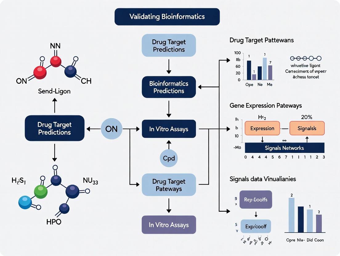
From In Silico to In Vitro: A Strategic Framework for Validating Bioinformatics Drug Target Predictions
This article provides a comprehensive guide for researchers and drug development professionals on bridging the critical gap between computational drug-target interaction (DTI) predictions and experimental validation.
From In Silico to In Vitro: A Strategic Framework for Validating Bioinformatics Drug Target Predictions
Abstract
This article provides a comprehensive guide for researchers and drug development professionals on bridging the critical gap between computational drug-target interaction (DTI) predictions and experimental validation. It explores the foundational principles of modern AI-driven DTI prediction models, including graph neural networks and evidential deep learning. The content details methodological workflows for prioritizing computational hits for in vitro testing, troubleshooting common pitfalls in assay design, and establishing robust validation frameworks to assess prediction accuracy and translational potential. By synthesizing strategies from foundational exploration to comparative analysis, this guide aims to enhance the efficiency and success rate of transitioning in silico discoveries into biologically confirmed leads.
The Computational Frontier: Understanding AI-Driven Drug Target Predictions
The drug discovery process is characterized by exceptionally high costs, extended timelines, and daunting attrition rates. Traditional development from initial research to market requires approximately $2.3 billion and spans 10–15 years, with over 90% of drug candidates failing to reach the market [1]. This inefficiency stems largely from inadequate target validation and unanticipated off-target effects early in the discovery pipeline. In this challenging landscape, in silico prediction technologies have evolved from complementary tools to indispensable assets, fundamentally reshaping how researchers identify and validate therapeutic targets.
This guide objectively compares the performance of leading computational drug-target prediction methods and details the experimental frameworks essential for validating their predictions. By integrating computational precision with rigorous experimental validation, research organizations can significantly de-risk the discovery pipeline and accelerate the development of safer, more effective therapeutics.
TheIn SilicoArsenal: Methodologies and Comparative Performance
Computational approaches for drug-target interaction (DTI) prediction have diversified significantly, ranging from traditional structure-based methods to modern machine learning platforms. The table below compares the primary methodologies and their characteristics.
Table 1: Key In Silico Drug-Target Prediction Methodologies
| Method Category | Representative Tools/Platforms | Core Approach | Data Requirements | Key Applications |
|---|---|---|---|---|
| Ligand-Centric | MolTarPred, SuperPred, PPB2 | 2D/3D chemical similarity searching, QSAR, pharmacophore modeling | Known bioactive compounds, chemical structures | Hit identification, lead optimization, drug repurposing |
| Target-Centric | RF-QSAR, TargetNet, CMTNN | Machine learning models (Random Forest, Naïve Bayes) per target | Bioactivity data (e.g., ChEMBL, BindingDB) | Target fishing, polypharmacology prediction |
| Structure-Based | Molecular Docking (AutoDock Vina), De Novo Design | Protein-ligand docking simulations, binding affinity prediction | 3D protein structures (PDB, AlphaFold) | Virtual screening, binding mechanism analysis |
| Integrated/Machine Learning | DeepTarget, MolTarPred, DTINet | Multimodal data integration, deep learning, network algorithms | Heterogeneous data (chemical, genomic, phenotypic) | Novel target discovery, mechanism of action prediction |
Performance Benchmarking of Prediction Tools
A 2025 systematic comparison of seven target prediction methods using an FDA-approved drug benchmark revealed significant performance variations. The study evaluated stand-alone codes and web servers using a shared dataset to ensure consistent comparison [2].
Table 2: Performance Comparison of Target Prediction Methods (2025 Benchmark)
| Method | Type | Algorithm/Approach | Key Database Source | Reported Performance Notes |
|---|---|---|---|---|
| MolTarPred | Ligand-centric | 2D similarity (MACCS/Morgan fingerprints) | ChEMBL 20 | Most effective method in benchmark; Morgan fingerprints with Tanimoto scores outperformed MACCS |
| CMTNN | Target-centric | ONNX runtime | ChEMBL 34 | Stand-alone code with modern architecture |
| RF-QSAR | Target-centric | Random Forest | ChEMBL 20 & 21 | Web server implementation |
| TargetNet | Target-centric | Naïve Bayes | BindingDB | Uses multiple fingerprint types |
| PPB2 | Ligand-centric | Nearest neighbor/Naïve Bayes/DNN | ChEMBL 22 | Considers top 2000 similar ligands |
| SuperPred | Ligand-centric | 2D/fragment/3D similarity | ChEMBL & BindingDB | Established method with comprehensive similarity approaches |
| ChEMBL | Target-centric | Random Forest | ChEMBL 24 | Official ChEMBL platform implementation |
The study found that MolTarPred emerged as the most effective method overall, with optimization notes indicating that Morgan fingerprints with Tanimoto similarity metrics outperformed other fingerprint and scoring combinations [2]. Performance optimization strategies such as high-confidence filtering (using ChEMBL confidence score ≥7) improved prediction reliability, though with some reduction in recall, making such filtering less ideal for drug repurposing applications where broader target space exploration is valuable.
Specialized Tools for Oncology Applications
In cancer drug discovery, DeepTarget has demonstrated superior performance in predicting both primary and secondary targets of small-molecule agents. Benchmark testing revealed that DeepTarget outperformed existing tools like RoseTTAFold All-Atom and Chai-1 in seven out of eight drug-target test pairs for predicting targets and their mutation specificity [3]. This tool integrates large-scale drug and genetic knockdown viability screens with omics data, uniquely capturing cellular context and pathway-level effects that often play crucial roles in oncology therapeutics beyond direct binding interactions.
From Prediction to Validation: Essential Experimental Frameworks
Computational predictions require rigorous experimental validation to confirm biological relevance. The following section outlines established experimental protocols for verifying in silico drug target predictions.
Experimental Validation Workflow
The transition from in silico prediction to biologically validated target involves a multi-stage process, illustrated below:
Key Experimental Protocols for Target Validation
Cellular Target Engagement Validation (CETSA)
The Cellular Thermal Shift Assay (CETSA) has emerged as a leading approach for validating direct target engagement in intact cells and tissues, addressing the critical gap between biochemical potency and cellular efficacy [4].
Protocol Summary:
- Cell Preparation: Treat intact cells with the drug compound or vehicle control across a range of concentrations and time points.
- Heat Challenge: Subject cell aliquots to different temperatures (typically 45-65°C) to denature proteins not stabilized by drug binding.
- Protein Solubility Analysis: Separate soluble (native) proteins from insoluble (denatured) aggregates and quantify target protein levels in the soluble fraction.
- Data Interpretation: Drug-induced thermal stabilization is evidenced by increased melting temperature (Tm) and greater remaining soluble target protein at higher temperatures.
Application Example: Recent work by Mazur et al. (2024) applied CETSA in combination with high-resolution mass spectrometry to quantitatively demonstrate dose- and temperature-dependent stabilization of DPP9 in rat tissue, confirming target engagement ex vivo and in vivo [4].
Functional Validation in Disease Models
After establishing target engagement, functional validation in disease-relevant models is essential.
Cancer Target Validation Protocol (e.g., CHEK1 in Soft Tissue Sarcoma):
- Gene Expression Analysis: Analyze transcriptomic data from patient samples (e.g., TCGA-SARC cohort) to correlate target expression with clinical outcomes.
- Immunohistochemistry (IHC): Perform IHC staining on patient tissue microarrays to validate protein-level expression and spatial distribution within tumor microenvironments [5].
- Genetic Perturbation: Implement CRISPR-Cas9 mediated knockout or RNA interference to assess the functional consequence of target modulation on cancer cell viability, proliferation, and invasion.
- Therapeutic Assessment: Evaluate the efficacy of target-specific inhibitors in patient-derived xenograft (PDX) models or cell line-derived xenografts.
Application Example: In situ analysis of independent soft tissue sarcoma validation cohorts revealed significant correlation between CHEK1 expression and tumor-infiltrating immune cells, establishing CHEK1 as a promising therapeutic target in combination with immune checkpoint inhibitor therapy [5].
Essential Research Reagent Solutions
Successful validation of computational predictions requires specific research reagents and platforms. The table below details key solutions for experimental confirmation of drug-target interactions.
Table 3: Essential Research Reagent Solutions for Target Validation
| Reagent/Platform | Primary Function | Key Features/Benefits | Representative Applications |
|---|---|---|---|
| CETSA Platform | Target engagement validation in physiologically relevant cellular contexts | Measures thermal stabilization of drug-target complexes in intact cells; provides system-level validation | Confirmation of direct binding; mechanism of action studies; biomarker development [4] |
| CRISPR-Cas9 Systems | Gene knockout and editing for functional validation | Precise genome manipulation; enables assessment of target essentiality | Functional genomics; target prioritization; synthetic lethality screening [6] |
| CIBERSORTx | Digital cytometry for tumor immune microenvironment deconvolution | Estimates immune cell fractions from bulk transcriptome data; no single-cell RNA-seq required | Tumor immunophenotyping; biomarker discovery; immunotherapy target identification [5] |
| AutoDock Vina | Molecular docking and virtual screening | Open-source; hybrid scoring function combining empirical and knowledge-based terms | Binding pose prediction; virtual screening; binding affinity estimation [7] |
| AlphaFold2 Models | Protein structure prediction for targets lacking experimental structures | High-accuracy 3D structure prediction from amino acid sequences | Expanding structural coverage for structure-based drug design [1] |
Integrated Workflow: A Case Study in HCV Drug Discovery
A comprehensive structural bioinformatics study on Hepatitis C Virus (HCV) demonstrates the powerful synergy between computational prediction and experimental validation [7]. The research employed an integrated workflow combining:
Computational Phase:
- Homology Modeling: Generated high-quality 3D structures for key HCV proteins (NS3 protease, NS5B polymerase) using MODELLER and I-TASSER
- Virtual Screening: Docked millions of compounds from the ZINC database against identified druggable sites using AutoDock Vina
- Binding Affinity Prediction: Ranked compounds based on calculated binding energies and interaction patterns
Experimental Validation Phase:
- Compound Testing: Evaluated top-ranked compounds in enzymatic assays for HCV protein inhibition
- Structural Confirmation: Determined crystal structures of key protein-ligand complexes to validate predicted binding modes
- Cellular Efficacy: Assessed antiviral activity in HCV replicon systems and primary hepatocyte models
This integrated approach identified promising drug targets including NS3 protease, NS5B polymerase, core protein, and NS5A, with detailed characterization of their binding pockets and interaction patterns [7]. The study demonstrates how computational approaches can prioritize the most promising targets and compounds for experimental investment, dramatically increasing the efficiency of the discovery pipeline.
The drug discovery bottleneck, characterized by prohibitive costs and unacceptable attrition rates, demands a fundamental transformation in approach. In silico prediction methods have evolved from supportive tools to indispensable components of modern drug discovery, enabling researchers to navigate the expansive landscape of potential targets and therapeutic compounds with unprecedented efficiency. As benchmark comparisons demonstrate, tools like MolTarPred for general target prediction and DeepTarget for oncology applications provide robust platforms for generating high-confidence hypotheses.
However, computational predictions alone cannot overcome the validation bottleneck. The full power of in silico approaches is realized only through rigorous experimental validation using established frameworks including CETSA for target engagement, functional assays in disease-relevant models, and translational studies that bridge cellular findings to clinical relevance. By integrating computational precision with experimental rigor, the drug discovery community can systematically address the historical challenges of high attrition and accelerate the development of transformative therapies for patients in need.
The accurate prediction of drug-target interactions (DTIs) is a critical bottleneck in the drug discovery pipeline. Traditional experimental methods for identifying DTIs are time-consuming, expensive, and low-throughput, often requiring over a decade and billions of dollars to bring a new drug to market [8]. Computational approaches have emerged as powerful tools to prioritize drug-target pairs for experimental validation, with deep learning architectures now demonstrating particular promise by learning complex patterns from large-scale biological data.
Among deep learning approaches, three core architectures have shown significant potential: Graph Neural Networks (GNNs), Transformers, and Autoencoders. These architectures differ fundamentally in how they represent and process molecular and sequence data, leading to distinct strengths and limitations in DTI prediction tasks. GNNs excel at modeling the inherent graph structure of molecules, Transformers capture long-range dependencies in protein sequences, and Autoencoders learn compressed representations that reveal latent patterns in heterogeneous biological networks.
This guide provides a systematic comparison of these architectures within the context of validating bioinformatics predictions with in vitro assays. For researchers and drug development professionals, understanding these architectural differences is crucial for selecting appropriate models, interpreting their predictions, and successfully translating computational findings into experimental validation.
Core Architectures and Their Methodologies
Graph Neural Networks (GNNs)
GNNs process data represented as graphs, making them naturally suited for molecular structures where atoms represent nodes and bonds represent edges. In DTI prediction, GNNs typically operate through message passing mechanisms where node features are updated by aggregating information from neighboring nodes [9] [10].
Key Methodological Components:
- Molecular Graph Representation: Drugs are represented as 2D molecular graphs derived from SMILES strings, with atoms as nodes and bonds as edges [10]. Each atom node is initialized with features including atom type, degree, number of implicit hydrogens, formal charge, and hybridization state.
- Graph Convolutional Layers: These layers update atom representations by combining a node's features with aggregated features from its neighbors. The Hetero-KGraphDTI framework employs a multi-layer message passing scheme that aggregates information from different edge types in heterogeneous graphs [8].
- Attention Mechanisms: Graph Attention Networks (GATs) assign importance weights to different edges during aggregation, enabling the model to focus on the most informative molecular substructures [8].
The GNN encoder in models like MGMA-DTI typically consists of a three-layer Graph Convolutional Network (GCN) that progressively aggregates information from neighboring atomic nodes to capture the topological structure of drug molecules [9].
Transformers
Transformers utilize self-attention mechanisms to capture global dependencies in sequential data, making them particularly effective for protein sequences where long-range interactions between amino acids are crucial for binding site formation [10].
Key Methodological Components:
- Self-Attention Mechanism: Computes attention weights for all pairs of elements in a sequence, allowing each position to attend to all other positions. This is particularly valuable for capturing non-local interactions in protein sequences that influence binding affinity.
- Positional Encodings: Since Transformers lack inherent sequential inductive bias, positional encodings are added to input embeddings to incorporate information about the relative or absolute position of tokens in the sequence.
- Multi-Head Attention: Employs multiple attention mechanisms in parallel to capture different types of relationships within the data.
In CAT-DTI, the Transformer architecture is combined with CNNs to encode both local features and global contextual information from protein sequences [10]. The model uses a convolution neural network combined with a Transformer to encode distance relationships between amino acids within protein sequences.
Autoencoders
Autoencoders learn compressed representations of input data through an encoder-decoder structure, making them valuable for integrating heterogeneous biological information and detecting latent patterns in DTI networks [11].
Key Methodological Components:
- Encoder Network: Maps input data to a lower-dimensional latent space representation through a series of transformative layers.
- Bottleneck Layer: Contains the compressed knowledge representation that ideally captures the most salient features of the input data.
- Decoder Network: Reconstructs the input data from the latent representation, ensuring the encoding retains critical information.
The DDGAE model exemplifies the modern autoencoder approach for DTIs, incorporating a Dynamic Weighting Residual Graph Convolutional Network (DWR-GCN) with residual connections to enable deeper networks without over-smoothing issues [11]. The framework employs a dual self-supervised joint training mechanism that integrates DWR-GCN and a graph convolutional autoencoder into a cohesive system.
Performance Comparison
The following tables summarize the performance of various GNN, Transformer, and Autoencoder-based models on standard DTI prediction benchmarks, providing quantitative comparisons across multiple evaluation metrics.
Table 1: Performance comparison of GNN-based models on benchmark datasets
| Model | Architecture | Dataset | AUC | AUPR | Accuracy | Other Metrics |
|---|---|---|---|---|---|---|
| Hetero-KGraphDTI [8] | GNN with Knowledge Integration | Multiple Benchmarks | 0.98 (avg) | 0.89 (avg) | - | - |
| MGMA-DTI [9] | GCN with Multi-order Gated Convolution | BindingDB | - | - | - | AUROC: 0.988, AUPRC: 0.828, F1: 0.930 |
| EviDTI [12] | GNN with Evidential Deep Learning | DrugBank | - | - | 82.02% | Precision: 81.90%, MCC: 64.29%, F1: 82.09% |
| EviDTI [12] | GNN with Evidential Deep Learning | Davis | - | - | - | Competitive across metrics |
| EviDTI [12] | GNN with Evidential Deep Learning | KIBA | - | - | - | Competitive across metrics |
Table 2: Performance comparison of Transformer-based models
| Model | Architecture | Dataset | AUC | AUPR | Accuracy | Other Metrics |
|---|---|---|---|---|---|---|
| CAT-DTI [10] | Cross-attention & Transformer | Multiple Benchmarks | - | - | - | Overall improvement vs. previous methods |
| MolTrans [8] | Transformer | KEGG | 0.98 | - | - | - |
Table 3: Performance comparison of Autoencoder-based models
| Model | Architecture | Dataset | AUC | AUPR | Accuracy | Other Metrics |
|---|---|---|---|---|---|---|
| DDGAE [11] | Graph Convolutional Autoencoder | DrugBank-based | 0.9600 | 0.6621 | - | - |
| optSAE + HSAPSO [13] | Stacked Autoencoder with Optimization | DrugBank & Swiss-Prot | - | - | 95.52% | Computational complexity: 0.010 s/sample |
Table 4: Cross-domain performance and generalization capabilities
| Model | Architecture | Cross-domain Performance | Uncertainty Quantification | Interpretability |
|---|---|---|---|---|
| EviDTI [12] | GNN with EDL | Strong in cold-start scenarios | Yes | Moderate |
| CAT-DTI [10] | Transformer with CDAN | Enhanced via domain adaptation | No | High (via attention) |
| DDGAE [11] | Autoencoder with DWR-GCN | - | No | Moderate |
Experimental Protocols and Methodologies
Model Training and Evaluation Protocols
Dataset Preparation and Splitting: Standard benchmarks for DTI prediction include BindingDB, BioSNAP, Human, DrugBank, Davis, and KIBA datasets. In most studies, datasets are randomly divided into training, validation, and test sets with typical ratios of 8:1:1 [12]. For cross-domain evaluation, special protocols are employed where models are trained on a source domain and tested on a different target domain to assess generalization capability [10].
Evaluation Metrics: The most common evaluation metrics include:
- AUC (Area Under the ROC Curve): Measures the model's ability to distinguish between interacting and non-interacting pairs across all classification thresholds.
- AUPR (Area Under the Precision-Recall Curve): Particularly important for imbalanced datasets where non-interactions vastly outnumber interactions.
- Accuracy, Precision, Recall, F1-score, and MCC (Matthews Correlation Coefficient): Provide complementary perspectives on model performance.
Negative Sampling Strategies: Given the positive-unlabeled nature of DTI data, sophisticated negative sampling frameworks are crucial. The Hetero-KGraphDTI framework implements three complementary strategies to generate reliable negative samples: random sampling, similarity-based filtering, and biological knowledge-based exclusion [8].
Experimental Workflows
The experimental workflow for developing and validating DTI prediction models typically follows these key stages:
Diagram 1: DTI Model Development and Validation Workflow
Architecture Integration Patterns
Modern DTI prediction models increasingly combine multiple architectural paradigms to leverage their complementary strengths:
Diagram 2: Hybrid Architecture Integration Pattern
The Scientist's Toolkit: Research Reagent Solutions
Table 5: Key research reagents and computational resources for DTI prediction
| Resource | Type | Function in DTI Prediction | Example Applications |
|---|---|---|---|
| DrugBank Database [11] | Chemical Database | Source of drug structures, target information, and known interactions | Feature extraction, ground truth labels, negative sampling |
| BindingDB [9] | Bioactivity Database | Provides binding affinity data for drug-target pairs | Model training and evaluation |
| ProtTrans [12] | Pre-trained Protein Language Model | Generates protein sequence representations using Transformer architectures | Feature extraction from target protein sequences |
| MG-BERT [12] | Pre-trained Molecular Model | Gener molecular representations from graph structures | Feature extraction from drug compounds |
| Gene Ontology (GO) [8] | Knowledge Base | Provides structured biological knowledge for regularization | Enhancing biological plausibility of predictions |
| RDKit [9] | Cheminformatics Library | Processes SMILES strings and generates molecular graphs | Drug feature extraction and representation |
The comparative analysis of GNNs, Transformers, and Autoencoders for DTI prediction reveals a complex landscape where each architecture offers distinct advantages. GNNs demonstrate exceptional performance in modeling molecular structures, with frameworks like Hetero-KGraphDTI achieving AUC scores up to 0.98. Transformers excel at capturing long-range dependencies in protein sequences, while Autoencoders like DDGAE show strong performance in learning compressed representations of heterogeneous biological networks.
For researchers validating predictions with in vitro assays, architectural selection should align with specific research goals and data characteristics. GNNs are preferable when molecular structure is paramount, Transformers when protein sequence context is critical, and Autoencoders when integrating diverse data sources. Emerging trends favor hybrid approaches that combine architectural strengths, such as CAT-DTI's integration of GNNs and Transformers with domain adaptation capabilities.
Uncertainty quantification, as implemented in EviDTI, represents a particularly valuable direction for experimental validation, as it helps prioritize predictions with higher confidence for laboratory testing. As these architectures continue to evolve, their ability to generate biologically interpretable predictions will be crucial for bridging the gap between computational forecasting and experimental confirmation in the drug discovery pipeline.
The process of drug discovery increasingly relies on computational models to predict interactions between potential drug compounds and their biological targets. Accurately interpreting the outputs of these models—from initial binding affinity scores to the quantification of predictive uncertainty—is critical for prioritizing candidates for costly and time-consuming in vitro and in vivo validation. As these computational tools grow more complex, moving from traditional docking scores to sophisticated deep learning and large language model (LLM) based predictions, the need for robust interpretation frameworks has never been greater. This guide objectively compares the performance and capabilities of various computational approaches used in bioinformatics for drug target prediction, with a specific focus on how their outputs should be interpreted and validated within an experimental research context. The ultimate goal is to provide researchers with a practical framework for translating computational predictions into scientifically sound hypotheses for experimental testing, thereby bridging the gap between in silico discovery and in vitro confirmation.
Comparative Performance of Drug-Target Interaction (DTI) Prediction Models
Different computational approaches offer varying strengths in predicting drug-target interactions. The table below summarizes the reported performance of several prominent methods, providing a baseline for objective comparison.
Table 1: Performance Comparison of DTI Prediction Models
| Model/Method | Core Approach | Reported AUC | Reported AUPR | Key Strengths | Interpretability |
|---|---|---|---|---|---|
| Hetero-KGraphDTI | Graph Neural Network with Knowledge Integration | 0.98 [14] | 0.89 [14] | Integrates multiple data types (chemical structures, protein sequences, interaction networks) | High (Attention weights identify salient molecular substructures and protein motifs) [14] |
| Multi-modal GCN (Ren et al.) | Graph Convolutional Network | 0.96 [14] | Information Not Provided | Integrates chemical structures, protein sequences, and PPI networks | Information Not Provided |
| Graph-based Model (Feng et al.) | Heterogeneous Network Learning | 0.98 (KEGG dataset) [14] | Information Not Provided | Learns from multiple heterogeneous networks (drug-drug, target-target, drug-target) | Information Not Provided |
| Traditional Fine-Tuned BERT/BART | Fine-tuned Encoder or Encoder-Decoder Models | ~0.65 (Macro-average across 12 BioNLP tasks) [15] | Information Not Provided | Superior performance in most BioNLP tasks (e.g., information extraction) compared to zero/few-shot LLMs [15] | Information Not Provided |
| GPT-4 (Zero/Few-Shot) | Large Language Model | ~0.51 (Macro-average across 12 BioNLP tasks) [15] | Information Not Provided | Excels in reasoning-related tasks (e.g., medical question answering) [15] | Lower (Prone to hallucinations and missing information) [15] |
From Raw Scores to Biological Meaning: Interpreting Key Outputs
Binding Affinity and Interaction Scores
Computational models generate scores that estimate the strength and likelihood of a drug-target interaction. These scores must be interpreted with a clear understanding of their methodological origins.
- Molecular Docking Scores: Tools like AutoDock Vina predict binding affinity using a hybrid scoring function that calculates the binding free energy (ΔGbinding). This function incorporates terms for attractive/repulsive forces (ΔGgauss), steric clashes (ΔGrepulsion), hydrophobic interactions (ΔGhydrophobic), hydrogen bonding (ΔGhydrogenbonding), and entropic loss due to conformational restriction (ΔGtorsional) [7]. A more negative ΔG_binding generally indicates a more stable and favorable binding interaction.
- Machine Learning Classification Scores: Models like Hetero-KGraphDTI output an interaction probability or a binary classification (interact/does not interact). The high Area Under the Curve (AUC) of 0.98 and Area Under the Precision-Recall Curve (AUPR) of 0.89 reported for this model indicate a strong ability to distinguish true interactions from non-interactions across multiple benchmark datasets [14]. When interpreting these scores, researchers should consider the precision-recall trade-off, especially when dealing with imbalanced datasets where non-interacting pairs are far more common.
The Critical Role of Uncertainty Quantification (UQ)
A model's predictive score is an incomplete picture without an estimate of its associated uncertainty. Uncertainty Quantification (UQ) is essential for assessing the reliability of predictions and is a fundamental requirement for evidence-based reasoning [16].
- Model Uncertainty (Epistemic Uncertainty): This arises from the model's architecture, training data, and optimization process. For generative AI models, this can be quantified by analyzing the variability of evaluation metrics, like precision-recall curves, across multiple training runs with different random initializations [17]. The formal definition of this "model-induced evaluation uncertainty" is the variance of the evaluation metric due to differences in model initialization [17].
- Performance in UQ Tasks: The ability of AI models to perform UQ tasks varies significantly with complexity. A 2025 study found that while reasoning models are generally capable of UQ (scores ≳70%) in simple tasks like judging which of two sample sets is larger, their performance drops to near random guessing (~33%) for complex inequalities requiring multiple intermediate calculations if not guided by specific UQ methods in the prompt [16].
- Data and Aleatoric Uncertainty: This refers to the inherent noise in the experimental data used to train the models. For DTI prediction, this includes variability in binding assay results, inconsistencies in publicly available databases, and incomplete biological context [18].
Experimental Protocols for Computational Validation
Protocol 1: Knowledge-Informed Graph Neural Network
This protocol is adapted from the Hetero-KGraphDTI framework, which integrates graph representation learning with biological knowledge [14].
- Graph Construction: Create a heterogeneous graph that integrates multiple data types. Nodes represent drugs and targets. Edges represent various relationships, such as drug-drug similarity (based on molecular structure), target-target similarity (based on protein sequence or protein-protein interactions), and known drug-target interactions.
- Feature Representation: Represent drugs via their molecular structures (e.g., as graphs or fingerprints) and targets via their protein sequences or structural features.
- Model Training: Train a Graph Neural Network (GNN), such as a Graph Convolutional Network (GCN) or Graph Attention Network (GAT), on the constructed graph. The model learns low-dimensional embeddings for drugs and targets by aggregating information from their local neighborhoods in the graph.
- Knowledge-Based Regularization: Integrate prior biological knowledge from sources like Gene Ontology (GO) and DrugBank during training. This is done using a regularization strategy that encourages the learned drug and target embeddings to be consistent with known ontological and pharmacological relationships [14].
- Prediction and Interpretation: Predict novel DTIs based on the learned embeddings. Use the model's integrated attention mechanisms to identify which molecular substructures and protein motifs are driving the predicted interaction, providing a degree of interpretability [14].
Protocol 2: Structural Bioinformatics Workflow for Novel Target Identification
This protocol outlines a standard workflow for identifying and evaluating novel drug targets within a viral proteome, as demonstrated in a Hepatitis C virus (HCV) study [7].
- Data Retrieval and Preprocessing: Obtain protein sequences for the target organism (e.g., from UniProt). Preprocess sequences to remove redundancy and low-quality regions using tools like CD-HIT with a sequence identity threshold of 90% [7].
- Homology Modeling: For proteins without experimentally determined structures, generate 3D models using homology modeling software like MODELLER or I-TASSER. Select high-resolution crystal structures from the PDB as templates, prioritizing those with a sequence identity of at least 30% and coverage over 80% [7].
- Molecular Docking: Perform molecular docking simulations using software like AutoDock Vina to predict binding sites and interactions. Prepare the protein structures by optimizing and refining them with energy minimization techniques (e.g., using the AMBER force field). Define the docking search space (grid box) around predicted druggable sites, typically with dimensions of 20 × 20 × 20 Å [7].
- Virtual Screening: Screen large compound libraries (e.g., from the ZINC database) against the target protein. Rank the resulting compounds based on their predicted binding energy.
- Post-Docking Analysis: Visually inspect the top-ranked compounds' binding modes and interactions with the target protein using molecular visualization software like PyMOL. Evaluate the drug-likeness of compounds using established filters such as Lipinski's Rule of Five [7].
- Molecular Dynamics (MD) Validation: To assess the stability of the predicted ligand-protein complexes, run MD simulations using a package like GROMACS with the AMBER force field. Solvate the complex in a water box and run simulations for a sufficient time scale to capture dynamic behavior and confirm complex stability [7].
Uncertainty Quantification Frameworks
UQ for Large Language Models (LLMs) in Scientific Workflows
As LLMs are integrated into complex scientific workflows, their ability to perform fundamental UQ tasks becomes critical. A benchmark suite known as "Tether" has been developed to evaluate this capability, focusing on a fundamental UQ problem: estimating whether one quantity is probably larger than another under uncertainty [16]. The benchmark includes two key tasks:
- Simple Inequality Test: The model must judge, with 95% confidence, whether one set of samples is "larger," "smaller," or if the result is "uncertain" compared to another set. LLMs have shown reasonable capability here, with scores around 70% [16].
- Complex Inequality Test: This task requires the model to assess interventional probabilities involving multiple intermediate calculations. Without explicit UQ methods provided in the prompt, LLM performance drops significantly to around 33% (random guessing) [16].
This highlights that while LLMs have potential for UQ, their application in complex biomedical reasoning requires carefully designed prompts and frameworks that explicitly guide uncertainty estimation.
UQ for Generative Models in Distribution Learning
For generative models used in tasks like de novo molecular design, UQ focuses on the confidence in the model's approximation of the target data distribution. A key approach involves analyzing model uncertainty [17].
- Ensemble-based Precision-Recall Curves: This method involves training the model multiple times with different random initializations. The variability (uncertainty) in the precision-recall curves across these runs is then quantified, providing insight into the model's stability and sensitivity to training instabilities [17].
- Total Evaluation Uncertainty: This metric captures the overall variability in a generative model's performance. It incorporates uncertainty from the model's random initialization and the use of finite sets of real and generated samples to estimate the true data and model distributions [17].
Successfully validating computational predictions requires a suite of experimental and computational resources. The table below lists key tools and their functions.
Table 2: Key Research Reagent Solutions for Validation
| Resource Name | Type | Primary Function in Validation | Key Features / Applications |
|---|---|---|---|
| CETSA (Cellular Thermal Shift Assay) | Experimental Assay | Validates direct drug-target engagement in intact cells and native tissue environments [4]. | Provides quantitative, system-level validation of binding, closing the gap between biochemical potency and cellular efficacy. |
| AutoDock Vina | Computational Tool | Performs molecular docking to predict ligand binding modes and affinities [7]. | Open-source; uses a hybrid scoring function to estimate binding free energy; widely used for virtual screening. |
| GROMACS | Computational Tool | Performs Molecular Dynamics (MD) simulations to assess the stability of predicted drug-target complexes [7]. | Highly efficient MD package; used to simulate the dynamic behavior of ligand-protein complexes in solvated environments. |
| DrugBank | Knowledge Base / Database | Provides comprehensive data on known drug-target interactions, mechanisms, and chemical information [18]. | Used for training computational models and validating novel predictions against known pharmacological data. |
| ChEMBL | Database | A manually curated database of bioactive molecules with drug-like properties, including bioactivity data [18]. | Provides bioactivity data for model training and benchmarking; essential for negative sampling during ML model development. |
| ZINC Database | Compound Library | A freely available collection of commercially available compounds for virtual screening [7]. | Contains millions of compounds that can be docked against a target of interest to identify potential hits. |
| PDB (Protein Data Bank) | Database | A global archive for experimentally determined 3D structures of biological macromolecules [18]. | Source of high-resolution protein structures for homology modeling, molecular docking, and structure-based drug design. |
| TTD (Therapeutic Target Database) | Database | Provides information on known and explored therapeutic targets, diseases, and pathways [18]. | Useful for contextualizing novel target predictions within existing knowledge of druggable targets. |
The landscape of computational drug target prediction is diverse, encompassing methods from knowledge-informed GNNs to structural bioinformatics and emerging LLMs. The most accurate models, such as Hetero-KGraphDTI, demonstrate that integrating multiple data types and prior biological knowledge is key to achieving high predictive performance (AUC > 0.95) [14]. However, a high predictive score is not a guarantee of experimental success. Rigorous interpretation that includes Uncertainty Quantification is essential for establishing trustworthiness and prioritizing the most reliable predictions for experimental validation. Frameworks now exist to quantify this uncertainty for both LLMs [16] and generative models [17]. The successful translation of in silico predictions to in vitro validations relies on a complementary toolkit of computational and experimental resources, where methods like CETSA provide the crucial empirical link by confirming target engagement in physiologically relevant contexts [4]. By applying these comparative insights and rigorous validation protocols, researchers can more effectively navigate the complex journey from computational prediction to confirmed biological activity.
In the field of bioinformatics and drug discovery, the accuracy and reliability of computational models for drug-target interaction (DTI) prediction are fundamentally dependent on the quality of the underlying data sources. BindingDB, DrugBank, and UniProt have emerged as three cornerstone databases that researchers routinely leverage for training and validating machine learning and deep learning models. These resources provide complementary types of biological and chemical information that, when integrated, offer a comprehensive foundation for developing predictive algorithms. The validation of computational predictions through in vitro assays represents a critical step in the drug discovery pipeline, bridging the gap between in silico predictions and biological relevance. This guide objectively compares these three key databases, evaluates their performance in experimental contexts, and provides detailed methodologies for their effective utilization in research workflows aimed at translational drug discovery.
Database Comparative Analysis
Table 1: Core Characteristics of Key Bioinformatics Databases
| Database | Primary Focus | Data Content & Size | Key Features | Data Formats |
|---|---|---|---|---|
| BindingDB [19] [20] | Binding affinity measurements | 2,114,159 binding data points between 8,202 protein targets and 928,022 small molecules [19] | Experimentally measured binding affinities (Ki, Kd, IC50); focuses on drug-target interactions | Web-accessible database; downloadable data |
| DrugBank [19] [21] | Comprehensive drug & target data | 14,443 drug molecules and 5,244 non-redundant protein sequences (Version 5.1.8) [19] | Integrates chemical, pharmacological, pharmaceutical data with comprehensive target information; drug-side effects; drug-drug interactions | Bioinformatics/cheminformatics resource; supports complex searches |
| UniProt [19] | Protein sequence & functional information | N/A (Most informative and comprehensive protein database) [19] | Manually annotated (Swiss-Prot) and automatically annotated (TrEMBL) sections; high-quality protein annotations from literature | Five sub-databases with specialized functions |
Table 2: Database Applications in Model Training and Experimental Validation
| Database | Role in DTI Model Training | Experimental Validation Support | Limitations & Considerations |
|---|---|---|---|
| BindingDB | Provides quantitative binding affinity data for regression models; defines negative DTIs (Ki/Kd/IC50/EC50/AC50/Potency >100 μM) [20] | Gold-standard for binding affinity validation; source of experimentally validated interactions [21] | Limited to proteins considered drug targets; binding measurements under specific conditions |
| DrugBank | Source of known drug-target pairs for binary classification; provides drug structures (SMILES) and target protein information [21] [19] | Provides clinically relevant drug-target pairs validated through experiments or extensive literature [21] | Focus on approved drugs and well-studied targets; limited for novel target discovery |
| UniProt | Source of protein sequences for feature extraction; enables similarity-based prediction across protein families [19] | Provides high-quality, manually annotated protein information with evidence-based assertions [19] | Functional annotations may be incomplete for less-studied proteins |
Experimental Protocols for Database Integration and Validation
Protocol 1: Construction of Gold-Standard DTI Datasets
Objective: Integrate data from BindingDB, DrugBank, and UniProt to create a high-confidence dataset for DTI model training and validation.
Materials:
- DrugBank database (drug and target information)
- BindingDB (binding affinity measurements)
- UniProt (protein sequence and functional annotation)
- HCDT 2.0 database (curated drug-gene, drug-RNA, drug-pathway interactions) [20]
Methodology:
- Data Collection: Download the latest versions of DrugBank, BindingDB, and UniProt databases through their official portals or APIs.
- Identifier Mapping: Standardize identifiers across databases using common accessions (e.g., UniProt IDs for proteins, PubChem IDs for compounds).
- Positive Instance Selection: Extract known drug-target pairs from DrugBank with clinical validation [21] and high-affinity interactions from BindingDB (Ki, Kd, IC50, EC50 ≤ 10 μM) [20].
- Negative Instance Selection: Define non-interacting pairs using BindingDB entries with binding affinity measurements >100 μM [20] or through biological sampling strategies [22].
- Feature Extraction:
- Dataset Splitting: Implement biologically-driven splitting strategies [22]:
- Warm start: Drugs and proteins shared between train and test sets
- Cold start: Unseen drugs or proteins in test set
Database Integration Workflow for DTI Model Training
Protocol 2: In Vitro Validation of Computational Predictions
Objective: Experimentally validate computationally predicted drug-target interactions using surface plasmon resonance (SPR) and cell-based assays.
Materials:
- Purified target proteins
- Compound libraries from predicted interactions
- SPR instrumentation (e.g., Biacore)
- Cell lines expressing target proteins
- Assay reagents for functional readouts
Methodology:
- Candidate Selection: Select top-ranking DTI predictions from computational models for experimental testing.
- SPR Binding Assays:
- Immobilize purified target proteins on SPR sensor chips
- Inject compound solutions at varying concentrations (e.g., 0.1-100 μM)
- Measure association and dissociation rates to determine binding affinity (KD)
- Compare with known binders and negative controls
- Functional Cell-Based Assays:
- Treat relevant cell lines with predicted compounds (dose-response)
- Measure downstream pathway activation or inhibition
- Assess functional responses (e.g., proliferation, apoptosis, signaling)
- Validation Criteria: Confirm interactions with KD < 10 μM and statistically significant functional effects (p < 0.05) compared to controls.
Performance Assessment in Research Applications
Table 3: Database Performance in Published DTI Prediction Studies
| Study/Model | Databases Used | Performance Metrics | Experimental Validation Outcome |
|---|---|---|---|
| DrugMAN [21] | DrugBank, BindingDB, CTD, Others | AUROC: 0.912, AUPRC: 0.837 (warm start); Minimal performance decrease in cold-start scenarios | Demonstrated robust generalization ability for real-world applications |
| ColdstartCPI [23] | BindingDB, ChEMBL | Outperformed state-of-the-art methods in cold-start conditions; Effective with sparse data | Predictions validated via molecular docking, binding free energy calculations, literature search |
| HCDT 2.0 [20] | 9 drug-gene, 6 drug-RNA, 5 drug-pathway databases | 1,224,774 drug-gene pairs; 38,653 negative DTIs | High-confidence interactions curated through experimental validation criteria |
Table 4: Key Research Reagent Solutions for DTI Validation
| Reagent/Resource | Function | Application Context |
|---|---|---|
| RDkit [19] | Python toolkit for cheminformatics | Compute molecular descriptors/fingerprints from compound structures |
| iFeature [19] | Python toolkit for protein sequence analysis | Generate feature descriptors from protein sequences for machine learning |
| ProtTrans [23] | Pre-trained protein language model | Extract protein features using transformer-based architectures |
| Mol2vec [23] | Unsupervised machine learning approach | Learn vector representations of molecular substructures |
| BIONIC [21] | Biological network integration framework | Learn node representations from multiple biological networks |
| SPR Instrumentation | Label-free binding affinity measurement | Validate direct molecular interactions in real-time |
Integration Strategies and Best Practices
Data Preprocessing and Quality Control
Effective utilization of BindingDB, DrugBank, and UniProt requires meticulous data preprocessing. For BindingDB, researchers should apply consistent thresholding for binding affinities (e.g., ≤10 μM for positive interactions and >100 μM for negative interactions) [20]. With DrugBank, careful attention should be paid to distinguishing between approved drugs, investigational drugs, and withdrawn compounds, as this affects the biological relevance of predictions. For UniProt, prioritization of manually curated Swiss-Prot entries over automatically annotated TrEMBL records ensures higher quality protein annotations [19].
Integrated Computational-Experimental Workflow for DTI Validation
Addressing Cold-Start Challenges
A significant limitation in many DTI prediction approaches is poor performance on novel compounds or targets (cold-start problem) [22] [23]. To address this, researchers should employ specialized models like ColdstartCPI [23] or DrugMAN [21] that demonstrate robustness in these scenarios. Additionally, incorporating pre-trained features from large chemical libraries (via Mol2vec) [23] or protein language models (ProtTrans) [23] can enhance generalization to unseen entities. Biologically-driven dataset splitting strategies that separate drugs and proteins based on structural or functional similarity during training-test set creation are essential for realistic performance assessment [22].
BindingDB, DrugBank, and UniProt each provide unique and complementary data types that are essential for training robust DTI prediction models. BindingDB offers quantitative binding affinity measurements critical for regression tasks, DrugBank provides clinically validated drug-target pairs with rich contextual information, and UniProt delivers comprehensive protein sequences and functional annotations. The integration of these resources, coupled with appropriate experimental validation protocols, creates a powerful framework for accelerating drug discovery. As computational methods continue to evolve, particularly with advances in deep learning and multimodal approaches [24], these established databases will remain foundational resources for training and validating the next generation of DTI prediction models. Researchers should prioritize biologically-relevant benchmarking, careful attention to cold-start scenarios, and rigorous in vitro validation to ensure computational predictions translate to biologically meaningful results.
In the field of drug-target interaction (DTI) prediction, the selection of appropriate performance metrics is not merely a technical formality but a critical determinant of a model's perceived utility and translational potential. For researchers and drug development professionals validating bioinformatics predictions with in vitro assays, understanding the nuances of these metrics is paramount for allocating precious experimental resources effectively. The Receiver Operating Characteristic (ROC) curve and its corresponding Area Under the Curve (AUC) serve as fundamental tools for evaluating the diagnostic performance of index tests, which in this context are computational models designed to discriminate between interacting and non-interacting drug-target pairs [25] [26].
The ROC curve is a graphical plot that illustrates the trade-off between a model's True Positive Fraction (TPF, or sensitivity) and its False Positive Fraction (FPF, which is 1-specificity) across all possible classification thresholds [25]. The AUC value, which ranges from 0.5 to 1.0, summarizes this curve and represents the probability that the model will rank a randomly chosen positive instance (a true interaction) higher than a randomly chosen negative instance [26]. An AUC of 0.5 indicates performance equivalent to random chance, while an AUC of 1.0 signifies perfect discrimination [26]. In clinical and diagnostic contexts, AUC values above 0.9 are considered excellent, 0.8-0.9 considerable, 0.7-0.8 fair, and below 0.7 of limited clinical utility [26].
The Area Under the Precision-Recall Curve (AUPRC) has emerged as a complementary metric, particularly valued in scenarios with class imbalance—a hallmark of DTI prediction datasets where known interactions are vastly outnumbered by unknown or non-interacting pairs [27]. While the ROC curve and its AUC remain indispensable for assessing a model's overall ranking ability, the precision-recall curve and its AUPRC focus on the model's performance in identifying positive instances, making it especially relevant when the positive class is the primary interest [27].
Comparative Analysis of AUC and AUPRC
Mathematical and Conceptual Foundations
The fundamental distinction between AUC and AUPRC lies in what they measure and how they weight different types of classification outcomes. AUC evaluates a model's ability to separate positive and negative classes across all thresholds, effectively measuring the probability that a random positive sample is ranked higher than a random negative sample [26]. This property makes it a robust metric for overall classification performance, as it is invariant to class imbalance and the specific classification threshold chosen [27].
AUPRC, in contrast, focuses specifically on the model's performance concerning the positive class by plotting precision (the proportion of true positives among all predicted positives) against recall (sensitivity, or the proportion of actual positives correctly identified) [27]. This focus makes AUPRC particularly sensitive to the model's ability to correctly identify positive instances without being overwhelmed by false positives—a critical consideration when validating predictions with expensive in vitro assays.
Recent mathematical analysis has revealed a probabilistic interrelationship between these metrics, demonstrating that while AUC weighs all false positives equally, AUPRC weighs false positives with the inverse of the model's likelihood of outputting a score greater than a given threshold [27]. This fundamental difference in weighting leads to distinct behavioral characteristics, especially in the context of class imbalance and model optimization priorities.
Behavioral Differences in Class-Imbalanced Scenarios
The widespread adage that "AUPRC is superior to AUC for model comparison under class imbalance" requires careful examination. While AUPRC values are indeed typically lower than AUC values in imbalanced datasets, this observation alone does not establish AUPRC's superiority for model comparison [27]. The critical consideration is not the absolute metric values but the relative rankings that different metrics confer upon models when making comparisons.
Research indicates that AUC and AUPRC implicitly prioritize different types of model improvements [27]. AUC optimization corresponds to a strategy where all classification errors are considered equally valuable to correct, regardless of where they occur in the score distribution. This approach is optimal for deployment scenarios where samples will be encountered across the entire score spectrum. AUPRC optimization, conversely, corresponds to prioritizing the correction of classification errors for samples assigned the highest scores first [27]. This strategy aligns with information retrieval settings where users primarily examine the top-k ranked predictions.
This distinction has profound implications for fairness and utility in DTI prediction. If the underlying dataset contains subpopulations with different prevalence rates (e.g., different protein families with varying numbers of known interactions), AUPRC will explicitly favor optimization for the higher-prevalence subpopulation, whereas AUC will optimize both subpopulations in an unbiased manner [27]. This bias can inadvertently introduce algorithmic disparities and should be carefully considered when evaluating models for broad deployment.
Practical Implications for DTI Prediction
For researchers validating DTI predictions with in vitro assays, the choice between AUC and AUPRC as a primary evaluation metric should align with the anticipated deployment context. If the goal is to generate a comprehensive ranking of all possible drug-target pairs for systematic exploration, AUC provides a more balanced assessment of overall ranking quality. However, if the research objective is to identify the most promising candidates for immediate experimental validation from the top-ranked predictions, AUPRC may better reflect the model's utility for this specific use case.
The most robust approach involves reporting both metrics alongside their confidence intervals, as each reveals different aspects of model performance. Furthermore, considering additional metrics such as precision at fixed recall levels or threshold-specific performance can provide a more complete picture of a model's operational characteristics.
Performance Benchmarking of State-of-the-Art DTI Prediction Models
Table 1: Comparative Performance of Recent DTI Prediction Models on Benchmark Datasets
| Model | Architecture | Dataset | AUC | AUPRC | Key Innovations |
|---|---|---|---|---|---|
| ImageMol [28] | Self-supervised Image Representation Learning | HIV, Tox21, BACE | 0.814 (HIV), 0.826 (Tox21), 0.939 (BACE) | N/R | Pretrained on 10M drug-like molecules; uses molecular images as input |
| EviDTI [12] | Evidential Deep Learning | DrugBank, Davis, KIBA | 0.820 (DrugBank Acc) | N/R | Integrates 2D/3D drug structures with target sequences; provides uncertainty estimates |
| DHGT-DTI [29] | Dual-view Heterogeneous Graph Network | Two benchmark datasets | N/R | N/R | Combines GraphSAGE (local features) and Graph Transformer (global features) |
| DDGAE [11] | Dynamic Weighting Residual GCN | Curated dataset (708 drugs, 1,512 targets) | 0.9600 | 0.6621 | Incorporates dynamic weighting graph convolution with residual connections |
| Hetero-KGraphDTI [14] | GNN with Knowledge-Based Regularization | Multiple benchmarks | 0.98 (avg) | 0.89 (avg) | Integrates biological knowledge graphs; uses attention mechanisms |
Table 2: Clinical Interpretation Guidelines for AUC Values [26]
| AUC Value Range | Interpretation | Suggested Clinical/Experimental Utility |
|---|---|---|
| 0.9 ≤ AUC ≤ 1.0 | Excellent | High confidence for experimental validation |
| 0.8 ≤ AUC < 0.9 | Considerable | Promising for targeted experimental follow-up |
| 0.7 ≤ AUC < 0.8 | Fair | Limited utility; may require further model refinement |
| 0.6 ≤ AUC < 0.7 | Poor | Questionable utility for experimental guidance |
| 0.5 ≤ AUC < 0.6 | Fail | No better than random chance |
The performance landscape of contemporary DTI prediction models reveals consistent advancement in both AUC and AUPRC values. As shown in Table 1, recent models leveraging graph neural networks and knowledge integration have achieved exceptional performance, with Hetero-KGraphDTI reporting an average AUC of 0.98 and AUPRC of 0.89 across multiple benchmarks [14]. The DDGAE model demonstrates similarly strong performance with an AUC of 0.9600, though its AUPRC of 0.6621 highlights the significant gap that can emerge between these metrics under class imbalance [11].
When interpreting these values, the guidelines in Table 2 provide useful reference points. Models achieving AUC values above 0.90 can be considered to offer excellent discriminatory power, suggesting high promise for guiding experimental validation [26]. However, it is crucial to consider the 95% confidence intervals around these point estimates, as a wide confidence interval may indicate unreliable performance despite a high point estimate [26].
The observed performance gains in recent models can be attributed to several architectural innovations: the integration of multiple data modalities (2D/3D molecular structures, protein sequences, interaction networks) [12]; the use of pre-training on large-scale molecular databases [28]; the incorporation of biological knowledge through regularization [14]; and advanced graph learning techniques that capture both local and global network structures [29] [11].
Experimental Protocols and Methodologies
Standard Evaluation Frameworks
Robust evaluation of DTI prediction models requires careful experimental design to avoid optimistic performance estimates. The field has converged on several key methodological practices:
Data Splitting Strategies: To assess model generalizability, datasets are typically divided using scaffold-based splits, where the training, validation, and test sets contain distinct molecular substructures [28]. This approach tests the model's ability to generalize to novel chemical entities rather than merely recognizing structural similarities. Alternative strategies include random splits and time-aware splits that simulate real-world deployment scenarios.
Cross-Validation: Most rigorous evaluations employ k-fold cross-validation (typically 5- or 10-fold) to account for variability in dataset composition and provide more stable performance estimates.
Benchmark Datasets: Commonly used benchmarks include DrugBank [12], Davis [12], KIBA [12], BACE [28], Tox21 [28], and specialized datasets for specific target families such as kinases [28] and cytochrome P450 enzymes [28]. These datasets vary in size, class imbalance, and biological context, enabling comprehensive assessment of model capabilities.
Specialized Protocols for DTI Prediction
Beyond standard evaluation practices, several specialized protocols address unique challenges in DTI prediction:
Cold-Start Evaluation: This scenario tests a model's ability to predict interactions for new drugs or targets not present during training [12]. This is accomplished by ensuring that specific drugs or targets (or both) are exclusively present in the test set, simulating the practical challenge of predicting interactions for novel entities.
Temporal Validation: For drug repurposing applications, models may be evaluated using time-split validation, where training data is limited to interactions discovered before a specific date, and test data consists of interactions discovered after that date.
Case Study Validation: Performance metrics are complemented by targeted case studies focusing on specific therapeutic areas. For example, several studies have validated predictions for Parkinson's disease treatments [29] or anti-SARS-CoV-2 molecules [28], providing concrete evidence of practical utility.
Diagram 1: Comprehensive Workflow for DTI Prediction Model Development and Validation. This diagram illustrates the standard experimental protocol from data collection through experimental validation, highlighting key stages and performance assessment components.
Essential Research Reagent Solutions for Experimental Validation
Table 3: Key Research Reagents and Resources for DTI Experimental Validation
| Reagent/Resource | Function in Experimental Validation | Representative Examples/Sources |
|---|---|---|
| Compound Libraries | Source of candidate drugs for testing | PubChem (10M+ compounds) [28], FDA-approved drug libraries |
| Target Proteins | Production of protein targets for binding assays | Recombinant expression systems, native protein purification |
| Binding Assay Kits | Measurement of direct molecular interactions | Fluorescence-based, radioisotope-based, surface plasmon resonance kits |
| Cell-Based Assay Systems | Assessment of functional effects in biological context | Cell lines with target overexpression, reporter gene assays |
| High-Throughput Screening Platforms | Automated testing of multiple compound-target pairs | Robotic liquid handling, automated microscopy, multi-well plate readers |
| Bioinformatics Databases | Source of known interactions and structural information | DrugBank [11], HPRD [11], ChEMBL, BindingDB |
| Knowledge Bases | Context for interpreting results and generating hypotheses | Gene Ontology [14], KEGG Pathways, Reactome |
The transition from computational prediction to experimental validation requires access to specialized reagents and resources, as summarized in Table 3. Compound libraries such as PubChem, which contains over 10 million drug-like molecules, provide the chemical starting point for experimental testing [28]. For target production, recombinant expression systems enable the production of purified proteins for in vitro binding assays, while cell-based systems allow assessment of functional effects in more physiologically relevant contexts.
Several experimental methodologies are commonly employed for validation. Binding assays measure direct physical interactions between drugs and targets using techniques such as surface plasmon resonance, fluorescence polarization, or radioligand binding. Functional assays assess the pharmacological consequences of these interactions, such as enzyme inhibition or receptor activation. High-throughput screening platforms enable the efficient testing of thousands of compound-target combinations, dramatically accelerating the validation process.
Critical to this pipeline are comprehensive bioinformatics databases such as DrugBank [11] and HPRD [11], which provide curated information on known drug-target interactions for benchmarking and reference. Biological knowledge bases including Gene Ontology [14] and pathway databases offer essential context for interpreting validation results and generating mechanistic hypotheses.
Diagram 2: Comparative Characteristics and Application Contexts of AUC and AUPRC. This diagram illustrates the distinct properties of each metric and the deployment scenarios where each excels.
The establishment of rigorous performance baselines through metrics like AUC and AUPRC is fundamental to advancing the field of drug-target interaction prediction. For researchers and drug development professionals validating computational predictions with in vitro assays, a nuanced understanding of these metrics enables more informed decision-making in both model selection and experimental prioritization.
AUC remains the gold standard for assessing a model's overall ranking capability, with values above 0.90 indicating excellent discriminatory power suitable for guiding experimental programs [26]. AUPRC provides complementary information, particularly valuable when the primary research objective is identifying high-confidence candidates from the top-ranked predictions [27]. The most robust approach involves considering both metrics alongside their confidence intervals and statistical significance.
As the field progresses, emerging techniques such as evidential deep learning for uncertainty quantification [12] and knowledge-guided representation learning [14] promise to further enhance predictive performance and translational utility. By strategically applying appropriate evaluation metrics and maintaining rigorous validation standards, the research community can continue to accelerate the identification of novel therapeutic interventions through computational approaches.
Bridging the Digital and Physical: Designing In Vitro Validation Pipelines
The advent of high-throughput technologies and sophisticated artificial intelligence has revolutionized the initial stages of drug discovery, enabling researchers to generate thousands of potential drug-target interactions (DTIs) through computational methods [30] [31]. While computational predictions provide valuable starting points, the transition from virtual hits to experimentally viable candidates remains a critical bottleneck. This challenge is particularly pronounced in bioinformatics-driven target identification, where the gap between in silico prediction and in vitro validation contributes significantly to attrition rates in later development stages [32] [33].
The fundamental question facing researchers is no longer how to generate computational hits, but how to prioritize them for expensive and time-consuming experimental validation. A systematic prioritization framework that integrates computational confidence metrics with experimentally practical validation strategies is essential for resource-efficient drug discovery. This guide establishes such a framework by comparing multiple prioritization and validation approaches, providing structured methodologies for bridging the computational-experimental divide.
Computational Prioritization: From Initial Hits to Triage
Defining Hit Criteria and Ligand Efficiency Metrics
The first step in transitioning from computational hits to experimental candidates involves establishing clear hit-calling criteria. Analysis of virtual screening studies reveals significant variation in how researchers define a "hit," with only approximately 30% of studies reporting clear, predefined activity cutoffs [34]. The most effective frameworks move beyond simple activity thresholds to incorporate ligand efficiency metrics that normalize biological activity against molecular properties.
Table 1: Established Hit Identification Criteria in Virtual Screening
| Metric Category | Specific Metrics | Typical Range for Hits | Strategic Importance |
|---|---|---|---|
| Potency Measures | IC₅₀, EC₅₀, Kᵢ, Kd | 1-100 µM [34] | Primary activity against intended target |
| Ligand Efficiency | LE (Ligand Efficiency) | ≥ 0.3 kcal/mol/heavy atom [34] | Normalizes potency by molecular size |
| Lipophilic Efficiency | LipE, LLE (Lipophilic Ligand Efficiency) | LipE > 5 [35] | Penalizes excessive lipophilicity |
| Structural Alert | PAINS filters, promiscuity checks | Elimination of flagged compounds [34] | Avoids compounds with problematic motifs |
While sub-micromolar activity is desirable, the majority of successful virtual screening studies employ hit criteria in the low to mid-micromolar range (1-100 µM), particularly for novel targets or scaffolds [34]. This pragmatic approach acknowledges that computational hits serve as starting points for optimization rather than final drug candidates.
The Traffic Light System for Multi-Parameter Hit Triage
Prioritizing computational hits requires evaluating multiple parameters simultaneously. The "Traffic Light" (TL) system provides a visual, quantitative framework for comparing hit series across diverse criteria [35]. This approach assigns scores of 0 (good), 1 (warning), or 2 (bad) across multiple parameters, generating a composite score that enables objective comparison of potential starting points.
Table 2: Example Traffic Light Analysis for Hit Triage [35]
| Evaluation Parameter | Compound A | Compound B | Rationale for Prioritization |
|---|---|---|---|
| Potency (IC₅₀) | 1.2 µM (+1) | 0.8 µM (0) | Compound B more potent |
| Ligand Efficiency | 0.45 (0) | 0.28 (+2) | Compound A uses molecular size more efficiently |
| cLogP | 2.1 (0) | 4.8 (+2) | Compound A has more favorable lipophilicity |
| Solubility | >200 µM (0) | Not tested (+2) | Compound A demonstrates good solubility |
| Selectivity | 15-fold (0) | 3-fold (+2) | Compound A shows better target specificity |
| Total Score | 1 | 8 | Compound A clearly preferred |
The TL system's flexibility allows research teams to incorporate additional experimental data as it becomes available, creating a dynamic prioritization framework that evolves throughout the hit-to-lead process. Teams can weight categories based on project-specific priorities, though equal weighting generally provides the most unbiased starting point [35].
Experimental Validation Strategies: A Comparative Framework
Orthogonal Corroboration vs. Traditional Validation
The framework of "experimental validation" requires refinement in the modern drug discovery context. Rather than a single gold-standard validation method, orthogonal corroboration using multiple experimental approaches provides greater scientific rigor [30]. This paradigm shift acknowledges that all experimental methods have limitations and that confidence increases when multiple approaches yield consistent results.
Table 3: Comparative Analysis of Experimental Validation Methods
| Computational Method | Traditional "Gold Standard" | Orthogonal Corroboration | Advantages of Orthogonal Approach |
|---|---|---|---|
| Variant Calling (WGS/WES) | Sanger sequencing [30] | High-depth targeted sequencing [30] | Better detection of low-frequency variants; more precise VAF estimates |
| Copy Number Aberration Calling | FISH (20-100 cells) [30] | Low-depth WGS of thousands of single cells [30] | Higher resolution for subclonal events; quantitative, statistical thresholds |
| Differential Protein Expression | Western Blot/ELISA [30] | Mass spectrometry (MS) [30] | Higher specificity based on multiple peptides; greater coverage and reproducibility |
| Differentially Expressed Genes | RT-qPCR [30] | RNA-seq [30] | Comprehensive transcriptome coverage; nucleotide-level resolution |
The selection of orthogonal methods should consider throughput, resolution, and quantitative capability. For example, mass spectrometry provides superior protein identification confidence compared to Western blotting when multiple peptides cover significant protein sequence (e.g., >5 peptides covering ~30% of sequence with E value < 10⁻¹⁰) [30].
Assessing Assay Quality and Performance Metrics
Regardless of the specific validation method selected, assessing assay quality is essential for interpreting results accurately. The Z' factor is a critical statistical parameter that evaluates assay robustness by incorporating both the assay signal dynamic range and data variation [36]:
Assays with Z' values between 0.5 and 1.0 are considered excellent for screening purposes, while values below 0.5 indicate poor assay quality unsuitable for reliable hit validation [36]. Additional metrics such as signal-to-background (S/B) ratio and EC₅₀/IC₅₀ values for reference compounds provide further assay characterization [36].
For concentration-response assays, the Toxicity Separation Index (TSI) and Toxicity Estimation Index (TEI) represent advanced performance metrics that evaluate how well in vitro data predict in vivo effects. These metrics are particularly valuable in safety assessment and toxicology prediction, where TSI values approaching 1.0 indicate excellent separation between toxic and non-toxic compounds [37].
Integrated Workflow: From Prediction to Confirmation
The Prioritization and Validation Pipeline
The following workflow diagram illustrates the complete pathway from computational hit identification to experimental candidate confirmation, integrating both computational and experimental elements:
Prioritization and Validation Workflow: This integrated pipeline shows the key decision points from initial computational hits through experimental confirmation, highlighting the critical transition between phases.
Target Engagement and Mechanism of Action
Confirmation of direct target engagement represents a crucial step in validating computational predictions. Cellular Thermal Shift Assay (CETSA) has emerged as a powerful method for demonstrating direct binding in physiologically relevant environments [4]. Unlike purely biochemical assays, CETSA confirms target engagement in intact cells and can be extended to tissue samples, providing a translational bridge between in vitro and in vivo systems [4].
For programs targeting specific mechanism of action (MoA) classes, distinguishing between activation and inhibition is essential. Recent computational frameworks like DTIAM enable prediction of activation/inhibition mechanisms alongside binding affinity, though these predictions require experimental confirmation using appropriate functional assays [31]. The expansion of mechanism-specific validation assays addresses a critical gap in early discovery, where misinterpretation of compound MoA contributes to later-stage failures.
Research Reagent Solutions for Experimental Validation
Table 4: Essential Research Reagents for Validation Workflows
| Reagent Category | Specific Examples | Primary Function | Considerations for Selection |
|---|---|---|---|
| Cell-Based Assay Systems | Reporter gene assays (luciferase), CETSA [36] [4] | Measure functional activity in cellular context | Prioritize systems with high Z' factors (>0.5) and physiological relevance |
| Target Engagement Reagents | CETSA kits, SPR chips, binding assay reagents [4] | Confirm direct compound-target interaction | Cellular vs. biochemical context; throughput requirements |
| Orthogonal Detection Reagents | MS-compatible reagents, specific antibodies, sequencing kits [30] | Enable multiple validation approaches | Compatibility across platforms; specificity validation |
| ADMET Profiling Tools | PAMPA plates, microsomal stability kits, CYP inhibition assays [35] | Assess drug-like properties early | Balance between throughput and predictivity; species relevance |
The transition from computational hit to experimental candidate requires a systematic framework that integrates computational triaging with orthogonal experimental validation. By applying ligand efficiency metrics, multi-parameter scoring systems like the Traffic Light approach, and orthogonal corroboration strategies, research teams can significantly improve prioritization efficiency. The evolving landscape of experimental methods, particularly in target engagement confirmation and mechanism of action studies, provides increasingly robust tools for bridging the computational-experimental divide. As computational methods continue to advance, the importance of rigorous, practical validation frameworks will only increase, ultimately accelerating the delivery of new therapeutic candidates to patients.
The drug discovery pipeline is increasingly initiated by bioinformatics predictions, which propose novel drug targets through computational analysis of complex biological data [38]. The transition from in silico prediction to tangible therapeutic candidate requires rigorous experimental validation, a process predominantly reliant on primary in vitro assays. These assays fall into two fundamental categories: those measuring binding affinity and those quantifying functional activity. Binding affinity assays confirm that a drug candidate physically interacts with its predicted target, fulfilling a primary requirement for activity. Functional activity assays advance this further by revealing the biological consequences of that interaction, determining whether the compound acts as an agonist, antagonist, or inverse agonist, and elucidating the magnitude and efficacy of its effect. This guide provides an objective comparison of these two critical assay paradigms, framing them within the essential process of translating computational predictions into biologically relevant outcomes with supporting experimental data to inform selection for early-stage validation.
Fundamental Principles and Direct Comparative Analysis
Core Conceptual Definitions
- Binding Affinity Measurements quantify the strength of the physical interaction between a compound (ligand) and its biological target (e.g., receptor, enzyme). The key parameter is the inhibition constant (Ki), a molar concentration value indicating the ligand concentration required to occupy 50% of the receptors at equilibrium. A lower Ki signifies a higher, more potent affinity [39].
- Functional Activity Measurements determine the biological effect or cellular response triggered by the ligand-target interaction. These assays measure parameters like second messenger production, ion flux, or gene expression. A critical output is the EC50 (half-maximal effective concentration) and the efficacy, which defines the ligand as a full/partial agonist or antagonist in a specific pathway [39].
Side-by-Side Comparison of Key Assay Characteristics
The choice between affinity and functional assays hinges on their complementary strengths and the specific question being asked. The table below summarizes their core attributes for direct comparison.
Table 1: Comparative Analysis of Binding vs. Functional In Vitro Assays
| Characteristic | Binding Affinity Assays | Functional Activity Assays |
|---|---|---|
| Primary Measurement | Physical interaction and occupancy (Ki/IC50) [39] | Biological effect and cellular response (EC50, Efficacy) [39] |
| Key Output Parameters | Inhibition Constant (Ki), Selectivity Ratio [39] | EC50, IC50, Intrinsic Activity (α), % Emax [39] |
| Information Gained | Confirms target engagement; Affinity and selectivity [39] | Reveals functional efficacy, agonist/antagonist properties, and signaling bias [40] |
| Typical Readouts | Radioligand displacement (e.g., with [³H]DAMGO) [39] | GTPγS binding, cAMP accumulation, calcium flux, reporter gene assays [39] |
| Throughput | Generally higher | Can be high, but often more complex than binding |
| Metabolic Requirements | Cell membrane preparations often sufficient; no functional system needed [39] | Requires live, responsive cells with intact signaling pathways [40] |
| Key Limitation | Cannot distinguish agonists from antagonists; provides no efficacy data [39] | More complex, costly; results can be system-dependent (cell type, receptor density) [40] |
Experimental Protocols and Methodologies
Detailed Protocol for a Binding Affinity Assay (Radioligand Binding)
Radioligand binding is a gold-standard technique for direct affinity measurement.
- Membrane Preparation: Harvest Chinese Hamster Ovary (CHO) cells stably expressing the human opioid receptor of interest. Homogenize cells and isolate crude plasma membranes via differential centrifugation [39].
- Assay Setup: In a binding buffer, incubate a fixed concentration of a tritiated radioligand (e.g., [³H]DAMGO for MOR), the membrane preparation, and increasing concentrations of the unlabeled test compound. Include wells for total binding (no competitor) and nonspecific binding (excess unlabeled ligand like naloxone) [39].
- Incubation and Termination: Incubate the reaction to equilibrium (e.g., 60-120 minutes at 25°C). Terminate the binding by rapid filtration through glass-fiber filters to trap the membrane-bound radioligand [39].
- Quantification and Analysis: Measure the radioactivity retained on the filters using a scintillation counter. Specific binding is calculated as total binding minus nonspecific binding. Data is analyzed using non-linear regression to determine the Ki of the test compound [39].
Detailed Protocol for a Functional Activity Assay ([³⁵S]GTPγS Binding)
The [³⁵S]GTPγS binding assay measures G-protein activation, a proximal step in GPCR signaling, providing functional data without a downstream reporter.
- Membrane Preparation: Use membranes from CHO cells expressing the target receptor, as in the binding assay [39].
- Activation Reaction: Incubate membranes with GDP (to stabilize G-proteins), a fixed concentration of [³⁵S]GTPγS (a non-hydrolyzable GTP analog), and increasing concentrations of the test compound. Include a buffer baseline and a reference full agonist control (e.g., DAMGO for MOR) [39].
- Termination and Filtration: Similar to radioligand binding, terminate the reaction by rapid filtration to separate bound from free [³⁵S]GTPγS [39].
- Data Analysis: Quantify bound radioactivity. The response for each compound is plotted as a percentage of the maximal stimulation by the reference full agonist. Non-linear regression analysis yields the EC50 (potency) and the Emax (efficacy, or intrinsic activity) [39].
Workflow for Integrated Assay Strategy in Target Validation
A robust validation strategy often employs both assay types sequentially. The following workflow diagrams a logical pathway from initial computational prediction to a functionally characterized lead.
Diagram 1: Integrated Assay Workflow
Data Presentation and Interpretation
Quantitative Data from a Case Study: 3-Benzylaminomorphinan Derivatives
The following table summarizes experimental data from a study on opioid receptor ligands, illustrating how binding and functional data are reported and interpreted together [39].
Table 2: Experimental Binding and Functional Data for Select Opioid Receptor Ligands [39]
| Compound | Binding Affinity Ki (nM) | Functional Activity [³⁵S]GTPγS |
|---|---|---|
| MOR-Selective Example | MOR: 0.42 nM | Full Agonist at MOR |
| 3-(3′-hydroxybenzyl)amino-17-methylmorphinan (4g) | KOR: 10 nM | - |
| DOR: 710 nM | ||
| KOR-Selective Example | MOR: 110 nM | Full Agonist at KOR |
| 2-(3′-hydroxybenzyl)amino-17-cyclopropylmethylmorphinan (17) | KOR: 0.73 nM | - |
| DOR: >10,000 nM | ||
| Reference Ligand | MOR: 0.21 nM | - |
| Levorphanol | KOR: 2.3 nM | - |
| DOR: 4.2 nM |
Data Interpretation: The MOR-selective example (4g) demonstrates that high affinity (sub-nanomolar Ki) translates to functional efficacy as a full agonist. The KOR-selective example (17) shows that high binding affinity and selectivity for KOR (>150-fold over MOR) is confirmed by its functional role as a KOR full agonist. This highlights the critical need for both datasets: 17 has high affinity for KOR, but without the functional assay, its agonist property would remain unknown.
The Scientist's Toolkit: Essential Research Reagents
Successful execution of these assays relies on specific, high-quality reagents.
Table 3: Key Research Reagent Solutions for Binding and Functional Assays
| Reagent / Solution | Function in Assay | Example Use Case |
|---|---|---|
| Cell Membranes | Source of overexpressed, purified target receptors for binding and proximal functional assays. | CHO cell membranes stably expressing human MOR, KOR, or DOR [39]. |
| Radioisotopes (³H, ³⁵S) | Provide highly sensitive, quantitative labels for detecting molecular interactions. | ³H-labeled DAMGO (MOR agonist) for binding; ³⁵S-GTPγS for G-protein activation [39]. |
| Scintillation Proximity Assay (SPA) Beads | Enable homogeneous "mix-and-read" formats by eliminating separation steps, increasing throughput. | Beads coupled with wheat germ agglutinin to capture membrane-bound radioactivity. |
| CETSA Kits | Measure cellular target engagement directly in a physiologically relevant environment, bridging biochemical and cellular assays. | Confirming compound binding to the native target in live cells post-prediction [41]. |
| Quality Control Tools (QbD) | A systematic framework to ensure assays are robust, precise, and reproducible by defining critical parameters. | Using Design of Experiments (DoE) to establish a reliable "design space" for assay conditions [42]. |
Strategic Application in the Drug Discovery Workflow
Pathway of In Vitro Assays in Target Validation
The strategic integration of these assays creates a powerful funnel for prioritizing compounds. The pathway below visualizes this multi-stage decision-making process, from initial binding confirmation to complex phenotypic assessment.
Diagram 2: Assay Integration Pathway
Aligning Assay Selection with Discovery Objectives
- Initial Hypothesis Testing (Binding Assays): Immediately following a bioinformatics prediction, binding assays are the most direct and efficient method to confirm that predicted compounds physically engage the intended target. A high-throughput binding screen can rapidly triage hundreds to thousands of computational hits [38].
- Lead Qualification (Functional Assays): For compounds with confirmed affinity, functional assays are non-negotiable for determining their pharmacological mode of action. This step is critical to discard silent binders or unexpected antagonists when an agonist is sought (or vice-versa), and to identify promising partial agonists with potentially superior therapeutic profiles [39].
- De-risking and Selectivity (Integrated Use): As shown in Table 2, combining a primary binding assay with functional selectivity profiling across related receptor subtypes is powerful. A compound might show excellent affinity for a target, but functional assays can reveal detrimental off-target efficacy or desirable selectivity, guiding medicinal chemistry efforts [39] [40].
- Advanced Cellular Models: For targets where complex biology is predicted, such as antibody-mediated degradation of pathological aggregates in Parkinson's disease, functional cellular models are indispensable. These assays can measure critical outcomes like the inhibition of fibril-induced α-synuclein aggregation, providing a more disease-relevant functional readout than simple binding [40].
Binding affinity and functional activity assays are not competing choices but sequential, complementary pillars of robust in vitro validation. Binding assays provide the foundational confirmation of target engagement predicted by bioinformatics, while functional assays reveal the critical biological context of that interaction—the efficacy, signaling bias, and ultimate therapeutic potential. A strategic, integrated approach, often beginning with high-throughput binding followed by focused functional profiling, creates an efficient and informative pipeline. This methodology ensures that computational predictions are rigorously tested, yielding high-quality, functionally characterized lead compounds that are more likely to succeed in subsequent, more complex, and costly in vivo studies.
Hepatitis C virus (HCV) infects an estimated 71 million people globally and is a leading cause of severe liver diseases, including cirrhosis and hepatocellular carcinoma [7]. While direct-acting antiviral (DAA) therapies have improved treatment outcomes, challenges such as drug resistance and side effects sustain the urgent need for novel therapeutic targets and strategies [7]. The HCV genome encodes a polyprotein that is cleaved into several structural and non-structural (NS) proteins [43]. Among these, the NS5B RNA-dependent RNA polymerase (RdRp) is a prime target for antiviral drug development because it is essential for viral RNA replication and has no direct counterpart in human cells [44] [45]. This case study examines the integrated application of structural bioinformatics and experimental methods to validate and inhibit the HCV NS5B polymerase.
Structural Bioinformatics Workflow for Target Analysis
Structural bioinformatics provides a powerful framework for predicting and evaluating potential drug targets, leveraging computational methods to bridge the gap between sequence information and drug discovery [7] [46]. A standard workflow for HCV target validation is depicted below.
Data Acquisition and 3D Structure Modeling
The process begins with acquiring high-quality HCV protein sequences from databases like UniProt [7] [46]. For well-characterized targets like NS5B, experimentally determined crystal structures (e.g., PDB IDs: 1NB4 for NS5B, 1CU1 for NS3 protease) are often available and used directly [7] [46]. When experimental structures are unavailable, homology modeling is employed using tools such as MODELLER and I-TASSER to generate reliable 3D models [7] [46]. Template selection is critical, typically requiring a sequence identity of at least 30% and coverage exceeding 80% [7] [46].
Binding Site Prediction and Molecular Docking
With a 3D structure in hand, computational characterization of the target protein follows. The NS5B polymerase has a classic right-hand topology with fingers, palm, and thumb subdomains forming an encircled active site [44] [47]. Key structural features include a β-hairpin loop that protrudes into the active site and a C-terminal tail that lines the RNA-binding cleft [44]. Molecular docking with software like AutoDock Vina predicts how small molecules (ligands) interact with the target [7] [46]. Docking simulations calculate binding affinity using a scoring function that accounts for intermolecular interactions, internal ligand energy, and torsional free energy [7] [46]. The search space for docking is defined by grid boxes centered on known active sites [7] [46].
Experimental Validation of Computational Predictions
Computational predictions require rigorous experimental validation to confirm biological relevance and therapeutic potential. Key experimental protocols and results for HCV NS5B are summarized below.
Key Experimental Assays and Protocols
- NS5B Polymerase Activity Assay: Recombinant NS5B protein catalyzes RNA synthesis. A standard reaction mixture includes the enzyme, RNA template, ribonucleoside triphosphates (rNTPs), and divalent metal ions (Mg²⁺ or Mn²⁺) in a suitable buffer [48]. Activity is measured by quantifying incorporated radiolabeled nucleotides [48].
- Inhibitor Screening: Compounds identified through virtual screening are tested for their ability to inhibit NS5B polymerase activity in biochemical assays [45]. IC₅₀ values (half-maximal inhibitory concentration) are determined by measuring residual polymerase activity at various compound concentrations [45].
- Cell-Based Antiviral Assays: Promising inhibitors are advanced to cell-based systems, such as the HCV subgenomic replicon system in Huh7 hepatoma cells [44] [45]. The EC₅₀ (half-maximal effective concentration) represents the compound's potency in inhibiting viral replication in cells, while the CC₅₀ (half-maximal cytotoxic concentration) indicates cellular toxicity. The Selective Index (SI = CC₅₀/EC₅₀) gauges the compound's safety window [45].
Validation of NS5B as a Drug Target
Experimental studies have validated NS5B's critical role and druggability. Research has demonstrated that recombinant NS5B is sufficient to synthesize full-length HCV RNA in vitro and that its C-terminal transmembrane helix is not essential for catalytic activity in vitro, facilitating the production of soluble protein for assays and crystallization [44] [48]. High-resolution crystal structures of NS5B in complex with inhibitors have revealed distinct binding sites for non-nucleoside inhibitors (NNIs) in the thumb I, thumb II, and palm I regions, providing a structural basis for drug design [45].
Case Study: Discovery of Benzimidazole Inhibitors
A compelling example of the integrated bioinformatics and experimental approach is the discovery of benzimidazole-based inhibitors [48]. Virtual screening identified this class of compounds, which were subsequently shown to be non-competitive with NTP substrates and to inhibit an initiation phase of polymerization [48]. The potency of these inhibitors was inversely proportional to the NS5B enzyme's affinity for the template/primer substrate [48]. This discovery highlighted a novel mechanism of action and expanded the repository of potential HCV therapeutics [48].
Performance Comparison of Key Reagents and Protocols
Comparative Analysis of NS5B Polymerase Constructs
The choice of NS5B construct significantly impacts experimental outcomes, particularly in inhibitor screening. The following table compares various recombinant NS5B constructs used in biochemical assays.
Table 1: Performance Comparison of Recombinant HCV NS5B Polymerase Constructs
| Construct Name | Description | Expression System | Key Characteristics | Application in Screening |
|---|---|---|---|---|
| HT-NS5B [48] | Full-length, N-terminal His-tag | Baculovirus (Sf21 insect cells) | Membrane-associated; requires detergents for solubility; lower affinity for template/primer (higher Km). | Ideal for identifying inhibitors of productive RNA binding. |
| NS5BΔ21-HT [48] | C-terminal 21aa truncation, C-terminal His-tag | E. coli | Soluble, high activity; high affinity for template/primer (low Km). | Standard for activity studies; less sensitive for certain inhibitor classes. |
| NS5BΔ57-HT [48] | C-terminal 57aa truncation, C-terminal His-tag | E. coli | Soluble, monomeric; retains core polymerase activity. | Useful for structural studies and specific enzymatic characterizations. |
Benchmarking Computational Tools for NS5B Inhibitor Discovery
Various computational strategies have been benchmarked for their efficacy in discovering novel NS5B inhibitors. The combined use of multiple methods often yields the best results.
Table 2: Virtual Screening Strategies for HCV NS5B Inhibitor Discovery
| Screening Method | Description | Key Performance Metrics | Identified Hit (Example) |
|---|---|---|---|
| Random Forest (RB-VS) [45] | Machine-learning model using 16 molecular descriptors. | Overall classification accuracy of 84.4% for identifying NS5B inhibitors. | Compound N2: EC₅₀ = 1.61 µM, CC₅₀ = 51.3 µM, SI=32.1 [45]. |
| e-Pharmacophore (PB-VS) [45] | Energy-based pharmacophore models from NS5B-inhibitor crystal structures (Palm I, Thumb I/II). | Effectively filters compounds based on interaction features critical for binding at specific allosteric sites. | Multiple hits with IC₅₀ values ranging from 2.01 to 23.84 µM [45]. |
| Molecular Docking (DB-VS) [45] | Glide SP and XP docking protocols. | Ranks compounds by predicted binding affinity and pose within the target site. | Five final hits with anti-HCV activity (EC₅₀: 1.61 - 21.88 µM) and minimal cytotoxicity [45]. |
The Scientist's Toolkit: Essential Research Reagents
Successful target validation relies on a suite of specialized reagents and software. The following table details key solutions used in the featured experiments.
Table 3: Key Research Reagent Solutions for HCV NS5B Target Validation
| Reagent / Software | Function | Specific Use Case |
|---|---|---|
| Recombinant NS5B (NS5BΔ21-HT) [48] | Catalytic core for biochemical RdRp assays. | In vitro polymerase activity and inhibition studies; high solubility and activity. |
| HCV Subgenomic Replicon System [44] | Cell-based model for viral replication. | Evaluating compound efficacy (EC₅₀) and cytotoxicity (CC₅₀) in a cellular context. |
| AutoDock Vina [7] [46] | Molecular docking software. | Predicting ligand-binding poses and calculating binding affinities (ΔG) during virtual screening. |
| GROMACS [7] [46] | Molecular dynamics (MD) simulation package. | Validating docking results and assessing the stability of protein-ligand complexes over time. |
| ZINC Database [7] [46] | Library of commercially available compounds. | Source of small molecules for in silico virtual screening campaigns. |
The synergy between structural bioinformatics and experimental biology is powerfully demonstrated in the validation of the HCV NS5B polymerase as a drug target. The workflow—from sequence acquisition and structural modeling to virtual screening and experimental confirmation—provides a robust blueprint for modern antiviral drug discovery. This integrated approach has not only deepened our understanding of NS5B's structure and function but has also directly led to the identification of novel inhibitor chemotypes with promising anti-HCV activity. As computational methods continue to advance, this pipeline will become increasingly vital for accelerating the development of new therapeutics against HCV and other pathogens.
Leveraging AlphaFold-Predicted Structures for Assay Design
The advent of deep learning-based protein structure prediction tools, particularly AlphaFold (AF), has revolutionized structural biology and bioinformatics. The AlphaFold Protein Structure Database now provides open access to over 200 million protein structure predictions, dramatically expanding the structural landscape available for drug discovery [49]. This availability raises a critical question for researchers: how reliably can these computational predictions be leveraged to design biological assays for validating drug targets? This guide provides an objective performance comparison between AlphaFold-predicted structures and alternative modeling approaches within the specific context of assay development, equipping scientists with the data needed to make informed decisions in their target validation workflows.
Performance Comparison of Structural Modeling Approaches
Selecting the appropriate structural modeling method is a foundational step in assay design. The table below provides a quantitative comparison of AlphaFold2 against other prominent structure prediction and modeling techniques.
Table 1: Performance Comparison of Protein Structure Modeling Approaches
| Method | Typical Application | Key Strengths | Key Limitations | Reported Accuracy/Performance |
|---|---|---|---|---|
| AlphaFold2 (AF2) | Monomeric protein structure prediction | High accuracy for stable folds; Excellent stereochemistry [50] | Misses conformational diversity; Underestimates ligand-binding pocket volumes [50] | Systematically underestimates pocket volumes by 8.4% on average; LBDs show high structural variability (CV=29.3%) [50] |
| Homology Modeling | Template-based structure prediction | Effective with high-identity templates (>35%) [51] | Accuracy drops sharply with lower sequence identity [52] | Model quality declines to 2-4 Å RMSD at 25% sequence identity [53] |
| DeepSCFold | Protein complex structure prediction | Captures structural complementarity from sequence [54] | Limited by availability of interaction data | 11.6% and 10.3% improvement in TM-score over AlphaFold-Multimer and AF3 in CASP15 [54] |
| FDA Framework | Protein-ligand binding affinity prediction | Integrates folding, docking, and affinity prediction [55] | Dependent on accuracy of each component | Comparable to state-of-the-art docking-free methods; superior generalizability in challenging splits [55] |
Beyond these quantitative metrics, the functional accuracy of binding sites is particularly relevant for assay design. A comprehensive analysis of nuclear receptor structures revealed that while AF2 achieves high overall accuracy, it systematically underestimates ligand-binding pocket volumes by 8.4% on average and captures only single conformational states, whereas experimental structures show functionally important asymmetry [50]. This has direct implications for designing binding assays, as the precise geometry of the binding pocket is critical for understanding ligand interactions.
Experimental Protocols for Method Evaluation
Protocol for Assessing Binding Pocket Geometry
Objective: To quantitatively compare the ligand-binding pocket volumes and geometries between AlphaFold-predicted structures and experimental reference structures.
Materials:
- Protein Structures: AF2-predicted models (from AlphaFold DB) and experimental reference structures (from PDB) for the target protein
- Software: P2Rank (for pocket prediction) [56], Fpocket (for geometric analysis) [56], PyMol (for visualization and measurement)
- Computing Resources: Workstation with sufficient RAM for structural analysis
Procedure:
- Obtain AF2-predicted structures from the AlphaFold Protein Structure Database and experimental structures from the Protein Data Bank for the same target [49]
- Prepare structures by removing ligands and water molecules, adding hydrogen atoms, and optimizing hydrogen bonding networks
- Identify binding pockets using P2Rank with default parameters [56]
- Calculate pocket volumes using Fpocket's Voronoi tessellation algorithm [56]
- Measure key geometric parameters: surface area, depth, and hydrophobicity distribution
- Perform statistical analysis comparing the distributions of these parameters between AF2 and experimental structures
- Conduct molecular docking with representative ligands to assess practical implications of volume differences
Expected Output: Quantitative comparison of pocket volumes and geometries, highlighting systematic biases in AF2 predictions that may impact ligand docking studies for assay design.
Protocol for Evaluating Conformational Diversity
Objective: To assess the ability of AF2 to capture the full spectrum of biologically relevant conformational states compared to experimental structures.
Materials:
- Structural Dataset: Multiple experimental structures of the same protein in different conformational states (e.g., apo, holo, different ligand-bound states)
- Software: MODELLER (for traditional homology modeling) [53], ClustalOmega (for sequence alignment) [52], PyMol (for structural alignment and analysis)
- Analysis Tools: Local Distance Difference Test (pLDDT) analysis from AF2 outputs
Procedure:
- Curate a set of experimental structures representing distinct conformational states
- Generate AF2 models using the same amino acid sequence for all cases
- Perform structural alignments of transmembrane domains or conserved structural cores
- Calculate root-mean-square deviation (RMSD) values for flexible regions (loops, terminal domains)
- Analyze pLDDT confidence scores correlated with regions of high structural variability
- Compare the diversity of conformational states between experimental structures and AF2 predictions
- Evaluate the functional implications of missing conformational states for assay development
Expected Output: Identification of protein regions where AF2 fails to capture biologically relevant conformational diversity, informing the limitations for certain types of functional assays.
Integrated Workflows for Assay Design
The following diagrams illustrate recommended workflows for incorporating AlphaFold-predicted structures into the assay design process, highlighting critical validation steps.
Comparative Structural Analysis Workflow
Diagram 1: Comparative structural analysis workflow for assessing AlphaFold2 predictions against experimental structures before assay design.
Integrated Folding-Docking-Affinity Framework
Diagram 2: Integrated Folding-Docking-Affinity (FDA) framework for structure-based assay design.
Essential Research Reagent Solutions
The table below details key reagents and computational tools essential for implementing the described experimental protocols.
Table 2: Essential Research Reagents and Computational Tools for Structural Validation
| Category | Item | Specific Example | Function in Assay Design |
|---|---|---|---|
| Computational Tools | Structure Prediction | AlphaFold2, ColabFold | Generate protein structural models from sequence [49] |
| Computational Tools | Molecular Docking | DiffDock, QuickBind, CWFBind | Predict ligand binding poses and conformations [56] [55] [57] |
| Computational Tools | Binding Site Detection | P2Rank, Fpocket | Identify and characterize potential ligand binding pockets [56] |
| Computational Tools | Structure Validation | MolProbity, QMEAN | Assess model quality and identify problematic regions [51] |
| Experimental Reference | Protein Structures | Protein Data Bank (PDB) | Provide experimental reference structures for validation [50] |
| Experimental Reference | Protein Sequences | UniProt/Swiss-Prot | Supply canonical sequences for structure prediction [51] |
| Analysis Software | Structural Biology | PyMol, ChimeraX | Visualize, compare, and analyze structural models [52] |
| Analysis Software | Sequence Analysis | ClustalOmega, MUSCLE | Generate alignments for homology modeling and validation [52] |
Discussion and Recommendations
The comparative analysis reveals that while AlphaFold2 has transformed structural biology, its application to assay design requires careful consideration of its specific limitations. The systematic underestimation of ligand-binding pocket volumes [50] suggests that researchers designing binding assays should consider corrective scaling or integration with experimental data when precise pocket geometry is critical. For proteins known to adopt multiple conformational states, supplementing AF2 predictions with traditional molecular dynamics or enhanced sampling methods may provide a more comprehensive structural landscape for assay development.
The performance data indicates that hybrid approaches that leverage the strengths of multiple methods often yield the most reliable outcomes. For instance, using AF2 for obtaining the overall fold, followed by specialized docking tools like QuickBind [57] or CWFBind [56] for ligand placement, and finally applying affinity prediction frameworks like FDA [55] creates a robust pipeline for structure-based assay design. This integrated strategy mitigates the individual limitations of each method while capitalizing on their respective strengths.
For researchers validating bioinformatics predictions with experimental assays, we recommend a tiered approach: begin with rapid AF2 predictions for initial assessment, proceed to comparative analysis against available experimental structures, employ specialized tools for modeling specific interactions (e.g., protein complexes with DeepSCFold [54]), and finally validate computationally with orthogonal methods before committing to experimental assay development. This systematic approach maximizes the value of AlphaFold-predicted structures while acknowledging and compensating for their documented limitations in the critical context of drug target validation.
Integrating Multi-Modal Data to Inform Experimental Strategy
The integration of multi-modal data represents a paradigm shift in bioinformatics and experimental drug discovery. Traditional, linear approaches to target validation, which often rely on single data sources (or modalities) such as genomic or clinical data, are increasingly being supplanted by strategies that integrate diverse data types simultaneously [58] [59]. This shift is driven by the recognition that complex biological systems and disease processes cannot be fully captured by isolated data streams. Multi-modal artificial intelligence (AI) is at the forefront of this transformation, leveraging advanced neural network architectures like Transformers to process and find hidden patterns across heterogeneous datasets, including genomic sequences, medical images, clinical health records, and molecular structures [58] [60]. The primary objective of this guide is to provide an objective comparison of multi-modal data integration approaches, focusing on their performance in generating drug target predictions that are robust and, crucially, translatable to successful in vitro experimental validation.
Comparative Analysis of Multi-Modal Data Integration Approaches
Different computational strategies have been developed to integrate multi-modal data for drug target prediction. The table below compares the core architectures, their applications, and key performance metrics as cited in recent literature.
Table 1: Comparison of Multi-Modal Data Integration Approaches for Drug Target Prediction
| Integration Approach | Core Architecture | Key Applications | Reported Performance & Experimental Validation |
|---|---|---|---|
| Multimodal Transformers | Transformer with self-attention mechanisms [58] | Biological age prediction ("deep aging clocks"), target discovery, Drug-Target Interaction (DTI) prediction [58] | Superior accuracy in predicting biological age and age-related disease risk vs. linear models; Improved DTI prediction by learning semantic information from biological sequences [58] |
| Graph-Based Integration | Graph Convolutional Networks (GCNs) [61] | Patient classification, biomarker identification, multi-omics data integration [61] | MOGONET enabled effective patient classification and biomarker identification from multi-omics data [61] |
| Multi-View Augmentation (Pisces) | Machine learning with data augmentation [62] | Drug combination synergy prediction, drug-drug interaction prediction [62] | Achieved state-of-the-art results on cell-line-based and xenograft-based synergy predictions; Identified a breast cancer drug-sensitive pathway in BRCA cell lines [62] |
| Convolutional/Recurrent NN Fusion | CNNs and RNNs for different data types [60] | Medical image analysis (CNNs), genomic sequence analysis (RNNs), integrated diagnostics [60] | CNNs can identify tumors in MRIs/X-rays; RNNs forecast disease development; combined use enables holistic diagnostic insights and personalized therapy [60] |
Experimental Protocols for Validating Multi-Modal AI Predictions
The transition from a computational prediction to a validated target requires a rigorous experimental strategy. Below are detailed methodologies for key experiments used to validate predictions from multi-modal AI models, such as those identifying a novel therapeutic target or a synergistic drug combination.
In Vitro Target Validation Cascade
This protocol is designed to validate the functional role of a putative target identified by a multi-modal AI model.
- 1. Cell Line Selection & Culture:
- Methodology: Select relevant human cancer or disease-specific cell lines (e.g., from the Cancer Cell Line Encyclopedia). For the Pisces approach, BRCA cell lines were used to identify a breast cancer pathway [62]. Culture cells according to ATCC protocols, maintaining optimal conditions (37°C, 5% CO₂) in validated growth media.
- 2. Gene Knockdown/Knockout using siRNA or CRISPR-Cas9:
- Methodology: Design and transfert sequence-specific small interfering RNAs (siRNAs) targeting the candidate gene. Alternatively, use CRISPR-Cas9 to generate stable knockout cell lines. Include non-targeting siRNA (scramble) and untreated cells as negative controls.
- 3. Phenotypic Assays:
- Viability & Proliferation (MTT/XTT Assay): Seed transfected cells in 96-well plates. After 72-96 hours, add MTT/XTT reagent and measure absorbance at 490-570 nm. A significant reduction in viability compared to controls indicates target essentiality [62].
- Apoptosis (Caspase-3/7 Activation Assay): Use a luminescent Caspase-Glo assay to quantify apoptosis induction 48 hours post-transfection.
- Migration & Invasion (Boyden Chamber Assay): Seed serum-starved cells in the upper chamber of a Transwell insert (with Matrigel for invasion). Assess migrated/invaded cells on the lower membrane after 24-48 hours via staining and counting.
- 4. Biomarker Confirmation (Western Blotting):
- Methodology: Lyse cells from experimental groups. Separate proteins via SDS-PAGE, transfer to a membrane, and probe with antibodies against the target protein and downstream pathway components (e.g., p-AKT, p-ERK). Use GAPDH or β-actin as a loading control.
Drug Combination Synergy Screening
This protocol validates AI-predicted synergistic drug interactions, such as those identified by the Pisces model [62].
- 1. Preparation of Drug Dilutions:
- Methodology: Prepare serial dilutions of each drug alone and in combination in a matrix format, covering a range of concentrations (e.g., 0.1 nM - 100 µM) using DMSO as a vehicle control.
- 2. Cell Treatment & Viability Assessment:
- Methodology: Seed cells in 384-well plates. After 24 hours, treat with pre-dosed drug combinations using an automated liquid handler. Incubate for 72-96 hours, then measure cell viability using a resazurin-based (AlamarBlue) or ATP-based (CellTiter-Glo) assay.
- 3. Synergy Scoring & Data Analysis:
- Methodology: Analyze raw luminescence/fluorescence data. Calculate combination indices (CI) using the Chou-Talalay method via software like CompuSyn, where CI < 1 indicates synergy, CI = 1 additivity, and CI > 1 antagonism. The Pisces model used such synergy scores as a key performance metric [62].
Visualizing the Multi-Modal Validation Workflow
The following diagram illustrates the logical workflow from data integration to experimental validation, a process central to the discussed approaches.
Multi-Modal Drug Discovery Workflow
The Scientist's Toolkit: Essential Reagents for Validation
The table below details key research reagents and their functions, which are essential for executing the experimental protocols described in Section 3.
Table 2: Key Research Reagent Solutions for In Vitro Validation
| Research Reagent / Kit | Provider Examples | Function in Experimental Validation |
|---|---|---|
| Validated Cell Lines | ATCC, Cancer Cell Line Encyclopedia (CCLE) | Provide biologically relevant in vitro models for testing target hypotheses and drug efficacy [62]. |
| siRNA / CRISPR-Cas9 Reagents | Dharmacon, Sigma-Aldrich, Thermo Fisher | Enable targeted gene knockdown or knockout to assess the functional impact of a putative target gene. |
| Cell Viability & Proliferation Kits (MTT, XTT, CellTiter-Glo) | Abcam, Sigma-Aldrich, Promega | Quantify the number of metabolically active or viable cells after genetic perturbation or drug treatment [62]. |
| Caspase-Glo Apoptosis Assay | Promega | Measure caspase-3/7 activation as a key indicator of programmed cell death induction. |
| Transwell Migration/Invasion Assays | Corning | Evaluate the metastatic potential of cells or the anti-migratory effect of a target inhibition. |
| Pathway-Specific Antibodies | Cell Signaling Technology, Abcam | Detect and quantify protein expression and activation (phosphorylation) of target and downstream pathway proteins via Western Blot. |
| Drug Compound Libraries | Selleck Chemicals, MedChemExpress | Provide well-characterized small molecules for combination screening and dose-response studies. |
| Genomic & Clinical Datasets (TCGA, TCIA) | NIH, National Cancer Institute | Serve as critical, large-scale data sources for training and refining multi-modal AI models [60]. |
Navigating Validation Challenges: Overcoming Pitfalls and Optimizing Assays
In the field of computational drug discovery, the "cold-start" problem represents a significant challenge, particularly when predicting interactions for novel targets or newly developed drugs. This problem arises when a prediction model must forecast outcomes for entities—such as a new drug or a new target—for which no prior interaction data exists. In the context of bioinformatics drug target prediction, this translates to the difficulty of validating potential drug-target interactions (DTIs) when confronting targets that lack historical bioactivity data [63]. The cold-start problem can be systematically broken down into several subtasks, each with a different level of predictive challenge. These include predicting known effects for a completely new drug–drug pair (dd^e), predicting for a new drug with an existing drug (d^de), and the most challenging task: predicting for two entirely new drugs (d^d^e) [63]. This guide objectively compares the performance of various computational strategies and their subsequent validation through experimental protocols, providing a framework for researchers to reliably advance novel target hypotheses into credible drug discovery candidates.
Comparative Analysis of Predictive Modeling Approaches
Performance Comparison of Machine Learning Models
Table 1: Performance of Machine Learning Algorithms on Tox21 Dataset for Target Prediction
| Machine Learning Algorithm | Reported Accuracy | Key Strengths | Validation Approach |
|---|---|---|---|
| Support Vector Classifier (SVC) | >0.75 [64] | Effective in high-dimensional spaces | Biological activity profiles from Tox21 qHTS [64] |
| Random Forest | >0.75 [64] | Handles non-linear relationships robust to overfitting | Biological activity profiles from Tox21 qHTS [64] |
| Extreme Gradient Boosting (XGB) | >0.75 [64] | High predictive accuracy, handles complex feature interactions | Biological activity profiles from Tox21 qHTS [64] |
| K-Nearest Neighbors (KNN) | >0.75 [64] | Simple, no training phase, leverages local similarity | Biological activity profiles from Tox21 qHTS [64] |
| Three-Step Kernel Ridge Regression | AUC-ROC: 0.843 (Hardest Cold-Start) to 0.957 (Easiest Task) [63] | Specifically designed for cold-start tasks, integrates multiple data kernels | Cross-validation schemes tailored to cold-start subtasks [63] |
The models trained on the Tox21 dataset, which contains quantitative high-throughput screening (qHTS) data for ~10,000 compounds across 78 in vitro assays, demonstrated consistently high accuracy exceeding 0.75 across multiple algorithms [64]. This performance is notable given the dataset's scope, which includes drugs, pesticides, consumer products, and industrial chemicals. For the specific challenge of cold-start prediction, the Three-Step Kernel Ridge Regression model shows a versatile performance range, achieving an AUC-ROC of 0.843 for the most difficult cold-start task (d^d^e) and up to 0.957 for easier scenarios where some interaction data is available (dde^) [63].
Critical Assessment of Model Reliability
Table 2: Reliability Assessment of Bioinformatics Predictors
| Assessment Method | Primary Function | Key Metrics | Application to Cold-Start |
|---|---|---|---|
| Fragmented Prediction Performance Plot (FPPP) | Determines relationship between data quantity and prediction reliability [65] | Sensitivity, Precision, Reliability R(X) vs. Data Amount X [65] | Identifies if model performance plateaus with sufficient data, indicating intrinsic reliability [65] |
| Cross-Validation Schemes | Validates model generalizability to unseen data [63] | AUC-ROC, Sensitivity, Precision [63] | Task-specific validation (e.g., leaving all data for a new drug out) is critical for cold-start [63] |
| Confusion Matrix Analysis | Quantifies classification performance [65] | True Positives, False Positives, Sensitivity, Precision [65] | Essential for understanding error types in novel target prediction |
A crucial yet often neglected aspect of bioinformatics prediction is estimating the amount of data required for reliable predictions. The Fragmented Prediction Performance Plot (FPPP) methodology monitors the relationship between prediction reliability and the amount of underlying information [65]. This is particularly relevant for cold-start problems, where the reliability of predictions for novel targets must be estimated despite limited direct data. The FPPP can determine whether a predictor's reliability becomes independent of the amount of data beyond a certain threshold, thus allowing estimation of its intrinsic reliability—a key factor for comparing different prediction methods [65].
Experimental Protocols for Validation
In Vitro Assay Validation Workflow
The following diagram illustrates a comprehensive workflow for validating computationally predicted drug-target interactions, integrating both computational and experimental phases.
Workflow for Validating Novel Target Predictions
Detailed Methodological Protocols
Computational Prediction and Molecular Docking
The initial phase involves systematic computational prediction of potential targets. In a recent study focusing on breast cancer targets, researchers compiled 23 compounds with known inhibitory effects on MCF-7 and MDA-MB cell lines. They performed 3D quantitative structure-activity relationship (3D-QSAR) analyses, generating 249 distinct conformers and constructing five pharmacophore models to identify key structural features influencing biological activity [66]. Molecular docking simulations were conducted using Discovery Studio 2019 Client with CHARMM for ligand shape refinement and charge distribution. Targets with LibDock scores exceeding 130 were selected for further analysis, providing insights into binding mechanisms [66]. For the Tox21-based models, researchers developed predictive models using SVC, KNN, Random Forest, and XGBoost algorithms trained on biological activity profiles from 78 in vitro assays to predict relationships between 143 gene targets and over 6,000 compounds [64].
Molecular Dynamics Simulation Protocol
To evaluate binding stability, molecular dynamics (MD) simulations were performed using GROMACS 2020.3. Protein structures were optimized with the AMBER99SB-ILDN force field, and water molecules were modeled with the TIP3P model [66]. The simulation protocol included:
- System Setup: Cubic boxes with a minimum atom-box boundary distance of 0.8 nm, hydrated with SOL water at 1000 g/L density. Chloride ions replaced solvent water molecules to maintain electrical neutrality [66].
- Energy Minimization: An initial energy minimization step to relax the system.
- Restrained MD: A 150 ps restrained MD simulation at 298.15 K.
- Unrestricted MD: Unrestricted MD simulations with a time step of 0.002 ps performed for 15 ns, maintaining isothermal-isobaric conditions at 298.15 K and 1 bar pressure, controlled by thermostats and barostats [66]. The motion trajectory of the molecule interacting with the target was analyzed using VMD 1.9.3 software, with data recorded every 200 frames from the initial to the 8220th frame [66].
In Vitro Validation Assays
Experimental validation of computational predictions utilized quantitative high-throughput screening (qHTS) data from the Tox21 program. The Tox21 10K compound library contains approximately 10,000 substances (8,971 distinct entities), including drugs, pesticides, consumer products, and industrial chemicals [64]. Compound activity was measured by the curve rank metric, ranging from -9 to 9, determined by attributes of the primary concentration-response curve including potency, efficacy, and quality. A notably positive curve rank indicates robust activation, while a large negative curve rank signifies potent inhibition of the assay target [64]. For cell-based validation, studies employed MCF-7 breast cancer cells, with antitumor activity measured by IC50 values. For instance, a recently designed Molecule 10 demonstrated potent antitumor activity with an IC50 value of 0.032 µM, significantly outperforming the positive control 5-FU (IC50 = 0.45 µM) [66].
The Scientist's Toolkit: Essential Research Reagent Solutions
Table 3: Key Research Reagents and Databases for Target Prediction and Validation
| Resource Name | Type | Primary Function | Relevance to Cold-Start |
|---|---|---|---|
| Tox21 10K Library [64] | Compound Library | Provides biological activity profiles for ~10,000 compounds across 78 assays | Training data for models predicting novel targets [64] |
| HCDT 2.0 Database [20] | Drug-Target Database | Contains 1,224,774 curated drug-gene pairs + 38,653 negative DTIs | Provides high-confidence interactions and negative examples [20] |
| SwissTargetPrediction [66] | Prediction Tool | Online tool for predicting potential therapeutic targets | Initial target hypothesis generation [66] |
| GROMACS [66] | MD Simulation Software | Analyzes protein-ligand binding dynamics through molecular dynamics | Validates binding stability of predicted interactions [66] |
| BindingDB [20] | Database | Provides experimental binding affinity data (Ki, Kd, IC50) | Source of positive and negative interaction data [20] |
| MCF-7 Cell Line [66] | Biological Model | ER+ human breast cancer cell line for in vitro testing | Experimental validation of predicted anticancer compounds [66] |
Addressing the cold-start problem in drug target prediction requires a multifaceted approach combining robust computational models with rigorous experimental validation. Machine learning algorithms including SVC, Random Forest, XGBoost, and specialized methods like Three-Step Kernel Ridge Regression demonstrate promising performance for novel target prediction, with accuracy exceeding 0.75 on benchmark datasets and AUC-ROC up to 0.843 for the most challenging cold-start scenarios. However, reliable application demands careful assessment through methodologies like Fragmented Prediction Performance Plots and appropriate cross-validation schemes that reflect real-world cold-start conditions. The integration of computational predictions with experimental validation through molecular docking, dynamics simulations, and in vitro assays—particularly leveraging resources like the Tox21 library and HCDT 2.0 database—provides a systematic framework for transforming predictions for novel targets into validated therapeutic opportunities. This comparative guide illustrates that while computational methods have advanced significantly, their true value in drug discovery emerges only through this integrated, validation-focused approach.
In silico methods for predicting drug-target interactions (DTIs) have gained significant attention for their potential to reduce drug development costs and shorten timelines [12]. However, a major challenge impedes their widespread adoption in practical applications: traditional deep learning models often produce overconfident predictions, where high predicted probabilities do not necessarily correspond to high confidence or accuracy [12] [67]. This phenomenon is particularly problematic in high-stakes fields like drug discovery, as it can lead to the costly pursuit of false positives in experimental validation [12].
Evidential Deep Learning (EDL) has emerged as a promising solution to this challenge. Unlike conventional neural networks that output simple probability distributions, EDL models directly quantify predictive uncertainty by modeling the evidence supporting predictions. This approach allows researchers to distinguish between reliable and uncertain predictions, thereby enabling more efficient resource allocation in downstream experimental processes [12] [67]. This guide provides a comprehensive comparison of EDL frameworks for DTI prediction, evaluating their performance against traditional methods and detailing the experimental protocols required for implementation.
Performance Comparison: EDL Frameworks vs. Traditional Methods
Benchmarking EviDTI Against Baseline Models
The EviDTI framework represents a significant advancement in reliable DTI prediction. It integrates multiple data dimensions—including drug 2D topological graphs, 3D spatial structures, and target sequence features—while employing EDL for uncertainty quantification [12]. The model's architecture comprises three main components: a protein feature encoder using ProtTrans, a drug feature encoder utilizing MG-BERT and geometric deep learning, and an evidential layer that outputs parameters for calculating prediction probability and uncertainty [12].
Experimental evaluations on benchmark datasets demonstrate EviDTI's competitive performance against 11 baseline models, including traditional machine learning methods (Random Forests, Support Vector Machines, Naive Bayesian) and state-of-the-art deep learning approaches (DeepConv-DTI, GraphDTA, MolTrans, HyperAttention, TransformerCPI, GraphormerDTI, AIGO-DTI, DLM-DTI) [12].
Table 1: Performance Comparison on DrugBank Dataset
| Model | Accuracy (%) | Precision (%) | MCC (%) | F1 Score (%) |
|---|---|---|---|---|
| EviDTI | 82.02 | 81.90 | 64.29 | 82.09 |
| RF | 71.07 | 70.69 | 42.29 | 70.87 |
| SVM | 70.15 | 69.83 | 40.45 | 69.91 |
| NB | 65.21 | 67.21 | 30.89 | 65.08 |
| DeepConv-DTI | 76.44 | 76.05 | 53.11 | 76.22 |
| GraphDTA | 78.33 | 77.89 | 56.87 | 78.10 |
| MolTrans | 80.12 | 79.85 | 60.40 | 80.01 |
Table 2: Performance on Challenging Imbalanced Datasets
| Dataset | Model | Accuracy (%) | Precision (%) | MCC (%) | F1 Score (%) | AUC (%) | AUPR (%) |
|---|---|---|---|---|---|---|---|
| Davis | EviDTI | 84.51 | 83.72 | 69.15 | 83.94 | 92.34 | 91.56 |
| Davis | Best Baseline | 83.71 | 83.12 | 68.25 | 81.94 | 92.24 | 91.26 |
| KIBA | EviDTI | 85.73 | 85.42 | 71.58 | 85.51 | 94.12 | 93.78 |
| KIBA | Best Baseline | 85.13 | 85.02 | 71.28 | 85.11 | 94.02 | 93.65 |
EviDTI demonstrates particularly strong performance on challenging, imbalanced datasets like Davis and KIBA, outperforming the best baseline models across multiple metrics [12]. Notably, in cold-start scenarios (predicting novel DTIs), EviDTI achieves 79.96% accuracy, 81.20% recall, and 79.61% F1 score, demonstrating robust performance for previously unseen drug-target pairs [12].
Uncertainty Quantification and Error Calibration
The primary advantage of EDL frameworks lies in their ability to provide well-calibrated uncertainty estimates alongside predictions. Research shows that evidential-based uncertainty can effectively calibrate prediction errors, allowing researchers to prioritize DTIs with higher confidence predictions for experimental validation [12]. This capability significantly enhances decision-making efficiency in drug discovery pipelines.
In comparative studies, EDL-based models have demonstrated superior uncertainty calibration compared to traditional softmax-based approaches. For instance, in ECG interpretation tasks, EDL models reduced overconfidence to 0.59%, compared to 12-22% in softmax-based baselines [67]. When low-confidence predictions were filtered using uncertainty thresholds, model performance improved substantially, reaching up to 93.59% accuracy [67].
Experimental Protocols and Implementation
EviDTI Implementation Framework
The experimental protocol for implementing EviDTI involves several critical stages:
Data Preparation and Preprocessing
- Collect drug-target interaction data from benchmark datasets (DrugBank, Davis, KIBA)
- Represent drugs as SMILES strings and molecular graphs
- Represent targets as amino acid sequences
- Split data into training, validation, and test sets (standard 8:1:1 ratio) [12]
Feature Extraction
- Protein Feature Encoder: Utilize ProtTrans pre-trained model to generate initial target representations, followed by feature extraction through a light attention mechanism to capture local interactions at residue level [12]
- Drug Feature Encoder:
- Generate 2D topological graph representations using MG-BERT pre-trained model, processed by 1DCNN
- Encode 3D spatial structures by converting them to atom-bond and bond-angle graphs, with representations obtained through GeoGNN module [12]
Evidence Learning and Uncertainty Quantification
- Concatenate target and drug representations
- Feed into evidential layer to obtain Dirichlet distribution parameters (α)
- Calculate prediction probabilities and corresponding uncertainty values [12]
Model Training and Validation
- Train model using evidence-based loss function
- Validate on separate validation set
- Evaluate on test set using multiple metrics (Accuracy, Precision, MCC, F1, AUC, AUPR) [12]
Diagram 1: EviDTI Framework Workflow. This illustrates the integrated architecture for evidence-based DTI prediction.
Alternative EDL Implementation Approaches
Prior-EDL for Few-Shot Learning In scenarios with limited labeled data, Prior-EDL incorporates simulated SAR prior knowledge to guide evidence assignment [68]. The implementation involves:
- Pre-training a teacher model on simulated SAR data to discover category correlations
- Representing these correlations as label distributions
- Embedding this prior knowledge into the target model via a specialized Prior-EDL loss function
- Fine-tuning with limited real data in a teacher-student network framework [68]
This approach has demonstrated significant improvements in few-shot learning scenarios, achieving recognition accuracies of 70.19% and 92.97% in 4-way 1-shot and 4-way 20-shot settings, respectively [68].
Knowledge Graph-Enhanced EDL Integrating biological knowledge graphs with EDL frameworks further enhances model performance:
- Extract protein embeddings from biomedical knowledge graphs using Node2Vec algorithm
- Enrich with contextualized sequence representations from ProteinBERT
- Combine multiple molecular fingerprint schemes with Uni-Mol pre-trained model for compound representation
- Fuse representations for input to CNN-based predictors [69]
This knowledge graph-enhanced approach has demonstrated superior performance in virtual screening applications, particularly for predicting novel DTIs for natural products against Alzheimer's disease [69].
Table 3: Key Research Reagent Solutions for EDL Implementation
| Resource Category | Specific Tools | Function in EDL Implementation |
|---|---|---|
| Protein Representation | ProtTrans, ProteinBERT | Generate sequence-based protein embeddings and features [12] [69] |
| Drug Representation | MG-BERT, Uni-Mol, GeoGNN | Encode 2D/3D molecular structures and topological information [12] [69] |
| Knowledge Bases | DrugBank, Gene Ontology, BindingDB | Provide biological context, interactions, and domain knowledge [14] [70] |
| Benchmark Datasets | Davis, KIBA, DrugBank | Standardized data for model training, validation, and comparison [12] |
| Uncertainty Quantification | EDL Frameworks, Dirichlet Distributions | Model prediction confidence and estimate epistemic/aleatoric uncertainty [12] [71] |
| Experimental Validation | In vitro binding assays, virtual screening platforms | Verify computational predictions with experimental evidence [72] [70] |
Case Study: Uncertainty-Guided Discovery of Tyrosine Kinase Modulators
A practical application of EviDTI demonstrates its utility in real-world drug discovery. In a case study focused on tyrosine kinase modulators, researchers used EviDTI's uncertainty-guided predictions to identify novel potential modulators targeting tyrosine kinase FAK and FLT3 [12]. By prioritizing predictions with high confidence scores, the model successfully identified candidate compounds that were subsequently validated experimentally.
This case study highlights how uncertainty quantification can accelerate drug discovery by focusing experimental resources on the most promising candidates, ultimately reducing both costs and development timelines [12]. The approach is particularly valuable for drug repurposing applications, where identifying new therapeutic uses for existing drugs requires high-confidence predictions of novel interactions [70].
Diagram 2: Uncertainty-Guided Validation Workflow. This shows how uncertainty estimates prioritize experimental efforts.
Evidential Deep Learning represents a paradigm shift in computational drug discovery, directly addressing the critical challenge of overconfidence in predictions. The comparative analysis presented in this guide demonstrates that EDL frameworks like EviDTI not only achieve competitive predictive performance but also provide well-calibrated uncertainty estimates that significantly enhance decision-making in experimental pipelines.
The integration of EDL with multimodal data representations—including molecular graphs, protein sequences, and knowledge graphs—creates a powerful framework for reliable DTI prediction. As these methodologies continue to evolve, their ability to quantify and communicate prediction uncertainty will play an increasingly vital role in accelerating drug discovery while reducing costly false positives. For researchers embarking on EDL implementation, the experimental protocols and resources outlined in this guide provide a solid foundation for developing robust, reliable predictive models that effectively bridge computational predictions and experimental validation.
In the pipeline of modern drug discovery, the integration of in silico predictions and in vitro validations has become a standard practice. However, researchers frequently encounter a critical challenge: significant discrepancies between computational forecasts and experimental results in the lab. Such divergences can lead to costly late-stage failures, making it imperative to understand their root causes. Framed within the broader thesis of validating bioinformatics drug target predictions, this guide objectively compares the performance of these two approaches. It provides a structured framework for scientists to diagnose and reconcile differences, thereby enhancing the reliability of the drug discovery process. The following sections will dissect the sources of variability, present comparative data, and offer actionable protocols for robust validation.
Comparative Analysis of In Silico and In Vitro Approaches
The divergence between in silico and in vitro results often stems from fundamental differences in their operating environments and inherent limitations. Understanding these factors is the first step toward reconciliation.
Key Factors Leading to Discrepancies:
- Model Abstraction vs. Biological Complexity: In silico models are necessarily simplified representations of biological systems. They may overlook complex, non-linear cellular interactions, tissue-level dynamics, and off-target effects that become apparent in a wet lab setting [1]. The "guilt-by-association" principle used in some network-based predictions, for instance, might not hold true in all biological contexts [1].
- Data Quality and Quantity: The performance of bioinformatics predictions is intrinsically linked to the amount and quality of the underlying data [73]. Predictors trained on small, noisy, or biased datasets may fail to generalize. The Fragmented Prediction Performance Plot (FPPP) is a tool that can monitor this relationship, helping determine if a prediction's reliability is independent of the data volume used [73].
- Experimental Model Physiology: Conventional in vitro models, such as 2D cell monolayers, often lack the physiological relevance of in vivo environments. The absence of a three-dimensional (3D) architecture, cell-cell interactions, and mechanical stimuli can drastically alter cell behavior and drug response [74] [75]. For nanoparticles, factors like agglomeration in culture media and cellular uptake differences between phagocytic and non-phagocytic cells are poorly replicated in simplistic monolayers [74].
- Parameter Identification in Computational Models: The accuracy of an in silico model is highly dependent on the parameters used for its calibration. A model calibrated with data from 2D monolayers may yield different parameters and less accurate predictions for 3D or in vivo phenomena compared to one calibrated with 3D data [75].
The table below summarizes the core characteristics of each method and the primary sources of divergence.
Table 1: Fundamental Comparison of In Silico and In Vitro Methods
| Aspect | In Silico Methods | In Vitro Models (Conventional 2D) | Primary Source of Divergence |
|---|---|---|---|
| System Environment | Simplified, digital abstraction of biology [1]. | Simplified, artificial plastic surface, high oxygen/glucose [74]. | Lack of physiological complexity in both models. |
| Predictive Reliability | Dependent on data volume and algorithm; can be monitored with FPPP [73]. | Low predictive value for human toxicity; improved by advanced models [74]. | Over-reliance on either can be misleading without cross-validation. |
| Data Input | Relies on existing databases (e.g., protein structures, compound libraries) [1] [73]. | Uses immortalized cell lines, primary cells, or co-cultures. | Sparse or low-quality data vs. non-physiological cell phenotypes. |
| Throughput & Cost | High throughput, lower cost per prediction [1]. | Lower throughput, higher cost per assay [74]. | Cost-pressure may lead to under-powered in vitro validation. |
| Key Limitation | Difficulty capturing dynamic, non-linear binding behaviors and off-target effects [1] [76]. | Lack of 3D structure, mechanical forces, and inter-cellular signaling [74] [75]. | Models fail to capture critical aspects of in vivo biology. |
Quantitative Data and Experimental Protocols
To systematically investigate discrepancies, it is essential to compare quantitative outcomes from both approaches under controlled conditions. The following data and detailed protocols serve as a template for such comparative studies.
Comparative Performance Data
A comparative analysis of the same computational model, when calibrated with data from different experimental setups, reveals significant variations in output and predictive power.
Table 2: Computational Model Parameters Calibrated with Different Experimental Data [75]
| Parameter Description | Calibrated with 2D Monolayer Data | Calibrated with 3D Spheroid Data | Calibrated with Combined 2D/3D Data |
|---|---|---|---|
| Proliferation Rate | 0.85 day⁻¹ | 0.42 day⁻¹ | 0.61 day⁻¹ |
| Drug-Induced Death Rate (Cisplatin) | 0.72 µM⁻¹·day⁻¹ | 0.35 µM⁻¹·day⁻¹ | 0.51 µM⁻¹·day⁻¹ |
| Cell-Cell Adhesion Strength | 0.15 (dimensionless) | 0.68 (dimensionless) | 0.42 (dimensionless) |
| Prediction Error vs. In Vivo | High (42%) | Low (15%) | Medium (28%) |
Key Insight: The parameters derived from 3D spheroid data, which more closely mimic the in vivo tumor microenvironment, resulted in a computational model with significantly higher accuracy when predicting in vivo outcomes [75]. This underscores the importance of using physiologically relevant data for in silico model calibration.
Detailed Experimental Protocols
Protocol 1: In Silico Off-Target Profiling using AI
This protocol is designed for the early assessment of drug safety by predicting off-target interactions, a common source of discrepancy in biological activity [76].
- Data Curation: Compound and target information are gathered from public databases such as ChEMBL and BindingDB. The data is formatted into a structured list of known drug-target pairs with associated affinity values.
- Feature Representation: Represent drugs as molecular graphs (atoms as nodes, bonds as edges) and proteins as sequences or graphs derived from structures (e.g., from AlphaFold) [1] [76].
- Model Training: A multi-task graph neural network is trained. The model learns to predict interactions for multiple off-targets simultaneously, improving its generalization by leveraging shared knowledge across tasks [76].
- Prediction & Analysis: The trained model predicts interaction affinities for a query compound against a panel of off-targets. The outcomes are then used for Adverse Drug Reaction (ADR) enrichment analysis to infer potential clinical side effects [76].
Protocol 2: Validation using a 3D Organotypic Model
This protocol validates predictions related to cancer cell adhesion and invasion, processes poorly captured by 2D models [75].
- Model Construction:
- Prepare a fibroblast-collagen I mix (5 ng/µl collagen, 4·10⁴ cells/ml fibroblasts) and add 100 µl per well of a 96-well plate. Incubate for 4 hours at 37°C and 5% CO₂.
- Seed 20,000 mesothelial cells in 50 µl of media on top of the fibroblast layer. Culture for 24 hours.
- Cell Seeding & Treatment: Seed the candidate cancer cells (e.g., PEO4 ovarian cancer cells at 1·10⁶ cells/ml in 2% FBS media) on top of the assembled organotypic layer.
- Incubation & Analysis: Allow cells to adhere and invade for a predetermined time (e.g., 24-72 hours). Fix the structure and stain for imaging. Quantify adhesion by counting cells attached to the layer and invasion by measuring the depth of penetration into the matrix using confocal microscopy [75].
Signaling Pathways and Experimental Workflows
The process of developing and validating a drug-target prediction can be conceptualized as a continuous cycle. The following diagram illustrates the integrated workflow, highlighting key steps where discrepancies can be introduced and addressed.
Diagram 1: Integrated Drug Target Validation Workflow
When a discrepancy is identified, a structured troubleshooting analysis is required to diagnose the root cause. The following pathway outlines a systematic investigative procedure.
Diagram 2: Systematic Troubleshooting Pathway for Discrepancies
The Scientist's Toolkit: Research Reagent Solutions
Selecting the appropriate reagents and tools is fundamental for generating reliable and reproducible data. The following table details key materials essential for the experiments cited in this guide.
Table 3: Essential Research Reagents and Materials
| Item Name | Function / Application | Example in Protocol |
|---|---|---|
| PEG-based Hydrogel | A biocompatible scaffold for 3D cell culture; provides mechanical support and RGD peptides for cell adhesion, enabling formation of physiologically relevant spheroids [75]. | 3D bioprinting of multi-spheroids for proliferation and drug testing [75]. |
| Collagen I | A major extracellular matrix protein; used to create a 3D gel that mimics the in vivo stromal environment for cell invasion and adhesion studies [75]. | Base layer in the 3D organotypic model for fibroblasts [75]. |
| CellTiter-Glo 3D | A luminescent assay optimized for 3D cultures to quantify cell viability by measuring ATP content; penetrates larger spheroids more effectively than colorimetric assays [75]. | End-point viability measurement in 3D printed spheroids after drug treatment [75]. |
| AlphaFold Protein Structures | Computationally predicted high-accuracy 3D protein structures; used in feature engineering for in silico models when experimental structures are unavailable [1]. | Providing protein graph inputs for structure-based DTI prediction models [1]. |
| Large Language Models (LLMs) | Pre-trained AI models capable of understanding biological context and vocabulary; used to capture generalized text features for drug and target representation [1]. | Feature engineering in models like MMDG-DTI for improved prediction generalizability [1]. |
Optimizing Assay Conditions to Reflect Physiological Relevance
In the critical process of validating bioinformatics drug target predictions, the transition from in silico findings to in vitro confirmation presents a substantial scientific challenge. The reliability of this validation hinges on how well optimized assay conditions mirror the complex physiological environment of human biology. Assays that fail to recapitulate key aspects of the native cellular context—such as protein modifications, cellular interactions, and tissue-level organization—risk generating misleading data that undermines drug discovery efforts. This guide objectively compares current assay technologies and methodologies, evaluating their capabilities for providing physiologically relevant data to confirm computational predictions.
Comparison of Assay Platforms for Physiological Relevance
The table below summarizes the key characteristics of major assay platforms used in target validation, highlighting their respective advantages and limitations for modeling human physiology.
Table 1: Comparison of Assay Platforms for Physiological Relevance
| Assay Platform | Key Physiological Features | Throughput | Primary Applications | Key Limitations |
|---|---|---|---|---|
| TR-FRET [77] | Detects protein-protein interactions in solution; uses recombinant proteins | High | Primary screening, binding affinity measurements | Limited cellular context; relies on purified components |
| Chemical Protein Stability Assay (CPSA) [78] | Uses cell lysates to maintain native protein conformations and post-translational modifications | High (384-1536 well) | Target engagement, early-stage screening | Does not capture cell-cell interactions or tissue-level organization |
| Organ-on-a-Chip (Liver MPS) [79] | Highly functional human hepatic tissues; incorporates Kupffer cells (immune component); perfusion system; can be maintained for up to two weeks | Medium | DILI assessment, mechanistic toxicology, metabolic studies | Lower throughput; specialized equipment required; higher cost |
| Cell-Based Assays [80] | Intracellular environment; signal transduction pathways; cellular phenotype responses | Medium to High | Mechanism of action, functional responses, cytotoxicity | Limited tissue complexity; may lack relevant cell populations |
Experimental Protocols for Key Assay Types
TR-FRET Protein-Protein Interaction Assay
This protocol details the establishment of a TR-FRET-based assay to monitor the interaction between SLIT2 and ROBO1, a therapeutically relevant signaling axis [77].
- Reagents: Recombinant human SLIT2 with C-terminal His-tag (Sino Biological, Cat. No. 11967-H08H); extracellular domain of ROBO1 fused to Fc region of human IgG1 (Sino Biological, Cat. No. 30073-H02H); anti-His monoclonal antibody d2-conjugate (Cisbio, Cat. No. 61HISDLF); anti-human IgG polyclonal antibody Tb-conjugate (Cisbio, Cat. No. 61HFCTAF); PPI Tb detection buffer (Cisbio, Cat. No. 61DB10RDF) [77].
- Procedure: Prepare assay mixture containing 5 nM final concentration each of SLIT2 and ROBO1, 0.25 nM anti-human IgG-Tb, and 2.5 nM anti-His-d2 in detection buffer. Add 2 µL of test compound (100 µM final concentration in 0.1% DMSO) or vehicle control to medium-binding white assay plates. Add 18 µL of assay mixture to each well. Incubate at room temperature for 1 hour protected from light. Read plates using a Tecan Infinite M1000 Pro plate reader or equivalent with donor excitation at 340 nm (bandwidth: 20 nm), donor emission at 620 nm (bandwidth: 10 nm), and acceptor emission at 665 nm (bandwidth: 10 nm). Perform 100 flashes per well with 500 µs integration time and 60 µs lag time [77].
- Data Analysis: Calculate TR-FRET signal as the ratio of fluorescence intensity at 665 nm to that at 620 nm, multiplied by 100. Classify compounds exhibiting ≥50% inhibition of the TR-FRET signal compared to DMSO controls as hits. Exclude compounds that alter donor fluorescence in a manner consistent with assay interference [77].
Chemical Protein Stability Assay (CPSA)
This protocol describes a label-free method for assessing target engagement in a more native cellular context [78].
- Principle: Measures ligand-induced protein stabilization when exposed to chemical denaturants. Proteins naturally unfold under denaturing conditions, but ligand-bound proteins resist unfolding, confirming target engagement [78].
- Procedure: Prepare cell lysates expressing the target protein of interest. Distribute lysates into 384-well or 1536-well plates. Add test compounds at desired concentrations. Apply chemical denaturant in a single-step, mix-and-read format. Incubate plates under defined conditions without lysis or material transfer steps. Measure protein stability using a standard plate reader [78].
- Data Analysis: Determine the degree of protein stabilization by comparing denaturation profiles of compound-treated samples versus vehicle controls. Significant right-shifts in denaturation curves indicate positive target engagement. The single-plate format minimizes handling variability and improves reproducibility [78].
Organ-on-a-Chip Drug-Induced Liver Injury (DILI) Assay
This protocol utilizes CN Bio's PhysioMimix DILI assay kit to assess hepatotoxicity in a more physiologically relevant human liver model [79].
- System Setup: Utilize the PhysioMimix Liver MPS platform containing highly functional and metabolically active hepatic tissues. Incorporate Kupffer cells to capture innate immune system contributions. Maintain systems under perfusion for up to two weeks to enable chronic toxicity assessment [79].
- Dosing Protocol: Apply test compounds to the liver model at clinically relevant concentrations. Include positive and negative controls for DILI assessment. Run triplicate wells for each test condition (kit allows simultaneous assessment of up to eight conditions) [79].
- Endpoint Analysis: Monitor multiple parameters including cell viability, metabolic function (albumin production, urea synthesis), enzyme leakage (ALT, AST), and morphological changes. Compare results to known DILI-positive and DILI-negative compounds to establish predictive validity [79].
Visualizing Experimental Workflows and Signaling Pathways
TR-FRET Experimental Workflow for SLIT2/ROBO1 Screening
SLIT2/ROBO1 Signaling Pathway in Tumor Microenvironment
Physiological Relevance Spectrum of Assay Platforms
The Scientist's Toolkit: Research Reagent Solutions
The table below details essential materials and their functions for establishing physiologically relevant assay systems.
Table 2: Essential Research Reagents for Physiologically Relevant Assays
| Reagent/Kit | Vendor | Primary Function | Key Features |
|---|---|---|---|
| Recombinant SLIT2 (His-tag) | Sino Biological | TR-FRET binding assays | Human recombinant, C-terminal His-tag for detection [77] |
| ROBO1 Fc-chimera | Sino Biological | TR-FRET binding assays | Extracellular domain fused to human IgG1 Fc region [77] |
| TR-FRET Detection Kit | Cisbio | Protein-protein interaction detection | Anti-His-d2 and anti-IgG-Tb conjugates for homogeneous assay [77] |
| CPSA Platform | Medicines Discovery Catapult | Target engagement in native lysates | Label-free, mix-and-read format using chemical denaturation [78] |
| PhysioMimix DILI Assay Kit | CN Bio | Human-relevant hepatotoxicity assessment | Liver MPS with Kupffer cells, 24-well format for triplicate testing [79] |
| Transcreener Assays | BellBrook Labs | Enzyme activity measurement | High-throughput screening for kinases, GTPases, helicases [80] |
Selecting appropriate assay conditions to reflect physiological relevance requires careful consideration of the scientific question, required throughput, and available resources. While high-throughput biochemical assays like TR-FRET provide excellent tools for initial screening, their limitations in capturing cellular context must be acknowledged. Incorporating more physiologically relevant models such as CPSA early in target validation, and leveraging advanced systems like organ-on-a-chip for specific applications like DILI assessment, creates a tiered approach that balances practical constraints with biological fidelity. This strategic integration of complementary assay technologies provides the most robust framework for validating bioinformatics predictions and advancing confident decisions in drug discovery pipelines.
Managing Data Sparsity and Bias in Training Data for Better Generalization
In the field of bioinformatics and drug discovery, the accuracy of computational models is fundamentally constrained by two pervasive data challenges: sparsity and bias. Data sparsity arises from the high costs and extensive timelines of wet-lab experiments, resulting in limited, heterogeneous bioactivity data [81] [82]. Concurrently, data bias can be introduced through skewed biological assays, non-representative chemical libraries, or imbalanced dataset construction, compromising the generalizability of predictions to real-world scenarios [83] [18].
This guide objectively compares contemporary computational frameworks designed to mitigate these challenges, with a specific focus on validating drug-target interaction (DTI) and affinity (DTA) predictions. We present performance comparisons, detailed experimental protocols, and essential toolkits to empower researchers in building more robust and reliable predictive models.
Comparative Analysis of Mitigation Frameworks
The following section provides a data-driven comparison of modern approaches, evaluating their efficacy in overcoming data limitations for drug-target prediction tasks.
Framework Performance on Benchmark Datasets
The table below summarizes the core architectures and comparative performance of three advanced frameworks on established bioactivity benchmarks like BindingDB, DAVIS, and KIBA [81] [84].
Table 1: Performance Comparison of Frameworks on Drug-Target Prediction Tasks
| Framework Name | Core Architecture | Key Mitigation Strategy | Reported Performance (AUC/ROC) | Primary Advantage |
|---|---|---|---|---|
| SSM-DTA [81] | Semi-Supervised Multi-task Learning | Combines DTA prediction with masked language modeling on paired and unpaired data | Superior performance on BindingDB, DAVIS, and KIBA | Effectively leverages large-scale unpaired data; addresses data scarcity directly |
| Meta-Transfer Learning [84] | Combined Meta- & Transfer Learning | Identifies optimal source samples & weight initializations to prevent negative transfer | Statistically significant increase in kinase inhibitor prediction | Algorithmically balances negative transfer; optimal for related tasks |
| Fairness-Aware DTI Models | Bias-Aware Algorithms (e.g., MinDiff) [85] | Incorporates fairness constraints into the loss function during training | Improved equity in prediction across molecular series & target families | Reduces algorithmic bias; promotes generalizability and fairness |
Quantitative Analysis of Negative Transfer Mitigation
A critical challenge in transfer learning is negative transfer, where using a poorly matched source domain degrades target task performance. The meta-transfer framework quantitatively addresses this [84].
Table 2: Impact of Meta-Learning on Mitigating Negative Transfer in Kinase Inhibitor Prediction
| Experimental Condition | Average Precision | F1-Score | Remarks |
|---|---|---|---|
| Standard Transfer Learning | 0.72 | 0.70 | Performance compromised by non-optimal source tasks |
| Meta-Transfer Learning | 0.81 | 0.79 | Statistically significant (p<0.05) performance increase |
| Model-Agnostic Meta-Learning (MAML) | 0.75 | 0.73 | Limited by inability to factor in instance-level similarities |
Experimental Protocols for Validation
To ensure that computational predictions hold true in a biological context, rigorous experimental validation is indispensable. Below are detailed protocols for key validation assays.
Cellular Target Engagement Validation using CETSA
The Cellular Thermal Shift Assay (CETSA) validates direct drug-target binding in a physiologically relevant cellular environment [4].
Detailed Protocol:
- Cell Culture & Treatment: Culture relevant cell lines (e.g., HEK293, primary cells) under standard conditions. Treat with the candidate compound at a range of concentrations (e.g., 1 nM - 100 µM) and a vehicle control (DMSO) for a predetermined time (e.g., 1-6 hours).
- Heat Challenge: Aliquot cell suspensions into PCR tubes. Heat each aliquot to a precise temperature (e.g., between 45°C and 65°C) for 3-5 minutes in a thermal cycler.
- Cell Lysis and Clarification: Lyse cells using freeze-thaw cycles or detergent-based lysis buffers. Centrifuge at high speed (e.g., 20,000 x g) to separate soluble, non-denatured protein from denatured aggregates.
- Protein Detection and Quantification: Analyze the soluble protein fraction by Western blot or, for higher throughput and quantitation, by high-resolution mass spectrometry (HR-MS) [4].
- Data Analysis: Calculate the percentage of soluble protein remaining post-heat challenge. A concentration-dependent or temperature-dependent stabilization of the target protein in the drug-treated samples versus the vehicle control confirms target engagement.
In Vitro Binding Affinity Determination
Direct measurement of binding affinity (e.g., Kd, Ki) is crucial for validating DTA predictions from models like SSM-DTA [81].
Detailed Protocol:
- Protein Purification: Express and purify the recombinant human target protein to homogeneity.
- Ligand Preparation: Serially dilute the candidate compound and a known reference ligand in the assay buffer.
- Assay Setup:
- For * enzymatic activity assays*, incubate the protein with the compound and a substrate. Measure the initial reaction velocity.
- For biophysical assays like Surface Plasmon Resonance (SPR), immobilize the target protein on a sensor chip and flow the compound over the surface.
- Data Acquisition and Fitting:
- For enzymatic assays, plot reaction velocity against compound concentration and fit the data to the Hill equation or the Morrison tight-binding equation to derive the Ki.
- For SPR, plot the response units at equilibrium against compound concentration and fit to a 1:1 binding model to determine the Kd.
Visualization of Workflows and Relationships
The following diagrams illustrate the logical structure and workflows of the compared methodologies.
Meta-Transfer Learning for Drug-Target Prediction
SSM-DTA Semi-Supervised Framework
The Scientist's Toolkit: Research Reagent Solutions
A successful validation pipeline relies on high-quality reagents and data resources. The table below lists key materials for the featured experiments.
Table 3: Essential Research Reagents and Resources for Experimental Validation
| Item Name | Function/Description | Relevance to Validation |
|---|---|---|
| CETSA Kit | Standardized reagents and protocols for Cellular Thermal Shift Assays. | Enables robust, reproducible target engagement studies in intact cells [4]. |
| SPR Instrumentation (e.g., Biacore) | Label-free technology for real-time analysis of biomolecular interactions. | Provides direct, quantitative measurement of binding kinetics (KD, kon, koff). |
| ChEMBL / BindingDB | Manually curated databases of bioactive molecules and their quantitative properties. | Primary sources for benchmarking and training DTI/DTA prediction models [84] [18]. |
| Protein Kinase Panel | A collection of purified, active human kinase proteins. | Essential for experimentally testing computational predictions of kinase inhibitor activity [84]. |
| Defined Cell Line Panels | Genetically characterized cell lines representing diverse tissue types or disease states. | Provides a biologically relevant system for cellular validation (CETSA, viability assays). |
| Labguru / Mosaic Software | Digital R&D platforms for managing samples, experiments, and metadata. | Ensures data traceability and integrity, which is critical for training reliable AI models [86]. |
Managing data sparsity and bias is not merely a preprocessing step but a foundational aspect of building generalizable predictive models in bioinformatics. As evidenced by the comparative data, integrated frameworks like SSM-DTA and Meta-Transfer Learning offer statistically significant improvements over conventional approaches by strategically leveraging unpaired data and algorithmically preventing negative transfer.
The ultimate test of any computational prediction remains its confirmation through well-designed in vitro assays, such as CETSA and binding affinity studies. By adopting the rigorous experimental protocols and utilizing the essential research tools outlined in this guide, scientists can bridge the gap between in silico predictions and tangible biological validation, thereby accelerating the discovery of more effective therapeutics.
Proving Predictive Power: Benchmarking and Translating Results
Designing Rigorous Experimental Validation Studies
The integration of computational predictions with experimental validation represents a cornerstone of modern drug discovery. While structural bioinformatics and machine learning approaches have dramatically accelerated the identification of potential drug targets, these computational findings require rigorous experimental verification to demonstrate therapeutic utility [7]. The fundamental challenge lies in bridging the gap between in silico predictions and biological reality—a process that demands carefully designed validation studies to confirm that computationally identified targets are not only structurally plausible but also biologically relevant and therapeutically viable.
Experimental validation serves as a crucial "reality check" for computational models, providing essential verification of reported results and demonstrating practical usefulness [87]. This is particularly critical in drug discovery, where computational predictions alone cannot substantiate claims that a drug candidate may outperform existing treatments without experimental support [87]. The validation process transforms hypothetical targets into validated therapeutic opportunities, building confidence in computational approaches and providing the necessary foundation for further investment in drug development.
This guide examines rigorous methodologies for validating bioinformatics predictions, comparing experimental approaches, and providing detailed protocols for confirming drug-target interactions through in vitro assays. By establishing standardized frameworks for experimental validation, researchers can ensure that computational advancements translate into tangible therapeutic progress.
Comparative Analysis of Computational Prediction Methods
Before designing validation experiments, researchers must understand the strengths and limitations of various computational prediction methods. Different approaches yield different types of predictions requiring distinct validation strategies. The table below compares major computational methods used for drug target prediction:
Table 1: Comparison of Computational Drug Target Prediction Methods
| Method Category | Key Features | Primary Applications | Strength | Limitations | Typical Performance Metrics |
|---|---|---|---|---|---|
| Structural Bioinformatics [7] | Homology modeling, molecular docking, molecular dynamics simulations | Binding site prediction, protein-ligand interactions, binding affinity estimation | High interpretability, structural insights | Limited by template availability, computational cost | Binding energy (ΔG), RMSD (<2.0Å) [7] |
| Graph Neural Networks [14] | Graph representation learning, knowledge-based regularization, heterogeneous graph integration | Large-scale DTI prediction, drug repurposing, novel interaction discovery | High accuracy (AUC: 0.98), handles multiple data types | "Black box" nature, requires large datasets | AUC, AUPR (0.89) [14] |
| Matrix Factorization [14] | Low-dimensional vector representation, latent factor modeling | Cold-start scenarios, similarity-based prediction | Simple implementation, proven effectiveness | Cold-start problem, limited biological interpretability | AUC, precision-recall |
The performance metrics indicate that graph-based approaches currently achieve the highest prediction accuracy, while structural bioinformatics methods provide more interpretable insights into binding mechanisms [7] [14]. This distinction is crucial when selecting validation approaches—high-accuracy predictions may require less extensive validation, whereas novel structural insights demand careful experimental confirmation of proposed binding mechanisms.
Experimental Validation Methodologies: A Comparative Framework
Rigorous experimental validation requires orthogonal approaches that collectively provide compelling evidence for computational predictions. The National Institute of Neurological Disorders and Stroke (NINDS) emphasizes that attention to principles of good study design and reporting transparency are essential to enable the scientific community to assess the quality of scientific findings [88]. The following table compares key experimental methodologies for validating computational predictions:
Table 2: Comparison of Experimental Validation Methodologies for Drug Target Predictions
| Validation Method | Experimental Readout | Key Controls | Information Gained | Throughput | Cost | Special Requirements |
|---|---|---|---|---|---|---|
| In Vitro Binding Assays [14] | Binding affinity (Kd, IC50), kinetic parameters | Negative controls (unrelated proteins), positive controls (known binders) | Direct binding confirmation, affinity quantification | Medium | Medium | Purified protein, labeled compounds |
| Cellular Efficacy Assays | Functional response (cAMP, calcium flux, pathway activation), viability | Vehicle controls, isotype controls, pathway inhibitors | Functional activity in physiological context | Medium-High | Medium | Cell lines, reporter systems |
| High-Throughput Screening [14] | Hit identification, dose-response curves | Reference compounds, DMSO controls, z-factor calculations | Confirmation of predicted interactions at scale | High | High | Automated systems, large compound libraries |
| Orthogonal Binding Methods | Thermal shift, SPR, NMR chemical shifts | Buffer controls, non-interacting proteins | Binding confirmation through alternative principles | Low-Medium | Medium-High | Specialized instrumentation |
The principle of orthogonal approaches, or triangulation, is specifically highlighted by NINDS as essential for bolstering inferences in rigorous study design [88]. This means employing multiple independent methods to confirm key findings, thereby reducing the likelihood that artifacts or methodological limitations produce false positive validations.
Detailed Experimental Protocols for Key Validation Assays
Surface Plasmon Resonance (SPR) Binding Assay
SPR provides label-free quantification of binding kinetics and affinity, making it ideal for validating computationally predicted drug-target interactions. This protocol follows rigorous design principles emphasizing blinding, randomization, and prospective statistical analysis [88].
Reagents and Equipment:
- SPR instrument (e.g., Biacore series)
- CMS sensor chips
- Running buffer: HBS-EP (10 mM HEPES, 150 mM NaCl, 3 mM EDTA, 0.05% v/v surfactant P20, pH 7.4)
- Purified target protein (>95% purity)
- Predicted binding compounds and negative control compounds
- Amine coupling kit (for protein immobilization)
Methodology:
- Surface Preparation: Activate CMS sensor chip surface with 1:1 mixture of 0.4 M EDC and 0.1 M NHS for 7 minutes at 5 μL/min flow rate.
- Ligand Immobilization: Dilute purified target protein to 10-50 μg/mL in 10 mM sodium acetate buffer (pH 4.0-5.0) and inject over activated surface until desired immobilization level (typically 5-10 kRU) is achieved.
- Surface Blocking: Deactivate remaining active esters with 1 M ethanolamine-HCl (pH 8.5) for 7 minutes.
- Binding Measurements: Serially dilute compounds in running buffer (typically 8 concentrations spanning 0.1-100 × predicted Kd) and inject over immobilized protein surface for 2-3 minutes association followed by 5-10 minutes dissociation.
- Data Analysis: Double-reference sensorgrams by subtracting reference flow cell and buffer blank responses. Fit data to 1:1 binding model to determine association (ka) and dissociation (kd) rate constants, from which equilibrium dissociation constant (Kd = kd/ka) is calculated.
Validation Parameters:
- Include known binders as positive controls and structurally similar non-binders as negative controls
- Perform experiments in randomized order with blinding to compound identity where possible
- Use statistical F-test to compare binding models and ensure adequate goodness-of-fit
Cellular Thermal Shift Assay (CETSA)
CETSA validates target engagement in biologically relevant environments by detecting ligand-induced thermal stabilization of target proteins.
Reagents and Equipment:
- Cultured cells expressing target protein
- Compound solutions (predicted binders and controls)
- Thermal cycler with accurate temperature control
- Lysis buffer: PBS with 0.8% IGEPAL CA-630 and protease inhibitors
- Protein quantification assay (e.g., BCA)
- Western blot equipment or quantitative MS instrumentation
Methodology:
- Compound Treatment: Treat cells with 10 μM compound or DMSO vehicle control for 2 hours at 37°C.
- Heat Denaturation: Aliquot cell suspensions (1 × 10^6 cells/tube) and heat at temperatures spanning predicted protein melting point (typically 8 temperatures from 37-65°C) for 3 minutes.
- Cell Lysis: Freeze-heat samples in liquid nitrogen and thaw at room temperature, followed by complete lysis.
- Protein Quantification: Separate soluble protein by centrifugation at 20,000 × g for 20 minutes and quantify target protein in supernatant by Western blot or mass spectrometry.
- Data Analysis: Fit temperature-dependent protein solubility curves to sigmoidal function to determine melting temperature (Tm) and calculate ΔTm between compound-treated and vehicle control samples.
Validation Parameters:
- Statistical significance assessed by Student's t-test of ΔTm values from ≥3 independent experiments
- Include standard binder as positive control and non-binding analog as negative control
- Power analysis to determine appropriate sample size based on expected effect size [88]
Experimental Workflow and Signaling Pathways
The following diagram illustrates the complete experimental validation workflow from computational prediction to confirmed target:
Figure 1: Experimental Validation Workflow for Computational Predictions
For signaling pathways affected by validated targets, the following diagram represents a generalized pathway analysis approach:
Figure 2: Signaling Pathway Modulation by Validated Targets
Research Reagent Solutions for Validation Studies
High-quality reagents are fundamental to rigorous experimental validation. The following table details essential reagents and their applications in validation workflows:
Table 3: Essential Research Reagents for Experimental Validation Studies
| Reagent Category | Specific Examples | Key Applications | Validation Requirements | Supplier Considerations |
|---|---|---|---|---|
| Recombinant Proteins [7] | NS3 protease, NS5B polymerase, core protein | Binding assays, enzymatic studies, structural biology | Purity (>95%), activity verification, endotoxin testing | Source reproducibility, lot consistency, comprehensive documentation |
| Cell-Based Assay Systems | Engineered cell lines, primary cells, reporter systems | Cellular target engagement, functional validation | Authentication, mycoplasma testing, stable expression | STR profiling, functional competence, passage number tracking |
| Chemical Libraries [7] | FDA-approved compounds, diverse screening libraries | Selectivity profiling, counter-screening, hit expansion | Purity verification, solubility profiling, structural diversity | QC documentation, storage conditions, replenishment availability |
| Detection Reagents | Fluorescent probes, antibodies, labeled substrates | Signal detection, quantification, localization | Specificity validation, minimal batch variation, optimal dynamic range | Application-specific validation, cross-reactivity profiling |
Authentication of key biological and chemical resources must be described in the authentication of key biological and/or chemical resources attachment, as emphasized by NINDS rigorous study design guidelines [88]. This includes verifying the identity, purity, and functionality of critical reagents to ensure experimental reproducibility.
Rigorous experimental validation remains the critical bridge between computational predictions and therapeutic applications. By implementing the comparative frameworks and detailed protocols outlined in this guide, researchers can establish robust validation pipelines that transform in silico predictions into confidently validated drug targets. The integration of orthogonal approaches, careful attention to experimental design principles, and comprehensive reporting standards collectively ensure that computational advances translate into tangible progress in drug discovery.
As computational methods continue to evolve, validation frameworks must similarly advance, incorporating more physiologically relevant models and increasingly sophisticated readouts. Through consistent application of rigorous validation standards, the drug discovery community can accelerate the translation of computational predictions into innovative therapies that address unmet medical needs.
The accurate prediction of drug-target interactions (DTIs) is a critical bottleneck in modern drug discovery. While traditional experimental methods are reliable, they are prohibitively expensive and time-consuming, often requiring over a decade and billions of dollars to bring a single new drug to market [1]. Computational in silico methods have emerged as powerful tools to prioritize candidate interactions for experimental validation, thereby accelerating the discovery pipeline. Among these, deep learning models that leverage complex representations of drugs and targets have shown remarkable promise.
This guide provides an objective comparative analysis of three recently published deep learning frameworks: EviDTI, SaeGraphDTI, and Hetero-KGraphDTI. Each model introduces a distinct architectural philosophy—ranging from uncertainty quantification and advanced sequence feature extraction to holistic knowledge graph integration. The performance of these models is benchmarked against established datasets and prior state-of-the-art methods. Aimed at researchers and drug development professionals, this comparison synthesizes quantitative results, delineates experimental protocols, and provides resources to facilitate the selection and application of these tools in real-world discovery projects, ultimately bridging the gap between computational prediction and in vitro assay validation.
The following section details the core innovations of each model and provides a quantitative comparison of their performance against standard benchmarks.
Core Architectural Innovations
- EviDTI utilizes Evidential Deep Learning (EDL) to provide uncertainty estimates alongside interaction predictions. This allows the model to distinguish between reliable and unreliable predictions, mitigating the risk of overconfidence in false positives. It integrates multi-dimensional drug representations, including 2D topological graphs and 3D spatial structures, with protein sequence features from a pre-trained model (ProtTrans) [12].
- SaeGraphDTI focuses on sequence attribute extraction to transform variable-length drug and target sequences into fixed-length, aligned attribute lists. This is achieved using one-dimensional convolutions with variable kernel sizes. These features are then processed within a graph neural network that incorporates similarity relationships to update node representations, providing a more comprehensive feature set for prediction [89].
- Hetero-KGraphDTI constructs a heterogeneous graph that integrates multiple data types, including chemical structures, protein sequences, and interaction networks. It employs a graph convolutional encoder with an attention mechanism and distinctively incorporates prior biological knowledge from ontologies like Gene Ontology (GO) and DrugBank through a knowledge-aware regularization framework, enriching the biological context of the learned representations [8].
Quantitative Performance Benchmarking
The table below summarizes the reported performance of EviDTI, SaeGraphDTI, and Hetero-KGraphDTI on their respective benchmark datasets. It is important to note that direct, head-to-head comparisons on identical test sets are not available in the literature; therefore, the following data is synthesized from the individual publications to illustrate the strengths of each model.
Table 1: Performance Comparison on Benchmark Datasets
| Model | Dataset | AUC | AUPR | Accuracy | Precision | F1-Score | Key Benchmark Models Outperformed |
|---|---|---|---|---|---|---|---|
| EviDTI [12] | DrugBank | - | - | 82.02% | 81.90% | 82.09% | DeepConv-DTI, GraphDTA, MolTrans, TransformerCPI, GraphormerDTI |
| Davis | - | - | ~90.8%* | ~91.6%* | ~92.0%* | ||
| KIBA | - | - | ~90.6%* | ~91.4%* | ~91.4%* | ||
| SaeGraphDTI [89] | Davis | - | - | Reported best results on most key metrics | GENNIUS, SGCL-DTI | ||
| E | - | - | Reported best results on most key metrics | ||||
| GPCR | - | - | Reported best results on most key metrics | ||||
| IC | - | - | Reported best results on most key metrics | ||||
| Hetero-KGraphDTI [8] | Multiple Benchmarks | 0.98 (Avg) | 0.89 (Avg) | - | - | - | Multi-modal GCNs, graph-based models from KEGG/DrugBank |
Note: Approximate values ("~") for EviDTI on Davis and KIBA datasets are inferred from textual descriptions of performance improvements over baselines [12]. SaeGraphDTI's publication states it achieved the best results on most key metrics across the four listed datasets compared to contemporary methods [89]. Hetero-KGraphDTI reports high average AUC and AUPR across several benchmarks [8].
Experimental Protocols and Methodologies
A critical factor in evaluating models is the rigor of their experimental design. The following workflows and protocols are derived from the original publications.
Data Preparation and Preprocessing
- EviDTI: Utilized three public benchmark datasets: DrugBank, Davis, and KIBA. For the Davis dataset, dissociation constants ((Kd)) were log-transformed ((pKd = -\log{10}(Kd/10^9))) and a threshold of 5.0 was applied to create binary labels (positive ≥5.0, negative <5.0). Data was randomly split into training, validation, and test sets in an 8:1:1 ratio [12] [89].
- SaeGraphDTI: Also used the Davis, E, GPCR, and IC datasets. For Davis, the same (pK_d) transformation and thresholding as EviDTI was applied. Drug molecules were represented as SMILES strings and proteins as amino acid sequences, which were then integer-encoded, padded/trimmed to a fixed length, and fed into an embedding layer [89].
- Hetero-KGraphDTI: Addressed the "Positive-Unlabeled" nature of DTI data by implementing a sophisticated negative sampling framework. This involved multiple strategies to generate reliable negative samples from the pool of unknown interactions, which do not necessarily represent true negatives [8].
Model Training and Evaluation Workflows
The core experimental workflows for each model are visualized below, illustrating the flow from input data to final prediction.
Diagram 1: EviDTI Workflow. The model processes multi-dimensional drug data and target sequences through specialized encoders. The concatenated representations are fed into an evidential layer that outputs both the interaction probability and a crucial uncertainty estimate [12].
Diagram 2: SaeGraphDTI Workflow. This model first extracts aligned attribute sequences from raw inputs. These attributes, along with a supplemented similarity network, are processed by a graph encoder and decoder to predict interactions [89].
Diagram 3: Hetero-KGraphDTI Workflow. The model builds a heterogeneous graph from multiple data sources. A graph convolutional network learns node embeddings, which are refined using a knowledge-aware regularization step that incorporates prior biological knowledge [8].
Successfully applying or validating these DTI prediction models requires a suite of computational and experimental resources. The following table lists key components referenced in the models' methodologies.
Table 2: Key Research Reagents and Resources
| Category | Resource / Reagent | Function in DTI Prediction |
|---|---|---|
| Computational Tools & Databases | SMILES Strings | A standardized system for representing the structure of drug molecules as a line of text, used as a primary input for drug feature extraction [89] [90]. |
| Amino Acid Sequences | The primary sequence of target proteins, used as input for protein feature encoders and pre-trained language models [12] [89]. | |
| ProtTrans / ESM2 | Pre-trained protein language models that generate semantically rich, context-aware feature embeddings from amino acid sequences, providing a powerful initial representation for targets [12] [90]. | |
| Gene Ontology (GO) / DrugBank | Knowledge graphs and databases used to integrate established biological knowledge and pharmacological relationships into the learning process, enhancing the model's biological plausibility [8]. | |
| Davis, KIBA, DrugBank Datasets | Publicly available benchmark datasets containing known drug-target interactions and binding affinities, essential for training and fairly comparing different DTI prediction models [12] [89]. | |
| Experimental Validation Assays | In Vitro Binding Assays | Biochemical experiments (e.g., measuring dissociation constants, (K_d)) used to confirm the physical binding between a predicted drug candidate and its target protein, providing ground-truth validation [8] [1]. |
| Tyrosine Kinase Assays | Specific experimental protocols used, for example, in the EviDTI case study to validate novel predictions for kinase modulators, demonstrating real-world utility [12]. |
The integration of artificial intelligence (AI) into drug discovery represents a paradigm shift, moving from purely human-driven, labor-intensive workflows to AI-powered engines capable of dramatically compressing development timelines. A critical measure of this transition's success is the translation of in silico predictions into experimentally validated outcomes in living systems. This guide objectively compares leading AI-driven drug discovery platforms by examining their publicly documented progress in advancing candidates from computational prediction to experimental and clinical validation. We focus specifically on the crucial bridge between bioinformatic prediction and in vitro and in vivo confirmation.
Comparative Analysis of AI-Driven Drug Discovery Platforms
The table below summarizes key performance metrics and experimental validation milestones for several leading AI-driven drug discovery platforms and their clinical candidates.
Table 1: Comparison of Select AI-Driven Drug Discovery Platforms and Candidates
| Company / Platform | Key AI Approach | Example Candidate(s) | Indication(s) | Key Experimental Validation & Latest Stage | Reported AI-Driven Efficiency |
|---|---|---|---|---|---|
| Insilico Medicine [91] | Generative AI; Integrated target-to-design pipelines | INS018-055 (TNIK Inhibitor) | Idiopathic Pulmonary Fibrosis (IPF) | Phase IIa trials completed with positive results; shown to engage target and modify disease in models [91]. | Target to Phase I in 18 months, significantly faster than industry average of 5 years [91]. |
| Exscientia [91] | Generative Chemistry; "Centaur Chemist" design | GTAEXS-617 (CDK7 Inhibitor) | Solid Tumors | Phase I/II trials; validated using patient-derived tumor samples ex vivo [91]. | Design cycles ~70% faster, requiring 10x fewer synthesized compounds than industry norms [91]. |
| Recursion [92] [91] | Phenomics-first screening; High-content cellular imaging | REC-1245 (RBM39 Degrader) | Biomarker-enriched Solid Tumors & Lymphoma | Phase 1 trials; validated in phenotypic screens using disease-relevant cellular models [92]. | Platform designed to rapidly map disease-associated cellular phenotypes [91]. |
| Schrödinger [91] | Physics-based + Machine Learning design | Zasocitinib (TAK-279) - TYK2 Inhibitor | Autoimmune Diseases | Phase III trials; physics-based design optimized for high selectivity and potency, confirmed in biochemical/cellular assays [91]. | Physics-enabled design strategy for late-stage clinical testing [91]. |
| Dose-Allied (MTS-004) [93] | AI-driven formulation platform (NanoForge) | MTS-004 (orally disintegrating tablet) | Pseudobulbar Affect (PBA) in ALS, Stroke | Phase III trial completed; formulation optimized for bioavailability and patient adherence, validated in clinical study across 48 centers [93]. | Preclinical formulation optimization cycle reduced from 1-2 years to 3 months [93]. |
Detailed Experimental Validation Protocols
A critical step in validating AI-driven predictions is demonstrating direct engagement between a drug candidate and its intended biological target in a physiologically relevant context. The following section details key experimental methodologies cited in the advancement of these candidates.
Cellular Target Engagement Validation (CETSA)
Objective: To confirm direct binding of a drug molecule to its protein target in intact cells, providing functional evidence of engagement within a complex cellular environment [4].
Protocol Details:
- Cell Preparation: Disease-relevant cell lines or primary cells are cultured and treated with the drug candidate across a range of concentrations and time points. A vehicle (e.g., DMSO) serves as a negative control.
- Heating & Denaturation: Aliquots of treated cells are heated to different temperatures (e.g., from 37°C to 65°C) in a thermal cycler. This step exploits the principle that drug binding often stabilizes the target protein, increasing its thermal denaturation temperature.
- Cell Lysis & Protein Solubilization: Heated cells are lysed, and soluble proteins are separated from aggregates by high-speed centrifugation or filtration.
- Target Protein Quantification: The amount of remaining soluble target protein in each sample is quantified. Techniques include:
- Immunoblotting (Western Blot): Using target-specific antibodies.
- High-Resolution Mass Spectrometry: For unbiased proteome-wide analysis of thermal stability, as demonstrated in a 2024 study quantifying drug-target engagement of DPP9 in rat tissue [4].
- Data Analysis: The melting curve (protein solubility vs. temperature) is plotted. A rightward shift in the melting curve in drug-treated samples compared to vehicle controls indicates target stabilization and successful engagement.
In Vivo Proof-of-Concept in Disease Models
Objective: To evaluate the ability of a drug candidate to modify disease progression in a living organism, providing critical proof-of-concept before human trials [94].
Protocol Details:
- Animal Model Selection: Genetically engineered or pharmacologically induced mouse models that recapitulate key aspects of the human disease are used. For example, projects funded by the Target ALS initiative use mouse models with TDP-43 or SOD1 pathology to test therapies for amyotrophic lateral sclerosis [94].
- Dosing Regimen: Animals are randomized into treatment groups (drug candidate, vehicle control, and potentially a standard-of-care control). The drug is administered via a clinically relevant route (e.g., oral gavage, injection) at predetermined doses and frequencies.
- Efficacy Endpoint Monitoring: Throughout the study, disease-relevant endpoints are monitored. These can include:
- Functional Assessments: Motor performance tests (e.g., rotarod, grip strength), cognitive tests, or disease-specific scoring systems.
- Biomarker Analysis: Collection of biofluids (blood, cerebrospinal fluid) or tissue biopsies to measure biomarkers of target engagement (e.g., phosphorylation status) and disease pathology (e.g., protein aggregation, inflammatory markers).
- Survival Analysis: Monitoring the lifespan of the animals in progressive disease models.
- Terminal Tissue Analysis: At the end of the study, tissues (e.g., brain, spinal cord, tumor) are harvested for histological examination (e.g., immunohistochemistry) to assess direct effects on pathology, such as reduced toxic protein buildup or tumor shrinkage [94].
AI-Driven Formulation Optimization & Clinical Validation
Objective: To use AI platforms for the rapid design and optimization of drug formulations, followed by validation in human clinical trials [93].
Protocol Details:
- In Silico Formulation Design: An AI platform (e.g., the NanoForge platform) uses quantum chemistry and molecular dynamics simulations to model interactions between the active pharmaceutical ingredient and various excipients. The goal is to predict formulations with optimal properties, such as solubility, stability, and bioavailability [93].
- Virtual Screening: The platform screens hundreds of thousands of potential formulation compositions and generates nano-level optimization plans to achieve target product profiles (e.g., an orally disintegrating tablet).
- Experimental Prototyping & In Vitro Testing: A shortlist of top-predicted formulations is manufactured and tested in vitro to confirm predictive metrics like dissolution rate and disintegration time.
- Clinical Trial Validation (Phase III): The final formulation progresses to large-scale, randomized, double-blind, placebo-controlled trials in patients to confirm efficacy and safety. For example, the MTS-004 trial for PBA involved 264 patients across 48 clinical centers, demonstrating not only efficacy but also improved patient-centric outcomes like ease of swallowing [93].
Visualizing Workflows and Pathways
AI-Driven Discovery to Experimental Validation Workflow
The following diagram illustrates the high-level workflow from AI-based discovery through to experimental and clinical validation, as demonstrated by the success stories in this guide.
Example Signaling Pathway for a Validated Target
This diagram outlines the simplified signaling pathway for TAK-279 (Zasocitinib), a TYK2 inhibitor discovered through a physics-based AI approach and validated through Phase III trials [91].
The Scientist's Toolkit: Key Research Reagents and Solutions
The experimental validation of AI-driven discoveries relies on a suite of specialized reagents and platforms. The table below details several key tools referenced in the success stories above.
Table 2: Essential Research Reagents and Solutions for Experimental Validation
| Reagent / Solution | Primary Function in Validation | Example Use Case |
|---|---|---|
| CETSA (Cellular Thermal Shift Assay) [4] | Measures drug-target engagement directly in intact cells or tissues by detecting ligand-induced thermal stabilization of the target protein. | Used to provide quantitative, system-level validation of direct binding, closing the gap between biochemical potency and cellular efficacy [4]. |
| Patient-Derived Cells / iPSCs [91] [95] | Provides a physiologically relevant human cellular model for testing compound efficacy and toxicity in a disease-specific context. | Exscientia uses patient-derived tumor samples for phenotypic screening; Sygnature uses iPSCs for target validation in disease models [91] [95]. |
| Genetically Engineered Mouse Models (GEMMs) [94] | Provides an in vivo system to evaluate a drug candidate's ability to modify disease progression and improve functional or survival outcomes. | Used by Target ALS grantees to test novel therapies (e.g., VX-745, LINE-1 inhibitors) in models of ALS with TDP-43 or SOD1 pathology [94]. |
| High-Content Imaging & Analysis [91] | Automates the quantification of complex phenotypic changes (morphology, protein localization) in cells in response to drug treatment. | Central to Recursion's phenomics-first platform, which maps disease-associated cellular features to identify and validate drug candidates [91]. |
| Target-Specific Antibodies [94] [95] | Enables detection, quantification, and localization of target proteins and downstream biomarkers in cells and tissues (e.g., via Western Blot, IHC). | Critical for assessing target expression, engagement (e.g., phosphorylation changes), and pathological outcomes in in vitro and in vivo studies [94]. |
| AI Formulation Platform (e.g., NanoForge) [93] | Uses quantum chemistry and molecular dynamics simulations to predict optimal drug-excipient interactions for designing advanced formulations. | Used by Dose-Allied to design the orally disintegrating MTS-004 tablet, dramatically accelerating the preclinical formulation cycle [93]. |
The success stories of Insilico Medicine, Exscientia, Recursion, Schrödinger, and others provide compelling, data-driven evidence that AI-driven predictions can be effectively translated into experimentally validated therapeutic candidates. The consistent theme across these case studies is the integration of robust computational AI platforms with rigorous, multi-stage experimental biology. Validation techniques like CETSA for cellular target engagement, efficacy studies in advanced animal models, and ultimately, successful human trials form the critical chain of evidence that moves an AI-generated molecule from a promising prediction to a proven clinical candidate. As the field matures, this tight integration of in silico discovery and empirical validation will become the standard for defining true success in AI-driven drug discovery.
A critical challenge in modern drug discovery lies in successfully bridging the gap between initial bioinformatics predictions and demonstrated cellular efficacy. Despite advances in computational target prediction, many candidates fail during later development stages due to insufficient understanding of their behavior in biologically relevant systems. The transition from biochemical confirmation to cellular efficacy represents a crucial validation point where promising compounds must demonstrate target engagement and functional modulation within the complex intracellular environment. This guide objectively compares leading experimental methodologies that assess this translational potential, providing researchers with quantitative data and standardized protocols for rigorous target validation.
The fundamental premise of translational assessment is that drug action requires not only binding to purified targets but also engagement within physiological environments. As molecular modalities have diversified to include protein degraders, RNA-targeting agents, and covalent inhibitors, the need for physiologically relevant confirmation of target engagement has become increasingly important [4]. Technologies that provide direct, in situ evidence of drug-target interaction have evolved from optional tools to strategic assets in de-risking drug development pipelines.
Comparative Analysis of Key Methodologies
Technology Performance Metrics
The following table summarizes the core operational characteristics and performance metrics of leading technologies for assessing target engagement and cellular efficacy.
| Method | Key Principle | Sample Type | Throughput | Key Advantage | Reported Enrichment |
|---|---|---|---|---|---|
| CETSA | Thermal stabilization upon ligand binding | Intact cells, tissues | Medium to High | Direct measurement in biologically relevant systems | Confirmed dose- and temperature-dependent stabilization ex vivo and in vivo [4] |
| DARTS | Protease resistance from ligand binding | Cell lysates, purified proteins | Medium | Label-free; works with unmodified small molecules | N/A [38] |
| Cellular Efficacy Assays | Functional response measurement | Live cells, co-culture systems | Variable | Direct assessment of biological effect | Hit enrichment rates >50-fold with AI integration [4] |
| In Vitro DMPK | ADME property assessment | Liver microsomes, cell monolayers | High | Early identification of pharmacokinetic liabilities | Can reduce late-stage failures linked to PK/metabolism (~80% attrition) [96] |
Method Selection Guidelines
Choosing the appropriate validation method depends on several factors, including the stage of discovery, target class, and specific research questions. CETSA (Cellular Thermal Shift Assay) has emerged as a leading approach for validating direct binding in intact cells and tissues, with recent work demonstrating its application in quantifying drug-target engagement of DPP9 in rat tissue, confirming dose- and temperature-dependent stabilization ex vivo and in vivo [4]. This technique offers the unique advantage of providing quantitative, system-level validation, effectively closing the gap between biochemical potency and cellular efficacy.
DARTS (Drug Affinity Responsive Target Stability) represents a complementary approach that monitors changes in protein stability of biologically active small molecule receptors by observing whether ligands protect target proteins from proteolytic degradation [38]. This method is particularly valuable in early discovery as it requires no chemical modification of compounds and can be applied to complex biological mixtures. However, DARTS is typically used in combination with other techniques such as liquid chromatography/tandem mass spectrometry, coimmunoprecipitation, and CETSA to validate and identify potential drug targets due to limitations including potential misbinding in complex protein libraries and challenges in detecting low-abundance proteins [38].
For comprehensive cellular efficacy assessment, functionally relevant assays measure the downstream consequences of target engagement, providing critical data on whether binding translates to meaningful biological effects. The integration of artificial intelligence with these platforms has demonstrated remarkable acceleration, with one study using deep graph networks to generate over 26,000 virtual analogs, resulting in sub-nanomolar inhibitors with over 4,500-fold potency improvement over initial hits [4].
Early in vitro DMPK (Drug Metabolism and Pharmacokinetics) studies provide essential data on a compound's absorption, distribution, metabolism, and excretion properties, helping researchers anticipate potential clinical failures due to poor bioavailability, rapid clearance, or drug-drug interactions [96]. These assays include metabolic stability tests using liver microsomes or hepatocytes, permeability assays (Caco-2, PAMPA), plasma protein binding measurements, CYP450 inhibition/induction assays, and transporter interaction studies.
Experimental Protocols for Core Methodologies
CETSA Protocol
Principle: Ligand binding stabilizes proteins against thermally induced denaturation and aggregation [4].
Step-by-Step Workflow:
- Sample Preparation: Treat intact cells or tissue samples with compound of interest versus vehicle control across desired concentration range and timepoints.
- Heat Challenge: Aliquot cell suspensions and heat at precise temperatures (typically 45-65°C) for 3-5 minutes.
- Cell Lysis: Rapidly lyse cells using freeze-thaw cycles or detergent-based lysis buffers.
- Protein Separation: Centrifuge to separate soluble (native) protein from insoluble (aggregated) protein.
- Target Detection: Quantify target protein in soluble fraction using Western blot, immunoassay, or high-resolution mass spectrometry.
- Data Analysis: Calculate melting temperature (Tm) shifts and apparent melting temperature (Tm,app) values from dose-response curves.
Key Controls: Include vehicle-only controls, reference compounds with known binding, and assessment of non-specific protein stabilization.
Recent Application: Mazur et al. (2024) applied CETSA in combination with high-resolution mass spectrometry to quantify drug-target engagement of DPP9 in rat tissue, confirming dose- and temperature-dependent stabilization ex vivo and in vivo [4].
DARTS Protocol
Principle: Ligand binding increases protein resistance to proteolysis by stabilizing structure [38].
Step-by-Step Workflow:
- Protein Library Preparation: Prepare cell lysates or purified protein solutions in appropriate buffer.
- Compound Treatment: Incubate protein aliquots with test compound or vehicle control.
- Protease Digestion: Add nonspecific protease (thermolysin, proteinase K) at varying concentrations.
- Reaction Termination: Stop proteolysis with specific protease inhibitors or EDTA.
- Protein Analysis: Separate proteins by SDS-PAGE or analyze by mass spectrometry.
- Target Identification: Compare protein degradation patterns between treated and control samples.
Critical Optimization Parameters: Protease concentration, digestion time and temperature, compound concentration, and buffer conditions must be empirically determined for each target [38].
Validation Requirements: Positive DARTS results should be confirmed through functional assays, coimmunoprecipitation, or other orthogonal methods to establish biological relevance [38].
In Vitro DMPK Screening Cascade
Strategic Implementation:
- Metabolic Stability: Assess compound half-life in liver microsomes or hepatocytes
- Permeability: Evaluate intestinal absorption potential using Caco-2 or PAMPA models
- Plasma Protein Binding: Determine free fraction available for pharmacological activity
- CYP450 Inhibition: Identify potential drug-drug interaction liabilities
- Transporter Interactions: Assess potential for tissue-specific accumulation or drug-drug interactions [96]
Data Integration: Results from these studies guide structure-optimization efforts to enhance metabolic stability, reduce transporter liability, and fine-tune permeability, resulting in drug candidates with improved pharmacokinetic properties and higher probability of clinical success [96].
Visualizing the Translational Assessment Workflow
Integrated Validation Strategy
Figure 1: Integrated Workflow for Assessing Translational Potential. This framework illustrates the sequential progression from bioinformatics predictions to clinically translatable results, with key experimental methods (green) validating each stage.
CETSA Experimental Workflow
Figure 2: CETSA Method Workflow. Detailed visualization of the CETSA protocol from compound treatment to data analysis, demonstrating the process for detecting target engagement through thermal stabilization.
The Scientist's Toolkit: Essential Research Reagents
| Research Tool | Function in Translational Assessment | Key Applications |
|---|---|---|
| CETSA Kits | Detect target engagement in intact cells | Thermal stabilization assays, dose-response studies, mechanism of action studies |
| DARTS Components | Identify binding without compound modification | Target identification, binding confirmation in complex mixtures |
| Liver Microsomes | Evaluate metabolic stability | Intrinsic clearance prediction, metabolite identification, species comparison |
| Caco-2 Cells | Assess intestinal permeability | Oral absorption prediction, transporter effects, formulation screening |
| CYP450 Assays | Identify drug interaction potential | Enzyme inhibition/induction screening, IC50 determination |
| Transporter Assays | Predict tissue distribution and clearance | Uptake/efflux assessment, drug-drug interaction potential |
| 3D Cell Culture Systems | Model tissue-level efficacy | Tumor microenvironment studies, pathway modulation assessment |
The translational assessment from biochemical confirmation to cellular efficacy requires a multifaceted experimental approach that integrates complementary methodologies. CETSA provides direct evidence of target engagement in physiologically relevant systems, DARTS offers a label-free approach for binding confirmation, cellular efficacy assays demonstrate functional consequences, and in vitro DMPK profiling identifies potential pharmacokinetic liabilities early in development.
Strategic implementation of these technologies at appropriate stages of the drug discovery pipeline enables researchers to de-risk development candidates, optimize compound properties, and build comprehensive evidence packages supporting clinical translation. Organizations leading the field are those that effectively combine computational foresight with robust experimental validation, maintaining mechanistic fidelity throughout the discovery process [4]. As drug discovery continues to evolve toward more complex target classes and therapeutic modalities, these integrated approaches to assessing translational potential will become increasingly critical for delivering innovative medicines to patients.
The pharmaceutical industry faces a persistent challenge of late-stage attrition, where investigational therapeutics fail in Phase II and III clinical trials after substantial resources have been invested. Industry-wide analyses reveal that efficacy failures account for the majority (over 50%) of project closures in late-phase development, representing the most significant cause of R&D productivity decline [97] [98]. The economic implications are staggering, with current estimates suggesting it costs approximately $1.8 billion to bring a new drug to market, a figure inflated largely by failures in late-stage development [97].
This attrition crisis is particularly pronounced for investigational therapeutics against unprecedented targets in complex diseases such as cancer. As noted in clinical cancer research, the innate complexity of biological networks decreases the probability that any single therapeutic manipulation will yield robust clinical activity when used alone, especially in solid malignancies with multiple relevant signaling aberrations [99]. This article examines how robust validation methodologies—spanning bioinformatics, assay development, and target assessment—can mitigate this attrition risk by front-loading the critical evaluation of drug targets and mechanisms earlier in the discovery pipeline.
The Relationship Between Validation Rigor and Attrition Rates
Quantitative Evidence Linking Validation to Clinical Success
AstraZeneca's development of a Human Target Validation (HTV) classification system provides compelling evidence that early validation rigor directly impacts downstream clinical success. This 10-point framework assesses targets based on human evidence supporting their relevance to disease, ranging from Level 10 (no human data) to Level 1 (human genetic evidence supporting target-disease linkage) [97].
When this HTV classification was applied to legacy R&D data spanning 50 years, targets classified as "high HTV" (substantial human validation evidence) demonstrated significantly higher rates of future clinical efficacy success compared to those with medium or low HTV classifications [97]. This demonstrates that systematic assessment of validation data can predict future clinical outcomes and portfolio risk.
The Economic Case for Enhanced Early Validation
The economic argument for robust validation is straightforward: failures identified early cost substantially less than failures occurring in Phase II or III trials. The majority of drug discovery and development costs are accumulated from Phase II to launch, making late-stage efficacy failures economically devastating [97]. As Rowinsky [99] notes in the context of cancer therapeutics, the rate of late-stage attrition will stymie progress in cancer therapy if maintained, necessitating radically different development, evaluation, and regulatory paradigms.
Table 1: Comparative Success Rates by Validation Level
| Validation Level | Typical Evidence Included | Predicted Clinical Success Rate | Stage Where Failure Typically Occurs |
|---|---|---|---|
| High HTV | Human genetic evidence, biomarker data | Significantly Higher | Preclinical/Phase I |
| Medium HTV | Tissue expression, preclinical models | Moderate | Phase II |
| Low HTV | Limited to no human data | Lower | Phase III/Submission |
Foundational Principles of Robust Validation
Assay Robustness and Reproducibility
The Assay Guidance Manual (AGM) program of the National Center for Advancing Translational Sciences (NCATS) emphasizes that every successful drug discovery campaign begins with the right assay—one that measures a biological process in a physiologically relevant and robust manner [100]. Robust assays with rigorous data analysis reporting standards help prevent the crisis of irreproducibility that has plagued biomedical research in recent decades [100].
Robustness in assay validation refers to the ability of a method to remain unaffected by small variations in method parameters [101]. This includes consistency across different instruments, analysts, and slight variations in incubation times or temperatures. As noted in a practical guide to immunoassay validation, robustness should be investigated during method development and reflected in the assay protocol before other validation parameters are assessed [101].
Comprehensive Validation Parameters
For cell-based assays used in high-throughput screening (HTS), robustness is determined through careful testing of assay conditions, including selection of appropriate cell models, assay sensitivity, and reproducibility [102]. The key parameters for developing a successful cell-based assay include:
- Assay Type Selection: Choosing appropriate readouts (colorimetric, fluorescence, luminescence) for different viability aspects
- Cell Line & Culture Conditions: Selecting disease-relevant cell lines and optimizing seeding density
- Assay Optimization: Determining optimal incubation times and reagent concentrations for best signal-to-noise ratio
- Controls & Normalization: Implementing positive and negative controls to define assay ranges
- Performance Metrics: Assessing sensitivity, specificity, and reproducibility through Z'-factor calculations and signal window assessments [102]
Integrated Validation Frameworks
The GOT-IT Recommendations for Target Assessment
The GOT-IT (Guidelines On Target validation for Innovative Therapeutics) working group has established recommendations to support academic scientists and funders of translational research in identifying and prioritizing target assessment activities [98]. This framework is designed to facilitate academia-industry collaboration and stimulate awareness of factors that make translational research more robust and efficient.
The GOT-IT framework emphasizes a timely focus on target-related safety issues, druggability, and assayability, as well as the potential for target modulation to achieve differentiation from established therapies [98]. By providing guiding questions for different areas of target assessment, it helps define a critical path to reach scientific goals as well as goals related to licensing, partnering with industry, or initiating clinical development programs.
Computational Prediction Validation with DTIAM
Recent advances in computational methods have created new opportunities for predicting drug-target interactions (DTIs). The DTIAM framework represents a unified approach for predicting interactions, binding affinities, and activation/inhibition mechanisms between drugs and targets [31]. This method learns drug and target representations from large amounts of label-free data through self-supervised pre-training, accurately extracting substructure and contextual information that benefits downstream prediction [31].
DTIAM addresses key limitations in earlier computational methods, including limited labeled data, cold start problems, and insufficient understanding of mechanisms of action (MoA) [31]. The system has demonstrated substantial performance improvements over other state-of-the-art methods, particularly in cold start scenarios where new drugs or targets are being evaluated [103].
Diagram 1: Integrated validation workflow combining computational and experimental approaches with structured assessment frameworks to reduce attrition risk.
Experimental Approaches for Robust Validation
Cell-Based Assay Development for HTS
Cell-based high-throughput screening platforms have significantly accelerated drug discovery by providing high-content, scalable, and clinically relevant data early in the screening pipeline [102]. These assays measure responses such as viability, proliferation, toxicity, and changes in signaling pathways, offering a closer approximation to human biology than traditional biochemical assays.
The stepwise process for robust cell-based assay development includes:
- Plating Cells in Multi-Well Tissue Culture Plates: Using standardized multi-well plates compatible with automation, employing automated liquid handling systems for uniform cell dispensing, and controlling incubation conditions.
- Adding Individual Drugs from a Large Library Source: Preparing compound libraries in master plates, using robotic liquid handlers for precise transfer, and including appropriate positive and negative controls.
- Implementing Cell Viability Assays Amenable to HTS: Selecting homogeneous, sensitive assays compatible with automation (e.g., ATP-based luminescent assays, resazurin reduction assays, tetrazolium salt assays).
- Plate Reader Detection and Analysis: Using automated plate readers integrated with robotic plate handlers, followed by data analysis normalized to controls and processed using specialized HTS analysis software [102].
Statistical Approaches for Assay Validation
Well-behaved, in vitro bioassays generally produce normally distributed values in their primary efficacy data, for which standard statistical analyses are appropriate [104]. However, assays may occasionally display unusually high variability outside these standard assumptions. In such cases, robust statistical methods may provide a more appropriate set of tools for both data analysis and assay optimization [104].
The NCATS Assay Guidance Manual specifically highlights the value of robust statistical methods for the analysis of bioassay data as an alternative to standard methods when dealing with unusual assay variability [100]. These approaches can help manage variability in assays that represent the best available option to address specific biological processes, even while optimization continues.
Table 2: Key Validation Parameters and Methodologies
| Validation Parameter | Experimental Methodology | Acceptance Criteria |
|---|---|---|
| Precision | Repeated measurements of same sample under normal operating conditions | CV < 20% for bioanalytical methods |
| Accuracy/Recovery | Spiking known amounts of analyte into biological matrix | 85-115% recovery |
| Dilutional Linearity | Serial dilution of high-concentration sample | Linear response with specified range |
| Specificity/Selectivity | Testing against potentially interfering substances | < 20% interference |
| Robustness | Deliberate variations in method parameters (time, temperature, etc.) | Insignificant impact on results |
The Scientist's Toolkit: Essential Research Reagent Solutions
Table 3: Essential Research Reagents for Robust Validation
| Reagent/Category | Function in Validation | Specific Examples |
|---|---|---|
| Cell Viability Assays | Measure compound effects on cell health | ATP-based luminescence (CellTiter-Glo), Metabolic reduction (Alamar Blue), Tetrazolium salts (MTT, XTT) |
| High-Content Screening Reagents | Multiplexed analysis of cellular phenotypes | Cell Painting kits, Fluorescent dyes for organelles, Antibodies for specific targets |
| Reporter Gene Systems | Monitor pathway activation/inhibition | Luciferase constructs, GFP reporters, SEAP systems |
| Specialized Assay Kits | Target-specific functional assessment | Butyrylcholinesterase inhibition assays, Calcium flux indicators, cAMP detection kits |
| 3D Culture Matrices | Physiologically relevant model systems | Extracellular matrix hydrogels, Spheroid formation plates, Scaffold-based systems |
Case Studies: Validation Successes and Failures
Successful Implementation of Robust Validation
The development and validation of immunoassays for SARS-CoV-2 antibodies during the COVID-19 pandemic demonstrates the successful application of robust validation principles under urgent timelines. Researchers established both a quantitative cell-based microneutralization (MNT) assay and Meso Scale Discovery's multiplex electrochemiluminescence (MSD ECL) assay for immunoglobulin G antibodies to SARS-CoV-2 spike, nucleocapsid, and receptor-binding domain proteins [105].
These assays underwent comprehensive validation assessing precision, accuracy, dilutional linearity, selectivity, and specificity using pooled human serum from COVID-19-confirmed recovered donors [105]. Both assays met prespecified acceptance criteria and demonstrated high specificity for different SARS-CoV-2 antigens with no significant cross-reactivity with seasonal coronaviruses. The correlation between neutralizing activity and antibody levels enabled accurate comparison of immune responses to different vaccines, facilitating global vaccine development efforts.
Learning from Failed Assay Development
Even well-executed validation efforts sometimes encounter limitations, and documenting these failures provides valuable learning opportunities. As highlighted in the Assay Guidance Manual special issue, one article shares lessons from a failed assay development campaign to discover small molecules that can rescue radiation damage [100]. This case demonstrates that even with good practices, extensive efforts, and strong rationale, scientists cannot always generate a robust assay for screening purposes.
The evidence consistently demonstrates that robust validation methodologies directly impact the economic viability of drug development by identifying likely failures earlier in the process when costs are lower. The implementation of systematic frameworks like the HTV classification and GOT-IT recommendations provides structured approaches to target assessment that can predict future clinical success rates.
As the pharmaceutical industry continues to face productivity challenges, integrating computational predictions with rigorous experimental validation represents the most promising path forward. Methods like DTIAM for drug-target interaction prediction combined with robust cell-based assays and structured target assessment frameworks create a comprehensive validation ecosystem that can substantially reduce late-stage attrition. The economic imperative is clear: investments in enhanced validation strategies yield substantial returns by converting late-stage failures into earlier, less costly decisions to terminate or redirect programs with low probability of success.
Diagram 2: Inverse relationship between validation rigor and late-stage attrition risk, demonstrating how comprehensive early validation filters out problematic targets before costly clinical development.
Conclusion
The successful integration of bioinformatics predictions with in vitro validation represents a paradigm shift in modern drug discovery, significantly accelerating the timeline from target identification to experimental confirmation. The key takeaway is that computational models are not replacements for bench science but powerful tools for generating high-probability hypotheses that must be rigorously tested. Future progress hinges on developing more interpretable and uncertainty-aware AI models, standardizing validation protocols across the industry, and fostering deeper collaboration between computational and experimental scientists. By adhering to the structured framework outlined—from foundational understanding to rigorous comparative validation—researchers can systematically bridge the in silico-in vitro gap, ultimately de-risking the drug development pipeline and bringing effective therapies to patients faster.
References
- 1. In silico methods for drug-target interaction prediction - PMC [https://pmc.ncbi.nlm.nih.gov/articles/PMC12570315/]
- 2. A precise comparison of molecular target prediction methods [https://pubs.rsc.org/en/content/articlehtml/2025/dd/d5dd00199d]
- 3. New Computational Tool Shows Strong Accuracy in ... [https://ascopost.com/news/november-2025/new-computational-tool-shows-strong-accuracy-in-predicting-cancer-drug-targets]
- 4. Top 5 Drug Discovery Trends 2025 Driving Breakthroughs [https://www.pelagobio.com/cetsa-drug-discovery-resources/blog/drug-discovery-trends-2025/]
- 5. Integrative bioinformatics analysis and experimental ... [https://www.nature.com/articles/s41698-025-01064-8]
- 6. A Comprehensive Guide to Validating Bioinformatics ... [https://arxiv.org/abs/2502.03478]
- 7. Structural bioinformatics approaches for predicting novel ... [https://www.nature.com/articles/s41598-025-12563-w]
- 8. Enhancing drug-target interaction prediction with graph ... [https://pmc.ncbi.nlm.nih.gov/articles/PMC12583218/]
- 9. MGMA-DTI: Drug target interaction prediction using multi ... [https://www.sciencedirect.com/science/article/abs/pii/S1476927125001094]
- 10. CAT-DTI: cross-attention and Transformer network with ... [https://bmcbioinformatics.biomedcentral.com/articles/10.1186/s12859-024-05753-2]
- 11. Drug-target interaction prediction based on graph ... [https://bmcbioinformatics.biomedcentral.com/articles/10.1186/s12859-025-06198-x]
- 12. Evidential deep learning-based drug-target interaction ... [https://www.nature.com/articles/s41467-025-62235-6]
- 13. Automated drug design for druggable target identification ... [https://www.nature.com/articles/s41598-025-18091-x]
- 14. Enhancing drug-target interaction prediction with graph ... [https://www.frontiersin.org/journals/bioinformatics/articles/10.3389/fbinf.2025.1649337/full]
- 15. Benchmarking large language models for biomedical ... [https://www.nature.com/articles/s41467-025-56989-2]
- 16. Uncertainty quantification by large language models [https://www.sciencedirect.com/science/article/pii/S2666827025001562]
- 17. Towards Uncertainty Quantification in Generative Model ... [https://arxiv.org/html/2511.10710v1]
- 18. Machine Learning for Multi-Target Drug Discovery [https://pmc.ncbi.nlm.nih.gov/articles/PMC12473769/]
- 19. Application of Machine Learning for Drug–Target ... [https://www.frontiersin.org/journals/genetics/articles/10.3389/fgene.2021.680117/full]
- 20. HCDT 2.0: A Highly Confident Drug-Target Database for ... [https://www.nature.com/articles/s41597-025-04981-2]
- 21. Drug–target interaction prediction by integrating ... [https://bmcbioinformatics.biomedcentral.com/articles/10.1186/s12859-024-05976-3]
- 22. Towards a more inductive world for drug repurposing ... [https://www.nature.com/articles/s42256-025-00987-y]
- 23. ColdstartCPI: Induced-fit theory-guided DTI predictive model ... [https://pmc.ncbi.nlm.nih.gov/articles/PMC12254244/]
- 24. A survey on deep learning for drug-target binding prediction [https://pmc.ncbi.nlm.nih.gov/articles/PMC12451107/]
- 25. Receiver Operating Characteristic (ROC) Curve Analysis for ... [https://pmc.ncbi.nlm.nih.gov/articles/PMC3755824/]
- 26. A guide to interpreting the area under the curve value - PMC [https://pmc.ncbi.nlm.nih.gov/articles/PMC10664195/]
- 27. A Closer Look at AUROC and AUPRC under Class ... [https://arxiv.org/html/2401.06091v1]
- 28. Accurate prediction of molecular properties and drug ... [https://www.nature.com/articles/s42256-022-00557-6]
- 29. DHGT-DTI: Advancing drug-target interaction prediction ... [https://www.sciencedirect.com/science/article/pii/S2095177925001534]
- 30. Re-evaluating experimental validation in the Big Data Era [https://genomebiology.biomedcentral.com/articles/10.1186/s13059-021-02292-4]
- 31. DTIAM: a unified framework for predicting drug-target ... [https://www.nature.com/articles/s41467-025-57828-0]
- 32. Validation guidelines for drug-target prediction methods - PubMed [https://pubmed.ncbi.nlm.nih.gov/39568436/]
- 33. Early phase drug discovery: Cheminformatics and ... [https://www.sciencedirect.com/science/article/abs/pii/S0968089612003598]
- 34. Hit Identification and Optimization in Virtual Screening [https://pmc.ncbi.nlm.nih.gov/articles/PMC3772997/]
- 35. Hit-to-lead in drug discovery [https://www.drugtargetreview.com/article/33018/hit-to-lead-in-drug-discovery/]
- 36. Understanding Assay Performance Metrics [https://indigobiosciences.com/understanding-assay-performance-metrics/]
- 37. Performance metrics of in vitro tests - PMC [https://pmc.ncbi.nlm.nih.gov/articles/PMC6785777/]
- 38. The Art of Finding the Right Drug Target: Emerging Methods ... [https://pmc.ncbi.nlm.nih.gov/articles/PMC11334170/]
- 39. Synthesis, Binding Affinity, and Functional In Vitro Activity of 3 ... [https://pmc.ncbi.nlm.nih.gov/articles/PMC3375359/]
- 40. Comparative assessment of binding and functional ... [https://www.sciencedirect.com/science/article/pii/S0753332225004561]
- 41. Primary screen [https://www.pelagobio.com/drug-discovery-services/lead-generation/primary-screen/]
- 42. Quality by Design for Preclinical In Vitro Assay Development [https://pmc.ncbi.nlm.nih.gov/articles/PMC11788254/]
- 43. HCV Genome and Life Cycle - Hepatitis C Viruses - NCBI - NIH [https://www.ncbi.nlm.nih.gov/books/NBK1630/]
- 44. Biochemical Activities of the HCV NS5B RNA-Dependent RNA ... [https://www.ncbi.nlm.nih.gov/books/NBK1629/]
- 45. Discovery of Novel Hepatitis C Virus NS5B Polymerase ... [https://journals.plos.org/plosone/article?id=10.1371/journal.pone.0148181]
- 46. Structural bioinformatics approaches for predicting novel ... [https://pmc.ncbi.nlm.nih.gov/articles/PMC12290103/]
- 47. Hepatitis C virus nonstructural protein 5B [https://en.wikipedia.org/wiki/Hepatitis_C_virus_nonstructural_protein_5B]
- 48. Specific inhibitors of HCV polymerase identified using an ... [https://pmc.ncbi.nlm.nih.gov/articles/PMC373316/]
- 49. AlphaFold Protein Structure Database [https://alphafold.ebi.ac.uk/]
- 50. Bridging prediction and reality: Comprehensive analysis of ... [https://www.sciencedirect.com/science/article/pii/S2001037025001734]
- 51. Homology modeling - MassBio [https://www.massbio.org/news/member-news/homology-modeling/]
- 52. Improving homology modeling from low-sequence identity ... [https://pmc.ncbi.nlm.nih.gov/articles/PMC7652349/]
- 53. Homology modeling [https://en.wikipedia.org/wiki/Homology_modeling]
- 54. High-accuracy protein complex structure modeling based ... [https://www.nature.com/articles/s41467-025-65090-7]
- 55. A Folding-Docking-Affinity framework for protein-ligand ... [https://www.nature.com/articles/s42004-025-01506-1]
- 56. Geometry-Awareness for Fast and Accurate Protein-Ligand ... [https://arxiv.org/html/2508.09499v1]
- 57. A Light-Weight And Interpretable Molecular Docking Model [https://pmc.ncbi.nlm.nih.gov/articles/PMC11537332/]
- 58. Multimodal Transformers and Their Applications in Drug ... [https://pmc.ncbi.nlm.nih.gov/articles/PMC11316220/]
- 59. From siloed data to breakthroughs: multimodal AI in drug ... [https://www.drugtargetreview.com/article/160597/from-siloed-data-to-breakthroughs-multimodal-ai-in-drug-discovery/]
- 60. Multimodal AI in Biomedicine: Pioneering the Future of ... [https://pmc.ncbi.nlm.nih.gov/articles/PMC12195918/]
- 61. Multimodal biomedical AI | Nature Medicine [https://www.nature.com/articles/s41591-022-01981-2]
- 62. Pisces: A multi-modal data augmentation approach for ... [https://www.sciencedirect.com/science/article/pii/S2666979X2500148X]
- 63. Cold-Start Problems in Data-Driven Prediction of Drug– ... [https://pmc.ncbi.nlm.nih.gov/articles/PMC8147651/]
- 64. Novel target identification towards drug repurposing based on ... [https://journals.plos.org/plosone/article?id=10.1371/journal.pone.0319865]
- 65. Detailed estimation of bioinformatics reliability through the... prediction [https://bmcbioinformatics.biomedcentral.com/articles/10.1186/1471-2105-8-380]
- 66. Target screening and optimization of candidate ... [https://www.frontiersin.org/journals/pharmacology/articles/10.3389/fphar.2025.1467504/full]
- 67. A Reliable Deep Learning Model for ECG Interpretation [https://www.mdpi.com/2073-8994/17/5/794]
- 68. Simulated SAR prior knowledge guided evidential deep ... [https://www.sciencedirect.com/science/article/abs/pii/S0924271624002818]
- 69. a knowledge graph-enhanced prediction model for drug ... [https://www.sciencedirect.com/science/article/pii/S1875536425609800?dgcid=rss_sd_all]
- 70. A deep learning-based model for drug-target affinity ... [https://link.springer.com/article/10.1007/s44395-025-00013-5]
- 71. Uncertainty Estimation by Flexible Evidential Deep Learning [https://openreview.net/forum?id=N6ujq5Yfwa]
- 72. A Systematic Review of Deep Learning Methodologies Used ... [https://pmc.ncbi.nlm.nih.gov/articles/PMC10095548/]
- 73. Detailed estimation of bioinformatics prediction reliability ... [https://pmc.ncbi.nlm.nih.gov/articles/PMC2148069/]
- 74. Comparison of conventional and advanced in vitro models ... [https://pmc.ncbi.nlm.nih.gov/articles/PMC6214528/]
- 75. A comparative analysis of 2D and 3D experimental data for ... [https://www.nature.com/articles/s41598-023-42486-3]
- 76. TOOLS In silico off-target profiling for enhanced drug safety ... [https://www.sciencedirect.com/science/article/pii/S2211383524000844]
- 77. Optimization and development of a high-throughput TR- ... [https://pmc.ncbi.nlm.nih.gov/articles/PMC12185226/]
- 78. Bringing Clarity and Efficiency to Early Drug Screening [https://app.scientist.com/blog/2025/10/15/bringing-clarity-and-efficiency-to-early-drug-screening-how-chemical-protein-stability-assays-cpsa-help-drive-decisions-at-the-hit-lead-discovery-stage]
- 79. CN Bio launches PhysioMimix® DILI assay kit: Human 24 [https://cn-bio.com/physiomimix-dili-assay-kit-human-24-launch/]
- 80. Hit to Lead Assays: Accelerating Early Drug Discovery [https://bellbrooklabs.com/hit-to-lead-assays/?srsltid=AfmBOoqdM5Q38BIQjA3-Kvz5fnQ3q-wrN_iC3Vx10Jlpd34hXExlIMZu]
- 81. Breaking the barriers of data scarcity in drug-target affinity ... [https://pubmed.ncbi.nlm.nih.gov/37903413/]
- 82. In silico methods for drug-target interaction prediction [https://www.sciencedirect.com/science/article/pii/S2667237525002206]
- 83. How To Mitigate Bias in Machine Learning Models [https://encord.com/blog/reducing-bias-machine-learning/]
- 84. A meta-learning framework to mitigate negative transfer in ... [https://www.nature.com/articles/s41598-025-22058-3]
- 85. Fairness: Mitigating bias | Machine Learning [https://developers.google.com/machine-learning/crash-course/fairness/mitigating-bias]
- 86. Inside ELRIG's Drug Discovery 2025 [https://www.drugtargetreview.com/article/190237/inside-elrigs-drug-discovery-2025/]
- 87. Experimental validation, anyone? [https://www.nature.com/articles/s43588-023-00462-x]
- 88. Rigorous Study Design and Transparent Reporting [https://www.ninds.nih.gov/funding/preparing-your-application/preparing-research-plan/rigorous-study-design-and-transparent-reporting]
- 89. SaeGraphDTI: drug–target interaction prediction based on ... [https://bmcbioinformatics.biomedcentral.com/articles/10.1186/s12859-025-06195-0]
- 90. Top-DTI: integrating topological deep learning and large ... [https://pmc.ncbi.nlm.nih.gov/articles/PMC12261471/]
- 91. Leading AI-Driven Drug Discovery Platforms [https://www.sciencedirect.com/science/article/abs/pii/S0031699725075118]
- 92. Progress of AI-Driven Drug–Target Interaction Prediction and ... [https://pmc.ncbi.nlm.nih.gov/articles/PMC12563295/]
- 93. 国内首款AI赋能制剂新药完成三期临床试验 [https://www.stdaily.com/web/gdxw/2025-10/22/content_419265.html]
- 94. Announcing the 2025 In Vivo Target Validation Grantees [https://www.targetals.org/2025/07/16/announcing-the-2025-in-vivo-target-validation-grantees/]
- 95. Target Validation [https://www.sygnaturediscovery.com/drug-discovery/integrated-drug-discovery/target-validation/]
- 96. How In Vitro DMPK Studies Can Prevent Late-Stage Drug ... [https://blog.crownbio.com/how-in-vitro-dmpk-studies-can-prevent-late-stage-drug-development-failures]
- 97. Development of Human Target Validation Classification ... [https://www.sciencedirect.com/science/article/abs/pii/S0022356524260882]
- 98. Improving target assessment in biomedical research [https://www.nature.com/articles/s41573-020-0087-3]
- 99. Curtailing the high rate of late-stage attrition ... [https://pubmed.ncbi.nlm.nih.gov/15217962/]
- 100. Assay Guidance Manual for Drug Discovery: Robust or Go ... [https://pmc.ncbi.nlm.nih.gov/articles/PMC9590373/]
- 101. A Practical Guide to Immunoassay Method Validation [https://www.frontiersin.org/journals/neurology/articles/10.3389/fneur.2015.00179/full]
- 102. Developing Robust In Vitro Cell-Based Assays for High ... [https://www.marinbio.com/developing-robust-in-vitro-cell-based-assays-for-high-throughput-screening-hts/]
- 103. A Robust Drug–Target Interaction Prediction Framework ... [https://pmc.ncbi.nlm.nih.gov/articles/PMC10531393/]
- 104. Addressing Unusual Assay Variability with Robust Statistics [https://www.sciencedirect.com/science/article/pii/S2472555222067727]
- 105. Robust validation and performance comparison of ... [https://journals.plos.org/plosone/article?id=10.1371/journal.pone.0262922]