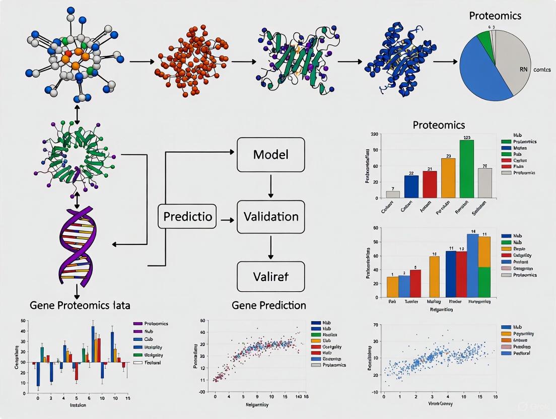
From Sequence to Function: Validating Gene Predictions with Modern Proteomics
This article provides a comprehensive guide for researchers and drug development professionals on integrating proteomic data to validate and refine computational gene predictions.
From Sequence to Function: Validating Gene Predictions with Modern Proteomics
Abstract
This article provides a comprehensive guide for researchers and drug development professionals on integrating proteomic data to validate and refine computational gene predictions. It covers the foundational principles of why protein-level evidence is crucial, detailing methodological workflows from LC-MS/MS to data analysis. The content addresses common challenges in experimental design and data interpretation, offering optimization strategies from recent large-scale studies. Furthermore, it explores advanced applications of validated targets in biomarker discovery and therapeutic development, highlighting how proteomic validation strengthens the link between genetic discoveries and clinical applications.
The Critical Link: Why Protein-Level Validation is Non-Negotiable for Gene Models
In silico gene prediction tools have become indispensable in modern genomic research and therapeutic development, offering a scalable method to interpret the vast landscape of human genetic variation. These computational models leverage artificial intelligence and machine learning to predict the functional impact of genetic variants, potentially accelerating precision medicine and drug target discovery [1] [2]. However, as these tools proliferate, a critical gap persists between computational predictions and biological reality—a disconnect that can significantly impact diagnostic accuracy and therapeutic decisions.
The fundamental challenge lies in the inherent complexity of genotype-phenotype relationships. While sequence-based AI models show great potential for high-resolution variant effect prediction, their practical value depends heavily on rigorous validation against experimental evidence [1] [3]. This review systematically assesses the limitations of current in silico prediction methods through the lens of proteomic and functional validation, providing researchers with a critical framework for evaluating these essential bioinformatic tools.
Performance Benchmarking: Quantitative Comparisons of Prediction Tools
Variant Effect Predictors in Human Traits
Independent benchmarking using population-scale biobanks has provided unbiased evaluations of computational variant effect predictors by assessing their ability to correlate with actual human traits. These studies circumvent the circularity concerns that plague many evaluations, as they utilize data not included in model training [2].
Table 1: Performance of Variant Effect Predictors in Human Cohort Studies
| Predictor | Performance in UK Biobank | Performance in All of Us | Key Strengths |
|---|---|---|---|
| AlphaMissense | Best or tied in 132/140 gene-trait combinations [2] | Consistent top performer [2] | Superior rare variant interpretation |
| VARITY | Not statistically different from AlphaMissense for some traits [2] | Strong correlation with human phenotypes [2] | Robust performance across diverse traits |
| ESM-1v | Tied with AlphaMissense for some binary traits [2] | Independent validation pending | Strong for specific variant classes |
| MPC | Competitive for medication use prediction [2] | Independent validation pending | Effective for pharmacogenomic applications |
In a comprehensive assessment of 24 predictors across 140 gene-trait associations in the UK Biobank, AlphaMissense significantly outperformed most other predictors, demonstrating the highest correlation with human traits based on rare missense variants [2]. This performance was subsequently confirmed in the independent All of Us cohort, establishing a robust benchmark for predictor selection in clinical and research settings.
Splicing Variant Prediction Tools
The accurate prediction of splicing variants presents particular challenges, as these may occur deep within introns or exons away from canonical splice sites. Benchmarking against the largest set of functionally assessed variants of uncertain significance (VUSs) revealed substantial variability in tool performance [4].
Table 2: Performance Comparison of Splicing Prediction Algorithms
| Tool | AUC | Sensitivity | Specificity | Optimal Application |
|---|---|---|---|---|
| SpliceAI | Highest single AUC (0.20 threshold) [4] | 89% | 86% | Deep intronic & canonical variants |
| Consensus Approach | Similar to SpliceAI (4/8 tools threshold) [4] | 91% | 85% | Comprehensive variant assessment |
| Weighted Combination | Potentially superior to single tools [4] | 93% | 87% | Critical clinical applications |
| CADD | Lower than SpliceAI [4] | 67% | 82% | Region-specific performance varies |
SpliceAI emerged as the best single algorithm, correctly prioritizing variants that impact splicing with high accuracy. However, a consensus approach combining multiple tools achieved similar performance, while a novel weighted approach incorporating relative scores from multiple algorithms showed potential for even greater accuracy, though this requires further validation [4].
Long-Range DNA Interaction Modeling
The prediction of long-range genomic interactions represents a particularly challenging frontier, as functional elements may influence gene regulation across megabase-scale distances. The DNALONGBENCH suite systematically evaluates this capability across five critical tasks [5].
Table 3: Performance on Long-Range Genomic Tasks (Scale: 0-1)
| Task | Expert Models | DNA Foundation Models | CNN | Most Effective Model |
|---|---|---|---|---|
| Enhancer-Target Gene Interaction | 0.841 [5] | 0.789-0.801 [5] | 0.762 [5] | ABC Model |
| eQTL Prediction | 0.721 [5] | 0.632-0.658 [5] | 0.601 [5] | Enformer |
| 3D Genome Organization | 0.841 [5] | 0.512-0.523 [5] | 0.488 [5] | Akita |
| Regulatory Sequence Activity | 0.712 [5] | 0.521-0.538 [5] | 0.498 [5] | Enformer |
| Transcription Initiation Signals | 0.733 [5] | 0.108-0.132 [5] | 0.042 [5] | Puffin-D |
Across all tasks, highly parameterized and specialized expert models consistently outperformed both DNA foundation models and simpler convolutional neural networks. The performance gap was especially pronounced for regression tasks such as contact map prediction and transcription initiation signal prediction, suggesting that current foundation models struggle with capturing sparse real-valued signals across long DNA contexts [5].
Experimental Validation Protocols: Bridging the In Silico-In Vivo Gap
Proteomic Validation of Genetic Findings
Proteomics provides a crucial intermediate validation layer between genetic predictions and phenotypic outcomes, offering direct evidence of functional molecular consequences. Recent advances demonstrate how machine learning applied to proteomic data can improve disease risk prediction while simultaneously validating potential drug targets [6] [7].
The Explainable Boosting Machine (EBM) framework has shown particular promise, achieving an AUROC of 0.785 for 10-year cardiovascular disease risk prediction by integrating proteomic data with clinical features [7]. This represents a significant improvement over traditional equation-based risk scores like PREVENT (AUROC: 0.767 with proteomics alone) and provides both global and local explanations for predictions, enabling researchers to identify which proteins contribute most to individual risk assessments [7].
Functional Splicing Assays
For splicing variants, experimental validation typically involves functional analyses to directly observe impacts on mRNA processing. The largest study of its kind functionally assessed 249 variants of uncertain significance (VUSs) from diagnostic testing, finding that 80 (32%) significantly impacted splicing, potentially enabling reclassification as "likely pathogenic" [4].
The experimental workflow typically includes:
- RNA extraction from patient-derived cells or appropriate tissue models
- Reverse transcription PCR to convert mRNA to cDNA
- Fragment analysis to detect abnormal splicing patterns
- Sanger sequencing to identify specific exon skipping, intron retention, or cryptic splice site usage
- Quantification of aberrant transcript proportions compared to normal controls
This functional evidence provides the highest level of validation for splicing predictions, though cell- and tissue-specific factors may influence results and require consideration in experimental design [4].
Longitudinal Proteomic Validation
Longitudinal study designs provide particularly powerful validation by capturing dynamic protein expression changes over time, offering more statistical power than cross-sectional approaches to detect true biological differences [8]. The Robust Longitudinal Differential Expression (RolDE) method was specifically developed to address the unique characteristics of proteomics data, including prevalent missing values and technical noise [8].
In comprehensive benchmarking using over 3000 semi-simulated spike-in datasets, RolDE achieved superior performance (IQR mean pAUC: 0.977) compared to other methods, demonstrating particular strength in handling missing values and diverse expression patterns [8]. This approach enables researchers to more confidently distinguish true longitudinal differential expression from technical artifacts when validating in silico predictions.
Critical Gaps and Limitations in Current Methodologies
Context Specificity and Generalizability
A fundamental limitation of many in silico prediction tools is their limited ability to account for biological context, including cell type, tissue specificity, and developmental stage. This is particularly problematic for regulatory variants, where effects may be highly context-dependent [1]. As noted in plant breeding applications—where these tools show promise but face similar limitations—"the accuracy and generalizability of sequence models heavily depend on the training data, highlighting the need for validation experiments" [1].
This challenge extends to human genomics, where models trained on bulk tissue data may fail to capture cell-type-specific regulatory effects, potentially leading to false positives or negatives in specific physiological or pathological contexts.
The Long-Range Challenge
As demonstrated in the DNALONGBENCH evaluation, capturing dependencies across very long genomic distances remains a major computational hurdle [5]. While specialized expert models like Enformer and Akita show reasonable performance for specific tasks, general-purpose DNA foundation models struggle with long-range interactions, particularly for predicting 3D genome organization and transcription initiation [5].
This limitation has direct implications for interpreting non-coding variation, as enhancers may regulate gene expression across megabase-scale distances, and current tools may miss these functional connections.
Data Quality and Technical Artifacts
Proteomic validation introduces its own technical challenges, as data quality significantly impacts validation reliability. Benchmarking studies of data-independent acquisition (DIA) mass spectrometry workflows—increasingly used for proteomic validation—reveal substantial variability in identification and quantification performance across different analysis tools [9].
For instance, in single-cell proteomic simulations, Spectronaut's directDIA workflow quantified 3,066 ± 68 proteins per run, compared to 2,753 ± 47 for PEAKS and fewer for DIA-NN under similar conditions [9]. These technical differences in validation methodologies can directly impact the apparent performance of in silico gene predictions.
A Framework for Robust Validation
The Scientist's Toolkit: Essential Research Reagent Solutions
Table 4: Key Experimental Resources for Validation Studies
| Resource Type | Specific Examples | Applications & Functions |
|---|---|---|
| Spectral Libraries | Sample-specific DDALib, PublicLib, AlphaPeptDeep predicted libraries [9] | Peptide identification in proteomic validation |
| Proteomic Platforms | Olink Explore Platform, TIMS-DIA (diaPASEF) [9] [7] | High-throughput protein quantification |
| Analysis Software | DIA-NN, Spectronaut, PEAKS Studio [9] | DIA mass spectrometry data processing |
| Functional Assay Systems | Patient-derived xenografts, Organoids, Tumoroids [10] | Experimental validation in biologically relevant models |
| Longitudinal Analysis Tools | RolDE, Limma, MaSigPro [8] | Detecting differential expression over time |
| Splicing Assay Systems | Mini-gene constructs, RT-PCR protocols [4] | Functional assessment of splicing variants |
Integrated Validation Workflow
To overcome the limitations of individual prediction tools, we propose a tiered validation framework:
Computational Cross-Validation: Employ multiple complementary algorithms with different underlying architectures and training data. Consensus approaches consistently outperform individual tools [4].
Proteomic Corroboration: Utilize quantitative proteomics to validate predicted molecular consequences, acknowledging both the power and limitations of current mass spectrometry methods [9] [7].
Functional Characterization: Implement targeted experiments (splicing assays, CRISPR-based functional studies) for high-priority predictions, particularly those with potential clinical implications [4].
Longitudinal Confirmation: Where possible, incorporate longitudinal designs to capture dynamic effects and enhance statistical power for detecting true biological signals [8].
In silico gene prediction tools have revolutionized genomic research but remain imperfect proxies for biological reality. Through rigorous benchmarking against proteomic and functional validation data, we can identify their strengths and limitations, enabling more informed tool selection and interpretation.
The most promising developments lie in integrated approaches that combine multiple computational strategies with experimental validation—such as the weighted combination method for splicing prediction that outperforms individual tools [4], or the explainable machine learning frameworks that simultaneously predict disease risk and identify biologically plausible biomarkers [7].
As these tools continue to evolve, maintaining a critical perspective on their limitations—particularly regarding context specificity, long-range interactions, and technical validation constraints—will be essential for translating computational predictions into meaningful biological insights and clinical applications. The gap between in silico predictions and biological reality is narrowing, but bridging it completely will require continued development of both computational and experimental methodologies alongside rigorous, multi-modal validation frameworks.
The Central Dogma of molecular biology outlines a straightforward flow of genetic information: from DNA to RNA to protein. In laboratory practice, this principle often leads to the use of mRNA abundance as a convenient proxy for protein levels. However, a growing body of evidence reveals that this relationship is far from linear, with mRNA levels frequently diverging from the functional effector molecules they encode [11] [12].
This discrepancy presents a significant challenge for validating gene predictions against proteomic data. While transcriptomic methods like RNA-Seq have become routine and reproducible, proteomic analyses remain more technically challenging [11]. Consequently, many studies are forced to extrapolate conclusions from mRNA to protein, an approach that often proves unjustified [11]. Understanding the mechanisms underlying this discordance is crucial for researchers, scientists, and drug development professionals who rely on accurate gene expression data for discovery and validation workflows.
Key Biological Mechanisms Driving Divergence
The relationship between mRNA and protein abundance is governed by a complex series of regulatory steps, each offering potential points of divergence.
Post-Transcriptional Regulation
After mRNA is synthesized, multiple mechanisms influence whether and how it becomes translated into protein:
- Translation Efficiency: The rate of ribosome movement along mRNA and the availability of free ribosomes significantly impact protein synthesis [11].
- tRNA Availability and Codon Usage: The abundance of specific tRNAs and codon optimization affects translation efficiency and protein yield [11].
- RNA Secondary Structure: Complex structures in the transcript itself can hinder or facilitate ribosomal binding and progression [11].
Post-Translational Regulation
Once synthesized, proteins undergo further processing that dissociates their abundance from initial mRNA levels:
- Protein Degradation: Proteins have widely varying half-lives regulated by degradation mechanisms like the ubiquitin-proteasome system [13].
- Post-Translational Modifications (PTMs): Phosphorylation, acetylation, ubiquitination, and glycosylation significantly alter protein function and stability without changing mRNA abundance [13].
- Protein Complex Formation: The assembly of proteins into complexes can influence their degradation kinetics and functional availability [14].
Evolutionary and Compensatory Mechanisms
Recent phylogenetic analyses across mammalian species reveal that protein abundances evolve under strong stabilizing selection, while mRNA abundances show greater divergence [15]. This suggests an evolutionary buffering system where:
- Mutations affecting mRNA abundances often have minimal impact on protein abundances [15]
- mRNA abundances adapt faster than protein abundances due to greater mutational opportunity [15]
- Compensatory evolution maintains protein abundance stability despite transcriptional changes [15]
Quantitative Comparison of mRNA and Protein Levels
Correlation Coefficients Across Studies
Table 1: Reported mRNA-Protein Correlation Coefficients Across Organisms and Conditions
| Study System | Correlation Coefficient (R) | Sample Size | Measurement Technique |
|---|---|---|---|
| Mouse Liver Tissues [11] | 0.27 (Pearson) | 100 mice | RNA-Seq + LC-MS |
| Yeast [11] | 0.58 (R²) | Log-transformed data | Multi-platform |
| S. cerevisiae [11] | 0.73 (R²) | Averaged technologies | Combined datasets |
| Rice and Maize [11] | <0.4 (Pearson) | Plant tissues | RNA-Seq + MS |
| Mammalian Cells [16] | ~0.40 (Pearson) | Multiple datasets | RNA-Seq + MS |
| Mouse Inner Ear Tissues [16] | 0.58 (Average) | Cochlea/vestibule | RNA-Seq + MS |
Protein-to-mRNA Ratios Across Tissues and Conditions
Table 2: Protein Conservation vs. mRNA Divergence Across Biological Contexts
| Dataset | Observation | Statistical Significance | Biological Interpretation |
|---|---|---|---|
| EAR (Mouse inner ear) [16] | Protein correlation between cochlea/vestibule: 0.97 vs mRNA: 0.94 | Higher protein conservation | Buffering maintains protein homeostasis across similar tissues |
| PRIMATE (Lymphoblastoid cells) [16] | 3/3 pairs showed higher protein correlation | Consistent pattern | Evolutionary conservation of protein levels across species |
| MMT (Mouse tissues) [16] | 9/10 tissue pairs showed higher protein correlation | p = 2.9×10⁻³ (Wilcoxon test) | Compensatory mechanisms operate across diverse tissues |
| NCI60 (Cancer cell lines) [16] | 24/36 cancer types showed higher protein correlation | p = 8.0×10⁻³ (Wilcoxon test) | Buffering persists but is less consistent in cancer |
Experimental Protocols for Parallel mRNA-Protein Analysis
Simultaneous Single-Cell mRNA-Protein Quantification
Recent methodological advances enable simultaneous measurement of mRNA and protein in the same cells, eliminating technical variability:
Proximity Sequencing (Prox-seq) Protocol [17]:
- Principle: Combines proximity ligation assay with single-cell sequencing to measure proteins, protein complexes, and mRNAs simultaneously
- Workflow:
- Target proteins with specific antibodies conjugated to DNA oligonucleotides
- When antibodies are in proximity (<40 nm), perform proximity ligation
- Sequence resulting DNA products to identify interacting proteins and complexes
- Simultaneously sequence transcriptome from the same single cells
- Applications: Identifying cell types, detecting protein complexes, discovering novel interactions in immune signaling
- Validation: Successfully identified naïve CD8+ T cells displaying CD8-CD9 complex and TLR signaling complexes
Dual Fluorescent Reporter System in Yeast [14]:
- Principle: CRISPR-based system for simultaneous quantification of mRNA and protein via dual fluorescent reporters
- Workflow:
- Engineer fluorescent transcriptional and translational reporters for genes of interest
- Image live cells to quantify both reporters simultaneously
- Map trans-acting loci affecting expression
- Key Finding: <20% of trans-acting loci had concordant effects on mRNA and protein
- Advantage: Eliminates environmental confounders and technical biases between separate measurements
Mass Spectrometry-Based Proteomics with RNA-Seq
For population-level studies, paired omics measurements provide complementary insights:
Matched Transcriptome-Proteome Analysis in Mammalian Systems [15] [16]:
- Tissue Collection: Standardized sampling procedures across multiple species (e.g., mammalian skin fibroblasts)
- RNA Sequencing: Standard RNA-seq protocols with quality controls
- Proteome Analysis: Liquid chromatography coupled with data-independent acquisition tandem mass spectrometry (DIA-MS)
- Key Consideration: Use standardized experimental protocols across all samples to minimize technical variation
- Phylogenetic Framework: Apply evolutionary models to distinguish mutational and selective influences on expression divergence
Figure 1: Experimental workflows for simultaneous mRNA-protein quantification. Three complementary approaches enable researchers to capture expression relationships at different biological scales and resolutions.
The Scientist's Toolkit: Essential Research Reagents and Solutions
Table 3: Key Research Reagents for mRNA-Protein Correlation Studies
| Reagent/Solution | Function | Application Examples |
|---|---|---|
| Antibody-DNA Oligo Conjugates [17] | Target proteins for proximity ligation assays | Prox-seq protein detection and complex identification |
| Dual Fluorescent Reporters [14] | Simultaneous monitoring of transcription and translation | Live-cell imaging of mRNA and protein dynamics |
| Data-Independent Acquisition (DIA) Reagents [15] | Comprehensive peptide quantification in mass spectrometry | Proteome analysis across multiple species |
| CRISPR-Cas9 Editing Tools [14] | Precise genetic manipulation | Engineering reporter systems and functional validation |
| Liquid Chromatography Columns [15] [11] | Peptide separation prior to mass spectrometry | Proteomic sample preparation |
| RNA-Seq Library Prep Kits [11] | Transcriptome library construction | mRNA abundance quantification |
| Protein Degradation Inhibitors | Preserve protein abundance profiles | Sample collection for accurate proteomics |
| Cross-linking Reagents [17] | Stabilize protein complexes | Studying protein interactions and complexes |
Implications for Drug Development and Therapeutic Discovery
The discordance between mRNA and protein levels has profound implications for pharmaceutical research and development:
Target Identification and Validation
Genetic studies increasingly integrate proteomic data to improve therapeutic target identification. A recent cross-population genome-wide association study of atrial fibrillation demonstrated that integrating genomic data with proteomic profiling significantly enhanced disease risk prediction and identified potential drug targets [18]. The study identified 28 circulating proteins with potential causal associations with AF, with protein risk scores outperforming traditional polygenic risk scores [18].
Pharmacogenomics and Biomarker Development
The move toward proteomics-driven precision medicine recognizes that proteins, as the primary effector molecules, provide more direct insight into disease mechanisms and treatment responses [13]. Several key considerations emerge:
- Post-translational modifications create functional protein diversity not predictable from mRNA [13]
- Protein-protein interactions and complex formation influence therapeutic efficacy [17] [13]
- FDA-approved biomarkers increasingly rely on protein rather than RNA measurements [13]
Figure 2: Relationship between mRNA-protein divergence mechanisms and therapeutic applications. Understanding biological causes enables methodological innovations that directly impact drug development success.
The divergence between mRNA and protein levels represents a fundamental consideration rather than a technical limitation in molecular biology. Quantitative comparisons reveal generally modest correlations (typically R=0.3-0.6) that vary by biological context, with protein levels often showing greater conservation across tissues and species than their corresponding mRNAs [16].
These findings carry significant implications for validating gene predictions against proteomic data. Researchers should prioritize:
- Direct protein measurement whenever possible for functional validation
- Simultaneous mRNA-protein quantification methods to eliminate technical variability
- Evolutionary perspectives that recognize stabilizing selection on protein abundances
- Multi-omics integration that accounts for post-transcriptional and post-translational regulation
As proteomic technologies continue advancing in accessibility and scalability [13], the research community moves closer to realizing proteomics-driven precision medicine that fully acknowledges the complex relationship between genetic information and its functional effectors.
The sequencing of a genome produces a vast list of predicted gene models, but this structural annotation is merely a starting point. The critical next step is functional annotation—linking these genomic elements to biological function [19] [20]. While computational predictions provide initial functional clues, they require experimental validation to confirm biological relevance. Proteomics, the large-scale study of proteins, has emerged as a powerful tool for bridging this gap, providing direct experimental evidence for the existence of predicted gene products and enabling more accurate functional characterization [21]. This guide examines the central role of proteomics in functional annotation workflows, objectively comparing its performance against alternative approaches and detailing the experimental methodologies that make it indispensable for genome annotation projects.
Proteomics as a Validation Tool for Gene Predictions
From In Silico Prediction to Experimental Confirmation
Structural annotation of newly sequenced genomes begins with electronic prediction of open reading frames (ORFs), which are typically released into public databases without experimental validation [19] [20]. These predicted proteins account for the majority of data for newly sequenced species but face a significant annotation challenge: highly curated databases like UniProtKB often exclude predicted gene products until experimental evidence confirms their in vivo expression [19] [20].
Proteomics addresses this limitation by providing direct experimental support for gene model predictions. In a landmark chicken genome study, researchers analyzed eight tissues and provided experimental confirmation for 7,809 computationally predicted proteins, corresponding to 51% of the chicken predicted proteins in NCBI at the time [19] [20]. This demonstrated the utility of high-throughput expression proteomics for rapid experimental structural annotation of a newly sequenced eukaryote genome [19] [20]. Importantly, this approach identified 30 proteins that were only electronically predicted or hypothetical translations in human, highlighting its power for cross-species validation [19] [20].
Orthology-Based Functional Annotation Transfer
Once protein expression is experimentally confirmed, proteomics data enables functional annotation through orthology mapping. By identifying human or mouse orthologs of experimentally supported proteins, Gene Ontology (GO) functional annotations can be transferred from the characterized orthologs to the newly confirmed proteins [19] [20]. In the chicken genome study, researchers identified orthologs for 77% (6,008) of the confirmed chicken proteins, then used this orthology to produce 8,213 GO annotations—representing an 8% increase in available chicken GO annotations and a doubling of non-IEA (Inferred from Electronic Annotation) annotations [19] [20].
Table 1: Performance Metrics for Functional Annotation Methods
| Annotation Method | Evidence Basis | Coverage | Accuracy | Limitations |
|---|---|---|---|---|
| Proteomics + Orthology Transfer | Experimental protein detection + evolutionary conservation | Moderate (e.g., 77% ortholog identification in chicken study) | High (direct protein confirmation + conserved function) | Limited to expressed proteins; requires related annotated species |
| Transcriptomics Co-expression | mRNA expression patterns | High (most transcribed genes) | Moderate (subject to transcriptional noise) | Poor correlation with protein abundance; accidental covariation [22] |
| Electronic Annotation (IEA) | Sequence similarity, functional motifs | Very High (can be automated genome-wide) | Variable (depends on motif specificity) | High false positive rate; no experimental support [19] |
| Genomic Context | Chromosomal colocalization, operon structure | Variable | Lower for eukaryotes | More reliable for prokaryotes; indirect functional inference |
Performance Comparison: Proteomics Versus Transcriptomics
While mRNA profiling has been the dominant approach for studying gene expression, proteome profiling provides distinct advantages for functional annotation. A systematic comparison of mRNA and protein coexpression networks for three cancer types revealed marked differences in wiring between these networks [22].
Protein coexpression was driven primarily by functional similarity between coexpressed genes, whereas mRNA coexpression was driven by both cofunction and chromosomal colocalization of the genes [22]. This fundamental difference has significant implications for function prediction: functionally coherent mRNA modules were more likely to have their edges preserved in corresponding protein networks than functionally incoherent mRNA modules [22].
The study concluded that proteomics strengthens the link between gene expression and function for at least 75% of Gene Ontology biological processes and 90% of KEGG pathways, demonstrating that proteome profiling outperforms transcriptome profiling for coexpression based gene function prediction [22].
Table 2: Direct Performance Comparison of Proteomics vs. Transcriptomics for Function Prediction
| Performance Metric | Proteomics Approach | Transcriptomics Approach | Performance Advantage |
|---|---|---|---|
| Driver of Coexpression | Functional similarity between genes [22] | Cofunction + chromosomal colocalization [22] | Proteomics provides more specific functional signals |
| Function Prediction Accuracy | Higher link to known functions | Lower specificity | Proteomics strengthens function links for 75% GO processes, 90% KEGG pathways [22] |
| Biological Relevance | Direct measurement of functional molecules | Proxy measurement (mRNA) | Proteomics directly detects functional entities |
| Functional Coherence | Higher in coexpressed modules | Lower coherence in coexpressed modules | Functionally coherent mRNA modules preserved in protein networks [22] |
Experimental Protocols and Workflows
Mass Spectrometry-Based Protein Identification
The core experimental methodology for proteomic validation involves liquid chromatography mass spectrometry (LC-MS)-based analysis [21]. The standard workflow encompasses:
Sample Preparation: Protein extraction from tissues or cells, potentially using Differential Detergent Fractionation (DDF) to enhance protein identification [19] [20]
Protein Digestion: Cleavage into peptides using trypsin or similar proteases
LC-MS/MS Analysis: Separation via liquid chromatography followed by mass spectrometry analysis
Database Searching: Matching acquired spectra against theoretical spectra from predicted protein databases
In the chicken genome study, this approach identified 48,583 peptides with a false discovery rate (FDR) of 0.9%, providing high-confidence support for protein existence [19] [20]. Although 58% of protein identifications were based on single-peptide matches, the low FDR and independent identification in multiple tissues provided strong evidence for in vivo expression [19] [20].
Differential Expression Analysis Workflows
For quantitative proteomics applications, differential expression analysis workflows typically encompass five key steps [23]:
- Raw Data Quantification: Using tools like MaxQuant or FragPipe
- Expression Matrix Construction
- Matrix Normalization
- Missing Value Imputation (MVI)
- Differential Expression Analysis with statistical methods
Optimizing these workflows is crucial for accurate results. A comprehensive study evaluating 34,576 combinatoric experiments revealed that optimal workflows are settings-specific, with normalization and DEA statistical methods exerting greater influence for label-free DDA and TMT data, while matrix type is additionally important for DIA data [23].
High-performing workflows for label-free data are enriched for directLFQ intensity, no normalization (referring to distribution correction methods not embedded with particular settings), and specific imputation methods (SeqKNN, Impseq, or MinProb), while eschewing simple statistical tools like ANOVA, SAM, and t-test [23].
Handling Missing Values in Proteomics Data
Missing values present a significant challenge in proteomics, as they can limit statistical power for comparisons between experimental groups. Traditional approaches include:
- Removal of high-missingness proteins (typically 50-80% missingness thresholds)
- Statistical imputation using methods like k-nearest neighbors (kNN) or random forest
Recent innovations include retention time (RT) boundary imputation rather than quantitation imputation. For each missing value, RT boundaries are imputed, then quantitation is obtained by integrating the chromatographic signal within the imputed boundaries [24]. This approach, implemented in tools like Nettle, yields more accurate quantitations than traditional proteomics imputation methods and increases the number of peptides with quantitations, leading to enhanced statistical power [24].
The Scientist's Toolkit: Essential Research Reagents and Solutions
Table 3: Key Research Reagent Solutions for Proteomics-Based Functional Annotation
| Reagent/Resource | Function | Application Notes |
|---|---|---|
| Differential Detergent Fractionation (DDF) Kits | Sequential extraction of cellular compartments | Enhances protein identification; critical for membrane-associated proteins [19] [20] |
| Trypsin/Lys-C Protease | Protein digestion into peptides | Essential sample preparation step for LC-MS/MS analysis |
| iTRAQ/TMT Labeling Reagents | Multiplexed protein quantification | Enables simultaneous analysis of multiple samples; improves throughput [22] [23] |
| Universal Proteomics Standard (UPS) Sets | Spike-in controls for quantification | Provides internal standards for differential expression studies [23] |
| Spectral Libraries (.blib files) | Reference databases for peptide identification | Critical for DIA-NN and Skyline analysis; can be enhanced with imputation [24] |
| Orthology Prediction Tools | Mapping genes between species | Enables functional annotation transfer (e.g., Homologene, Inparanoid, Treefam) [19] [20] |
| Functional Annotation Pipelines | Automated annotation workflows | Tools like FA-nf integrate multiple approaches for comprehensive annotation [25] |
Integrated Annotation Workflows
Effective functional annotation typically requires integrating multiple complementary approaches. Pipeline tools like FA-nf, implemented in Nextflow, provide containerized workflows that integrate different annotation approaches including NCBI BLAST+, DIAMOND, InterProScan, and KEGG [25]. These pipelines begin with protein sequence FASTA files and optionally structural annotation in GFF format, producing comprehensive annotation reports including GO assignments [25].
Similarly, the AgBase functional annotation workflow employs three annotation tools in concert: GOanna (for BLAST-based GO annotation transfer), InterProScan (for protein family and domain identification), and KOBAS (for KEGG Orthology terms and pathway annotation) [26].
Proteomics provides an essential bridge between genomic sequence data and biological understanding by experimentally validating predicted gene models and enabling accurate functional annotation. The experimental evidence generated through mass spectrometry-based proteomics addresses critical limitations of purely computational predictions, while orthology-based annotation transfer leverages evolutionary conservation to assign biological meaning.
When compared to transcriptomic approaches, proteomics demonstrates superior performance for function prediction, with protein coexpression networks more specifically reflecting functional relationships than mRNA coexpression networks. As proteomics technologies continue to advance in sensitivity, throughput, and quantification accuracy, their role in functional annotation workflows will become increasingly central to extracting biological insight from genomic sequences.
For researchers engaged in genome annotation projects, integrating proteomic validation provides the critical path "from candidate to confirmation"—transforming in silico predictions into biologically validated functional elements.
The high failure rate in clinical drug development, estimated at 90%, is often attributed to inadequate target validation. Within this challenging landscape, human genetic evidence has emerged as a powerful tool for establishing the causal role of genes in human disease, with drug mechanisms supported by such evidence demonstrating a 2.6 times greater probability of success from clinical development to approval [27]. This review systematically compares how different genetic and proteomic validation methodologies perform in prioritizing drug targets, with a specific focus on validating gene predictions against experimental proteomics data.
Table 1: Performance Comparison of Genetic and Proteomic Validation Approaches
Table 1 summarizes the key performance metrics of different genetic and proteomic validation methods as presented in recent literature.
| Methodology | Primary Function | Key Performance Metrics | Advantages | Limitations |
|---|---|---|---|---|
| Gene-Disease Level DOE Prediction [28] | Predicts direction of therapeutic effect for gene-disease pairs | Macro-averaged AUROC: 0.59 (improves with genetic evidence) | Incorporates genetic associations across allele frequency spectrum; models dose-response. | Performance is currently modest and highly dependent on available genetic data. |
| Gene-Level DOE-Specific Druggability [28] | Predicts suitability for activation/inhibition across all diseases | Macro-averaged AUROC: 0.95 for activator/inhibitor druggability | Leverages gene/protein embeddings; outperforms existing druggability predictors. | Disease-agnostic; does not guarantee therapeutic utility for a specific indication. |
| Sparse Plasma Protein Signatures [29] | Predicts 10-year disease risk for drug target indication | Median ΔC-index: +0.07 over clinical models; Detection Rate at 10% FPR: 45.5% | Clinically useful prediction for 67 diseases; points directly to druggable protein targets. | Predictive power varies by disease pathology; enrichment for hematological/immunological diseases. |
| Proteogenomic Causal Inference (pQTL MR/Colocalization) [30] | Establishes causal links between protein abundance and disease | Identified 43 colocalizing associations with posterior probability >80% | Provides high-confidence causal inference; instruments novel proteins like LTK for T2D. | Requires large sample sizes for robust pQTL discovery; can be confounded by pleiotropy. |
Experimental Protocols for Genetic and Proteomic Validation
Protocol for Direction of Effect (DOE) Prediction and Validation
Objective: To predict whether a therapeutic should activate or inhibit a target protein for a given disease.
Methodology Summary: A multi-level machine learning framework integrates diverse data inputs [28]:
- Input Features:
- Genetic Associations: Effect directions from variants across the allelic series (common, rare, ultrarare) to model dose-response relationships [28].
- Gene and Protein Embeddings: Continuous representations from GenePT (NCBI gene summaries) and ProtT5 (amino acid sequences) [28].
- Tabular Features: Gene-level characteristics such as LOEUF (constraint), dosage sensitivity, and mode of inheritance [28].
- Model Training: Three distinct models are trained:
- DOE-specific druggability for 19,450 protein-coding genes.
- Isolated DOE among 2,553 known druggable genes.
- Gene-disease-specific DOE for 47,822 gene-disease pairs.
- Validation: Model performance is assessed via AUROC and calibration plots, with successful predictions shown to be associated with clinical trial success [28].
Protocol for Proteogenomic Causal Inference via pQTLs
Objective: To establish a causal relationship between genetically predicted plasma protein levels and disease risk, thereby validating the protein as a therapeutic target.
Methodology Summary: This workflow uses Mendelian randomization (MR) and colocalization, as exemplified in a Scottish cohort study [30]:
- Step 1: Protein Quantitative Trait Locus (pQTL) Discovery
- Proteomic Profiling: Measure thousands of plasma proteins (e.g., 6,432 via SomaLogic v4.1 aptamer-based technology) in a large cohort [30].
- Genome-Wide Association Analysis: Perform GWAS for each protein to identify genetic variants (pQTLs) associated with its abundance levels. Use significance thresholds of P < 5×10⁻⁸ for cis-pQTLs (within 1 Mb of the gene) and a more stringent P < 6.6×10⁻¹² for trans-pQTLs [30].
- Step 2: Causal Inference via Mendelian Randomization
- Instrument Variable Selection: Use the identified, independent pQTLs as genetic instruments for the protein of interest.
- MR Analysis: Perform a two-sample MR analysis to estimate the causal effect of the protein on the disease outcome of interest, using summary statistics from large disease GWAS.
- Step 3: Colocalization Analysis
- Statistical Colocalization: Apply Bayesian colocalization methods (e.g., COLOC) to calculate the posterior probability (PP > 80% is a common threshold) that the pQTL and the disease GWAS signal in a locus share a single causal variant [30]. This step is critical to rule out confounding by distinct, but physically close, causal variants.
- Output: High-confidence, colocalized associations suggest that modifying the protein will directly alter disease risk, providing strong genetic support for target prioritization [30].
Visualizing Experimental Workflows
Diagram 1: Proteogenomic Causal Inference Workflow
Diagram 2: From Genetic Association to Therapeutic Direction
The Scientist's Toolkit: Essential Research Reagents & Platforms
Table 2 lists key reagents, technologies, and databases essential for implementing the genetic and proteomic validation protocols described above.
| Tool / Reagent | Type | Primary Function in Validation | Key Features / Examples |
|---|---|---|---|
| SomaScan Platform [31] [30] | Proteomics Technology (Aptamer-based) | High-throughput quantification of thousands of plasma proteins for pQTL discovery. | SomaScan v4.1 measures ~7,000 proteins; used in large consortia (GNPC) and cohort studies [31]. |
| Olink Explore Platform [29] | Proteomics Technology (Antibody-based) | High-sensitivity proteomic profiling for disease prediction models. | Olink Explore 1536+Expansion targets 2,923 proteins; used in UK Biobank Pharma Proteomics Project [29]. |
| GWAS Catalog [32] | Database | Foundational resource for identifying coincident genetic associations between traits and diseases. | Contains ~29,500 genome-wide significant associations; enables hypothesis generation for target identification [32]. |
| COLOC / Co-localization Software [32] | Statistical Software Package | Tests whether two traits (e.g., pQTL and disease GWAS) share a single causal variant in a genomic locus. | Critical for confirming a shared genetic mechanism and strengthening causal inference in MR studies [32]. |
| Gene & Protein Embeddings (e.g., GenePT, ProtT5) [28] | AI-Derived Feature Set | Provides deep, contextual representations of gene/protein function for machine learning models. | Improves performance of gene-level models predicting druggability and direction of effect [28]. |
| Large-Scale Biobanks (e.g., UK Biobank) [32] [29] | Cohort Resource | Provides integrated genetic, proteomic, and phenotypic data on a massive scale for discovery. | Enables systematic pQTL mapping and agnostic discovery of protein-disease links with high statistical power [29]. |
The integration of genetic evidence and proteomic validation represents a paradigm shift in target validation for drug discovery. Quantitative comparisons demonstrate that proteogenomic frameworks like pQTL-based causal inference provide among the highest levels of validation confidence by linking genetic variants to specific, measurable protein effects on disease. Furthermore, sparse protein signatures derived from large-scale proteomics offer a direct path to clinically actionable biomarkers and targets, particularly for conditions like multiple myeloma and motor neuron disease. As these data-driven approaches mature, their systematic application, supported by the essential research tools detailed herein, promises to de-risk therapeutic development and usher in a new era of precision medicine.
The Validation Pipeline: LC-MS/MS Workflows and Bioinformatics Analysis
Liquid Chromatography-Tandem Mass Spectrometry (LC-MS/MS) has emerged as a cornerstone technology for untargeted proteomics, enabling the comprehensive identification and quantification of proteins within complex biological samples. Within the specific context of validating gene predictions against experimental data, LC-MS/MS provides prima facie evidence for the existence of predicted genes by confirming their translation into proteins [33]. This orthogonal validation is critical, as transcriptomic data alone can confirm gene expression but not translation, and computational predictions frequently generate multiple candidate gene models for a single genomic locus [33]. The integration of experimental proteomic data directly into genomic annotation pipelines significantly enhances the quality and reliability of genome annotation, much as expressed sequence tag (EST) data has done historically [33]. This guide objectively compares the performance of LC-MS/MS with other proteomic technologies, providing the experimental data and protocols essential for researchers engaged in gene prediction validation and systems biology.
Core Principles of LC-MS/MS in Untargeted Proteomics
Untargeted LC-MS/MS proteomics aims to identify and quantify as many proteins as possible from a sample without prior selection. The typical workflow involves digesting proteins into peptides, separating them via liquid chromatography, and then analyzing them with a tandem mass spectrometer. The instrument operates in data-dependent acquisition (DDA) mode, automatically selecting the most abundant precursor ions for fragmentation to generate MS/MS spectra [34]. These spectra are subsequently matched against theoretical spectra derived from a protein sequence database to achieve identification [35].
Quantification can be achieved through label-free methods or by using isobaric chemical labels (e.g., Tandem Mass Tags, TMT). The power of this approach for genome annotation was demonstrated in a study on Aspergillus niger, where 405 identified peptide sequences were mapped to 214 different genomic loci. This data provided direct experimental support for specific gene models, and in 6% of these loci, the proteomic evidence suggested that a model other than the annotators' chosen "best" model was the correct one [33].
Technology Performance Comparison
The selection of a proteomic technology involves critical trade-offs between coverage, specificity, and throughput. The following table provides a structured comparison of LC-MS/MS with the proximity extension assay (PEA), a leading affinity-based technology, based on recent large-scale evaluations [35].
Table 1: Comparative Performance of LC-MS/MS and Affinity-Based Proteomics
| Performance Metric | LC-MS/MS | Olink PEA | Technical and Biological Implications |
|---|---|---|---|
| Detection Principle | Direct detection of peptide mass/charge [35] | Indirect detection via antibody binding [35] | MS provides direct sequence evidence; PEA relies on binder specificity. |
| Typical Proteome Coverage | ~2,500-2,600 proteins [35] | ~2,900 proteins [35] | Coverage is complementary; combined use covers >60% of reference plasma proteome [35]. |
| Protein Abundance Range | Mid to high-abundance proteins [35] | Superior for low-abundance proteins (e.g., cytokines) [35] | MS may miss key signaling molecules; PEA may miss high-abundance structural proteins. |
| Precision (Median CV) | 6.8% [35] | 6.3% [35] | Both platforms demonstrate high and comparable technical precision. |
| Key Strengths | • Direct peptide evidence for gene validation [33]• Discovery of novel proteins [35]• No affinity reagents required | • High sensitivity for low-abundance targets• Excellent throughput• Simplified data analysis | MS is superior for confirming gene models and ORFs; PEA for high-throughput biomarker screening. |
| Key Limitations | • Complex sample preparation• Lower throughput• Limited sensitivity for very low-abundance proteins | • Limited to pre-defined protein targets• Potential for antibody cross-reactivity• No direct sequence information | MS is not ideal for rapid, targeted screening; PEA is less suited for exploratory research in poorly characterized organisms. |
Beyond this direct comparison, the specific configuration of the LC-MS/MS workflow itself greatly impacts performance. A landmark study evaluating 34,576 combinatoric workflows found that optimal workflows are highly specific to the quantification setting (e.g., label-free DDA, DIA, or TMT) [23]. Key steps like data normalization and the choice of differential expression analysis statistical method were identified as having an outsized influence on final results for most data types [23].
Experimental Protocols for Gene Model Validation
Sample Preparation and Protein Extraction
The foundation of a successful LC-MS/MS experiment is robust and reproducible sample preparation. For microbial or fungal cells, such as A. niger, a typical protocol is as follows [33]:
- Cell Lysis: Grind harvested mycelia (e.g., 100 mg) using a pestle and mortar under liquid nitrogen. Subsequently, lyse the cells via mechanical disruption with glass beads.
- Protein Precipitation: Precipitate proteins from the lysate using trichloroacetic acid (TCA) to remove contaminants and concentrate the protein.
- Protein Quantification: Determine the protein concentration of the resulting extract using a standardized assay like micro-BCA.
For complex samples like blood plasma or serum, where a few high-abundance proteins dominate, an additional high-abundance protein depletion step is critical. This expands the dynamic range, allowing for the detection of lower-abundance proteins [36]. This can be achieved using affinity columns designed to remove specific abundant proteins (e.g., albumin, IgG) [36].
Gel Electrophoresis and In-Gel Digestion
- Separation: Separate protein extracts using one-dimensional SDS-PAGE (e.g., 10%, 12%, and 15% gels). Stain the gels with Coomassie R250 to visualize protein bands.
- Band Excision: Excise gel bands from top to bottom of the lane.
- Tryptic Digestion: Perform in-gel digestion with trypsin, a protocol that involves steps to reduce, alkylate, and enzymatically cleave proteins into peptides [33].
- Peptide Extraction: Extract the resulting peptides from the gel pieces using acetonitrile and dry them prior to LC-MS/MS analysis [33].
LC-MS/MS Analysis and Data Processing
- Chromatography: Reconstitute dried peptides and load them onto a nanoflow HPLC system equipped with a trapping column for desalting. Separate the peptides on a reversed-phase analytical column (e.g., C18, 75 µm i.d., 15 cm length) using a long, shallow acetonitrile gradient (e.g., 5–90% solvent B over 1 hour) [33].
- Mass Spectrometry: The HPLC system is coupled online to a tandem mass spectrometer (e.g., a Q-TOF instrument). Data acquisition is performed in data-dependent acquisition (DDA) mode: the instrument first performs an MS1 scan to measure peptide precursor ions, then automatically selects the most intense ions for fragmentation (MS2) to generate sequence spectra [33].
- Peptide Identification: Generate peak lists from the raw data and search them against a protein sequence database using search engines like Mascot [33]. The database should include both forward and reversed sequences to facilitate false discovery rate (FDR) calculation. Techniques like Average Peptide Scoring (APS) can be used to iteratively calculate peptide filters and improve confident protein identification [33].
- Mapping to Genome: Map the confidently identified peptide sequences back to the genomic loci and the available predicted gene models. This provides direct experimental evidence for the existence and structure of the predicted genes [33].
The following diagram illustrates this multi-step workflow, from the initial biological sample to validated gene models.
Optimized Data Analysis and Workflow Integration
The identification of differentially expressed proteins is a multi-step process, and the choice of methods at each step significantly impacts the results. An extensive benchmarking study identified that high-performing workflows for label-free data are often characterized by the use of directLFQ intensity, no normalization (or specific normalization methods), and specific imputation algorithms like SeqKNN, Impseq, or MinProb [23].
To maximize proteome coverage and resolve inconsistencies, ensemble inference—integrating results from multiple top-performing individual workflows—has been shown to be beneficial. This approach can lead to gains in performance metrics like partial area under the curve (pAUC) by up to 4.61% [23]. This is particularly powerful when integrating results from different quantification approaches (e.g., topN, directLFQ, MaxLFQ), as they provide complementary information [23].
Table 2: Key Steps and High-Performing Method Choices in Differential Expression Analysis
| Workflow Step | Description | High-Performing Method Examples |
|---|---|---|
| Quantification Setting | Defines the experimental platform and data type (e.g., DDA, DIA, TMT). | Workflow performance is highly setting-specific [23]. |
| Expression Matrix Construction | Defines how peptide-level data is summarized into a protein-level matrix. | directLFQ intensity, MaxLFQ, topN intensities [23]. |
| Normalization | Corrects for technical variation between samples. | "No normalization" (for specific settings), specific distribution correction methods [23]. |
| Missing Value Imputation (MVI) | Replaces missing data points, a common issue in proteomics. | SeqKNN, Impseq, MinProb (probabilistic minimum) [23]. |
| Differential Expression Analysis | Statistical method to identify significant protein abundance changes. | Methods like limma; simple tests (t-test, ANOVA) are often lower-performing [23]. |
The Scientist's Toolkit: Essential Research Reagents and Materials
The following table details key reagents and materials required for implementing the LC-MS/MS protocols described in this guide.
Table 3: Essential Reagents and Materials for LC-MS/MS Proteomics Workflows
| Item Name | Function / Application | Specific Example |
|---|---|---|
| Trypsin/Lys-C Mix (MS-grade) | Enzymatic digestion of proteins into peptides for MS analysis. | Promega (Madison, WI, USA) [36]. |
| Depletion Column | Removal of high-abundance proteins from serum/plasma to enhance detection of low-abundance proteins. | Agilent Human 14 multiple affinity removal column [36]. |
| Mass Spectrometry-Grade Solvents | Sample preparation and mobile phases for LC-MS/MS to minimize background contamination. | Acetonitrile (ACN), Water (H₂O), Formic Acid (FA) from Fisher Scientific [36]. |
| Buffers and Additives for Digestion | Create optimal conditions for enzymatic digestion and protein handling. | Ammonium Bicarbonate (ABC), Dithiothreitol (DTT), Iodoacetamide (IAA) from Sigma-Aldrich [36]. |
| Internal Standard Peptides | Monitoring instrument stability and performance during the LC-MS/MS run. | Stable isotope-labeled peptides (e.g., caffeine-13C3, L-Leucine-D7) added to the extraction solvent [34]. |
| LC Column | Chromatographic separation of peptides prior to mass spectrometry. | Reversed-phase C18 column (e.g., Waters ACQUITY Premier HSS T3) [34]. |
LC-MS/MS-based proteomics stands as an indispensable, orthogonal method for validating computational gene predictions, providing direct experimental evidence of translation that is not available from transcriptomic data alone. While affinity-based platforms like Olink offer superior throughput and sensitivity for specific low-abundance proteins, LC-MS/MS provides unmatched specificity, the ability to discover novel proteins, and does not rely on pre-defined affinity reagents [33] [35]. The performance of an LC-MS/MS workflow is not monolithic but depends on a synergistic combination of steps from sample preparation to data analysis. By adopting optimized and, where appropriate, ensemble workflows, researchers can robustly leverage this powerful technology to refine genome annotations, confirm gene structures, and build a more accurate understanding of biological systems.
In the context of validating gene predictions against proteomics data, the accuracy of protein-level evidence is paramount. Gene modulation tools like CRISPR and siRNA alter genomic or transcriptomic sequences, but their functional consequences must be confirmed by observing changes in the actual protein output [37]. Among the various proteomic workflows available, GeLC-MS/MS—which combines protein separation via SDS-PAGE with liquid chromatography-tandem mass spectrometry—provides a robust, reproducible, and accessible platform for this critical validation step [38] [39]. This guide objectively compares the performance of GeLC-MS/MS with alternative proteomic methods and provides detailed experimental protocols to implement this technique effectively in gene prediction validation research.
The GeLC-MS/MS workflow integrates classical biochemical separation with modern mass spectrometry, creating a powerful tool for protein identification and characterization. This method is particularly valuable for researchers studying the proteomic effects of gene manipulations, as it provides visible assessment of protein samples and deep proteome coverage without absolute dependence on specific antibodies [39] [37].
Figure 1: GeLC-MS/MS workflow for proteomic analysis. The process begins with protein extraction and proceeds through fractionation, digestion, and final LC-MS/MS analysis, enabling comprehensive protein identification and quantification.
Detailed Experimental Protocols
Protein Sample Preparation
Efficient protein extraction and preparation are critical for obtaining an accurate representation of the proteome under study. Proteins can be prepared from various sources including tissues, bodily fluids, or cell cultures, with preparation methods often involving mechanical lysis, solubilization in buffer, and subcellular fractionation [38].
Reduction and Alkylation: Add 5 mM TCEP to the sample and incubate at room temperature for 20 minutes to reduce disulfide bonds. Then add 10 mM iodoacetamide (IAA) to alkylate free cysteines, incubating in the dark at room temperature for 20 minutes. Quench the reaction with 10 mM DTT, incubating for another 20 minutes in the dark [39].
Protein Precipitation: For samples >500 μg/mL, use methanol-chloroform precipitation: Dilute sample to ~100 μL, add 400 μL methanol and vortex, add 100 μL chloroform and vortex, then add 300 μL water and vortex. Centrifuge at 14,000 × g for 1 minute, remove aqueous and organic layers, retaining the middle protein disk. Add 400 μL methanol, vortex, and centrifuge for 2 minutes [39].
SDS-PAGE Separation and In-Gel Digestion
Gel Electrophoresis: Use precast Bis-Tris 4-12% gradient gels. Add LDS sample buffer (4×) to protein samples with reducing agent and heat at 70°C for 10 minutes. Centrifuge at 2,400 × g for 30 seconds to remove insoluble material before loading [38].
Whole Gel Processing: After electrophoresis and Coomassie staining, destain the entire gel. Perform washing, reduction, and alkylation steps on the intact gel before slicing into 5-20 equal segments based on pre-stained molecular weight markers. This "whole gel" approach significantly reduces processing time compared to conventional methods where each slice is processed individually [40].
In-Gel Digestion: Destain gel pieces with 25 mM ammonium bicarbonate/50% acetonitrile. Add trypsin (10 ng/μL in 25 mM ammonium bicarbonate) and incubate overnight at 37°C. Extract peptides with 1% formic acid, then desalt using StageTips or similar methods before LC-MS/MS analysis [38] [39].
LC-MS/MS Analysis
Chromatography Setup: Use trap column (ZORBAX 300SB-C18, 5 × 0.3 mm, 5 μm) and self-packed analytical column (100 μm i.d. × 150 mm fused silica with C18 resin). Employ gradient elution with Solvent A (0.1% formic acid in water) and Solvent B (0.1% formic acid in acetonitrile) [38].
Mass Spectrometry Parameters: Use high-resolution mass spectrometers (e.g., LTQ Orbitrap) with data-dependent acquisition. For quantitative analyses, consider stable isotope dimethyl labeling to improve accuracy by enabling precise comparison between samples within a single LC-MS run [41].
Performance Comparison with Alternative Methods
Depth of Proteome Coverage
GeLC-MS/MS provides significant advantages for in-depth proteome coverage compared to simpler fractionation approaches, particularly for complex samples.
Table 1: Comparison of protein and peptide identification across fractionation methods
| Method | Proteome Depth | Unique Advantages | Limitations |
|---|---|---|---|
| GeLC-MS/MS (2-D/repetitive) | Moderate protein identifications [42] | Visual QC, removes interferents, compatible with detergents [38] [40] | Limited high MW protein recovery [38] |
| 3-D Fractionation (Protein-level) | Substantially more unique peptides and proteins, including low-abundance species [42] | Highest proteome depth, overcomes MS undersampling [42] | More complex, potential sample loss [42] |
| Solution Digestion (MudPIT) | High peptide identifications [40] | Amenable to automation, higher throughput [40] | Less effective for abundant protein depletion [38] |
Quantitative Performance and Reproducibility
GeLC-MS/MS shows excellent performance for quantitative proteomics, particularly when combined with stable isotope labeling strategies.
Table 2: Quantitative performance characteristics of GeLC-MS/MS
| Parameter | Performance | Experimental Context |
|---|---|---|
| Identification Reproducibility | >88% overlap between technical replicates [40] | Triplicate analysis of HCT116 cell lysate and FFPE tissue |
| Quantification Precision | CV <20% on protein quantitation [40] | Label-free spectral counting |
| Quantification Accuracy | High accuracy with stable isotope dimethyl labeling [41] | Comparative analysis between samples |
| Correlation with Conventional Method | R² = 0.94 for spectral counts [40] | Comparison of whole gel vs. in-gel digestion procedures |
Applications in Gene Prediction Validation
Connecting Genomic and Proteomic Data
For researchers validating gene predictions, GeLC-MS/MS provides a direct link between genetic manipulations and their protein-level consequences. When gene modulation tools like siRNA or CRISPR are employed, mRNA and protein levels may not always correlate, making protein-level verification essential [37]. In one application, researchers used LC-MS/MS proteomics to confirm protein expression changes in cells treated with in-house designed siRNA targeting the epidermal growth factor receptor (EGFR), identifying 73 significantly differentially expressed proteins [37].
Figure 2: Role of GeLC-MS/MS in validating gene predictions. The method provides critical protein-level validation between transcript analysis and biological interpretation, confirming the functional effects of gene modulation.
Biomarker Discovery and Verification
GeLC-MS/MS plays a crucial role in biomarker verification pipelines. The method enables the detection of protein forms that may result from gene mutations or alternative splicing events, providing critical information for selecting appropriate surrogate peptides for targeted assays [43]. In one workflow, GeLC/MS characterization allowed visualization of different forms of a protein in cerebral spinal fluid, informing appropriate peptide selection for subsequent assay development [43].
The Scientist's Toolkit: Essential Research Reagents
Table 3: Key reagents and materials for GeLC-MS/MS experiments
| Reagent/Material | Function | Examples/Specifications |
|---|---|---|
| Precast Gels | Protein fractionation by molecular weight | NuPAGE Bis-Tris 4-12% gradient gels [39] |
| Reducing Agents | Break protein disulfide bonds | TCEP, DTT [38] [39] |
| Alkylating Agents | Prevent reformation of disulfide bonds | Iodoacetamide [38] [39] |
| Protease | Digest proteins into peptides | Sequencing-grade trypsin [38] [39] |
| LC Columns | Peptide separation | C18 trap and analytical columns [38] |
| Mass Spectrometer | Peptide identification and quantification | High-resolution instruments (e.g., LTQ Orbitrap) [38] [43] |
GeLC-MS/MS represents a robust, versatile platform for proteomic analysis that balances practical considerations with analytical performance. For researchers validating gene predictions, this method provides the critical protein-level evidence needed to confirm the functional consequences of genetic manipulations. While alternative methods may offer advantages in specific scenarios such as ultimate proteome depth or throughput, GeLC-MS/MS remains an excellent choice for comprehensive protein identification and quantification, particularly when analyzing complex samples or when visual assessment of protein quality is desirable. The continuous development of streamlined protocols and quantitative enhancements ensures that GeLC-MS/MS will remain a cornerstone technique in functional proteomics and gene validation research.
In the field of proteomics, the validation of gene predictions relies heavily on robust data processing workflows for protein identification, quantification, and differential expression analysis. As proteomics technologies advance, researchers require clear guidance on selecting appropriate bioinformatic tools that deliver accurate and reproducible results. This guide provides an objective comparison of leading software platforms, evaluates their performance based on published benchmark studies, and outlines standardized experimental protocols to ensure data integrity. The focus on practical implementation aims to equip researchers with the knowledge needed to effectively connect genomic predictions with protein-level evidence, thereby strengthening multi-omics integration in biomedical research and drug development.
Comparative Performance of Proteomics Software
Objective evaluation of proteomics software requires examination of key performance metrics including proteome coverage, quantitative accuracy, precision, and completeness of data. Independent benchmarking studies provide crucial insights beyond vendor claims, enabling researchers to select optimal tools for their specific applications, particularly for validating gene predictions against experimental proteomics data.
Performance Benchmarking of DIA Analysis Tools
Data-Independent Acquisition (DIA) mass spectrometry has emerged as a powerful technique for comprehensive protein quantification, especially in single-cell proteomics. A recent benchmarking study evaluated three prominent software tools—DIA-NN, Spectronaut, and PEAKS Studio—using simulated single-cell samples consisting of mixed proteomes from human, yeast, and E. coli cells at 200 pg total input levels [44].
Table 1: Performance Comparison of DIA Analysis Software in Single-Cell Proteomics
| Software | Quantification Strategy | Proteins Quantified (Mean ± SD) | Peptides Quantified (Mean ± SD) | Quantitative Precision (Median CV) | Quantitative Accuracy |
|---|---|---|---|---|---|
| Spectronaut | directDIA (library-free) | 3066 ± 68 proteins | 12,082 ± 610 peptides | 22.2–24.0% | High accuracy |
| DIA-NN | Library-free with deep learning | 2607 proteins (at 50% completeness) | 11,348 ± 730 peptides | 16.5–18.4% | Highest accuracy |
| PEAKS Studio | Sample-specific library | 2753 ± 47 proteins | Not specifically reported | 27.5–30.0% | Comparable accuracy |
The study revealed significant differences in software performance. Spectronaut's directDIA workflow demonstrated the highest detection capabilities, quantifying the greatest number of proteins and peptides [44]. However, DIA-NN achieved superior quantitative precision with lower median coefficients of variation (CV) and outperformed other tools in quantitative accuracy, as measured by closeness of experimental fold-change values to theoretical expectations in ground-truth samples [44]. PEAKS Studio showed intermediate performance in proteome coverage but somewhat lower precision in quantification [44].
Comparison of MS1-Based Label-Free Quantification Tools
For label-free quantification approaches, a systematic evaluation compared MaxQuant and Proteome Discoverer using spiked-in human proteins (UPS1) in a yeast background across a wide dynamic range [45]. This study assessed six different MS1-based quantification methods:
Table 2: Performance of MS1-Based Quantification Methods in MaxQuant and Proteome Discoverer
| Software | Quantification Method | Dynamic Range | Reproducibility | Sensitivity for Differential Analysis | Specificity/Accuracy |
|---|---|---|---|---|---|
| Proteome Discoverer | Normalized Intensity (PD-nI) | Wide | High | Highest sensitivity for narrow abundance ratios | High accuracy |
| Proteome Discoverer | Normalized Area (PD-nA) | Wide | High | High sensitivity | High accuracy |
| MaxQuant | LFQ (normalized intensity) | Moderate | Moderate | Slightly lower sensitivity | Highest specificity |
| MaxQuant | Raw intensity (MQ-I) | Moderate | Moderate | Lower sensitivity | High specificity |
The investigation found that Proteome Discoverer, particularly with normalized quantification methods (PD-nI and PD-nA), outperformed MaxQuant in quantification yield, dynamic range, and reproducibility [45]. PD's normalized methods were most accurate in estimating abundance ratios between groups and most sensitive when comparing samples with narrow abundance ratios. Conversely, MaxQuant methods generally achieved slightly higher specificity, accuracy, and precision values [45]. The study also demonstrated that applying optimized log ratio-based thresholds could maximize specificity, accuracy, and precision in differential analysis.
Key Selection Criteria for Proteomics Software
Beyond performance metrics, several practical factors influence software selection for proteomics workflows [46]:
- Compatibility: Support for vendor-specific raw data formats (Thermo .raw, SCIEX .wiff, Bruker .d) or open formats (mzML, mzXML)
- Quantification Strategy: Specialization in label-free (LFQ), isobaric tagging (TMT/iTRAQ), metabolic labeling (SILAC), or DIA methods
- Usability: Graphical user interface (GUI) versus command-line operation, with GUIs being more accessible for beginners
- Reproducibility and Transparency: Open-source tools (Skyline, MaxQuant, OpenMS, FragPipe, DIA-NN) provide code visibility, while commercial software (Proteome Discoverer, Spectronaut) offers professional support
- Cost and Licensing: Free academic tools versus commercial licenses with associated costs
- Integration Capabilities: Compatibility with downstream statistical analysis tools and visualization platforms
Experimental Protocols for Benchmarking Studies
Standardized experimental protocols are essential for generating reproducible and comparable data in proteomics. The methodologies described below are derived from published benchmark studies and can be adapted for evaluating proteomics software performance in specific research contexts.
Benchmarking Protocol for DIA-Based Single-Cell Proteomics
The benchmarking framework for DIA-based single-cell proteomics involved several critical steps [44]:
Sample Preparation:
- Simulated single-cell samples were created using tryptic digests of human HeLa cells, yeast, and Escherichia coli proteins mixed in defined proportions
- A reference sample (S3) contained 50% human, 25% yeast, and 25% E. coli proteins
- Test samples (S1, S2, S4, S5) maintained equivalent human protein abundance while varying yeast and E. coli proportions with expected ratios from 0.4 to 1.6 relative to reference
- Total protein input was maintained at 200 pg to mimic single-cell protein levels
Mass Spectrometry Analysis:
- Samples were analyzed using diaPASEF on a timsTOF Pro 2 mass spectrometer
- Six technical replicates (repeated injections) were performed for each sample to assess reproducibility
- Trapped ion mobility spectrometry (TIMS) was utilized to enhance sensitivity by excluding singly charged contaminating ions
Data Analysis Workflow:
- Multiple analysis strategies were evaluated including library-free and library-based approaches
- Sample-specific spectral libraries (DDALib) were generated from DDA injections of individual organisms (2 ng) on the same LC-MS/MS system
- Public spectral libraries (PublicLib) were compiled from community resources using timsTOF data of HeLa, yeast, and E. coli digests (200 ng) with high-pH reversed-phase fractionation
- Predicted spectral libraries were generated using AlphaPeptDeep for whole-proteome scale prediction
Performance Evaluation Metrics:
- Identification metrics: Number of proteins and peptides quantified, data completeness across replicates
- Precision: Coefficient of variation (CV) of protein quantities among technical replicates
- Accuracy: Deviation of measured fold-change values from expected ratios in ground-truth mixtures
- Statistical significance: T-test p-values and Cohen's d effect sizes for comparing fold-change distributions
DIA Benchmarking Workflow: This diagram illustrates the key steps in benchmarking DIA analysis software, from sample preparation to performance evaluation.
Protocol for Comparing MS1-Based Label-Free Quantification
The comparative evaluation of MaxQuant and Proteome Discoverer followed a rigorous methodology [45]:
Sample Design and Data Sets:
- Primary data set: UPS1 standard (48 human proteins) spiked at nine different amounts (100 to 0.1 fmol) into a constant background of yeast cell lysate (2 μg)
- Secondary data set: Yeast background with (YH) and without (Y) 25 fmol spiked-in human proteins for specificity assessment
- Triplicate or quadruplicate runs for each condition to enable statistical analysis
Mass Spectrometry Parameters:
- LC-MS/MS analysis using nanoRS UHPLC system coupled to LTQ-Orbitrap Velos mass spectrometer
- Data-dependent acquisition mode with survey scans at 60,000 resolution
- Top 20 most intense ions selected for CID fragmentation
- 75-minute gradient for peptide separation
Protein Identification and Quantification:
- Database searching against combined Saccharomyces cerevisiae and UPS1 human protein databases
- False discovery rate (FDR) set to 1% for both peptide and protein identifications
- Six quantification methods compared:
- MaxQuant: Raw intensity (MQ-I) and normalized LFQ intensity (MQ-L)
- Proteome Discoverer: Intensity (PD-I), normalized intensity (PD-nI), area (PD-A), and normalized area (PD-nA)
- Chromatographic peak alignment with 10-minute time windows
Statistical Analysis:
- Correlation analysis between measured and expected protein abundance values
- Calculation of sensitivity, specificity, accuracy, and precision for differential analysis
- Application of log ratio-based thresholds to optimize classification performance
Signaling Pathways and Data Processing Workflows
Understanding the relationship between proteomics data processing and biological interpretation is essential for validating gene predictions. The following workflows and pathways illustrate how bioinformatic analysis connects to functional biology.
Integrated Multi-Omics Data Analysis Pathway
Proteomics data processing does not occur in isolation but rather as part of an integrated multi-omics framework. This is particularly relevant for studies aiming to validate gene predictions against experimental proteomics data.
Multi-Omics Integration Pathway: This workflow demonstrates how proteomics data integrates with genomic and transcriptomic data to validate gene predictions and enable functional analysis.
Biomarker Discovery Workflow in Disease Proteomics
Proteomic biomarker discovery represents a key application where protein identification, quantification, and differential expression analysis converge. A recent study on amyotrophic lateral sclerosis (ALS) illustrates a comprehensive workflow [47]:
Experimental Design:
- Case-control study with 183 ALS patients and 309 controls (healthy individuals and other neurological diseases)
- Independent replication cohort with 48 ALS patients and 75 controls
- Plasma proteomics using Olink Explore 3072 platform measuring 2,886 proteins after quality control
Data Processing and Analysis:
- Proteome-wide association testing using generalized linear regression adjusted for age, sex, and collection tube type
- False discovery rate (FDR) control for multiple testing (FDR < 0.05)
- Machine learning for binary classification (ALS vs. controls) using 33 differentially abundant proteins plus clinical parameters
- Pathway enrichment analysis of significant proteins using Gene Ontology and other resources
Key Findings:
- 33 plasma proteins significantly differentially abundant in ALS discovery cohort
- 14 proteins replicated in independent cohort with high concordance (R = 0.83)
- Enrichment in pathways related to skeletal muscle development, energy metabolism, and NMDA receptor-mediated excitotoxicity
- Machine learning model achieved high diagnostic accuracy (AUC = 98.3%)
Research Reagent Solutions for Proteomics Studies
Standardized reagents and materials are fundamental to reproducible proteomics research. The following table details essential research reagent solutions used in benchmark experiments, providing a reference for researchers designing similar studies.
Table 3: Essential Research Reagents for Proteomics Benchmarking Studies
| Reagent/Material | Specifications | Experimental Function | Example Use Case |
|---|---|---|---|
| Standard Protein Mixtures | UPS1 (48 human proteins), defined ratios in complex background | Ground-truth reference for quantification accuracy assessment | Evaluating dynamic range and linearity of quantification [45] |
| Mixed Organism Proteomes | Human (HeLa), yeast, E. coli digests in precise proportions | Simulated single-cell samples with known protein ratios | Benchmarking DIA analysis software performance [44] |
| Spectral Libraries | Sample-specific DDA, public repository data, or in-silico predicted | Reference for peptide identification in DIA data analysis | Enabling library-based and library-free DIA analysis strategies [44] |
| Quality Control Standards | Standard digests, retention time calibration mixtures | Monitoring instrument performance and data quality | Ensuring consistent MS performance across experiments [44] [45] |
| Sample Preparation Kits | Protein extraction, digestion, and clean-up kits | Standardizing sample processing before MS analysis | Minimizing technical variability in sample preparation [44] |
The comparative analysis of proteomics software reveals a complex landscape where tool selection significantly impacts protein identification, quantification, and differential expression results. For DIA-based workflows, DIA-NN demonstrates advantages in quantitative precision and accuracy, while Spectronaut excels in proteome coverage. In MS1-based label-free quantification, Proteome Discoverer with normalized methods provides superior dynamic range and sensitivity, whereas MaxQuant offers slightly higher specificity. These performance characteristics must be balanced against practical considerations including usability, cost, and integration capabilities. As proteomics continues to evolve as a critical technology for validating gene predictions, standardized benchmarking protocols and appropriate software selection become increasingly important for generating biologically meaningful and reproducible results. Researchers should align their tool selection with specific experimental goals, whether prioritizing comprehensive proteome coverage for discovery studies or precise quantification for targeted validation.
The completion of genome sequencing projects provided the foundational blueprint of life, but the interpretation of these sequences—particularly the accurate annotation of genes—remains a significant challenge. Gene prediction algorithms provide computational forecasts of gene structures; however, their accuracy must be confirmed through experimental evidence at the protein level. Mass spectrometry-based proteomics has emerged as a powerful technology for this validation, enabling researchers to detect translated gene products directly. This guide compares the core bioinformatics strategies and tools that facilitate the critical translation of spectral data into confident peptide identifications against predicted gene models, a process essential for advancing genome annotation and understanding functional biology.
The fundamental challenge lies in the computational matching of experimentally observed tandem mass spectra to peptide sequences derived from in silico digestion of predicted protein sequences. While seemingly straightforward, this process is complicated by factors such as database completeness, genetic variations, post-translational modifications, and technical noise, all of which can lead to false positives or missed identifications. This comparison examines how different computational approaches balance these competing demands of sensitivity, specificity, and practicality.
Core Computational Strategies for Peptide-to-Gene Model Mapping
Database Search: The Conventional Workhorse
Database search is the most widely used strategy, systematically comparing acquired spectra against theoretical spectra generated from a protein sequence database derived from gene models. The workflow involves enzymatically digesting predicted protein sequences in silico, generating theoretical fragmentation patterns for resulting peptides, and matching these against experimental spectra. The peptide-spectrum match (PSM) with the highest similarity score suggests the most likely peptide identity [48].
Key tools employing this strategy include MyriMatch, MS-GF+, and Comet [48] [49]. Their performance heavily depends on the completeness and accuracy of the underlying protein database. If a gene model is missing, incorrect, or incomplete in the database, the corresponding peptide cannot be identified through this method. This limitation has driven the development of more sophisticated database search workflows that incorporate known genetic variations. For example, specialized databases like CanProVar integrate cancer-related coding variants and polymorphisms from dbSNP, enabling identification of variant peptides that would be missed in reference databases [49].
De Novo Sequencing: Database-Free Identification
De novo sequencing bypasses the need for a protein database entirely by directly interpreting tandem mass spectra to deduce peptide sequences based solely on mass differences between fragment ions. This approach is invaluable for discovering novel peptide sequences not present in reference databases, such as those resulting from genetic variations, splicing isoforms, or unannotated genes [50].
Early de novo algorithms relied on spectrum graphs and decision trees, but recent advances have incorporated deep learning architectures. PowerNovo exemplifies this evolution, using an ensemble of Transformer models for spectrum interpretation and BERT-based natural language processing for peptide sequence quality assessment [50]. Similarly, Casanovo utilizes transformer architecture, demonstrating how modern neural networks improve sequencing accuracy [50].
Comparative studies indicate that de novo methods achieve 39-60% peptide-level recall depending on the dataset, with transformer-based frameworks generally outperforming older architectures like RNN and LSTM [50]. However, de novo sequencing remains challenged by spectral noise, difficulty with longer peptides, and incomplete fragmentation patterns.
Hybrid and Specialized Approaches
Emerging hybrid approaches combine elements of both database search and de novo methods. For instance, error-tolerant searches in tools like Mascot allow for consideration of all possible amino acid substitutions arising from single-base changes, though this significantly expands the search space and complicates statistical validation [49].
Machine learning filters like Percolator and WinnowNet represent another advancement, applying re-scoring algorithms to improve discrimination between correct and incorrect PSMs. WinnowNet, which uses curriculum learning in deep learning, has demonstrated superior performance in identifying true peptides at equivalent false discovery rates compared to other tools [48].
Table 1: Comparison of Core Peptide Identification Strategies
| Strategy | Key Tools | Strengths | Limitations | Ideal Use Cases |
|---|---|---|---|---|
| Database Search | MyriMatch, MS-GF+, Comet, Percolator | High throughput, established statistical frameworks | Limited to annotated sequences, database-dependent | Well-annotated model organisms, verification of predicted gene models |
| De Novo Sequencing | PowerNovo, Casanovo, DeepNovo | Discovers novel peptides, no database requirement | Lower recall rates, challenged by noisy spectra | Non-model organisms, variant discovery, immunopeptidomics |
| Variant-Sensitive Search | CanProVar workflow, error-tolerant Mascot | Identifies known polymorphisms and mutations | Increased false discovery risk, requires careful validation | Cancer proteomics, personalized medicine, population studies |
| Machine Learning Filters | WinnowNet, MS2Rescore, DeepFilter | Improved PSM re-scoring, reduced false positives | Requires extensive training data | Complex metaproteomic samples, quality-sensitive applications |
Experimental Data and Performance Comparison
Platform and Search Engine Performance Metrics
The choice of proteomics platform and search engine significantly impacts peptide identification rates. A large-scale comparison of Olink Explore 3072 and SomaScan v4 platforms revealed important differences in performance characteristics. The Olink platform demonstrated a higher proportion of assays (72%) with supporting cis protein quantitative trait loci (pQTL) evidence compared to SomaScan (43%), suggesting potentially better assay performance [51]. However, SomaScan assays showed lower median coefficients of variation (9.9% versus 16.5% for Olink), indicating better precision [51].
Critically, the correlation between matching assays across platforms was only modest (median Spearman correlation: 0.33), with considerable numbers of proteins showing different genomic associations between platforms [51]. These differences can substantially influence biological conclusions when integrating protein levels with disease studies.
For search engines, benchmarking against entrapment databases provides rigorous performance assessment. In such evaluations, machine learning-based filters consistently improve identification rates. WinnowNet, in particular, has demonstrated superiority, achieving higher numbers of identifications at equivalent false discovery rates across multiple datasets [48].
Table 2: Performance Comparison of Proteomics Platforms and Bioinformatics Tools
| Tool/Platform | Key Performance Metric | Result | Context/Benchmark |
|---|---|---|---|
| Olink Explore 3072 | Proportion with cis pQTL support | 72% | Higher evidence for assay performance [51] |
| SomaScan v4 | Proportion with cis pQTL support | 43% | Lower than Olink [51] |
| Olink | Median coefficient of variation | 16.5% | Higher variability [51] |
| SomaScan | Median coefficient of variation | 9.9% | Better precision [51] |
| Platform Comparison | Median correlation between matching assays | 0.33 | Modest agreement [51] |
| PowerNovo | Peptide-level recall | 39-60% | Varies by dataset [50] |
| WinnowNet | Identifications at 1% FDR | Highest | Outperformed Percolator, MS2Rescore, DeepFilter [48] |
Experimental Protocols for Method Validation
Protocol: Variant Peptide Detection Workflow
The detection of variant peptides requires specialized workflows to address increased false discovery risks. The following protocol, adapted from CanProVar implementation, enables reliable identification [49]:
- Database Construction: Integrate known coding variations from sources like CanProVar or dbSNP into a protein sequence database. Annotate each variant with source information.
- Spectra Searching: Search MS/MS spectra against the variant-enriched database using search engines such as MyriMatch, Mascot, or X!Tandem with these parameters:
- Precursor mass tolerance: 10-20 ppm (depending on instrument)
- Fragment mass tolerance: 0.5-1.0 Da
- Fixed modification: Carbamidomethylation (C)
- Variable modifications: Oxidation (M), Acetylation (protein N-term)
- Enzyme: Trypsin (or other proteases) with 1-2 missed cleavages
- False Discovery Rate Estimation: Use a modified FDR estimation approach that accounts for the expanded search space. The target-decoy strategy can be applied with decoy sequences generated by reversing or shuffling target sequences.
- Validation: Confirm identified variants through genomic sequencing when possible. For the colorectal cancer cell line analysis, 23 out of 26 randomly selected variants (88%) were validated by genomic sequencing [49].
Protocol: De Novo Sequencing with PowerNovo
PowerNovo provides a complete pipeline for de novo sequencing and includes these key steps [50]:
- Data Preparation: Convert raw spectra to standard formats (e.g., mzML) using tools like ProteoWizard's msConvert.
- Spectrum Processing:
- Preprocess spectra: remove low-intensity noise peaks, normalize intensities
- Detect precursor charge states and masses
- Sequence Prediction:
- Apply transformer model to translate spectral peaks to peptide sequences
- Use beam search strategy with multiple hypotheses (typically 5-10)
- Sequence Assessment:
- Evaluate generated sequences using BERT model for quality assessment
- Filter out decoy-like sequences and correct noisy residues
- Peptide Assembly and Protein Inference:
- Assemble overlapping peptides into protein sequences
- Apply parsimony principles to minimize protein redundancy
Visualization of Bioinformatics Workflows
Database Search Strategy for Gene Model Validation
Integrated Multi-Method Peptide Identification
Essential Research Reagents and Computational Tools
Table 3: Key Research Reagent Solutions for Peptide-Gene Model Mapping
| Category | Specific Tools/Reagents | Function | Considerations |
|---|---|---|---|
| Proteolytic Enzymes | Trypsin, Chymotrypsin, GluC, AspN | Protein digestion into analyzable peptides | Trypsin is gold standard; Chymotrypsin better for membrane proteins [52] |
| Proteomics Platforms | Olink Explore 3072, SomaScan v4 | High-throughput protein measurement | Differ in precision, correlation, and genetic associations [51] |
| Search Engines | Comet, MyriMatch, MS-GF+ | Database peptide spectrum matching | Performance varies; often used in combination [48] |
| Machine Learning Filters | WinnowNet, Percolator, DeepFilter | PSM re-scoring to improve identification | WinnowNet shows superior performance in benchmarks [48] |
| De Novo Sequencers | PowerNovo, Casanovo, DeepNovo | Database-free peptide sequencing | PowerNovo uses transformer-BERT ensemble [50] |
| Variant Databases | CanProVar, dbSNP, COSMIC | Source of known coding variations | Essential for variant peptide detection [49] |
| Spectral Libraries | NIST, MassIVE-KB | Reference spectra for validation | Important for training and evaluation [50] |
The validation of predicted gene models through proteomic data represents a critical intersection of genomics and proteomics. As this comparison demonstrates, multiple bioinformatics strategies exist for mapping peptide spectra to gene models, each with distinct strengths and limitations. Database search remains the most reliable method for verifying existing gene annotations, while de novo sequencing provides discovery power for novel peptides. Variant-sensitive approaches bridge these extremes by incorporating known polymorphisms into search databases.
The field is rapidly evolving toward deep learning methods that improve identification rates through better PSM re-scoring (WinnowNet) and more accurate de novo sequencing (PowerNovo). Future developments will likely focus on integrating multi-omics data—combining proteomic evidence with transcriptional and epigenetic information—to provide more comprehensive gene model validation. Additionally, as proteogenomic applications expand into clinical domains, particularly for rare disease diagnosis [53] [54] and cancer biomarker discovery [49], the accuracy and reliability of these bioinformatics approaches will become increasingly critical for translational research and personalized medicine.
Multiple myeloma (MM), the second most common hematologic malignancy, is characterized by the uncontrolled proliferation of plasma cells in the bone marrow. Despite remarkable therapeutic advancements that have quadrupled life expectancy over the past four decades, relapse remains a significant challenge due to the disease's heterogeneous biology and evolving clonal architecture [55] [56]. The discovery and validation of biomarkers are thus critical for early detection, risk stratification, therapy selection, and monitoring treatment response. This case study examines a real-world research investigation that successfully identified and validated myeloperoxidase (MPO) as a promising biomarker in multiple myeloma through an integrated approach combining genomic predictions with proteomic validation [55]. The research exemplifies the growing paradigm in oncology research that bridges computational analyses of large genomic datasets with rigorous proteomic and functional validation to uncover biologically and clinically relevant biomarkers.
Methodologies: Integrated Workflow for Biomarker Discovery
The identified case study employed a multi-stage, integrated methodology to discover and validate MM biomarkers, with particular emphasis on transitioning from gene expression predictions to protein-level validation.
Data Acquisition and Differential Expression Analysis
The investigation utilized four independent datasets from the Gene Expression Omnibus (GEO) database, designating GSE118985 as the training cohort and GSE6477, GSE24870, and GSE125361 as validation cohorts [55]. This multi-cohort approach strengthened the findings by reducing dataset-specific biases. Differential expression analysis between MM and control samples was performed using the LIMMA package in R, applying stringent criteria (|log(fold change)| >1 and adjusted p-value < 0.05) to identify 269 differentially expressed genes (DEGs) - 145 upregulated and 124 downregulated in MM [55].
Protein-Protein Interaction Network and Hub Gene Identification
To prioritize genes with central biological importance, researchers constructed a protein-protein interaction (PPI) network using the STRING database and analyzed it with Cytoscape's CytoHubba tool [55]. Topological analysis based on degree values identified five hub genes, with myeloperoxidase (MPO) emerging as the most significant due to its highest degree value and diagnostic performance [55].
Validation and Causal Inference Approaches
The study employed multiple validation strategies:
- Diagnostic Performance: Receiver operating characteristic (ROC) curves assessed the diagnostic accuracy of hub genes.
- External Validation: MPO expression differences were confirmed across three independent validation cohorts.
- Mendelian Randomization (MR): Two-sample MR analysis using genome-wide association study (GWAS) data evaluated the causal relationship between MPO and MM risk, mitigating confounding factors and reverse causation that often plague observational studies [55].
Table 1: Key Experimental Datasets and Platforms Used in the Case Study
| Dataset/Platform | Source | Application in Study |
|---|---|---|
| GEO Datasets (GSE118985, etc.) | Gene Expression Omnibus | Differential expression analysis between MM and controls [55] |
| STRING Database | Search Tool for Interacting Genes | Construction of PPI network [55] |
| Cytoscape with CytoHubba | Open-source bioinformatics platform | Topological analysis and hub gene identification [55] |
| Olink Platform | Proximity extension assay technology | Validation of bone marrow plasma proteome (complementary study) [57] |
| GWAS Catalog (prot-b-29, ieu-b-4957) | MRC Integrative Epidemiology Unit | Mendelian randomization analysis for causal inference [55] |
Functional Enrichment and Immune Correlates
Gene Ontology (GO) and Kyoto Encyclopedia of Genes and Genomes (KEGG) analyses explored the biological pathways associated with the identified DEGs [55]. Additionally, the CIBERSORT algorithm quantified the infiltration levels of 22 immune cell types in the MM microenvironment, and Spearman's correlation analysis investigated relationships between MPO and specific immune populations [55].
Diagram 1: Integrated workflow for myeloma biomarker discovery and validation, showing the progression from genomic data analysis to proteomic and functional validation.
Comparative Analysis: Myeloma Biomarker Platforms and Performance
The case study's approach can be contextualized within the broader landscape of myeloma biomarker research, which utilizes diverse technological platforms with varying strengths and applications.
Cross-Platform Comparison of Biomarker Discovery Approaches
Table 2: Comparison of Biomarker Discovery Platforms in Multiple Myeloma Research
| Platform/Technology | Principle | Key Biomarkers Identified | Advantages | Limitations |
|---|---|---|---|---|
| Microarray + PPI Network | Gene expression profiling + network topology | MPO (Myeloperoxidase) [55] | Identifies functionally central genes; utilizes public data resources | Limited to transcript level; requires proteomic validation |
| Olink Proteomics | Proximity extension assay for high-sensitivity protein detection | BCMA, FCRL5, TACI, CD79B, SLAM family proteins [57] | Broad dynamic range; avoids high-abundance protein masking | Limited to pre-defined protein panels |
| LC-MS/MS with PLLB | Mass spectrometry with peptide ligand library beads for low-abundance protein enrichment | Serum amyloid A, vitamin D-binding protein, integrin alpha-11 [58] | Discovers novel proteins; identifies low-abundance candidates | Complex sample preparation; semi-quantitative |
| Single-Cell Proteomics | Multiplexed mass cytometry to characterize cellular heterogeneity | CD45-/CD138+ plasma cells, BCMA-high subpopulations [59] | Reveals cellular heterogeneity; identifies rare subpopulations | Technically challenging; lower throughput |
| Serum BCMA (sBCMA) ELISA | Immunoassay for measuring shed BCMA in serum | B-cell Maturation Antigen (BCMA) [60] | Clinically practical; correlates with tumor burden | Limited to single protein; less discovery potential |
Performance Metrics of Emerging Myeloma Biomarkers
The diagnostic and prognostic performance of biomarkers varies significantly, influencing their potential clinical utility.
Table 3: Performance Characteristics of Key Myeloma Biomarkers
| Biomarker | Sample Type | Diagnostic Performance | Prognostic Value | Therapeutic Relevance |
|---|---|---|---|---|
| MPO [55] | Bone marrow / Blood | High AUC values across validation cohorts | Causal association with MM risk (MR analysis) | Potential immune-related pathways |
| sBCMA [60] | Blood | 10x higher in MM vs. healthy controls | Predicts outcomes; monitors treatment response | BCMA is target for CAR-T and bispecific antibodies |
| BCMA [57] | Bone marrow plasma | Significant elevation in MM vs. MGUS/controls | Correlates with plasma cell percentage | Established therapeutic target |
| Cereblon [61] | Bone marrow | Predicts response to IMiDs | OS 9.1 vs. 27 months (low vs. high expression) | Mechanism of IMiD resistance |
| SLAMF7 [57] | Bone marrow plasma | Clear separation of MM from controls | Correlates with disease progression | Target of elotuzumab |
Experimental Protocols: Detailed Methodologies
Proteomic Sample Preparation and Analysis
Complementary proteomic studies provide examples of detailed experimental workflows for biomarker verification. One investigation utilized bone marrow aspirates from 10 MGUS patients, 8 MM patients, and 5 healthy controls [57]. The Olink platform was employed to overcome the challenge of high-abundance proteins masking biologically important low-abundance markers, as this technology provides a broad dynamic range without requiring extensive fractionation [57]. For LC-MS/MS-based approaches, researchers have used peptide ligand library beads (PLLBs) to deplete high-abundance serum proteins, followed by 1D-gel separation, in-gel tryptic digestion, and LC-MS/MS analysis on instruments like the Thermo LTQ-Orbitrap [58]. These proteomic methods enable direct protein-level validation of candidate biomarkers identified through genomic approaches.
Single-Cell Proteomic Characterization
For tumor heterogeneity studies, a detailed protocol involves processing bone marrow aspirates, staining with metal-labeled antibodies (29-parameter panel), and analysis by mass cytometry followed by computational clustering approaches [59]. This methodology revealed a shift from CD45-positive/CD138-low plasma cell subpopulations in precursor states to CD45-negative/CD138-high populations in advanced MM, providing insights into disease evolution [59].
Diagram 2: Bone marrow plasma proteomic analysis workflow, highlighting the process from sample collection to therapeutic target identification.
The Scientist's Toolkit: Essential Research Reagents and Platforms
Successful biomarker discovery and validation requires specialized reagents, platforms, and computational tools.
Table 4: Essential Research Reagents and Platforms for Myeloma Biomarker Research
| Reagent/Platform | Function | Application in Biomarker Research |
|---|---|---|
| Peptide Ligand Library Beads (PLLB) | Enrich low-abundance proteins from complex samples | Enable detection of rare protein biomarkers in serum/plasma [58] |
| Olink Panels | High-sensitivity proteomic analysis via proximity extension assay | Quantify hundreds of proteins in minimal sample volume with wide dynamic range [57] |
| Metal-Labeled Antibodies | Enable multiplexed protein detection by mass cytometry | Single-cell proteomic analysis of tumor heterogeneity and immune microenvironment [59] |
| CIBERSORT Algorithm | Computational deconvolution of immune cell fractions | Correlate biomarker expression with specific immune populations in TME [55] |
| STRING Database | Protein-protein interaction network repository | Identify hub genes and functional modules from gene expression data [55] |
| CytoHubba Plugin | Topological analysis of biological networks | Prioritize central genes in PPI networks based on multiple algorithms [55] |
Discussion: Clinical Implications and Future Directions
The identification of MPO as a myeloma biomarker through an integrated genomics-proteomics approach exemplifies the power of hypothesis-free discovery frameworks for uncovering novel disease biology [55]. The strong association of MPO with immune-related pathways suggests potential involvement in modulating the tumor immune microenvironment, a critical factor in myeloma progression and treatment response. This finding aligns with growing recognition of the immune microenvironment's role in long-term survival, where patients achieving durable remissions typically exhibit healthier bone marrow immune environments with robust T-cell and natural killer (NK) cell activity [62].
The clinical translation of biomarker research is increasingly evident in myeloma management. For instance, serum B-cell maturation antigen (sBCMA) has emerged as a clinically practical biomarker that correlates with tumor burden, predicts outcomes, and effectively monitors treatment response [60]. Similarly, cereblon expression serves as a predictive biomarker for response to immunomodulatory drugs, with patients in the highest quartile of cereblon expression experiencing significantly longer overall survival (27 months vs. 9.1 months in the lowest quartile) [61]. These examples underscore the clinical value of validated biomarkers in personalizing treatment approaches.
Future directions in myeloma biomarker research include increased focus on minimal residual disease (MRD) monitoring as a surrogate endpoint, with MRD negativity becoming a key treatment goal associated with longer progression-free survival [56]. The ongoing MIDAS trial represents efforts to develop MRD-guided treatment strategies, potentially allowing for therapy intensification or de-escalation based on individual response [62]. Additionally, bispecific antibodies and CAR-T therapies targeting biomarkers like BCMA, GPRC5D, and FcRH5 are revolutionizing treatment for relapsed/refractory myeloma, with response rates of 50-70% in heavily pretreated patients [62]. As these therapies move into earlier lines of treatment, companion biomarkers will become increasingly important for patient selection and response monitoring.
This case study demonstrates a successful real-world application of integrated genomic and proteomic approaches for biomarker discovery in multiple myeloma. The identification and validation of MPO highlights the value of combining computational biology methods (differential expression analysis, PPI networks) with rigorous statistical validation (Mendelian randomization) to establish both association and causality. The broader landscape of myeloma biomarker research reveals a maturation toward clinically applicable tools like sBCMA for disease monitoring and cereblon for treatment selection, alongside emerging technologies like single-cell proteomics that resolve tumor heterogeneity. As therapeutic options expand for myeloma patients, validated biomarkers will play an increasingly critical role in personalizing treatment strategies, monitoring response with unprecedented sensitivity, and ultimately improving long-term outcomes for this complex hematologic malignancy.
Optimizing Accuracy: Navigating Pitfalls in Proteomic Data for Gene Validation
In the field of proteomics, particularly for research focused on validating gene predictions against experimental data, the depth and reliability of results are fundamentally constrained by initial sample preparation strategies. The complexity of biological samples presents a significant analytical challenge, as proteins exist in diverse forms and concentrations across a dynamic range that can exceed 10 orders of magnitude [63] [64]. Without proper fractionation, high-abundance proteins tend to dominate mass spectrometry analysis, suppressing signals from less abundant species and creating a significant detection bias [65] [63]. This is particularly problematic for gene validation studies, where incomplete proteome coverage can lead to false negatives and inaccurate conclusions about gene expression and protein existence.
Effective sample preparation and fractionation strategies directly address these challenges by reducing sample complexity, expanding dynamic range, and enhancing detection sensitivity for low-abundance proteins [65]. By systematically comparing different approaches, this guide provides researchers with evidence-based recommendations for designing proteomics workflows that maximize proteome coverage, thereby strengthening the foundation for gene prediction validation.
Core Fractionation Strategies and Their Methodologies
Fractionation techniques operate at different stages of the proteomics workflow, each with distinct mechanisms for reducing sample complexity. The optimal choice depends on sample type, analytical goals, and available instrumentation.
Peptide-Level Fractionation
Peptide-level fractionation occurs after protein digestion and separates resulting peptides based on specific physicochemical properties before LC-MS/MS analysis.
High-pH Reversed-Phase Fractionation (HpH) separates peptides based on hydrophobicity using a volatile high-pH elution solution with increasing acetonitrile concentrations [66]. This method provides orthogonal separation to standard low-pH LC-MS, significantly reducing sample complexity. In comparative studies, FASP-HpH demonstrated superior performance, identifying 2,134 proteins from yeast lysate—substantially more than other methods tested [66].
Gas Phase Fractionation (GPF) utilizes the mass spectrometer's resolving power to iteratively analyze narrow m/z windows [66]. While this approach reduces ion interference, it requires substantial instrument time and may lead to information loss as only a portion of the sample is analyzed in each run [66].
Protein-Level Fractionation
SDS-PAGE Fractionation separates intact proteins by molecular weight before in-gel digestion [66]. This well-established method is robust for handling complex mixtures and is particularly beneficial for membrane proteins [67]. However, peptide extraction from gel matrices can result in significant sample loss, especially with limited starting material [63] [66].
Subcellular Fractionation
Subcellular fractionation isolates specific cellular compartments before protein extraction, effectively reducing sample complexity at the source. Specialized kits enable stepwise separation of cytoplasmic, membrane, nuclear, and cytoskeletal proteins [68]. For example, phase-separating detergents like Triton X-114 can selectively extract hydrophobic membrane proteins from hydrophilic cytosolic proteins [68]. This approach is invaluable for organelle-specific proteomics and verifying subcellular localization of predicted gene products.
Comparative Performance Analysis of Fractionation Methods
Direct comparisons of fractionation techniques reveal significant differences in protein identification capabilities and practical implementation requirements.
Table 1: Comparative Performance of Fractionation Methods in Yeast Proteome Analysis
| Fractionation Method | Separation Principle | Proteins Identified | Advantages | Limitations |
|---|---|---|---|---|
| FASP-HpH [66] | Peptide hydrophobicity at high pH | 2,134 | Highest coverage; orthogonal separation | Multiple fractions increase processing time |
| SDS-PAGE (16 fractions) [66] | Protein molecular weight | 1,357 | Handles complex mixtures; good for membrane proteins | Potential peptide loss during extraction |
| FASP-GPF [66] | Sequential m/z windows in MS | 1,035 | Reduces ion interference | High instrument time; potential information loss |
| PreOmics iST-Fractionation [65] | Dipole-moment/mixed-phase | 40-50% increase vs. unfractionated | Fast (10 min hands-on); minimal fractions | Proprietary technology |
The performance advantage of FASP-HpH is particularly notable, as it contributed 94% of the total 2,269 proteins identified when results from multiple methods were combined [66]. This comprehensive coverage is especially valuable for gene prediction validation, where missing low-abundance proteins could lead to incorrect conclusions about gene expression.
Table 2: Practical Implementation Considerations
| Method | Hands-on Time | Instrument Time | Technical Expertise | Scalability |
|---|---|---|---|---|
| FASP-HpH [66] | Moderate | Moderate | High | Good with automation |
| SDS-PAGE [66] | High | Moderate | Moderate | Limited by gel processing |
| FASP-GPF [66] | Low | High | High | Limited by MS availability |
| PreOmics iST-Fractionation [65] | Low (10 min) | Low | Low | Excellent |
Integrated Workflows and Experimental Design
Successful proteomics experiments integrate fractionation with optimized sample preparation to maximize coverage while maintaining reproducibility.
Comprehensive Workflow for Maximum Coverage
The following diagram illustrates an integrated workflow combining the most effective strategies for maximizing proteome coverage:
Critical Sample Preparation Considerations
Protein Extraction and Digestion: Efficient cell lysis is fundamental, with mechanical methods generally preferred over detergent-based approaches [67]. When detergents are necessary for membrane protein solubilization, they must be thoroughly removed before MS analysis using methods like FASP [67]. For digestion, trypsin is most common, often preceded by Lys-C predigestion for more complete cleavage, especially in urea-containing buffers [67].
Contaminant Removal: Samples require careful desalting and removal of MS-incompatible components such as polyethylene glycols, lipids, and nucleic acids [67]. Keratin contamination from skin and hair must be minimized through proper technique, including using laminar flow hoods, protective clothing, and gloves [67].
Sample Compatibility: The choice of buffers, detergents, and salts significantly impacts downstream MS analysis. Volatile salts are preferred, and sodium dodecyl sulfate (SDS) should be replaced with MS-compatible detergents like n-dodecyl-β-D-maltoside (DDM) when necessary [67].
Alternative Platforms and Emerging Technologies
While LC-MS/MS with fractionation remains the gold standard for comprehensive proteome coverage, alternative platforms offer complementary advantages.
Olink Proximity Extension Assay (PEA) uses antibody-based pairs for highly multiplexed protein detection, demonstrating high precision (median CV 6.3%) and excellent coverage of low-abundance proteins, particularly cytokines and signaling molecules [64]. However, this targeted approach is limited to predefined protein panels and doesn't provide direct peptide detection [64].
SOMAscan utilizes aptamer-based protein capture, measuring up to 7,000 proteins in large-scale studies [31]. This platform has been successfully applied to biomarker discovery in neurodegenerative diseases [31].
A recent comparative study demonstrated that Olink and fractionation-based MS offer complementary proteome coverage, with Olink excelling for low-abundance signaling proteins and MS providing better coverage of mid-to-high abundance proteins, including enzymes and metabolic proteins [64]. Combining both platforms covered 63% of the reference plasma proteome [64].
The Scientist's Toolkit: Essential Research Reagents
Table 3: Key Research Reagents for Proteomics Sample Preparation
| Reagent/Category | Function | Examples & Notes |
|---|---|---|
| Lysis Buffers [63] [68] | Cell disruption and protein solubilization | Detergent-based (DDM, CYMAL-5) or chaotropic (urea, thiourea); protease inhibitors essential |
| Reducing Agents [63] | Break disulfide bonds | Tris(2-carboxyethyl)phosphine (TCEP) or dithiothreitol (DTT) |
| Alkylating Agents [63] | Prevent reformation of disulfides | Iodoacetamide or iodoacetic acid |
| Proteases [63] [67] | Protein digestion to peptides | Trypsin (most common), Lys-C (often used before trypsin), Glu-C, Lys-C |
| Fractionation Kits [65] [68] | Complexity reduction | PreOmics iST-Fractionation; Thermo Scientific Subcellular Protein Fractionation Kit |
| Chromatography Resins [66] | Peptide separation | High-pH reversed phase; ion exchange; hydrophobic interaction |
| Depletion/Enrichment [63] | Target specific subsets | Immunoaffinity depletion; PTM enrichment (IMAC for phosphorylation) |
Maximizing proteome coverage requires strategic implementation of fractionation techniques tailored to specific research goals. For gene prediction validation studies where comprehensive coverage is essential, FASP with high-pH reversed-phase fractionation currently provides the highest protein identification rates [66]. This approach offers the orthogonal separation needed to detect low-abundance gene products that might otherwise be missed.
For higher-throughput studies or when focusing on specific protein classes, simplified fractionation methods like the PreOmics iST-Fractionation kit provide a sensible balance between processing time and proteomic depth, typically delivering 40-50% more protein identifications compared to unfractionated samples [65]. When targeting predefined protein panels or analyzing large cohorts, affinity-based platforms like Olink offer complementary advantages with high precision and sensitivity for low-abundance proteins [64].
The integration of optimized sample preparation with appropriate fractionation strategies enables researchers to achieve the comprehensive proteome coverage necessary for robust validation of gene predictions, ultimately strengthening the foundation for proteogenomic studies and biomarker discovery.
For researchers validating gene predictions against proteomics data, selecting an optimal workflow for differential expression analysis (DEA) is critical for accuracy and reliability. This guide objectively compares leading tools and methodologies based on recent, extensive benchmarking studies, providing a data-driven foundation for your research decisions.
Differential expression analysis in proteomics typically involves a multi-step process: raw data quantification, expression matrix construction, normalization, missing value imputation (MVI), and finally, statistical testing for DEA. The combinatorial explosion of available methods for each step makes identifying a robust workflow particularly challenging [23]. Recent large-scale benchmarking studies have systematically evaluated thousands of potential workflow combinations on gold-standard spike-in datasets to identify high-performing rules and strategies. This guide synthesizes these findings to help you navigate the data deluge and conquer your differential expression analysis.
Performance Comparison of Core Analysis Tools
DIA Data Analysis Software for Single-Cell Proteomics
For data-independent acquisition (DIA) mass spectrometry, particularly in single-cell proteomics, the choice of software significantly impacts protein detection and quantitative accuracy. The following table summarizes the performance of three leading tools, benchmarked on simulated single-cell-level proteome samples [44].
| Software Tool | Analysis Strategy | Proteins Quantified (Mean ± SD) | Peptides Quantified (Mean ± SD) | Quantitative Precision (Median CV) |
|---|---|---|---|---|
| Spectronaut | directDIA (library-free) | 3,066 ± 68 | 12,082 ± 610 | 22.2% - 24.0% |
| PEAKS Studio | Library-based | 2,753 ± 47 | Information Missing | 27.5% - 30.0% |
| DIA-NN | Library-free | Information Missing | 11,348 ± 730 | 16.5% - 18.4% |
Key Insights:
- Spectronaut's directDIA strategy demonstrates superior proteome coverage, quantifying the highest number of proteins and peptides [44].
- DIA-NN excels in quantitative precision, showing lower median coefficients of variation (CV), which is crucial for detecting subtle expression changes [44].
- The "best" tool can be context-dependent. While Spectronaut leads in coverage, DIA-NN's library-free workflow achieved higher quantitative accuracy in its benchmark, and its performance is less impacted when applying stringent data completeness filters [44].
Benchmarking of Differential Expression Analysis Workflows
A landmark 2024 study evaluated an unprecedented 34,576 workflow combinations on 24 spike-in datasets to identify optimal strategies for DEA. The following table condenses the high-performing rules identified for different proteomics platforms [23].
| Proteomics Platform | High-Performing Workflow Components |
|---|---|
| Label-Free DDA/DIA | Intensity Type: directLFQ Normalization: None (no distribution correction) Missing Value Imputation: SeqKNN, Impseq, or MinProb |
| All Platforms | DEA Statistical Tools: Avoid simple tools like ANOVA, SAM, and standard t-test, which are enriched in low-performing workflows. |
Key Insights:
- Optimal workflows are predictable and platform-specific. Machine learning models could classify workflow performance with high accuracy (F1 score > 0.84) [23].
- The normalization method and choice of DEA statistical tool are particularly influential steps for label-free and TMT data [23].
- Ensemble inference, which integrates results from multiple top-performing individual workflows, can expand differential proteome coverage. This approach improved partial AUC by up to 4.61% and G-mean scores by up to 11.14% [23].
Methods for Longitudinal Proteomics Data
Longitudinal study designs track changes over time, offering more statistical power than cross-sectional designs. A 2022 benchmark of 15 methods for longitudinal proteomics data, which is often noisy with missing values, found that the Robust longitudinal Differential Expression (RolDE) method performed best overall [8]. RolDE was the most tolerant to missing values and was the top method in ranking results in a biologically meaningful way across over 3,000 semi-simulated datasets [8].
Experimental Protocols for Benchmarking
Understanding the experimental design behind these benchmarks is key to assessing their validity and applicability to your research.
Protocol: Benchmarking DIA Single-Cell Proteomics Workflows
This protocol is derived from the 2025 study benchmarking informatics workflows for DIA-based single-cell proteomics [44].
- Sample Preparation: Create simulated single-cell-level proteome samples. The benchmark used mixtures of tryptic digests from human HeLa cells, yeast, and E. coli proteins in defined ratios. The total protein abundance injected into the LC-MS/MS system was 200 pg to mimic single-cell input levels.
- Data Acquisition: Analyze samples using a diaPASEF method on a timsTOF Pro 2 mass spectrometer. Perform multiple technical replicate injections for each sample.
- Data Analysis: Process the raw DIA data using the software tools and strategies under investigation (e.g., DIA-NN, Spectronaut, PEAKS with both library-free and library-based approaches).
- Performance Evaluation:
- Identification Performance: Assess the number of proteins and peptides quantified and data completeness across replicates.
- Quantitative Performance: Calculate the coefficient of variation (CV) across replicates to measure precision. For accuracy, compute the log2 fold changes of measured protein quantities against the known theoretical ratios in the spike-in design.
Protocol: Large-Scale DEA Workflow Comparison
This protocol is based on the 2024 study that performed combinatoric optimization of DEA workflows [23].
- Dataset Curation: Assemble a large collection of gold-standard spike-in datasets from public repositories. The benchmark included 12 label-free DDA, 5 TMT, and 7 label-free DIA datasets.
- Workflow Construction: Define all possible combinations of methods for the five key steps of a DEA workflow: quantification tool, matrix construction, normalization, missing value imputation, and differential expression statistical test.
- Workflow Execution & Ranking: Run each of the 34,576 workflows on the benchmark datasets. Rank their performance using a composite of five metrics: partial Area Under the ROC Curve (pAUC) at different false positive rates, normalized Matthew’s correlation coefficient (nMCC), and the G-mean.
- Pattern Mining & Ensemble Inference: Apply frequent pattern mining techniques to top-ranked workflows to uncover conserved high-performing rules. Develop an ensemble inference method to integrate results from top-performing individual workflows and evaluate its performance gains.
Visualizing Optimal Workflow Selection
The diagram below outlines a logical pathway for selecting an optimal differential expression analysis workflow based on your data type and goals, incorporating findings from the benchmark studies.
The Scientist's Toolkit: Essential Research Reagents & Materials
The following table lists key solutions and materials referenced in the benchmarked experiments, which are crucial for designing and validating your own differential expression workflows.
| Research Reagent / Solution | Function in Differential Expression Analysis |
|---|---|
| Spike-in Protein Standards (e.g., UPS1) | Provides a known, quantifiable background of proteins at defined ratios in a complex sample (like yeast or cell lysate). Serves as ground truth for benchmarking the accuracy and precision of quantification workflows [8] [23]. |
| Hybrid Proteome Samples (e.g., HeLa/Yeast/E. coli Mix) | Simulates complex biological samples with known protein composition and ratios. Used to benchmark performance, especially in specialized contexts like single-cell proteomics where total protein input is minimal [44]. |
| Tandem Mass Tag (TMT) Kits | Enables multiplexed proteomics, where multiple samples are labeled with different isotopic tags, mixed, and analyzed simultaneously. Reduces run-to-run variability and is a key platform for which specific DEA workflows are optimized [23]. |
| diaPASEF Method | A specific data acquisition method for mass spectrometry that combines trapped ion mobility spectrometry (TIMS) with data-independent acquisition (DIA). Popular in single-cell proteomics for its improved sensitivity; requires specialized informatics tools for data analysis [44]. |
| Spectral Library (Sample-specific or Public) | A curated collection of peptide spectra used to identify and quantify peptides from DIA mass spectrometry data. The choice between generating a sample-specific library, using a public library, or using a library-free (directDIA) strategy is a major variable in DIA analysis workflows [44]. |
In mass spectrometry-based proteomics, the "missing value problem" is a significant obstacle that can compromise the integrity of downstream statistical analyses and biological interpretations. Missing values, which can range from 15% to over 40% of data points in label-free quantification experiments, arise from various mechanisms including the semi-stochastic nature of precursor selection, low-abundance proteins falling below instrument detection limits, and technical artifacts [69] [70]. The handling of these missing values—particularly through imputation methods that replace missing measurements with estimated values—profoundly impacts the accuracy of differential expression analysis and the validation of gene predictions against proteomic evidence.
Within proteogenomic frameworks, where mass spectrometry data validates or refines computational gene predictions, the choice of imputation method becomes critically important. Accurate imputation ensures that protein expression patterns genuinely reflect biological reality rather than technical artifacts, enabling more reliable confirmation of gene models [71] [72]. This comparison guide focuses on three intelligent imputation strategies—SeqKNN, Impseq, and MinProb—evaluating their performance characteristics, optimal use cases, and practical implementation within proteomics workflows for researchers, scientists, and drug development professionals.
Understanding Missing Value Mechanisms
Proper selection of imputation methods requires understanding the different mechanisms that cause missing values, as methods are often optimized for specific types of missingness:
- Missing Not at Random (MNAR): Values are missing due to low abundance below the instrument's detection limit. This is the most common mechanism in proteomics and results in values missing systematically in specific experimental conditions [73] [69].
- Missing at Random (MAR): Values are missing due to technical artifacts during sample preparation or instrument runs, where the missingness relates to other observed variables but not the missing value itself [73].
- Missing Completely at Random (MCAR): Values are missing randomly with no relationship to any other variable, often due to instrument random errors [70].
Most real-world datasets contain a mixture of these mechanisms, complicating imputation method selection. The following diagram illustrates the decision pathway for selecting appropriate imputation strategies based on the dominant missing mechanism:
SeqKNN (Sequential k-Nearest Neighbors)
SeqKNN extends the traditional k-nearest neighbors algorithm by incorporating temporal or sequential patterns in the data. It operates on the principle that proteins with similar expression patterns across samples are likely to have similar biological functions or regulatory mechanisms [74]. For missing value imputation, SeqKNN identifies k proteins with the most similar expression profiles to the protein with missing values and imputes using a weighted average of these neighbors' expressions. The sequential component makes it particularly suitable for time-series proteomics data where expression trends follow biological rhythms or experimental treatments.
Impseq (Imputation for Sequential Data)
Impseq is specifically designed for datasets with inherent sequential structure, such as time-course experiments or ordered experimental conditions. Unlike methods that assume independence between samples, Impseq leverages the autocorrelation structure in sequential data to improve imputation accuracy [23]. It models the progression of protein expression across the sequence, allowing it to make more informed predictions for missing values based on both similar proteins and temporal trends. This method is particularly valuable in developmental biology studies or intervention-based proteomics where understanding dynamics is crucial.
MinProb (Probabilistic Minimum)
MinProb addresses the MNAR mechanism by assuming missing values result from low abundance below the detection limit. It combines deterministic and probabilistic elements, first calculating the minimum detectable value across the dataset (MinDet) then imputing missing values with random draws from a Gaussian distribution centered near this minimum [73] [69]. This approach preserves the low-abundance characteristics of missing values while introducing realistic variability. The method is particularly effective for typical proteomics scenarios where missing values predominantly represent truly low-abundance proteins rather than technical artifacts.
Performance Comparison and Experimental Data
Large-Scale Benchmarking Studies
Recent comprehensive studies have evaluated these imputation methods within complete differential expression analysis workflows. A landmark study analyzing 34,576 combinatorial workflows across 24 gold-standard spike-in datasets found that high-performing workflows for label-free data were enriched for SeqKNN, Impseq, and MinProb imputation methods while eschewing simpler statistical approaches [23]. The following table summarizes quantitative performance metrics from large-scale benchmarking:
Table 1: Performance Metrics of Imputation Methods in Differential Expression Analysis
| Method | Missing Mechanism | pAUC(0.01) Improvement | G-mean Improvement | Optimal Data Type | Key Strengths |
|---|---|---|---|---|---|
| SeqKNN | MAR/MCAR | Up to 2.17% | Up to 7.32% | Time-series data | Captures local similarity structure; preserves expression correlations |
| Impseq | MAR/MCAR | Up to 2.45% | Up to 8.15% | Ordered experiments | Leverages sequential patterns; superior for temporal data |
| MinProb | MNAR | Up to 3.82% | Up to 11.14% | Label-free DDA/DIA | Optimal for low-abundance missingness; maintains true negative distribution |
The performance advantages were particularly pronounced in label-free data acquisition modes (both DDA and DIA), where missing values are most problematic. MinProb demonstrated the greatest improvements in G-mean scores (up to 11.14%), reflecting its ability to balance sensitivity and specificity in downstream differential expression testing [23].
Direct Method Comparison Studies
Focused comparisons on imputation accuracy provide additional insights into method performance. The following table synthesizes results from multiple studies that evaluated these methods using different missing value simulations and accuracy metrics:
Table 2: Imputation Accuracy Under Different Missing Value Mechanisms
| Method | MNAR (NRMSE) | MCAR (NRMSE) | Mixed (NRMSE) | Computational Speed | Scalability |
|---|---|---|---|---|---|
| SeqKNN | 0.78 | 0.52 | 0.64 | Medium | Good for moderate datasets |
| Impseq | 0.81 | 0.48 | 0.61 | Medium | Good for moderate datasets |
| MinProb | 0.55 | 0.87 | 0.72 | Fast | Excellent for large datasets |
These results demonstrate that MinProb achieves superior accuracy for MNAR data (NRMSE=0.55) by correctly modeling the left-censored nature of low-abundance missing values [70]. However, its performance deteriorates when applied to MCAR data (NRMSE=0.87), where methods like Impseq and SeqKNN excel due to their ability to leverage correlations in the observed data [70]. This highlights the importance of matching the imputation method to the predominant missing mechanism in the dataset.
Experimental Protocols for Imputation Evaluation
Standardized Evaluation Framework
To ensure fair and reproducible comparison of imputation methods, researchers should follow standardized evaluation protocols. The following workflow diagram outlines a robust experimental framework for imputation assessment:
Simulation of Missing Value Mechanisms
MNAR Simulation Protocol:
- Begin with a complete proteomics dataset (after filtering proteins with >80% missing values)
- Apply log2 transformation to normalize intensity distributions
- Introduce MNAR missingness using quantile cut-off: remove values below a specified quantile threshold (e.g., 10th, 20th, 30th percentiles) across the entire dataset or within specific experimental groups
- The proportion of missing values introduced should reflect realistic rates (typically 10-40%) based on the specific proteomics platform [70]
MCAR Simulation Protocol:
- Start with the same preprocessed complete dataset
- Randomly replace observed values with 'NA' across the entire data matrix
- Use fixed missing rates (e.g., 10%, 20%, 30%) to evaluate performance across conditions
- Ensure random selection follows a uniform distribution without bias toward specific samples or proteins [70]
Mixed Mechanism Simulation:
- Combine both MNAR and MCAR approaches
- Allocate a portion (1-β) to MCAR and β portion to MNAR
- Typical ratios include β = 0.1, 0.5, and 0.9 to represent different mixing proportions
- This approach best reflects real-world scenarios where multiple missing mechanisms coexist [70]
Evaluation Metrics
Root Mean Square Error (RMSE): Measures the average magnitude of imputation errors, with lower values indicating better performance. [ RMSE = \sqrt{\frac{1}{n}\sum{i=1}^{n}(yi - \hat{y}i)^2} ] where (yi) represents the true known values and (\hat{y}_i) represents the imputed values.
Normalized RMSE (NRMSE): Standardizes RMSE to allow comparison across datasets with different scales. [ NRMSE = \frac{RMSE}{\sigmay} ] where (\sigmay) is the standard deviation of the true values.
Sum of Ranks (SOR): Combines multiple performance metrics into a single score by ranking methods across different criteria and summing the ranks, with lower values indicating better overall performance [70].
The Scientist's Toolkit: Research Reagent Solutions
Table 3: Essential Tools and Resources for Proteomics Imputation Analysis
| Tool/Resource | Function | Implementation | Key Features |
|---|---|---|---|
| OpDEA | Workflow optimization and performance evaluation | Web server (http://www.ai4pro.tech:3838/) | Guides workflow selection based on benchmarking data; supports multiple quantification platforms |
| PIMMS | Deep learning-based imputation | Python/snakemake workflow (https://github.com/RasmussenLab/pimms) | Implements collaborative filtering, denoising autoencoders, and variational autoencoders for large datasets |
| imputeLCMD | Multiple imputation methods for left-censored data | R package | Provides MinProb, QRILC, and other MNAR-focused methods; compatible with standard proteomics pipelines |
| ArchS4 | Gene expression correlation resource | Web resource (https://maayanlab.cloud/archs4/) | Co-expression data for guilt-by-association predictions; useful for validating imputation results |
| PrismEXP | Gene annotation prediction | Web interface/Python package (https://maayanlab.cloud/prismexp/) | Stratified co-expression analysis; improves functional predictions for proteogenomic validation |
Integration with Proteogenomic Validation
The accurate imputation of missing values in proteomics data plays a crucial role in proteogenomic frameworks that validate computational gene predictions against experimental protein evidence. High-quality imputation ensures that protein expression patterns used to validate gene models reflect biological reality rather than technical artifacts. For example, in pan-cancer proteogenomic studies conducted by the Clinical Proteomic Tumor Analysis Consortium (CPTAC), complete proteomic profiles enable more reliable connections between genomic aberrations and cancer phenotypes [72].
Similarly, in the validation of ab initio gene prediction tools like Helixer—which uses deep learning to identify gene structures directly from genomic DNA—high-confidence proteomic evidence is essential for benchmarking prediction accuracy [71]. Properly imputed proteomics data provides more comprehensive evidence for confirming exon boundaries, splice variants, and novel coding regions predicted by computational methods. The selection of appropriate imputation strategies directly impacts the reliability of these validation exercises, with method choice influencing both the sensitivity and specificity of gene model confirmation.
The selection of imputation methods for proteomics data requires careful consideration of the predominant missing value mechanism, dataset characteristics, and downstream analytical goals. Based on current benchmarking evidence:
- MinProb excels for typical label-free proteomics datasets where MNAR mechanisms dominate, providing superior performance in differential expression analysis.
- SeqKNN and Impseq offer advantages for time-series or sequentially structured experiments where MAR mechanisms are more prevalent.
- Hybrid approaches that combine multiple methods may provide the most robust solution for complex datasets with mixed missing mechanisms.
As proteogenomic integrations become increasingly important for validating computational gene predictions, the role of accurate imputation will grow correspondingly. Future developments in deep learning approaches show promise for adapting to complex missingness patterns while scaling to increasingly large datasets [69]. Regardless of methodological advances, the fundamental principle remains: the choice of imputation strategy should be guided by the biological context, data characteristics, and analytical objectives of each specific study.
In the field of proteomics and genomics, the validation of gene predictions against experimental protein evidence is a critical process. High-throughput technologies like mass spectrometry generate complex data that requires sophisticated computational workflows for analysis. Benchmarking these workflows is essential to identify optimal strategies for accurate differential expression analysis and gene model verification. This guide explores the key metrics used in evaluating bioinformatics workflows, with a specific focus on the partial area under the curve (pAUC) and geometric mean (G-mean), and their application in assessing workflow performance for validating gene predictions against proteomics data.
Core Metrics for Workflow Evaluation
Understanding pAUC and G-mean
In workflow benchmarking, selecting appropriate performance metrics is crucial for accurate evaluation:
Partial AUC (pAUC): This metric refines the traditional area under the ROC curve by focusing on specific, clinically or biologically relevant ranges of false positive rates. Researchers often calculate pAUC at false positive rate thresholds of 0.01, 0.05, or 0.1, emphasizing performance in regions where false discoveries are most costly or problematic [23]. This is particularly valuable in proteomic studies where follow-up validation experiments are resource-intensive.
G-mean (Geometric Mean): This metric represents the geometric mean of specificity and recall (sensitivity), providing a balanced measure that accounts for both false positives and false negatives [23]. G-mean is especially useful when dealing with imbalanced datasets where the number of truly non-differentially expressed proteins vastly exceeds the differential ones.
Additional Supporting Metrics: Comprehensive workflow evaluation typically incorporates complementary metrics including normalized Matthew's correlation coefficient (nMCC) and the full AUC, which together provide a multi-faceted view of performance [23].
Quantitative Benchmarking Results
Recent large-scale studies have demonstrated the practical utility of these metrics in evaluating proteomics workflows:
Table 1: Performance Gains from Ensemble Workflow Inference in Proteomics
| Quantification Setting | Improvement in pAUC | Improvement in G-mean | Key Workflow Components |
|---|---|---|---|
| MQ_DDA (MaxQuant Label-Free DDA) | Up to 4.61% | Up to 11.14% | directLFQ intensity, no normalization, SeqKNN/Impseq/MinProb imputation [23] |
| Label-Free DIA | Gains observed | Gains observed | Matrix type crucial; directLFQ intensity recommended [23] |
| TMT Data | Gains observed | Gains observed | Normalization and DEA statistical methods most influential [23] |
A comprehensive study evaluating 34,576 combinatorial workflow variations across 24 gold-standard spike-in datasets found that ensemble inference approaches—integrating results from multiple top-performing workflows—consistently outperformed individual workflows [23]. This large-scale benchmarking revealed that optimal workflows demonstrate predictable, conserved properties that can be identified through machine learning approaches with high accuracy (F1 scores > 0.84) [23].
Experimental Protocols for Benchmarking
Proteomics Workflow Benchmarking Framework
The standard approach for benchmarking differential expression analysis workflows involves multiple carefully designed stages:
Dataset Selection and Curation
Workflow Component Variation
Performance Evaluation
Diagram 1: Proteomics workflow benchmarking process
Proteogenomic Validation Framework
Proteogenomic approaches leverage mass spectrometry data to validate and refine gene predictions:
Sample Preparation and Mass Spectrometry
Proteogenomic Mapping
Benchmarking Gene Prediction Tools
Comparative Performance Analysis
Workflow Component Performance
Different workflow components significantly impact overall performance metrics:
Table 2: High-Performing Workflow Components by Data Type
| Data Type | Optimal Expression Matrix | Recommended Normalization | High-Performing MVI Methods | Best Performing DEA Tools |
|---|---|---|---|---|
| Label-Free DDA | directLFQ intensity | None | SeqKNN, Impseq, missForest | DEqMS, limma, ROTS [23] [75] |
| Label-Free DIA | directLFQ intensity | None | MinDet, Impseq | limma, ROTS [23] [75] |
| TMT | TMT-Integrator abundance | None | SeqKNN, bpca | limma, proDA [23] [75] |
Benchmarking studies have revealed that the relative importance of workflow components varies by data type. For label-free DDA and TMT data, normalization and differential expression analysis statistical methods exert greater influence, while for label-free DIA data, the matrix type is equally important [23].
High-performing workflows consistently avoid simple statistical tools like ANOVA, SAM, and t-test, which are enriched in low-performing workflows [23]. The high-performing rules show that optimality has conserved properties that can be identified through frequent pattern mining techniques [23].
Ensemble Workflow Strategies
The integration of multiple top-performing workflows through ensemble inference has demonstrated significant performance improvements:
Diagram 2: Ensemble inference workflow integration
The Scientist's Toolkit
Essential Research Reagents and Solutions
Table 3: Key Reagents and Computational Tools for Proteogenomic Benchmarking
| Reagent/Tool | Function | Application Context |
|---|---|---|
| Spike-in Protein Standards | Provide known ground truth for benchmarking | Evaluating workflow accuracy and precision [23] |
| Olink Explore Platform | Measures ~3,000 plasma proteins | Disease prediction and biomarker discovery [80] |
| DIA-NN Software | Analyzes data-independent acquisition MS data | Protein identification and quantification [9] |
| Spectronaut Software | DIA data analysis with directDIA workflow | Library-free proteomic analysis [9] |
| FragPipe Platform | Computational proteomics workflow | Label-free and TMT data analysis [23] |
| MaxQuant Software | Quantitative proteomics software | MaxLFQ intensity-based quantification [23] |
| OpDEA Resource | Workflow optimization tool | Guidance for differential expression analysis [75] |
The rigorous benchmarking of bioinformatics workflows using metrics like pAUC and G-mean is fundamental to advancing proteogenomic research. The evidence demonstrates that no single workflow performs optimally across all scenarios, but rather, setting-specific optimal strategies exist with conserved properties. The emergence of ensemble inference approaches that integrate results from multiple top-performing workflows shows particular promise, delivering improvements of up to 4.61% in pAUC and 11.14% in G-mean in benchmark studies [23].
For researchers validating gene predictions against proteomics data, these benchmarking insights provide critical guidance for workflow selection and implementation. The field continues to evolve with advancements in mass spectrometry technology, computational methods, and multi-omics integration, promising even more accurate and comprehensive approaches for bridging genomic predictions with proteomic evidence.
In the field of functional genomics, the initial prediction of gene function is merely the starting point. The true scientific challenge lies in the rigorous validation of these predictions against empirical biological evidence. Without robust validation strategies, researchers risk building elaborate hypotheses upon unstable foundations, potentially leading entire research programs down unproductive paths. This guide examines current methodologies for validating gene predictions against proteomics data, with particular emphasis on avoiding confirmation bias—the unconscious tendency to favor evidence that supports pre-existing hypotheses while disregarding contradictory data.
The integration of multi-omics data presents both unprecedented opportunities and substantial validation challenges. While genomic and transcriptomic data can suggest numerous potential gene functions, these predictions frequently fail to correlate with actual protein-level activity due to complex post-transcriptional regulation, protein degradation mechanisms, and post-translational modifications. This discrepancy underscores the essential role of proteomic validation in confirming gene function predictions. By examining current platforms, experimental designs, and analytical frameworks, this guide provides researchers with structured approaches to strengthen validation practices and minimize analytical bias throughout the gene-to-function pipeline.
Comparative Performance of Proteomics Technologies
The selection of appropriate proteomics technologies fundamentally shapes the validation process, influencing everything from data completeness to analytical conclusions. Different technological platforms offer distinct trade-offs between coverage, sensitivity, throughput, and quantitative accuracy, making platform selection a critical determinant of validation success.
Mass Spectrometry-Based Platforms
Mass spectrometry (MS) has emerged as a cornerstone technology for large-scale, untargeted proteomic validation, offering the distinct advantage of comprehensive characterization without requiring pre-specified targets. Recent benchmarking studies reveal significant performance variations across popular data-independent acquisition (DIA) software tools and workflows [44].
Table 1: Performance Benchmarking of DIA Software Tools in Single-Cell Proteomics [44]
| Software Tool | Quantification Precision (Median CV) | Proteins Identified (Mean ± SD) | Quantitative Accuracy | Recommended Use Case |
|---|---|---|---|---|
| DIA-NN | 16.5–18.4% | 11,348 ± 730 peptides | High | Library-free analysis, high quantitative accuracy |
| Spectronaut | 22.2–24.0% | 3,066 ± 68 proteins | Moderate | Maximum proteome coverage, directDIA workflow |
| PEAKS | 27.5–30.0% | 2,753 ± 47 proteins | Moderate | Sample-specific library-based identification |
The benchmarking data demonstrates that DIA-NN achieves superior quantitative precision, whereas Spectronaut excels in proteome coverage, identifying 11% more proteins than PEAKS and 23% more than DIA-NN under comparable conditions [44]. These performance characteristics directly impact validation outcomes; tools with higher precision are better suited for detecting subtle protein abundance changes, while those with greater coverage reduce false negatives in validation studies.
Affinity-Based Proteomic Platforms
Affinity-based platforms like SomaScan and Olink provide complementary advantages for targeted validation studies, particularly in clinical and large-scale population contexts. These platforms utilize specific binding reagents (aptamers or antibodies) to quantify predefined protein panels with high sensitivity and throughput.
Industry applications demonstrate how these platforms facilitate validation in complex biological matrices. In large-scale proteomic studies investigating GLP-1 receptor agonists, researchers selected the SomaScan platform specifically because the abundance of published literature using this technology facilitated direct comparison with existing datasets [81]. Similarly, the Regeneron Genetics Center is utilizing the Olink Explore HT platform for a massive proteomics project involving 200,000 samples from the Geisinger Health Study, leveraging the platform's standardized assays for consistent protein quantification across vast sample sets [81].
Emerging Protein Sequencing Technologies
Novel protein sequencing technologies are beginning to offer alternative approaches for validation experiments. Quantum-Si's Platinum Pro benchtop single-molecule protein sequencer represents this emerging category, providing single-amino-acid resolution without the complexity of mass spectrometry instrumentation [81]. While these technologies currently offer more limited throughput compared to established platforms, their ability to directly read protein sequences without enzymatic digestion presents a potentially transformative approach for validating specific gene products, particularly those with novel sequences or modifications.
Experimental Design for Comprehensive Validation
Robust experimental design provides the foundation for avoiding cherry-picking and analytical bias throughout the validation process. The strategies below address common pitfalls in translating gene predictions to protein-level validation.
Cross-Omics Concordance Analysis
Systematically evaluating the relationship between transcriptomic predictions and proteomic measurements represents a crucial first validation step. Research demonstrates that tissue-specific protein abundance shows only moderate correlation with RNA expression (mean correlation coefficient = 0.46), highlighting the limitations of relying solely on transcriptomic data for gene function prediction [82].
This discrepancy was clearly demonstrated in a study integrating proteomics with genome-wide association studies (GWAS), which revealed that proteomic data could identify unique disease-associated genes missed by transcriptomic approaches. For example, the CREB1 gene was linked to bipolar disorder based on protein data but not RNA data, illustrating how protein-level validation can uncover functionally relevant relationships invisible to transcriptomic analysis alone [82].
Multi-Software Consensus Strategies
Employing multiple analytical tools and requiring consensus findings significantly reduces software-specific biases in proteomic data interpretation. Benchmarking studies consistently show that different DIA analysis solutions yield varying identification and quantification results, suggesting that reliance on a single software package may introduce platform-specific artifacts [44] [83].
The LFQbench R-package provides a standardized framework for comparing results across multiple software tools, enabling researchers to identify consistent protein quantification patterns regardless of analytical platform [83]. This approach facilitates the implementation of consensus strategies where only proteins consistently identified and quantified across multiple analytical workflows advance to further validation stages.
Independent Cohort Validation
The replication of findings in independent cohorts represents perhaps the most crucial safeguard against cherry-picking and overfitting. A 2025 study on amyotrophic lateral sclerosis (ALS) biomarkers exemplifies this approach, where researchers first identified 33 differentially abundant plasma proteins in a discovery cohort (183 ALS cases versus 309 controls), then replicated these findings in an independent replication cohort (48 ALS cases versus 75 controls) [47].
This rigorous approach confirmed 14 of the 33 proteins with statistical significance, while 9 additional proteins showed consistent directional effects, resulting in a high overall concordance of 0.83 between discovery and replication analyses [47]. Such independent validation ensures that identified protein signatures reflect genuine biological phenomena rather than cohort-specific peculiarities or statistical artifacts.
Diagram 1: Multi-stage validation workflow for gene function predictions. This framework emphasizes independent verification and consensus across methods to minimize bias.
Analytical Frameworks to Minimize Bias
Beyond experimental design, specific analytical frameworks provide structured approaches to minimize bias during data interpretation and validation.
Mendelian Randomization for Causal Inference
Mendelian randomization offers a powerful approach for distinguishing causal relationships from mere correlations in gene-protein-disease pathways. This method uses genetic variants as instrumental variables to test whether modifiable exposures (e.g., protein levels) causally influence disease outcomes.
In the ALS biomarker study, researchers implemented two-sample Mendelian randomization using merged summary-level ALS GWAS data alongside cis-protein quantitative trait loci (pQTL) data for the 33 plasma proteins differentially abundant in ALS [47]. Crucially, none of the analyses reached statistical significance, suggesting that the observed differential protein abundance in ALS patients was not directly driven by inherited genetic variation encoding the levels of these proteins, but rather represented consequences of the disease process itself [47]. This finding fundamentally reshaped the biological interpretation of the results, highlighting how analytical techniques that test causal assumptions can prevent misinterpretation of correlative data.
Pathway-Centric Rather Than Protein-Centric Analysis
Shifting analytical focus from individual proteins to integrated biological pathways provides a robust safeguard against cherry-picking statistically significant results from multi-dimensional omics datasets. Instead of highlighting individual proteins that meet significance thresholds, pathway analysis evaluates whether functionally related protein groups show coordinated changes that align with biological plausibility.
In the ALS study, enrichment analysis of the 33 differentially abundant plasma proteins revealed significant associations with multiple biological processes, with most pathways showing strong connections to skeletal muscle and neuronal function [47]. These pathway findings—including "skeletal muscle development and degeneration," "energy metabolism," and "NMDA receptor-mediated excitotoxicity"—corroborated earlier ALS research and provided coherent biological context for the individual protein changes [47]. This pathway-centric approach helps researchers avoid overinterpreting individual protein changes that may represent statistical artifacts rather than biologically meaningful signals.
The Scientist's Toolkit: Essential Research Reagents and Platforms
Table 2: Key Research Reagent Solutions for Proteomic Validation
| Category | Specific Examples | Primary Function in Validation |
|---|---|---|
| Proteomic Profiling Platforms | Olink Explore 3072, SomaScan, timsTOF Pro 2 | Large-scale protein quantification and discovery |
| Data Analysis Software | DIA-NN, Spectronaut, PEAKS, LFQbench | Protein identification, quantification, and benchmarking |
| Spectral Libraries | Sample-specific DDA libraries, Public repositories (e.g., ProteomeXchange), Predicted libraries (AlphaPeptDeep) | Reference for peptide identification in DIA analysis |
| Chromatin Analysis Tools | HOMER, BWA, Integrative Genomics Viewer (IGV) | Identification and visualization of regulatory elements |
| Functional Validation Reagents | CRISPR-Cas9 systems (CRISPRa, CRISPRi), Luciferase reporter vectors, Antibodies for ChIP-seq | Experimental confirmation of gene regulatory functions |
The selection of appropriate reagents and platforms should align with specific validation objectives. For discovery-phase validation requiring broad proteome coverage, mass spectrometry platforms like the timsTOF Pro 2 with DIA-NN or Spectronaut software provide largely unbiased protein quantification [44]. For targeted validation in large sample cohorts, affinity-based platforms like Olink or SomaScan offer standardized, high-throughput solutions [81]. Functional validation typically requires specialized reagents such as CRISPR systems for perturbing gene function or luciferase reporters for testing regulatory elements [84].
The integration of proteomic data into gene function validation represents more than a technical advancement—it embodies a fundamental commitment to scientific rigor. As proteomic technologies continue to evolve, enabling increasingly comprehensive and accessible protein measurement, the research community must correspondingly strengthen its analytical standards and validation practices. The frameworks and methodologies presented in this guide provide concrete strategies for minimizing cherry-picking and analytical bias, but their effectiveness ultimately depends on consistent implementation across the research lifecycle. By adopting these practices, researchers can accelerate the translation of genomic discoveries into meaningful biological insights and therapeutic advances, building a more robust and reproducible foundation for biomedical science.
Beyond Confirmation: Translating Validated Targets into Biomedical Insights
In the pursuit of personalized medicine, a fundamental challenge persists: distinguishing mere correlations from true causal relationships in biological data. While genome-wide association studies (GWAS) successfully identify genetic variants linked to diseases, they often fall short of revealing the underlying mechanisms. Similarly, proteomic studies can identify proteins associated with disease states but cannot determine whether these changes are causes or consequences of pathology. The integration of proteomic signatures with genetic evidence has emerged as a powerful solution to this challenge, enabling researchers to establish causal pathways and identify validated therapeutic targets.
This paradigm shift is driven by the recognition that proteins, as the primary functional executors of genetic information, offer a more direct understanding of disease mechanisms. As noted in a 2025 review, "The complex relationship between genetic and environmental factors is pivotal in shaping health outcomes. While genetic makeup provides the fundamental blueprint, it is the interaction with environmental influences that dictates the health trajectory of an individual" [13]. This interaction is most readily observed at the proteomic level, where dynamic changes reflect both genetic predisposition and environmental influences.
Technological advances now enable the large-scale analysis necessary for these integrative approaches. The Pharma Proteomics Project, a consortium of 13 major pharmaceutical companies, exemplifies this trend with its ambitious plan to analyze proteomic data across 600,000 UK Biobank samples using the Olink Explore HT platform [85]. Such large datasets provide the statistical power needed to robustly connect genetic variation to protein abundance and ultimately to clinical outcomes.
Analytical Frameworks for Causal Inference
Foundational Methods and Approaches
Several analytical frameworks have been developed specifically to establish causality from observational data. These methods leverage genetic variants as instrumental variables to overcome the confounding factors that typically plague observational studies.
Table 1: Key Causal Inference Methods in Proteomic-Genetic Integration
| Method | Underlying Principle | Primary Application | Key Assumptions |
|---|---|---|---|
| Mendelian Randomization (MR) | Uses genetic variants as instrumental variables to test causal effects of proteins on diseases [86] [87] | Establishing whether protein level changes cause disease or are consequences | Genetic variants must strongly associate with protein levels and affect outcome only through protein |
| Colocalization Analysis | Determines whether genetic associations for protein levels and diseases share the same causal variant [86] | Validating shared genetic mechanisms between protein abundance and disease risk | Single causal variant per locus for both traits |
| Protein Quantitative Trait Loci (pQTL) Mapping | Identifies genetic variants that influence protein abundance levels [85] | Discovering genetic regulators of the proteome | Sufficient sample size and protein coverage |
| Multi-Trait Analysis | Jointly analyzes genetic associations across related traits to boost statistical power [86] | Identifying novel genetic loci for underpowered traits | Genetic correlation between traits exists |
Mendelian Randomization has become particularly prominent in proteomic studies. As demonstrated in a 2025 study of aging phenotypes, MR analysis of 2,920 plasma proteins from 48,728 UK Biobank participants identified 17 proteins causally linked to biological age acceleration and 37 to PhenoAge acceleration [87]. This approach effectively establishes temporal precedence—since genetic variants precede disease onset—thereby strengthening causal inference.
Colocalization analysis provides complementary evidence by determining whether the same genetic variant influences both protein levels and disease risk, suggesting a shared causal mechanism. In a delirium study, colocalization analysis helped triangulate proteomic and genetic evidence to identify potentially useful drug target proteins [86].
Integrated Workflow for Causal Proteogenomic Analysis
The following diagram illustrates a comprehensive workflow for establishing causality through proteomic-genetic integration:
Figure 1: Integrated workflow for establishing causal relationships between genetic variants, proteins, and disease outcomes through sequential analytical steps.
This workflow begins with the generation of both genetic and proteomic data, proceeds through a series of analytical steps that progressively refine causal evidence, and culminates in the identification of high-confidence therapeutic targets. Each step addresses specific aspects of causal inference, with the combination providing stronger evidence than any single method alone.
Experimental Platforms and Protocols
Proteomic Profiling Technologies
Large-scale proteomic studies rely on advanced technological platforms capable of measuring hundreds to thousands of proteins simultaneously with high precision. Each platform offers distinct advantages depending on the research context.
Table 2: Comparison of Major Proteomic Profiling Platforms
| Platform/Technology | Measurement Principle | Throughput Capacity | Key Advantages | Representative Use Cases |
|---|---|---|---|---|
| Olink PEA | Proximity Extension Assay | 3,000 proteins across 55,000+ samples [85] | High specificity, wide dynamic range | UK Biobank Pharma Proteomics Project [87] [85] |
| SOMAscan | DNA aptamer-based protein capture | 1,301-4,979 proteins depending on version [88] | Extensive multiplexing, good reproducibility | Semaglutide proteomic studies [81] |
| LC-MS/MS | Liquid chromatography with tandem mass spectrometry | 2,500-4,000 proteins per study [88] | Untargeted discovery, comprehensive coverage | Deep proteome profiling [88] |
| Quantum-Si Platinum Pro | Single-molecule protein sequencing | Benchtop accessibility [81] | Single-amino acid resolution, no special expertise needed | Laboratory protein sequencing |
| Multiplexed Immunoassays | Antibody-based imaging | Dozens of proteins in same sample [81] | Spatial context preservation, high-plex protein mapping | Spatial biology platforms |
The Olink platform has seen particularly widespread adoption in large cohort studies. The UK Biobank Pharma Proteomics Project utilized the Olink Explore 3072 platform, which integrates four panels (cardiometabolic, inflammation, neurology, and oncology) to capture 2,923 unique proteins from plasma samples [87]. This platform represents a careful balance between proteome coverage, sample throughput, and analytical performance.
Mass spectrometry-based approaches remain invaluable for discovery-phase research. As noted by Can Ozbal, CEO of Momentum Biotechnologies, "With mass spectrometry, we do not need to know up front what we seek to measure—the mass spectrometer will tell us" [81]. This untargeted advantage makes MS ideal for comprehensive proteome characterization without pre-specified hypotheses.
Integrated Genomic-Proteomic Analysis Protocol
The following protocol outlines a standardized approach for generating and integrating genomic and proteomic data to establish causal relationships:
Sample Preparation Phase
- Collect and process blood samples using standardized protocols to minimize pre-analytical variability
- For plasma preparation: Use EDTA tubes, centrifuge at 2,000-3,000 × g for 10-15 minutes within 30 minutes of collection
- For serum preparation: Allow blood to clot for 30 minutes at room temperature before centrifugation
- Aliquot and store samples at -80°C until analysis [87] [89]
Genomic Profiling
- Extract DNA from blood samples using automated purification systems
- Conduct genome-wide genotyping using standardized arrays
- Perform imputation to increase variant coverage across the genome
- Apply quality control filters: sample call rate >98%, variant call rate >95%, Hardy-Weinberg equilibrium p > 1×10^-6, minor allele frequency >0.01
Proteomic Profiling
- Thaw plasma/serum samples on ice and remove lipids via centrifugation
- For Olink PEA: Incubate samples with paired DNA-labeled antibody probes
- Allow proximity-dependent hybridization and extension to create DNA templates
- Quantify using microfluidic real-time PCR (Fluidigm) or next-generation sequencing [87]
- Normalize protein expression values (NPX) using internal and inter-plate controls
Data Integration and Quality Control
- Apply sample exclusion criteria: >50% missing protein data [87]
- Impute remaining missing protein values using k-nearest neighbors algorithm
- Inverse rank normalize protein expression values to address non-normality
- Annotate proteins with genetic variant information from dbSNP
- Perform principal component analysis to identify and adjust for batch effects
This protocol emphasizes standardization at each step to ensure comparability across samples and batches—a critical consideration when analyzing thousands of individuals. The quality control measures are particularly important for minimizing technical artifacts that could generate spurious associations.
Key Research Findings and Applications
Case Study: Delirium Pathophysiology and Biomarker Discovery
A 2025 study exemplifies the power of integrated proteomic-genetic analysis for elucidating complex neurocognitive conditions. Researchers conducted a genetic meta-analysis of delirium using multi-ancestry data from 1,059,130 individuals, identifying the Apolipoprotein E (APOE) gene as a strong delirium risk factor independent of dementia [86]. This finding resolved previous uncertainty about APOE's role in delirium.
The study further identified plasma proteins associated with up to 16-year incident delirium risk in the UK Biobank (32,652 participants; 541 cases), revealing protein biomarkers implicating brain vulnerability, inflammation, and immune response processes [86]. Through Mendelian randomization and colocalization analyses, the researchers triangulated genetic and proteomic evidence to identify potential drug targets for delirium.
Notably, the combination of proteomic data with APOE-ε4 status and demographics significantly improved incident delirium prediction compared to demographics alone [86], demonstrating the clinical value of integrated models. This finding highlights how proteomic signatures can enhance risk stratification beyond traditional genetic and demographic factors.
Case Study: Aging Phenotypes and Proteomic Signatures
Another 2025 investigation analyzed 2,920 plasma proteomic biomarkers from 48,728 UK Biobank participants to decipher the proteomic landscape of multidimensional aging phenotypes [87]. The study employed MR analyses to determine causal effects of plasma proteome on various aging metrics, including biological age acceleration, frailty index, leukocyte telomere length, and healthspan.
The analysis identified genetically determined levels of 17 proteins causally linked to biological age acceleration, 37 to PhenoAge acceleration, 12 to frailty index, 18 to leukocyte telomere length, and 1 to healthspan [87]. Replication in the FinnGen cohort confirmed a subset of these associations, strengthening the causal evidence.
Integrative analysis identified 71 distinct plasma proteins associated with multidimensional aging phenotypes, of which 12 represent promising candidates for drug targeting, primarily involved in inflammatory processes and cellular senescence [87]. This systematic approach demonstrates how proteomic-genetic integration can identify potential therapeutic targets for complex, multifactorial processes like aging.
The Scientist's Toolkit: Essential Research Reagents and Solutions
Table 3: Essential Research Tools for Proteomic-Genetic Integration Studies
| Tool/Category | Specific Examples | Function/Application | Technical Considerations |
|---|---|---|---|
| Proteomic Profiling Platforms | Olink Explore, SOMAscan, MSD, LC-MS/MS | Multiplexed protein quantification | Platform choice balances coverage, sensitivity, and throughput [81] [88] |
| Genotyping Arrays | UK Biobank Axiom Array, Global Screening Array | Genome-wide variant genotyping | Coverage varies by population; imputation improves utility |
| Protein Databases | UniProt, Human Protein Atlas, HAGR, STRING | Protein annotation and functional information | Database selection impacts functional interpretation [88] |
| Genetic Databases | UK Biobank, FinnGen, All of Us, gnomAD | Genetic variant frequencies and associations | Ancestral diversity affects generalizability |
| Statistical Software | MR-Base, TwoSampleMR, COLOC, METAL | Causal inference and genetic analysis | Methodological assumptions must be verified |
| Sample Collection Kits | EDTA blood collection tubes, PAXgene Blood DNA tubes | Standardized biological sample collection | Pre-analytical variability significantly impacts proteomic measurements [89] |
This toolkit represents the essential components for conducting integrated proteomic-genetic studies. Platform selection is particularly critical, as it determines the scope and quality of the generated data. The trend toward consolidation around established platforms like Olink in large consortia such as the UK Biobank Pharma Proteomics Project reflects the importance of standardization for cross-study comparisons [85].
Database selection significantly impacts the biological interpretation of results. As noted in a 2025 analysis of proteomic databases, "Proteomic databases and experimental studies individually contain valuable information about aging biomarkers. Using data from different sources within biomedical research poses challenges for improving and optimizing methodological solutions" [88]. Careful database selection and integration are therefore essential for maximizing biological insights.
Comparative Performance of Methodological Approaches
Analytical Method Performance
Different causal inference methods offer complementary strengths and limitations. The following diagram illustrates how these methods compare in their ability to establish different aspects of causal relationships:
Figure 2: Performance characteristics of different causal inference methods, highlighting their complementary strengths in establishing various aspects of causal relationships between proteins and diseases.
Mendelian Randomization provides the strongest evidence for causal direction but depends on the availability of appropriate genetic instruments. Colocalization analysis offers robust evidence for shared causal mechanisms but requires precise genetic mapping. Multi-trait analysis enhances discovery power for genetic loci but provides more indirect evidence of causality. pQTL mapping serves as the foundational step that enables the other approaches.
Technology Platform Performance
The performance characteristics of proteomic technologies vary significantly across key metrics important for large-scale studies:
Sensitivity and Specificity: Olink PEA demonstrates exceptional specificity due to its dual-antibody requirement, while SOMAscan offers broad dynamic range [81] [88]. Mass spectrometry provides unambiguous identification through mass matching but with generally lower sensitivity for low-abundance proteins.
Multiplexing Capacity: Olink Explore 3072 measures 2,923 proteins simultaneously, while SOMAscan platforms range from 1,301 to 4,979 proteins depending on the version [88]. LC-MS/MS typically identifies 2,500-4,000 proteins per study but with greater variability between runs.
Reproducibility and Precision: Affinity-based platforms like Olink and SOMAscan generally show high technical reproducibility (CVs <10%), making them suitable for large cohort studies [88]. LC-MS/MS reproducibility has improved with data-independent acquisition (DIA) methods but remains more variable than affinity-based platforms.
Sample Throughput: Olink processes hundreds of samples per week, enabling population-scale studies [85]. LC-MS/MS throughput has increased with shorter gradient times but typically remains below affinity-based methods.
Cost Efficiency: Per-sample costs decrease significantly with higher throughput platforms, making projects like the 600,000-sample UK Biobank proteomic study financially feasible [85].
The choice of platform involves trade-offs between these performance characteristics. Large consortia have increasingly standardized on affinity-based platforms like Olink for very large studies due to their combination of high throughput, good reproducibility, and extensive multiplexing [85].
The integration of proteomic signatures with genetic evidence represents a paradigm shift in our ability to establish causality in biological systems. By leveraging genetic variants as instrumental variables, researchers can distinguish causal drivers from reactive changes, leading to more confident identification of therapeutic targets. As the field advances, several trends are shaping its future trajectory.
The scale of proteomic studies continues to expand dramatically. The Pharma Proteomics Project's plan to analyze 600,000 UK Biobank samples represents an order-of-magnitude increase from previous studies [85]. This expansion enables more robust causal inference through increased statistical power and better representation of diverse populations.
Methodological refinements are enhancing causal inference. Multi-trait analyses that leverage genetic correlations between related traits boost power to detect novel associations [86]. Longitudinal proteomic measurements are beginning to capture dynamic changes in response to interventions and disease progression [85]. And the integration of additional omics layers—transcriptomics, metabolomics, epigenomics—provides increasingly comprehensive views of biological systems.
As these trends converge, proteomics is poised to transform precision medicine. In the short term, proteomics is being integrated into clinical trials to identify pharmacodynamic biomarkers and mechanisms of action [85]. In the longer term, proteomic profiling may enter routine clinical care, enabling truly personalized disease risk assessment and treatment selection. The establishment of causal relationships between proteins and diseases represents a critical step toward this future, ensuring that interventions target biologically validated pathways rather than mere correlations.
Sparse protein signatures—predictive models built from a small number of circulating proteins—are emerging as a powerful tool for quantifying individual disease risk. By capturing dynamic physiological states, these signatures address a key limitation of static genetic predictors and demonstrate superior performance over traditional clinical models. Groundbreaking research leveraging large-scale biobanks has validated that models containing as few as 5 to 20 proteins can significantly improve the 10-year risk prediction for dozens of common and rare diseases, including multiple myeloma, motor neuron disease, and pulmonary fibrosis [29] [90]. This guide provides a detailed comparison of their performance against established methods, the experimental data supporting their efficacy, and the protocols for their development, framed within the critical context of validating genetic predictions with proteomic data.
Performance Benchmarking: Sparse Signatures vs. Established Models
Extensive analyses, primarily from the UK Biobank Pharma Proteomics Project (UKB-PPP), provide robust quantitative data on how sparse protein signatures compare to traditional risk-assessment tools. The tables below summarize key performance metrics.
Table 1: Predictive Performance Comparison for Select Diseases
| Disease | Model Type | Performance Metric (C-index) | Key Predictive Proteins |
|---|---|---|---|
| Multiple Myeloma | Clinical Model | Baseline [29] | - |
| Sparse Protein Signature (5 proteins) | ΔC-index +0.25 [29] | FCRLB, QPCT, SLAMF7, TNFRSF17 [29] [90] | |
| Non-Hodgkin Lymphoma | Clinical Model | Baseline [29] | - |
| Sparse Protein Signature | ΔC-index +0.21 [29] | - | |
| Celiac Disease | Clinical Model | Baseline [29] | - |
| Sparse Protein Signature | ΔC-index +0.31 [29] | - | |
| Motor Neuron Disease | Clinical Model | Baseline [29] | - |
| Sparse Protein Signature | ΔC-index +0.11 [29] | - | |
| Cardio-Kidney-Metabolic (CKM) Disease | Traditional Risk Factors | C-index 0.71 [91] | - |
| Proteomic Risk Score (238 proteins) | ΔC-index +0.03 [91] | Proteins in inflammation & metabolic pathways [91] | |
| All-Cause Mortality (5-year) | Clinical & Lifestyle Factors | AUC 0.49-0.57 [92] | - |
| Parsimonious Protein Panel | AUC 0.62-0.68 [92] | ADM, SERPINA1, PLAUR [92] |
Table 2: Model Performance Across Multiple Diseases
| Comparison | Findings | Data Source |
|---|---|---|
| Proteins vs. Basic Clinical Models | Sparse protein signatures (5-20 proteins) showed significantly better prediction for 67 out of 218 diseases tested. Median improvement in C-index was 0.07 [29]. | UK Biobank (N=41,931) [29] |
| Proteins vs. Clinical Assays | For 52 diseases, protein models outperformed models that combined basic clinical information with data from 37 routine blood assays [29] [90]. | UK Biobank (N=41,931) [29] |
| Proteins vs. Polygenic Risk Scores (PRS) | Proteins outperformed PRS for all diseases in a direct comparison, with the exception of breast cancer, where performance was similar [90]. | UK Biobank [90] |
| Linear vs. Non-Linear Models | Neural network proteomic models outperformed linear models for 11 of 27 outcomes (e.g., multiple sclerosis, Parkinson's), capturing complex, non-linear relationships for greater predictive accuracy [93]. | UK Biobank (N=53,030) [93] |
Experimental Protocols for Signature Development
The development of sparse protein signatures follows a rigorous, multi-stage pipeline in large, phenotypically rich cohorts. The workflow below outlines the key stages from data collection to model validation.
Detailed Methodologies
Cohort Selection & Phenotyping: Studies typically leverage large, prospective cohorts like the UK Biobank. For example, one analysis included 41,931 individuals without disease at baseline and ascertained incident cases of 218 diseases over 10 years of follow-up via electronic health records, including primary care, hospital admissions, and cancer and death registries [29]. Prevalent cases and incidents within the first 6 months are typically excluded to mitigate reverse causality [29] [94].
Proteomic Measurement: The primary technology featured in these studies is the Olink Explore platform, which uses Proximity Extension Assay (PEA) technology [29] [91] [93]. This highly specific multiplex immunoassay allows for the simultaneous measurement of up to 2,923 - 2,924 unique plasma proteins from a single, small volume plasma sample [29] [91]. Data is reported as Normalized Protein eXpression (NPX) values on a log2 scale.
Feature Selection & Model Training: A common approach is a three-step machine learning framework:
- Feature Selection: The cohort is split, with one half used to identify the most predictive proteins for a specific disease outcome.
- Model Optimization: A quarter of the cohort is used to tune model hyperparameters.
- Validation: The final quarter is used for unbiased performance assessment [29] [95]. Regularized regression methods like LASSO (Least Absolute Shrinkage and Selection Operator) or Elastic Net are widely used for creating sparse models, as they shrink the coefficients of non-informative proteins to zero, retaining only the most predictive 5-20 proteins [91] [93]. Performance is measured using metrics like the C-index (concordance index) and Likelihood Ratio (LR).
Biological Validation & Interpretation: To move beyond prediction and toward biological insight, top protein hits are often validated through orthogonal methods. A prime example is the use of single-cell RNA sequencing (scRNA-seq) of bone marrow from newly diagnosed patients, which confirmed that four of the five predictor proteins for multiple myeloma were specifically expressed in plasma cells, aligning perfectly with the disease's known pathology [29] [90].
The Scientist's Toolkit: Essential Research Reagents & Platforms
Success in this field relies on a suite of specialized reagents, platforms, and computational tools.
Table 3: Essential Research Solutions for Proteomic Predictive Model Development
| Tool Category | Specific Examples | Function & Application |
|---|---|---|
| Multiplex Proteomics Platforms | Olink Explore (PEA technology) [29] [96], SomaScan (SOMAmer technology) [31] | High-throughput, highly specific measurement of thousands of proteins from minimal plasma sample volume. The foundation for signature discovery. |
| Cohort Resources | UK Biobank Pharma Proteomics Project (UKB-PPP) [29] [94], Global Neurodegeneration Proteomics Consortium (GNPC) [31] | Large-scale, deeply phenotyped population cohorts with paired proteomic and longitudinal health data, providing the statistical power for model development. |
| Machine Learning & Statistical Software | R, Python with scikit-learn, PyTorch/TensorFlow | Environments for implementing LASSO, Elastic Net, and neural network models for feature selection and risk score construction [94] [93]. |
| Biological Validation Tools | Single-cell RNA sequencing (scRNA-seq) [29], Gene Ontology (GO) enrichment analysis [91] | Used to confirm the cellular origin of predictor proteins and identify enriched biological pathways, adding mechanistic insight to predictive models. |
| Cloud Computing Platforms | Amazon Web Services (AWS), Google Cloud Genomics, AD Workbench [97] [31] | Provide the scalable computational power and collaborative, secure environments needed to store and analyze terabyte-scale proteomic datasets. |
Validating Genetic Insights with Proteomic Dynamics
A core thesis in modern molecular biology is the use of proteomic data to validate and refine genetic predictions. While polygenic risk scores (PRS) offer static, lifelong risk estimates based on DNA, their functional impact is often mediated through protein expression. Sparse protein signatures serve as a dynamic and functional readout, bridging the gap between genetic predisposition and manifested pathology.
- Functional Validation of Genetic Loci: Proteins that are strong disease predictors and are also encoded by genes in loci identified through genome-wide association studies (GWAS) provide direct functional evidence. This pinpoints the specific molecular effector linking a genetic variant to disease risk.
- Capturing Non-Genetic Influences: Proteomic signatures integrate the effects of environment, lifestyle, and current health status, explaining why they often outperform PRS [29] [31]. For instance, a protein signature can reflect the impact of a recent infection, dietary changes, or undiagnosed pathology that a PRS cannot capture.
- Revealing Shared Biology: Proteins like Growth Differentiation Factor 15 (GDF15) have been identified as important predictors for multiple, seemingly unrelated diseases, including cardiovascular, metabolic, and neurodegenerative conditions [94] [93]. This highlights shared pathophysiological pathways (e.g., inflammation, metabolic stress) that may not be apparent from genetic studies alone. The diagram below illustrates how proteomic data integrates diverse influences to power prediction models.
The evidence demonstrates that sparse plasma protein signatures offer a significant advance in disease risk prediction, consistently outperforming models based on basic clinical data, routine blood assays, and often, polygenic risk scores. Their strength lies in providing a parsimonious, dynamic, and functionally relevant snapshot of an individual's health state.
For researchers and drug developers, this translates to powerful applications in improved clinical trial cohort selection by identifying high-risk individuals [90], novel biomarker discovery for diseases with diagnostic delays, and deeper insights into shared disease mechanisms. Future work must focus on external validation in more ethnically diverse populations, the transition from relative to absolute protein quantification for clinical assay development, and the continued integration of multi-omics data to build the most comprehensive predictive models possible [29] [95] [31].
The central dogma of biology once suggested a straightforward relationship between gene transcription and protein expression. However, modern systems biology has revealed that the correlation between mRNA and protein abundances can be surprisingly low due to complex regulatory mechanisms [98]. This comparative guide examines the methodologies, challenges, and computational strategies for aligning proteomic findings with transcriptomic datasets, providing researchers with practical frameworks for validating gene predictions against experimental proteomics data. The integration of these complementary data layers offers unprecedented insights into functional biology, enabling more accurate biomarker discovery and therapeutic target identification in drug development.
Fundamental Disconnect Between Transcriptomic and Proteomic Data
The implicit assumption of a proportional relationship between mRNA transcripts and their corresponding proteins has been challenged by multiple studies demonstrating poor correlation between these molecular layers [98]. This disconnect arises from numerous biological and technical factors:
Biological Factors Influencing mRNA-Protein Correlation
- Different half-lives: mRNA and proteins exhibit distinct turnover rates
- Post-transcriptional regulation: MicroRNAs and RNA-binding proteins modulate translation
- Translational efficiency: Influenced by sequence features like Shine-Dalgarno sequences in prokaryotes and codon adaptation index [98]
- Post-translational modifications: Phosphorylation, glycosylation, and other PTMs alter protein function without affecting transcription
- Ribosome density: The number of ribosomes on transcripts significantly impacts translation rates [98]
Technical Considerations
- Measurement timing: Temporal delays between mRNA expression and protein synthesis
- Analytical sensitivity: Differing detection limits for various mRNA and protein technologies
- Sample preparation: Variability in extraction efficiency and stability of molecules
Understanding these factors is crucial for designing integrated analyses and interpreting discrepant findings between transcriptomic and proteomic datasets.
Methodological Frameworks for Data Generation
Transcriptomic Profiling Technologies
Table 1: Comparative Analysis of Transcriptomic Profiling Technologies
| Technology | Throughput | Sensitivity | Applications | Key Considerations |
|---|---|---|---|---|
| DNA Microarray | Moderate | Lower | Gene expression profiling | Requires prior genome knowledge, inexpensive |
| RNA-Seq | High | High | Novel transcript discovery, splicing variants | High coverage, reveals new insights |
| SAGE | Moderate | Moderate | Quantitative transcript analysis | Simultaneous analysis of multiple transcripts |
| MPSS | Moderate | High | Digital transcript counting | Similar to SAGE with different sequencing approach |
RNA sequencing (RNA-Seq) has emerged as a revolutionary tool for transcriptomic profiling, offering advantages in transcript coverage, accuracy of quantification, and ability to detect novel transcripts [98]. Despite this, microarray technology remains widely used due to its reliability and cost-effectiveness for well-annotated genomes [98].
Proteomic Profiling Technologies
Table 2: Comparative Analysis of Proteomic Profiling Technologies
| Technology | Principle | Sensitivity | Throughput | Key Applications |
|---|---|---|---|---|
| 2D-GE/2D-DIGE | Gel separation by charge/mass | Moderate | Low | Protein separation, post-translational modifications |
| LC-MS/MS | Liquid chromatography coupled to tandem mass spectrometry | High | High | Protein identification and quantification |
| PEA | DNA-oligonucleotide labeled antibodies with PCR readout | Very High (pg/mL) | High | Targeted biomarker validation |
| MALDI Imaging | Mass spectrometry imaging | High | Moderate | Spatial proteomics, tissue distribution |
| Reverse-phase protein array | Protein microarray | High | High | Quantitative analysis of protein expressions |
Mass spectrometry-based techniques have become the gold standard for proteomic profiling, with LC-MS/MS enabling high-sensitivity quantification of thousands of proteins in complex mixtures [98]. Proximity extension assays (PEA) offer exceptional sensitivity and specificity for targeted protein detection, typically outperforming LC-MS methods with broader dynamic range and superior precision within the pg/mL range [99].
Computational Integration Strategies
Multi-Omics Integration Approaches
Table 3: Computational Tools for Multi-Omics Data Integration
| Tool | Year | Methodology | Integration Capacity | Data Type |
|---|---|---|---|---|
| Seurat v4/v5 | 2020/2022 | Weighted nearest-neighbor, Bridge integration | mRNA, protein, chromatin accessibility, spatial | Matched |
| MOFA+ | 2020 | Factor analysis | mRNA, DNA methylation, chromatin accessibility | Matched |
| totalVI | 2020 | Deep generative modeling | mRNA, protein | Matched |
| GLUE | 2022 | Variational autoencoders | Chromatin accessibility, DNA methylation, mRNA | Unmatched |
| LIGER | 2019 | Integrative non-negative matrix factorization | mRNA, DNA methylation | Unmatched |
| Cobolt | 2021 | Multimodal variational autoencoder | mRNA, chromatin accessibility | Mosaic |
Integration strategies can be categorized into three main approaches [100]:
- Vertical integration: Merges data from different omics within the same set of samples (matched data)
- Horizontal integration: Merges the same omic type across multiple datasets
- Diagonal integration: Merges different omics from different cells or studies (unmatched data)
The choice of integration strategy depends on experimental design, with matched data (profiled from the same cells) enabling more straightforward integration using the cell itself as an anchor [100].
Workflow for Integrated Analysis
The following diagram illustrates a generalized workflow for integrating transcriptomic and proteomic data:
Experimental Protocols for Integrated Analysis
Sample Preparation Protocol
Integrated Transcriptomic and Proteomic Analysis from Tissue Samples [101]
Tissue Collection and Preservation
- Snap-freeze tissue samples in liquid nitrogen immediately after collection
- Store at -80°C until processing
- Divide tissue for parallel transcriptomic and proteomic analysis
RNA Extraction for Transcriptomics
- Homogenize tissue in TRIzol reagent
- Separate RNA using chloroform phase separation
- Precipitate RNA with isopropanol
- Wash RNA pellet with 75% ethanol
- Resuspend in nuclease-free water and quantify using spectrophotometry
Protein Extraction for Proteomics
- Lyse tissue in RIPA buffer with protease and phosphatase inhibitors
- Centrifuge at 14,000 × g for 15 minutes at 4°C
- Collect supernatant for protein quantification
- Determine protein concentration using BCA assay
Quality Control Measures
- Assess RNA integrity number (RIN) > 8.0 for RNA-Seq
- Verify protein integrity by SDS-PAGE
- Ensure matched samples come from same tissue aliquot
Transcriptomic Sequencing Protocol
RNA Library Preparation and Sequencing [101]
RNA Library Preparation
- Fragment mRNA to 200-300 bp fragments
- Synthesize first-strand cDNA using random primers and reverse transcriptase
- Synthesize second-strand cDNA
- Perform end repair, A-tailing, and adapter ligation
- Enrich cDNA fragments by PCR amplification
Sequencing and Data Processing
- Perform paired-end sequencing on Illumina platform
- Align sequences to reference genome
- Calculate gene expression values (FPKM or TPM)
- Identify differentially expressed genes (DEGs) using DESeq2 with |log2FC| > 1 and p < 0.05
Proteomic Analysis Protocol
TMT-based Quantitative Proteomics [101]
Protein Digestion and Labeling
- Digest 100μg protein per sample with trypsin
- Label peptides with TMT reagents
- Pool labeled peptides from all samples
LC-MS/MS Analysis
- Separate peptides using Easy nLC 1200 system
- Analyze by LC-MS/MS on Q Exactive HF-X mass spectrometer
- Acquire data in data-dependent acquisition mode
Protein Identification and Quantification
- Search RAW files against protein database using Proteome Discoverer
- Identify differentially expressed proteins (DEPs) with |log2FC| > 1.2 and p < 0.05
- Perform functional annotation using GO and KEGG databases
Case Studies in Integrated Analysis
Epilepsy Research Application
A comprehensive transcriptomic and proteomic analysis of human brain tissue from epilepsy patients identified 1,604 differentially expressed genes (DEGs) and 694 differentially expressed proteins (DEPs) [101]. Integrated analysis revealed enrichment in biological processes including D-aspartate transport, transmembrane transport, cell junctions, and metabolic processes. The study validated three key proteins (TPPP3, PCSK1, and DPYSL3) using orthogonal methods including RT-qPCR, Western blot, and immunohistochemistry, demonstrating the power of integrated omics for identifying novel therapeutic targets.
NSCLC Subtype Differentiation
Comparative analysis of transcriptomic and proteomic profiles between lung adenocarcinoma (LUAD) and squamous cell carcinoma (LUSC) revealed subtype-specific molecular signatures [102]. Transcriptomic analysis highlighted differential gene expression related to cell differentiation for LUSC and cellular structure and immune response regulation for LUAD. Proteomic analysis identified differential protein expression related to extracellular structure for LUSC and metabolic processes for LUAD. This direct comparison proved more informative about subtype-specific pathways than comparisons with control tissues.
Glioblastoma Combination Therapy
Integration of transcriptomics, proteomics, and loss-of-function screening identified WEE1 as a target for combination with dasatinib in proneural glioblastoma [103]. The SamNet 2.0 algorithm integrated functional genomic and proteomic data to reveal combination therapy targets. Validation experiments demonstrated robust synergistic effects through combined inhibition, propagating DNA damage in glioblastoma stem cells. This approach exemplifies how multi-omics integration can identify effective combination therapies for treatment-resistant cancers.
The Scientist's Toolkit: Essential Research Reagents
Table 4: Essential Research Reagents for Integrated Transcriptomic-Proteomic Studies
| Reagent/Category | Specific Examples | Function | Application Notes |
|---|---|---|---|
| RNA Extraction Kits | TRIzol, RNeasy | High-quality RNA isolation | Maintain RNA integrity (RIN > 8.0) |
| Protein Lysis Buffers | RIPA buffer | Comprehensive protein extraction | Include protease/phosphatase inhibitors |
| Protein Quantification Assays | BCA, Bradford assays | Accurate protein concentration measurement | Essential for normalization |
| Mass Spectrometry Grade Enzymes | Trypsin, Lys-C | Specific protein digestion | Ensure complete digestion for LC-MS/MS |
| Isotopic Labeling Reagents | TMT, iTRAQ | Multiplexed quantitative proteomics | Enable simultaneous analysis of multiple samples |
| Library Preparation Kits | Illumina TruSeq | RNA library preparation for sequencing | Maintain representation of all transcripts |
| Chromatography Columns | C18 columns | Peptide separation for LC-MS/MS | Critical for resolution in proteomics |
| Quality Control Assays | Bioanalyzer, Qubit | Assess nucleic acid and protein quality | Essential pre-analytical validation |
Analytical Framework for Data Integration
The following diagram illustrates the conceptual relationship between transcriptomic and proteomic data and the biological insights gained from their integration:
The alignment of proteomic findings with transcriptomic datasets represents a powerful approach for validating gene predictions and uncovering novel biological mechanisms. While technical and biological challenges remain in correlating these data layers, methodological standardization, appropriate computational tools, and orthogonal validation strategies enable robust integrated analyses. The case studies presented demonstrate how this approach drives discovery in neuroscience, oncology, and therapeutic development. As multi-omics technologies continue to advance, integrated transcriptomic-proteomic analysis will play an increasingly critical role in precision medicine and drug development pipelines.
Genomic data provides a blueprint of cellular potential, but it is the proteome that executes biological function and serves as the primary theater for drug action. The central thesis of modern proteogenomics is that genetic predictions must be rigorously validated against experimental proteomic data to accurately interpret biological states. This validation is particularly crucial when informing therapeutic direction, where distinguishing between pathway activation and inhibition can determine clinical success or failure. High-throughput proteomic platforms now enable researchers to move beyond correlative genomic associations to causal protein-level measurements that directly reveal drug mechanism of action. This guide objectively compares the performance of leading proteomic technologies and their application in validating therapeutic hypotheses, with particular emphasis on their capabilities in detecting post-translational modifications, quantifying pathway activity, and providing the evidence needed to confidently determine whether key signaling nodes are activated or suppressed in response to treatment.
Platform Comparison: Olink vs. SomaScan Technologies
Two platforms currently dominate high-throughput proteomics: Olink's Proximity Extension Assay (PEA) technology and SomaLogic's SomaScan aptamer-based platform. Both utilize affinity-based binding but differ fundamentally in their underlying biochemistry and readout methodologies. Olink's PEA technology uses paired antibodies labeled with DNA oligonucleotides that only generate an amplifiable DNA barcode when both antibodies bind their target in close proximity, which is then quantified using next-generation sequencing (NGS). This dual-recognition requirement provides exceptional specificity, reducing off-target binding and false positives [104]. In contrast, SomaScan employs single-stranded DNA aptamers (SOMAmers) that undergo conformational change upon protein binding, with quantification based on modified nucleotides that enable protein-specific identification [51].
Recent large-scale comparisons using data from the UK Biobank Pharma Proteomics Project (Olink Explore 3072 data from >50,000 participants) and Icelandic populations (SomaScan v4 data from 36,000 individuals) provide robust performance metrics for both platforms (Table 1) [51].
Table 1: Performance Comparison of Olink and SomaScan Platforms
| Performance Metric | Olink Explore 3072 | SomaScan v4 |
|---|---|---|
| Median CV (Precision) | 16.5% (All assays)14.7% (Shared proteins) | 9.9% (All assays)9.5% (Shared proteins) |
| Median Inter-platform Correlation | 0.33 (Spearman) | 0.33 (Spearman) |
| Assays with cis-pQTL Support | 72% of assays | 43% of assays |
| Dilution Group Impact | Lowest correlation in lowest dilution group | Lowest correlation in lowest dilution group |
| Detection of Intracellular Proteins | 48% of assays | 49% of assays |
| Detection of Secreted Proteins | 24% of assays | 21% of assays |
Platform Selection for Activation/Inhibition Studies
The choice between platforms depends heavily on the specific therapeutic question. Olink demonstrates superior genetic validation support, with 72% of its assays having detected cis protein quantitative trait loci (pQTLs) compared to 43% for SomaScan, suggesting stronger evidence for assay performance and biological relevance [51]. This genetic validation is crucial when linking protein measurements to genomic predictions.
For phospho-protein studies specifically aimed at determining activation states, the platform choice becomes more complex. While SomaScan demonstrates better precision metrics (lower CV), Olink's dual antibody approach may provide more specific recognition of protein epitopes, potentially offering advantages in distinguishing post-translationally modified proteins. However, reverse-phase protein array (RPPA) platforms with laser capture microdissection often provide the highest specificity for phospho-epitope quantification in tissue samples, as demonstrated in studies of AKT inhibitor response [105].
Experimental Design for Therapeutic Validation
Proteogenomic Workflow for Target Validation
Diagram: Proteogenomic Validation Workflow
Figure 1: Proteogenomic workflow integrating genomic and transcriptomic data to create custom protein databases for mass spectrometry-based detection of protein variants and isoforms, enabling precise assessment of pathway activation or inhibition states.
The foundational proteogenomic workflow begins with generating sample-specific protein databases from next-generation sequencing (NGS) data. Genomic DNA and RNA are sequenced, with genetic variants and transcript isoforms identified and translated into protein sequences. These custom databases are then used to search mass spectrometry (MS) data, enabling detection of variant-specific peptides and novel protein isoforms that would be missed in standard database searches. This approach is particularly valuable for identifying patient-specific mutations that alter protein function or drug response [106].
Biomarker Validation for AKT Inhibition Response
The I-SPY 2 trial of the AKT inhibitor MK2206 provides a compelling case study in using phospho-proteomics to determine pathway inhibition and predict therapeutic response. Researchers hypothesized that response to MK2206 would be predicted by pretreatment levels of phosphorylation of AKT kinase substrates. The experimental protocol measured 26 phospho-proteins and 10 genes in the AKT-mTOR-HER pathway from 150 patients (94 in MK2206 arm, 56 controls) using laser capture microdissection (LCM)-enriched tumor epithelium to ensure accurate measurement of signaling proteins [105].
Table 2: Key Predictive Biomarkers for AKT Inhibitor Response
| Biomarker Category | HER2+ Subset Association | TN Subset Association | Biological Interpretation |
|---|---|---|---|
| pAKT | Not predictive | Lower in responders | Baseline pathway activation predicts sensitivity |
| pmTOR | Higher in responders | Lower in responders | Differential pathway regulation by subtype |
| pTSC2 | Higher in responders | Lower in responders | Differential pathway regulation by subtype |
| AKT1 Mutation | Not predictive | Not predictive | Mutational status insufficient for prediction |
| PIK3CA Mutation | Not predictive | Higher in responders | Context-dependent predictive value |
| Phospho-substrate Panel | Predictive (multiple substrates) | Predictive (multiple substrates) | Superior to genetic markers alone |
The critical finding was that phospho-protein biomarkers provided more accurate prediction of MK2206 response than gene expression or protein biomarkers alone. Importantly, the direction of association differed by breast cancer subtype: in HER2+ tumors, responders had higher levels of multiple AKT kinase substrate phospho-proteins (e.g., pmTOR, pTSC2), while in triple-negative (TN) tumors, responders had lower levels of the same phospho-proteins. This demonstrates the necessity of contextual interpretation when determining activation versus inhibition states [105].
AI-Driven Genetic Interpretation with popEVE
For rare disease applications where proteomic validation is challenging, the popEVE AI model represents a significant advancement in interpreting genetic variants. popEVE combines deep evolutionary information from the original EVE model with human population data from sources like the UK Biobank and gnomAD. This integration enables the model to produce scores that can be compared across genes, ranking variants by their likelihood of causing disease [53] [54].
In validation studies, popEVE analyzed approximately 30,000 patients with severe developmental disorders who had not received diagnoses. The model achieved a diagnosis in about one-third of cases and identified variants in 123 genes not previously linked to developmental disorders, 25 of which have been independently confirmed by other labs. This demonstrates how computational models can prioritize variants for functional validation, focusing experimental resources on the most promising candidates [53].
Signaling Pathway Mapping for Activation Assessment
AKT Signaling Pathway and Measurement Points
Diagram: AKT Pathway Measurement Points
Figure 2: AKT signaling pathway with key measurement points for assessing activation状态. Phosphorylation events at AKT, mTOR, TSC2, and FOXO1/3 provide critical information about pathway activity and serve as biomarkers for response to AKT inhibitors like MK2206.
The AKT pathway illustrates the complexity of determining activation states in therapeutic contexts. Measurements should focus on phosphorylation events rather than total protein levels, as demonstrated in the MK2206 trial where phospho-proteins but not total proteins predicted response. The specific phosphorylation sites and their cellular context must be carefully considered, as the same phospho-protein can have opposite predictive value in different cancer subtypes [105].
Research Reagent Solutions Toolkit
Table 3: Essential Research Reagents for Proteogenomic Validation
| Reagent/Category | Specific Examples | Function in Validation |
|---|---|---|
| Proteomics Platforms | Olink Explore HT, Olink Reveal, SomaScan v4 | High-throughput protein quantification |
| Sample Preparation | Laser Capture Microdissection (LCM) | Tumor epithelium enrichment |
| Antibody-Based Assays | Proximity Extension Assay (PEA) | High-specificity protein detection |
| Mass Spectrometry | LC-ESI/MS, LC-MALDI | Untargeted protein identification |
| Genetic Analysis | popEVE AI model, EVE | Variant effect prediction |
| Pathway Analysis | Phospho-specific antibodies | Activation state determination |
| Database Resources | UK Biobank, gnomAD, PeptideAtlas | Population frequency data |
Discussion: Strategic Implementation in Drug Development
The integration of proteomic validation platforms into therapeutic development requires strategic decision-making based on the specific phase of research and biological questions being addressed. For early target identification and validation, Olink's platform provides strong genetic support through its higher percentage of assays with cis-pQTL evidence, connecting protein measurements to genomic predictions [51]. For clinical trial biomarker assessment, especially for kinase inhibitors, phospho-protein measurements using RPPA or targeted mass spectrometry provide the most direct evidence of target engagement and pathway modulation [105].
The critical insight from comparative studies is that platform selection fundamentally influences biological interpretation. Researchers reported that "a considerable number of proteins had genomic associations that differed between the platforms," which could lead to different conclusions about therapeutic mechanism [51]. This underscores the necessity of aligning technology selection with therapeutic questions and employing orthogonal validation when making crucial decisions about activation versus inhibition states.
The emerging paradigm combines multiple technologies: NGS-based proteomics for scale, mass spectrometry for novel variant detection, and AI tools for variant interpretation. This multi-platform approach provides complementary evidence for determining therapeutic direction, ensuring that conclusions about pathway activation and inhibition rest on robust experimental validation across multiple technological domains.
The translation of basic biological discoveries into clinically applicable biomarkers and druggable targets is a complex, multi-stage process fundamental to advancing precision medicine. This journey begins at the laboratory bench with fundamental research and culminates at the patient bedside with new diagnostics and therapies. Central to this pipeline is the critical need for validation, particularly the use of experimental proteomics data to confirm and refine computational gene predictions. This integration ensures that potential targets are not just genomic artifacts but are genuinely expressed and functionally relevant proteins. The convergence of multi-omics technologies and sophisticated bioinformatics has significantly accelerated this discovery process, yet it demands rigorous comparison of methodologies and a clear understanding of their performance to ensure reliable, clinically actionable outcomes [33] [107].
Biomarker Classification and Clinical Utility
Biomarkers are measurable indicators of biological processes, pathogenic processes, or pharmacological responses to therapeutic intervention. They are categorized based on their specific clinical application, which in turn dictates their validation pathway.
- Diagnostic Biomarkers are used to detect or confirm the presence of a disease. An example is prostate-specific antigen (PSA) for prostate cancer screening [108].
- Prognostic Biomarkers provide information about the likely course of a disease, independent of treatment. For instance, the Nottingham Prognostic Index combines tumor size, lymph node status, and grade to predict breast cancer outcomes [109] [110].
- Predictive Biomarkers identify patients who are most likely to respond to a specific therapy. HER2 overexpression in breast cancer predicts response to trastuzumab, while EGFR mutations in non-small cell lung cancer (NSCLC) predict response to tyrosine kinase inhibitors [111] [110] [108].
- Pharmacodynamic Biomarkers measure a biological response to a therapeutic intervention, such as a decrease in viral load following antiviral treatment [108].
Key Distinction: Prognostic vs. Predictive
Understanding the difference between prognostic and predictive biomarkers is crucial for clinical trial design and patient management.
- A prognostic biomarker informs about the overall disease aggressiveness. For example, a STK11 mutation is associated with a poorer outcome in non-squamous NSCLC regardless of the therapy chosen [111].
- A predictive biomarker informs about the effect of a specific treatment. Statistically, a predictive biomarker is identified through a significant interaction test between the treatment and the biomarker in a randomized clinical trial. For example, the IPASS study showed that EGFR mutation status significantly interacted with treatment (gefitinib vs. chemotherapy), defining which patient subgroup benefited from the targeted therapy [111] [109].
Table 1: Biomarker Types and Their Clinical Applications
| Biomarker Type | Primary Function | Statistical Validation | Exemplary Biomarker |
|---|---|---|---|
| Diagnostic | Detect or confirm disease | Sensitivity, Specificity, PPV, NPV | PSA for prostate cancer [108] |
| Prognostic | Indicate disease outcome independent of treatment | Main effect test of association with outcome | STK11 mutation in NSCLC [111] |
| Predictive | Predict response to a specific therapy | Interaction test between treatment and biomarker | HER2 for trastuzumab in breast cancer [111] [110] |
| Pharmacodynamic | Measure biological response to a treatment | Change in biomarker level pre- and post-treatment | Viral load in HIV [108] |
Experimental Workflows for Discovery and Validation
The path from a potential biomarker or target to a validated entity requires a structured workflow. For proteomic data, this typically involves several key steps, each with multiple methodological options.
A Typical Proteomics Workflow for Differential Expression
Differential expression analysis is a cornerstone of discovery, used to identify proteins that are significantly altered between disease and control states. A recent large-scale benchmarking study evaluated 34,576 combinatoric workflows to identify optimal strategies [23].
A standard workflow encompasses:
- Raw Data Quantification: Using software like MaxQuant or FragPipe for Data-Dependent Acquisition (DDA) data, or DIA-NN and Spectronaut for Data-Independent Acquisition (DIA) data.
- Expression Matrix Construction: Creating a matrix of protein abundances across samples.
- Matrix Normalization: Correcting for technical variation. "No normalization" was surprisingly found to be a high-performing option in some label-free contexts [23].
- Missing Value Imputation (MVI): Addressing missing data with algorithms like SeqKNN, Impseq, or MinProb, which were enriched in high-performing workflows [23].
- Differential Expression Analysis: Applying statistical tests (e.g., t-test, limma) to identify significantly altered proteins.
Table 2: High-Performing Method Choices in Proteomics Workflows [23]
| Workflow Step | Commonly Used Options | High-Performing Options Identified |
|---|---|---|
| Quantification (DDA) | MaxQuant, FragPipe | FragPipe (context-dependent) |
| Matrix Type | TopN, MaxLFQ, directLFQ | directLFQ, Top0 (for ensemble) |
| Normalization | Various distribution corrections | No normalization (for label-free) |
| Missing Value Imputation | KNN, MinDet, QRILC | SeqKNN, Impseq, MinProb |
| Differential Analysis | t-test, SAM, ANOVA | limma (context-dependent) |
The study found that optimal workflows are predictable and setting-specific. For label-free DDA and TMT data, normalization and the choice of statistical method for differential analysis were most influential. For DIA data, the matrix type was also critical. Furthermore, the research demonstrated that an ensemble inference approach, which integrates results from multiple top-performing individual workflows, can expand differential proteome coverage and improve performance metrics like partial AUC (pAUC) by up to 4.61% [23].
Validating Gene Predictions with Proteomics Data
Proteomic data serves as prima facie evidence for validating and refining computational gene models generated during genome annotation. A study on the Aspergillus niger genome demonstrated this powerful application [33].
Detailed Experimental Protocol:
- Sample Preparation: A. niger mycelia are ground and lysed using mechanical glass bead lysis. Proteins are extracted via TCA precipitation.
- Protein Separation: Extracts are separated by molecular weight using 10%, 12%, and 15% SDS-PAGE gels, which are then stained with Coomassie R250.
- In-Gel Digestion: Gel bands are excised from top to bottom and subjected to in-gel tryptic digestion to break proteins into peptides [33].
- LC-MS/MS Analysis: Peptides are separated by nanoflow liquid chromatography and analyzed by tandem mass spectrometry (LC-MS/MS) on a platform like a Q-Tof instrument, which fragments peptides to generate product ion spectra.
- Data Processing: Peak lists (.pkl files) are generated from raw spectra. These are searched against a database of all available gene model predictions from the genome (e.g., 87,287 models for A. niger) using search engines like Mascot. To control for false discoveries, searches are run against both forward and reversed databases, and thresholds are set using methods like Average Peptide Scoring (APS) to maintain a defined False Discovery Rate (FDR) [33].
- Mapping and Validation: Confidently identified peptide sequences are mapped back to their corresponding genomic loci. A locus may have multiple candidate gene models. The model that most parsimoniously matches all identified peptides is considered the most strongly supported by the experimental data. This can confirm the annotator's "best" model or reveal a more accurate model, as was the case for 6% of loci in the A. niger study. The peptides also provide direct evidence of intron-exon boundaries and translated regions [33].
Diagram 1: Proteomic Validation of Gene Models
The Druggable Target Discovery Pipeline
Discovering a druggable target extends beyond identifying a dysregulated protein; it requires establishing a causal link to disease and "druggability" with a therapeutic modality.
From Biomarker to Target
A common pathway involves:
- Identification: Using differential expression (genomic, transcriptomic, proteomic) in diseased vs. healthy tissues to pinpoint candidate proteins.
- Functional Validation: Using in vitro and in vivo models to demonstrate that modulating the target (e.g., via knockdown, knockout, or inhibition) alters the disease phenotype.
- Druggability Assessment: Evaluating the target's structure, the presence of binding pockets, and its membership in a protein class (e.g., kinase, GPCR) with known pharmacology.
Proteomics technologies like Reverse Phase Protein Array (RPPA) can be instrumental in this pipeline. RPPA allows for the targeted, high-throughput quantification of specific proteins and their post-translational modifications (e.g., phosphorylation) across many samples, revealing activated signaling pathways that represent potential therapeutic vulnerabilities [110].
The Role of Artificial Intelligence
AI and machine learning are revolutionizing target discovery by integrating complex, high-dimensional data.
- Multi-Omics Integration: AI platforms can process genomic, transcriptomic, proteomic, and clinical data to prioritize tumor-selective antigens ideal for targeted therapies like antibody-drug conjugates (ADCs) [112]. For example, Lantern Pharma's RADR platform used such an approach to identify 82 prioritized targets, including clinically validated ones like HER2 and NECTIN4 [112].
- Pattern Recognition: Deep learning models can uncover non-intuitive patterns in large datasets that traditional hypothesis-driven approaches might miss. This is particularly valuable for identifying meta-biomarkers—composite signatures from multiple data types that more accurately capture disease complexity [109].
Diagram 2: AI-Driven Discovery Workflow
Essential Research Reagents and Technologies
A successful discovery pipeline relies on a suite of core technologies and reagents.
Table 3: The Scientist's Toolkit for Biomarker and Target Discovery
| Tool Category | Specific Technology/Reagent | Primary Function in Discovery |
|---|---|---|
| Separation & Analysis | SDS-PAGE | Separate proteins by molecular weight prior to MS analysis [33] |
| Nanoflow LC-MS/MS | Identify and quantify peptides/proteins with high sensitivity [33] [110] | |
| Targeted Assays | Reverse Phase Protein Array (RPPA) | High-throughput, targeted profiling of specific proteins and signaling pathways [110] |
| Immunoassays | Immunohistochemistry (IHC) | Validate tissue-specific protein localization and expression [112] |
| Bioinformatics | Search Engines (Mascot, MaxQuant) | Match MS/MS spectra to peptide sequences in a database [33] [23] |
| False Discovery Rate (FDR) Tools | Estimate and control for false positive identifications in high-throughput data [33] | |
| AI Platforms | Graph Neural Networks (GNNs) | Model biological pathways and protein interactions for target identification [109] [112] |
Validation and Regulatory Considerations
The final and most critical hurdle is the rigorous validation of a biomarker or target to ensure it is reliable, reproducible, and clinically meaningful.
The validation process is multi-faceted [110]:
- Analytical Validation (Verification): Confirms that the test or assay itself is accurate, precise, and reproducible. This involves determining its sensitivity, specificity, and intrinsic measurements of error.
- Clinical/Biological Validation: Demonstrates that the biomarker reliably correlates with the clinical outcome or biological state in the relevant patient population. This requires showing how the biomarker behaves as a function of biological variability.
- Clinical Utility: The highest bar, proving that using the biomarker to guide clinical decisions actually improves patient outcomes and that the benefits outweigh the risks.
Regulatory bodies like the FDA emphasize the co-development of drugs and companion diagnostics. A prominent example is the requirement for HER2 testing to select patients for trastuzumab treatment, ensuring the therapy is given to those most likely to benefit [110]. The European Union's In Vitro Diagnostic Regulation (IVDR) further stresses the need for robust clinical evidence, transparency, and standardized performance across laboratories, creating a stringent framework for biomarker approval [107].
The path from bench to bedside in biomarker and target discovery is a rigorous, iterative journey fueled by technological innovation. The integration of proteomics data is indispensable for moving beyond genomic predictions to validate functionally expressed targets. As the field advances, the optimal combination of experimental workflows, the power of AI for data integration, and the adherence to stringent, multi-stage validation will be paramount. The future lies in the seamless combination of these elements—multi-omics integration, AI-driven discovery, and robust validation protocols—to deliver on the promise of precision medicine and bring effective, targeted therapies to patients faster.
Conclusion
The integration of proteomic data is a cornerstone for the robust validation of computational gene predictions, transforming hypothetical models into biologically and therapeutically relevant knowledge. As demonstrated, this process is not merely a confirmatory step but a powerful discovery engine that reveals functional protein signatures, clarifies disease mechanisms, and directly informs drug development—from identifying novel biomarkers to determining the correct direction of therapeutic effect. Future progress hinges on continued methodological refinements in mass spectrometry sensitivity, the development of standardized and optimized bioinformatic workflows, and the systematic integration of multi-omics data. For biomedical research, this disciplined approach to validation is paramount for successfully translating the vast promise of genomics into tangible clinical applications and effective new therapies.
References
- 1. In silico prediction of variant effects: promises and limitations ... [https://pmc.ncbi.nlm.nih.gov/articles/PMC12304032/]
- 2. Benchmarking computational variant effect predictors by their ... [https://genomebiology.biomedcentral.com/articles/10.1186/s13059-024-03314-7]
- 3. promises and limitations for precision plant breeding [https://pubmed.ncbi.nlm.nih.gov/40719915/]
- 4. Comparison of in silico strategies to prioritize rare genomic ... [https://www.nature.com/articles/s41598-021-99747-2]
- 5. a benchmark suite for long-range DNA prediction tasks [https://www.nature.com/articles/s41467-025-65077-4]
- 6. Interpretable machine learning leverages proteomics to ... [https://pubmed.ncbi.nlm.nih.gov/40389651/]
- 7. Interpretable machine learning leverages proteomics to ... [https://www.nature.com/articles/s43856-025-00872-0]
- 8. Benchmarking tools for detecting longitudinal differential ... [https://www.nature.com/articles/s41467-022-35564-z]
- 9. Benchmarking informatics workflows for data-independent ... [https://pmc.ncbi.nlm.nih.gov/articles/PMC12639053/]
- 10. In Silico Models in Oncology: Validating AI-Driven ... [https://blog.crownbio.com/in-silico-models-in-oncology-validating-ai-driven-predictive-frameworks]
- 11. Workability of mRNA Sequencing for Predicting Protein ... [https://pmc.ncbi.nlm.nih.gov/articles/PMC10671741/]
- 12. On the Dependency of Cellular Protein Levels on mRNA ... [https://www.sciencedirect.com/science/article/pii/S0092867416302707]
- 13. The coming era of proteomics-driven precision medicine - PMC [https://pmc.ncbi.nlm.nih.gov/articles/PMC12365760/]
- 14. Simultaneous quantification of mRNA and protein in single ... [https://elifesciences.org/articles/60645]
- 15. Macroevolutionary divergence of gene expression driven ... [https://pmc.ncbi.nlm.nih.gov/articles/PMC12314801/]
- 16. Reduced changes in protein compared to mRNA levels across ... [https://bmcgenomics.biomedcentral.com/articles/10.1186/s12864-017-3683-9]
- 17. Quantification of extracellular proteins, protein complexes ... [https://www.nature.com/articles/s41592-022-01684-z]
- 18. Cross-population GWAS and proteomics improve risk ... [https://www.nature.com/articles/s41467-025-61720-2]
- 19. Experimental-confirmation and functional-annotation of predicted proteins in the chicken genome - PMC [https://pmc.ncbi.nlm.nih.gov/articles/PMC2204016/]
- 20. Experimental-confirmation and functional-annotation of predicted proteins in the chicken genome | BMC Genomics | Full Text [https://bmcgenomics.biomedcentral.com/articles/10.1186/1471-2164-8-425]
- 21. Proteomics for validation of automated gene model predictions - PubMed [https://pubmed.ncbi.nlm.nih.gov/19241050/]
- 22. Proteome Profiling Outperforms Transcriptome Profiling for ... [https://pmc.ncbi.nlm.nih.gov/articles/PMC5217778/]
- 23. Optimizing differential expression analysis for proteomics ... [https://www.nature.com/articles/s41467-024-47899-w]
- 24. Improved quantitative accuracy in data-independent ... - PMC [https://pmc.ncbi.nlm.nih.gov/articles/PMC12154835/]
- 25. FA-nf: A Functional Annotation Pipeline for Proteins from Non ... [https://pmc.ncbi.nlm.nih.gov/articles/PMC8535801/]
- 26. Functional Annotation Workflow — AgBase 1.0 documentation [https://agbase-docs.readthedocs.io/en/latest/agbase/workflow.html]
- 27. Refining the impact of genetic evidence on clinical success [https://www.nature.com/articles/s41586-024-07316-0]
- 28. Genetic evidence informs the direction of therapeutic ... [https://www.nature.com/articles/s44386-025-00027-0]
- 29. Proteomic signatures improve risk prediction for common ... [https://www.nature.com/articles/s41591-024-03142-z]
- 30. Efficient candidate drug target discovery through ... [https://www.nature.com/articles/s42003-025-08738-w]
- 31. The Global Neurodegeneration Proteomics Consortium [https://pmc.ncbi.nlm.nih.gov/articles/PMC12353841/]
- 32. Genetic-Driven Druggable Target Identification and ... [https://pmc.ncbi.nlm.nih.gov/articles/PMC6088790/]
- 33. Exploiting proteomic data for genome annotation and gene ... [https://pmc.ncbi.nlm.nih.gov/articles/PMC2644712/]
- 34. Untargeted Metabolomic Analysis Using UPLC–MS/MS ... [https://pmc.ncbi.nlm.nih.gov/articles/PMC12237617/]
- 35. Comparative evaluation of Olink Explore 3072 and mass ... [https://pmc.ncbi.nlm.nih.gov/articles/PMC12586489/]
- 36. LC-MS/MS-Based Proteomics Approach for the Identification ... [https://pmc.ncbi.nlm.nih.gov/articles/PMC10046664/]
- 37. Confirming Gene Modulation Effects with LC-MS ... [https://www.genscript.com/lc-ms-proteomics.html]
- 38. GeLC-MS/MS Analysis of Complex Protein Mixtures - PMC [https://pmc.ncbi.nlm.nih.gov/articles/PMC4416643/]
- 39. Sample preparation for proteomic analysis using a GeLC ... [https://pmc.ncbi.nlm.nih.gov/articles/PMC4959781/]
- 40. Whole gel processing procedure for GeLC-MS/MS based ... [https://proteomesci.biomedcentral.com/articles/10.1186/1477-5956-11-17]
- 41. Highly accurate and precise quantification strategy using ... [https://www.sciencedirect.com/science/article/pii/S0006291X21003296]
- 42. of extensive protein fractionation and repetitive... Comparison [https://pubmed.ncbi.nlm.nih.gov/20014860/]
- 43. Protein Biomarker Validation: A Mass Spectrometry Approach [https://www.longdom.org/open-access/protein-biomarker-validation-a-mass-spectrometry-approach-36708.html]
- 44. Benchmarking informatics workflows for data-independent ... [https://www.nature.com/articles/s41467-025-65174-4]
- 45. Comparative Evaluation of MaxQuant and Proteome ... [https://pmc.ncbi.nlm.nih.gov/articles/PMC8280745/]
- 46. Choosing the Right Proteomics Data Analysis Software [https://www.metwarebio.com/proteomics-software-comparison-lc-msms-lfq-tmt-silac-dia/]
- 47. A plasma proteomics-based candidate biomarker panel ... [https://www.nature.com/articles/s41591-025-03890-6]
- 48. Enhancing peptide identification in metaproteomics ... [https://www.nature.com/articles/s41467-025-63977-z]
- 49. A Bioinformatics Workflow for Variant Peptide Detection in ... [https://pmc.ncbi.nlm.nih.gov/articles/PMC3098595/]
- 50. PowerNovo: de novo peptide sequencing via tandem mass ... [https://www.nature.com/articles/s41598-024-65861-0]
- 51. Large-scale plasma proteomics comparisons through ... [https://www.nature.com/articles/s41586-023-06563-x]
- 52. Protein cleaver: an interactive web interface for in silico ... [https://www.frontiersin.org/journals/bioinformatics/articles/10.3389/fbinf.2025.1576317/full]
- 53. New AI model could speed rare disease diagnosis [https://www.broadinstitute.org/news/new-ai-model-could-speed-rare-disease-diagnosis]
- 54. Proteome-Wide AI Model Supports Rare Disease ... [https://www.genengnews.com/topics/artificial-intelligence/proteome-wide-ai-model-supports-rare-disease-diagnosis-using-evolution/]
- 55. Identification of biomarkers in multiple myeloma [https://pmc.ncbi.nlm.nih.gov/articles/PMC11200096/]
- 56. Mount Sinai Highlights Key Updates and Trends for ... [https://www.mountsinai.org/about/newsroom/2025/mount-sinai-highlights-key-updates-and-trends-for-multiple-myeloma-awareness-month-2025]
- 57. Defining the proteome of bone marrow plasma in multiple ... [https://www.nature.com/articles/s41408-025-01417-3]
- 58. Proteomic Analysis for Finding Serum Pathogenic Factors ... [https://pmc.ncbi.nlm.nih.gov/articles/PMC4832954/]
- 59. Single-cell proteomic analysis reveals Multiple Myeloma ... [https://www.sciencedirect.com/science/article/pii/S1476558625000685]
- 60. Research Projects on Multiple Myeloma ... [https://berensoncancercenter.com/research-projects/]
- 61. Case: Biomarkers in Multiple Myeloma [https://www.personalizedmedonc.com/articles/case-biomarkers-in-multiple-myeloma]
- 62. October - November 2025: What's New in Myeloma? [https://www.myeloma.org/blog/october-november-2025-whats-new-myeloma]
- 63. Protein Sample Preparation for Mass Spectrometry [https://www.thermofisher.com/us/en/home/life-science/protein-biology/protein-biology-learning-center/protein-biology-resource-library/pierce-protein-methods/sample-preparation-mass-spectrometry.html]
- 64. Comparative evaluation of Olink Explore 3072 and mass ... [https://www.nature.com/articles/s42004-025-01753-2]
- 65. How Peptide Fractionation Increases Protein Identifications ... [https://www.preomics.com/blog/peptide-fractionation-protein-identifications-proteomics-depth]
- 66. Comparison of protein and peptide fractionation ... [https://www.sciencedirect.com/science/article/abs/pii/S1570023220313295]
- 67. Sample Preparation Guide for Mass Spectrometry–Based ... [https://www.spectroscopyonline.com/view/sample-preparation-guide-mass-spectrometry-based-proteomics]
- 68. Overview of Cell Fractionation and Organelle Isolation [https://www.thermofisher.com/sa/en/home/life-science/protein-biology/protein-biology-learning-center/protein-biology-resource-library/pierce-protein-methods/cell-fractionation-organelle-isolation.html]
- 69. Imputation of label-free quantitative mass spectrometry ... [https://pmc.ncbi.nlm.nih.gov/articles/PMC11208500/]
- 70. Comparative assessment and novel strategy on methods ... [https://www.nature.com/articles/s41598-022-04938-0]
- 71. Helixer: ab initio prediction of primary eukaryotic gene ... [https://www.nature.com/articles/s41592-025-02939-1]
- 72. Proteogenomic data and resources for pan-cancer analysis [https://www.sciencedirect.com/science/article/pii/S1535610823002192]
- 73. Multiple Imputation Approaches Applied to the Missing ... [https://pmc.ncbi.nlm.nih.gov/articles/PMC8431783/]
- 74. PrismEXP: gene annotation prediction from stratified ... [https://pmc.ncbi.nlm.nih.gov/articles/PMC9979837/]
- 75. OpDEA [http://www.ai4pro.tech:3838/]
- 76. Integrative subcellular proteomic allows analysis ... accurate prediction [https://pmc.ncbi.nlm.nih.gov/articles/PMC4864458/]
- 77. Gene Validation and Remodelling Using Proteogenomics ... [https://www.frontiersin.org/journals/microbiology/articles/10.3389/fmicb.2021.665396/full]
- 78. Exploring crop genomes : assembly features, gene ... prediction [https://bmcgenomics.biomedcentral.com/articles/10.1186/s12864-024-10521-w]
- 79. A benchmark study of ab initio gene prediction methods in ... [https://bmcgenomics.biomedcentral.com/articles/10.1186/s12864-020-6707-9]
- 80. Proteomic signatures improve risk prediction for common and ... [https://pmc.ncbi.nlm.nih.gov/articles/PMC11405273/]
- 81. 2025 Proteomics Breakthroughs [https://www.genengnews.com/topics/drug-discovery/2025-proteomics-breakthroughs/]
- 82. Integrating Proteomics and GWAS to Identify Key Tissues ... [https://www.mdpi.com/2079-7737/14/5/554]
- 83. A multi-center study benchmarks software tools for label-free ... [https://pmc.ncbi.nlm.nih.gov/articles/PMC5120688/]
- 84. Prediction of target genes and functional types of cis ... [https://bmcbiol.biomedcentral.com/articles/10.1186/s12915-025-02313-9]
- 85. What's next for proteomics? [https://frontlinegenomics.com/whats-next-for-proteomics/]
- 86. Dissecting the genetic and proteomic risk factors for delirium [https://www.nature.com/articles/s43587-025-01018-6]
- 87. Proteomic landscape of multidimensional aging phenotypes [https://pmc.ncbi.nlm.nih.gov/articles/PMC12535002/]
- 88. Integrated Analysis of Proteomic Marker Databases and ... [https://www.mdpi.com/2227-7382/13/4/57]
- 89. Data-Independent Acquisition-Based Quantitative ... [https://pubmed.ncbi.nlm.nih.gov/40402807/]
- 90. Blood Proteins Predict 10-Year Risk of 67 Diseases [https://www.insideprecisionmedicine.com/topics/precision-medicine/blood-proteins-predict-10-year-risk-of-67-diseases/]
- 91. Large-Scale Proteomics-Based Risk Score for the Prediction ... [https://pmc.ncbi.nlm.nih.gov/articles/PMC12554283/]
- 92. A plasma-based protein signature association with all-cause ... [https://journals.plos.org/plosone/article?id=10.1371/journal.pone.0336845]
- 93. Proteomic risk scores for predicting common diseases using ... [https://pmc.ncbi.nlm.nih.gov/articles/PMC12219849/]
- 94. Plasma proteomic profiles predict individual future health risk [https://pmc.ncbi.nlm.nih.gov/articles/PMC10684756/]
- 95. Proteomic signatures enhance risk prediction for both ... [https://www.news-medical.net/news/20240724/Proteomic-signatures-enhance-risk-prediction-for-both-common-and-rare-diseases.aspx]
- 96. Protein signatures outperforming individual biomarkers for ... [https://olink.com/blog/protein-signatures-outperforming-individual-biomarkers-for-improved-clinical-outcomes]
- 97. 2025 and Beyond: The Future of Genomic Data Analysis ... [https://blog.crownbio.com/2025-and-beyond-the-future-of-genomic-data-analysis-and-innovations-in-genomics-services]
- 98. Integrated Analysis of Transcriptomic and Proteomic Data - PMC [https://pmc.ncbi.nlm.nih.gov/articles/PMC3637682/]
- 99. Prospective proteomics for discovering biomarkers in lung ... [https://pmc.ncbi.nlm.nih.gov/articles/PMC12554480/]
- 100. A Guide to Multi-omics Integration Strategies [https://frontlinegenomics.com/a-guide-to-multi-omics-integration-strategies/]
- 101. A transcriptomic and proteomic analysis and comparison of ... [https://www.nature.com/articles/s41598-025-00986-4]
- 102. Comparative Analysis of Transcriptomic and Proteomic ... [https://www.sciencedirect.com/org/science/article/pii/S1535390725000162]
- 103. Integration of transcriptomics, proteomics and loss-of- ... [https://www.sciencedirect.com/science/article/pii/S0304383524006608]
- 104. NGS Proteomics [https://info.olink.com/ngs-based-proteomics]
- 105. Mechanism of action biomarkers predicting response to AKT ... [https://pmc.ncbi.nlm.nih.gov/articles/PMC7532145/]
- 106. Proteogenomics: Integrating Next-Generation Sequencing ... [https://pmc.ncbi.nlm.nih.gov/articles/PMC4991544/]
- 107. Insights from Biomarkers & Precision Medicine 2025 [https://www.signifyresearch.net/insights/biomarkers-billions-and-breakthroughs-insights-from-biomarkers-precision-medicine-2025/]
- 108. Biomarker Analysis in Drug Development [https://blog.crownbio.com/biomarker-analysis-drug-development-precision-medicine]
- 109. AI-Powered Biomarker Discovery: 7 ... [https://lifebit.ai/blog/ai-powered-biomarker-discovery/]
- 110. Proteomics and biomarkers in clinical trials for drug ... [https://pmc.ncbi.nlm.nih.gov/articles/PMC3158266/]
- 111. Biomarker Discovery and Validation: Statistical Considerations [https://pmc.ncbi.nlm.nih.gov/articles/PMC8012218/]
- 112. Leveraging artificial intelligence in antibody-drug conjugate development: from target identification to clinical translation in oncology | npj Precision Oncology [https://www.nature.com/articles/s41698-025-01159-2]