Interpreting PCA Plots in Transcriptomics: A Practical Guide for Biomedical Researchers
This article provides a comprehensive framework for interpreting Principal Component Analysis (PCA) plots in transcriptomics studies.
Interpreting PCA Plots in Transcriptomics: A Practical Guide for Biomedical Researchers
Abstract
This article provides a comprehensive framework for interpreting Principal Component Analysis (PCA) plots in transcriptomics studies. Tailored for researchers, scientists, and drug development professionals, it bridges foundational concepts with practical applications—from exploratory data analysis and quality control to troubleshooting common pitfalls and validating findings through integrative approaches. By synthesizing best practices and current methodologies, this guide empowers researchers to extract meaningful biological insights from high-dimensional transcriptomic data and make informed decisions in experimental design and analysis.
Understanding PCA Fundamentals: From Variance to Biological Insight
What PCA Reveals About Your Transcriptomic Dataset Structure
Principal Component Analysis (PCA) serves as a fundamental exploratory tool in transcriptomics research, transforming high-dimensional gene expression data into a lower-dimensional space that reveals underlying biological structure. This technical guide examines how PCA uncovers sample clustering, batch effects, and biological variability within transcriptomic datasets. We detail standardized protocols for implementing PCA in RNA-seq analysis, address critical methodological considerations including normalization strategies and dimensionality interpretation, and explore advanced applications integrating machine learning. For drug development professionals and research scientists, proper interpretation of PCA plots provides essential quality control and biological insights that guide subsequent analytical decisions in transcriptomic studies.
Transcriptomic technologies, including microarrays and RNA sequencing (RNA-Seq), generate high-dimensional data where thousands of genes represent variables across typically far fewer samples [1]. This dimensionality presents significant challenges for visualization and interpretation. Principal Component Analysis (PCA) addresses this by performing a linear transformation that converts correlated gene expression variables into a set of uncorrelated principal components (PCs) that successively capture maximum variance in the data [2]. The resulting low-dimensional projections enable researchers to identify patterns, clusters, and outliers within their datasets based on transcriptome-wide similarities.
In practical terms, PCA reveals the dominant directions of variation in gene expression data, allowing scientists to determine whether samples cluster by biological group (e.g., disease state, tissue type, treatment condition) or by technical artifacts (e.g., batch effects, RNA quality) [3] [4]. The unsupervised nature of PCA makes it particularly valuable for quality assessment before proceeding to supervised analyses like differential expression testing. When properly executed and interpreted, PCA provides critical insights into dataset structure that guide analytical strategy throughout the transcriptomics research pipeline.
Theoretical Foundations of PCA
Mathematical Principles
PCA operates through an eigen decomposition of the covariance matrix or through singular value decomposition (SVD) of the column-centered data matrix [2]. Given a data matrix X with n samples (rows) and p genes (columns), where the columns have been centered to mean zero, the covariance matrix S is computed as S = (1/(n-1))X'X. The principal components are derived by solving the eigenvalue problem:
Sa = λa
where λ represents the eigenvalues and a represents the eigenvectors of the covariance matrix S [2]. The eigenvectors, termed PC loadings, indicate the weight of each gene in the component, while the eigenvalues quantify the variance captured by each component. The projections of the original data onto the new axes, called PC scores, position each sample in the reduced dimensional space and are computed as Xa [2].
Geometric Interpretation
Geometrically, PCA performs a rotation of the coordinate system to align with the directions of maximal variance [5]. The first principal component (PC1) defines the axis along which the projection of the data points has maximum variance. The second component (PC2) is orthogonal to PC1 and captures the next greatest variance, with subsequent components following this pattern while maintaining orthogonality [5]. This geometric transformation allows researchers to view the highest-variance aspects of their high-dimensional transcriptomic data in just two or three dimensions while preserving the greatest possible amount of information about sample relationships.
Practical Implementation for Transcriptomic Data
Standardized PCA Workflow
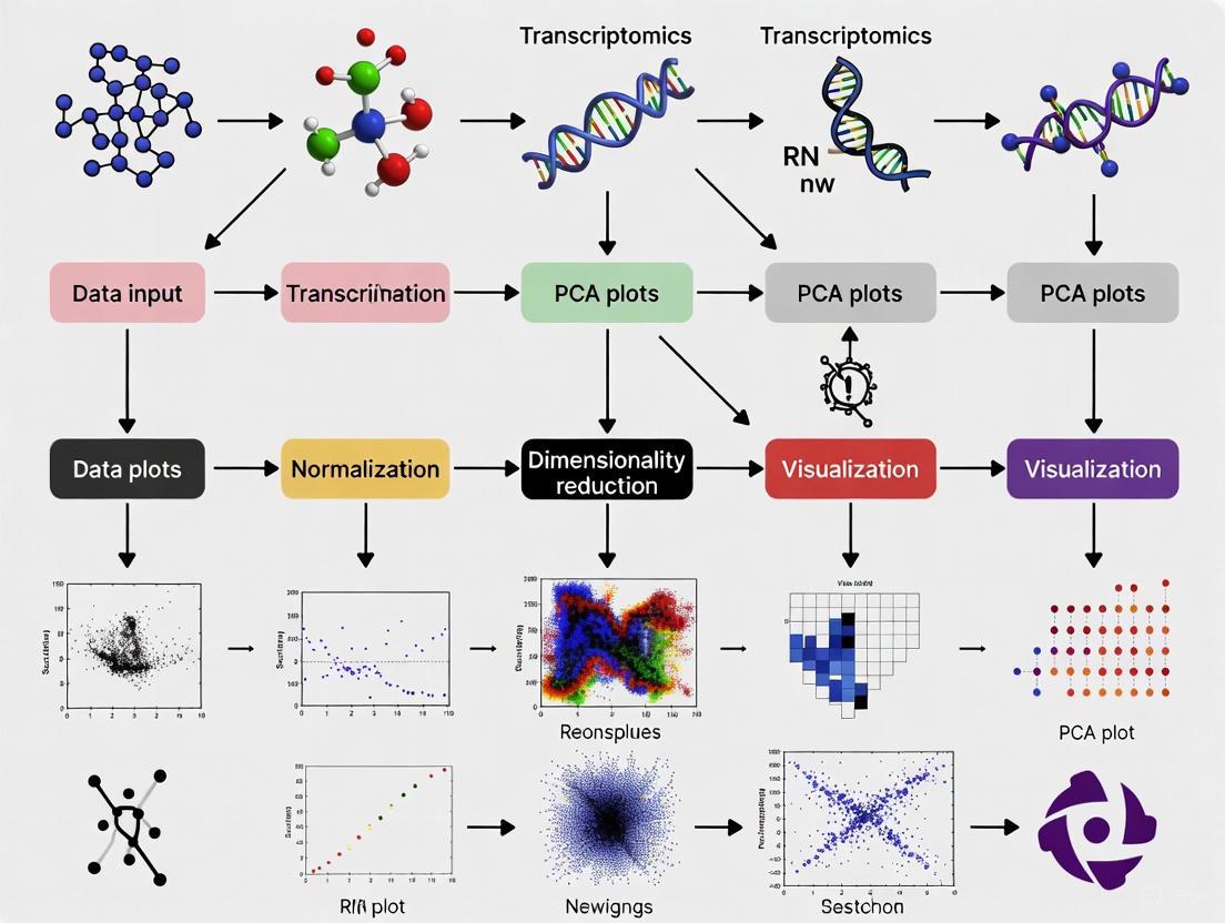
Figure 1: PCA workflow for transcriptomic data analysis showing key steps from raw data to interpretation.
Data Preprocessing Protocols
Normalization Methods
Normalization is a critical preprocessing step that ensures technical variability does not dominate biological signal in PCA. Different normalization methods can significantly impact PCA results and interpretation [6]. For RNA-seq count data, effective normalization must account for library size differences and variance-mean relationships. A comprehensive evaluation of 12 normalization methods found that the choice of normalization significantly influences biological interpretation of PCA models, with certain methods better preserving biological variance while others more effectively remove technical artifacts [6].
Data Filtering and Transformation
Prior to PCA, genes with low counts across samples should be filtered to reduce noise. A common approach is to retain only genes with counts per million (CPM) > 1 in at least the number of samples corresponding to the size of the smallest group. Following normalization and filtering, variance-stabilizing transformations such as log2(X+1) are typically applied to count data to prevent a few highly variable genes from dominating the PCA results [3].
Computational Execution
In R, PCA can be performed using the prcomp() function, which accepts a transposed normalized count matrix with samples as rows and genes as columns [3]. The function centers the data by default, and the scale parameter can be set to TRUE to standardize variables, which is particularly recommended when genes exhibit substantially different variances [3]. The computational output includes the PC scores (sample_pca$x), eigenvalues (sample_pca$sdev^2), and variable loadings (sample_pca$rotation) that can be extracted for further analysis and visualization [3].
Interpreting PCA Results in Transcriptomics
Variance Explained and Component Significance
Table 1: Typical variance distribution across principal components in transcriptomic data
| Principal Component | Percentage of Variance Explained | Cumulative Variance | Typical Biological Interpretation |
|---|---|---|---|
| PC1 | 15-40% | 15-40% | Major biological signal (e.g., tissue type) |
| PC2 | 10-25% | 25-65% | Secondary biological signal or major batch effect |
| PC3 | 5-15% | 30-80% | Additional biological signal or technical factor |
| PC4+ | <5% each | 80-100% | Minor biological signals, noise, or stochastic effects |
The variance explained by each principal component is calculated from the eigenvalues (λ) as λi/Σλ × 100% [3]. A scree plot visualizes the eigenvalues in descending order and helps determine the number of meaningful components; a sharp decline ("elbow") typically indicates transition from biologically relevant components to noise [3] [7]. In large heterogeneous transcriptomic datasets, the first 3-6 components often capture the majority of structured biological variation, though the specific number depends on dataset complexity and effect sizes [4].
Interpreting Sample Patterns in Score Plots
PC score plots reveal sample relationships and cluster patterns. Samples with similar expression profiles cluster together in the projection, while outliers appear separated from main clusters. When biological groups form distinct clusters in PC space, this indicates that inter-group differences exceed intra-group variability. Strong batch effects often manifest as clustering by processing date, sequencing lane, or other technical factors, potentially obscuring biological signals [4]. Color-coding points by experimental factors (treatment, tissue type) and technical covariates (batch, RNA quality metrics) facilitates identification of variance sources.
Extracting Biological Meaning from Loadings
Gene loadings indicate each gene's contribution to components. Genes with large absolute loadings (positive or negative) for a specific PC strongly influence that component's direction. Loading analysis can reveal biological processes driving sample separation; for example, if PC1 separates tumor from normal samples, genes with high PC1 loadings likely include differentially expressed genes relevant to cancer pathology [3]. Functional enrichment analysis of high-loading genes provides biological interpretation of components, connecting mathematical transformations to biological mechanisms.
Key Insights from PCA in Transcriptomic Studies
Intrinsic Dimensionality of Gene Expression Data
The intrinsic dimensionality of transcriptomic data—the number of components needed to capture biologically relevant information—has been debated. Early studies of large heterogeneous microarray datasets suggested surprisingly low dimensionality, with the first 3-4 principal components capturing major biological axes like hematopoietic lineage, neural tissue, and proliferation status [4]. However, subsequent work revealed that higher components frequently contain additional biological signal, particularly for comparisons within similar tissue types [4]. The apparent dimensionality depends heavily on dataset composition; larger sample sizes representing more biological conditions increase the number of meaningful components.
Sample Composition Effects
PCA results are strongly influenced by sample composition within the dataset. Studies have demonstrated that sample size disparities across biological groups can skew principal components toward representing the largest groups [4]. In one computational experiment, reducing the proportion of liver samples from 3.9% to 1.2% eliminated the liver-specific component, while increasing liver sample representation strengthened this signal [4]. This underscores that PCA reveals dominant variance sources in the specific dataset analyzed, which may reflect technical artifacts, sampling bias, or true biological signals.
Information Distribution Across Components
While early components capture the largest variance sources, biologically relevant information distributes across multiple components. Analysis of residual information after removing the first three components shows that tissue-specific correlation patterns persist [4]. The information ratio criterion quantifies phenotype-specific information distribution between projected and residual spaces, revealing that comparisons within large groups (e.g., different brain regions) retain substantial information in higher components [4]. This explains why focusing exclusively on the first 2-3 components may miss biologically important signals, particularly for subtle phenotypic differences.
Advanced Applications and Integration
Integration with Machine Learning Approaches
Machine learning enhances PCA applications in transcriptomics through several advanced frameworks. Gene Network Analysis methods like Weighted Gene Co-expression Network Analysis (WGCNA) group genes into modules based on expression pattern similarity, with PCA sometimes applied to reduce dimensionality before network construction [8]. Biomarker discovery pipelines combine PCA for dimensionality reduction with machine learning classifiers (e.g., LASSO, support vector machines) to identify compact gene signatures predictive of disease states or treatment responses [8]. These approaches leverage PCA's ability to distill thousands of genes into manageable components while preserving essential biological information.
Drug Connectivity Mapping
The Drug Connectivity Map (cMap) resource applies PCA-like dimensionality reduction to gene expression profiles from drug-treated cells, creating signature vectors that enable comparison across compounds [8]. Researchers can project their own transcriptomic data into this space to identify drugs that reverse disease signatures—for example, finding compounds that shift expression toward normal patterns. Similar approaches using the Cancer Therapeutics Response Portal (CTRP) and Genomics of Drug Sensitivity in Cancer (GDSC) databases help connect transcriptomic profiles to therapeutic sensitivity [8].
Single-Cell and Spatial Transcriptomics
In single-cell RNA sequencing (scRNA-seq), PCA represents a standard step in preprocessing before nonlinear dimensionality reduction techniques (t-SNE, UMAP) that visualize cell clusters [8]. PCA denoises expression data and reduces computational requirements for subsequent analyses. For spatial transcriptomics, PCA helps identify spatial expression patterns by reducing dimensionality while maintaining spatial relationships, revealing gradients and regional specifications in tissue contexts.
Experimental Considerations and Best Practices
Research Reagent Solutions
Table 2: Essential computational tools for PCA in transcriptomics
| Tool/Resource | Function | Application Context |
|---|---|---|
| prcomp() (R function) | PCA computation | Standard PCA implementation from centered/transformed count matrices |
| DESeq2 (R package) | Data normalization and transformation | Variance-stabilizing transformation of count data prior to PCA |
| edgeR (R package) | Data normalization and filtering | TMM normalization and low-expression gene filtering |
| SCONE (R package) | Normalization assessment | Evaluation of multiple normalization methods for optimal PCA performance |
| Omics Playground (platform) | Interactive analysis | GUI-based PCA with integration of multiple normalization approaches |
| Drug cMap Database | Reference data | Comparison of study data with drug perturbation signatures |
Methodological Limitations and Alternatives
PCA has several limitations that researchers should consider. As a linear method, PCA may fail to capture complex nonlinear relationships in gene expression data [7]. When biological effects are small relative to technical noise, PCA may not reveal relevant clustering without prior supervised adjustment [4]. Additionally, PCA assumes that high-variance directions correspond to biological signals, which may not hold if technical artifacts introduce substantial variance [6].
Several methodological adaptations address these limitations. Kernel PCA extends the approach to capture nonlinear structures [7]. Robust PCA methods reduce sensitivity to outliers [2]. For datasets with known batch effects, * supervised normalization* or batch correction methods should be applied before PCA to prevent technical variance from dominating components [6]. When PCA fails to reveal expected biological structure despite evidence from other analyses, nonlinear dimensionality reduction techniques may better capture the underlying data geometry.
Validation and Reproducibility
To ensure PCA results reflect biological truth rather than dataset-specific artifacts, several validation approaches are recommended. Subsampling validation assesses stability of principal components across dataset variations. Independent replication confirms that similar components emerge in comparable datasets. Biological validation through experimental follow-up of loading-based hypotheses provides the strongest evidence for correct interpretation. For method selection, objective criteria such as the ability to recover known biological groups should guide choice of normalization and preprocessing strategies [6].
PCA remains an indispensable tool for initial exploration of transcriptomic data, providing critical insights into data quality, batch effects, and biological group separation. When properly implemented with appropriate normalization and preprocessing, PCA reveals the intrinsic structure of gene expression data and guides subsequent analytical steps. As transcriptomic technologies evolve toward single-cell resolution and spatial profiling, PCA and its extensions continue to provide a mathematical foundation for reducing dimensionality while preserving biological information. For drug development and clinical translation, careful interpretation of PCA plots ensures that analytical decisions are grounded in a comprehensive understanding of dataset structure and variance components.
Principal Component Analysis (PCA) stands as a cornerstone statistical technique in transcriptomics research, enabling researchers to reduce the overwhelming dimensionality of gene expression data and extract meaningful biological patterns. This technical guide provides an in-depth examination of PCA plot interpretation, focusing on the core elements of scores, variance explained, and component meaning. Within the context of a broader thesis on multivariate data exploration in omics sciences, we detail how PCA reveals sample clustering, identifies outliers, and captures major sources of variation in high-throughput transcriptomics datasets. By synthesizing current methodologies and visualization approaches, this whitepaper equips researchers and drug development professionals with the analytical framework necessary to transform complex gene expression matrices into actionable biological insights.
Principal Component Analysis (PCA) is an unsupervised multivariate statistical technique that applies orthogonal transformations to convert a set of potentially intercorrelated variables into a set of linearly uncorrelated variables called principal components (PCs) [9]. In transcriptomics research, where expression data for thousands of genes can be overwhelming to explore, PCA serves as a vital tool for emphasizing variation and bringing out strong patterns in datasets [3] [10]. The technique distills the essence of complex datasets while maintaining fidelity to the original information, enabling the construction of robust mathematical frameworks that encapsulate characteristic profiles of biological samples [9].
PCA operates as a dimensionality reduction technique that transforms the original set of variables into a new set of uncorrelated variables called principal components [11]. This process involves calculating the eigenvectors and eigenvalues of the covariance or correlation matrix of the data, where the eigenvectors represent the directions of maximum variance in the data, and the corresponding eigenvalues represent the amount of variance explained by each eigenvector [11]. For transcriptome-wide studies, PCA provides a powerful approach to understand patterns of similarity between samples based on gene expression profiles, making high-dimensional data more amenable to visual exploration through projections onto the first few principal components [3].
Theoretical Foundations of PCA
Mathematical Underpinnings
The mathematical foundation of PCA involves solving an eigenvalue/eigenvector problem. Given a data matrix X with n samples (rows) and p variables (columns, e.g., genes), where the columns are centered to have zero mean, the principal components are derived from the covariance matrix S = (1/(n-1))X′X [2]. The PCA solution involves finding the eigenvectors a and eigenvalues λ that satisfy the equation:
Sa = λa
The eigenvectors, termed PC loadings, represent the axes of maximum variance, while the eigenvalues quantify the amount of variance captured by each corresponding principal component [2]. The full set of eigenvectors of S form an orthonormal set of vectors, and the new variables (PC scores) are obtained as linear combinations Xa of the original data [2]. PCA can also be understood through singular value decomposition (SVD) of the column-centered data matrix X*, providing both algebraic and geometric interpretations of the technique [2].
Key Components of PCA
- PC Loadings: The eigenvectors a_k, which represent the weight or contribution of each original variable to the principal component. These are sometimes called the "axis" or "direction" of maximum variance [3] [2].
- PC Scores: The values of the new variables for each observation, obtained by projecting the original data onto the principal components (Xa_k) [3] [2].
- Eigenvalues: Represent the variance explained by each principal component. The sum of all eigenvalues equals the total variance in the original dataset [3].
- Variance Explained: The proportion of total variance captured by each PC, calculated as λ_k/Σλ for each component k [3].
Table 1: Key Elements of a PCA Output
| Element | Description | Interpretation in Transcriptomics |
|---|---|---|
| PC Loadings | Eigenvectors of covariance matrix | Weight of each gene's contribution to a PC |
| PC Scores | Projection of samples onto PCs | Coordinates of samples in PC space |
| Eigenvalues | Variance captured by each PC | Importance of each PC in describing data structure |
| Variance Explained | Percentage of total variance per PC | How much of the total gene expression variability a PC captures |
| Biplot | Combined plot of scores and loadings | Shows both samples and genes in PC space |
Interpreting PCA Results
Variance Explained and Scree Plots
The variance explained by each principal component is fundamental to interpreting PCA results. The first principal component (PC1) captures the most pronounced feature in the data, with subsequent components (PC2, PC3, etc.) representing increasingly subtler aspects [9]. A scree plot displays how much variation each principal component captures from the data, with the y-axis representing eigenvalues (amount of variation) and the x-axis showing the principal components in order [3] [12].
In an ideal scenario, the first two or three PCs capture most of the information, allowing researchers to ignore the rest without losing important information [12]. The scree plot should show a steep curve that bends at an "elbow" point before flattening out, with this elbow representing the optimal number of components to retain [12]. For datasets where the scree plot doesn't show a clear elbow, two common approaches are:
- Kaiser rule: Retain PCs with eigenvalues of at least 1 [12]
- Proportion of variance: Selected PCs should collectively describe at least 80% of the total variance [12]
Table 2: Methods for Determining Significant PCs in Transcriptomics
| Method | Approach | Advantages | Limitations |
|---|---|---|---|
| Scree Plot | Visual identification of "elbow" point | Intuitive, easy to implement | Subjective interpretation |
| Kaiser Rule | Keep PCs with eigenvalues ≥1 | Objective threshold | May retain too many or too few components |
| Variance Explained | Retain PCs that cumulatively explain >80% variance | Ensures sufficient information retention | Threshold is arbitrary; may miss biologically relevant subtle patterns |
| Parallel Analysis | Compare to PCA of random datasets | Statistical robustness | Computationally intensive |
Score Plots and Sample Patterns
The PCA score plot visualizes samples in the reduced dimension space of the principal components, typically showing PC1 versus PC2 [3] [9]. Each point in the score plot corresponds to an individual sample, with different colors representing distinct groups or experimental conditions [9]. Interpretation of score plots focuses on several key patterns:
- Clustering: Well-clustered biological replicates indicate good technical repeatability. Samples that cluster together have similar gene expression profiles across the most variable genes [9].
- Separation between groups: Distinct groupings along PC1 or PC2 may reflect treatment effects, genetic differences, or time points in an experiment [9].
- Outliers: Samples that deviate from their expected clusters may indicate poor sample quality, experimental artifacts, or biologically interesting anomalies [9].
- Gradients: Continuous distributions of samples along a PC may represent gradual changes in gene expression due to processes like development, time courses, or dose responses.
In transcriptomics, the first few PCs often capture major biological effects, with PC1 frequently separating samples based on the strongest source of variation, such as tissue type or major treatment effect, while subsequent PCs may capture more subtle biological signals or technical artifacts [13].
Loading Interpretation and Biplots
PC loadings indicate how strongly each original variable (gene) influences a principal component. The further away a loading vector is from the origin, the more influence that variable has on the PC [12]. Biplots combine both score and loading information in a single visualization, enabling researchers to see both samples and variables simultaneously [14] [12].
In a biplot:
- The bottom and left axes represent PC scores for the samples
- The top and right axes represent loadings for the variables [12]
- The angles between loading vectors indicate correlations between variables:
For transcriptomic data, loading interpretation helps identify genes that drive the separation observed in the score plot, connecting patterns in sample clustering to specific gene expression changes.
Practical Implementation for Transcriptomics
Data Preprocessing and PCA Computation
For RNA-seq data analysis, proper preprocessing is essential for meaningful PCA results. The standard approach involves:
- Data normalization: Account for sequencing depth and other technical biases
- Transformation: Often log-transformation of normalized counts to stabilize variance
- Centering and scaling: Variables (genes) are typically centered (mean-zero) and may be scaled to unit variance, especially when genes are measured on different scales [3]
In R, PCA can be computed using the prcomp() function:
The prcomp() function returns an object containing:
sdev: Standard deviations of principal componentsrotation: The matrix of variable loadingsx: The rotated data (scores) [3]
Visualization Workflow
The PCA visualization workflow in transcriptomics typically involves:
The Researcher's Toolkit for PCA in Transcriptomics
Table 3: Essential Computational Tools for PCA in Transcriptomics Research
| Tool/Function | Application | Key Features | Implementation |
|---|---|---|---|
| prcomp() | PCA computation in R | Uses singular value decomposition, preferred for numerical accuracy [3] | Base R function |
| varianceExplained | Calculate PC contribution | Computes percentage and cumulative variance from PCA object [3] | pc_eigenvalues <- sample_pca$sdev^2 |
| Scree Plot | Determine significant PCs | Visualize variance explained by each component [3] [12] | qplot(x = PC, y = pct, data = pc_eigenvalues_df) |
| Score Plot | Visualize sample relationships | Scatterplot of PC1 vs PC2 with sample labels/colors [3] | ggplot(pc_scores_df, aes(PC1, PC2, color = group)) + geom_point() |
| Biplot | Combined scores and loadings | Overlay variable influence on sample projection plot [14] [12] | biplot(sample_pca, choices = c(1, 2)) |
| bigsnpr | Large-scale genetic PCA | Efficient PCA for very large datasets [13] | R package from CRAN |
Advanced Considerations in Transcriptomics
Pitfalls and Limitations
While PCA is powerful, researchers must recognize its limitations:
- Linear assumptions: PCA assumes linear relationships between variables, while biological systems often exhibit nonlinear behaviors [11]
- No group awareness: As an unsupervised method, PCA doesn't use known group labels and may fail to differentiate predefined groups clearly [9]
- Interpretability challenges: Beyond the first few PCs, biological meaning becomes harder to extract [9]
- Horseshoe effect: An artifact where gradient data folds in on itself, potentially distorting ecological patterns [11]
- Sensitivity to preprocessing: Results can be heavily influenced by normalization, transformation, and scaling decisions
In genetics specifically, some PCs may capture linkage disequilibrium structure rather than population structure, requiring careful interpretation and potentially specialized LD pruning methods [13].
Comparison to Alternative Methods
For transcriptomics data, researchers should consider when PCA is the most appropriate tool versus alternative dimensionality reduction methods:
Table 4: PCA vs Alternative Dimensionality Reduction Methods for Transcriptomics
| Method | Type | Preserves | Best For | Transcriptomics Application |
|---|---|---|---|---|
| PCA | Linear | Global structure [15] | Exploratory analysis, outlier detection [16] | Bulk RNA-seq QC, population structure [3] [16] |
| t-SNE | Non-linear | Local structure [15] | Cluster visualization [15] [16] | scRNA-seq cell type identification [15] [16] |
| UMAP | Non-linear | Local + some global [15] | Large datasets, clustering [15] | scRNA-seq, visualization of complex manifolds [15] |
For single-cell RNA-seq data, t-SNE and UMAP are often preferred over PCA because they better capture the complex manifold structures and distinct cell populations characteristic of single-cell datasets [16]. However, PCA is frequently used as an initial step before t-SNE or UMAP to reduce computational complexity [16].
Principal Component Analysis remains an essential exploratory tool in transcriptomics research, providing a robust framework for visualizing high-dimensional gene expression data. Through careful interpretation of variance explained, sample scores, and variable loadings, researchers can identify major patterns of biological variation, assess data quality, generate hypotheses, and guide subsequent analyses. While acknowledging its limitations and understanding when alternative methods might be more appropriate, mastering PCA plot interpretation provides drug development professionals and researchers with a fundamental skill for extracting meaningful insights from complex transcriptomics datasets. As omics technologies continue to evolve, the principles of PCA interpretation will remain relevant for transforming high-dimensional data into biological understanding.
The Critical Role of Variance Explained by PC1 and PC2
In high-dimensional biological data analysis, particularly in transcriptomics, Principal Component Analysis (PCA) serves as a fundamental dimensionality reduction technique. The variance explained by the first and second principal components (PC1 and PC2) provides critical insights into dataset structure, data quality, and underlying biological signals. This whitepaper explores the mathematical foundations, interpretation methodologies, and practical applications of PC1 and PC2 variance in transcriptomics research, emphasizing how these metrics guide experimental conclusions and analytical decisions in drug development pipelines. We demonstrate that proper interpretation of these components enables researchers to identify batch effects, detect outliers, uncover biological subtypes, and streamline subsequent analyses—all essential capabilities for advancing therapeutic discovery.
Principal Component Analysis (PCA) is a linear dimensionality reduction technique that transforms high-dimensional data into a new coordinate system defined by orthogonal principal components (PCs), where the first component (PC1) captures the maximum variance in the data, and each subsequent component captures the remaining variance under the constraint of orthogonality [17] [5]. In transcriptomics studies, where datasets often contain expression measurements for thousands of genes across multiple samples, PCA provides an indispensable tool for data exploration, quality control, and hypothesis generation.
The principal components are derived from the eigenvectors of the data's covariance matrix, with corresponding eigenvalues representing the amount of variance explained by each component [18] [19]. The variance explained by PC1 and PC2 is particularly crucial as these components typically capture the most substantial sources of variation in the dataset, potentially reflecting key biological signals, technical artifacts, or experimental batch effects that require further investigation.
Mathematical Foundations of Variance Explanation
Computational Framework
PCA operates through a systematic computational process that transforms original variables into principal components:
Data Standardization: Before performing PCA, continuous initial variables are standardized to have a mean of zero and standard deviation of one, ensuring that variables with larger scales do not dominate the variance structure [18] [5]. This is critical in transcriptomics where expression values may span different ranges. The standardization formula for each value is:
[ Z = \frac{X-\mu}{\sigma} ]
where (\mu) is the mean of independent features and (\sigma) is the standard deviation [18].
Covariance Matrix Computation: PCA calculates the covariance matrix to understand how variables vary from the mean relative to each other [18] [5]. For a dataset with (p) variables, this produces a (p \times p) symmetric matrix where the diagonal represents variances of each variable and off-diagonal elements represent covariances between variable pairs [5].
Eigen decomposition: The eigenvectors and eigenvalues of the covariance matrix are computed, where eigenvectors represent the directions of maximum variance (principal components), and eigenvalues represent the magnitude of variance along these directions [18] [19]. The eigenvector with the highest eigenvalue becomes PC1, followed by PC2 with the next highest eigenvalue under the orthogonality constraint [17].
Quantifying Variance Explanation
The proportion of total variance explained by each principal component is calculated by dividing its eigenvalue by the sum of all eigenvalues [17]. For the (k^{th}) component:
[ \text{Variance Explained}(PCk) = \frac{\lambdak}{\sum{i=1}^p \lambdai} ]
where (\lambda_k) is the eigenvalue for component (k), and (p) is the total number of components [17]. PC1 and PC2 collectively often capture a substantial portion of total variance in high-dimensional transcriptomics data, making them particularly informative for initial data exploration.
Table 1: Variance Explanation Interpretation Guidelines in Transcriptomics
| Variance Distribution | Potential Interpretation | Recommended Actions |
|---|---|---|
| PC1 explains >40% of variance | Single dominant technical or biological effect (e.g., batch effect, treatment response) | Investigate sample metadata for correlates; consider correction if technical |
| PC1 & PC2 explain >60% collectively | Strong structured data with potentially meaningful biological subgroups | Proceed with subgroup analysis and differential expression |
| Multiple components with similar variance | Complex dataset with multiple contributing factors | Consider additional components in analysis; explore higher-dimensional relationships |
| No components with substantial variance | Unstructured data, potentially high noise | Quality control assessment; consider alternative experimental approaches |
Methodological Protocols for Variance Analysis
Experimental Workflow for PCA in Transcriptomics Studies
The following diagram illustrates the standard analytical workflow for conducting and interpreting PCA in transcriptomics research:
Variance Interpretation Protocol
Scree Plot Analysis: Create a scree plot displaying eigenvalues or proportion of variance explained by each component in descending order [19]. The "elbow" point—where the curve bends sharply—often indicates the optimal number of meaningful components to retain for further analysis [19].
Cumulative Variance Calculation: Compute cumulative variance explained by sequential components to determine the number needed to capture a predetermined threshold of total variance (typically 70-90% in exploratory analysis) [20].
Component Loading Examination: Identify variables (genes) with the highest absolute loadings on PC1 and PC2, as these contribute most significantly to these components' variance [19]. In transcriptomics, genes with high loadings often represent biologically meaningful pathways or responses.
Sample Projection Visualization: Project samples onto the PC1-PC2 plane and color-code by experimental conditions, batches, or biological groups to identify patterns, clusters, or outliers [18] [19].
Applications in Transcriptomics Research
Spatial Transcriptomics and Domain Identification
In spatial transcriptomics, PCA-based approaches have demonstrated state-of-the-art performance in identifying biologically meaningful spatial domains. The NichePCA algorithm exemplifies how a reductionist PCA approach can rival more complex methods in unsupervised spatial domain identification across diverse single-cell spatial transcriptomic datasets [21]. In this context, variance explained by PC1 and PC2 often corresponds to:
- Spatial gene expression gradients that define tissue architecture
- Cell-type-specific expression programs that segregate within complex tissues
- Regional microenvironment signatures that influence cellular function
The exceptional execution speed, robustness, and scalability of PCA-based methods make them particularly valuable for large-scale spatial transcriptomics studies in drug development contexts [21].
Quality Control and Batch Effect Detection
PC1 and PC2 frequently capture technical artifacts and batch effects that must be identified before biological interpretation:
- Batch Effects: When samples cluster by processing date, sequencing lane, or preparation batch along PC1 or PC2, this indicates technical variance dominating biological signals [19].
- Outlier Detection: Samples that appear as extreme outliers along major components often indicate quality issues requiring further investigation.
- Library Size Confounds: PCA can reveal associations between principal components and technical covariates like library size, which may confound biological interpretations if unaddressed [21].
Biological Discovery and Subtype Identification
When technical artifacts are properly accounted for, variance in PC1 and PC2 often reveals meaningful biological structure:
- Disease Subtypes: Distinct sample clusters along PC1/PC2 may represent molecularly distinct disease subtypes with therapeutic implications.
- Treatment Response: Separation of treated vs. control samples along major components suggests strong transcriptional responses to interventions.
- Developmental or Temporal Trajectories: Ordered progression of samples along PC1 may reflect continuous biological processes such as development, differentiation, or disease progression.
Table 2: Research Reagent Solutions for PCA in Transcriptomics
| Research Reagent | Function in PCA Workflow | Application Context |
|---|---|---|
| Single-cell RNA-seq kits (10x Genomics) | Generate high-dimensional transcript count data for PCA input | Single-cell spatial transcriptomics studies [21] |
| Universal Sentence Encoder (Google) | Text-to-numeric transformation for text mining integration | Converting textual metadata for integrated analysis [22] |
| Normalization algorithms (e.g., SCTransform) | Standardize library sizes before PCA | Removing technical variation that could dominate PC1 [21] |
| Spatial barcoding oligonucleotides | Enable spatial transcriptomic profiling | PCA-based spatial domain identification [21] |
| Dimensionality reduction libraries (Scikit-learn) | Perform efficient PCA computation on large matrices | Standardized implementation of PCA algorithm [18] |
Case Study: NichePCA for Spatial Domain Identification
Experimental Protocol
The following case study exemplifies the application of PCA variance analysis in spatial transcriptomics:
- Data Collection: Acquire spatial transcriptomics data using established platforms (e.g., 10x Genomics Visium, MERFISH, or Slide-seq) [21].
- Data Preprocessing: Perform standard quality control, normalization, and log-transformation of gene expression counts to minimize technical artifacts.
- PCA Implementation: Apply PCA to the normalized expression matrix using efficient computational frameworks capable of handling large-scale transcriptomic data.
- Component Selection: Retain the top k principal components based on scree plot analysis, typically capturing 50-80% of total variance in spatial transcriptomics datasets.
- Spatial Domain Assignment: Project spatial transcriptomics spots onto PC1 and PC2, using their coordinates to identify spatially coherent domains through clustering.
- Biological Validation: Validate identified domains using known marker genes and histological features to confirm biological relevance.
Interpretation Framework
The analytical process for interpreting PC1 and PC2 in spatial transcriptomics can be visualized as follows:
Performance Outcomes
In benchmark evaluations across six single-cell spatial transcriptomic datasets, the NichePCA approach demonstrated that simple PCA-based algorithms could rival the performance of ten competing state-of-the-art methods in spatial domain identification [21]. Key findings included:
- Computational Efficiency: Significantly faster execution compared to more complex neural network-based methods.
- Robust Performance: Consistent accuracy across diverse tissue types and technological platforms.
- Intuitive Interpretation: Direct mapping between component loadings and biologically meaningful gene programs.
Advanced Considerations in Variance Interpretation
Limitations and Mitigation Strategies
While PC1 and PC2 variance explanation provides valuable insights, researchers must consider several limitations:
- Non-Linear Relationships: PCA captures linear relationships only; non-linear dimensionality reduction techniques (e.g., UMAP, t-SNE) may be required for complex datasets [19].
- Interpretation Challenges: Principal components are mathematical constructs that may not always align with discrete biological categories [18].
- Scale Sensitivity: PCA results are sensitive to data scaling decisions, requiring careful normalization approaches tailored to transcriptomic data [18].
- Information Loss: Over-reliance on only PC1 and PC2 may miss biologically important signals captured in higher components [18].
Integration with Complementary Methods
For comprehensive transcriptomics analysis, PCA should be integrated with other analytical approaches:
- Differential Expression Analysis: Use PC-defined sample groupings to guide focused differential expression testing.
- Pathway Enrichment Analysis: Apply gene set enrichment to high-loading genes on PC1 and PC2 to identify biological processes.
- Multi-Omics Integration: Correlate transcriptional principal components with similar components from other data modalities (e.g., epigenomics, proteomics).
The variance explained by PC1 and PC2 serves as a critical gateway to understanding high-dimensional transcriptomics data, providing a powerful framework for identifying both technical artifacts and biologically meaningful patterns. Through proper standardization, computational implementation, and interpretive protocols, researchers can leverage these components to enhance data quality assessment, reveal novel biological insights, and guide therapeutic development decisions. The continued development of PCA-based methodologies, exemplified by approaches like NichePCA in spatial transcriptomics, ensures that these fundamental dimensionality reduction techniques will remain essential tools in the evolving landscape of transcriptional research and drug development.
Identifying Sample Clustering and Biological Replicate Consistency
Principal Component Analysis (PCA) is a fundamental dimensionality reduction technique that transforms high-dimensional transcriptomic data into a set of orthogonal variables called principal components (PCs), which capture maximum variance in the data [5] [9]. This method is particularly valuable for exploring sample clustering and assessing biological replicate consistency in RNA-seq experiments, serving as a critical first step in quality control and data exploration [23] [9].
In transcriptomics, where datasets typically contain thousands of genes (variables) measured across relatively few samples, PCA helps mitigate the "curse of dimensionality" by reducing data complexity while preserving essential patterns [24]. By projecting samples into a lower-dimensional space defined by the first few principal components, researchers can visually assess technical variability, biological consistency, and potential batch effects [23]. The application of PCA following Occam's razor principle—as demonstrated by the NichePCA algorithm for spatial transcriptomics—shows that simple PCA-based approaches can rival complex methods in performance while offering superior execution speed, robustness, and scalability [21].
Methodological Protocols for PCA in Transcriptomics
Experimental Design and RNA-seq Processing
Proper experimental design is paramount for meaningful PCA results. Biological replicates (distinct biological samples) rather than technical replicates are essential for assessing true biological variation [25] [26]. The ENCODE consortium standards recommend a minimum of two biological replicates for RNA-seq experiments, with replicate concordance measured by Spearman correlation of >0.9 between isogenic replicates [27].
For bulk RNA-seq experiments, libraries should be prepared from mRNA (polyA+ enriched or rRNA-depleted) and sequenced to a depth of 20-30 million aligned reads per replicate [27]. The ENCODE Uniform Processing Pipeline utilizes STAR for read alignment and RSEM for gene quantification, generating both FPKM and TPM values for downstream analysis [27]. To minimize batch effects, researchers should process controls and experimental conditions simultaneously, isolate RNA on the same day, and sequence all samples in the same run [23].
PCA Computational Workflow
The computational implementation of PCA follows a standardized five-step process adapted for transcriptomic data [5]:
Step 1: Data Standardization Prior to PCA, raw count data (e.g., TPM or FPKM values) must be standardized and centered to ensure each gene contributes equally to the analysis. This involves subtracting the mean and dividing by the standard deviation for each gene across samples. Standardization prevents genes with naturally larger expression ranges from dominating the variance structure [5].
Step 2: Covariance Matrix Computation The standardized data is used to compute a covariance matrix that captures how all gene pairs vary together. This p×p symmetric matrix (where p equals the number of genes) identifies correlated genes that may represent redundant information [5].
Step 3: Eigen Decomposition Eigenvectors and eigenvalues are computed from the covariance matrix. The eigenvectors represent the directions of maximum variance (principal components), while eigenvalues indicate the amount of variance explained by each component [5].
Step 4: Component Selection Researchers select the top k components that capture sufficient variance (typically 70-90% cumulative variance). The feature vector is formed from the eigenvectors corresponding to these selected components [5].
Step 5: Data Projection The original data is projected onto the new principal component axes to create transformed coordinates for each sample, which are then visualized in 2D or 3D PCA plots [5].
The diagram below illustrates this workflow in transcriptomics data analysis:
Interpreting PCA Plots for Biological Replicates
Quality Assessment and Outlier Detection
PCA plots serve as powerful tools for quality assurance in transcriptomics. When analyzing biological replicates, researchers should initially examine the clustering pattern of quality control (QC) samples, which are technical replicates prepared by pooling sample extracts. These QC samples should cluster tightly on the PCA plot, indicating analytical consistency [9].
Biological replicates from the same experimental group should demonstrate intra-group similarity, appearing as clustered patterns on the PCA plot. Samples that deviate significantly from their group clusters, particularly those outside the 95% confidence ellipse, may represent outliers requiring further investigation [9]. In datasets with sufficient sample sizes, such outliers are typically excluded from subsequent analysis [9].
Assessing Replicate Consistency and Group Separation
The interpretation of PCA plots for biological replicates follows a systematic approach [9]:
Check Variance Explained: Examine how much variation PC1 and PC2 account for individually and cumulatively. Higher percentages (typically >70% combined) indicate better representation of the dataset's structure.
Assess Replicate Clustering: Well-clustered biological replicates indicate good biological repeatability and technical consistency. Dispersion within a group reflects biological variability.
Evaluate Group Separation: Distinct groupings along PC1 or PC2 suggest strong treatment effects, genetic differences, or temporal changes. Overlap between groups may indicate weak effects or the need for supervised methods.
Identify Patterns and Trends: Regular patterns across components may reveal underlying experimental factors influencing gene expression.
Table 1: Interpretation Framework for PCA Plots of Biological Replicates
| Pattern Observed | Interpretation | Recommended Action |
|---|---|---|
| Tight clustering of replicates within groups | High replicate consistency, low technical variation | Proceed with differential expression analysis |
| Discrete separation between experimental groups | Strong biological effect of treatment/condition | Investigate group-specific expression patterns |
| Overlapping group clusters with no clear separation | Weak or no group differences | Consider increased replication or alternative methods |
| Single outlier sample distant from group cluster | Potential sample quality issue | Examine QC metrics, consider exclusion |
| QC samples dispersed rather than clustered | Technical variability in processing | Troubleshoot experimental protocol |
Research shows that with only three biological replicates, most differential expression tools identify just 20-40% of significantly differentially expressed genes detected with higher replication [26]. This substantially rises to >85% for genes changing by more than fourfold, but to achieve >85% sensitivity for all genes requires more than 20 biological replicates [26].
Quantitative Benchmarks and Performance Metrics
Replication Guidelines from Empirical Studies
Empirical studies provide specific guidance on biological replication requirements for RNA-seq experiments. With three biological replicates, most tools identify only 20-40% of significantly differentially expressed genes detected with full replication (42 replicates), though this rises to >85% for genes with large expression changes (>4-fold) [26]. To achieve >85% sensitivity for all differentially expressed genes regardless of fold change magnitude, more than 20 biological replicates are typically required [26].
For standard transcriptomics experiments, a minimum of six biological replicates per condition is recommended, increasing to at least 12 when identifying differentially expressed genes with small fold changes is critical [26]. These guidelines ensure sufficient statistical power while considering practical constraints.
Table 2: Biological Replication Guidelines for RNA-seq Experiments
| Experimental Goal | Minimum Replicates | Sensitivity Range | Key Considerations |
|---|---|---|---|
| Pilot studies/large effect sizes | 3-5 | 20-40% for all DE genes; >85% for >4-fold changes | Limited power for subtle expression differences |
| Standard differential expression | 6-12 | ~60-85% for all DE genes | Balance of practical constraints and statistical power |
| Comprehensive detection including subtle effects | >20 | >85% for all DE genes | Required for detecting small fold changes with high confidence |
| ENCODE standards | ≥2 | Spearman correlation >0.9 between replicates | Minimum standard for consortium data generation |
Benchmarking Clustering Performance
Recent benchmarking of 28 single-cell clustering algorithms on transcriptomic and proteomic data reveals that methods like scDCC, scAIDE, and FlowSOM demonstrate top performance across multiple metrics including Adjusted Rand Index (ARI), Normalized Mutual Information (NMI), and clustering accuracy [28]. These methods show consistent performance across different omics modalities, suggesting robust generalization capabilities.
For assessing replicate consistency in clustering results, the Adjusted Rand Index (ARI) serves as a primary metric, quantifying clustering quality by comparing predicted and ground truth labels with values from -1 to 1 [28]. Normalized Mutual Information (NMI) measures the mutual information between clustering and ground truth, normalized to [0,1], with values closer to 1 indicating better performance [28].
Implementation and Practical Applications
Research Reagent Solutions
Table 3: Essential Research Reagents for RNA-seq and PCA Analysis
| Reagent/Resource | Function | Application Notes |
|---|---|---|
| ERCC Spike-in Controls | External RNA controls for normalization | Creates standard baseline for RNA quantification; Ambion Mix 1 at ~2% of final mapped reads [27] |
| Poly(A) Selection Kits | mRNA enrichment from total RNA | NEBNext Poly(A) mRNA magnetic isolation kits provide high-fidelity selection [23] |
| Strand-Specific Library Prep Kits | Maintain transcriptional directionality | Critical for accurately quantifying overlapping transcripts |
| CD45 Microbeads | Immune cell enrichment | Magnetic-activated cell sorting for specific cell populations [23] |
| Collagenase D | Tissue dissociation | Enzymatic digestion for single-cell suspensions [23] |
| PicoPure RNA Isolation Kit | RNA extraction from sorted cells | Maintains RNA integrity from low-input samples [23] |
Troubleshooting Common PCA Challenges
Several common challenges arise when interpreting PCA plots for biological replicate consistency:
Mixed Group Clustering: When sample groups intermingle without distinct separation, reevaluate grouping criteria to ensure they represent the primary factors influencing transcriptomic profiles [9]. Consider whether other uncontrolled factors (e.g., lineage, batch effects) might be dominating the variance structure.
High Intra-group Variability: Excessive dispersion within biological replicates suggests either technical artifacts or genuine biological heterogeneity. Examine sample-level quality metrics (RNA integrity numbers, alignment rates) and consider whether the biological system inherently exhibits high variability [23] [9].
Low Cumulative Variance: When the first two principal components explain only a small percentage of total variance (<50%), the data may contain numerous technical artifacts or highly heterogeneous samples. In such cases, examine higher components (PC3, PC4) for group separation or apply batch correction methods before re-running PCA [9].
The limitations of PCA must be acknowledged in transcriptomics research. As an unsupervised method, PCA does not incorporate known group labels and may fail to highlight biologically relevant separations that are minor compared to other sources of variation [9]. When clear group differences are expected but not apparent in PCA plots, supervised methods like PLS-DA (Partial Least Squares Discriminant Analysis) may provide better separation [9].
PCA remains an indispensable tool for evaluating sample clustering and biological replicate consistency in transcriptomics research. By following standardized protocols for experimental design, data processing, and interpretation, researchers can extract meaningful insights from high-dimensional gene expression data. The quantitative benchmarks and methodological frameworks presented here provide actionable guidance for implementing PCA in diverse transcriptomic applications, from quality control to exploratory data analysis. As transcriptomic technologies continue to evolve, PCA will maintain its role as a foundational approach for visualizing and validating the consistency of biological replicates in gene expression studies.
Detecting Outliers and Quality Control Assessment
In transcriptomic research, outliers are observations that lie outside the overall pattern of a distribution, posing significant challenges for data interpretation and analysis [29]. The high-dimensional nature of RNA sequencing data, where thousands of genes (variables) are measured across typically few biological replicates (observations), creates a classic "curse of dimensionality" problem that makes outlier detection particularly challenging [24]. In this context, outliers may arise from technical variation during complex multi-step laboratory protocols or from true biological differences, necessitating accurate detection methods to ensure research validity [29].
Principal Component Analysis (PCA) serves as a fundamental tool for dimensionality reduction and quality control assessment in transcriptomics [9]. This unsupervised multivariate statistical technique applies orthogonal transformations to convert potentially intercorrelated variables into a set of linearly uncorrelated principal components (PCs), with the first component (PC1) capturing the most pronounced variance in the dataset [9]. The visualization of samples in reduced dimensional space (typically PC1 vs. PC2) enables researchers to assess sample clustering, identify outliers, and evaluate technical reproducibility [9]. However, traditional PCA is highly sensitive to outlying observations, which can distort component orientation and mask true data structure [29].
PCA Fundamentals and Visualization in Transcriptomics
Theoretical Foundation of PCA
Principal Component Analysis operates by identifying the eigenvectors of the sample covariance matrix, creating new variables (principal components) that capture decreasing amounts of variance in the data [29] [9]. For a typical RNA-seq dataset structured as an N × P matrix, where N represents the number of samples (observations) and P represents the number of genes (variables), PCA distills the essential information into a minimal number of components while preserving data covariance [24] [30]. Each principal component represents a linear combination of the original variables, with the constraint that all components are mutually orthogonal, thereby eliminating multicollinearity in the transformed data [9].
Interpreting PCA Plots for Quality Assessment
The interpretation of PCA plots follows established guidelines focused on several key aspects. Researchers should first examine the percentage of variance explained by each principal component, as higher values indicate better representation of the dataset's structure [9]. Subsequent analysis involves assessing the clustering of biological replicates, where tight clustering indicates good technical repeatability, while dispersed patterns suggest potential issues [9]. The separation between experimental groups along principal components may reflect treatment effects or biological differences of interest [9]. Finally, samples that fall beyond the 95% confidence ellipse or show substantial distance from their group peers may be classified as outliers requiring further investigation [9].
Table 1: Key Elements for PCA Plot Interpretation in Transcriptomics
| Element | Interpretation | Implications |
|---|---|---|
| Variance Explained | Percentage of total data variance captured by each PC | Higher percentages (>70% combined for PC1+PC2) indicate better representation of data structure |
| Replicate Clustering | Proximity of biological replicates within experimental groups | Tight clustering indicates good technical reproducibility; dispersed patterns suggest issues |
| Group Separation | Distinct grouping of samples along principal components | May reflect treatment effects, biological differences, or batch effects |
| Outlier Position | Samples distant from main clusters or beyond confidence ellipses | Potential technical artifacts, biological extremes, or sample mishandling |
Robust PCA Methods for Advanced Outlier Detection
Limitations of Classical PCA
Classical PCA (cPCA) demonstrates high sensitivity to outlying observations, which can substantially distort the orientation of principal components and compromise their ability to capture the variation of regular observations [29]. This limitation is particularly problematic in transcriptomics, where the prevalence of high-dimensional data with small sample sizes increases the potential impact of outliers on analytical outcomes [29]. Furthermore, cPCA relies on visual inspection of biplots for outlier identification, an approach that lacks statistical justification and may introduce unconscious biases during data interpretation [29].
Robust PCA Algorithms and Implementation
Robust PCA (rPCA) methods address the limitations of classical approaches by applying robust statistical theory to obtain principal components that remain stable despite outlying observations [29]. These methods simultaneously enable accurate outlier identification and categorization [29]. Two prominent rPCA algorithms include PcaHubert, which demonstrates high sensitivity in outlier detection, and PcaGrid, which exhibits the lowest estimated false positive rate among available methods [29]. These algorithms are implemented in the rrcov R package, which provides a common interface for computation and visualization [29].
The application of rPCA methods to RNA-seq data analysis has demonstrated remarkable efficacy in multiple simulated and real biological datasets. In controlled tests using positive control outliers with varying degrees of divergence, the PcaGrid method achieved 100% sensitivity and 100% specificity across all evaluations [29]. When applied to real RNA-seq data profiling gene expression in mouse cerebellum, both rPCA methods consistently detected the same two outlier samples that classical PCA failed to identify [29]. This performance advantage positions rPCA as a superior approach for objective outlier detection in transcriptomic studies.
Table 2: Comparison of PCA Methods for Outlier Detection in Transcriptomics
| Method | Key Features | Performance Metrics | Implementation |
|---|---|---|---|
| Classical PCA | Standard covariance decomposition; Sensitive to outliers | Subjective visual inspection; Prone to missed outliers | Various R packages (stats, FactoMineR) |
| PcaHubert | Robust algorithm with high sensitivity | High detection sensitivity; Moderate false positive rate | rrcov R package |
| PcaGrid | Grid-based robust algorithm | 100% sensitivity/specificity in validation studies; Low false positive rate | rrcov R package |
Experimental Protocols for Outlier Detection
Workflow for rPCA-Based Outlier Detection
Detailed Methodological Protocol
Step 1: Data Preprocessing and Quality Control Begin with raw FASTQ files from RNA sequencing experiments. Perform quality control using FastQC to assess sequence quality, GC content, adapter contamination, and overrepresented sequences [31]. Process reads with Trimmomatic to remove adapter sequences and trim low-quality bases using the following command structure:
Align processed reads to the appropriate reference genome using HISAT2 with default parameters [31].
Step 2: Expression Quantification and Normalization Generate count data using featureCounts from the Subread package with the following command structure:
Normalize raw counts using appropriate methods such as transcripts per million (TPM) for cross-sample comparisons or variance-stabilizing transformations for differential expression analysis [32]. For TPM calculation, use the formula: TPM = (Reads per transcript × 10^6) / (Transcript length × Total reads).
Step 3: Robust PCA Implementation Install and load required R packages:
Execute robust PCA using the PcaGrid method:
Step 4: Outlier Identification and Validation Calculate statistical cutoffs for outlier classification based on robust distances. Implement Tukey's fences method using the interquartile range (IQR):
Validate identified outliers through biological investigation, including sample metadata review, experimental condition verification, and potential technical artifact assessment [29] [33].
Quantitative Assessment of Outlier Detection Methods
Performance Metrics and Statistical Cutoffs
The accurate identification of outliers requires establishing appropriate statistical thresholds that balance detection sensitivity with false positive rates. Research indicates that using interquartile ranges (IQR) around the median of expression values provides robust outlier identification less affected by data skewness and extreme values [32]. Tukey's fences method identifies outliers as data falling below Q1 - k × IQR or above Q3 + k × IQR, where Q1 and Q3 represent the 1st and 3rd quartiles, respectively [32]. For conservative outlier detection in transcriptomic data, a threshold of k = 5 (corresponding to approximately 7.4 standard deviations in a normal distribution) effectively minimizes false positives while maintaining detection capability [32].
Empirical studies demonstrate that at k = 3, approximately 3-10% of all genes (approximately 350-1350 genes) exhibit extreme outlier expression in at least one individual across various tissues [32]. These numbers continuously decline with increasing k-values without a clear natural cutoff, supporting the selection of more conservative thresholds for rigorous outlier detection [32]. The number of detectable outlier genes directly correlates with sample size, with approximately half of the outlier genes remaining detectable even with only 8 individuals sampled [32].
Impact of Outlier Removal on Downstream Analysis
The removal of technical outliers significantly improves the performance of differential gene expression detection and subsequent functional analysis [29]. Comparative studies evaluating eight different data analysis strategies demonstrated that outlier removal without batch effect modeling performed best in detecting biologically relevant differentially expressed genes validated by quantitative reverse transcription PCR [29]. In classification studies, the removal of outliers notably changed classification performance, with improvement observed in most cases, highlighting the importance of reporting classifier performance both with and without outliers for accurate model assessment [33].
Table 3: Impact of Outlier Removal on Transcriptomics Analysis
| Analysis Type | Impact of Outlier Retention | Impact of Outlier Removal | Validation Method |
|---|---|---|---|
| Differential Expression | Decreased statistical power; Inflated variance | Improved detection of biologically relevant DEGs | qRT-PCR validation [29] |
| Classification Accuracy | Inflated or deflated performance estimates | More reproducible classifier performance | Bootstrap validation [33] |
| Pathway Analysis | Potentially spurious pathway identification | More biologically plausible functional enrichment | Literature consistency [29] |
The Scientist's Toolkit: Essential Research Reagents and Computational Tools
Table 4: Essential Research Reagent Solutions for Transcriptomics Outlier Detection
| Tool/Reagent | Function | Application Notes |
|---|---|---|
| rrcov R Package | Implementation of robust PCA methods | Primary package for PcaGrid and PcaHubert algorithms [29] |
| FastQC | Quality control of raw sequence data | Identifies potential technical issues in sequencing [31] |
| Trimmomatic | Read trimming and adapter removal | Improves data quality before alignment [31] |
| HISAT2 | Read alignment to reference genome | Generates BAM files for expression quantification [31] |
| featureCounts | Gene-level expression quantification | Generates count matrix from aligned reads [31] |
| DESeq2 | Differential expression analysis | Includes variance-stabilizing transformation for PCA input [32] |
Advanced Concepts and Future Directions
Biological Significance of Expression Outliers
Emerging research challenges the conventional practice of automatically dismissing all outlier expression values as technical artifacts. Studies demonstrate that outlier gene expression patterns represent a biological reality occurring universally across tissues and species, potentially reflecting "edge of chaos" effects in gene regulatory networks [32]. These patterns manifest as co-regulatory modules, some corresponding to known biological pathways, with sporadic generation rather than Mendelian inheritance [32]. In rare disease diagnostics, transcriptome-wide outlier patterns have successfully identified individuals with minor spliceopathies caused by variants in minor spliceosome components, demonstrating the diagnostic value of systematic outlier analysis [34] [35].
Methodological Considerations and Limitations
While PCA and robust PCA provide powerful approaches for outlier detection, researchers should acknowledge their limitations. PCA represents an unsupervised method that does not incorporate known group labels, potentially limiting its ability to capture condition-specific effects [9]. The interpretability of principal components decreases substantially beyond the first few components, potentially burying important biological variation in lower dimensions [9]. Furthermore, concerns have been raised about the potential for PCA results to be manipulated through selective sample or marker inclusion, highlighting the importance of transparent reporting and methodological rigor [30]. These limitations emphasize the necessity of complementing PCA with other quality assessment methods and biological validation to ensure robust research conclusions.
Recognizing Batch Effects and Technical Artifacts
In high-throughput transcriptomics research, batch effects represent systematic technical variations introduced during experimental processes that are unrelated to the biological variables of interest. These artifacts arise from differences in technical conditions such as sequencing runs, reagent lots, personnel, or instruments and can profoundly distort downstream analysis if not properly identified and mitigated [36] [37]. The primary challenge lies in distinguishing these technical artifacts from true biological signals, as batch effects can masquerade as apparent biological patterns in unsupervised analyses [38].
Principal Component Analysis (PCA) serves as an indispensable tool for quality assessment and exploratory data analysis in transcriptomics. By transforming high-dimensional gene expression data into a lower-dimensional space defined by principal components (PCs), PCA reveals the major sources of variation across samples [38] [3]. When applied systematically, PCA provides critical insights into data structure, enabling researchers to identify batch effects, detect sample outliers, and uncover underlying biological patterns before proceeding with more specialized analyses [38]. This technical guide outlines comprehensive methodologies for recognizing and addressing batch effects within the context of PCA-based transcriptomics research.
Detecting Batch Effects via PCA Visualization
Fundamental Principles of PCA for Transcriptomics
PCA reduces the complexity of transcriptomic datasets containing thousands of gene expression measurements by identifying orthogonal directions of maximum variance, known as principal components. The algorithm decomposes the data matrix into PCs ordered by the amount of variance they explain, with the first PC (PC1) capturing the largest source of variation, the second PC (PC2) the next largest, and so on [3]. For gene expression data, samples are typically represented as rows and genes as columns in the input matrix, which is often centered and scaled to ensure all genes contribute equally to the analysis [38] [3].
The three key pieces of information obtained from PCA include:
- PC scores: Coordinates of samples in the new PC space
- Eigenvalues: Variance explained by each PC
- Variable loadings: Weight or correlation of each original variable with the PCs [3]
In transcriptomics, PCA enables researchers to project high-dimensional gene expression data onto 2D or 3D scatterplots using the first few PCs, making patterns of similarity and difference between samples visually accessible [3].
Interpreting PCA Plots for Batch Effect Identification
Batch effects in PCA plots manifest as distinct clustering of samples according to technical rather than biological variables. The table below outlines key visual indicators of batch effects in PCA visualizations:
Table 1: Identifying Batch Effects in PCA Plots
| Observation | Suggests Batch Effect | Suggests Minimal Batch Effect |
|---|---|---|
| Sample Clustering by Color (Batch) | Different batches form separate, distinct clusters | Batches are thoroughly mixed together |
| Sample Separation by Shape (Biological Group) | No clear pattern by biological group | Clear separation according to biological conditions |
| Confidence Ellipses | Ellipses for different batches are separate with minimal overlap | Ellipses for biological groups are distinct, while batch ellipses overlap substantially |
| PC Variance Explanation | Very high percentage of variance explained by early PCs, potentially indicating technical dominance | Balanced variance distribution across PCs |
| Outlier Patterns | Samples cluster strictly by processing date, operator, or instrument | Outliers may exist but don't correlate with technical factors |
When examining PCA plots, researchers should follow a systematic approach: First, note the percentage of variance explained by each PC (higher values in early PCs may indicate dominant technical artifacts). Second, observe whether samples cluster by batch labels rather than biological groups. Third, assess whether within-batch distances are smaller than between-batch distances for similar biological samples [39].
The following diagram illustrates a typical workflow for detecting batch effects using PCA:
Quantitative Assessment of Batch Effects
Beyond visual inspection, several quantitative metrics help confirm the presence of batch effects:
- Percentage of Variance Explained: When PC1 explains an exceptionally high percentage of total variance (often >30-50%), this may indicate a dominant technical artifact [39]
- Distance Metrics: Mean distances between batches should be compared to mean distances within batches and between biological groups
- Standard Deviation Ellipses: Calculating multivariate standard deviation ellipses at 2.0 and 3.0 standard deviations helps identify outliers that may represent batch effects [38]
Table 2: Variance Patterns and Their Interpretation in PCA
| Variance Pattern | Potential Interpretation | Recommended Action |
|---|---|---|
| PC1 explains >50% variance | Strong batch effect or dominant technical factor | Investigate experimental processing dates and technical variables |
| Variance spread evenly across multiple PCs | Biological complexity or multiple biological factors | Proceed with biological interpretation |
| Early PCs show batch clustering | Significant batch effect requiring correction | Apply batch correction before biological analysis |
| Later PCs show biological patterns | Biological signal masked by technical variation | Use supervised approaches or batch correction |
Experimental Protocols for Batch Effect Assessment
PCA Computation and Visualization Protocol
The following step-by-step protocol outlines the standard methodology for performing PCA to assess batch effects in transcriptomic data using R:
Data Preprocessing: Begin with normalized count data. Filter out low-expressed genes (e.g., keeping genes expressed in at least 80% of samples). Transform the count matrix as needed (e.g., log transformation for RNA-seq data) [37].
Matrix Transformation: Transpose the filtered count matrix so that samples become rows and genes become columns, preparing for PCA computation [3].
PCA Computation: Use R's
prcomp()function, typically withscale. = TRUEto ensure all genes contribute equally regardless of their original expression levels [3].Variance Calculation: Compute the percentage of variance explained by each principal component to inform interpretation [3].
Visualization: Create a PCA plot colored by batch and shaped by biological condition using ggplot2 or similar packages [37].
Quality Control and Outlier Detection Protocol
Effective PCA-based quality assessment includes systematic outlier detection:
Standard Deviation Threshold Method: Calculate multivariate standard deviation ellipses in PCA space with common thresholds at 2.0 and 3.0 standard deviations, corresponding to approximately 95% and 99.7% of samples as "typical," respectively [38].
Group-Specific Considerations: When biological groups have inherently different variance structures, apply group-specific thresholds to prevent inappropriate flagging of biologically distinct samples [38].
Metadata Integration: Carefully examine samples flagged as potential outliers in the context of available metadata and experimental design before deciding on exclusion [38].
The following workflow diagram illustrates the complete process from raw data to batch effect correction:
Batch Effect Correction Strategies
Once batch effects are identified, several computational approaches can mitigate their impact:
Empirical Bayes Methods (ComBat/ComBat-seq): These methods use an empirical Bayes framework to adjust for batch effects while preserving biological signals. ComBat-seq is specifically designed for RNA-seq count data and operates directly on raw counts [37].
Linear Model Adjustments (removeBatchEffect): The
removeBatchEffectfunction from the limma package works on normalized expression data and uses linear modeling to remove batch-associated variation [37].Mixed Linear Models: These incorporate batch as a random effect in a linear mixed model, providing a sophisticated approach for complex experimental designs with nested or crossed random effects [37].
Covariate Inclusion: Rather than transforming the data, this approach includes batch as a covariate in downstream statistical models for differential expression analysis [37].
Practical Implementation of Correction Methods
ComBat-seq Implementation:
removeBatchEffect Implementation:
Mixed Linear Model Implementation:
Table 3: Comparison of Batch Effect Correction Methods
| Method | Data Type | Key Advantages | Limitations |
|---|---|---|---|
| ComBat-seq | Raw count data | Specifically designed for RNA-seq; preserves count nature | May be conservative with small sample sizes |
| removeBatchEffect | Normalized expression | Well-integrated with limma-voom workflow | Not for direct use in differential expression |
| Mixed Linear Models | Normalized expression | Handles complex designs; accounts for random effects | Computationally intensive for large datasets |
| Covariate Inclusion | Any | Statistically sound; no data transformation | Reduces degrees of freedom; requires known batches |
Table 4: Key Research Reagent Solutions for Batch Effect Assessment
| Resource Category | Specific Tools/Packages | Primary Function |
|---|---|---|
| PCA Implementation | R: prcomp function [3]SAS: PRINCOMP procedure [40]MATLAB: princomp [40] | Core PCA computation |
| Batch Correction | ComBat/ComBat-seq [37]limma (removeBatchEffect) [37]sva package [37] | Batch effect adjustment |
| Visualization | ggplot2 [37]ggprism [37] | PCA plot generation |
| Data Preprocessing | edgeR [37]DESeq2 | Normalization and filtering |
| Specialized Frameworks | EIGENSOFT (SmartPCA) [30]PLINK [30] | Population genetics PCA |
Recognizing batch effects and technical artifacts through PCA represents a critical first step in ensuring the validity of transcriptomics research. The systematic application of PCA-based quality assessment enables researchers to distinguish technical artifacts from biological signals, thereby safeguarding against misleading conclusions and enhancing research reproducibility. By implementing the protocols and correction strategies outlined in this guide, researchers can confidently address batch effects while preserving biological signals of interest. As transcriptomic technologies continue to evolve and dataset complexity grows, robust quality assessment practices incorporating PCA will remain essential for generating credible scientific insights in drug development and basic research.
Principal Component Analysis (PCA) has become a cornerstone technique in transcriptomics research, enabling scientists to navigate the complexities of high-dimensional gene expression datasets. This unsupervised multivariate statistical technique distills the essence of complex data while maintaining fidelity to the original information, making it indispensable for exploring biological patterns in yeast transcriptome studies [9]. In the model organism Saccharomyces cerevisiae, PCA provides a powerful lens for visualizing cellular responses to environmental stresses, identifying outliers, assessing technical reproducibility, and uncovering hidden biological patterns that might otherwise remain obscured in thousands of gene expression measurements [3] [9].
This technical guide examines the application of PCA within a specific yeast transcriptomics investigation: a 2025 study profiling the temporal responses of S. cerevisiae and the hybrid brewing yeast S. pastorianus to plasma membrane stress [41]. Through this case study, we will explore the experimental design, computational workflow, and interpretative framework that transform PCA from a statistical technique into a biological discovery tool for researchers, scientists, and drug development professionals.
Experimental Context: Yeast Transcriptomics Under Plasma Membrane Stress
Biological Rationale and Experimental Design
The case study explores how yeasts adapt to plasma membrane (PM) stress, a biologically and industrially relevant challenge. During fermentation, S. pastorianus produces approximately 7% ethanol (EtOH), which directly induces PM and cell wall stress [41]. The experimental design compared the temporal cellular responses of S. cerevisiae BY4741 and S. pastorianus Weihenstephan 34/70 during adaptation to two distinct PM stressors: 7% ethanol and 0.01% SDS (sodium dodecyl sulfate) [41].
Cells were cultured in YPD media at 25°C and harvested at six critical time points following stress exposure (0.5, 1, 2, 4, 8, and 20 hours). This time-resolved approach captured both immediate and adaptive transcriptional responses, with three biological replicates collected for all conditions to ensure statistical robustness [41]. The experimental workflow below illustrates the complete process from cell culture to data visualization:
The Scientist's Toolkit: Essential Research Reagents and Materials
Table 1: Key research reagents and computational tools for yeast RNA-seq and PCA analysis
| Category | Specific Item/Software | Function in Experimental Workflow |
|---|---|---|
| Laboratory Reagents | YPD Media (Yeast Extract, Peptone, Dextrose) | Standard medium for yeast cell cultivation [41] [42] |
| RNeasy Mini Kit (QIAGEN) | Total RNA extraction from yeast cell pellets [41] | |
| Acid Phenol (CHCl3) | RNA separation during extraction, particularly for robust yeast cell walls [42] | |
| NEBNext Poly(A) mRNA Magnetic Isolation Module | mRNA enrichment and library preparation for sequencing [41] | |
| Bioinformatics Tools | FastQC (v0.11.9) | Initial quality assessment of raw sequencing reads [41] |
| Trimmomatic (v0.39) | Removal of low-quality reads and adapter sequences [41] | |
| STAR (v2.7.8) | Spliced read alignment to reference genomes [41] | |
| featureCounts (v2.0.1) | Gene-level read quantification from aligned reads [41] | |
| DESeq2 (v1.38.3) | Data normalization and differential expression analysis [41] | |
| R Software (v4.2.2) | Primary environment for statistical computing and PCA [41] |
Computational Methodology: Implementing PCA for Transcriptome Exploration
Data Preprocessing and Quality Control
Robust PCA analysis requires meticulous data preprocessing. The case study employed a comprehensive quality control pipeline beginning with raw FASTQ files. Initial quality assessment was performed using FastQC software to evaluate read quality, adapter contamination, and base composition [41]. Low-quality reads and adapters were subsequently removed using Trimmomatic software [41].
For S. cerevisiae, reads were mapped to the reference genome R64-1-1, while for the hybrid S. pastorianus, a custom reference genome was generated from published genome information (GenBank: BBYY00000000.1) using the Yeast Annotation Pipeline [41]. This species-specific alignment approach ensured accurate read mapping and quantification. Gene-level read counts were generated using featureCounts, and the resulting count matrices were normalized using DESeq2's median-of-ratios method to account for library size differences [41].
PCA Execution and Variance Extraction
The PCA was performed on variance-stabilized transformed (VST) data using the DESeq2 package in R [41]. The fundamental mathematical operation behind PCA involves applying orthogonal transformations to convert a set of potentially intercorrelated variables (gene expression levels) into a set of linearly uncorrelated variables called principal components (PCs) [9]. The first principal component (PC1) captures the most pronounced variance in the data, with subsequent components (PC2, PC3, etc.) representing increasingly subtler aspects [9].
In R, the prcomp() function is typically used to compute PCA, requiring a transposed matrix where samples are rows and gene expression values are columns [3]. The analysis yields three essential elements: PC scores (coordinates of samples on new PC axes), eigenvalues (variance explained by each PC), and variable loadings (weight of each gene on particular PCs) [3].
Table 2: Critical parameters for PCA implementation in transcriptomic studies
| Parameter | Setting in Case Study | Rationale and Consideration |
|---|---|---|
| Data Transformation | Variance Stabilizing Transformation (VST) | Reduces dependence of variance on mean expression levels [41] |
| Data Standardization | Typically centering (mean=0) but scaling optional | Scaling (unit variance) recommended if variables on different scales [3] |
| Gene Selection | Top 500 most variable genes common practice | Focuses analysis on genes contributing most to sample differences [43] |
| Variance Calculation | Eigenvalues from covariance matrix | Represents variance explained by each principal component [3] |
| Component Focus | Typically PC1 and PC2 for initial visualization | First two components usually capture largest variance sources [9] |
Interpretation Framework: Extracting Biological Meaning from PCA Output
Variance Explained and Data Structure
The initial step in PCA interpretation involves quantifying how much variance each principal component explains. This is typically visualized through a Scree Plot, which shows the fraction of total variance explained by successive principal components [3]. In the yeast stress study, PCA revealed distinct transcriptomes between ethanol- and SDS-treated cells in both yeast species, with biological replicates showing similar transcriptome patterns, indicating high reproducibility [41].
The variance explained by each PC is calculated from the eigenvalues obtained from the prcomp() object in R (pc_eigenvalues <- sample_pca$sdev^2) [3]. This information is crucial for assessing whether the first few components adequately represent the dataset's structure. A higher percentage of variance explained by early components indicates that the PCA effectively captures the major sources of variation in the data.
Visualizing Sample Relationships and Identifying Patterns
The core interpretive visualization in PCA is the score plot, which displays samples in the reduced dimensional space of the first two or three principal components [9]. The case study demonstrated that PCA could effectively separate samples based on treatment type (ethanol vs. SDS) and species (S. cerevisiae vs. S. pastorianus), revealing their distinct transcriptional phenotypes during adaptation to PM stress [41].
The following diagram illustrates the key steps and decision points in interpreting PCA results for transcriptomic studies:
When interpreting PCA plots, researchers should systematically evaluate several key aspects [9]:
- Variance Explained: Higher percentages for PC1 and PC2 indicate better representation of dataset structure
- Sample Clustering: Well-clustered biological replicates indicate good technical repeatability
- Group Separation: Distinct groupings along PC1 or PC2 may reflect treatment effects, genetic differences, or temporal patterns
- Outlier Presence: Samples situated beyond confidence ellipses may represent technical artifacts or genuine biological variation
In the yeast stress study, correlation analysis confirmed that correlation coefficients between biological replicates were higher than between different conditions, demonstrating high data reproducibility [41].
Advanced Applications and Integration with Other Analytical Methods
PCA in Quality Control and Outlier Detection
Beyond exploratory data analysis, PCA serves crucial functions in quality control for transcriptomics studies. The case study employed PCA to confirm reproducibility between replicates, showing that biological replicates had similar transcriptome patterns across multiple time points and conditions [41]. This application is particularly valuable for identifying potential outliers or technical artifacts that might compromise downstream analysis.
In quality control applications, the expectation is that intra-group metabolite distribution among biological replicates will exhibit high similarity, manifesting as a clustered pattern on the PCA plot [9]. Samples that deviate from this pattern, particularly those situated beyond the 95% confidence ellipse, may be classified as outliers worthy of further investigation or exclusion [9]. This approach helps researchers identify potential sample mishandling, RNA degradation, or other technical issues that could confound biological interpretation.
Complementary Analytical Approaches
While PCA provides powerful unsupervised exploration, it is most effective when integrated with other analytical methods. The yeast stress study combined PCA with differential expression analysis to comprehensively characterize transcriptional responses [41]. This integrated approach leverages the strengths of both unsupervised pattern discovery (PCA) and supervised hypothesis testing (differential expression).
For classification tasks where clear group separation is expected, supervised methods like PLS-DA (Partial Least Squares Discriminant Analysis) or OPLS-DA (Orthogonal Projections to Latent Structures Discriminant Analysis) may offer better group discrimination than PCA [9]. Additionally, weighted gene co-expression network analysis (WGCNA) can identify modules of co-expressed genes that may correspond to functional pathways, providing another dimension of transcriptional organization beyond the major variance components captured by PCA [43].
Principal Component Analysis remains an indispensable tool in the transcriptomics toolkit, providing a robust framework for initial data exploration, quality assessment, and hypothesis generation. The case study of yeast response to plasma membrane stress demonstrates how PCA can reveal fundamental biological patterns across species, treatments, and time courses. By following the experimental protocols, computational workflows, and interpretation frameworks outlined in this guide, researchers can leverage PCA to extract meaningful biological insights from complex transcriptomic datasets, ultimately advancing our understanding of cellular responses in both model organisms and translationally relevant contexts.
Implementing PCA in Transcriptomic Analysis: Best Practices and Workflows
In transcriptomics research, the interpretation of Principal Component Analysis (PCA) plots is a fundamental step for exploring data structure, identifying batch effects, and uncovering sample relationships. However, the reliability of these visualizations is profoundly dependent on the preprocessing steps applied to the raw RNA-seq data prior to dimensionality reduction. Normalization, scaling, and transformation form the critical computational foundation that ensures biological signal—rather than technical artifacts—is captured in downstream analyses. Without appropriate preprocessing, PCA plots can present misleading patterns that lead to incorrect biological conclusions [44] [45]. This technical guide examines the core preprocessing methodologies that enable accurate interpretation of PCA within the broader thesis of transcriptomics research, providing drug development professionals and researchers with both theoretical understanding and practical protocols.
The Critical Role of Preprocessing in PCA Interpretation
Principal Component Analysis is notoriously sensitive to technical variance in RNA-seq data. The first principal components often capture the largest sources of variation in the dataset, which may reflect unwanted technical effects such as sequencing depth, library preparation protocols, or sample quality rather than biological differences of interest [45] [46]. Proper preprocessing aims to mitigate these technical confounders so that biological signal can emerge in the visualized components.
The relationship between preprocessing choices and PCA outcomes was starkly demonstrated in a bladder cancer study, which found that log-transformation played a crucial role in centroid-based classifiers. Analyses performed on non-log-transformed data resulted in poor classification rates and low agreement with reference classifications, directly impacting the separation of molecular subtypes in reduced-dimensional space [44]. Similarly, the choice of normalization method significantly influences the accuracy of gene coexpression networks, which inherently affects the covariance structures that PCA seeks to capture [45].
Table: Impact of Preprocessing Choices on PCA Outcomes
| Preprocessing Step | Effect on PCA Interpretation | Consequence of Omission |
|---|---|---|
| Between-Sample Normalization | Controls for library size differences between samples | PC1 predominantly reflects sequencing depth rather than biology |
| Log Transformation | Stabilizes variance across expression levels | Highly expressed genes dominate component loadings disproportionately |
| Within-Sample Normalization | Adjusts for gene length biases | Long genes appear artificially important in component interpretation |
| Batch Effect Correction | Reduces technical cohort differences | Population structure may be confounded with processing batches |
Core Preprocessing Methods
Normalization Techniques
Normalization addresses systematic technical variations to make expression values comparable across samples and experiments. Different normalization methods target specific technical biases:
Between-sample normalization methods adjust for differences in sequencing depth across samples. The Trimmed Mean of M-values (TMM) method identifies a set of stable genes assuming that most genes are not differentially expressed and uses them to calculate scaling factors [47] [45]. The Upper Quartile (UQ) method uses the upper quartile of counts for each sample after excluding genes with zero counts across all samples. Counts adjusted by size factors (CTF/CUF) represent another approach where raw counts are directly adjusted using calculated size factors without explicit library size correction [45].
Within-sample normalization addresses gene-specific biases, particularly gene length. Transcripts Per Million (TPM) adjusts for both sequencing depth and gene length, making it suitable for comparing expression levels of different genes within the same sample [48]. The calculation involves two steps: first normalizing for gene length, then for sequencing depth. Reads Per Kilobase Million (RPKM/FPKM) follows a similar concept but applies library size normalization first [48].
Table: Comparison of RNA-seq Normalization Methods
| Method | Type | Key Formula | Best Use Cases | Limitations |
|---|---|---|---|---|
| TMM | Between-sample | $$ \text{Scaling factor} = \exp(\frac{1}{n} \sum{i: q{li} \in Q} \log \frac{Y{gi}/Ng}{Y{gi}/Nr}) $$ | Differential expression with global DE assumption | Sensitive to composition bias in extreme cases |
| TPM | Within-sample | $$ \text{TPM} = \frac{\frac{\text{Reads}}{\text{Gene length}}}{\sum \frac{\text{Reads}}{\text{Gene length}}}} \times 10^6 $$ | Gene-level comparisons within sample | Not ideal for between-sample comparisons without additional normalization |
| CTF | Between-sample | $$ \text{CTF} = \frac{\text{Raw counts}}{\text{TMM size factor}} $$ | Coexpression network analysis | Less familiar to researchers accustomed to conventional methods |
| UQ | Between-sample | $$ \text{Scaling factor} = \frac{\text{Sample upper quartile}}{\text{Reference upper quartile}} $$ | Datasets with composition bias | Performs poorly with low-expression profiles |
Data Transformation Methods
Transformation techniques modify the distribution of expression values to meet the assumptions of statistical methods, many of which underpin PCA:
Log transformation is the most widely applied method for RNA-seq data, typically implemented as log2(count + 1) where a pseudocount of 1 is added to handle zero counts [48]. This transformation effectively stabilizes variance across the mean-expression range and converts the multiplicative relationships inherent in count data into additive relationships more suitable for linear methods. The importance of log transformation was highlighted in a bladder cancer classification study, where non-log-transformed data resulted in low correlation values and high rates of unclassified samples in consensusMIBC and TCGAclas classifiers [44].
Variance stabilizing transformation (VST) models the mean-variance relationship in the data and transforms counts to eliminate this dependency. This approach can be particularly useful when dealing with datasets with diverse expression ranges.
The hyperbolic arcsine function provides an alternative transformation that handles zeros naturally without pseudocounts, though it is less commonly used for RNA-seq data [45].
Scaling Techniques
Scaling methods adjust the relative weight of genes in subsequent analyses:
Standardization (Z-score transformation) centers each gene's expression values around zero with unit variance, ensuring that highly expressed genes do not automatically dominate the analysis. This is calculated as (expression - mean)/standard deviation.
Mean centering subtracts the average expression of each gene across all samples, which is inherently performed during PCA computation.
Quantile normalization forces the distribution of expression values to be identical across samples, an aggressive approach more commonly used in microarray analysis than RNA-seq.
Experimental Protocols and Workflows
Standard RNA-seq Preprocessing Protocol
A robust preprocessing workflow for RNA-seq data prior to PCA should incorporate the following steps:
Quality Control and Filtering: Remove low-quality samples and genes with consistently low counts across samples. The
filterByExprfunction from edgeR provides a systematic approach for gene filtering [47].Between-Sample Normalization: Apply TMM or a similar method to adjust for library size differences. For a typical RNA-seq dataset, this can be implemented in R:
Within-Sample Normalization (if needed): For analyses comparing expression across different genes, apply TPM normalization using gene lengths:
Transformation: Apply log2 transformation to stabilize variance:
Batch Effect Correction (if needed): Use methods like ComBat or remove unwanted variation (RUV) when batch information is available.
Gene Filtering: Remove uninformative genes with low variance across samples to reduce noise in PCA.
Benchmarking Workflow for Preprocessing Method Selection
To evaluate different preprocessing approaches for a specific dataset, implement the following benchmarking protocol:
- Process the same dataset through multiple normalization/transformation workflows
- Perform PCA on each processed dataset
- Calculate quality metrics such as:
- Percentage of variance explained by known biological factors versus technical factors
- Within-group versus between-group distances in PCA space
- Silhouette scores for known sample groupings
- Compare the biological interpretability of the resulting PCA plots
- Select the optimal workflow that maximizes biological signal and minimizes technical artifacts
A comprehensive benchmarking study evaluated 36 different workflows combining various normalization and transformation methods, finding that between-sample normalization had the biggest impact on constructing accurate gene coexpression networks [45].
Figure 1: Standard RNA-seq preprocessing workflow highlighting key steps from raw reads to PCA visualization.
The Scientist's Toolkit
Table: Essential Tools for RNA-seq Preprocessing
| Tool Name | Function | Key Features | Implementation |
|---|---|---|---|
| FastQC | Quality Control | Visual quality reports, sequence bias detection | Java-based, standalone |
| Trimmomatic | Read Trimming | Flexible adapter removal, quality filtering | Java command-line |
| STAR | Read Alignment | Spliced alignment, high accuracy | C++ executable |
| featureCounts | Quantification | Fast read counting, assignment to features | R/Bioconductor |
| Salmon | Quantification | Alignment-free, fast transcript-level estimation | C++ command-line |
| DESeq2 | Normalization | Size factor estimation, robust to composition bias | R/Bioconductor |
| edgeR | Normalization | TMM normalization, good with low replicates | R/Bioconductor |
| limma | Transformation | VST, voom method for count data | R/Bioconductor |
Advanced Considerations
Method Selection Guidelines
The optimal preprocessing strategy depends on several factors:
Sample size influences normalization performance. TMM and related methods perform better with larger sample sizes (n > 10), while for very small sample sizes, simpler approaches like TPM may be more stable [45].
Data complexity should guide transformation choices. For heterogeneous datasets with multiple tissue types or experimental conditions, more aggressive variance stabilization may be necessary.
Downstream analysis goals determine the appropriate preprocessing. Studies focused on coexpression network analysis benefit from CTF normalization, while differential expression analyses typically use TMM or similar between-sample normalization [45].
Quality Assessment of Preprocessing
Evaluating preprocessing success is crucial before interpreting PCA results:
PCA of technical factors should show minimal association between principal components and technical covariates such as sequencing batch, library size, or RNA quality metrics.
Biological signal preservation should be maximized, where known biological groups separate in principal component space.
Variance stabilization can be assessed by plotting the mean versus variance relationship before and after transformation.
Figure 2: Decision tree for selecting appropriate RNA-seq preprocessing methods based on dataset characteristics.
Proper normalization, scaling, and transformation of RNA-seq data constitute the critical foundation for meaningful PCA interpretation in transcriptomics research. These preprocessing steps directly control whether the resulting visualizations reveal biological truth or technical artifacts. As demonstrated across multiple studies, method selection should be guided by experimental design, sample characteristics, and research objectives rather than default settings [44] [45] [46]. Through systematic implementation of the protocols and guidelines presented herein, researchers and drug development professionals can ensure their PCA visualizations yield biologically valid insights, ultimately advancing the interpretation of complex transcriptomic data in both basic research and therapeutic contexts.
Principal Component Analysis (PCA) serves as a fundamental dimensionality reduction technique in transcriptomics research, enabling researchers to visualize high-dimensional gene expression data and identify underlying patterns. This technical guide provides a comprehensive examination of PCA implementation in R, with particular emphasis on the prcomp() function and its alternatives, specifically tailored for the analysis of transcriptomic datasets. We present detailed methodologies, comparative performance analyses, and practical applications to equip researchers with the knowledge to select appropriate tools for their specific analytical needs in drug development and biomarker discovery.
Transcriptomic studies, particularly those utilizing RNA sequencing (RNA-seq), routinely generate high-dimensional data where the number of measured genes (P) far exceeds the number of biological samples (N). This P≫N scenario creates significant challenges for visualization, analysis, and interpretation [24]. The curse of dimensionality refers to these computational and analytical challenges that arise when working with high-dimensional data spaces [24].
Principal Component Analysis addresses these challenges by transforming the original variables into a new set of uncorrelated variables called principal components (PCs), ordered by the amount of variance they explain from the original data [49]. In transcriptomics, PCA enables researchers to:
- Assess data quality and identify batch effects
- Detect sample outliers and technical artifacts
- Visualize global gene expression patterns
- Identify dominant sources of variation in the data
- Generate hypotheses regarding biological mechanisms [50]
Mathematical Foundations of PCA
Core Algorithmic Approaches
PCA implementations primarily utilize two mathematical approaches:
Singular Value Decomposition (SVD): The prcomp() function employs SVD, which factorizes the data matrix X (m × n) into three matrices:
[ X = UDV^T ]
where U contains the left singular vectors (sample scores), D is a diagonal matrix of singular values, and V contains the right singular vectors (variable loadings) [51].
Eigenvalue Decomposition: The princomp() function uses eigendecomposition of the covariance matrix:
[ \text{Covariance Matrix} = QΛQ^{-1} ]
where Q contains the eigenvectors and Λ is a diagonal matrix of eigenvalues [51].
The SVD approach is generally preferred for numerical accuracy, particularly with datasets containing many zero values or wide value ranges, common in transcriptomic count data [51].
Covariance Matrix Interpretation
The covariance matrix represents a linear transformation that contains information about how variables co-vary. The eigenvectors of the covariance matrix represent the principal components (directions of maximum variance), while the eigenvalues indicate the amount of variance explained by each component [52]. For normalized data (mean-centered and scaled), the covariance matrix becomes a correlation matrix, with diagonal elements equal to 1 [52].
The prcomp() Function: A Deep Dive
Implementation and Syntax
prcomp() is part of R's built-in stats package, requiring no additional installation. The basic syntax is:
Critical parameters:
x: Numeric data matrix (samples as rows, features as columns)center: Logical indicating whether variables should be mean-centeredscale.: Logical indicating whether variables should be scaled to unit varianceretx: Logical indicating whether rotated variables should be returned
Result Interpretation
The prcomp() function returns an object containing:
sdev: Standard deviations of principal componentsrotation: Variable loadings (eigenvectors)x: Sample scores (rotated data)center,scale.: Centering and scaling used
For transcriptomic applications, it is generally recommended to set center = TRUE and scale. = TRUE to account for variables measured on different scales [49].
Alternative PCA Implementations in R
Comprehensive Function Comparison
Table 1: Comparison of PCA Functions in R
| Function | Package | Mathematical Basis | Key Features | Transcriptomics Suitability |
|---|---|---|---|---|
prcomp() |
stats | SVD | Fast, memory efficient, preferred for wide data | Excellent for large gene expression matrices |
princomp() |
stats | Eigenvalue decomposition | Similar to prcomp, but less numerically stable | Good, but may fail with large datasets |
PCA() |
FactoMineR | SVD | Detailed results, extensive visualization options | Excellent, with specialized graphical outputs |
dudi.pca() |
ade4 | SVD | Part of comprehensive multivariate analysis framework | Good, integrates with other multivariate methods |
acp() |
amap | SVD | Parallel computing capabilities | Suitable for very large datasets |
pca() |
pcaMethods | SVD/Eigen | Handles missing data via different algorithms | Excellent for proteomics with missing values |
Specialized Functions for Transcriptomics
FactoMineR::PCA() provides enhanced output including:
- Automatic calculation of contributions and squared cosines
- Comprehensive visualization tools
- Integration with supplementary variables
- Hypothesis testing capabilities [49]
pcaExplorer is a specialized Bioconductor package that provides an interactive Shiny interface for PCA exploration of transcriptomic data, featuring:
- Automated generation of reproducible reports
- Integration with DESeq2 and edgeR objects
- Functional interpretation of principal components via GO enrichment
- Publication-ready graphics [50]
Experimental Protocols for Transcriptomics PCA
Standardized Workflow for RNA-seq Data Analysis
Table 2: Essential Research Reagents and Computational Tools
| Item | Function | Example/Implementation |
|---|---|---|
| Raw Count Matrix | Primary gene expression data | Output from featureCounts or HTSeq |
| Metadata Table | Sample annotations | Experimental conditions, batches, replicates |
| Normalization Method | Adjusts for technical variability | DESeq2's median-of-ratios, TPM, FPKM |
| Quality Control Metrics | Assess data quality | FastQC, RSeQC, MultiQC |
| Annotation Database | Gene identifier conversion | ENSEMBL, ENTREZ, HGNC symbols |
| Pathway Analysis Tool | Functional interpretation | GO, KEGG, Reactome enrichment |
PCA Workflow for Transcriptomics Data
Critical Normalization Considerations
Normalization profoundly impacts PCA results in transcriptomic studies. A recent comprehensive evaluation of 12 normalization methods revealed that:
- Data characteristics after normalization significantly affect correlation patterns
- PCA model complexity varies across normalization methods
- Biological interpretation is highly dependent on normalization choice
- Sample clustering in low-dimensional space shows normalization-dependent patterns [6]
Recommended normalization approaches for RNA-seq data include:
- DESeq2's median-of-ratios method for count-based PCA
- Variance stabilizing transformation (VST) for homoscedastic variance
- Trimmed Mean of M-values (TMM) for between-sample normalization
- Transcripts Per Million (TPM) for within-sample normalization
Case Study: PCA in Tumor-Initiating Cell Research
Experimental Design and Methodology
A 2025 study investigating transcriptomic differences between tumor-initiating cells (TICs) and non-TICs employed PCA as a central analytical tool [53]:
Sample Preparation:
- Cell lines: TIC (TS10, TS32) and non-TIC (32A, Epi)
- RNA extraction using RNeasy purification kit
- Quality control thresholds: A260/280 (1.9-2.1), RIN ≥7.0
- Library preparation with low-input RNA-seq kit
- Illumina NextSeq sequencing (76-base single-end reads)
Data Processing:
- Quality assessment with FastQC and MultiQC
- Adapter trimming with Trim Galore
- Alignment to GRCh38 using STAR
- Gene-level quantification with featureCounts
- FPKM calculation for cross-sample comparison
PCA Application:
- Multi-resolution analysis including biological replicates
- Identification of top 100 variable genes
- State-space embedding to infer cell state transitions
- Integration with hierarchical clustering and differential expression
Key Findings and Visualization
PCA-Revealed Differences Between TIC and Non-TIC Populations
The PCA analysis revealed:
- Distinct transcriptomic signatures separating TIC and non-TIC populations
- Enrichment of non-coding RNAs (MIR4737, SNORD19) in TICs
- Upregulation of metabolic transporters (SLC25A1, SLC16A1, FASN)
- Activation of specific pathways including oxidative phosphorylation and PI3K-Akt signaling
- Reversible state transitions between TIC and non-TIC states [53]
Advanced Applications and Visualization Techniques
Biplot Interpretation in Transcriptomics
Biplots enable simultaneous visualization of both samples and variables in PCA space. In prcomp(), the biplot() function generates these visualizations, where:
- Points represent individual samples
- Arrows represent variables (genes)
- Arrow direction indicates correlation structure
- Arrow length indicates variable contribution to components [14]
For transcriptomic applications, biplots help identify genes driving sample separation, though they can become cluttered with high-dimensional data. The pcaExplorer package provides enhanced biplot visualization with interactive functionality [50].
Functional Interpretation of Components
Advanced PCA implementations enable functional interpretation of principal components by:
- Identifying genes with high loadings on each component
- Performing gene set enrichment analysis on these genes
- Linking statistical patterns to biological pathways
- Generating hypotheses about biological mechanisms [50]
The pcaExplorer package automates this process by calculating enriched GO terms for genes with high loadings in each principal component direction [50].
Best Practices and Recommendations
Function Selection Guidelines
Based on our analysis, we recommend:
- Standard applications:
prcomp()for its numerical stability and efficiency - Exploratory analysis:
FactoMineR::PCA()for comprehensive output and visualization - Interactive analysis:
pcaExplorerfor user-friendly exploration and reporting - Large datasets:
acp()from the amap package for parallel computing capabilities - Data with missing values:
pcaMethods::pca()with appropriate missing value handling
Transcriptomics-Specific Considerations
- Always normalize data appropriately before PCA, understanding that normalization method affects interpretation [6]
- Address batch effects proactively, as they can dominate biological signal in PCA
- Incorporate quality metrics into PCA visualization to identify technical artifacts
- Validate findings with complementary methods (differential expression, clustering)
- Utilize interactive tools like pcaExplorer for thorough exploratory analysis [50]
Proper implementation of PCA is crucial for extracting meaningful biological insights from transcriptomic data. The prcomp() function provides a robust, efficient foundation for PCA implementation, while alternative functions offer specialized capabilities for particular analytical scenarios. Through careful attention to normalization, interpretation, and visualization, researchers can leverage PCA to uncover meaningful patterns in high-dimensional transcriptomic data, advancing drug development and biological discovery. The integration of PCA with interactive exploration tools and functional analysis represents the current state-of-the-art in transcriptomic data exploration.
Principal Component Analysis (PCA) is an indispensable multivariate technique for exploring high-dimensional transcriptomics data. It reduces the complexity of datasets containing thousands of genes to a few principal components (PCs) that capture the most significant biological variability [6] [24]. In RNA-sequencing (RNA-seq) analysis, PCA provides a compact representation of gene expression data with minimal information loss, enabling researchers to identify patterns, detect outliers, assess batch effects, and visualize sample relationships in a low-dimensional space [54] [55].
The application of PCA to transcriptomic count data presents unique challenges. RNA-seq data consists of discrete counts rather than continuous measurements, with technical biases and measurement variability that can obscure biological signals [54]. The discrete nature of count data, along with its heteroscedastic noise properties (where variance depends on mean expression), means that standard PCA applied to raw counts may produce misleading results [54]. Therefore, appropriate preprocessing, normalization, and transformation are essential prerequisites for obtaining biologically meaningful PCA results [6] [54].
This guide provides a comprehensive framework for generating and interpreting PCA plots from count data within the context of transcriptomics research, emphasizing the critical considerations for obtaining reliable and interpretable results.
Theoretical Foundation: PCA in the Context of Count Data
Mathematical Principles of PCA
PCA operates by identifying the directions of maximum variance in high-dimensional data through eigenvalue decomposition of the covariance matrix or singular value decomposition (SVD) of the data matrix [56] [55]. Consider a gene expression data matrix X ∈ ℝ^(m×n) where m represents genes (features) and n represents samples (observations). Each element x_ij corresponds to the expression value of gene i in sample j.
Assuming the data is centered (each feature has mean zero), PCA identifies principal components (PCs) as orthonormal vectors vk that maximize the variance of the projections:
tki = vk^T xi
where tki represents the projection of sample i onto the k-th principal component [56]. In matrix form, this transformation becomes:
ti = V^T xi
where V ∈ ℝ^(m×p) is the matrix whose columns are the orthonormal vectors vk [56].
The Curse of Dimensionality in Transcriptomics
Transcriptomic datasets epitomize the "curse of dimensionality" problem. A typical RNA-seq experiment might measure 20,000+ genes (dimensions) across only dozens or hundreds of samples, creating a scenario where P ≫ N (variables far exceed observations) [24]. This high-dimensional space is sparse, with data points spread across numerous dimensions, making analysis, clustering, and visualization challenging [24]. PCA addresses this by projecting data into a lower-dimensional subspace that captures the essential biological variability while minimizing the influence of technical noise [54].
Challenges of Count Data in PCA
The application of PCA to count-based transcriptomic data presents specific statistical challenges:
- Discrete Distribution: Count data typically follows a negative binomial distribution for RNA-seq data, violating the normality assumption of standard PCA [54].
- Mean-Variance Relationship: The variance of count data depends on its mean, creating heteroscedastic noise that can dominate the principal components if not properly addressed [54].
- Compositional Nature: Sequencing data is compositional, as an increase in one transcript's count necessarily decreases the relative proportion of others [6].
- Library Size Differences: Varying sequencing depths between samples create technical artifacts that can obscure biological signals [6].
These characteristics necessitate specialized preprocessing approaches before applying PCA to count data.
Experimental Design and Data Preparation
Research Reagent Solutions for Transcriptomics
Table 1: Essential reagents and tools for RNA-seq analysis
| Category | Specific Tool/Platform | Function in Analysis |
|---|---|---|
| Sequencing Platforms | Illumina NovaSeq, PacBio Sequel, Oxford Nanopore | Generate raw read data from RNA samples |
| Quality Control Tools | FastQC, MultiQC | Assess sequence quality and identify technical issues |
| Alignment Tools | STAR, HISAT2, Bowtie2 | Map sequence reads to reference genome |
| Quantification Tools | featureCounts, HTSeq-count, kallisto | Generate count matrices from aligned reads |
| Normalization Methods | TPM, FPKM, DESeq2's median-of-ratios, TMM | Adjust for technical variability and library size differences |
| Analysis Environments | R/Bioconductor, Python with scikit-learn | Provide computational frameworks for PCA implementation |
Quality Control and Preprocessing
Before normalization and PCA, rigorous quality control is essential. The initial count matrix should be filtered to remove genes with low expression, as these contribute primarily noise rather than biological signal. Common approaches include:
- Removing genes with zero counts across all samples
- Filtering genes with fewer than 10 counts in a minimum number of samples (e.g., in at least 80% of samples per group)
- Eliminating genes with low variance across samples, as these contribute little to component separation
Additionally, samples with abnormal library sizes, low mapping rates, or poor quality metrics should be identified and potentially excluded from analysis.
Critical Preprocessing Steps for Count Data
Normalization Methods for Transcriptomic Data
Normalization is arguably the most critical step when applying PCA to count data, as it removes technical artifacts while preserving biological signals [6]. Different normalization methods can significantly impact the PCA results and their biological interpretation [6].
Table 2: Comparison of normalization methods for RNA-seq count data
| Normalization Method | Mathematical Principle | Impact on PCA | Best Use Cases |
|---|---|---|---|
| DESeq2's Median-of-Ratios | Estimates size factors based on the geometric mean of counts | Preserves inter-sample differences; robust to outliers | Differential expression-focused studies |
| EdgeR's TMM (Trimmed Mean of M-values) | Trims extreme log fold changes and library sizes | Reduces composition effects; good for diverse samples | Data with large expression range differences |
| Upper Quartile | Scales using upper quartile of counts excluding top expressed genes | Mitigates influence of highly expressed genes | When few genes dominate counts |
| TPM (Transcripts Per Million) | Accounts for gene length and sequencing depth | Enables within-sample comparison but not ideal for PCA | Single-sample comparisons and isoform analysis |
| FPKM/RPKM | Similar to TPM but with different scaling | Comparable to TPM with similar limitations | Visualization but not recommended for between-sample PCA |
| Biwhitening (BiPCA) | Adaptive rescaling of rows and columns to standardize noise variances | Makes noise homoscedastic; reveals true data rank | Advanced analysis requiring signal-noise separation [54] |
A comprehensive evaluation of 12 normalization methods found that although PCA score plots may appear similar across different normalization techniques, the biological interpretation of the models can depend heavily on the method applied [6]. Therefore, researchers should select normalization approaches aligned with their biological questions and validate findings across multiple methods when possible.
Data Transformation Approaches
After normalization, count data often requires transformation to stabilize variance and make the data more amenable to PCA:
- Logarithmic Transformation: Applying log2(count + 1) helps stabilize variance across the dynamic range of expression and makes the data more symmetric. The pseudocount (+1) avoids issues with zero counts.
- Variance Stabilizing Transformation (VST): Implemented in packages like DESeq2, VST simultaneously addresses mean-variance dependence and library size differences.
- Regularized Log Transformation (rlog): An alternative to VST that similarly stabilizes variance across the mean expression range.
These transformations are particularly important because PCA is sensitive to the scale of variables, and untransformed count data with its mean-variance relationship can cause highly expressed genes to dominate the principal components regardless of their biological relevance.
Implementing PCA for Transcriptomic Data
Computational Workflow
The following diagram illustrates the complete workflow for generating PCA plots from raw count data:
Hands-on Implementation Guide
Step 1: Data Input and Quality Control
Begin with a count matrix where rows represent genes and columns represent samples. Implement quality control checks:
Step 2: Normalization and Transformation
Apply appropriate normalization and transformation:
Step 3: PCA Computation
Perform the principal component analysis:
Step 4: Visualization
Generate PCA plots colored by experimental conditions:
Advanced PCA Applications and Considerations
Biwhitened PCA for Enhanced Signal Detection
For challenging datasets with significant technical noise, Biwhitened PCA (BiPCA) provides a theoretically grounded alternative [54]. BiPCA employs adaptive rescaling of rows and columns to standardize noise variances across both dimensions, making the noise homoscedastic and analytically tractable [54]. The procedure involves:
- Biwhitening: Applying optimal row and column scaling factors to transform heteroscedastic noise into homoscedastic noise with uniform variance
- Rank Estimation: Using the Marchenko-Pastur distribution to distinguish signal components from noise in the biwhitened data
- Signal Recovery: Applying optimal singular value shrinkage to denoise the data and recover the underlying biological signals
BiPCA has demonstrated superior performance in recovering true data dimensionality and enhancing biological interpretability across multiple omics modalities, including single-cell RNA-seq, spatial transcriptomics, and chromatin conformation data [54].
Addressing Missing Data in PCA
Missing data presents particular challenges for PCA in genomic applications. In ancient DNA research, where missing genotype information is common, probabilistic approaches have been developed to quantify projection uncertainty [56]. The TrustPCA framework provides uncertainty estimates for PCA projections, which is particularly valuable when working with sparse data where missingness might bias results [56]. While developed for ancient DNA, these principles apply to transcriptomics when dealing with low-quality samples or dropouts in single-cell RNA-seq data.
Spatial Transcriptomics and Enhanced Visualization
For spatially resolved transcriptomic data, standard PCA may be insufficient as it ignores spatial relationships between measurement locations. Spatially-aware dimensionality reduction methods like SpaSNE extend traditional approaches by incorporating both molecular and spatial information into the loss function [57]. This integration preserves both gene expression patterns and spatial organization, providing more biologically meaningful visualizations for spatially-resolved data [57].
Interpretation and Validation of PCA Results
Evaluating PCA Quality and Reliability
Several metrics help assess the quality and reliability of PCA results:
- Scree Plot Analysis: The scree plot displays eigenvalues (variance explained) by each principal component. Look for an "elbow" point where additional components explain minimal additional variance.
- Cumulative Variance: Determine how many components are needed to explain a substantial portion (e.g., 70-90%) of total variance.
- Silhouette Scores: Measure clustering quality when samples are grouped by known conditions in the PCA space [57].
- Shephard Diagram: Evaluate the correlation between original distances and PCA space distances to assess preservation of data structure [57].
Biological Interpretation of Principal Components
Interpreting the biological meaning of principal components involves examining the genes that contribute most strongly to each component:
Methodological Limitations and Considerations
Despite its utility, PCA has important limitations that researchers must consider:
- Normalization Sensitivity: PCA results can be heavily influenced by the choice of normalization method, potentially leading to different biological interpretations [6].
- Artifact Potential: PCA can produce misleading patterns that reflect technical artifacts rather than biology, especially with improper preprocessing [30].
- Linearity Assumption: PCA captures linear relationships but may miss important nonlinear structures in the data.
- Interpretation Challenges: Principal components are mathematical constructs that may not always align with clear biological axes.
- Reference Bias: In projection approaches, results depend heavily on the reference dataset used to define the principal axes [56] [30].
Properly implemented PCA remains a powerful tool for exploratory analysis of transcriptomic count data, enabling researchers to visualize high-dimensional gene expression patterns in an intuitive low-dimensional space. The critical steps of quality control, normalization, and transformation ensure that PCA captures biological rather than technical variance. By following the comprehensive workflow outlined in this guide—from raw count processing through advanced interpretation—researchers can leverage PCA to generate meaningful insights into transcriptomic regulation, identify sample relationships, and form hypotheses for further investigation. As transcriptomic technologies continue to evolve, incorporating advancements like BiPCA [54] and spatial-aware dimensionality reduction [57] will further enhance our ability to extract biological knowledge from complex count-based datasets.
Single-cell RNA sequencing (scRNA-seq) has revolutionized our understanding of cellular heterogeneity in complex tissues. A critical step in the analysis of scRNA-seq data is the integration of multiple datasets from different conditions, technologies, or species to identify shared cell types and states. Principal Component Analysis (PCA) is a foundational tool for visualizing high-dimensional scRNA-seq data in a low-dimensional space, allowing researchers to assess the overall structure and variation within and between samples. The interpretation of PCA plots is profoundly enhanced by the strategic coloring of data points based on sample metadata—such as treatment, cell type, and experimental condition—which facilitates the detection of batch effects, biological signals, and the efficacy of data integration. This whitepaper provides an in-depth technical guide for integrating sample metadata and interpreting PCA plots within the broader thesis of extracting biologically meaningful insights from transcriptomics research. We detail methodologies from established computational tools, provide visualizations of core workflows, and summarize key reagents, aiming to equip researchers and drug development professionals with the knowledge to accurately discern technical artifacts from true biological phenomena.
In single-cell transcriptomics, dimensionality reduction techniques like PCA are indispensable for exploratory data analysis. PCA transforms high-dimensional gene expression data into a set of linearly uncorrelated variables known as principal components (PCs), which capture the greatest axes of variance in the data. The resulting scatter plots provide a first glimpse into the global structure of the dataset, revealing potential clusters of cells and overarching patterns.
However, the raw PCA plot is a canvas awaiting context. Sample metadata—the descriptive data about each cell or sample—provides this essential context. By coloring the points on a PCA plot based on metadata covariates, researchers can immediately investigate hypotheses about the sources of observed variation. For instance, if points cluster strongly by batch or sample_id, it suggests a strong technical batch effect that may need correction before biological analysis. Conversely, if coloring by cell_type reveals distinct, cohesive clouds of points, it validates the identified cellular taxonomy. In multi-condition experiments, coloring by treatment can reveal global transcriptional shifts and whether these shifts are consistent across cell types.
The challenge lies in the fact that these sources of variation are often confounded. A well-integrated dataset, where cells of the same type from different conditions cluster together, is a prerequisite for robust downstream comparative analysis, such as identifying cell-type-specific responses to perturbation. This guide outlines the principles and practices for achieving this integration and correctly attributing variance in PCA plots.
Core Concepts and Methodologies for Data Integration
The effective integration of multiple scRNA-seq datasets is a non-trivial task that requires methods capable of distinguishing shared biological states from dataset-specific technical effects. Below, we detail the workflows of two leading integration methodologies.
Seurat's Canonical Correlation Analysis (CCA) and Alignment Workflow
The Seurat toolkit provides a widely adopted anchor-based integration workflow designed to identify shared cell populations across different datasets [58] [59]. This method is particularly powerful for integrating data across different conditions, technologies, or species.
The workflow involves several key steps. First, it identifies shared correlation structures across datasets using Canonical Correlation Analysis (CCA). CCA is applied to the scRNA-seq datasets to identify sets of canonical vectors where the correlation of gene-level projections is maximized between datasets, effectively uncovering a shared gene-gene covariance structure [58]. Next, the algorithm identifies anchors, or pairs of cells from different datasets that are mutual nearest neighbors in the CCA space. These anchors represent cells that are hypothesized to be in a shared biological state. Finally, the method performs dataset alignment using these anchors. Non-linear "warping" algorithms, such as dynamic time warping, are applied to align the datasets into a single, conserved low-dimensional space, correcting for differences in feature scale and population density [58].
A practical application involves integrating PBMC data from control and interferon-β stimulated conditions. Without integration, cells cluster both by cell type and stimulation status. After Seurat's integration, cells first and foremost cluster by cell type, with cells from both conditions intermingling within each cluster, thus enabling a clear comparison of the stimulated versus control state within each defined cell population [58] [59].
The LEMUR Model for Multi-Condition Data
A more recent approach, Latent Embedding Multivariate Regression (LEMUR), directly models multi-condition single-cell data using a continuous latent space, avoiding premature discretization of cells into clusters [60].
LEMUR's strategy is to decompose the variation in the data into four explicit sources: the known experimental conditions, unobserved cell types or states represented by a low-dimensional manifold, interactions between conditions and cell states, and unexplained residual variability [60]. It fits a multi-condition extension of PCA, finding a separate low-dimensional subspace for each condition that is linked to a common latent space through parametric transformations. A key feature of LEMUR is its ability to predict the gene expression profile of any cell under any condition, enabling counterfactual analysis and cluster-free differential expression testing [60].
The following diagram illustrates the logical workflow for approaching data integration and the interpretation of PCA plots, incorporating the principles of both Seurat and LEMUR.
Accounting for Nuisance Variation: The Cellular Detection Rate
A critical aspect of modeling single-cell data, often reflected in PCA, is controlling for nuisance variation. A key metric is the Cellular Detection Rate (CDR), defined as the fraction of genes expressed above background in a single cell [61]. The CDR often correlates strongly with principal components, acting as a proxy for unobserved technical factors (e.g., cell size, viability, amplification efficiency) and biological factors like cell volume [61].
Failure to account for the CDR can confound analysis. For example, in a model of T-cell activation, the CDR accounted for a significant portion of the deviance in gene expression, comparable to the treatment effect itself [61]. Statistical frameworks like MAST (Model-based Analysis of Single-cell Transcriptomics) use a two-part generalized linear model that explicitly includes the CDR as a covariate to disentangle these effects, thereby improving the sensitivity and specificity of differential expression testing [61]. When visualizing data, it is good practice to color PCA plots by the CDR to check if it is a major driver of heterogeneity.
A Practical Workflow for Integration and Visualization
This section provides a step-by-step protocol for integrating datasets and creating insightful PCA-colored plots, drawing from established best practices and toolkits.
Step-by-Step Protocol for Seurat-based Integration
The following protocol is adapted from the Seurat integration introduction [59] and can be executed using Seurat v5 in R.
- Setup and Preprocessing: Load the datasets, which in Seurat v5 can be stored as different layers within a single object. For example, for a dataset of PBMCs under control (
CTRL) and stimulated (STIM) conditions, split the RNA assay by thestimmetadata column. Then, perform standard preprocessing—normalization, identification of highly variable features, and scaling—on the unsplit object [59]. - Initial Unintegrated Analysis: Run PCA on the scaled data from all cells. Perform clustering and project the cells into a UMAP. Visualize the UMAP, coloring points by both
seurat_clustersand thestimcondition. At this stage, cells will likely cluster both by cell type and by stimulation condition, confirming the need for integration [59]. - Perform Integration: Use the
IntegrateLayersfunction in Seurat with theCCAIntegrationmethod. This function takes the original (unintegrated) PCA as input and returns a new dimensional reduction calledintegrated.cca. This new reduction represents a shared space where technical differences between layers (e.g.,CTRLandSTIM) have been mitigated [59]. - Integrated Downstream Analysis: Re-join the layers in the RNA assay. Then, using the
integrated.ccareduction, re-compute the cell neighbor graph and perform clustering. Finally, compute a new UAP based on the integrated space [59]. - Visualization and Interpretation: Create a UMAP of the integrated data. Color the plot by
seurat_annotations(cell type) to verify that similar cell types from different conditions now form coherent clusters. Use thesplit.byargument inDimPlotto view the two conditions side-by-side, which should show nearly identical distributions of cell types [59].
Coloring PCA Plots for Diagnostic and Biological Insight
The interpretation of PCA plots hinges on strategic coloring. The table below summarizes key metadata types and what their patterns imply.
Table 1: Interpretation of PCA Plots Colored by Different Metadata
| Metadata to Color By | What to Look For | Interpretation |
|---|---|---|
| Batch / Sample ID | Strong clustering or separation of points by batch. | Indicates a batch effect. Integration is required to avoid confounded biological analysis [62]. |
| Cell Type | Distinct, cohesive groups of points. | Validates biological heterogeneity and successful cell type identification. In integrated data, the same type from different batches should co-cluster. |
| Condition / Treatment | Global shifts in the position of clouds of points from different conditions. | Suggests a systematic transcriptional response to the condition. In well-integrated data, this should be discernible within cell types. |
| Cellular Detection Rate (CDR) | A gradient or correlation of the CDR value with a principal component. | Indicates that nuisance variation (technical or biological) is a major source of variance and should be statistically controlled for [61]. |
To avoid over-correction, it is crucial to check that biological signals are preserved after integration. Distinct cell types should not be artificially merged into a single cluster on the UMAP. A complete overlap of samples from very different conditions can also be a sign of over-correction, where the method has removed the biological signal of interest along with the technical noise [62].
Advanced Applications and Case Studies
The principles of metadata integration and visualization are broadly applicable across various single-cell technologies and biological questions.
Integration of Atlas-Scale Data with scMerge2
With the emergence of multi-sample, multi-condition atlas-scale studies, integration methods must be scalable. scMerge2 addresses this challenge through three key innovations: hierarchical integration to capture local and global variation, pseudo-bulk construction for computational scalability, and pseudo-replication within conditions [63].
In a benchmark study integrating ~1 million cells from COVID-19 studies, scMerge2 was not only computationally efficient but also outperformed other methods in downstream differential expression analysis. When detecting differentially expressed genes between conditions, scMerge2 achieved a lower false discovery rate (FDR) and higher true positive rate (TPR) compared to unadjusted data or data adjusted with other integration methods like fastMNN and Seurat [63]. This demonstrates that effective integration and visualization directly power more accurate biological discovery.
Analysis of Multi-Condition Experiments
Properly integrated data enables robust multi-sample comparisons. The state-of-the-art approach for differential expression analysis across conditions involves working with "pseudo-bulk" profiles [64]. This involves aggregating counts for all cells of a specific type within each sample to create a single expression profile per sample per cell type. These pseudo-bulk profiles can then be analyzed with established bulk RNA-seq tools like edgeR or DESeq2, which properly account for biological replication at the sample level [64].
Table 2: Key Reagent Solutions for Single-Cell Transcriptomics
| Research Reagent / Tool | Function in Experiment |
|---|---|
| 10x Chromium | A high-throughput droplet-based platform for capturing single cells and barcoding their RNA [65]. |
| Fluorescence-Activated Cell Sorter (FACS) | Enriches specific populations of cells from a tissue sample using fluorescently-labeled antibodies [65]. |
| Unique Molecular Identifiers (UMIs) | Short nucleotide tags attached to each mRNA molecule during reverse transcription to correct for amplification bias and accurately quantify transcript abundance [65]. |
| Seurat R Toolkit | A comprehensive software package for the loading, processing, integration, and analysis of single-cell genomics data [58] [59]. |
| Harmony | A rapid integration algorithm that projects cells into a shared embedding, often used for batch correction [60] [62]. |
Another advanced method, LEMUR, facilitates cluster-free differential expression analysis. After integration and modeling, LEMUR can predict the differential expression for each gene in every cell. It then identifies connected neighborhoods of cells in the latent space that show consistent differential expression for a particular gene, which are then validated using pseudo-bulk aggregation and statistical testing [60]. This approach moves beyond discrete clusters to find more nuanced, continuous patterns of gene regulation.
The integration of sample metadata is not a mere cosmetic step in single-cell analysis; it is the fundamental process by which high-dimensional data is rendered interpretable. Coloring PCA and UMAP plots by treatment, cell type, and condition allows researchers to diagnose data quality, assess the success of integration, and formulate biological hypotheses. As single-cell technologies mature and atlas-scale studies become commonplace, the rigorous application of these principles—enabled by powerful tools like Seurat, LEMUR, and scMerge2—will be essential for translating the complexity of transcriptomic data into meaningful insights in health, disease, and drug development. The workflows and diagnostics outlined in this guide provide a roadmap for researchers to ensure their conclusions are built upon a foundation of robust and well-integrated data.
Principal Component Analysis (PCA) serves as a fundamental tool in transcriptomics research for exploratory data analysis, enabling researchers to visualize complex gene expression patterns and assess sample similarities. While two-dimensional PCA plots are ubiquitous in the literature, the decision to incorporate a third or additional principal components is critical for accurate biological interpretation. This technical guide provides a structured framework for determining when to move beyond 2D visualizations, grounded in statistical rigor and practical considerations specific to transcriptomic studies. We present quantitative thresholds, detailed methodologies from published transcriptomics experiments, and visualization strategies that together form a comprehensive approach to leveraging the full potential of PCA in high-dimensional biological data analysis.
In the field of transcriptomics, researchers routinely encounter datasets with dimensionality challenges, where the number of genes (variables) far exceeds the number of samples (observations). This "curse of dimensionality" is particularly acute in RNA-sequencing studies, where expression levels for 20,000+ genes may be measured across fewer than 100 samples [24]. Principal Component Analysis addresses this challenge by transforming the original high-dimensional gene expression space into a new coordinate system defined by orthogonal principal components (PCs) that sequentially capture maximum variance.
The first two components (PC1 and PC2) typically form the basis for the familiar 2D PCA scatter plot, which has become a standard for initial data exploration and quality control in transcriptomics. However, biological complexity often necessitates examination of additional components. Component selection must balance variance capture against interpretability, with specific considerations for transcriptomic data where batch effects, biological replication, and technical artifacts can distribute variance across multiple dimensions [6]. The normalization method applied to gene count data significantly influences the PCA solution and must be considered when deciding how many components to interpret [6].
Theoretical Foundations: When to Add the Third Component
Quantitative Criteria for Component Selection
The decision to progress from 2D to 3D PCA visualization should be guided by objective, quantifiable metrics that evaluate the additional information gained by including a third principal component. Table 1 summarizes the key statistical thresholds and their practical interpretations in transcriptomics research.
Table 1: Quantitative Criteria for Adopting 3D PCA in Transcriptomics
| Criterion | Threshold Value | Interpretation in Transcriptomic Context |
|---|---|---|
| Variance Explained by PC3 | >5% of total variance | PC3 captures biologically meaningful signal beyond technical noise |
| Cumulative Variance (PC1-PC3) | >70% of total variance | Key biological patterns are sufficiently represented in first three components |
| Eigenvalue (PC3) | >1 (Kaiser Criterion) | PC3 captures more variance than any original standardized variable |
| Scree Plot Elbow | Point after which eigenvalues plateau | Additional components beyond PC3 yield diminishing returns |
| Between-Group Separation | Improved silhouette width | PC3 enhances separation of predefined sample groups (e.g., treatment conditions) |
Biological Justification for Additional Components
In transcriptomics, the variance captured by successive principal components often corresponds to distinct biological factors. While PC1 frequently represents the strongest source of variation (such as batch effects or major treatment conditions), and PC2 may capture secondary experimental factors, PC3 often reveals subtler biological signals:
- Cellular subpopulations: In single-cell RNA-seq, PC3 may distinguish rare cell types not apparent in PC1/PC2 space
- Temporal patterns: In time-series experiments, PC3 can capture non-linear progression patterns
- Interaction effects: PC3 may reveal gene-gene or gene-environment interactions
- Stochastic expression: Biological noise in gene expression can manifest in higher components
For example, in a study comparing 2D versus 3D cervical cancer cell culture models, PCA applied to transcriptomic data revealed that the third principal component captured critical differences in tumor microenvironment representation that were not apparent in conventional 2D visualizations [66].
Practical Implementation and Workflow
Data Preprocessing for Transcriptomic PCA
The foundation of meaningful PCA begins with appropriate data preprocessing. Gene expression count data requires specific normalization before PCA application to avoid technical artifacts dominating biological signals:
Normalization Method Selection: Based on comprehensive evaluations of 12 normalization methods for RNA-seq data, the choice significantly impacts PCA results and interpretation [6]. Common approaches include:
- Counts Per Million (CPM) with prior filtering of low-expression genes
- Regularized log transformation (rlog) for stabilization of variances
- Transcripts Per Million (TPM) for between-sample comparability
Data Scaling and Centering: Standardization (mean-centering and division by standard deviation) ensures each gene contributes equally to the PCA, preventing highly expressed genes from dominating the components [67].
Quality Control: Filter genes with low expression (e.g., raw counts >10 in at least 3 samples) to reduce noise [66].
PCA Execution and Component Evaluation
The following workflow outlines the systematic approach to implementing PCA and evaluating component significance in transcriptomic studies:
Diagram 1: PCA Implementation and Component Evaluation Workflow for Transcriptomic Data
Technical Implementation in R
For researchers implementing this workflow in R, the following code demonstrates practical PCA execution and evaluation:
The output allows researchers to quantitatively assess the contribution of each component:
Table 2: Example PCA Variance Output from Testicular Transcriptomics Data [68]
| Principal Component | Variance Explained | Cumulative Variance |
|---|---|---|
| PC1 | 48.2% | 48.2% |
| PC2 | 18.7% | 66.9% |
| PC3 | 8.3% | 75.2% |
| PC4 | 5.1% | 80.3% |
| PC5 | 3.8% | 84.1% |
In this example from a transcriptomic study of boar testicular development [68], PC3 explains 8.3% of variance and brings cumulative variance to 75.2%, exceeding the 70% threshold and justifying 3D visualization.
Case Study: 2D vs 3D Culture Transcriptomics
Experimental Design and PCA Application
A compelling illustration of 3D PCA utility comes from a study comparing transcriptomic profiles of cervical cancer cells grown in 2D versus 3D culture systems [66]. The experimental design incorporated:
- Biological Replication: Three distinct cell passages for 2D cultures with two replicates per passage
- Condition Comparison: 3D spheroid cultures with three biological replicates
- Hybrid Reference Alignment: Reads aligned to concatenated human (GRCh38) and HPV genomes
- Quality Control: Poisson Distance calculation and PCA visualization using rlog-transformed values
The researchers performed RNA sequencing on SiHa cervical cancer cells grown under both conditions, with alignment to a custom human-virus reference genome to capture both host and viral transcriptomes [66].
Research Reagent Solutions
Table 3: Essential Research Reagents for Transcriptomic PCA Studies [66] [69]
| Reagent/Resource | Function in Experimental Design | Specific Example |
|---|---|---|
| Nunclon Sphera U-bottom Plates | Enables 3D spheroid formation for culture comparison | ThermoFisher #174925 |
| PureLink RNA Mini Kit | RNA extraction preserving integrity for sequencing | ThermoFisher #12183025 |
| Illumina NovaSeq 6000 | High-throughput RNA sequencing | Illumina #20012850 |
| STAR Aligner | Fast, accurate read alignment to reference genome | Version 2.7.10b |
| RSEM | Transcript/gene-level abundance quantification | Version 1.3.3 |
| DESeq2 | Differential expression analysis informing PCA interpretation | Version 1.38.3 |
PCA Results and Interpretation
In this study, PCA revealed critical limitations of 2D visualization. While the 2D PCA plot showed some separation between culture conditions, incorporation of PC3 revealed:
- Additional Biological Variance: PC3 captured gene expression patterns related to immune activation and tissue remodeling specific to 3D cultures
- HPV Oncogene Correlation: Expression of HPV16 E6/E7 oncogenes correlated with separation along the third component
- Culture-Specific Signaling: Pathways upregulated in 3D cultures (angiogenesis, matrix metalloproteinases) loaded strongly on PC3
The inclusion of the third principal component enabled researchers to identify 79 significantly differentially expressed genes in 3D versus 2D culture that were independent of HPV16 viral gene effects [66]. This finding would have been obscured in conventional 2D PCA visualization.
Advanced Visualization Techniques
Creating Effective 3D PCA Plots
While 2D PCA plots are easily generated and interpreted, 3D visualizations require careful execution to maximize interpretability:
Interpreting 3D Visualizations
Effective interpretation of 3D PCA plots requires attention to:
- Interactive Exploration: Rotation, zooming, and panning to identify cluster separation from multiple angles
- Variance Attribution: Relating spatial arrangement to biological covariates (e.g., batch, treatment, genotype)
- Loadings Integration: Examining which genes contribute most to PC3 to infer biological meaning
For spatially resolved transcriptomics, tools like VT3D enable projection of gene expression onto any 2D plane within a 3D PCA space, bridging dimensional gaps in data exploration [70].
Integration with Downstream Analysis
Connecting PCA to Differential Expression
PCA should not be performed in isolation but rather integrated with downstream transcriptomic analyses:
Diagram 2: Integration of 3D PCA with Downstream Transcriptomic Analyses
Pathway-Level Interpretation
The biological interpretation of additional principal components is strengthened through pathway enrichment analysis of genes with high loadings. In the testicular transcriptomics study [68], researchers performed:
- Gene Ontology Analysis: Identification of biological processes associated with genes loading strongly on each significant PC
- Parametric Gene Set Enrichment: Using PGSEA with GO biological process gene sets
- Functional Validation: Connecting PC loadings to known reproductive biology pathways
This approach revealed that while PC1 captured broad developmental processes, PC3 was enriched for specific signaling pathways related to steroid hormone secretion and stem cell differentiation [68].
The decision to progress from 2D to 3D principal component analysis in transcriptomics research should be guided by quantitative variance thresholds, biological context, and specific research questions. As demonstrated through case studies in cancer biology and reproductive development, the third principal component often captures biologically meaningful variance that would otherwise remain hidden in conventional 2D visualizations. By implementing the systematic workflow, statistical criteria, and visualization techniques outlined in this guide, researchers can more fully exploit the analytical power of PCA while maintaining rigorous interpretative standards. The integration of 3D PCA with downstream differential expression and pathway analyses creates a comprehensive framework for extracting maximal biological insight from complex transcriptomic datasets.
Leveraging PCA for Experimental Quality Assessment and Reproducibility
Principal Component Analysis (PCA) is a powerful statistical technique for dimensionality reduction that simplifies complex datasets by transforming potentially correlated variables into a smaller set of uncorrelated variables called principal components (PCs) [19]. In transcriptomics research, where datasets often contain measurements for thousands of genes (variables) across far fewer samples (observations), PCA addresses the "curse of dimensionality" [24]. This phenomenon occurs when the number of variables (P) greatly exceeds the number of observations (N), creating mathematical and computational challenges that PCA effectively mitigates [40] [24].
PCA is fundamentally a linear algebra-based method that identifies the directions of maximum variance in high-dimensional data [19]. The first principal component (PC1) captures the largest possible variance in the data, with each succeeding component capturing the next highest variance while being orthogonal (uncorrelated) to previous components [19]. For transcriptomics researchers, PCA serves as an essential tool for exploratory data analysis, quality assessment, and visualization of high-dimensional gene expression data [40].
Core Mathematical Framework and Computation
The computational foundation of PCA relies on eigen decomposition of the covariance matrix or singular value decomposition (SVD) of the standardized data matrix [40]. The mathematical procedure follows a well-defined sequence of steps designed to extract the principal components that capture the essential patterns in the data.
Table: Key Mathematical Components in PCA
| Component | Mathematical Definition | Interpretation in Transcriptomics |
|---|---|---|
| Eigenvectors | Directions of maximum variance | Representative expression patterns or "eigengenes" |
| Eigenvalues | Variance along each eigenvector | Importance of each expression pattern |
| Principal Components | Orthogonal linear combinations of original variables | New uncorrelated variables representing dominant biological signals |
| Loadings | Correlation coefficients between original variables and PCs | Contribution of each gene to the principal components |
The standard computational workflow for PCA involves:
Data Standardization: Standardize or normalize the data to ensure all variables have the same scale, which is crucial when variables are measured in different units [71]. This centers the data around a mean of zero and standard deviation of one [19].
Covariance Matrix Computation: Calculate the covariance matrix to identify correlations between all pairs of variables in the dataset [19] [71]. The covariance matrix represents the relationships between variables, showing how they co-vary [71].
Eigenvalue and Eigenvector Calculation: Compute the eigenvalues and corresponding eigenvectors of the covariance matrix [71]. Eigenvalues represent the variance explained by each principal component, while eigenvectors define the direction of each component [19] [71].
Component Selection: Sort eigenvalues in descending order and select the top k eigenvectors corresponding to the k largest eigenvalues to form the projection matrix [71]. The number of components retained is typically determined by the cumulative proportion of variance explained or the scree plot method [19].
Data Transformation: Multiply the original data matrix by the projection matrix to obtain the transformed dataset in the reduced-dimensional space [71].
PCA for Quality Assessment in Transcriptomics
Detecting Batch Effects and Technical Artifacts
In transcriptomics studies, batch effects represent systematic technical variations that can confound biological signals and compromise reproducibility. PCA effectively visualizes these artifacts by projecting high-dimensional data into 2D or 3D spaces where batch-related clustering becomes apparent [19]. When samples cluster primarily by processing date, sequencing lane, or laboratory technician rather than biological groups in the PCA plot, this indicates significant batch effects that must be addressed before biological interpretation [40].
The sensitivity of PCA to technical artifacts stems from its capture of major variance sources in the data. Since batch effects often introduce substantial systematic variation, they frequently dominate the early principal components, making them readily detectable through visual inspection of PCA score plots. This quality assessment application enables researchers to identify technical confounders early in the analysis pipeline, preventing misinterpretation of batch-driven patterns as biological findings.
Identifying Outliers and Data Quality Issues
PCA serves as a powerful anomaly detection tool in transcriptomics quality control. Outlier samples appear as distinct points separated from the main cluster in PCA space, potentially indicating poor RNA quality, sample mishandling, or other quality issues [19]. By identifying these outliers, researchers can make informed decisions about sample inclusion or exclusion, thereby enhancing the reliability of downstream analyses.
The mathematical basis for outlier detection lies in PCA's sensitivity to samples with unusual expression patterns across multiple genes. Unlike univariate approaches that examine one gene at a time, PCA captures multivariate patterns that reflect coordinated biological processes or technical artifacts. Samples with sufficiently divergent patterns from the majority will occupy peripheral positions in the PCA projection, flagging them for further investigation before proceeding with differential expression or other analyses.
Assessing Experimental Reprodubility
PCA enables direct visualization of technical and biological replicates to assess experimental reproducibility. In a well-controlled experiment with high reproducibility, replicates should cluster tightly together in PCA space, while showing clear separation from samples representing different biological conditions or treatment groups [19]. This application provides an intuitive visual assessment of data quality and experimental consistency.
The tightness of replicate clustering in principal component space reflects the consistency of gene expression patterns across repeated measurements. Scattered replicates suggest problematic variability that may undermine statistical power and reproducible findings. By applying PCA to quality assessment, researchers can quantitatively evaluate whether their experimental protocols yield sufficiently reproducible data before investing in more complex, time-consuming analyses.
Table: Quality Indicators in PCA Plots and Their Interpretations
| PCA Pattern | Quality Interpretation | Recommended Action |
|---|---|---|
| Tight clustering of replicates | High experimental reproducibility | Proceed with downstream analysis |
| Separation by processing date | Significant batch effects | Apply batch correction methods |
| Isolated outlier samples | Potential quality issues | Investigate RNA quality metrics and possibly exclude |
| Mixing of different biological conditions | Weak biological signal or overwhelming technical variation | Optimize experimental design or increase sample size |
| Clear separation of experimental groups | Strong biological signal | Ideal pattern for biological interpretation |
Advanced PCA Applications in Transcriptomics
Supervised and Sparse PCA Variations
While standard PCA is unsupervised, supervised PCA incorporates outcome variables to guide the dimensionality reduction, potentially enhancing the detection of biologically relevant patterns [40]. This approach is particularly valuable in transcriptomics when researchers have specific hypotheses about relationships between gene expression and clinical outcomes or experimental conditions.
Sparse PCA represents another important variation that produces principal components with sparse loadings, meaning many coefficients are exactly zero [40]. This enhances interpretability by identifying smaller subsets of genes that drive each component, addressing a key limitation of standard PCA where all genes contribute to all components with typically non-zero loadings. For large-scale transcriptomics studies, sparse PCA facilitates more biologically interpretable dimension reduction by highlighting specific genes rather than complex linear combinations of all measured genes.
Integration with Other Omics Data
PCA-based approaches enable integrative analysis of transcriptomics data with other omics modalities, such as epigenetics data [72]. By applying PCA to different data types from the same biological samples, researchers can identify coordinated variations across molecular layers, potentially revealing novel regulatory relationships.
The application of Kernel PCA extends these integration capabilities by capturing nonlinear relationships through the kernel trick [73]. This approach projects data into a higher-dimensional feature space where nonlinear patterns become linearly separable, then applies standard PCA in this transformed space. For complex transcriptomics data where gene expression relationships may not be strictly linear, Kernel PCA can capture more nuanced biological patterns than linear PCA.
Pathway and Network-Based PCA
Moving beyond gene-level analysis, PCA can be applied to predefined groups of genes representing biological pathways or network modules [40]. Instead of analyzing all genes simultaneously, this approach conducts PCA separately on genes within the same pathway or network module, generating pathway-level scores that represent the coordinated behavior of functionally related genes.
This application transforms thousands of individual gene measurements into a manageable number of pathway activity scores, reducing dimensionality while enhancing biological interpretability. These scores can then be used in downstream analyses to identify pathway-level differences between experimental conditions, potentially providing more robust and reproducible insights than individual gene analysis.
Experimental Protocols and Implementation
Standardized PCA Protocol for Transcriptomics Quality Control
Sample Preparation and RNA Sequencing
- Extract high-quality RNA from biological samples (minimum RIN > 8 for animal tissues)
- Prepare sequencing libraries using standardized protocols (e.g., Illumina TruSeq)
- Sequence on an appropriate platform (Illumina NovaSeq, HiSeq, or NextSeq)
- Aim for a minimum of 30 million reads per sample for standard bulk RNA-seq
Data Preprocessing and Normalization
- Perform quality control on raw sequencing data using FastQC
- Align reads to the appropriate reference genome (e.g., STAR aligner for human/mouse)
- Generate gene count matrices using featureCounts or HTSeq
- Normalize raw counts using the DESeq2 median-of-ratios method or edgeR's TMM
- Apply variance-stabilizing transformation (DESeq2) or log2(CPM+1) transformation (edgeR)
PCA Implementation and Visualization
- Standardize the normalized expression matrix to mean-centered and unit variance
- Compute PCA using the prcomp() function in R or scikit-learn in Python
- Generate a scree plot to visualize the proportion of variance explained by each component
- Create 2D and 3D PCA score plots colored by experimental groups and batch variables
- Calculate and visualize PC loadings to identify genes driving separation
Case Study: PCA in Congenital Adrenal Hyperplasia Research
A 2021 study demonstrated the application of PCA for assessing treatment efficacy in pediatric patients with congenital adrenal hyperplasia (CAH) [74]. The research utilized PCA to create endocrine profiles from multiple hormone measurements, successfully distinguishing between patients with optimal versus suboptimal treatment outcomes.
Experimental Protocol:
- Collected longitudinal data from 33 CAH patients with 406 total clinical visits
- Measured serum concentrations of 17-OHP, DHEAS, androstenedione, and testosterone
- Expressed all hormone values as sex- and age-adjusted standard deviation scores
- Applied PCA to the standardized hormone measurements using the prcomp() function in R
- Calculated endocrine profile scores for each patient visit based on PCA loadings
- Evaluated treatment efficacy independently using clinical parameters (height, BMI, blood pressure)
Key Findings:
- PCA-derived endocrine profiles strongly predicted treatment efficacy
- For classical CAH: AUC=92%, accuracy 95% (p=1.8e-06)
- For non-classical CAH: AUC=80%, accuracy 91% (p=0.004)
- Demonstrated PCA's utility for optimizing complex treatment regimens using multiple biomarkers
This case study illustrates how PCA can transform multiple correlated biomarkers into a single composite score that effectively represents treatment efficacy, providing a model for similar applications in transcriptomics quality assessment.
Table: Essential Computational Tools for PCA in Transcriptomics
| Tool/Software | Application Context | Key Functions | Implementation |
|---|---|---|---|
| R Statistical Environment | General purpose statistical computing | Comprehensive PCA implementation via prcomp() and princomp() functions | [40] [74] |
| Python Scikit-learn | General purpose machine learning | PCA and sparse PCA implementations with multiple optimization options | [40] |
| SAS PRINCOMP | Commercial statistical analysis | Enterprise-level PCA with extensive diagnostic statistics | [40] |
| MATLAB Princomp | Engineering and computational research | Matrix-based PCA implementation with visualization tools | [40] |
| NIA Array Analysis | Specialized bioinformatics | Web-based tools for microarray data analysis including PCA | [40] |
Table: Critical Quality Metrics for PCA-Based Assessment
| Metric | Calculation Method | Interpretation Guidelines |
|---|---|---|
| Variance Explained | Eigenvalues / Total Sum of Eigenvalues | PC1 should explain substantial variance; aim for >20% in transcriptomics |
| Scree Plot Elbow | Visual inspection of variance explained | Optimal component number at the "elbow" point of the scree plot |
| Batch Effect Strength | PERMANOVA on PC scores with batch as predictor | p < 0.05 indicates significant batch effects requiring correction |
| Replicate Dispersion | Mean distance between replicates in PC space | Smaller values indicate better experimental reproducibility |
| Biological Effect Strength | PERMANOVA on PC scores with group as predictor | p < 0.05 indicates significant separation by biological groups |
Troubleshooting and Best Practices
Addressing Common PCA Challenges
Data Preparation and Preprocessing
- Challenge: PCA results sensitive to data scaling decisions [71]
- Solution: Always standardize data (mean-centered, unit variance) when variables have different scales [19] [71]
- Challenge: Missing data can compromise PCA validity
- Solution: Implement appropriate imputation methods or remove variables/samples with excessive missingness
Interpretation and Validation
- Challenge: Principal components may lack clear biological interpretation [71]
- Solution: Examine loadings to identify genes driving component separation; consider sparse PCA for more interpretable results [40]
- Challenge: Overinterpretation of minor components representing noise
- Solution: Focus interpretation on components with eigenvalues >1 (Kaiser criterion) [74]
Stability and Reproducibility
- Challenge: PCA results may vary with sample subsets
- Solution: Ensure sufficient sample size; recent research suggests ~100 samples for stable PC1 and PC2, potentially thousands for higher-order components [75]
- Challenge: Non-linear relationships not captured by standard PCA
- Solution: Consider Kernel PCA for datasets with suspected nonlinear patterns [73]
Enhancing Reproducibility Through PCA Documentation
To ensure fully reproducible PCA analyses, researchers should document:
- The exact data preprocessing and normalization steps applied before PCA
- The specific software implementation and version used
- The variance explained by each retained component
- The number of components retained and justification for this choice
- Any outliers removed and the criteria for their exclusion
- The relationships between experimental metadata and principal components
This documentation enables other researchers to understand precisely how PCA was applied for quality assessment and facilitates direct comparison across studies, strengthening the reliability of transcriptomics research.
PCA remains an indispensable tool for quality assessment and reproducibility enhancement in transcriptomics research. Its ability to visualize high-dimensional data, detect technical artifacts, identify outliers, and assess experimental consistency makes it fundamental to rigorous omics science. By implementing standardized PCA protocols and following established best practices, researchers can significantly strengthen the reliability and interpretability of their transcriptomics findings, ultimately advancing reproducible drug development and biological discovery.
Solving Common PCA Challenges: Pitfalls and Optimization Strategies
Principal Component Analysis (PCA) is a foundational tool in transcriptomics research, providing an unsupervised method to visualize global gene expression patterns and assess sample similarity. The technique works by transforming high-dimensional gene expression data into a new set of orthogonal variables called principal components (PCs), which capture the maximum variance in the data [17] [5]. However, researchers frequently encounter a critical challenge: the expected sample groups fail to form distinct clusters on PCA plots. This poor separation can lead to misinterpretation or obscure biologically relevant patterns in transcriptomics data.
The occurrence of poorly separated clusters often indicates underlying issues with data quality, experimental design, or biological complexity that must be systematically addressed. Within the context of a broader thesis on interpreting PCA plots for transcriptomics research, understanding these separation failures is paramount for drawing accurate biological conclusions. This guide provides a comprehensive framework for diagnosing and addressing poor cluster separation, combining statistical rigor with biological interpretation to enhance the reliability of transcriptomics analyses in drug development and basic research.
Understanding PCA in Transcriptomics
Mathematical and Conceptual Foundations
Principal Component Analysis operates on the fundamental principle of identifying directions of maximum variance in high-dimensional data. For a transcriptomics dataset with samples as rows and genes as columns, PCA constructs linear combinations of the original genes called principal components (PCs). These PCs are defined such that the first PC (PC1) captures the largest possible variance, the second PC (PC2) captures the next largest variance while being orthogonal to PC1, and so on [17] [5]. Mathematically, this transformation is represented as T = XW, where X is the original data matrix, W is the matrix of weights (eigenvectors), and T is the resulting score matrix containing the principal components [17].
The proportion of total variance explained by each PC is determined by its corresponding eigenvalue, with the first few components typically capturing the most biologically relevant information [40]. In transcriptomics, PCA serves multiple essential functions: exploratory data analysis and visualization, identification of outliers, assessment of batch effects, and preliminary evaluation of sample grouping patterns before formal clustering or differential expression analysis [3] [40].
Standard PCA Workflow in Transcriptomics
A standardized PCA workflow for transcriptomics data involves several critical steps to ensure reliable results. The following diagram illustrates this process:
Standard PCA workflow highlighting key analytical decision points (yellow) that significantly impact cluster separation.
The standardization step (highlighted in yellow) is particularly crucial, as it ensures that all genes contribute equally to the analysis by centering (subtracting the mean) and scaling (dividing by the standard deviation) the expression values [5]. Without proper standardization, highly expressed genes can dominate the variance structure, potentially obscuring biologically relevant patterns in lower-abundance transcripts.
Technical Causes of Poor Cluster Separation
Biological and Experimental Factors
Poor separation of sample groups in PCA plots can stem from multiple biological and experimental sources. Understanding these factors is essential for accurate interpretation and appropriate remedial actions:
- Weak biological signal: When transcriptional differences between experimental conditions are subtle relative to technical variation or intrinsic biological noise, distinct clusters may fail to emerge. This commonly occurs with gentle pharmacological perturbations, closely related cell subtypes, or early time points in time-series experiments [4].
- Insufficient sample size: Limited biological replicates reduce statistical power to detect genuine expression patterns. Small sample sizes increase the influence of outlier samples and may fail to adequately represent population-level transcriptional heterogeneity [4].
- High within-group heterogeneity: Substantial biological variability among samples within the same experimental group can obscure between-group differences. This frequently occurs with patient-derived samples, heterogeneous tissue specimens, or asynchronous cell cultures [4].
- Batch effects: Technical artifacts introduced during sample processing, different sequencing batches, or multiple experimenters can introduce systematic variation that masks biological signals. Batch effects often manifest as the primary source of variation in PCA, displacing samples by technical rather than biological factors [4].
Analytical and Computational Factors
Analytical decisions during data processing and dimensionality reduction significantly impact cluster separation:
- Inappropriate preprocessing: Inadequate normalization to correct for library size differences, ineffective low-expression gene filtering, or improper transformation of count data can distort the variance structure [3].
- Suboptimal gene selection: Including uninformative or noisy genes in the PCA computation dilutes biologically relevant signals. Highly variable genes that drive cell-type or condition-specific differences should be prioritized for clustering analyses [76].
- Standardization issues: Failure to properly center and scale gene expression values allows highly expressed genes to dominate the variance structure regardless of their biological informativeness [5].
- Over-reliance on early PCs: Biologically relevant variation may reside in higher-order principal components beyond the typically visualized PC1 and PC2 [4]. Research has demonstrated that tissue-specific information often resides in these higher components, with one study finding that the first three PCs explained only approximately 36% of total variability in a large human transcriptome dataset [4].
Diagnostic Framework for Separation Issues
Systematic Assessment Workflow
When faced with poor cluster separation in PCA, a systematic diagnostic approach is essential for identifying the root cause and appropriate remedies. The following workflow provides a structured methodology for troubleshooting separation issues:
Systematic diagnostic workflow for identifying root causes of poor cluster separation in transcriptomics PCA.
Quantitative Assessment Metrics
Several quantitative metrics can assist in diagnosing separation issues. The following table summarizes key diagnostic measurements and their interpretation:
Table 1: Quantitative Metrics for Diagnosing PCA Separation Issues
| Metric | Calculation Method | Interpretation Guidelines | Thresholds for Concern |
|---|---|---|---|
| Variance Explained by PC1 | Eigenvalue of PC1 / Sum of all eigenvalues | Low values suggest no dominant biological signal | <20% total variance |
| Between-Group Variance Ratio | Trace between covariance matrix / Trace within covariance matrix | Measures effect size for group separation | Ratio <2 indicates weak separation |
| Average Silhouette Width | Mean of (b-a)/max(a,b) where a=within-cluster distance, b=nearest-cluster distance | Quantifies cluster compactness and separation | Values <0.2 indicate poor clustering |
| Batch Effect Contribution | PERMANOVA R² value for batch variable | Quantifies technical variation magnitude | R² >0.3 requires correction |
Application of these metrics to the diagnostic workflow enables objective assessment of separation quality and guides selection of appropriate remediation strategies.
Experimental Protocols for Enhanced Separation
Technical Optimization Methods
When diagnostic assessment identifies specific issues, targeted experimental protocols can improve cluster separation:
Protocol 1: Batch Effect Correction Using Combat
- Principle: Empirical Bayes framework for removing batch effects while preserving biological signals
- Procedure:
- Input normalized count matrix with batch and biological group annotations
- Estimate batch effect parameters using empirical Bayes shrinkage
- Adjust expression values by removing batch-specific mean and variance components
- Validate correction by demonstrating reduced batch association in PCA
- Considerations: Works best when batch groups contain samples from multiple biological conditions
Protocol 2: Feature Selection Based on Highly Variable Genes
- Principle: Focus PCA on genes with highest biological variance-to-mean ratio
- Procedure:
- Calculate mean expression and variance for all genes across samples
- Compute variance-to-mean ratio or use specialized methods (e.g., SCTransform, Seurat's FindVariableFeatures)
- Select top 2,000-5,000 most variable genes for PCA input
- Verify selection by comparing biological signal strength pre- and post-selection
- Considerations: Reduces dilution of signal by non-informative genes
Protocol 3: Sample Quality Control and Outlier Removal
- Principle: Remove technical outliers that distort variance structure
- Procedure:
- Calculate sample-level QC metrics (total counts, detected genes, mitochondrial percentage)
- Compute robust Mahalanobis distances based on QC metrics
- Flag samples as outliers using predetermined thresholds (e.g., >3 median absolute deviations)
- Perform PCA with and without outliers to assess impact
- Considerations: Balance outlier removal with preservation of biological variability
Advanced Analytical Approaches
When standard PCA fails to reveal expected clusters, advanced dimensionality reduction techniques may capture relevant biological signals:
Protocol 4: Supervised PCA for Hypothesis-Driven Analysis
- Principle: Incorporate outcome or group information to guide dimension reduction
- Procedure:
- Pre-select genes most correlated with the outcome variable of interest
- Perform standard PCA on this gene subset rather than the entire transcriptome
- Project all samples into this supervised component space
- Compare separation with unsupervised approach
- Considerations: Introduces supervision bias but enhances power for specific hypotheses
Protocol 5: Joint Dimension Reduction and Clustering (DR-SC)
- Principle: Simultaneously optimize low-dimensional embedding and cluster assignments
- Procedure:
- Implement the DR-SC algorithm that unifies probabilistic PCA with hidden Markov random field clustering
- Initialize with standard PCA results
- Iteratively update cluster assignments and low-dimensional embeddings
- Continue until convergence of the expectation-maximization algorithm
- Considerations: Particularly effective for spatial transcriptomics where neighborhood information is available [76]
Protocol 6: Pathway-Level PCA
- Principle: Perform PCA on pre-defined gene sets rather than individual genes
- Procedure:
- Map genes to biological pathways using KEGG, Reactome, or GO databases
- Calculate pathway activity scores for each sample (e.g., mean expression, PCA-based scores)
- Perform PCA on pathway-level activity matrix
- Interpret components in terms of coordinated biological processes
- Considerations: Reduces dimensionality and enhances biological interpretability
Research Reagent Solutions
Successful PCA-based clustering requires appropriate analytical tools and computational resources. The following table details essential research reagents and their applications:
Table 2: Essential Research Reagents and Computational Tools for Transcriptomics PCA
| Reagent/Tool | Function | Application Context | Implementation Example |
|---|---|---|---|
| prcomp R function | Standard PCA implementation | General transcriptomics data exploration | pca_result <- prcomp(t(expression_matrix), center=TRUE, scale=TRUE) |
| Combat algorithm | Batch effect correction | Multi-batch studies with technical variation | sva::ComBat(dat=expression_matrix, batch=batch_vector) |
| DR-SC R package | Joint dimension reduction and spatial clustering | Spatial transcriptomics with neighborhood structure | DR.SC::DR.SC(expression_matrix, K=clusters, spatial=coordinates) |
| Scater package | Quality control and visualization | Comprehensive QC metric calculation and outlier detection | scater::calculateQCMetrics(), scater::plotPCA() |
| Seurat toolkit | Single-cell RNA-seq analysis | Single-cell and spatial transcriptomics preprocessing | Seurat::FindVariableFeatures(), Seurat::RunPCA() |
| FactoMineR package | Advanced multivariate analysis | Detailed PCA output and visualization options | FactoMineR::PCA(expression_matrix, graph=FALSE) |
These computational reagents form the foundation for robust PCA implementation and troubleshooting in transcriptomics studies. Selection should be guided by specific data types (bulk RNA-seq, single-cell, spatial transcriptomics) and the particular separation challenges encountered.
Poor cluster separation in PCA represents a common but addressable challenge in transcriptomics research. Through systematic diagnostic assessment and targeted application of advanced experimental protocols, researchers can significantly enhance their ability to detect biologically meaningful patterns in high-dimensional gene expression data. The integration of methodological rigor with biological insight remains essential for transforming PCA from a simple visualization tool into a powerful analytical framework for transcriptomics discovery. As technologies evolve toward increasingly complex experimental designs and higher-resolution molecular profiling, these principles will continue to underpin valid interpretation of multivariate patterns in pharmaceutical development and basic biological research.
The analysis of transcriptomic data presents a unique challenge known as the "curse of dimensionality," a phenomenon where data becomes sparse and distances between points become more similar in high-dimensional spaces, making distinguishing between different classes or patterns difficult [77]. In the context of gene expression studies, researchers routinely handle datasets containing measurements for tens of thousands of genes across a limited number of biological samples, creating a "large d, small n" scenario where the number of variables (genes) far exceeds the number of observations (samples) [40]. This high-dimensional characteristic renders many traditional statistical techniques, such as regression analysis, directly inapplicable without first reducing the dimensionality of the data [40].
The abundance of data in current transcriptomics datasets requires the development of clever algorithms to extract important information effectively [77]. In high-dimensional spaces, the data becomes increasingly sparse, and the number of possible interactions between features grows exponentially, leading to increased computational complexity and resource requirements [77]. Without proper dimensionality reduction, researchers risk models that are overfit, computationally expensive, and difficult to interpret. Principal Component Analysis (PCA) serves as a powerful dimension reduction approach that constructs linear combinations of gene expressions, called principal components (PCs), which are orthogonal to each other and can effectively explain variation of gene expressions with a much lower dimensionality [40].
Principal Component Analysis: Theoretical Foundation
Mathematical Framework of PCA
Principal Component Analysis is a multivariate technique that reduces data complexity while preserving data covariance [30]. Mathematically, PCA operates by finding the eigenvalues and eigenvectors of the covariance matrix of the input data. Denoting gene expressions as a vector ( X = (X1, X2, ..., X_p) ), and assuming these expressions have been properly normalized and centered to mean zero, the sample variance-covariance matrix ( \Sigma ) is computed based on independent and identically distributed observations [40]. The principal components are then defined as the eigenvectors with non-zero eigenvalues, sorted by the magnitudes of corresponding eigenvalues, with the first principal component having the largest eigenvalue [40].
The principal components possess several important statistical properties: (1) different PCs are orthogonal to each other, effectively solving collinearity problems encountered with correlated gene expressions; (2) the dimensionality of PCs can be much lower than that of the original gene expressions, alleviating high-dimensionality problems; (3) the variation explained by PCs decreases sequentially, with the first few components often explaining the majority of variation; and (4) any linear function of original variables can be expressed in terms of PCs, meaning that when focusing on linear effects, using PCs is equivalent to using original gene expressions [40].
Key Concepts in PCA Interpretation
The interpretation of PCA results relies on three fundamental concepts: principal component scores, eigenvalues, and variable loadings. The PC scores represent the coordinates of samples on the new principal component axes, effectively transforming the original data into the new PCA coordinate system [3]. Eigenvalues represent the variance explained by each principal component, which can be used to calculate the proportion of variance in the original data that each axis explains [3]. The variable loadings (eigenvectors) reflect the weight that each original variable contributes to a particular principal component, which can be thought of as the correlation between the PC and the original variables [3].
In transcriptomics, PCA applications extend beyond mere dimensionality reduction. The technique is invaluable for exploratory analysis and data visualization, allowing researchers to project high-dimensional gene expressions onto a small number of PCs for graphical examination [40]. PCA also facilitates clustering analysis by capturing most variation in the first few components while the remaining PCs are assumed to capture residual noises, enabling effective clustering of genes or samples [40]. Furthermore, in regression analysis for pharmacogenomic studies, the first few PCs can serve as covariates for predicting disease outcomes, circumventing the high-dimensionality problem that would make standard regression analysis impossible with the original gene expressions [40].
Experimental Protocols for PCA in Transcriptomics
Data Preprocessing and Standardization
The initial critical step in PCA implementation involves proper data preprocessing and standardization. Gene expression data should be properly normalized, centered to mean zero, and ideally scaled to have variance one to make genes more comparable [40]. In practice, standardization is often achieved using techniques like the StandardScaler in Python, which centers the data on the mean and scales it by dividing by the standard deviation [77] [78]. This ensures that the PCA is not unduly influenced by genes with higher absolute expression levels [3]. For transcriptome data, it is particularly important to apply this scaling because variables (genes) may be on different scales, and without standardization, the PCA results would be dominated by genes with higher expression ranges rather than those with the most meaningful variation [3].
The following table summarizes key preprocessing steps and their implications for PCA outcomes:
Table 1: Data Preprocessing Steps for PCA in Transcriptomics
| Processing Step | Implementation | Impact on PCA |
|---|---|---|
| Centering | Subtract mean from each gene expression value | Ensures first PC describes direction of maximum variance rather than mean |
| Scaling | Divide by standard deviation for each gene | Prevents genes with naturally high expression from dominating PCA results |
| Normalization | Adjust for technical variations (batch effects, library size) | Reduces non-biological sources of variation in PCA results |
| Missing Value Imputation | Estimate missing expression values | Allows complete data matrix required for PCA computation |
PCA Implementation Protocol
The implementation of PCA for transcriptomics data follows a systematic protocol. Using R programming, PCA can be computed with the prcomp() function, which requires a transposed version of the expression table where samples are rows and genes are columns [3]. The function outputs an object containing the rotation matrix (loadings), principal component scores, and standard deviations of principal components. The eigenvalues, representing the variance explained by each PC, can be derived by squaring the standard deviations (sample_pca$sdev^2) [3].
In Python, the scikit-learn library provides a straightforward implementation through its PCA class [79]. After initializing the PCA object with the desired number of components, the fit_transform() method simultaneously fits the model to the standardized data and applies the dimensionality reduction [79]. The explained variance ratio for each component can be accessed via the explained_variance_ratio_ attribute, while the principal components themselves (eigenvectors) are available through the components_ attribute [79] [78].
The following experimental workflow outlines the complete process from raw data to PCA interpretation:
Diagram 1: Experimental Workflow for PCA in Transcriptomics
Determining the Number of Significant Components
A critical step in PCA analysis involves determining the appropriate number of principal components to retain for downstream analysis. Several methods exist for this purpose, though there is no universal consensus on the optimal approach [30]. The Tracy-Widom statistic has been proposed to determine the number of components, though this method is highly sensitive and may inflate the number of PCs considered significant [30]. In practice, many researchers use ad hoc strategies, with some selecting the first two PCs as standard practice, while others may choose an arbitrary number or follow package-specific recommendations [30].
A more principled approach involves examining the proportion of variance explained by successive components through a scree plot, which shows the fraction of total variance explained by each principal component [3]. The "elbow" method suggests retaining components up to the point where the explained variance drops precipitously. For a more objective threshold, researchers may retain enough components to explain a predetermined percentage of total variance (e.g., 90% or 95%) [78]. In transcriptomics applications, where the goal is often to reduce dimensionality while preserving biological signal, selecting components that cumulatively explain 70-90% of the total variance typically balances information retention with dimensionality reduction.
Quantitative Assessment of PCA Results
Explained Variance Analysis
The variance explained by each principal component provides crucial information about the relative importance of each component in capturing the structure of the original data. The first principal component (PC1) always explains the most variance, with each subsequent component explaining progressively less [79]. The proportion of variance explained can be calculated as the eigenvalue for each component divided by the sum of all eigenvalues, typically expressed as a percentage [3].
The following table illustrates a typical variance distribution across principal components in a transcriptomics study:
Table 2: Explained Variance in PCA for Transcriptomics Data
| Principal Component | Individual Explained Variance (%) | Cumulative Explained Variance (%) |
|---|---|---|
| PC1 | 28.7 | 28.7 |
| PC2 | 16.3 | 45.0 |
| PC3 | 8.5 | 53.5 |
| PC4 | 5.7 | 59.2 |
| PC5 | 5.4 | 64.6 |
| PC6 | 3.3 | 67.9 |
| PC7 | 3.0 | 70.9 |
| PC8 | 1.9 | 72.8 |
| PC9 | 1.6 | 74.4 |
| PC10 | 1.5 | 75.9 |
This pattern demonstrates how the first few components capture the majority of variance in the dataset, with PC1 and PC2 together explaining 45% of the total variance [3] [78]. In practice, the exact distribution varies depending on the correlation structure of the original variables, with highly correlated datasets showing more concentrated variance in the first few components.
Component Selection Based on Variance Thresholds
For dimensionality reduction applications, researchers often select principal components based on a predetermined variance explanation threshold. A common approach is to choose the minimum number of components that collectively explain a substantial portion (e.g., 90-95%) of the total variance in the dataset [79]. This threshold represents a trade-off between dimensionality reduction and information preservation.
The cumulative explained variance plot (Pareto chart) visually represents this relationship, showing how variance accumulates with the addition of each successive component [3] [78]. From the data in Table 2, retaining the first seven principal components would capture approximately 90% of the total variance in the dataset, effectively reducing the dimensionality from thousands of genes to just seven composite variables while preserving most of the relevant information [78]. This reduced dataset can then be used for downstream analyses such as clustering, classification, or regression, alleviating the curse of dimensionality while maintaining biological signal.
Visualization Strategies for PCA Interpretation
Essential PCA Plots for Transcriptomics
Effective visualization is crucial for interpreting PCA results in transcriptomics research. The following visualization strategies provide complementary insights into different aspects of the PCA output:
Explained Variance Plot: This bar plot displays the percentage of total variance explained by each individual principal component, allowing researchers to quickly identify which components contribute most to data structure [78].
Cumulative Explained Variance Plot: This line plot shows the cumulative variance explained as successive components are added, helping researchers determine how many components to retain for a desired variance threshold [78].
2D/3D Scatter Plots: These plots visualize sample relationships by projecting them onto the first two or three principal components, potentially revealing clusters, outliers, or patterns corresponding to biological groups or experimental conditions [78].
Loading Plots: These visualizations display the contribution of original variables (genes) to each principal component, identifying genes that drive the observed sample separations in scatter plots [78].
The following diagram illustrates the relationship between different PCA visualization types and their interpretive value:
Diagram 2: PCA Visualization Framework for Transcriptomics
Color Accessibility in Scientific Visualization
When creating PCA visualizations for publication, careful color selection is essential for accessibility, particularly for readers with color vision deficiencies. The most common type of color blindness is red-green color blindness, which makes it difficult or impossible to distinguish between red and green shades [80]. To ensure equitable access to scientific materials, researchers should avoid using red and green as contrasting colors in their visualizations [80].
Instead, researchers should adopt color-blind-friendly palettes that vary in lightness and saturation as well as hue [80]. Effective color combinations include magenta with green or blue with orange, which provide sufficient contrast for individuals with color vision deficiencies [80]. Additionally, where interpretation of information depends on accurate color distinction, researchers should incorporate other discriminative elements such as different shapes, patterns, or textual labels to ensure the information remains accessible regardless of color perception [80].
The following table presents a color-blind-friendly palette suitable for scientific visualizations:
Table 3: Color-Blind-Friendly Palette for PCA Visualizations
| Color Name | Hex Code | RGB Values | Recommended Use |
|---|---|---|---|
| Vermillion | #D55E00 | (213, 94, 0) | Highlighting key groups |
| Reddish Purple | #CC79A7 | (204, 121, 167) | Secondary groups |
| Blue | #0072B2 | (0, 114, 178) | Primary groups |
| Yellow | #F0E442 | (240, 228, 66) | Emphasis points |
| Bluish Green | #009E73 | (0, 158, 115) | Tertiary groups |
Advanced PCA Applications in Transcriptomics
Supervised PCA and Sparse PCA
Recent methodological advances have extended traditional PCA to address specific limitations in transcriptomics applications. Supervised PCA incorporates response variable information into the dimension reduction process, potentially enhancing the relevance of selected components for predictive modeling [40]. This approach is particularly valuable when the research goal involves building models to predict clinical outcomes or experimental conditions from gene expression patterns.
Sparse PCA incorporates regularization to produce principal components with sparse loadings, meaning many loading coefficients are exactly zero [40]. This enhances interpretability by effectively performing variable selection alongside dimension reduction, identifying a subset of genes that contribute meaningfully to each component rather than including all genes with non-zero weights. For transcriptomics studies with tens of thousands of genes, sparse PCA can dramatically improve the biological interpretability of results by highlighting specific genes rather than producing components with diffuse contributions across many genes.
Pathway and Network-Informed PCA
Traditional PCA applied to entire transcriptomics datasets may overlook the biological organization of genes into pathways and network modules. Advanced applications now incorporate this structural information by performing PCA on predefined groups of biologically related genes [40]. For example, researchers can conduct PCA separately on genes within the same pathway or network module, using the resulting PCs to represent pathway-level or module-level activity [40].
This approach offers several advantages: (1) it respects the biological organization of gene function, (2) it reduces dimensionality within biologically meaningful units, and (3) it facilitates interpretation by connecting patterns to established biological pathways. When studying interactions between biological systems, researchers can extend this approach by conducting PCA on combined gene sets from interacting pathways, including cross-terms to capture potential interactions [40].
Methodological Considerations and Limitations
Critical Assessment of PCA Interpretation
While PCA is widely used in transcriptomics and population genetics, researchers must be aware of its limitations and potential misinterpretations. A recent comprehensive evaluation demonstrated that PCA results can be artifacts of the data and can be easily manipulated to generate desired outcomes by modulating the choice of populations, sample sizes, or selection of markers [30]. This finding raises concerns about the potential for PCA to introduce bias in genetic investigations.
PCA applications in transcriptomics should be approached with appropriate caution regarding several methodological aspects. The technique is parameter-free and nearly assumption-free, with no measures of significance, effect size evaluations, or error estimates, creating a "black box" where complex calculations cannot be easily traced [30]. Additionally, there is no consensus on the number of principal components to analyze, with different researchers adopting varying strategies from using the first two components to selecting hundreds of PCs based on variable criteria [30].
Best Practices for Robust PCA Applications
To ensure robust and reproducible PCA results in transcriptomics research, researchers should adopt the following best practices:
Document Analysis Parameters: Clearly report all preprocessing steps, standardization methods, and software implementations used for PCA.
Assess Sensitivity: Conduct sensitivity analyses by varying sample composition, gene selection criteria, and normalization approaches to ensure findings are robust.
Validate Biologically: Corroborate PCA findings with alternative analytical approaches and biological validation experiments where possible.
Contextualize Variance Explanation: Interpret components in the context of their variance explanation, recognizing that biologically important signals may be distributed across multiple components.
Avoid Overinterpretation: Recognize that apparent clusters in PCA plots may reflect technical artifacts rather than biological reality, particularly when sample sizes are small or batch effects are present.
The following decision framework outlines the process for responsible application and interpretation of PCA in transcriptomics:
Diagram 3: PCA Interpretation Decision Framework
The Scientist's Toolkit: Essential Research Reagents
Table 4: Essential Computational Tools for PCA in Transcriptomics
| Tool/Resource | Function | Implementation |
|---|---|---|
| R Statistical Environment | Data preprocessing, PCA implementation, and visualization | prcomp() function for PCA computation [3] |
| Python Scikit-learn | Machine learning implementation including PCA | PCA class with fit_transform() method [79] |
| Colorblind-Friendly Palettes | Accessible visualization for color-blind readers | Predefined color sets avoiding red-green contrasts [80] [81] |
| Variance Explanation Metrics | Determining significant component number | Eigenvalues, scree plots, cumulative variance thresholds [3] [78] |
| Bioconductor Packages | Transcriptomics-specific preprocessing and analysis | Normalization, batch effect correction, specialized visualization |
Outliers in transcriptomics data represent observations that deviate significantly from the majority of the data distribution and can substantially impact the interpretation of Principal Component Analysis (PCA) plots and downstream analyses [33] [29]. These deviations may arise from technical artifacts during complex RNA-seq protocols or reflect genuine biological extremes [29] [32]. The accurate identification and appropriate handling of outliers is therefore crucial for ensuring robust and reproducible research findings in transcriptomics, particularly in drug development contexts where classifier performance must be reliably estimated [33].
This technical guide provides an in-depth examination of outlier detection methodologies and exclusion criteria framed within the context of interpreting PCA plots for transcriptomics research. We synthesize current methodologies, experimental protocols, and practical implementation strategies to equip researchers with a comprehensive framework for outlier management in high-dimensional gene expression data.
Outlier Detection Methodologies
Principal Component Analysis-Based Approaches
PCA is fundamentally a dimensionality reduction technique that transforms high-dimensional transcriptomics data into a set of linearly uncorrelated variables termed principal components (PCs) [3] [24]. When applied to RNA-seq data, which typically contains thousands of genes (variables) measured across far fewer samples, PCA projects samples into a reduced-dimensional space where the first few PCs capture the greatest variance in the dataset [3] [24]. Visual inspection of PCA biplots, particularly PC1 versus PC2, has traditionally been the standard approach for identifying outlier samples in the field [29].
Classical PCA (cPCA), however, is highly sensitive to outlying observations, which can disproportionately influence the component calculation and potentially mask true outliers or create artificial ones [29]. To address this limitation, robust PCA (rPCA) methods have been developed that are less influenced by extreme values. These methods provide statistical objectivity to outlier detection that surpasses visual inspection alone [29]. Research demonstrates that rPCA methods, particularly the PcaGrid algorithm, achieve 100% sensitivity and specificity in detecting outlier samples across simulated and real biological RNA-seq datasets [29].
Table 1: Comparison of PCA-Based Outlier Detection Methods
| Method | Key Characteristics | Advantages | Limitations |
|---|---|---|---|
| Classical PCA (cPCA) | Standard PCA using eigenvalue decomposition of covariance matrix | Widely available, intuitive visualization | Highly sensitive to outliers; subjective interpretation |
| PcaGrid | rPCA using grid search for robust subspace estimation | High accuracy (100% sensitivity/specificity in tests); low false positive rate | Computationally intensive for very large datasets |
| PcaHubert | rPCA using projection pursuit and M-estimation | High sensitivity; good for initial outlier detection | Higher estimated false positive rate than PcaGrid |
| Bagplot Algorithm | Bivariate boxplot applied to PCA scores | Visualizes depth of points; identifies outliers in 2D PCA space | Limited to two dimensions at a time |
Statistical and Computational Methods
Beyond PCA-based approaches, several statistical methods have been developed specifically for outlier detection in transcriptomics data. These methods often employ different statistical frameworks and are particularly valuable for identifying aberrant gene expression patterns that might reflect rare biological events or technical artifacts.
The OutSingle method utilizes a log-normal approach for count modeling combined with singular value decomposition (SVD) and optimal hard thresholding for confounder control [82]. This approach offers computational efficiency and has demonstrated superior performance compared to previous state-of-the-art models like OUTRIDER in detecting biologically aberrant counts masked by confounding effects [82].
FRASER and FRASER2 focus specifically on detecting splicing outliers by examining transcriptome-wide patterns of aberrant splicing [34]. These methods have proven particularly valuable for identifying rare diseases caused by variants impacting spliceosome function, enabling diagnosis of conditions like minor spliceopathies through detection of excess intron retention outliers in minor intron-containing genes [34].
For defining statistical thresholds for outlier detection, Tukey's fences method provides a robust approach based on interquartile ranges (IQR) [32]. This method identifies outliers as data points falling below Q1 - k×IQR or above Q3 + k×IQR, where Q1 and Q3 represent the first and third quartiles, respectively [32]. The value of k can be adjusted based on stringency requirements, with k=3 corresponding to approximately 4.7 standard deviations above the mean (P ≈ 2.6×10⁻⁶) and k=5 providing an extremely conservative threshold corresponding to 7.4 standard deviations (P ≈ 1.4×10⁻¹³) [32].
Table 2: Statistical Thresholds for Outlier Detection Based on IQR
| k-value | Equivalent SD in Normal Distribution | Theoretical P-value | Application Context |
|---|---|---|---|
| 1.5 | 2.7 SD | ~0.069 | Standard outlier detection in low-dimensional data |
| 3.0 | 4.7 SD | ~2.6×10⁻⁶ | Stringent threshold accounting for multiple testing |
| 5.0 | 7.4 SD | ~1.4×10⁻¹³ | Extreme conservative threshold for critical applications |
Exclusion Criteria and Decision Framework
Determining Appropriate Exclusion Criteria
The decision to exclude outliers requires careful consideration of both statistical evidence and biological context. While statistical methods can identify extreme values, exclusion criteria should be based on understanding the potential origins and implications of these outliers.
Technical artifacts arising from RNA-seq protocol variations, sample degradation, sequencing errors, or batch effects generally warrant exclusion as they do not reflect biological reality and can distort downstream analyses [29]. Systematic approaches for identifying technical outliers include evaluating RNA quality metrics, examining alignment rates, and assessing concordance with other samples from the same treatment group [29].
Biological outliers represent genuine extreme values reflecting actual biological variation [32]. Recent research indicates that outlier patterns of gene expression represent biological reality occurring universally across tissues and species [32]. In studies of outbred mice, different individuals harbor very different numbers of outlier genes, with some showing extreme numbers in only one out of several organs [32]. Such biological extremes may provide valuable insights and should be carefully evaluated before exclusion.
A bootstrap approach for estimating outlier probabilities for each sample provides a quantitative framework for exclusion decisions [33]. This method involves repeatedly resampling datasets, detecting outliers in each resampled set using methods like bagplots or PCA-Grid, and calculating relative outlier frequencies [33]. Researchers can then establish probability thresholds for exclusion based on the specific research context and risk tolerance for false positives versus false negatives.
Impact on Downstream Analyses
The exclusion or retention of outliers can significantly impact the performance of classifiers and differential expression analysis in transcriptomics research. Studies demonstrate that removing outliers generally improves classification results, with classifier performance changing notably after outlier removal [33]. For example, in evaluations of transcriptomics classifiers using simulated gene expression data with artificial outliers, outlier removal typically improved classification performance, though the magnitude of improvement varied across datasets and classifier types [33].
In differential expression analysis, removing outliers detected by rPCA methods without batch effect modeling has been shown to perform best in detecting biologically relevant differentially expressed genes when validated with qRT-PCR [29]. This highlights how appropriate outlier management can enhance the signal-to-noise ratio in transcriptomic studies.
Experimental Protocols and Workflows
Bootstrap Outlier Probability Assessment
This protocol describes a method for estimating outlier probabilities for each sample using bootstrap resampling and robust outlier detection methods, enabling data-driven exclusion decisions [33].
Bootstrap Outlier Detection Workflow
Materials and Reagents:
- RNA-seq dataset with sample information
- Computing environment with R programming language
- R packages:
rrcov(for PcaGrid),aplpack(for bagplot),pcaPP(for robust PCA)
Procedure:
- Bootstrap Resampling: Generate 100 bootstrap datasets by sampling with replacement from the original dataset [33].
- Dimension Reduction: For each bootstrap dataset, perform dimension reduction via PCA to project samples into the space of principal components [33] [3].
- Outlier Detection: Apply outlier detection methods (bagplot or PCA-Grid) to the principal component scores of each bootstrap dataset to identify outlying samples [33].
- Frequency Calculation: For each sample in the original dataset, calculate the frequency with which it was identified as an outlier across all bootstrap iterations [33].
- Probability Assessment: Compute outlier probabilities as the relative frequencies of outlier detection for each sample [33].
- Exclusion Decision: Establish a probability threshold for exclusion (e.g., >0.5) and flag samples exceeding this threshold for potential removal [33].
Robust PCA Outlier Detection Protocol
This protocol describes the application of robust PCA methods specifically designed for accurate outlier detection in RNA-seq data with small sample sizes [29].
Robust PCA Outlier Detection Workflow
Materials and Reagents:
- Normalized RNA-seq count data
- R programming environment
- R package:
rrcov(provides PcaGrid and PcaHubert functions)
Procedure:
- Data Preparation: Format the RNA-seq data into a samples-by-genes matrix, ensuring proper normalization to account for library size differences [29].
- rPCA Implementation: Apply robust PCA algorithms (PcaGrid or PcaHubert) to the normalized data matrix using the
PcaGrid()orPcaHubert()functions from therrcovpackage [29]. - Distance Calculation: Compute robust distances for each sample based on the rPCA output, which measures how far each sample is from the center of the majority of the data [29].
- Threshold Determination: Calculate the outlier detection threshold based on the chi-square distribution with degrees of freedom equal to the number of retained principal components [29].
- Outlier Identification: Flag samples with robust distances exceeding the determined threshold as potential outliers [29].
- Biological Validation: Investigate flagged samples for potential technical issues or unusual biological characteristics before making final exclusion decisions [29] [32].
Implementation and Best Practices
The Researcher's Toolkit
Table 3: Essential Computational Tools for Outlier Management
| Tool/Package | Primary Function | Application Context | Key Reference |
|---|---|---|---|
| rrcov R Package | Provides PcaGrid and PcaHubert functions | Robust PCA for outlier detection in high-dimensional data | [29] |
| OutSingle | Outlier detection using SVD with optimal hard threshold | Identifying aberrant gene expression in RNA-seq data | [82] |
| FRASER/FRASER2 | Detecting splicing outliers | Identifying rare diseases through aberrant splicing patterns | [34] |
| aplpack R Package | Provides bagplot functionality | Bivariate outlier detection on PCA plots | [33] |
Reporting Standards and Documentation
Transparent reporting of outlier management practices is essential for research reproducibility. We strongly advocate that researchers always report classifier performance with and without outliers in training and test data to provide a more comprehensive picture of model robustness [33]. Documentation should include:
- Complete Method Specification: The specific outlier detection method used (e.g., PcaGrid with k=3), including software versions and parameter settings [33] [29].
- Sample Identification: Clear identification of which samples were flagged as outliers and their calculated outlier probabilities [33].
- Exclusion Rationale: Justification for excluding or retaining each flagged sample, including any technical or biological evidence considered [29] [32].
- Impact Assessment: Comparison of key analysis results (e.g., classifier performance, differentially expressed genes) before and after outlier removal [33] [29].
This comprehensive approach to outlier management ensures both the robustness of analytical results and the transparency required for reproducible research in transcriptomics and drug development.
Principal Component Analysis (PCA) is a foundational dimensionality reduction technique widely used in transcriptomics research to extract meaningful patterns from high-dimensional genomic data. By transforming large sets of variables into a smaller set of uncorrelated principal components that capture the maximum variance, PCA enables researchers to visualize complex datasets, identify outliers, detect batch effects, and uncover underlying biological structures [5] [83]. The technique is particularly valuable for analyzing gene expression data where the number of variables (genes) typically far exceeds the number of observations (samples), creating the classic "curse of dimensionality" problem common in biological data analysis [24].
The effectiveness of PCA in revealing biologically relevant information depends critically on proper parameter optimization across three fundamental areas: data preprocessing (scaling and centering), component selection, and result interpretation. Each decision in this pipeline significantly impacts the analytical outcome and biological conclusions drawn from transcriptomics studies. This technical guide provides a comprehensive framework for optimizing these parameters within the context of transcriptomics research, with practical protocols and implementation guidelines tailored for researchers, scientists, and drug development professionals.
Theoretical Foundations of PCA in Transcriptomics
Mathematical Principles
PCA operates by identifying new variables, called principal components (PCs), which are constructed as linear combinations of the initial variables (e.g., gene expression values). These components are orthogonal to each other and are calculated in sequence such that the first component (PC1) accounts for the largest possible variance in the dataset, the second component (PC2) captures the next highest variance under the constraint of being uncorrelated with the first, and so on [5]. Geometrically, principal components represent the directions of the data that explain maximal variance – the lines along which data points show the largest dispersion [5].
Mathematically, this process is accomplished through eigen decomposition of the covariance matrix or singular value decomposition (SVD) of the data matrix. For a data matrix X with n observations (samples) and p variables (genes), the covariance matrix is a p × p symmetric matrix where the diagonal elements represent the variances of each variable and the off-diagonal elements represent the covariances between variables [5]. The eigenvectors of this covariance matrix give the principal components (directions of maximum variance), while the corresponding eigenvalues indicate the amount of variance explained by each component [5].
Biological Interpretation in Transcriptomics
In transcriptomics applications, PCA serves as a powerful tool for exploring sample relationships, identifying batch effects, detecting outliers, and visualizing global gene expression patterns. When samples cluster together in PCA space, they share similar gene expression profiles, which may correspond to similar biological states, disease subtypes, or treatment responses [12] [83]. The positioning of samples along specific principal components can reveal biologically meaningful patterns when interpreted in conjunction with gene loadings.
The power of PCA in transcriptomics stems from its ability to handle the high-dimensional nature of gene expression data, where measuring thousands of genes across limited samples creates mathematical and computational challenges [24]. By reducing dimensionality while preserving essential information, PCA enables researchers to bring out strong patterns from complex biological datasets and formulate hypotheses about underlying biological mechanisms [12].
Data Preprocessing: Centering and Scaling
The Critical Role of Preprocessing
Data preprocessing is a crucial first step in PCA that significantly influences the results and their biological interpretation. Proper preprocessing ensures that all variables contribute equally to the analysis and that the principal components reflect true biological signals rather than technical artifacts or measurement scale differences [5] [84].
Centering involves subtracting the variable mean from each data point, which repositionsthe coordinate system to the center of the data cloud [83]. This step is mathematically necessary because PCA finds lines and planes that best approximate the data in the least squares sense, and these must pass through the origin of the coordinate system [83] [84]. Without centering, the first principal component may be forced to point toward the center of the data cloud rather than along the direction of maximum variance [84].
Scaling, often called standardization, adjusts variables to have comparable ranges by dividing centered values by their standard deviations [5]. This prevents variables with inherently larger ranges from dominating the PCA simply due to their measurement scales [5] [84]. In transcriptomics, where expression values may come from different measurement technologies (e.g., RNA-seq, microarrays) or represent different types of genomic features, scaling ensures balanced contributions from all variables.
Preprocessing Methodologies
Table 1: Data Preprocessing Methods for PCA
| Method | Procedure | When to Use | Transcriptomics Application |
|---|---|---|---|
| Centering Only | Subtract variable mean: ( x_{centered} = x - \bar{x} ) | When variables are naturally on comparable scales | RNA-seq data normalized to similar distributions |
| Unit Variance Scaling (Standardization) | Center and divide by standard deviation: ( x_{scaled} = \frac{x - \bar{x}}{s} ) | Default approach for variables on different scales | Integrating gene expression data from different platforms |
| Other Normalizations | Various range-based transformations | Specific data types with known range limitations | Pre-normalized count data requiring additional adjustment |
The following experimental protocol outlines the standardized approach for data preprocessing prior to PCA in transcriptomics studies:
Protocol 1: Data Preprocessing for Transcriptomics PCA
Data Quality Assessment: Examine the raw data for missing values, outliers, and technical artifacts. In transcriptomics, this may include checking for failed samples or systematically low-quality measurements.
Initial Transformation: Apply necessary data-specific transformations. For RNA-seq data, this typically involves log2 transformation of count data to stabilize variance across the expression range.
Centering: Calculate the mean expression for each gene across all samples and subtract this mean from each expression value. This centers the data at the origin.
Variance Assessment: Compute the variance of each gene. In transcriptomics, many genes may show minimal variation across samples and can be filtered prior to analysis to reduce noise.
Scaling Decision:
- If analyzing genes with comparable biological interpretation and measurement scales (e.g., similar normalized expression values), centering alone may suffice.
- If including diverse genomic features with different measurement scales (e.g., integrating gene expression with methylation data), apply unit variance scaling.
Scaled Data Verification: Confirm that preprocessed data has mean zero for all variables and, if scaled, unit variance.
The impact of preprocessing decisions can be substantial. As demonstrated in a study classifying mycobacteria from Raman spectra, centered and unscaled data provided optimal classification accuracy when selecting principal components based on cumulative percent variance [85]. Similarly, in transcriptomics, the choice between centering alone versus full standardization depends on the biological question and data characteristics.
Component Selection Strategies
Determining the Number of Significant Components
Selecting the optimal number of principal components to retain represents a critical balance between dimensionality reduction and information preservation. Retaining too few components risks losing biologically important signals, while retaining too many introduces noise and reduces the effectiveness of dimensionality reduction [12]. Several established methods guide this decision, each with distinct advantages and limitations.
The scree plot provides a visual representation of the variance explained by each successive component, typically showing a steep curve that bends at an "elbow" point before flattening out [12]. This elbow point, where the curve changes from steep to gradual descent, often indicates the optimal cutoff between significant and noise components. The Kaiser criterion retains components with eigenvalues greater than 1, based on the rationale that a component should explain at least as much variance as a single standardized variable [12]. The cumulative variance approach sets a threshold (often 80-90% of total variance) and retains the minimum number of components needed to exceed this threshold [12].
Table 2: Component Selection Methods for Transcriptomics
| Method | Implementation | Advantages | Limitations |
|---|---|---|---|
| Scree Plot | Visual identification of "elbow" in variance plot | Intuitive; provides visual data assessment | Subjective; dependent on researcher interpretation |
| Kaiser Criterion | Retain components with eigenvalues > 1 | Simple objective threshold | May retain too many or too few components in transcriptomics |
| Cumulative Variance | Retain components until ~80% variance explained | Ensures minimum information retention | May include irrelevant variance from technical noise |
| Parallel Analysis | Compare to eigenvalues from random data | Statistical robustness; reduces overfitting | Computationally intensive for large transcriptomics datasets |
Biological Validation of Component Selection
In transcriptomics research, statistical component selection should be complemented with biological validation to ensure retained components capture biologically meaningful variation. This can be achieved by:
Correlation with Sample Metadata: Assessing whether principal components correlate with known biological covariates (e.g., disease status, treatment group, cell type).
Gene Set Enrichment Analysis: Testing whether genes with high loadings on specific components are enriched for biologically relevant pathways or functions.
Technical Artifact Identification: Determining whether components primarily capture technical variation (e.g., batch effects, RNA quality metrics) rather than biological signals.
The following protocol provides a systematic approach for component selection in transcriptomics studies:
Protocol 2: Component Selection for Transcriptomics
Eigenvalue Calculation: Perform PCA on preprocessed data and extract eigenvalues for all possible components.
Initial Scree Assessment: Create a scree plot of eigenvalues versus component number. Identify potential elbow points where the explained variance drops substantially.
Apply Multiple Criteria:
- Apply Kaiser criterion (eigenvalue > 1)
- Determine components needed to explain 80% cumulative variance
- Note the scree plot elbow point
Cross-Method Consensus: Identify components retained across multiple methods as high-confidence significant components.
Biological Relevance Assessment:
- Correlate component scores with sample metadata
- Perform gene set enrichment on high-loading genes
- Check for association with technical covariates
Final Selection: Choose components that are both statistically significant and biologically interpretable, prioritizing those aligned with research objectives.
Recent advances in transcriptomics have introduced specialized PCA approaches like sparse PCA, which generates loading vectors with exact zero values, effectively performing variable selection during dimensionality reduction [86]. This method is particularly valuable for identifying specific gene subsets that drive observed patterns, as demonstrated in cancer research where sparse PCA optimized gene set collections to reflect patterns of gene activity in dysplastic tissue [86].
Visualization and Interpretation in Transcriptomics
PCA Biplots for Integrated Sample and Gene Visualization
The PCA biplot serves as a powerful visualization tool that simultaneously displays both sample relationships (scores) and variable contributions (loadings) [12] [87]. In transcriptomics, this enables researchers to connect sample clustering patterns with the genes responsible for those patterns. The biplot merges a standard PCA plot showing sample positions with a loading plot showing gene influences as vectors [12].
In a typical biplot, the bottom and left axes represent PC scores for samples, while the top and right axes represent loadings for genes [12]. Samples positioned close together share similar expression profiles across the genes most influential on the displayed components. The further a gene vector lies from the origin, the stronger its influence on the principal components [12]. Vector directions indicate correlation patterns: genes with small angles between their vectors are positively correlated, those forming approximately 90° angles are uncorrelated, and those approaching 180° are negatively correlated [12].
Table 3: Interpreting PCA Biplot Elements in Transcriptomics
| Biplot Element | Interpretation | Transcriptomics Example |
|---|---|---|
| Sample Position | Similar expression profiles | Clustered samples may share cell type or disease state |
| Distance from Origin | Strength of gene influence | Genes far from origin are strong drivers of population structure |
| Angle Between Vectors | Correlation between genes | Small angle: co-expressed genes; ~180°: anti-correlated genes |
| Vector Direction | Relationship to components | Genes pointing along PC1 direction have high influence on PC1 |
| Sample-Vector Proximity | Association between sample and gene | Samples near gene vector have high expression of that gene |
Workflow for Transcriptomics PCA
The following diagram illustrates the complete PCA workflow for transcriptomics data analysis, from raw data to biological interpretation:
Diagram 1: PCA Workflow for Transcriptomics
Advanced Applications in Transcriptomics Research
Sparse PCA for Gene Selection
Standard PCA generates components that are linear combinations of all input genes, making biological interpretation challenging. Sparse PCA addresses this limitation by producing loading vectors with exact zero values, effectively selecting subsets of genes that contribute most strongly to each component [86]. This approach is particularly valuable in transcriptomics for identifying co-expressed gene modules and optimizing gene set collections for specific biological contexts.
In cancer research, sparse PCA has been used to optimize the Molecular Signatures Database (MSigDB) Hallmark collection for 21 solid human cancers profiled by The Cancer Genome Atlas [86]. By identifying subsets of genes within each set that show significant co-expression in specific tumor types, this approach improved the biological relevance of gene sets for cancer transcriptomics analysis [86]. The optimization process leveraged the first three sparse principal components to create refined gene sets, with evaluation based on survival association statistics showing improved biological utility after optimization [86].
PCA in Single-Cell Transcriptomics
Single-cell RNA sequencing (scRNA-seq) presents unique challenges for PCA implementation due to its extreme sparsity, technical noise, and increased dimensionality. In neurosciences, scRNA-seq has enabled identification of diverse brain cell types, elucidation of developmental pathways, and discovery of mechanisms underlying neurological diseases [88]. PCA serves as a critical first step in standard scRNA-seq analysis workflows for dimensionality reduction before clustering and visualization.
The high dimensionality of scRNA-seq data (measuring thousands of genes across thousands of cells) makes PCA essential for computational tractability and biological interpretation. Specialized implementations address single-cell specific challenges, including robust handling of zero-inflated distributions and integration of experimental batches. In these applications, PCA not only reduces dimensionality but also helps identify rare cell populations and visualize developmental trajectories.
Machine Learning Integration
PCA biplots have emerged as valuable tools for interpreting machine learning predictions in biological contexts where multiple correlated covariates are present [87]. Unlike some explainable machine learning methods that require uncorrelated covariates, biplots naturally handle correlated variables and provide goodness-of-fit metrics for evaluating visualization accuracy [87].
In digital soil mapping, biplots have successfully aided interpretation of random forest predictions by visualizing relationships between samples, prediction patterns, and environmental covariates [87]. This approach translates effectively to transcriptomics, where biplots can help interpret supervised learning models predicting clinical outcomes from gene expression data by revealing how predictive genes contribute to sample stratification.
Research Reagent Solutions
Table 4: Essential Computational Tools for Transcriptomics PCA
| Tool/Resource | Function | Implementation Notes |
|---|---|---|
| R Statistical Environment | Primary platform for PCA computation | Comprehensive packages for statistics and visualization |
| Python Scikit-learn | Machine learning implementation of PCA | Integration with broader ML workflows and deep learning |
| Seurat | Single-cell RNA-seq analysis | Specialized PCA implementations for single-cell data |
| EESPCA Method | Sparse PCA for large datasets | Efficient identification of zero loadings without cross-validation |
| BioVinci | Interactive visualization platform | Drag-and-drop interface for PCA biplots and scree plots |
Optimizing scaling, centering, and component selection parameters is essential for extracting biologically meaningful insights from transcriptomics data using PCA. Appropriate preprocessing ensures that analytical results reflect biological reality rather than technical artifacts, while thoughtful component selection balances dimensionality reduction with information preservation. Visualization through biplots and scree plots enables integrated interpretation of both sample patterns and variable influences, connecting molecular features with phenotypic outcomes.
For transcriptomics researchers, these parameter optimization decisions should be guided by both statistical criteria and biological knowledge. The frameworks and protocols presented here provide a systematic approach for implementing PCA in transcriptomics studies, with special considerations for emerging applications in single-cell analysis and machine learning interpretation. As transcriptomics technologies continue to evolve, proper implementation of foundational methods like PCA remains crucial for generating reliable, interpretable, and biologically relevant findings in basic research and drug development.
When to Suspect Technical Artifacts Versus Biological Variation
Principal Component Analysis (PCA) is a foundational dimensionality-reduction technique extensively used in transcriptomics research to visualize complex dataset structures and identify patterns of variation [89]. By transforming high-dimensional gene expression data into a simplified two or three-dimensional space, PCA enables researchers to observe sample clustering and identify potential outliers. However, a critical challenge in interpreting PCA plots lies in distinguishing variation caused by true biological signals from systematic noise introduced by technical artifacts, commonly known as batch effects [90].
Technical artifacts represent non-biological variations arising from experimental procedures such as different sequencing runs, reagent lots, personnel, or processing times [37]. These artifacts can confound biological interpretation if misidentified, leading to incorrect conclusions about treatment effects, disease subtypes, or biological mechanisms. Within the context of transcriptomics research, this guide provides a comprehensive framework for differentiating these sources of variation, employing quantitative metrics, and implementing effective correction strategies.
Theoretical Foundation: PCA and Its Limitations in Transcriptomics
Mathematical Principles of PCA
Principal Component Analysis operates through a linear transformation process that converts potentially correlated variables (gene expression levels) into a set of linearly uncorrelated variables called principal components (PCs) [89]. These components are ordered such that the first PC (PC1) captures the greatest variance in the data, the second PC (PC2) captures the next highest variance under the constraint of orthogonality to PC1, and so forth.
Mathematically, given a centered data matrix Y (N×D) where N represents genes and D represents samples, the covariance matrix S is calculated. The eigenvalues (λ₁ ≥ λ₂ ≥ ··· ≥ λD) and corresponding eigenvectors (u₁, u₂, ..., uD) of S are then computed. The matrix U containing the eigenvectors corresponding to the largest L eigenvalues is used to obtain the principal components X (D×L) through the transformation X = UᵀY [89]. The proportion of total variance explained by each component provides insight into the dominant sources of variation within the dataset.
inherent Limitations of PCA in Transcriptomics
Despite its widespread application, conventional PCA possesses several limitations that impact its utility for distinguishing technical from biological variation:
- Sensitivity to Largest Variance Sources: PCA prioritizes directions of maximum variance, which may represent technical artifacts rather than biological signals if batch effects constitute the largest variation [90].
- Subjectivity in Interpretation: Visual inspection of PCA plots is inherently subjective, with no built-in statistical tests to validate observations [90].
- Dimensionality Reduction Artifacts: Reduction to 2D or 3D visualization may obscure important patterns captured in higher components [89].
- No Quantitative Batch Metrics: Conventional PCA lacks objective metrics to quantify the magnitude of batch effects [89].
- Outlier Sensitivity: The technique is sensitive to outliers that can disproportionately influence component orientation [89].
Identifying Technical Artifacts Versus Biological Variation
Visual Patterns in PCA Plots
Table 1: Diagnostic Patterns in PCA Visualization
| Pattern Type | Technical Artifact Indicators | Biological Variation Indicators |
|---|---|---|
| Cluster Distribution | Clusters align with processing batches, plating arrangements, or sequencing dates [91] | Clusters correspond to biological conditions, disease subtypes, or treatment responses [92] |
| Within-Group Dispersion | Homogeneous biological samples show wide separation across batches [89] | Homogeneous biological samples cluster tightly regardless of technical factors |
| Trajectory Patterns | Temporal trends align with processing order rather than experimental timeline [89] | Progressive changes reflect biological processes (e.g., disease progression, development) |
| Group Centroids | Significant centroid separation between technical batches [89] | Significant centroid separation between biological groups |
Quantitative Metrics for Differentiation
To address the subjectivity of visual interpretation, several quantitative approaches have been developed:
Dispersion Separability Criterion (DSC) The DSC metric provides an objective measure to quantify differences between pre-defined groups in PCA space [89]. Defined as DSC = Db/Dw, where Db = trace(Sb) and Dw = trace(Sw), it represents the ratio of between-group dispersion to within-group dispersion. Higher DSC values indicate greater separation between groups. The metric is accompanied by a permutation test to assess statistical significance, providing a p-value for the observed separation [89].
Guided PCA (gPCA) and Batch Effect Test Statistic gPCA extends traditional PCA by incorporating a batch indicator matrix to specifically guide the analysis toward detecting batch-associated variation [90]. The method provides a test statistic (δ) that quantifies the proportion of variance attributable to batch effects:
δ = (Var(PC1gPCA))/(Var(PC1PCA))
where PC1gPCA represents the first principal component from guided PCA, and PC1PCA represents the first principal component from conventional PCA [90]. A permutation-based p-value determines whether δ is significantly larger than expected by chance, formally testing for batch effect presence.
Table 2: Quantitative Metrics for Technical Artifact Identification
| Metric | Calculation | Interpretation | Threshold Guidelines |
|---|---|---|---|
| DSC | DSC = trace(Sb)/trace(Sw) [89] | Higher values indicate greater group separation | DSC > 1 suggests significant separation; validate with permutation p-value |
| gPCA δ statistic | δ = Var(PC1gPCA)/Var(PC1PCA) [90] | Values near 1 indicate dominant batch effects | δ > 0.3-0.5 often indicates problematic batch effects; use permutation p-value < 0.05 |
| Variance Explained | Proportion of total variance in early PCs | Early PCs dominated by technical factors | PC1 explaining >50% of variance may indicate technical dominance |
Experimental Protocols for Systematic Evaluation
Protocol 1: Initial PCA Visualization and Assessment
Purpose: To perform preliminary assessment of technical and biological variation patterns in transcriptomic data.
Materials:
- Normalized gene expression matrix (samples × genes)
- Sample metadata with technical and biological covariates
- Statistical software with PCA capabilities (R/Python)
Procedure:
- Center and scale the expression matrix to mean zero and unit variance
- Perform singular value decomposition (SVD) on the centered matrix
- Extract principal components and variance explained for each PC
- Generate PCA biplots colored by:
- Technical factors (batch, plate, date, sequencing center)
- Biological factors (condition, treatment, disease status, cell type)
- Calculate group centroids for each biological and technical group
- Compute dispersion metrics around centroids
- Document clustering patterns aligned with technical versus biological factors
Interpretation: Strong clustering according to technical factors with minimal biological grouping suggests dominant batch effects requiring correction.
Protocol 2: gPCA Batch Effect Significance Testing
Purpose: To formally test whether batch effects represent a statistically significant source of variation.
Materials:
- Normalized expression data
- Batch indicator matrix
- gPCA implementation (gPCA R package)
Procedure:
- Construct batch indicator matrix Y where Y_ij = 1 if sample i belongs to batch j
- Perform gPCA on the expression data using the batch indicator matrix
- Calculate the test statistic δ = Var(PC1gPCA)/Var(PC1PCA)
- Generate permutation null distribution by randomly shuffling batch labels M times (M ≥ 1000)
- Compute permutation-based p-value as the proportion of permuted δ values exceeding observed δ
- Calculate percentage of total variation explained by batch: %Varbatch = (Var(PC1gPCA)/total variance) × 100
Interpretation: Significant p-value (p < 0.05) and %Var_batch > 10% indicate substantial batch effects requiring correction [90].
Protocol 3: DSC Calculation for Group Separation Quantification
Purpose: To objectively quantify the degree of separation between pre-defined groups in PCA space.
Materials:
- Principal component coordinates
- Group definitions (both technical and biological)
- DSC implementation (PCA-Plus R package)
Procedure:
- Project samples into PCA space using first L principal components (typically L=2-10)
- Calculate within-group scatter matrix: Sw = Σ(k=1)^K Σ(i∈Ck) (xi - μk)(xi - μk)ᵀ
- Calculate between-group scatter matrix: Sb = Σ(k=1)^K nk(μk - μ)(μ_k - μ)ᵀ
- Compute DSC = trace(Sb)/trace(Sw)
- Perform permutation test by randomly shuffling group labels to generate null distribution
- Calculate empirical p-value from permutation distribution
Interpretation: Higher DSC values indicate greater separation; compare DSC values for technical versus biological groupings to identify dominant variation sources [89].
Figure 1: PCA Artifact Assessment Workflow
Advanced Detection Methodologies
Enhanced PCA Visualization (PCA-Plus)
PCA-Plus incorporates several enhancements to conventional PCA that improve batch effect detection [89]:
- Group Centroids: Calculates and visualizes central points for each pre-defined group
- Sample-Dispersion Rays: Shows variation within groups through rays extending from centroids
- Differential Coloring: Distinct coloring schemes for centroids, rays, and sample points
- Trend Trajectories: Visualizes temporal or progressive patterns
- Separation Index (DSC): Quantitative measure of group differences
These enhancements facilitate more intuitive interpretation of complex patterns and provide objective metrics to supplement visual assessment.
Covariate-Loading Integration Analysis
This methodology examines the association between principal components and experimental covariates to identify sources of variation:
Procedure:
- Perform PCA to obtain component scores
- Calculate correlation coefficients between PC scores and continuous covariates
- Perform ANOVA between PC scores and categorical covariates
- Identify PCs significantly associated with technical factors
- Assess whether technical-factor-associated PCs explain substantial proportion of total variance
Interpretation: When technical factors show strong association with early PCs (particularly PC1) that explain large variance proportions, batch effects likely dominate the data structure.
Batch Effect Correction Strategies
Correction Method Comparison
Table 3: Batch Effect Correction Methods for Transcriptomic Data
| Method | Mechanism | Use Cases | Advantages | Limitations |
|---|---|---|---|---|
| ComBat-seq [37] | Empirical Bayes framework operating directly on count data | RNA-seq count data with known batches | Presesrves integer counts; handles small sample sizes via information sharing | May over-correct if biological signal correlates with batch |
| removeBatchEffect (limma) [37] | Linear model adjustment of normalized expression data | Microarray or normalized RNA-seq data | Fast; integrates well with limma-voom workflow | Not recommended for direct use in differential expression analysis |
| Mixed Linear Models [37] | Incorporates batch as random effect in linear model | Complex designs with multiple random effects | Handles hierarchical batch structures; sophisticated error modeling | Computationally intensive for large datasets |
| Covariate Inclusion [91] | Includes batch as covariate in statistical models | Differential expression analysis | Statistically sound; preserves biological variation | Requires balanced design; limited when batch confounds with condition |
Protocol 4: ComBat-seq Batch Effect Correction
Purpose: To remove batch effects from RNA-seq count data while preserving biological signals.
Materials:
- Raw count matrix
- Batch information
- Biological group information (optional)
- ComBat-seq implementation (R package)
Procedure:
- Filter low-count genes: retain genes with counts > 0 in ≥80% of samples
- Specify batch variable and optional biological group variable
- Apply ComBat-seq algorithm:
- Models counts with negative binomial distribution
- Estimates batch parameters via empirical Bayes
- Adjusts counts using parameter estimates
- Generate PCA plots before and after correction
- Compare DSC values for technical versus biological groups pre- and post-correction
Interpretation: Successful correction shows reduced clustering by batch in PCA space while maintaining biological grouping [37].
Protocol 5: Statistical Modeling with Batch Covariates
Purpose: To account for batch effects during differential expression analysis without altering count data.
Materials:
- Normalized expression data
- Batch and biological condition metadata
- Differential expression framework (DESeq2, edgeR, limma)
Procedure for DESeq2:
- Construct design matrix:
design = ~ batch + condition - Create DESeqDataSet with raw counts and design matrix
- Perform standard DESeq2 analysis:
dds <- DESeq(dds) - Extract results:
results(dds, contrast=c("condition", "treated", "control")) - Compare results with and without batch in design matrix
Interpretation: Including batch in the design matrix accounts for batch variation during statistical testing, reducing false positives caused by technical artifacts [91].
Figure 2: Batch Effect Correction Strategy Selection
The Scientist's Toolkit: Essential Research Reagents and Computational Tools
Table 4: Essential Resources for PCA-Based Artifact Detection
| Resource Category | Specific Tools/Packages | Primary Function | Application Context |
|---|---|---|---|
| PCA Enhancement | PCA-Plus R package [89] | Enhanced visualization with centroids, dispersion rays, and DSC metric | Batch effect detection and quantification in any multivariate data |
| Statistical Testing | gPCA R package [90] | Formal testing for batch effect significance using guided PCA | Determining statistical significance of suspected technical artifacts |
| Batch Correction | ComBat-seq [37] | Batch effect adjustment for RNA-seq count data | Removing technical artifacts while preserving biological signals |
| Differential Expression | DESeq2, edgeR, limma [91] [37] | Statistical analysis with batch covariate inclusion | Account for batch effects during formal hypothesis testing |
| Visualization | ggplot2, ggprism [37] | Publication-quality PCA visualization | Creating clear, interpretable plots for technical and biological assessment |
| Normalization | TMM, RLog, Voom [37] | Data preprocessing and normalization | Preparing data for reliable PCA and downstream analysis |
Validation and Best Practices
Validation Protocol for Correction Efficacy
Purpose: To verify that batch correction methods successfully remove technical artifacts without removing biological signals.
Procedure:
- Calculate DSC values for technical groups before and after correction
- Calculate DSC values for biological groups before and after correction
- Perform gPCA batch effect significance test on corrected data
- Compare differential expression results with and without batch correction
- Validate known biological signals persist after correction
Success Metrics:
- Technical group DSC decreases significantly post-correction
- Biological group DSC remains stable or increases post-correction
- gPCA batch test becomes non-significant post-correction
- Known biological truths are preserved in differential expression results
Documentation and Reporting Standards
Comprehensive documentation of batch effect assessment and correction is essential for reproducible research:
- Pre-correction Assessment: Report PCA visualizations colored by technical and biological factors, DSC values, and gPCA test results
- Correction Rationale: Justify selected correction method based on data type and experimental design
- Post-correction Validation: Document correction efficacy using quantitative metrics and visualization
- Methodological Transparency: Specify software versions, parameters, and code availability
Distinguishing technical artifacts from biological variation in PCA plots requires a systematic approach combining visual inspection, quantitative metrics, and statistical testing. The framework presented in this guide—incorporating enhanced visualization techniques like PCA-Plus, objective metrics like DSC and gPCA δ, and appropriate correction strategies—provides transcriptomics researchers with a comprehensive methodology for accurate data interpretation. By rigorously applying these protocols, researchers can safeguard against misinterpretation of technical artifacts as biological discoveries, thereby enhancing the reliability and reproducibility of transcriptomic research.
In transcriptomics research, Principal Component Analysis (PCA) is a fundamental tool for exploring high-dimensional gene expression data. It reduces the complexity of datasets containing thousands of genes into principal components (PCs) that capture the greatest sources of variation, allowing researchers to visualize sample relationships and identify major patterns, such as batch effects or biological groupings [9] [93]. The first principal component (PC1) accounts for the most variance, followed by PC2, and so on, with each subsequent component explaining progressively less variation [19] [94]. However, the raw PCA results can be confounded by technical artifacts and population structure, making accurate interpretation challenging without proper statistical controls. This guide details two advanced techniques—LD Pruning and Covariate Adjustment—that are critical for ensuring the biological validity of PCA findings in transcriptomic studies.
The power of PCA in transcriptomics lies in its ability to transform correlated gene expression variables into a smaller set of uncorrelated principal components. These components represent linear combinations of the original genes, reoriented into new axes of maximal variance [9]. When applied to RNA-Seq data, PCA is typically performed on a normalized expression matrix where rows represent samples and columns represent genes [93]. The resulting PCA plot, typically visualizing PC1 versus PC2, provides the first glimpse into the data's structure, revealing whether samples cluster by experimental condition, batch processing date, or other latent factors [9]. Effective interpretation of these plots requires understanding that PCA is an unsupervised method that reflects all major sources of variation—both biological and technical—without distinguishing between them based on known sample labels [9].
The Critical Role of LD Pruning in Population Genetics
Linkage Disequilibrium (LD) pruning is a essential preprocessing step in genetic studies that ensures the validity of population structure inference through PCA. LD occurs when alleles at different loci are correlated due to their proximity on chromosomes, violating the statistical assumption of independence between genetic markers. When applied to transcriptomics, LD pruning of genetic data helps create accurate representations of population structure that can later be used as covariates.
The process of LD pruning involves filtering single nucleotide polymorphisms (SNPs) to remove those in high correlation with each other. This is typically achieved by calculating the squared correlation coefficient (r²) between pairs of SNPs within a sliding window across the genome. SNP pairs exceeding a predetermined r² threshold (commonly 0.1 to 0.5) are identified, and one SNP from each highly correlated pair is excluded from subsequent analysis [95]. This ensures that the remaining SNPs contribute independent information to the PCA, preventing biased results where regions of high LD would disproportionately influence the principal components.
Table 1: Key Parameters for LD Pruning in Transcriptomics Studies
| Parameter | Recommended Setting | Biological Rationale |
|---|---|---|
| Window Size | 50-100 SNPs | Balances computational efficiency with LD block detection |
| Step Size | 5-10 SNPs | Determines how quickly the window moves across the genome |
| r² Threshold | 0.1-0.5 | Lower values create more stringent independence; 0.2 is standard |
| Minor Allele Frequency (MAF) Cutoff | >0.01-0.05 | Removes uninformative rare variants while retaining diversity |
In practice, LD pruning is performed using tools such as PLINK before conducting PCA on genotype data. For instance, in a large-scale lung cancer study involving 13,722 Chinese individuals, researchers performed PCA "using linkage disequilibrium (LD)-pruned common variants" to ensure proper analysis of population structure [95]. This step was crucial for identifying true genetic associations by first characterizing the underlying population stratification that could otherwise confound results.
Covariate Adjustment for Confounding Factor Removal
Covariate adjustment is a statistical procedure that removes the effects of known confounding variables from PCA results, allowing researchers to focus on biological signals of interest. In transcriptomics, failing to adjust for covariates can lead to misinterpretation of PCA plots where technical artifacts or demographic factors masquerade as biological phenomena. Common confounding factors in transcriptomic studies include batch effects, RNA quality metrics, age, sex, and population structure [95] [96].
The mathematical foundation of covariate adjustment involves building a regression model where gene expression values are predicted based on the known covariates. The residuals from this model—representing the variation not explained by the covariates—are then used as the input for PCA. This process effectively "subtracts out" the influence of the specified confounders, allowing the principal components to capture primarily the biological variation of interest. For example, in forensic transcriptomics for age estimation, researchers observed "a considerable amount of unwanted variation in the targeted sequencing data" which necessitated specialized normalization methods like dSVA (surrogate variable analysis) to detect the distinct signals associated with chronological age [96].
Table 2: Common Covariates in Transcriptomics PCA and Their Impact
| Covariate Type | Typical Effect on PCA | Adjustment Method |
|---|---|---|
| Batch Effects | Strong separation by processing date | Include batch as categorical covariate |
| Sex | Dimorphic gene expression patterns | Include sex as binary covariate |
| Age | Gradual shifts along major PCs | Include age as continuous covariate |
| Population Structure | Continental/ancestral groupings | Include genetic PCs as continuous covariates |
| RNA Integrity Number (RIN) | Quality-driven clustering | Include RIN score as continuous covariate |
The power of covariate adjustment was demonstrated in a comprehensive lung cancer study where researchers addressed population stratification by projecting their samples "to the region overlapping EAS (East Asia) samples from the 1000 genome project" and confirmed that "no evidence of potential population stratification for the study subjects was observed" after these adjustments [95]. This rigorous approach ensured that the resulting genetic associations were not confounded by underlying population differences.
Integrated Workflow for Transcriptomics PCA
Implementing LD pruning and covariate adjustment requires a systematic approach that begins with experimental design and continues through data preprocessing and statistical analysis. The following workflow outlines the key steps for proper PCA interpretation in transcriptomics research, with particular emphasis on integrating genetic and expression data.
Figure 1: Integrated workflow for transcriptomics PCA with LD pruning and covariate adjustment.
Practical Implementation Guide
The implementation of this workflow requires specific statistical tools and packages. For LD pruning, software such as PLINK provides optimized algorithms for handling large-scale genomic data. The pruning process involves iterating through chromosomal regions, calculating pairwise LD statistics, and removing redundant markers until all remaining SNPs meet the independence threshold. For covariate adjustment, the R programming language offers flexible modeling capabilities through functions like lm() for linear regression, though specialized packages such as sva or limma provide enhanced functionality for handling the high-dimensional nature of transcriptomic data [93].
When performing PCA on adjusted expression data, the prcomp() function in R is commonly used, with careful attention to whether data should be centered and scaled [3] [93]. As noted in transcriptomics tutorials, "By default, the prcomp() function does the centering but not the scaling. See the ?prcomp help to see how to change this default behaviour" [3]. For RNA-Seq data, where genes may have different expression ranges, scaling is particularly recommended to prevent highly expressed genes from dominating the principal components simply due to their magnitude rather than their biological importance.
Validation and Best Practices
Validating the effectiveness of LD pruning and covariate adjustment requires both statistical and biological assessment. Statistically, researchers should examine the variance explained by each principal component before and after adjustment, with successful covariate removal manifesting as reduced importance of early PCs that previously corresponded to technical artifacts. Biologically, the adjusted PCA should demonstrate better alignment with experimental groups of interest while minimizing separation based on known confounders.
Best practices established through large-scale studies indicate that "a stable model exists for PC1 and PC2 variables for only 100 samples. For higher orders of PCs (PC3-PC6) 1000s of samples are sometimes required for a stable model" [75]. This has important implications for interpreting PCA results across studies of different sizes and suggests that higher PCs from smaller datasets should be interpreted with caution. Additionally, researchers should document all parameters used in both LD pruning (window size, step size, r² threshold) and covariate adjustment (specific covariates included, transformation methods) to ensure reproducibility.
Essential Research Reagents and Computational Tools
Implementing robust PCA with proper LD pruning and covariate adjustment requires both wet-lab reagents and computational resources. The following table details key solutions essential for generating and analyzing transcriptomic data.
Table 3: Research Reagent Solutions for Transcriptomics PCA Studies
| Reagent/Tool | Specific Function | Application Context |
|---|---|---|
| RNA Extraction Kits | Isolation of high-quality RNA | Preserves transcript integrity for accurate expression quantification |
| Whole Transcriptome Assay | Library preparation for RNA-Seq | Enables genome-wide expression profiling (e.g., Illumina) |
| Targeted RNA-Seq Panels | Focused expression analysis | Reduces cost for specific gene panels; used in forensic age estimation [96] |
| Genotyping Arrays | Genome-wide SNP profiling | Provides genetic data for LD pruning and population structure analysis |
| DESeq2 | RNA-Seq data normalization | Differential expression analysis and data transformation [93] |
| PLINK | Genome data analysis | Performs LD pruning on genotype data prior to PCA [95] |
| stats::prcomp() | Principal Component Analysis | R function for performing PCA on expression matrices [93] |
LD pruning and covariate adjustment represent sophisticated methodological approaches that transform PCA from a simple visualization technique into a powerful tool for biological discovery in transcriptomics. By properly accounting for population structure through LD pruning and removing the confounding effects of technical and demographic variables through covariate adjustment, researchers can ensure that the patterns revealed in PCA plots reflect genuine biological signals rather than experimental artifacts or population stratification. As transcriptomic studies continue to increase in scale and complexity, with applications ranging from basic research to drug development and forensic science [96], these advanced techniques will become increasingly essential for extracting meaningful insights from high-dimensional gene expression data.
Interpreting Overlapping Clusters and Weak Group Differences
Principal Component Analysis (PCA) serves as a fundamental statistical technique in transcriptomics research for exploring high-dimensional gene expression data. By reducing data dimensionality, PCA transforms complex gene expression profiles into a simplified set of principal components that capture the greatest variance within the dataset. The first principal component (PC1) aligns with the largest source of variance, followed by PC2 representing the next largest remaining variance, and so on [9] [3]. This transformation enables researchers to visualize global expression patterns and assess biological replicate consistency through score plots, where each point represents a sample's projection onto the new principal component axes [9] [3].
In transcriptomic studies, where researchers often analyze thousands of genes across limited samples, PCA provides a crucial first step in identifying underlying data structure [24]. The application extends beyond visualization to quality control, outlier detection, and initial assessment of group differences based on experimental conditions, treatments, or genetic backgrounds [9]. When interpreting PCA plots, researchers primarily examine clustering patterns of biological replicates, separation between experimental groups, and the presence of outliers that may indicate technical artifacts or unexpected biological variation [9]. Well-clustered replicates indicate good technical repeatability, while distinct groupings along PC1 or PC2 may reflect treatment effects or genetic differences [9].
Technical Foundations of PCA
Mathematical and Computational Principles
The PCA analytical process operates through orthogonal transformation of potentially intercorrelated variables into linearly uncorrelated principal components. This transformation compresses original data into n principal components that describe the characteristics of the original dataset [9]. The mathematical operation involves computing eigenvectors and eigenvalues from the covariance matrix of the original data, with the eigenvectors representing the directions of maximum variance (principal components) and the eigenvalues quantifying the amount of variance captured by each component [3].
For a gene expression matrix with samples as rows and genes as columns, the PCA computation typically begins with data standardization. As noted in transcriptomics tutorials, "Often it is a good idea to standardize the variables before doing the PCA. This is often done by centering the data on the mean and then scaling it by dividing by the standard deviation. This ensures that the PCA is not too influenced by genes with higher absolute expression" [3]. The prcomp() function in R, commonly used for transcriptome analysis, performs this centering by default, though scaling must be explicitly specified [3].
The output of a PCA analysis provides three essential types of information: (1) PC scores representing sample coordinates on new PC axes; (2) eigenvalues quantifying variance explained by each PC; and (3) variable loadings (eigenvectors) reflecting the "weight" that each gene contributes to particular PCs [3]. These loadings can be interpreted as the correlation between the PC and original gene expression values [3].
Visualization and Interpretation Framework
PCA results are customarily depicted through score plots that display samples along principal component axes. The interpretation of these plots requires careful attention to several aspects. First, researchers should note the variance explained by each PC, typically displayed on axis labels [9]. A higher percentage indicates better representation of the dataset's structure. Second, well-clustered biological replicates indicate good technical repeatability, while outliers may suggest sample issues or meaningful biological variation [9]. Third, distinct groupings along PC1 or PC2 may reflect treatment effects or genetic differences [9].
Interpreting overlapping clusters requires understanding what PCA highlights and what it might obscure. As an unsupervised method, PCA doesn't consider predefined group labels and may fail to differentiate known groups clearly if the biological signal is subtle compared to other sources of variation [9]. This limitation becomes particularly relevant in transcriptomics studies where treatment effects may be masked by stronger individual-to-individual variation or technical noise.
Table 1: Key Elements of PCA Output in Transcriptomics
| Component | Description | Interpretation in Transcriptomics |
|---|---|---|
| PC Scores | Coordinates of samples on new PC axes | Similar scores indicate similar global gene expression patterns |
| Eigenvalues | Variance explained by each PC | Indicates how much dataset structure each PC captures |
| Loadings | Weight of each gene on PCs | Genes with high loadings drive the separation along that PC |
| Variance Explained | Percentage of total variance per PC | Determines how much information is retained in visualization |
Quantitative Assessment of Cluster Separation
Statistical Significance Testing
While visual inspection of PCA plots provides initial insights, quantitative assessment of cluster separation is essential for robust interpretation in transcriptomics research. The Mahalanobis distance provides a standardized metric to quantify the distance between group centroids in multivariate space, accounting for the covariance structure of the data [97]. This distance metric is defined as:
DM(PC1,PC2) = d′CW-1d
where d represents the Euclidean difference vector between centroids for two groups, and CW-1 is the inverse of the within-group covariance matrix [97].
To determine statistical significance of observed separations, researchers can employ the two-sample Hotelling's T² test, which produces a statistic related to an F-distribution [97]. This approach allows calculation of a p-value indicating whether the observed separation between groups exceeds what would be expected by random chance. The application of this rigorous statistical framework helps prevent overinterpretation of visually apparent but statistically insignificant separations in PCA plots [97].
Variance Explanation Thresholds
In addition to inter-group separation, the proportion of variance explained by principal components provides crucial context for interpreting overlapping clusters. The cumulative variance explained by successive PCs indicates how completely the visualization represents the complete dataset. Standard practice often uses the first 2-3 PCs for visualization, but these may capture only a fraction of total variance in complex transcriptomic datasets [79].
As demonstrated in cancer transcriptomics studies, researchers should report "the first PC at which >95% of the variance in the data is explained, and the explained variance ratio for the first 2 and 3 components" [79]. When group differences are subtle, they may be captured in later PCs that explain minimal variance, making them difficult to visualize in standard 2D PCA plots. In such cases, examining additional dimensions or employing supervised methods may be necessary [9].
Table 2: Statistical Measures for Cluster Separation Analysis
| Metric | Calculation | Interpretation | Application Context |
|---|---|---|---|
| Mahalanobis Distance | DM = d′CW-1d | Quantifies standardized distance between group centroids | Multivariate separation accounting for covariance structure |
| Hotelling's T² | T² = [n₁n₂/(n₁+n₂)] × DM² | Multivariate generalization of t-test | Statistical significance of group separation |
| Variance Explained | λi/Σλ × 100% | Proportion of total variance captured by PC | Context for interpretability of visualization |
| Jaccard Index | Ja,b = |Sa∩Sb|/|Sa∪Sb| | Measures overlap between clusters | Spatial overlap in density-based assessment |
Methodological Protocols for Transcriptomics
Experimental Workflow for PCA Analysis
The standard workflow for PCA in transcriptomics studies encompasses multiple stages from sample preparation to computational analysis. In a representative study examining testis development in Mangalica and Camborough boars, researchers followed a rigorous protocol [68]. Testis samples were collected from 14-day-old boars, preserved in TRIzol reagent at -80°C, and total RNA was extracted following manufacturer's instructions [68]. RNA quality and quantity were measured using spectrophotometry (DS-11) and bioanalyzer systems, with samples requiring RNA Integrity Number (RIN) ≥ 8 and rRNA ratio (28S/18S) ≥ 1.4 for sequencing library construction [68].
Following quality control, sequencing libraries were prepared and sequenced on Illumina NovaSeq 6000 systems, generating approximately 20 million 150bp paired-end reads per library [68]. Pre-processing of sequencing data included adapter removal and quality trimming using Trim Galore! with default settings (Phred quality score threshold of 20, minimum read length of 20bp) [68]. Trimmed reads were aligned to the reference genome (Sscrofa 11.1) using HISAT2, and transcript quantification was performed with StringTie [68]. The resulting count files were then used for PCA and differential expression analysis using tools like DESeq2 within integrated Differential Expression and Pathway analysis (iDEP.95) frameworks [68].
Diagram 1: Transcriptomics PCA Workflow
Research Reagent Solutions
Table 3: Essential Research Reagents for Transcriptomics PCA Studies
| Reagent/Kit | Manufacturer | Function in Protocol |
|---|---|---|
| TRIzol Reagent | Invitrogen | RNA stabilization and initial extraction from tissue samples |
| RNeasy Mini Kit | Qiagen | RNA purification including DNase I treatment |
| Agilent 2100 Bioanalyzer | Agilent Technologies | RNA integrity assessment (RIN calculation) |
| TruSeq Stranded Total RNA Library Prep Kit | Illumina | Sequencing library preparation with ribosomal RNA depletion |
| NovaSeq 6000 System | Illumina | High-throughput sequencing platform |
| Hisat2 | Open Source | Read alignment to reference genome |
| DESeq2 | Open Source | Differential expression analysis and data normalization |
| Trim Galore! | Babraham Institute | Adapter removal and quality trimming of sequencing reads |
Interpreting Ambiguous Cluster Patterns
Biological and Technical Causes of Overlap
Overlapping clusters in PCA plots can stem from multiple biological and technical sources. Biologically, weak treatment effects compared to individual variation may result in incomplete separation between experimental groups [97]. In transcriptomics studies examining subtle phenotypes or complex genetic backgrounds, the gene expression differences between conditions may be minimal relative to the natural variation between individuals. This scenario was observed in metabonomics studies where some datasets exhibited "no distinct clustering of the points for the two groups and the points from the two groups appeared to be evenly intermixed" despite different treatments [97].
Technical factors also contribute to overlapping clusters. Batch effects, RNA degradation, or suboptimal sequencing depth can obscure biological signals. The importance of rigorous quality control is highlighted in studies that require "samples with RNA Integrity Number (RIN) ≥ 8 and rRNA ratio (28S/18S) ≥ 1.4" for sequencing library construction [68]. Additionally, inappropriate normalization methods or inclusion of outlier samples can further complicate cluster separation.
Analytical Strategies for Weak Separations
When encountering overlapping clusters or weak group differences, researchers can employ several analytical strategies to enhance interpretability. First, examining additional principal components beyond PC1 and PC2 may reveal separation in higher dimensions [9] [79]. Three-dimensional PCA plots incorporating PC3 can sometimes expose separations not visible in standard 2D visualizations [9].
Second, supervised methods like PLS-DA (Partial Least Squares - Discriminant Analysis) can enhance separation by incorporating class labels [97]. However, these methods carry risk of "increased apparent separation [as] an artifact of the PLS-DA algorithm and not reflect variances that truly distinguish between the groups" [97].
Third, focusing on specific gene subsets rather than global transcriptomes may improve separation. As demonstrated in cancer research, "From over 20,000 genes, we can define two linear, uncorrelated features that explain enough variance in the data to allow us to differentiate between two groups of interest" [79]. Targeted analysis of biologically relevant gene sets may highlight subtle but meaningful expression differences.
Diagram 2: PCA Interpretation Decision Framework
Advanced Applications in Transcriptomics
Integration with Other Omics Data
PCA applications in transcriptomics increasingly involve integration with other data modalities, such as spatial transcriptomics and single-cell RNA sequencing. In spatial transcriptomics, PCA helps identify patterns of gene expression across tissue structures, though standard visualization approaches face challenges when "neighboring clusters are assigned similar colors that are hard for human eyes to differentiate" [98]. Advanced methods like Palo optimize "color palette assignments to cell or spot clusters in a spatially aware manner" by calculating spatial overlap scores between clusters and assigning visually distinct colors to neighboring clusters [98].
In single-cell RNA-seq analysis, PCA serves as a critical preprocessing step before nonlinear dimensionality reduction techniques like t-SNE and UMAP [98]. The massive dimensionality of single-cell data (thousands of genes across thousands of cells) makes PCA essential for initial noise reduction and identification of major sources of variation. Studies demonstrate that "it takes 'only' 129 features to explain 95% of the variance" in some single-cell datasets, enabling more efficient downstream analysis [79].
Case Study: Livestock Transcriptomics
A compelling application of PCA in transcriptomics appears in agricultural research comparing testis development between Mangalica and Camborough boars [68]. Researchers performed RNA-seq analysis on testicular tissue from 14-day-old animals of both breeds, followed by PCA to visualize global expression patterns. The resulting plot "showed the correlation of the matrix between samples" and enabled assessment of breed-specific expression patterns [68].
This study exemplifies proper interpretation of clustering patterns in biological context. The researchers used PCA not as a definitive endpoint but as an exploratory tool to inform subsequent differential expression analysis. By integrating PCA results with functional enrichment analysis, they identified key biological processes distinguishing the reproductive traits of these pig breeds, potentially illuminating "genetic diversity of Mangalica and Camborough boars" [68].
Similarly, in cattle rumen development studies, transcriptome analysis across eight timepoints revealed "significant genetic changes, particularly between 12 and 26 months" [99]. PCA would naturally facilitate visualization of these developmental trajectories and identification of critical transition points in rumen function during growth stages.
Interpreting overlapping clusters and weak group differences in PCA requires integration of statistical rigor and biological knowledge. Quantitative metrics like Mahalanobis distance and Hotelling's T² provide objective assessment of separation significance, while variance explained values contextualize the biological relevance of visualized patterns [97]. Through standardized protocols and appropriate interpretation frameworks, researchers can avoid overinterpreting subtle separations while still extracting meaningful insights from complex transcriptomic datasets.
Beyond Basic PCA: Validation, Integration, and Advanced Applications
Validating PCA Findings with Differential Expression Analysis
Principal Component Analysis (PCA) serves as a fundamental dimensionality reduction technique in transcriptomics research, where researchers routinely analyze thousands of genes across multiple samples. This "large d, small n" problem (where the number of genes greatly exceeds sample size) makes PCA an essential first step in exploratory data analysis [40]. PCA transforms high-dimensional gene expression data into a reduced set of uncorrelated variables called principal components (PCs), which capture the maximum variance in the data [9]. The first principal component (PC1) aligns with the largest source of variance, followed by PC2 capturing the next largest source, and so on [3].
In transcriptomics, PCA provides crucial initial insights into data structure, quality, and potential batch effects before conducting formal hypothesis-driven analyses like differential expression. However, PCA findings themselves require rigorous validation to ensure biological interpretability rather than technical artifacts [100]. This technical guide outlines a comprehensive framework for validating PCA findings through integration with differential expression analysis, providing transcriptomics researchers with methodologies to enhance the reliability of their conclusions within the broader context of interpreting PCA plots for biological insight.
Technical Foundations of PCA
Mathematical and Computational Principles
PCA operates through singular value decomposition (SVD) of the expression data matrix X, factoring it into three components: X = U × D × V^T, where U contains the left singular vectors, D is a diagonal matrix of singular values, and V contains the right singular vectors [100]. The principal component scores (Z) are obtained as Z = X × V = U × D, representing the projections of the original data onto the new component axes [100].
For computational implementation, R's prcomp() function is commonly used, which requires the input data as a transposed matrix where rows represent samples and columns represent genes [3]. Critical preprocessing considerations include centering (subtracting the mean) and scaling (dividing by standard deviation) of variables, particularly when genes exhibit different expression ranges [3].
Table 1: Key Elements of PCA Output
| Element | Description | Interpretation |
|---|---|---|
| PC Scores | Coordinates of samples on new PC axes | Reveals sample clustering patterns |
| Eigenvalues | Variance explained by each PC | Determines importance of each component |
| Loadings | Weight of each original variable on PCs | Identifies genes driving separation |
| Variance Explained | Percentage of total variance captured by each PC | Induces how well PCs represent original data |
PCA Interpretation Framework
Interpreting PCA results begins with assessing the variance explained by each component. The scree plot (eigenvalues vs. component order) helps determine how many components to retain for analysis [100]. Sample clustering patterns in PC space provide insights into biological and technical groupings, while outliers may indicate sample quality issues [9]. Genes with high loadings on specific components represent those contributing most to the observed separations.
The percentage of variance explained by each component indicates its importance for understanding data structure. As successive components explain decreasing proportions of variance, researchers must balance capturing sufficient information while maintaining simplicity [3]. In practice, the first 2-3 components often capture the most biologically relevant patterns in transcriptomic data.
Pre-Analysis Validation of PCA Readiness
Prerequisite Statistical Checks
Before performing PCA, four key validation checks establish dataset suitability:
- Correlation Assessment: Most variable correlations should ideally range between 0.3-0.9, indicating sufficient relationship for dimension reduction without perfect multicollinearity [101].
- Kaiser-Meyer-Olkin (KMO) Criterion: Measures sampling adequacy for each variable and the complete dataset, with values >0.8 considered good and <0.5 indicating inadequacy [101].
- Bartlett's Test of Sphericity: Evaluates whether variables are sufficiently correlated for PCA by testing if the correlation matrix significantly diverges from an identity matrix (p < 0.05 suggests suitability) [101].
- Determinant Check: Values should be <0.00001 to indicate acceptable multicollinearity levels [101].
These validation steps ensure the dataset contains meaningful covariance structure for PCA to extract informative components rather than noise.
Addressing Batch Effects in Multi-Cohort Studies
In studies integrating multiple datasets (common in meta-analyses), batch effects represent a major confounder in PCA. The ComBat algorithm effectively corrects these systematic technical variations, as demonstrated in prostate cancer studies integrating GEO datasets [102] [103]. PCA plots before and after batch correction visually demonstrate mitigation of technical artifacts, with improved clustering by biological rather than technical groups [102] [103].
Table 2: Batch Effect Correction Methods
| Method | Mechanism | Use Case |
|---|---|---|
| ComBat | Empirical Bayes framework | Multi-dataset integration |
| Mean Centering | Subtracting group averages | Mild technical variability |
| SVA (Surrogate Variable Analysis) | Models unknown covariates | Unaccounted technical factors |
| PCA-based Correction | Regressing out technical components | Known batch effects |
Differential Expression Analysis Methodology
Experimental Design and Processing
Differential expression analysis identifies genes with statistically significant expression changes between experimental conditions. For microarray data, the limma package provides robust statistical methods utilizing linear models and empirical Bayes moderation [102] [103]. For RNA-seq data, DESeq2 and edgeR offer specialized methods for count-based data [104].
Experimental design considerations include adequate sample size (typically ≥3 per group), proper normalization to remove technical biases, and appropriate multiple testing correction (e.g., Benjamini-Hochberg false discovery rate). Quality control steps including RNA integrity assessment, outlier detection, and normalization verification precede formal differential expression testing.
Statistical Frameworks and Thresholds
The standard differential expression analysis workflow includes: (1) normalization to remove technical variability, (2) statistical testing using modified t-tests, (3) multiple testing correction, and (4) effect size filtering. Commonly applied thresholds include |log2FC| > 0.5-1.0 and adjusted p-value (FDR) < 0.05 [102] [105].
More stringent thresholds may be applied for candidate biomarker selection, such as |log2FC| > 1.5 and p-value < 0.01, particularly when prioritizing genes for experimental validation [104]. The specific thresholds should reflect the research context and desired balance between discovery and specificity.
Integrating PCA and Differential Expression Findings
Concordance Analysis Framework
Validating PCA findings requires establishing concordance between the genes driving principal components (high-loading genes) and differentially expressed genes (DEGs). This analytical triangulation ensures that the patterns observed in unsupervised analysis (PCA) reflect the same biological signals identified in supervised analysis (differential expression).
The methodological workflow involves: (1) extracting genes with highest absolute loadings on components showing biological separation, (2) identifying significantly differentially expressed genes between comparison groups, and (3) calculating the overlap between these gene sets using statistical tests like hypergeometric distribution analysis [102].
Case Study: Prostate Cancer Biomarker Discovery
A 2025 study on prostate cancer exemplifies this integrative approach, where researchers analyzed four GEO datasets (GSE32448, GSE46602, GSE69223, GSE6956) [102]. After batch effect correction using ComBat, PCA revealed clear separation between tumor and normal samples. Differential expression analysis identified 49 genes overlapping with exosome-related genes from the ExoCarta database [102].
Through machine learning feature selection (LASSO regression, random forest, SVM), three key biomarkers emerged: EEF2, LGALS3, and MYO1D [102]. These genes demonstrated high predictive power (AUC = 0.786 for EEF2) and were functionally validated through molecular docking studies showing strong interactions with small molecules like cycloheximide [102]. This multi-method convergence strengthened the biological validity of the findings.
Advanced Validation Techniques
Machine Learning Integration
Machine learning algorithms provide robust validation of PCA and differential expression findings through independent feature selection methods. Commonly employed algorithms include:
- LASSO Regression: Performs both feature selection and regularization by penalizing absolute coefficient size, effectively shrinking less important coefficients to zero [102] [103].
- Random Forest: An ensemble method that evaluates feature importance through random subspace selection and bootstrapping, providing robust rankings of variable importance [102] [103].
- Support Vector Machines: Identify features that maximize separation between classes in high-dimensional space [103].
When multiple machine learning methods consistently select the same gene subsets as those identified through PCA and differential expression, confidence in their biological importance increases substantially.
Bootstrap Validation of PCA Stability
The nonparametric bootstrap method assesses PCA stability by resampling datasets with replacement to generate confidence regions around PC scores [100]. This approach evaluates whether observed separations in PCA plots remain consistent across sampling variations.
The procedure involves: (1) generating multiple bootstrap samples from the original data, (2) performing PCA on each resample, (3) calculating confidence regions for PC scores, and (4) assessing the stability of sample positions in principal planes [100]. Small, non-overlapping confidence regions indicate stable PCA results, while large, overlapping regions suggest findings may not generalize beyond the specific sample.
Functional Validation of Integrated Gene Sets
Enrichment Analysis Methodology
Functional enrichment analysis determines whether overlapping gene sets from PCA and differential expression analyses represent biologically coherent pathways. The clusterProfiler R package implements Gene Ontology (GO) and Kyoto Encyclopedia of Genes and Genomes (KEGG) enrichment analyses using hypergeometric tests to identify overrepresented biological processes, molecular functions, cellular components, and pathways [102] [103].
Standard protocols include: (1) preparing the background gene set (typically all expressed genes), (2) submitting the target gene list for enrichment testing, (3) applying multiple testing correction (FDR < 0.05), and (4) visualizing results using bar plots, bubble charts, or circular visualization plots [102]. Significant enrichment provides evidence that the identified genes participate in coordinated biological processes rather than representing random associations.
Experimental Validation Approaches
While computational validation provides important evidence, experimental confirmation remains essential for establishing biological relevance:
- Cell Line Models: Comparing gene expression in relevant cell lines (e.g., prostate epithelial vs. cancer cell lines) validates disease relevance [104].
- Clinical Samples: Measuring gene expression in independent patient cohorts using qRT-PCR or RNA-seq confirms findings in human populations [104] [105].
- Liquid Biopsy Applications: For translational studies, detecting candidate biomarkers in plasma samples establishes potential clinical utility [104].
A 2025 study on prostate cancer diagnostic biomarkers exemplifies this approach, where computationally identified markers (AOX1 and B3GNT8) were validated in plasma samples from PCa and benign prostatic hyperplasia patients, demonstrating superior diagnostic accuracy compared to PSA alone (combined AUC = 0.91) [104].
The Scientist's Toolkit
Table 3: Essential Research Reagents and Computational Tools
| Category | Specific Tools | Application | Key Functions |
|---|---|---|---|
| Statistical Programming | R (prcomp, factominer) | PCA implementation | Data transformation, SVD computation |
| Differential Expression | limma, DESeq2, edgeR | Identifying DEGs | Linear models, count-based analysis |
| Functional Analysis | clusterProfiler, GSEA | Pathway enrichment | GO, KEGG, MSigDB analysis |
| Batch Correction | ComBat, SVA, RUV | Technical noise removal | Multi-study integration |
| Machine Learning | glmnet, randomForest, e1071 | Feature selection | LASSO, RF, SVM algorithms |
| Visualization | ggplot2, pheatmap, enrichplot | Results communication | Publication-quality graphics |
Validating PCA findings through differential expression analysis represents a critical methodological framework in transcriptomics research. This integrated approach transforms unsupervised exploratory findings into biologically validated insights through concordance analysis, machine learning validation, and functional enrichment. The case studies in prostate cancer research demonstrate how this methodology identifies robust biomarkers with potential clinical applications.
As transcriptomics technologies evolve toward single-cell resolution and multi-omics integration, these validation principles will remain fundamental for distinguishing technical artifacts from biological truth. Researchers should implement these protocols as standard practice to ensure their PCA interpretations reflect genuine biological phenomena rather than statistical noise or technical confounders, ultimately advancing the translation of transcriptomic discoveries into meaningful biological insights and clinical applications.
In the analysis of high-dimensional biological data, such as transcriptomics, the choice of dimensionality reduction and classification technique is paramount to extracting meaningful biological insights. Principal Component Analysis (PCA) stands as a foundational unsupervised method, while Partial Least Squares-Discriminant Analysis (PLS-DA) and Orthogonal Partial Least Squares-Discriminant Analysis (OPLS-DA) represent powerful supervised alternatives. This technical guide provides an in-depth comparative framework for these methods, contextualized within transcriptomics research and drug development. We elucidate core principles, application-specific advantages, and practical implementation protocols to empower researchers in selecting and applying the optimal analytical approach for their specific research questions and data structures.
Core Principles and Mathematical Foundations
Principal Component Analysis (PCA): An Unsupervised Approach
PCA is an unsupervised multivariate statistical analysis method that employs orthogonal transformation to convert a set of potentially correlated variables into a set of linearly uncorrelated variables called principal components (PCs). These PCs successively capture the greatest variance in the data, with PC1 representing the most significant feature in a multidimensional data matrix, PC2 the next most significant, and so forth [106]. Mathematically, given a centred data matrix X, PCA reduces to solving an eigenvalue/eigenvector problem for the covariance matrix S, or equivalently, obtaining the singular value decomposition (SVD) of X [2]. The principal components themselves are the linear combinations X*ak, where ak are the eigenvectors (PC loadings), and the variances of these PCs are the corresponding eigenvalues [2].
Partial Least Squares-Discriminant Analysis (PLS-DA): A Supervised Extension
PLS-DA is a supervised multivariate dimensionality reduction tool. It can be considered a "supervised" version of PCA, as it incorporates known class labels or group information (Y) during the modeling process. This allows PLS-DA not only to reduce dimensionality but also to facilitate feature selection and classification by maximizing the covariance between the data matrix (X) and the class membership matrix (Y) [106]. The model is designed to find latent variables that not only explain the variation in X but also are predictive of the class assignments in Y.
Orthogonal Partial Least Squares-Discriminant Analysis (OPLS-DA): Enhancing Interpretability
OPLS-DA integrates orthogonal signal correction (OSC) with the PLS-DA framework. Its key innovation is the separation of the X matrix into two distinct parts: Y-predictive variation and Y-orthogonal (uncorrelated) variation [106]. This separation filters out structured noise and variations unrelated to the class separation, such as batch effects or subtle environmental differences between treated samples [106]. Consequently, OPLS-DA models often provide clearer group separation and improved interpretability of the biological phenomena of interest compared to PLS-DA.
Comparative Analysis: Capabilities and Limitations
Table 1: Comparative summary of PCA, PLS-DA, and OPLS-DA characteristics.
| Feature | PCA | PLS-DA | OPLS-DA |
|---|---|---|---|
| Type | Unsupervised | Supervised | Supervised |
| Primary Advantage | Data visualization, outlier detection, assessment of biological replicates [106] | Identifies differential features, builds classification models [106] | Improves accuracy and reliability of differential analysis by removing orthogonal noise [106] |
| Key Disadvantage | Unable to identify differential metabolites/features based on class [106] | May be affected by noise; risk of overfitting [106] | Higher computational complexity; risk of overfitting (Medium-High) [106] |
| Risk of Overfitting | Low | Medium | Medium–High |
| Ideal Use Case | Exploratory analysis, quality control [106] | Classification, biomarker discovery [106] | Classification with improved clarity, complex data with noise [106] |
Application in Transcriptomics and Multi-Omics Integration
In transcriptomics, where datasets often contain expressions of thousands of genes (P) across far fewer samples (N), the curse of dimensionality is a significant challenge [24]. PCA is indispensable for initial quality control, allowing researchers to visualize overall data structure, detect outliers, and assess the consistency of biological replicates before proceeding to more complex analyses [106].
Supervised methods like PLS-DA and OPLS-DA are crucial for hypothesis-driven research. They are extensively used to identify genes with expression patterns that are most discriminatory between pre-defined sample groups (e.g., diseased vs. healthy, treated vs. untreated) [106]. The integration of transcriptomics with other data layers, such as metabolomics, is a powerful approach to understanding biological systems. In these integrated studies, PCA, PLS-DA, and OPLS-DA are frequently applied to each data type individually and to the combined dataset to distinguish tissue-specific patterns or identify multi-omics biomarkers [106]. For instance, a study on Elaeagnus mollis seeds integrated transcriptomics and metabolomics, using analyses that revealed co-enrichment of differentially expressed genes (DEGs) and differentially accumulated metabolites (DAMs) in specific pathways like flavonoid biosynthesis, providing mechanistic insights into seed viability decline [107].
Experimental Protocol for Transcriptomics Data Analysis
The following diagram illustrates a standard analytical workflow for transcriptomics data, incorporating PCA and supervised methods.
Protocol Details
Data Preprocessing and Quality Control:
- Input: Raw gene count matrix from RNA-Seq or microarray.
- Normalization: Apply appropriate normalization (e.g., TPM for RNA-Seq, RMA for microarrays) to correct for technical variation.
- Filtering: Filter out genes with low expression or minimal variance across samples to reduce noise.
- Transformation: Often, log2-transformation is applied to stabilize variance.
Principal Component Analysis (PCA):
- Aim: Exploratory data analysis and quality assessment.
- Procedure: Perform PCA on the preprocessed and scaled data matrix using an algorithm such as
princompin R orsklearn.decomposition.PCAin Python. - Visualization: Generate a PCA scores plot (PC1 vs. PC2, etc.). Color the data points by known experimental factors (e.g., batch, treatment) to assess their influence [108].
- Interpretation: Examine the plot to identify outliers, check the tightness of biological replicate clusters, and understand the major, unsupervised sources of variation in the dataset [106]. This step is critical before proceeding to supervised analysis.
Partial Least Squares-Discriminant Analysis (PLS-DA):
- Aim: To find a linear model that separates sample classes and identifies key discriminatory genes.
- Procedure: Use a function such as
plsda(from themixOmicsR package). The Y-input is a categorical vector representing the pre-defined sample classes. - Validation: Internal cross-validation is essential to assess model performance and avoid overfitting. Use permutation tests to evaluate the statistical significance of the model [106].
Orthogonal Partial Least Squares-Discriminant Analysis (OPLS-DA):
- Aim: To separate class-predictive variation from orthogonal variation, yielding a more interpretable model.
- Procedure: Use a function such as
opls(from theroplsR package). The algorithm will automatically decompose the X-matrix into predictive and orthogonal components. - Validation: As with PLS-DA, rigorous internal cross-validation is crucial due to the method's higher risk of overfitting [106].
The Scientist's Toolkit: Essential Research Reagents and Materials
Table 2: Key research reagents and computational tools for transcriptomics and multi-omics studies.
| Item / Reagent | Function / Application |
|---|---|
| RNA Sequencing Kit(e.g., Illumina Stranded mRNA Prep) | Preparation of sequencing libraries from RNA samples for transcriptome profiling [109]. |
| UPLC-ESI-Q-Orbitrap-MS System | High-resolution mass spectrometry system used for untargeted metabolomics profiling in integrated studies [107] [109]. |
| Cell Viability Assay Kit(e.g., CCK-8) | In vitro assessment of cell viability and proliferation in response to drug treatments or genetic perturbations [109]. |
| R or Python Environment | Core computational platforms for statistical analysis and implementation of PCA, PLS-DA, and OPLS-DA. |
R Packages: mixOmics, ropls |
Specialized software libraries providing robust, well-documented functions for performing PLS-DA and OPLS-DA [106]. |
| FastQC / Cutadapt | Bioinformatics tools for quality control and adapter trimming of raw sequencing reads prior to alignment and quantification [109]. |
Visualization and Accessibility in Data Presentation
Effective visualization of PCA, PLS-DA, and OPLS-DA results is critical for communication. Scores plots are the primary tool for visualizing sample clustering and separation. When coloring groups in these plots, it is essential to choose a color-blind friendly palette. The most common type of color blindness is red-green, so these colors should not be used as the sole contrasting scheme [80]. Instead, use palettes with good overall variability in hue and lightness, such as those suggested by Wong (2011) [80]. Furthermore, do not rely on color alone to convey information; augment colors with different shapes or text labels to ensure accessibility for all readers [80].
PCA, PLS-DA, and OPLS-DA are complementary tools in the transcriptomics researcher's arsenal. PCA is the starting point for all exploratory analysis and quality control, providing an unbiased view of data structure. When class labels are defined, PLS-DA offers a powerful supervised approach for classification and feature selection. OPLS-DA builds upon this by refining the model to enhance biological interpretability. The choice of method should be guided by the research question, with a typical workflow beginning with unsupervised PCA before proceeding to supervised analyses. Rigorous validation is paramount, especially for supervised methods, to ensure that derived models and biological insights are robust and reliable.
The integration of transcriptomics data from multiple studies and technology platforms is a powerful strategy for enhancing the statistical power and generalizability of biological discoveries. However, such integration is challenged by technical variations, known as batch effects, which can obscure true biological signals. MetaPCA emerges as a critical dimension-reduction technique that addresses these challenges by enabling a unified exploratory analysis of diverse transcriptomic datasets. This guide details the methodology, visualization, and interpretation of MetaPCA within the broader context of a thesis on interpreting PCA plots for transcriptomics research, providing a technical roadmap for researchers, scientists, and drug development professionals.
Principal Component Analysis (PCA) is a cornerstone of exploratory transcriptomics, providing a low-dimensional projection of high-dimensional gene expression data. It reveals the inherent structure of the data, such as the clustering of samples based on biological condition or the presence of outliers [110]. In a multi-study context, standard PCA applied to naively merged data often produces plots where the primary separation of samples is driven by their study of origin rather than biological phenotype. MetaPCA overcomes this by identifying a consensus subspace that preserves coherent biological patterns across different studies and platforms.
Core Methodology and Experimental Protocol
The implementation of MetaPCA requires a meticulous workflow, from data collection and preprocessing to the final consensus projection. The following diagram and table summarize the key stages of this protocol.
Figure 1: The MetaPCA workflow for cross-platform transcriptomic data integration.
Detailed Experimental Protocol
Data Acquisition and Quality Assessment: Begin by downloading RNA-seq data from public repositories like GEO using tools such as the SRA Toolkit [111]. For each dataset, perform an initial quality assessment. Tools like FastQC provide sequencing quality metrics, while a Transcript Integrity Number (TIN) score should be calculated using RSeQC to evaluate RNA quality [110]. Generate two Principal Component Analysis (PCA) plots at this stage: a gene expression PCA (using FPKM or TPM values) to assess sample clustering and a TIN score PCA to map RNA quality. This dual-PCA approach is critical for identifying and excluding low-quality samples that could skew the integrated analysis [110].
Cross-Study Normalization: Normalization is a pivotal step that profoundly impacts the PCA solution and its biological interpretation [6]. Apply a robust normalization method to correct for inter-study technical variation. The choice of method (e.g., TPM for within-sample comparison, or more advanced cross-sample methods) must be documented, as different methods can induce distinct correlation patterns in the data, leading to different interpretations of the same underlying biology [6].
Common Gene Intersection and Feature Selection: Identify the set of genes common to all K studies to be integrated. The analysis is restricted to this common gene set to ensure comparability. Furthermore, feature selection may be performed by focusing on highly variable genes across the studies to reduce noise and computational complexity before proceeding to the individual PCA steps.
Individual PCA and Consensus Building: Perform PCA individually on each of the K preprocessed and normalized datasets. The core of MetaPCA involves integrating the principal components from these individual analyses to construct a consensus subspace. This step algorithmically finds a single set of principal components that best represent the shared variance structure across all studies, effectively harmonizing the data from different platforms.
Projection and Final Visualization: Project the expression data from all K studies into the newly defined consensus subspace. This creates a unified, low-dimensional representation of the entire multi-study dataset. The final MetaPCA projection is then visualized, typically using a scatter plot of the first two principal components (PC1 vs. PC2), with samples color-coded by their biological group and shaped by their study of origin. This plot should be inspected for the clear separation of biological phenotypes with minimal confounding by study batch.
Data Characteristics and Normalization Impact
The table below outlines common data characteristics assessed during MetaPCA and the profound impact of normalization choices.
Table 1: Key Data Characteristics and the Impact of Normalization on MetaPCA
| Data Characteristic | Assessment Method | Impact on MetaPCA Interpretation |
|---|---|---|
| RNA Quality | Transcript Integrity Number (TIN) score PCA [110] | Low-quality samples (e.g., low TIN) appear as outliers; inclusion can distort the consensus subspace and lead to false conclusions. |
| Sample Heterogeneity | Gene Expression PCA on individual studies [110] | Reveals if samples within a group are transcriptionally similar. Spatially distinct samples (like C0 in [110]) can reduce the number of identified biomarkers if included. |
| Technical Variation | Evaluation of correlation patterns post-normalization [6] | Different normalization methods control for technical variation differently, which can alter the PCA model's complexity, sample clustering, and gene ranking. |
| Cross-Platform Consistency | Consensus subspace stability | The robustness of the consensus axes is dependent on the biological signal being coherent and stronger than the residual technical noise after normalization. |
Successful execution of a MetaPCA project requires a suite of bioinformatics tools and resources. The following table catalogs the essential components of the meta-transcriptomics toolkit.
Table 2: Research Reagent Solutions for Meta-Transcriptomics and MetaPCA
| Tool / Resource | Primary Function | Role in the Workflow |
|---|---|---|
| SRA Toolkit [111] | Data Download | Fetches raw sequencing data (.sra files) from public repositories and converts them to FASTQ format. |
| FastQC [110] [111] | Sequencing Quality Control | Provides an initial report on read quality, per-base sequence quality, and adapter contamination. |
| RSeQC [110] | RNA-Seq Quality Control | Calculates the Transcript Integrity Number (TIN), a crucial metric for evaluating RNA degradation. |
| Trimmomatic [110] [111] | Read Trimming | Removes low-quality sequences and adapter sequences from the raw sequencing reads. |
| Salmon [111] | Transcript Quantification | Provides fast and bias-aware quantification of transcript abundances, generating TPM values. |
| eggNOG-mapper [111] | Functional Annotation | Annotates genes with functional categories, Gene Ontology (GO) terms, and KEGG pathways. |
| R/Bioconductor | Statistical Analysis & Visualization | The primary environment for performing normalization, differential expression analysis, and generating PCA and other plots. |
| Snakemake [111] | Workflow Management | Automates and manages the entire analysis pipeline, ensuring reproducibility and traceability of results. |
Visualization and Interpretation of MetaPCA Results
Interpreting MetaPCA plots requires moving beyond simple visual clustering to a nuanced understanding of what the consensus components represent.
Figure 2: A logic flow for interpreting a MetaPCA plot, leading to different analytical actions.
Successful Integration: A successful MetaPCA plot shows samples clustering primarily by their biological condition (e.g., tumor vs. normal), with samples from different studies representing the same condition overlapping in the consensus space. This indicates that the technical variation between studies has been effectively mitigated and the conserved biological signal is dominant. In such cases, the consensus components can be trusted for downstream analysis, such as identifying biomarker genes highly weighted on these components.
Failed Integration and Troubleshooting: If samples cluster strongly by their study of origin, it indicates that batch effects remain dominant. This necessitates a re-evaluation of the preprocessing steps. Investigate the following:
- Normalization Method: The chosen method may be inadequate for the specific platforms being integrated. Experiment with other advanced cross-study normalization techniques [6].
- Data Quality: Re-inspect the TIN score PCA and gene expression PCA of individual studies. The inclusion of a low-quality sample (like C3 in [110]) or a sample from a spatially distinct region (like C0 in [110]) can severely degrade the consensus.
- Feature Selection: The common gene set might be too large and noisy. Applying more stringent feature selection to focus on highly variable genes can improve integration.
The power of MetaPCA, when properly executed, lies in its ability to provide a robust, integrated view of transcriptomic landscapes across multiple studies, thereby accelerating biomarker discovery and drug development by leveraging the vast expanse of publicly available genomic data.
This case study investigates the application of transcriptomic analyses to unravel the molecular underpinnings of prostate cancer disparities across diverse racial and ethnic populations. Through the lens of principal component analysis (PCA) and other bioinformatic approaches, we demonstrate how differential gene expression patterns contribute to variable disease incidence and aggressiveness observed in Black, Asian, and White patient populations. Our analysis integrates findings from major genomic consortia including GENIE and TCGA, revealing pathway-specific alterations and novel signatures associated with disease progression in different demographic groups. The study provides a technical framework for analyzing multi-ethnic transcriptomic data while highlighting critical biological differences that may inform more targeted therapeutic strategies and reduce health disparities in prostate cancer management.
Prostate cancer demonstrates significant disparities in incidence and mortality rates across racial and ethnic groups. Black men experience disproportionately high rates of prostate cancer incidence and mortality, with incidence rates of 172 per 100,000 cases compared to 99 and 55 among White and Asian men, respectively [112]. Conversely, Asian men demonstrate notably lower incidence and mortality rates, creating a compelling basis for exploring genomic pathways potentially involved in mediating these opposing trends [112].
Transcriptomics has emerged as a powerful tool for investigating the molecular basis of health disparities. The integration of large-scale genomic datasets with clinical and demographic information enables researchers to identify population-specific alterations in gene expression that may contribute to differential disease outcomes [112]. Principal component analysis (PCA) serves as a fundamental computational approach for reducing the dimensionality of transcriptomic data, visualizing sample relationships, and identifying patterns of gene expression variation across diverse populations [113].
This case study examines how transcriptomic analyses, particularly PCA, can elucidate biological factors contributing to prostate cancer disparities. We explore methodological considerations for analyzing diverse populations, present key findings from recent studies, and discuss implications for targeted therapeutic development.
Analytical Methods for Transcriptomic Studies in Diverse Populations
Dataset Acquisition and Processing
Research in prostate cancer transcriptomics utilizes several major publicly available datasets. The Genomics Evidence Neoplasia Information Exchange (GENIE) consortium, comprising eight cancer institutions worldwide, provides genomic profiles with substantial representation across racial groups [112]. The Cancer Genome Atlas (TCGA) prostate adenocarcinoma (PRAD) dataset offers additional molecular profiling data, though with more limited diversity in self-reported racial composition [112].
Critical considerations for dataset processing include:
- Batch Effect Correction: Significant technical artifacts can be introduced by different RNA-sequencing library generation techniques and quantification algorithms. The hybridization capture sequencing (HCS) technique has been identified as a notable source of batch effects that must be corrected prior to analysis [113].
- Data Normalization: For integrated analyses across multiple studies, normalized count data should be converted to FPKM (Fragments Per Kilobase of transcript per Million mapped reads) format and log2-transformed to ensure comparability [114].
- Ancestry Validation: When possible, self-reported race should be validated using genetic ancestry estimation tools like STRUCTURE to account for genetic admixture and reduce biases associated with socially constructed racial categories [112].
Principal Component Analysis in Transcriptomic Studies
PCA is employed in prostate cancer transcriptomics to visualize sample relationships and identify major sources of variation in gene expression data. In studies integrating over 1,000 clinical tissue samples ranging from normal prostate to metastatic castration-resistant prostate cancer (CRPC), the first two principal components typically capture biologically meaningful patterns [113].
The analytical workflow involves:
- Data Preprocessing: Filtering lowly expressed genes, normalization, and variance stabilization.
- Covariance Matrix Computation: Calculating the covariance matrix of gene expression across samples.
- Eigenvalue Decomposition: Identifying eigenvectors (principal components) and eigenvalues (variance explained).
- Result Interpretation: PC1 often correlates with enhanced proliferation, while PC2 frequently anticorrelates with canonical androgen receptor (AR) signaling [113]. PC3 may separate cancers harboring truncal mutations in SPOP and FOXA1 from those with ETS family transcription factor fusions [113].
Differential Expression Analysis
Differential expression analysis between racial groups employs statistical methods capable of handling smaller sample sizes in underrepresented populations. The limma software package, which implements linear models with empirical Bayes moderation, is particularly effective for these comparisons [112]. Analysis typically applies a fold-change cutoff of ±1.5 for defining upregulated and downregulated genes, with subsequent validation in independent cohorts [112].
Key Transcriptomic Findings Across Populations
Mutation and Copy Number Alteration Frequencies
Genomic analyses of prostate cancer across racial groups reveal distinct patterns of mutations and copy number alterations (CNAs). Studies utilizing GENIE v11 data have investigated pathway-oriented genetic mutation frequencies characterized by race, with particular focus on DNA damage repair (DDR) pathways [112].
Table 1: Select Genomic Alterations in Prostate Cancer by Racial Group
| Gene/Pathway | Function | Black Men | Asian Men | White Men |
|---|---|---|---|---|
| DDR Pathway Genes | DNA repair mechanisms | Emerging distinct patterns [112] | Emerging distinct patterns [112] | Historically better characterized [112] |
| EZH2 | Polycomb repressive complex member | Upregulated in progression [113] | Upregulated in progression [113] | Upregulated in progression [113] |
| AR Signaling | Androgen response | Distinct regulation [112] | Distinct regulation [112] | Reference group [112] |
| TP53/MDM2 | Apoptosis/survival | Variant frequencies [112] | Variant frequencies [112] | Variant frequencies [112] |
Differentially Expressed Genes and Signatures
Comparative transcriptomic analyses have identified genes uniquely upregulated in one racial group while concurrently downregulated in another. These differentially expressed genes can be broadly categorized into functional groups including non-coding regions, microRNAs, immunoglobulin coding, metabolic pathways, and protein-coding regions [112].
Recent spatial multi-omics approaches have identified an aggressive prostate cancer (APC) signature predictive of increased risk of relapse and metastasis. This 26-gene signature contains 18 genes with higher expression in aggressive disease (primarily related to immune response processes) and 8 genes with higher expression in non-aggressive disease [115]. A complementary chemokine-enriched gland (CEG) signature characterized by upregulated expression of pro-inflammatory chemokines has been associated with aggressive disease in morphologically benign glands [115].
Table 2: Transcriptomic Signatures in Prostate Cancer Aggressiveness
| Signature | Gene Count | Association | Key Characteristics | Prognostic Value |
|---|---|---|---|---|
| APC Signature | 26 genes (18 upregulated, 8 downregulated in aggressive disease) | Aggressive prostate cancer | Immune response genes, enriched across all histopathology classes | Predictive of increased risk of relapse and metastasis [115] |
| CEG Signature | 7 chemokines | Non-cancerous glands in aggressive cancer patients | Club-like cell enrichment, immune cell infiltration in stroma | Marks microenvironment permissive for aggression [115] |
| ProstaTrend-ffpe | 204 genes | Biochemical recurrence | Derived from FFPE biopsies, validated across 9 cohorts | Improves outcome prediction in localized disease [116] |
Pathway Alterations in Disease Progression
Trajectory inference analysis of prostate cancer transcriptomes has identified a uniform progression path characterized by specific transcriptional changes. The top upregulated gene along this trajectory is EZH2, a member of the polycomb-repressive complex-2 (PRC2), followed by other chromatin remodelers such as DNA methyltransferases (DNMTs) [113].
Additional progression-associated pathways include:
- G2-M Checkpoint Genes: Upregulation of AR-regulated genes that promote G2-M cell cycle progression, while AR-regulated differentiation genes are suppressed [113].
- Macrophage Polarization: A continuous shift from M1-like toward M2-like pro-tumorigenic macrophage phenotypes, with concomitant upregulation of CD24, a "don't eat me" signal for M1 macrophages [113].
- Metabolic Reprogramming: Reduced citrate and zinc levels associated with loss of normal prostate secretory gland functions, particularly in aggressive disease subtypes [115].
Visualization of Transcriptomic Workflows and Pathways
Analytical Workflow for Population-Based Transcriptomics
The following diagram illustrates the integrated workflow for analyzing prostate cancer transcriptomes across diverse populations:
Progression-Associated Transcriptional Pathways
The trajectory to prostate cancer progression involves coordinated alterations in multiple transcriptional pathways:
Research Reagent Solutions
The following table outlines essential research tools and resources for conducting transcriptomic studies in diverse prostate cancer populations:
Table 3: Essential Research Resources for Prostate Cancer Transcriptomics
| Resource | Type | Key Features | Application in Diverse Populations |
|---|---|---|---|
| GENIE Database | Genomic dataset | Collaborative consortium, 8 cancer institutions, metastatic tumor subtype data | Race-specific mutation and CNA frequencies [112] |
| TCGA PRAD | Molecular dataset | Prostate adenocarcinoma molecular profiles, clinical data | Ancestry analysis, differential expression by race [112] |
| CTPC Dataset | Transcriptomic resource | 1840 samples across 9 PCa cell lines, normalized FPKM values | Gene expression comparison across models [114] |
| UCSC Xena | Analysis platform | Differential expression pipeline, limma integration | Race subgroup comparisons, volcano plots [112] |
| GSEA Tool | Computational method | Pathway enrichment analysis, hallmark gene sets | Identifying dysregulated pathways by population [112] |
| ProstaTrend-ffpe | Prognostic signature | 204-gene panel, validated on FFPE biopsies | Outcome prediction in localized disease [116] |
Discussion and Clinical Implications
The integration of transcriptomic analyses with racial and ethnic demographic data provides unprecedented insights into the biological basis of prostate cancer disparities. PCA and related dimensionality reduction techniques serve as essential tools for visualizing and interpreting the complex gene expression patterns that differentiate these populations.
Key implications include:
- Biomarker Development: Population-specific transcriptomic signatures may improve risk stratification and treatment selection. The ProstaTrend-ffpe signature demonstrates how prognostic markers can be adapted for formalin-fixed paraffin-embedded (FFPE) biopsies, enhancing clinical applicability [116].
- Therapeutic Targets: Identification of differentially expressed genes and pathways across racial groups may reveal novel therapeutic targets. The prominent upregulation of EZH2 along the progression trajectory suggests potential efficacy of EZH2 inhibitors across populations [113].
- Clinical Trial Design: Enhanced understanding of transcriptomic diversity should inform more inclusive clinical trial designs, ensuring that investigational therapies are evaluated across the full spectrum of molecular subtypes present in diverse populations.
Future directions should focus on expanding diverse representation in genomic studies, integrating multi-omics approaches, and developing validated clinical assays that incorporate population-specific molecular features to advance precision medicine in prostate cancer.
This case study demonstrates the critical importance of incorporating diverse populations in prostate cancer transcriptomic research. Through the application of PCA and other bioinformatic approaches, researchers can identify distinct molecular subtypes, progression trajectories, and therapeutic vulnerabilities across racial and ethnic groups. The continued expansion of genomic resources with enhanced diversity, coupled with advanced analytical methods, will be essential for addressing health disparities and advancing precision oncology in prostate cancer.
As transcriptomic technologies evolve toward spatial resolution and single-cell applications, opportunities will expand to unravel the complexity of the tumor microenvironment and its variation across populations. These advances promise to deliver more effective, personalized therapeutic strategies for all men affected by prostate cancer, regardless of racial or ethnic background.
Principal Component Analysis (PCA) is an indispensable statistical technique for dimensionality reduction in transcriptomic studies, enabling researchers to extract key patterns from high-dimensional gene expression data. In traditional bulk or single-cell RNA sequencing (scRNA-seq), PCA simplifies complex datasets by transforming original variables into a set of linearly uncorrelated principal components (PCs) that capture maximum variance. However, when applied to spatial transcriptomics data—which preserves the spatial localization of gene expression within tissue architecture—conventional PCA faces significant limitations. Standard PCA methods do not incorporate the rich spatial information inherent in these datasets, potentially overlooking critical biological insights related to tissue organization and cellular microenvironment interactions [117] [118].
The integration of temporal dimensions further complicates PCA applications in transcriptomics. Time-course experiments capture dynamic biological processes, including development, aging, and disease progression, generating data where both spatial context and temporal dynamics are essential for accurate interpretation. Recognizing these challenges, computational biologists have developed specialized PCA variants that explicitly incorporate spatial and temporal dependencies, revolutionizing our ability to interpret complex transcriptomic landscapes [118] [119] [73]. These advanced methods preserve spatial correlation structures while capturing temporal dynamics, providing powerful tools for unraveling the spatiotemporal regulation of gene expression.
Methodological Advancements in Spatial PCA
Limitations of Conventional PCA for Spatial Data
Traditional PCA approaches applied to spatial transcriptomics data suffer from several critical shortcomings. They typically ignore the spatial correlation structure between neighboring tissue locations, effectively treating each measurement as independent. This assumption violates a fundamental property of spatial biology—that proximal cells or spots often share similar gene expression profiles due to shared microenvironmental cues, direct communication, and common developmental lineages. Consequently, conventional PCA may fail to identify biologically meaningful patterns that are spatially organized, potentially leading to misinterpretations of the underlying tissue architecture [118]. Studies have demonstrated that standard PCA performs suboptimally for spatial domain detection compared to spatially-aware methods, with one evaluation showing conventional PCA achieving a median adjusted Rand index (ARI) of only 0.556 compared to 0.784 for specialized spatial dimension reduction methods [119].
Spatially-Aware PCA Variants
SpatialPCA
SpatialPCA represents a significant advancement by incorporating spatial location information through a kernel matrix that explicitly models spatial correlation across tissue locations. Building upon probabilistic PCA, SpatialPCA uses spatial coordinates as additional input and assumes that low-dimensional components of neighboring locations should be more similar than those from distant locations. This approach effectively preserves spatial context while reducing dimensionality, enabling more accurate identification of spatially organized domains and structures [118]. The method generates "spatial PCs" that maintain spatial correlation information, which can be integrated with established single-cell analysis tools for enhanced spatial domain detection and trajectory inference. In benchmark evaluations, SpatialPCA demonstrated superior performance for spatial domain detection in simulated single-cell resolution spatial transcriptomics, achieving median ARIs between 0.439 and 0.942 across different scenarios [118].
GraphPCA
GraphPCA introduces an interpretable, quasi-linear dimension reduction algorithm that combines the strengths of graphical regularization with PCA. This method incorporates spatial neighborhood structure as graph constraints during the dimension reduction process, enforcing similarity between adjacent spots in the resulting low-dimensional embedding. A key advantage of GraphPCA is its closed-form solution, which enables rapid processing of massive datasets, including those from high-resolution technologies like Slide-seq and Stereo-seq [119]. The algorithm includes a tunable hyperparameter (λ) that controls the strength of spatial constraints, with values between 0.2 and 0.8 recommended for tissues with evident layered structures. In comprehensive benchmarking, GraphPCA demonstrated remarkable robustness across varying sequencing depths, noise levels, spot sparsity, and expression dropout rates, maintaining high performance even with only 10% of original sequencing depth or 60% dropout rates [119].
Kernel PCA in Spatial RNA Velocity
Kernel PCA extends these capabilities further through nonlinear dimensionality reduction using radial basis function (RBF) kernels. The KSRV framework employs Kernel PCA to integrate scRNA-seq with spatial transcriptomics data, addressing the challenge of inferring RNA velocity in spatial contexts. This approach projects both data types into a shared nonlinear latent space, enabling accurate prediction of spliced and unspliced transcripts at spatial locations [73]. The kernel approach captures complex gene expression patterns that linear methods might miss, particularly important for understanding developmental trajectories and cellular differentiation dynamics within tissue contexts.
Table 1: Comparison of Spatial PCA Methods
| Method | Underlying Principle | Key Features | Optimal Use Cases | Performance Metrics |
|---|---|---|---|---|
| SpatialPCA | Probabilistic PCA with spatial kernel matrix | Models spatial correlation structure; preserves spatial context in low-dimensional components | Spatial domain detection; trajectory inference on tissue | Median ARI: 0.439-0.942 in simulated single-cell data [118] |
| GraphPCA | PCA with graphical regularization | Closed-form solution; fast processing; tunable spatial constraint (λ=0.2-0.8) | Large-scale high-resolution data; robust to noise and dropouts | Median ARI: 0.784 on synthetic data; works with 10% sequencing depth [119] |
| Kernel PCA (KSRV) | Nonlinear PCA with RBF kernel | Captures complex patterns; integrates scRNA-seq and spatial data | Spatial RNA velocity; developmental trajectory inference | Accurate spatial velocity prediction; k=50 neighbors optimal [73] |
PCA for Temporal Transcriptomics Analysis
Capturing Dynamic Biological Processes
Temporal transcriptomics experiments generate multidimensional data where gene expression is measured across multiple time points, capturing dynamic processes such as development, disease progression, or treatment responses. Conventional PCA applications to time-course data often treat each time point independently, potentially missing important temporal dependencies and transition patterns. Advanced approaches now incorporate temporal smoothness constraints or integrate with RNA velocity analysis to better model these dynamics [73] [120].
In brain aging studies, researchers have applied PCA as an initial quality control and outlier detection step before conducting sophisticated temporal analyses. For example, one investigation analyzed 840 samples across 15 brain regions at 7 time points (3-28 months), using PCA to identify and exclude outlier samples based on clustering patterns before proceeding with differential expression analysis [120]. This approach ensured data quality for subsequent temporal analysis of immune and metabolic changes during brain aging.
Temporal-Spatial Integration
The most biologically informative analyses integrate both temporal and spatial dimensions. The KSRV framework exemplifies this integration by combining Kernel PCA with RNA velocity to reconstruct spatial differentiation trajectories [73]. This method projects both single-cell and spatial data into a shared latent space, then uses k-nearest neighbors regression (with k=50 determined optimal) to predict spliced and unspliced counts at spatial locations. The resulting velocity vectors capture both transcriptional dynamics and spatial localization, enabling reconstruction of developmental trajectories within tissue architecture.
For complex time-course spatial transcriptomics, such as studies of brain development and aging, researchers have employed specialized sampling strategies followed by PCA-based data integration. One study examined mouse brains at three key timepoints—postnatal day 21 (development), 3 months (adult), and 28 months (aged)—using spatial transcriptomics to identify region-specific gene expression dynamics [121]. Such designs enable the identification of distinct transcriptional programs active during different life stages, with PCA facilitating the integration of these temporal snapshots into a coherent model of transcriptomic evolution.
Practical Implementation and Workflows
Experimental Design Considerations
Implementing PCA for temporal and spatial transcriptomics requires careful experimental planning. For spatial studies, selection of appropriate spatial transcriptomics technology is crucial, with considerations including spatial resolution (subcellular, cellular, or multi-cell spots), transcriptome coverage (whole transcriptome vs. targeted), and compatibility with temporal sampling [117] [122]. For temporal studies, the frequency and number of time points should reflect the biological process under investigation, with sufficient replication to distinguish technical variability from true biological changes.
In a comprehensive brain aging study, researchers collected samples from 15 distinct brain regions at seven time points (3, 12, 15, 18, 21, 26, and 28 months), creating a detailed spatiotemporal atlas of aging-related changes [120]. This design enabled the identification of regionally staggered immune activation and contrasting metabolic adaptations across different brain areas and aging stages.
Data Processing Protocols
Effective PCA application requires appropriate data preprocessing. For spatial transcriptomics data, this typically includes:
- Quality Control: Filtering low-quality spots or cells based on unique molecular identifier (UMI) counts, gene detection, and mitochondrial percentage [31]
- Normalization: Accounting for technical variability using methods like SCTransform or log-normalization
- Spatial Coordination: Incorporating spatial coordinates as key inputs for spatially-aware PCA variants
- Feature Selection: Identifying highly variable genes to reduce noise and computational burden
For temporal studies, additional considerations include:
- Temporal Alignment: Ensuring proper alignment of samples across time points
- Batch Effect Correction: Addressing technical variability between different sequencing batches
- Trend Analysis: Identifying genes with significant temporal patterns before dimensionality reduction
The following workflow diagram illustrates a standard processing pipeline for spatial transcriptomics data incorporating spatial-aware PCA:
Diagram 1: Spatial Transcriptomics PCA Workflow. This workflow illustrates the standard processing pipeline from raw sequencing data to spatial-aware PCA analysis.
Downstream Analysis Applications
The low-dimensional representations generated by spatial and temporal PCA enable various downstream analyses:
- Spatial Domain Detection: Identifying spatially coherent regions with similar gene expression patterns, facilitated by the preservation of spatial relationships in the principal components [118] [119]
- Trajectory Inference: Reconstructing developmental or differentiation pathways by ordering cells or spots along pseudotemporal trajectories [73]
- Differential Expression Testing: Identifying genes with distinct spatial or temporal expression patterns using the reduced-dimensional representation to account for underlying structure
- Multi-sample Integration: Combining datasets from multiple specimens, time points, or technologies while preserving spatial and temporal relationships
Table 2: Research Reagent Solutions for Spatial Transcriptomics
| Reagent/Technology | Function | Application Context | Considerations |
|---|---|---|---|
| 10x Genomics Visium | Spatial barcoding for whole transcriptome | General spatial transcriptomics; tissue architecture studies | 55μm resolution; whole transcriptome; compatible with FFPE [121] |
| MERFISH | Multiplexed error-robust FISH | High-resolution imaging; subcellular localization | Targeted gene panels; subcellular resolution; requires specialized imaging [117] [73] |
| SeqFISH+ | Sequential fluorescence in situ hybridization | High-plex transcript imaging; spatial context | 10,000 genes; subcellular resolution; complex workflow [117] |
| Slide-seqV2 | Sequencing-based spatial transcriptomics | High-resolution spatial mapping | 10μm resolution; lower capturing efficiency [117] |
| Trimmomatic | Read trimming | Preprocessing of raw sequencing data | Removes adapters; quality filtering [31] |
| HISAT2/STAR | Read alignment | Mapping sequences to reference genome | Splice-aware alignment; fast processing [31] |
| featureCounts | Gene counting | Quantifying gene expression | Assigns reads to genomic features [31] |
Case Studies and Biological Insights
Brain Development and Aging
Spatial-temporal PCA approaches have revealed fundamental insights into brain biology across the lifespan. One study employing spatial transcriptomics on mouse brains at three life stages (P21, 3 months, and 28 months) identified distinct transcriptional programs characterizing development and aging [121]. During development, gene expression patterns were enriched for neurogenesis, synaptic plasticity, and myelination, reflecting active circuit formation. In contrast, aging was characterized by decreased myelination-related gene expression and increased inflammatory and glial activation pathways, particularly within the hippocampus.
Another comprehensive investigation of brain aging analyzed 15 brain regions across 7 time points, revealing regionally staggered immune activation with distinct timing and magnitude: the subventricular zone showed strongest immune activation at 26 months, the thalamus peaked at 21 months, while the olfactory bulb maintained low immune activation [120]. Metabolic functions also showed region-specific aging patterns, with mitochondrial mPTP pathway genes significantly upregulated in the thalamus but downregulated in the cortex. These findings demonstrate how spatial-temporal analysis can uncover complex, region-specific aging trajectories that would be obscured in bulk tissue analyses.
Cancer and Disease Microenvironments
Spatial PCA methods have proven valuable for characterizing tumor microenvironments and understanding cancer progression. SpatialPCA has identified key molecular and immunological signatures in tumor microenvironments, including tertiary lymphoid structures that shape gradual transcriptomic transitions during tumorigenesis and metastasis [118]. By preserving spatial context, these methods enable researchers to map the complex cellular ecosystems within tumors and identify spatially restricted therapeutic targets.
The OmiCLIP framework represents another innovative approach, integrating histology images with transcriptomics using a foundation model trained on 2.2 million paired tissue images and transcriptomic data points across 32 organs [123]. This multimodal integration allows researchers to predict spatial gene expression from standard H&E-stained images, potentially reducing the need for extensive spatial transcriptomics profiling in clinical settings.
Developmental Biology
In developmental biology, spatial-temporal PCA methods have illuminated the complex processes of embryogenesis and tissue patterning. The KSRV framework has successfully reconstructed spatial differentiation trajectories in the mouse brain and during mouse organogenesis, demonstrating how RNA velocity can be integrated with spatial information to predict cell fate decisions within developing tissues [73]. These approaches have revealed how developmental trajectories are spatially organized and how localized signaling environments influence cellular differentiation pathways.
The following diagram illustrates the computational workflow for spatial RNA velocity analysis, which incorporates Kernel PCA for temporal-spatial integration:
Diagram 2: Spatial RNA Velocity with Kernel PCA. This workflow shows how Kernel PCA integrates scRNA-seq and spatial data to infer RNA velocity in spatial contexts.
Interpretation Guidelines and Best Practices
Interpreting Spatial PCA Results
When analyzing results from spatial PCA, several interpretation guidelines should be followed:
- Spatial Coherence: Verify that identified patterns show spatial coherence, with similar principal component scores in neighboring regions unless biological boundaries exist
- Variance Explained: Examine the proportion of variance explained by each spatial PC to determine how many components warrant biological interpretation
- Gene Loadings: Analyze gene loadings to connect spatial patterns with specific biological processes or cell type markers
- Validation: Correlate spatial PC patterns with histological features or known anatomical boundaries when available
For GraphPCA, the spatial constraint parameter λ significantly influences results. Studies recommend λ values between 0.2-0.8 for tissues with evident layered structure, with excessively high values causing spatial constraints to dominate and potentially obscure genuine biological signals [119].
Interpreting Temporal PCA Results
For temporal analyses, consider these interpretation principles:
- Temporal Smoothness: Look for smooth transitions along temporal PCs unless abrupt biological transitions are expected
- Phase Transitions: Identify potential phase transitions or critical time points where expression patterns shift dramatically
- Trajectory Analysis: Connect temporal PC patterns with known biological processes or developmental stages
- Replication Consistency: Ensure temporal patterns are consistent across biological replicates
In brain aging studies, researchers have successfully connected temporal PC patterns with waves of immune activation and metabolic decline across different brain regions, revealing both universal and region-specific aging processes [120].
Method Selection Guidelines
Choosing appropriate PCA methods depends on specific research questions and data characteristics:
- For well-structured tissues with clear spatial organization: GraphPCA or SpatialPCA provide excellent performance with reasonable computational requirements
- For complex developmental processes with nonlinear patterns: Kernel PCA approaches like KSRV offer superior capability to capture complex trajectories
- For large-scale high-resolution data: GraphPCA's closed-form solution enables efficient processing of massive datasets
- For integration of multiple modalities: Methods like OmiCLIP that combine imaging and transcriptomics provide comprehensive tissue characterization [123]
Spatial and temporal PCA methods represent a significant advancement over conventional approaches for transcriptomics data analysis. By explicitly incorporating spatial relationships and temporal dependencies, these specialized techniques enable researchers to extract biologically meaningful patterns that would otherwise remain hidden. As spatial transcriptomics technologies continue to evolve, generating increasingly high-resolution and comprehensive datasets, sophisticated dimension reduction approaches will become even more essential for unraveling the complex architecture and dynamics of biological systems.
The integration of these methods with multimodal data, including proteomics, epigenomics, and histology images, promises to further enhance our understanding of tissue organization and function across development, homeostasis, and disease. Future methodological developments will likely focus on improving computational efficiency for massive datasets, enhancing integration of multiple data modalities, and developing more intuitive visualization tools for interpreting high-dimensional spatial-temporal patterns.
Correlating PCA Patterns with Clinical Outcomes and Phenotypic Data
Principal Component Analysis (PCA) serves as a fundamental statistical technique for analyzing high-dimensional transcriptomics data. It employs orthogonal transformation to convert sets of potentially correlated variables (gene expression levels) into a set of linearly uncorrelated variables called principal components (PCs) [124]. These components are ordered such that the first PC (PC1) has the largest possible variance, with each succeeding component having the highest variance possible under the constraint that it is orthogonal to the preceding components [9]. This process effectively reduces the dimensionality of complex gene expression datasets while preserving major patterns of variation, making it indispensable for initial exploratory analysis in transcriptomics research.
In clinical transcriptomics, where researchers often deal with thousands of gene expression measurements across numerous samples, PCA provides an unsupervised method to visualize global gene expression patterns, identify outliers, assess batch effects, and detect inherent sample groupings [3] [4]. The components generated can reveal underlying biological structures that may correlate with clinical outcomes, treatment responses, or phenotypic characteristics. By examining how samples cluster along these components, researchers can formulate hypotheses about biological mechanisms driving disease progression, treatment resistance, or other clinically relevant outcomes [4].
Theoretical Foundations of PCA
Mathematical Principles
PCA operates by identifying the principal components that capture the greatest variance in the data through an eigen decomposition of the covariance matrix. Given a gene expression matrix X with m samples (rows) and n genes (columns), where each column has zero mean, the covariance matrix C is calculated as:
C = (1/(m-1)) X^T X
The eigenvectors of C form the principal components, while the corresponding eigenvalues represent the variance explained by each component [124] [9]. The first principal component PC1 is defined as the linear combination of the original variables that captures the maximum variance in the data:
PC1 = w1X1 + w2X2 + ... + wpXp
where w = (w1, w2, ..., w_p) is the vector of weights (loadings) for the first principal component satisfying ||w|| = 1. Subsequent components capture the maximum remaining variance while being orthogonal to previous components.
Key Outputs of PCA
PCA generates three fundamental outputs that researchers use to interpret transcriptomic data:
- PC scores (coordinates of samples on new axes): These represent the transformed sample coordinates in the new principal component space and are used to visualize sample patterns and groupings [3]
- Eigenvalues (variance explained by each PC): These quantify the amount of variance captured by each principal component, typically expressed as both actual variance and percentage of total variance [3]
- Variable loadings (eigenvectors): These indicate the contribution of each original variable (gene) to each principal component, helping identify genes that drive observed sample separations [3]
Table 1: Key Outputs from PCA and Their Interpretation in Transcriptomics
| Output | Description | Interpretation in Transcriptomics |
|---|---|---|
| PC Scores | Coordinates of samples in PC space | Sample patterns, clusters, and outliers |
| Eigenvalues | Variance explained by each PC | Importance of each dimension in the data |
| Loadings | Weight of each gene on PCs | Genes driving separation along each component |
| Variance Explained | Percentage of total variance per PC | How well PCs represent original data |
| Scree Plot | Visualization of eigenvalues | Decision on number of relevant PCs |
Methodological Workflow for PCA in Transcriptomics
Experimental Design and Data Preparation
Proper experimental design is crucial for obtaining biologically meaningful PCA results. Researchers should ensure adequate sample size, appropriate balancing of clinical covariates, and proper randomization to avoid confounding technical artifacts with biological signals [125]. For transcriptomics studies, the Minimum Information About a Plant Phenotyping Experiment (MIAPPE) protocol provides a useful framework for documenting critical metadata, including sample origin, processing protocols, and environmental conditions that might influence gene expression patterns [126].
Data preprocessing must address several key considerations before applying PCA. The process typically includes:
- Data normalization to remove technical variations while preserving biological signals
- Handling missing values through imputation or removal
- Data transformation (e.g., log transformation for RNA-seq data) to stabilize variance
- Data scaling to standardize variables, particularly important when using correlation matrix-based PCA [3]
For gene expression data, it is standard practice to center the data (subtract the mean) and scale (divide by standard deviation) to give equal weight to all genes, preventing highly expressed genes from dominating the analysis [3].
PCA Implementation Protocol
The following protocol provides a step-by-step methodology for performing PCA on transcriptomics data using R, the most common environment for such analyses:
Step 1: Data Preparation
- Format data as a matrix with samples as rows and genes as columns
- Apply appropriate transformation (e.g., log2 for RNA-seq counts)
- Center and scale the data as needed
Step 2: Perform PCA
- Use the
prcomp()function in R for PCA computation - Set parameters based on data characteristics
Step 3: Variance Assessment
- Calculate variance explained by each component
- Create scree plot to visualize variance distribution
Step 4: Result Visualization
- Generate 2D or 3D score plots colored by clinical or phenotypic groups
- Create loading plots to identify influential genes
Step 5: Interpretation and Validation
- Correlate PC scores with clinical outcomes
- Validate findings through statistical testing and cross-validation
Correlating PCA Results with Clinical Outcomes
Statistical Approaches for Correlation
Several statistical methods can formally evaluate relationships between principal components and clinical or phenotypic variables. The choice of method depends on the nature of the clinical outcome (continuous, categorical, time-to-event) and the study design:
- Linear/Logistic Regression: PC scores can serve as independent variables predicting continuous clinical measurements (linear regression) or binary clinical outcomes (logistic regression) [124] [127]
- Survival Analysis: For time-to-event outcomes (e.g., overall survival, progression-free survival), Cox proportional hazards models can incorporate PC scores as covariates to assess their prognostic value [4]
- Multivariate Analysis of Variance (MANOVA): When multiple related clinical outcomes exist, MANOVA can test whether PC scores differ across groups defined by combinations of clinical variables
- Canonical Correlation Analysis (CCA): This technique identifies linear relationships between two sets of variables (PC scores and multiple clinical measurements) [126]
For studies with repeated measures or hierarchical data structures (e.g., multiple samples from the same patient), mixed-effects models can account for within-subject correlations while testing associations between PC scores and clinical outcomes.
Interpretation Framework for Clinical Correlation
Interpreting PCA patterns in the context of clinical outcomes requires a systematic approach:
- Identify Clinical Associations with Components: Determine which principal components show significant associations with clinical outcomes of interest through statistical testing
- Examine Sample Clustering: Assess whether samples cluster along clinically relevant PC dimensions based on disease status, treatment response, or other phenotypic groupings [9]
- Investigate Loadings: Analyze genes with high loadings on clinically relevant components to identify potential biological mechanisms
- Validate Findings: Use cross-validation, bootstrapping, or independent datasets to verify associations
Table 2: Strategies for Correlating PCA Patterns with Clinical Data
| Clinical Data Type | Statistical Method | Interpretation Focus |
|---|---|---|
| Continuous Outcomes (e.g., blood pressure, biomarker levels) | Linear Regression with PC scores | Direction and magnitude of association between components and outcomes |
| Binary Outcomes (e.g., disease vs. healthy, responder vs. non-responder) | Logistic Regression with PC scores | Component differences between groups; predictive performance |
| Time-to-Event Data (e.g., survival, recurrence) | Cox Proportional Hazards models | Hazard ratios associated with component scores |
| Categorical Phenotypes (e.g., disease subtypes, tumor grades) | MANOVA, Discriminant Analysis | Separation of phenotypic groups in PC space |
| Longitudinal Measurements | Mixed-effects models | PC trajectory patterns over time and clinical correlations |
Advanced Applications and Case Studies
Case Study: PCA in LVAD Decision Support Implementation
A notable example of PCA applied to clinical implementation data comes from a study examining factors associated with successful implementation of a left ventricular assist device (LVAD) decision support tool across nine clinical sites [127]. Researchers collected multi-level site characteristics including organizational factors (patient volume), patient population factors (health literacy, sickness level), clinician characteristics (attitudes, readiness for change), and implementation process factors.
PCA revealed that site characteristics associated with successful implementation (measured by "reach") loaded on two distinct dimensions:
- Dimension 1: Represented organizational infrastructure and standardization, characteristic of larger, more established clinics
- Dimension 2: Captured positive attitudinal orientations, specifically openness and capacity to give and receive decision support among coordinators and patients
This analysis demonstrated how PCA could identify latent factors governing complex implementation success patterns, providing insights for tailoring implementation strategies to different clinic profiles [127].
Case Study: Re-evaluating Dimensionality in Gene Expression Data
A comprehensive analysis of large-scale gene expression microarray data challenged the prevailing assumption that most biologically relevant information in transcriptomics is captured in the first three principal components [4]. Researchers performed PCA on a dataset of 5,372 samples from 369 different tissues, cell lines, and disease states, reproducing earlier findings that the first three PCs separated hematopoietic cells, malignancy-related samples, and neural tissues.
However, when they decomposed the dataset into information contained in the first three PCs versus the residual space, they made a critical discovery: while the first three PCs captured broad correlations across tissues, tissue-specific information predominantly remained in the residual space (higher components) [4]. Using an information ratio criterion to quantify phenotype-specific information, they demonstrated that for comparisons within large-scale groups (e.g., between two brain regions or two hematopoietic cell types), most information was contained in higher components beyond the first three.
This finding has important implications for transcriptomics research, suggesting that restricting analysis to the first few PCs may miss biologically important tissue-specific or condition-specific signals [4].
Visualization and Interpretation of Results
Effective Visualization Principles
Creating informative visualizations is essential for interpreting PCA results in the context of clinical outcomes. Adherence to established data visualization principles significantly enhances communication of findings [128]:
- Diagram First: Prioritize the information to be shared before engaging with visualization software [128]
- Use Effective Geometries: Select appropriate plot types that accurately represent distributions and relationships without distorting effects
- Maximize Data-Ink Ratio: Remove non-data ink and redundant elements to focus attention on key patterns [128]
- Ensure Color Contrast: Use color palettes with sufficient contrast that remain interpretable for readers with color vision deficiencies [129]
For clinical correlation studies, the most valuable visualizations include:
- Score plots colored by clinical variables or outcomes
- Biplots displaying both sample scores and variable loadings
- Variance explained plots showing cumulative information capture
- Correlation heatmaps between PC scores and clinical measurements
Interpretation Guidelines
Interpreting PCA results requires careful consideration of both statistical patterns and biological context:
- Assess Variance Explained: Higher percentage values for early components indicate better representation of the dataset's structure in reduced dimensions [9]
- Evaluate Sample Clustering: Well-clustered biological replicates indicate good technical reproducibility, while distinct separations along components may reflect clinical groupings [9]
- Identify Outliers: Samples situated beyond the 95% confidence ellipse may represent technical artifacts or biologically distinct cases worthy of further investigation [9]
- Correlate with Clinical Data: Systematic patterns in component scores across clinical groups suggest biologically meaningful associations
- Examine Loadings: Genes with high absolute loadings on clinically relevant components represent potential biomarkers or mechanistic elements
When clinical correlations appear in higher components (beyond PC1-3), researchers should recognize that these may represent subtle but biologically important signals rather than merely noise [4].
Research Reagent Solutions for Transcriptomics
Table 3: Essential Research Reagents and Platforms for PCA-Focused Transcriptomics
| Reagent/Platform | Function | Application in PCA Workflow |
|---|---|---|
| RNA Extraction Kits (e.g., Qiagen RNeasy, TRIzol) | High-quality RNA isolation | Initial sample preparation for reliable gene expression data |
| RNA Quality Assessment (e.g., Bioanalyzer, TapeStation) | RNA integrity verification | Quality control to prevent technical outliers in PCA |
| Microarray Platforms (e.g., Affymetrix, Illumina) | Genome-wide expression profiling | Generating input data for PCA analysis |
| RNA-Seq Library Prep Kits (e.g., Illumina TruSeq) | Preparation of sequencing libraries | Alternative to microarrays for expression data generation |
| Normalization Tools (e.g., RMA, DESeq2, edgeR) | Technical variation removal | Data preprocessing before PCA application |
| Statistical Software (e.g., R, Python with scikit-learn) | PCA implementation and visualization | Performing PCA and creating visualizations |
| Bioinformatics Platforms (e.g., Metware Cloud) | Integrated analysis environment | Streamlined PCA implementation and interpretation [9] |
Limitations and Complementary Approaches
While PCA offers powerful exploratory capabilities, researchers should recognize its limitations:
- Unsupervised Nature: PCA does not utilize known class labels or clinical outcomes in its computation, potentially missing group differences that are clinically important [9]
- Linear Assumptions: PCA captures linear relationships but may miss nonlinear patterns in gene expression data
- Variance Focus: Components capture directions of maximum variance, which may not always align with biologically or clinically relevant signals [4]
- Interpretation Challenges: Biological interpretation becomes increasingly difficult beyond the first few components [9]
When PCA reveals limited separation of clinically defined groups, or when researchers have specific hypotheses about predefined clinical categories, supervised methods often provide better discrimination:
- PLS-DA (Partial Least Squares Discriminant Analysis): A supervised alternative that finds components that maximize covariance between expression data and clinical outcomes [9]
- OPLS-DA (Orthogonal PLS-DA): Extends PLS-DA by separating variation related to clinical outcomes from unrelated variation [9]
- DIABLO: Integrates multiple omics datasets to identify multimodal biomarkers associated with clinical outcomes
These supervised approaches often provide enhanced ability to identify expression patterns specifically associated with clinical phenotypes of interest while facilitating biomarker discovery.
Principal Component Analysis remains a cornerstone technique for exploring transcriptomics data and identifying patterns that correlate with clinical outcomes and phenotypic variables. By following systematic protocols for data preparation, analysis, and interpretation, researchers can effectively leverage PCA to generate biologically and clinically meaningful insights from high-dimensional gene expression data. The integration of PCA results with clinical metadata through appropriate statistical methods enables identification of novel biomarkers, disease subtypes, and prognostic patterns that advance personalized medicine approaches.
As transcriptomics technologies continue to evolve, producing increasingly complex and multimodal datasets, PCA will maintain its fundamental role in the initial exploration and dimensional reduction of these data. However, researchers should complement PCA with supervised approaches when seeking to maximize separation of predefined clinical groups or when developing predictive models for clinical outcomes. Through thoughtful application and interpretation, PCA patterns can significantly contribute to bridging the gap between high-throughput transcriptomics data and clinically actionable insights.
The healthcare and life sciences sectors are experiencing a transformative moment where biological understanding, technology, and data are coalescing to leverage unprecedented opportunities for innovation [130]. Artificial Intelligence (AI) and Machine Learning (ML) have advanced from speculation to working technologies that can make actual differences in patient care and drug development [130]. We have transcended previous discussions about whether AI will help and are now asking more nuanced questions about how to deploy these technologies responsibly to deliver reliable and reproducible results that produce meaningful value in clinical and translational research [130]. This transition is particularly evident in transcriptomics research, where ML integration is accelerating the journey from computational discoveries to clinical applications.
The integration of machine learning with transcriptomics data represents a paradigm shift in biomedical research. Single-cell RNA sequencing (scRNA-seq) has revolutionized cellular heterogeneity analysis by decoding gene expression profiles at the individual cell level, while ML has emerged as a core computational tool for clustering analysis, dimensionality reduction modeling, and developmental trajectory inference [131]. This powerful combination is advancing cellular heterogeneity analysis and precision medicine development, fundamentally enhancing our understanding of biological phenomena including embryonic development, immune regulation, and tumor progression [131]. As the field evolves, key challenges include data heterogeneity, insufficient model interpretability, and weak cross-dataset generalization capability [131], all of which must be addressed to achieve successful clinical translation.
Current Landscape of ML in Transcriptomics
Methodological Spectrum and Applications
The application of machine learning in transcriptomics encompasses a diverse methodological spectrum. Research hotspots have concentrated on random forest (RF) and deep learning models, showing a distinct transition from algorithm development to clinical applications such as tumor immune microenvironment analysis [131]. The global research landscape is dominated by China and the United States, which together account for approximately 65% of research output, with China leading in publication volume (54.8%) while the US demonstrates stronger academic influence through an H-index of 84 and 37,135 total citations [131]. This geographical distribution highlights the global interest in leveraging ML-transcriptomics integration for biomedical advances.
ML technologies have evolved to include both traditional methods and advanced deep learning approaches. Traditional ML methods include supervised learning (e.g., Random Forest, Support Vector Machines), unsupervised learning (e.g., k-means, PCA), and reinforcement learning [132]. Deep learning, a subset of ML, primarily relies on artificial neural networks to allow automatic feature extraction from raw data through a multi-layer architecture [132]. While traditional ML methods require hand-engineered features, DL leverages large-scale neural networks to learn these representations in an end-to-end manner [132]. Recent advances include the use of Transformer-based large language models in omics, which have significantly increased read length for omics sequence fragments to predict long-range interactions and scarce data tasks [132].
Table 1: Key Machine Learning Applications in Transcriptomics Research
| Application Domain | Key ML Methods | Primary Functions | Clinical/Research Utility |
|---|---|---|---|
| Cellular Heterogeneity Analysis | Clustering algorithms (hierarchical, graph-based), Dimensionality reduction (PCA, t-SNE, UMAP) | Identify cell types or states, Visualize high-dimensional data | Discovery of rare cell populations, Tumor microenvironment characterization |
| Trajectory Inference | Deep learning models (e.g., TIGON) | Reconstruct cellular developmental pathways | Understanding differentiation processes, Disease progression modeling |
| Cell Type Annotation | Combined deep learning and statistical approaches | Automated cell classification | Improved accuracy and efficiency in cell typing |
| Gene Interaction Modeling | Support vector machines, Random forest, Artificial neural networks | Model gene interactions and regulatory networks | Pathway analysis, Therapeutic target identification |
| Disease Classification | eXtreme Gradient Boosting, Neural networks, Logistic regression | Distinguish disease states based on expression profiles | Diagnostic applications, Patient stratification |
The Role of PCA in Transcriptomics Research
Principal Component Analysis (PCA) serves as a fundamental dimensionality reduction technique in transcriptomics research, simplifying complex datasets by reducing the number of variables while retaining key information [5]. PCA identifies new uncorrelated variables (principal components) that capture the maximum variance in the data [5]. This is particularly valuable in transcriptomics, where researchers often analyze datasets with tens of thousands of genes (variables) across relatively few samples, creating the classic "curse of dimensionality" problem [24]. In such scenarios, PCA reduces dimensionality while preserving essential patterns and trends, enabling visualization, analysis, and interpretation that would otherwise be challenging or impossible with high-dimensional data.
The computation of PCA involves a systematic process beginning with standardization of the range of continuous initial variables to ensure each contributes equally to the analysis [5]. Next, the covariance matrix computation identifies correlations between variables, followed by eigen decomposition to determine the principal components of the data [5]. The eigenvectors represent the directions of maximum variance (principal components), while eigenvalues represent the amount of variance carried in each component [5]. Researchers then select the most significant components, often using scree plots or cumulative variance thresholds, before finally projecting the data into the new coordinate system defined by the principal components [5]. This process effectively transforms the data into a lower-dimensional space while preserving the most critical information.
Interpreting PCA Plots in Transcriptomics Research
Fundamental Principles of PCA Visualization
Interpreting PCA plots requires understanding both the statistical foundations and biological context of the data. A PCA plot is typically a scatter plot created by using the first two principal components as axes, with the first principal component (PC1) as the x-axis and the second principal component (PC2) as the y-axis [19]. The position of each point represents the values of PC1 and PC2 for that observation, allowing researchers to visualize relationships between samples [19]. The direction and length of plot arrows (in biplots) indicate the loadings of the variables, showing how each original variable contributes to the principal components [78]. Variables with high loadings for a particular component are strongly correlated with that component, highlighting which features have significant impact on data variations.
The explained variance of each principal component is crucial for interpretation. The first principal component explains the most data variance, with each subsequent component accounting for less variance [19]. Researchers can visualize this through explained variance plots, which show the percentage of total variance captured by each component, and cumulative explained variance plots, which display the progressive variance capture [78]. In practice, the first few components typically capture the majority of meaningful biological signal, while later components often represent technical noise or biologically irrelevant variation. Understanding these variance distributions helps researchers determine whether patterns observed in 2D or 3D PCA plots faithfully represent the true biological structure of the data.
Advanced PCA Visualization Techniques
Several advanced visualization techniques enhance the interpretability of PCA in transcriptomics research. The explained variance plot displays how much of the total variance in the data is captured by each principal component, typically showing the first few components covering a substantial portion of the overall variance [78]. The cumulative explained variance plot addresses dimensionality reduction decisions by showing the progressive variance capture, helping researchers determine how many components to retain to preserve a desired percentage of total variance (e.g., 90%) [78]. For visualizing sample relationships, 2D and 3D component scatter plots display the data projected onto the first 2-3 principal components, often colored by experimental conditions or sample characteristics to identify patterns and clusters.
Biplots provide particularly rich information by combining observation coordinates with variable loading vectors in the same plot [78]. This visualization allows researchers to see both how samples cluster and which original variables (genes) contribute most to the separation along each principal component. The angle between vectors indicates correlation between variables, with small angles suggesting positive correlation, right angles indicating no correlation, and opposite directions showing negative correlation [78]. While traditionally easier to create in R, Python implementations now enable comprehensive biplot generation. These advanced techniques, when applied judiciously, transform PCA from a black-box dimensionality reduction method to an interpretable tool for exploratory data analysis in transcriptomics.
Table 2: Essential PCA Visualizations for Transcriptomics Research
| Visualization Type | Key Question Addressed | Interpretation Guidelines | Utility in Transcriptomics |
|---|---|---|---|
| Explained Variance Plot | How much total variance does each principal component capture? | First components typically capture most variance; sharp drop often indicates transition from signal to noise | Determines data dimensionality and identifies technical artifacts |
| Cumulative Variance Plot | How many components needed to retain X% of variance? | Elbow point indicates optimal component number; 70-90% variance often sufficient | Guides dimensionality reduction for downstream analysis |
| 2D/3D Scatter Plot | How do samples cluster in reduced dimension space? | Spatial proximity indicates similarity; separation suggests differential expression | Identifies batch effects, cell types, disease subtypes |
| Biplot | How do original variables contribute to component separation? | Vector direction and length indicate variable influence; angles show correlations | Identifies marker genes driving cluster separation |
| Loading Score Plot | Which specific variables contribute most to a component? | Highest absolute loading scores indicate most influential variables | Pinpoints key genes associated with biological processes |
ML Integration with Transcriptomics: Methodological Frameworks
Feature Selection and Model Development
The integration of ML with transcriptomics requires sophisticated feature selection strategies to address the "curse of dimensionality" inherent in transcriptome data, where tens of thousands of genes can be profiled in a single RNA-seq experiment versus limited numbers of subjects [133]. One effective approach implements multiple feature selection methods (e.g., ANOVA F-test, AUC, and Kruskal-Wallis test) to identify the most relevant features, followed by consensus analysis to determine genes common across methods [133]. This robust feature selection pipeline reduces dimension and improves efficiency and interpretability of downstream analyses, enabling the identification of a core set of disease-relevant genes with strong predictive power.
Once informative features are selected, researchers apply various ML classification models to transcriptomics data. Common approaches include neural networks, logistic regression, eXtreme Gradient Boosting (XGB), and random forest [133]. The dataset is typically partitioned into training (to learn potential underlying patterns), validation (to tune model performance across different hyperparameter choices), and external test sets (to evaluate prediction performance) [133]. Multiple iterations of randomized data splitting ensure robustness and provide confidence intervals for performance metrics. Among algorithms, XGB often demonstrates superior performance with high AUC-ROC statistics and sensitivity, making it particularly valuable for transcriptomics-based classification [133].
Explainable AI and Biological Validation
The clinical translation of ML models in transcriptomics necessitates explainable AI approaches to build trust and provide biological insights. SHAP (Shapley Additive exPlanations) analysis explains transcriptome-based predictions by computing the contributions of each feature (gene) to individual predictions [133]. Shapley values indicate how every gene expression value influences the prediction for each sample relative to an average prediction [133]. Positive values indicate features favoring disease class prediction, while negative values suggest protective effects. This approach ranks feature importance for classification and helps identify potential biomarker genes and biological mechanisms underlying model predictions.
Biological validation of ML-derived findings represents a critical step toward clinical translation. Cellular deconvolution of ML-identified gene signatures can reveal cell-type specific enrichment patterns, particularly in immune cells like microglia and astrocytes in neurodegenerative diseases [133]. Independent validation using single-cell data strengthens findings when ML-identified genes show differential expression across cell types or conditions [133]. Integration with genome-wide association study (GWAS) data can identify regulatory variants at identified loci, potentially revealing novel disease associations [133]. This multi-faceted validation framework ensures that ML-derived signatures reflect genuine biological phenomena rather than technical artifacts or statistical anomalies.
Experimental Protocols and Workflows
Integrated ML-Transcriptomics Analysis Pipeline
The following workflow diagram illustrates a comprehensive pipeline for integrating machine learning with transcriptomics data, from raw data processing to biological insights:
Detailed Experimental Protocol for ML-Transcriptomics Integration
Protocol Title: Integrated Machine Learning and Transcriptomics Analysis for Biomarker Discovery
Sample Preparation and RNA Sequencing:
- Isolate high-quality RNA from tissue or cell samples using standardized extraction protocols
- Perform quality assessment using Bioanalyzer or TapeStation (RIN > 8.0 required)
- Prepare sequencing libraries using poly-A selection for mRNA enrichment
- Sequence on Illumina platform to achieve minimum of 30 million reads per sample
- Include appropriate controls and replicates (recommended n ≥ 3 for each condition)
Bioinformatic Preprocessing:
- Perform adapter trimming and quality filtering using Fastp (v0.23.2)
- Align reads to reference genome (GRCh38) using STAR aligner (v2.7.10a)
- Quantify gene-level counts using featureCounts (v2.0.3)
- Normalize raw counts using DESeq2 median-of-ratios method
- Filter lowly expressed genes (counts < 10 in more than 90% of samples)
Dimensionality Reduction and Visualization:
- Apply log2 transformation to normalized counts (with pseudocount of 1)
- Perform Principal Component Analysis using scikit-learn (v1.3) implementation
- Generate explained variance and cumulative variance plots to determine data dimensionality
- Create 2D/3D PCA scatter plots colored by experimental conditions
- Generate biplots to visualize variable contributions to principal components
Machine Learning Classification:
- Implement three feature selection methods (ANOVA F-test, AUC, Kruskal-Wallis)
- Identify consensus genes appearing across all feature selection methods
- Partition data into training (64%), validation (16%), and test (20%) sets
- Train multiple classifiers (XGBoost, Random Forest, Neural Networks, Logistic Regression)
- Optimize hyperparameters using Bayesian optimization with 5-fold cross-validation
- Evaluate model performance using AUC-ROC, sensitivity, specificity on held-out test set
Explainable AI and Biological Interpretation:
- Compute SHAP values for all samples using the TreeExplainer algorithm
- Generate summary plots of top 20 most important features
- Perform gene set enrichment analysis on top ML-derived features
- Validate findings using independent single-cell RNA-seq dataset
- Integrate with GWAS data to identify potential regulatory variants
Table 3: Essential Research Reagents and Computational Resources for ML-Transcriptomics
| Category | Item | Specification/Version | Function/Purpose |
|---|---|---|---|
| Wet-Lab Reagents | RNA Extraction Kit | Qiagen RNeasy Mini Kit | High-quality RNA isolation from tissue/cells |
| RNA Quality Assessment | Agilent Bioanalyzer RNA Nano Chip | RNA integrity number (RIN) determination | |
| Library Preparation | Illumina Stranded mRNA Prep | Sequencing library construction | |
| Sequencing Reagents | Illumina NovaSeq 6000 S4 Flow Cell | High-throughput RNA sequencing | |
| Bioinformatic Tools | Quality Control | Fastp v0.23.2 | Adapter trimming and quality filtering |
| Alignment | STAR aligner v2.7.10a | Splice-aware read alignment to reference genome | |
| Quantification | featureCounts v2.0.3 | Gene-level read counting | |
| Normalization | DESeq2 v1.40.1 | Count normalization and differential expression | |
| ML Libraries | Dimensionality Reduction | scikit-learn v1.3.0 | PCA implementation and visualization |
| Machine Learning | XGBoost v1.7.0 | Gradient boosting for classification | |
| Explainable AI | SHAP v0.44.0 | Model interpretation and feature importance | |
| Deep Learning | TensorFlow v2.13.0 | Neural network implementation | |
| Validation Resources | Single-Cell Data | CellxGene | Independent validation dataset |
| Genomic Annotations | GENCODE v44 | Comprehensive gene annotations | |
| Pathway Databases | MSigDB v2023.2 | Gene set enrichment analysis |
Future Directions and Concluding Perspectives
The integration of machine learning with transcriptomics research is poised for transformative advances across multiple dimensions. Future directions should optimize deep learning architectures, enhance model generalization capabilities, and promote technical translation through multi-omics and clinical data integration [131]. Interdisciplinary collaboration represents the key to overcoming current limitations in data standardization and algorithm interpretability, ultimately realizing deep integration between single-cell technologies and precision medicine [131]. As these technologies mature, we anticipate increased focus on transfer learning approaches that enable mapping of trained models to related research questions, though careful attention must be paid to avoiding negative transfer events through context-based quality control and appropriate transfer boundaries [132].
The clinical translation of ML-transcriptomics findings requires addressing several critical challenges. Model interpretability remains a significant barrier to clinical adoption, necessitating continued development of explainable AI approaches like SHAP that provide transparent reasoning for predictions [133]. Robustness across diverse populations and datasets must be improved through techniques that identify and mitigate distribution shifts [19]. Furthermore, regulatory science must evolve to establish frameworks for validating and approving AI-driven clinical decision support systems [24]. Despite these challenges, the accelerating convergence of machine learning and transcriptomics holds tremendous promise for revolutionizing disease classification, drug development, and personalized treatment strategies, ultimately advancing toward a future where multi-omics data and AI are seamlessly integrated into routine clinical practice [130] [132].
Conclusion
Mastering PCA plot interpretation is essential for extracting maximum value from transcriptomic datasets. This guide demonstrates how PCA serves as both a foundational exploratory tool and a robust method for quality control, outlier detection, and pattern recognition. By understanding both its capabilities and limitations, researchers can effectively identify biological signals, troubleshoot analytical challenges, and build validated, reproducible findings. As transcriptomics continues to evolve toward multi-omics integration and clinical applications, PCA remains a critical first step in transforming high-dimensional data into actionable biological insights that can drive drug discovery and precision medicine initiatives.
References
- 1. Transcriptomics technologies - PMC [https://pmc.ncbi.nlm.nih.gov/articles/PMC5436640/]
- 2. Principal component analysis: a review and recent ... [https://pmc.ncbi.nlm.nih.gov/articles/PMC4792409/]
- 3. Exploring transcriptome-wide changes using PCA [https://tavareshugo.github.io/data-carpentry-rnaseq/03_rnaseq_pca.html]
- 4. Principal components analysis and the reported low ... [https://www.nature.com/articles/srep25696]
- 5. Principal Component Analysis (PCA): Explained Step-by- ... [https://builtin.com/data-science/step-step-explanation-principal-component-analysis]
- 6. Normalization of gene counts affects principal components ... [https://www.sciencedirect.com/science/article/pii/S1874939924000543]
- 7. Principal Component Analysis: A Method for Determining ... [https://pmc.ncbi.nlm.nih.gov/articles/PMC4676806/]
- 8. Beyond Basics: Transcriptomics Data Analysis [https://bigomics.ch/blog/beyond-basic-analysis-how-you-can-extract-the-most-information-out-of-your-transcriptomics-data/]
- 9. Deciphering PCA: Unveiling Multivariate Insights in Omics ... [https://www.metwarebio.com/pca-for-omics-analysis/]
- 10. Principal Component Analysis explained visually [https://setosa.io/ev/principal-component-analysis/]
- 11. Principal Component Analysis (PCA) - The Tangled Bank [https://tangledbank.netlify.app/BCB743/PCA.html]
- 12. How to read PCA biplots and scree plots | by BioTuring Team [https://bioturing.medium.com/how-to-read-pca-biplots-and-scree-plots-186246aae063]
- 13. Efficient toolkit implementing best practices for principal ... [https://pmc.ncbi.nlm.nih.gov/articles/PMC7750941/]
- 14. What do the arrows in a PCA biplot mean? - Cross Validated [https://stats.stackexchange.com/questions/27080/what-do-the-arrows-in-a-pca-biplot-mean]
- 15. PCA vs t-SNE vs UMAP: Visualizing the Invisible in Your Data [https://medium.com/@laakhanbukkawar/pca-vs-t-sne-vs-umap-visualizing-the-invisible-in-your-data-92cb2baebdbb]
- 16. why PCA for RNA-Seq but tSNE for scRNA-seq? [https://www.biostars.org/p/315663/]
- 17. Principal component analysis [https://en.wikipedia.org/wiki/Principal_component_analysis]
- 18. ( Principal ) - GeeksforGeeks Component Analysis PCA [https://www.geeksforgeeks.org/data-analysis/principal-component-analysis-pca/]
- 19. What Is Principal ( Component )? | IBM Analysis PCA [https://www.ibm.com/think/topics/principal-component-analysis]
- 20. Principal Component Analysis geometry node [https://www.sidefx.com/docs/houdini/nodes/sop/pca.html]
- 21. PCA-based spatial domain identification with state-of-the- ... [https://pubmed.ncbi.nlm.nih.gov/39775801/]
- 22. Principal Component Analysis of Text Data [https://www.displayr.com/principal-component-analysis-of-text-data/]
- 23. A Beginner's Guide to Analysis of RNA Sequencing Data [https://pmc.ncbi.nlm.nih.gov/articles/PMC6096346/]
- 24. Exploring Principal Component Analysis (PCA) [https://bioinformaticamente.com/2025/03/11/exploring-principal-component-analysis-pca/]
- 25. Handling replicates | In-depth-NGS-Data-Analysis-Course [https://hbctraining.github.io/In-depth-NGS-Data-Analysis-Course/sessionV/lessons/06_handling-replicates-bedtools.html]
- 26. How many biological replicates are needed in an RNA-seq ... [https://pmc.ncbi.nlm.nih.gov/articles/PMC4878611/]
- 27. Bulk RNA-seq Data Standards and Processing Pipeline [https://www.encodeproject.org/data-standards/rna-seq/long-rnas/]
- 28. Comparative benchmarking of single-cell clustering ... [https://genomebiology.biomedcentral.com/articles/10.1186/s13059-025-03719-y]
- 29. Robust principal component analysis for accurate outlier ... [https://bmcbioinformatics.biomedcentral.com/articles/10.1186/s12859-020-03608-0]
- 30. Principal Component Analyses (PCA)-based findings in ... [https://www.nature.com/articles/s41598-022-14395-4]
- 31. A Guide to Basic RNA Sequencing Data Processing and ... [https://pmc.ncbi.nlm.nih.gov/articles/PMC12067304/]
- 32. Patterns of extreme outlier gene expression suggest an edge ... [https://genomebiology.biomedcentral.com/articles/10.1186/s13059-025-03709-0]
- 33. Assessing Outlier Probabilities in Transcriptomics Data ... [https://pmc.ncbi.nlm.nih.gov/articles/PMC9956321/]
- 34. Transcriptome-wide outlier approach identifies individuals ... [https://www.sciencedirect.com/science/article/pii/S0002929725003507]
- 35. Transcriptome-wide outlier approach identifies individuals ... [https://pubmed.ncbi.nlm.nih.gov/40975062/]
- 36. Assessing and mitigating batch effects in large-scale omics ... [https://pmc.ncbi.nlm.nih.gov/articles/PMC11447944/]
- 37. How to Analyze RNAseq Data for Absolute Beginners 21 [https://ngs101.com/how-to-analyze-rnaseq-data-for-absolute-beginners-21-a-comprehensive-guide-to-batch-effects-covariates-adjustment/]
- 38. Omics Data PCA | Principal Component Analysis of ... [https://sapient.bio/resources/omics-data-pca-analysis/]
- 39. geo数据集合并理解并准确解读PCA图中的批次效应对于 ... [https://blog.csdn.net/zhangfeng1133/article/details/151969778]
- 40. Principal component analysis based methods in ... [https://pmc.ncbi.nlm.nih.gov/articles/PMC3220871/]
- 41. Time-resolved transcriptomics of S. cerevisiae and S. ... [https://www.nature.com/articles/s41597-025-05565-w]
- 42. Protocol for RNA-seq Expression Analysis in Yeast - PMC [https://pmc.ncbi.nlm.nih.gov/articles/PMC8481016/]
- 43. Integrative analysis of Hub1 overexpression - PubMed Central [https://pmc.ncbi.nlm.nih.gov/articles/PMC12502333/]
- 44. The importance of data transformation in RNA-Seq ... [https://pmc.ncbi.nlm.nih.gov/articles/PMC11812149/]
- 45. Robust normalization and transformation techniques for ... [https://genomebiology.biomedcentral.com/articles/10.1186/s13059-021-02568-9]
- 46. A survey of best practices for RNA-seq data analysis [https://genomebiology.biomedcentral.com/articles/10.1186/s13059-016-0881-8]
- 47. 10 Best Practices for Effective RNA-Seq Data Analysis - Focal [https://www.getfocal.co/post/10-best-practices-for-effective-rna-seq-data-analysis]
- 48. RNA-Seq Data Scaling and Normalization • BS831 [https://montilab.github.io/BS831/articles/docs/RNAseq_ScalNorm.html]
- 49. 5 functions to do Principal Components Analysis in R [https://www.gastonsanchez.com/visually-enforced/how-to/2012/06/17/PCA-in-R/]
- 50. an R/Bioconductor package for interacting with RNA-seq ... [https://pmc.ncbi.nlm.nih.gov/articles/PMC6567655/]
- 51. PCA Alternatives in R Programming - Cross Validated [https://stats.stackexchange.com/questions/547705/pca-alternatives-in-r-programming]
- 52. Principal Component Analysis (PCA) For Dummies [https://www.billconnelly.net/?p=697]
- 53. Interpretation of the Transcriptome-Based Signature of Tumor ... [https://pmc.ncbi.nlm.nih.gov/articles/PMC12384272/]
- 54. Principled PCA separates signal from noise in omics count ... [https://pmc.ncbi.nlm.nih.gov/articles/PMC11838471/]
- 55. Principal Component Analysis (PCA): A Crucial Tool In ... [https://www.interactivebrokers.com/campus/ibkr-quant-news/principal-component-analysis-pca-a-crucial-tool-in-data-classification/]
- 56. A probabilistic approach to visualize the effect of missing data ... [https://bmcgenomics.biomedcentral.com/articles/10.1186/s12864-025-11728-1]
- 57. Dimensionality reduction for visualizing spatially resolved ... [https://pmc.ncbi.nlm.nih.gov/articles/PMC11831803/]
- 58. Integrating single-cell transcriptomic data across different ... [https://pmc.ncbi.nlm.nih.gov/articles/PMC6700744/]
- 59. Introduction to scRNA-seq integration [https://satijalab.org/seurat/articles/integration_introduction]
- 60. Analysis of multi-condition single-cell data with latent ... [https://pmc.ncbi.nlm.nih.gov/articles/PMC11906359/]
- 61. MAST: a flexible statistical framework for assessing ... [https://genomebiology.biomedcentral.com/articles/10.1186/s13059-015-0844-5]
- 62. 4 Handy Tips For Correcting Batch Effects | ScRNA-seq ... [https://www.pythiabio.com/post/4-handy-tips-for-correcting-batch-effects-scrna-seq-analysis-101]
- 63. Atlas-scale single-cell multi-sample multi-condition data ... [https://www.nature.com/articles/s41467-023-39923-2]
- 64. Chapter 14 Multi-sample comparisons [https://bioconductor.org/books/3.12/OSCA/multi-sample-comparisons.html]
- 65. SINGLE-CELL TRANSCRIPTOME ANALYSIS IN HEALTH ... [https://pmc.ncbi.nlm.nih.gov/articles/PMC10883422/]
- 66. Deciphering the divergent transcriptomic landscapes of ... [https://pmc.ncbi.nlm.nih.gov/articles/PMC11347336/]
- 67. Practical PCA Techniques for R Users [https://www.numberanalytics.com/blog/practical-pca-r-techniques]
- 68. Transcriptome analysis of testes gene expression to ... [https://www.nature.com/articles/s41597-025-05196-1]
- 69. Comparative analysis between 2D and 3D colorectal ... [https://www.nature.com/articles/s41598-023-45144-w]
- 70. VT3D: a visualization toolbox for 3D transcriptomic data [https://www.sciencedirect.com/science/article/pii/S1673852723000772]
- 71. An introduction to principal components analysis for ... - Editage [https://www.editage.com/insights/an-introduction-to-principal-components-analysis-for-biomedical-researchers]
- 72. Comprehensive guide for epigenetics and transcriptomics ... [https://www.sciencedirect.com/science/article/pii/S2666166725000139]
- 73. KSRV: a Kernel PCA-Based framework for inferring spatial ... [https://www.frontiersin.org/journals/genetics/articles/10.3389/fgene.2025.1695803/full]
- 74. The Application of Principal Component Analysis on ... [https://pmc.ncbi.nlm.nih.gov/articles/PMC8438425/]
- 75. Best practices of utilizing principal component analysis in ... [https://www.sciencedirect.com/science/article/pii/S0883292725000782]
- 76. Joint dimension reduction and clustering analysis of single ... [https://pmc.ncbi.nlm.nih.gov/articles/PMC9262606/]
- 77. A Deep Dive into Dimensionality Reduction Methods [https://medium.com/@vipulrajderkar/reducing-the-noise-a-deep-dive-into-dimensionality-reduction-methods-6ce7a7f7b997]
- 78. 5 PCA Visualizations You Must Try On Your Next Data ... [https://darioradecic.substack.com/p/top-5-pca-visualizations]
- 79. Visualization Tools for Feature Importance and Principal ... [https://medium.com/cascade-bio-blog/creating-visualizations-to-better-understand-your-data-and-models-part-1-a51e7e5af9c0]
- 80. Colour Blindness & Colour Choices - CamBioc Teaching [https://cambiocteach.com/accessibility/colourchoice/]
- 81. color-blind-friendly Color Palette [https://www.color-hex.com/color-palette/49436]
- 82. a novel method of detecting and injecting outliers in RNA ... [https://pmc.ncbi.nlm.nih.gov/articles/PMC10089674/]
- 83. What Is Principal Component Analysis (PCA) and How It Is ... [https://www.sartorius.com/en/knowledge/science-snippets/what-is-principal-component-analysis-pca-and-how-it-is-used-507186]
- 84. Normalizing vs Scaling before PCA - Cross Validated [https://stats.stackexchange.com/questions/385775/normalizing-vs-scaling-before-pca]
- 85. Effect of Principal Component Analysis Centering and ... [https://pubmed.ncbi.nlm.nih.gov/27888200/]
- 86. Gene set optimization for cancer transcriptomics using ... [https://pmc.ncbi.nlm.nih.gov/articles/PMC12258712/]
- 87. Biplots for understanding machine learning predictions in ... [https://www.sciencedirect.com/science/article/pii/S1574954124004345]
- 88. A practical guide for single-cell transcriptome data analysis ... [https://www.sciencedirect.com/science/article/pii/S0168010225000653]
- 89. PCA-Plus: Enhanced principal component analysis with ... [https://pmc.ncbi.nlm.nih.gov/articles/PMC10802324/]
- 90. A new statistic for identifying batch effects in high- ... [https://pmc.ncbi.nlm.nih.gov/articles/PMC3810845/]
- 91. Batch effect correction [https://www.biostars.org/p/382007/]
- 92. Transcriptomic signature defines two subtypes of locally ... [https://pmc.ncbi.nlm.nih.gov/articles/PMC10229793/]
- 93. Creating and modifying scatter plots: PCA and Volcano [https://bioinformatics.ccr.cancer.gov/docs/btep-training-2024/ScatterPlots_ggplot2/ScatterPlots_PCA_Volcano/]
- 94. What is Principal Component Analysis (PCA)? 5-Step Guide [https://julius.ai/articles/principal-component-analysis-pca]
- 95. Non-coding genetic elements of lung cancer identified ... [https://www.nature.com/articles/s41467-025-62459-6]
- 96. Testing an mRNA marker panel for forensic age estimation ... [https://www.sciencedirect.com/science/article/pii/S1872497325000626]
- 97. Quantification and statistical significance analysis of group ... [https://pmc.ncbi.nlm.nih.gov/articles/PMC4523310/]
- 98. spatially aware color palette optimization for single-cell and ... [https://pmc.ncbi.nlm.nih.gov/articles/PMC9272793/]
- 99. Transcriptome analysis reveals age-specific growth ... [https://bmcgenomics.biomedcentral.com/articles/10.1186/s12864-025-11712-9]
- 100. Validation in Principal Components Analysis Applied to ... [https://pmc.ncbi.nlm.nih.gov/articles/PMC4170877/]
- 101. Principal component analysis: Validation checks in R [https://medium.com/@kavengik/principal-component-analysis-validation-checks-in-r-d0e2f5b5afa0]
- 102. Integration of exosome-related genes and differential ... [https://pmc.ncbi.nlm.nih.gov/articles/PMC12586773/]
- 103. Interpretable machine learning driven biomarker identification ... [https://tau.amegroups.org/article/view/139846/html]
- 104. Integrating multi-cohort machine learning and clinical sample ... [https://cancerci.biomedcentral.com/articles/10.1186/s12935-025-03788-w]
- 105. Clinical feature and gene expression analysis in low prostate ... [https://journals.plos.org/plosone/article?id=10.1371/journal.pone.0321728]
- 106. PCA vs PLS-DA vs OPLS-DA: Which One to Choose for ... [https://www.metwarebio.com/metabolomic-analyses-comparison-of-pca-pls-da-and-opls-da/]
- 107. Integrated transcriptomics and metabolomics to explore the ... [https://bmcgenomics.biomedcentral.com/articles/10.1186/s12864-025-11483-3]
- 108. Plotting PCA scores with color [https://stackoverflow.com/questions/14286858/plotting-pca-scores-with-color]
- 109. The integration of transcriptomics and metabolomics ... [https://bmccancer.biomedcentral.com/articles/10.1186/s12885-025-14899-y]
- 110. A Simple Guideline to Assess the Characteristics of RNA- ... [https://pmc.ncbi.nlm.nih.gov/articles/PMC6241233/]
- 111. metaTP: a meta-transcriptome data analysis pipeline with ... [https://pmc.ncbi.nlm.nih.gov/articles/PMC12034179/]
- 112. a Genomics and Transcriptomics Analysis by Race in Prostate ... [https://pmc.ncbi.nlm.nih.gov/articles/PMC10686215/]
- 113. Dynamic prostate cancer transcriptome analysis delineates ... [https://www.nature.com/articles/s41467-021-26840-5]
- 114. CTPC, a combined transcriptome dataset of human ... [https://pmc.ncbi.nlm.nih.gov/articles/PMC9771918/]
- 115. Spatial multi-omics identifies aggressive prostate cancer ... [https://www.nature.com/articles/s41467-025-65161-9]
- 116. A reliable transcriptomic risk-score applicable to formalin-fixed ... [https://molmed.biomedcentral.com/articles/10.1186/s10020-024-00789-9]
- 117. Advances in spatial transcriptomics and related data ... [https://pmc.ncbi.nlm.nih.gov/articles/PMC10193345/]
- 118. Spatially aware dimension reduction for ... [https://www.nature.com/articles/s41467-022-34879-1]
- 119. GraphPCA: a fast and interpretable dimension reduction ... [https://genomebiology.biomedcentral.com/articles/10.1186/s13059-024-03429-x]
- 120. Spatiotemporal transcriptomic characteristics of immune and ... [https://translational-medicine.biomedcentral.com/articles/10.1186/s12967-025-07171-1]
- 121. Spatial Transcriptomics Reveals Regional and Temporal ... [https://pmc.ncbi.nlm.nih.gov/articles/PMC12189371/]
- 122. A guidebook of spatial transcriptomic technologies, data ... [https://www.sciencedirect.com/science/article/pii/S2001037023000156]
- 123. A visual–omics foundation model to bridge histopathology ... [https://www.nature.com/articles/s41592-025-02707-1]
- 124. Principal components analysis in clinical studies - PMC [https://pmc.ncbi.nlm.nih.gov/articles/PMC5599285/]
- 125. SPIRIT 2025 statement: Updated guideline for protocols of ... [https://pmc.ncbi.nlm.nih.gov/articles/PMC12037212/]
- 126. Unlocking the potential of plant phenotyping data through ... [https://pmc.ncbi.nlm.nih.gov/articles/PMC7477990/]
- 127. A principal components analysis of factors associated with ... [https://bmcmedinformdecismak.biomedcentral.com/articles/10.1186/s12911-021-01468-z]
- 128. Principles of Effective Data Visualization - PMC [https://pmc.ncbi.nlm.nih.gov/articles/PMC7733875/]
- 129. Best Practices - Data Visualization [https://libguides.gwu.edu/dataviz/best_practices]
- 130. Artificial Intelligence in Clinical and Translational Science [https://pmc.ncbi.nlm.nih.gov/articles/PMC12580936/]
- 131. Global trends in machine learning applications for single-cell ... [https://hereditasjournal.biomedcentral.com/articles/10.1186/s41065-025-00528-y]
- 132. Machine learning and multi-omics integration [https://translational-medicine.biomedcentral.com/articles/10.1186/s12967-025-06425-2]
- 133. Integrating explainable machine learning and ... [https://www.nature.com/articles/s41525-025-00507-2]