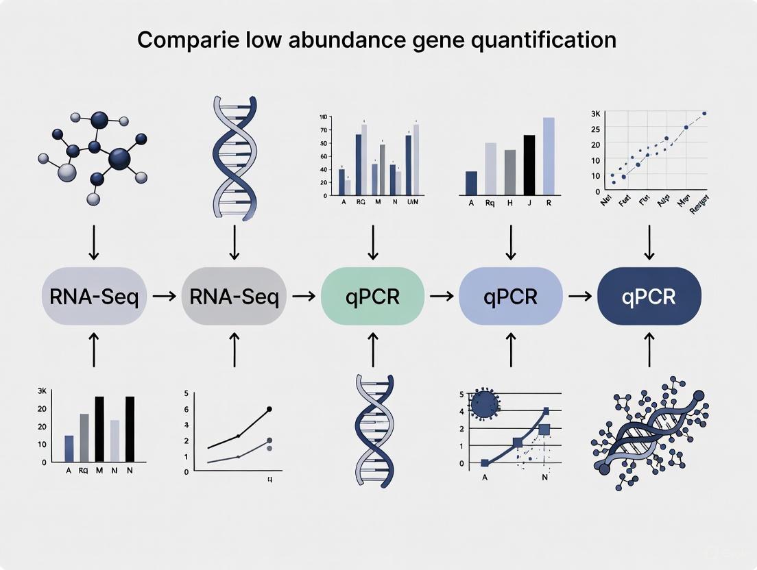
Low Abundance Gene Quantification: RNA-Seq vs qPCR - A Researcher's Guide to Accuracy and Application
Accurately quantifying low-abundance transcripts is critical for advancing research in drug development, biomarker discovery, and understanding complex disease mechanisms.
Low Abundance Gene Quantification: RNA-Seq vs qPCR - A Researcher's Guide to Accuracy and Application
Abstract
Accurately quantifying low-abundance transcripts is critical for advancing research in drug development, biomarker discovery, and understanding complex disease mechanisms. This article provides a comprehensive guide for researchers and scientists comparing two cornerstone technologies: quantitative PCR (qPCR) and RNA Sequencing (RNA-Seq). We explore the foundational principles of each method, detail their specific applications and methodologies for challenging targets, address key troubleshooting and optimization strategies to mitigate technical artifacts and enhance sensitivity, and present a direct comparative analysis to guide technology selection for validation studies. By synthesizing current evidence and best practices, this review empowers professionals to make informed decisions that enhance the reliability and impact of their gene expression studies.
The Critical Challenge of Low-Abundance Transcripts in Biomedical Research
Low-abundance genes, while challenging to detect and quantify, play disproportionately significant roles in cellular regulation, disease mechanisms, and therapeutic targeting. Their expression profiles offer critical insights into pathological states and represent promising biomarker candidates for early disease detection. This technical review examines the comparative capabilities of RNA-Seq and qPCR for quantifying low-abundance transcripts, evaluating methodological precision, experimental requirements, and analytical considerations. We synthesize evidence from recent studies that benchmark these technologies across various applications, from single-cell analysis to clinical biomarker discovery. The findings indicate that method selection profoundly impacts detection reliability for low-expression genes, with implications for research validity and clinical translation. We provide evidence-based guidelines for optimizing experimental designs to accurately capture the biological significance of these molecularly elusive yet functionally critical genetic elements.
Low-abundance transcripts, often expressed at mere copies per cell, constitute a substantial portion of the transcriptome with outsized functional importance. These genes frequently encode key regulatory molecules including transcription factors, signaling receptors, and non-coding RNAs that govern critical cellular processes. Their expression patterns provide sensitive indicators of pathological states, yet their quantification presents substantial technical challenges due to their low expression levels and susceptibility to technical noise.
The detection of low-abundance genes has profound implications for understanding disease mechanisms. In oncology, minority alleles and mutation-bearing transcripts present at low frequencies can signal emergent treatment resistance [1]. In immunology, differential expression of HLA genes at low levels significantly modifies disease outcomes for HIV, autoimmune conditions, and cancer [2]. Furthermore, single-cell RNA sequencing (scRNA-seq) studies reveal that low-abundance transcripts enable fine discrimination of cell states and types, but require specialized approaches for reliable quantification [3].
Accurate measurement of these genes is technically demanding. Research indicates that low-abundance RNAs exhibit high missing rates in sequencing data - approximately 90% at single-cell level and 40% even in pseudo-bulk analyses [3]. This detection failure stems from methodological limitations rather than biological absence, underscoring the critical importance of selecting appropriate quantification strategies for research and clinical applications.
Technical Challenges in Quantifying Low-Abundance Genes
Fundamental Detection Limitations
Quantifying low-abundance genes presents distinct challenges that differ significantly from measuring moderately or highly expressed transcripts. The dominant source of error for low-abundance RNAs in RNA-Seq is Poisson sampling noise due to finite read depth [4]. This stochastic sampling variation means that insufficient reads map to these transcripts for reliable quantification, leading to high measurement variability and reduced statistical power for detecting differential expression.
The relationship between gene expression level and measurement precision demonstrates that lower expression correlates strongly with higher relative error [4]. While highly expressed transcripts can be measured with relative errors of 20% or less, only 41% of all transcript targets achieve this precision level across technical replicates. For the 41% most strongly expressed transcripts, 84% can be measured reliably, indicating a strong expression-level bias in quantification accuracy.
Platform-Specific Constraints
RNA-Seq Limitations
In RNA-Seq, increased sequencing depths yield diminishing returns for low-abundance transcript detection. While 100 million reads generally detect most expressed genes, approximately 500 million reads are needed to accurately quantify 72% of gene expression levels [4]. Beyond this point, additional sequencing provides minimal gains for low-abundance targets because high-abundance transcripts dominate sequencing capacity - 7% of abundant transcripts consume over 75% of all read alignments [4]. Extrapolation studies suggest a maximum of 60% of all known transcripts can be measured reliably even at theoretically impractical depths of 10 billion reads [4].
Additional RNA-Seq complications include:
- Normalization artifacts: RPKM and TPM assume constant total RNA content across samples, causing distortion in low-abundance transcript comparisons when overall RNA composition differs [4]
- Library preparation biases: PCR amplification preferentially amplifies certain sequences, while GC content and sequence composition affect reverse transcription efficiency [5]
- RNA degradation effects: Degradation underrepresents longer transcripts and overrepresents 3' ends, disproportionately affecting already scarce targets [5]
Single-Cell RNA-Seq Considerations
scRNA-seq exhibits particularly pronounced challenges for low-abundance genes, with average missing rates of 90% at the single-cell level [3]. This "dropout" phenomenon results from technical factors including low mRNA input, capture efficiency, amplification bias, and limited sequencing depth. Precision and accuracy are generally low at single-cell resolution, with reproducibility strongly influenced by cell count and RNA quality [3].
Comparative Analysis of Quantification Platforms
RNA-Seq Versus qPCR: Performance Benchmarks
Multiple studies have systematically compared RNA-Seq and qPCR for gene expression quantification, revealing method-specific strengths and limitations particularly relevant for low-abundance genes.
Table 1: RNA-Seq vs. qPCR Performance Characteristics
| Parameter | RNA-Seq | qPCR |
|---|---|---|
| Dynamic Range | Higher dynamic range [5] | Limited dynamic range |
| Sensitivity | Can detect low-abundance transcripts but with precision limitations [4] | Highly sensitive and specific, suitable for validating RNA-seq results [5] |
| Throughput | Genome-wide, unbiased approach [5] | Practical for small-to-medium gene sets [6] |
| Low-Abundance Precision | Struggles with accurate quantification of low-abundance transcripts [4] | High precision for targeted low-abundance genes [2] |
| Multiplexing Capacity | Essentially unlimited | Limited to few targets per reaction without extensive optimization [6] |
| Novel Feature Discovery | Detects novel transcripts, isoforms, and variants [5] | Restricted to known sequences |
A comprehensive benchmarking study using the MAQCA and MAQCB reference samples demonstrated high gene expression correlations between RNA-Seq and qPCR data across five processing workflows (Pearson correlation R² = 0.798-0.845) [7]. However, when comparing fold changes between samples, approximately 15-19% of genes showed inconsistent differential expression calls between RNA-Seq and qPCR [7]. These inconsistent genes were typically shorter, had fewer exons, and were lower expressed compared to genes with consistent expression measurements [7].
For HLA gene quantification, a specialized comparison revealed only moderate correlation between qPCR and RNA-Seq (0.2 ≤ rho ≤ 0.53 for HLA-A, -B, and -C) despite using HLA-tailored bioinformatic pipelines [2]. This highlights the particular challenges in quantifying polymorphic low-abundance genes.
Microarray Capabilities for Low-Abundance Transcripts
Despite being an older technology, microarrays demonstrate particular advantages for low-abundance RNA profiling. In contrast to RNA-Seq, where high-abundance RNAs consume disproportionate sequencing capacity, microarray hybridization minimizes cross-target competition - the presence of unrelated high-abundance sequences little affects detection of poorly-expressed transcripts [4].
This technical difference translates to practical sensitivity advantages. For long non-coding RNAs, microarrays routinely detect 7,000-12,000 species compared to only 1,000-4,000 detectable by RNA-Seq even with >120 million reads [4]. This superior sensitivity for low-abundance targets has led researchers to select microarrays over RNA-Seq in clinical studies where detection sensitivity is paramount [4].
Emerging Platforms and Methodological Innovations
Digital PCR
Digital PCR (dPCR) provides absolute quantification of nucleic acids by partitioning reactions into thousands of individual amplifications. Recent comparisons of droplet-based (ddPCR) and nanoplate-based (ndPCR) systems show both platforms achieve high precision across most analyses with similar detection and quantification limits [8]. dPCR demonstrates particular advantages for quantifying targets present in low abundances and is less susceptible to inhibition from sample matrix effects compared to qPCR [8].
Key dPCR performance characteristics:
- Limit of Detection: 0.17 copies/µL for ddPCR, 0.39 copies/µL for ndPCR [8]
- Limit of Quantification: 4.26 copies/µL for ddPCR, 1.35 copies/µL for ndPCR [8]
- Precision: Coefficient of variation 6-13% for ddPCR, 7-11% for ndPCR [8]
Targeted RNA-Seq Approaches
Targeted sequencing methods focusing on specific gene subsets address some limitations of whole transcriptome RNA-Seq. However, primer amplification-based targeted approaches face development challenges and typically content limits of 500-1000 genes [6]. These methods still require RNA extraction and reverse transcription but can enhance sensitivity for predetermined target sets.
Single-Cell and Single-Nucleus RNA-Seq
scRNA-seq and snRNA-seq enable resolution of low-abundance transcripts at cellular resolution but require specialized experimental designs. Evidence-based guidelines recommend at least 500 cells per cell type per individual to achieve reliable quantification [3]. The signal-to-noise ratio is a key metric for identifying reproducible differentially expressed genes in single-cell studies [3].
Experimental Design Considerations for Low-Abundance Gene Analysis
RNA-Seq Optimization Strategies
Table 2: RNA-Seq Experimental Design Recommendations
| Parameter | Recommendation | Impact on Low-Abundance Detection |
|---|---|---|
| Sequencing Depth | 20-30 million reads for standard applications; >100 million for rare transcripts [5] | Higher depth improves low-abundance detection but with diminishing returns [4] |
| Biological Replicates | Minimum 3 per condition [5] | Critical for statistical power to detect differential expression of low-abundance genes |
| RNA Quality | RNA Integrity Number (RIN) assessment; DNase treatment [5] | Prevents degradation artifacts and genomic DNA contamination |
| Library Preparation | Consistent methods across samples; PCR-free options available [5] | Reduces technical variability and amplification biases |
| Multiplexing | Unique barcodes for sample pooling [5] | Enables cost-effective deeper sequencing |
Method Selection Guidelines
Research objectives should drive platform selection for low-abundance gene studies:
- Hypothesis-free discovery: RNA-Seq provides unbiased genome-wide coverage but requires validation of low-abundance findings [5]
- Targeted quantification of known genes: qPCR or dPCR offer superior sensitivity and precision for defined gene sets [2] [8]
- Clinical biomarker applications: Microarrays or targeted RNA-Seq may provide better sensitivity for specific low-abundance targets [4]
- Single-cell resolution: scRNA-seq requires specialized normalization and sufficient cell numbers (≥500 per cell type) [3]
For RNA-Seq data processing, quantification methods show varying performance for low-abundance genes. Studies evaluating isoform expression quantification found Net-RSTQ and eXpress provide more consistent results across platforms compared to Cufflinks, RSEM, or Kallisto [9].
Applications in Disease Research and Biomarker Development
Cancer Genomics
Low-abundance gene mutations serve as critical biomarkers in oncology. Detection of EGFR mutations (L747_S752 del, G719A, T790M) present at frequencies as low as 1% enables identification of resistant subclones in non-small cell lung cancer [1]. Novel enrichment methods like combined polymerase and ligase chain reaction can selectively amplify these minority alleles from background wild-type sequences, dramatically improving detection sensitivity [1].
Immunology and Infectious Disease
HLA expression levels quantitatively influence disease outcomes, with low-level variations significantly modifying HIV progression, autoimmune risk, and viral control [2]. These effects persist despite modest expression differences, highlighting the functional importance of precise low-abundance quantification. For example, higher HLA-C expression associates with better HIV control, while elevated HLA-A expression impairs it [2].
Neurological Disorders
Single-nucleus RNA sequencing of brain tissues reveals cell-type-specific low-abundance transcripts with implications for neurological diseases. However, studies consistently show inadequate cell numbers for specific neuronal subtypes in even large-scale datasets, limiting detection power for rare transcripts in functionally important cell populations [3].
Accurate quantification of low-abundance genes remains technically challenging but biologically essential. Method selection should be guided by research objectives, with RNA-Seq providing discovery potential and targeted approaches (qPCR, dPCR) offering validation rigor. No single platform currently optimizes all parameters - sensitivity, precision, throughput, and cost must be balanced based on experimental needs.
Emerging methodologies including molecular indexing, unique molecular identifiers, and partitioned amplification strategies show promise for enhancing low-abundance detection. Additionally, cross-platform validation remains critical for studies where low-abundance genes drive primary conclusions. As evidence accumulates regarding the functional significance of precisely tuned low-level gene expression, methodological rigor in quantifying these molecularly elusive targets becomes increasingly fundamental to biological insight and clinical translation.
Diagram: Experimental Workflow for Low-Abundance Gene Analysis
Research Reagent Solutions
Table 3: Essential Reagents for Low-Abundance Gene Analysis
| Reagent Category | Specific Examples | Function & Importance |
|---|---|---|
| RNA Stabilization | RNAlater, PAXgene | Preserves RNA integrity, critical for low-abundance targets |
| Reverse Transcriptase | SuperScript IV, LunaScript | High efficiency reverse transcription maximizes cDNA yield |
| Target Enrichment | NEBNext rRNA Depletion Kit | Removes abundant ribosomal RNAs, enhancing detection sensitivity |
| Library Preparation | SMARTer Stranded, KAPA HyperPrep | Minimizes bias in RNA-Seq library construction |
| Unique Molecular Identifiers | IDT UMI Adapters | Distinguishes biological signal from amplification noise |
| Digital PCR Reagents | ddPCR EvaGreen Supermix, QIAcuity PCR Master Mix | Enables absolute quantification of rare targets |
| Nuclease-Free Water | Ambion Nuclease-Free Water | Prevents RNA degradation during experimental procedures |
The accurate quantification of gene expression is a cornerstone of modern molecular biology, with critical applications in basic research, clinical diagnostics, and drug development. Among the various technologies available, quantitative PCR (qPCR) and RNA Sequencing (RNA-seq) have emerged as two principal methods for measuring transcript abundance. While qPCR is widely recognized for its sensitivity, accuracy, and low cost, RNA-seq provides a comprehensive, genome-wide view of the transcriptome [10]. The selection between these methods becomes particularly crucial when investigating low-abundance transcripts, which often include key regulatory genes, transcription factors, and non-coding RNAs. Understanding the fundamental principles, technical requirements, and limitations of each technology is essential for designing robust experiments and generating reliable data, especially when quantifying rare transcripts that may drive important biological processes or serve as biomarkers in disease states.
Fundamental Principles of qPCR
Core Mechanism and Quantification
Quantitative PCR (qPCR), also known as real-time PCR, is a method for detecting and quantifying specific DNA sequences in real-time as amplification occurs. The fundamental principle relies on monitoring the fluorescence emitted during each PCR cycle, which is directly proportional to the amount of amplified product. The key quantification parameter is the quantification cycle (Cq), previously known as Ct or Cp value, which represents the PCR cycle number at which the fluorescence signal crosses a predetermined threshold [11]. This threshold is set within the exponential phase of amplification, where the reaction efficiency is optimal. The Cq value is inversely correlated with the initial template concentration: a lower Cq indicates a higher starting amount of the target sequence, while a higher Cq corresponds to a lower initial abundance [11]. According to the MIQE guidelines, Cq values above 30-35 are often considered unreliable for quantification due to poor reproducibility, presenting a significant challenge for low-abundance transcripts [12].
Detection Chemistry and Signal Generation
qPCR utilizes various fluorescence-based detection chemistries, with the two most common being:
- DNA-binding dyes: Non-specific intercalating dyes like SYBR Green that fluoresce when bound to double-stranded DNA.
- Fluorogenic probes: Sequence-specific probes such as TaqMan probes that utilize fluorescence resonance energy transfer and provide higher specificity through an oligonucleotide probe with a reporter and quencher dye.
The fluorescence signal is captured by specialized instruments during each amplification cycle, generating amplification plots that track fluorescence versus cycle number. Proper baseline correction and threshold setting are critical for accurate Cq determination, as incorrect settings can significantly alter calculated Cq values and subsequent quantification [11].
Quantitative Analysis Methods
qPCR data can be analyzed using two primary quantification strategies:
- Absolute quantification: Determines the exact copy number of the target sequence by comparison to a standard curve of known concentrations.
- Relative quantification: Compares the expression level of a target gene between different samples after normalization to one or more reference genes. The efficiency-adjusted model (Pfaffl method) accounts for variations in amplification efficiency between different targets, providing more accurate results than methods that assume 100% efficiency [11].
Diagram 1: qPCR Workflow and Quantification Principle. This diagram illustrates the multi-step process from RNA sample to quantitative results, highlighting the critical role of proper threshold setting and baseline correction during data analysis.
Fundamental Principles of RNA-Seq
High-Throughput Sequencing Approach
RNA-seq is a comprehensive, high-throughput method that utilizes next-generation sequencing technologies to profile the entire transcriptome. Unlike qPCR, which targets specific known sequences, RNA-seq provides an unbiased view of RNA populations without prior knowledge of gene sequences. The core principle involves converting RNA populations into a library of cDNA fragments with adapters attached to one or both ends, followed by sequencing these fragments in a massively parallel manner to generate short reads [13]. These reads are then mapped to a reference genome or transcriptome, and the expression level for each gene is quantified based on the number of reads that map to its exonic regions. Normalization methods such as FPKM or TPM account for gene length and sequencing depth, enabling comparisons between genes within a sample and across different samples.
Bioinformatics Processing and Challenges
The analysis of RNA-seq data involves multiple computational steps that present unique challenges, particularly for complex gene families like the Human Leukocyte Antigen system. Standard alignment approaches that rely on a single reference genome often fail to accurately represent the extreme polymorphism and sequence similarity between paralogs at HLA loci, leading to misalignment and biased quantification [2]. This has motivated the development of specialized computational pipelines that account for known HLA diversity during the alignment step, significantly improving expression estimation accuracy for these challenging genes [2]. Additional technical biases in RNA-seq can arise from batch effects, library preparation protocols, and GC content variations, which must be carefully controlled during experimental design and data analysis ['t Hoen et al. 2013 as cited in citation:1].
Visualization and Interpretation
Advanced visualization approaches are essential for interpreting the complex data generated by RNA-seq, particularly for analyzing transcript isoforms and splice variants. While traditional tools like the Integrative Genomics Viewer display reads stacked onto a genomic reference, graph-based visualization methods offer complementary insights into transcript diversity [13]. These methods represent RNA-seq assemblies as networks where nodes correspond to reads and edges represent sequence similarity, enabling better appreciation of complex transcript topology in 3D space. This approach is particularly valuable for identifying issues in assembly, detecting repetitive sequences within transcripts, and characterizing splice variants that might be missed by reference-based methods [13].
Diagram 2: RNA-seq Workflow and Analysis Pipeline. This diagram outlines the key steps in RNA-seq processing, highlighting both standard procedures and specialized approaches needed for accurate analysis of complex gene families and transcript isoforms.
Comparative Analysis: qPCR vs. RNA-Seq
Technical Performance and Applications
Understanding the relative strengths and limitations of qPCR and RNA-seq is essential for selecting the appropriate method for specific research applications, particularly when investigating low-abundance transcripts.
Table 1: Comparative Analysis of qPCR and RNA-Seq Technologies
| Parameter | qPCR | RNA-Seq |
|---|---|---|
| Sensitivity | High sensitivity for known targets; limited for Cq >30-35 [12] | Variable sensitivity; requires sufficient sequencing depth for low-abundance targets |
| Multiplexing Capacity | Traditionally limited to 4-6 targets; advanced methods like CCMA enable higher multiplexing [14] | Genome-wide; profiles all transcripts simultaneously |
| Throughput | Low to medium throughput; limited by reaction number | High throughput; sequences millions of fragments in parallel |
| Dynamic Range | ~7-8 log range; suitable for abundant and moderate targets | ~5 log range; limited for very low and very high expression |
| Target Requirement | Requires prior sequence knowledge for primer/probe design | No prior knowledge needed; enables novel transcript discovery |
| Quantitative Accuracy | High accuracy for proper targets; gold standard for validation [10] | Moderate accuracy; subject to various technical biases |
| Cost per Sample | Low to moderate | Moderate to high, especially with deep sequencing |
| Turnaround Time | Fast (<2 hours after cDNA synthesis) [12] | Moderate to long (days to weeks including data analysis) |
| Data Complexity | Simple; direct Cq interpretation | Complex; requires advanced bioinformatics expertise |
Correlation Between Technologies
Direct comparisons between qPCR and RNA-seq have revealed important insights about their correlation and appropriate applications. A 2023 study analyzing HLA class I gene expression found only moderate correlation between expression estimates from qPCR and RNA-seq, with correlation coefficients (rho) ranging from 0.2 to 0.53 for HLA-A, -B, and -C genes [2]. This moderate correlation highlights the technical challenges in comparing results across these different platforms, including differences in what each method actually measures (specific amplicons vs. overall gene representation) and the various normalization strategies employed. However, RNA-seq has been shown to accurately estimate gene expression means compared to qPCR when appropriate bioinformatic approaches are used, though measures of expression variability may differ significantly, particularly across different environmental conditions [10].
Advanced Methodologies for Low-Abundance Transcript Detection
Enhancing qPCR Sensitivity
The inherent sensitivity limitation of conventional qPCR for low-abundance transcripts (Cq >30) has prompted the development of innovative pre-amplification strategies. The STALARD method provides a targeted approach that selectively amplifies polyadenylated transcripts sharing a known 5'-end sequence before quantification [12]. This two-step process involves reverse transcription using a gene-specific primer-tailed oligo(dT) primer, followed by limited-cycle PCR using only the gene-specific primer. This approach minimizes amplification bias caused by differential primer efficiency when comparing similar transcripts, a common challenge in isoform-specific qPCR. When applied to the low-abundance VIN3 transcript in Arabidopsis thaliana, STALARD successfully amplified the target to reliably quantifiable levels that conventional RT-qPCR failed to detect [12].
Reference Gene Selection Strategies
Accurate normalization is crucial for both qPCR and RNA-seq data interpretation, particularly for low-abundance targets where technical variation can have substantial effects. Traditional housekeeping genes often exhibit unexpectedly high expression variance across different conditions, compromising their utility as normalizers [10]. RNA-seq enables a whole-transcriptome approach to reference gene selection, identifying stably expressed genes that outperform classical housekeeping genes. Recent research demonstrates that a stable combination of non-stable genes can outperform single reference genes for qPCR data normalization [15]. This approach identifies a fixed number of genes whose individual expressions balance each other across experimental conditions, providing more robust normalization than single reference genes.
Table 2: Research Reagent Solutions for Gene Expression Analysis
| Reagent/Tool | Function | Application Context |
|---|---|---|
| TaqPath ProAmp Master Mix | Enzyme mix for robust amplification | qPCR reactions, including CCMA [14] |
| HiScript IV 1st Strand cDNA Synthesis Kit | High-efficiency reverse transcription | cDNA synthesis for both qPCR and RNA-seq library prep [12] |
| AMPure XP SPRI magnetic beads | Nucleic acid purification and size selection | Library cleanup for RNA-seq; PCR product purification [14] [12] |
| Gene-Specific Primers with Tailored Thermodynamics | Target-specific amplification | STALARD method for low-abundance transcripts [12] |
| HLA-Tailored Bioinformatics Pipelines | Accurate alignment of polymorphic sequences | RNA-seq analysis of extreme polymorphism at HLA loci [2] |
| Graphia Professional | Network-based visualization of transcript assemblies | RNA-seq data interpretation and isoform analysis [13] |
Experimental Design Considerations
Optimizing experimental design is particularly critical when studying low-abundance transcripts. For qPCR, the MIQE guidelines provide a framework for ensuring data quality, including proper validation of amplification efficiency, determination of the lower limit of quantification, and selection of appropriate reference genes [11] [12]. When using RNA-seq for low-abundance targets, sufficient sequencing depth must be prioritized to ensure adequate coverage of rare transcripts. Specialized library preparation methods, such as those incorporating targeted enrichment or ribosomal RNA depletion, can significantly improve detection of low-abundance targets. Additionally, experimental conditions significantly impact expression variability measures, suggesting that reference genes should be selected using transcriptome data that either specifically matches the study conditions or covers a broad range of biological and environmental diversity [10].
Both qPCR and RNA-seq offer powerful but distinct approaches to gene expression quantification, with complementary strengths that can be strategically leveraged in research and diagnostic applications. qPCR remains the gold standard for sensitive, accurate quantification of known targets, while RNA-seq provides an unparalleled comprehensive view of the transcriptome. For low-abundance gene quantification, methodological innovations like STALARD for qPCR and specialized bioinformatic pipelines for RNA-seq are pushing the boundaries of what these technologies can detect. The selection between these methods should be guided by the specific research question, target abundance, required throughput, and available resources. As both technologies continue to evolve, their synergistic application—using RNA-seq for discovery and qPCR for validation—will continue to drive advances in our understanding of gene regulation in health and disease.
The accurate quantification of gene expression, especially for low-abundance transcripts, is a cornerstone of modern molecular research in fields like drug development and clinical diagnostics. The choice of technology directly influences the biological conclusions that can be drawn. This guide provides an in-depth technical comparison between two cornerstone technologies—quantitative PCR (qPCR) and RNA Sequencing (RNA-Seq)—focusing on their sensitivity, dynamic range, and their consequent impact on reliable detection.
While RNA-seq is often considered the gold standard for whole-transcriptome analysis, its performance is highly dependent on sequencing depth and data processing workflows [16]. In contrast, qPCR remains the benchmark for sensitive and precise quantification of specific targets [17] [18]. Understanding the technical boundaries of each method is essential for designing robust experiments, interpreting data correctly, and ultimately, for making confident decisions in research and development.
Quantitative Face-Off: qPCR vs. RNA-Seq
Direct comparisons between qPCR and RNA-Seq reveal a complex performance landscape. The following tables summarize key comparative data and technical specifications.
Table 1: Performance Comparison of qPCR and RNA-Seq from Benchmarking Studies
| Metric | qPCR | RNA-Seq (Various Workflows) | Context & Notes |
|---|---|---|---|
| Expression Correlation | Benchmark | R²: 0.798 - 0.845 [16] | Pearson correlation to qPCR data for protein-coding genes. |
| Fold-Change Correlation | Benchmark | R²: 0.927 - 0.934 [16] | Correlation of MAQCA/MAQCB fold changes with qPCR. |
| Limit of Detection (LoD) | Well-defined (e.g., 0.003 pg/reaction) [19] | Read-count dependent (e.g., ~20 counts) [20] | RNA-Seq's LoD is probabilistic and varies with sequencing depth. |
| Limit of Quantification (LoQ) | Defined via standard stats (e.g., 0.03 pg/reaction) [19] | Not strictly defined | RNA-Seq quantification reliability is a gradient [20]. |
| Impact on Differential Expression | N/A | ~85% concordance with qPCR; 15% non-concordant genes [16] | Inconsistent genes are often lower expressed, smaller, with fewer exons [16]. |
Table 2: Technical Specifications and Dynamic Range
| Characteristic | qPCR | Bulk RNA-Seq |
|---|---|---|
| Theoretical Dynamic Range | Up to 9 logs [18] | Dependent on read depth [20] |
| Effective Dynamic Range | Constrained by sample quality, RT efficiency [18] | ~10,000 genes detectable at 10 million reads [20] |
| Key Precision Metric | Coefficient of Variation (CV) [18] | Reproducibility between replicates [16] [20] |
| Primary Normalization | Fundamental (absolute) or relative units [18] | Counts, TPM, FPKM [20] |
| Sensitivity for Low-Abundance Targets | Very high for targeted assays [19] [17] | Lower for genes with counts <20; improved with greater depth [20] |
Experimental Protocols for Benchmarking and Validation
Protocol for qPCR Assay Validation for Residual DNA Detection
This protocol, adapted from a study validating a test for residual Vero cell DNA in vaccines, outlines the key steps for establishing a sensitive and precise qPCR assay [19].
Step 1: Target and Primer/Probe Design
- Select a target sequence unique to the genome of interest, with a high copy number to enhance sensitivity. For Vero cells, a 172 bp tandem repeat sequence (~6.8x10^6 copies/haploid genome) was used [19].
- Design primers and probes for a short amplicon (e.g., 99 bp) to maximize amplification efficiency and robustness.
- Example Sequences: [19]
- Forward Primer:
5′-CTGCTCTGTGTTCTGTTAATTCATCTC-3′ - Reverse Primer:
5′-AAATATCCCTTTGCCAATTCCA-3′ - Probe:
5′-CCTTCAAGAAGCCTTTCGCTAAG-3′(FAM-labeled)
- Forward Primer:
Step 2: Reaction Setup
- Prepare a master mix containing buffer, enzymes, dNTPs, primers, and probe.
- Use a total reaction volume of 30 µL, adding 10 µL of DNA standard or sample.
- Critical: Use low-binding tubes and dedicated, nuclease-free pipettes to prevent adsorption and contamination.
Step 3: Thermal Cycling
- Perform amplification on a real-time PCR instrument with the following program:
- Initial Denaturation: 95°C for 10 minutes.
- 40 Cycles:
- Denaturation: 95°C for 15 seconds.
- Annealing/Extension: 60°C for 1 minute.
- Perform amplification on a real-time PCR instrument with the following program:
Step 4: Validation and Data Analysis
- Linearity and Range: Run a standard curve with a 10-fold dilution series of known DNA standards (e.g., from 0.3 fg/µL to 30 pg/µL). The assay should demonstrate a linear response across the intended range [19].
- Limit of Detection (LoD) & Quantification (LoQ): Determine LoD and LoQ using standard statistical methods. The described Vero DNA assay achieved an LoD of 0.003 pg/reaction and an LoQ of 0.03 pg/reaction [19].
- Precision: Assess repeatability by testing multiple replicates across different runs. The coefficient of variation (CV) should be within acceptable limits (e.g., 12.4%-18.3%) [19].
Protocol for Validating RNA-Seq Workflows Against qPCR
This protocol is based on benchmarking studies that compare different RNA-Seq analysis workflows against whole-transcriptome qPCR data [16].
Step 1: Sample Selection and RNA Preparation
- Use well-characterized RNA reference samples (e.g., MAQCA and MAQCB from the MAQC consortium) [16].
- Isolve high-quality total RNA using a method that preserves RNA integrity (RIN > 8.0). Treat samples with DNase to remove genomic DNA contamination.
Step 2: Data Generation
- qPCR Data: Perform whole-transcriptome qPCR for all protein-coding genes. Use multiple technical replicates to ensure precision [16] [18].
- RNA-Seq Data: Sequence the same RNA samples. Include biological and technical replicates. Process the data through multiple common workflows (e.g., STAR-HTSeq, Kallisto, Salmon) for comparison [16].
Step 3: Data Alignment and Normalization
- Align qPCR assays to the transcripts detected in the RNA-seq data to ensure comparability [16].
- For RNA-Seq, filter genes based on a minimal expression level (e.g., 0.1 TPM in all replicates) to avoid bias from low-expressed genes.
- Convert gene-level counts to TPM for correlation analysis with qPCR Cq values [16].
Step 4: Correlation and Discrepancy Analysis
- Expression Correlation: Calculate Pearson correlation between log-transformed RNA-Seq TPM values and normalized qPCR Cq values for each workflow [16].
- Fold-Change Correlation: Calculate gene expression fold changes between sample groups (e.g., MAQCA vs. MAQCB) and correlate these between RNA-Seq and qPCR [16].
- Identify Non-Concordant Genes: Classify genes based on their differential expression status between the two methods. Investigate the characteristics (e.g., expression level, gene length, exon count) of genes where the methods disagree [16].
Visualization of Workflows and Logical Relationships
The following diagrams illustrate the core workflows for qPCR and RNA-Seq, highlighting steps that critically influence their sensitivity and detection limits.
High-Sensitivity qPCR Assay Workflow
RNA-Seq Dynamic Range and Analysis
The Scientist's Toolkit: Essential Reagents and Materials
Table 3: Key Research Reagent Solutions for Sensitive Nucleic Acid Detection
| Item | Function/Description | Example Use Case |
|---|---|---|
| qPCR Assay Reagents | Pre-designed primer/probe sets, master mix containing polymerase, dNTPs, and optimized buffer. | Targeted quantification of specific genes or contaminants (e.g., residual host cell DNA) [19]. |
| RNA Stabilization Reagents | Reagents that immediately stabilize RNA at the point of sample collection to preserve integrity. | Critical for obtaining high RIN scores, ensuring accurate representation of the transcriptome. |
| Stranded mRNA Library Prep Kits | Kits for converting RNA into sequencing libraries, preserving strand-of-origin information. | Standard for bulk RNA-Seq; improves transcript annotation and detects antisense expression [21]. |
| Ultra-Low Input RNA Library Kits | Specialized kits using proprietary amplification (e.g., THOR technology) for minimal RNA input. | Enables RNA-Seq from single cells or rare samples by improving mRNA capture efficiency [22]. |
| Unique Molecular Identifiers (UMIs) | Short random nucleotide sequences used to tag individual RNA molecules before amplification. | Corrects for PCR amplification bias, improving quantification accuracy in both RNA-Seq and qPCR [22]. |
The accurate quantification of low-abundance genes is a critical challenge in molecular biology with profound implications for understanding immune regulation, cancer progression, and autoimmune diseases. This whitepaper examines the technical considerations of RNA-Seq versus qPCR methodologies for measuring lowly expressed genes, focusing on their application to key immunoregulatory molecules. We explore how precise measurement of low-expression genes, particularly in the major histocompatibility complex (MHC) and immune checkpoint pathways, provides crucial insights into disease mechanisms and therapeutic development. The content is framed within a broader thesis on methodological comparisons, providing researchers with actionable protocols, analytical frameworks, and practical tools for advancing research in immuno-genomics.
Gene expression profiling represents a fundamental tool for elucidating biological processes in health and disease. While highly expressed genes often dominate transcriptomic analyses, low-abundance transcripts frequently encode critical regulatory proteins with disproportionate biological impact. This is particularly evident in immunology, where precisely controlled expression of antigen presentation machinery, immune checkpoints, and regulatory molecules determines the balance between effective immunity and pathological autoimmunity [23] [24].
The technical challenges associated with accurately quantifying low-expression genes necessitate rigorous methodological comparisons. Traditional qPCR has served as the gold standard for targeted gene expression analysis due to its sensitivity and reproducibility. However, the emergence of high-throughput RNA-Seq offers comprehensive transcriptome-wide profiling capabilities. Understanding the strengths, limitations, and appropriate applications of each method is essential for researchers investigating the subtle gene expression changes that underlie immune dysregulation in cancer and autoimmune conditions [2].
Biological Significance of Lowly Expressed Genes
MHC Class I Expression in Immune Surveillance and Evasion
Major histocompatibility complex (MHC) class I molecules play an indispensable role in cellular immunity by presenting intracellular peptides to CD8+ T cells. Despite their critical function, these genes often demonstrate modest expression levels that are precisely regulated. Downregulation of MHC class I represents a common immune evasion mechanism across multiple cancer types, enabling tumors to escape CD8+ T cell-mediated destruction [24].
Table 1: Mechanisms of Low MHC I Expression in Cancer and Functional Consequences
| Mechanism | Molecular Basis | Functional Impact |
|---|---|---|
| Genetic Alterations | Gene deletion, loss-of-function mutations | Complete absence of antigen presentation machinery |
| Transcriptional Inhibition | Epigenetic silencing, transcription factor dysregulation | Reduced mRNA synthesis |
| Post-transcriptional Regulation | Reduced mRNA stability, microRNA targeting | Altered transcript abundance despite normal transcription |
| Protein Degradation | Enhanced ubiquitin-proteasome pathway activity | Reduced cell surface presentation despite adequate mRNA |
| Defective Trafficking | Disruption of endocytic recycling | Impaired antigen loading and surface expression |
The biological significance of MHC expression extends beyond absolute levels to include allele-specific variation. For instance, higher HLA-C expression associates with better control of HIV-1, while elevated HLA-A expression impairs HIV control [2]. These nuanced relationships highlight the importance of precise, allele-specific quantification methods that can resolve subtle expression differences with potentially opposing functional consequences.
Immune Checkpoint Regulation in Autoimmunity and Cancer
Immune checkpoint receptors and ligands represent another class of immunologically critical genes that often exhibit low to moderate expression levels under physiological conditions. These molecules, including PD-1, CTLA-4, and others, maintain self-tolerance by modulating T cell activation thresholds. In autoimmunity, insufficient checkpoint expression may contribute to loss of tolerance, while in cancer, excessive checkpoint expression in the tumor microenvironment facilitates immune evasion [23] [25].
Therapeutic manipulation of immune checkpoints through antibody-mediated blockade has revolutionized cancer treatment. However, response variability remains substantial, partly due to differences in baseline and induced expression of checkpoint genes. Similarly, in autoimmune diseases, expression quantitative trait loci (eQTLs) that modify checkpoint gene expression may influence disease susceptibility and progression [23].
Regulatory T Cell Function and Low-Abundance Transcripts
Regulatory T cells (Tregs) characterized by expression of the transcription factor FOXP3 maintain immune tolerance through multiple mechanisms. The FOXP3 gene itself is typically expressed at moderate levels, and its precise regulation is essential for immune homeostasis. Germline or transient deletion of FOXP3+ Tregs unleashes fatal multiorgan autoimmunity in mice, while humans with FOXP3 mutations develop IPEX syndrome [23].
Treg cells harbor a TCR repertoire skewed toward self-antigen recognition that overlaps substantially with autoreactive conventional CD4+ T cells. The functional specialization of these cells depends on the precise expression of key regulatory genes, many of which are low-abundance transcripts that present quantification challenges [23].
Technical Considerations for Quantifying Low-Abundance Genes
Methodological Comparison: qPCR versus RNA-Seq
The accurate quantification of low-expression genes presents distinct technical challenges that differ significantly between qPCR and RNA-Seq approaches. Understanding these methodological differences is essential for appropriate experimental design and data interpretation in immunology research.
Table 2: Comparison of qPCR and RNA-Seq for Low-Abundance Gene Quantification
| Parameter | qPCR | RNA-Seq |
|---|---|---|
| Sensitivity | High (can detect single copies) | Variable (depends on sequencing depth) |
| Dynamic Range | ~7-8 logs | ~5 logs for standard depths |
| Multiplexing Capacity | Low (typically 1-6 targets per reaction) | High (entire transcriptome) |
| Normalization Requirements | Critical, requires stable reference genes | Less dependent on single reference genes |
| Allele-Specific Resolution | Requires specialized assay design | Possible with appropriate bioinformatics |
| Sample Throughput | Moderate to high | Lower for standard workflows |
| Cost Per Sample | Low to moderate | Moderate to high |
| Technical Variability | Low (when optimized) | Moderate to high |
Normalization Strategies for Low-Abundance Targets
Normalization represents a particularly critical consideration when quantifying low-expression genes, as technical variability can disproportionately affect measurements. For qPCR, the use of multiple reference genes with demonstrated stability across experimental conditions is essential. Recent evidence suggests that combinations of non-stable genes may outperform traditional "housekeeping" genes when carefully selected [15].
For RNA-Seq, normalization approaches include DESeq2's median-of-ratios method, TPM (transcripts per million), and others that minimize the impact of technical artifacts such as GC content, transcript length, and library size. These methods generally show better performance for low-abundance genes compared to simple normalization approaches [26].
Correlation Between Methodologies
Direct comparisons between qPCR and RNA-Seq for immunologically relevant genes reveal moderate correlations that vary by target. In studies comparing HLA class I expression quantification, correlations between qPCR and RNA-Seq ranged from 0.2 to 0.53 for HLA-A, -B, and -C [2]. These findings highlight the challenges in comparing absolute expression values across platforms and the importance of platform-specific validation.
Diagram Title: Experimental Workflows for Gene Expression Quantification
Detailed Experimental Protocols
qPCR Protocol for Low-Abundance Immune Genes
Sample Preparation and RNA Isolation
- Starting Material: 10-200 ng of high-quality RNA extracted using RNeasy or AllPrep DNA/RNA kits (Qiagen)
- RNA Quality Assessment: Qubit 2.0 for quantification, NanoDrop for purity (A260/280 ~2.0), TapeStation 4200 for RIN >7.0
- DNAse Treatment: Mandatory to remove genomic DNA contamination using RNAse-free DNAse
cDNA Synthesis
- Reverse Transcription: Use random hexamers and oligo-dT primers for comprehensive coverage
- Reaction Conditions: 25°C for 10 minutes, 37°C for 120 minutes, 85°C for 5 minutes
- Enzyme Selection: High-efficiency reverse transcriptase with RNase inhibitor
qPCR Reaction Setup
- Reaction Volume: 10-20 µl containing 1-10 ng cDNA equivalent
- Chemistry: SYBR Green or TaqMan probes depending on specificity requirements
- Primer Design: Amplicons of 70-150 bp spanning exon-exon junctions
- Validation: Efficiency curves (90-110%) with R² >0.98 for each assay
Data Analysis
- Quality Control: Melting curve analysis for SYBR Green, amplification efficiency validation
- Normalization: Minimum of three reference genes selected by stability algorithms (geNorm, NormFinder)
- Statistical Analysis: ΔΔCt method for relative quantification with technical replicates
RNA-Seq Protocol for Comprehensive Immune Profiling
Library Preparation
- RNA Input: 10-200 ng total RNA (quality and quantity similar to qPCR requirements)
- Library Kit: TruSeq stranded mRNA kit (Illumina) for FF samples; SureSelect XTHS2 for FFPE
- Capture Method: Whole exome capture using SureSelect Human All Exon V7 + UTR (Agilent)
Sequencing and Quality Control
- Platform: NovaSeq 6000 (Illumina)
- Depth: Minimum 30 million reads per sample, 50+ million recommended for low-abundance targets
- Quality Metrics: Q30 >90%, PF >80%, assessment via FastQC and FastqScreen
Bioinformatic Processing
- Alignment: STAR aligner v2.4.2 against hg38 reference genome
- Deduplication: UMI-based deduplication with umitools for accurate quantification
- Quantification: Kallisto v0.44.0 with Ensembl GRCH37 reference transcriptome
- Batch Correction: ComBat or similar methods for technical batch effect correction
Special Considerations for HLA Genes
- Reference Selection: Use customized references incorporating HLA allele diversity
- Alignment Parameters: Adjust to accommodate high polymorphism regions
- Expression Estimation: HLA-tailored pipelines (e.g., OptiType, HLA-HD) for allele-specific quantification
The Scientist's Toolkit: Essential Research Reagents and Solutions
Table 3: Key Research Reagent Solutions for Low-Abundance Gene Analysis
| Reagent/Category | Specific Examples | Function/Application |
|---|---|---|
| RNA Extraction Kits | RNeasy Mini Kit, AllPrep DNA/RNA FFPE Kit | Maintain RNA integrity, especially from challenging samples like FFPE |
| Reverse Transcriptases | High Capacity cDNA Reverse Transcription Kit | Ensure efficient cDNA synthesis from low-input RNA |
| qPCR Master Mixes | SYBR Green Master Mix, TaqMan Gene Expression Master Mix | Provide sensitive, specific detection with minimal background |
| RNA-Seq Library Prep | TruSeq Stranded mRNA, SureSelect XTHS2 RNA | Preserve strand information, efficient library construction from low-quality RNA |
| Target Enrichment | SureSelect Human All Exon V7 + UTR | Comprehensive coverage of coding transcriptome including immune genes |
| Reference Materials | ERCC RNA Spike-In Mixes | Monitor technical variability, validate assay sensitivity |
| Quality Control Tools | Qubit RNA HS Assay, TapeStation High Sensitivity RNA ScreenTape | Accurate quantification and integrity assessment of limited samples |
| Bioinformatic Tools | STAR aligner, Kallisto, DESeq2, HLA-HD | Specialized analysis of immune genes, allele-specific expression |
Signaling Pathways Linking Low Expression to Disease Phenotypes
Diagram Title: Mechanisms of Immune Evasion in Cancer
The precise quantification of low-abundance genes encoding immunologically critical molecules represents both a technical challenge and a scientific opportunity. As methodologies continue to evolve, the research community must maintain rigorous standards for assay validation, normalization, and data interpretation. The complementary strengths of qPCR and RNA-Seq suggest that a hybrid approach—using RNA-Seq for discovery and qPCR for targeted validation—may offer the most robust framework for investigating the biological significance of low-expression genes in immune regulation.
Future methodological developments, including single-cell sequencing, digital PCR, and emerging third-generation sequencing technologies, promise to enhance our ability to resolve subtle expression differences in biologically critical low-abundance transcripts. These technical advances, coupled with improved bioinformatic tools for allele-specific and isoform-aware quantification, will deepen our understanding of how precise gene expression control shapes immune responses in health and disease.
Methodologies in Practice: Protocols for qPCR and RNA-Seq on Low-Abundance Targets
In the context of gene expression analysis, the accurate quantification of low-abundance transcripts presents a significant technical challenge. While next-generation sequencing (RNA-Seq) provides a comprehensive, hypothesis-free view of the transcriptome, quantitative PCR (qPCR) remains the gold standard for sensitive, specific, and cost-effective validation and targeted quantification of gene expression [27]. The reliability of qPCR data, especially for low-copy-number RNAs, is heavily dependent on rigorous experimental design, execution, and analysis. This guide details the core best practices for qPCR, framed within a modern research workflow that often uses RNA-Seq for discovery and qPCR for confirmation, with a particular emphasis on achieving rigor and reproducibility in quantifying challenging targets.
Assay Design for Specificity and Sensitivity
The foundation of a successful qPCR experiment is a well-designed assay. For low-abundance transcripts, maximizing sensitivity and specificity is paramount to distinguish a true signal from background noise.
Design Principles: Assays should be designed to be 70-200 base pairs in length to ensure efficient amplification [28]. Primers must be specific, ideally spanning an exon-exon junction to avoid amplification of genomic DNA contamination. The use of in silico tools like Primer-BLAST is recommended to verify specificity, and this should be confirmed empirically by sequencing the PCR product and checking for a single peak in the melting curve analysis [28].
Variant-Specific Quantification: When quantifying specific splice variants or isoforms, careful assay design is critical. Researchers should identify the NCBI RefSeq transcript accession number of the specific variant and use this to search for a predesigned assay that detects only that variant. If none are available, a custom assay must be designed to target the unique exon-exon boundary of the isoform [27].
Advanced Methods for Low-Abundance Targets: Conventional RT-qPCR often has limited sensitivity, with quantification cycle (Cq) values above 30-35 being considered unreliable [12]. To overcome this, novel methods like STALARD (Selective Target Amplification for Low-Abundance RNA Detection) have been developed. This two-step RT-PCR method uses a gene-specific primer tailed with an oligo(dT) sequence for reverse transcription, followed by a limited-cycle PCR using only the gene-specific primer. This selectively amplifies polyadenylated transcripts sharing a known 5'-end sequence, dramatically improving the detection and reliable quantification of low-abundance isoforms [12].
The following diagram illustrates the STALARD workflow for enhancing detection of low-abundance RNAs.
The MIQE Guidelines 2.0: Ensuring Reproducibility
The Minimum Information for Publication of Quantitative Real-Time PCR Experiments (MIQE) guidelines establish a standardized framework to ensure the transparency, reproducibility, and reliability of qPCR data. A updated version, MIQE 2.0, has been released to reflect advances in technology and the complexities of contemporary qPCR applications [29].
Core Philosophy: The fundamental principle is that a transparent, clear, and comprehensive description of all experimental details is necessary to ensure the repeatability and reproducibility of qPCR results. This allows other scientists to critically evaluate and replicate the work [29].
Key Reporting Requirements: MIQE 2.0 offers clarified and streamlined reporting requirements. Crucially, researchers are encouraged to export and provide raw fluorescence data to enable independent re-analysis [29] [30]. The guidelines emphasize that quantification cycle (Cq) values must be converted into efficiency-corrected target quantities and should be reported with prediction intervals, not just as mean values [29]. Furthermore, the detection limit and dynamic range for each assay must be stated.
Assay Information Disclosure: For assay design, the guidelines require detailed disclosure. When using commercial assays like TaqMan, providing the unique Assay ID is typically sufficient, as this permanently links to a specific oligo sequence. For full compliance, the amplicon context sequence (the full PCR amplicon) can be provided, which is available in the Assay Information File or can be generated using the supplier's online tools [31].
Robust Normalization Strategies
Normalization is a critical step to control for technical variability introduced during sample processing, and the choice of strategy can dramatically impact data interpretation, particularly for subtle expression changes.
Reference Gene (RG) Normalization
This is the most common method, but it requires careful validation.
Gene Selection and Validation: The classical "housekeeping" genes (e.g., GAPDH, ACTB) are not universally stable and their expression can vary with experimental conditions and pathologies [32]. Therefore, RGs must be validated for the specific sample type and condition under investigation. Algorithms like geNorm and NormFinder are used to rank candidate RGs based on their expression stability across all samples [32] [28]. The MIQE guidelines recommend using at least two validated reference genes [28].
Stability in Canine Models: A 2025 study on canine gastrointestinal tissues highlighted that the most stable RGs were RPS5, RPL8, and HMBS. The study also noted that ribosomal protein genes (RPS5, RPL8) tend to be co-regulated, so using RGs from different functional classes is advisable [32].
Alternative Normalization Methods
For larger profiling studies, other methods can be more robust.
Global Mean (GM) Normalization: This method uses the geometric mean of the expression of a large set of genes (e.g., >55) as the normalization factor. In the canine study, the GM method was the best-performing strategy for reducing technical variability across tissues and disease states when a large set of genes was profiled [32].
Algorithm-Only Approaches: Methods like NORMA-Gene provide a normalization factor calculated via least squares regression using the expression data of at least five genes, without the need for predefined RGs. A 2025 study in sheep showed that NORMA-Gene was better at reducing variance in target gene expression than using traditional reference genes and requires fewer resources [28].
The table below summarizes and compares these key normalization methods.
Table 1: Comparison of qPCR Normalization Strategies
| Method | Principle | When to Use | Advantages | Disadvantages |
|---|---|---|---|---|
| Reference Genes (RG) | Normalizes to the geometric mean of 2+ validated, stably expressed endogenous genes. | Targeted gene expression studies with a small number of targets. | Well-established; MIQE-recommended; cost-effective for few targets. | Requires extensive validation; no universally stable RGs; potential for co-regulation. |
| Global Mean (GM) | Normalizes to the geometric mean of all expressed genes in the assay. | High-throughput studies profiling tens to hundreds of genes. | Highly robust; no need for RG validation; outperforms RG in some complex designs [32]. | Requires a large number of genes (>55) for reliability [32]. |
| NORMA-Gene | Algorithm calculates a normalization factor from all provided gene expression data. | Studies with at least 5 target genes; limited resources for RG validation. | Reduces variance effectively; no prior RG validation needed [28]. | Less familiar to some researchers; requires a minimum number of genes. |
Data Analysis and Adherence to FAIR Principles
Moving beyond the traditional 2−ΔΔCT method is key to improving statistical rigor and reproducibility.
Beyond 2−ΔΔCT: The widespread reliance on the 2−ΔΔCT method often overlooks variability in amplification efficiency, which can introduce significant bias. Analysis of Covariance (ANCOVA) is a flexible multivariable linear modeling approach that offers greater statistical power and robustness, as its P-values are not affected by variations in qPCR amplification efficiency [30].
Promoting Reproducibility with FAIR Data: To facilitate rigor, researchers are encouraged to share raw qPCR fluorescence data alongside detailed analysis scripts that take the raw data through to the final figures and statistical tests. Using general-purpose data repositories (e.g., figshare) and code repositories (e.g., GitHub) promotes transparency and allows others to verify and build upon the findings [30].
The Scientist's Toolkit: Research Reagent Solutions
Table 2: Essential Reagents and Kits for qPCR Workflows
| Item | Function | Example Application |
|---|---|---|
| TaqMan Gene Expression Assays | Predesigned, optimized probe-based assays for specific gene targets. | Gold-standard for target quantification and verification of RNA-Seq results [27]. |
| TaqMan Array Cards | 384-well microfluidic cards pre-spotted with assays for high-throughput profiling. | Profiling a focused panel of targets (12-384 genes) with minimal reagent use and a streamlined workflow [27]. |
| HiScript IV 1st Strand cDNA Synthesis Kit | High-efficiency reverse transcriptase for converting RNA to cDNA. | First-strand cDNA synthesis in the STALARD method and conventional RT-qPCR [12]. |
| SeqAmp DNA Polymerase | PCR enzyme used in pre-amplification protocols. | Limited-cycle, target-specific pre-amplification in the STALARD method [12]. |
| Oligo(dT) Primers & Gene-Specific Primers (GSP) | Primers for cDNA synthesis and PCR amplification. | Reverse transcription of polyadenylated RNA and specific amplification of target cDNA. |
qPCR and RNA-Seq: A Complementary Workflow
The choice between qPCR and RNA-Seq is not a binary one; they are highly complementary technologies.
Defining the Roles: RNA-Seq is ideal for discovery-based research, such as detecting novel transcripts, identifying differentially expressed genes without prior knowledge, and analyzing transcript isoform diversity [27]. qPCR is the preferred method for targeted, high-precision quantification, verification of RNA-Seq results, and follow-up studies on a defined panel of genes [27].
Integrated Workflow: In a robust experimental pipeline, qPCR is used both upstream and downstream of RNA-Seq. Upstream, qPCR can check cDNA library quality and integrity before costly sequencing [27]. Downstream, qPCR is the gold-standard method for validating key findings from the RNA-Seq dataset [27] [32]. This combined approach ensures data integrity from start to finish.
The following chart summarizes this complementary relationship and the standard workflow for low-abundance transcript analysis.
RNA sequencing (RNA-Seq) has revolutionized transcriptomics by enabling genome-wide quantification of RNA abundance with finer resolution, improved accuracy, and lower background noise compared to earlier methods like microarrays [33]. For researchers investigating low-abundance genes—a critical challenge in fields from Mendelian disease diagnostics to drug mechanism discovery—thoughtful experimental design is paramount. The choice between library preparation methods, sequencing depth, and coverage parameters directly determines an experiment's power to detect and quantify rare transcripts accurately. This guide provides a detailed framework for optimizing these key decisions, with a specific focus on challenges relevant to researchers comparing RNA-Seq to qPCR for low-abundance targets, where conventional RT-qPCR often reaches its sensitivity limits with quantification cycle (Cq) values above 30-35 considered unreliable [12].
Library Preparation Strategies: Poly-A Selection vs. Ribosomal RNA Depletion
The initial library preparation method fundamentally defines the transcriptome you will measure, influencing which RNA species are captured and how effectively sequencing reads are utilized [34].
Poly-A Enrichment: Targeted Capture of Mature mRNA
Mechanism: Poly(A) enrichment uses oligo(dT) primers attached to magnetic beads to capture RNA molecules containing poly(A) tails, primarily enriching for mature messenger RNAs (mRNAs) and many polyadenylated long non-coding RNAs (lncRNAs) [34] [35].
Key Advantages:
- High Exonic Read Yield: Delivers 70-71% usable exonic reads, dramatically focusing sequencing power on protein-coding regions [35].
- Cost Efficiency for mRNA Studies: Requires fewer total reads to achieve sufficient exonic coverage—approximately 13.5 million poly(A)-selected reads can detect as many genes as a typical microarray, compared to 35–65 million reads with ribodepletion methods [35] [36].
- Reduced Background: Effectively removes ribosomal RNA (rRNA) and other non-informative RNA classes without requiring species-specific probes [37] [38].
Critical Limitations:
- Incompatibility with Degraded Samples: Heavily relies on intact poly(A) tails, making it suboptimal for degraded RNA (e.g., FFPE samples) where it produces strong 3' bias [34] [35].
- Exclusion of Non-polyadenylated Transcripts: Completely misses important RNA species including replication-dependent histone mRNAs, many non-coding RNAs, and bacterial transcripts [34].
- Potential Capture Bias: May preferentially capture mRNAs with longer poly(A) tails, potentially misrepresenting transcript abundances [35].
Ribosomal RNA Depletion: Comprehensive Transcriptome Capture
Mechanism: rRNA depletion uses sequence-specific DNA probes that hybridize to cytosolic and mitochondrial rRNAs, which are then removed via RNase H digestion or affinity capture, preserving both polyadenylated and non-polyadenylated RNAs [34] [38].
Key Advantages:
- Broad Transcriptome Coverage: Captures diverse RNA types including pre-mRNA, lncRNAs, snoRNAs, and non-polyadenylated transcripts [35].
- Compatibility with Challenging Samples: Performs robustly with degraded, fragmented, or FFPE RNA where poly(A) tails may be compromised [34] [35].
- Uniform Coverage: Provides more even 5'-to-3' coverage across transcripts, valuable for splicing and isoform analysis [35].
Critical Limitations:
- Lower Efficiency for mRNA Studies: Yields significantly fewer exonic reads (22-46%), requiring 50-220% more sequencing to achieve equivalent exonic coverage compared to poly(A) selection [35].
- Species-Specific Probes: Requires matched depletion probes for different organisms; incomplete rRNA removal leads to wasted sequencing capacity [34] [35].
- Increased Bioinformatics Complexity: Higher proportions of intronic and intergenic reads increase data volume and analytical demands [35].
Decision Framework: Choosing the Optimal Method
Table 1: Method Selection Guide Based on Experimental Conditions
| Experimental Condition | Recommended Method | Rationale | Sequencing Depth Adjustment |
|---|---|---|---|
| High-quality eukaryotic RNA (RIN ≥8) | Poly-A selection | Maximizes exonic read yield and cost-efficiency for mRNA studies | Standard depth (20-60 million reads) typically sufficient |
| Degraded/FFPE samples | rRNA depletion | Does not rely on intact poly(A) tails; more resilient to fragmentation | May require 50-100% additional reads for equivalent exon coverage |
| Non-coding RNA discovery | rRNA depletion | Captures both polyA+ and non-polyA transcripts (lncRNAs, snoRNAs, etc.) | 30-100% more reads than poly-A studies due to diverse transcript types |
| Prokaryotic transcriptomics | rRNA depletion | Bacterial mRNAs lack poly(A) tails; poly-A capture is ineffective | Varies by species and depletion efficiency |
| Low-abundance mRNA quantification | Poly-A selection | Concentrates sequencing power on target molecules | May require elevated depth (>100 million reads) for rare transcripts |
| Alternative splicing analysis | rRNA depletion (paired-end) | Provides more uniform transcript coverage for isoform resolution | Higher depth (60-150 million reads) improves splice junction detection |
Table 2: Performance Comparison Across Tissue Types (Based on Zhao et al. Data) [35]
| Performance Metric | Blood Tissue | Colon Tissue |
|---|---|---|
| Poly-A Exonic Reads | 71% | 70% |
| rRNA Depletion Exonic Reads | 22% | 46% |
| Extra Reads Needed with Depletion | +220% | +50% |
Sequencing Depth and Coverage: Optimizing for Low-Abundance Transcripts
Depth Requirements for Different Research Applications
Sequencing depth—typically defined as the total number of mapped reads rather than average base coverage—must be matched to experimental goals [39].
Table 3: Recommended Sequencing Depth by Research Application [37]
| Research Goal | Recommended Reads | Low-Abundance Considerations |
|---|---|---|
| Gene expression profiling | 5-25 million | May miss low-expression genes; suitable only for highly expressed targets |
| Standard differential expression | 30-60 million | Detects moderately expressed genes; minimum for publication-quality DGE |
| Alternative splicing analysis | 60-100 million | Improves splice junction coverage; enables isoform quantification |
| Transcriptome assembly | 100-200 million | Captures more transcript diversity; improves novel isoform discovery |
| Low-abundance & rare transcript detection | 200 million - 1 billion | Essential for comprehensive capture of rare splicing events and low-expression genes |
Ultra-Deep Sequencing for Critical Applications
For diagnostic applications and challenging detection scenarios, ultra-deep sequencing provides remarkable benefits. Recent research demonstrates that while standard depths (50-150 million reads) miss critical information, increasing depth to 200 million reads reveals pathogenic splicing abnormalities invisible at lower depths, with further improvements up to 1 billion reads [39]. This ultra-deep approach achieves near-saturation for gene detection, though isoform detection continues to benefit from additional depth [39].
The relationship between sequencing depth and low-abundance transcript detection follows a logarithmic pattern—initial depth increases capture abundant transcripts efficiently, while progressively deeper sequencing is required for increasingly rare transcripts. For Mendelian disorder diagnostics, this means that variants of uncertain significance (VUSs) with subtle splicing effects may only be detectable at depths exceeding 200 million reads [39].
Experimental Protocols for Low-Abundance Transcript Detection
Standard RNA-Seq Workflow for Low-Abundance Targets
For comprehensive transcriptome analysis with sensitivity to low-abundance transcripts, the following protocol is recommended:
Sample Preparation:
- Input Material: 100ng - 1μg total RNA (quality-dependent)
- RNA Integrity: RIN ≥8 for poly-A selection; RIN ≥5 for rRNA depletion
- Replicates: Minimum 3 biological replicates per condition; increased replicates improve power more than excessive depth when biological variability is high [33]
Library Preparation (rRNA Depletion for Maximum Sensitivity):
- rRNA Removal: Use species-matched ribodepletion probes (e.g., RiboCop) [38]
- Library Construction: Fragment RNA, synthesize cDNA with random primers
- Adapter Ligation: Use unique dual indexing to enable sample multiplexing
- Quality Control: Verify library size distribution and concentration
Sequencing Parameters:
- Depth: 100-200 million reads per sample for low-abundance targets [37]
- Read Type: Paired-end (2×100 bp or 2×150 bp) for improved mapping and isoform resolution [37]
- Platform: Illumina for standard applications; Ultima for cost-effective ultra-deep sequencing [39]
Bioinformatic Processing:
- Quality Control: FastQC/multiQC for quality metrics [33]
- Adapter Trimming: Trimmomatic or Cutadapt [33]
- Alignment: STAR or HISAT2 to reference genome [33]
- Quantification: featureCounts or HTSeq-count for gene-level analysis; Salmon or Kallisto for transcript-level analysis [33]
- Normalization: DESeq2's median-of-ratios or edgeR's TMM for differential expression [33]
Targeted Methods for Extreme Low-Abundance Challenges
When conventional RNA-Seq remains insufficient for extremely rare transcripts, specialized targeted approaches offer alternatives:
STALARD (Selective Target Amplification for Low-Abundance RNA Detection): This two-step RT-PCR method selectively amplifies polyadenylated transcripts sharing a known 5'-end sequence before quantification, dramatically improving sensitivity for predefined targets [12].
Workflow:
- Reverse Transcription: Use gene-specific oligo(dT) primer to incorporate adapter sequence
- Limited-Cycle PCR: 9-18 cycles with gene-specific primer only
- Quantification: Standard qPCR or sequencing analysis
Advantages:
- Overcomes sensitivity limitations of conventional RT-qPCR (Cq >30)
- Avoids amplification biases from different primer efficiencies
- Compatible with both qPCR and long-read sequencing validation [12]
Table 4: Key Research Reagent Solutions for RNA-Seq Workflows
| Reagent/Kit | Function | Application Notes |
|---|---|---|
| RiboCop rRNA Depletion | Bead-based removal of rRNA | Preserves expression profiles; 1.5-hour protocol; compatible with various library preps [38] |
| Poly(A) RNA Selection Kit | Oligo(dT) bead-based mRNA enrichment | High stringency; part of CORALL mRNA-Seq bundle; efficient cytoplasmic rRNA removal [38] |
| CORALL Total RNA-Seq | Whole transcriptome library prep | Works with both poly-A selection and ribodepletion; enables full transcriptome analysis [40] |
| QuantSeq 3' mRNA-Seq | 3'-end focused library prep | Streamlined workflow; 1-5 million reads/sample; ideal for degraded/FFPE samples [40] |
| STALARD Reagents | Targeted pre-amplification | Standard lab reagents; SeqAmp DNA polymerase; gene-specific primers for low-abundance targets [12] |
| HiScript IV cDNA Synthesis Kit | High-efficiency reverse transcription | Used for first-strand cDNA synthesis in standard and specialized protocols [12] |
Workflow Visualization and Decision Pathways
The following diagram illustrates the key decision points for designing an RNA-Seq experiment optimized for low-abundance transcript detection:
Diagram 1: RNA-Seq Experimental Design Decision Pathway
Optimizing RNA-Seq workflows for low-abundance gene quantification requires careful balancing of library preparation methods, sequencing depth, and application-specific considerations. Poly-A selection provides the most cost-effective approach for high-quality samples targeting protein-coding genes, while ribosomal RNA depletion offers broader transcriptome coverage and compatibility with challenging sample types. For rare transcript detection, ultra-deep sequencing (200 million to 1 billion reads) reveals biological signals inaccessible at standard depths, though targeted methods like STALARD provide alternatives for predefined targets. By matching these strategic decisions to specific research goals and sample constraints, researchers can maximize their potential to uncover meaningful biological insights in the challenging realm of low-abundance transcription.
The accurate quantification of gene expression, particularly for low-abundance transcripts, is a fundamental challenge in molecular biology research, with significant implications for understanding disease mechanisms and drug development. Traditional methods like quantitative PCR (qPCR) face limitations in sensitivity and scalability, especially when targeting rare RNA species [2] [12]. Low-abundance transcripts, often characterized by quantification cycle (Cq) values above 30-35, are notoriously difficult to measure reliably with conventional RT-qPCR due to poor reproducibility at these levels [12]. Furthermore, studying alternative splicing isoforms or non-coding RNAs adds another layer of complexity, as isoform-specific qPCR is often confounded by differential primer efficiency when comparing similar transcripts [12].
The emergence of Targeted RNA Sequencing (Targeted RNA-Seq) and novel enrichment techniques represents a paradigm shift in low-abundance RNA detection. These methods bridge the gap between the highly focused but limited qPCR and the comprehensive but resource-intensive whole transcriptome sequencing. Targeted RNA-Seq enables researchers to deeply sequence specific transcripts of interest, providing both quantitative and qualitative information with enhanced sensitivity [41]. By concentrating sequencing power on predefined genes, these approaches achieve a higher sequencing depth for the targets of interest, making them particularly suited for detecting and quantifying rare transcripts that might be missed in broader transcriptomic surveys [41] [42]. This technical guide explores the core methodologies, experimental protocols, and research applications of these advanced techniques within the context of low-abundance gene quantification, providing researchers with the framework to select and implement optimal strategies for their specific investigative needs.
Core Technologies and Principles
Targeted RNA Sequencing (Targeted RNA-Seq)
Targeted RNA-Seq is a powerful methodology that focuses next-generation sequencing (NGS) capacity on a specific subset of transcripts, enabling deep characterization of genes of interest while omitting undesired regions [41] [43]. This approach is achieved through two primary strategies: hybridization capture-based enrichment and amplicon-based panels, both designed to provide quantitative gene expression information for a focused set of genes.
Enrichment-based targeted RNA-Seq utilizes biotinylated probes that hybridize to cDNA or RNA targets of interest, which are then pulled down using streptavidin-coated magnetic beads before sequencing [43]. This method offers several distinct advantages, including compatibility with difficult sample types such as formalin-fixed paraffin-embedded (FFPE) tissue, the ability to detect both known and novel fusion gene partners, and a broad dynamic range for profiling gene expression [41]. The xGen Hyb Probes, for instance, are individually synthesized, 5'-biotinylated oligos that enrich for fragments corresponding to targets of interest, with protocols capable of handling low-input samples requiring as little as 10 ng of total RNA or 20–100 ng of FFPE RNA [41] [43].
In contrast, amplicon-based targeted RNA-Seq employs gene-specific primers to directly amplify the transcripts of interest through a PCR-mediated approach. The QIAseq Targeted RNA Panels exemplify this technology, utilizing a two-stage PCR-based library preparation that incorporates Unique Molecular Indices (UMIs) to eliminate PCR duplication and amplification bias [44]. These UMIs, which are 12-base random molecular barcodes incorporated into the gene-specific primers during the first extension step, enable digital counting of original RNA molecules by tracking unique barcode-target combinations, resulting in more accurate, unbiased gene expression analysis [44]. This method requires minimal RNA input (as little as 25 ng total RNA) and does not require enrichment or rRNA depletion steps, streamlining the workflow to a simple one-day library construction process [44].
STALARD: A Novel Enrichment Technique
The STALARD (Selective Target Amplification for Low-Abundance RNA Detection) method represents a significant advancement in targeted pre-amplification strategies, specifically designed to overcome both low transcript abundance and primer-induced bias that plague conventional approaches [12]. Developed to address the critical sensitivity limitations of standard RT-qPCR for low-abundance transcript isoforms, STALARD provides a rapid (<2 hours), targeted two-step RT-PCR method using standard laboratory reagents.
The fundamental principle of STALARD involves selective amplification of polyadenylated transcripts that share a known 5′-end sequence, enabling efficient quantification of low-abundance isoforms without the requirement for distinct reverse primers that introduce amplification bias [12]. The method's innovation lies in its two-step process: first, reverse transcription is performed using an oligo(dT) primer tailed at its 5′-end with a gene-specific sequence that matches the 5′ end of the target RNA (with T substituted for U). This gene-specific adapter is incorporated into the resulting cDNA. In the second step, limited-cycle PCR (<12 cycles) is performed using only this gene-specific primer, which now anneals to both ends of the cDNA, specifically amplifying the target transcript [12].
This elegant approach offers several distinct advantages over conventional methods. By using a single primer that anneals to both ends of the cDNA, STALARD minimizes amplification bias caused by primer selection and reduces nonspecific amplification. When applied to challenging targets like the extremely low-abundance antisense transcript COOLAIR in Arabidopsis thaliana, STALARD successfully resolved inconsistencies reported in previous studies and even revealed novel polyadenylation sites not captured by existing annotations, particularly when combined with nanopore sequencing [12]. The method's compatibility with both qPCR and long-read sequencing makes it a versatile tool for analyzing transcript variants and identifying previously uncharacterized 3′-end structures, provided that isoform-specific 5′-end sequences are known in advance [12].
Comparative Analysis with Traditional Methods
When evaluating advanced targeted RNA analysis methods against traditional approaches like qPCR and whole transcriptome RNA-Seq, several critical differentiators emerge that are particularly relevant for low-abundance transcript quantification.
qPCR, while remaining a gold standard for analyzing a small number of genes (typically 1-10 targets) due to its speed, affordability, and high sensitivity, faces significant limitations in scalability and discovery power [45] [42]. The technology can only detect known sequences, lacks multiplexing capabilities for high-target numbers, and has limited mutation resolution [45] [46]. Most importantly for low-abundance studies, conventional RT-qPCR has limited sensitivity for transcripts with Cq values above 30, which are often considered unreliable according to MIQE guidelines [12]. Furthermore, isoform-specific qPCR is frequently confounded by differential primer efficiency when comparing similar transcripts, introducing substantial bias in quantification [12].
Whole transcriptome RNA-Seq provides an unbiased, comprehensive view of gene expression, enabling discovery of novel transcripts, splice variants, and gene fusions [42]. However, this approach requires high-quality RNA, deep sequencing coverage to detect rare transcripts, and sophisticated bioinformatics support, making it resource-intensive in terms of cost and computational demands [42]. For focused studies where only specific genes or pathways are of interest, whole transcriptome sequencing can be inefficient, as a significant portion of sequencing capacity is devoted to non-target transcripts.
Targeted RNA-Seq strategically positions itself between these approaches, offering the multiplexing capability and discovery power of NGS while concentrating sequencing resources on genes of interest. As illustrated in Table 1, this focused approach enables higher sequencing depth for target genes, enhanced sensitivity for low-abundance transcripts, and more cost-effective profiling compared to whole transcriptome methods [41] [43] [42]. The ability to start with low RNA input amounts and work with challenging sample types like FFPE tissues further expands its utility in real-world research and clinical settings [41] [43].
Table 1: Comparative Analysis of RNA Quantification Methods
| Feature | qPCR | Whole Transcriptome RNA-Seq | Targeted RNA-Seq | STALARD |
|---|---|---|---|---|
| Optimal Target Number | 1-10 genes [42] | Entire transcriptome [42] | Dozens to thousands [41] | Individual isoforms [12] |
| Sensitivity | Limited for Cq>30 [12] | Varies with sequencing depth [42] | High (detects low-abundance transcripts) [41] | Very High (for known 5' end transcripts) [12] |
| Discovery Power | None (known sequences only) [45] | High (novel transcripts, fusions, isoforms) [42] | Moderate (limited to panel design) [41] [42] | Low (requires known 5' end) [12] |
| Sample Input | Low (minimal RNA required) [42] | Moderate to High (varies by protocol) [42] | Low (10-100 ng total RNA) [41] [44] | Low (1 µg total RNA) [12] |
| Handles FFPE/Degraded RNA | Moderate | Challenging [42] | Good (specifically designed for FFPE) [41] [43] | Information Not Available |
| Primary Application | Validation of known biomarkers [42] | Discovery, biomarker identification [42] | Focused profiling, biomarker validation [41] [42] | Quantifying low-abundance isoforms [12] |
Technical Performance and Validation
The adoption of any new methodology requires rigorous performance validation against established benchmarks. For targeted RNA analysis techniques, key metrics include sensitivity, specificity, reproducibility, and accuracy in quantifying transcript abundance, particularly for low-expression genes.
In comparative studies between RNA-Seq and qPCR for challenging genomic regions, researchers have observed only moderate correlation between expression estimates. One study focusing on HLA class I genes—notoriously difficult due to extreme polymorphism—reported correlation coefficients (rho) between 0.2 and 0.53 for HLA-A, -B, and -C when comparing qPCR and RNA-Seq quantification [2]. This highlights the technical challenges in quantifying complex gene families and underscores the importance of method selection based on the specific biological targets.
The sensitivity and dynamic range of targeted RNA-Seq demonstrates significant advantages over array-based methods. In cardiac transcriptome studies, RNA sequencing showed superior dynamic range for mRNA expression and enhanced specificity for reporting low-abundance transcripts compared to microarrays, with the majority of regulated genes in disease models falling into the lower-abundance category where RNA-Seq proved more accurate [47]. This enhanced sensitivity enables detection of subtle changes in gene expression, down to 10%, providing statistical power for identifying biologically relevant but modest expression differences [45].
The incorporation of Unique Molecular Indices (UMIs) in modern targeted RNA-Seq panels has substantially improved quantification accuracy by eliminating PCR amplification bias. The QIAseq Targeted RNA System demonstrates exceptional performance metrics, with 97% specificity due to proprietary primer design, strong reproducibility across technical replicates (correlation coefficients >0.99), and high uniformity with 97% of assays within 20% of median molecular tag counts [44]. This digital counting approach enables highly reliable quantification down to approximately 100 copies of an RNA target in 25 ng of total RNA, pushing the boundaries of low-abundance detection [44].
For the STALARD method, validation experiments demonstrated its ability to reliably amplify the low-abundance VIN3 transcript to quantifiable levels that conventional RT-qPCR failed to detect consistently [12]. Furthermore, when applied to genes with known splicing patterns during vernalization (FLM, MAF2, EIN4, and ATX2), STALARD successfully reflected these changes, including cases where conventional RT-qPCR failed to detect relevant isoforms, confirming its utility for accurate splice variant quantification [12].
Table 2: Quantitative Performance Metrics of Targeted RNA Methods
| Performance Metric | Targeted RNA-Seq (Enrichment) | Targeted RNA-Seq (Amplicon with UMIs) | STALARD |
|---|---|---|---|
| Detection Sensitivity | Detects rare variants and lowly expressed genes [45] | ~100 copies in 25 ng total RNA [44] | VIN3 transcript with Cq>30 [12] |
| Input RNA Requirements | 10 ng total RNA or 20-100 ng FFPE RNA [41] | 25 ng total RNA [44] | 1 µg total RNA [12] |
| Specificity/Uniformity | High on-target rates (>98%) [43] | 97% specificity, 97% uniformity [44] | Amplification bias minimized [12] |
| Reproducibility | Information Not Available | Correlation coefficients >0.99 [44] | Information Not Available |
| Dynamic Range | Broad dynamic range [41] | Wide dynamic range [44] | Reliable quantification of low-abundance isoforms [12] |
Experimental Protocols and Workflows
Targeted RNA-Seq Workflow
The workflow for targeted RNA sequencing follows a structured pathway that can be adapted based on the specific enrichment strategy (hybridization capture or amplicon-based) and sample requirements. The Illumina integrated targeted RNA-Seq workflow exemplifies a streamlined process that simplifies the entire procedure from library preparation to data analysis and biological interpretation [41].
A generalized protocol for hybridization capture-based targeted RNA-Seq involves the following key steps:
- RNA Extraction: Total RNA is extracted from source material (tissue, blood, or cell culture) using appropriate isolation methods. For degraded samples like FFPE, specialized extraction kits are recommended.
- Library Preparation: The xGen Broad-Range RNA Library Prep Kit supports a wide input range, including low-quality FFPE RNA samples with RIN >2 or DV200 >30 [43]. This stranded RNA-seq workflow utilizes Adaptase technology to produce libraries following first-strand cDNA synthesis, with minimal adapter dimers so adapter titration is not required.
- Hybridization Capture: Libraries are combined with biotinylated probes (xGen Hyb Probes) that target specific regions of interest. The xGen Hybridization and Wash Kit includes buffers, Cot DNA, and streptavidin-coated magnetic beads for this process. xGen Universal Blockers are used to prevent non-specific pull-down of fragments during the hybridization capture reaction [43].
- Amplification and Indexing: Captured libraries are amplified using high-fidelity PCR mixes, with the option to incorporate unique dual indexes (up to 1536 UDI primer pairs) for multiplexing multiple samples [43].
- Sequencing and Analysis: Enriched libraries are sequenced on appropriate NGS platforms, with data analysis pipelines for read alignment, normalization, and differential expression.
For amplicon-based approaches like QIAseq Targeted RNA Panels, the workflow differs significantly:
- The process begins with converting total RNA into cDNA using gene-specific primers (GSP1) containing 12-base UMIs in a multiplex primer panel [44].
- After bead purification to remove residual primers, a PCR is performed with a second pool of gene-specific adapter primers (GSP2) and the RS2 primer, which primes off a common tag on the GSP1 primers [44].
- Another bead cleanup is performed, followed by a universal PCR with RS2 and FS2 primers that adds sample-indexing barcodes [44].
- The final library is cleaned, quantified, and sequenced, with data analysis tools available for read alignment, data normalization, and differential gene expression [44].
STALARD Protocol
The STALARD method employs a targeted pre-amplification approach with the following detailed methodology [12]:
- Primer Design: Two types of primers are designed: a gene-specific primer (GSP), and a GSP-tailed oligo(dT)~24~VN primer (GSoligo(dT); where V = adenine (A), guanine (G), or cytosine (C) and N = any bases). GSPs are designed to match the 5′-end sequences of the target RNA (with thymine replacing uracil), using Primer3 software with a melting temperature (Tm) of 62°C, GC content of 40–60%, and no predicted hairpin or self-dimer structures.
- cDNA Synthesis: First-strand cDNA is synthesized from 1 µg of total RNA using a reverse transcription kit and 1 µL of 50 µM GSoligo(dT) primer. The resulting cDNA carries the GSP sequence at its 5' end.
- Targeted Pre-amplification: PCR amplification is performed using 1 µL of 10 µM GSP and DNA polymerase in a 50 µL reaction. Thermal cycling conditions include: initial denaturation at 95°C for 1 min; 9–18 cycles of 98°C for 10 s (denaturation), 62°C for 30 s (annealing), and 68°C for 1 min per kb (extension); and a final extension at 72°C for 10 min.
- Purification and Analysis: PCR products are purified using AMPure XP beads at a 1.0:0.7 (product:beads) ratio and eluted in RNase-free water for subsequent qPCR analysis or in elution buffer for nanopore sequencing.
STALARD Workflow
Research Applications and Case Studies
Targeted RNA analysis methods have demonstrated significant utility across diverse research domains, particularly in scenarios requiring sensitive detection of low-abundance transcripts or focused profiling of specific pathways.
In cancer research, targeted RNA-Seq has proven invaluable for monitoring gene expression and transcriptome changes to better understand which variants are expressed and which may affect tumorigenesis and progression [41]. For instance, the TruSight RNA Pan-Cancer Panel has been employed to understand the role of fusion genes in pediatric leukemia, providing insights into cancer pathways that inform therapeutic strategies [41]. Similarly, targeted panels have been used in chemoprevention studies for familial adenomatous polyposis patients, identifying mRNA signatures of duodenal neoplasia that could serve as early detection biomarkers [44].
In immunogenomics and HLA research, where extreme polymorphism presents unique quantification challenges, targeted approaches have enabled more accurate expression analysis of HLA class I and II loci, which are essential elements of innate and acquired immunity [2]. The development of HLA-tailored computational pipelines has minimized the bias of standard approaches relying on a single reference genome, facilitating studies of associations between HLA expression levels and outcomes in viral infections like HIV-1 and autoimmune conditions [2].
In drug development research, genomic sequencing solutions support all phases of the drug development pipeline, from target identification to biomarker validation [41]. Targeted RNA panels enable characterization of gene expression profiles from a custom panel with a few defined targets to broader panels, providing pharmacodynamic readouts and mechanism of action studies [41]. The robustness of these methods for FFPE samples is particularly valuable for leveraging clinical trial archives and biobanks [41] [43].
For functional genomics and basic research, methods like STALARD have enabled precise quantification of low-abundance regulatory transcripts that were previously difficult to study. In Arabidopsis thaliana, STALARD successfully amplified and quantified the extremely low-abundance antisense transcript COOLAIR, resolving inconsistencies reported in previous studies and revealing novel polyadenylation sites not captured by existing annotations [12]. This application demonstrates how targeted enrichment techniques can provide new biological insights into gene regulation mechanisms.
The Scientist's Toolkit: Research Reagent Solutions
Implementing targeted RNA analysis methods requires specific reagents and tools optimized for each approach. The following table outlines essential research solutions available for these advanced methodologies.
Table 3: Essential Research Reagents for Targeted RNA Analysis
| Product/Technology | Vendor | Key Features | Applications |
|---|---|---|---|
| TruSight RNA Pan-Cancer Panel [41] | Illumina | 1,385 genes; detects fusions, variants; FFPE-compatible | Cancer research, fusion detection, expression profiling |
| xGen Broad-Range RNA Library Prep Kit [43] | IDT | Works with low-quality/FFPE RNA (RIN>2, DV200>30); Adaptase technology | Degraded or limited sample analysis, clinical samples |
| xGen Hyb Probes & Panels [43] | IDT | Individually synthesized biotinylated oligos; custom or predesigned panels | Hybridization capture, target enrichment |
| QIAseq Targeted RNA Panels [44] | QIAGEN | UMI technology; 12-96 indices; minimal input (25 ng); one-day workflow | Digital expression counting, multiplexed targeted RNA-Seq |
| STALARD Reagents [12] | Standard molecular biology suppliers | GSoligo(dT) primers; DNA polymerase; AMPure XP beads | Low-abundance isoform quantification, splice variant analysis |
| Illumina Stranded mRNA Prep [45] | Illumina | Simple, scalable, cost-effective; rapid single-day solution | Coding transcriptome analysis, expression profiling |
Method Selection Framework
Choosing the appropriate targeted RNA analysis method requires careful consideration of multiple experimental parameters and research objectives. The decision framework should account for the number of targets, abundance levels, sample quality, and available resources.
For studies involving 1-20 target genes where maximum sensitivity and speed are priorities, and when targeting known sequences without the need for novel isoform discovery, qPCR remains the recommended approach [46] [42]. Its established workflows, rapid turnaround (1-3 days), and cost-effectiveness for low-plex analysis make it ideal for focused validation studies or clinical assays of established biomarkers [42].
When the target number expands to dozens or hundreds of genes, particularly when including low-abundance transcripts or when working with limited or compromised RNA samples, targeted RNA-Seq panels offer significant advantages [41] [42]. Amplicon-based approaches with UMI technology, like QIAseq panels, provide digital counting accuracy and are excellent for expression quantification of predefined targets [44]. Hybridization capture methods offer more flexibility for detecting novel fusion partners or when the target space is larger [41].
For the most challenging low-abundance transcripts that conventional RT-qPCR fails to detect reliably (Cq>30), especially when these transcripts share known 5'-end sequences, STALARD provides enhanced sensitivity without requiring specialized instrumentation [12]. Its unique single-primer amplification strategy minimizes bias for isoform quantification and enables detection of rare splicing variants.
When sample quality is severely compromised, as with extensively degraded FFPE material, methods specifically validated for these challenging samples should be selected. The xGen Broad-Range RNA Library Prep Kit, for instance, is designed for low-quality inputs with RIN>2 or DV200>30, ensuring reliable results from suboptimal samples [43].
Method Selection Guide
Targeted RNA analysis methods have revolutionized our ability to quantify low-abundance transcripts, offering researchers an expanding toolkit for precise gene expression measurement. From the highly multiplexed capabilities of targeted RNA-Seq panels to the exquisite sensitivity of novel enrichment techniques like STALARD, these advanced methodologies address critical gaps in the transcriptional analysis landscape.
As the field continues to evolve, several trends are shaping the future of targeted RNA analysis. The integration of unique molecular indices (UMIs) has established a new standard for quantification accuracy by eliminating PCR amplification bias [44]. The growing compatibility with challenging sample types, including FFPE tissues and low-input samples, continues to expand the practical applications of these methods in both research and clinical settings [41] [43]. Furthermore, the development of specialized bioinformatic pipelines for particular gene families, as demonstrated in HLA research, is overcoming historical challenges in quantifying complex genomic regions [2].
For researchers focused on low-abundance gene quantification, the strategic selection of analysis methods should be guided by the specific experimental context, considering factors such as target number, transcript abundance, sample quality, and discovery requirements. By aligning methodological capabilities with biological questions, scientists can leverage these advanced technologies to uncover new insights into gene regulation, disease mechanisms, and therapeutic interventions, pushing the boundaries of what is detectable in the transcriptomic landscape.
The accurate quantification of challenging genetic targets, such as those with high polymorphism or low expression levels, represents a significant hurdle in molecular biology research. This challenge is particularly acute in immunology and oncology, where genes like the Human Leukocyte Antigen (HLA) and various non-coding RNAs (ncRNAs) play critical roles in disease pathogenesis and treatment response. The central methodological dilemma for researchers revolves around choosing between established, targeted approaches like quantitative PCR (qPCR) and comprehensive, high-throughput techniques like RNA sequencing (RNA-seq). This case study examines the technical challenges and innovative solutions for quantifying these difficult targets, framed within the broader thesis of comparing RNA-Seq and qPCR methodologies in low-abundance gene research. The extreme polymorphism of HLA genes and the low abundance of many ncRNAs test the limits of both technologies, making them ideal subjects for this methodological comparison [48] [49]. Understanding the capabilities and limitations of each platform is essential for researchers and drug development professionals working in precision medicine, biomarker discovery, and therapeutic development.
Technical Challenges in Target Quantification
Obstacles in HLA Gene Analysis
The analysis of HLA genes presents unique challenges due to their exceptional genetic diversity and complex regulation. HLA class I and II loci are essential elements of innate and acquired immunity, serving critical functions in antigen presentation to T cells and modulation of NK cell activity [48]. While genome-wide association studies have clarified their significant influence on disease outcome, accurate quantification remains problematic. Traditional quantification methods face several hurdles:
- Extreme Polymorphism: The high degree of sequence variation at HLA genes complicates the design of specific primers and probes for targeted approaches like qPCR. This polymorphism also creates mapping ambiguities and alignment biases in RNA-seq analyses, potentially skewing expression estimates [48] [50].
- Expression Level Variability: HLA expression levels have been implicated in disease outcomes, adding another dimension to HLA diversity that impacts immune response variability across individuals. However, quantifying these expression levels accurately has proven difficult with standard approaches [48] [51].
- Technical Discrepancies: Studies directly comparing qPCR and RNA-seq for HLA expression quantification have found only moderate correlation between these methods (0.2 ≤ rho ≤ 0.53 for HLA-A, -B, and -C), indicating significant technical and biological factors affect each method differently [48] [52].
Difficulties in Non-Coding RNA Quantification
Non-coding RNAs present a different set of quantification challenges, primarily stemming from their low abundance and structural characteristics:
- Low Abundance: Many ncRNAs, including circular RNAs (circRNAs) and antisense transcripts, are expressed at minimal levels that fall near or below the detection limit of conventional RT-qPCR. According to MIQE guidelines, quantification cycle (Cq) values above 30-35 are often considered unreliable due to poor reproducibility, creating a significant detection barrier for rare transcripts [53].
- Structural Complexity: Circular RNAs possess covalently closed circular structures that make them resistant to exonuclease degradation, but their back-splice junctions (BSJs) can be difficult to distinguish from linear RNA isoforms with standard RNA-seq libraries and alignment algorithms [49].
- Isoform Discrimination: Many ncRNAs exist in multiple isoforms with distinct functions. For example, the Arabidopsis thaliana antisense transcript COOLAIR exists in multiple isoforms partly due to alternative splicing, and precise quantification of these individual variants is technically challenging with conventional methods [53].
Methodological Approaches & Comparative Performance
qPCR-Based Solutions
STALARD Method for Low-Abundance Transcripts
To address the sensitivity limitations of conventional RT-qPCR, researchers developed STALARD (Selective Target Amplification for Low-Abundance RNA Detection), a rapid (<2 hours) and targeted two-step RT-PCR method using standard laboratory reagents [53]. This method selectively amplifies polyadenylated transcripts sharing a known 5′-end sequence, enabling efficient quantification of low-abundance isoforms that would otherwise be undetectable.
The STALARD workflow employs:
- Gene-Specific Reverse Transcription: Uses an oligo(dT) primer tailed with a gene-specific sequence matching the 5' end of the target RNA.
- Limited-Cycle PCR Amplification: Employs PCR with only the gene-specific primer, which anneals to both ends of the cDNA, specifically amplifying the target transcript without requiring a separate reverse primer.
When applied to Arabidopsis thaliana, STALARD successfully amplified the low-abundance VIN3 transcript to reliably quantifiable levels and revealed novel COOLAIR polyadenylation sites not captured by existing annotations [53].
BASIC Assay for HLA-B*27 Detection
For specific HLA target detection, the BASIC (BASIS and CRISPR/Cas12a) platform combines isothermal amplification with CRISPR-based detection for rapid HLA-B*27 genotyping [54]. This method addresses the need for point-of-care testing with:
- Bridging Primer Assisted Slippage Isothermal Amplification (BASIS): Efficiently amplifies all HLA-B*27 genotypes using universal primers targeting conserved regions.
- CRISPR/Cas12a Signal Detection: Specifically recognizes pathogenic HLA-B*27 amplicons through a designed gRNA, achieving fluorescence signal output with single-base discrimination.
The BASIC assay demonstrates excellent analytical performance, completing detection in 1 hour with sensitivity up to 100 aM and perfect concordance with qPCR results in clinical validation [54].
RNA-Seq Based Solutions
Long-Read RNA-seq for HLA Splicing Analysis
Recent advances in long-read RNA sequencing technologies have enabled significant improvements in HLA gene analysis. When combined with bioinformatic methods like isoLASER, long-read RNA-seq can clearly segregate cis- and trans-directed splicing events in individual samples, providing insights into the genetic regulation of HLA genes that were challenging to achieve with short-read data [55].
The isoLASER method performs three major tasks:
- De Novo Variant Calling: Uses a local reassembly approach based on de Bruijn graphs to identify nucleotide variation.
- Gene-Level Phasing: Employs k-means read clustering using variant alleles to phase variants and group reads into haplotypes.
- Allelic Linkage Analysis: Tests linkage between phased haplotypes and alternatively spliced exonic segments.
This approach has successfully uncovered cis-directed splicing in the highly polymorphic HLA system, revealing disease-specific events in Alzheimer's disease-relevant genes [55].
CIRI3 for Circular RNA Detection
For non-coding RNA research, CIRI3 represents a significant advancement in circular RNA detection and quantification from large-scale RNA-seq datasets [49]. This alignment-based tool addresses key challenges in circRNA analysis through:
- Dynamic Multithreaded Task Partitioning: Improves processing efficiency for terabyte-scale datasets.
- Blocking Search Strategy: Identifies junction reads more efficiently by segmenting the reference genome into blocks and indexing high-confidence back-splice junctions.
- Smith-Waterman Local Alignment: Enhances classification accuracy for circRNA detection.
In performance benchmarks, CIRI3 processed a 295-million-read dataset in just 0.25 hours—8-149 times faster than other tools—while maintaining superior detection accuracy and lower memory requirements [49].
Comparative Performance Data
Table 1: Performance Comparison of Quantification Methods for Challenging Targets
| Method | Target Application | Sensitivity/LOD | Time to Result | Key Advantages |
|---|---|---|---|---|
| STALARD [53] | Low-abundance isoforms | Detects transcripts with Cq>30 | <2 hours | Simple, accessible; requires known 5'-end sequence |
| BASIC [54] | HLA-B*27 detection | 100 aM | 1 hour | Discriminates pathogenic subtypes; point-of-care suitable |
| Long-read RNA-seq + isoLASER [55] | HLA splicing analysis | Identifies allele-specific splicing | Varies by sequencing depth | Distinguishes cis- and trans-directed splicing |
| CIRI3 [49] | circRNA detection | Highest F1 score (0.74) in benchmarks | 0.25h for 295M reads | 8-149x faster than other tools; low memory usage |
Table 2: Direct Comparison of qPCR vs. RNA-seq for HLA Expression Quantification [48] [51] [52]
| Parameter | qPCR | RNA-seq |
|---|---|---|
| Correlation between platforms | Moderate (0.2 ≤ rho ≤ 0.53 for HLA-A, -B, -C) | Moderate (0.2 ≤ rho ≤ 0.53 for HLA-A, -B, -C) |
| Throughput | Lower (single-plex to multiplex) | Higher (genome-wide) |
| Polymorphism handling | Requires allele-specific design | Mapping challenges due to high polymorphism |
| Expression quantification | Relative or absolute quantification | Estimates from read counts |
| Cost per sample | Lower | Higher |
Experimental Protocols
Principle: Selective Target Amplification for Low-Abundance RNA Detection through targeted pre-amplification.
Procedure:
- RNA Input: Use 1 μg of total RNA extracted with standard methods (e.g., Nucleozol).
- Reverse Transcription: Perform first-strand cDNA synthesis using:
- HiScript IV 1st Strand cDNA Synthesis Kit
- 1 μL of 50 μM GSoligo(dT) primer (gene-specific oligo(dT) primer)
- Incubate at recommended temperature and time
- Target Amplification:
- Set up 50 μL PCR reaction with:
- 1 μL of 10 μM Gene-Specific Primer (GSP)
- SeqAmp DNA Polymerase
- Thermal cycling:
- Initial denaturation: 95°C for 1 min
- 9-18 cycles of: 98°C for 10s, 62°C for 30s, 68°C for 1min/kb
- Final extension: 72°C for 10 min
- Set up 50 μL PCR reaction with:
- Product Analysis:
- Purify PCR products with AMPure XP beads (1.0:0.7 product:beads ratio)
- Quantify using qPCR, digital PCR, or long-read sequencing
Critical Considerations:
- GSP should be designed with Tm of 62°C, GC content 40-60%, and no predicted secondary structures
- Limited cycles (9-18) prevent amplification bias and nonspecific products
- Method requires prior knowledge of 5'-end sequence of target transcripts
Principle: Combination of BASIS isothermal amplification and CRISPR/Cas12a detection.
Procedure:
- DNA Extraction:
- Extract gDNA from 200 μL whole blood using commercial kits (e.g., Z-ME-0038 Nucleic Acid Extraction Kit)
- Elute in nuclease-free water and store at -20°C
- BASIS Amplification:
- Prepare 20 μL reaction containing:
- 0.5 μM Forward Primer
- 0.5 μM Backward Primer
- 0.5 μM Bridging Primer
- 10 μL WarmStart LAMP 2× MIX
- 1 μL Fluorescence Dye
- 5 μL deionized water
- 2 μL DNA template
- Incubate at 65°C for 1 minute with 40 cycles
- Prepare 20 μL reaction containing:
- CRISPR/Cas12a Detection:
- Prepare 20 μL reaction containing:
- 0.1 μM Cas12a
- 0.1 μM gRNA
- 0.2 μM Reporter
- 2 μL of 10× Reaction Buffer
- 12 μL DEPC water
- 2 μL BASIS amplification products
- Incubate at 37°C for 1 minute with 20 cycles in real-time PCR instrument
- Prepare 20 μL reaction containing:
- Result Interpretation:
- Fluorescence signal indicates positive detection
- Confirm with polyacrylamide gel electrophoresis (PAGE) if needed
Design Considerations:
- Primers target conserved sequences in exon 2, intron 2, and part of exon 3
- gRNA designed with base mismatches to increase specificity for pathogenic subtypes
- Assay discriminates HLA-B27:06 and HLA-B27:09 (non-pathogenic) from pathogenic subtypes
Visualization of Workflows
STALARD Workflow for Low-Abundance Transcript Detection
BASIC Assay Workflow for HLA-B*27 Detection
The Scientist's Toolkit: Essential Research Reagents & Materials
Table 3: Research Reagent Solutions for Challenging Target Quantification
| Reagent/Material | Function | Example Applications |
|---|---|---|
| HiScript IV 1st Strand cDNA Synthesis Kit [53] | High-efficiency reverse transcription | STALARD method for low-abundance transcripts |
| SeqAmp DNA Polymerase [53] | High-fidelity PCR amplification | Target pre-amplification in STALARD |
| WarmStart LAMP 2× MIX [54] | Isothermal amplification | BASIS component in BASIC HLA-B*27 assay |
| Cas12a Enzyme [54] | CRISPR-based nucleic acid detection | Specific signal generation in BASIC assay |
| AMPure XP Beads [53] | PCR product purification | Clean-up of amplification products |
| Oligo(dT) with Gene-Specific Tails [53] | Target-specific reverse transcription | Selective cDNA synthesis in STALARD |
| BSA-Free Tag Polymerase | Reduced inhibition in complex samples | Improved amplification efficiency |
| Nucleic Acid Extraction Kits (e.g., Z-ME-0038) [54] | High-quality gDNA isolation from blood | Sample preparation for HLA genotyping |
The quantification of challenging targets like HLA genes and non-coding RNAs requires careful consideration of methodological approaches and their limitations within the broader context of qPCR versus RNA-seq technologies. While qPCR-based methods like STALARD and BASIC offer sensitive, rapid, and accessible solutions for specific targets, RNA-seq approaches—particularly long-read sequencing combined with sophisticated bioinformatics tools like isoLASER and CIRI3—provide unparalleled capability for discovering novel isoforms and allele-specific expression patterns. The moderate correlation observed between qPCR and RNA-seq for HLA expression quantification underscores that these methods often capture different aspects of gene expression, suggesting they may be complementary rather than directly interchangeable. For researchers and drug development professionals, the optimal approach depends on the specific research question, with targeted methods preferable for clinical applications requiring speed and sensitivity, and comprehensive sequencing approaches more suitable for discovery-phase research. As both technologies continue to evolve, their synergistic application will likely yield the most complete understanding of complex biological systems involving these challenging but clinically important targets.
Overcoming Technical Hurdles: Optimization and Artifact Mitigation
The accuracy of RNA sequencing (RNA-Seq) is fundamental to modern genomics, particularly for quantifying low-abundance transcripts in research comparing RNA-Seq to qPCR. However, RNA preparations frequently become contaminated with genomic DNA (gDNA), a problem often disregarded in RNA-Seq studies despite its potential to generate misleading results [56] [57] [58]. Such contamination originates from the incomplete digestion of gDNA by DNase during total RNA extraction [56] [58]. While the impact of gDNA contamination is well-scrutinized in RT-qPCR studies, its assessment is frequently neglected in RNA-seq workflows [56] [58]. This oversight is especially critical when studying low-abundance transcripts, as contaminating gDNA can significantly alter transcript quantification, thereby raising false discovery rates (FDRs) and compromising the reliability of downstream biological interpretations [56] [57] [58]. This technical guide examines the impact of gDNA contamination on RNA-Seq analysis, provides methodologies for its detection and correction, and frames these findings within the broader context of accurate gene expression quantification.
Experimental Evidence: Systematic Impact of gDNA Contamination
Quantifying the Effect on Gene Expression and FDR
A systematic investigation into gDNA contamination added different concentrations of gDNA (0% to 10%) to total RNA preparations and subjected them to RNA-seq analysis using two common library preparation methods: polyadenylated transcript enrichment (Poly(A) Selection) and ribosomal RNA depletion (Ribo-Zero) [56] [58]. The study revealed that even after standard DNase treatment, approximately 1.8% residual gDNA contamination remains in total RNA preparations [56] [58]. This contamination disproportionately affects the quantification of low-abundance transcripts, which are particularly vulnerable to being misrepresented by gDNA-derived signals [56].
The impact on differential expression analysis was profound. As gDNA contamination increased in Ribo-Zero libraries, the number of falsely identified differentially expressed genes (DEGs) rose dramatically, directly increasing the FDR [56] [58]. At low gDNA concentrations (0.01% and 0.1%), hundreds of false DEGs were detected, escalating to 5,533 false DEGs at 10% gDNA contamination [56] [58]. Furthermore, these artifactual DEGs led to higher rates of false enrichment in pathway analyses, potentially misdirecting biological conclusions [56] [57].
Table 1: Impact of gDNA Contamination on False Discovery Rates in RNA-Seq
| gDNA Contamination Level | Library Prep Method | Number of False DEGs Detected | Primary Transcripts Affected |
|---|---|---|---|
| 0% (Control) | Ribo-Zero | Baseline | N/A |
| 0.01% | Ribo-Zero | 504 | Low-abundance transcripts |
| 0.1% | Ribo-Zero | 477 | Low-abundance transcripts |
| 1% | Ribo-Zero | 1,134 | Low-abundance transcripts |
| 10% | Ribo-Zero | 5,533 | Low-abundance transcripts |
| No DNase Treatment | Ribo-Zero | 867 | Low-abundance transcripts |
| Various Levels | Poly(A) Selection | ~303 (average) | Minimal effect |
Comparative Vulnerability of Library Preparation Methods
The study demonstrated significant differences in how library preparation methods respond to gDNA contamination. Ribo-Zero libraries showed substantially greater sensitivity to gDNA contamination compared to Poly(A) Selection libraries [56] [58]. Expression profiling via hierarchical cluster analysis and principal component analysis revealed that Ribo-Zero libraries with high gDNA levels (1% and 10%) clustered separately from uncontaminated controls, whereas Poly(A) Selection libraries showed minimal clustering changes except in non-DNase treated samples [56].
At the single-gene level, 510 genes showed expression levels correlating with gDNA concentration in Ribo-Zero libraries, compared to only 2 genes in Poly(A) Selection libraries [56]. Notably, 94.1% of these affected genes in Ribo-Zero libraries were low-abundance transcripts (expressed at log₂ FPKM < 0) [56]. This highlights both the method-dependent impact of contamination and the particular vulnerability of low-expression genes.
Methodologies for Detection and Correction
Experimental Workflow for Assessing gDNA Contamination
The experimental design for systematically evaluating gDNA contamination involves a structured workflow from sample preparation through data analysis. The following diagram illustrates this process:
Computational Detection and Correction Strategies
Researchers developed a linear regression model to quantify gDNA contamination levels by analyzing the mapping ratio within intergenic regions [56] [58]. For Ribo-Zero libraries, the significant regression equation (F(1,13) = 241.6, p < 0.001, R² = 0.949) enabled precise estimation of contamination levels [56] [58]. The fitted model was represented as:
mappingratioIRRZ = 0.658 · DNAa + 0.658 · 0.018 + 0.035 + ε
Where 0.018 represents the 1.8% residual gDNA contamination after DNase treatment [56] [58]. This model can be rearranged to predict total gDNA contamination:
gDNA = (mappingratioIRRZ - 0.035) / 0.658 + ε
For comprehensive decontamination of sequencing data, bioinformatics tools like CLEAN provide specialized functionality [59]. CLEAN is a pipeline for removing unwanted sequences from both long- and short-read techniques, using mapping-based or k-mer-based approaches to identify and remove contaminating sequences [59].
Table 2: Research Reagent Solutions for gDNA Contamination Management
| Reagent/Resource | Function/Purpose | Application Context |
|---|---|---|
| DNase I Enzyme | Digests residual gDNA during RNA extraction | Initial RNA purification |
| ValidPrime Assay | Estimates gDNA background in RT-qPCR data | qPCR-based validation studies |
| CLEAN Pipeline | Removes contaminating sequences from FASTQ files | Bioinformatics preprocessing |
| Poly(A) Selection | Enriches for polyadenylated transcripts | Library preparation - less vulnerable to gDNA |
| Ribo-Zero Depletion | Removes ribosomal RNA | Library preparation - more vulnerable to gDNA |
| Spike-in Controls | Quantifies technical variation | Experimental quality control |
The Broader Context: gDNA Contamination in the RNA-Seq vs. qPCR Paradigm
Implications for Low-Abundance Transcript Quantification
The reliable quantification of low-abundance transcripts represents a critical challenge in molecular biology, with significant implications for the ongoing comparison of RNA-Seq and qPCR methodologies. gDNA contamination specifically threatens accurate measurement of these transcripts, as the contaminating DNA signals can overwhelm genuine low-level expression signals [56]. This vulnerability directly impacts the perceived reliability of RNA-Seq for detecting subtle expression changes, potentially favoring qPCR in methodological comparisons when proper contamination controls are not implemented.
The SEQC project found that low-expression genes consistently show larger quantification deviations between RNA-Seq and qPCR benchmarks [60]. When pipelines were evaluated using low-expression genes, the log-ratio deviation between RNA-seq and qPCR increased substantially (from 0.27-0.63 to 0.45-0.69) compared to all genes [60]. This highlights the particular susceptibility of low-abundance transcripts to technical artifacts, including gDNA contamination.
Integration with Other Contamination Challenges
gDNA contamination does not exist in isolation but interacts with other contamination challenges in genomics. Single-cell RNA-seq workflows face analogous issues with "ambient RNA" contamination, where cell-free mRNAs distort transcriptome interpretation [61]. Similarly, mass spectrometry proteomics encounters false discovery rate control challenges that parallel those in transcriptomics [62]. These intersecting contamination landscapes emphasize the need for comprehensive quality control approaches across omics technologies.
Advanced methodologies like single-cell DNA-RNA sequencing (SDR-seq) are emerging to simultaneously profile genomic DNA loci and gene expression in thousands of single cells [63]. While powerful for linking genotypes to phenotypes, these integrated approaches introduce additional complexity in distinguishing true biological signals from technical artifacts, necessitating robust contamination control strategies.
gDNA contamination presents a significant and frequently underestimated threat to RNA-Seq data integrity, particularly for studies focusing on low-abundance transcripts and those comparing RNA-Seq to qPCR performance. The evidence demonstrates that contamination levels as low as 0.01% can generate hundreds of false differentially expressed genes, disproportionately affecting low-expression transcripts and potentially misleading biological interpretations.
To mitigate these risks, researchers should: (1) implement rigorous DNase treatment protocols while recognizing their limitations in completely eliminating gDNA; (2) select library preparation methods with awareness of their differential susceptibility to gDNA artifacts; (3) employ computational correction methods when contamination is suspected; and (4) maintain heightened skepticism regarding results involving low-abundance transcripts in potentially contaminated samples. These strategies, integrated within a framework of comprehensive quality control, will enhance the reliability of gene expression data and strengthen conclusions in the ongoing methodological comparison between RNA-Seq and qPCR for sensitive transcript detection.
Quantitative Polymerase Chain Reaction (qPCR) remains a cornerstone technique for targeted gene expression analysis, prized for its sensitivity, reproducibility, and ease of use. However, its accuracy is fundamentally dependent on optimal reaction efficiency and specificity. When quantifying low-abundance transcripts—a critical challenge in both basic research and drug development—two technical biases become particularly detrimental: variations in primer amplification efficiency and the formation of amplification artifacts. These biases disproportionately affect results when template concentration is low, potentially leading to inaccurate quantification and erroneous biological conclusions. In the broader context of selecting analytical tools for gene expression studies, understanding these qPCR-specific limitations is essential for appropriately positioning it against comprehensive but resource-intensive methods like RNA-Seq. While RNA-Seq offers hypothesis-free discovery capability for novel transcripts, qPCR provides the cost-effective, highly sensitive validation required for focused studies [64] [45]. This guide details the origins, detection, and mitigation of primer efficiency and artifact biases to ensure the robust data integrity required for critical applications in research and therapeutic development.
Understanding and Quantifying Primer Efficiency
The Fundamental Concept of PCR Efficiency
PCR amplification efficiency (E) is a critical parameter defining the proportion of template molecules that are successfully duplicated in each PCR cycle. In an ideal reaction, the amount of target DNA doubles every cycle, corresponding to 100% efficiency (E=2) [65]. In practice, however, efficiency is influenced by a multitude of factors including primer design, template quality, and reaction conditions. Variations in efficiency directly impact the calculated initial template concentration, making accurate quantification unreliable without proper efficiency correction [66]. Efficiency values between 90% and 110% are generally considered acceptable, though optimal performance is typically observed between 90% and 100% [65] [67].
Perhaps counter-intuitively, reported efficiencies can exceed 100%. This phenomenon does not indicate physical duplication of more than two copies per cycle, but rather points to technical issues such as polymerase inhibition in more concentrated samples [65]. Inhibitors—including carryover contaminants from nucleic acid isolation like ethanol, phenol, or SDS, or biological components such as heparin or hemoglobin—flatten the standard curve slope, leading to artificially inflated efficiency calculations. This artifact underscores the necessity of verifying reaction efficiency rather than assuming ideal performance.
Calculating PCR Efficiency: The Standard Curve Method
The most established method for determining PCR efficiency involves generating a standard curve using a serial dilution of a known template amount.
Experimental Protocol:
- Prepare a serial dilution series of your template (cDNA or DNA), typically using 5-10-fold dilutions across at least 5 points [67].
- Run the qPCR reaction for all dilution points, including multiple technical replicates (at least 3) for each dilution to ensure precision.
- Record the Ct (threshold cycle) value for each replicate.
- Plot the average Ct value for each dilution against the logarithm (base 10) of its initial concentration.
- Perform linear regression analysis on the data points to obtain the slope of the trendline.
- Calculate the efficiency using the formula: Efficiency (%) = [10(-1/slope) - 1] × 100 [65] [67].
Table 1: Example Data for PCR Efficiency Calculation
| Dilution Factor | Log10(Dilution Factor) | Mean Ct Value |
|---|---|---|
| Undiluted | 0 | 20.5 |
| 1:10 | -1 | 23.8 |
| 1:100 | -2 | 27.3 |
| 1:1000 | -3 | 30.6 |
| 1:10000 | -4 | 34.0 |
In this example, if the slope of the plot is -3.32, the efficiency calculates as [10(-1/-3.32) - 1] × 100 = 100%, indicating a doubling of product each cycle. A slope of -3.58 corresponds to an efficiency of 90%, while a slope of -3.10 corresponds to 110% efficiency [65].
Advanced Efficiency-Corrected Quantification Methods
Traditional relative quantification methods like the 2^(-ΔΔCt) method assume perfect and equal amplification efficiency for both target and reference genes across all samples [66]. This assumption is frequently violated in practice, leading to significant quantification errors. Studies show that even a 5% difference in PCR efficiency between a target and a reference gene can lead to a miscalculation of the expression ratio by 432% [66].
To overcome this, individual efficiency-corrected methods are recommended. These methods calculate the initial amount of nucleic acid in each sample individually based on its own observed amplification efficiency, providing more accurate results, especially when efficiencies vary [66]. Furthermore, novel analysis methods like the f0% method have been developed to address fundamental limitations of the Ct method. The f0% method uses a modified flexible sigmoid function to fit the amplification curve, estimates the initial fluorescence, and reports it as a percentage of the predicted maximum fluorescence. This approach has been shown to reduce the coefficient of variation (CV%), variance, and absolute relative error compared to the traditional Ct, LinRegPCR, and Cy0 methods, thereby enhancing quantification accuracy [68].
Figure 1: Workflow of the Advanced f0% qPCR Analysis Method. This method addresses key limitations of traditional Ct-based analysis by directly modeling the entire amplification curve to estimate the initial template amount [68].
Detection and Control of Amplification Artifacts
Types and Origins of Nonspecific Products
Amplification artifacts are unintended products that generate background fluorescence, leading to overestimation of the target concentration. The two primary categories are:
- Primer-dimers: Short, low molecular weight products formed by self-annealing of primers due to complementarity, particularly at their 3' ends. These typically have low melting temperatures (Tm) [69].
- Off-target (nonspecific) products: Longer amplicons amplified from genomic regions with partial homology to the primers. These may have a Tm similar to or higher than the specific product [69].
The formation of these artifacts is not a random occurrence but is governed by reaction conditions. A key finding is that the balance between primer, template, and non-template DNA concentrations is a critical determinant. Artifacts are more likely to occur at low template concentrations and are also influenced by the concentration of non-template cDNA, which can act as a sink for primers or facilitate mis-priming through "jumping" PCR [69]. Furthermore, operational factors like long bench times during plate setup can significantly increase artifact formation, even when using hot-start PCR protocols, possibly due to partial primer degradation or nonspecific interactions before the reaction begins [69].
Experimental Protocol for Artifact Identification
A multi-step validation protocol is essential to confirm reaction specificity.
Melting Curve Analysis:
- After amplification, slowly heat the product from a temperature below to above the expected Tm (e.g., 65°C to 95°C) while continuously monitoring fluorescence.
- Plot the negative derivative of fluorescence over temperature (-dF/dT) versus temperature.
- A single, sharp peak indicates a single, specific product. Multiple peaks or a broad peak suggest the presence of artifacts [69].
Gel Electrophoresis:
- Run qPCR products on a high-percentage agarose gel (e.g., 2-4%) or a polyacrylamide gel for better resolution of small products.
- A single, discrete band of the expected size confirms specificity. A smear or multiple bands indicate nonspecific amplification or primer-dimer formation.
Modification of Cycling Protocol to Suppress Artifact Signal: A simple yet effective modification to the standard SYBR Green I protocol involves adding a brief heating step after the elongation phase.
- Standard Cycle: Denaturation -> Annealing -> Elongation.
- Modified Cycle: Denaturation -> Annealing -> Elongation -> Brief high-temperature hold (e.g., 80-85°C) for fluorescence acquisition. By measuring fluorescence at a temperature above the Tm of primer-dimers but below the Tm of the specific product, the signal from the artifacts is effectively excluded, yielding a more accurate Ct value for the intended amplicon [69].
Figure 2: Troubleshooting Workflow for Amplification Artifacts. A systematic approach to identify and correct the common causes of nonspecific amplification in qPCR [69].
The Scientist's Toolkit: Essential Reagents and Materials
Table 2: Key Research Reagent Solutions for Optimizing qPCR Experiments
| Item | Function & Rationale | Optimization Guidance |
|---|---|---|
| Hot-Start DNA Polymerase | Prevents primer extension during reaction setup by requiring thermal activation, thereby reducing primer-dimer formation and early mis-priming [69]. | Choose master mixes formulated for robust amplification and inhibitor tolerance. |
| SYBR Green I Dye | A cost-effective DNA intercalating dye that fluoresces upon binding double-stranded DNA, enabling real-time monitoring of amplification [67]. | Always pair with post-amplification melting curve analysis to verify product specificity. |
| TaqMan Probes | Sequence-specific hydrolysis probes that provide superior specificity over intercalating dyes by requiring hybridization for fluorescence emission [68]. | Ideal for multiplexing assays or when working with complex templates prone to artifacts. |
| ROX Passive Reference Dye | Used for signal normalization to correct for well-to-well variations in reaction volume or pipetting inaccuracies, improving data reproducibility [67]. | Essential for instruments that require plate normalization. |
| Primer Design Software | In-silico tools (e.g., Primer-BLAST, Oligoanalyzer) are crucial for ensuring primer specificity, optimal Tm (~60°C), and minimal self-complementarity (ΔG ≥ -9 kcal/mol) [69]. | A mandatory step to avoid artifacts at the design stage. |
| Nucleic Acid Purification Kits | High-quality isolation is critical for removing contaminants (e.g., heparin, ethanol, proteins) that inhibit polymerase activity and cause aberrant efficiency [65]. | Check absorbance ratios (A260/280) and consider inhibitor-tolerant master mixes for difficult samples. |
qPCR in Context: Comparison with RNA-Seq for Low-Abundance Targets
The choice between qPCR and RNA-Seq for quantifying low-abundance transcripts hinges on the research question's scope and available resources. Both techniques have distinct, complementary strengths.
Sensitivity and Dynamic Range: qPCR is exceptionally sensitive and can detect very low copy numbers, making it suitable for rare transcripts. RNA-Seq's sensitivity is a function of sequencing depth; deeper sequencing increases sensitivity and dynamic range but also cost [64] [45].
Discovery Power vs. Targeted Quantification: This is the primary differentiator. qPCR is limited to detecting known, predefined targets. In contrast, RNA-Seq is a hypothesis-free approach that can identify novel transcripts, alternatively spliced isoforms, and single nucleotide variants, providing a comprehensive view of the transcriptome [64] [45].
Throughput and Cost: qPCR is more cost-effective and rapid for profiling a low to moderate number of targets (e.g., ≤ 20) across many samples. RNA-Seq becomes more economical and less cumbersome when analyzing hundreds to thousands of genes simultaneously [70] [45]. For this reason, a common practice is to use RNA-Seq for unbiased discovery and qPCR for rigorous validation of key findings on a larger sample set [64].
Table 3: Strategic Comparison between qPCR and RNA-Seq
| Parameter | qPCR | RNA-Seq |
|---|---|---|
| Throughput | Low to medium (best for ≤ 20 targets) | High (can profile >1000 targets in a single run) [45] |
| Discovery Power | Low (only detects known sequences) | High (detects novel transcripts, isoforms, and SNPs) [45] |
| Sensitivity | Very High | Configurable with sequencing depth [45] |
| Absolute Quantification | Possible with standard curve | Yes, based on read counts [45] |
| Cost per Sample | Lower for limited targets | Higher, but cost per data point can be lower for large panels [70] |
| Technical Validation | Often the end-point "gold standard" for validation | Requires downstream validation (often by qPCR) [64] |
| Optimal Use Case | Targeted validation, high-throughput screening of known genes, low-abundance targets in focused studies | Discovery-driven research, whole-transcriptome analysis, detection of structural variants [64] [45] |
Robust qPCR quantification, especially for low-abundance genes central to many drug development and research pathways, demands rigorous attention to technical details. Primers must be meticulously designed and their amplification efficiency rigorously calculated and corrected for in the final analysis. The presence of amplification artifacts must be proactively assessed through melting curve analysis and gel electrophoresis, with experimental conditions optimized to suppress them. By systematically addressing these biases—leveraging advanced analysis methods like f0% and individual efficiency corrections, and adhering to optimized protocols—researchers can ensure the data generated is both precise and accurate. Recognizing the technical limitations and strengths of qPCR also allows for its strategic deployment, often in conjunction with RNA-Seq, to build a compelling and reliable narrative in gene expression studies.
Accurate quantification of low-abundance RNA transcripts presents a significant technical challenge in molecular biology, with critical implications for research and drug development. These transcripts, including alternative isoforms, non-coding RNAs, and key regulatory molecules, often exist at levels that push against the detection limits of conventional technologies. In the context of the broader debate between RNA-Seq and qPCR for gene quantification, sensitivity optimization becomes paramount. Reverse transcription-quantitative real-time PCR (RT-qPCR) has traditionally faced limitations in sensitivity for low-abundance transcript isoforms, as quantification cycle (Cq) values above 30 are often considered unreliable according to the Minimum Information for Publication of Quantitative Real-Time PCR Experiments (MIQE) guidelines [53]. Meanwhile, transcriptome-wide analyses can address this limitation but often require costly deep sequencing and complex bioinformatics to accurately quantify low-abundance isoforms [53]. This technical guide provides researchers with evidence-based strategies to enhance detection sensitivity across platforms through optimized input RNA quality, strategic pre-amplification, and appropriate experimental replication.
Input RNA Quality: The Foundation for Sensitive Detection
The integrity and purity of input RNA serve as the fundamental basis for any sensitive quantification assay, directly impacting the efficiency of reverse transcription and subsequent amplification steps.
Quality Assessment and Impact
RNA integrity should be rigorously quantified using methods such as the RNA Integrity Number (RIN), with higher values (typically >8.0) indicating better preservation of transcript integrity. Degraded RNA samples manifest in biased quantification results, particularly affecting longer transcripts and potentially obscuring isoform-specific expression patterns. For sensitive detection of low-abundance targets, the starting RNA quantity must be sufficient to ensure target molecules are present in the reaction, yet balanced against the introduction of inhibitors or excessive background.
Sample-Specific Considerations
Different sample types present unique challenges for RNA quality. Formalin-Fixed Paraffin-Embedded (FFPE) samples often contain fragmented RNA requiring specialized extraction and quantification approaches. Single-cell RNA-sequencing (scRNA-seq) deals with minimal RNA quantities, where efficient capture and reverse transcription are critical [71] [72]. For plant and fungal samples, specialized extraction protocols are needed to remove contaminants like polysaccharides and polyphenols that can inhibit enzymatic reactions [73]. The selection of RNA extraction methods should consider the specific RNA species of interest, as some kits may not efficiently recover small RNAs or other non-coding RNAs relevant to the study [74].
Pre-amplification Strategies: Enhancing Signal Above Noise
Pre-amplification techniques specifically designed to enrich target transcripts before quantification can dramatically improve detection sensitivity for low-abundance targets.
Targeted Pre-amplification Methods
STALARD (Selective Target Amplification for Low-Abundance RNA Detection) This recently developed two-step RT-PCR method selectively amplifies polyadenylated transcripts sharing a known 5′-end sequence, enabling efficient quantification of low-abundance isoforms [53]. The protocol involves:
- Reverse Transcription: Using an oligo(dT) primer tailed with a gene-specific sequence matching the 5′ end of the target RNA.
- Limited-Cycle PCR: Performing PCR (<12 cycles) using only the gene-specific primer, which anneals to both ends of the cDNA, specifically amplifying the target transcript without requiring a separate reverse primer.
STALARD has successfully amplified extremely low-abundance transcripts like the Arabidopsis antisense transcript COOLAIR, resolving inconsistencies reported in previous studies [53]. Its key advantage lies in minimizing amplification bias caused by primer selection while significantly enhancing detection sensitivity for known targets.
CRISPR-Based Pre-amplification CRISPR-Cas systems have emerged as versatile platforms for RNA detection, offering high specificity and programmability [75]. The SATCAS method combines simultaneous amplification and testing (SAT) reactions with Cas13a-mediated cleavage in a single-pot system [75]. The process begins with reverse transcription of the RNA target into cDNA, followed by hybridization and extension using specific primers that introduce a T7 promoter. This enables transcription by T7 RNA polymerase, generating abundant RNA products that are then recognized by Cas13a for detection. Such integrated systems enhance sensitivity while maintaining specificity through CRISPR-based recognition.
Full-Transcript Versus 3'-End Sequencing
For scRNA-seq applications, the choice between full-length and 3'-end sequencing protocols significantly impacts sensitivity for low-abundance transcripts. Full-length scRNA-seq methods (e.g., Smart-Seq2, MATQ-Seq) offer superior detection of lowly expressed genes and enable isoform usage analysis, allelic expression detection, and identification of RNA editing due to comprehensive transcript coverage [71] [72]. In contrast, 3'-end counting protocols (e.g., Drop-Seq, inDrop) typically enable higher throughput of cells at lower cost per cell but may miss some low-abundance transcripts [71].
Table 1: Comparison of Pre-amplification and Enhanced Detection Methods
| Method | Mechanism | Sensitivity Gain | Best Applications | Limitations |
|---|---|---|---|---|
| STALARD | Target-specific pre-amplification using single primer | Enables detection of transcripts with Cq>30 | Quantifying known low-abundance isoforms with shared 5' ends | Requires known 5' sequence; not for novel transcript discovery |
| CRISPR-Cas13 | CRISPR-guided recognition with collateral cleavage | Single-molecule detection in optimized systems | Point-of-care detection; viral RNA quantification | Requires guide RNA design; optimization needed for different targets |
| Full-length scRNA-seq | Comprehensive transcript coverage | Superior for low-abundance genes | Isoform analysis, rare cell population detection | Higher cost per cell; lower throughput |
| Spike-in Controls | External RNA controls of known concentration | Enables absolute quantification and normalization | Quality control; cross-experiment normalization | Requires careful titration and validation |
Appropriate replication is fundamental to achieving statistically robust results in low-abundance transcript detection, ensuring that observed differences represent true biological variation rather than technical artifacts or random chance.
Determining Adequate Sample Sizes
Recent large-scale empirical studies have provided specific guidance for replication in transcriptomic studies. An analysis of murine RNA-seq datasets revealed that experiments with sample sizes (N) of 4 or less produce highly misleading results, with high false positive rates and failure to discover genes later found with higher N [76]. For a cutoff of 2-fold expression differences, an N of 6-7 mice is required to consistently decrease the false positive rate to below 50% and increase detection sensitivity above 50% [76]. Performance continues to improve with higher replication, with N of 8-12 significantly better recapitulating results from larger experiments [76].
A separate investigation into the replicability of bulk RNA-Seq experiments found that results from underpowered experiments (typically with fewer than 6 replicates per condition) are unlikely to replicate well [77]. This analysis of 18,000 subsampled RNA-Seq experiments demonstrated that while low replicability doesn't necessarily imply low precision of results, cohorts with more than five replicates achieve substantially better performance metrics [77].
Biological Versus Technical Replicates
The strategic use of replication requires careful distinction between biological and technical replicates:
- Biological Replicates are independent biological samples (e.g., different individuals, animals, or cell culture preparations) that account for natural biological variation. These are essential for drawing conclusions about biological phenomena and should be prioritized in experimental design. For most applications, 4-8 biological replicates per group are recommended to achieve reliable results [74].
- Technical Replicates involve repeated measurements of the same biological sample to assess technical variation introduced by the experimental workflow. While useful for optimizing protocols, they cannot substitute for biological replicates when making biological inferences [74].
Table 2: Replication Guidelines for Sensitive Transcript Detection
| Experimental Goal | Minimum Recommended N | Ideal N | Key Considerations |
|---|---|---|---|
| Initial screening studies | 6-7 per group | 8-12 per group | Enables detection of ≥2-fold changes with acceptable FDR [76] |
| Definitive differential expression | 8 per group | 12+ per group | Required for robust detection of modest fold changes (<1.5) [77] |
| Rare transcript quantification | 8 per group | 15+ per group | Higher variability often associated with low-abundance targets |
| Single-cell RNA-seq | 3-5 individuals per group | 8+ individuals per group | Multiple cells per individual; depends on population heterogeneity [71] |
| qPCR validation | 5-6 biological replicates | 8+ biological replicates | Technical replicates can assess assay precision; biological replicates essential for inference |
The Pitfall of Increasing Fold-Change Thresholds
A common strategy to compensate for underpowered experiments is to raise the fold-change threshold for declaring significance. However, empirical evidence demonstrates that this approach is no substitute for adequate replication. Studies in murine models show that raising fold-change thresholds in underpowered experiments results in consistently inflated effect sizes and causes a substantial drop in sensitivity of detection [76]. This practice, known as the "winner's curse," leads to overestimation of true effect sizes and failure to detect biologically relevant but modest expression changes.
Integrated Workflows and Quality Control
Implementing robust quality control measures throughout the experimental workflow is essential for sensitive and reliable detection of low-abundance transcripts.
Spike-In Controls and Quality Metrics
Artificial spike-in controls, such as Sequins, ERCC, and SIRV spike-ins, are valuable tools for monitoring technical performance across the entire workflow [78] [74]. These synthetic RNA molecules of known concentration added to samples provide:
- Internal standards for normalization across samples
- Quality control measures for large-scale experiments
- Assessment of technical variability, sensitivity, and dynamic range
- Evaluation of quantification accuracy for both gene expression and isoform detection [78]
The Singapore Nanopore Expression (SG-NEx) project has demonstrated the utility of spike-in controls in long-read RNA sequencing, enabling robust evaluation of different RNA-seq protocols' performance characteristics [78].
Computational Quality Control
For RNA-seq experiments, computational quality control steps are critical for sensitive detection:
- Read Trimming and Filtering: Tools like fastp and Trim Galore effectively remove adapter sequences and low-quality bases, improving mapping rates and downstream analysis accuracy [73].
- Alignment Strategy: Selection of appropriate alignment tools and parameters significantly impacts detection sensitivity, particularly for alternatively spliced transcripts [73].
- Batch Effect Management: For large studies processed in multiple batches, experimental design should enable statistical correction of batch effects through randomization and balanced processing [74].
The Scientist's Toolkit: Essential Reagents and Materials
Table 3: Key Research Reagent Solutions for Sensitive RNA Quantification
| Reagent/Material | Function | Application Notes |
|---|---|---|
| High-Fidelity Reverse Transcriptase | Converts RNA to cDNA with high efficiency | Critical for first-step efficiency in both qPCR and RNA-seq |
| Target-Specific Primers with UMI | Selective amplification and molecular counting | Reduces amplification bias; enables digital counting in both NGS and qPCR [71] |
| Spike-In RNA Controls | External standards for normalization | Essential for quality control and cross-platform normalization [78] [74] |
| CRISPR-Cas13 Reagents | Programmable RNA detection system | Enables highly specific detection; compatible with amplification methods [75] |
| RNA Integrity Protection Reagents | Preserves RNA quality during storage | Particularly important for clinical samples with low-abundance targets |
| Single-Cell Barcoding Reagents | Enables multiplexing of single-cell samples | Essential for scRNA-seq studies of rare cell populations [71] [72] |
Optimizing sensitivity for low-abundance transcript detection requires an integrated approach addressing input quality, targeted signal enhancement, and appropriate replication. The strategic selection of methods should be guided by the specific research question, considering whether the focus is on discovering novel transcripts or accurately quantifying known targets. As methodological advancements continue, particularly in areas like CRISPR-based detection and long-read sequencing, researchers have an expanding toolkit for tackling the challenges of low-abundance gene quantification. By implementing the evidence-based practices outlined in this guide—rigorous quality control, strategic pre-amplification when needed, and adequate biological replication—researchers can significantly enhance the sensitivity and reliability of their transcript quantification studies across both qPCR and RNA-seq platforms.
Optimizing Sensitivity Workflow - This diagram outlines the decision process for selecting appropriate sensitivity optimization strategies based on transcript abundance and research goals.
STALARD Method Workflow - This diagram illustrates the two-step STALARD method for targeted amplification of low-abundance transcripts with known 5' end sequences.
Accurate quantification of Human Leukocyte Antigen (HLA) gene expression presents unique computational challenges due to the exceptional polymorphism and sequence similarity among HLA genes. While RNA sequencing (RNA-seq) offers a comprehensive approach for transcriptome-wide expression analysis, traditional quantification pipelines often fail to accurately capture HLA diversity, leading to biased expression estimates. This technical review examines specialized bioinformatic strategies that address these limitations through HLA-tailored alignment, allele-specific quantification, and unique molecular identifiers. Within the broader context of low abundance gene quantification, these specialized methods demonstrate improved correlation with qPCR and cell surface protein measurements, providing researchers with validated frameworks for reliable HLA expression analysis in immunogenetic studies, transplantation matching, and therapeutic development.
The HLA gene complex represents a critical frontier in immunogenetics, where expression levels significantly modulate disease outcomes across HIV infection, autoimmune conditions, and cancer immunotherapy [2]. Unlike most human genes, classical HLA class I and II genes exhibit extreme polymorphism with over 21,000 documented alleles in the IPD-IMGT/HLA database, creating fundamental challenges for standard RNA-seq quantification methods [79]. Traditional approaches that align short reads to a single reference genome systematically fail because reads from divergent alleles either misalign or are discarded due to mismatches [2]. Furthermore, the high degree of sequence homology between HLA paralogs results in substantial cross-mapping, where reads from one gene incorrectly align to another, biasing expression estimates [2] [80].
The limitations of standard RNA-seq workflows have historically positioned qPCR as the preferred method for HLA expression quantification despite its lower throughput [81]. However, recent benchmarking studies reveal only moderate correlation (0.2 ≤ rho ≤ 0.53) between qPCR and RNA-seq expression estimates for HLA-A, -B, and -C genes, highlighting significant methodological discrepancies [2] [81]. These technical challenges necessitate specialized computational approaches that account for HLA diversity through customized reference databases, optimized alignment strategies, and molecular barcoding techniques to achieve accurate, allele-resolved expression quantification.
Core Computational Challenges in HLA RNA-seq Analysis
Polymorphism-Induced Reference Bias
Standard RNA-seq alignment to linear reference genomes introduces systematic reference bias for HLA genes. With the GRCh38 reference genome containing only a single sequence per HLA gene, reads from divergent alleles containing numerous mismatches align poorly or not at all. This results in underestimated expression for non-reference alleles and compromised data quality [2]. Studies indicate that approximately 5-15% of HLA reads may be lost through this mechanism, with the effect most pronounced for alleles with greater phylogenetic distance from reference sequences [79].
Paralogous Cross-Mapping and Quantification Ambiguity
The evolutionary history of the HLA system includes multiple gene duplication events, creating regions of high sequence similarity between paralogs. During alignment, reads from these conserved regions map equally well to multiple genes, creating quantification ambiguity. Without specialized handling, these multi-mapping reads are typically discarded, leading to data loss and underestimation of expression [80]. Alternatively, when randomly assigned, they introduce noise that correlates with expression levels of similar paralogs, potentially creating false positive findings in differential expression studies [2].
PCR Amplification Biases
During library preparation, PCR amplification introduces two distinct biases in HLA quantification: (1) differential amplification efficiency between alleles due to sequence variation in primer binding sites, and (2) overrepresentation of specific molecules through preferential amplification [79] [80]. These technical artifacts are particularly problematic for determining allele-specific expression ratios, as they can create apparent expression differences where none exist biologically. Studies incorporating unique molecular identifiers (UMIs) have demonstrated that amplification biases can distort allele expression ratios by up to 40% in severe cases [80].
HLA-Tailored Bioinformatics Strategies
Custom Reference-Based Alignment
Constructing sample-specific HLA references represents the most effective strategy for overcoming reference bias. This approach integrates four key steps:
- Pre-typing: Determine HLA alleles present in a sample through targeted sequencing or preliminary genotyping from RNA-seq data [82] [83].
- Reference construction: Retrieve corresponding nucleotide sequences from the IPD-IMGT/HLA database for all identified alleles [83].
- Custom alignment: Map RNA-seq reads against this personalized reference rather than the standard genome [2].
- Expression quantification: Count reads mapping uniquely to each allele or gene.
This method dramatically improves mapping rates and accuracy by ensuring all sample alleles are equally represented in the reference. Implementation requires computational infrastructure for automated reference generation and may involve tools like HLA-HD or OptiType for the initial genotyping step [82] [84].
Unique Molecular Identifiers for PCR Duplicate Removal
Integration of UMIs into RNA-seq library protocols enables precise identification and collapse of PCR duplicates, providing several advantages for HLA quantification:
Table 1: Benefits of UMI Integration in HLA RNA-seq
| Feature | Impact on HLA Quantification | Technical Consideration |
|---|---|---|
| Duplicate Identification | Distinguishes biological duplicates from PCR artifacts | Requires 5-10 nucleotide UMI length for sufficient complexity [80] |
| Amplification Bias Correction | Eliminates overrepresentation from preferential amplification | Enables absolute transcript counting rather than relative abundance |
| Allele-Specific Quantification | Provides accurate allele expression ratios | Particularly crucial for heterozygous genotypes with expression differences |
The STRT (Single-Cell Tagged Reverse Transcription) method with UMI incorporation has demonstrated superior accuracy in quantifying allele-specific expression differences in HLA genes, with technical variability reduced by up to 60% compared to standard protocols [80].
Long-Read Sequencing for Haplotype Resolution
Emerging long-read sequencing technologies (Oxford Nanopore, PacBio) address fundamental limitations of short-read approaches for HLA analysis:
- Complete transcript sequencing: Single reads often span entire HLA transcripts, enabling direct phasing of polymorphisms and eliminating assembly requirements [83].
- Isoform characterization: Long reads directly reveal alternative splicing patterns and transcript isoforms that are challenging to reconstruct from short reads.
- Reduced multi-mapping: Longer sequences contain more polymorphic sites, making them less likely to align ambiguously to multiple genes or alleles.
While long-read technologies historically suffered from higher error rates (median 87.7-93.7% accuracy for Nanopore R9.4-R10.3 flow cells), recent improvements have made them increasingly viable for HLA applications [83]. These platforms are particularly valuable for characterizing novel splice variants and haplotype-specific expression, with studies demonstrating successful quantification of allele-specific exon utilization in primary human lymphocytes [83].
Integrated Computational Workflows
Several specialized computational workflows integrate multiple strategies for comprehensive HLA analysis:
Table 2: Integrated Workflows for HLA Quantification
| Workflow | Key Features | Input Data | Strengths |
|---|---|---|---|
| consHLA | Consensus typing across multiple data sources | Germline WGS, Tumor WGS, Tumor RNA-seq | 97.9% concordance with gold standard typing; identifies somatic HLA alterations [82] |
| HLA-HD | Comprehensive class I and II typing | WGS, RNA-seq | Three-field resolution; handles both sequencing types [82] |
| NeoOncoHLA | Personalized reference building | WES, RNA-seq | Identifies novel alleles and tumor-specific variants [84] |
| UMI-HLA | Molecular barcode integration | Targeted RNA-seq | Absolute transcript counting; minimal amplification bias [80] |
These integrated workflows demonstrate the trend toward combining orthogonal data types and methodological approaches to overcome the individual limitations of each technique. The consHLA workflow exemplifies this principle, achieving 97.9% concordance with clinical gold standard typing through integration of germline WGS, tumor WGS, and tumor RNA-seq data [82].
Experimental Protocols for Methodological Validation
Protocol: RNA-seq and qPCR Correlation Analysis
A standardized protocol for validating RNA-seq quantification methods against qPCR was described in Aguiar et al. (2023) [2]:
Sample Preparation
- Obtain PBMCs from 96 healthy donors using Ficoll-Paque density gradient centrifugation.
- Extract total RNA using RNeasy kits with DNase treatment.
- Assess RNA quality using Bioanalyzer (RIN > 8 required).
Parallel Analysis
- RNA-seq Library Preparation: Use Illumina Stranded mRNA Prep with UMIs. Sequence on Illumina platform (2x150 bp) to depth of 30-50 million reads per sample.
- qPCR Analysis: Design locus-specific primers for HLA-A, -B, -C. Perform triplicate reactions with standard curve quantification using reference genes for normalization.
Bioinformatic Processing
- Process RNA-seq data through both standard (STAR-HTSeq) and HLA-optimized (custom pipeline) workflows.
- For HLA-optimized analysis:
- Extract HLA reads by alignment to full IPD-IMGT/HLA database.
- Genotype samples using HLA-HD or OptiType.
- Build sample-specific references.
- Realign reads and quantify expression.
- Correct for PCR duplicates using UMIs.
- Calculate correlation coefficients between qPCR and both RNA-seq methods.
This protocol revealed moderate correlations (rho: 0.2-0.53) between standard RNA-seq and qPCR, with improvements from HLA-optimized approaches particularly evident for low-expression alleles [2].
Protocol: Cell Surface Validation of mRNA Quantification
For studies requiring protein-level validation, a multi-platform approach incorporating cell surface expression measurements is essential:
- Parallel Measurement: For a subset of samples, perform RNA-seq, qPCR, and flow cytometry using HLA-specific antibodies (e.g., HLA-C specific monoclonal antibodies) [2].
- Normalization Strategy: Normalize RNA measurements to household genes and protein measurements to surface markers.
- Correlation Analysis: Assess relationships between mRNA quantification methods and protein levels.
This multi-modal validation approach helps distinguish technical discrepancies between RNA quantification methods from true biological differences between mRNA and protein expression, providing a more comprehensive assessment of methodology performance [2].
Performance Benchmarking and Validation
Comparison with qPCR and Cell Surface Expression
Critical evaluation of HLA-tailored pipelines requires comparison against established quantification methods. A comprehensive 2023 study analyzing matched RNA-seq, qPCR, and HLA-C cell surface expression data revealed several key findings:
Table 3: Method Comparison for HLA Expression Quantification
| Method Comparison | Correlation Range | Technical Limitations | Optimal Use Case |
|---|---|---|---|
| qPCR vs. Standard RNA-seq | 0.20 ≤ rho ≤ 0.53 [2] | Reference bias, multi-mapping reads | Population-level screening |
| qPCR vs. HLA-Optimized RNA-seq | Improved but still moderate | Computational complexity, need for pre-typing | Allele-specific expression studies |
| mRNA vs. Cell Surface Protein | Variable by locus | Post-transcriptional regulation effects | Functional immunology studies |
These findings highlight that while HLA-optimized pipelines improve upon standard RNA-seq, important methodological differences remain between quantification approaches. The observed moderate correlations underscore the challenge of comparing "different molecular phenotypes" measured through distinct technical principles [2].
Inter-Platform Consistency Assessment
RNA-seq quantification consistency can be evaluated through comparison of multiple computational workflows applied to the same dataset. A benchmarking study comparing five analysis workflows (Tophat-HTSeq, Tophat-Cufflinks, STAR-HTSeq, Kallisto, and Salmon) found:
- High overall concordance for fold-change measurements between MAQCA and MAQCB reference samples (R² = 0.927-0.934 across methods) [16].
- Method-specific inconsistencies affecting a small but reproducible set of genes, typically characterized by lower expression, smaller size, and fewer exons [16].
- Approximately 85% consistency between RNA-seq and qPCR for differential expression calls, with the remaining 15% showing mostly small fold-change differences (ΔFC < 1) [16].
These results suggest that while most genes can be reliably quantified by multiple methods, a specific subset requires careful validation, particularly when studying subtle expression differences.
Research Reagent Solutions
Essential reagents and computational tools for implementing HLA-tailored quantification:
Table 4: Essential Research Reagents and Tools
| Category | Specific Product/Tool | Application | Considerations |
|---|---|---|---|
| RNA Isolation | RNeasy Mini Kit (Qiagen) [80] | High-quality RNA from PBMCs | Include DNase treatment step |
| Library Prep | Illumina Stranded mRNA Prep [21] | Standard RNA-seq | Compatible with UMI integration |
| Targeted Enrichment | HLA-specific PCR primers [79] | Amplification of HLA loci | Risk of amplification bias |
| UMI Adapters | STRT-V3-T30-VN oligo [80] | Molecular barcoding | 10bp UMIs provide sufficient complexity |
| Genotyping | HLA-HD [82] | Preliminary allele identification | Required for custom reference approaches |
| Consensus Typing | consHLA [82] | Integrated analysis | Combines WGS and RNA-seq data |
| Long-Read Sequencing | Oxford Nanopore cDNA-PCR Sequencing [83] | Full-length transcript sequencing | Higher error rate but superior phasing |
Visualized Workflows
HLA RNA-seq Analysis Pipeline
Multi-Method Validation Framework
Specialized bioinformatic approaches have dramatically improved the accuracy of HLA expression quantification from RNA-seq data. Through custom reference building, UMI integration, long-read sequencing, and consensus workflows, researchers can now overcome the fundamental challenges posed by HLA polymorphism and paralogous homology. While correlation with qPCR remains moderate, these optimized pipelines provide unprecedented capacity for allele-specific expression analysis at scale. As these methods continue to mature, they promise to illuminate the role of HLA expression variation in disease susceptibility, transplantation outcomes, and immunotherapy response, advancing both basic immunology and clinical applications.
Head-to-Head Comparison: Validating Results and Choosing the Right Tool
Accurate gene expression quantification is foundational to biological research and clinical diagnostics, yet measuring low-abundance transcripts presents significant technical challenges. While reverse transcription quantitative PCR (qPCR) has long been considered the gold standard for targeted gene expression analysis, RNA sequencing (RNA-seq) offers an unbiased, genome-wide approach that continues to gain prominence in research and clinical settings. The central question remains: how well do these two technologies agree, particularly for genes expressed at low levels? This question is especially pertinent for researchers investigating subtle expression differences in disease subtypes, rare transcriptional events, or minimally expressed regulatory genes. Discrepancies between these methods can lead to conflicting biological interpretations, making it essential to understand the technical limitations and strengths of each approach. This review synthesizes current evidence on the correlation between RNA-seq and qPCR, with particular focus on performance characteristics for low-abundance genes, methodological considerations affecting agreement, and best practices for experimental design in studies requiring precise transcript quantification.
Quantitative Agreement Between RNA-seq and qPCR: Systematic Evidence
Multiple benchmarking studies have systematically compared RNA-seq and qPCR performance using well-characterized reference samples. The overall correlation between these technologies is generally high, but significant discrepancies emerge for specific gene classes, particularly low-abundance transcripts.
Table 1: Summary of RNA-seq and qPCR Correlation Studies
| Study Reference | Overall Correlation (Pearson R²) | Low-Abundance Gene Performance | Key Findings Specific to Low-Abundance Genes |
|---|---|---|---|
| Teng et al. (2017) [16] | 0.798-0.934 (fold-change) | Reduced accuracy | 15.1-19.4% non-concordant differentially expressed genes; issues with smaller genes with fewer exons |
| HLA Study (2023) [2] | 0.2-0.53 (expression) | Moderate correlation | Technical and biological factors complicate comparisons for polymorphic HLA genes |
| STALARD (2025) [12] | N/A (method development) | Significant improvement | Conventional RT-qPCR Cq values >30 often unreliable; new method enhances low-abundance detection |
| gDNA Contamination Study (2022) [56] | N/A (artifact analysis) | Highly susceptible | Low-abundance transcripts disproportionately affected by genomic DNA contamination in RNA-seq |
A comprehensive benchmark study analyzing the well-characterized MAQC samples found high overall fold-change correlations between RNA-seq and qPCR (R² = 0.927-0.934 across five processing workflows) [16]. However, approximately 15.1-19.4% of genes showed non-concordant differential expression results between the technologies. These inconsistent genes were typically smaller, had fewer exons, and were lower expressed compared to genes with consistent expression measurements, highlighting a systematic pattern of discrepancy for specific genomic features [16].
For highly polymorphic genes like human leukocyte antigen (HLA) class I genes, a more moderate correlation between qPCR and RNA-seq expression estimates has been reported (0.2 ≤ rho ≤ 0.53) [2]. This reduced agreement stems from technical challenges including alignment difficulties due to extreme polymorphism and cross-alignments between paralogs, which are particularly problematic for accurate quantification of low-abundance variants [2].
The fundamental sensitivity limits of conventional qPCR itself contribute significantly to observed discrepancies. According to MIQE guidelines, quantification cycle (Cq) values above 30-35 are often considered unreliable due to poor reproducibility [12]. This poses particular challenges for low-abundance transcripts, which frequently yield Cq values in this problematic range, potentially explaining some disagreements with RNA-seq measurements.
Methodological Factors Influencing Correlation
RNA-seq Technical Variability
Large-scale multi-center studies reveal that both experimental and bioinformatics factors introduce substantial variability into RNA-seq results, particularly affecting low-abundance gene quantification:
Library Preparation: mRNA enrichment methods (e.g., poly-A selection) and strandedness significantly impact results, with poly-A selection demonstrating greater resilience to genomic DNA contamination compared to ribosomal RNA depletion approaches [56].
Bioinformatics Pipelines: A comprehensive assessment of 140 bioinformatics pipelines showed that each step—including read alignment, quantification, and normalization—contributes to variability, with significant effects on low-abundance gene detection [85].
Genomic DNA Contamination: RNA preparations contaminated with genomic DNA disproportionately affect quantification of low-abundance transcripts, potentially generating false-positive results [56]. The mapping ratio within intergenic regions can help estimate and correct for this contamination.
qPCR Technical Considerations
qPCR performance for low-abundance genes is influenced by several methodological factors:
Amplification Efficiency: Differential primer efficiency when comparing similar transcripts can confound accurate quantification, particularly for alternative splice variants [12].
Detection Limits: Cq values above 30 are often considered unreliable according to MIQE guidelines, creating fundamental sensitivity limitations for low-abundance targets [12].
Template Quality: The effectiveness of DNase treatment during RNA extraction significantly impacts results, as residual genomic DNA can lead to false-positive amplification, especially problematic for low-abundance genuine transcripts [56].
Experimental Protocols for Method Comparison
Benchmarking Study Design
Robust comparisons between RNA-seq and qPCR require carefully controlled experiments using well-characterized reference materials:
Sample Selection: The MAQC consortium established reference RNA samples (MAQCA and MAQCB) from defined cell lines that provide stable expression baselines for method comparisons [16]. Similarly, the Quartet project provides reference materials specifically designed to evaluate subtle differential expression relevant to clinical applications [85].
RNA Processing: Total RNA should be rigorously treated with DNase to minimize genomic DNA contamination, with quality control measures including RNA integrity number (RIN) assessment and quantification of residual DNA [56].
Spike-in Controls: Adding known concentrations of synthetic RNA controls (e.g., ERCC spikes) enables absolute quantification and assessment of technical performance across the dynamic range [85].
Replication: Both technical and biological replicates are essential for distinguishing methodological variance from true biological signal, particularly for low-abundance genes where technical noise predominates.
Targeted Methods for Low-Abundance Transcripts
The STALARD (Selective Target Amplification for Low-Abundance RNA Detection) method provides a targeted approach to overcome sensitivity limitations for known low-abundance transcripts [12]:
Table 2: STALARD Workflow and Applications
| Step | Description | Purpose |
|---|---|---|
| Reverse Transcription | Uses oligo(dT) primer tailed with gene-specific sequence matching 5' end of target RNA | Incorporates adapter sequence into cDNA |
| Limited-Cycle PCR | 9-18 cycles using only gene-specific primer (no reverse primer) | Specifically amplifies target transcript without primer bias |
| Quantification | qPCR or nanopore sequencing of amplified products | Enables sensitive detection of low-abundance isoforms |
This two-step method selectively amplifies polyadenylated transcripts sharing a known 5'-end sequence, significantly improving detection and quantification of low-abundance isoforms that conventional RT-qPCR fails to reliably detect [12]. When applied to Arabidopsis thaliana, STALARD successfully amplified the low-abundance VIN3 transcript to reliably quantifiable levels and revealed novel polyadenylation sites not captured by existing annotations [12].
The Scientist's Toolkit: Essential Reagents and Methods
Table 3: Research Reagent Solutions for Low-Abundance Gene Quantification
| Reagent/Method | Function | Considerations for Low-Abundance Genes |
|---|---|---|
| DNase Treatment | Removes genomic DNA from RNA preparations | Critical for minimizing false positives; effectiveness should be verified [56] |
| ERCC Spike-in Controls | Synthetic RNA standards for normalization | Enables absolute quantification and technical performance assessment [85] |
| Poly(A) Selection | mRNA enrichment method | More resistant to gDNA contamination than ribosomal depletion [56] |
| STALARD Primers | Gene-specific tailed oligo(dT) primers | Enables targeted pre-amplification of low-abundance targets [12] |
| Stranded Library Prep | Maintains transcript orientation | Reduces ambiguity in transcript assignment, improves accuracy [85] |
| Quality Control Assays | Assess RNA integrity and purity | Essential for identifying samples prone to quantification artifacts [56] |
Best Practices and Recommendations
Based on current evidence, researchers should adopt the following practices when comparing RNA-seq and qPCR for low-abundance gene analysis:
Experimental Design:
- Incorporate technical replicates specifically for low-abundance targets
- Use reference materials with known expression patterns when possible
- Employ spike-in controls across the abundance spectrum
RNA-seq Specific Considerations:
- Select library preparation methods based on application (poly-A selection for mRNA, ribosomal depletion for broader transcriptome)
- Implement rigorous gDNA contamination assessment and correction
- Apply multiple bioinformatics pipelines to identify consistent findings
qPCR Optimization:
- Validate primer efficiency for each target, especially for similar isoforms
- Use pre-amplification methods like STALARD for very low-abundance targets
- Establish clear Cq value thresholds based on validation experiments
Data Interpretation:
- Treat low-abundance genes with measured expression values near detection limits with caution
- Consider orthogonal validation for critical low-abundance findings
- Report abundance levels alongside fold-change values to provide context
The agreement between RNA-seq and qPCR for low-abundance genes is context-dependent, with overall strong correlation but important discrepancies for specific gene classes. Methodological factors including RNA-seq library preparation, bioinformatics processing, qPCR amplification efficiency, and sample quality all significantly impact correlation. The emerging consensus indicates that while RNA-seq provides powerful discovery capabilities, qPCR remains essential for validating findings, particularly when enhanced with targeted pre-amplification approaches like STALARD for challenging low-abundance targets. Optimal experimental design leverages the complementary strengths of both technologies, with careful attention to methodological details that most significantly impact low-abundance transcript quantification. As both technologies continue to evolve, along with the development of innovative methods that bridge their respective limitations, the research community moves closer to reliable quantification of the entire dynamic range of transcriptional activity.
The accurate quantification of gene expression, especially for low-abundance transcripts, is a cornerstone of modern molecular biology research, with significant implications for biomarker discovery, drug development, and understanding disease mechanisms. Among the available technologies, quantitative PCR (qPCR) and RNA Sequencing (RNA-Seq) have emerged as two of the most widely adopted methods. Each technique offers a distinct set of advantages and limitations concerning sensitivity, throughput, cost, and discovery power. For researchers focusing on the critical area of low abundance gene quantification, selecting the appropriate method is paramount, as it can profoundly influence data reliability, interpretability, and project feasibility. This technical guide provides an in-depth comparison of qPCR and RNA-Seq, framing their operational characteristics within the context of a research thesis dedicated to quantifying challenging, low-level gene expression. It is designed to equip researchers, scientists, and drug development professionals with the detailed methodological and economic data necessary to make informed, project-specific decisions.
Quantitative PCR (qPCR)
qPCR is a well-established, targeted technique for quantifying the expression of a predefined set of genes. It operates by amplifying and detecting specific cDNA sequences in real-time using fluorescence, providing a highly sensitive and precise measurement of transcript abundance [42]. Its workflow is characteristically swift, typically delivering results in 1 to 3 days, and requires minimal input RNA [42]. However, its fundamental limitation is its scope: it can only detect known sequences for which probes or primers have been designed, offering no capability for novel transcript discovery [45]. Furthermore, its scalability is constrained, making it inefficient for profiling more than a modest number of genes (typically 1-10) simultaneously [42]. Amplification steps, while enabling high sensitivity, can also introduce bias, particularly at extreme RNA input levels [42].
RNA Sequencing (RNA-Seq)
RNA-Seq is a comprehensive, discovery-oriented approach that leverages next-generation sequencing (NGS) to profile the entire transcriptome [42]. It can be applied in a transcriptome-wide manner to sequence all RNA molecules or in a targeted fashion using panels focused on specific gene sets [42]. A key advantage of RNA-Seq is that it is a "hypothesis-free" method, requiring no prior knowledge of sequence information [45]. This allows for the detection of novel transcripts, alternatively spliced isoforms, gene fusions, and non-coding RNAs [45]. RNA-Seq provides a wider dynamic range than qPCR for quantifying gene expression without signal saturation and is superior for high-throughput studies involving thousands of targets or multiple samples [45]. The main trade-offs are its higher cost per sample, greater computational demands, and the need for sophisticated bioinformatics support for data analysis [42].
Direct Comparison Table
The table below summarizes the core technical and operational characteristics of qPCR and RNA-Seq, providing a direct comparison across key parameters relevant to research on low abundance genes.
Table 1: Comparative analysis of qPCR and RNA-Seq technologies.
| Feature | qPCR | RNA-Seq |
|---|---|---|
| Throughput & Scalability | Low to medium; ideal for 1-10 targets [42]. | High; can profile entire transcriptomes or hundreds to thousands of targeted genes [45]. |
| Sensitivity | Very high; excellent for detecting low-abundance transcripts [45]. | High; can detect subtle gene expression changes down to 10% and identify rare variants, though sensitivity for very low-expressing genes can be lower than qPCR [45] [42]. |
| Dynamic Range | Wide, but can be affected by amplification bias at extreme concentrations [42]. | Wider dynamic range than qPCR, without background noise or signal saturation issues [45]. |
| Discovery Power | None; limited to detecting known, pre-specified transcripts [45]. | High; can identify novel transcripts, splice variants, fusion genes, and non-coding RNAs [42] [45]. |
| Cost (Per Sample) | Low; cost-effective for small-scale studies [70]. | Variable; can range from under $50 to over $150 depending on sequencing depth and library prep [86]. |
| Typical Workflow Duration | 1-3 days [42]. | Several days to weeks, including data analysis time [42]. |
| Ease of Data Analysis | Relatively simple; requires minimal bioinformatics expertise [42]. | Complex; requires advanced bioinformatics tools and support [42] [87]. |
| Sample Input/Quality | Minimal RNA input required [42]. | Generally requires high-quality RNA, though some specialized protocols are more tolerant [42]. |
| Primary Application | Validation of known biomarkers, focused, hypothesis-driven studies [42]. | Exploratory research, biomarker discovery, comprehensive transcriptome analysis [42]. |
Detailed Methodologies and Protocols
qPCR Workflow for Gene Expression Quantification
The reliable quantification of gene expression using qPCR depends on a meticulous, multi-stage protocol.
Step 1: RNA Extraction and Qualification Total RNA is extracted from biological samples (e.g., cells, tissues) using solvent-based methods like TRIzol (approximately $2.20 per sample) or silica-membrane column kits such as RNeasy (approximately $7.10 per sample) [86]. RNA integrity and concentration are critical and are typically assessed using an instrument like the Agilent Bioanalyzer with an RNA Nano chip (approximately $4.10 per sample) [86]. Only samples with high RNA Integrity Numbers (RIN > 8.0) should proceed to reverse transcription.
Step 2: Reverse Transcription to cDNA High-quality RNA is reverse-transcribed into complementary DNA (cDNA) using a reverse transcriptase enzyme. This step often includes priming with oligo(dT) primers to select for mRNA, or with random hexamers to convert total RNA. The resulting cDNA library is a stable template for the subsequent amplification steps.
Step 3: Real-Time PCR Amplification and Detection Gene-specific primers and probes are designed for each target gene. The cDNA is combined with these primers, a fluorescent DNA-binding dye (e.g., SYBR Green) or sequence-specific probes (e.g., TaqMan), and a PCR master mix. The reaction is run in a real-time PCR instrument, which thermal cycles the samples and measures the accumulating fluorescence at the end of each cycle. The cycle threshold (Cq), at which the fluorescence signal crosses a background threshold, is used for quantification, with a lower Cq indicating higher initial template abundance.
Step 4: Data Normalization and Analysis To account for technical variations in RNA input and enzymatic efficiency, Cq values of the target genes are normalized to stable, highly expressed reference genes (e.g., GAPDH, ACTB). However, recent studies emphasize that these traditional reference genes may not be ideal for all biological conditions. Software tools like "Gene Selector for Validation" (GSV) have been developed to identify the most stable and suitable reference genes directly from RNA-seq data, ensuring more reliable normalization in downstream qPCR validation [88]. Normalized data are then analyzed using the comparative Cq (ΔΔCq) method to calculate fold-change differences in gene expression between experimental conditions.
RNA-Seq Workflow for Transcriptome Analysis
RNA-Seq involves a more complex workflow that integrates sophisticated wet-lab procedures with advanced bioinformatics.
Step 1: RNA Extraction and Quality Control As with qPCR, the process begins with the extraction of high-quality total RNA, verified using a Bioanalyzer. The quality requirement is often more stringent for transcriptome-wide RNA-Seq to ensure the integrity of full-length transcripts.
Step 2: Library Preparation This is a critical and often the most expensive step. For mRNA sequencing, several kit options are available, including the TruSeq stranded mRNA prep kit (approximately $64.40 per sample) and more cost-effective, early-barcoding options like the BRB-seq kit (approximately $19.70 per sample) [86]. The library preparation process typically involves:
- mRNA Enrichment: Poly(A) selection to capture messenger RNA from total RNA.
- cDNA Synthesis: Fragmentation of mRNA followed by reverse transcription into double-stranded cDNA.
- Adapter Ligation: Addition of platform-specific sequencing adapters, which often include sample-specific barcodes (indexes) to allow for multiplexing of hundreds of samples in a single sequencing run. Library quality is checked, for example, with a Bioanalyzer DNA chip (approximately $4.30 per sample) [86].
Step 3: High-Throughput Sequencing The pooled libraries are loaded onto an NGS platform, such as an Illumina NovaSeq. The cost per sample is highly dependent on the level of multiplexing. For instance, using a high-capacity S4 flow cell at full capacity with a TruSeq library can cost as little as $36.90 per sample for 150bp paired-end reads, whereas the same library on a smaller SP flow cell can cost $96 per sample [86]. The required sequencing depth is a key variable, with 20-25 million reads per sample being common for standard differential expression analysis, while 3-5 million reads may suffice for 3'-end counting methods like BRB-seq [86].
Step 4: Bioinformatics Data Analysis The raw sequencing data (reads) undergo a multi-step computational analysis, which can be a significant undertaking. A typical pipeline includes:
- Quality Control and Trimming: Assessing read quality using tools like FastQC and trimming low-quality bases or adapter sequences.
- Alignment/Mapping: Mapping the cleaned reads to a reference genome or transcriptome using aligners like STAR.
- Quantification: Generating count data for each gene or transcript, often using tools that account for multi-mapping reads across splice junctions.
- Differential Expression Analysis: Identifying statistically significant changes in gene expression between conditions using specialized software packages like DESeq2. Cloud-based analysis pipelines, such as Illumina BaseSpace, are available at an approximate cost of $2 per sample, plus ongoing data storage fees [86].
Visualizing Experimental Workflows
To clarify the procedural steps and decision points involved in selecting and executing these methodologies, the following diagrams outline the core workflows.
Technology Selection and Experimental Strategy
Diagram 1: A decision workflow for choosing between RNA-Seq and qPCR based on project goals and resources.
Core qPCR Experimental Workflow
Diagram 2: The core four-step workflow for quantitative PCR (qPCR) gene expression analysis.
Core RNA-Seq Experimental Workflow
Diagram 3: The core four-step workflow for RNA Sequencing (RNA-Seq) analysis.
The Scientist's Toolkit: Essential Research Reagents and Materials
The following table details key reagents, kits, and software solutions essential for executing qPCR and RNA-Seq experiments, particularly in the context of studying low abundance genes.
Table 2: Key research reagents and materials for qPCR and RNA-Seq workflows.
| Item Name | Function/Application | Specific Example/Cost |
|---|---|---|
| RNA Extraction Kits | Isolation of high-quality total RNA from various sample types (cells, tissues, FFPE). | QIAgen RNeasy Kit (~$7.10/sample) [86]. |
| Agilent Bioanalyzer | Microfluidics-based platform for assessing RNA integrity (RIN) and library fragment size. | RNA 6000 Nano Kit (~$4.10/sample) [86]. |
| qPCR Master Mix | Contains enzymes, dNTPs, and buffer for efficient and specific cDNA amplification. | SYBR Green or TaqMan probe-based kits. |
| Reference Gene Assays | Pre-designed primer/probe sets for stable genes used to normalize qPCR data. | Assays for GAPDH, ACTB, or species-specific genes. |
| Stranded mRNA Prep Kit | Library preparation for RNA-Seq, including mRNA selection, fragmentation, and adapter ligation. | Illumina TruSeq Stranded mRNA Kit (~$64.40/sample) [86]. |
| Cost-Effective Library Prep | Early barcoding and pooling of samples to drastically reduce library prep costs. | Alithea MERCURIUS BRB-seq Kit (~$19.70/sample) [86]. |
| NGS Flow Cell | The consumable containing the nanostructures where cluster generation and sequencing occur. | Illumina NovaSeq S4 Flow Cell (cost varies by multiplexing) [86]. |
| Sequence Alignment Software | Maps sequencing reads to a reference genome/transcriptome. | STAR (free, open-source) [86]. |
| Differential Expression Tool | Statistical analysis of gene expression changes between conditions. | DESeq2 (free, open-source) [86]. |
| GSV Software | Identifies optimal reference and variable candidate genes from RNA-seq data for qPCR validation. | Gene Selector for Validation (GSV) [88]. |
The choice between qPCR and RNA-Seq for low abundance gene quantification is not a matter of declaring a universal winner but of strategically aligning the technology with the research objective. qPCR remains the undisputed gold standard for targeted validation, offering unparalleled sensitivity, precision, and cost-effectiveness for profiling a limited number of known genes. In contrast, RNA-Seq provides a powerful, hypothesis-generating platform for discovery, capable of delivering a comprehensive view of the transcriptome, including novel features and complex regulatory events. Acknowledging their complementary strengths, many sophisticated research projects now adopt an integrated approach, using RNA-Seq for broad-scale discovery and qPCR for robust, high-confidence validation of key findings. By carefully considering the parameters of sensitivity, throughput, cost, and discovery power outlined in this guide, researchers can effectively navigate this technological landscape to optimize their experimental designs and advance our understanding of gene expression at its most challenging frontiers.
In the field of gene expression analysis, particularly for low-abundance transcripts, the combination of RNA sequencing (RNA-seq) and quantitative PCR (qPCR) represents a powerful synergistic partnership. While RNA-seq provides an unbiased, genome-wide view of the transcriptome, qPCR delivers highly sensitive and specific quantification for targeted genes. This technical guide examines the established pipeline for validating RNA-seq findings with qPCR and explores the emerging reverse validation paradigm where RNA-seq confirms qPCR discoveries. Within the specific context of low-abundance gene quantification—encompassing rare transcripts, non-coding RNAs, and poorly expressed genes—this relationship becomes particularly critical due to the unique technical challenges inherent in both methodologies. The validation pipeline is not merely a one-way verification process but an iterative cycle that enhances the reliability of gene expression data, especially for researchers in academic and drug development settings where accurate quantification of subtle expression changes can significantly impact research conclusions and therapeutic development.
Technical Challenges in Low-Abundance Transcript Quantification
Statistical and Distributional Considerations for Low-Abundance RNAs
The quantification of low-abundance transcripts presents distinct statistical challenges that differ substantially from those of highly expressed genes. RNA-seq data for low-abundance genes, including many long non-coding RNAs (lncRNAs) and low-expression mRNAs, often deviates from the Negative Binomial (NB) distribution assumed by most differential expression analysis tools like DESeq and edgeR [89]. Research on lncRNA and low-abundance mRNA data from TCGA HNSC studies reveals that the coefficient of variation (CV) for most low-expression genes remains close to CV = 1 and does not change with gene-wise mean, particularly for lncRNA genes below the 80th percentile [89]. This pattern suggests an underlying Exponential distribution (with density function f(X)=1/λe^(-X/λ), E(X)=λ, Var(X)=λ²) may be more appropriate for modeling low-count RNA-seq data rather than the traditionally assumed NB or Log-Normal distributions [89].
Platform-Specific Limitations and Biases
Both RNA-seq and qPCR face technical limitations when quantifying rare transcripts, though the nature of these limitations differs between platforms:
RNA-seq limitations:
- Alignment challenges due to reference genome incompleteness, particularly problematic for highly polymorphic gene families like HLA genes [2]
- Cross-alignment between paralogous genes leading to quantification biases [2]
- Lower per-gene linearity of response in low-input RNA-seq protocols [90]
- Substantial impacts from selected alignment, quantification, and normalization tools on distribution patterns of transcript abundance [89]
qPCR limitations:
- Requirement for stable reference genes that are often difficult to identify for specific biological conditions [88]
- Reverse transcription efficiency variations that disproportionately affect low-abundance targets [91]
- Detection limits that may fail to capture extremely rare transcripts without optimized protocols [91]
Table 1: Key Challenges in Low-Abundance Transcript Quantification by Technology
| Challenge Type | RNA-seq Specific | qPCR Specific | Both Technologies |
|---|---|---|---|
| Sensitivity Issues | Mapping and alignment efficiency for rare transcripts | Reverse transcription efficiency variations | Stochastic sampling effects |
| Technical Variability | Gene-wise dispersion estimation inaccuracies | Reference gene stability issues | Batch effects and reagent variability |
| Statistical Considerations | Inappropriate distributional assumptions | Amplification efficiency variations | Multiple testing corrections needed |
| Protocol Optimization | Library preparation biases with low input | Primer design for homologous genes | Sample quality and integrity effects |
When is Validation Necessary? A Decision Framework
Scenarios Requiring qPCR Validation of RNA-seq Data
Orthogonal validation with qPCR is particularly recommended in these specific scenarios:
Small sample sizes with limited biological replicates: When RNA-seq data is based on a small number of biological replicates, proper statistical tests may lack power, making qPCR validation on additional samples crucial for verification [92].
Critical findings central to research conclusions: When an entire research story depends on differential expression of only a few genes, especially if expression levels are low and/or differences are small [93].
Novel or unexpected discoveries: When RNA-seq reveals unexpected expression patterns, particularly for non-coding RNAs, novel transcripts, or genes with previously uncharacterized expression [92].
Low-abundance transcripts with small fold-changes: Studies have shown that approximately 1.8% of genes show severely non-concordant results between RNA-seq and qPCR, with the majority of these being lowly expressed genes with fold changes lower than 2 [93].
Scenarios Where RNA-seq Validation of qPCR Findings is Valuable
The reverse validation paradigm—using RNA-seq to confirm qPCR findings—is advantageous in these contexts:
Expanding discovery scope: When qPCR identifies interesting expression patterns in a few genes and researchers need to understand the broader transcriptomic context [92].
Hypothesis generation: When targeted qPCR studies yield unexpected results that warrant unbiased exploration of additional affected pathways or genes.
Technical confirmation: When utilizing RNA-seq as a confirmatory method on a new, larger set of samples provides greater confidence that results reflect biology rather than technological artifacts [92].
Diagram 1: Decision Framework for Validation Approaches. This flowchart guides researchers in determining when to employ qPCR validation of RNA-seq results or the reverse validation paradigm.
Experimental Design and Methodologies
Reference Gene Selection for qPCR Validation
Proper reference gene selection is critical for accurate qPCR validation, particularly for low-abundance transcripts where normalization artifacts are magnified. Traditional housekeeping genes (e.g., ACTB, GAPDH) and ribosomal proteins (e.g., RpS7, RpL32) often demonstrate expression instability across different biological conditions [88]. The GSV (Gene Selector for Validation) software provides a systematic approach for identifying optimal reference genes directly from RNA-seq data using the following criteria [88]:
- Expression greater than zero in all libraries analyzed
- Low variability between libraries (standard variation of log₂(TPM) < 1)
- No exceptional expression in any library (at most twice the average of log₂ expression)
- High expression level (average of log₂(TPM) > 5)
- Low coefficient of variation (< 0.2)
This methodology represents a significant improvement over function-based reference gene selection, as it identifies stable genes specific to the experimental context rather than relying on presumed housekeeping functions.
RNA-seq Protocols for Low-Abundance Transcripts
Accurate RNA-seq quantification of low-abundance transcripts requires careful protocol selection and optimization. Low-input RNA-seq protocols have demonstrated only slightly reduced per-gene linearity compared to standard protocols while requiring at least two orders of magnitude less sample material [90]. For rare transcript detection, longer and more accurate lrRNA-seq sequences have been shown to produce more accurate transcript identifications than those with increased read depth, whereas greater read depth improves quantification accuracy [94].
Table 2: Comparison of RNA-seq and qPCR Technical Performance for Low-Abundance Transcripts
| Performance Metric | RNA-seq | qPCR | Technical Implications |
|---|---|---|---|
| Lower Limit of Detection | 10-100 copies/cell (varies with protocol) | 1-10 copies/cell (with optimized RT) | qPCR offers ~10x better sensitivity for rare transcripts |
| Dynamic Range | >10⁵ (theoretical) | 10⁷-10⁸ (practical) | Both sufficient for biological range |
| Accuracy for Low FC | Moderate (varies with expression level) | High (with proper validation) | qPCR more reliable for small (<2x) fold changes |
| Precision (Technical Replicates) | CV 10-20% | CV 5-15% | qPCR typically more precise |
| Multiplexing Capacity | Genome-wide | Limited (typically <6-plex) | RNA-seq provides context |
| Sample Throughput | Moderate to high | High to very high | qPCR better for large sample numbers |
| Handling of Homologous Genes | Problematic (cross-mapping) | Excellent (with specific primers) | qPCR superior for gene families |
Optimized Workflows for Rare Transcript Analysis
Strand-specific RT-qPCR for rare non-coding RNAs: Detection of rare antisense transcripts at immunoglobulin loci requires strand-specific reverse transcription from RNA with specific experimental controls to exclude false signals from RT random priming [95]. This method has been optimized for small cell numbers and includes multiplex RT reactions followed by cDNA amplification.
Enhanced sensitivity qPCR protocols: For challenging samples with inherently low RNA amounts or trace amounts of viral RNA, reverse transcriptase efficiency becomes critical. optimized systems can significantly improve detection of low-copy transcripts through enhanced processivity and reduced primer-dimer formation [91].
Bioinformatic processing for HLA and polymorphic genes: Accurate RNA-seq quantification of highly polymorphic genes requires specialized computational pipelines that account for known diversity in the alignment step, as standard approaches relying on a single reference genome produce biased quantification [2].
Diagram 2: Integrated RNA-seq and qPCR Validation Workflow. This diagram illustrates the parallel and integrated steps for comprehensive transcript validation, emphasizing protocol optimization for low-abundance targets.
Table 3: Research Reagent Solutions for Validation Experiments
| Reagent/Resource | Primary Function | Application Notes |
|---|---|---|
| PrimeScript Reverse Transcriptase | cDNA synthesis with high efficiency | Critical for detecting low-level RNAs; reduces primer-dimer formation [91] |
| GSV (Gene Selector for Validation) Software | Reference gene selection from RNA-seq data | Identifies stable, highly expressed genes specific to experimental conditions [88] |
| Low Input Library Prep Kits | RNA-seq library preparation from limited samples | Enables sequencing from small quantities while maintaining linearity [90] |
| Strand-Specific RT Reagents | Directional cDNA synthesis | Essential for accurate quantification of antisense transcripts [95] |
| Digital PCR Reagents | Absolute quantification without reference standards | Enables precise copy number determination for rare transcripts [96] |
| HLA-Tailored Bioinformatics Pipelines | Specialized alignment and quantification | Addresses challenges of highly polymorphic gene families [2] |
Data Interpretation and Concordance Analysis
Understanding Technology-Specific Biases
When comparing RNA-seq and qPCR results, researchers must recognize the inherent technological biases that affect apparent concordance. Studies comparing HLA class I gene expression found only moderate correlation between qPCR and RNA-seq (0.2 ≤ rho ≤ 0.53 for HLA-A, -B, and -C), highlighting both technical and biological factors that must be considered when comparing quantifications from different platforms [2]. The comprehensive analysis by Everaert et al. revealed that approximately 15-20% of genes show non-concordant results when comparing five RNA-seq analysis pipelines to qPCR data, with 93% of these non-concordant genes showing fold changes lower than 2 and approximately 80% showing fold changes lower than 1.5 [93].
For low-abundance transcripts specifically, the agreement between technologies is further complicated by the different statistical distributions of measurement error and the impact of low-count normalization strategies in RNA-seq. The finding that low-abundance mRNAs and lncRNAs frequently demonstrate a coefficient of variation close to 1 suggests that the standard negative binomial assumption of RNA-seq analysis tools may be inappropriate for these transcripts, potentially contributing to discordance with qPCR measurements [89].
Strategies for Resolving Discordant Results
When RNA-seq and qPCR yield conflicting results for the same genes, systematic troubleshooting should include:
Examining RNA-seq alignment metrics: Check for multi-mapping reads, particularly for genes with homologous family members [2].
Verifying qPCR amplification efficiency: Ensure target and reference genes have similar and nearly optimal amplification efficiencies [96].
Assessing transcript characteristics: Consider GC content, secondary structure, and length, which differently impact each technology [93].
Evaluating expression level: Recognize that discordance is more common for lowly expressed genes and those with small fold changes [93].
Confirming sample integrity: Use RNA integrity numbers and qPCR reference gene stability metrics to identify degradation issues.
The validation pipeline between RNA-seq and qPCR represents an essential component of rigorous transcriptomics research, particularly for low-abundance genes where technical artifacts disproportionately impact results. By implementing the decision framework, optimized protocols, and analysis strategies outlined in this technical guide, researchers can significantly enhance the reliability of their gene expression findings. The bidirectional validation approach—recognizing that both technologies have complementary strengths—provides a more comprehensive understanding of transcriptome dynamics than either method alone. For drug development professionals and research scientists, this robust validation framework ensures that critical decisions regarding biomarker identification, therapeutic target validation, and mechanism of action studies are supported by concordant data from orthogonal technological platforms. As both RNA-seq and qPCR technologies continue to evolve, maintaining this principled approach to validation will remain essential for generating scientifically sound and reproducible results in transcriptomics research.
Accurate gene expression analysis is a cornerstone of modern molecular biology, with critical applications in biomarker discovery, drug development, and understanding fundamental biological processes. When targeting low-abundance transcripts—such as key transcription factors, non-coding RNAs, or alternatively spliced isoforms—researchers face significant methodological challenges. The choice between quantitative PCR (qPCR) and RNA sequencing (RNA-Seq) involves balancing sensitivity, accuracy, throughput, and resource constraints. Low-abundance genes often exhibit expression levels near the detection limit of these technologies, where their quantification is most vulnerable to technical noise and methodological artifacts. For instance, in RNA-Seq, accurate quantification is jointly affected by choices in sequence mapping, quantification algorithms, and normalization methods [60]. Similarly, conventional RT-qPCR struggles with reliable quantification of transcripts that yield quantification cycle (Cq) values above 30-35, as recommended by the MIQE guidelines [12]. This technical guide provides a structured decision-making framework to help researchers select the optimal quantification approach based on specific project goals, experimental scale, and available resources.
Quantitative PCR (qPCR)
qPCR remains the gold standard for targeted gene expression quantification due to its sensitivity, reproducibility, and wide dynamic range [97]. The method relies on measuring the amplification of target cDNA during polymerase chain reaction, with quantification cycle (Cq) values representing the cycle number at which fluorescence crosses a detection threshold.
Key Considerations for Low-Abundance Targets:
- Sensitivity Limitations: Conventional RT-qPCR has limited sensitivity for low-abundance transcript isoforms, as Cq values above 30 are often considered unreliable [12].
- Normalization Criticality: Accuracy depends heavily on proper normalization using stable reference genes, with advanced methods like the weighted geometric mean (as in InterOpt) showing significant improvement over conventional geometric mean aggregation [98].
- Advanced Variations: Methods like STALARD (Selective Target Amplification for Low-Abundance RNA Detection) address sensitivity limitations through targeted pre-amplification, specifically enhancing detection of low-abundance polyadenylated transcripts sharing a known 5′-end sequence [12].
RNA Sequencing (RNA-Seq)
RNA-Seq provides a comprehensive, genome-wide approach for transcriptome analysis that enables discovery of novel transcripts and splicing variants. For low-abundance genes, the technology faces particular challenges that require careful experimental design and bioinformatic analysis.
Key Considerations for Low-Abundance Targets:
- Sequencing Depth: Deeper sequencing (typically 20-30 million reads per sample) increases sensitivity for detecting lowly expressed transcripts [33].
- Pipeline Selection: The choice of mapping, quantification, and normalization methods significantly impacts accuracy for low-expression genes, with pipeline components showing joint effects on expression estimation [60].
- Long-Read Advantages: Long-read RNA-seq technologies better capture full-length transcripts, with libraries producing longer, more accurate sequences yielding more accurate transcripts than those with increased read depth alone [94].
Decision Matrix: Selecting the Optimal Approach
The following decision matrix integrates technical requirements with practical constraints to guide method selection for low-abundance gene quantification.
Table 1: Strategic Decision Matrix for Method Selection
| Project Parameter | qPCR | RNA-Seq |
|---|---|---|
| Number of Targets | Limited (1-50 targets) | Large-scale (>50 targets) or discovery-based |
| Sample Throughput | High (96-384 well plates) | Moderate (limited by sequencing capacity and cost) |
| Required Sensitivity | Very high (with optimized pre-amplification) | Moderate to high (depends on sequencing depth) |
| Absolute Quantification | Possible with standard curves | Relative quantification (requires normalization) |
| Transcript Isoform Resolution | Limited (requires isoform-specific assays) | High (can distinguish splice variants) |
| Novel Transcript Discovery | Not suitable | Excellent capability |
| Hands-on Technical Time | Low to moderate | High (library preparation and bioinformatics) |
| Bioinformatics Expertise | Minimal | Extensive required |
| Cost per Sample | Low to moderate | Moderate to high |
| Optimal Project Scope | Validation studies, focused panels | Exploratory studies, biomarker discovery |
Application to Low-Abundance Scenarios
For low-abundance targets specifically, consider these additional factors:
Table 2: Specialized Considerations for Low-Abundance Targets
| Scenario | Recommended Approach | Technical Justification |
|---|---|---|
| Extremely low abundance (Cq >35) | qPCR with pre-amplification (e.g., STALARD) | Selective target amplification improves detection limit without deep sequencing costs [12] |
| Low abundance with unknown isoforms | Long-read RNA-Seq | Captures full-length transcripts for accurate isoform identification [94] |
| Multiplexed low-abundance targets | Targeted RNA-Seq | Balances sensitivity with throughput for focused panels |
| Limited sample material | Single-cell or low-input RNA-Seq | Maximizes information from minimal input while characterizing heterogeneity |
| Absolute copy number needed | Digital PCR or absolute qPCR | Provides molecule counting without reference genes |
| Validation studies | Multiplex qPCR | Confirms findings with higher sensitivity and throughput |
Experimental Protocols for Low-Abundance Detection
STALARD Protocol for Enhanced qPCR Sensitivity
STALARD (Selective Target Amplification for Low-Abundance RNA Detection) is a two-step RT-PCR method designed to overcome sensitivity limitations of conventional RT-qPCR [12].
Workflow Steps:
- Primer Design: Design a gene-specific primer (GSP) matching the 5′-end sequence of the target RNA (with thymine replacing uracil).
- cDNA Synthesis: Perform first-strand cDNA synthesis using an oligo(dT) primer tailed at its 5′-end with the GSP sequence.
- Target Amplification: Perform limited-cycle PCR (9-18 cycles) using only the GSP, which anneals to both ends of the cDNA.
- Quantification: Use standard qPCR with isoform-specific primers for final quantification.
Critical Considerations:
- Requires knowledge of the 5′-end sequence of target transcripts
- Optimize cycle number to avoid amplification bias
- Includes appropriate controls for amplification efficiency
Optimized RNA-Seq Pipeline for Low-Abundance Genes
Based on comprehensive evaluations of RNA-seq pipelines [60], the following workflow maximizes accuracy for low-expression genes:
Processing Steps:
- Quality Control: Assess raw read quality using FastQC or multiQC [33].
- Read Trimming: Remove adapter sequences and low-quality bases using Trimmomatic or fastp [33].
- Sequence Mapping: Align reads to reference genome using splice-aware aligners (STAR, HISAT2) [33].
- Quantification: Generate count matrices using featureCounts or HTSeq-count [33].
- Normalization: Apply composition-adjusted methods (e.g., DESeq2's median-of-ratios, edgeR's TMM) [33].
Pipeline Optimization Findings:
- Normalization methods constitute the largest statistically significant source of variation in accuracy [60]
- For low-expression genes, pipelines with [Bowtie2 multi-hit + count-based] showed largest deviation from qPCR standards [60]
- Median normalization with most mapping and quantification algorithms provided highest accuracy [60]
Visualization of Method Selection and Workflows
Decision Pathway for Method Selection
qPCR with Pre-amplification Workflow
RNA-Seq Analysis Workflow
Research Reagent Solutions
Table 3: Essential Research Reagents for Low-Abundance Gene Quantification
| Reagent/Category | Function | Example Products/Technologies |
|---|---|---|
| Reverse Transcriptase | Converts RNA to cDNA for downstream analysis | HiScript IV 1st Strand cDNA Synthesis Kit |
| Target-Specific Primers | Enables selective amplification of target sequences | STALARD GSP-tailed oligo(dT) primers [12] |
| Hot-Start DNA Polymerase | Reduces non-specific amplification in PCR | SeqAmp DNA Polymerase |
| RNA Stabilization Reagents | Preserves RNA integrity during sample storage | RNAlater, Nucleozol |
| Library Preparation Kits | Prepares RNA samples for sequencing | Illumina TruSeq, SMARTer kits |
| Spike-In Controls | Normalizes technical variation in RNA-Seq | ERCC RNA Spike-In Mix |
| Quality Control Tools | Assesses RNA and library quality | Bioanalyzer, Fragment Analyzer |
| Normalization Algorithms | Corrects technical biases in quantification | DESeq2, edgeR, InterOpt [33] [98] |
| Unique Molecular Identifiers (UMIs) | Corrects for PCR amplification biases in RNA-Seq | Various UMI adapter systems |
The strategic selection between qPCR and RNA-Seq for low-abundance gene quantification requires careful consideration of project-specific goals, scale constraints, and available resources. For focused validation studies targeting known transcripts, qPCR with pre-amplification methods like STALARD provides superior sensitivity and practical efficiency. For discovery-oriented research requiring comprehensive transcriptome characterization, RNA-Seq with optimized pipelines offers unparalleled breadth despite greater computational demands. The decision matrices, protocols, and workflows presented in this guide provide a structured framework for researchers to align their methodological choices with experimental requirements, ultimately enhancing the reliability and biological relevance of gene expression data in the challenging context of low-abundance targets. As technologies evolve, emerging approaches like long-read sequencing and improved normalization algorithms continue to expand our capabilities for precise gene quantification across diverse research applications.
Conclusion
The choice between qPCR and RNA-Seq for low-abundance gene quantification is not a matter of one being universally superior, but rather of strategic alignment with project objectives. qPCR remains the gold standard for sensitive, precise, and cost-effective validation of a limited number of known targets. In contrast, RNA-Seq offers unparalleled discovery power for novel transcripts and genome-wide profiling, though it requires careful optimization to accurately quantify low-expression genes. Future directions point towards the increased use of targeted RNA-Seq panels and novel enrichment methods like STALARD that bridge the gap between these technologies, offering high sensitivity for predefined targets without sacrificing throughput. For robust findings, particularly in clinical and regulatory settings, a combined approach—using RNA-Seq for unbiased discovery followed by qPCR for rigorous validation—will continue to be a powerful paradigm. As both technologies evolve, the scientific community's ability to reliably interrogate the entire transcriptome, including its most subtly expressed elements, will profoundly accelerate biomarker discovery and therapeutic development.
References
- 1. Enrichment of low-abundance gene mutation by a ... [https://pmc.ncbi.nlm.nih.gov/articles/PMC12212806/]
- 2. Comparison between qPCR and RNA-seq reveals challenges ... [https://pmc.ncbi.nlm.nih.gov/articles/PMC9883133/]
- 3. Precision and Accuracy in Quantitative Measurement of Gene ... [https://pmc.ncbi.nlm.nih.gov/articles/PMC12603356/]
- 4. Microarrays Are More Adept at Low Abundance RNA ... [https://www.arraystar.com/reviews/microarrays-are-more-adept-at-low-abundance-rna-profiling/]
- 5. - RNA technology and experimental design | Genomics... | Fiveable seq [https://fiveable.me/genomics/unit-5/rna-seq-technology-experimental-design/study-guide/Hw7oXBfT3MgnG1hu]
- 6. Limitations of RNA-Seq [https://www.biospyder.com/rnaseq-limitations]
- 7. Benchmarking of RNA -sequencing analysis workflows using... [https://pmc.ncbi.nlm.nih.gov/articles/PMC5431503/]
- 8. Comparing the precision of two digital PCR applications for ... [https://www.nature.com/articles/s41598-025-13143-8]
- 9. A large-scale comparative study of isoform expressions measured on... [https://bmcgenomics.biomedcentral.com/articles/10.1186/s12864-020-6643-8]
- 10. A Whole-Transcriptome Approach to Evaluating Reference ... [https://pmc.ncbi.nlm.nih.gov/articles/PMC5386857/]
- 11. PCR/qPCR Data Analysis [https://www.sigmaaldrich.com/US/en/technical-documents/technical-article/genomics/qpcr/data-analysis?srsltid=AfmBOop2P9gT9ib5txbClxIoEDN2-EsrP-HPA5v7-caPf8zaP6UY7lQP]
- 12. STALARD: Selective Target Amplification for Low-Abundance ... [https://plantmethods.biomedcentral.com/articles/10.1186/s13007-025-01443-z]
- 13. Visualization and analysis of RNA-Seq assembly graphs [https://pmc.ncbi.nlm.nih.gov/articles/PMC6698738/]
- 14. Advancing quantitative PCR with color cycle multiplex ... [https://pmc.ncbi.nlm.nih.gov/articles/PMC11417387/]
- 15. A stable combination of non-stable genes outperforms ... [https://www.nature.com/articles/s41598-024-82651-w]
- 16. Benchmarking of RNA-sequencing analysis workflows using whole-transcriptome RT-qPCR expression data | Scientific Reports [https://www.nature.com/articles/s41598-017-01617-3]
- 17. Methods to determine limit of detection and ... [https://www.sciencedirect.com/science/article/pii/S2214753516300286]
- 18. Quantitative PCR (qPCR) Data Analysis [https://www.thermofisher.com/us/en/home/life-science/pcr/real-time-pcr/real-time-pcr-learning-center/gene-expression-analysis-real-time-pcr-information/precision-qpcr.html]
- 19. Development and validation of a qPCR assay for the ... [https://pmc.ncbi.nlm.nih.gov/articles/PMC12536431/]
- 20. The Dynamic Range of RNA-Seq | Subio Platform [https://www.subioplatform.com/products/subioplatform/the-dynamic-range-of-rna-seq]
- 21. RNA Sequencing | RNA-Seq methods & workflows [https://www.illumina.com/techniques/sequencing/rna-sequencing.html]
- 22. Introducing High-Definition Single-cell RNA-Seq [https://www.lexogen.com/blog/introducing-high-definition-single-cell-rna-seq/]
- 23. Learning from the nexus of autoimmunity and cancer - PMC [https://pmc.ncbi.nlm.nih.gov/articles/PMC9986833/]
- 24. Mechanisms of low MHC I expression and strategies for ... [https://www.sciencedirect.com/science/article/pii/S0304383524008279]
- 25. Immune evasion in cancer: mechanisms and cutting-edge ... [https://www.nature.com/articles/s41392-025-02280-1]
- 26. Validation of a Transcriptome-Based Assay for Classifying ... [https://pmc.ncbi.nlm.nih.gov/articles/PMC10300170/]
- 27. Real-Time PCR (qPCR) and Whole-Transcriptome NGS [https://www.thermofisher.com/uk/en/home/life-science/pcr/real-time-pcr/ngs-comparison.html]
- 28. A comparison of normalization methods for the expression of ... [https://pmc.ncbi.nlm.nih.gov/articles/PMC12312509/]
- 29. MIQE 2.0: Revision of the Minimum Information for ... [https://pubmed.ncbi.nlm.nih.gov/40272429/]
- 30. Analyzing qPCR data: Better practices to facilitate rigor and ... [https://www.sciencedirect.com/science/article/pii/S2405580825004431]
- 31. MIQE Guidelines | Thermo Fisher Scientific - US [https://www.thermofisher.com/us/en/home/life-science/pcr/real-time-pcr/real-time-pcr-assays/why-choose-taqman-assays/publish-real-time-pcr-results.html]
- 32. Evaluation of normalisation strategies for qPCR data ... [https://www.nature.com/articles/s41598-025-06657-8]
- 33. a practical guide to RNA sequencing data analysis [https://pmc.ncbi.nlm.nih.gov/articles/PMC12599993/]
- 34. Choosing Poly(A) Selection or rRNA Depletion for RNA-seq [https://www.revvity.com/blog/polya-selection-vs-rrna-depletion-guide-picking-right-upstream-method]
- 35. Poly(A) Enrichment vs. rRNA Depletion Strategy [https://rna.cd-genomics.com/resource/choose-polya-vs-rrna-depletion.html]
- 36. Comparison of RNA-Seq by poly (A) capture, ribosomal ... [https://www.rna-seqblog.com/comparison-of-rna-seq-by-poly-a-capture-ribosomal-rna-depletion-and-dna-microarray-for-expression-profiling/]
- 37. Considerations for RNA Seq read length and coverage [https://knowledge.illumina.com/library-preparation/rna-library-prep/library-preparation-rna-library-prep-reference_material-list/000001243]
- 38. rRNA depletion and Poly A Selection by Lexogen [https://www.lexogen.com/blog/rrna-depletion-and-poly-a-selection-by-lexogen/]
- 39. The utility of ultra-deep RNA sequencing in Mendelian ... [https://www.sciencedirect.com/science/article/pii/S0002929725003696]
- 40. Whole Transcriptome vs 3' mRNA-Seq: How to Choose ... [https://www.lexogen.com/blog/choosing-between-whole-transcriptome-total-rna-seq-and-3-mrna-seq/]
- 41. Targeted RNA Sequencing | Focus on specific transcripts ... [https://www.illumina.com/techniques/sequencing/rna-sequencing/targeted-rna-seq.html]
- 42. BLOG: Choosing the Right RNA Analysis Method [https://www.eurofins-viracorbiopharma.com/resource-library/posts/2025/june/blog-choosing-the-right-rna-analysis-method-rna-seq-nanostring-or-qpcr/]
- 43. Targeted RNA sequencing | IDT [https://www.idtdna.com/pages/products/next-generation-sequencing/methods/targeted-rna-sequencing]
- 44. QIAseq Targeted RNA Panels and Indices [https://www.qiagen.com/us/products/discovery-and-translational-research/next-generation-sequencing/rna-sequencing/targeted-rna-panels/qiaseq-targeted-rna-panels-and-indices]
- 45. NGS vs qPCR [https://www.illumina.com/science/technology/next-generation-sequencing/beginners/advantages/ngs-vs-qpcr.html]
- 46. Next Generation Sequencing (NGS) vs. PCR [https://www.cd-genomics.com/resource-ngs-vs-pcr.html]
- 47. Deep mRNA Sequencing for In Vivo Functional Analysis ... [https://pmc.ncbi.nlm.nih.gov/articles/PMC2891025/]
- 48. Comparison between qPCR and RNA-seq reveals ... [https://link.springer.com/article/10.1007/s00251-023-01296-7]
- 49. Detecting and quantifying circular RNAs in terabyte-scale ... [https://www.nature.com/articles/s41587-025-02835-1]
- 50. a next generation sequencing consensus-based HLA typing ... [https://pmc.ncbi.nlm.nih.gov/articles/PMC12363109/]
- 51. Comparison Between qPCR and RNA-Seq Reveals ... [https://www.ega-archive.org/studies/phs003177]
- 52. Comparison between qPCR and RNA-seq reveals ... [https://www.broadinstitute.org/publications/broad1333386]
- 53. STALARD: Selective Target Amplification for Low- ... [https://pmc.ncbi.nlm.nih.gov/articles/PMC12482509/]
- 54. The rapid detection of human HLA-B*27 gene based on ... [https://www.nature.com/articles/s41598-025-98642-4]
- 55. Long-read RNA-seq demarcates cis- and trans-directed ... [https://www.nature.com/articles/s41467-025-64605-6]
- 56. Genes expressed at low levels raise false discovery rates in RNA samples contaminated with genomic DNA | BMC Genomics | Full Text [https://bmcgenomics.biomedcentral.com/articles/10.1186/s12864-022-08785-1]
- 57. Genes expressed at low levels raise false discovery rates in RNA samples contaminated with genomic DNA - PubMed [https://pubmed.ncbi.nlm.nih.gov/35922750/]
- 58. Genes expressed at low levels raise false discovery rates in RNA samples contaminated with genomic DNA - PMC [https://www.ncbi.nlm.nih.gov/pmc/articles/PMC9351092/]
- 59. Targeted decontamination of sequencing data with CLEAN [https://pmc.ncbi.nlm.nih.gov/articles/PMC12288876/]
- 60. Impact of RNA-seq data analysis algorithms on gene ... [https://www.nature.com/articles/s41598-020-74567-y]
- 61. Understanding and mitigating the impact of ambient mRNA ... [https://journals.plos.org/plosone/article?id=10.1371/journal.pone.0332440]
- 62. Assessment of false discovery rate control in tandem mass ... [https://www.nature.com/articles/s41592-025-02719-x]
- 63. Functional phenotyping of genomic variants using joint ... [https://www.nature.com/articles/s41592-025-02805-0]
- 64. Real-Time PCR (qPCR) and Whole-Transcriptome NGS [https://www.thermofisher.com/us/en/home/life-science/pcr/real-time-pcr/ngs-comparison.html]
- 65. Understanding qPCR Efficiency and Why It Exceeds 100% [https://biosistemika.com/blog/qpcr-efficiency-over-100/]
- 66. An improvement of the 2ˆ(–delta delta CT) method for ... [https://pmc.ncbi.nlm.nih.gov/articles/PMC4280562/]
- 67. How To Interpret RT-qPCR Results [https://www.goldbio.com/blogs/articles/how-to-interpret-rt-qpcr-results?srsltid=AfmBOooqjImy2KS6i1cngymxHjwCRmxqXVwtnuZIPORouo5bvSjoAD22]
- 68. a reliable and accurate approach for qPCR analysis | BMC ... [https://bmcbioinformatics.biomedcentral.com/articles/10.1186/s12859-024-05630-y]
- 69. Amplification of nonspecific products in quantitative ... [https://pmc.ncbi.nlm.nih.gov/articles/PMC5727009/]
- 70. Cost comparison for qRT-PCR and RNA-Seq [https://www.rna-seqblog.com/cost-comparison-for-qrt-pcr-and-rna-seq/]
- 71. Navigating single-cell RNA-sequencing: protocols, tools ... [https://pmc.ncbi.nlm.nih.gov/articles/PMC12085826/]
- 72. Navigating single-cell RNA-sequencing: protocols, tools ... [https://genomicsinform.biomedcentral.com/articles/10.1186/s44342-025-00044-5]
- 73. A comprehensive workflow for optimizing RNA-seq data analysis [https://bmcgenomics.biomedcentral.com/articles/10.1186/s12864-024-10414-y]
- 74. RNA-Seq Experimental Design Guide for Drug Discovery [https://www.lexogen.com/blog/planning-for-success-a-strategic-design-guide-for-rna-seq-experiments-in-drug-discovery/]
- 75. CRISPR–Cas based platforms for RNA detection [https://pubs.rsc.org/en/content/articlehtml/2025/cc/d5cc03257a?page=search]
- 76. Optimized murine sample sizes for RNA sequencing ... [https://www.nature.com/articles/s41467-025-65022-5]
- 77. Replicability of bulk RNA-Seq differential expression and ... [https://pmc.ncbi.nlm.nih.gov/articles/PMC12077797/]
- 78. A systematic benchmark of Nanopore long-read RNA ... [https://www.nature.com/articles/s41592-025-02623-4]
- 79. Bioinformatics Strategies, Challenges, and Opportunities for ... [https://pmc.ncbi.nlm.nih.gov/articles/PMC6876610/]
- 80. HLA RNA Sequencing With Unique Molecular Identifiers ... [https://www.frontiersin.org/journals/immunology/articles/10.3389/fimmu.2021.629059/full]
- 81. Comparison between qPCR and RNA-seq reveals ... [https://texasbiomedical.theopenscholar.com/kulkarni-lab/publications/comparison-between-qpcr-and-rna-seq-reveals-challenges-quantifying-hla-expression]
- 82. a next generation sequencing consensus-based HLA typing ... [https://bmcbioinformatics.biomedcentral.com/articles/10.1186/s12859-025-06223-z]
- 83. Allele-specific quantification of human leukocyte antigen ... [https://www.frontiersin.org/journals/immunology/articles/10.3389/fimmu.2023.1199618/full]
- 84. Provide Accurate Method for HLA Typing in Clinic [https://www.cd-genomics.com/biomedical-ngs/resource/hla-typing-methods-applications-clinical-implications.html]
- 85. A real-world multi-center RNA-seq benchmarking study ... [https://www.nature.com/articles/s41467-024-50420-y]
- 86. Budgeting for an mRNA-seq project? How much does RNA ... [https://alitheagenomics.com/blog/budgeting-for-an-mrna-seq-project-here-are-the-main-cost-drivers-to-keep-an-eye-on]
- 87. Next-Generation Sequencing versus qPCR and Microarrays [https://www.rna-seqblog.com/next-generation-sequencing-versus-qpcr-and-microarrays/]
- 88. RNA-seq validation: software for selection of reference and ... [https://bmcgenomics.biomedcentral.com/articles/10.1186/s12864-024-10511-y]
- 89. Low-abundance RNA-Seq data distribution [https://bio-protocol.org/exchange/minidetail?id=4151257&type=30]
- 90. Low-cost, low-input RNA-seq protocols perform nearly as ... [https://pmc.ncbi.nlm.nih.gov/articles/PMC4380159/]
- 91. Achieving greater sensitivity for qPCR detection of rare ... [https://www.takarabio.com/learning-centers/real-time-pcr/overview/videos-qpcr-analysis-for-challenging-inputs?srsltid=AfmBOoqOgbICKDvFpixWsCmD6d3fg4DMS92DdL2UggLTn16RzRH8wORj]
- 92. Do I Need to Validate My RNA-Seq Results With qPCR? [https://www.rna-seqblog.com/do-i-need-to-validate-my-rna-seq-results-with-qpcr/]
- 93. Do results obtained with RNA-sequencing require ... [https://pmc.ncbi.nlm.nih.gov/articles/PMC7823214/]
- 94. Systematic assessment of long-read RNA-seq methods for ... [https://www.nature.com/articles/s41592-024-02298-3]
- 95. Rare Transcripts Quantification by Strand-Specific RT-qPCR [https://pubmed.ncbi.nlm.nih.gov/40699419/]
- 96. Absolute vs. Relative Quantification for qPCR [https://www.thermofisher.com/us/en/home/life-science/pcr/real-time-pcr/real-time-pcr-learning-center/real-time-pcr-basics/absolute-vs-relative-quantification-real-time-pcr.html]
- 97. BioInformatics & relative quantification using real time PCR [https://www.gene-quantification.de/relative.html]
- 98. InterOpt: Improved gene expression quantification in qPCR ... [https://www.sciencedirect.com/science/article/pii/S2589004223020229]