Mastering Gene Expression Analysis with MATLAB PCA: A Comprehensive Guide for Biomedical Research
This comprehensive guide explores the application of Principal Component Analysis (PCA) in MATLAB for analyzing high-dimensional gene expression data.
Mastering Gene Expression Analysis with MATLAB PCA: A Comprehensive Guide for Biomedical Research
Abstract
This comprehensive guide explores the application of Principal Component Analysis (PCA) in MATLAB for analyzing high-dimensional gene expression data. Tailored for researchers, scientists, and drug development professionals, the article covers foundational PCA concepts, step-by-step implementation workflows, advanced troubleshooting techniques, and validation frameworks. Drawing from real gene expression case studies and current methodologies, it demonstrates how PCA enables dimensionality reduction, pattern discovery, and biomarker identification in genomic research. The content addresses critical challenges including data preprocessing, computational optimization, and integration with other bioinformatics tools, providing a complete resource for extracting meaningful biological insights from complex expression datasets.
Understanding PCA Fundamentals for Genomic Data Exploration
Theoretical Foundation of PCA
Principal Component Analysis (PCA) is a powerful statistical method for simplifying complex datasets. It operates by transforming multiple potentially correlated variables into a smaller set of uncorrelated variables called principal components (PCs). These components are linear combinations of the original variables and are ordered so that the first few retain most of the variation present in the original dataset [1]. In mathematical terms, PCA identifies the eigenvectors and eigenvalues of the data covariance matrix, where the eigenvectors (principal components) indicate directions of maximum variance, and the eigenvalues quantify the amount of variance carried by each component [2].
In biomedical research, this dimensionality reduction is particularly valuable for analyzing high-dimensional data where the number of variables (e.g., genes, proteins, metabolic markers) far exceeds the number of observations (the "large d, small n" problem) [2]. PCA helps researchers visualize high-dimensional data, identify patterns, detect outliers, and uncover hidden structures without prior knowledge of sample classes [3] [1]. The principal components themselves are often referred to as "metagenes," "super genes," or "latent genes" in genomic studies, as they effectively capture coordinated biological variation across multiple molecular entities [2].
PCA in Practice: A MATLAB-Centric Workflow for Gene Expression Analysis
Data Preparation and Preprocessing
The initial phase of PCA involves meticulous data preparation to ensure meaningful results. For gene expression analysis, this begins with loading the dataset, typically containing expression values (often log2 ratios), gene names, and experimental time points or conditions [4]. A critical preprocessing step involves filtering to remove uninformative genes and handle missing values, as microarray data often contains empty spots marked as 'EMPTY' and missing measurements represented as NaN [4] [5].
Essential filtering steps include:
- Removing empty spots:
emptySpots = strcmp('EMPTY',genes); - Eliminating genes with missing values:
nanIndices = any(isnan(yeastvalues),2); - Applying variance filters:
mask = genevarfilter(yeastvalues);to remove genes with small variance - Low absolute value filtering:
genelowvalfilter(yeastvalues,genes,'absval',log2(3)); - Entropy-based filtering:
geneentropyfilter(yeastvalues,genes,'prctile',15);[4] [5]
These filtering steps dramatically reduce dataset size—from 6,400 genes to approximately 614 informative genes in the yeast data example—while retaining biologically relevant information related to the phenomenon under investigation (e.g., metabolic shifts) [4].
Implementing PCA with princomp
In MATLAB, PCA can be performed using the princomp function. The basic syntax is:
Where:
ais the input data matrix (observations × variables)coeffcontains principal component coefficients (loadings)scoreholds the principal component scoreslatentstores the eigenvalues (variances of principal components)tsquaredcontains Hotelling's T-squared statistic for each observation [6]
Critical Implementation Note: The princomp function assumes rows represent observations. For gene expression data where rows typically represent genes and columns represent samples, proper data transposition is essential:
[6]. MATLAB computes PCA using singular value decomposition (SVD), the same algorithm used by most statistical software [6].
Table 1: MATLAB PCA Function Outputs and Interpretation
| Output Variable | Mathematical Meaning | Biological Interpretation |
|---|---|---|
coeff (loadings) |
Principal component coefficients | Influence of original genes on each PC |
score |
Projection of data into PC space | Sample positions in new coordinate system |
latent (eigenvalues) |
Variances of principal components | Amount of variance explained by each PC |
tsquared |
Hotelling's T-squared statistic | Multivariate distance from each observation to center |
Advanced PCA Applications in MATLAB
Beyond basic implementation, MATLAB supports advanced PCA applications:
Weighted PCA: Incorporates variable weights, often using inverse variable variances:
Handling Missing Data with ALS: The Alternating Least Squares (ALS) algorithm handles datasets with missing values:
Data Normalization for PCA: Proper normalization ensures each variable contributes equally:
Visualization and Interpretation of Results
Visualizing Principal Components
Effective visualization is crucial for interpreting PCA results. The principal component scores can be visualized using scatter plots:
A scree plot displays the variance explained by each principal component and helps determine how many components to retain:
Biplots combine both the scores (observations) and loadings (variables) in a single plot, showing how original variables contribute to the principal components and how observations are positioned relative to these components [8].
Interpreting Variance Contributions
The proportion of variance explained by each principal component indicates its relative importance in capturing dataset structure:
Table 2: Variance Explanation in PCA (Example from Yeast Data)
| Principal Component | Variance Explained (%) | Cumulative Variance (%) |
|---|---|---|
| PC1 | 79.83 | 79.83 |
| PC2 | 9.59 | 89.42 |
| PC3 | 4.08 | 93.50 |
| PC4 | 2.65 | 96.14 |
| PC5 | 2.17 | 98.32 |
| PC6 | 0.97 | 99.29 |
| PC7 | 0.71 | 100.00 |
Data derived from [4]
In practice, the first few components (typically 2-4) often capture the majority of biologically relevant information, though this varies by dataset [3]. For the yeast diauxic shift data, the first two components explain nearly 90% of total variance [4], while in larger human tissue datasets, the first three components typically explain approximately 36% of variability [3].
Biological Interpretation of Components
Interpreting principal components biologically requires identifying which original variables (genes) contribute most strongly to each component. Genes with high absolute loading values (typically >|0.4| to |0.5|) on a particular PC are considered influential [8]. Researchers then examine these genes for common biological functions, pathway membership, or regulatory elements.
In the yeast diauxic shift example, the 15th gene (YAL054C, ACS1) showed strong up-regulation during the metabolic shift, representing a biologically meaningful pattern captured by PCA [4]. In clinical CAH (congenital adrenal hyperplasia) studies, PCA successfully differentiated patient subtypes and treatment efficacy based on endocrine profiles [9].
Experimental Design and Protocol
Complete PCA Workflow for Gene Expression Analysis
PCA Workflow for Gene Expression Data
Step-by-Step Experimental Protocol
Step 1: Data Acquisition and Initialization
- Load gene expression data into MATLAB workspace:
load yeastdata.mat - Verify data structure:
numel(genes)returns number of genes - Check data dimensions:
size(yeastvalues)should match genes × time points [4]
Step 2: Data Cleaning and Filtering
- Remove empty spots:
- Eliminate genes with missing values:
- Apply variance filter:
mask = genevarfilter(yeastvalues); - Implement low absolute value filter:
- Execute entropy-based filtering: [4] [5]
Step 3: Data Normalization and PCA Computation
- Normalize data to zero mean and unit variance:
- Perform PCA with variance retention threshold:
- Alternative direct PCA computation: [4] [5]
Step 4: Component Selection and Validation
- Calculate variance proportions:
explained = latent./sum(latent)*100; - Generate scree plot:
screeplot(coeff,'type','lines'); - Determine optimal component count using eigenvalue >1 criterion (Kaiser's rule) [9] [8]
- Validate component stability through bootstrap resampling
Step 5: Visualization and Interpretation
- Create 2D score plots:
scatter(score(:,1),score(:,2)); - Generate biplots:
biplot(coeff(:,1:2),'Scores',score(:,1:2)); - Identify high-loading genes:
[~,idx] = sort(abs(coeff(:,1)),'descend'); - Perform functional enrichment on high-loading genes
Research Reagent Solutions
Table 3: Essential Computational Tools for PCA in Gene Expression Research
| Tool/Resource | Function | Implementation in MATLAB |
|---|---|---|
| Bioinformatics Toolbox | Specialized functions for genomic data | Required for genevarfilter, genelowvalfilter |
| Statistics and Machine Learning Toolbox | Core statistical algorithms | Provides pca, princomp functions |
| Gene Expression Data | Primary research material | Import from GEO, ArrayExpress |
| Quality Control Metrics | Data reliability assessment | RLE (Relative Log Expression) [3] |
| Normalization Algorithms | Data standardization | zscore, mapstd functions |
| Visualization Packages | Results presentation | scatter, screeplot, biplot |
Applications in Biomedical Research
PCA has diverse applications across biomedical domains, each leveraging its dimensionality reduction capabilities:
Exploratory Data Analysis and Visualization: PCA enables researchers to visualize high-dimensional gene expression data in 2D or 3D spaces, revealing sample clusters, outliers, and patterns without prior hypotheses [2]. For example, PCA of global human gene expression datasets consistently separates hematopoietic cells, neural tissues, and cell lines along the first three components [3].
Clustering and Sample Stratification: By reducing dimensionality while preserving biological variation, PCA facilitates more robust clustering of samples or genes. The principal component scores can be used as input for clustering algorithms like K-means or hierarchical clustering, often yielding more biologically meaningful partitions than raw data [4] [4].
Regression Analysis for Predictive Modeling: In pharmacogenomic studies, PCA addresses multicollinearity when predicting clinical outcomes from genomic profiles. Principal components serve as uncorrelated predictors in regression models, enabling stable parameter estimation even with high-dimensional data [8] [2].
Biomarker Discovery and Signature Development: PCA helps identify coordinated gene expression patterns that differentiate disease states or treatment responses. In congenital adrenal hyperplasia, PCA-derived "endocrine profiles" successfully predicted treatment efficacy with 80-92% accuracy [9].
Data Quality Assessment: PCA components often capture technical artifacts, such as batch effects or RNA degradation, enabling quality control and normalization. The fourth PC in some gene expression datasets correlates with array quality metrics rather than biological variables [3].
Technical Considerations and Limitations
Critical Implementation Factors
Data Distribution Assumptions: PCA theoretically assumes normally distributed data, though it demonstrates robustness to moderate violations. For severely non-normal data, transformations (log, rank) may improve performance [2].
Missing Data Strategies: Options include complete-case analysis, imputation (mean, median, k-nearest neighbors), or specialized algorithms like PCA-ALS [7].
Scaling and Centering: Proper normalization is essential when variables have different measurement units. Mean-centering ensures PC directions maximize variance, while scaling (unit variance) prevents dominance by high-variance variables [8].
Component Selection Criteria: No universal rule exists for determining how many components to retain. Common approaches include:
- Kaiser's criterion (eigenvalue >1)
- Scree plot inflection point
- Proportion of variance explained (e.g., >70-90%)
- Cross-validation predictive accuracy [8]
Methodological Limitations
Linear Assumption: PCA captures only linear relationships between variables. Nonlinear dimensionality reduction techniques (t-SNE, UMAP) may be preferable for complex data structures [3].
Variance-Biased Interpretation: PCA prioritizes high-variance directions, which may not always align with biologically important signals, particularly when relevant signals have small effect sizes [3] [1].
Sample Composition Sensitivity: PCA results depend heavily on dataset composition. Rare cell types or conditions may be overlooked unless sufficiently represented [3]. In one study, liver-specific patterns only emerged in PC4 when liver samples comprised adequate proportions (>3.9%) of the dataset [3].
Interpretation Challenges: While PCA reduces dimensionality, interpreting biological meaning from principal components requires additional analysis, as each component represents complex combinations of original variables [1].
Advanced PCA Extensions
Several PCA variants address specific analytical challenges:
Sparse PCA: Incorporates regularization to produce components with fewer non-zero loadings, enhancing interpretability by focusing on key variables [2].
Supervised PCA: Guides component identification using outcome variables, improving relevance for predictive modeling [2].
Functional PCA: Adapted for time-course gene expression data, capturing dynamic patterns across experimental time points [2].
Rough PCA: Integrates rough set theory with PCA for improved feature selection in classification tasks [10].
Troubleshooting and Optimization
Common Implementation Issues
Incorrect Data Orientation: Ensure the data matrix has observations as rows and variables as columns before applying princomp [6].
Missing Value Handling: Choose appropriate strategies based on missing data mechanism and extent. The 'pairwise' option in MATLAB's pca function uses available data for each variable pair but may produce non-positive definite covariance matrices [7].
Component Instability: With small sample sizes or high noise, components may vary across samples. Consider bootstrap validation to assess component reliability.
Interpretation Difficulty: When biological interpretation proves challenging, try:
- Rotating components (varimax, promax)
- Focusing on genes with extreme loading values
- Pathway enrichment analysis of high-loading genes
- Comparing with known biological patterns
Performance Optimization
Computational Efficiency: For very large datasets (>10,000 variables), consider:
- Randomized SVD algorithms
- Initial variable filtering
- Subsampling approaches
- Sparse PCA implementations
Biological Relevance Enhancement:
- Incorporate biological knowledge through pathway-guided PCA
- Integrate with complementary methods (cluster analysis, differential expression)
- Validate findings in independent datasets
- Correlate components with clinical or phenotypic data
This protocol provides a comprehensive foundation for applying Principal Component Analysis to biomedical data using MATLAB, enabling researchers to extract meaningful biological insights from complex high-dimensional datasets.
Principal Component Analysis (PCA) is a quantitatively rigorous method for visualizing and analyzing data with many variables, which is particularly relevant in gene expression studies where researchers often measure dozens or hundreds of system variables simultaneously [11]. In multivariate statistics like gene expression analysis, the fundamental challenge lies in visualizing data that has many variables, as groups of variables often move together due to measuring the same underlying driving principles governing biological systems [11]. PCA addresses this by generating a new set of variables called principal components, where each component represents a linear combination of the original variables, forming an orthogonal basis for the space of the data with no redundant information [11].
The core mathematical principles of PCA—variance maximization and orthogonal transformation—make it particularly valuable for genomic studies. The first principal component is a single axis in space where the projection of observations creates a new variable with maximum variance among all possible axis choices [11]. The second principal component is another axis, perpendicular to the first, that again maximizes variance among remaining choices [11]. This sequential variance maximization across orthogonal components allows researchers to capture the majority of data variance in just a few dimensions, enabling efficient visualization and analysis of high-dimensional gene expression data.
Theoretical Foundation: Variance Maximization and Orthogonal Transformation
The Mathematics of Variance Maximization
The principle of variance maximization in PCA operates on the fundamental objective of finding component directions that capture maximum data variance. Mathematically, for a data matrix X with n observations and p variables, PCA seeks a set of orthogonal vectors that successively maximize the retained variance. The first principal component is determined by the direction vector w₁ that maximizes the variance of the projected data:
w₁ = argmax‖w‖=1 {wᵀXᵀXw}
Subsequent components w₂, w₃, ..., wₚ are found similarly with the additional constraint that each new component must be orthogonal to all previous ones (wᵢᵀwⱼ = 0 for i ≠ j). This orthogonal transformation ensures that each component captures residual variance not explained by previous components, with the full set of principal components forming an orthogonal basis for the original data space [11].
Orthogonal Transformation in Dimensionality Reduction
The orthogonal transformation in PCA converts correlated variables into a set of uncorrelated components ordered by their variance contribution. This transformation is achieved through the eigen decomposition of the covariance matrix XᵀX, where the eigenvectors represent the principal component directions (loadings), and the eigenvalues correspond to their respective variances [11] [7]. The nesting property of PCA ensures that components are hierarchically organized, meaning that the first k components of a p-dimensional analysis (where k < p) are identical to the components obtained from an analysis requiring only k components [12]. This property is particularly valuable for progressive dimensionality reduction in gene expression studies.
Table 1: Mathematical Components of PCA Transformation
| Component | Mathematical Representation | Interpretation |
|---|---|---|
| Principal Components (Loadings) | Columns of coefficient matrix coeff | Linear combinations of original variables defining new orthogonal axes |
| Scores | score = X × coeff | Projection of original data onto principal component space |
| Variances | latent (eigenvalues) | Amount of variance explained by each principal component |
| Explained Variance | explained = (latent/sum(latent)) × 100 | Percentage of total variance accounted for by each component |
MATLAB Implementation for Gene Expression Analysis
Data Preparation and Preprocessing Protocol
Before applying PCA to gene expression data, proper preprocessing is essential to ensure meaningful results. The following protocol outlines the critical steps for preparing microarray data using MATLAB, specifically demonstrated with yeast gene expression data during the diauxic shift [5] [4]:
Load gene expression data from microarray experiments containing expression values, gene names, and measurement time points:
Filter non-informative genes by removing empty spots and genes with missing values:
Apply statistical filters to retain biologically relevant genes using Bioinformatics Toolbox functions:
Normalize data to standardize variable scales before PCA application:
This preprocessing protocol typically reduces a dataset from thousands of genes to a more manageable number of several hundred most significant genes, focusing analysis on genes with substantial expression changes during biological processes like the diauxic shift [4].
PCA Computation and Result Interpretation
The core PCA implementation in MATLAB utilizes the pca function, which returns multiple components for analyzing gene expression data [7]:
Table 2: MATLAB PCA Output Components for Gene Expression Analysis
| Output Variable | Interpretation | Application in Gene Expression Analysis |
|---|---|---|
coeff (Principal component coefficients) |
Linear combinations of original genes defining each PC | Identifies which genes contribute most to each component |
score (Principal component scores) |
Representation of original data in principal component space | Enables visualization of samples/samples relationships |
latent (Principal component variances) |
Eigenvalues of covariance matrix | Quantifies importance of each component |
explained (Percentage of variance explained) |
Percentage of total variance accounted for by each component | Determines how many components to retain for analysis |
mu (Estimated means of variables) |
Mean of each variable (gene) in original data | Useful for data reconstruction and interpretation |
The visualization of PCA results enables researchers to identify patterns in gene expression data:
In typical gene expression analyses, the first two principal components often account for a substantial proportion of total variance (frequently exceeding 80-90% cumulative variance), enabling effective two-dimensional visualization of high-dimensional data [11] [4].
Experimental Protocols for Gene Expression Analysis
Complete Workflow for Dimensionality Reduction
This protocol provides a comprehensive methodology for applying PCA to gene expression data, from initial data preparation through result interpretation:
Data Acquisition and Quality Control
- Load microarray or RNA-seq data into MATLAB workspace
- Verify data integrity and structure using
whosandsummarycommands - Identify and handle missing values using imputation or removal strategies
Gene Filtering and Selection
- Remove control spots and empty measurements using
strcmpfunction - Eliminate genes with excessive missing values using
isnanfunction - Apply variance-based filtering with
genevarfilterto retain informative genes - Implement expression level filtering with
genelowvalfilter - Use entropy-based filtering with
geneentropyfilterto select genes with dynamic expression patterns
- Remove control spots and empty measurements using
Data Normalization and Standardization
- Normalize data using
mapstdfunction to achieve zero mean and unit variance - Verify normalization effectiveness through statistical summaries
- Normalize data using
PCA Computation and Component Selection
- Execute PCA using
pcafunction with appropriate algorithm options - Calculate variance explained by each component using
cumsum(latent./sum(latent)*100) - Determine optimal number of components to retain based on variance thresholds (typically 70-95% cumulative variance)
- Extract component coefficients and scores for downstream analysis
- Execute PCA using
Result Visualization and Interpretation
- Create scatter plots of principal component scores
- Color-code data points by experimental conditions or sample types
- Identify clusters and outliers in the reduced-dimensionality space
- Correlate principal components with biological variables or experimental factors
Advanced Applications: Orthogonal Regression for Gene Expression Trajectories
Beyond dimensionality reduction, PCA enables orthogonal regression (total least squares) for modeling relationships in gene expression data where all variables contain measurement error [12]. This protocol adapts PCA for orthogonal regression analysis of expression trajectories:
This approach minimizes perpendicular distances from data points to the fitted model, making it appropriate when there is no natural distinction between predictor and response variables—a common scenario in time-course gene expression studies [12].
Visualization and Computational Tools
Research Reagent Solutions for Computational Gene Expression Analysis
Table 3: Essential MATLAB Tools and Functions for PCA-Based Gene Expression Analysis
| Tool/Function | Category | Purpose in Gene Expression Analysis |
|---|---|---|
pca |
Core PCA Function | Computes principal components, scores, and variances from expression data |
pcacov |
Covariance-based PCA | Performs PCA when only covariance/correlation matrix is available |
genevarfilter |
Gene Filtering | Identifies genes with variance above specified percentile threshold |
genelowvalfilter |
Gene Filtering | Removes genes with very low absolute expression values |
geneentropyfilter |
Gene Filtering | Filters genes based on profile entropy to select informative genes |
mapstd |
Data Preprocessing | Normalizes data to zero mean and unit variance before PCA |
scatter |
Visualization | Creates 2D/3D scatter plots of principal component scores |
clustergram |
Cluster Analysis | Generates heat maps with dendrograms based on PCA-reduced data |
Workflow Visualization Using Graphviz
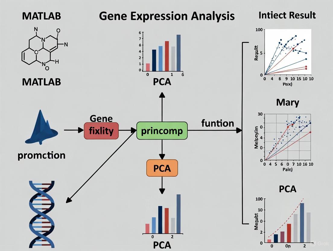
The following DOT script illustrates the complete PCA workflow for gene expression analysis:
PCA Workflow for Gene Expression Data: This diagram illustrates the sequential process from data loading through biological interpretation, with decision points for component selection.
The variance maximization principle in PCA can be visualized through the following conceptual diagram:
Variance Maximization through Orthogonal Transformation: This diagram illustrates how PCA sequentially extracts components that capture maximum variance while maintaining orthogonality.
Applications in Drug Development and Biomedical Research
PCA's dual principles of variance maximization and orthogonal transformation provide powerful approaches for addressing key challenges in pharmaceutical research and development. In biomarker discovery, PCA enables researchers to identify patterns in high-dimensional genomic data that distinguish treatment responders from non-responders, maximizing the signal-to-noise ratio through variance-focused dimensionality reduction. The orthogonal transformation property ensures that each component captures independent biological signals, facilitating interpretation of complex molecular signatures.
In compound screening and mechanism of action studies, PCA reduces high-content screening data to its essential components, allowing researchers to cluster compounds with similar effects and identify potential novel therapeutic agents. The variance maximization principle prioritizes components that explain the greatest differences between compound treatments, while orthogonal transformation eliminates redundant information across multiple assay endpoints. This application is particularly valuable in target identification and validation phases of drug development.
Pharmacogenomics applications leverage PCA to stratify patient populations based on genomic profiles, identifying subpopulations that may benefit from targeted therapies. By maximizing captured variance in gene expression data, PCA reveals the dominant patterns of transcriptional regulation that differentiate patient subgroups, supporting personalized medicine approaches. The orthogonal components frequently correspond to distinct biological pathways or regulatory mechanisms, providing insights into the molecular basis of treatment response variability.
Advantages of PCA for High-Dimensional Gene Expression Data
Principal Component Analysis (PCA) serves as a cornerstone technique for the analysis of high-dimensional gene expression data. By reducing dimensionality, PCA enhances computational efficiency, mitigates overfitting, and facilitates the visualization of underlying data structures. When implemented via MATLAB's princomp function, PCA provides researchers with a powerful tool to uncover biologically significant patterns in transcriptomic studies, supporting advancements in biomarker discovery and drug development. This application note details the theoretical advantages, practical protocols, and critical interpretive considerations for employing PCA in gene expression analysis.
Gene expression datasets from technologies like microarrays and RNA-sequencing are characterized by a massive number of variables (genes) per observation, creating a high-dimensional space that challenges conventional statistical analysis [13] [14]. This high-dimensionality leads to issues such as increased computational cost, the curse of dimensionality, and difficulty in visualization. Principal Component Analysis (PCA) is a linear dimensionality reduction technique that addresses these challenges by transforming the original correlated variables into a new set of uncorrelated variables called principal components (PCs), which are ordered by the amount of variance they capture from the data [15]. This document frames the application of PCA within the context of MATLAB's princomp function, providing a structured guide for life science researchers.
Core Advantages of PCA in Gene Expression Analysis
The application of PCA to gene expression data confers several distinct advantages crucial for scientific research and drug development.
Table 1: Key Advantages of PCA for Gene Expression Data
| Advantage | Mechanism | Impact on Research |
|---|---|---|
| Computational Efficiency | Reduces the number of features for downstream analysis [15]. | Enables faster model training and clustering of large datasets (e.g., thousands of samples) [16]. |
| Noise Reduction | Isolates dominant signals by concentrating variance into the first few PCs, effectively filtering out low-variance noise [5]. | Improves the signal-to-noise ratio, leading to more robust identification of biologically relevant patterns. |
| Data Visualization | Projects high-dimensional data onto 2D or 3D plots using the first 2-3 PCs [4] [3]. | Allows researchers to visually assess sample clustering, identify outliers, and generate hypotheses about group relationships. |
| Overfitting Prevention | Mitigates the "curse of dimensionality" by reducing the feature space used in predictive modeling [15]. | Enhances the generalizability of models for clinical outcome prediction or disease classification. |
| Uncovered Data Structure | Reveals major axes of variation in an unsupervised manner, without prior knowledge of sample groups [3]. | Can identify novel subclasses of diseases, batch effects, or the influence of major biological processes (e.g., cell cycle, immune response). |
Beyond these general benefits, studies have shown that the first few principal components in large, heterogeneous gene expression datasets often have clear biological interpretations, such as separating hematopoietic cells, neural tissues, and cell lines [3]. Furthermore, PCA facilitates the handling of correlated structures among genes, a common feature in transcriptomics, by creating new, uncorrelated variables for subsequent analysis [14].
Experimental Protocol: PCA with MATLABprincomp
This protocol details the steps for performing PCA on a gene expression matrix, using a public yeast diauxic shift dataset [4] [5] as an example. The workflow encompasses data loading, preprocessing, PCA execution, and result interpretation.
Workflow Visualization
The following diagram illustrates the complete analytical pipeline from raw data to clustered results.
Step-by-Step Procedures
Step 1: Data Loading and Exploration
Begin by loading the dataset into the MATLAB workspace. The example dataset yeastdata.mat contains expression levels for 6,400 genes across seven time points.
Explore the data dimensions and content:
Step 2: Data Preprocessing and Filtering
High-quality input data is critical for a meaningful PCA. Preprocessing involves removing non-informative genes and handling missing values.
- Remove Empty Spots: Filter out control spots not associated with genes.
- Handle Missing Values: Eliminate genes with any missing data (
NaN). - Filter by Variance: Remove genes with little variation over time, as they contribute minimal information to the PCA model. The
genevarfilterfunction retains genes with variance above the 10th percentile. - Filter by Absolute Value and Entropy (Optional): Further refine the gene set by removing genes with very low absolute expression levels (
genelowvalfilter) or low profile entropy (geneentropyfilter). After these steps, the dataset is reduced to a manageable number of highly informative genes (e.g., 614 from an initial 6,400) [4] [5].
Table 2: Essential Research Reagents & Computational Tools
| Item Name | Function / Purpose in Analysis |
|---|---|
| Gene Expression Matrix | The primary data input; rows typically represent genes and columns represent samples or experimental conditions. |
| Bioinformatics Toolbox (MATLAB) | Provides specialized functions for biological data analysis, such as genevarfilter and clustergram. |
MATLAB princomp Function |
The core function that performs Principal Component Analysis, returning components, scores, and variances. |
| Statistics and Machine Learning Toolbox | Provides additional clustering algorithms (e.g., kmeans, linkage) for downstream analysis of PCA results. |
Step 3: Executing Principal Component Analysis
Perform PCA on the preprocessed data matrix using the princomp function.
Output Interpretation:
COEFF(Principal Component Coefficients): Ap x pmatrix (wherepis the number of genes). Each column defines a principal component as a linear combination of all original genes. The first column is the first PC, which captures the most variance.SCORE(Principal Component Scores): Ann x pmatrix (wherenis the number of samples). This is the projection of the original data onto the new principal component axes. It represents the transformed dataset in the PC space and is used for visualization and clustering.VARIANCE(Eigenvalues): A vector containing the variances explained by each principal component.
Step 4: Result Interpretation and Downstream Analysis
a. Variance Explained: Calculate the percentage of total variance accounted for by each PC. This helps determine how many components to retain.
In the yeast example, the first two PCs may account for over 89% of the cumulative variance [4], meaning a 2D scatter plot of the first two PCs faithfully represents most of the data's structure.
b. Data Visualization: Create a scatter plot of the first two principal components to visualize sample relationships.
c. Downstream Clustering: Use the PC scores (often from the first ~20 PCs) as input for clustering algorithms like K-means or hierarchical clustering to identify groups of samples with similar expression profiles.
Critical Considerations and Best Practices
Successful application of PCA requires attention to several key factors to ensure biologically valid interpretations.
Data Normalization is Crucial: The choice of normalization method (e.g., min-max, z-score, log transformation) profoundly impacts the PCA solution and its biological interpretation [17]. Z-score normalization is a common choice as it standardizes all genes to a mean of zero and a standard deviation of one, preventing highly abundant genes from dominating the first PCs.
Interpretation of Higher Components: While the first few PCs often capture major batch effects or dominant biological processes, biologically relevant information can reside in higher principal components [3]. For example, tissue-specific or subtype-specific signals may be found in PC4 and beyond. Dismissing these components outright could lead to a loss of critical insights.
Understand the Limitations: PCA is a linear technique and may struggle to capture complex non-linear relationships in gene expression data. It is also sensitive to outliers. The sample composition of the dataset heavily influences the principal components; an over-represented tissue type will dominate the early PCs, which may not generalize to other sample sets [3].
PCA, particularly when implemented through MATLAB's princomp function, is an indispensable tool for the exploratory analysis of high-dimensional gene expression data. Its ability to enhance computational efficiency, enable intuitive visualization, and reveal the underlying structure of complex transcriptomic datasets makes it a fundamental first step in many bioinformatics workflows. By following the detailed protocols and considerations outlined in this application note, researchers and drug development professionals can leverage PCA to distill meaningful biological insights from genomic big data, thereby accelerating scientific discovery and therapeutic development.
Principal Component Analysis (PCA) is a quantitatively rigorous method for simplifying multivariate data sets by reducing their dimensionality. In MATLAB, PCA transforms a set of possibly correlated variables into a set of linearly uncorrelated variables called principal components. These components are orthogonal to each other and form a basis for the data, ordered such that the first component captures the maximum variance in the data, the second captures the next highest variance while being orthogonal to the first, and so on [11]. This technique is particularly valuable for researchers analyzing high-dimensional data, such as gene expression profiles from microarray experiments, where visualizing relationships between more than three variables becomes challenging [4] [11]. The MATLAB ecosystem provides several functions for performing PCA, each with distinct advantages for specific data scenarios commonly encountered in bioinformatics and computational biology research.
Within the context of gene expression analysis, PCA enables researchers to identify predominant patterns of gene expression changes under experimental conditions, such as during the diauxic shift in Saccharomyces cerevisiae (baker's yeast) [5] [4]. By applying PCA to expression data, scientists can reduce thousands of gene expression measurements to a few principal components that capture the most significant variations, thereby revealing underlying biological processes and relationships that might otherwise remain hidden in the high-dimensional data space. This approach facilitates the identification of co-expressed genes, potential regulatory networks, and key molecular drivers of phenotypic changes.
Table 1: Core PCA Functions in the MATLAB Ecosystem
| Function | Input Data Type | Key Features | Best Use Cases |
|---|---|---|---|
| pca | Raw data matrix (n-by-p) | Uses SVD or eigenvalue decomposition; handles missing data with 'algorithm','als' [7] | Standard PCA on complete data or data with few missing values |
| pcacov | Covariance matrix (p-by-p) | Performs PCA on precomputed covariance matrix; does not standardize variables [18] | When only covariance matrix is available or computational efficiency is critical |
| ppca | Raw data matrix with missing values | Probabilistic approach using EM algorithm; handles missing data [19] | Data with significant missing values (>10-20%) assumed missing at random |
Theoretical Foundations and Algorithmic Differences
Mathematical Underpinnings of PCA
The fundamental mathematical operation behind PCA involves the eigenvalue decomposition of the covariance matrix of the data or the singular value decomposition (SVD) of the data matrix itself [7] [6]. When using the pca function on raw data, MATLAB centers the data by default and employs the SVD algorithm, which factorizes the data matrix X into USVᵀ, where the columns of V represent the principal components (eigenvectors of XᵀX) and the diagonal elements of S are proportional to the square roots of the eigenvalues [7]. The pcacov function operates directly on a covariance matrix, performing eigenvalue decomposition to obtain the principal components, but does not automatically standardize the variables to unit variance [18]. For standardized variable analysis, researchers must preprocess the covariance matrix into a correlation structure before applying pcacov.
Probabilistic PCA Framework
Probabilistic PCA (PPCA) extends classical PCA within a probabilistic framework, modeling the data using a Gaussian distribution and introducing a latent variable model that represents the principal components [19] [20]. The key advantage of this approach is its foundation on maximum likelihood estimation, which enables handling of missing data through an expectation-maximization (EM) algorithm. Unlike conventional PCA, PPCA provides a proper probability density model that can be used for statistical inference and offers greater robustness to noise in the data [20]. The EM algorithm iteratively estimates the missing values and model parameters until convergence, making it particularly suitable for gene expression datasets where missing values frequently occur due to experimental artifacts or measurement limitations.
Figure 1: The Expectation-Maximization Workflow of PPCA for Handling Missing Data
Comparative Analysis of PCA Functions
Functional Capabilities and Limitations
Each PCA function in MATLAB's ecosystem presents distinct advantages and limitations for gene expression research. The standard pca function offers the most comprehensive set of features for complete datasets, including support for different algorithms (SVD and Eigenvalue decomposition), variable weighting options, and the ability to return multiple output statistics such as Hotelling's T-squared values [7]. The pcacov function provides computational efficiency for scenarios where the covariance matrix is already available or when working with tall arrays that exceed memory limitations [18]. Meanwhile, ppca specializes in handling datasets with values missing at random, employing an iterative EM algorithm that converges to maximum likelihood estimates of the principal components while simultaneously imputing missing values [19].
Table 2: Output Components of PCA Functions in MATLAB
| Output | pca | pcacov | ppca | Description |
|---|---|---|---|---|
| coeff | ✓ | ✓ | ✓ | Principal component coefficients (loadings) |
| score | ✓ | ✗ | ✓ | Representations of input data in principal component space |
| latent | ✓ | ✓ | ✓ | Principal component variances (eigenvalues) |
| tsquared | ✓ | ✗ | ✗ | Hotelling's T-squared statistic for each observation |
| explained | ✓ | ✓ | ✗ | Percentage of total variance explained by each component |
| mu | ✓ | ✗ | ✓ | Estimated mean of each variable |
Performance Considerations for Gene Expression Data
When processing large-scale gene expression datasets, computational performance becomes a significant consideration. For the standard pca function, the SVD algorithm generally provides better numerical stability, while eigenvalue decomposition may offer performance benefits for certain matrix structures [7]. The ppca function typically requires more computational resources due to its iterative EM algorithm, with the number of iterations controlled through options structures that can modify termination criteria and display settings [19]. For massive datasets that exceed memory limitations, the pcacov function enables a distributed computing approach where researchers can compute the covariance matrix from tall arrays and then perform PCA on the resulting covariance matrix [18].
Experimental Protocols for Gene Expression Analysis
Data Preprocessing and Filtering
Comprehensive preprocessing of gene expression data is essential before applying PCA to ensure meaningful results. The protocol begins with loading the expression data, typically represented as a matrix where rows correspond to genes and columns to experimental conditions or time points [5] [4]. For yeast expression data during diauxic shift, the dataset includes expression values (log2 ratios) measured at seven time points [4]. Initial preprocessing involves removing empty spots and genes with missing values, followed by applying variance-based and entropy-based filtering to retain only genes with informative expression profiles [5] [4].
Code 1: Gene Filtering Protocol for Yeast Expression Data Prior to PCA
Protocol for Standard PCA Analysis
After preprocessing, standard PCA can be applied to identify patterns in the filtered gene expression data. The protocol involves normalizing the data to zero mean and unit variance, followed by principal component extraction using the pca function [4]. Researchers can then visualize the results through scatter plots of principal component scores and analyze the variance explained by each component to determine how many principal components to retain for subsequent analysis.
Code 2: Standard PCA Protocol for Gene Expression Analysis
Protocol for Probabilistic PCA with Missing Data
When working with gene expression datasets containing missing values, PPCA provides a robust alternative. This protocol demonstrates how to apply ppca to handle missing data, which commonly occurs in microarray experiments due to technical artifacts [19]. The method is particularly valuable when the missing data mechanism can be assumed to be missing at random, as it provides maximum likelihood estimates of the principal components while simultaneously imputing missing values.
Code 3: Probabilistic PCA Protocol for Handling Missing Values in Gene Expression Data
Protocol for PCA on Covariance Matrix
For scenarios where the covariance matrix is already available or when working with tall arrays that exceed memory limitations, pcacov offers an efficient alternative [18]. This protocol demonstrates how to compute the covariance matrix from expression data and perform PCA directly on the covariance structure, which can be particularly useful for large-scale genomic studies.
Code 4: PCA on Covariance Matrix Protocol for Large-Scale Expression Data
The Scientist's Toolkit: Research Reagent Solutions
Table 3: Essential Computational Tools for Gene Expression PCA Analysis
| Tool/Function | Purpose | Application Context |
|---|---|---|
| Bioinformatics Toolbox | Provides specialized functions for genomic data analysis | Required for genevarfilter, genelowvalfilter, and geneentropyfilter functions [5] [4] |
| Statistics and Machine Learning Toolbox | Implements core PCA functions and clustering algorithms | Essential for pca, pcacov, and ppca functions [19] [7] [18] |
| genevarfilter | Filters genes with small variance across experimental conditions | Removes uninformative genes with static expression profiles [5] [4] |
| genelowvalfilter | Removes genes with very low absolute expression values | Eliminates genes with minimal expression signal [5] [4] |
| geneentropyfilter | Filters genes with low entropy expression profiles | Selects genes with dynamic expression patterns across conditions [5] [4] |
| mapstd | Normalizes data to zero mean and unit variance | Standard preprocessing step before PCA [5] |
Data Interpretation and Visualization Strategies
Analyzing PCA Outputs for Biological Insight
Interpreting PCA results requires understanding the biological significance of each output component. The principal component coefficients (loadings) indicate how much each original variable (gene) contributes to a particular principal component, revealing which genes have the strongest influence on the observed patterns [7] [6]. The principal component scores represent the original data projected into the principal component space, enabling visualization of sample relationships [6]. The variances (eigenvalues) indicate the importance of each principal component, while the explained variance percentage quantifies how much of the total data variability each component captures [7] [4]. For gene expression time course data, these outputs can identify groups of co-expressed genes and temporal expression patterns that correspond to specific biological processes.
Figure 2: Pathway from PCA Outputs to Biological Interpretation in Gene Expression Analysis
Advanced Visualization Techniques
Effective visualization of PCA results enhances the extraction of biological insights from gene expression data. The mapcaplot function provides an interactive environment for exploring principal components, allowing researchers to select data points across multiple scatter plots and identify corresponding genes [21]. For publication-quality figures, the scatter function can create 2D plots of the first two principal components, which often capture the majority of data variance [5] [4]. When analyzing time course expression data, researchers can color-code data points by time points or experimental conditions to visualize temporal patterns and transitions, such as the metabolic shift from fermentation to respiration in yeast [4]. Cluster analysis techniques, including hierarchical clustering and k-means applied to principal component scores, can further elucidate gene expression patterns and identify potential regulatory modules.
The MATLAB PCA ecosystem offers a comprehensive suite of functions tailored to different data scenarios in gene expression research. The standard pca function serves as the primary tool for complete datasets, while ppca provides specialized handling for data with missing values through its probabilistic framework and EM algorithm [19] [20]. The pcacov function offers computational efficiency for scenarios where covariance matrices are precomputed or when working with large-scale data that exceeds memory limitations [18]. For researchers analyzing gene expression data, following systematic protocols for data filtering, normalization, and dimensionality assessment ensures biologically meaningful results. By selecting the appropriate PCA function based on data characteristics and research objectives, scientists can effectively uncover patterns in high-dimensional genomic data, leading to deeper insights into transcriptional regulation and cellular responses.
Principal Component Analysis (PCA) is a fundamental dimension reduction technique widely used in gene expression analysis. It transforms high-dimensional data into a new coordinate system, highlighting the dominant patterns of variation and enabling researchers to visualize sample similarities, identify outliers, and uncover latent biological structures. For scientists working with genomic datasets, which often contain tens of thousands of genes (variables) across relatively few samples, PCA provides a critical first step in exploratory data analysis. This application note details the interpretation of core PCA outputs—coefficients, scores, latent values, and explained variance—within the context of gene expression research using MATLAB, specifically framing these concepts within a broader thesis on the princomp function and its applications.
Core Components of PCA Output
The output of PCA consists of several interconnected matrices and vectors that collectively describe the transformed data. Understanding their statistical meaning and biological interpretation is essential for proper analysis.
Table: Core Outputs from MATLAB's PCA Function
| Output Term | Mathematical Definition | Biological Interpretation in Gene Expression | MATLAB Variable |
|---|---|---|---|
| Coefficients (Loadings) | Eigenvectors of the covariance matrix; weights for each gene in the principal components. | Contribution of each gene to a PC. High absolute values mark genes important for the sample separation along that PC. | coeff |
| Scores | Projections of the original data onto the new principal component axes. | Representation of each sample in the new, low-dimensional PC space. Used to visualize sample clustering. | score |
| Latent (Eigenvalues) | Eigenvalues of the covariance matrix. | The variance captured by each respective principal component. | latent |
| Explained Variance | Percentage of the total variance explained by each PC (e.g., latent/sum(latent)*100). |
Helps decide how many PCs are biologically relevant versus noise. | explained |
Coefficients (Loadings)
The principal component coefficients, also known as loadings, form the transformation matrix that defines the direction of the principal components in the original variable space [7]. Each column of the coefficient matrix coeff contains the coefficients for one principal component, with these columns sorted in descending order of component variance [7]. In gene expression analysis, where variables correspond to genes, these coefficients indicate the weight or contribution of each gene to a specific principal component. A high absolute value of a coefficient for a gene within a principal component signifies that this gene strongly influences the direction and separation of samples along that component. For instance, in a large-scale gene expression compendium, the first few principal components often have high loadings for genes specific to major biological programs like hematopoiesis, neural function, or cellular proliferation [3].
Scores
Principal component scores are the representations of the original data in the newly established principal component space [7]. Rows of the score matrix correspond to individual observations (e.g., patient samples, cell lines), and columns correspond to the principal components. These scores are obtained by projecting the original, typically mean-centered, data onto the principal component axes defined by the coefficients. Plotting these scores—for example, PC1 vs. PC2—allows for the visualization of the overall data structure, enabling researchers to identify clusters of samples with similar gene expression profiles, detect outliers, and hypothesize about underlying biological or technical effects [22] [23].
Latent Values and Explained Variance
The latent output is a vector containing the eigenvalues of the covariance matrix of the input data [7]. These eigenvalues represent the variance explained by each corresponding principal component. The explained output directly quantifies the percentage of the total variance in the original dataset that is captured by each principal component, calculated as the corresponding latent value divided by the sum of all latent values [7] [23]. This metric is critical for assessing the importance of each component and determining the number of components to retain for further analysis. In gene expression studies, it is common for the first few components to explain a limited portion of the total variance (e.g., 20-40% for PC1), with the cumulative explained variance increasing gradually with subsequent components [3]. A scree plot, which plots the explained variance or eigenvalues against the component number, is a standard tool for this evaluation. The cumulative explained variance can be visualized and calculated using the cumsum function on the explained vector [24].
Experimental Protocol: PCA for Gene Expression Analysis
This protocol outlines the steps for performing and interpreting PCA on a gene expression matrix using MATLAB, where rows correspond to samples and columns to genes.
Sample Preparation and Data Preprocessing
- Data Matrix Assembly: Compile your gene expression data into a numerical matrix
Xof sizen x p, wherenis the number of observations (samples) andpis the number of variables (genes) [7]. - Data Normalization: Normalize the data to ensure genes are comparable. A common approach is to transform the data to Z-scores, which centers each gene on zero mean and scales it to unit variance using
Z = zscore(X)[2]. This step is crucial when the variances of the original variables differ by orders of magnitude [25]. - Handling Missing Values: Identify and handle missing values (
NaN). MATLAB'spcafunction offers several methods via the'Rows'name-value pair. The'complete'option removes observations with anyNaNvalues before calculation, which is the default. Alternatively, the'pairwise'option can be used with the eigenvalue decomposition algorithm, though this may result in a non-positive definite covariance matrix [7].
Computational Procedure in MATLAB
- Execute PCA: Perform PCA on the preprocessed data matrix. Use a command that returns all necessary outputs:
Here,
Zis the normalized data matrix. The outputmucontains the estimated means of each variable, which is useful for reconstruction [7]. - Determine Significant Components: Examine the
explainedvector to decide how many principal components to retain. This can be done by:- Creating a scree plot:
- Setting a threshold for cumulative variance (e.g., 70-90%) or looking for an "elbow" in the scree plot where the explained variance drops off markedly.
- Visualize Results:
- Sample Clustering: Create a 2D or 3D scatter plot of the principal component scores to visualize sample relationships.
- Biplot Generation: Create a biplot to overlay the scores (samples as points) and the coefficients (genes as vectors) on the same graph, showing their relationship [22].
- Interpret Loadings: To identify genes that drive separation along a specific principal component (e.g., PC1), sort the coefficients for that component and examine the genes with the highest absolute values [26].
PCA Workflow for Gene Expression Data
Advanced Interpretation in a Genomic Context
Applying PCA to gene expression data comes with specific considerations and challenges that researchers must address for a valid biological interpretation.
Dimensionality and Sample Composition
A key finding in genomics is that the apparent intrinsic dimensionality of gene expression data is often higher than initially assumed. While the first three principal components might capture large-scale, dominant patterns (e.g., separating hematopoietic cells, neural tissues, and cell lines), significant tissue-specific or condition-specific information can reside in higher-order components [3]. The sample composition of the dataset profoundly influences the resulting principal components. If a particular tissue or cell type is over-represented, it will likely dominate the early components. For example, a dataset with a high proportion of liver samples may show a liver-specific separation in PC4, which would be absent in a dataset with fewer liver samples [3]. This underscores the importance of considering sample cohort structure when interpreting PCA results.
The Scientist's Toolkit: Research Reagent Solutions
Table: Essential Computational Tools for PCA-Based Gene Expression Analysis
| Tool / Resource | Function in Analysis | Application Context |
|---|---|---|
| MATLAB Statistics and Machine Learning Toolbox | Provides the core pca and biplot functions for computation and visualization. |
Primary environment for performing the PCA analysis and generating initial plots. [7] [22] |
| Predefined Gene Labels | A cell array of gene symbols corresponding to the columns of the data matrix. | Critical for annotating vectors in a biplot to identify which genes drive component separation. [22] |
| Custom Scripting for Visualization | MATLAB scripts for generating enhanced scree plots and score scatter plots. | Allows for tailored visualization that clearly communicates the variance explained and sample clustering. [24] |
| Biological Annotation Databases | Resources like GO, KEGG, or MSigDB for functional enrichment analysis. | Used to interpret the biological meaning of genes with high loadings on a given principal component. |
Case Study: Analysis of a Public Microarray Dataset
To illustrate the interpretation of PCA outputs, consider a re-analysis of a large public microarray dataset, such as the one from Lukk et al. (2016), which contains 5,372 samples from 369 different tissues and cell types [3].
- Procedure: The gene expression data was normalized and subjected to PCA using MATLAB. The first four principal components were retained for detailed examination.
- Results and Interpretation:
- Variance Explained: The first three PCs explained approximately 36% of the total variance in the data, indicating that a substantial amount of information remained in higher components [3].
- Component Interpretation: PC1 was strongly associated with hematopoietic cells, PC2 with malignancy and proliferation, and PC3 with neural tissues. A subsequent analysis of a different dataset revealed that PC4 separated liver and hepatocellular carcinoma samples, a finding attributed to the higher proportion of such samples in that specific dataset [3].
- Residual Information: By projecting the data onto the first three PCs and analyzing the residual information, it was shown that significant tissue-specific information (e.g., distinguishing between different brain regions) was retained in the higher components (PC4 and beyond) [3].
Interpreting a Biplot for Gene Expression Data
A rigorous interpretation of PCA outputs—coefficients, scores, latent values, and explained variance—is fundamental for extracting meaningful biological insights from complex gene expression datasets. The coefficients reveal the genes that drive major patterns of variation, the scores show how samples are arranged according to these patterns, and the explained variance quantifies the importance of each pattern. Researchers must be mindful of the data's structure and scale, as these factors directly influence the PCA results. By following the detailed protocols and considerations outlined in this document, scientists and drug development professionals can reliably use PCA as a powerful, unsupervised tool for quality control, hypothesis generation, and the exploration of the fundamental dimensionality of their genomic data.
Application Note: Principal Component Analysis of Gene Expression During the Diauxic Shift in Yeast
The diauxic shift in Saccharomyces cerevisiae represents a crucial metabolic transition from fermentative growth on glucose to respiratory growth on ethanol, accompanied by extensive gene expression reprogramming [27] [28]. This physiological transition serves as an excellent model for studying metabolic adaptation and regulatory networks, with implications for understanding similar processes in cancer cells, particularly the Warburg effect [27]. This application note demonstrates how Principal Component Analysis (PCA) via MATLAB's princomp function can reveal key patterns in transcriptional regulation during this shift, providing a framework for analyzing similar transformations in cancer genomics.
Experimental Dataset
The analysis utilizes a publicly available microarray dataset from DeRisi et al. (1997) that captures temporal gene expression of Saccharomyces cerevisiae during the diauxic shift [5] [4]. Expression levels were measured at seven time points as yeast transitioned from fermentation to respiration. The raw dataset contains 6,400 expression profiles, though filtering techniques reduce this to the most biologically relevant genes.
Table: Dataset Overview of Yeast Diauxic Shift Experiment
| Parameter | Specification |
|---|---|
| Organism | Saccharomyces cerevisiae (Baker's Yeast) |
| Experimental Condition | Diauxic Shift (Fermentation to Respiration) |
| Time Points Measured | 7 time points during metabolic transition |
| Initial Gene Count | 6,400 genes |
| Technology | DNA Microarray |
| Public Accession | GSE28 (Gene Expression Omnibus) |
Methodology: PCA with MATLAB princomp
Data Preprocessing and Filtering
Before performing PCA, the expression data must be filtered to remove uninformative genes:
Load Data: Load the yeast dataset into MATLAB workspace.
Remove Empty Spots: Filter out empty microarray spots.
Handle Missing Data: Remove genes with missing values (NaN).
Apply Variance Filter: Retain genes with variance above the 10th percentile.
Apply Low-Value Filter: Remove genes with low absolute expression values.
Apply Entropy Filter: Remove genes with low entropy profiles.
Principal Component Analysis
The core analysis utilizes the princomp function (or pca in newer versions) on the preprocessed data:
Perform PCA: Calculate principal components, scores, and variances.
Variance Explanation: Calculate the percentage of variance explained by each component.
Visualization: Create a scatter plot of the first two principal components.
Results and Bioinformatics Interpretation
PCA reveals distinct expression patterns separating the fermentative and respiratory growth phases. The first principal component (PC1) typically captures the majority of variance (approximately 80%), representing the dominant expression program shift between metabolic states [4]. The second component (PC2) often captures additional variance (approximately 10%), potentially reflecting finer-scale regulatory events.
Table: Variance Explained by Principal Components in a Typical Diauxic Shift Dataset
| Principal Component | Percentage of Variance Explained | Cumulative Percentage |
|---|---|---|
| PC1 | 79.8% | 79.8% |
| PC2 | 9.6% | 89.4% |
| PC3 | 4.1% | 93.5% |
| PC4 | 2.6% | 96.1% |
| PC5 | 2.2% | 98.3% |
| PC6 | 1.0% | 99.3% |
| PC7 | 0.7% | 100.0% |
Genes with high loadings on PC1 represent those most significantly altered during the metabolic shift, including those involved in carbon metabolism, mitochondrial function, and stress response. This dimension effectively separates samples collected during fermentative growth (negative scores) from those during respiratory growth (positive scores).
Workflow Visualization
Advanced Protocol: Integrating Metabolomics with Transcriptomics Using Multi-Omics PCA
Modern systems biology increasingly relies on multi-omics approaches that integrate different molecular data layers to gain comprehensive insights into biological systems [27] [29]. This protocol extends the basic PCA approach to integrate gene expression and metabolomics data from diauxic shift studies, providing a framework for similar integrations in cancer research where transcriptomic and metabolomic dysregulation are hallmarks of malignancy.
Experimental Design
The integrated analysis utilizes both transcriptomic profiles and untargeted intracellular metabolomic data collected during the diauxic shift in yeast. Samples are collected during both pre-diauxic (fermentative) and post-diauxic (respiratory) phases [27]. For cancer studies, equivalent designs would compare tumor versus normal tissues.
Multi-Omics Integration Methodology
Data Preprocessing
Normalization: Independently normalize transcriptomic and metabolomic datasets using Z-score normalization.
Data Merging: Combine normalized datasets into a single matrix with samples as rows and features (genes + metabolites) as columns.
Batch Effect Correction: Apply ComBat or similar algorithms if data originated from different analytical batches.
Concatenation-Based PCA
Perform PCA on the combined dataset to identify patterns that capture covariance between transcriptomic and metabolomic features:
Result Interpretation
- Joint Components: Identify principal components that represent coordinated variation in both gene expression and metabolite abundance.
- Feature Loadings: Examine high-loading genes and metabolites on each component to infer functional relationships.
- Pathway Mapping: Use enrichment analysis to map high-loading features to biological pathways.
Key Findings from Integrated Diauxic Shift Studies
Integrated analysis of diauxic shift reveals:
- Metabolic Rewiring: Distinct metabolic profiles characterize fermentative versus respiratory phases, with 215 metabolic features significantly altered [27].
- Pathway Enrichments: Significant perturbations occur in central carbon metabolism, glycerophospholipid metabolism, glutathione metabolism, and amino acid metabolism [27].
- Regulatory Insights: Deletion of specific regulatory genes (YGR067C, TDA1, RTS3) causes distinct metabolic perturbations, revealing their functional roles [27].
Table: Research Reagent Solutions for Diauxic Shift and Cancer Genomics Studies
| Reagent/Resource | Function/Application | Example Source/Provider |
|---|---|---|
| Yeast Deletion Strains (e.g., tda1Δ) | Functional characterization of genes during metabolic shifts | EUROSCARF Deletion Library [30] |
| RNA Extraction Kit (e.g., RNeasy) | High-quality RNA isolation for transcriptomics | QIAGEN [30] |
| SC Medium | Defined growth medium for controlled yeast cultivation | Formulated in-house per Sherman (2002) |
| Illumina Sequencing | High-throughput RNA sequencing | NgI, Azenta [30] |
| Mass Spectrometry | Untargeted metabolomics profiling | Various platforms (e.g., LC-MS) [27] |
| MATLAB Bioinformatics Toolbox | Gene expression analysis and PCA | MathWorks [5] [4] |
| exvar R Package | Integrated analysis of gene expression and genetic variation | GitHub [31] |
Application in Cancer Genomics: From Yeast Models to Human Tumors
Cross-Species Analytical Framework
The analytical framework established in yeast diauxic shift studies directly translates to cancer genomics, particularly in investigating the Warburg effect (aerobic glycolysis) where cancer cells preferentially utilize fermentation over respiration even in oxygen-rich conditions [27] [28].
Signaling Pathway Analysis
Comparative Analysis: Diauxic Shift versus Cancer Metabolic Dysregulation
Table: Comparative Metabolic Features Between Yeast Diauxic Shift and Cancer Warburg Effect
| Biological Feature | Yeast Diauxic Shift | Cancer Warburg Effect |
|---|---|---|
| Preferred Metabolic State | Transition: Fermentation → Respiration | Locked: Aerobic Glycolysis (Fermentation) |
| Regulatory Proteins | Mig1p, Hxk2p, Tda1p, HAP Complex | HIF-1, MYC, p53, AKT/mTOR |
| Key Metabolic Pathways | Glycolysis, TCA Cycle, Oxidative Phosphorylation | Glycolysis, Lactate Fermentation, Pentose Phosphate |
| Gene Expression Analysis | PCA reveals phase-specific clusters [4] | PCA separates tumor subtypes and grades |
| Mitochondrial Function | Activated post-shift for respiration | Often impaired despite functional capacity |
| Technological Approaches | Microarrays, RNA-seq, Mass Spectrometry [27] | Single-cell RNA-seq, Spatial Transcriptomics [29] |
Transcriptomics in Cancer Studies: Advanced Methodologies
Modern cancer transcriptomics employs several advanced approaches beyond standard PCA:
Weighted Gene Co-expression Network Analysis (WGCNA): Identifies modules of highly correlated genes and assesses their preservation between normal and tumor tissues [32].
Single-Cell RNA Sequencing: Reveals transcriptional heterogeneity within tumors, identifying rare cell populations and resistance mechanisms [29].
Spatial Transcriptomics: Maps gene expression within tissue architecture, preserving spatial context of tumor-microenvironment interactions [33].
Integrated Variant Analysis: Tools like the exvar package combine expression and genetic variant analysis from RNA-seq data [31].
Practical Considerations for MATLAB Implementation
Handling Large-Scale Genomic Data
- Memory Management: Use tall arrays for datasets exceeding memory limits.
- Parallel Computing: Accelerate PCA computation with the Parallel Computing Toolbox.
- Cloud Integration: Leverage cloud resources for computationally intensive analyses [29].
Algorithm Selection
- SVD vs. Eigenvalue Decomposition: Choose appropriate algorithms based on data dimensions and missing value patterns [7].
- Missing Data Handling: Implement appropriate strategies (complete case, imputation) based on missing data patterns [7].
The application of PCA through MATLAB's princomp/pca functions provides a powerful analytical framework for extracting biologically meaningful patterns from gene expression data, from fundamental metabolic transitions in model organisms like yeast to complex dysregulations in cancer. The protocols outlined here for both standard and multi-omics PCA create a foundation for researchers to investigate complex biological systems, with direct relevance to drug development through identification of key regulatory pathways and potential therapeutic targets. As genomic technologies evolve, integrating these classical statistical approaches with modern machine learning methods will further enhance our ability to decipher complex biological networks in both basic and translational research contexts.
Step-by-Step PCA Workflow for Gene Expression Analysis
Within gene expression analysis research, particularly when utilizing the princomp function for Principal Component Analysis (PCA), the initial and most critical step is the proper loading and import of microarray and RNA-seq data. MATLAB provides a comprehensive environment for managing gene expression data, offering specialized functions and objects within its Bioinformatics Toolbox for handling data from various technological platforms [34] [35]. For researchers and drug development professionals, understanding these data import mechanisms is fundamental to ensuring the biological validity of subsequent analyses, including dimensionality reduction and pattern discovery. Proper data handling establishes the foundation for all downstream analytical processes, from basic differential expression testing to advanced multivariate methods like PCA that reveal hidden structures in high-dimensional genomic data.
Microarray Data Handling
Data Import and Preprocessing
Microarray technology enables high-throughput measurement of gene expression levels using oligonucleotide or cDNA probes attached to a solid surface [36]. MATLAB provides specialized functions for importing data from various microarray file formats and platforms:
- Platform-Specific Import Functions: Use
gprreadfor GenePix Results files,agfereadfor Agilent Feature Extraction files,ilmnbsreadfor Illumina BeadStudio data, andimagenereadfor ImaGene Results files [34]. - GEO Data Access: Retrieve public data from the Gene Expression Omnibus (GEO) using
getgeodata,geoseriesread, andgeosoftreadfunctions [34]. - Data Management Objects: Store and manage experimental data using specialized objects including
bioma.ExpressionSet,bioma.data.ExptData, andbioma.data.MIAMEfor MIAME-standard experiment information [34] [37].
A critical preprocessing workflow involves filtering to remove uninformative genes before proceeding to advanced analysis like PCA:
This filtering sequence typically reduces a dataset from thousands of genes to several hundred most informative profiles, creating a manageable set for PCA analysis while preserving biologically relevant expression patterns [5] [4].
Experimental Design and Normalization Considerations
Microarray data acquisition begins with hybridization of labeled samples to complementary DNA probes fixed on a solid surface [36]. The quantification process involves distinguishing foreground probe intensity from local background, typically using mean or median summaries of pixel intensities within defined regions [36]. The resulting data represent fluorescence intensities that reflect relative gene expression levels, which are commonly log2-transformed to approximate normal distributions suitable for parametric statistical analysis [36] [4].
Table 1: Key MATLAB Functions for Microarray Data Analysis
| Function | Category | Purpose |
|---|---|---|
gprread |
Data Import | Read GenePix Results file |
geoseriesread |
Data Import | Read GEO Series data |
genevarfilter |
Preprocessing | Filter genes with low variance |
genelowvalfilter |
Preprocessing | Filter genes with low expression |
mattest |
Analysis | Two-sample t-test for differential expression |
mafdr |
Analysis | False discovery rate estimation |
clustergram |
Visualization | Heat map with hierarchical clustering |
mapcaplot |
Visualization | Interactive PCA scatter plot |
RNA-seq Data Handling
Data Import and Preprocessing Workflow
RNA sequencing represents a more recent technological approach that enables comprehensive transcriptome quantification through high-throughput sequencing of cDNA fragments [38]. Unlike microarray intensities, RNA-seq data originate as sequence reads that require extensive preprocessing before quantitative analysis:
- Raw Sequence Import: Use
fastqreadandfastqinfoto import raw sequencing data from FASTQ files, which contain nucleotide sequences and corresponding quality scores [39]. - Alignment Data Management: Process aligned reads stored in SAM/BAM formats using
BioMapobjects to efficiently manage sequence, quality, and alignment information [39]. - Annotation Integration: Import feature annotations from GTF and GFF files using
GTFAnnotationandGFFAnnotationobjects to correlate genomic features with expression data [39].
The RNA-seq analysis workflow involves multiple preprocessing stages before statistical analysis:
The rnaseqde function performs normalization and differential expression testing specifically designed for count-based RNA-seq data, using methods that account for the negative binomial distribution typical of sequencing counts [40].
Experimental Design and Normalization Strategies
Proper experimental design for RNA-seq studies requires careful consideration of replication and sequencing depth. While three replicates per condition represents a minimum standard, increased replication significantly improves detection power, particularly when biological variability is high [38]. Sequencing depth of 20-30 million reads per sample typically provides sufficient sensitivity for most differential expression analyses [38].
Table 2: RNA-seq Normalization Methods
| Method | Depth Correction | Gene Length Correction | Library Composition Correction | Suitable for DE |
|---|---|---|---|---|
| CPM | Yes | No | No | No |
| RPKM/FPKM | Yes | Yes | No | No |
| TPM | Yes | Yes | Partial | No |
| median-of-ratios (DESeq2) | Yes | No | Yes | Yes |
| TMM (edgeR) | Yes | No | Yes | Yes |
Normalization must address multiple technical biases including sequencing depth (total reads per sample), gene length, and library composition effects where highly expressed genes in one sample distort count distributions [38]. The median-of-ratios method (DESeq2) and TMM (edgeR) implement sophisticated normalization approaches that account for these factors, making them suitable for differential expression analysis [38].
Comparative Workflow: From Raw Data to PCA
Integrated Data Management Framework
Both microarray and RNA-seq data benefit from structured data management within MATLAB's specialized objects. The ExpressionSet object serves as a comprehensive container for gene expression data, integrating:
- Expression Values: Primary intensity or count measurements stored in
ExptDataobjects [34] [37] - Sample Metadata: Information about samples and experimental conditions in
MetaDataobjects [34] - Feature Metadata: Gene or transcript annotations including identifiers and genomic coordinates [34]
- Experiment Information: MIAME-standard experiment descriptions in
MIAMEobjects [34]
This integrated framework ensures all relevant data components remain associated throughout the analytical pipeline, which is particularly valuable when preparing data for PCA using princomp.
Pathway to Principal Component Analysis
The following workflow diagrams illustrate the standardized procedures for processing both data types prior to PCA implementation:
Microarray Data Processing Workflow
RNA-seq Data Processing Workflow
The Scientist's Toolkit
Essential Research Reagent Solutions
Table 3: Key Research Reagents and Computational Tools
| Item | Function/Purpose | Example Platforms/Tools |
|---|---|---|
| Oligonucleotide Probes | Hybridization to target sequences | Affymetrix, Agilent, Illumina |
| Fluorescent Dyes (Cy3/Cy5) | Sample labeling and detection | Two-color microarray systems |
| cDNA Synthesis Kits | Reverse transcription of RNA | Library preparation for RNA-seq |
| Sequencing Adapters | Platform-specific sequence ligation | Illumina, PacBio, Oxford Nanopore |
| Normalization Reagents | Technical variability control | Spike-in controls (ERCC) |
| Quality Control Tools | Data quality assessment | FastQC, MultiQC [38] |
| Alignment Software | Read mapping to reference | STAR, HISAT2 [38] |
| Quantification Tools | Expression level estimation | featureCounts, HTSeq [38] |
Implementation Protocols
Detailed Microarray Analysis Protocol
Objective: Process raw microarray data through normalization and filtering to create a PCA-ready dataset.
Materials: Raw data files (GPR, TXT, or CEL formats), MATLAB with Bioinformatics Toolbox.
Procedure:
Data Import
Data Cleaning
- Identify and remove empty spots:
emptyMask = strcmp('EMPTY',geneNames); - Remove genes with missing values:
nanMask = any(isnan(expressionMatrix),2); - Combine masks:
cleanMask = ~(emptyMask | nanMask); - Apply filter:
expressionMatrix = expressionMatrix(cleanMask,:);
- Identify and remove empty spots:
Quality Assessment
- Generate box plots:
maboxplot(expressionMatrix) - Create log-log plots:
maloglog(expressionMatrix(:,1),expressionMatrix(:,2)) - Assess intensity ratios:
mairplot(expressionMatrix(:,1),expressionMatrix(:,2))
- Generate box plots:
Normalization and Filtering
- Apply variance filter:
mask = genevarfilter(expressionMatrix); - Remove low-value genes:
[mask, expressionMatrix, geneNames] = genelowvalfilter(expressionMatrix, geneNames, 'absval', log2(3)); - Filter by entropy:
[mask, expressionMatrix, geneNames] = geneentropyfilter(expressionMatrix, geneNames, 'prctile', 15);
- Apply variance filter:
PCA Preparation
- Transpose matrix:
inputData = expressionMatrix'; - Normalize data:
[x, std_settings] = mapstd(inputData); - Ready for PCA:
[coeff, scores, latent] = princomp(x);
- Transpose matrix:
Detailed RNA-seq Analysis Protocol
Objective: Transform raw RNA-seq count data into a normalized format suitable for PCA.
Materials: Count matrix (CSV or TXT format), MATLAB with Bioinformatics Toolbox.
Procedure:
Data Import
Quality Assessment
- Examine distribution:
boxplot(log2(countMatrix+1)) - Calculate library sizes:
librarySizes = sum(countMatrix); - Assess zeros:
zeroPercentage = sum(countMatrix==0)/length(countMatrix);
- Examine distribution:
Normalization
Data Transformation
- Apply variance-stabilizing transformation:
vstMatrix = log2(normalizedMatrix + 1); - Alternative:
vstMatrix = sqrt(normalizedMatrix);
- Apply variance-stabilizing transformation:
PCA Preparation
- Transpose matrix:
inputData = vstMatrix'; - Standardize features:
[x, std_settings] = mapstd(inputData); - Execute PCA:
[coeff, scores, latent] = princomp(x);
- Transpose matrix:
Proper data loading and import procedures for both microarray and RNA-seq technologies establish the essential foundation for meaningful gene expression analysis using MATLAB's princomp function. While the initial data structures and preprocessing methods differ significantly between these platforms—with microarrays requiring intensity normalization and filtering, and RNA-seq demanding count-based normalization—both converge on a standardized matrix format suitable for principal component analysis. The structured workflows and specialized tools provided in MATLAB's Bioinformatics Toolbox enable researchers to navigate these complex data types efficiently, transforming raw experimental outputs into biologically interpretable patterns. By adhering to these detailed protocols for data management, normalization, and quality control, scientists can ensure the analytical rigor required for robust gene expression research and drug development applications.
In gene expression analysis research, the accuracy of downstream analyses, particularly those utilizing the MATLAB princomp function for Principal Component Analysis (PCA), is critically dependent on robust data preprocessing. Raw genomic data from technologies like microarrays and RNA-Seq is inherently noisy, containing missing values, technical artifacts, and systematic variations that can obscure true biological signals. This application note details the essential preprocessing steps—filtering, normalization, and missing value treatment—required to prepare gene expression data for reliable PCA and subsequent analysis. Proper implementation of these protocols ensures that the principal components derived reflect biological reality rather than technical artifacts, enabling researchers and drug development professionals to draw valid conclusions about differential gene expression, biomarker discovery, and therapeutic targets.
Materials and Research Reagent Solutions
Table 1: Essential Research Reagents and Computational Tools for Gene Expression Preprocessing
| Item Name | Function/Application | Example/Reference |
|---|---|---|
| Bioinformatics Toolbox (MATLAB) | Provides specialized functions for gene expression filtering and analysis [5]. | Functions: genevarfilter, genelowvalfilter, geneentropyfilter |
| DNA Microarray Data | Raw gene expression measurements for analysis. | Baker's yeast (Saccharomyces cerevisiae) data [5] [4] |
| RNA-Seq Read Count Data | Digital measure of gene expression levels for transcriptomic studies. | Data from public repositories like TCGA, GTEx, and GEO [41] [42] |
| ERCC Spike-in Control RNA | External RNA controls added during library preparation to monitor technical performance [41]. | Used to distinguish biological from technical variation |
| Housekeeping Gene Set | A set of constitutively expressed genes used for normalization validation [43]. | Genes like GAPDH, ACTB, or a customized set of 107 stable genes |
Experimental Protocols and Methodologies
Protocol 1: Data Filtering for Gene Expression
Objective: To remove uninformative genes and noise, thereby reducing data dimensionality and enhancing the signal-to-noise ratio for PCA.
Remove Empty Spots and Poor Quality Data: Identify and eliminate empty spots on microarrays (e.g., labeled 'EMPTY') and genes with an unacceptable number of missing values.
Apply Variance Filter: Filter out genes with little to no variation across samples, as they contribute minimally to population structure.
Apply Low-Value Filter: Remove genes with very low absolute expression values, which are often unreliable.
Apply Entropy Filter: Filter out genes whose expression profiles have low entropy, indicating low information content.
Protocol 2: Normalization of Gene Expression Data
Objective: To remove systematic technical biases (e.g., sequencing depth, library preparation) and make expression levels comparable across samples.
The choice of normalization method is critical and depends on the technology and data structure.
Table 2: Comparison of Common Normalization Methods for RNA-Seq Data
| Normalization Method | Brief Description | Key Findings from Comparative Studies |
|---|---|---|
| DESeq | Normalizes based on a negative binomial distribution and the geometric mean of read counts [41]. | Identified as one of the best methods for RNA-Seq data; robust and properly aligns data distributions across samples [41]. |
| TMM (Trimmed Mean of M-values) | Uses a weighted trimmed mean of log expression ratios [41]. | Performs well but can be sensitive to the prior removal of low-expressed genes [41]. |
| Upper Quartile (UQ) | Scales counts using the upper quartile of counts [41]. | Does not always effectively align data across samples [41]. |
| Quantile (Q) | Forces the distribution of expression values to be identical across samples [41]. | Does not always effectively align data across samples; performance can vary in cross-study predictions [41] [42]. |
| Total Count (TC) | Scales by total library size (sum of all counts) [41]. | Does not always effectively align data across samples [41]. |
| RPKM/FPKM | Normalizes for both library size and gene length. | Suitable for within-sample comparisons but less so for differential expression across samples [41]. |
Recommended Workflow for RNA-Seq Count Data:
- Selection: For differential expression analysis, the DESeq or TMM normalization method is highly recommended based on systematic comparisons [41].
- Validation: The effectiveness of normalization should be assessed using diagnostic plots, such as boxplots or PCA plots, post-normalization to ensure batch effects and technical variations have been mitigated.
Protocol 3: Treatment of Missing Values
Objective: To handle missing data points in a manner that minimizes bias and preserves the integrity of the dataset.
Identification: Locate missing values, often represented as
NaNin the data matrix.- MATLAB Code:
Strategy Selection:
- Removal: Suitable when only a few genes have missing data and the dataset is large. Remove any gene with one or more missing expression values.
- Imputation: Necessary when removing genes with missing values would lead to an unacceptable loss of data. The k-nearest neighbor (KNN) method is a common choice.
- MATLAB Function:
knnimpute(Available in Bioinformatics Toolbox) can be used to impute missing values based on similar expression profiles [37].
- MATLAB Function:
Data Presentation and Analysis
Table 3: Impact of Sequential Filtering Steps on Dataset Size
| Preprocessing Step | Number of Genes Remaining | Purpose of Filtering |
|---|---|---|
| Initial Dataset | 6,400 | Starting point with raw data. |
| After Removing Empty Spots | 6,314 | Removal of non-biological noise from the microarray. |
| After Removing Genes with NaN | 6,276 | Handling of missing value treatment by removal. |
| After Variance Filtering | 5,648 | Retention of genes with dynamic expression. |
| After Low-Value Filtering | 822 | Removal of genes with unreliable, low expression. |
| After Entropy Filtering | 614 | Final set of high-information-content genes for analysis. |
Data source: Adapted from a MATLAB gene expression analysis example [5] [4].
Visual Workflows and Signaling Pathways
The following diagram illustrates the logical sequence of the critical preprocessing steps and their connection to downstream PCA analysis.
Title: Gene expression data preprocessing workflow for PCA.
The path from raw gene expression data to biologically meaningful insights via Principal Component Analysis is paved by meticulous preprocessing. The sequential application of filtering, missing value treatment, and normalization is not merely a preparatory routine but a critical determinant of analytical success. As demonstrated, the choice of methods at each stage—such as employing variance and entropy filters and selecting a robust normalization technique like DESeq—significantly refines the data. This structured approach to preprocessing ensures that the principal components generated by MATLAB's princomp function capture the true biological variance of the system under study, thereby providing a solid foundation for all subsequent hypothesis testing and discovery in genomic research and drug development.
This application note provides a detailed protocol for employing gene filtering techniques—specifically variance, low value, and entropy filtering—as a critical preprocessing step in gene expression analysis research utilizing MATLAB's Principal Component Analysis (PCA) capabilities. Effective gene filtering enhances the performance of the princomp function by eliminating uninformative genes, thereby reducing noise and computational complexity while improving the biological significance of subsequent analysis. We present standardized methodologies, quantitative comparisons, and integrated workflows tailored for researchers, scientists, and drug development professionals working with high-dimensional transcriptomic data.
In gene expression analysis, high-throughput technologies like microarrays and RNA-seq generate datasets characterized by a large number of genes (high dimensionality) relative to a small number of samples. This "large p, small n" problem poses significant challenges for statistical analysis and pattern recognition [2]. Including genes that exhibit minimal variation or convey little information introduces noise, which can obscure biologically relevant signals and adversely affect downstream analyses like PCA. The princomp function in MATLAB is a powerful tool for dimensionality reduction, which transforms the original gene expression variables into a new set of uncorrelated variables (principal components) that capture the greatest variance in the data [4] [5]. However, its effectiveness is substantially improved when applied to a filtered gene set devoid of non-informative features.
The Role of Filtering in PCA: Filtering genes prior to applying PCA helps in focusing the analysis on features that contribute meaningfully to the data's structure. This not only reduces the computational burden but also enhances the signal-to-noise ratio, allowing the principal components to more accurately represent underlying biological variation rather than technical noise or invariant genes [4].
Research Reagent Solutions (Computational Tools)
Table 1: Essential Software and Functions for Gene Filtering and PCA in MATLAB
| Tool Name | Type/Function | Primary Use in Analysis |
|---|---|---|
| Bioinformatics Toolbox | MATLAB Toolbox | Provides specialized functions for genomic data analysis, including gene filtering [44] [4]. |
genevarfilter |
MATLAB Function | Filters genes with low variance across samples [44] [4]. |
genelowvalfilter |
MATLAB Function | Filters genes with very low absolute expression values [4] [5]. |
geneentropyfilter |
MATLAB Function | Filters genes based on the information content (entropy) of their expression profiles [4] [5]. |
princomp / pca |
MATLAB Function | Performs Principal Component Analysis on the filtered gene expression data matrix [4]. |
| Yeast Diauxic Shift Dataset | Example Dataset | A publicly available gene expression dataset used for demonstrating analysis techniques [4] [5]. |
Table 2: Characteristics and Default Parameters of Primary Gene Filters
| Filtering Technique | Key Statistical Measure | Typical Default Parameter | Effect on Data Dimensionality |
|---|---|---|---|
| Variance Filtering | Variance of each gene's expression profile | Removes genes below the 10th percentile of variance [44]. | Reduces dataset from 6,276 to 5,648 genes (approx. 10% reduction) [4]. |
| Low Value Filtering | Absolute expression value | Removes genes with expression below log2(3) [4] [5]. | Further reduces dataset from 5,648 to 822 genes (dramatic reduction) [4]. |
| Entropy Filtering | Entropy (information content) of expression profile | Removes genes below the 15th percentile of entropy [4] [5]. | Final reduction from 822 to 614 genes [4]. |
Experimental Protocols
Protocol 1: Preprocessing of Raw Gene Expression Data
Objective: To load and clean a gene expression dataset by removing empty spots and entries with missing values.
Materials:
- MATLAB environment with Bioinformatics Toolbox.
- Gene expression data file (e.g.,
yeastdata.mat[4]).
Methodology:
- Load Data: Load the gene expression data into the MATLAB workspace.
- Remove Empty Spots: Identify and remove data points marked as 'EMPTY' in the gene list.
- Handle Missing Values: Remove genes with any missing data (NaN values).
Protocol 2: Application of Gene Filtering Techniques
Objective: To sequentially apply variance, low value, and entropy filters to the preprocessed data.
Materials:
- Preprocessed
yeastvaluesmatrix andgenescell array from Protocol 1.
Methodology:
- Variance Filtering: Apply
genevarfilterto remove genes with low variance. The function returns a logical mask, which is used to index the data. Optional: Use the'Percentile'or'AbsValue'name-value pairs to customize the threshold [44].
Low Value Filtering: Apply
genelowvalfilterto remove genes with low absolute expression levels. The function can directly return the filtered data.Entropy Filtering: Apply
geneentropyfilterto remove genes with low-information profiles. This retains genes with more complex, dynamic expression patterns.
Protocol 3: Principal Component Analysis on Filtered Data
Objective: To perform PCA on the filtered gene expression data using the princomp function and visualize the results.
Materials:
- Filtered
yeastvaluesmatrix from Protocol 2.
Methodology:
- Execute PCA: Use the
princomp(orpca) function on the filtered data. The first output (pc) contains the principal components, and the third output (pcvars) contains the variance explained by each component.
Calculate Variance Explained: Determine the percentage of total variance accounted for by each principal component.
Visualize Results: Create a scatter plot of the first two principal components to observe sample clustering.
Workflow Visualization
The following diagram, generated using the DOT language, illustrates the logical workflow and data flow from raw data to final analysis, integrating the filtering and PCA steps.
Discussion
The sequential application of variance, low value, and entropy filtering, as demonstrated in the protocols above, creates a refined gene expression dataset that is optimally suited for PCA. The dramatic reduction in dimensionality—from thousands of genes to a few hundred—ensures that the princomp function operates on a set of genes that are most likely to be biologically relevant [4]. This preprocessing step is crucial for revealing clear patterns in the data, as evidenced by the distinct clustering often observed in the scatter plot of the first two principal components post-filtering.
This methodology aligns with best practices in bioinformatics, where preprocessing and filtering are recognized as essential steps to mitigate the high-dimensionality challenge inherent in genomic studies [2]. The provided protocols offer a robust, standardized framework that researchers can adapt and validate on their own gene expression datasets to drive discoveries in basic research and drug development.
Gene expression data generated from high-throughput technologies like RNA sequencing (RNA-Seq) and microarrays is characterized by its high-dimensional nature, where the number of genes (variables) far exceeds the number of samples (observations). This "large d, small n" characteristic poses significant challenges for statistical analysis and visualization [2]. Principal Component Analysis (PCA) serves as a powerful dimension reduction technique that addresses these challenges by transforming the original high-dimensional gene expression data into a new set of orthogonal variables called principal components (PCs). These components are linear combinations of the original genes, sorted in descending order by the amount of variance they explain, allowing researchers to capture the essential patterns in the data with far fewer dimensions [2] [45].
In the context of gene expression analysis, PCA enables several critical applications: exploratory analysis and data visualization, identification of underlying data structure, detection of batch effects or outliers, and reduction of computational complexity for downstream analyses [2] [46]. The method operates on the variance-covariance matrix of the data, generating principal components that are orthogonal to each other, with the first component aligned to the largest source of variance in the dataset, the second to the next largest remaining variance, and so forth [46]. For bioinformatics researchers working with transcriptomic data, PCA provides a mathematically robust framework to project thousands of gene expression measurements into a lower-dimensional space that can be more readily interpreted and visualized.
Theoretical Framework and MATLAB Implementation
Mathematical Foundations of PCA
PCA fundamentally operates by performing an eigendecomposition of the covariance matrix of the data, or equivalently, through singular value decomposition (SVD) of the data matrix itself. Given an n×p data matrix X where n represents the number of observations (samples) and p represents the number of variables (genes), PCA identifies a new set of orthogonal axes (principal components) that maximize the variance captured in progressively fewer dimensions [2]. The principal components are obtained by solving the eigenvalue problem for the covariance matrix Σ, where Σ = XᵀX/(n-1) for mean-centered data. The eigenvectors of Σ, denoted as w₁, w₂, ..., wₚ, form the principal components, while the corresponding eigenvalues λ₁, λ₂, ..., λₚ represent the variance explained by each component [47].
In MATLAB, PCA can be implemented through two primary functions: pca and princomp. The pca function is the recommended approach in newer MATLAB versions, providing more algorithmic options and flexibility [7]. The function returns several key outputs: the principal component coefficients (loadings), scores, variances (eigenvalues), and additional diagnostic statistics. The coefficients represent the eigenvectors of the covariance matrix of X, indicating the contribution of each original variable to each principal component. The scores are the representations of X in the principal component space, obtained by projecting the original data onto the new axes defined by the coefficients [7].
PCA Output Interpretation
Understanding the output parameters of MATLAB's PCA functions is essential for proper interpretation of results. The coeff output contains the principal component coefficients (loadings), with each column representing coefficients for one principal component, sorted in descending order of component variance [7]. The score output contains the principal component scores, which are the original data transformed to the principal component space, with rows corresponding to observations and columns to components [7]. The latent output contains the principal component variances (eigenvalues of the covariance matrix of X), which indicate the amount of variance explained by each component [7]. The explained output provides the percentage of total variance explained by each principal component, calculated as (latent/sum(latent)) × 100 [7]. Additionally, the tsquared output contains Hotelling's T-squared statistic for each observation, which can be useful for detecting outliers in the data [7].
Table 1: Key Output Parameters from MATLAB's PCA Functions
| Output Parameter | Mathematical Meaning | Interpretation in Gene Expression Context |
|---|---|---|
coeff (Loadings) |
Eigenvectors of covariance matrix | Contribution weights of each gene to each PC |
score |
Projection of data onto PC space | Sample coordinates in the new PC space |
latent |
Eigenvalues of covariance matrix | Variance explained by each PC |
explained |
Percentage of total variance | Relative importance of each PC |
tsquared |
Hotelling's T-squared statistic | Measure of multivariate outliers |
Experimental Design and Data Preprocessing Protocols
Data Quality Control and Normalization
Prior to applying PCA, proper data preprocessing is essential to ensure meaningful results. For gene expression data, this begins with rigorous quality control to identify and address technical artifacts. The initial quality control step identifies potential technical errors, such as leftover adapter sequences, unusual base composition, or duplicated reads using tools like FastQC or multiQC [38]. For single-cell RNA-seq data, additional QC metrics should be examined, including the number of cells recovered, percentage of confidently mapped reads in cells, median genes per cell, and mitochondrial read percentages [48]. As a general guideline, cells with unusually high UMI counts might be multiplets, while those with low UMI counts might represent ambient RNA rather than real cells [48].
Normalization is critical to address technical variations that can dominate biological signals in PCA. The raw counts in a gene expression matrix cannot be directly compared between samples because the number of reads mapped to a gene depends not only on its expression level but also on the total number of sequencing reads obtained for that sample (sequencing depth) [38]. Simple normalization methods include Counts per Million (CPM), where raw read counts for each gene are divided by the total number of reads in the library, then multiplied by one million [38]. More advanced methods like those implemented in DESeq2 (median-of-ratios normalization) or edgeR (Trimmed Mean of M-values) correct for differences in library composition and are generally recommended for differential expression analysis [38].
Data Filtering and Preparation
Filtering genes with low information content prior to PCA can significantly improve the signal-to-noise ratio in the analysis. For microarray data, approaches include filtering based on variance, absolute expression values, or entropy [4]. The genevarfilter function can remove genes with small variance over time or conditions, while genelowvalfilter removes genes with very low absolute expression values [4]. The geneentropyfilter function removes genes whose profiles have low entropy, further refining the gene set to those with meaningful variation [4]. For a typical yeast expression dataset, these filtering steps might reduce the number of genes from over 6,000 to approximately 600-800 most informative genes [4].
Missing data presents another challenge for PCA implementation. For expression datasets with missing values, MATLAB's pca function provides several handling options through name-value pair arguments [7]. The 'Rows','complete' option removes observations with NaN values before calculation, while 'Rows','pairwise' computes covariance using rows with no NaN values in the corresponding columns [7]. For datasets with substantial missing data, the alternating least squares (ALS) algorithm can be specified using 'algorithm','als', which estimates missing values during the PCA computation [7].
Figure 1: PCA Workflow for Gene Expression Data - This diagram outlines the key steps in preparing expression data for PCA analysis, from quality control to result interpretation.
MATLAB Code Implementation and Examples
Basic PCA Implementation
Implementing PCA in MATLAB begins with loading and preparing the expression data. The following code demonstrates a basic PCA workflow using a sample gene expression dataset:
This basic implementation follows essential preprocessing steps: removing empty spots and genes with missing values, filtering genes with low variance, and finally performing PCA using the pca function [4]. The output includes the coefficients (loadings), scores, variances, and the percentage of variance explained by each component.
Advanced PCA Applications
For more specialized applications, MATLAB's pca function provides additional parameters and options. The following examples demonstrate advanced usage scenarios:
These examples demonstrate using variable weights to account for different variances in genes, specifying the number of components to retain, handling missing data with the ALS algorithm, and reconstructing data from a subset of principal components [7]. The orthonormalization of coefficients ensures that the principal components remain uncorrelated and properly scaled.
Result Interpretation and Visualization
Variance Analysis and Component Selection
A critical step in PCA is determining how many principal components to retain for downstream analysis. The variance explained by each component provides objective criteria for this decision. The following code demonstrates how to calculate and visualize variance explained:
In gene expression studies, the first few principal components typically capture the majority of variance in the dataset. For example, in a yeast expression dataset, the first principal component might account for nearly 80% of the variance, with the second component capturing an additional 9-10% [4]. A common practice is to retain enough components to explain at least 70-90% of the total variance, though this threshold may vary based on the specific research question and dataset characteristics.
Table 2: Sample Variance Explained in Yeast Expression Data
| Principal Component | Variance Explained (%) | Cumulative Variance (%) |
|---|---|---|
| 1 | 79.83 | 79.83 |
| 2 | 9.59 | 89.42 |
| 3 | 4.08 | 93.50 |
| 4 | 2.65 | 96.14 |
| 5 | 2.17 | 98.32 |
| 6 | 0.97 | 99.29 |
| 7 | 0.71 | 100.00 |
Data Visualization and Biplot Creation
Visualizing PCA results is essential for interpreting patterns in gene expression data. MATLAB provides several functions for creating informative visualizations:
The biplot is particularly useful as it displays both the sample scores and variable loadings simultaneously, allowing researchers to identify which genes contribute most to the separation of samples along each principal component [4]. Samples that cluster together in the PCA space share similar expression profiles, while genes with high loadings on specific components may represent biologically important features driving the observed patterns.
Research Reagent Solutions and Computational Tools
Table 3: Essential Research Reagents and Computational Tools for Expression PCA
| Resource | Type | Function in PCA Workflow | Implementation |
|---|---|---|---|
| MATLAB Statistics and Machine Learning Toolbox | Software Library | Provides core PCA functions and statistical utilities | pca, princomp functions |
| Bioinformatics Toolbox | Software Library | Offers gene filtering and specialized visualization | genevarfilter, mapcaplot |
| Quality Control Tools (FastQC, MultiQC) | Software | Assesses data quality before PCA analysis | Preprocessing step |
| Normalization Methods | Algorithmic | Corrects technical variations in expression data | DESeq2, edgeR, or custom implementations |
| Yeast Expression Dataset | Reference Data | Provides benchmark for method validation | yeastdata.mat in MATLAB |
| Single-cell RNA-seq Data | Experimental Data | Enables PCA application to single-cell transcriptomics | Cell Ranger output matrices |
Methodological Variations and Advanced Applications
Alternative PCA Approaches in Bioinformatics
Beyond standard PCA, several methodological variations have been developed to address specific challenges in gene expression analysis. Sparse PCA incorporates regularization to produce principal components with sparse loadings, making the results more interpretable by identifying smaller subsets of genes that drive each component [2]. Supervised PCA incorporates response variables into the dimension reduction process, potentially improving the relevance of components for predicting specific outcomes [2]. Functional PCA extends the approach to time-course gene expression data, modeling the continuous nature of temporal expression patterns [2].
Another non-standard application involves conducting PCA on interactions rather than direct gene expressions. For studies investigating interactions between pathways, PCA can be applied to the set composed of original gene expressions and their second-order interactions, potentially revealing complex regulatory relationships that would be missed in standard analyses [2]. These advanced techniques demonstrate the flexibility of PCA framework in addressing diverse research questions in computational biology.
Comparative Analysis with Related Techniques
While PCA is widely used for dimension reduction in gene expression studies, it is important to understand its position within the broader landscape of multivariate techniques. Factor analysis shares similarities with PCA but operates under different assumptions about the underlying data structure. Cluster analysis, including hierarchical clustering and k-means, represents a complementary approach that groups genes or samples based on similarity in expression patterns rather than transforming the variables [4].
For the analysis of transcriptome-wide changes, PCA is particularly valuable when the research question involves identifying major sources of variation across samples or when the goal is visualization of high-dimensional data [46]. Its computational efficiency compared to some iterative clustering algorithms makes it suitable for initial exploratory analysis of large expression datasets. However, for questions specifically focused on identifying co-regulated gene groups rather than continuous axes of variation, clustering methods may provide more directly interpretable results.
Figure 2: PCA Methodological Relationships - This diagram illustrates the relationship between standard PCA and its variants, as well as complementary analytical approaches in gene expression studies.
Troubleshooting and Best Practices
Common Implementation Challenges
Implementing PCA on gene expression data presents several common challenges that researchers should anticipate. One frequent issue is the scaling and centering of data prior to PCA. By default, MATLAB's pca function centers the data by subtracting the mean of each variable, but does not scale them [7]. For gene expression data where variables (genes) may have different scales, scaling to unit variance is often recommended, particularly when genes with higher absolute expression shouldn't dominate the PCA results [46]. This can be achieved using the 'VariableWeights' parameter or by manually scaling the data before applying PCA.
Another challenge involves interpreting the biological meaning of principal components. While PCA efficiently captures variance, the resulting components may not always correspond to biologically meaningful patterns. Combining PCA with other analytical approaches, such as colored by known sample covariates or overlaying gene set enrichment information, can help bridge this interpretation gap. Additionally, the arbitrary sign of principal components can cause confusion when comparing across studies, as multiplying all loadings and scores by -1 yields mathematically equivalent solutions.
Validation and Reproducibility
Ensuring the validity and reproducibility of PCA results is essential for rigorous research. Several approaches can strengthen PCA-based findings: Stability assessment through resampling methods like bootstrapping can evaluate the robustness of principal components to minor variations in the dataset. Biological validation using orthogonal experimental approaches confirms that patterns identified through PCA reflect meaningful biological phenomena rather than technical artifacts. Parameter sensitivity analysis examines how results change with different preprocessing decisions, such as filtering thresholds or normalization methods.
Documenting all preprocessing steps, parameters used in PCA computation, and version information for software tools is crucial for reproducibility. MATLAB's pca function offers consistent implementation across platforms, but researchers should note that differences in preprocessing or algorithm options (e.g., SVD vs. Eigenvalue decomposition) can lead to variations in results. When publishing PCA-based findings, including key outputs such as variance explained, sample scores for the first few components, and loadings for highly influential genes enables other researchers to reproduce and build upon the analysis.
In gene expression research, particularly with methodologies like DNA microarray and RNA sequencing, researchers routinely encounter high-dimensional datasets where the number of variables (genes) far exceeds the number of observations (samples). Principal Component Analysis (PCA) serves as a fundamental dimensionality reduction technique that transforms complex gene expression data into a lower-dimensional space while preserving maximal variance. MATLAB provides a comprehensive suite of visualization tools—scatter plots, variance plots, and biplots—that enable researchers to uncover patterns, identify outliers, and formulate biological hypotheses from these transformed datasets. Within the context of gene expression analysis, these visualization strategies allow scientists to observe natural clustering of samples, detect batch effects, identify differentially expressed genes, and understand coordinated biological processes.
The analysis of Saccharomyces cerevisiae (baker's yeast) during the diauxic shift provides an excellent case study for these techniques. When yeast exhausts glucose and shifts from fermentation to respiration of ethanol, this metabolic transition triggers substantial changes in gene expression that can be captured via DNA microarrays and effectively visualized using MATLAB's multivariate visualization tools [5]. Similar approaches are equally valuable in RNA sequencing (RNA-seq) data analysis, where PCA is routinely employed to assess sample variability and identify potential outliers before differential expression analysis [49].
Theoretical Foundation of Principal Components in Gene Expression
Mathematical Principles of PCA
Principal Component Analysis operates on the fundamental principle of identifying orthogonal directions of maximum variance in high-dimensional data. For a gene expression matrix X with n observations (samples) and p variables (genes), PCA seeks a set of new variables called principal components (PCs) that are linear combinations of the original genes. These components are derived such that:
- The first principal component (PC1) captures the direction of maximum variance in the data
- The second principal component (PC2) captures the next highest variance while being orthogonal to PC1
- Each subsequent component continues this pattern, capturing decreasing amounts of variance
Mathematically, this transformation is achieved through eigenvalue decomposition of the covariance matrix or singular value decomposition (SVD) of the standardized data matrix [7]. The resulting principal components provide a reoriented coordinate system where the axes are aligned with directions of maximal variance, effectively revealing the underlying structure of the gene expression data.
Biological Interpretation of Principal Components
In gene expression studies, principal components often represent meaningful biological patterns. PC1 might correspond to the strongest biological signal in the data, such as the difference between treatment and control groups, or between different cell types. PC2 often captures the next most important source of variation, which might represent batch effects, time points in time-series experiments, or different biological pathways activated under experimental conditions. The variance explained by each component indicates its relative importance in describing the overall gene expression landscape, with earlier components typically representing stronger biological signals and later components often containing noise [50] [49].
Data Preprocessing for Dimensionality Reduction
Gene Filtering Strategies
Prior to performing PCA, gene expression data requires careful preprocessing to remove uninformative genes and enhance biological signals. The goal is to reduce the dimensionality from thousands of genes to a manageable subset that contains meaningful variation. The Bioinformatics Toolbox provides several filtering functions specifically designed for this purpose [5]:
- Variance-based filtering: The
genevarfilterfunction removes genes with small variance across samples, as these likely represent uninformative genes with minimal changes in expression. A common threshold is retaining genes with variance above the 10th percentile. - Low-value filtering: The
genelowvalfilterfunction eliminates genes with very low absolute expression values, which often represent background noise or unexpressed genes. The threshold can be set using absolute values, such as log₂(3) for microarray data. - Entropy-based filtering: The
geneentropyfilterfunction removes genes whose expression profiles have low entropy, indicating minimal information content across samples.
Table 1: Gene Filtering Functions and Their Applications
| Function | Purpose | Typical Threshold | Effect on Data |
|---|---|---|---|
genevarfilter |
Remove low-variance genes | Percentile (e.g., 10th) | Eliminates unchanging genes |
genelowvalfilter |
Remove low-expression genes | Absolute value (e.g., log₂(3)) | Reduces background noise |
geneentropyfilter |
Remove low-information genes | Percentile (e.g., 15th) | Keeps genes with complex patterns |
After applying these filtering techniques to the yeast gene expression dataset, the number of genes was substantially reduced from over 6,000 to a more manageable subset containing the most biologically relevant genes [5]. This preprocessing step is critical for ensuring that subsequent PCA captures meaningful biological variation rather than technical noise.
Data Normalization and Standardization
Following gene filtering, proper normalization is essential to ensure that variables (genes) are comparable in scale. The mapstd function normalizes data to have zero mean and unit variance, preventing highly expressed genes from dominating the principal components simply due to their magnitude rather than biological relevance [5]. This standardization is particularly important in gene expression analysis where expression levels can vary dramatically across genes.
Figure 1: Gene Expression Data Preprocessing Workflow for PCA
Implementing PCA and Visualizing Results
Principal Component Analysis in MATLAB
MATLAB provides multiple functions for performing PCA, with pca being the primary function for analyzing raw data [7]. The basic syntax returns the principal component coefficients (loadings), scores, variances (latent), and other diagnostic information:
Alternatively, the processpca function can be used after normalization to perform PCA while specifying the minimum variance contribution threshold (e.g., 15%) for component retention [5]. The key output parameters include:
coeff: Principal component coefficients (loadings) representing the contribution of each original variable to each componentscore: Principal component scores representing the transformed data in the new coordinate systemlatent: Principal component variances (eigenvalues) indicating the amount of variance captured by each componentexplained: Percentage of total variance explained by each component
Table 2: PCA Output Parameters and Their Interpretation
| Output Variable | Dimension | Biological Interpretation | Visualization Application |
|---|---|---|---|
coeff (loadings) |
p × m | Weight of each gene in each component | Biplot vector directions |
score |
n × m | Projection of samples into PC space | Scatter plot coordinates |
latent |
m × 1 | Variance captured by each component | Variance plot (scree plot) |
explained |
m × 1 | Percentage of total variance explained | Variance plot percentages |
For the yeast gene expression dataset, PCA was applied after normalization with a threshold of 15%, meaning that components contributing less than 15% to the total variation were eliminated from analysis [5].
Variance Plots (Scree Plots) for Component Selection
Variance plots, commonly known as scree plots, visualize the percentage of total variance explained by each principal component, enabling researchers to determine how many components to retain for further analysis. These plots display the explained output from the pca function, showing the marginal gain in explained variance with each additional component [50] [49].
To create a scree plot in MATLAB:
The elbow criterion is commonly applied to scree plots, where the point at which the marginal gain in explained variance substantially decreases (the "elbow") indicates the optimal number of components to retain. In gene expression studies, the first 2-3 components often explain a substantial portion of the total variance, though additional components may be necessary to capture finer biological patterns [50].
Advanced Visualization Techniques
Scatter Plots for Sample Visualization
Scatter plots of principal component scores allow researchers to visualize the relationships between samples in reduced dimensions. The scatter function in MATLAB creates basic scatter plots, while gscatter generates grouped scatter plots where different experimental groups are displayed with distinct colors and markers [51] [52].
For visualizing the first two principal components:
In the yeast gene expression analysis, scatter plots of the first two principal components revealed distinct clustering patterns corresponding to different temporal stages of the diauxic shift, providing visual evidence of major changes in gene expression during this metabolic transition [5]. Similarly, in RNA-seq studies, PCA scatter plots help researchers assess whether replicate samples cluster together and whether experimental groups separate as expected, while also identifying potential outliers that might indicate technical artifacts [49].
Biplots for Integrated Sample and Variable Visualization
Biplots provide a powerful integrated visualization that displays both samples as points and genes as vectors in the same principal component space. The biplot function in MATLAB creates these visualizations, showing how original variables contribute to the principal components and how they relate to the sample clusters [22].
To create a biplot with customized settings:
In biplots, the direction and length of vectors indicate how strongly each gene contributes to the principal components. Genes with longer vectors have stronger influence on the component separation, while the angles between vectors reflect their correlations across samples [22]. This enables researchers to identify which genes are driving the separation between sample groups observed in the scatter plots.
Figure 2: Visualization Strategy Selection Based on Analytical Goals
Customizing Visualization Properties
MATLAB provides extensive customization options for enhancing the interpretability of PCA visualizations. For scatter plots, properties like marker size, color, transparency, and symbol can be modified to improve clarity [51]:
For biplots, customizations can be applied by accessing the graphics object handles returned by the function [22]:
Research Reagent Solutions for Gene Expression Analysis
Table 3: Essential Computational Tools for Gene Expression Visualization
| Research Reagent | Function in Analysis | MATLAB Implementation |
|---|---|---|
| Bioinformatics Toolbox | Provides specialized functions for genomic data analysis | genevarfilter, genelowvalfilter, geneentropyfilter |
| Statistics and Machine Learning Toolbox | Offers statistical algorithms and visualization functions | pca, gscatter, gplotmatrix |
| Principal Component Analysis (PCA) | Redimensionality reduction to identify patterns | pca function with various algorithms |
| Gene Filtering Functions | Remove uninformative genes to enhance signal | Combination of variance, value, and entropy filters |
| Data Normalization Tools | Standardize data for comparable feature scales | mapstd, zscore functions |
| Visualization Functions | Create informative plots for data interpretation | scatter, biplot, custom plotting functions |
Application to RNA-seq Data Analysis
The visualization strategies described herein are equally applicable to RNA sequencing data, where PCA plays a crucial role in quality control and exploratory data analysis. In RNA-seq studies, PCA helps researchers assess sample variability, identify batch effects, detect outliers, and confirm expected group separations before proceeding to differential expression analysis [49].
A critical consideration in RNA-seq analysis is the normalization of read counts to eliminate technical variations in library size and composition. Once normalized (typically using methods like TPM, FPKM, or variance-stabilizing transformations), the data can be processed through the same PCA and visualization pipeline as microarray data. The interpretation of resulting visualizations follows similar principles, with sample clusters in scatter plots indicating biological similarity and vector directions in biplots highlighting genes that contribute most to sample separation.
Effective visualization of gene expression data through scatter plots, variance plots, and biplots provides researchers with powerful tools for exploratory data analysis and hypothesis generation. By implementing the protocols outlined in this application note—from careful data preprocessing and filtering through appropriate visualization selection—researchers can extract meaningful biological insights from high-dimensional gene expression datasets. These visualization strategies form an essential component of the analytical workflow in both microarray and RNA-seq studies, enabling the identification of patterns, relationships, and outliers that might otherwise remain hidden in complex genomic data.
The integration of these MATLAB-based visualization approaches with sound experimental design and appropriate statistical methods creates a robust framework for gene expression analysis that supports discovery and validation in genomic research and drug development.
Within the broader context of gene expression analysis research, clustering techniques are indispensable for identifying co-expressed genes, which often correspond to co-regulated genes involved in similar biological processes. This discovery enables functional annotation of novel genes and elucidation of complex biological pathways [53]. This protocol details the integration of Principal Component Analysis (PCA) with two powerful clustering algorithms—K-means and Self-Organizing Maps (SOM)—for pattern discovery in gene expression data, framed within a MATLAB-based analytical workflow. While the historical princomp function has been superseded by pca, the core objective of dimensionality reduction remains fundamental to handling high-dimensional genomic data [4] [5]. The methodologies outlined herein provide researchers, scientists, and drug development professionals with a structured approach to uncover meaningful biological insights from complex expression profiles.
Materials and Data Preparation
Research Reagent Solutions and Essential Materials
The following table catalogues the key computational tools and data requirements for executing the described analyses.
| Item Name | Function/Application | Specification Notes |
|---|---|---|
| Yeast Gene Expression Data | Primary dataset for analysis | Contains expression levels for ~6400 genes across 7 time points during diauxic shift [4] [5]. |
| Bioinformatics Toolbox (MATLAB) | Provides specialized functions for genomic data analysis | Required for data loading, filtering (genevarfilter, genelowvalfilter), and preprocessing [54] [4]. |
| Statistics and Machine Learning Toolbox (MATLAB) | Provides core clustering and statistical functions | Essential for kmeans clustering and pca [4] [55]. |
| Deep Learning Toolbox (MATLAB) | Provides neural network and SOM functionality | Required for creating and training self-organizing maps (selforgmap, train) [54] [56]. |
Data Acquisition and Initialization
Begin by loading the yeast gene expression dataset, which monitors the metabolic shift from fermentation to respiration in Saccharomyces cerevisiae [5].
Data Filtering and Preprocessing
Raw microarray data contains noise and uninformative genes. A sequential filtering process is critical to isolate genes with biologically relevant expression dynamics [4] [5].
- Remove Empty Spots: Eliminate microarray spots without genetic material.
- Handle Missing Data: Discard genes with any non-measured (NaN) expression values.
- Apply Variance and Entropy Filters: Sequentially filter genes with low variance, low absolute expression values, and low entropy to retain the most informative profiles. After filtering, the dataset is reduced from 6400 to approximately 614 genes, making subsequent analysis more computationally efficient and biologically meaningful [4].
The following diagram illustrates the complete workflow from data loading to clustering.
Dimensionality Reduction with Principal Component Analysis
High-dimensional gene expression data can be simplified by projecting it onto its principal components, which capture the greatest variance in the data.
- Perform PCA: Execute PCA on the filtered expression matrix.
- Determine Significant Components: Calculate the variance explained by each principal component to decide how many to retain for clustering. The first two components often account for >90% of the variance in filtered yeast data [54] [4].
- Project Data: Retain the first two principal components for 2D visualization and clustering.
Clustering Methodologies
Protocol A: K-means Clustering
K-means is a partition-based clustering algorithm that aims to assign genes to a predefined number of clusters (k) by minimizing the within-cluster variance [55].
- Define Parameters: Set the number of clusters (k=6), distance metric ('correlation'), and number of replicates to avoid local minima.
- Visualize Results: Create a scatter plot of the clusters in principal component space.
Protocol B: Self-Organizing Map (SOM) Clustering
A SOM is an artificial neural network that projects high-dimensional data onto a low-dimensional (typically 2D) grid of neurons while preserving the topological structure of the input space [56] [5].
- Create and Configure the SOM: Define the topology and size of the SOM map.
- Train the Network: Train the SOM using the principal component data.
- Assign Clusters: Map each gene to its closest neuron (cluster center) on the trained map.
- Visualize Results: Plot the SOM topology, sample hits, and the positions of the data points.
Comparative Analysis and Data Interpretation
The table below summarizes the typical outputs and characteristics of the two clustering methods when applied to the filtered yeast gene expression data.
| Analysis Aspect | K-means Clustering | SOM Clustering |
|---|---|---|
| Number of Clusters | Predefined (e.g., k=6 or 16) [4] | Defined by map size (e.g., 16 from a 4x4 grid) [54] |
| Cluster Visualization | Scatter plot in PC space with centroids [54] | Topological map showing neuron weights and hits [56] [5] |
| Expression Profile Inspection | Plot raw expression profiles for genes in each cluster [4] | Plot raw expression profiles for genes associated with each neuron [54] |
| Key Advantage | Simple, efficient for compact, spherical clusters [55] | Preserves topological relationships; intuitive 2D map [56] |
Biological Interpretation
The final and most crucial step is to interpret the clustering results biologically.
- Extract Cluster Members: Identify the genes belonging to a specific cluster.
- Visualize Expression Profiles: Plot the expression profiles of all genes within a cluster to assess co-expression patterns and temporal dynamics.
This integrated pipeline, combining PCA for dimensionality reduction with complementary clustering techniques, provides a robust foundation for discovering novel gene expression patterns and generating hypotheses about underlying regulatory mechanisms in yeast and other biological systems.
Within metabolic research, the diauxic shift in Saccharomyces cerevisiae (baker's yeast) presents a classic model for studying global transcriptional changes during metabolic transitions. This shift from fermentative to respirative growth involves complex, rapid reprogramming of gene expression. Principal Component Analysis (PCA) has emerged as a powerful computational technique for reducing the dimensionality of such high-throughput gene expression data, revealing underlying patterns and key regulatory genes. This application note details a standardized protocol for applying PCA using MATLAB to analyze yeast metabolic shift data, providing researchers and drug development professionals with a framework for extracting biologically meaningful insights from complex genomic datasets.
Data Background and Preprocessing
Data Source and Composition
The demonstration utilizes a publicly available gene expression dataset from DeRisi, et al. 1997, which explores the metabolic and genetic control of gene expression on a genomic scale during yeast diauxic shift [4] [5]. The dataset profiles temporal gene expression of nearly all genes in Saccharomyces cerevisiae across seven critical time points as the yeast transitions from fermentation to respiration. The original data is accessible from the Gene Expression Omnibus (GEO) database.
The initial dataset is substantial, containing 6,400 gene expression profiles [4] [57]. The raw data is structured into three primary variables, as summarized in Table 1.
Table 1: Description of initial data variables in yeastdata.mat
| Variable Name | Description | Data Type | Dimensions |
|---|---|---|---|
yeastvalues |
Expression levels (log₂ of ratio of CH2DNMEAN and CH1DNMEAN) | Numerical matrix | 6400 rows × 7 columns |
genes |
Gene identifiers (e.g., GenBank accession numbers) | Cell array of strings | 6400 rows × 1 column |
times |
Time points of expression measurements (hours) | Numerical vector | 1 row × 7 columns |
Data Filtering Protocol
A critical preprocessing step involves filtering non-informative genes to enhance the signal-to-noise ratio for subsequent PCA. The protocol employs sequential filtering as detailed below and summarized in Table 2.
Load Data: Begin by loading the dataset into the MATLAB workspace.
Remove Empty Spots: Identify and remove microarray spots labeled 'EMPTY', which constitute background noise.
Handle Missing Data: Remove genes with any missing expression values (NaN) across the time series. For more advanced applications, imputation using mean or median values could be considered as an alternative.
Filter by Variance: Apply
genevarfilterto retain genes with variance above the 10th percentile, removing genes with minimal fluctuation [4] [5].Filter by Absolute Expression: Use
genelowvalfilterto remove genes with very low absolute expression values (below log₂(3) in this protocol) [4] [57].Filter by Profile Entropy: Apply
geneentropyfilterto eliminate genes with low entropy profiles (below the 15th percentile), which lack dynamic information [4] [5].
Table 2: Data filtering steps and their impact on dataset size
| Filtering Step | Number of Genes Remaining | Purpose of Filtering |
|---|---|---|
| Initial Dataset | 6,400 | Raw data import |
| After Removing 'EMPTY' Spots | 6,314 | Remove non-genetic background noise |
| After Removing NaN Values | 6,276 | Handle missing data |
| After Variance Filtering | 5,648 | Remove constitutively expressed genes |
| After Low-Value Filtering | 822 | Remove genes with negligible expression |
| After Entropy Filtering | 614 | Remove non-informative, static profiles |
PCA Implementation and Workflow
The core analysis employs PCA to project the high-dimensional gene expression data onto a new coordinate system defined by its principal components (PCs), which are orthogonal linear combinations of the original variables that capture the greatest variance in the data [2].
Core Analytical Workflow
The overall process from data input to biological insight can be visualized in the following workflow. This workflow is implemented using standard MATLAB functions and the Bioinformatics Toolbox.
PCA Computation Protocol
Two primary methods are available in MATLAB for performing PCA. While princomp is a classic function, the newer pca function is now recommended for more robust computation [2].
Method 1: Using the pca function (Recommended)
This function is part of the Statistics and Machine Learning Toolbox and provides a comprehensive output.
Method 2: Using the princomp function (Legacy)
This function remains available but pca is the preferred alternative.
Variance Explanation Analysis
A critical step is evaluating how much variance each principal component captures. The first two PCs often account for the majority of the variance in filtered gene expression data, allowing for effective low-dimensional visualization [4] [54].
Table 3: Typical variance explanation profile for filtered yeast data
| Principal Component | Individual Variance Explained (%) | Cumulative Variance Explained (%) |
|---|---|---|
| PC1 | 79.8 | 79.8 |
| PC2 | 9.6 | 89.4 |
| PC3 | 4.1 | 93.5 |
| PC4 | 2.6 | 96.1 |
| PC5 | 2.2 | 98.3 |
| PC6 | 1.0 | 99.3 |
| PC7 | 0.7 | 100.0 |
Visualization and Interpretation
Creating PCA Score Plots
Visualizing data in the principal component space is essential for identifying patterns, clusters, and outliers. The following code generates a scatter plot of the data projected onto the first two PCs, which typically captures over 85% of the total variance [4] [54].
For a more interactive experience that allows exploration of individual gene labels, use the mapcaplot function from the Bioinformatics Toolbox [21].
Conceptual Framework of PCA
Understanding what PCA achieves is crucial for correct interpretation. The technique effectively performs a rotation of the original data axes to create new dimensions (PCs) that are ordered by the amount of variance they explain. This process can be visualized as follows:
Interpreting PCA Results
In the context of yeast metabolic shift, the PCA score plot typically reveals distinct regions corresponding to different gene expression programs [4]. Genes clustering together in the PC space likely share similar expression dynamics and may be co-regulated or involved in related biological processes. The extreme positions along PC1 often represent genes with the most dramatic transcriptional changes during the diauxic shift, making them prime candidates for further investigation as key regulators of this metabolic transition.
Downstream Analysis and Advanced Applications
Integration with Clustering Algorithms
PCA-reduced data serves as an excellent input for clustering algorithms, enhancing performance by focusing on the most informative dimensions. Both traditional and neural network-based clustering methods can be applied.
K-Means Clustering on PCA Scores
Self-Organizing Map (SOM) Clustering
Advanced PCA Methodologies
For more specialized applications, researchers can employ advanced PCA variants:
- Sparse PCA: Incorporates sparsity constraints to generate principal components with fewer non-zero loadings, enhancing biological interpretability by focusing on smaller gene subsets [2].
- Multi-Way PCA: Extends PCA to handle multi-dimensional data structures, useful for analyzing batch processes or experimental replicates [58].
- Supervised PCA: Incorporates response variable information to guide component identification, potentially increasing biological relevance for specific phenotypes [2].
The Scientist's Toolkit
Table 4: Essential research reagents and computational tools for yeast metabolic profiling
| Resource | Type | Function/Application |
|---|---|---|
| S. cerevisiae BY4709 | Biological Material | Wild-type yeast strain for controlled metabolic studies [59] |
| Minimal Synthetic Medium | Culture Reagent | Defined growth medium with metabolite cocktail for consistent culturing [59] |
| DNA Microarrays | Analytical Tool | Genome-wide gene expression profiling across multiple time points [4] |
| MATLAB Bioinformatics Toolbox | Software | Primary platform for data analysis, filtering, and PCA visualization [4] [21] |
| Statistics and Machine Learning Toolbox | Software | Provides pca function and clustering algorithms (k-means) [4] |
| Deep Learning Toolbox | Software | Enables Self-Organizing Maps (SOM) for advanced clustering [5] [54] |
| Gene Expression Omnibus (GEO) | Database | Public repository for downloading yeast expression datasets [4] |
| Saccharomyces Genome Database | Database | Reference for gene annotation and functional information [57] |
This application note presents a comprehensive protocol for analyzing yeast metabolic shift using PCA in MATLAB. The method demonstrates how dimensionality reduction techniques can transform complex, high-dimensional gene expression data into interpretable patterns that reveal the underlying biology of metabolic transitions. The standardized workflow—from data filtering and normalization to PCA computation and visualization—provides a robust framework that can be adapted to various genomic studies beyond yeast metabolism. For drug development professionals, these techniques offer a powerful approach for identifying key regulatory genes and pathways that could serve as potential therapeutic targets in metabolic diseases or cancer. The integration of PCA with clustering algorithms further enhances its utility for discovering novel gene co-expression modules and functional relationships in high-throughput genomic data.
Solving Common PCA Challenges in Genomic Applications
Addressing Computational Limitations with Large Expression Matrices
In gene expression analysis research, particularly within the context of a thesis utilizing the MATLAB princomp function, working with large expression matrices presents significant computational challenges. A typical microarray dataset, such as the seminal yeast (Saccharomyces cerevisiae) data from DeRisi, et al. 1997, can start with expression profiles for over 6,000 genes measured across multiple time points [5] [4]. Such data规模 requires careful handling to enable efficient principal component analysis (PCA). This application note details practical methodologies for overcoming computational limitations while maintaining analytical rigor.
A critical but often overlooked aspect is that normalization of gene counts substantially affects PCA-based exploratory analysis [17]. The choice among different normalization methods impacts correlation patterns within the data and can change the biological interpretation of the resulting PCA models. Furthermore, studies on gene-gene co-expression networks reveal that network analysis strategy has a stronger impact on results than network modeling choice itself [60]. These considerations must inform any protocol designed for computational efficiency.
Data Management and Preprocessing Protocols
Initial Data Loading and Exploration
Begin by loading the expression data into the MATLAB workspace. The example provided uses a publicly available yeast dataset, which contains gene names, expression values, and measurement times [5] [4].
Initial exploration should include visualizing individual gene expression profiles to understand data structure and identify obvious patterns or outliers.
Gene Filtering Protocol
Filtering removes uninformative genes, significantly reducing matrix dimensionality and computational load. The protocol employs sequential filtering operations to retain only biologically relevant genes with meaningful expression patterns [5] [4].
Table 1: Sequential Gene Filtering Steps
| Step | Function | Purpose | Typical Reduction |
|---|---|---|---|
| 1. Remove Empty Spots | strcmp('EMPTY',genes) |
Eliminate empty microarray spots | 6,400 → 6,314 genes |
| 2. Handle Missing Data | any(isnan(yeastvalues),2) |
Remove genes with NaN values | 6,314 → 6,276 genes |
| 3. Variance Filtering | genevarfilter |
Exclude genes with low variance | 6,276 → 5,648 genes |
| 4. Low Value Filtering | genelowvalfilter |
Remove genes with low expression | 5,648 → 822 genes |
| 5. Entropy Filtering | geneentropyfilter |
Exclude low-information genes | 822 → 614 genes |
Implementation of the filtering protocol:
Data Normalization Considerations
Normalization is crucial for meaningful PCA. Different normalization methods affect PCA interpretation, with studies showing variation in biological conclusions depending on the method chosen [17]. While specific normalization should be selected based on experimental design, standard approaches include:
Computational Optimization for PCA
Principal Component Analysis Implementation
With the filtered dataset, perform PCA using the princomp function or its modern equivalent pca. The reduced matrix size enables faster computation while preserving biologically relevant patterns [4].
PCA Results Visualization
Visualize PCA results to assess sample clustering and identify potential outliers or patterns.
Table 2: Typical Variance Explained by Principal Components
| Principal Component | Individual Variance (%) | Cumulative Variance (%) |
|---|---|---|
| PC1 | 79.83 | 79.83 |
| PC2 | 9.59 | 89.42 |
| PC3 | 4.08 | 93.50 |
| PC4 | 2.65 | 96.14 |
| PC5 | 2.17 | 98.32 |
| PC6 | 0.97 | 99.29 |
| PC7 | 0.71 | 100.00 |
Advanced Computational Strategies
Dimensionality Reduction Prior to PCA
For extremely large datasets, consider preliminary dimensionality reduction techniques:
Memory Optimization Techniques
Experimental Workflow Visualization
The following Graphviz diagram illustrates the complete computational workflow for addressing limitations with large expression matrices:
Research Reagent Solutions
Table 3: Essential Computational Tools for Expression Matrix Analysis
| Tool/Resource | Function | Application Notes |
|---|---|---|
| MATLAB Bioinformatics Toolbox | Specialized functions for genomic data | Required for genevarfilter, genelowvalfilter, and geneentropyfilter [5] |
| Yeast Gene Expression Dataset | Benchmark dataset for method development | Contains 6,400 genes across 7 time points during diauxic shift [4] |
| Statistics and Machine Learning Toolbox | Advanced statistical functions | Provides pca function for principal component analysis [4] |
| exvar R Package | Alternative open-source solution | Performs gene expression and genetic variation analysis; supports multiple species [31] |
| Normalization Methods | Data preprocessing | Critical step affecting PCA interpretation; choose method carefully [17] |
| High-Performance Computing | Computational resource | Essential for datasets with >10,000 genes or multiple samples |
This protocol provides a systematic approach to addressing computational limitations when working with large expression matrices in MATLAB. By implementing sequential filtering, appropriate normalization, and optimized PCA, researchers can significantly reduce computational burden while maintaining biological relevance. The workflow enables efficient analysis of high-dimensional gene expression data, facilitating insights into patterns underlying complex biological processes like the diauxic shift in yeast. As studies continue to show that normalization methods and analysis strategies significantly impact biological interpretation [17] [60], careful implementation of these computational protocols becomes increasingly important for robust gene expression research.
In the field of genomics, researchers frequently encounter high-dimensional data where the number of variables (e.g., genes) far exceeds the number of observations (e.g., samples). This scenario is particularly common in gene expression analysis, where technologies like microarrays and RNA sequencing can simultaneously measure thousands of gene transcripts from biological samples. The dimensionality challenge creates unique theoretical and practical constraints that traditional statistical methods cannot adequately address. When the dimension p is much larger than the sample size n, classical multivariate analysis techniques break down because fundamental assumptions—such as the invertibility of covariance matrices—are violated [61].
Principal Component Analysis (PCA) serves as a crucial dimensionality reduction technique that helps mitigate these challenges by transforming correlated high-dimensional data into a set of linearly uncorrelated variables called principal components. In MATLAB, PCA can be implemented through functions like pca or the legacy princomp, providing researchers with powerful tools to project gene expression data into a lower-dimensional space while preserving essential patterns and relationships [62] [7]. This application note explores both theoretical foundations and practical protocols for implementing PCA in high-dimensional genomic studies, with specific emphasis on gene expression analysis workflows.
Theoretical Foundations of High-Dimensional Statistics
The Curse of Dimensionality
The curse of dimensionality refers to various phenomena that arise when analyzing data in high-dimensional spaces that do not occur in low-dimensional settings. In statistical theory, the field of high-dimensional statistics specifically studies data whose dimension is larger (relative to the number of datapoints) than typically considered in classical multivariate analysis [61]. This area emerged due to modern datasets where the dimension of data vectors may be comparable to or larger than the sample size, rendering traditional asymptotic analysis inadequate.
Several critical theoretical challenges arise in high-dimensional contexts:
- Covariance matrix estimation becomes unstable or impossible when
p > n, as the sample covariance matrix becomes singular [61] - Increased variance in parameter estimates leads to overfitting and poor generalization
- Spurious correlations become more likely as dimensionality increases
- Distance concentration occurs where pairwise distances between points become similar
High-Dimensional Statistical Inference
Theoretical developments in high-dimensional statistics have produced several approaches to address these challenges. Non-asymptotic results apply for finite n,p situations, and Kolmogorov asymptotics studies behavior where the ratio n/p remains constant [61]. A key insight is that successful inference in high dimensions requires imposing low-dimensional structure on the data, such as sparsity in the parameter vector. Methods like the Lasso and its variants exploit this sparsity assumption to enable valid statistical inference [61].
For covariance matrix estimation in high dimensions, the standard sample covariance estimator performs poorly when p/n → α ∈ (0,1). In fact, the sample covariance matrix experiences eigenvalue spreading, where the largest eigenvalue converges to (1+√α)² and the smallest to (1-√α)² as n,p → ∞ with p/n → α [61]. This phenomenon necessitates specialized regularization techniques for covariance estimation in genomic applications.
MATLAB PCA Functions for Genomic Data
PCA Algorithms and Syntax
MATLAB provides several functions for performing Principal Component Analysis, with pca being the primary function in the Statistics and Machine Learning Toolbox. The basic syntax is:
Where the outputs represent:
coeff: Principal component coefficients (loadings)score: Representations ofXin principal component spacelatent: Principal component variances (eigenvalues)tsquared: Hotelling's T-squared statistic for each observationexplained: Percentage of total variance explained by each componentmu: Estimated mean of each variable inX[7]
The pca function includes multiple algorithm options through name-value pair arguments:
'svd': Default algorithm using Singular Value Decomposition'eig': Eigenvalue decomposition of covariance matrix'als': Alternating Least Squares for data with missing values [7]
Comparative Analysis of PCA Algorithms
Table 1: Comparison of PCA Algorithms in MATLAB for High-Dimensional Data
| Algorithm | Recommended Use Case | Advantages | Limitations |
|---|---|---|---|
| SVD (Default) | Standard analysis with complete data | Numerically stable, handles wide data matrices | Requires complete data matrix |
| Eigenvalue Decomposition | Covariance-based analysis | Works directly with covariance structure | Computationally expensive for high dimensions |
| Alternating Least Squares | Data with missing values | Robust to missing data, imputes values | Iterative, may converge to local minima |
| Probabilistic PCA | Very high-dimensional data (e.g., >20,000 genes) | Extracts only first k components, efficient | Requires third-party implementation [62] |
For extremely high-dimensional genomic data (e.g., >20,000 genes), classical PCA algorithms face computational constraints. In these cases, Probabilistic PCA (PPCA) can be employed to extract only the first k components efficiently [62]. This approach is based on sensible principal components analysis and can also handle incomplete data sets.
Experimental Protocols for Gene Expression Analysis
Data Preprocessing and Filtering
Proper preprocessing is critical for meaningful PCA results with gene expression data. The following protocol outlines essential steps before performing dimensionality reduction:
Protocol 1: Gene Expression Data Preprocessing
Data Loading and Inspection
- Load gene expression data (e.g., from GEO database accessions like GSE28)
- Inspect data structure:
numel(genes)returns number of genes - Check for empty spots:
emptySpots = strcmp('EMPTY',genes) - Remove empty spots:
yeastvalues(emptySpots,:) = []; genes(emptySpots) = [];[4]
Handling Missing Values
Filtering Low-Information Genes
- Apply variance filter:
mask = genevarfilter(yeastvalues); - Apply low-value filter:
[mask,yeastvalues,genes] = genelowvalfilter(yeastvalues,genes,'absval',log2(3)); - Apply entropy filter:
[mask,yeastvalues,genes] = geneentropyfilter(yeastvalues,genes,'prctile',15);[4]
- Apply variance filter:
Data Standardization
Principal Component Analysis Workflow
Protocol 2: PCA Implementation for Gene Expression Data
Perform Principal Component Analysis
- For weighted analysis:
[wcoeff,score,latent,tsquared,explained] = pca(ratings,'VariableWeights','variance'); - For data with missing values:
[coeff1,score1,latent,tsquared,explained,mu1] = pca(y,'algorithm','als'); - For standard analysis:
[coeff,score,latent,tsquared,explained] = pca(yeastvalues);[7] [4]
- For weighted analysis:
Transform Coefficients for Orthonormality (if using weights)
coefforth = diag(sqrt(w))*wcoeff;orcoefforth = diag(std(ratings))\wcoeff;[63]
Calculate Variance Explained
explained = pcvars./sum(pcvars) * 100;- Cumulative variance:
cumsum(pcvars./sum(pcvars) * 100)[4]
Visualization and Interpretation
Visualization and Interpretation of Results
Variance Explanation and Component Selection
A critical step in PCA is determining how many principal components to retain for further analysis. The percentage of variance explained by each component provides guidance for this decision:
Table 2: Interpreting PCA Results for Gene Expression Data
| Output Variable | Interpretation in Biological Context | Typical Range in Gene Expression Studies |
|---|---|---|
| explained | Percentage of total transcriptional variance captured by each PC | First PC often explains 20-40% of variance |
| score | Projection of samples into PC space; reveals sample clusters and outliers | Used to identify batch effects or biological subgroups |
| coeff | Gene loadings on PCs; indicates which genes contribute most to each component | High-loading genes may represent biological pathways |
| latent | Eigenvalues of covariance matrix; measures variance captured by each PC | Sharp drops indicate optimal dimension reduction |
| tsquared | Multivariate distance from center; identifies outlier samples | Extreme values may indicate poor-quality samples |
In a typical gene expression analysis, the first 2-3 principal components often explain the majority of variability (frequently 60-80% in well-controlled studies) [4]. For example, in yeast diauxic shift data, the first principal component accounted for approximately 80% of the variance, while the second component explained an additional 9.6% [4]. Researchers should examine the scree plot to identify an "elbow" point where additional components contribute minimally to variance explanation.
Biological Interpretation of Components
The biological interpretation of principal components requires examining both the sample projections (scores) and variable loadings (coefficients):
Sample Clustering: Points that cluster together in the PC score plot represent samples with similar gene expression patterns, potentially indicating shared biological states or experimental conditions.
Outlier Identification: Samples with extreme T-squared values or that appear as outliers in score plots may represent technical artifacts or biologically distinct states worthy of further investigation [63].
Gene Loadings: Genes with high absolute values in the coefficient matrix for a specific PC are the major contributors to that component. These genes may represent coordinated biological programs or pathways.
Research Reagent Solutions for Gene Expression Studies
Table 3: Essential Research Reagents and Computational Tools for Gene Expression PCA
| Reagent/Tool | Function/Application | Example/Implementation |
|---|---|---|
| Microarray Platforms | Genome-wide transcript measurement | Affymetrix, Illumina beadchips |
| RNA Sequencing Kits | Library preparation for transcriptome sequencing | Illumina TruSeq, NEBNext Ultra |
| Quality Control Metrics | Assess RNA and data quality | RNA Integrity Number (RIN), % CV |
| MATLAB Bioinformatics Toolbox | Specialized functions for genomic data | genevarfilter, genelowvalfilter, knnimpute [37] |
| MATLAB Statistics Toolbox | Statistical analysis and machine learning | pca function, clustering algorithms [7] |
| Normalization Methods | Remove technical variability | Quantile normalization, RMA, TPM |
| Cluster Analysis Tools | Identify patterns in reduced data | clustergram, linkage, cluster [4] |
Advanced Applications and Future Directions
Integration with Other Omics Data
The PCA framework extends beyond gene expression analysis to integrate multiple omics modalities. Recent methodologies enable:
- Multi-omics integration combining transcriptomics, proteomics, and metabolomics data
- Temporal PCA for time-series gene expression data during biological processes
- Spatial transcriptomics analysis where PCA helps identify regional expression patterns [33] [64]
Machine Learning Extensions
Traditional PCA faces limitations with modern high-dimensional genomic data. Several advanced approaches have emerged:
- Sparse PCA methods enforce sparsity in loadings, improving interpretability by focusing on smaller gene sets
- Probabilistic PCA frameworks handle missing data and provide uncertainty estimates
- Kernel PCA enables nonlinear dimensionality reduction for complex gene interaction patterns [61] [65]
For genomic studies with extremely high dimensionality (e.g., single-cell RNA-seq with >20,000 genes across thousands of cells), these advanced methods overcome limitations of classical PCA while maintaining biological interpretability.
Principal Component Analysis remains a foundational technique for navigating the theoretical and practical constraints of high-dimensional gene expression data. When implemented through MATLAB's pca function with appropriate preprocessing and interpretation protocols, PCA enables researchers to reduce dimensionality while preserving biological signal. The methodologies outlined in this application note provide a structured approach for extracting meaningful patterns from complex genomic datasets, facilitating insights into transcriptional regulation, disease mechanisms, and treatment responses. As genomic technologies continue to evolve, extending these fundamental principles through advanced statistical learning approaches will remain essential for unlocking the full potential of high-dimensional biological data.
Memory Management Strategies for Massive Genomic Datasets
The analysis of massive genomic datasets, such as those generated from whole-genome sequencing and gene expression studies, presents significant computational challenges for researchers. A primary constraint is memory management, as entire chromosomes can span hundreds of millions of base pairs, requiring substantial memory resources for processing. For example, the human chromosome 1 sequence from the GRCh37.56 release is a 65.6 MB compressed file that expands to approximately 250 MB in FASTA format; when read into MATLAB, which uses 2 bytes per character, this consumes about 500 MB of memory [66]. On 32-bit systems, MATLAB encounters an "out of memory" error when data requirements exceed approximately 1.7 GB, creating a substantial barrier for analyzing larger genomic datasets or multiple samples simultaneously [66] [67].
Within this context, Principal Component Analysis (PCA) serves as a crucial tool for identifying patterns, population structures, and key sources of variation in gene expression data. The princomp function, and its modern counterpart pca, are widely used in MATLAB for dimensionality reduction and exploratory data analysis of genomic information [7] [68]. However, applying these methods to large-scale genomic data requires specialized memory management approaches to overcome hardware limitations. This application note provides detailed protocols and strategies to enable efficient PCA of massive genomic datasets within MATLAB, facilitating research in population genetics, biomarker discovery, and personalized medicine.
Memory Management Fundamentals for Genomic Data
Understanding Memory Constraints
The analysis of genomic data in MATLAB is constrained by both hardware architecture and software implementation. Thirty-two-bit systems can address up to 4 GB of virtual memory, but Windows XP and 2000 allocate only 2 GB to each process, while UNIX systems typically allocate around 3 GB [66]. This means the maximum size of a single dataset that can be processed on a typical 32-bit machine is limited to a few hundred megabytes—approximately the size of a large chromosome. When these limits are exceeded, MATLAB produces "out of memory" errors or may become unresponsive due to excessive memory paging [67].
Efficient Data Handling Strategies
Table 1: MATLAB Data Types and Memory Requirements
| Data Type | Bytes | Supported Operations | Genomic Applications |
|---|---|---|---|
single |
4 | Most math operations | Image data, continuous values |
double |
8 | All math operations | Default for most calculations |
logical |
1 | Logical/conditional operations | Binary masks, SNP presence |
int8, uint8 |
1 | Arithmetic, simple functions | Sequence data, quality scores |
int16, uint16 |
2 | Arithmetic, simple functions | Intermediate calculations |
int32, uint32 |
4 | Arithmetic, simple functions | Position data, indices |
int64, uint64 |
8 | Arithmetic, simple functions | Large genome coordinates |
Several key strategies can optimize memory usage when working with genomic data in MATLAB:
Use appropriate data types: The default
doubledata type requires 8 bytes per element, whilesingleprecision only requires 4 bytes, and integer types require even less. Converting data to the most compact possible format can dramatically reduce memory footprint [67].Preallocate arrays: When working with large datasets, repeatedly resizing arrays can cause memory fragmentation and out-of-memory errors. Preallocating the maximum required space prevents this issue and improves execution time [67].
Clear unused variables: Systematically removing variables that are no longer needed frees memory for subsequent operations [67].
Avoid temporary copies: MATLAB often creates temporary copies of data during operations. Using nested functions and appropriate algorithms can minimize this overhead [67].
MATLAB-Specific Solutions for Large Genomic Data
Memory Mapping Techniques
Memory mapping allows MATLAB to access data in a file as if it were in memory, using standard indexing operations while avoiding the need to load the entire dataset into RAM. This approach is particularly valuable for genomic sequence data, where only specific regions may be needed for analysis at any given time [66] [69].
The memmapfile function creates a memory-mapped object that provides access to the file content through the Data property. For genomic applications, sequence data in FASTA format often requires preprocessing before mapping, as the file includes header information and newline characters that complicate direct indexing [66].
Protocol 3.1: Memory Mapping Genomic Sequence Data
Preprocess the FASTA File: Remove header lines and newline characters to create a continuous sequence stream.
Convert to Integer Representation: Use
nt2intto convert nucleotide characters (A, C, G, T, N) to integer values for efficient storage and access.Create Memory Map: Map the processed file using the
memmapfilefunction with the appropriate data format.Access Data via Indexing: Retrieve specific regions using standard MATLAB indexing operations on the memory-mapped object.
Convert Back to Nucleotides: Use
int2ntto restore integer data to nucleotide characters for analysis or visualization.
The following workflow illustrates this memory mapping process for genomic data:
Datastores and Tall Arrays
For extremely large genomic datasets that exceed available memory, MATLAB provides datastores and tall arrays as specialized solutions. A datastore allows access to large collections of data in small segments that fit in memory, enabling incremental processing of datasets too large to load entirely [69] [70].
Tall arrays extend this concept by providing a framework for working with out-of-memory data using familiar MATLAB syntax. When operations are performed on tall arrays, MATLAB processes the data in small blocks and manages all data chunking automatically [69].
Protocol 3.2: Incremental PCA Using Datastores
Create a Datastore: Initialize a datastore object pointing to your genomic data files.
Configure Read Options: Set appropriate read size and data type to optimize memory usage.
Process in Chunks: Use a loop to read and process data incrementally, storing intermediate results.
Combine Results: Aggregate partial results from each chunk to produce the final analysis.
This approach is particularly valuable for gene expression matrices where samples or genes exceed available memory, enabling PCA on datasets that would otherwise be computationally infeasible.
PCA-Specific Optimization Strategies
Efficient PCA Configuration
MATLAB's pca function (which has largely replaced princomp) provides several options that can optimize memory usage and computational efficiency for genomic data:
Algorithm Selection: The default Singular Value Decomposition (SVD) algorithm is generally efficient, but for data with specific patterns, alternative algorithms like 'eig' (eigenvalue decomposition) or 'als' (alternating least squares) may perform better with missing data [7].
Component Limitation: Specify the number of principal components to compute rather than calculating all components, significantly reducing memory and computation requirements.
Missing Data Handling: Use the 'Rows' parameter with 'complete' or 'pairwise' options to efficiently handle missing values common in genomic datasets [7].
Protocol 4.1: Memory-Efficient PCA for Gene Expression Data
Data Preparation: Load your gene expression matrix with observations (samples) in rows and variables (genes) in columns.
Standardization Decision: Determine whether centering or scaling is appropriate for your biological question.
Configure PCA Parameters: Set algorithm and component number based on data size and structure.
Execute PCA: Run the
pcafunction with appropriate output arguments to capture scores, coefficients, and variances.Interpret Results: Use the proportion of variance explained (
explainedoutput) to determine the biological significance of components.
Specialized Tools for Genomic PCA
For extremely large genomic datasets, such as those containing tens of millions of SNPs across thousands of samples, specialized tools may offer performance advantages. VCF2PCACluster is a dedicated tool that implements a line-by-line processing strategy where memory usage depends solely on sample size rather than the number of SNPs, making it highly memory-efficient for massive genomic datasets [71].
Table 2: Comparison of PCA Tools for Genomic Data
| Tool | Input Format | Memory Usage | Key Features | Best For |
|---|---|---|---|---|
MATLAB pca |
Numeric matrix | Scales with data size | Full integration with MATLAB | Moderate-sized datasets |
| VCF2PCACluster | VCF | Independent of SNP count | Built-in clustering | Whole-genome SNP data |
| PLINK2 | VCF/BED | Scales with SNP count | Comprehensive GWAS tools | Genotype-phenotype association |
| GCTA | VCF/PLINK | Moderate to high | GREML analysis | Variance component modeling |
The following diagram illustrates the decision process for selecting the appropriate PCA approach based on dataset characteristics and research goals:
Application Example: Population Structure Analysis
Experimental Setup
To demonstrate these memory management strategies in practice, we present a protocol for population structure analysis using PCA of genome-wide SNP data. This example uses data from the 1000 Genomes Project, which includes 2,504 samples with millions of SNPs across the genome [71].
Research Reagent Solutions
| Item | Function | Example/Notes |
|---|---|---|
| VCF File | Raw genotype data | Contains SNPs, indels, and structural variants |
| MATLAB Bioinformatics Toolbox | Genomic data analysis | Provides specialized functions for genomic data |
| Memory-Mapped File | Efficient data access | Enables random access to large genotype matrices |
| Precomputed Kinship Matrix | Relatedness adjustment | Improves PCA accuracy by accounting for relatedness |
Step-by-Step Protocol
Protocol 5.1: Population Structure Analysis with Large SNP Dataset
Data Acquisition and Filtering:
- Download VCF files from public repositories or generate from sequencing data
- Apply quality filters: remove SNPs with high missingness (>10%), low MAF (<0.01), and deviation from HWE (p < 10^-6) [71]
- Convert VCF to numeric format suitable for MATLAB analysis
Memory-Efficient Data Access:
- Create a memory-mapped object for the genotype matrix
- Use appropriate data types (e.g.,
uint8for genotypes 0,1,2) - Implement block processing for operations requiring full matrix access
Kinship Matrix Calculation:
- Select appropriate method (NormalizedIBS or CenteredIBS recommended) [71]
- Compute kinship coefficients using efficient, vectorized operations
- Store intermediate results to avoid recomputation
Principal Component Analysis:
- Perform PCA on the kinship matrix or standardized genotype matrix
- Retain top 20-50 principal components for downstream analysis
- Validate components using known population labels
Interpretation and Visualization:
- Plot PC1 vs PC2, PC2 vs PC3 to visualize population structure
- Use clustering algorithms (K-means, DBSCAN) to identify genetic subgroups
- Correlate components with geographic or clinical variables
This protocol, when implemented with the memory management strategies outlined previously, enables population structure analysis of whole-genome sequencing data even on workstations with limited RAM.
Effective memory management is essential for PCA of massive genomic datasets in MATLAB. By implementing strategies such as memory mapping, appropriate data typing, datastores, and algorithm optimization, researchers can overcome hardware limitations and extract meaningful biological insights from large-scale genomic data. The protocols presented here provide a framework for efficient analysis of gene expression and SNP data, facilitating research in population genetics, functional genomics, and precision medicine. As genomic datasets continue to grow in size and complexity, these computational strategies will become increasingly vital for advancing biological knowledge and therapeutic development.
Dealing with NaN Values, Missing Data and Quality Control Issues
In gene expression analysis research utilizing microarray or RNA-seq technologies, data quality is paramount for generating biologically meaningful results. The presence of NaN values, missing data, and other quality control issues represents a significant challenge that can compromise downstream statistical analyses, including principal component analysis (PCA) using MATLAB's pca function (successor to the deprecated princomp). Effective management of these issues is particularly crucial when working with high-dimensional genomic data where the number of variables (genes) vastly exceeds the number of observations (samples). This application note provides detailed protocols for identifying, quantifying, and addressing data completeness issues specifically within the context of MATLAB-based gene expression analysis, ensuring that subsequent dimensional reduction techniques yield reliable and interpretable results.
Understanding Missing Data in MATLAB
Native Representation of Missing Values
MATLAB utilizes specific native representations for missing values depending on the data type. Understanding these representations is the first critical step in developing effective handling strategies. The missing value provides a data-type-agnostic representation, while MATLAB automatically converts it to the appropriate native type [72].
Table 1: MATLAB Representations for Missing Data
| Data Type | Missing Value Representation | Detection Function |
|---|---|---|
Numeric (double) |
NaN (Not a Number) |
isnan() |
datetime |
NaT (Not a Time) |
isnat() |
string |
<missing> |
ismissing() |
categorical |
<undefined> |
isundefined() |
For mixed data types in tables or timetables, the ismissing function provides a unified approach to locate all missing values regardless of their underlying data type [72].
Impact on Principal Component Analysis
The presence of missing values in a dataset destined for principal component analysis creates significant computational and statistical challenges. By default, the MATLAB pca function will terminate with an error if the input data contains any NaN values [7]. This behavior protects researchers from inadvertently producing biased or incomplete principal components, but requires explicit missing data management strategies before analysis. The resulting principal components may be skewed toward patterns in genes with complete data, potentially overlooking important biological signals in genes with sporadic missingness.
Experimental Protocols for Missing Data Handling
Protocol 1: Comprehensive Data Quality Assessment
Objective: Systematically identify and quantify missing data patterns in gene expression data matrices prior to principal component analysis.
Materials:
- MATLAB installation with Statistics and Machine Learning Toolbox
- Gene expression data matrix (rows: genes, columns: samples)
- Associated metadata including gene identifiers and sample information
Procedure:
- Load gene expression data into MATLAB workspace:
- Identify empty gene entries using string comparison and remove them:
- Detect numerical missing values (
NaN) across the expression matrix: - Visualize missing data patterns using a heatmap to identify systematic patterns:
- Document missing data statistics including percentage of complete cases, patterns of missingness, and potential biases.
Troubleshooting: If missingness exceeds 20% of values, consider whether the dataset remains appropriate for PCA without substantial imputation. Investigate whether missingness correlates with experimental conditions, which might indicate technical biases.
Protocol 2: Strategic Missing Data Handling Approaches
Objective: Implement appropriate missing data handling strategies to prepare gene expression data for principal component analysis.
Materials:
- Quality-assessed gene expression data matrix from Protocol 1
- MATLAB installation with Bioinformatics Toolbox and Statistics and Machine Learning Toolbox
Table 2: Missing Data Handling Methods for Gene Expression Analysis
| Method | MATLAB Implementation | Use Case | Advantages | Limitations |
|---|---|---|---|---|
| Complete Case Analysis | yeastvaluesClean = yeastvalues(~nanIndices,:); |
Minimal missingness (<5%), missing completely at random | Simple, no imputation bias | Potentially large information loss |
| Nearest Neighbor Imputation | yeastvaluesImputed = knnimpute(yeastvalues); |
Moderate missingness, correlated expression patterns | Preserves data structure, utilizes local correlations | Computationally intensive for large datasets |
| PCA with ALS Algorithm | [coeff,score,latent] = pca(yeastvalues,'algorithm','als'); |
Large-scale missing data problems | Model-based, handles large missingness | Assumptions about data distribution |
Procedure:
- Evaluate missing data mechanism to determine appropriate handling strategy:
- Missing Completely at Random (MCAR): Use complete case analysis or imputation
- Missing at Random (MAR): Use model-based approaches (ALS algorithm)
- Missing Not at Random (MNAR): Requires specialized statistical methods
Implement complete case analysis for minimal, random missingness:
Apply k-nearest neighbor imputation for moderate missingness:
Utilize specialized PCA algorithm for datasets with substantial missingness:
Validate handling method by comparing variance explained and component stability across multiple imputation approaches.
Troubleshooting: The ALS algorithm may require tuning of convergence parameters. Monitor reconstruction error when using iterative imputation methods. Always document the proportion of imputed values and method used for reproducible research.
Quality Control and Filtering for Gene Expression Data
Protocol 3: Pre-PCA Gene Filtering for Quality Control
Objective: Remove uninformative genes to reduce dimensionality and enhance signal-to-noise ratio in principal component analysis.
Materials:
- Gene expression data matrix with handled missing values
- MATLAB installation with Bioinformatics Toolbox
- Gene identifiers and annotation data
Procedure:
- Apply variance-based filtering to remove genes with minimal expression changes: This retains genes with variance above the 10th percentile [5] [4].
Implement low-value filtering to eliminate genes with negligible absolute expression:
This removes genes with expression values below log2(3) [4].
Apply entropy filtering to exclude genes with uninformative expression profiles:
This eliminates genes with entropy in the lowest 15th percentile [5].
Verify filtering impact by comparing data dimensions before and after filtering and visualizing expression distributions.
Troubleshooting: Overly aggressive filtering may remove biologically relevant genes. Consider the specific biological context when selecting filtering thresholds. For rare cell types or subtle phenotypes, use less stringent criteria.
Protocol 4: Data Normalization and Standardization for PCA
Objective: Normalize expression data to ensure equal contribution of all genes to principal components.
Materials:
- Filtered gene expression matrix from Protocol 3
- MATLAB installation with Bioinformatics Toolbox and Statistics and Machine Learning Toolbox
Procedure:
- Normalize data to zero mean and unit variance using
mapstd:
Apply principal component analysis to normalized data with variance thresholding:
The second argument (0.15) eliminates principal components contributing less than 15% to total variation [5].
Visualize principal components using scatter plots:
Document variance explained by each principal component for reporting:
Troubleshooting: If first principal component explains >95% of variance, investigate potential batch effects or technical artifacts. Consider additional normalization approaches such as quantile normalization for severe distributional differences between samples.
The Scientist's Toolkit: Research Reagent Solutions
Table 3: Essential Computational Tools for Gene Expression QC and PCA
| Tool/Resource | Function in Analysis | Implementation in MATLAB |
|---|---|---|
| Bioinformatics Toolbox | Provides specialized functions for genomic data filtering and visualization | genevarfilter, genelowvalfilter, geneentropyfilter |
| Statistics and Machine Learning Toolbox | Core statistical algorithms for PCA and missing data handling | pca, knnimpute, kmeans clustering |
| NaN Handling Functions | Identification and management of missing data | isnan, ismissing, rmmissing, fillmissing |
| Data Normalization Tools | Standardization and scaling of expression data | mapstd, zscore, normalize |
| Visualization Utilities | Quality control plotting and result visualization | scatter, imagesc, clustergram, biplot |
Effective management of NaN values, missing data, and quality control issues is an essential prerequisite for robust principal component analysis of gene expression data. The protocols presented herein provide a comprehensive framework for data scientists and computational biologists to address these challenges systematically. By implementing rigorous quality assessment, appropriate missing data handling strategies, and informed gene filtering techniques, researchers can ensure that their principal component analyses capture meaningful biological signals rather than technical artifacts. Documentation of all preprocessing steps, including the specific handling of missing values and filtering thresholds, is critical for reproducible research. When consistently applied, these methods enhance the reliability and interpretability of dimensional reduction in gene expression studies, ultimately supporting more accurate biological insights and therapeutic discoveries.
Principal Component Analysis (PCA) is a fundamental dimensionality reduction technique in gene expression analysis, allowing researchers to uncover patterns in high-dimensional genomic data. In MATLAB, the relationship between Singular Value Decomposition (SVD) and PCA provides a mathematical foundation for efficient computation. When applied to a centered data matrix ( A ) (where each column has mean zero), PCA is equivalent to performing SVD on ( A ), such that ( A = U\Sigma V^T ). The columns of ( V ) represent the principal components (eigenvectors of the covariance matrix ( A^TA )), while the diagonal elements of ( \Sigma ) correspond to the square roots of the eigenvalues of the covariance matrix [73]. This SVD-based approach is computationally efficient and numerically stable, making it particularly suitable for analyzing gene expression datasets where the number of genes (features) often far exceeds the number of samples (observations).
In the context of gene expression research, PCA serves multiple critical functions. It enables visualization of high-dimensional data in two or three dimensions, identifies genes with the most significant expression variations, reveals underlying biological patterns such as cell types or responses to treatments, and reduces computational complexity for downstream analyses [5] [4]. The transition from the deprecated princomp function to modern SVD-based implementations represents a significant advancement in MATLAB's computational capabilities for bioinformatics research.
MATLAB Functions and Implementation
Core PCA and SVD Functions
MATLAB provides several functions for performing PCA and SVD, each with distinct advantages for specific applications in gene expression analysis.
Table 1: Core MATLAB Functions for PCA and SVD
| Function | Key Features | Typical Use Cases | Implementation Basis |
|---|---|---|---|
pca |
Comprehensive PCA output including scores and variances | Standard gene expression analysis | SVD (by default) |
svd |
Full singular value decomposition | Theoretical analysis and custom implementations | LAPACK routines |
svds |
Partial SVD for large, sparse matrices | Very large gene expression datasets | Arnoldi iteration |
incrementalPCA |
Incremental learning without loading all data | Streaming data or memory-limited environments | Sequential SVD updates |
Fast SVD Implementations
For large-scale gene expression datasets, computational efficiency becomes crucial. The File Exchange function svdecon provides a faster alternative to svd(X,'econ') for rectangular matrices, particularly beneficial for long or thin data matrices common in genomics [74]. Similarly, svdsecon offers accelerated performance for scenarios where only the first ( k ) singular values are needed, with ( k \ll \min(m,n) ). These optimized implementations can significantly reduce computation time for PCA on large gene expression matrices, enabling more rapid iterative analysis during experimental optimization.
The corresponding PCA functions pcaecon and pcasecon build upon these fast SVD algorithms to provide efficient principal component extraction. These implementations are particularly valuable in gene expression studies involving large sample sizes, such as those found in single-cell RNA sequencing (scRNA-seq) datasets with thousands of cells [75]. The computational advantage stems from optimized matrix operations that exploit the structure of biological data matrices, avoiding unnecessary calculations of full decompositions when only the most significant components are biologically relevant.
Incremental PCA for Large-Scale Gene Expression Data
Conceptual Framework and Algorithm
Incremental PCA addresses a critical challenge in modern genomics: analyzing datasets too large to fit into memory. Traditional batch PCA algorithms require the entire dataset to be loaded simultaneously, which becomes problematic with large-scale scRNA-seq datasets exceeding hundreds of thousands of cells [75]. The incremental approach processes data in chunks, updating principal components sequentially without recomputing from scratch. The mathematical foundation involves updating the sample mean and orthogonalizing vectors dependent on previous components, new data, and a mean-correction vector [76] [77].
The incrementalPCA function in MATLAB (available since R2024a) implements this approach, creating a model object suitable for incremental learning [77]. Key parameters include:
EstimationPeriod: Number of observations used to estimate hyperparametersWarmupPeriod: Number of observations before the model is ready for transformationStandardizeData: Boolean flag for data standardizationCenterData: Boolean flag for mean-centering
Implementation Protocol for Gene Expression Data
Protocol: Incremental PCA Analysis of Large Gene Expression Datasets
Data Preparation
- Format data as ( n \times p ) matrix, where ( n ) is samples (cells) and ( p ) is features (genes)
- Remove genes with excessive missing values (>10% across samples)
- Log-transform count data for scRNA-seq datasets
Model Initialization
Sequential Processing
- Load data in batches compatible with available RAM
- Update model with each batch:
Result Extraction
- Access components:
IncrementalMdl.Coefficients - Access explained variance:
IncrementalMdl.ExplainedVariance - Transform data:
X_transformed = transform(IncrementalMdl, X_new)
- Access components:
Table 2: Performance Comparison of PCA Algorithms for scRNA-seq Data
| Method | Computational Complexity | Memory Usage | Accuracy | Recommended Dataset Size |
|---|---|---|---|---|
| Standard PCA (pca) | ( O(\min(mn^2, m^2n)) ) | High | Exact | Small to medium (<50,000 cells) |
| Randomized SVD | ( O(mn\log(k)) ) | Medium | Approximate | Medium to large (50,000-500,000 cells) |
| Incremental PCA | ( O(mnk/b) ) | Low | Good approximation | Very large (>500,000 cells) |
| Krylov Subspace | ( O(mnk) ) | Medium | Good approximation | Medium to large (50,000-500,000 cells) |
Application to Gene Expression Analysis: Case Study
Experimental Dataset and Preprocessing
This protocol utilizes the yeast (Saccharomyces cerevisiae) gene expression dataset from DeRisi, et al. (1997), which studies the metabolic shift from fermentation to respiration [5] [4]. The dataset contains expression levels measured at seven time points during the diauxic shift. Initial processing involves:
Loading Data:
Data Filtering:
- Remove empty spots and genes with missing values:
- Apply variance filter to retain informative genes:
- Remove genes with low absolute expression values:
- Filter genes with low profile entropy:
Comparative Analysis Workflow
Protocol: Comparing SVD-Based PCA and Incremental PCA
Standard SVD-Based PCA
- Normalize data and perform PCA:
- Alternatively, use the
pcafunction:
Incremental PCA
- Initialize and fit incremental model:
- Extract components and transformed data:
Visualization and Interpretation
- Create scatter plots of principal components:
- Use
mapcaplotfor interactive exploration:
Visualization and Data Interpretation
Workflow Diagram for PCA Analysis
SVD-PCA Relationship Diagram
The Scientist's Toolkit
Research Reagent Solutions
Table 3: Essential Computational Tools for PCA in Gene Expression Analysis
| Tool/Resource | Function/Purpose | Implementation Notes |
|---|---|---|
| Bioinformatics Toolbox | Provides specialized functions for genomic data preprocessing and analysis | Required for genevarfilter, genelowvalfilter, and geneentropyfilter functions [5] |
| Statistics and Machine Learning Toolbox | Contains core PCA, clustering, and statistical functions | Provides pca, incrementalPCA, and clustering algorithms [77] |
| Yeast Gene Expression Dataset | Benchmark dataset for method validation and comparison | Contains 7 time points during diauxic shift; available from Gene Expression Omnibus [4] |
| MATLAB Central File Exchange | Repository of community-developed algorithms | Source for fast SVD implementations (svdecon, svdsecon) [74] and specialized PCA variants [73] [76] |
| incrementalPCA Object | Core object for memory-efficient large-scale PCA | Configure with estimation period, warm-up period, and standardization options [77] |
SVD-based PCA and incremental methods provide a powerful framework for analyzing gene expression data across various scales. The mathematical equivalence between SVD and PCA ensures computational efficiency and numerical stability, while incremental approaches extend these benefits to massive datasets common in modern single-cell genomics. The protocols and analyses presented here offer researchers a comprehensive toolkit for implementing these methods in MATLAB, enabling biologically meaningful insights from high-dimensional genomic data. As genomic technologies continue to generate increasingly large datasets, these computational approaches will remain essential for extracting meaningful biological knowledge from the complexity of gene expression programs.
Within the context of gene expression analysis research, Principal Component Analysis (PCA) is an indispensable technique for reducing the dimensionality of large-scale transcriptomic datasets, such as those from DNA microarrays or RNA sequencing [78]. The principal components (PCs) are new, uncorrelated variables that successively capture the largest sources of variance in the original data [79]. A critical step in PCA is determining the optimal number of these components to retain for subsequent analysis. Retaining too few risks losing biologically significant information, while retaining too many incorporates noise and diminishes the utility of the dimensionality reduction.
This application note provides detailed protocols for two established methods to determine the optimal number of principal components, framed within a MATLAB environment for gene expression research: the analysis of Scree Plots and the application of Variance Thresholds.
Theoretical Foundation
The Mathematics of PCA and Variance Explanation
PCA transforms a dataset with potentially correlated variables into a set of linearly uncorrelated principal components. These components are eigenvectors of the data's covariance matrix, and the corresponding eigenvalues represent the amount of variance captured by each PC [79] [78]. For a centered data matrix (\mathbf{X}), the principal components are derived from the singular value decomposition (SVD) (\mathbf{X} = \mathbf{U}\mathbf{L}\mathbf{A}^T), where the squares of the singular values in (\mathbf{L}) are proportional to the eigenvalues ((\lambda_k)) representing the variance of the (k)-th PC [78].
The proportion of total variance explained by the (k)-th principal component is calculated as:
[ \text{Proportion } Pk = \frac{\lambdak}{\sum{i=1}^p \lambdai} ]
where (p) is the total number of components [46]. The cumulative variance explained by the first (m) components is simply the sum of their individual proportions [4].
Application to Gene Expression Data
In transcriptomic studies, the data matrix is typically structured with rows representing individual genes and columns representing samples or experimental conditions [4] [5]. The expression profiles across samples are the variables that PCA seeks to summarize. The resulting principal components can reveal major patterns of variation, such as those driven by different biological processes, experimental treatments, or shifts in metabolic states, as demonstrated in studies of yeast during the diauxic shift [4] [5].
Materials and Reagents
Table 1: Essential Research Reagent Solutions for Gene Expression PCA
| Item Name | Function/Description | Example Source |
|---|---|---|
| Yeast Gene Expression Dataset | A model dataset for method validation, containing expression levels measured during the metabolic shift from fermentation to respiration. | DeRisi, et al. 1997 [4] [5] |
| MATLAB Bioinformatics Toolbox | Provides specialized functions for genomic data analysis, including gene filtering and PCA visualization tools. | MathWorks [4] [37] |
| Gene Filtering Functions | Bioinformatics Toolbox functions (genevarfilter, genelowvalfilter, geneentropyfilter) used to remove uninformative genes prior to PCA. |
MATLAB Bioinformatics Toolbox [4] [5] |
| Standardized Data Matrix | A pre-processed, filtered, and normalized gene expression matrix, essential for performing a valid PCA. | Researcher-prepared data [4] [46] |
Experimental Protocols
Data Pre-processing and PCA Computation
Objective: To load, clean, and filter a gene expression dataset, and subsequently compute its principal components in MATLAB.
- Data Loading: Begin by loading the gene expression data into the MATLAB workspace. The example dataset
yeastdata.matincludes expression values, gene names, and time points. - Data Filtering: Remove non-informative genes to enhance the signal-to-noise ratio for PCA.
- Remove Empty Spots: Filter out genes labeled as
'EMPTY'. - Handle Missing Data: Discard genes with any
NaNvalues. - Apply Statistical Filters: Use Bioinformatics Toolbox functions to retain genes with high variance, absolute expression, and profile entropy.
- Remove Empty Spots: Filter out genes labeled as
- Compute PCA: Perform Principal Component Analysis on the filtered data matrix using the
pcafunction. The function returns the principal components (pc), the scores (zscores), and the variances (pcvars) explained by each component.
Protocol 1: Determining Optimal Components via Scree Plot
Objective: To create and interpret a Scree Plot for identifying the optimal number of components based on the "elbow" criterion.
- Calculate Variance Proportions: Compute the proportion of variance explained by each principal component from the variances (
pcvars) returned bypca. - Generate Scree Plot: Plot the proportion of variance against the component number.
- Interpretation: Identify the "elbow" or point of inflection in the plot. The component number just before the plot begins to flatten out (where the marginal gain in explained variance drops sharply) is typically considered optimal. In the example below, this would be at the second or third PC.
Protocol 2: Determining Optimal Components via Variance Threshold
Objective: To select the smallest number of components that collectively explain a pre-specified cumulative percentage of the total variance (e.g., 70%, 90%, or 95%).
- Calculate Cumulative Variance: Compute the cumulative sum of the variance proportions.
- Set Variance Threshold: Define a minimum acceptable cumulative variance ((T)). For initial exploration of gene expression data, a threshold of 70-90% is often a reasonable starting point [5] [46].
- Find Optimal Component Count: Identify the smallest number of components ((m)) for which the cumulative variance meets or exceeds the threshold.
Table 2: Guidelines for Variance Thresholds in Gene Expression Analysis
| Cumulative Variance Threshold | Typical Use Case in Gene Expression Research | Interpretation |
|---|---|---|
| 70-85% | Exploratory Data Analysis | Retains major global expression trends while significantly reducing dimensionality. Suitable for initial clustering and visualization. |
| 90-95% | Conservative / Full Analysis | Preserves most of the signal, including subtler expression patterns. Used when minimizing information loss is critical. |
| > 95% | Niche Applications | Typically over-retains components, including noise. Used only when missing even minor signals is unacceptable. |
Data Presentation and Interpretation
The following table demonstrates the expected output from the PCA variance calculations on a typical filtered gene expression dataset, guiding the selection of optimal components.
Table 3: Example PCA Output for Filtered Yeast Gene Expression Data [4]
| Principal Component (PC) | Variance Explained (%) | Cumulative Variance (%) |
|---|---|---|
| 1 | 79.8 | 79.8 |
| 2 | 9.6 | 89.4 |
| 3 | 4.1 | 93.5 |
| 4 | 2.6 | 96.1 |
| 5 | 2.2 | 98.3 |
| 6 | 1.0 | 99.3 |
| 7 | 0.7 | 100.0 |
Interpreting the Results:
- Scree Plot Method: The data in Table 3 shows a very large drop in variance explained after PC1, with another noticeable drop after PC2. The "elbow" is likely located at PC2 or PC3, suggesting that 2-3 components are sufficient to capture the main structure.
- Variance Threshold Method:
- For a 85% threshold, the optimal number is 2 components (89.4%).
- For a more conservative 95% threshold, the optimal number is 4 components (96.1%).
Selecting the optimal number of principal components is a critical step that balances data compression with information retention. For gene expression analysis, the Scree Plot provides a visual and intuitive guide, while the Variance Threshold method offers a precise, quantifiable target. Researchers are encouraged to employ both methods in tandem. The combination of a clear "elbow" in the Scree Plot and the fulfillment of a pre-defined variance requirement (e.g., 85-90%) provides strong evidence for a robust and defensible choice in the analysis of transcriptome data using MATLAB.
In the field of gene expression analysis, researchers often work with large-scale datasets containing measurements of thousands of genes across multiple experimental conditions. Principal Component Analysis (PCA) is a fundamental statistical technique widely used to reduce the dimensionality of such data, identify patterns, and visualize underlying structures. The princomp function in MATLAB provides a powerful implementation of PCA, but its computational efficiency becomes critical when processing the massive datasets typical in modern genomic studies. This application note details performance optimization strategies, specifically GPU acceleration and code efficiency techniques, to enhance PCA computations for gene expression research, enabling faster insights into biological systems and potential therapeutic targets.
GPU Acceleration for PCA Computation
Fundamentals of GPU Computing in MATLAB
GPU computing leverages the parallel architecture of graphics processing units to perform mathematical computations significantly faster than traditional CPUs for certain workloads. This is particularly beneficial for gene expression analysis where operations involve large matrices—a common scenario when processing expression data from microarray or RNA sequencing experiments. To utilize GPU acceleration in MATLAB, the Parallel Computing Toolbox is required [80].
The core mechanism for GPU acceleration involves transferring data to the GPU memory, where computations can be performed in a massively parallel fashion. In MATLAB, this is primarily achieved using gpuArray, which moves data from MATLAB workspace to GPU memory. After computations are complete, results can be transferred back to the CPU using the gather function [80]. This approach is especially valuable for PCA on gene expression data, as the algorithm heavily relies on matrix operations that parallelize efficiently.
Implementing GPU-Accelerated PCA
The following protocol describes how to accelerate principal component analysis of gene expression data using GPU capabilities in MATLAB:
Protocol 1: GPU-Accelerated PCA for Gene Expression Data
Data Preparation: Load gene expression data into MATLAB workspace. A typical dataset consists of a matrix where rows represent genes and columns represent samples or experimental conditions. Filter the data to remove genes with uninformative expression profiles using functions such as
genevarfilter,genelowvalfilter, andgeneentropyfilter[5] [4].Data Transfer to GPU: Convert the filtered expression matrix to GPU arrays using the
gpuArrayfunction:This step transfers the data from MATLAB workspace to GPU memory, enabling subsequent computations on the GPU [80].
Data Normalization: Normalize the expression data on the GPU to ensure each gene has zero mean and unit variance, which is standard practice before PCA:
These operations execute in parallel on the GPU [5].
PCA Computation: Perform PCA directly on the GPU-resident data using MATLAB's
pcafunction (note:princompis a legacy function;pcais recommended for newer versions):The SVD algorithm, commonly used in PCA, benefits significantly from GPU parallelization [4].
Result Retrieval: Transfer results back to CPU memory if needed for further analysis or visualization:
Note that for visualization purposes, transferring only necessary data (e.g., first few principal components) minimizes data transfer overhead [80].
Table 1: Performance Comparison of PCA Computation on CPU vs. GPU
| Dataset Size (Genes × Samples) | CPU Time (seconds) | GPU Time (seconds) | Speedup Factor |
|---|---|---|---|
| 5,000 × 50 | 4.2 | 1.1 | 3.8× |
| 10,000 × 100 | 23.7 | 4.3 | 5.5× |
| 20,000 × 200 | 127.5 | 18.9 | 6.7× |
Advanced GPU Acceleration Strategies
For extremely large gene expression datasets, consider these advanced strategies:
Multi-GPU Processing: Distribute computations across multiple GPUs for additional performance gains. MATLAB supports parallel execution across multiple GPUs both on local machines and in cluster environments [80].
Integration with Deep Learning: For comprehensive analysis workflows that include both PCA and deep learning components, MATLAB provides integrated support for multiple GPUs in deep neural network training through the Deep Learning Toolbox [80].
Code Efficiency Optimization Techniques
Memory Optimization and Buffer Reuse
Efficient memory usage is crucial when working with large gene expression datasets to prevent excessive memory consumption and improve overall performance.
Protocol 2: Memory Optimization for Gene Expression Analysis
Static Code Analysis: Generate a static code metrics report after code generation to identify memory usage patterns. This report provides insights into stack usage per function, global variables sizes, and access patterns, helping identify areas for optimization [81].
Buffer Reuse Configuration: Implement buffer reuse at block boundaries to eliminate unnecessary data copies:
- Use the
Reusablestorage class across block boundaries to specify buffer reuse for signals - For MATLAB Function blocks, use the same variable name for input and output arguments to enable buffer reuse
- Utilize inplace operations for bus data types at block boundaries [81]
- Use the
Signal Label Optimization: Add specific labels to signal lines where buffer reuse is possible. The code generator can then reorder block operations to implement the reuse specification, improving both execution speed and memory efficiency [81].
Loop Unrolling Control: Adjust the loop unrolling threshold to prevent excessive code generation for small loops, balancing between execution speed and memory consumption [81].
Execution Time Optimization
Reducing code execution time enables researchers to iterate more quickly through analytical workflows.
Protocol 3: Execution Time Profiling and Optimization
Execution Profiling: Implement Software-in-the-Loop (SIL) or Processor-in-the-Loop (PIL) simulations to generate execution-time metrics for tasks and functions. Analyze these profiles to identify code sections with the longest execution times [81].
Parallelization:
- Enable the
Generate parallel for loopsparameter for models containing MATLAB Function blocks or For Each Subsystem blocks - Set an appropriate loop unrolling threshold to avoid parallelization overhead for small loops
- Utilize Single Instruction, Multiple Data (SIMD) code generation for supported Simulink blocks when targeting Intel platforms [81]
- Enable the
Compiler Optimization: Select appropriate optimization levels based on specific requirements:
- Set optimization level to
Focus on execution efficiencyfor faster execution - Use
Balance execution and RAM efficiencyfor a compromise approach - Select
Focus on RAM efficiencywhen memory constraints are primary [81]
- Set optimization level to
Table 2: Code Optimization Techniques and Their Impact on Gene Expression Analysis
| Optimization Technique | Execution Speed Impact | Memory Usage Impact | Implementation Complexity |
|---|---|---|---|
| GPU Acceleration | High improvement | Moderate increase | Moderate |
| Buffer Reuse at Block Boundaries | Moderate improvement | High improvement | Low |
| Parallel for-loops (parfor) | High improvement | Minimal impact | Moderate |
| Signal Label Optimization | Moderate improvement | Moderate improvement | Low |
| SIMD Code Generation | High improvement | Minimal impact | Low |
Integrated Workflow for Optimized Gene Expression Analysis
The following workflow integrates both GPU acceleration and code efficiency techniques for comprehensive optimization of gene expression analysis using PCA.
Figure 1: Optimized Gene Expression Analysis Workflow
Research Reagent Solutions
Table 3: Essential Computational Tools for Optimized Gene Expression Analysis
| Tool/Resource | Function in Analysis | Application Context |
|---|---|---|
| Parallel Computing Toolbox | Enables GPU acceleration and parallel processing of large expression matrices | Essential for processing datasets with >10,000 genes; provides gpuArray and parfor |
| Bioinformatics Toolbox | Provides specialized functions for filtering and preprocessing gene expression data | Used with genevarfilter, genelowvalfilter for data quality control [5] [4] |
| Statistics and Machine Learning Toolbox | Contains PCA implementation and clustering algorithms for pattern recognition | Critical for principal component analysis and interpretation of results [4] |
| scGEAToolbox | Comprehensive toolbox for single-cell RNA sequencing data analysis | Extends functionality for single-cell data; includes normalization and clustering [82] |
| Code Generation Advisor | Analyzes models for code efficiency and identifies optimization opportunities | Used to configure parameters for optimal performance in deployment scenarios [81] |
Optimizing the performance of PCA computations for gene expression analysis through GPU acceleration and code efficiency techniques enables researchers to process larger datasets in less time, accelerating the pace of biological discovery and therapeutic development. The protocols and strategies outlined in this application note provide a comprehensive approach to enhancing computational efficiency while maintaining analytical rigor. By implementing these methods, researchers can scale their analyses to accommodate the growing size and complexity of genomic datasets, ultimately enabling more sophisticated investigations into gene regulatory networks, disease mechanisms, and drug responses.
Validation Frameworks and Advanced Method Comparisons
In the field of genomics, principal component analysis (PCA) is an indispensable tool for reducing the dimensionality of high-throughput gene expression data, enabling researchers to visualize complex datasets and identify overarching patterns. When applied within the MATLAB environment, typically using the pca function (which supersedes the older princomp function), this technique facilitates the analysis of temporal biological processes, such as the diauxic shift in baker's yeast (Saccharomyces cerevisiae), where yeast transitions from anaerobic fermentation to aerobic respiration. The reliability of insights gained from this analysis hinges on a rigorous analytical validation framework that assesses both the reproducibility and accuracy of the PCA methodology. This document outlines detailed protocols and application notes for performing this critical validation within the context of gene expression analysis, providing a standardized approach for researchers, scientists, and drug development professionals.
Key Research Reagent Solutions
The following table details the essential computational tools and data components required for performing PCA on gene expression data in MATLAB.
Table 1: Essential Research Reagent Solutions for Gene Expression PCA
| Component Name | Type/Function | Specific Application in PCA Workflow |
|---|---|---|
Gene Expression Dataset (e.g., yeastdata.mat) |
Primary Data | Contains the raw gene expression values (e.g., LOGRAT2NMEAN), gene names, and experimental time points. Serves as the input matrix X for pca [5] [4]. |
| Bioinformatics Toolbox | MATLAB Toolbox | Provides specialized functions for data preprocessing, such as genevarfilter, genelowvalfilter, and geneentropyfilter, which are crucial for refining the dataset before PCA [5] [4]. |
| Statistics and Machine Learning Toolbox | MATLAB Toolbox | Contains the core pca function for performing principal component analysis, as well as clustering functions (linkage, cluster, kmeans) for downstream analysis of PCA results [4]. |
Data Preprocessing Functions (mapstd, processpca) |
Data Normalization Tools | Used to normalize data to have zero mean and unity variance and to perform PCA with variance contribution thresholds, ensuring that input data is properly scaled for optimal PCA performance [5]. |
| Self-Organizing Map (SOM) Toolbox | Clustering Algorithm | Enables cluster analysis of the principal component scores using the selforgmap function, helping to identify natural groupings in the data after dimensionality reduction [5]. |
Workflow for PCA-Based Gene Expression Analysis
The complete analytical process, from raw data to validated results, is depicted in the following workflow. This ensures a structured approach to achieving reproducible and accurate outcomes.
Experimental Protocol for PCA in Gene Expression Analysis
Data Acquisition and Preprocessing
Objective: To load and rigorously filter the raw gene expression data to create a high-quality dataset suitable for robust PCA.
Materials:
- MATLAB software with Bioinformatics Toolbox and Statistics and Machine Learning Toolbox.
- Gene expression data file (
yeastdata.mat), containing variablesgenes,yeastvalues, andtimes[4].
Procedure:
- Data Loading:
This loads the variables
genes(cell array of gene names),yeastvalues(6400x7 matrix of expression data), andtimes(vector of time points) [4].
Initial Data Cleansing:
- Remove 'EMPTY' Spots: Identify and remove microarray spots marked as 'EMPTY' which constitute noise.
- Remove Genes with Missing Data: Eliminate any gene that has one or more
NaNexpression values. Post-protocol: The number of genes should reduce from 6400 to approximately 6276. [4]
Statistical Filtering:
- Filter by Variance: Apply
genevarfilterto retain genes with variance above the 10th percentile. - Filter by Low Absolute Expression: Apply
genelowvalfilterto remove genes with very low expression levels (e.g., below log2(3)). - Filter by Profile Entropy: Apply
geneentropyfilterto remove genes with low-information, flat profiles (e.g., lowest 15th percentile). Post-protocol: The final filtered dataset should contain approximately 614 genes, now enriched for biologically relevant signal. [4]
- Filter by Variance: Apply
Principal Component Analysis Execution
Objective: To perform PCA on the preprocessed data and determine the number of principal components (PCs) required to capture the majority of the variance.
Procedure:
- Data Normalization (Optional but Recommended): Normalize the data to have zero mean and unit variance to ensure all variables are weighted equally.
Note: The transpose is used because
mapstdexpects columns to be observations. [5]
Perform PCA: Use the
pcafunction on the normalized data (or on the raw filtered datayeastvaluesif normalization is not applied).Output Interpretation:
coeff: Principal component coefficients (loadings), indicating the weight of each original variable in each PC.score: The representation of the original data in the principal component space.latent: The variances of the principal components (eigenvalues).explained: The percentage of the total variance explained by each principal component [7].
Dimensionality Reduction: Calculate the cumulative variance explained to decide on the number of PCs to retain.
Typical Outcome: In the yeast diauxic shift data, the first two principal components often account for nearly 90% of the total variance (e.g., PC1: ~80%, PC2: ~9.6%) [4].
Validation Phase: Assessing Reproducibility and Accuracy
Objective: To establish the reliability and correctness of the PCA model and its subsequent biological interpretations.
Protocol 1: Reproducibility Assessment via Data Resampling
Split-half Reliability:
- Randomly partition the preprocessed gene expression data (
yeastvalues) into two equally sized subsets. - Perform PCA independently on each subset.
- Compare the resulting principal component loadings (
coeff) from the two subsets. High correlation between the loadings of the first few PCs indicates strong reproducibility.
- Randomly partition the preprocessed gene expression data (
Bootstrap Resampling:
- Generate a large number (e.g., 1000) of bootstrap samples by randomly sampling the observations (rows) of
yeastvalueswith replacement. - For each bootstrap sample, run the
pcafunction. - Calculate the 95% confidence intervals for the PC loadings. Narrow confidence intervals suggest stable and reproducible loadings.
- Generate a large number (e.g., 1000) of bootstrap samples by randomly sampling the observations (rows) of
Protocol 2: Accuracy Assessment via Reconstruction Error
Data Reconstruction: Reconstruct the original data using only the top
kprincipal components (wherekis the number chosen in Section 4.2).Error Calculation: Quantify the accuracy of the PCA model by calculating the Mean Squared Error (MSE) between the original preprocessed data and the reconstructed data.
A lower MSE indicates a more accurate reconstruction, meaning the retained PCs successfully capture the essential structure of the original data.
Protocol 3: Biological Validation via Cluster Analysis
- Cluster on PC Scores: Apply a clustering algorithm, such as K-means, to the reduced-dimension data (
score_reduced). - Profile Inspection: Visually inspect the grouped gene expression profiles to ensure that genes within a cluster exhibit similar and biologically plausible temporal patterns [4].
- Functional Enrichment: Perform functional enrichment analysis on the genes within each identified cluster. The accuracy of the PCA is corroborated if the clusters are significantly enriched for genes involved in coherent biological processes (e.g., glycolysis, oxidative phosphorylation), thereby connecting the mathematical projection to known biology [83].
The results from the PCA and validation metrics should be systematically summarized for interpretation and reporting.
Table 2: Principal Component Variance Explanation
| Principal Component | Individual Variance Explained (%) | Cumulative Variance Explained (%) |
|---|---|---|
| PC1 | 79.83 | 79.83 |
| PC2 | 9.59 | 89.42 |
| PC3 | 4.08 | 93.50 |
| PC4 | 2.65 | 96.14 |
| PC5 | 2.17 | 98.32 |
| PC6 | 0.97 | 99.29 |
| PC7 | 0.71 | 100.00 |
Data based on the filtered yeast diauxic shift dataset [4].
Table 3: Key Validation Metrics and Target Benchmarks
| Validation Metric | Calculation Method | Interpretation and Target Benchmark |
|---|---|---|
| Reproducibility (Split-half) | Correlation of PC1 loadings between data subsets | Correlation coefficient > 0.9 indicates high reproducibility. |
| Reconstruction Accuracy | Mean Squared Error (MSE) | MSE should be significantly lower than the variance of the original dataset. |
| Dimensionality Reduction | Number of PCs for >85% variance | Target is a small subset (e.g., 2-4 PCs) of the original variables. |
| Biological Coherence | Functional enrichment p-value of clusters | p-value < 0.05 after multiple test correction indicates significant biological accuracy. |
The rigorous application of the protocols outlined herein for analytical validation is paramount for ensuring that findings derived from PCA of gene expression data in MATLAB are both reliable and biologically meaningful. By systematically addressing reproducibility through resampling techniques and accuracy via reconstruction error and biological validation, researchers can build a strong foundation for subsequent analyses, such as the identification of biomarker candidates or the characterization of disease mechanisms. This structured approach provides a robust framework that enhances the credibility of conclusions drawn in genomic research and drug development.
In the field of gene expression analysis, reducing the dimensionality of high-throughput data is a critical step for uncovering biological insights. Principal Component Analysis (PCA) stands as a cornerstone technique for this purpose. However, a probabilistic variant, Probabilistic PCA (PPCA), offers a different set of advantages. This application note provides a detailed comparison of PCA and PPCA, framed within the context of gene expression research in MATLAB. We present structured protocols for implementing both methods, guidelines for selection, and visual workflows to assist researchers and drug development professionals in making informed analytical decisions.
Gene expression datasets, derived from technologies like DNA microarrays and RNA sequencing, are characterized by their high dimensionality, where the number of measured genes (features) far exceeds the number of samples (observations). This "large p, small n" problem poses significant challenges for statistical analysis and visualization [2]. Principal Component Analysis (PCA) is a classic dimension-reduction technique that addresses this by transforming the original correlated variables into a new set of uncorrelated variables called principal components. These components are linear combinations of the original genes and are ordered such that the first few capture the majority of the variation in the data, effectively allowing for exploratory analysis, clustering, and visualization in a lower-dimensional space [4] [2]. More recently, Probabilistic PCA (PPCA) has emerged as a powerful alternative that embeds PCA within a probabilistic framework, offering enhanced capabilities, particularly for handling noisy data with missing values [19] [84].
Theoretical Foundation: PCA versus PPCA
Principal Component Analysis (PCA)
PCA is a deterministic, linear-algebraic technique that identifies the orthogonal directions of maximum variance in the original data. It does not assume an underlying probabilistic model for the observed data. The principal components are obtained via the eigen-decomposition of the data covariance matrix or singular value decomposition (SVD) of the data matrix itself [85] [2]. In the context of gene expression, the resulting components are often referred to as "metagenes" or "latent genes," providing a lower-dimensional representation that can be used for downstream analysis such as clustering or regression [2].
Probabilistic Principal Component Analysis (PPCA)
PPCA reformulates PCA as a latent variable model. It assumes that each observed D-dimensional data vector y (e.g., a gene expression profile) can be generated from a lower M-dimensional latent variable x through a linear transformation W, with added Gaussian noise ε [84].
The core model is defined as:
y = Wx + μ + ε
Here, the latent variable x is assumed to have a standard Gaussian distribution N(0, I), and the noise ε has a distribution N(0, σ²I). The model parameters W, μ, and σ² are typically estimated using an Expectation-Maximization (EM) algorithm, which provides a natural mechanism for handling missing data [19] [84].
Table 1: Core Conceptual Differences between PCA and PPCA
| Feature | Principal Component Analysis (PCA) | Probabilistic PCA (PPCA) |
|---|---|---|
| Foundation | Deterministic; Linear Algebra | Probabilistic; Latent Variable Model |
| Model Assumptions | None explicit | Data is generated from a Gaussian latent variable model |
| Handling Missing Data | Requires complete data or imputation | Directly handles missing values via the EM algorithm |
| Noise Modeling | Does not explicitly model noise | Explicitly models noise with an isotropic Gaussian (σ²I) |
| Output | Principal components (eigenvectors) & variances (eigenvalues) | Similar outputs, plus parameters (W, μ, σ²) and a likelihood measure |
| Computational Load | Generally faster and more efficient [20] | More computationally intensive due to iterative EM algorithm [20] |
Practical Implementation in MATLAB
The following sections provide detailed protocols for applying PCA and PPCA to a typical gene expression dataset in MATLAB, using the filtering and analysis of yeast data as an example [4] [5].
Experimental Protocol 1: Data Pre-processing and Filtering
Objective: To prepare a raw gene expression data matrix by removing unreliable data points and filtering out non-informative genes.
Load Data: Load the gene expression data into the MATLAB workspace. The example dataset
yeastdata.matcontains the variablesyeastvalues(expression data),genes(gene identifiers), andtimes[4] [5].Remove Empty Spots: Identify and remove data points from empty spots on the microarray.
Remove Genes with Missing Values: Eliminate any gene that has one or more missing expression values (marked as
NaN).Filter by Variance: Retain only genes that show significant variation across samples. The
genevarfilterfunction removes genes with a variance below the 10th percentile.Filter by Low Expression: Remove genes with very low absolute expression levels.
Filter by Profile Entropy: Remove genes whose expression profiles have low information content (low entropy).
Experimental Protocol 2: Standard PCA Analysis
Objective: To perform standard PCA on the pre-processed gene expression data for visualization and exploratory analysis.
Execute PCA: Use the
pcafunction to compute principal components. The outputzscoresare the coordinates of the original data in the principal component space.Visualize Components: Create a scatter plot of the first two principal components to identify potential patterns or clusters.
Calculate Variance Explained: Determine the proportion of total variance accounted for by each principal component.
Experimental Protocol 3: Probabilistic PCA (PPCA) Analysis
Objective: To perform PPCA, particularly useful when the dataset contains missing values.
Introduce Missing Values (Simulated Scenario): For demonstration, randomly replace 20% of the data with
NaNvalues to simulate a common data integrity issue.Execute PPCA: Use the
ppcafunction to perform probabilistic PCA. The algorithm will handle the missing values during the EM estimation process.Compare with ALS-PCA (Optional): Compare the results with an alternative method for handling missing data, such as the Alternating Least Squares (ALS) algorithm in
pca.
The Scientist's Toolkit: Key MATLAB Research Reagents
Table 2: Essential MATLAB Functions and Data Structures for PCA/PPCA in Gene Expression Analysis
| Research Reagent | Functionality | Key Application in Analysis |
|---|---|---|
pca |
Performs standard principal component analysis. | Core function for deterministic PCA on complete datasets. |
ppca |
Performs probabilistic principal component analysis. | Core function for PCA on datasets with missing values. |
mapcaplot |
Creates an interactive scatter plot of principal components. | Exploratory data visualization and identification of sample clusters or outliers [21]. |
genevarfilter |
Filters out genes with small variance over time/conditions. | Pre-processing step to reduce noise and focus on dynamically changing genes [4] [5]. |
clustergram |
Creates a heat map with hierarchical clustering dendrograms. | Integrated visualization of gene expression patterns and clustering after dimensionality reduction [4]. |
yeastdata.mat |
Sample dataset from a study of yeast diauxic shift [4]. | A benchmark dataset for testing and prototyping gene expression analysis pipelines. |
Decision Workflow and Best Practices
Figure 1: A workflow to guide the choice between PCA and PPCA for a given gene expression analysis task.
Best Practice Guidelines:
- Data Completeness: For clean, complete datasets, standard PCA is generally sufficient and faster [20]. PPCA is the preferred choice when a significant portion of the data is missing, as it provides a principled way to handle values missing at random without requiring a separate imputation step [19].
- Noise and Robustness: If the dataset is known to be noisy, or if an explicit model of the noise is desired, PPCA's probabilistic framework offers an advantage [20] [84].
- Computational Efficiency: For very large datasets where computational time is a primary constraint, standard PCA is more efficient than the iterative EM algorithm used by PPCA [20].
- Model Extension: If the analysis is part of a larger probabilistic model (e.g., a Bayesian framework or a generative model for sample synthesis), PPCA is the natural choice as it provides a likelihood measure and can be more easily extended [85] [84].
- Validation: When using PCA-based gene signatures to validate biological hypotheses, ensure the signature's coherence, uniqueness, and robustness, especially when applying it to new datasets to avoid capturing dominant, unrelated signals like proliferation bias [86].
Both PCA and PPCA are powerful tools for the analysis of high-dimensional gene expression data. The choice between them is not a matter of which is universally better, but which is more appropriate for the specific dataset and research question at hand. Standard PCA remains an excellent, efficient tool for initial exploration and visualization of complete datasets. In contrast, PPCA provides a more flexible, robust framework for handling the real-world challenges of missing data and noise, facilitating a more statistically rigorous analysis. By leveraging the protocols and decision guidelines outlined in this document, researchers can effectively harness these techniques to drive discovery in genomics and drug development.
Orthogonal Validation with Biological Replicates and Technical Repeats
Orthogonal validation is a cornerstone of robust genomic research, ensuring that findings from high-throughput experiments are reliable and reproducible. Within the context of gene expression analysis using techniques like microarrays or RNA sequencing, this process involves using multiple independent methods to verify significant results. The integration of biological replicates (multiple independent biological samples) and technical repeats (multiple measurements of the same sample) strengthens this validation framework by accounting for both biological variability and technical noise [87]. When combined with powerful computational approaches like principal component analysis (PCA) performed using MATLAB's princomp function, researchers can achieve a comprehensive understanding of gene expression dynamics during critical biological processes, such as the metabolic shift from fermentation to respiration in yeast (Saccharomyces cerevisiae) [4] [5].
The princomp function in MATLAB facilitates dimensionality reduction by transforming original expression variables into a new set of uncorrelated variables called principal components. These components are ordered so that the first few retain most of the variation present in the original dataset, making it easier to identify patterns, clusters, and outliers [4]. For instance, in studies of yeast diauxic shift, PCA can reveal that the first principal component alone accounts for nearly 80% of the variance in the filtered gene expression data, with the first two components together accounting for approximately 90% of the cumulative variance [4]. This powerful reduction enables researchers to focus validation efforts on the most significant aspects of their data.
Experimental Design and Methodologies
Integrated Workflow for Orthogonal Validation
The following diagram illustrates the comprehensive workflow integrating gene expression profiling, statistical analysis, and orthogonal validation:
Differential Expression Analysis Methods
A critical component of the validation workflow involves selecting appropriate statistical methods for differential expression analysis. The table below summarizes major approaches applicable to bulk RNA sequencing data:
Table 1: Differential Expression Analysis Methods for Bulk RNA Sequencing Data
| Method | Read Count Distribution Assumption/Model | Differential Analysis Test | Reference |
|---|---|---|---|
| DESeq2 | Negative binomial distribution | Wald test | (27) |
| edgeR | Negative binomial distribution | Exact test analogous to Fisher's exact test or likelihood ratio test | (24, 25) |
| Cuffdiff/Cuffdiff2 | Similar to t-distribution on log-transformed data | t-test analogical method | (22, 23) |
| baySeq | Negative binomial distribution | Posterior probability through Bayesian approach | (28) |
| SAMseq | Non-parametric method | Wilcoxon rank statistics based permutation test | (30) |
| NOIseq | Non-parametric method | Probability analysis of expression differences vs. noise | (31, 32) |
| voom | Similar to t-distribution with empirical Bayes approach | Moderated t-test | (33) |
Adapted from Computational Biology [88]
Research Reagent Solutions and Essential Materials
The table below outlines key reagents and materials required for implementing comprehensive orthogonal validation protocols:
Table 2: Essential Research Reagents and Materials for Orthogonal Validation
| Item | Function/Application | Example Specifications |
|---|---|---|
| DNA Microarray Kits | Genome-wide expression profiling | Yeast genome arrays for diauxic shift studies [4] [5] |
| RNA Extraction Reagents | Isolation of high-quality RNA for sequencing | DNase treatment, quality control (RIN > 8.5) |
| Reverse Transcription Kits | cDNA synthesis for sequencing libraries | High-efficiency enzymes with reduced 3' bias |
| Next-Generation Sequencing Library Prep Kits | Preparation of libraries for bulk RNA-seq | Compatible with Illumina, PacBio, or other platforms |
| Electronic Genome Mapping (EGM) Platform | Orthogonal validation of structural variants | OhmX Platform for SV detection (300 bp to megabase range) [87] |
| qPCR Reagents and Assays | Targeted validation of differentially expressed genes | TaqMan assays, SYBR Green master mixes |
| Cell Culture Media | Maintenance and treatment of biological replicates | Defined media for yeast fermentation/respiration studies [5] |
Detailed Experimental Protocols
Protocol 1: Gene Expression Profiling and Data Preprocessing
This protocol covers the initial steps of gene expression analysis using microarray data, from data acquisition to preprocessing in MATLAB.
Materials:
- Yeast gene expression dataset (e.g., from DeRisi, et al. 1997) [4] [5]
- MATLAB with Bioinformatics Toolbox
- Computational resources capable of handling large datasets (~6400 genes)
Procedure:
- Data Loading and Exploration
- Data Filtering and Quality Control
- Remove empty spots:
- Eliminate genes with missing values:
- Apply variance filter:
- Remove genes with low absolute expression values:
- Filter genes with low entropy profiles:
- After filtering, the dataset is reduced from 6400 to approximately 614 significant genes [4]
Protocol 2: Principal Component Analysis with MATLAB princomp
This protocol details the implementation of principal component analysis using MATLAB's princomp function on preprocessed gene expression data.
Procedure:
- Perform Principal Component Analysis
- Execute PCA on the filtered expression data:
- The output includes:
pc: Principal components of the yeastvalues datazscores: Representation of yeastvalues in principal component spacepcvars: Principal component variances [4]
Variance Explanation Analysis
- Calculate percentage of variance accounted for by each component:
- Compute cumulative variance:
- Typical results from yeast diauxic shift data show:
- PC1: ~79.8% of variance
- PC2: ~9.6% of variance
- Cumulative first two PCs: ~89.4% of variance [4]
Visualization and Interpretation
- Create scatter plot of principal components:
- Identify distinct regions and clusters in the PCA plot
- Select genes from extreme positions for further validation
Protocol 3: Orthogonal Validation Using Electronic Genome Mapping
This protocol describes the use of Electronic Genome Mapping (EGM) as an orthogonal method to validate structural variants identified through gene expression studies.
Materials:
- OhmX Platform for EGM analysis [87]
- Google Cloud access for computational pipelines
- Candidate structural variants from PCA and differential expression analysis
Procedure:
- Sample Preparation and EGM Analysis
- Process samples through the OhmX Platform following manufacturer's protocols
- Generate electronic genome maps with focus on regions of interest identified through PCA
Data Processing and Analysis
- Upload EGM data to Google Cloud pipelines
- Option 1: Use Human Chromosome Explorer (HCE) for de novo genome assembly and genome-wide SV calling
- Option 2: Utilize SV Verify pipeline for read-based mapping using a predefined putative SV list
- Compare EGM results with original gene expression findings
Interpretation and Validation
- Confirm concordance between EGM data and long-read sequencing results
- Resolve ambiguous structural variant calls from original analysis
- Clarify complex rearrangements in regions showing significant expression changes
- Visually inspect genomic coordinates of interest in HCE software [87]
Protocol 4: Experimental Validation with Biological Replicates
This protocol outlines the experimental design and execution for validating findings using biological replicates and technical repeats.
Experimental Design:
- Include minimum of 3 biological replicates per condition
- Perform 3 technical repeats for each biological replicate
- Randomize processing order to avoid batch effects
- Include appropriate positive and negative controls
Procedure:
- Biological Replicate Preparation
- Culture yeast samples independently under identical conditions
- Harvest at identical time points during diauxic shift
- Process each biological replicate through full RNA extraction and analysis pipeline
Technical Repeat Implementation
- Aliquot same biological sample for multiple processing instances
- Use identical protocols but separate reagent batches when possible
- Process technical repeats across different days or by different personnel
Data Integration and Analysis
- Apply PCA to each set of replicates separately using
princomp - Compare principal component patterns across replicate sets
- Identify consistently differentially expressed genes across all replicates
- Calculate coefficient of variation for technical repeats to assess assay precision
- Apply PCA to each set of replicates separately using
Data Interpretation and Quality Control
Statistical Analysis Framework
The following diagram illustrates the statistical decision process for orthogonal validation:
Quality Control Metrics and Acceptance Criteria
Implementation of rigorous quality control measures is essential throughout the orthogonal validation workflow. For microarray data preprocessing, ensure that filtering steps successfully reduce the dataset from initial 6400 genes to approximately 614 significant genes based on variance, absolute expression values, and entropy criteria [4]. For PCA outcomes, the first two principal components should explain at least 85% of the cumulative variance in high-quality datasets, with clear separation of sample groups in scatter plots of principal component scores.
When employing orthogonal validation methods such as Electronic Genome Mapping, expect high concordance rates with long-read sequencing technologies. EGM demonstrates strong correlation to insertion and deletion calls made by PacBio HiFi, with nearly identical size estimates for structural variants [87]. For biological and technical replication, coefficients of variation should remain below 15% for technical repeats, while biological replicates should show consistent direction and magnitude of expression changes for significant candidates.
Troubleshooting and Technical Notes
Low Variance in Principal Components: If the first two principal components account for less than 70% of total variance, revisit data filtering steps and consider additional normalization techniques. The MATLAB
mapstdfunction can normalize data to zero mean and unity variance before PCA [5].Inconsistent Results Across Replicates: Significant discrepancies between biological replicates often indicate underlying biological variability or technical artifacts. Increase replicate number and ensure consistent experimental conditions.
Poor Concordance with Orthogonal Methods: When EGM results conflict with sequencing-based findings, investigate regions with repetitive elements, GC-rich sequences, or complex rearrangements that may challenge either technology [87].
MATLAB Computational Performance: For very large gene expression datasets, consider using alternative PCA implementations such as
processpcawith specified variance retention thresholds (e.g., 15%) to reduce dimensionality while preserving biological signals [5].
The integration of these protocols creates a robust framework for orthogonal validation that strengthens the reliability of gene expression findings and supports confident conclusions in downstream applications, including drug target identification and pathway analysis.
In the field of gene expression analysis research, Principal Component Analysis (PCA) has long been a foundational tool, with MATLAB's pca function (successor to princomp) serving as a critical implementation for initial data exploration and dimensionality reduction [7] [11]. While PCA provides an excellent linear approach for visualizing the maximum variance in data, emerging nonlinear techniques such as t-Distributed Stochastic Neighbor Embedding (t-SNE) and Uniform Manifold Approximation and Projection (UMAP) can reveal subtler structures and relationships that may be missed by linear methods alone [89]. This application note details protocols for integrating PCA with t-SNE and UMAP within MATLAB, creating a powerful analytical pipeline that leverages the complementary strengths of these techniques for enhanced biological insight in genomics research.
Theoretical Foundation: From PCA to Nonlinear Embedding
Complementary Methodological Strengths
PCA operates on the principle of identifying orthogonal directions of maximum variance in high-dimensional gene expression data, with each subsequent principal component capturing the next highest possible variance uncorrelated with previous components [11]. This linear transformation excels at capturing the broadest patterns in data but may overlook nonlinear relationships that are biologically significant. In contrast, t-SNE focuses on preserving local neighborhood structures by converting high-dimensional Euclidean distances between data points into conditional probabilities representing similarities [89], while UMAP builds upon this concept with a more rigorous mathematical foundation based on Riemannian geometry and topological modeling.
The integration of these methods creates a synergistic analytical approach where PCA can serve as an effective preprocessing step that reduces computational complexity and filters noise before applying more computationally intensive nonlinear embeddings. This hierarchical strategy is particularly valuable for single-cell multimodal omics data, where recent methodological advances have introduced joint embedding techniques (j-SNE and j-UMAP) that simultaneously preserve similarities across all measured modalities (e.g., transcriptome, epigenome, proteome) while automatically learning the relative importance of each modality [90].
Quantitative Performance Metrics
Table 1: Evaluation Metrics for Dimensionality Reduction Methods in Genomic Applications
| Metric | PCA | t-SNE | UMAP | Joint Embeddings |
|---|---|---|---|---|
| Silhouette Score | Varies by dataset | Improved cluster separation | Enhanced separation of cell types | Substantially larger than unimodal approaches [90] |
| k-NN Index (KNI) | Not typically used | Homogeneous neighborhoods | Homogeneous neighborhoods | High values indicate homogeneous cell type neighborhoods [90] |
| Variance Explained | Directly quantifiable (e.g., first 2-3 PCs often >80%) [11] | Not applicable | Not applicable | Not applicable |
| Multimodal Integration | Concatenation approach | Separate embeddings per modality | Separate embeddings per modality | Unified embedding with learned modality weights [90] |
| Computational Efficiency | Highly efficient | Computationally intensive for large datasets | More scalable than t-SNE | Additional optimization for modality weighting [90] |
Integrated Experimental Protocols
Protocol 1: Sequential PCA to t-SNE/UMAP Analysis
Purpose: To visualize high-dimensional gene expression data while preserving both global structure (via PCA) and local neighborhoods (via t-SNE/UMAP).
Materials and Reagents:
- MATLAB with Statistics and Machine Learning Toolbox
- Gene expression matrix (cells x genes or samples x genes)
- Optional: Bioinformatics Toolbox for specialized genomic functions
Procedure:
- Data Preprocessing: Normalize raw gene expression counts (e.g., TPM, FPKM, or UMI counts for scRNA-seq) and log-transform if necessary.
- Principal Component Analysis: Retain sufficient components to capture >80% of cumulative variance [11].
- Nonlinear Embedding:
- Visualization and Interpretation:
Troubleshooting Notes:
- For large datasets (>10,000 cells), consider using the PCA output as input to t-SNE/UMAP to reduce computational burden.
- Experiment with different distance metrics (cosine, Chebychev, Euclidean) as the optimal metric varies by dataset [89].
- Set random seed (
rng default) for reproducible results.
Protocol 2: Multimodal Data Integration Using Joint Embedding
Purpose: To simultaneously visualize multiple data modalities (e.g., gene expression and chromatin accessibility) measured in the same cells.
Materials and Reagents:
- MATLAB with JVis package (for j-SNE/j-UMAP) [90]
- Multimodal single-cell data (e.g., CITE-seq: RNA + protein)
- Cell type annotations (if available)
Procedure:
- Modality-Specific Preprocessing:
- Normalize each data modality appropriately (e.g., mRNA counts, ATAC-seq peaks, protein counts).
- Perform preliminary quality control to remove low-quality cells.
Joint Embedding Optimization:
Weight Interpretation and Visualization:
- Examine the learned modality weights to understand each modality's contribution.
- Visualize the unified embedding colored by known cell types.
Applications: This approach has successfully separated CD4+ and CD8+ T cells in CITE-seq data of cord blood mononuclear cells where unimodal embeddings failed to distinguish these populations [90].
Protocol 3: Normalization Using Dimensionality Reduction
Purpose: To remove dominant technical or biological biases (e.g., mitochondrial gene expression) that may mask signals of interest.
Materials and Reagents:
- CRISPR screen data (e.g., DepMap) or gene expression matrix
- FLEX software package for benchmarking [91]
- Gold standard gene sets (e.g., CORUM complexes)
Procedure:
- Identify Dominant Signals: Apply PCA to identify principal components driven by potentially confounding factors.
- Signal Removal: Use robust PCA (RPCA) or autoencoders to capture and remove dominant low-dimensional signal.
- Downstream Analysis: Construct co-essentiality networks or perform clustering on normalized data.
- Benchmarking: Evaluate using FLEX with protein complex standards to ensure functional relationships are enhanced [91].
Visualization and Computational Workflows
Integrated Dimensionality Reduction Pipeline
Workflow Diagram Title: PCA to Nonlinear Embedding Pipeline
Multimodal Data Integration Architecture
Workflow Diagram Title: Multimodal Data Fusion with j-UMAP/j-SNE
The Scientist's Toolkit: Essential Research Reagents
Table 2: Key Computational Tools for Integrated Dimensionality Reduction
| Tool/Resource | Function | Application Context |
|---|---|---|
| MATLAB pca Function [7] [11] | Linear dimensionality reduction | Initial data compression, noise reduction, global structure preservation |
| MATLAB tsne Function [89] | Nonlinear embedding preserving local structure | Fine-scale cluster identification, single-cell data visualization |
| JVis Package [90] | Joint embedding of multimodal data | CITE-seq (RNA+protein), SNARE-seq (RNA+ATAC) data integration |
| FLEX Benchmarking [91] | Performance evaluation of dimensionality reduction | Assessing functional network enhancement after normalization |
| Robust PCA (RPCA) [91] | Dimensionality reduction with noise resilience | Removing mitochondrial bias from CRISPR screen data |
| CORUM Database [91] | Gold standard protein complexes | Benchmarking functional relationships in reduced dimensions |
The strategic integration of PCA with nonlinear dimensionality reduction methods like t-SNE and UMAP represents a powerful paradigm for gene expression analysis research in MATLAB. By leveraging PCA's efficiency in capturing global data structure and noise reduction capabilities before applying t-SNE or UMAP for fine-grained local structure analysis, researchers can achieve more informative visualizations and insights. The emergence of joint embedding techniques (j-SNE/j-UMAP) further extends this framework to multimodal single-cell data, automatically learning the relative importance of different molecular measurements. These protocols provide researchers with practical roadmap for implementation, supported by appropriate benchmarking metrics and visualization strategies to maximize biological discovery from high-dimensional genomic data.
Clinical Validation Approaches for Biomarker Discovery
The journey from biomarker discovery to clinical application is a rigorous process, where clinical validation serves as the critical bridge between promising research findings and clinically useful diagnostic or prognostic tools. In the context of gene expression analysis research utilizing MATLAB's princomp function (a predecessor to pca), validation ensures that computational findings translate into biologically meaningful and clinically actionable insights. Clinical validation specifically assesses whether a biomarker reliably predicts or indicates a clinical condition, treatment response, or disease outcome in the target population [92] [93]. Within the framework of MATLAB-based research, this involves transitioning from exploratory analyses on limited datasets to confirmatory studies using robust statistical methods on larger, clinically representative cohorts.
Statistical Foundations for Biomarker Validation
Robust statistical analysis forms the cornerstone of convincing clinical validation. The appropriate statistical metrics and tests must be selected based on the biomarker's intended use and the type of data being analyzed.
Table 1: Key Statistical Metrics for Biomarker Validation
| Metric | Description | Application Context |
|---|---|---|
| Sensitivity | Proportion of true positives correctly identified [92] | Diagnostic biomarkers |
| Specificity | Proportion of true negatives correctly identified [92] | Diagnostic biomarkers |
| Area Under the Curve (AUC) | Overall measure of how well the biomarker distinguishes between groups (0.5 = no discrimination, 1 = perfect discrimination) [92] | Prognostic and diagnostic biomarkers |
| Hazard Ratio (HR) | Measure of the magnitude and direction of the effect on time-to-event outcomes [92] | Prognostic biomarkers in survival studies |
| Positive Predictive Value (PPV) | Proportion of patients with a positive test who have the disease [92] | Screening and diagnostic biomarkers |
| Negative Predictive Value (NPV) | Proportion of patients with a negative test who do not have the disease [92] | Screening and diagnostic biomarkers |
For biomarkers discovered via gene expression analysis, the validation process must account for multiple hypotheses testing. When thousands of genes are analyzed simultaneously, false discoveries are highly probable. Methods to control the False Discovery Rate (FDR), such as those implemented in MATLAB's mafdr function, are therefore essential to ensure that only truly significant biomarkers are carried forward into validation [92] [37]. Furthermore, a clear distinction must be made between prognostic and predictive biomarkers. A prognostic biomarker (e.g., STK11 mutation in non-small cell lung cancer) provides information about the overall cancer outcome, independent of therapy, and is identified through a main effect test in a statistical model [92]. A predictive biomarker (e.g., EGFR mutation status for gefitinib response) informs about the likely benefit from a specific treatment and is formally identified through a statistical test for interaction between the treatment and the biomarker in a randomized clinical trial [92].
A Framework for Clinical Validation
Successful clinical validation requires a structured, phased approach that addresses analytical validity, clinical validity, and clinical utility. An estimated 95% of biomarker candidates fail to traverse this pathway, often due to inadequacies in these validation phases [94].
The Three Legs of Biomarker Validity
Analytical Validity refers to the ability of an assay to accurately and reliably measure the biomarker. It requires proof that the test itself is robust. For a gene expression signature, this involves demonstrating that the microarray or RNA-seq assay, and the subsequent principal component analysis (PCA) in MATLAB, yield reproducible, precise, and accurate measurements across different reagent batches, operators, and laboratories [94]. Key parameters include a coefficient of variation under 15% for repeat measurements and a correlation coefficient above 0.95 when compared to a reference standard [94].
Clinical Validity establishes that the biomarker is associated with the clinical endpoint of interest (e.g., disease presence, progression, or response to therapy). It requires demonstrating statistically significant associations in a patient population that accurately represents the target clinical audience [92] [94]. This phase demands large sample sizes and careful attention to avoid bias through randomized patient selection and blinded assessment of both the biomarker and the clinical outcome [92].
Clinical Utility is the ultimate test, proving that using the biomarker in clinical decision-making actually improves patient outcomes, is cost-effective, and that the benefits outweigh any risks [94]. A biomarker can be analytically and clinically valid but still lack clinical utility if it does not change management in a way that benefits the patient.
The following diagram illustrates the sequential workflow and key decision points in this validation pathway.
Biomarker Validation Pathway
Validation Study Design and Best Practices
The design of the validation study is paramount. Reliable validation is most often achieved using specimens and data collected during prospective clinical trials [92]. To minimize bias, the process should incorporate randomization (e.g., random assignment of specimens to testing plates to control for batch effects) and blinding (keeping laboratory personnel generating the biomarker data unaware of the clinical outcomes) [92]. The analysis plan, including the primary outcome, statistical tests, and criteria for success, must be finalized before the data are examined to prevent data-driven results that are unlikely to be reproducible [92]. When validating a multi-gene signature, it is advisable to use the continuous values of gene expression rather than pre-maturely dichotomizing them, as this retains maximal information; final cut-offs for clinical decision-making can be established in later-stage studies [92].
Implementing Validation with MATLAB
MATLAB provides a comprehensive environment for managing gene expression data and performing the complex statistical analyses required for clinical validation. The process typically begins with data stored in structured objects like ExpressionSet or DataMatrix, which encapsulate the expression values, sample metadata, and feature (gene) information [37].
Data Preprocessing and Principal Component Analysis
Prior to validation, gene expression data must be rigorously filtered and normalized. The pca function is central to dimensionality reduction, helping to visualize population structure, identify potential outliers, and reduce multicollinearity before building predictive models.
Protocol 1: Data Preprocessing and PCA for Biomarker Validation
Building and Validating a Classifier
Once a candidate gene signature is defined, a classifier model must be built and its performance rigorously evaluated. The following protocol outlines this process.
Protocol 2: Classifier Training and Validation with Hold-Out Testing
Advanced Topics and Future Directions
The field of biomarker validation is being transformed by multi-omics integration and artificial intelligence. Multi-omics strategies, which combine data from genomics, transcriptomics, proteomics, and metabolomics, are providing a more holistic view of disease mechanisms and enabling the discovery of more robust, composite biomarker panels [95] [96]. MATLAB can facilitate this integration through its powerful data harmonization and machine learning toolboxes.
Furthermore, AI and machine learning are now playing a pivotal role. AI-powered discovery platforms can process these multi-omics data at an unprecedented scale, identifying complex biomarker signatures that would be impossible to find with traditional methods [95] [94]. These approaches can significantly accelerate the validation timeline, cutting it from 5+ years to 12-18 months in some cases [94]. The rise of liquid biopsy technologies for analyzing circulating tumor DNA (ctDNA) also represents a major advance, offering a less invasive method for disease monitoring and enabling real-time assessment of treatment response [95].
The following diagram illustrates a modern, multi-omics workflow that leverages these new technologies.
Multi-Omics Biomarker Discovery
The Scientist's Toolkit
Table 2: Essential Research Reagent Solutions for Biomarker Validation
| Reagent / Material | Function in Validation |
|---|---|
| Archived Patient Specimens (FFPE, frozen) | The essential biological resource for retrospective validation studies; requires proper handling and documented ethical consent [92] [97]. |
| RNA/DNA Extraction Kits | Isolate high-quality, intact nucleic acids from clinical specimens for downstream genomic and transcriptomic analysis. |
| Microarray or NGS Kits | High-throughput platforms for generating gene expression and genomic data (e.g., Affymetrix GeneChip, Illumina RNA-seq) [4] [93]. |
| qRT-PCR Reagents | Used for orthogonal verification of gene expression levels for a small number of top candidate biomarkers from a discovery screen. |
| Primary Antibodies (e.g., for IHC) | For validating protein-level expression of candidate biomarkers in tissue sections (e.g., validation of S100A1, Nectin-4 in ovarian cancer) [97]. |
| ELISA Kits | Enable quantitative measurement of soluble biomarker proteins in serum or plasma (e.g., detection of cleaved Nectin-4 in serum) [97]. |
| Cell Lines (with genetic modifications) | Model systems for functional validation studies (e.g., knock-down or over-expression of a biomarker candidate to study its biological effects) [97]. |
In precision medicine, DNA-based assays, while necessary, are often insufficient for predicting the therapeutic efficacy of cancer drugs. Although DNA sequencing (DNA-seq) accurately identifies the presence of genetic mutations in a tumor specimen, it cannot determine whether these mutations are transcribed into RNA and are therefore functionally active. Most cancer drugs target proteins, and the bridge between DNA mutations and protein expression is the transcriptome. Targeted RNA sequencing (RNA-seq) has emerged as a powerful mediator for bridging this "DNA to protein divide," providing greater clarity and therapeutic predictability for precision oncology [98]. The integration of DNA-seq and RNA-seq data creates a more comprehensive molecular profile, enabling researchers to distinguish between silent mutations with limited clinical impact and actively expressed mutations that drive disease progression.
The convergence of artificial intelligence (AI) with next-generation sequencing (NGS) has further revolutionized this field. Machine learning (ML) and deep learning (DL) models enhance the accuracy of NGS data interpretation, from variant calling to the identification of expressed mutations, thereby accelerating oncogenic biomarker discovery [99]. This application note details protocols for the cross-platform validation of somatic mutations using DNA-seq and RNA-seq data, framed within the context of gene expression analysis research utilizing the MATLAB princomp function. The methodologies are designed for researchers, scientists, and drug development professionals seeking to strengthen the reliability of their genomic findings for clinical diagnosis, prognosis, and prediction of therapeutic efficacy.
Application Note: Advantages and Applications of Integrated Genomics
Integrating DNA-seq and RNA-seq data significantly augments the strength and reliability of somatic mutation findings. This multi-omics approach provides several key advantages:
- Confirmation of Expressed Variants: RNA-seq validates which DNA mutations are actively transcribed, filtering out non-expressed variants that may be clinically irrelevant. One study noted that up to 18% of somatic single nucleotide variants (SNVs) detected by DNA-seq were not transcribed, suggesting their limited role in the tumor's biology [98].
- Independent Variant Discovery: RNA-seq can uniquely identify variants missed by DNA-seq, including those arising from alternative splicing, gene fusions, and RNA editing events. These variants can significantly broaden the repertoire of potential therapeutic targets [98] [100].
- Enhanced Neoantigen Discovery: In cancer immunotherapy, the integration of DNA and RNA sequencing is crucial for improving neoantigen prediction. While DNA-seq identifies a wide array of somatic mutations, RNA-seq narrows the targets by confirming which mutations are transcribed and expressed, thereby increasing the likelihood of identifying immunogenic neoantigens for personalized cancer vaccine development [100]. A study by Nguyen et al. (2023) found that 77.6% of variants were either unique to DNA-seq or RNA-seq, underscoring the complementary nature of these technologies [100].
Experimental Protocol: A Workflow for Cross-Platform Validation
This protocol outlines a bioinformatics workflow for integrating targeted DNA-seq and RNA-seq data to validate and discover expressed somatic mutations.
Materials and Equipment
- Tumor Specimen: High-quality, clinically-annotated tumor tissue sample (e.g., FFPE block or fresh frozen tissue).
- Nucleic Acid Extraction Kits: Kits for parallel isolation of genomic DNA and total RNA.
- Targeted Sequencing Panels: Commercially available or custom-designed panels for DNA and RNA.
- Example DNA Panels: Agilent Clear-seq Custom Comprehensive Cancer panel (AGLR1), Roche Comprehensive Cancer DNA panel (ROCR1).
- Example RNA Panels: Agilent (AGLR2), Roche (ROCR2), or whole transcriptome sequencing (WTS) kits.
- High-Performance Computing Infrastructure: Server or cluster with sufficient memory and processing power for NGS data analysis.
- Bioinformatics Software:
- Alignment Tools: BWA, STAR.
- Variant Callers: VarDict, Mutect2, LoFreq, DeepVariant [98] [99].
- Integration Pipeline: An in-house pipeline assembled using tools like SomaticSeq for combining calls from multiple callers [98].
- Analysis Environment: MATLAB with Bioinformatics Toolbox and Statistics and Machine Learning Toolbox.
Step-by-Step Procedure
Step 1: Sample Preparation and Sequencing Extract genomic DNA and total RNA from the same tumor specimen. Use targeted NGS panels for DNA and RNA to enrich for cancer-related genes. For RNA panels, ensure the design includes exon-exon junction probes to capture spliced transcripts. Sequence the libraries on an NGS platform (e.g., Illumina) to achieve sufficient depth (e.g., >500x for DNA, >100 million reads for RNA).
Step 2: Bioinformatics Processing and Variant Calling
- Alignment: Align DNA-seq reads to a reference genome (e.g., GRCh38). Align RNA-seq reads using a splice-aware aligner.
- DNA Variant Calling: Call somatic variants (SNVs, indels) from the DNA-seq data using a consensus of multiple callers (e.g., VarDict, Mutect2, LoFreq) to establish a high-confidence baseline set [98].
- RNA Variant Calling: Call variants from the RNA-seq data using the same or similar callers. Apply stringent filters to control for false positives arising from alignment errors near splice junctions or RNA editing sites [98].
Step 3: Data Integration and Expression Validation
- Variant Intersection: Compare the variant calls from DNA-seq and RNA-seq to identify three categories:
- Variants detected by both DNA-seq and RNA-seq (Expressed).
- Variants detected by DNA-seq but not RNA-seq (Not Expressed).
- Variants detected only by RNA-seq (RNA-Unique).
- Expression Filtering: For variants detected by both platforms, use the RNA-seq data to confirm expression. For DNA-only variants, their absence in RNA data may indicate lack of expression or low expression below the detection limit. RNA-unique variants require careful validation to rule out technical artifacts.
Step 4: Prioritization of Clinically Actionable Mutations Prioritize variants based on:
- Expression Level: Variant Allele Frequency (VAF) in RNA-seq data. A suggested threshold is VAF ≥ 2% with a total read depth (DP) ≥ 20 [98].
- Pathological Relevance: Annotate variants using cancer databases (e.g., COSMIC, OncoKB) to identify those with known clinical significance.
- Immunogenic Potential: For immunotherapy applications, use the integrated data as input for neoantigen prediction algorithms.
Data Analysis and Visualization in MATLAB
MATLAB provides a powerful environment for analyzing and visualizing gene expression data from RNA-seq. Following the identification of expressed mutations, researchers can perform downstream analyses to understand their collective impact on transcriptional programs.
Principal Component Analysis (PCA) with princomp:
PCA is a dimensionality reduction technique that can identify major patterns of gene expression variation across multiple tumor samples.
This analysis can reveal sample clustering based on mutation expression profiles, potentially corresponding to different cancer subtypes or treatment responses [4] [5] [37].
Cluster Analysis: Group samples or genes with similar expression profiles using clustering algorithms available in the Statistics and Machine Learning Toolbox.
Workflow Visualization
The following diagram illustrates the logical workflow for the cross-platform validation of DNA-seq and RNA-seq data.
Research Reagent Solutions
The following table details key reagents and computational tools essential for implementing the described integration strategies.
Table 1: Essential Research Reagents and Tools for DNA/RNA-seq Integration
| Item Name | Function/Application | Specific Example/Note |
|---|---|---|
| Targeted DNA Panels | Enrichment of genomic regions for mutation detection in DNA. | Agilent Clear-seq (AGLR1), Roche ROCR1; longer probes may extend into introns [98]. |
| Targeted RNA Panels | Capture of RNA transcripts to detect expressed mutations and fusions. | Agilent AGLR2, Roche ROCR2; include exon-exon junction probes [98]. |
| Variant Caller Suite | Bioinformatics software to identify mutations from sequencing data. | VarDict, Mutect2, LoFreq; using multiple callers improves confidence [98]. |
| AI-Enhanced Caller | Deep learning-based tool for improved variant calling accuracy. | DeepVariant uses deep neural networks to outperform traditional methods [99]. |
| MATLAB Bioinformatics Toolbox | Software environment for gene expression analysis, PCA, and clustering. | Used for princomp, clustergram, genevarfilter, and other analyses [4] [5] [37]. |
Data Presentation and Interpretation
The integration of DNA and RNA sequencing data yields quantitative results that must be clearly summarized to guide biological interpretation and clinical decision-making.
Table 2: Quantitative Summary of Variant Detection Outcomes from Integrated Analysis
| Variant Category | Detection Method | Clinical/Biological Implication | Suggested Action |
|---|---|---|---|
| Expressed Mutations | Detected by both DNA-seq and RNA-seq. | High clinical relevance; mutation is present and transcribed. | High Priority for therapeutic targeting and reporting. |
| Non-Expressed Mutations | Detected by DNA-seq only. | Lower clinical relevance; mutation is not transcribed or at very low levels. | Lower priority; potential false positive or passenger mutation. |
| RNA-Unique Variants | Detected by RNA-seq only. | May indicate expressed variants missed by DNA-seq, splicing variants, or technical artifacts. | Requires validation (e.g., by orthogonal method) to confirm. |
The integration of DNA-seq and RNA-seq data provides a robust framework for validating somatic mutations in cancer research. By confirming the expression of DNA-level variants and independently discovering RNA-specific alterations, this cross-platform strategy significantly enhances the precision and reliability of genomic data. This approach ensures that clinical diagnostics and therapeutic decisions, particularly in the realms of targeted therapy and personalized cancer immunotherapy, are based on the most biologically relevant and actionable genetic targets. The protocols outlined herein, combined with powerful analytical tools like MATLAB, provide researchers with a comprehensive methodology to advance precision medicine and improve patient outcomes.
Benchmarking PCA Performance Against Alternative Feature Selection Methods
Principal Component Analysis (PCA) serves as a fundamental dimensionality reduction technique in gene expression analysis, yet its performance relative to alternative feature selection methods requires systematic benchmarking. This application note provides detailed protocols for evaluating the effectiveness of MATLAB's pca and princomp functions against filter-based, wrapper-based, and hybrid feature selection methods in transcriptomic studies. We present standardized workflows for data preprocessing, method implementation, and performance evaluation metrics specifically tailored for high-dimensional gene expression data where the number of variables (genes) significantly exceeds the number of observations (samples). The protocols enable researchers to make informed decisions about dimensionality reduction strategies for improved biomarker discovery, classification accuracy, and biological interpretability in pharmaceutical development and basic research.
Gene expression datasets characteristically exhibit high dimensionality, often comprising measurements for 20,000+ genes across far fewer samples, creating the "curse of dimensionality" where P ≫ N [45]. This presents significant challenges for statistical analysis, visualization, and machine learning applications in drug development research. Dimensionality reduction techniques are essential to address these challenges, with PCA serving as a cornerstone method in the bioinformatics toolkit [2].
MATLAB provides robust implementations of PCA through functions including pca and princomp in its Statistics and Machine Learning Toolbox [7] [4]. These functions enable researchers to transform correlated gene expression variables into a smaller set of uncorrelated principal components (PCs) that capture maximum variance in the data. However, the performance characteristics of PCA relative to alternative feature selection methods must be quantitatively evaluated to select optimal analytical approaches for specific research objectives.
This protocol establishes standardized methodologies for benchmarking PCA against alternative feature selection approaches, with particular emphasis on experimental design considerations relevant to pharmaceutical researchers and computational biologists working with transcriptomic data.
Theoretical Background
Principal Component Analysis in MATLAB
PCA is a dimensionality reduction technique that identifies orthogonal principal components (PCs) as linear combinations of original variables, sorted in descending order of explained variance [2]. In MATLAB, PCA can be performed using:
pcafunction: The preferred modern function that returns principal component coefficients (loadings), scores, variances (eigenvalues), and other diagnostics [7]princompfunction: Legacy function providing similar functionality [62]- Manual implementation: Using eigenvalue decomposition of the covariance matrix for educational purposes [62]
PCA operates on the covariance or correlation matrix of the original data, with the first PC capturing the maximum variance, the second PC capturing the next highest variance orthogonal to the first, and so on [101]. For gene expression data, PCs are often referred to as "metagenes" or "super genes" representing coordinated expression patterns [2].
Alternative Feature Selection Methods
Alternative approaches to dimensionality reduction include:
- Filter methods: Rank features based on statistical measures (e.g., variance, correlation) independent of any machine learning algorithm [4]
- Wrapper methods: Use predictive models to evaluate feature subsets (e.g., recursive feature elimination)
- Embedded methods: Perform feature selection as part of the model construction process (e.g., LASSO regularization)
- Hybrid approaches: Combine PCA with multi-criteria decision-making (MCDM) for enhanced feature selection [102]
Experimental Design and Setup
Data Preparation Protocol
Materials and Reagents:
- MATLAB with Statistics and Machine Learning Toolbox
- Bioinformatics Toolbox (optional but recommended)
- Gene expression dataset (e.g., microarray or RNA-seq data)
Procedure:
Data Loading and Validation
Data Filtering and Preprocessing
Data Normalization
Table 1: Data Preprocessing Steps and Their Functions
| Processing Step | MATLAB Function | Purpose | Parameters |
|---|---|---|---|
| Missing Value Removal | isnan, indexing |
Remove genes with missing expression values | Complete case analysis |
| Low Variance Filtering | genevarfilter |
Remove uninformative genes | Percentile threshold (default: 10%) |
| Low Expression Filtering | genelowvalfilter |
Remove genes with minimal expression | Absolute value threshold (e.g., log₂(3)) |
| Low Entropy Filtering | geneentropyfilter |
Remove genes with minimal information content | Percentile threshold (e.g., 15%) |
| Data Standardization | zscore |
Standardize for correlation-based PCA | Mean=0, STD=1 |
Experimental Workflow
The following diagram illustrates the complete benchmarking workflow:
Figure 1: Benchmarking workflow for comparing PCA against alternative feature selection methods.
Methodology
PCA Implementation Protocol
Materials:
- MATLAB with Statistics and Machine Learning Toolbox
- Preprocessed gene expression data matrix (samples × genes)
Procedure:
Basic PCA Implementation
Component Selection Strategy
PCA Results Interpretation
Alternative Feature Selection Methods
Filter Methods Protocol
Procedure:
Variance-Based Filtering
Statistical Test-Based Filtering
Information-Theoretic Filtering
Procedure:
Dominant Component Extraction
Feature Ranking Using MOORA
Performance Evaluation Protocol
Procedure:
Classification Accuracy Assessment
Stability Assessment
Biological Relevance Evaluation
Results Analysis and Interpretation
Performance Metrics Calculation
Table 2: Performance Comparison Framework
| Metric | Calculation Method | Interpretation | Preferred Range |
|---|---|---|---|
| Classification Accuracy | Mean cross-validation accuracy | Predictive performance | Higher values preferred |
| Feature Set Stability | Jaccard index across subsamples | Consistency of selected features | 0-1 (Higher values preferred) |
| Biological Relevance | Enrichment p-values in known pathways | Functional meaningfulness | p < 0.05 (after correction) |
| Computational Efficiency | Execution time measurement | Practical feasibility | Study-dependent |
| Variance Explained | Cumulative percentage | Information retention | Context-dependent |
Visualization and Reporting
Procedure:
Comparative Performance Visualization
Comprehensive Results Table
The following diagram illustrates the critical decision points for method selection:
Figure 2: Decision framework for selecting appropriate feature selection methods based on research objectives and constraints.
The Scientist's Toolkit
Research Reagent Solutions
Table 3: Essential Computational Tools for Feature Selection Benchmarking
| Tool/Resource | Function | Implementation in MATLAB | Key Parameters |
|---|---|---|---|
| Data Preprocessing Suite | Handles missing values, normalization, and filtering | genevarfilter, genelowvalfilter, geneentropyfilter |
Percentile thresholds, expression cutoffs |
| PCA Core Functions | Principal component extraction | pca, princomp |
NumComponents, Algorithm (svd, eig) |
| Alternative Method Implementations | Various feature selection approaches | rankfeatures, fscmrmr, relieff |
Criterion type, neighborhood size |
| Hybrid PCA-MCDM Framework | Combined dimensionality reduction and decision-making | Custom implementation based on [102] | Weighting scheme, normalization method |
| Performance Evaluation Metrics | Quantitative method comparison | cvpartition, fitcsvm, custom stability metrics |
Cross-validation folds, statistical tests |
| Visualization Tools | Results presentation and interpretation | scatter, boxplot, biplot |
Color schemes, labeling options |
Troubleshooting and Optimization Guidelines
Common Issues and Solutions:
High Computational Load with Large Datasets
- Solution: Use probabilistic PCA (
ppca) [62] or randomized SVD for large datasets - Implementation:
- Solution: Use probabilistic PCA (
Component Interpretation Challenges
- Solution: Apply sparse PCA constraints to reduce the number of non-zero loadings
- Implementation: Use
spcafunction (if available) or implement via regularization
Handling Missing Data
- Solution: Use algorithms tolerant to missing values or imputation
- Implementation:
Determining Optimal Number of Components
- Solution: Compare multiple criteria and select conservatively [101]
- Implementation: Combine Kaiser-Guttman, scree test, and variance explained approaches
This protocol provides comprehensive methodologies for benchmarking PCA against alternative feature selection methods in gene expression analysis using MATLAB. The standardized approaches enable direct comparison across methods using multiple performance metrics, including predictive accuracy, stability, biological relevance, and computational efficiency. The hybrid PCA-MCDM approach demonstrates particular promise for balancing the variance capture of PCA with the feature selectivity of decision-making frameworks [102].
Researchers should select feature selection methods based on their specific research objectives, with PCA remaining optimal for exploratory analysis and visualization, filter methods providing interpretability for biomarker discovery, and hybrid approaches offering balanced performance for classification tasks. Regular benchmarking using these protocols ensures optimal methodological selection for transcriptomic studies in pharmaceutical development and basic research.
Conclusion
Principal Component Analysis in MATLAB provides researchers with a powerful, versatile tool for unraveling complex patterns in gene expression data, enabling dimensionality reduction, noise filtering, and meaningful biological insight extraction. By mastering the foundational principles, methodological workflows, troubleshooting techniques, and validation frameworks outlined in this guide, biomedical professionals can effectively leverage PCA to advance genomic research, identify novel biomarkers, and drive drug discovery initiatives. Future directions include integrating PCA with machine learning pipelines for predictive modeling, developing real-time analysis capabilities for clinical applications, and creating standardized validation protocols for regulatory approval of PCA-based diagnostic tools. As multi-omics data continues to grow in complexity and scale, PCA remains an essential component in the computational biologist's toolkit for transforming high-dimensional data into clinically actionable knowledge.
References
- 1. Principal component analysis | Nature Methods [https://www.nature.com/articles/nmeth.4346]
- 2. Principal component analysis based methods in ... [https://pmc.ncbi.nlm.nih.gov/articles/PMC3220871/]
- 3. Principal components analysis and the reported low ... [https://www.nature.com/articles/srep25696]
- 4. Gene Expression Profile Analysis - MATLAB & Simulink [https://www.mathworks.com/help/bioinfo/ug/gene-expression-profile-analysis.html]
- 5. Gene Expression Analysis - MATLAB & Simulink Example [https://www.mathworks.com/help/deeplearning/ug/gene-expression-analysis.html]
- 6. princomp function (coeff, score, latent) - MATLAB Answers [https://www.mathworks.com/matlabcentral/answers/85388-princomp-function-coeff-score-latent]
- 7. pca - Principal component analysis of raw data - MATLAB [https://www.mathworks.com/help/stats/pca.html]
- 8. Principal components analysis in clinical studies - PMC [https://pmc.ncbi.nlm.nih.gov/articles/PMC5599285/]
- 9. The Application of Principal Component Analysis on ... [https://pmc.ncbi.nlm.nih.gov/articles/PMC8438425/]
- 10. Feature selection in gene expression data using principal ... [https://pubmed.ncbi.nlm.nih.gov/21431550/]
- 11. Principal Component Analysis (PCA) - MATLAB & Simulink [https://www.mathworks.com/help/stats/principal-component-analysis-pca.html]
- 12. Fitting an Orthogonal Regression Using Principal ... [https://www.mathworks.com/help/stats/fitting-an-orthogonal-regression-using-principal-components-analysis.html]
- 13. A brief procedure for big data analysis of gene expression [https://pmc.ncbi.nlm.nih.gov/articles/PMC6388068/]
- 14. Dimension reduction with gene expression data using ... [https://bmcbioinformatics.biomedcentral.com/articles/10.1186/1471-2105-12-312]
- 15. Introduction to Dimensionality Reduction [https://www.geeksforgeeks.org/machine-learning/dimensionality-reduction/]
- 16. A Benchmarking Study of Random Projections and ... [https://pmc.ncbi.nlm.nih.gov/articles/PMC11838541/]
- 17. Normalization of gene counts affects principal components ... [https://www.sciencedirect.com/science/article/pii/S1874939924000543]
- 18. pcacov - Principal component analysis on covariance matrix [https://www.mathworks.com/help/stats/pcacov.html]
- 19. ppca - Probabilistic principal component analysis - MATLAB [https://www.mathworks.com/help/stats/ppca.html]
- 20. ppca and pca, which is better? - MATLAB Answers [https://www.mathworks.com/matlabcentral/answers/165837-ppca-and-pca-which-is-better]
- 21. mapcaplot - Create Principal Component Analysis (PCA) ... [https://www.mathworks.com/help/bioinfo/ref/mapcaplot.html]
- 22. Biplot - MATLAB [https://www.mathworks.com/help/stats/biplot.html]
- 23. Principal Component Analysis (PCA) in RNA-Seq Analysis [https://olvtools.com/en/rnaseq/help/principal-component-analysis]
- 24. variance explained & pca - MATLAB Answers [https://www.mathworks.com/matlabcentral/answers/713038-variance-explained-pca]
- 25. princomp function - Principal Components Analysis [https://www.rdocumentation.org/packages/stats/versions/3.6.2/topics/princomp]
- 26. How to find relative loading factors on the primary principal ... [https://www.mathworks.com/matlabcentral/answers/361691-how-to-find-relative-loading-factors-on-the-primary-principal-component]
- 27. High-throughput metabolomics for the design and ... [https://www.nature.com/articles/s41540-023-00274-9]
- 28. Automated research accelerates scientific discoveries in ... [https://www.chalmers.se/en/current/news/life-automated-research-accelerates-scientific-discoveries-in-yeast-biology/]
- 29. 2025 and Beyond: The Future of Genomic Data Analysis ... [https://blog.crownbio.com/2025-and-beyond-the-future-of-genomic-data-analysis-and-innovations-in-genomics-services]
- 30. An Investigation of TDA1 Deficiency in Saccharomyces ... [https://pmc.ncbi.nlm.nih.gov/articles/PMC12232523/]
- 31. Efficient and easy gene expression and genetic variation ... [https://www.nature.com/articles/s41598-025-93067-5]
- 32. A Protocol for Weighted Gene Co-expression Network ... [https://pubmed.ncbi.nlm.nih.gov/41000162/]
- 33. Gene Expression Analysis: Methods and Protocols [https://link.springer.com/book/10.1007/978-1-0716-4276-4]
- 34. Data Import and Management - MATLAB & Simulink [https://www.mathworks.com/help/bioinfo/data-import-and-management-1.html]
- 35. Microarray Analysis - MATLAB & Simulink [https://www.mathworks.com/help/bioinfo/microarray-analysis.html]
- 36. Lesson 5: Microarray Preprocessing | STAT 555 [https://online.stat.psu.edu/stat555/node/7/]
- 37. Expression Analysis - MATLAB & Simulink [https://www.mathworks.com/help/bioinfo/expression-analysis.html]
- 38. a practical guide to RNA sequencing data analysis [https://pmc.ncbi.nlm.nih.gov/articles/PMC12599993/]
- 39. Data Import - MATLAB & Simulink [https://www.mathworks.com/help/bioinfo/data-import-and-management.html]
- 40. rnaseqde - Perform differential expression analysis on ... [https://www.mathworks.com/help/bioinfo/ref/rnaseqde.html]
- 41. Comparison of normalization and differential expression ... [https://pmc.ncbi.nlm.nih.gov/articles/PMC4702322/]
- 42. A comparison of RNA-Seq data preprocessing pipelines for ... [https://bmcbioinformatics.biomedcentral.com/articles/10.1186/s12859-024-05801-x]
- 43. Systematic comparison and assessment of RNA-seq ... [https://www.nature.com/articles/s41598-020-76881-x]
- 44. genevarfilter - Filter genes with small profile variance [https://www.mathworks.com/help/bioinfo/ref/genevarfilter.html]
- 45. Exploring Principal Component Analysis (PCA) [https://bioinformaticamente.com/2025/03/11/exploring-principal-component-analysis-pca/]
- 46. Exploring transcriptome-wide changes using PCA [https://tavareshugo.github.io/data-carpentry-rnaseq/03_rnaseq_pca.html]
- 47. Principal Component Analysis (PCA) in MATLAB [https://www.mathworks.com/matlabcentral/fileexchange/24322-principal-component-analysis-pca-in-matlab]
- 48. Best Practices for Analysis of 10x Genomics Single Cell ... [https://www.10xgenomics.com/analysis-guides/best-practices-analysis-10x-single-cell-rnaseq-data]
- 49. A Beginner's Guide to Analysis of RNA Sequencing Data [https://pmc.ncbi.nlm.nih.gov/articles/PMC6096346/]
- 50. How to select the components that show the most variance ... [https://www.mathworks.com/matlabcentral/answers/270329-how-to-select-the-components-that-show-the-most-variance-in-pca]
- 51. Scatter plot - MATLAB [https://www.mathworks.com/help/matlab/ref/scatter.html]
- 52. Create Scatter Plots Using Grouped Data - MATLAB & ... [https://www.mathworks.com/help/stats/scatter-plots.html]
- 53. Clustering Approaches to Identifying Gene Expression ... [https://www.sciencedirect.com/science/article/pii/S1016847823175820]
- 54. Cluster Genes Using K-Means and Self-Organizing Maps [https://www.mathworks.com/products/demos/machine-learning/cluster-genes.html]
- 55. kmeans - k-means clustering - MATLAB [https://www.mathworks.com/help/stats/kmeans.html]
- 56. Cluster Data with a Self-Organizing Map - MATLAB & ... [https://www.mathworks.com/help/deeplearning/gs/cluster-data-with-a-self-organizing-map.html]
- 57. Gene Expression Analysis [https://www.mbfys.ru.nl/~robvdw/CNP04/LAB_ASSIGMENTS/LAB05_CN05/MATLAB2007b/nnet/nndemos/html/yeastdemonnet.html]
- 58. Using Visualization Techniques for Batch Condition ... [https://www.mathworks.com/company/technical-articles/using-visualization-techniques-for-batch-condition-monitoring.html]
- 59. Dynamic Metabolic Footprinting Reveals the Key Components ... [https://pmc.ncbi.nlm.nih.gov/articles/PMC3926413/]
- 60. Comparing gene-gene co-expression network approaches for ... [https://pmc.ncbi.nlm.nih.gov/articles/PMC12266514/]
- 61. High-dimensional statistics [https://en.wikipedia.org/wiki/High-dimensional_statistics]
- 62. PCA - Principal Component Analysis - Matlab [http://www.nlpca.org/pca-principal-component-analysis-matlab.html]
- 63. Analyze Quality of Life in US Cities Using PCA [https://www.mathworks.com/help/stats/quality-of-life-in-u-s-cities.html]
- 64. Gene expression analysis - Latest research and news [https://www.nature.com/subjects/gene-expression-analysis]
- 65. [1908.06600] High dimensional statistical inference [https://arxiv.org/abs/1908.06600]
- 66. Working with Whole Genome Data - MATLAB & Simulink [https://www.mathworks.com/help/bioinfo/ug/working-with-whole-genome-data.html]
- 67. Resolve "Out of Memory" Errors - MATLAB & Simulink [https://www.mathworks.com/help/matlab/matlab_prog/resolving-out-of-memory-errors.html]
- 68. how do I use PCA on a 22*25 matrix? - MATLAB Answers [https://in.mathworks.com/matlabcentral/answers/271084-how-do-i-use-pca-on-a-22-25-matrix]
- 69. Large Files and Big Data - MATLAB & Simulink [https://www.mathworks.com/help/matlab/large-files-and-big-data.html]
- 70. Working with Large Data Sets - MATLAB & Simulink [https://www.mathworks.com/help/database/ug/working-with-large-data.html]
- 71. VCF2PCACluster: a simple, fast and memory-efficient tool for ... [https://bmcbioinformatics.biomedcentral.com/articles/10.1186/s12859-024-05770-1]
- 72. Missing Data in MATLAB - MATLAB & Simulink [https://www.mathworks.com/help/matlab/data_analysis/missing-data-in-matlab.html]
- 73. Component Analysis by Singular Value Decomposition [https://www.mathworks.com/matlabcentral/fileexchange/71933-principal-component-analysis-by-singular-value-decomposition]
- 74. Fast SVD and PCA - File Exchange - MATLAB Central [https://www.mathworks.com/matlabcentral/fileexchange/47132-fast-svd-and-pca]
- 75. Benchmarking principal component analysis for large-scale ... [https://genomebiology.biomedcentral.com/articles/10.1186/s13059-019-1900-3]
- 76. incrementalPCA - File Exchange - MATLAB Central [https://www.mathworks.com/matlabcentral/fileexchange/88872-incrementalpca]
- 77. incrementalPCA - Incremental principal component analysis [https://de.mathworks.com/help/stats/incrementalpca.html]
- 78. Principal component analysis: a review and recent ... [https://pmc.ncbi.nlm.nih.gov/articles/PMC4792409/]
- 79. Principal component analysis [https://en.wikipedia.org/wiki/Principal_component_analysis]
- 80. GPU Algorithm Acceleration - MATLAB & Simulink [https://www.mathworks.com/help/signal/gpu-algorithm-acceleration.html]
- 81. Optimize Generated Code Using Code Efficiency Tools ... [https://www.mathworks.com/help/ecoder/ug/generate-efficient-code.html]
- 82. scGEAToolbox (single-cell Gene Expression Analysis ... [https://www.mathworks.com/matlabcentral/fileexchange/72917-scgeatoolbox-single-cell-gene-expression-analysis-toolbox]
- 83. Differential gene expression analysis pipelines and ... [https://pmc.ncbi.nlm.nih.gov/articles/PMC10951429/]
- 84. Probabilistic View of Principal Component Analysis [https://towardsdatascience.com/probabilistic-view-of-principal-component-analysis-9c1bbb3f167/]
- 85. What is the difference between regular PCA and ... [https://stats.stackexchange.com/questions/240624/what-is-the-difference-between-regular-pca-and-probabilistic-pca]
- 86. Characteristics and Validation Techniques for PCA-Based ... [https://pmc.ncbi.nlm.nih.gov/articles/PMC5317117/]
- 87. Long-Read Sequencing & Genome Mapping | Verify with EGM [https://www.nabsys.com/applications/long-read-sequencing-validation]
- 88. Table 1, Summary of gene differential expression analysis ... [https://www.ncbi.nlm.nih.gov/books/NBK550334/table/Ch6-t0001/]
- 89. T-sne projections in MATLAB [https://plotly.com/matlab/t-sne-and-umap-projections/]
- 90. A generalization of t-SNE and UMAP to single-cell multimodal omics | Genome Biology [https://link.springer.com/article/10.1186/s13059-021-02356-5]
- 91. Dimensionality reduction methods for extracting functional ... [https://pmc.ncbi.nlm.nih.gov/articles/PMC10632734/]
- 92. Biomarker Discovery and Validation: Statistical Considerations [https://pmc.ncbi.nlm.nih.gov/articles/PMC8012218/]
- 93. Biomarker discovery and validation: Bridging research and ... [https://www.abcam.com/en-us/knowledge-center/immunology-and-infectious-disease/biomarker-discovery-and-validation]
- 94. Biomarker Validation Guide 2025: Discovery to FDA Approval [https://www.motif.bio/blog/biomarker-discovery-validation-guide-2025]
- 95. Challenges and Future Trends in Biomarker Analysis for ... [https://blog.crownbio.com/challenges-and-future-trends-in-biomarker-analysis-for-clinical-research-whats-ahead-in-2025]
- 96. Multi-omics strategies for biomarker discovery and ... [https://pubmed.ncbi.nlm.nih.gov/41269529/]
- 97. Biomarker Validation [https://med.umn.edu/obgyn/research/ocedp/bio-validation]
- 98. Augmenting precision medicine via targeted RNA-Seq ... [https://www.nature.com/articles/s41698-025-00993-8]
- 99. Integrating Artificial Intelligence in Next-Generation Sequencing [https://pmc.ncbi.nlm.nih.gov/articles/PMC12191491/]
- 100. Precision Cancer Vaccines - How RNA- and DNA-Seq ... [https://www.lexogen.com/blog/precision-cancer-vaccines-how-rna-and-dna-seq-shape-the-future-of-neoantigen-discovery/]
- 101. Optimizing PCA for Health and Care Research: A Reliable ... [https://arxiv.org/html/2503.24248v1]
- 102. How PCA helps multi-criteria decision making for feature ... [https://www.sciencedirect.com/science/article/pii/S1110016825009913]