Resolving Overlapping Gene Predictions in Bacterial Genomes: From Annotation Challenges to Functional Discovery
Overlapping genes, once considered rare anomalies in bacteria, are now recognized as a widespread genomic feature present in approximately one-third of all microbial genes.
Resolving Overlapping Gene Predictions in Bacterial Genomes: From Annotation Challenges to Functional Discovery
Abstract
Overlapping genes, once considered rare anomalies in bacteria, are now recognized as a widespread genomic feature present in approximately one-third of all microbial genes. These overlapping coding sequences present significant challenges for accurate genome annotation, often leading to misidentification and incomplete functional characterization. This article provides a comprehensive guide for researchers and drug development professionals, exploring the fundamental biology of overlapping genes, detailing state-of-the-art computational and experimental methods for their resolution, addressing common troubleshooting scenarios, and presenting robust validation frameworks. By synthesizing current research and emerging technologies, we aim to equip scientists with the knowledge to accurately identify and characterize these complex genetic elements, ultimately unlocking their potential in biomedical research and therapeutic development.
The Overlapping Gene Landscape: Prevalence, Patterns, and Biological Significance
FAQs on Terminology and Structure
What is the formal definition of an overlapping gene?
The definition varies between eukaryotes and prokaryotes. In prokaryotes and viruses, an overlapping gene is defined when the coding sequences (CDSs) of two genes share at least one nucleotide on either the same or opposite strands [1] [2]. In eukaryotes, the definition is broader, considering an overlap to occur when at least one nucleotide is shared between the outermost boundaries of the primary mRNA transcripts of two or more genes. This eukaryotic definition includes 5′ and 3′ untranslated regions (UTRs) along with introns [1] [2].
How are overlapping genes classified based on their structure?
Overlapping genes are classified by their relative position and direction of transcription. The three primary topologies are detailed in the table below [1] [2] [3]:
| Topology | Also Known As | Strand Direction | Description |
|---|---|---|---|
| Unidirectional | Tandem | → → | The 3' end of one gene overlaps with the 5' end of another gene on the same strand [1]. |
| Convergent | End-on | → ← | The 3' ends of the two genes overlap on opposite strands [1] [2]. |
| Divergent | Tail-on | ← → | The 5' ends of the two genes overlap on opposite strands [1] [2]. |
Furthermore, the relationship between the genes can be overlapped (only part of each gene sequence is shared) or nested (one gene is entirely enclosed within the boundaries of a larger gene) [2].
What are the phases in overlapping genes?
"Phase" describes the offset of the reading frames used by the two overlapping coding sequences [1].
| Phase | Offset | Description |
|---|---|---|
| In-phase (Phase 0) | 0 nucleotides | The shared sequences use the same reading frame. Unidirectional genes with phase 0 are often considered alternative start sites of the same gene [1]. |
| Out-of-phase (Phase 1) | 1 nucleotide | The shared sequences use different reading frames [1]. |
| Out-of-phase (Phase 2) | 2 nucleotides | The shared sequences use different reading frames [1]. |
The following diagram illustrates the primary structural configurations of overlapping genes.
Troubleshooting Guide: Resolving Overlapping Gene Predictions in Bacterial Genomes
Problem: Standard annotation pipelines fail to predict or incorrectly flag overlapping genes.
Background: Many standard genome annotation pipelines penalize or exclude predictions where coding sequences overlap, especially completely nested ones, due to historical biases [2] [4].
Solution:
- Use Overlap-Tolerant Tools: Employ annotation algorithms that are more tolerant of overlapping open reading frames (ORFs), such as Glimmer3 or BG7 [2].
- Leverage Specialized Algorithms: For targeted analysis, use custom algorithms designed specifically for viral or bacterial overlapping gene identification, like OLGenie [2].
- Manual Curation Justification: When using standard pipelines like the NCBI Prokaryotic Genome Annotation Pipeline, be prepared to provide individual biological justification for manually curating overlapping CDSs, as they are not allowed by default rules [4].
Problem: Difficulty in distinguishing a true overlapping gene from a mis-annotation.
Background: A significant challenge in the field is confirming that a predicted overlapping gene is a true biological feature and not an artifact of incorrect start/stop codon assignment [5].
Solution:
- Experimental Validation: Use proteogenomic methods to confirm the expression of the predicted gene product.
- Method: Ribo-Seq (Ribosome Profiling), particularly variants that capture initiating ribosomes, can provide direct evidence of translation from the alternative reading frame [2] [6].
- Method: Mass Spectrometry-based Proteomics can confirm the existence of the predicted peptide. Use unbiased six-frame translations of the genomic region to create a search database, though this requires careful control of false-discovery rates [2].
- Bioinformatic Evidence:
- Phylogenetic Conservation: Search for homologs of the predicted overlapping gene in related microbial strains or species. Genes with homologs are less likely to be mis-annotations [5].
- Codon Usage & Selection Pressure: Analyze the sequence for evolutionary signatures. A study showed that overlapping genes have homologs in more microbes and are more conserved than non-overlapping genes [5]. The mean number of synonymous substitutions in overlapping regions is often significantly lower than in non-overlapping regions due to stronger constraints [1].
Problem: Uncertainty about the functional and evolutionary implications of a discovered overlap.
Background: The discovery of an overlap raises questions about its purpose and how it evolved.
Solution:
- Functional Implications: Recognize that overlaps can be a mechanism for gene regulation. In prokaryotes, unidirectional overlaps (the most common type) may allow for transcriptional and translational co-regulation of the two genes [1] [5].
- Evolutionary Origins: Understand the potential mechanisms for the formation of the overlap:
- Overprinting: The de novo creation of a novel ORF within a pre-existing gene through mutations, while the original gene's function is preserved [1] [3].
- Sequence Extension: Loss of a stop codon (allowing upstream extension) or loss of an initiation codon (allowing downstream extension) can create overlaps with neighboring genes [1].
- Assess Selective Pressures: Be aware that the two overlapping genes can evolve under different selection pressures. One frame might be under positive selection while the other is under purifying selection, which can be detected by analyzing the rates and types of nucleotide substitutions [1].
The experimental workflow for identifying and validating overlapping genes is summarized below.
Research Reagent Solutions
Essential materials and computational tools for studying overlapping genes are listed below.
| Reagent / Tool | Function / Application |
|---|---|
| Ribo-Seq (Ribosome Profiling) | Genome-scale method to map the exact positions of translating ribosomes, enabling the discovery of overlapping ORFs within known genes [2] [6]. |
| Retapamulin | A translation initiation inhibitor used in Ribo-Seq protocols for bacteria (e.g., E. coli) to pause ribosomes on start codons, greatly improving the identification of novel translation initiation sites [2]. |
| Mass Spectrometry | Used in proteogenomics to provide physical evidence of peptides translated from overlapping genes by matching mass spectra to theoretical digests of predicted proteins [2]. |
| CRISPR-Cas9 / dCas9 | Reverse genetics tools used to disrupt or modulate the expression of a predicted overlapping gene to test its function and necessity [2]. |
| OLGenie | A specialized algorithm for identifying overlapping genes, particularly in viral genomes [2]. |
| Glimmer3 | A gene-finding system for microbial genomes that is more tolerant of overlapping ORF predictions than many standard pipelines [2]. |
In bacterial genomics, overlapping genes are defined as adjacent genes whose coding sequences partially overlap. These genomic features present significant challenges for accurate gene prediction and annotation, particularly in large-scale metagenomic studies. This technical support center provides troubleshooting guides and FAQs to help researchers in academia and drug development overcome the challenges associated with overlapping gene predictions in bacterial genomes, framed within the context of resolving these issues for more accurate functional analyses.
The tables below summarize key quantitative findings on overlapping genes from recent large-scale studies.
Table 1: Prevalence of Novel and Overlapping Genes in Bacterial Genomes
| Organism | Total Novel Proteins Identified | Proportion Overlapping Annotated Genes | Taxonomic Restriction Level |
|---|---|---|---|
| Escherichia coli | 492 | Majority (embedded within annotated genes) | 48.3% genus-specific [7] |
| Salmonella enterica | 108 | 92.6% partially/completely embedded | 16.7% genus-specific [7] |
| Mycobacterium tuberculosis | 588 | Significant portion (overlapping categories similar) | 34.5% species-complex specific [7] |
Table 2: Performance of Gene Prediction Methods
| Prediction Approach | Number of Genes Predicted | Increase from Baseline | Key Advantage |
|---|---|---|---|
| Lineage-Specific Workflow | 846,619,045 | +14.7% (108 million genes) | Uses correct genetic code per taxonomy [8] |
| Single Tool (Pyrodigal) | 737,874,876 | Baseline | Standardized but limited approach [8] |
| Mass Spectrometry Validation | 39 novel proteins in E. coli | Limited by detection sensitivity | Direct protein evidence [7] |
Frequently Asked Questions (FAQs)
1. Why are approximately one-third of microbial genes consistently found in overlapping regions?
Current research indicates that overlapping genes are not merely annotation artifacts but fundamental genomic features. In studies of E. coli and Salmonella, the majority of novel proteins were found to be embedded within previously annotated genes [7]. This prevalence is consistent across diverse bacterial taxa, suggesting overlapping organization may play roles in genomic compression and coordinated regulation.
2. What are the primary technical challenges in predicting overlapping genes accurately?
The main challenges include:
- Short Length & Homology Detection: Proto-genes are particularly difficult to detect through standard homology searches due to their short lengths [7].
- Tool Limitations: Standard prokaryotic annotation tools perform poorly with eukaryotic gene structures, and vice versa [8].
- Database Bias: Current reference databases have significant geographic biases, with approximately 70% of microbial reference data originating from European and North American populations, limiting comprehensive detection [9].
3. How does lineage-specific gene prediction improve detection of overlapping genes?
Lineage-specific prediction uses taxonomic assignment of genetic fragments to select the correct genetic code and appropriate prediction tools for each lineage. This approach has been shown to increase the landscape of captured microbial proteins by 78.9%, including many previously hidden functional groups that often reside in overlapping regions [8].
4. What experimental validation exists for computationally predicted overlapping genes?
Mass spectrometry provides the most direct validation, though it faces sensitivity challenges with short, weakly expressed proteins. In one comprehensive study, only 39 novel proteins in E. coli were confirmed with high-confidence peptide-spectrum matches, most based on just a single detectable peptide [7]. Ribosome profiling offers complementary evidence of translation.
Troubleshooting Guides
Problem: High False Positive Rates in Overlapping Gene Prediction
Symptoms: Prediction tools identify numerous overlapping genes that lack experimental validation or show no sequence conservation.
Solutions:
- Implement Multi-Tool Consensus: Use a combination of at least three gene prediction tools selected based on taxonomic assignment to reduce individual tool biases [8].
- Apply Transcription Guidance: Use transcriptomic data to create reduced search databases that improve signal-to-noise ratio in downstream analyses [7].
- Incorporate Metatranscriptomic Validation: Filter predictions based on evidence of expression; one study found 39.1% of singleton protein clusters showed metatranscriptomic expression [8].
Figure 1: Workflow for reducing false positives in gene prediction.
Problem: Inability to Detect Homologs for Taxonomically Restricted Overlapping Genes
Symptoms: Blastp searches fail to identify homologs for a significant portion of novel genes, making functional inference difficult.
Solutions:
- Manual Synteny Analysis: Instead of relying solely on automated homology detection, examine syntenic regions in outgroup genomes for homologous non-coding sequences [7].
- Lineage-Specific Databases: Expand reference databases with population-specific genomes. The Gut Microbiome Reference (GMR) containing 478,588 genomes significantly improved detection of novel species and their genes [9].
- Function-Independent Characterization: Analyze sequence properties like codon adaptation indices and amino acid composition compared to non-coding regions when functional annotation isn't possible [7].
Problem: Technical Artifacts in Proteomic Validation of Overlapping Genes
Symptoms: Mass spectrometry detects unannotated proteins and decoy sequences at comparable levels, creating validation uncertainty.
Solutions:
- Stringent Thresholding: Manually analyze fragmentation spectra at very low false discovery rates (q value < 0.0001) where no decoy proteins are detected [7].
- Multi-Condition Sampling: Collect proteomic data across different growth phases and conditions to distinguish stochastic expression from genuine translation [7].
- Multi-Strain Validation: Confirm detection across multiple strains of the same species to rule out strain-specific artifacts.
Experimental Protocols
Protocol 1: Lineage-Specific Gene Prediction for Metagenomic Assemblies
Purpose: To accurately predict protein-coding genes, including overlapping genes, from metagenomic data across diverse taxonomic groups.
Materials:
- Metagenomically assembled contigs with taxonomic assignments
- High-performance computing cluster
- Taxonomic-specific gene prediction tools (e.g., AUGUSTUS for eukaryotes, Pyrodigal for prokaryotes)
Procedure:
- Taxonomic Assignment: Assign taxonomy to all contigs using Kraken2 or similar tool [8].
- Tool Selection: Based on taxonomic assignment, select the optimal combination of three gene prediction tools for that specific lineage [8].
- Parameter Customization: Apply the correct genetic code and adjust gene size parameters according to taxonomic group.
- Consensus Prediction: Generate consensus predictions from multiple tools, giving priority to genes predicted by more than one tool.
- Quality Filtering: Remove incomplete protein predictions and apply size filters appropriate for small proteins.
Validation:
- Compare against metatranscriptomic data to verify expression
- Check against independent protein catalogues like UHGP or MiProGut [8]
Protocol 2: Mass Spectrometry Validation of Novel Overlapping Genes
Purpose: To provide experimental validation of computationally predicted overlapping genes via proteomic detection.
Materials:
- Bacterial cultures from multiple growth conditions
- Mass spectrometry system with high sensitivity
- Custom database containing all predicted ORFs
Procedure:
- Sample Preparation: Grow bacterial strains under multiple conditions and phases to maximize protein expression diversity [7].
- Database Construction: Create a targeted database containing all predicted open reading frames from the genome.
- Peptide Detection: Search mass spectra against the custom database using standard thresholds.
- Stringent Validation: Manually examine all fragmentation spectra for unannotated peptides at extremely low false discovery rates (q value < 0.0001) [7].
- Cross-Validation: Confirm detection across multiple biological replicates and strains.
Troubleshooting:
- If decoy matches remain high despite stringent thresholds, apply transcription-guided database reduction to improve sensitivity [7].
- For proteins detected with only one peptide, seek additional validation through ribosome profiling.
Research Reagent Solutions
Table 3: Essential Materials for Overlapping Gene Research
| Reagent/Resource | Function | Example/Specification |
|---|---|---|
| MiProGut Catalogue | Reference for protein sequence identification | 29,232,514 protein clusters [8] |
| Gut Microbiome Reference (GMR) | Population-balanced genome collection | 478,588 high-quality microbial genomes [9] |
| CheckM | Genome quality assessment | Assesses completeness and contamination [9] |
| MetaBAT2 | Genome binning tool | Bins contigs into metagenome-assembled genomes [9] |
| dRep | Genome clustering | Clusters genomes at 95% ANI threshold [9] |
Advanced Visualization of Research Methodology
Figure 2: Integrated workflow for overlapping gene identification and validation.
Frequently Asked Questions (FAQs)
FAQ 1: Why do standard gene prediction tools fail to identify all overlapping genes in bacterial genomes?
Standard gene prediction tools are often optimized for specific genetic architectures and can miss genes that do not fit their expected models. A major limitation is that these tools are frequently designed for genes that do not overlap. When applied to genomes with overlapping reading frames, they can identify at most 7 out of 11 known genes, as they are confounded by sequences that encode multiple proteins in different frames [10]. Furthermore, many pipelines do not automatically account for the diversity of genetic codes used by different bacterial lineages, leading to spurious or incomplete protein predictions [11].
FAQ 2: How can I accurately quantify the expression levels of overlapping genes from my RNA-seq data?
Quantifying expression for overlapping genes is challenging because standard RNA-seq analysis methods often cannot distinguish which DNA strand was the original template for transcription, leading to overestimation. A tool specifically designed for this purpose is IAOseq. It uses the distribution of reads along transcribed regions to infer the abundance of each overlapping gene individually. Compared to other common methods, IAOseq shows better estimation accuracy and avoids the average 1.6-fold overestimation typical of other approaches [12].
FAQ 3: What is the evolutionary advantage of overlapping genes?
Overlapping genes are under strong evolutionary constraint because a single nucleotide mutation can affect the function and regulation of two or more proteins simultaneously. This intertwined relationship suppresses random mutations and promotes conservation. Evidence suggests that overlapping gene architectures are a stringent test of evolutionary fitness, as any mutations in overlapping regions must satisfy the functional constraints of all proteins they encode. This leads to a slower evolutionary turnover and a greater number of conserved homologs compared to non-overlapping genes [10].
Troubleshooting Guides
Problem: Incomplete Gene Annotation in Metagenomic Assemblies
Symptoms:
- Your functional analysis of a microbial community reveals gaps, missing known metabolic pathways.
- Gene catalogues derived from your metagenomes lack proteins that are known to exist in reference genomes.
- You suspect the presence of small or overlapping genes that your pipeline is not capturing.
Diagnosis Flow:
- Step 1: Check the taxonomic composition of your sample. The use of a single, standard gene prediction tool (e.g., one designed for Bacteria) will perform poorly on sequences from Archaea, viruses, or eukaryotes present in your sample [11].
- Step 2: Verify the genetic code. Many microbes use alternative genetic codes. Predicting genes with the wrong code will introduce frameshifts and spurious stop codons, resulting in incomplete proteins [11].
- Step 3: Investigate small proteins. Standard tools often apply minimum length thresholds that filter out small functional proteins [11].
Solutions: Implement a lineage-specific gene prediction workflow. This approach uses the taxonomic assignment of each contig to select the most appropriate gene prediction tool and parameters.
- Taxonomic Assignment: Use a classifier like Kraken 2 to assign a taxonomy to each contig in your assembly [11].
- Tool Selection: Based on the taxonomy, use a combination of gene prediction tools optimized for that lineage. Research indicates that using a combination of three tools provides the most comprehensive coverage [11].
- Parameter Customization: Configure the selected tools with the correct genetic code and adjust parameters to allow the prediction of small proteins [11].
Expected Outcome: Applying this workflow to human gut metagenomes increased the landscape of captured microbial proteins by 78.9%, including many previously hidden functional groups and 3,772,658 small protein clusters [11].
Problem: Low Expression or Yield of an Overlapping Gene of Interest
Symptoms:
- Cloning or expressing an overlapping gene results in unexpectedly low protein yield.
- PCR amplification or sequencing of the region is problematic.
Root Causes and Corrective Actions:
| Root Cause | Mechanism of Failure | Corrective Action |
|---|---|---|
| Suboptimal Codon Usage | The host organism's tRNA pools may not match the gene's native codon usage, slowing translation and reducing yield. | Use a tool to identify EGs with optimal codon usage bias (e.g., high tRNA Adaptation Index). Fuse your GOI to this EG to improve stability and expression [13]. |
| Inefficient Ligation/Assembly | The complex structure of overlapping regions can make them difficult to clone using standard methods. | Consider advanced assembly techniques like Gibson assembly, which can be more effective for complex genetic structures [10]. |
| Mutation during Cloning | The sequence may be toxic or unstable in the host, leading to selective pressure for loss-of-function mutants. | Fuse the Gene of Interest (GOI) to an Essential Gene (EG). This applies selective pressure against deleterious mutations, as mutations that disrupt the GOI-EG fusion also disrupt an essential function, enhancing evolutionary stability [13]. |
Experimental Protocols
Protocol 1: Validating Overlapping Gene Expression with IAOseq
Purpose: To accurately quantify the expression levels of overlapping genes from standard RNA-seq data.
Reagents and Equipment:
- RNA-seq library from your sample of interest.
- IAOseq software (freely available at: http://lifecenter.sgst.cn/main/en/IAO_seq.jsp) [12].
Methodology:
- Data Input: Prepare your RNA-seq alignment files (e.g., BAM format) and a reference annotation file in GTF format that includes the coordinates of the overlapping genes.
- Software Execution: Run IAOseq according to the provided documentation. The algorithm analyzes the distribution of sequencing reads along the transcribed regions to deconvolute the expression signal for each overlapping gene.
- Output Analysis: The tool will output the inferred expression levels (e.g., in TPM or FPKM) for each gene. Compare these results with outputs from standard quantification methods to assess the degree of overestimation previously present [12].
Protocol 2: Enhancing Gene Stability via Gene Fusion (STABLES Strategy)
Purpose: To maintain long-term, stable expression of a gene of interest (GOI) by fusing it to an essential endogenous gene (EG), thereby countering selective pressure to lose the GOI.
Research Reagent Solutions:
| Reagent / Solution | Function in the Experiment |
|---|---|
| Machine Learning Model (EG Selector) | Predicts the optimal Essential Gene (EG) partner for a given GOI based on bioinformatic features (codon usage, GC content, mRNA folding energy) to maximize stability and expression [13]. |
| "Leaky" Stop Codon | A stop codon with a positive read-through rate, placed between the GOI and EG. Enables production of both the GOI protein alone and the GOI-EG fusion protein, ensuring high yield of the GOI product while the host remains dependent on the fusion for viability [13]. |
| Optimized Protein Linker | A peptide sequence fusing the C-terminus of the GOI to the N-terminus of the EG. Selected using biophysical models to minimize protein misfolding and maintain the function of both proteins [13]. |
| Gibson Assembly Master Mix | Used for the seamless assembly of the GOI, linker, and EG into a single open reading frame under a shared promoter [10]. |
Methodology:
- EG Selection: Input your GOI sequence into a machine learning framework (e.g., combining k-nearest neighbors and XGBoost models) to receive a ranked list of recommended EGs for fusion. The top candidates typically achieve expression in the >98th percentile [13].
- Linker Design: Use biophysical models to compare the intrinsic disorder profiles of the GOI and EG. Select a commercial linker peptide that minimizes the change in disorder at the fusion junction to prevent misfolding [13].
- Genetic Construct Design: Design a single open reading frame in the order: Shared Promoter - GOI - Leaky Stop Codon - Linker - EG.
- Host Engineering: Delete the native copy of the selected EG from the host genome and replace it with the designed fusion construct.
- Validation: Measure the expression and stability of your GOI over multiple generations (e.g., 15 days). The STABLES strategy has been experimentally validated to show substantially improved stability and production for proteins like human proinsulin in S. cerevisiae [13].
Experimental Workflows
Diagram: Workflow for Lineage-Specific Gene Prediction
The following diagram illustrates the bioinformatics pipeline for accurately predicting genes, including overlapping genes, from metagenomic data.
Bioinformatics Pipeline for Gene Prediction
Diagram: STABLES Gene Fusion Strategy
The following diagram outlines the core genetic architecture of the STABLES strategy for maintaining stable gene expression.
Genetic Architecture of STABLES Strategy
FAQs: Overlapping Genes in Bacterial Genomes
1. What are the common types of gene overlaps found in bacterial genomes? In bacterial genomes, overlaps are primarily classified by the relative orientation and reading frame of the two genes involved. The most common configuration is the same-strand overlap (also called tandem or unidirectional), where both genes are on the same DNA strand. The opposite-strand overlap occurs when genes are on different strands, which can be further divided into convergent (3' ends overlap) and divergent (5' ends overlap) types [1]. Regarding reading frames, overlaps are classified by "phase," which is the nucleotide offset between the two coding sequences: phase 0 (in-frame), phase 1 (1-nucleotide offset), or phase 2 (2-nucleotide offset) [1] [14].
2. Which overlap type is most frequent in bacteria and why? Same-strand (tandem) overlaps are by far the most abundant type in bacterial genomes [5] [14]. This is largely because approximately 70% of genes in an average bacterial genome are located on the same strand, making this arrangement more probable [14]. Furthermore, compositional factors, specifically the frequency of initiation codons in different phases, also contribute to the prevalence of specific same-strand overlap types [14].
3. Is there a bias in the reading frame offsets (phases) used in overlapping genes? Yes, there is a distinct and well-documented phase bias. For same-strand overlaps, long overlaps are significantly more frequent in phase 1 than in phase 2 [5] [14]. This bias is not primarily due to selection but can be explained by a neutral, compositional model: the codons that combine to form initiation codons appear more frequently in phase 1 than in phase 2 given universal amino-acid frequencies and species-specific codon usage [14]. In contrast, for opposite-strand overlaps, the distribution across the three possible phases is much more even [5].
4. What is the evolutionary significance of these distribution patterns? The patterns indicate that while some overlaps may be conserved for functional reasons, such as co-regulating gene expression [5] [1], many may arise from neutral mutational processes. The strong correlation between the potential for creating overlaps (e.g., start codon frequency in a given phase) and the observed overlap frequency suggests that a significant portion can be explained without invoking selective advantage, providing a null model for neutral evolution [14]. Functional overlaps are typically maintained by purifying selection, which can be detected using specific computational methods [15].
5. Could these overlaps be annotation errors rather than real biological features? While misannotation can occur, several lines of evidence confirm overlapping genes are real biological features. Genes involved in overlaps are often highly conserved and have homologs in more organisms than non-overlapping genes [5]. Furthermore, dedicated detection methods that look for signatures of purifying selection acting on both reading frames can distinguish functional overlaps from spurious ones [15]. Analyses show that hypothetical (less-confidently annotated) genes are actually less likely to overlap, reducing the likelihood that overlaps are mere annotation artifacts [5].
Troubleshooting Guide: Resolving Overlapping Gene Predictions
Common Computational Challenges & Solutions
| Challenge | Underlying Cause | Recommended Solution |
|---|---|---|
| Distinguishing functional overlaps from spurious ORFs | Non-functional ORFs may appear intact by chance; annotation programs often fail with overlaps [15]. | Apply methods that directly test for evolutionary selection (e.g., SLG or FB method) [15]. |
| Low sensitivity in detecting true positives | Method limitations under high sequence divergence or short overlap length [15]. | Use a combined approach; ensure sequence divergence is <50% for reliable results [15]. |
| Phase and orientation bias misinterpretation | Misattributing neutral, compositional bias to selective pressure [14]. | Use the codon-frequency-based null model to test if observed bias exceeds neutral expectation [14]. |
| Sequence interdependence complicating analysis | A mutation affects two coding sequences simultaneously, violating standard evolutionary models [15]. | Employ models specifically designed for overlapping genes, such as codon-based Markov models [15]. |
Experimental Validation Workflow
The following diagram outlines a core methodology for validating a predicted overlapping gene pair, from initial bioinformatic identification to functional confirmation.
Detailed Methodological Steps:
Initial Computational Detection:
- Input: Annotated bacterial genome sequence.
- Process: Scan for adjacent gene pairs that share one or more nucleotides in their coding sequences (CDS) on the same or opposite strands [5] [1]. Filter out very short overlaps (e.g., < 4 bp) which are often non-functional.
- Output: A list of candidate overlapping gene pairs.
Annotation Evidence Check:
- Objective: Assess the quality of the existing annotation for both genes.
- Protocol: Use databases like NCBI and BPhyOG [14]. Check for supporting evidence such as homology to known proteins, expression data (e.g., RNA-seq), and conservation of the overlapping region across multiple bacterial species [5]. Be aware that standard annotation pipelines may miss valid overlapping genes [15].
Selection Pressure Analysis (Key Test for Functionality):
- Objective: Determine if the overlapping region shows a signature of purifying selection, indicating it is functional.
- Protocol (SLG Method): a. Sequence Alignment: Obtain orthologous sequences for the candidate overlapping region from related bacterial species. b. Model Fitting: Use a maximum-likelihood framework with a Markov model of codon substitution designed for overlapping sequences. c. Likelihood-Ratio Test: Compare two models: one where the overlapping ORF is assumed to be under no selection (Model 1) and another where it is under selection (Model 2). A significant result suggests the overlapping ORF is functional [15].
Final Experimental Validation:
- Objective: Provide direct biochemical evidence for the expression and function of both genes.
- Protocols:
- Ribo-Seq (Ribosome Profiling): Use inhibitors like retapamulin to capture translating ribosomes, which can reveal novel translation initiation sites within existing genes, confirming the expression of the overlapping ORF [6].
- Mutagenesis: Introduce synonymous mutations in the overlap region that are silent for one gene but disruptive to the other. If both genes are functional, this should produce a distinct phenotype or expression change [15] [1].
Frequency and Types of Gene Overlap in Microbes
Table 1: Overall distribution of overlap types across microbial genomes. Data shows that tandem overlaps are dominant, and their phase distribution is highly non-uniform [5].
| Overlap Direction | Relative Frequency | Common Phase Offsets (Reading Frame) |
|---|---|---|
| Tandem (→ →) | 84% | +1 (2 + 3n shared bases): 25.9%+2 (1 + 3n shared bases): 57.8%In-phase (0): 0.1% |
| Antiparallel (→ ← / ← →) | 16% | Phase 0/-1/-2: ~4-6% each (evenly distributed) |
Properties of Overlaps in Human and Mouse Genomes
Table 2: A comparative view of overlaps in higher eukaryotes, showing a strong bias towards different-strand (antiparallel) overlaps, unlike the pattern in prokaryotes [16].
| Species | Total Unique Genes in Overlap | Same-Strand Overlap Pairs | Different-Strand Overlap Pairs | Most Common Antiparallel Type |
|---|---|---|---|---|
| Human | 9.0% | 8.1% | 91.9% | Convergent (~46%) |
| Mouse | 7.4% | 10.3% | 89.7% | Convergent (~54%) |
Research Reagent Solutions
Table 3: Essential materials and tools for the study of overlapping genes.
| Item | Function / Application |
|---|---|
| BPhyOG Database | A specialized database providing pre-computed data on overlapping genes from numerous bacterial genomes, useful for initial screening and comparative analysis [14]. |
| SLG Method Software | A computational tool implementing a maximum-likelihood framework to test for purifying selection in overlapping genes, crucial for distinguishing functional ORFs from spurious ones [15]. |
| Retapamulin | A translation initiation inhibitor used in Ribo-seq protocols to accurately map start codons and reveal novel, translated overlapping genes that are otherwise difficult to detect [6]. |
| CLUSTALW / MEGA | Software packages used for multiple sequence alignment and phylogenetic analysis, essential for preparing data for evolutionary selection tests [15]. |
| FastQC / MultiQC | Quality control tools for high-throughput sequencing data, ensuring that downstream analyses of overlapping genes are based on reliable sequence data. |
Functional Roles in Gene Expression Regulation and Genome Compression
Technical Troubleshooting Guides
Problem: Inaccurate Overlapping Gene Prediction in Bacterial Genomes
Issue: Computational tools are failing to accurately predict overlapping genes, leading to incomplete or incorrect genome annotations.
| Observed Symptom | Potential Root Cause | Recommended Solution |
|---|---|---|
| Gene-finding algorithms (e.g., Glimmer) fail to annotate a known overlapping gene. | Standard annotation pipelines often assume genes are distinct and non-overlapping [5]. | Manually validate predictions using the NCBI Open Reading Frame Finder (ORF Finder) with settings for alternative genetic codes and multiple reading frames [17]. |
| A predicted overlapping gene pair shows atypical codon usage or amino acid composition. | The sequence composition of overlapping genes can differ significantly from non-overlapping genes due to dual coding constraints [6]. | Use comparative genomics; check if the gene has homologs in other microbes, as overlapping genes are often more conserved [5]. |
| High rate of apparent overlapping genes in a new genome annotation. | Potential misannotation, a common issue where coding sequences are incorrectly defined [5]. | Perform a phylogenetic profile analysis; genes labeled "hypothetical" are less likely to overlap, which can help identify false positives [5]. |
Experimental Protocol for Validation:
- Identify Candidate Regions: Using annotation files (e.g., from the NCBI Prokaryotic Genome Annotation Pipeline [6]), extract the nucleotide sequence of the suspected overlapping region.
- In Silico ORF Mapping: Input the sequence into the ORF Finder tool [17]. Set the tool to identify all ORFs using six possible translation frames.
- Sequence Similarity Search: Use the BLAST tool [17] to search for homologs of each predicted ORF against non-redundant protein databases.
- Experimental Confirmation: For high-priority candidates, use techniques like Ribo-seq with retapamulin to map translation initiation sites empirically and confirm the translation of overlapping ORFs [6].
Problem: Sequencing Preparation Errors Compromising Overlap Detection
Issue: Poor-quality next-generation sequencing (NGS) library preparation generates data with biases or artifacts that obscure the detection of valid overlapping genes.
| Observed Symptom | Potential Root Cause | Recommended Solution |
|---|---|---|
| Low library complexity and high duplicate rates in RNA-seq data. | Degraded RNA input or overamplification during PCR [18]. | Use fluorometric quantification (e.g., Qubit) instead of absorbance alone; reduce the number of PCR cycles during library amplification [18]. |
| Persistent adapter-dimer peaks (~70-90 bp) in final library. | Inefficient ligation or overly aggressive purification leading to loss of short fragments, which may include small overlapping genes [18]. | Titrate adapter-to-insert molar ratios; optimize bead-based cleanup parameters to avoid excluding short fragments [18]. |
| DNA degradation and low yield during genomic DNA extraction. | High nuclease content in tissues (e.g., liver, pancreas) or improper sample storage [19]. | Flash-freeze samples in liquid nitrogen; use recommended amounts of Proteinase K for efficient lysis and nuclease inactivation [19]. |
Experimental Protocol for Robust NGS Library Prep:
- Input QC: Assess DNA/RNA quality using an instrument like a BioAnalyzer. Ensure 260/230 and 260/280 ratios are within optimal ranges [18].
- Fragmentation & Ligation: Optimize fragmentation conditions (e.g., sonication time, enzyme concentration) to achieve the desired insert size. Use fresh ligase and buffer, and titrate adapter concentration to minimize dimer formation [18].
- Limited-Cycle Amplification: Perform the minimum number of PCR cycles necessary for library construction to avoid overamplification artifacts and bias [18].
- Size Selection: Use a double-sided bead cleanup to precisely select the target fragment range, ensuring removal of adapter dimers while retaining library diversity [18].
Frequently Asked Questions (FAQs)
Q1: What is the prevalence and functional significance of overlapping genes? Overlapping genes, where adjacent genes share at least one nucleotide, are a consistent feature in approximately one-third of all microbial genes [5]. They are not merely artifacts of genome compression but are functionally integrated, often involved in the coordinated regulation of gene expression [6] [5].
Q2: How can I visually analyze and confirm an overlapping gene region? The NCBI Sequence Viewer provides a configurable graphical display of nucleotide sequences and their annotated features, allowing for visual inspection of overlapping gene annotations on the same or opposite strands [17].
Q3: Our lab's manual NGS preps are inconsistent. How can we improve reliability? Sporadic failures in manual preps are often due to human factors. Implement strict Standard Operating Procedures (SOPs) with highlighted critical steps, use master mixes to reduce pipetting errors, and introduce "waste plates" as a checkpoint to prevent accidental sample discarding [18].
Q4: What are the common properties of overlapping genes? They are highly conserved, with homologs in more organisms than non-overlapping genes [5]. They are predominantly found on the same DNA strand (tandem overlaps, 84%) and most common with a +2 reading frame shift, which avoids unstable in-phase overlaps requiring stop codon read-through [5].
Q5: How can I compress large genomic datasets for storage and sharing? Reference-based compression tools are highly efficient. For example, the GRS tool uses a reference genome and Huffman coding to compress data, achieving compression ratios of up to 159-fold for human genome data [20]. Newer methods like the Genotype Representation Graph (GRG) can compress terabytes of data into gigabytes, enabling local analysis [21].
Experimental Workflow & Logical Diagrams
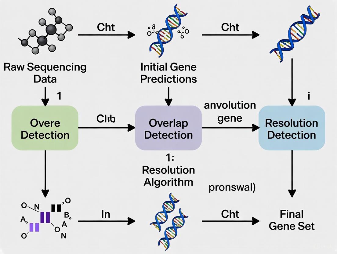
Workflow for Resolving Overlapping Gene Predictions
Regulatory Logic of an Overlapping Gene System
The Scientist's Toolkit: Research Reagent Solutions
| Reagent / Tool | Primary Function | Application Context |
|---|---|---|
| Retapamulin | A translation initiation inhibitor used in Ribo-seq protocols. | Enables precise mapping of translation start sites, crucial for identifying novel, short overlapping ORFs within larger genes [6]. |
| Monarch Spin gDNA Extraction Kit | Purifies high-quality, high-molecular-weight genomic DNA. | Provides clean, intact DNA input for whole-genome sequencing, which is foundational for accurate gene prediction and overlap detection [19]. |
| Proteinase K | A broad-spectrum serine protease for sample digestion. | Essential for lysing tissues and inactivating nucleases during DNA extraction, preventing degradation that could obscure overlapping regions [19]. |
| ORF Finder | A graphical tool for identifying all open reading frames in a sequence. | The primary bioinformatics tool for performing a six-frame translation to visually identify potential overlapping coding sequences [17]. |
| BLAST (Basic Local Alignment Search Tool) | Finds regions of local similarity between sequences. | Used to infer functional and evolutionary relationships for predicted overlapping ORFs, helping to confirm they are real genes [17]. |
Overlapping genes (OLGs), where nucleotide sequences encode multiple proteins in different reading frames, represent a fascinating aspect of genomic architecture. Once considered rare outside of viral genomes, they are now recognized as functional components in prokaryotic and eukaryotic organisms. In bacterial genome research, accurate identification and annotation of these features are crucial, as they can be sources of novel genes, play roles in gene regulation, and present significant challenges for standard annotation pipelines [6] [22]. This guide provides troubleshooting support for researchers working to resolve overlapping gene predictions in bacterial systems.
FAQ: Understanding Overlapping Genes
Q1: Are overlapping genes a common feature in bacterial genomes? Yes, overlapping genes are a recognized feature in bacterial genomes. They are functionally integrated and widespread, though their detection has been historically challenging. For instance, a recent study mapping transcriptional overlaps in Escherichia coli identified 165 convergent and 16 divergent excludons—a specific type of overlapping transcriptional unit involved in gene regulation [23].
Q2: What are the main biological functions of overlapping genes? Overlapping genes serve several key functions:
- Genome Compression: Maximizing the coding capacity of genomes under size constraints, which is particularly relevant for viruses and bacteria with small genomes [24] [6].
- Gene Regulation: Enabling coordinated or mutually exclusive expression of neighboring genes through mechanisms like transcriptional interference. The recently identified "excludons" in E. coli and Staphylococcus aureus are a prime example of this regulatory function [23].
- Generation of Novelty: Allowing new genes to originate within existing genes through a process called "overprinting," without requiring major increases in genome size [24] [6].
Q3: Why are standard gene prediction tools inadequate for detecting overlapping genes? Most standard gene prediction algorithms are designed to identify non-overlapping genes and often exclude or misannotate long protein-coding overlapping sequences. The NCBI's rules for annotating prokaryotic genes, for example, do not typically allow for genes completely embedded within another gene in a different frame without specific, individual justification [22]. Specialized computational methods are required for their detection.
Troubleshooting Guide: Resolving Overlapping Gene Annotations
Problem 1: Gene Prediction Pipeline Fails to Identify Known Overlapping Genes
Issue: Your standard annotation pipeline (e.g., using Prokka or RAST) annotates only a subset of the expected genes in a genome known to contain overlaps, such as bacteriophage ΦX174.
Solution:
- Employ Custom Annotation Pipelines: Standard tools predicted at most 7 of the 11 known genes in ΦX174. Develop a custom pipeline that combines ORF-finding with homology searches against specialized protein databases to identify all functional genes, including those in overlapping frames [10].
- Utilize Specialized Detection Tools: Use tools specifically designed for OLG detection, which rely on statistical tests beyond simple ORF identification. The following table summarizes key methods:
Table 1: Computational Tools for Detecting Overlapping Genes
| Tool Name | Methodology | Key Application | Sensitivity/Specificity Notes |
|---|---|---|---|
| Codon Permutation/Synonymous Mutation Test [24] | Identifies ORFs significantly longer than expected by chance using randomization tests. | Screening single virus/genome sequences; useful for metagenomic data. | Sensitivity improves for overlaps >50 nt; combined test offers lowest false discovery rate. |
| Synplot2 [25] | Analyzes alignments for significant reduction in variability at synonymous sites. | Requires multiple homologous sequences with a range of diversity. | 95% sensitivity on a test set of 21 known OLGs. |
| FRESCo [25] | Finds regions of excess synonymous constraint in aligned sequences. | Identifies overlaps and conserved RNA structures. | Reported 100% specificity in simulations. |
| OLGenie [25] | Calculates dN/dS ratios to estimate selection pressures on two overlapping ORFs. | Evaluates evolutionary constraints in dual-coding regions. | 66% sensitivity, 68% specificity on a known test set. |
Visual Workflow for Overlapping Gene Detection: The diagram below outlines a general computational workflow for identifying candidate overlapping genes.
Problem 2: Experimental Validation of Predicted Overlapping Genes
Issue: You have a computational prediction for a novel overlapping gene in your bacterial genome of interest and need to design experiments to validate its expression and function.
Solution:
- Proteogenomics and Ribosome Profiling: Use mass spectrometry-based proteomics to detect peptides translated from the alternative reading frame. Combine this with ribosome profiling (Ribo-seq), which maps the positions of translating ribosomes, to provide direct evidence that the overlapping ORF is translated [6]. In E. coli, the use of the antibiotic retapamulin in Ribo-seq has enabled the discovery of many novel translation initiation sites within existing genes [6].
- Transcriptional Analysis: For overlaps involving untranslated regions (UTRs), such as excludons, use strand-specific RNA-seq to confirm the presence of overlapping convergent or divergent transcripts. Tools like ExcludonFinder can systematically identify such overlaps from transcriptomic data [23].
- Functional Assays: After establishing expression, use CRISPR-based functional screens or gene knockout techniques to determine the phenotypic impact of the overlapping gene on bacterial growth, pathogenicity, or response to stress [6].
Problem 3: Resolving Overlapping Annotations in a GFF File
Issue: Your genome annotation file (GFF/GTF) contains multiple overlapping gene models that are not isoforms, and you need to resolve them to proceed with protein prediction or other downstream analyses.
Solution:
- Use the AGAT Toolkit: This suite of tools is designed for handling annotation files.
- First, merge overlapping loci that are on the same strand and of the same feature type (e.g., mRNA) using the command:
agat_convert_sp_gxf2gxf.pl --gff myFile.gff --merge_loci -o myFile_lociMerged.gff[26]. - Then, to simplify the annotation and retain only the longest isoform where multiple models exist for a gene, use:
agat_sp_keep_longest_isoform.pl --gff myFile_lociMerged.gff -o myFile_lociMerged_longestIsoform.gff[26].
- First, merge overlapping loci that are on the same strand and of the same feature type (e.g., mRNA) using the command:
- Critical Consideration: Before collapsing annotations, ensure the overlapping genes are not bona fide, distinct genes. Nested genes, where one gene resides within the intron of another, are a known biological reality in many organisms [26] [27]. Manual curation and evidence review (e.g., transcript support) are essential.
Research Reagent Solutions
Table 2: Key Reagents and Tools for Studying Bacterial Overlapping Genes
| Item/Tool | Function in Research | Specific Example/Application |
|---|---|---|
| ExcludonFinder [23] | A computational tool to map transcriptional overlaps (excludons) from RNA-seq data. | Systematically identified 181 excludons in E. coli and 38 in S. aureus from public datasets. |
| Retapamulin [6] | An antibiotic that inhibits translation initiation; used in ribosome profiling to capture novel start sites. | Enabled Ribo-seq discovery of new translation initiation sites within existing E. coli genes. |
| OGRE [28] | A bioinformatics tool to calculate and visualize overlaps between genomic regions and public annotations. | Downstream analysis to associate candidate genes with regulatory elements like promoters and TFBS. |
| Strand-specific RNA-seq | Allows precise mapping of transcripts to their DNA strand of origin, crucial for identifying antisense overlaps. | Validation of divergent and convergent transcriptional overlaps in bacterial excludons [23]. |
| PhyloCSF [25] | Uses phylogenetic codon substitution frequencies to distinguish protein-coding from non-coding regions. | Detected strong protein-coding signatures for overlapping ORFs (ORF3c, ORF9b) in sarbecoviruses. |
Advanced Detection Pipelines: Integrating Computational and Experimental Approaches
For researchers investigating bacterial genomes, a significant challenge is the accurate resolution of overlapping genes, where two or more coding sequences share the same nucleotide sequence in different reading frames. These features are crucial for understanding pathogenesis, antibiotic resistance, and genome evolution but are often fragmented or misassembled in short-read assemblies. Long-read sequencing technologies directly address this by spanning repetitive and complex genomic regions, enabling the reconstruction of complete, contiguous genomes necessary for accurate gene prediction and functional analysis. This guide provides troubleshooting and best practices to leverage these technologies effectively within your research.
Frequently Asked Questions (FAQs)
1. How does long-read sequencing specifically improve the detection of overlapping genes? Short-read sequencing often fails to span entire overlapping regions, leading to fragmented assemblies that can split these genes into separate contigs. Long-read sequencing generates reads that are thousands of bases long, which can easily span the entire length of an overlapping gene pair. This provides the necessary context to correctly assemble the region and identify the distinct, functional open reading frames (ORFs) that share the same genomic space. Accurate assembly is a prerequisite for bioinformatic tools that identify overlapping ORFs longer than expected by random chance, a key signature of functional overlapping genes [24] [6].
2. What are the key differences between major long-read sequencing platforms? The two primary long-read technologies are Pacific Biosciences (PacBio) and Oxford Nanopore Technologies (ONT). A newer method, the Illumina Complete Long Read (ICLR) assay, also shows promise [29].
Table: Comparison of Long-Read Sequencing Technologies
| Technology | Typical Read Length | Key Strength | Considerations for Bacterial Assembly |
|---|---|---|---|
| PacBio HiFi | 15,000 - 20,000 bases [30] | Very high accuracy (99.9%) [30] | Ideal for high-quality, finished genomes; excellent for resolving repeats. |
| ONT (e.g., Kit 114) | 5,000 - 10,000+ bases (ultralong possible) [31] [32] | Real-time sequencing; lower initial cost [33] | Accuracy has improved (~99% with latest chemistry) [31] [32]. |
| Illumina ICLR | ~6,000 - 7,000 bases (sub-assembled) [29] | High accuracy; low DNA input requirements [29] | Synthetic long-read method; performance in highly complex regions is evolving [29]. |
3. My long-read assembly is fragmented. What are the main causes? Fragmentation in long-read assemblies can often be traced to issues before sequencing. The most common cause is insufficient input DNA quality. Degraded or sheared DNA will not yield long reads, regardless of the platform's capabilities [18] [32]. Other factors include:
- Insufficient Sequencing Depth: While long reads require less depth than short reads, inadequate coverage fails to provide enough overlap for assemblers. For ONT, a depth of ~75x with the latest chemistry can produce high-quality finished genomes [31].
- High Error Rates: Although improving, elevated error rates can break contigs during assembly. Using the most accurate base-calling models and proper polishing is essential [33] [31].
Troubleshooting Guide
Table: Common Long-read Sequencing Issues and Solutions
| Problem | Potential Causes | Corrective Actions |
|---|---|---|
| Low Library Yield [18] | - Degraded or impure DNA input- Inaccurate quantification- Overly aggressive purification | - Re-purify input DNA; check purity (260/280 ~1.8)- Use fluorometric quantification (e.g., Qubit)- Optimize bead-based cleanup ratios [18] |
| Short Read Lengths | - DNA shearing during extraction/handling- Contaminants inhibiting enzymes- Old or expired library prep kits | - Use gentle extraction methods for HMW DNA- Avoid vortexing; pipette slowly [18]- Ensure reagents are fresh and stored correctly [32] |
| High Error Rates in Assembly | - Raw reads with low per-base accuracy- Insufficient polishing | - Use latest chemistry (e.g., ONT SQK-LSK114, PacBio HiFi)- Polish assemblies using tools like Medaka (for ONT) or with high-accuracy short reads [33] [31] |
| Adapter Dimers in Library | - Suboptimal adapter-to-insert molar ratio [18] | - Titrate adapter concentration- Include rigorous size selection to remove dimers [18] |
Experimental Protocols for Genome Assembly and Validation
Protocol 1: High-Quality Bacterial Genome Assembly using Oxford Nanopore Long Reads Only
This protocol, adapted from recent studies, allows for the generation of finished bacterial genomes without the need for complementary short-read sequencing [31].
- DNA Extraction: Extract high-molecular-weight (HMW) gDNA using a gentle kit (e.g., TIANamp Bacteria DNA Kit). Avoid vigorous mixing or freeze-thaw cycles to prevent shearing. Validate DNA purity and length using a Femto Pulse or TapeStation.
- Library Preparation & Sequencing: Prepare a sequencing library using the ONT Ligation Sequencing Kit V14 (SQK-LSK114) according to the manufacturer's instructions. Sequence on a GridION or PromethION device using an R10.4.1 flow cell. Perform base-calling in super-accuracy mode using Guppy.
- Read Filtration: Use NanoFilt to remove reads shorter than 1,000 bp and with a quality value below Q10.
- De Novo Assembly: Assemble the filtered reads using Flye (v2.8.2+) with default parameters.
- Polishing: Perform multiple rounds of error correction (typically three) using Medaka to produce a final, high-quality consensus genome (>99.99% accuracy) [31].
Protocol 2: Resolving Overlapping Genes from a Finished Genome Assembly
Once a high-quality, contiguous genome is assembled, use this bioinformatic method to identify candidate functional overlapping genes [24].
- Identify All Open Reading Frames (ORFs): Use a tool like
getorf(EMBOSS) or Prodigal to identify all possible ORFs in all six reading frames of your assembled genome. - Perform Randomization Test: For each known (annotated) gene in the genome, use a custom script to randomize the codon order while preserving the amino acid sequence of the original gene. This creates a null distribution of expected ORF lengths in the overlapping frames.
- Calculate Statistical Significance: Compare the length of the actual overlapping ORFs found in the randomized sequence to the null distribution. An ORF that is significantly longer than expected by chance (e.g., p < 0.001) is a strong candidate for a functional overlapping gene, as its length suggests evolutionary selection against stop codons [24].
- Functional Validation: Candidate genes require functional validation through laboratory techniques such as ribosome profiling or proteomics to confirm translation [6].
The Scientist's Toolkit: Essential Research Reagents & Materials
Table: Key Resources for Long-Read Genome Assembly
| Item | Function | Example Products / Tools |
|---|---|---|
| HMW DNA Extraction Kit | To isolate long, intact DNA strands crucial for long-read data. | TIANamp Bacteria DNA Kit, chemagen technology with M-PVA beads [31] [32] |
| Long-read Library Prep Kit | To prepare DNA fragments for sequencing on the chosen platform. | PacBio SMRTbell Prep Kit 3.0, ONT Ligation Sequencing Kit SQK-LSK114 [31] [32] |
| Fluorometric Quantifier | To accurately quantify double-stranded DNA concentration without contamination bias. | Qubit Fluorometer, PicoGreen [18] |
| Assembly Software | To reconstruct the genome sequence from long reads. | Flye, Canu, HiCanu, Unicycler (for hybrid assembly) [33] [31] |
| Polishing Tool | To correct systematic errors in the consensus sequence of the draft assembly. | Medaka (for ONT), Pilon (with short reads) [33] [31] |
Frequently Asked Questions (FAQs)
Q1: What are the primary differences between MAKER2, BRAKER3, and Prokka, and when should I use each one?
Table 1: Comparison of Genome Annotation Pipelines
| Feature | Prokka | BRAKER3 | MAKER2 |
|---|---|---|---|
| Primary Use Case | Rapid annotation of bacterial, archaeal, and viral genomes [34] | Accurate annotation of large, complex eukaryotic genomes [35] | Flexible annotation of eukaryotic genomes, integrating multiple sources of evidence [36] [35] |
| Annotation Method | Combined homology-based and ab initio [34] | Evidence-driven and ab initio (integrates RNA-seq and protein evidence) [35] | Evidence-driven and ab initio (can integrate multiple tools and evidences) [35] |
| Key Inputs | Genome sequence (contigs or assembled genome) [34] | Genome sequence, RNA-seq data (BAM/FASTQ/SRA), and protein database [35] | Genome sequence, and can use evidence from ESTs, proteins, and RNA-seq alignments [36] |
| Automation Level | High; self-contained [34] | High; automated model training [35] | Moderate; may require manual configuration and training of gene predictors [35] |
| Typical Runtime | Fast (minutes to hours) [34] | Varies; can be days for large eukaryotic genomes [36] | Varies; can be days to weeks for large plant genomes [36] |
Q2: How can I resolve the BRAKER3 error "error, file/folder not found: genome_gmst.gtf"?
This error often indicates a problem during the execution of the GeneMark-ETP component within BRAKER3. The troubleshooting steps are as follows [37]:
- Check GeneMark-ETP Dependencies: Ensure that all required software for GeneMark-ETP, including Perl modules, are correctly installed and accessible.
- Inspect Error Logs: Examine the detailed error logs specified in the BRAKER output (e.g.,
errors/GeneMark-ETP.stderr). Look for upstream warnings or errors, such as "Use of uninitialized value" or issues with input data parsing [37]. - Validate Input Evidence: This error can occur if GeneMark-ETP does not receive sufficient or correctly formatted evidence (RNA-seq or protein) from the input data to generate initial gene predictions. Verify the quality and alignment of your input BAM files or protein databases [37].
Q3: Why does Prokka sometimes not assign expected gene names to my bacterial genome?
Prokka assigns names based on sequence similarity to its internal databases. If expected gene names (like "lpxC") are missing from the final FAA and FFN files, but are present in the GFF or TSV files, follow this guide [38]:
- Use the
--addgenesFlag: This flag instructs Prokka to add a "gene" feature for every "CDS" feature in the output, which can help ensure gene names are propagated to all file formats. - Provide a Custom Protein Database: Use the
--proteinsflag with a GenBank or FASTA file from a closely related species. This gives Prokka higher-quality, lineage-specific references for annotation, improving the accuracy of assigned gene names [38]. - Check All Output Files: The gene names might be correctly annotated in the GFF file (
prokka.gff) and the tab-separated file (prokka.tsv). The issue might be specific to how the FASTA files are generated [38].
Troubleshooting Common Workflow Errors
BRAKER3 Installation and Initialization Issues
A common issue when installing BRAKER3 via Conda involves a Perl script failure when checking the Java version [39].
- Error Message:
Use of uninitialized value $2 in concatenation...andFailed to execute: java -version...[39]. - Solution: The script's regular expression for parsing the Java version may need adjustment. Modify the line in
braker.pl(around line 2344) as follows [39]:- Original Code (may fail):
java -version 2>&1 | grep 'openjdk version' | awk -F['''.'] -v OFS=. '{print ,}' - Modified Code:
java -version 2>&1 | grep 'java version' | awk -F '[\".]' -v OFS=. '{print $2,$3}'
- Original Code (may fail):
Prokka Annotation Refinement for Bacterial Genomes
To enhance annotation quality for a specific bacterial strain and avoid generic product names, follow this experimental protocol [34] [38]:
- Obtain Reference Annotation: Download a GenBank (.gbk) file of a closely related, well-annotated genome.
- Run Prokka with Custom Reference:
- The
--proteinsflag provides curated, lineage-specific annotation evidence. - The
--addgenesflag ensures the inclusion of gene features.
- The
- Validate Output: Check the
.gffand.tsvoutput files for the presence of your expected gene names. The final.faaand.ffnfiles should now also reflect these names [38].
Managing Long Runtimes for Large Genomes
Annotation of large eukaryotic genomes (e.g., soybean, other plants) with MAKER2 or BRAKER3 can take days to weeks [36].
- Strategy: Allocate sufficient computational resources and time from the start of your project.
- Best Practices:
- Use Pre-aligned Evidence: Providing pre-aligned evidence (e.g., protein and mRNA alignments) to MAKER2 can significantly reduce runtime [36].
- Parallelize Processing: Ensure the pipeline is configured to run on a multi-core system or high-performance computing cluster [36].
- Monitor Logs: Allow the job to process and monitor the logs for progress indicators rather than assuming the job has stalled [36].
Workflow Diagrams
BRAKER3 Eukaryotic Genome Annotation Workflow
Prokka Bacterial Genome Annotation Workflow
The Scientist's Toolkit
Table 2: Essential Research Reagent Solutions for Annotation Pipelines
| Item Name | Function in Experiment |
|---|---|
| High-Quality Genome Assembly | The foundational input for all annotation pipelines. Accuracy and contiguity are critical for correct gene structure prediction [40]. |
| RNA-seq Data (for BRAKER3/MAKER2) | Provides direct extrinsic evidence of transcribed regions, intron-exon boundaries, and splice sites, greatly improving structural annotation accuracy in eukaryotes [35]. |
| Curated Protein Database | A FASTA file of proteins from a broad clade of the target genome. Used for homology-based searches to identify conserved coding regions and assign functional domains [35]. |
| Lineage-Specific Reference Annotations (for Prokka) | A GenBank file from a closely related species. Used with the --proteins flag to significantly improve the accuracy of gene name and product assignments in bacterial genomes [38]. |
| Repeat Masking Tool (e.g., RepeatMasker) | Identifies and masks repetitive DNA sequences. This is a critical first step in eukaryotic structural annotation to prevent spurious gene predictions [40]. |
Frequently Asked Questions (FAQs)
Q1: What is a read-to-read overlap, and why is it critical in bacterial genome assembly? A read-to-read overlap is a sequence match between two reads originating from the same locus in a larger genome sequence. It is the foundational first step in the Overlap-Layout-Consensus (OLC) assembly paradigm, the dominant method for long-read assembly. In OLC, these overlaps are used to build an overlap graph, which is traversed to produce a layout of the reads and, finally, a consensus sequence. The accuracy of initial overlap detection is a major efficiency bottleneck and directly influences the quality of the final assembly [41].
Q2: My bacterial genome assembly has surprisingly long co-directional gene overlaps. Are these likely real? Probably not. Research analyzing 338 fully sequenced prokaryotic genomes indicates that very long co-directional overlaps (e.g., >60 bp) are frequently the result of annotation errors, not functional biological features. One study of 715 such long co-directional overlaps found that 100% were misannotations. The most common causes are a mispredicted start codon in the downstream gene or a frameshift mutation that fragmented a single gene into two overlapping annotations [42]. You should verify the annotation of these genes.
Q3: Which overlap detection tool is most efficient for Oxford Nanopore Technologies (ONT) data? Benchmarking studies have shown that Minimap is the most computationally efficient, specific, and sensitive method for overlap detection on ONT datasets. For Pacific Biosciences (PB) data, GraphMap and DALIGNER were identified as the most specific and sensitive tools in the tested versions [41].
Q4: How can I systematically analyze overlaps between my genomic regions and public annotations? You can use specialized bioinformatics tools like OGRE (Overlapping annotated Genomic Regions). OGRE automates the process of calculating, visualizing, and analyzing overlaps between your input regions (e.g., in BED or GFF format) and public annotations for elements like promoters, CpG islands, and transcription factor binding sites. It provides statistical summaries and easy-to-understand visualizations without requiring advanced programming skills [28].
Q5: What are the main algorithmic strategies used by overlap detection tools? Most state-of-the-art tools use a seed-and-extend approach. They first identify short, exact subsequences (seeds) shared between reads to discover candidate overlaps quickly. They then perform a more computationally intensive step to extend these seeds and verify the full overlap. These specialized algorithms are designed to handle the high error rates associated with long-read technologies like ONT and PB [41].
Troubleshooting Guides
Guide 1: Resolving Suspected Gene Annotation Errors in Bacterial Genomes
Problem: Your annotated bacterial genome contains genes with unusually long overlaps, or your overlap detection tool yields a high rate of long co-directional overlaps, which may indicate widespread annotation errors.
Investigation and Solution Protocol:
Follow this systematic protocol to identify and correct common annotation errors that cause long overlaps.
Table 1: Common Types of Long Overlap Misannotations and Their Signatures
| Error Category | Frequency | Key Indicator | Proposed Correction |
|---|---|---|---|
| 5'-end extension of downstream gene | ~57% of cases | Downstream gene is longer than its orthologs at the 5'-end; alternative upstream start codon exists. | Re-annotate the downstream gene's start codon to a downstream, conserved alternative. |
| Fragmentation of a single gene | ~23% of cases | Both overlapping genes map to a single, longer gene in a closely related species. | Merge the two gene annotations into a single gene model. |
| 3'-end extension of upstream gene | ~9.5% of cases | Upstream gene is longer than its orthologs at the 3'-end; stop codon is missing. | Identify the correct in-frame stop codon, potentially in the overlapping region. |
| 5' & 3'-end extension | ~10% of cases | A combination of the above; both genes are longer than their orthologs. | Correct both the start and stop codons of the respective genes. |
Experimental Protocol: Ortholog Comparison for Overlap Validation
- Identify Candidate Overlaps: Extract all pairs of genes with overlaps longer than a chosen threshold (e.g., 60 bp) from your genome annotation file (GFF/GBK).
- Retrieve Orthologs: For each gene in the overlapping pair, perform a BLAST search against a database of well-annotated reference genomes from closely related species to identify orthologous sequences.
- Compare Gene Lengths: Align the protein or nucleotide sequence of your query gene with its orthologs. A significant length discrepancy (extension or truncation) in the query gene is a strong indicator of a misannotation.
- Manual Curation:
- For a suspected 5'-end extension, scan the region upstream of the current downstream gene's start codon for in-frame ATG, GTG, or TTG codons that would bring its length in line with its orthologs.
- For a suspected fragmentation, check if the concatenated sequence of the two overlapping genes produces a single, contiguous open reading frame that aligns well with a single ortholog.
- For a suspected 3'-end extension, examine the end of the upstream gene for a point mutation or frameshift that has disrupted the native stop codon, causing translation to extend to the next available in-frame stop.
- Re-annotate and Re-assess: Implement the corrected gene models and re-check that the overlap is either resolved or reduced to a biologically plausible size.
The following diagram illustrates the logical workflow for diagnosing these common misannotation types.
Guide 2: Troubleshooting Overlap Detection Tool Performance
Problem: Your overlap detection software (e.g., Minimap, DALIGNER, GraphMap) is running slowly, consuming excessive memory, or producing an unexpectedly low number of overlaps.
Investigation and Solution Protocol:
Table 2: Troubleshooting Overlap Detection Tools
| Symptom | Potential Cause | Solution |
|---|---|---|
| Low number of detected overlaps | High sequencing error rate overwhelming the seed-based detection. | Use a tool specifically designed for error-prone long reads (e.g., Minimap for ONT). Pre-correct reads using an error-correction step before overlapping. Adjust the tool's sensitivity parameters (e.g., reduce the minimum seed length). |
| High memory usage | The algorithm's design or large genome size. | Check if the tool has a streaming or batch-processing mode. Allocate more RAM if possible. For large genomes, use tools known for better scalability like Minimap [41]. |
| Long run time | Non-optimized algorithms for the data type or system. | Ensure you are using the most computationally efficient tool for your data type (e.g., Minimap for ONT) [41]. Utilize multi-threading if supported by the tool. |
| Imprecise overlap boundaries | Extension step is not accurately aligning error-rich regions. | Adjust alignment scoring parameters within the tool. Post-process overlaps with a more sensitive local aligner. |
The Scientist's Toolkit
Table 3: Essential Bioinformatics Tools and Resources for Overlap Analysis
| Tool or Resource | Primary Function | Relevance to Overlap Detection |
|---|---|---|
| Minimap [41] | Sequence overlap detection and alignment | Fast and efficient overlap detection for long reads, particularly from Oxford Nanopore Technologies. |
| GraphMap [41] | Sequence overlap detection and alignment | Sensitive and specific overlap detection for Pacific Biosciences reads. |
| DALIGNER [41] | Sequence overlap detection | Sensitive and specific overlap detection for Pacific Biosciences reads. |
| OGRE [28] | Genomic region overlap analysis | Calculates and visualizes overlaps between input genomic regions and public annotations (e.g., promoters, CpG islands). |
| ProOvErlap [43] | Statistical feature overlap/proximity | Assesses the statistical significance of overlaps between genomic intervals (BED files) using randomization tests. |
| Ortholog Databases (e.g., NCBI) | Comparative genomics | Provides sequences for validating gene models and identifying potential annotation errors causing long overlaps [42]. |
Protein Identification through Reporter Transposon-Sequencing (PIRT-Seq) represents a groundbreaking genetics-based approach designed to identify translated open reading frames (ORFs) throughout bacterial genomes at scale and independent of existing genome annotation. This high-resolution whole-genome assay overcomes the significant limitations of traditional protein detection methods, which often overlook small or overlapping genes. The advent of high-density mutagenesis and data-mining studies suggests the existence of further coding potential within bacterial genomes, as small or overlapping genes are prevalent across all domains of life but frequently escape detection due to annotation challenges. PIRT-Seq addresses this gap by combining transposon insertion sequencing using a dual-selection transposon with a translation reporter, enabling condition-dependent identification of protein coding sequences (CDSs) in a high-throughput manner [44].
When applied to the well-characterised species Escherichia coli, PIRT-Seq revealed over 200 putative novel protein coding sequences, mostly comprising short CDSs (<50 amino acids). These included highly conserved proteins neighboring functionally important genes, with chromosomal tags successfully validating the expression of selected CDSs. As a complementary method to whole cell proteomics and ribosome trapping, PIRT-Seq provides researchers with a powerful tool for future high-throughput genetics investigations to determine the existence of unannotated genes across multiple bacterial species [44]. This technology is particularly valuable in the context of resolving overlapping gene predictions in bacterial genomes research, as it directly identifies translated regions regardless of their genomic arrangement or annotation status.
Frequently Asked Questions (FAQs)
Q1: What makes PIRT-Seq superior to traditional annotation methods for identifying overlapping genes? PIRT-Seq operates independently of genome annotation biases that typically exclude overlapping genes. Standard genome annotation programs routinely disallow overlapping genes with long protein-coding overlapping sequences outside of viruses, and NCBI's rules for prokaryotic gene annotation do not permit genes completely embedded in another gene in a different frame without individual justification [22]. PIRT-Seq bypasses these limitations by directly assessing translation through a reporter system, enabling detection of overlapping ORFs that conventional pipelines would miss.
Q2: Can PIRT-Seq distinguish between functional coding sequences and spurious ORFs? Yes, this is a key strength of the technology. By requiring both transposon insertion and translation reporter activity, PIRT-Seq specifically identifies ORFs that are actually translated into proteins under the experimental conditions. This functional validation is crucial for distinguishing genuine coding sequences from the numerous spurious ORFs present in bacterial genomes, particularly for small or overlapping genes where traditional sequence-based prediction algorithms have high error rates [44].
Q3: What types of novel genes has PIRT-Seq successfully identified? In proof-of-concept studies on E. coli, PIRT-Seq discovered over 200 putative novel protein coding sequences. These were predominantly short CDSs (<50 amino acids) and included proteins that are highly conserved and neighbor functionally important genes. The method is particularly effective for identifying small proteins and short open reading frame encoded peptides that are often overlooked in standard genome annotations [44].
Q4: How does PIRT-Seq handle condition-dependent gene expression? A significant advantage of PIRT-Seq is its utility as a high-throughput method for testing conditional gene expression. The approach can identify protein CDSs that are expressed under specific experimental conditions, providing insights into the condition-dependent translatome that would be missed by static genome annotation methods [44].
Q5: What bacterial species are suitable for PIRT-Seq analysis? While the initial validation was performed in E. coli, the method is designed to be adaptable to multiple bacterial species. The developers anticipate it will serve as a starting point for future high-throughput genetics investigations to determine the existence of unannotated genes across diverse bacterial species [44].
Troubleshooting Guide: Common Experimental Issues and Solutions
Library Preparation and Quality Control Issues
Table 1: Troubleshooting Library Preparation and Quality Control in PIRT-Seq
| Problem | Possible Causes | Recommended Solutions |
|---|---|---|
| Low library yield | Poor input DNA quality, inaccurate quantification, inefficient ligation | Re-purify input DNA using clean columns or beads; use fluorometric quantification (Qubit) rather than UV; titrate adapter:insert molar ratios; ensure fresh ligase and buffer [18] |
| Adapter dimer formation | Excess adapters, improper adapter-to-insert ratio, inefficient purification | Optimize adapter concentration; use bead cleanup with adjusted bead:sample ratios; implement two-step indexing instead of one-step PCR [18] |
| Size selection issues | Incorrect bead ratio, over-drying beads, inefficient washing | Use correct bead:sample volume ratio; avoid over-drying beads (keep shiny, not cracked); ensure adequate washing steps; verify size distribution with BioAnalyzer [18] |
| Amplification bias | Too many PCR cycles, enzyme inhibitors, primer exhaustion | Reduce number of amplification cycles; re-purify to remove inhibitors; optimize primer concentrations; use high-fidelity polymerases [45] |
| Cross-contamination between wells | Improper pooling, splash between wells during processing | Implement careful liquid handling techniques; use seal plates properly during incubation; include control wells to monitor contamination [18] |
Transposon Integration and Reporter Expression Issues
Table 2: Troubleshooting Transposon Integration and Reporter Expression in PIRT-Seq
| Problem | Possible Causes | Recommended Solutions |
|---|---|---|
| Poor transposon integration efficiency | Suboptimal transposase activity, incorrect DNA quantity, inhibitor carryover | Titrate transposase concentration; verify DNA quality and quantity; ensure fresh reaction buffers; include positive control for integration [44] |
| Inconsistent reporter expression | Position effects, poor translation initiation, genetic context | Test multiple insertion sites per gene; verify reporter construct design; check for required genetic elements (RBS, start codon); validate with control constructs [44] |
| High background signal | Non-specific reporter expression, false positive insertions | Optimize selection conditions; include dual-selection strategy; implement rigorous statistical cutoffs; verify hits with orthogonal methods [44] |
| Missing expected genes | Insufficient library coverage, essential genes, condition-specific expression | Achieve >100x library coverage; use condition-appropriate growth conditions; combine data from multiple conditions; employ complementary approaches [44] |
| Difficulty amplifying insertion sites | Complex genomic regions, inefficient PCR, primer issues | Optimize PCR conditions with additives for GC-rich regions; use polymerases with high processivity; design multiple primer sets; extend amplification times [45] |
Experimental Protocol and Workflow
Detailed PIRT-Seq Methodology
The PIRT-Seq protocol integrates dual-selection transposon mutagenesis with a translation reporter system in a streamlined workflow:
Step 1: Library Construction and Transposon Mutagenesis Begin by preparing the bacterial strain of interest and employing a dual-selection transposon system that incorporates a translation reporter. The transposon design is critical—it must include selectable markers and a reporter construct that can indicate translational activity. The transposon mutagenesis is performed to achieve comprehensive coverage, typically aiming for saturating mutagenesis where each gene is targeted multiple times across the population [44].
Step 2: Selection and Sequencing Apply dual selection to enrich for productive transposon insertions that generate in-frame fusions with translated ORFs. This selection process is crucial for filtering out non-productive insertions and background noise. Following selection, harvest the genomic DNA and prepare sequencing libraries specifically designed to capture transposon-genome junctions. High-throughput sequencing is then performed to map insertion sites and quantify reporter activity [44].
Step 3: Data Analysis and ORF Identification Process the sequencing data to identify translated ORFs through a multi-step bioinformatic pipeline:
- Map sequencing reads to the reference genome
- Identify transposon insertion sites and their frequency
- Analyze reporter expression patterns associated with each insertion
- Integrate insertion and expression data to call translated ORFs
- Compare results with existing annotation to identify novel genes
- Validate selected novel CDSs using chromosomal tags [44]
Workflow Visualization
Diagram 1: PIRT-Seq experimental workflow showing major steps from bacterial culture to validation.
Research Reagent Solutions
Table 3: Essential Research Reagents for PIRT-Seq Experiments
| Reagent/Category | Specific Function | Recommendations and Notes |
|---|---|---|
| Dual-selection transposon | Enables selection of productive insertions and translation reporting | Custom design required; must include selectable markers and translation reporter; optimize for your bacterial system [44] |
| High-fidelity DNA polymerase | Amplification of transposon insertion sites | Choose polymerases with high processivity for complex templates; use hot-start versions to prevent non-specific amplification [45] |
| Library preparation kit | Construction of sequencing libraries | Select kits compatible with transposon junction sequencing; consider cost-effectiveness for high-throughput applications [18] |
| DNA purification beads | Size selection and cleanup | Magnetic beads preferred for high-throughput processing; optimize bead:sample ratio for your fragment sizes [18] |
| Quantification reagents | Accurate measurement of DNA concentrations | Use fluorometric methods (Qubit) rather than spectrophotometry for precise quantification of usable DNA [18] |
| Selection antibiotics | Enrichment of successful transposon integrations | Titrate concentrations carefully; use fresh stocks; include appropriate controls for selection efficiency [44] |
| Cell lysis reagents | Release of nucleic acids for library prep | Optimize for bacterial species; ensure complete lysis while preserving DNA integrity [45] |
| Sequence adapters | Compatibility with sequencing platform | Include unique barcodes for multiplexing; verify compatibility with your transposon design [18] |
Data Interpretation Framework
Analyzing and Validating Results
Distinguishing True Positive ORFs from Background Noise The PIRT-Seq data analysis pipeline requires careful statistical handling to distinguish genuine translated ORFs from background noise. Implement a multi-step filtering approach that considers both the density of transposon insertions and the strength of translation reporter signal. Genes with statistically significant reporter expression and a pattern of permissive insertion sites should be prioritized for further validation. For overlapping gene predictions, pay particular attention to regions where insertions in different reading frames produce distinct reporter outputs, as this may indicate multiple functional coding sequences in the same genomic location [44] [22].
Integration with Existing Genomic Data Cross-reference PIRT-Seq findings with existing genome annotations and complementary functional genomics data. Look for conservation patterns across related bacterial species, as conserved overlapping genes are more likely to represent functional coding sequences rather than random ORFs. For genes completely embedded within annotated genes in different reading frames, examine the constraint on sequence evolution—natural OLGs often show specific patterns of purifying selection that maintain function in both reading frames simultaneously [6] [22].
Data Analysis Visualization
Diagram 2: PIRT-Seq data analysis pipeline from raw sequencing data to validated ORF predictions.
Advanced Applications in Bacterial Genomics
PIRT-Seq provides particularly powerful insights for investigating overlapping genes in bacterial genomes, which represent a fascinating aspect of genomic architecture with important evolutionary and functional implications. When applying PIRT-Seq to this specific challenge, researchers should consider several key aspects of overlapping gene biology:
Evolutionary Constraints on Overlapping Genes Natural overlapping genes face unique evolutionary constraints as mutations in overlapping regions can potentially affect multiple proteins simultaneously. Research has shown that protein domains from diverse bacteria can be synthetically constructed to overlap while retaining high similarity to natural sequences, with approximately 10% of constructed sequences being indistinguishable from typical sequences in their protein family [22]. This surprising flexibility is largely due to the redundancy of the genetic code and evolutionary exchangeability of many amino acids. When interpreting PIRT-Seq results for overlapping genes, look for evidence of these evolutionary constraints, such as specific patterns of sequence conservation that maintain function in both reading frames.
Functional Implications of Gene Overlaps Overlapping genes in bacteria are not merely genomic curiosities—they can play important roles in gene regulation and cellular function. Same-strand overlapping gene pairs may enable efficient co-expression of functionally related proteins, while antisense overlaps could create regulatory interactions between the overlapping genes [22]. PIRT-Seq's ability to assess translation under different conditions makes it particularly valuable for investigating these potential regulatory functions. When designing PIRT-Seq experiments focused on overlapping genes, include multiple growth conditions to capture condition-specific overlapping translation events that might be missed in standard laboratory conditions.
The integration of PIRT-Seq with other functional genomics approaches creates a powerful framework for comprehensively characterizing the coding potential of bacterial genomes, particularly for challenging cases involving small proteins, overlapping genes, and condition-specific coding sequences that have historically escaped detection through conventional annotation pipelines.
Ribo-Seq and Retapamulin-Assisted Ribosome Profiling for Translational Evidence
Ribosome Profiling (Ribo-seq) has revolutionized the study of gene expression by providing a snapshot of translation at codon resolution. For bacterial genomics, where overlapping genes and complex genetic architectures are common, this technique is invaluable. Retapamulin-assisted Ribo-seq (Ribo-RET) represents a significant methodological advancement, specifically enabling the genome-wide mapping of alternative translation initiation sites. This technical support center provides a comprehensive guide to implementing these techniques within the context of resolving overlapping gene predictions in bacterial genomes, addressing common challenges, and providing validated solutions for researchers and drug development professionals.
Core Methodology and Reagents
Research Reagent Solutions
The following table details the key reagents and their specific functions in Ribo-RET experiments.
| Reagent Name | Function in Experiment | Key Usage Notes |
|---|---|---|
| Retapamulin (RET) | Arrests initiating ribosomes at start codons by binding the peptidyl transferase center (PTC) and preventing the first peptide bond formation. [46] | Use at 100-fold the minimal inhibitory concentration (MIC); treatment for 5 minutes is sufficient to stall initiation complexes. [46] |
| Tetracycline (TET) | Prevents aminoacyl-tRNAs from entering the ribosomal A-site; can inhibit both initiation and elongation. [46] | Less specific than RET for initiation mapping; leads to a broader and smaller start-codon peak in metagene analysis. [46] |
| RNase I | Digests mRNA regions not protected by ribosomes, generating ribosome-protected footprints (RPFs). [47] | Standard enzyme for bacterial Ribo-seq; concentration must be optimized to avoid over- or under-digestion. [47] |
| Micrococcal Nuclease (MNase) | Digests DNA and RNA; used in some single-cell Ribo-seq protocols as its activity can be stringently controlled by Ca²⁺ chelation. [47] | Has A/U cleavage preference, which can hamper precise determination of footprint boundaries; requires computational correction. [47] |
| Cell-free Translation System | An in vitro system (e.g., from E. coli) used to validate translation initiation at candidate start codons identified by Ribo-RET. [46] | Used in conjunction with toeprinting assays to confirm RET-induced stalling at specific start codons. [46] |
Experimental Protocols
Standard Ribo-RET Protocol for Mapping Bacterial Translation Initiation Sites
This protocol is adapted from the work of Meydan et al. (2019) and is designed for mapping translation initiation sites (TIS) in Escherichia coli. [46] [48] [49] It can be adapted for other bacterial species with appropriate optimization.
Step-by-Step Procedure:
Cell Culture and Treatment:
- Grow the bacterial strain (e.g., E. coli BW25113 ΔtolC) to the desired growth phase (typically mid-log phase).
- Divide the culture into two aliquots: one for the experimental group and one as an untreated control.
- Add Retapamulin to the experimental culture to a final concentration of 100-fold the MIC. For the control, add an equivalent volume of solvent (e.g., DMSO).
- Incubate both cultures for 5 minutes with aeration.
Cell Harvesting and Lysis:
- Rapidly harvest cells by centrifugation and immediately flash-freeze the cell pellets in liquid nitrogen.
- Lyse the frozen cell pellets using a cryogenic milling apparatus or a commercial lysis buffer suitable for preserving ribosome-mRNA complexes. The lysis buffer should contain cycloheximide (or an equivalent inhibitor for bacteria) to arrest ribosome translocation during the process.
Ribosome Footprint Generation:
- Digest the lysate with RNase I. The concentration and digestion time must be empirically determined to generate ribosome-protected fragments (RPFs) of ~28-30 nucleotides, which correspond to a single ribosome.
- Stop the RNase digestion, and clarify the lysate by centrifugation.
Footprint Purification:
- Isolate the monosome fraction by sucrose density gradient centrifugation.
- Extract the RNA from the purified monosome fraction using acid phenol-chloroform.
- Isulate the RPFs (~28-30 nt) by size selection on a denaturing polyacrylamide gel.
Library Preparation and Sequencing:
- Deplete the rRNA from the size-selected RNA sample using commercially available kits.
- Construct a sequencing library for the RPFs. This typically involves end-repair of the RNA fragments, adapter ligation, reverse transcription, and PCR amplification.
- Perform deep sequencing on an Illumina platform to generate single-end reads.
Validation Protocol: In Vitro Translation and Toeprinting
To biochemically validate internal translation initiation sites (iTIS) discovered by Ribo-RET, perform an in vitro toeprinting assay. [46]
- Template Preparation: Clone the gene of interest, including its upstream regulatory sequence, into a suitable plasmid under the control of a bacteriophage promoter (e.g., T7).
- In Vitro Transcription: Generate mRNA templates by in vitro transcription from the linearized plasmid.
- Translation Reaction: Incubate the mRNA with an E. coli cell-free translation system under conditions that support protein synthesis.
- Antibiotic Stalling: Set up parallel reactions, adding Retapamulin or Tetracycline to the translation mix.
- Primer Extension: Isolate the RNA from the reactions and use a fluorescently labeled DNA primer that is complementary to a region downstream of the putative start codon. Perform reverse transcription.
- Analysis: Resolve the cDNA products on a sequencing gel. A reverse transcriptase that stalls upon encountering a ribosome bound to a start codon will produce a truncated cDNA product ("toeprint"). A strong toeprint signal at a specific codon in the RET-treated sample, but not in the control, confirms it as a bona fide translation initiation site.
Workflow Visualization
The following diagram illustrates the logical workflow and key decision points in a Ribo-RET experiment for bacterial genomes.
Frequently Asked Questions (FAQs) and Troubleshooting
Experimental Design and Execution
Q1: Why is Retapamulin preferred over other antibiotics like Tetracycline for mapping translation initiation sites?
A: Retapamulin exhibits a superior specificity for stalling initiating ribosomes. It binds the peptidyl transferase center and allows the 70S initiation complex to assemble at the start codon but prevents the formation of the first peptide bond. [46] In contrast, Tetracycline inhibits the entry of aminoacyl-tRNAs into the A-site and can stall ribosomes during both initiation and elongation, leading to a noisier signal and making it difficult to distinguish initiating ribosomes from elongating ones. [46] Ribo-RET produces a sharper, more dramatic peak of ribosome density exclusively at start codons.
Q2: What is a key consideration when treating cells with Retapamulin before Ribo-seq?
A: The duration of treatment is critical. A 5-minute treatment with a high concentration (100x MIC) is used to ensure that elongating ribosomes have enough time to run off the mRNA, while initiating ribosomes remain trapped at the start codons. [46] Shorter treatments may not clear elongating ribosomes, contaminating the initiation signal.
Data Analysis and Computational Challenges
Q3: Our Ribo-seq pipeline failed during the P-site identification step with an error. What could be the cause?
A: This is a known issue in some Ribo-seq analysis pipelines (e.g., Ribowaltz in the riboseq-flow). The error "Process RIBOSEQ:IDENTIFY_PSITES terminated with an error exit status (134)" is often related to insufficient memory allocation. [50] As a temporary workaround, you can set the skip_psite parameter to true to complete the rest of the analysis. Monitor the repository of your chosen pipeline for bug fixes and updates addressing this memory issue.
Q4: For bacterial Ribo-seq, is it better to align reads to the genome or to the transcriptome?
A: This is a nuanced decision.
- Genome-based alignment is generally suitable and avoids the loss of reads that map to exons shared by multiple transcript isoforms, which is less of a concern in bacteria. [51] It also allows for the discovery of unannotated, off-frame, or overlapping open reading frames (ORFs).
- Transcript-based alignment requires a well-annotated transcriptome. In eukaryotes, a significant proportion of reads mapping to shared exons can be lost if only uniquely mapped reads are considered. [51] This approach is less common for bacterial Ribo-seq where the primary goal is often novel ORF discovery.
Q5: How does Ribo-RET help resolve overlapping gene predictions in bacterial genomes?
A: Traditional gene finders can miss overlapping or internal ORFs. Ribo-RET provides direct translational evidence by revealing ribosomes stalled at the start codons of these unannotated ORFs. [46] [52] If a ribosome is stalled at an AUG (or other start codon) within an annotated gene—either in-frame or out-of-frame—it provides strong evidence for an internal translation initiation site (iTIS). This indicates that the genomic locus produces more than one protein, thereby expanding the functional proteome and refining genome annotation. [46] [49]
Technical Optimization
Q6: We are working with low-input bacterial samples. Are there adapted Ribo-seq protocols?
A: Yes, recent advancements have led to protocols for low-input and even single-cell Ribo-seq, though these are more established in eukaryotic systems. Techniques like LiRibo-seq and Ribo-lite employ ligation-free, one-pot library preparation to minimize sample loss and can work with as few as 1,000 cells. [47] These methods often skip the rRNA depletion step to further reduce material loss, though this may require deeper sequencing. The field is rapidly evolving, and these methods are becoming more accessible.
Proteogenomics represents a powerful intersection of genomics and proteomics, enabling the discovery and validation of novel protein sequences, including overlapping genes (OLGs), which were once thought to be rare outside of viral genomes [6]. In bacterial genomics, OLGs are adjacent genes that share at least one nucleotide in their coding sequences and are a consistent feature, with approximately one-third of all genes in microbial genomes being overlapping [5]. The validation of these predicted proteins presents unique challenges, as standard protein databases used in mass spectrometry (MS) often lack these non-canonical variations. This technical support center provides targeted troubleshooting guides and FAQs to assist researchers in overcoming the specific obstacles encountered when using mass spectrometry to validate overlapping proteins in bacterial systems, thereby supporting the broader research aim of resolving overlapping gene predictions.
Troubleshooting Guide: Common Experimental Obstacles and Solutions
| Problem Area | Specific Issue | Potential Cause | Recommended Solution |
|---|---|---|---|
| Peptide Identification | Low peptide counts or coverage for a predicted protein [53]. | Protein abundance is too low; protein loss during sample prep; suboptimal peptide size after digestion. | Scale up sample input; use protein concentration methods (e.g., cell fractionation or immunoprecipitation); optimize digestion time or use a combination of proteases (double digestion) [53]. |
| Peptide Identification | "No significant results" despite good spectra. | Standard protein database lacks the novel overlapping protein sequence. | Construct a sample-specific custom database using proteogenomics: translate the bacterial genome or assembled transcriptome in six frames [54] [55]. |
| Sample Preparation | Protein degradation during processing. | Action of native proteases in the sample. | Add a broad-spectrum, EDTA-free protease inhibitor cocktail (e.g., PMSF) to all buffers during preparation. Remove inhibitors before the trypsin digestion step [53]. |
| Sample Preparation | Keratin or polymer contamination in spectra. | Contamination from dust, skin, hair, or lab plastics. | Use filter tips, single-use pipettes, and HPLC-grade water. Avoid autoclaving plastics and detergents for glassware. Wear gloves and a mask during sample handling [53] [56]. |
| Database Search & FDR | Reduced peptide identification sensitivity. | Searching an excessively large, custom proteogenomic database [55]. | Use a reduced transcriptome-informed database or apply post-search filtering based on transcript expression evidence. Consider FDR control methods robust to small database sizes [55]. |
| Database Search & FDR | Anti-conservative, inaccurate False Discovery Rate (FDR). | Using Target-Decoy Competition (TDC) with an excessively small, reduced database [55]. | Employ alternative FDR control methods that are less sensitive to database size to ensure the robustness of biological conclusions [55]. |
Frequently Asked Questions (FAQs)
General Proteogenomics Concepts
Q1: What is proteogenomics and why is it crucial for studying overlapping genes in bacteria? Proteogenomics is the use of genomic or transcriptomic nucleotide sequencing data to create customized protein databases for mass spectrometry searching [54]. This is essential for validating overlapping genes because standard databases contain a generic set of proteins and lack the sample-specific variations, including the unique protein sequences resulting from overlapping reading frames, precluding their detection without a custom database [54] [6].
Q2: How common are overlapping genes in bacterial genomes? Overlapping genes are a consistent feature across all sequenced microbial genomes. A strong linear relationship exists between the total number of genes and the number of overlapping genes, with approximately one-third of all genes in a genome being part of an overlapping pair [5].
Q3: What are the main types of gene overlaps? Overlaps are categorized by the relative direction of the gene pairs:
- Tandem Overlaps (→→): Genes on the same strand. These constitute the majority (~84%) of overlaps [5].
- Antiparallel Overlaps (→← or ←→): Genes on opposite strands. These are less common (~16%) [5].
Experimental Design and Sample Preparation
Q4: What are the critical steps to consider before starting a mass spectrometry experiment? Before beginning, define your biological question and confirm MS is the right tool. Assess your sample type, the abundance of your target protein(s), and how to maintain stable protein modifications. Plan to avoid contaminants and include appropriate controls. Decide on the digestion enzyme and analysis software beforehand [53].
Q5: How should I handle and store my protein samples to ensure stability? Keep all protein samples at a low temperature during processing (4°C) and store them frozen at -20°C to -80°C. When storing gel pieces, they can be kept at 4°C for short periods (1-2 weeks) or at -20°C to -80°C for longer-term storage without affecting subsequent MS identification [53] [56].
Q6: Which staining method is better for gel-based samples, Coomassie or silver staining? Coomassie brilliant blue staining is preferred. While silver staining is acceptable, it has a slightly lower identification success rate. Using tandem MS for silver-stained proteins can greatly improve the identification rate [56].
Data Acquisition and Analysis
Q7: My organism is not a model bacterium. How can I achieve successful protein identification? If your specific bacterium is not well-annotated, you can achieve successful identification by using the protein database of the closest, well-annotated model organism. If spectra are high-quality but yield no matches, this may indicate a novel protein, and de novo sequencing technologies should be used for in-depth analysis [56].
Q8: What are the key parameters to evaluate the success of a mass spectrometry identification?
- Intensity: Measures peptide abundance [53].
- Peptide Count: The number of different peptides detected from the same protein. A low count may indicate low abundance or suboptimal digestion [53].
- Coverage: The percentage of the protein's sequence covered by the identified peptides. In complex samples, 1-10% can be sufficient for identification [53].
- Statistical Significance (P-value/Q-value/Score): Indicates the confidence that a peptide identification is not a random event. A P-value/Q-value of < 0.05 is generally considered significant [53].
Q9: What is the difference between primary and tandem (secondary) mass spectrometry?
- Primary MS (Peptide Mass Fingerprinting, PMF): Identifies proteins by accurately measuring the mass of enzymatic fragments and comparing them to a theoretical library. It is less reliable for complex samples [56].
- Tandem MS (MS/MS): Selects individual peptides for fragmentation, generating sequence data that provides higher reliability and is the standard for proteogenomic applications [56].
Experimental Workflow & Visualization
The following diagram illustrates the core proteogenomic workflow for validating overlapping proteins, from sample preparation to final database search and validation.
Research Reagent Solutions
The table below lists key reagents and materials essential for conducting a successful proteogenomics experiment for overlapping protein validation.
| Item | Function / Application | Key Considerations |
|---|---|---|
| Protease Inhibitor Cocktail (EDTA-free) | Prevents protein degradation during sample preparation by inhibiting a broad range of proteases (aspartic, serine, cysteine) [53]. | Must be EDTA-free and removed before the trypsin digestion step. PMSF is a recommended component [53]. |
| Trypsin (Protease) | The standard enzyme for proteolytic digestion in bottom-up proteomics, cleaving proteins into peptides at the C-terminal side of lysine and arginine [53]. | Digestion time or protease type may need optimization. A "double digestion" with a second protease can be used for problematic proteins [53]. |
| High-pH Reversed-Phase Peptide Fractionation Kit | Reduces sample complexity by fractionating peptides prior to LC-MS analysis, increasing the number of quantifiable peptides/proteins in complex samples [57]. | Particularly useful for multiplexed samples to improve depth of analysis. |
| Pierce Quantitative Fluorometric Peptide Assay | Accurately quantifies peptide concentration after digestion and clean-up [57]. | Ensures equal peptide amounts are loaded for each LC-MS analysis, critical for reproducibility. |
| LC-MS/MS System Suitability Standard | A standardized protein or peptide digest used to calibrate and assess the performance of the LC-MS/MS system [57]. | Verifies system performance before running valuable experimental samples; uses calibration solutions for recalibration. |
The table below summarizes key quantitative findings from research on overlapping genes and proteogenomics, which can guide experimental expectations and data interpretation.
| Metric | Observed Value / Range | Context / Interpretation |
|---|---|---|
| Frequency of Overlapping Genes | ~33% of all genes [5] | Consistent across Eubacteria, Archaebacteria, plasmids, and chromosomes. |
| Distribution of Overlap Types | Tandem (→→): 84%; Antiparallel (→←/←→): 16% [5] | Based on analysis of all publicly available microbial genomes. |
| Overlap Size Distribution | >70% are <15 bp; >85% are <30 bp [5] | Overlap sizes are skewed towards shorter lengths. |
| Successful PMF Identification Score | Score > 60 (P < 0.05) [56] | For peptide mass fingerprinting (primary MS). |
| Successful Tandem MS Score | Score > 60, or score <60 with ≥1 peptide score >30 [56] | For tandem mass spectrometry (MS/MS). |
| Typical Protein Coverage | 1-10% in complex proteome samples [53] | This level of coverage is often sufficient for confident protein identification. |
Core Concepts: Genomic Context and Function Inference
What is the principle of "guilt by association" in genomics?
The core hypothesis is that "you shall know a gene by the company it keeps". Functionally related genes in prokaryotic genomes are often positioned next to each other in operons or gene clusters. This genomic colocalization allows the function of an unknown gene to be inferred from its neighboring, functionally characterized genes [58].
How can NLP models leverage this principle for function prediction?
Generative genomic language models, such as Evo, learn the semantic relationships between genes across prokaryotic genomes. By training on vast genomic datasets, these models learn the distributional semantics of gene function. This enables a technique called semantic design, where a model can be prompted with a DNA sequence of known function to generate novel, functionally related sequences, effectively performing a genomic 'autocomplete' [58].
Troubleshooting Common Experimental & Computational Issues
Our semantic design experiments are generating sequences with low functional enrichment. How can we improve this?
Low functional enrichment often stems from suboptimal prompting strategies. The Evo model's performance was significantly enhanced by using a structured, multi-faceted prompting approach [58].
Solution: Implement a multi-context prompting strategy. Do not prompt with only a single gene sequence. Instead, curate a set of prompts that include:
- The toxin and antitoxin gene sequences.
- The reverse complements of these sequences.
- The upstream genomic context of the toxin/antitoxin locus.
- The downstream genomic context of the toxin/antitoxin locus. This approach leverages the model's understanding of operonic structure and was key to successfully generating functional toxin-antitoxin systems [58].
We are encountering memory errors during the aggregation steps of our gene-variant workflow. What is the likely cause and solution?
This is a common issue when processing genes with an unusually high number of variants or very long genes. Genes like RYR2 or SCN5A are frequently problematic [59].
Solution: Increase the memory allocation for specific tasks in your workflow. The following table summarizes the recommended changes from default values for a WDL-based workflow:
Table: Recommended Memory Allocation for Gene-Variant Workflow Tasks
| Workflow File | Task Name | Parameter | Default Value | Recommended Value |
|---|---|---|---|---|
quick_merge.wdl |
split |
memory |
1 GB | 2 GB |
quick_merge.wdl |
first_round_merge |
memory |
20 GB | 32 GB |
quick_merge.wdl |
second_round_merge |
memory |
10 GB | 48 GB |
annotation.wdl |
fill_tags_query |
memory allocation |
2 GB | 5 GB |
annotation.wdl |
annotate |
memory allocation |
1 GB | 5 GB |
annotation.wdl |
sum_and_annotate |
memory allocation |
5 GB | 10 GB |
Source: Adapted from Genomics England troubleshooting guide [59]
For autosomal genes, why do we sometimes observe haploid (hemizygous-like) calls (AC_Hemi_variant > 0) in our data?
This occurs when a variant is located within a known deletion on the other chromosome for the same sample. These haploid calls are not an error in the aggregation process but originate from the single-sample gVCFs [59].
Solution: Interpret these calls in the context of adjacent variants. For example, a haploid ALT call for an A>T SNP may be explained by a heterozygous call for a 2 bp deletion immediately upstream on the other chromosome. The SNP is called as haploid because it is located within this deletion [59].
Experimental Validation Protocols
Protocol: Validating Generated Toxin-Antitoxin Systems using Growth Inhibition Assays
This protocol is used to experimentally test the function of AI-generated toxin-antitoxin (T2TA) systems [58].
1. Principle: T2TA systems consist of a toxin protein that inhibits bacterial growth under stress and an antitoxin that neutralizes the toxin. Functional validation involves expressing the generated toxin gene and observing growth inhibition, followed by co-expressing the generated antitoxin to demonstrate rescue.
2. Reagents and Materials:
- Bacterial expression strain (e.g., E. coli)
- Plasmid vectors for inducible expression
- LB broth and agar plates
- Appropriate antibiotics for selection
- Inducer (e.g., IPTG)
3. Procedure:
- Cloning: Clone the AI-generated toxin gene (e.g.,
EvoRelE1) into an inducible expression plasmid. - Toxin Assay: Transform the toxin plasmid into the expression strain. Grow cultures with and without inducer and measure optical density (OD600) over time.
- Antitoxin Assay: Co-clone or co-transform the generated antitoxin gene with the toxin gene. Repeat the growth curve measurements with and without induction.
- Analysis: Calculate the relative survival percentage. A functional toxin will show strong growth inhibition (e.g., ~70% reduction), which is restored when the functional antitoxin is co-expressed [58].
4. Workflow Diagram: The diagram below illustrates the logical process for designing and validating a generated gene system.
Protocol: Using Semantic Design for Anti-CRISPR Protein Discovery
This methodology describes how to use the Evo model to design novel anti-CRISPR (Acr) proteins, which lack sequence similarity to known natural proteins [58].
1. Principle: The model is prompted with genomic contexts known to be associated with phage defence systems, such as CRISPR-Cas loci. The model's understanding of functional gene relationships allows it to generate novel DNA sequences enriched for anti-CRISPR functions.
2. Procedure:
- Prompt Curation: Identify and extract DNA sequence prompts from microbial genomes in regions encoding known CRISPR-Cas systems or defence islands.
- Sequence Generation: Use the Evo 1.5 model to generate thousands of novel sequences from these prompts.
- Filtering: Apply novelty filters to select generated sequences with no significant sequence or predicted structural similarity to known Acrs in databases.
- Validation: Test the top candidate proteins in vitro for their ability to inhibit the activity of a matching CRISPR-Cas system.
Table: Key Research Reagents and Computational Tools for Genomic NLP
| Item Name | Type | Function / Application |
|---|---|---|
| Evo 1.5 Model | Genomic Language Model | A generative model trained on prokaryotic DNA; core engine for semantic design and in-context genomic autocomplete tasks [58]. |
| SynGenome Database | AI-Generated Database | A resource of over 120 billion base pairs of AI-generated sequences from prompts for 9,000 functions; enables semantic design across diverse biological functions [58]. |
| mmlong2 Workflow | Bioinformatics Tool | A metagenomic workflow optimized for recovering high-quality prokaryotic genomes (MAGs) from complex environmental samples using long-read data [60]. |
| GeneTEA | NLP-based Analysis Tool | A natural language processing model that performs overrepresentation analysis (ORA) by learning from free-text gene descriptions, reducing redundancy from traditional gene-set databases [61]. |
| Growth Inhibition Assay | Experimental Protocol | Standard functional assay for validating the activity of generated toxic genes, such as in toxin-antitoxin systems [58]. |
Diagram: High-Level Workflow for Genome-Resolved Metagenomics
The following diagram outlines the process of recovering genomes from complex environments, which is foundational for building genomic context databases.
Annotation Pitfalls and Resolution Strategies: Overcoming Common Challenges
Distinguishing Real Overlaps from Annotation Errors and Misassemblies
FAQs: Overlapping Genes in Bacterial Genomes
1. What are overlapping genes, and why are they important in bacterial genomics? Overlapping genes (OLGs) are pairs of genes whose coding sequences (CDSs) partially or entirely share the same genomic nucleotide sequence but are translated in different reading frames [62] [2]. Long considered a hallmark of viral genomes, they are now known to be widespread and functionally integrated into prokaryotic genomes [62]. They can play roles in genome compression, coordinated regulation of gene expression, and the origin of novel genes [2] [22]. However, their accurate identification is crucial, as misannotation can lead to incorrect functional predictions in genomic and metagenomic studies [42].
2. My automated annotation pipeline shows long co-directional gene overlaps. Should I trust these results? You should treat these results with extreme caution. A systematic analysis of 338 fully-sequenced prokaryotic genomes concluded that among co-directional overlaps longer than 60 base pairs, there was not a single real one found; all were the product of misannotation [42]. Automated annotation pipelines often penalize or are biased against predicting overlapping genes, but when they do allow them, long overlaps are frequently errors [62] [42].
3. What are the most common types of misannotation that create false long overlaps? A manual analysis of long co-directional overlaps classified erroneous predictions into five main categories [42]. The table below summarizes these categories and their frequency:
Table 1: Common Categories of Misannotation Leading to Long Gene Overlaps
| Category | Description | Frequency in Co-directional Overlaps >60 bp |
|---|---|---|
| 5'-end Extension | Mispredicted start codon or frameshift at the 5'-end of the downstream gene. | 57% (409 cases) |
| Gene Fragmentation | A frameshift mutation/sequencing error fragments one gene into an overlapping pair. | 23% (163 cases) |
| 3'-end Extension | Frameshift at the 3'-end or point mutation at the stop codon of the upstream gene. | 9.5% (68 cases) |
| 5' & 3'-end Extension | A combination of 5'-end and 3'-end extension errors. | 10% (71 cases) |
| Redundant Prediction | Two gene predictions entirely or almost entirely overlap and are in the same frame. | 0.5% (4 cases) |
4. What computational methods can help distinguish real overlaps from errors? Specialized computational tools can identify evolutionary constraints indicative of a true dual-coding region. These methods analyze alignments of homologous sequences to detect atypical patterns, such as a significant reduction in synonymous site variability [25].
Table 2: Computational Tools for Detecting Real Overlapping Genes
| Tool Name | Key Principle | Application Notes |
|---|---|---|
| Synplot2 | Identifies regions with a statistically significant reduction in variability at synonymous sites [25]. | User-friendly web tool. Requires sequences with a suitable level of divergence [25]. |
| FRESCo | Finds regions of excess synonymous constraints (similar to Synplot2) [25]. | Available as a script. Demonstrated high specificity in tests [25]. |
| OLGenie | Estimates functional constraints by calculating the dN/dS ratio (nonsynonymous to synonymous substitutions) for two overlapping reading frames [62] [25]. | Useful for evaluating selection pressures on interdependent sequences [25]. |
| cRegions | Compares observed and expected nucleotide conservation to detect regions under unexpected selection [25]. | Can detect various functional elements, including short overlapping ORFs [25]. |
5. Besides computational checks, what experimental methods can validate overlapping genes? Proteogenomics and ribosome profiling (Ribo-Seq) are key modern methods for validating the translation of overlapping open reading frames (ORFs) [62] [2].
- Proteogenomics: This approach uses mass spectrometry-based proteomics to confirm the expression of gene products. Peptides identified from mass spectrometry are matched back to genomic sequences, providing direct evidence of translation, including for non-canonical ORFs [2].
- Ribosome Profiling (Ribo-Seq): This method captures and sequences the fragments of mRNA bound by ribosomes, providing a "snapshot" of translation in vivo. Translation initiation site-specific Ribo-Seq variants, which use inhibitors like retapamulin, can precisely map start codons and reveal translation within previously annotated genes [62] [2].
Troubleshooting Guide: Resolving Suspicious Gene Overlaps
Workflow for Diagnosing Long Gene Overlaps
The following diagram outlines a systematic workflow to diagnose and resolve long gene overlaps in your bacterial genome annotation.
Step-by-Step Diagnostic Procedures
Step 1: Ortholog Length Check
- Objective: To identify discrepancies in gene length that suggest a misannotation.
- Protocol:
- Identify Orthologs: For each gene in the overlapping pair, perform a BLAST search against a database of well-annotated genomes from closely related species to find orthologous genes.
- Compare Protein Lengths: Align the protein sequence of your query gene with its orthologs. A significant difference in length at the N- or C-terminus is a strong indicator of an error.
- Interpret Results:
- If the upstream gene is longer than its orthologs at the 3'-end, it may suffer from a 3'-end extension error [42].
- If the downstream gene is longer than its orthologs at the 5'-end, the start codon is likely mispredicted [42].
- If the upstream gene is longer and the downstream gene is shorter, a single gene may have been fragmented by a frameshift error [42].
Step 2: Investigate Alternative Start Codons and Frameshifts
- Objective: To identify the specific sequence feature causing the apparent overlap.
- Protocol:
- For suspected 5'-end extension, manually inspect the region upstream of the current downstream gene's start codon for in-frame alternative start codons (e.g., ATG, GTG, TTG). A more plausible start codon may exist that would resolve the overlap [42].
- For suspected gene fragmentation, examine the junction between the two genes. Look for a frameshift mutation (insertion/deletion) that, if corrected, would fuse the two predicted CDSs into a single, longer gene that matches the length of orthologs [42].
Step 3: Apply Computational Tools for Evolutionary Constraints
- Objective: To seek positive evidence of a functional dual-coding region.
- Protocol:
- Gather Sequences: Collect multiple homologous nucleotide sequences for the genomic region containing the overlap. These sequences should have a suitable range of divergence to provide evolutionary signal [25].
- Create an Alignment: Generate a multiple sequence alignment.
- Run Analysis Tools: Use the alignment as input for tools like Synplot2 or FRESCo [25].
- Interpret Output: A statistically significant reduction in synonymous variability across the overlapping region supports the hypothesis that both reading frames are under purifying selection and thus functional [25].
The Scientist's Toolkit: Key Research Reagents & Solutions
Table 3: Essential Materials and Tools for Investigating Overlapping Genes
| Item / Reagent | Function / Application |
|---|---|
| Retapamulin | A translation initiation inhibitor used in specific Ribo-Seq protocols to pause ribosomes at start codons, enabling precise mapping of translation initiation sites (TIS) for canonical and overlapping genes [62]. |
| AGAT Toolkit | A suite of bioinformatics utilities for handling gene annotation files (GFF/GTF). It can merge overlapping loci and filter isoforms, helping to clean and standardize annotations before analysis [26]. |
| Pfam Database | A large collection of protein families and domains. Used to assess the functional domains in putative overlapping genes and in synthetic studies to understand the constraints on OLG formation [22]. |
| HiFi Reads (PacBio) | High-fidelity long-read sequencing data. Provides highly accurate long sequences that are crucial for producing correct genome assemblies, thereby reducing misassemblies that can create false overlaps [63] [64]. |
| CloseRead Pipeline | A specialized tool for assessing assembly errors in complex genomic regions by visualizing read mapping mismatches and coverage breaks. Useful for verifying the assembly quality of a locus containing a candidate overlap [64]. |
Frequently Asked Questions (FAQs) and Troubleshooting Guides
FAQ 1: Why have small proteins and short ORFs been historically overlooked in genome annotations?
Small proteins (typically ≤100 amino acids) and their corresponding short Open Reading Frames (sORFs) have been systematically overlooked due to a combination of historical and technical constraints in genome annotation pipelines [65].
- Historical Length Cutoffs: Early bioinformatic gene prediction tools implemented minimum length cutoffs (often 100 codons) to reduce false-positive rates. This was based on the statistical assumption that a stretch of 100 codons without a stop codon provided a significant signal, while shorter sequences were often dismissed as noise [66] [65].
- Technical Limitations: Traditional laboratory protocols and mass spectrometry approaches were not optimized for the detection and characterization of such small proteins, reinforcing their neglect [65].
- Annotation Bias: This has led to a persistent taxonomic bias in databases, where small proteins are better characterized in clinically relevant model organisms (e.g., E. coli and Salmonella enterica) and remain largely unannotated in non-model bacteria [65].
FAQ 2: What is the evidence that overlapping genes and short ORFs are functionally important?
Overlapping genes are not rare artifacts but a conserved and functional feature across all domains of life. Modern genome-scale methods have revealed their widespread nature and functional roles [6].
- Prevalence: Overlapping genes are a consistent feature across microbial genomes, with approximately one-third of all genes involving an overlap [5]. They are evolutionarily stable and are often more conserved than non-overlapping genes [5] [6].
- Proven Functions: Identified small proteins perform essential cellular functions, including roles as regulatory proteins, membrane-associated or secreted proteins, toxin-antitoxin systems, and virulence factors [65]. Overlaps are also hypothesized to function in the regulation of gene expression [5].
Table 1: Characteristics of Overlapping Genes in Microbes
| Feature | Observation | Functional Implication |
|---|---|---|
| Prevalence | ~1/3 of all microbial genes are involved in an overlap [5]. | A common genomic architecture, not an anomaly. |
| Conservation | Overlapping genes have homologs in more organisms (13% increase) than non-overlapping genes [5]. | Suggests strong selective pressure and functional importance. |
| Direction | 84% tandem (→→); 16% antiparallel (→← or ←→) [5]. | Selective pressures maintain this pattern, potentially for co-regulation. |
| Phase (Frame Offset) | Tandem overlaps are most common in +1 and +2 reading frames; in-phase (0) overlaps are exceedingly rare [5]. | Prevents unstable stop-codon read-through and imposes specific coding constraints. |
FAQ 3: Our lab wants to identify novel small proteins. What are the best-practice experimental methods?
A combination of modern sequencing and proteomics techniques is required to overcome historical annotation biases.
- Ribosome Profiling (Ribo-seq): This is a powerful technique that maps all actively translated sequences in a cell by sequencing ribosome-protected mRNA fragments. It provides a snapshot of translation with sub-codon precision, allowing for the identification of sORFs outside of annotated regions [66]. The use of translation initiation inhibitors like lactimidomycin or retapamulin can help precisely map start sites [66] [6].
- Mass Spectrometry (Peptidomics): Improvements in mass spectrometry, including protocols that reduce proteolysis and enrich for small peptides, allow for the direct detection of small proteins. This provides evidence for their stability and abundance [66].
- Proteogenomics: This approach integrates proteomic data (from mass spectrometry) with genomic or transcriptomic data to validate and refine gene models, leading to the discovery of many novel overlapping genes and noncanonical ORFs [6].
The following diagram illustrates a typical integrated workflow for discovering and validating small proteins.
FAQ 4: We have Ribo-seq data. How do we computationally annotate short ORFs and avoid false positives?
A robust computational pipeline is crucial for accurate sORF annotation. The key is to move beyond simple ORF calling and integrate multiple lines of evidence.
- Phylogenetic Conservation: Cross-species comparisons can identify sORFs that are under evolutionary constraint, which is a strong indicator of function. This requires specialized tools adjusted for the short length of sORFs [66].
- Sequence Composition & Bias: Functional sORFs are often subject to codon usage bias, unlike random, non-translated sequences. Look for the presence of canonical start codons and ribosomal binding sites (RBS), though note that their features might differ from those of longer genes [65].
- Dedicated Databases: Use specialized databases like sORFdb to compare your sequences against known, high-quality small proteins and their families. Using Hidden Markov Models (HMMs) from such databases improves the consistency of identification [65].
- Filtering: Employ tools like AntiFam to filter out false-positive matches to common non-coding sequences or other artifacts [65].
The workflow below outlines a decision process for evaluating predicted sORFs.
FAQ 5: How can we address biases in our microbiome studies related to DNA extraction and sequencing?
Microbiome sequencing data is confounded by multiple protocol-dependent biases, with DNA extraction being one of the most significant [67] [68].
- Extraction Bias: Different bacterial taxa have varying cell wall structures, leading to differential lysis efficiency and DNA recovery across extraction protocols. This significantly distorts the observed microbial composition [68]. A 2025 study demonstrated that this bias is predictable based on bacterial cell morphology, suggesting that using mock community controls can enable computational correction [68].
- Contamination: Contaminating DNA from lab reagents and operators is a major issue, particularly for low-biomass samples. Always include negative controls (blanks) that are carried through the entire extraction and sequencing process to identify contaminants [67] [68].
- Best Practices:
- Consistency: Use the same validated extraction protocol, including the same kit, lysis conditions (e.g., bead-beating), and buffers, for all samples in a study [67] [68].
- Controls: Include both positive controls (mock communities with known composition) and negative controls (blanks) in every sequencing run [67] [68].
- Randomization: Randomize samples during extraction and sequencing to prevent batch effects from confounding biological results [67].
Table 2: Troubleshooting Common Experimental Biases
| Problem | Possible Cause | Solution |
|---|---|---|
| Low diversity of small proteins detected. | Historical length cutoffs in standard annotation pipelines. | Use dedicated small protein databases (e.g., sORFdb) and Ribo-seq guided annotation [65]. |
| Inconsistent small protein yields in MS. | Degradation during sample preparation; co-precipitation of salts. | Use column-based kits with proteinase K; add carriers like GlycoBlue during precipitation [69]. |
| High false-positive sORF predictions. | Prediction based on ORF calling alone without functional evidence. | Integrate evidence from Ribo-seq, phylogenetic conservation, and homology to known families [66] [65]. |
| Distorted taxonomic profiles in microbiome data. | DNA extraction bias; variation in lysis efficiency between species. | Use a single, validated protocol with mechanical lysis; employ mock communities for bias correction [67] [68]. |
The Scientist's Toolkit: Research Reagent Solutions
Table 3: Essential Resources for Small Protein Research
| Resource | Type | Function and Application |
|---|---|---|
| sORFdb | Database | A dedicated database for finding and comparing sORFs and small proteins in bacteria, complete with families and HMMs [65]. |
| Ribo-seq Inhibitors (Lactimidomycin/Retapamulin) | Chemical Reagent | Preferentially arrests initiating ribosomes, allowing for precise mapping of translation start sites in Ribo-seq experiments [66] [6]. |
| ZymoBIOMICS Mock Communities | Positive Control | Defined microbial communities with even or staggered compositions used to quantify and correct for technical biases (e.g., in DNA extraction) in microbiome studies [68]. |
| AntiFam Database | Computational Tool | A collection of HMMs used to identify and filter out false-positive protein sequences that are common non-coding RNAs or other artifacts [65]. |
| KOfam HMMs | Computational Tool | Hidden Markov Models from the KEGG database for annotating KEGG Orthologs (KOs), enabling functional profiling of genes, including small proteins [70]. |
Addressing Sequence Conservation Challenges in Genus-Restricted Overlaps
Troubleshooting Guides
Guide 1: Resolving False Positive Overlapping Gene Predictions
Problem: During annotation, two adjacent genes are incorrectly predicted to overlap due to a misidentified start/stop codon or a sequencing error. Symptoms: BLASTp of individual genes shows high divergence from expected homologs; the overlapping region encodes for unlikely or impossible amino acid sequences. Solution:
- Confirm Homology: Perform a careful BLASTp search of each gene sequence against a curated database (e.g., RefSeq) to verify the annotated protein products.
- Inspect the Nucleotide Sequence: Manually review the nucleotide sequence in the putative overlap region for sequencing errors or frameshifts.
- Re-predict Gene Boundaries: Use an alternative gene-finding tool (e.g., GeneMark or Prodigal) with different model parameters to check for consistency in start/stop codon assignment.
- Validate with Transcriptomic Data: If available, use RNA-seq data to confirm the transcribed regions and exon-intron boundaries (in relevant organisms).
Guide 2: Handling Low Conservation Signals in Overlap Regions
Problem: Sequence alignment of orthologous genes from different species within the same genus shows weak or no conservation in the overlapping region, making it difficult to confirm if the overlap is functionally significant or real. Symptoms: Multiple sequence alignments show high variability or gaps specifically in the overlapping nucleotide segment; phylogenetic analysis yields conflicting trees for the two genes. Solution:
- Use Sensitive Alignment Methods: Switch from simple BLASTn to more sensitive genome alignment tools that use spaced-word matches, which are better for detecting distant homologies [71].
- Analyze at the Amino Acid Level: Translate the overlapping region in all six reading frames and check for conservation of the resulting amino acid sequences, as selective pressure may act on the protein product.
- Check for Structural Conservation: If the overlapping region is part of a coding sequence, use protein structure prediction tools to see if the secondary or tertiary structure is conserved despite low sequence similarity.
Guide 3: Mitigating Reference Database Errors
Problem: Taxonomic misannotation or contaminated sequences in public databases lead to incorrect conclusions about the conservation and distribution of an overlapping gene pair. Symptoms: A supposedly genus-restricted overlap appears in a distantly related organism; the genomic context of the overlap is inconsistent across homologs. Solution:
- Use Curated Databases: Prefer curated databases like RefSeq over GenBank where possible, and consider using specialized databases like GTDB for prokaryotes, acknowledging their limitations [72].
- Verify Taxonomy: Use tools like Average Nucleotide Identity (ANI) analysis to confirm the taxonomic assignment of source genomes used in your analysis [72].
- Database Sanitization: Before a large-scale analysis, filter the reference database to remove known contaminated sequences. One study identified over 2 million contaminated sequences in GenBank [72].
Frequently Asked Questions (FAQs)
FAQ 1: What is the prevalence of overlapping genes in microbial genomes?
Approximately one-third of all genes in microbial genomes are involved in an overlapping gene pair. This relationship is consistent across both Eubacteria and Archaebacteria [5].
FAQ 2: Are overlapping genes more conserved than non-overlapping genes?
Yes, research has shown that overlapping genes have homologs in a significantly higher number of organisms (a 13% increase) compared to non-overlapping genes, suggesting they are more conserved [5].
FAQ 3: What are the common phases for overlapping genes?
The phase, or reading frame offset, of an overlap is not random. Tandem overlaps (genes on the same strand) are most common in the +1 and +2 reading frames, while in-phase (0) overlaps are exceedingly rare. Antiparallel overlaps (genes on opposite strands) are more evenly distributed across the three possible phases [5].
FAQ 4: What is a major source of error in reference sequence databases, and how does it affect overlap studies?
Taxonomic misannotation is a pervasive issue. It is estimated to affect about 1% of genomes in the curated RefSeq database and 3.6% in GenBank. These errors can cause false positives or false negatives when studying the conservation profile of genus-restricted overlaps [72].
FAQ 5: What tools can improve the detection of homologous genes in distantly related species for conservation analysis?
Using alignment tools that rely on spaced-word matches instead of exact word matches can significantly improve sensitivity. For example, replacing the anchoring algorithm in Mugsy with one based on filtered spaced-word matches produced superior alignments for distantly related genomes [71]. For gene cluster discovery, tools like Spacedust use fast, sensitive structure comparison with Foldseek to find remote homologies [73].
Experimental Protocols
Protocol 1: Identifying and Characterizing Overlapping Genes in a Novel Bacterial Genome
Objective: To accurately identify and characterize pairs of overlapping genes from a newly sequenced bacterial genome assembly.
Materials:
- Software: Gene prediction tool (e.g., Glimmer, Prodigal), BLAST+ suite, Multiple genome aligner (e.g., Mugsy, progressiveMauve), Custom Perl/Python scripts.
- Databases: NCBI RefSeq database, NCBI Taxonomy database.
Methodology:
- Gene Prediction: Run a standardized gene-finding tool (e.g., Glimmer) on the finished genome sequence to obtain coding sequence (CDS) coordinates.
- Overlap Detection:
- Parse the GFF file or coordinate output from the gene predictor.
- Identify all pairs of adjacent genes (on same or opposite strands) where the stop codon of the upstream gene and the start codon of the downstream gene are separated by ≤ 0 nucleotides (i.e., they share at least one nucleotide).
- Record the overlap length, direction (tandem or antiparallel), and phase (reading frame offset).
- Conservation Analysis:
- For each gene in an overlapping pair, perform a BLASTp search against the RefSeq protein database to find homologs.
- Extract the genomic sequences for a set of closely related species (within the same genus) from public databases.
- Use a multiple genome alignment tool to create a whole-genome alignment.
- Check the aligned regions corresponding to the overlap to see if the overlap is conserved in other species.
- Data Recording: Record all data in a table for analysis (see Data Presentation section).
Protocol 2: Using Spacedust for De Novo Discovery of Conserved Gene Clusters
Objective: To systematically discover conserved gene clusters, which may include overlapping genes, across a set of related bacterial genomes using the Spacedust tool [73].
Materials:
- Software: Spacedust, Foldseek, MMseqs2.
- Hardware: High-performance computing cluster (recommended for large datasets).
Methodology:
- Input Preparation: Compile a set of query genomes (e.g., your newly sequenced isolates) and a set of target genomes (e.g., reference genomes from the same genus) in FASTA or GenBank format.
- Homology Search: Spacedust uses Foldseek and MMseqs2 to perform an all-versus-all homology search between proteins in the query and target genomes. This step identifies homologous protein matches ("hits") with high sensitivity, even at low sequence identities [73].
- Cluster Detection:
- For each query-target genome pair, Spacedust runs a greedy cluster detection algorithm.
- It starts with each protein hit as its own cluster and iteratively adds neighboring hits if they improve the cluster's significance score.
- The significance score is based on two novel P values: a clustering P value (probability of finding k matches within a window of m genes by chance) and an ordering P value (probability of finding n gene pairs in conserved order by chance) [73].
- Output Analysis: The output is a list of conserved gene clusters. Analyze these clusters to identify those that contain overlapping gene pairs, noting their frequency and conservation across the genus.
Workflow Diagram
Workflow for identifying and characterizing overlapping genes.
Data Presentation
Table 1: Characteristics of Overlapping Genes in Microbial Genomes
| Characteristic | Observed Value | Notes |
|---|---|---|
| Prevalence | ~1/3 of all genes | Consistent across Eubacteria, Archaebacteria, chromosomes, and plasmids [5]. |
| Conservation | 13% more homologs | Overlapping genes have homologs in significantly more microbes than non-overlapping genes [5]. |
| Direction | 84% Tandem; 16% Antiparallel | Tandem overlaps (→→) are the dominant type [5]. |
| Common Phase | Tandem: +1 and +2Antiparallel: Evenly distributed | In-phase (0) tandem overlaps are extremely rare [5]. |
| Size Distribution | >70% are <15 bp; >85% are <30 bp | Overlap sizes are skewed towards shorter lengths [5]. |
Table 2: Common Issues and Mitigation Strategies for Reference Databases
| Issue | Potential Consequence | Mitigation Strategy |
|---|---|---|
| Taxonomic Misannotation | False positive/negative conservation signals for overlaps. | Use ANI analysis; prefer curated databases like RefSeq; manual inspection [72]. |
| Database Contamination | Detection of overlaps in incorrect taxonomic contexts. | Filter databases using tools that identify and remove contaminated sequences [72]. |
| Unspecific Taxonomic Labelling | Inability to determine if an overlap is genus-restricted. | Use databases that annotate to the most specific taxonomic level possible [72]. |
| Low Sensitivity for Distant Homology | Failure to detect conserved overlaps in deeper phylogenies. | Use alignment tools with spaced-word matches or structure-based search (Foldseek) [71] [73]. |
The Scientist's Toolkit: Research Reagent Solutions
| Tool / Resource | Function / Purpose | Application in Overlap Studies |
|---|---|---|
| Spacedust | A tool for systematic, de novo discovery of conserved gene clusters across multiple genomes [73]. | Identifies clusters of genes with conserved neighborhood, which includes overlapping genes, even with remote homology. |
| Filtered Spaced-Word Matches (FSWM) | A method to generate anchor points for genome alignment using patterns of match and don't-care positions [71]. | Improves sensitivity of genome alignments for distantly related species, aiding conservation analysis of overlaps. |
| Foldseek | A fast and sensitive protein structure comparison tool [73]. | Used by Spacedust to find homologous protein matches with high sensitivity, crucial for detecting conserved function in low-identity overlaps. |
| Average Nucleotide Identity (ANI) | A standard for defining species boundaries based on genome-wide sequence similarity [72]. | Verifies the taxonomic assignment of genomes, ensuring that conservation analysis of overlaps is performed on correctly identified groups. |
| Curated RefSeq Database | NCBI's non-redundant, curated database of genomes, transcripts, and proteins [72]. | Provides a higher-quality ground truth for homology searches and conservation checks, reducing errors from database issues. |
Differentiating Functional Overlaps from Pervasive Translation
FAQs: Core Concepts and Common Challenges
Q1: What is the fundamental difference between a functional overlapping gene and pervasive transcription?
A1: A functional overlapping gene is a translated open reading frame (ORF) that overlaps a known annotated gene and shows evidence of biological purpose, such as being under purifying selection, having regulated expression, and encoding a protein that confers a phenotype [74] [75]. In contrast, pervasive transcription (and its counterpart, pervasive translation) refers to the widespread, often spurious transcription of RNA and translation of short ORFs across the genome. These events frequently lack conservation and show no evidence of selective pressure, representing potential transcriptional "noise" or a pool for the evolution of new genes [76] [77].
Q2: My ribosome profiling data shows many translated short ORFs within annotated genes. How can I tell if they are functional?
A2: Translation alone is not sufficient evidence for function. You should investigate these key features:
- Evolutionary Conservation: Look for signatures of purifying selection, where the sequence is more conserved than expected by chance. A lack of conservation among closely related species often indicates non-functionality [76] [77].
- Regulated Expression: Functional elements often show condition-dependent expression changes (e.g., across growth phases or under stress), whereas spurious transcription is more likely to be constitutive [74].
- Presence of Regulatory Signals: Identify defined promoters, transcription start sites, and ribosome binding sites upstream of the ORF [75].
Q3: Why are long, same-strand overlapping genes usually in a phase-1 frameshift?
A3: The bias for phase-1 overlaps (a 1-nucleotide frameshift) in same-strand overlaps is largely explained by compositional factors. The frequency of start codons (ATG, GTG, TTG) is inherently higher in phase 1 than in phase 2 within coding sequences. This is determined by the universal genetic code and species-specific codon usage, making the potential for creating phase-1 overlaps greater through neutral mutational processes. This can serve as a null model, and significant deviations from this expectation may indicate selective advantage [78].
Q4: I have identified a potential antisense overlapping gene. What is the minimum evidence required to propose it as a functional, protein-coding gene?
A4: A strong case for a novel overlapping protein-coding gene should include, at a minimum:
- Transcriptional Evidence: Validation of an active promoter and transcription start site for the antisense RNA [75].
- Translation Evidence: Direct proof from ribosome profiling and mass spectrometry to confirm the ORF is translated into a protein [74] [75].
- Evolutionary Evidence: Data supporting purifying selection on the coding sequence [74].
- Phenotypic Evidence: A demonstrable phenotype (e.g., growth advantage/disadvantage under specific conditions) from a genetically engineered mutant where the new gene's translation is specifically arrested [75].
Troubleshooting Guides
Issue: High Background of Putative Overlaps in Genomic Searches
Problem: Automated scans of a bacterial genome identify hundreds of long ORFs that overlap annotated genes, but the vast majority are likely non-functional.
Solution:
- Filter for Evolutionary Signals: Perform a comparative genomic analysis with closely related species. Filter out ORFs that are not conserved. Functional overlapping genes are often, though not always, conserved. A notable exception is taxonomically restricted genes that are under purifying selection within their lineage [74].
- Analyze Sequence Constraints: Check for evidence of purifying selection on the putative ORF. A functional sequence will have a lower rate of non-synonymous nucleotide substitutions (dN) than synonymous substitutions (dS). Pervasive translations typically do not show this constraint [76] [77].
- Check for Promoter Conservation: For antisense transcripts, analyze if the promoter regions show evidence of conservation and purifying selection, which is a hallmark of functionality [76].
Table 1: Key Differentiators Between Functional and Spurious Overlaps
| Feature | Functional Overlapping Gene | Pervasive Transcription/Translation |
|---|---|---|
| Evolutionary Conservation | Under purifying selection; conserved sequence [74] | Not conserved; neutrally evolving [76] [77] |
| Expression | Often regulated (e.g., condition-specific) [74] | Frequently constitutive and unregulated [76] |
| Regulatory Signals | Defined promoter and Shine-Dalgarno sequence [75] | May lack defined regulatory elements [77] |
| Protein Detection | Verifiable by mass spectrometry [74] [75] | Typically not detected as a stable protein [77] |
| Phenotype | Gene disruption confers a phenotype [75] | Typically no observable phenotype [76] |
| Codon Usage | Adapted to the host's tRNA pool [74] | Often reflects genomic background without adaptation [77] |
Issue: Validating Translation of an Overlapping Gene
Problem: Standard ribosome profiling can be ambiguous for distinguishing initiating ribosomes from those in elongation, making it hard to confirm the translation of a novel, overlapping ORF.
Solution:
- Use Ribo-RET (Ribosome Profiling with Retapamulin): Treat cells with retapamulin prior to lysis. This antibiotic traps initiating ribosomes, providing a high-resolution map of start codons and enabling confident identification of novel, overlapping ORFs [77].
- Combine Proteomic Validation: Use mass spectrometry to detect peptides unique to the predicted protein product of the overlapping gene. This provides direct biochemical evidence of translation [74] [75].
- Verify with Mutants: Create a translationally arrested mutant by introducing synonymous mutations that disrupt the proposed start codon or Shine-Dalgarno sequence of the overlapping gene without affecting the sense gene. Confirm the loss of protein production via Western blot or mass spectrometry [75].
Issue: Determining the Evolutionary Origin of an Overlap
Problem: It is unclear whether a detected gene overlap is functional or an artifact of mis-annotation or a recent, non-adaptive mutation.
Solution:
- Apply the "Overprinting" Framework: Assume the overlap consists of an ancestral gene and a novel gene. The younger gene can be identified by its more restricted phylogenetic distribution and, in some cases, less optimized codon usage [1].
- Test Neutral Model Predictions: For same-strand overlaps, compare the observed phase bias (prevalence of phase-1) to the expected frequency based on the genomic GC content and start codon frequencies in different phases. A significant deviation from this neutral model suggests selective forces are at play [78].
- Analyze Evolutionary Rates: Examine the rates of sequence evolution in the overlapping region. If both genes are functional and under constraint, the overlapping segment will be highly conserved. If they are under opposing pressures, one frame may show positive selection while the other is under purifying selection [1].
Experimental Protocols
Protocol 1: Validating a Candidate Overlapping Gene
This protocol outlines a multi-step validation process for a candidate overlapping protein-coding gene, based on the evidence required for a high-confidence assertion [74] [75].
Step 1: Transcriptional Validation
- Objective: Confirm the candidate ORF is transcribed from its own promoter.
- Method: Use 5' RACE (Rapid Amplification of cDNA Ends) to identify the transcription start site (TSS). Validate the promoter activity using a reporter gene assay (e.g., GFP).
- Key Reagents: Specific primers for the antisense transcript; RNA extraction kit; 5' RACE system; cloning vectors.
Step 2: Translational Validation
- Objective: Prove the transcript is translated.
- Method:
- Perform Ribo-RET to map initiating ribosomes to the candidate start codon [77].
- Use mass spectrometry (LC-MS/MS) to detect unique peptides derived from the predicted protein sequence.
- Key Reagents: Retapamulin; ribosome profiling kit; mass spectrometer; protein database including the novel ORF.
Step 3: Functional Validation
- Objective: Demonstrate the gene product confers a biological function.
- Method: Create a precise, translationally arrested mutant (see Troubleshooting Guide above). Conduct competitive growth experiments between the wild-type and mutant strains under various physiological conditions (e.g., different pH levels, carbon sources, stresses) to identify a phenotype [75].
- Key Reagents: Materials for genetic manipulation (e.g., primers for overlap-extension PCR, suicide vectors); materials for conjugation or transformation; culture media for stress conditions.
Protocol 2: Ribosome Profiling with Retapamulin (Ribo-RET)
This protocol details the use of retapamulin to capture initiating ribosomes, a key method for discovering overlapping ORFs [77].
Workflow:
Title: Ribo-RET Workflow for Mapping Translation Initiation
Key Steps:
- Treatment: Grow bacterial culture to the desired OD. Add retapamulin to a final concentration (e.g., 10 µg/mL) and incubate for 1-2 minutes to trap initiating ribosomes.
- Lysis and Digestion: Rapidly chill the culture and harvest cells. Lyse cells and digest the lysate with a specific nuclease (e.g., MNase) to generate ribosome-protected mRNA fragments (RPFs).
- Library Preparation: Purify RPFs, and then size-select ~30 nt fragments via gel extraction. Construct a sequencing library with steps for rRNA depletion, adapter ligation, and reverse transcription.
- Data Analysis: Map sequenced reads to the genome. A sharp, significant peak of RPF reads at a start codon (AUG, GUG, UUG) indicates an active translation initiation site.
The Scientist's Toolkit: Research Reagent Solutions
Table 2: Essential Reagents for Studying Overlapping Genes
| Research Reagent | Function/Brief Explanation | Key Application Example |
|---|---|---|
| Retapamulin | Antibiotic that traps ribosomes at translation initiation sites, allowing precise mapping of start codons [77]. | Ribo-RET protocol for discovering novel overlapping ORFs initiated within annotated genes [77]. |
| Suicide Vectors (e.g., pKNG101) | Plasmids that cannot replicate in the target strain unless integrated into the chromosome via homologous recombination. Used for targeted gene manipulation [75]. | Creating precise, translationally arrested mutants of an overlapping gene without affecting the sense gene [75]. |
| 5' RACE System | A molecular biology technique to identify the exact 5' end of an RNA transcript, confirming the Transcription Start Site (TSS) [75]. | Validating that a candidate antisense overlapping gene is transcribed from its own promoter [75]. |
| Ribosome Profiling Kit | Commercial kits that provide optimized reagents for the key steps in ribosome profiling, including nuclease digestion and ribosome footprint isolation. | Standard ribosome profiling to assess overall translation and Ribo-RET for initiation sites [77]. |
| Anti-FLAG/S-tag Antibodies | Antibodies against short epitope tags (FLAG, S-tag) used for detecting proteins that lack specific antibodies. | Western blot detection of a novel overlapping gene product when the protein is expressed with an N- or C-terminal tag [75]. |
Frequently Asked Questions (FAQs)
Q1: What are the primary types of overlaps encountered in bacterial gene predictions? The primary overlap types are defined by Phase, Direction, and Size. Phase overlaps involve reading frame conflicts. Directional overlaps can be convergent, divergent, or tandem. Size refers to the length of the overlapping nucleotide sequence, a critical factor in distinguishing true functional overlaps from assembly or prediction artifacts [79].
Q2: How can I resolve phase conflicts between overlapping gene predictions? Phase conflicts, where gene models suggest different reading frames for the same genomic region, can be addressed using tools that integrate multiple lines of evidence. A recommended strategy involves using a framework like HelixerPost, which combines deep learning base-wise predictions with a hidden Markov model (HMM) to assemble coherent gene models that respect reading frame boundaries, thereby resolving phase conflicts [79].
Q3: My analysis pipeline flags many small-sized overlaps. Are these biologically relevant or prediction errors? Small-sized overlaps require careful evaluation. First, review the genomic sequence and assembly quality. Use a high-quality, curated set of gene models from a closely related organism as a reference for comparison. Experimentally validated protocols, such as the genomic DNA extraction and sequencing methods used for marine bacteria, are crucial. These involve Illumina NovaSeq 6000 sequencing, de novo assembly with SPAdes v3.15, and rigorous annotation with Prokka v1.14.6 to minimize misannotation that creates false small overlaps [80].
Q4: What is the impact of overlap direction on gene function and regulation? The direction of overlap (convergent, divergent, tandem) can significantly impact gene regulation, particularly the sharing of promoter and terminator regions. Divergent overlaps may indicate shared bidirectional promoters, while tandem overlaps could involve operon structures. Accurately defining these with ab initio tools is a critical first step in functional analysis [79].
Troubleshooting Guides
Problem: High False Positive Rate in Overlap Prediction Your pipeline identifies an unusually high number of overlapping genes, many of which may be false positives.
| Potential Cause | Diagnostic Steps | Solution |
|---|---|---|
| Low sequencing depth or poor assembly quality. | Check assembly metrics (N50, contig count). Map reads back to assembly to identify misassemblies. | Re-sequence with higher coverage or use a different assembler. The marine bacteria study used Illumina NovaSeq for robust data [80]. |
| Overly sensitive parameters in gene prediction tool. | Run the prediction tool on a genome with a well-curated annotation and compare results. | Adjust prediction stringency. For deep learning tools, use a phylogenetically appropriate pretrained model (e.g., Helixer's vertebratev0.3m_0080) [79]. |
| Insufficient filtering of small-sized overlaps. | Calculate the length distribution of all predicted overlaps. | Apply a minimum size threshold based on empirical data from your organism to filter out likely artifacts. |
Problem: Inconsistent Resolution of Phase Overlaps The same genomic region is annotated with different phase overlaps in separate analysis runs.
| Potential Cause | Diagnostic Steps | Solution |
|---|---|---|
| Stochastic elements in de novo gene finders. | Run the gene prediction tool multiple times with the same input and parameters. | Use a deterministic tool or a tool with integrated post-processing. HelixerPost applies a consistent HMM to raw predictions, improving phase F1 scores and model consistency [79]. |
| Lack of evolutionary conservation evidence. | Perform a BLAST search of the ambiguous region against a non-redundant protein database. | Integrate homology-based evidence from tools like BLASTp into the annotation pipeline to validate or reject phase overlaps. The marine bacteria study used BLASTp (E-value <1e-5) to identify EPS biosynthesis genes [80]. |
Problem: Failure to Predict Biologically Validated Overlaps Your computational pipeline misses known overlapping genes confirmed by other experiments.
| Potential Cause | Diagnostic Steps | Solution |
|---|---|---|
| Gene prediction tool is biased against non-standard gene structures. | Check if the tool's training data included genomes with known overlaps. | Use a tool designed for a broad phylogenetic range, like Helixer, which has shown high performance across plants, vertebrates, and invertebrates, suggesting better generalization [79]. |
| Low expression of one or both overlapping genes in RNA-seq data. | Inspect RNA-seq coverage tracks across the locus in question. | Do not rely solely on transcriptomic evidence for gene callers. Use ab initio predictors that can find genes based on sequence features alone, which is their primary strength [79]. |
Experimental Protocols for Validation
Protocol 1: Genomic Sequencing and De Novo Assembly for High-Quality Input Data A high-quality genome assembly is the foundation for accurate gene prediction and overlap analysis.
- DNA Extraction: Use the CTAB method for high-molecular-weight genomic DNA extraction [80].
- Library Preparation & Sequencing: Prepare DNA libraries using the NEBNext Ultra II DNA Library Prep Kit. Sequence on an Illumina NovaSeq 6000 platform (2 × 150 bp paired-end reads) for sufficient depth and quality [80].
- Quality Control: Assess raw read quality with FastQC. Trim adapters and low-quality bases using Trimmomatic [80].
- Genome Assembly: Perform de novo assembly using SPAdes v3.15 with default parameters. Evaluate assembly completeness using metrics like N50 and BUSCO [80].
Protocol 2: Ab Initio Gene Prediction and Model Reconciliation This protocol uses Helixer to generate initial gene models and highlights steps for overlap resolution.
- Installation: Install Helixer via its GitHub repository or through the Galaxy ToolShed [79].
- Gene Prediction: Run Helixer on your assembled genome (FASTA format) using the command:
Helixer.py --fasta-path genome.fasta --model vertebrate_v0.3_m_0080.h5 --output-path predictions.gff3. Choose a pretrained model (e.g.,vertebrate,land_plant,invertebrate,fungi) appropriate for your organism [79]. - Model Post-processing: The raw base-wise predictions are automatically processed by HelixerPost, an integrated HMM, to produce finalized, coherent gene models. This step is critical for resolving phase conflicts and defining correct intron-exon boundaries [79].
- Overlap Analysis: Parse the output GFF3 file to identify genomic loci where predicted gene models overlap in phase, direction, or size for further biological investigation.
Data Presentation: Key Parameters from Literature
Table 1: Performance Metrics of Gene Prediction Tools on Phase Identification (Phase F1 Score) [79]
| Tool | Plants (Median) | Vertebrates (Median) | Invertebrates (Median) | Fungi (Median) |
|---|---|---|---|---|
| HelixerPost | 0.92 | 0.93 | 0.89 | 0.86 |
| AUGUSTUS | 0.76 | 0.78 | 0.84 | 0.85 |
| GeneMark-ES | 0.72 | 0.74 | 0.82 | 0.86 |
Note: Phase F1 score measures the accuracy of predicting the correct coding sequence phase, which is directly relevant to resolving phase overlaps. A higher score is better.
Table 2: Impact of Sample Size on RNA-seq Analysis Reliability [81]
| Sample Size (N) | Median False Discovery Rate (FDR) | Median Sensitivity |
|---|---|---|
| 3 | 28% - 38% | < 20% |
| 5 | ~20% | ~30% |
| 6-7 | < 50% | > 50% |
| 8-12 | ~10% (Diminishing returns) | ~70-80% |
Note: This data, derived from murine RNA-seq studies, underscores that underpowered experiments (low N) yield highly misleading results, including inflated effect sizes. This is a critical consideration when using transcriptomic data to validate overlapping genes.
The Scientist's Toolkit: Research Reagent Solutions
Table 3: Essential Materials for Genomic and Gene Prediction Workflows
| Item | Function/Brief Explanation | Example Source / Catalog Number |
|---|---|---|
| NEBNext Ultra II DNA Library Prep Kit | Prepares high-quality sequencing libraries from genomic DNA for Illumina platforms. | New England Biolabs (NEB) |
| SPAdes v3.15 | Software for de novo genome assembly from sequencing reads. Produces contiguous assemblies critical for accurate gene prediction. | https://github.com/ablab/spades |
| Prokka v1.14.6 | Rapid annotation software for prokaryotic genomes. Provides a standard set of gene calls for comparison and validation. | https://github.com/tseemann/prokka |
| Helixer | Deep learning-based tool for ab initio eukaryotic gene prediction from genomic sequence alone. Outputs structural annotations in GFF3 format. | https://github.com/weberlab-hhu/Helixer |
| FastQC | Quality control tool for high-throughput sequencing data. Identifies problems originating from sequencing or library preparation. | https://www.bioinformatics.babraham.ac.uk/projects/fastqc/ |
| antiSMASH v7.0 | Identifies and annotates biosynthetic gene clusters in bacterial and fungal genomes, which often contain complex, overlapping gene structures. | https://antismash.secondarymetabolites.org |
Workflow Visualization
The following diagram illustrates a robust workflow for bacterial genome analysis and overlap resolution, integrating the tools and protocols discussed.
Gene Prediction and Overlap Analysis Workflow
The next diagram details the core process within the Helixer tool for generating and reconciling gene models, which is key to resolving phase conflicts.
Helixer Gene Model Reconciliation Process
Frequently Asked Questions (FAQs)
Q1: My metagenomic co-assembly is failing due to excessive memory requirements. What strategies can I use? A sequential co-assembly approach can drastically reduce memory requirements. This method involves assembling reads from one sample first, then mapping reads from subsequent samples to this initial assembly to avoid redundant assembly of duplicate reads. This strategy uses less memory, is faster than traditional one-step co-assembly, and also reduces assembly errors. It enables the assembly of very large datasets (e.g., terabyte-scale) that are intractable for traditional co-assembly [82].
Q2: How can I improve the recovery of high-quality microbial genomes from complex environments like soil?
Employing deep long-read sequencing combined with advanced bioinformatic workflows like mmlong2 is highly effective. This workflow incorporates several optimizations: differential coverage binning (using read mapping information from multiple samples), ensemble binning (using multiple binner tools on the same metagenome), and iterative binning (binning the metagenome multiple times). This approach has successfully recovered tens of thousands of high- and medium-quality metagenome-assembled genomes (MAGs) from highly complex terrestrial samples [60].
Q3: Why is data quality so critical in bioinformatics, and what are the consequences of poor data? Bioinformatics follows the "Garbage In, Garbage Out" (GIGO) principle. The quality of your input data directly determines the reliability of your results. Poor data quality can lead to incorrect scientific conclusions, wasted resources, and in clinical settings, potential misdiagnoses. One review found that a significant percentage of published research contains errors traceable to data quality issues at the collection or processing stage [83].
Q4: What are overlapping genes, and why are they relevant for my bacterial genome research? Overlapping genes are a common feature in prokaryotic, eukaryotic, and viral genomes where genes, open reading frames, or even coding sequences overlap one another. In sequenced prokaryotes, more than 29% of annotated genes overlap at least one of their two flanking genes. Understanding their topology and biogenesis is crucial for a complete picture of genome biology, as they can regulate gene expression and constrain sequence evolution [6] [84].
Q5: Are there specialized databases for plasmid sequences that can aid my analysis? Yes, the PLSDB database is a curated resource for plasmid sequences. Its 2025 update hosts over 72,000 entries and provides comprehensive annotations, including protein-coding genes, antimicrobial resistance genes, biosynthetic gene clusters, host ecosystem information, and mobility typing. This can be an invaluable tool for analyzing horizontal gene transfer and antibiotic resistance in your genomic data [85].
Troubleshooting Guides
Issue 1: High Memory Usage and Long Run Times for Metagenome Assembly
Problem: Co-assembly of multiple metagenomic samples is consuming too much memory or taking too long, or failing entirely on large datasets.
Solution: Implement a Sequential Co-Assembly Workflow.
- Diagnosis: Traditional co-assembly processes all reads from all samples simultaneously, leading to high computational redundancy.
- Recommended Protocol: The sequential co-assembly method reduces redundant sequence assembly [82].
- Initial Assembly: Assemble reads from the first sample using a standard assembler like MEGAHIT.
- Read Mapping: Map reads from the next sample against the initial assembly using a tool like Bowtie. Unmapped reads are those novel to the second sample.
- Incremental Assembly: Assemble only the novel, unmapped reads from the second sample.
- Merge Assemblies: Combine the initial assembly with the new assembly from the unmapped reads to create an updated co-assembly.
- Iterate: Repeat steps 2-4 for all subsequent samples in your dataset.
Expected Outcome: This approach has been shown to use less memory, run faster, and produce significantly fewer assembly errors compared to traditional co-assembly. It also enables the assembly of datasets that would be too large for a one-step method [82].
Issue 2: Low Yield of High-Quality Genomes from Complex Samples
Problem: When processing metagenomic data from highly complex environments (e.g., soil), you recover an unsatisfactorily low number of high-quality MAGs.
Solution: Utilize deep long-read sequencing and an optimized binning workflow.
- Diagnosis: The enormous microbial diversity and high microdiversity in soils make short-read assembly and standard binning challenging.
- Recommended Protocol: Follow the
mmlong2workflow, which includes several key steps [60]:- Deep Sequencing: Generate deep long-read sequencing data (~100 Gbp per sample) using Nanopore sequencing.
- Assembly and Polishing: Perform metagenome assembly and subsequent polishing of contigs.
- Multi-tiered Binning:
- Differential Coverage Binning: Incorporate read mapping information from multiple samples to leverage abundance variations.
- Ensemble Binning: Run multiple binning tools (e.g., MetaBAT2, MaxBin2) on the same assembly and aggregate the results.
- Iterative Binning: After an initial round of binning, remove the binned sequences and re-bin the remaining contigs. This recovers MAGs that were missed in the first pass.
- Quality Assessment: Check MAGs for completeness and contamination using standard tools.
Expected Outcome: This comprehensive strategy significantly increases the number of recovered high- and medium-quality MAGs from complex terrestrial habitats, often revealing thousands of previously undescribed microbial species [60].
Table 1: Performance Comparison of Assembly Methods on a Simulated Mouse Microbiome Dataset
| Method | Assembly Time | Memory Usage | Assembly Errors | Handles Very Large Datasets (e.g., TB) |
|---|---|---|---|---|
| Traditional Co-assembly | Baseline | Baseline | Baseline | No (Fails) |
| Sequential Co-assembly | Reduced | Reduced | Significantly Fewer [82] | Yes [82] |
Table 2: MAG Recovery Metrics from the mmlong2 Workflow on 154 Complex Terrestrial Samples [60]
| Metric | Value | Details |
|---|---|---|
| Total MAGs Recovered | 23,843 | From 154 soil/sediment samples |
| High-Quality (HQ) MAGs | 6,076 | - |
| Medium-Quality (MQ) MAGs | 17,767 | - |
| Dereplicated Species-Level MAGs | 15,640 | Represents previously undescribed diversity |
| MAGs Recovered via Iterative Binning | 3,349 (14.0%) | Highlighting the method's added value |
The Scientist's Toolkit: Research Reagent Solutions
Table 3: Essential Tools and Databases for Genomic Analysis
| Tool / Database | Function | Use Case / Relevance |
|---|---|---|
| Sequential Co-assembly Pipeline | Reduces computational resources and errors in metagenome assembly [82] | Managing large-scale, multi-sample metagenomics projects in resource-constrained settings. |
mmlong2 Workflow |
Optimized binning workflow for recovering MAGs from complex samples [60] | Maximizing genome yield from challenging environments like soil and sediment. |
| PLSDB | Curated database of plasmid sequences with extensive annotations [85] | Analyzing plasmid-borne genes, antimicrobial resistance, and horizontal gene transfer. |
| FastQC | Provides quality control metrics for sequencing read data [83] | Initial QC check to identify issues in sequencing runs or sample preparation. |
| NCBI E-utils & BLAST APIs | Programmatic interfaces to access genomic databases and analysis tools [86] | Automating genomic queries (e.g., gene location, function) within analysis scripts or LLM-augmented systems. |
Validation Frameworks and Functional Assessment: From Prediction to Biological Insight
In bacterial genomics, overlapping genes (OLGs) are pairs or sets of genes whose coding sequences partially share the same nucleotide sequence [1]. Their identification and experimental validation are crucial for accurate genome annotation, especially in the context of pathogenicity and bacterial evolution [24] [1]. Overlapping genes are classified based on their relative position and phase [1]:
- Unidirectional (→ →): The 3' end of one gene overlaps the 5' end of another on the same strand.
- Convergent (→ ←): The 3' ends of two genes overlap on opposite strands.
- Divergent (← →): The 5' ends of two genes overlap on opposite strands.
Resolving these predictions requires robust experimental techniques for chromosomal manipulation. This technical support center provides detailed troubleshooting guides and protocols for key methods like recombineering and CRISPR/Cas9, enabling the precise mutagenesis and tagging necessary to validate the function of overlapping genes.
Frequently Asked Questions (FAQs) and Troubleshooting Guides
Troubleshooting Common Mutagenesis Challenges
| Problem | Possible Cause | Recommended Solution |
|---|---|---|
| No or low PCR product [87] [88] | Suboptimal annealing temperature; inadequate elongation time; poor primer design. | Use a Tm+3 annealing temperature for Q5 polymerase; allow 20–30 sec/kb for elongation; use online design tools (e.g., NEBaseChanger) [87]. |
| Too many background colonies (wild-type sequence) [87] [89] | Excessive template DNA; incomplete digestion of template. | Use ≤10 ng of template DNA; increase DpnI digestion time to 2 hours [87] [89]. |
| Colonies lack desired mutation [89] | Incomplete DpnI digestion of methylated template DNA. | Use a dam methylase-positive E. coli host for template prep; increase DpnI digestion time [89]. |
| Low mutagenesis efficiency in CRISPR/Cas9 [90] | Inefficient guide RNA (gRNA); low concentration or length of donor DNA. | Re-select gRNA using BLAST to avoid off-targets; use double-stranded donor DNAs (ds-DNAs) of optimized length and concentration [90]. |
Addressing Recombineering and Counterselection Hurdles
Q: What can be done if recombineering efficiency is low when introducing a point mutation or an epitope tag?
A: Low efficiency can stem from poor recombination or issues with counterselection. The FRUIT (Flexible Recombineering Using Integration of thyA) method offers a high-efficiency, scarless solution [91]. This PCR-based method uses the thyA gene as both a selectable and counter-selectable marker. Success depends on:
- Using highly purified PCR products for the thyA cassette.
- Ensuring optimal induction of the λ Red genes (Exo, Beta, Gam) in the recombineering strain with arabinose.
- Using fresh counter-selection plates (M9 minimal medium lacking thymine) [91].
Q: Are there alternative counterselection markers to thyA?
A: Yes, several counterselection systems are available. The galK gene is another widely used marker. Cells with a functional galK gene can be counter-selected on media containing 2-deoxy-galactose (DOG) [92] [93]. The choice of system (thyA, galK, or rpsL) often depends on the bacterial species and the specific genetic background of your strain [92] [93].
Detailed Experimental Protocols
Protocol 1: FRUIT for Scarless Chromosomal Mutagenesis
The FRUIT protocol enables the introduction of point mutations, deletions, and epitope tags into the chromosomes of enteric bacteria without leaving "scar" sequences [91].
Materials and Reagents:
- Bacterial Strain: E. coli or Salmonella enterica ΔthyA strain.
- Plasmid: pKD46 or similar, expressing λ Red recombinase (inducible by arabinose).
- Oligonucleotides: "Targeting" primers for thyA amplification; "Flanking" and "Mutagenesis" primers for SOEing PCR.
- Media: LB with ampicillin and thymine; M9 minimal medium without thymine.
Workflow:
- Amplify the thyA Cassette: PCR-amplify the thyA gene with "Targeting" primers that have 40-nucleotide 5' overhangs homologous to the target site.
- First Recombineering: Electroporate the purified PCR product into a ΔthyA strain expressing the λ Red genes. Select for integrants on M9 minimal medium without thymine. This creates a "thyA+ intermediate" strain.
- SOEing PCR: Synthesize the desired mutant sequence using SOEing PCR with the "Flanking" and "Mutagenesis" primers. The product will contain the mutation and flanking homology regions.
- Second Recombineering and Counterselection: Electroporate the SOEing PCR product into the "thyA+ intermediate" strain. Plate cells onto media containing thymine and trimethoprim. Cells that have excised the thyA gene via the second recombination event will survive, yielding the desired scarless mutant [91].
FRUIT Method Workflow for Scarless Mutagenesis
Protocol 2: CRISPR/Cas9-Mediated Genome Editing
This protocol describes a highly efficient and robust method for generating seamless mutations in E. coli using CRISPR/Cas9 coupled with λ Red recombineering, validated for high-throughput applications [90].
Materials and Reagents:
- Plasmids:
- pCasRed: Carries Cas9, λ Red genes (inducible), and tracrRNA; Chloramphenicol resistant (Cm^R^).
- pCRISPR-SacB-gDNA: Carries the guide RNA (gRNA) and a Kanamycin resistance-sacB counter-selection cassette (Km^R^-sacB).
- Donor DNA (dDNA): Synthetic double-stranded DNA (dsDNA) containing the desired mutation and homology arms.
Workflow:
- Strain Preparation: Transform the strain containing pCasRed with pCRISPR-SacB-gDNA. Induce the λ Red machinery with arabinose.
- Co-transformation: Electroporate the synthetic dsDNA donor (dDNA) into the prepared strain.
- Selection and Screening: Plate cells on kanamycin to select for cells that have repaired the Cas9-induced double-strand break via homologous recombination using the dDNA.
- Plasmid Curing: Grow positive colonies on LB with 5% sucrose. The sacB gene is toxic in sucrose, forcing the loss of pCRISPR-SacB-gDNA. The pCasRed plasmid can be cured by growth at elevated temperature [90].
CRISPR/Cas9 Genome Editing Workflow
Research Reagent Solutions
Key materials and reagents used in the featured experimental protocols for chromosomal tagging and mutagenesis.
| Reagent / System | Function in Experiment | Key Feature |
|---|---|---|
| λ Red Recombinase [91] [90] | Mediates homologous recombination between linear DNA/donor DNA and the bacterial chromosome. | Essential for high-efficiency recombineering in E. coli and Salmonella. |
| CRISPR/Cas9 System [90] | Creates targeted double-strand breaks in the chromosome, dramatically enhancing recombination efficiency with donor DNA. | Enables extremely high efficiency (up to 100%) and robustness for genome editing. |
| Counterselection Markers | ||
| ∙ thyA (FRUIT) [91] | Selectable/counter-selectable marker for scarless mutagenesis. | Selected on minimal media without thymine; counter-selected with trimethoprim. |
| ∙ galK [92] [93] | Selectable/counter-selectable marker for markerless gene deletion. | Selected on galactose media; counter-selected with 2-deoxy-galactose (DOG). |
| ∙ sacB [90] | Counter-selectable marker on a plasmid. | Toxicity in the presence of sucrose forces plasmid loss. |
| Synthetic Donor DNA (dDNA) [90] | Carries the desired mutation; serves as a template for homologous repair of Cas9-induced breaks. | Using double-stranded DNAs (ds-DNAs) enhances mutagenesis efficiency and robustness. |
Troubleshooting Guides and FAQs
Common Experimental Challenges
Problem: Low or No Genetic Diversity Detected in Target Region
- Symptoms: Sequencing reveals minimal polymorphism; measures like θπ and Tajima's D are extremely low.
- Potential Cause & Solution:
- Cause 1: Overly stringent quality filtering or variant calling parameters are discarding true, low-frequency variants.
- Solution: Re-examine filtering pipelines. Manually inspect BAM files at sites of potential low-frequency variants and consider using methods specifically designed for detecting rare variants [94].
- Cause 2: The genomic region is subject to intense purifying selection, leading to a genuine lack of diversity.
- Solution: Compare diversity levels in your focal region to a carefully chosen neutral baseline (e.g., ancestral repeats, degenerate sites). A significant reduction is evidence of purifying selection [95].
Problem: Distorted Site Frequency Spectrum (SFS) Complicates Interpretation
- Symptoms: An excess of both rare and high-frequency variants is observed, creating a U-shaped or non-monotonic SFS.
- Potential Cause & Solution:
- Cause: This is a known signature of linked purifying selection (Background Selection). Strong selection against deleterious mutations at linked sites can cause neutral variants to exhibit sweep-like behavior or persist at low frequencies [94].
- Solution: Do not misinterpret this pattern solely as evidence of positive selection or population expansion. Use analytical models that account for background selection to disentangle the signals. Be aware that these distortions are most pronounced in larger sample sizes [94].
Problem: Difficulty Isolating a Direct Selection Signal from Confounding Factors
- Symptoms: Signals of purifying selection in a regulatory element disappear or weaken after controlling for other factors.
- Potential Cause & Solution:
- Cause 1: The signal is not direct but is caused by "linked-purifying selection" from a nearby functional element, such as a protein-coding gene [95].
- Solution: Perform a spatial analysis. Stratify your elements (e.g., enhancers, promoters) based on their physical distance from coding sequences. A signal that diminishes with distance suggests linked, rather than direct, selection [95].
- Cause 2: Demographic history (e.g., population bottlenecks, expansions) is creating patterns that mimic selection.
- Solution: Always use a selection-neutral genomic reference (e.g., intergenic regions not predicted to be functional) to control for the effects of demography when calculating test statistics [95].
Problem: Overlapping Gene Predictions Obscure Selection Signals
- Symptoms: In bacterial genomes, divergent or convergent transcription in untranslated regions (UTRs) makes it difficult to assign selection signals to a specific gene.
- Potential Cause & Solution:
- Cause: The genes may be part of an "excludon," a regulatory structure where overlapping transcription leads to mutually exclusive expression [23].
- Solution: Use tools like ExcludonFinder to map transcriptional overlaps genome-wide. Analyze selection pressures on each gene in the pair separately under different conditions, as their expression is anti-correlated [23].
Quantitative Data Reference Tables
Table 1: Expected Diversity (θπ) and Tajima's D Under Different Scenarios
This table provides reference values for key population genetic statistics, helping to interpret your empirical results. The "non-annotated" class serves as a neutral baseline [95].
| Genomic Region / Class | θπ (African) | θπ (Non-African) | Tajima's D (African) | Tajima's D (Non-African) | Interpretation |
|---|---|---|---|---|---|
| Non-annotated (Neutral Baseline) | ~0.00101 | ~0.00072 | -0.451 to -0.482 | 0.105 to 0.149 | Neutral standard; reflects demography |
| Coding Sequence (CDS) | 0.00050 | 0.00036 | - | - | Strong purifying selection |
| Untranslated Region (UTR) | 0.00074 | 0.00053 | - | - | Moderate purifying selection |
| Promoter | 0.00083 | 0.00059 | -0.582 | -0.031 | Evidence of purifying selection |
| Enhancer | 0.00092 | 0.00066 | -0.510 | 0.070 | Weak to moderate purifying selection |
Table 2: Troubleshooting Guide for Selection Analysis
| Observed Pattern | Potential Biological Cause | Recommended Action |
|---|---|---|
| General reduction in diversity | Background Selection | Compare to a neutral effective population size model; assess strength of linked selection [94]. |
| Excess of rare variants | Recent population expansion OR Strong purifying selection | Use neutral reference to control for demography; check for linked selection distorting the SFS [94] [95]. |
| Excess of high-frequency variants | Positive selection OR Purifying selection on linked sites | Model background selection to see if it explains the pattern before inferring positive selection [94]. |
| Signal lost after controlling for proximity to CDS | Linked Purifying Selection | Conclude the signal is not direct but linked to a nearby coding element [95]. |
Detailed Methodologies for Key Experiments
Protocol 1: Calculating Site Frequency Spectrum (SFS) to Detect Purifying Selection
Purpose: To characterize the distribution of allele frequencies in a sample, which is distorted by purifying selection.
Workflow Overview:
Steps:
- Variant Calling: Process raw sequencing data through a standard pipeline (alignment, duplicate marking, base quality recalibration) to generate a high-quality VCF file.
- SFS Construction: Use a tool like
easySFSto generate the folded or unfolded SFS from the VCF. The SFS is a vector(p₁, p₂, ..., pₙ₋₁)wherepᵢis the number of polymorphisms at frequencyi/Nin the sample. - Neutral Baseline: Generate an SFS for a set of genomic regions believed to be neutrally evolving (e.g., ancestral repeats, degenerate sites) to serve as a baseline [95].
- Analysis: Compare the SFS of your target region to the neutral baseline. Purifying selection is indicated by a significant excess of rare variants (a skew towards the left side of the spectrum) [94].
Protocol 2: Controlling for Demography and Linked Selection
Purpose: To isolate the signal of direct purifying selection on a genomic element from confounding factors.
Workflow Overview:
Steps:
- Stratification: Classify your functional elements (e.g., enhancers from ENCODE) based on their physical distance to the nearest protein-coding gene (e.g., 0-5kb, 5-50kb, >50kb) [95].
- Calculate Statistics: For each distance bin, calculate population genetic statistics like diversity (θπ) and Tajima's D.
- Neutral Comparison: Calculate the same statistics for a neutral genomic reference set, controlling for overall demographic history [95].
- Interpretation: A signal of purifying selection that remains strong and consistent across all distance bins suggests direct selection. A signal that is strongest near coding sequences and weakens with distance is likely due to linked purifying selection [95].
The Scientist's Toolkit: Research Reagent Solutions
Table 3: Essential Resources for Genomic Selection Analysis
| Item / Resource | Function / Application | Key Notes |
|---|---|---|
| High-Quality Reference Genome | Baseline for read alignment and variant calling. | Essential for accurate annotation of functional elements (e.g., ENCODE annotations) and gene models [95]. |
| Stranded RNA-seq Data | Determines direction of transcription; critical for identifying overlapping genes and excludons. | Preferentially map reads to coding sequences (CDS) to ensure data quality for tools like ExcludonFinder [23]. |
| ExcludonFinder | A computational tool to systematically identify overlapping transcriptional units (excludons) in bacterial genomes. | Available as a web server and command-line tool. Integrates RNA-seq data to find convergent and divergent overlaps in UTRs [23]. |
| Selection-Neutral Genomic Reference | A set of genomic regions used to control for demographic history. | Allows differentiation of selection signals from demographic events like bottlenecks. Examples include ancestral repeats or intergenic regions far from any functional element [95]. |
| Population Genetic Toolkits | Software for calculating key statistics (θπ, Tajima's D, SFS). | Examples include VCFtools, PLINK, and custom scripts for population genetic analysis. |
Frequently Asked Questions
Q1: What are the primary conservation applications of comparative genomics across different species? Comparative genomics provides powerful tools for biodiversity conservation. Key applications include:
- Characterizing conservation units: Identifying evolutionarily significant units and management units with much higher resolution than traditional methods [96]
- Understanding hybridization: Quantifying degrees of ancient or recent hybridization between threatened and non-threatened species [96]
- Assessing extinction risk: Reference genomes from single individuals can help identify populations with low genetic diversity to prioritize conservation efforts [97]
- Informing conservation strategies: Genomic analyses can reveal population decline history and potential for recovery by examining deleterious genetic variants [97]
Q2: How can researchers detect functional overlapping genes in bacterial genomes? Functional overlapping genes can be detected using specialized tools and approaches:
- ExcludonFinder: A web-based tool for systematic detection of overlapping transcriptional signals between neighboring genes using transcriptome datasets [23]
- Randomization tests: Methods that identify overlapping open reading frames that significantly exceed expected lengths, suggesting functional importance [24]
- Comparative analysis: Examining synonymous sites that exhibit reduced nucleotide substitution rates, indicating functional constraints [24]
Q3: What are common issues with reference sequence databases in comparative genomics? Reference database problems are pervasive and include:
- Incorrect taxonomic labeling: Misannotation affects approximately 3.6% of prokaryotic genomes in GenBank and 1% in RefSeq [98]
- Database contamination: Systematic evaluations have identified 2,161,746 contaminated sequences in NCBI GenBank [98]
- Taxonomic underrepresentation: Limited coverage of certain taxonomic groups [98]
- Sequence quality issues: Poor quality reference sequences with fragmentation, incompleteness, or errors [98]
Q4: What methods are available for microbiome DNA enrichment from host-contaminated samples? Microbiome enrichment is crucial for samples with high host DNA contamination:
- CpG methylation-based enrichment: Uses MBD2-Fc protein to selectively bind and remove CpG-methylated host DNA while retaining microbial DNA [99]
- Selective cell lysis: Traditional method requiring live cells with limitations in bacterial DNA recovery [99]
- 16S rRNA sequencing: Avoids host contamination by targeting prokaryote-specific genes [100]
Troubleshooting Guides
Issue 1: Poor Microbial Genome Recovery from Complex Samples
Problem: Low yield of high-quality microbial genomes from complex environmental samples like soil.
| Solution Approach | Implementation | Expected Outcome |
|---|---|---|
| Deep long-read sequencing | Nanopore sequencing (~100 Gbp per sample) of 154 complex environmental samples [60] | Recovery of 15,314 previously undescribed microbial species [60] |
| Advanced binning workflows | Custom mmlong2 workflow featuring multicoverage and iterative binning [60] | 23,843 total MAGs (6,076 high-quality, 17,767 medium-quality) [60] |
| Multi-platform validation | Use both Illumina and Oxford Nanopore libraries for cross-validation [23] | Improved accuracy of genome recovery and annotation |
Prevention: Consider ecological differences between sample types; coastal habitats yield higher MAG recovery than agricultural fields due to differences in microbial community composition and microdiversity [60].
Issue 2: Host DNA Contamination in Microbiome Samples
Problem: Host genomic DNA overwhelms microbial signals in samples like saliva, soft tissues, or infected specimens.
Solutions:
- CpG methylation enrichment: Use commercial kits (e.g., NEBNext Microbiome DNA Enrichment Kit) to exploit differential methylation patterns [99]
- Marker gene approaches: Employ 16S rRNA sequencing for prokaryote-specific targeting [100]
- Bioinformatic filtering: Implement computational subtraction of host sequences post-sequencing
Validation: Ensure microbial diversity remains intact after enrichment by comparing relative abundance of species between enriched and unenriched samples [99].
Issue 3: Accurate Detection of Functional Overlapping Genes
Problem: Distinguishing true functional overlapping genes from random occurrences.
Step-by-Step Resolution:
- Initial screening: Use randomization tests to identify ORFs exceeding expected lengths [24]
- Transcriptional validation: Apply ExcludonFinder to RNA-seq data to detect overlapping transcriptional signals [23]
- Evolutionary analysis: Examine purifying selection patterns in overlapping regions [24]
- Experimental confirmation: Validate functional importance through ribosome profiling or proteogenomics [6]
Performance Metrics:
- For overlaps >50 nucleotides: High sensitivity and low false discovery rates [24]
- Combined codon permutation and synonymous mutation tests provide optimal balance [24]
Experimental Protocols & Workflows
Protocol 1: Excludon Mapping in Bacterial Genomes
Purpose: Identify genome-wide transcriptional overlaps between neighboring genes.
Materials:
- Stranded RNA-seq data (Illumina or Nanopore)
- Reference genome annotation (.GFF format)
- ExcludonFinder tool (https://excludonfinder-unavarra.com) [23]
Methodology:
- Data acquisition: Retrieve stranded RNA-seq samples from SRA database
- Quality filtering: Retain samples with ≥80% of aligned reads mapped to CDS
- Alignment: Use BWA-mem2 (Illumina) or minimap2 (Nanopore)
- Coverage calculation: Generate strand-specific genomic coverage using Samtools
- Excludon identification: Run ExcludonFinder with default parameters
- Validation: Confirm findings through:
- Terminator prediction with TranstermHP
- Short RNA-seq read abundance analysis
- Single-cell expression data correlation [23]
Protocol 2: Overlapping Gene Detection Using Randomization Tests
Purpose: Identify candidate functional overlapping genes using single genome sequences.
Materials:
- Viral, bacterial, or eukaryotic genome sequences
- Custom scripts for codon permutation and synonymous mutation tests
- Statistical computing environment (R, Python)
Methodology:
- ORF identification: Scan all reading frames for open reading frames
- Codon permutation test:
- Permute codon positions in original reading frame
- Measure ORF lengths in other reading frames
- Repeat to generate expected distribution [24]
- Synonymous mutation test:
- Generate random synonymous mutations in original frame
- Measure ORF lengths in alternative frames [24]
- Statistical evaluation: Compare observed ORF lengths to expected distributions
- Functional prediction: Identify ORFs significantly exceeding expected lengths (P < 0.05)
Interpretation: ORFs with lengths exceeding random expectations suggest purifying selection against stop codons, indicating potential functionality [24].
Research Reagent Solutions
| Reagent/Tool | Primary Function | Application Context |
|---|---|---|
| ExcludonFinder | Detection of transcriptional overlaps between neighboring genes | Bacterial excludon mapping in E. coli and S. aureus [23] |
| NEBNext Microbiome DNA Enrichment Kit | Selective depletion of host DNA based on CpG methylation | Microbiome enrichment from host-contaminated samples [99] |
| MBD2-Fc protein | Binds CpG-methylated DNA for host DNA removal | Microbiome enrichment workflows [99] |
| mmlong2 workflow | Metagenome assembly and binning for complex samples | Recovery of MAGs from terrestrial habitats [60] |
| Randomization test algorithms | Identification of functional overlapping genes | Overlap detection in viral and bacterial genomes [24] |
| Stranded RNA-seq protocols | Strand-specific transcriptome mapping | Detection of antisense transcription in excludons [23] |
Diagnostic Tables
Table 1: Expected Performance Metrics for Overlapping Gene Detection
| Overlap Length | Detection Sensitivity | False Discovery Rate | Recommended Test |
|---|---|---|---|
| <50 nucleotides | Low | Variable | Combined test only |
| 50-300 nucleotides | Moderate | <10% | Synonymous mutation test |
| >300 nucleotides | High | <5% | Any single test |
| All lengths | Moderate | Lowest | Combined test [24] |
Table 2: Microbial Genome Recovery from Different Habitats
| Habitat Type | Median MAGs per Sample | Assembly Efficiency | Key Challenges |
|---|---|---|---|
| Coastal samples | High (154-204) | 62.2% mapped reads | Salinity-tolerant organisms |
| Agricultural fields | Low (34-89) | 45.0% mapped reads | High microdiversity, no dominant species |
| Bogs, mires, fens | Variable | Suboptimal DNA yield | Contaminants compromise sequencing [60] |
FAQs: Overlapping Genes in Bacterial Genomes
What are overlapping genes and why are they significant in bacterial genomics? Overlapping genes (OGs) are adjacent genes that share part of their nucleotide sequence, meaning a single base pair can be part of the coding sequence for two different genes [5] [6]. Originally discovered in viruses and thought to be a mechanism for genome size minimization, they are now understood to be a consistent feature across approximately one-third of all microbial genes and are involved in the regulation of gene expression [5] [6]. Their prevalence and conservation suggest they have important functional roles, and their constrained sequences can influence molecular evolution [6].
How can I distinguish a true overlapping gene from an annotation error? Misannotation is a common concern. True overlapping genes are often evolutionarily conserved. Analyses indicate that genes involved in overlaps have homologs in more organisms (a 13% increase) compared to non-overlapping genes [5]. Furthermore, the characteristics of hypothetical genes (often denoting lower annotation confidence) are less likely to overlap, suggesting that bona fide overlaps are not primarily the result of misidentification [5]. Advanced bioinformatic pipelines, such as the NCBI Prokaryotic Genome Annotation Pipeline, incorporate multiple lines of evidence to improve prediction accuracy [6].
What are the common types of overlaps found in bacterial genomes? Overlaps are categorized by the relative direction of transcription and the "phase" or reading frame offset. The majority (84%) are tandem overlaps (→→), where both genes are on the same DNA strand [5]. The remaining 16% are antiparallel overlaps (→← or ←→), where genes are on opposite strands [5]. The phase distribution is non-random, with tandem overlaps predominantly in the +1 and +2 reading frame shifts, while antiparallel overlaps are more evenly distributed across phases [5].
Why is my attempt to knockout an overlapping gene consistently unsuccessful? This is a classic challenge in functional characterization. Due to the shared sequence, mutating one gene in an overlap can have deleterious effects on its overlapping partner, potentially making the cell non-viable [6]. A successful knockout may require precise, silent mutations that disrupt the target gene's function without affecting the amino acid sequence or regulatory signals of the partner gene. Alternatively, using knockdown techniques (e.g., CRISPR interference) to temporarily reduce expression can be a more effective strategy for studying essential overlapping genes [6].
Table: Common Characteristics of Overlapping Genes in Microbes
| Characteristic | Detail | Implication |
|---|---|---|
| Prevalence | ~1/3 of all microbial genes [5] | A common genomic feature, not a rarity. |
| Conservation | Homologs found in 13% more organisms [5] | Suggests important functional roles and evolutionary stability. |
| Primary Direction | 84% Tandem (→→) [5] | Indicates a strong bias in genomic architecture. |
| Typical Overlap Size | >70% are shorter than 15 bp [5] | Most overlaps are relatively short. |
| Common Phase (Tandem) | +1 and +2 frame shifts [5] | In-phase (0) overlaps are exceedingly rare due to evolutionary instability. |
Troubleshooting Guides
Issue: Discrepancies in Gene Predictions and Annotations
Problem: Different gene prediction tools or genome databases (e.g., RefSeq vs. INSDC) report different structures for the same genomic region, leading to confusion about the existence or boundaries of overlapping genes.
Solution:
- Consult Curated Reference Databases: Use NCBI's RefSeq database as a baseline. RefSeq records are distinguished by their accession number format (e.g.,
NP_for proteins,NM_for mRNAs) and involve varying levels of computational and manual curation [101]. Be aware that model RefSeqs (accessions likeXM_/XP_) are computational predictions, whileNM_/NP_accessions are more likely to be curated and validated [101]. - Analyze Phylogenetic Conservation: Use BLAST or similar tools to check for the conservation of both potential open reading frames (ORFs) across related bacterial species. A true functional overlap is more likely to be conserved [5] [6].
- Inspect Supporting Evidence: Look for experimental evidence, such as ribosome profiling (Ribo-seq) or proteomics data, which can provide direct proof of translation for both ORFs in the overlapping region [6].
- Re-annotate with High-Resolution Tools: Employ advanced ab initio prediction tools that use methods like period-3 spectral analysis. These tools can more accurately identify coding regions by detecting the 3-base periodicity inherent to protein-coding sequences, which non-coding regions lack [102].
Diagnostic workflow for resolving annotation discrepancies
Issue: Failed Functional Validation of a Predicted Overlapping Gene
Problem: Your experiments (e.g., knockout, mutation) do not yield a phenotypic effect, leading you to question if the predicted overlapping gene is functional.
Solution:
- Verify Expression: First, confirm that the gene is expressed under your experimental conditions. Use RT-PCR or RNA-seq to detect transcripts.
- Check for Redundancy: The gene product might be functionally redundant. Investigate the genome for paralogs that could compensate for the loss of function.
- Design Silent Mutations: Instead of a full knockout, use CRISPR-Cas9 or other methods to introduce silent mutations that specifically disrupt the regulatory elements (e.g., ribosome binding site) of the target ORF while leaving the overlapping partner's sequence completely untouched [6] [103].
- Use Controlled Knockdown: Employ CRISPR interference (CRISPRi) to selectively repress the transcription of one gene in the overlap. This allows for the study of gene function without permanently altering the DNA sequence, thereby avoiding unintended effects on the partner gene [6] [103].
- Test in a Heterologous System: Clone the overlapping region into a plasmid and express it in a different bacterial host. This allows you to test the function of the genes in isolation from their native genomic context.
Issue: Difficulty in Predicting the Functional Impact of Mutations in Overlapping Regions
Problem: It is challenging to predict whether a specific mutation in an overlapping region will affect one or both genes.
Solution:
- Leverage Deep Learning Models: Use advanced sequence-based models like Enformer, which are trained to predict gene expression and chromatin effects from DNA sequence. While developed for eukaryotes, the principle of integrating long-range sequence context is forward-thinking for complex genetic elements [104].
- Analyze Selective Constraints: The overlapping region is under dual evolutionary pressure. Analyze the sequence for codons that are optimal for one gene but suboptimal for the other—a hallmark of a functional overlap. Mutations in these "compromise" positions are more likely to be deleterious [6].
- Perform Saturation Mutagenesis: Systematically mutate each nucleotide in the overlapping region and use a high-throughput reporter assay (e.g., GFP fusion for each gene) to quantify the functional impact on both genes simultaneously.
Experimental strategies for functional validation
Experimental Protocols
Protocol 1: Computational Identification and Validation of Overlapping Genes
Objective: To accurately identify overlapping protein-coding genes in a bacterial genome sequence and assess their validity.
Materials:
- Genome Sequence File: Bacterial genome in FASTA format.
- Bioinformatics Software: BLAST suite, Gene prediction tool (e.g., Prokka, Glimmer).
- Computing Resources: Workstation with command-line access.
Methodology:
- Gene Prediction: Run a standard ab initio gene prediction tool (e.g., Glimmer) on your genome to generate an initial set of gene calls.
- Identify Overlaps: Parse the output GFF file to find adjacent genes located on the same or opposite strands that share one or more nucleotides.
- Cross-Reference with RefSeq: Download the latest RefSeq annotation for your organism from the NCBI FTP site. Compare your identified overlaps with the RefSeq annotations to see if they are already documented [101].
- Assess Conservation: a. For each gene in a putative overlap, use BLASTP to search against a non-redundant protein database. b. A true overlap is better supported if both genes have significant homologs in other species, especially if the overlapping region itself is conserved [5].
- Check for Period-3 Signal: Convert the DNA sequence of the overlapping region into a numerical signal and perform a Fourier transform or use a least-norm spectral estimator. A strong period-3 peak is indicative of a protein-coding region and can help confirm the coding potential of both frames in the overlap [102].
Protocol 2: Biochemical Validation via Ribosome Profiling (Ribo-seq)
Objective: To obtain experimental evidence of translation for both open reading frames within an overlapping region.
Materials:
- Bacterial Culture: Log-phase culture of the target bacterium.
- Reagents: Cycloheximide or retapamulin (translation inhibitor), Nuclease (e.g., RNase I), Size selection beads (e.g., SPRI beads), Library preparation kit for NGS.
- Equipment: Ultracentrifuge, Thermocycler, Next-generation sequencer.
Methodology:
- Harvest and Snap-Freeze: Rapidly harvest bacterial cells and flash-freeze them to arrest cellular processes.
- Nuclease Digestion: Lyse cells and treat with a nuclease that degrades all RNA fragments not protected by the ribosome. This leaves only the ribosome-protected mRNA fragments (RPFs).
- Library Preparation and Sequencing: Isolate the RPFs, convert them into a sequencing library, and sequence them using high-throughput methods [6].
- Data Analysis: a. Map the RPF sequences to the reference genome. b. Observe the ribosome occupancy (read density) across the three possible reading frames. Simultaneous ribosome occupancy in two different reading frames within the same genomic region provides direct, physical evidence of a functional overlapping gene pair [6].
The Scientist's Toolkit: Research Reagent Solutions
Table: Essential Reagents and Resources for Overlapping Gene Research
| Item | Function/Benefit | Example/Note |
|---|---|---|
| Retapamulin | A translation initiation inhibitor used in Ribo-seq to capture initiating ribosomes, revealing novel, overlapping translation start sites [6]. | More effective than cycloheximide for capturing initiation events in bacteria [6]. |
| CRISPR-Cas9 Systems | Enables precise genome editing for creating knockouts, introducing point mutations, or performing knockdowns (via CRISPRi) to test gene function [103]. | Guide RNA design is critical; use predictive algorithms for optimal efficiency [103]. |
| NCBI RefSeq Database | A non-redundant, curated set of reference sequences providing a reliable baseline for gene model comparison and annotation [101]. | Distinguish model (XM_/XP_) from curated (NM_/NP_) accessions [101]. |
| Ribo-seq Kit | A commercial kit streamlining the multi-step protocol for ribosome profiling, improving reproducibility. | Includes reagents for nuclease digestion, ribosome isolation, and RNA fragment purification. |
| Predictive Software (e.g., for guide RNA design) | Algorithms that rank guide RNA sequences for CRISPR experiments based on high-throughput activity data, saving time and resources [103]. | Increases the success rate of CRISPR-mediated genetic interventions [103]. |
In the analysis of bacterial genomes, accurately resolving overlapping gene predictions is a common challenge. Determining which computational tool performs best requires robust benchmarking using standardized metrics. Two of the most critical metrics for this task are sensitivity and specificity, which help quantify a tool's ability to correctly identify true gene features while avoiding false predictions. These metrics are derived from a confusion matrix, which categorizes every prediction made by a tool into one of four outcomes [105]:
- True Positive (TP): The tool correctly predicts a gene that is truly present.
- False Positive (FP): The tool incorrectly predicts a gene that is not actually present.
- True Negative (TN): The tool correctly recognizes that a gene is absent.
- False Negative (FN): The tool fails to predict a gene that is present.
Defining the Metrics
Based on these outcomes, sensitivity and specificity are calculated as follows [105]:
| Metric | Formula | Interpretation |
|---|---|---|
| Sensitivity (Recall)(True Positive Rate) | TP / (TP + FN) |
Out of all the real genes in the genome, how many did the tool correctly find? A high sensitivity means the tool misses few real genes. |
| Specificity(True Negative Rate) | TN / (TN + FP) |
Out of all the genomic regions that are not genes, how many did the tool correctly identify as non-genes? A high specificity means the tool produces few false alarms. |
Related Metrics: Precision and F1-Score
While sensitivity and specificity are crucial, other related metrics provide additional insight, especially when dealing with imbalanced data where the number of non-genes vastly outnumbers the number of genes [105].
- Precision (Positive Predictive Value):
TP / (TP + FP)- Interpretation: Out of all the gene predictions the tool made, how many were actually correct? This is crucial for assessing the reliability of a positive result.
- F1-Score:
2 * (Precision * Recall) / (Precision + Recall)- Interpretation: The harmonic mean of precision and recall, providing a single balanced metric for a tool's accuracy, especially useful when you need to balance the cost of false positives and false negatives.
Experimental Protocols for Benchmarking
To reliably benchmark gene prediction tools for your bacterial genomics research, a rigorous methodological framework is essential [106].
Defining the Purpose and Scope
Begin by clearly stating the benchmarking goal. A neutral benchmark aims to impartially compare all available tools for a specific task, while a method development benchmark might focus on demonstrating the advantages of a new tool against a select few state-of-the-art alternatives [106].
Selection of Methods
Choose which gene prediction tools to include. For a comprehensive comparison, include all relevant, freely available tools. For a focused study, select a representative subset, including the current best-performing methods and a simple baseline method. The selection should be justified to avoid bias [106].
Selection and Design of Benchmark Datasets
The choice of reference datasets is one of the most critical steps. There are two primary approaches [106]:
- Simulated Data: Computer-generated reads where the "ground truth" of gene locations is known. This allows for exact calculation of performance metrics. It is crucial to ensure simulations accurately reflect properties of real bacterial sequencing data (e.g., read length, error profiles) [106] [107].
- Real Experimental Data with Ground Truth: Using lab-generated data where the true gene set has been validated. This can be achieved through methods like:
- Spiking known synthetic sequences.
- Using orthogonal validation methods like quantitative PCR.
- Leveraging curated reference databases of bacterial genes as a gold standard [106].
A robust benchmark should include a variety of datasets to evaluate tools under a wide range of conditions (e.g., different GC-content, phylogenetic diversity) [107].
Execution and Analysis
- Standardized Execution: Run all tools on the benchmark datasets using the same computational resources. Using default parameters is common, but any parameter tuning must be applied equally to all tools to prevent bias [106].
- Performance Calculation: For each tool and dataset, compare its predictions against the known ground truth to populate the confusion matrix. Calculate sensitivity, specificity, precision, and F1-score [105].
- Ranking and Trade-off Analysis: Use the calculated metrics to rank tools. Often, a trade-off exists between sensitivity and specificity/precision. Visualizing this trade-off with ROC (Receiver Operating Characteristic) or precision-recall curves can help identify the optimal tool for your specific research needs [105].
The Scientist's Toolkit: Research Reagent Solutions
| Item | Function in Benchmarking |
|---|---|
| Reference Genome | A high-quality, fully annotated bacterial genome serves as the foundation for generating simulated reads or as a baseline for comparison. |
| Ground Truth Dataset | A validated set of known genes (e.g., from a well-curated database or via experimental validation) against which tool predictions are compared. |
| Simulation Software | Tools like CAMISIM [108] that generate synthetic sequencing reads with controlled properties, embedding known gene sequences to create a testable truth set. |
| Computational Tools | The gene prediction software being evaluated (e.g., tools like Prokka, GeneMark, etc.). Ensure all are installed in comparable computing environments. |
| Validation Scripts | Custom or published scripts (e.g., in Python or R) to automatically compare tool output files against the ground truth and calculate performance metrics. |
Frequently Asked Questions (FAQs)
What is the difference between sensitivity and precision?
Sensitivity (Recall) concerns itself with missing real genes. If your research question prioritizes finding every possible gene, even at the risk of some false positives, you should maximize sensitivity. Precision concerns itself with trusting your positive results. If acting on a false gene prediction is costly (e.g., in drug target identification), you should maximize precision [105].
My dataset is highly imbalanced (very few genes compared to the whole genome). Are sensitivity and specificity still the best metrics?
This is a key insight. In imbalanced scenarios—which are typical in genomics where non-coding regions dominate—specificity can be deceptively high. A precision-recall analysis is often more informative because it focuses on the positive class (the genes) and does not use the overwhelming number of true negatives in its calculation, providing a clearer picture of tool performance for your task [105].
How can a tool have high sensitivity but low F1-score?
The F1-score balances precision and recall (sensitivity). A tool can have high sensitivity (it finds most real genes) but if it also produces a large number of false positives (low precision), its F1-score will be penalized. This indicates that while the tool is thorough, its output is unreliable [105].
- Source: Over-prediction in regions with sequence composition similar to real genes (e.g., repetitive elements, chance open reading frames).
- Mitigation:
- Apply an abundance filter; require a minimum level of supporting evidence (e.g., read coverage).
- Use ensemble approaches or intersect predictions from multiple tools that use different classification strategies (e.g., k-mer, alignment, marker-based). Pairing tools can combine their respective advantages and reduce individual tool errors [107].
How do I choose a threshold for a tool that outputs a probability score rather than a binary call?
Many tools output a classification score. To convert this to a binary "gene/no gene" call, you must set a threshold. Use ROC curves or precision-recall curves to visualize how different thresholds affect the trade-off between sensitivity/specificity or precision/recall. The optimal threshold depends on whether your research prioritizes minimizing false negatives or false positives [105].
Integration with Multi-Omics Data for Systems-Level Understanding
Frequently Asked Questions (FAQs)
General Multi-Omics Concepts
What is multi-omics integration and why is it important for bacterial genomics? Multi-omics integration refers to the combined analysis of different biological data layers—such as genomics, transcriptomics, proteomics, and metabolomics—to provide a comprehensive understanding of biological systems [109]. In bacterial genomics, this approach helps connect genetic variation to cellular function, moving beyond static genomic analyses toward dynamic, integrative approaches that can unravel complex genotype-phenotype relationships, such as antibiotic resistance mechanisms and virulence factors [110].
What are the main challenges when integrating multi-omics data from bacterial studies? Key challenges include:
- Data heterogeneity: Different omics layers use diverse measurement techniques, resulting in varied data types, scales, and formats [109] [111].
- High dimensionality and volume: Large datasets require significant computational resources and careful statistical handling to avoid overfitting [109] [111].
- Missing data points: Gaps commonly occur due to technical limitations, especially in metabolomics and single-cell techniques [111].
- Biological complexity: The relationship between genes, transcripts, proteins, and metabolites is not simply one-to-one, complicating integration [111].
- Population structure: Bacterial clonality and pervasive linkage disequilibrium can confound association studies [110].
How does multi-omics integration help resolve overlapping gene predictions in bacterial genomes? By integrating multiple functional evidence layers, you can validate and refine gene model annotations. For instance, a predicted gene region showing corresponding transcript expression (transcriptomics), protein abundance (proteomics), and associated metabolites (metabolomics) provides strong evidence for a true functional gene, helping distinguish real genes from spurious predictions [110] [112].
Data Preprocessing & Normalization
What is the best way to preprocess different omics data for joint analysis? Effective preprocessing involves several critical steps tailored to each data type [113] [109]:
- Quality control: Remove low-quality data points, filter rare features, and check for outliers.
- Normalization: Apply technique-specific normalization (e.g., quantile normalization for transcriptomics, log transformation for metabolomics).
- Batch effect correction: Account for technical variations using methods like linear models (e.g., limma).
- Scaling: Standardize data to a common scale using methods like z-score normalization.
- Feature selection: Filter highly variable features to reduce dimensionality and focus on informative signals.
How do I handle different data scales across metabolomics, proteomics, and transcriptomics datasets? Each omics layer requires specific normalization approaches before integration [109]:
- Metabolomics: Use log transformation to stabilize variance and reduce skewness.
- Proteomics: Apply quantile normalization for uniform distribution across samples.
- Transcriptomics: Implement size factor normalization with variance stabilization for count-based data. After individual normalization, apply scaling methods like z-score normalization to standardize all datasets to a common scale for integration.
Should I remove technical variations and batch effects before integrative analysis? Yes, this is critical. If clear technical factors (e.g., batch effects) are present, regress them out beforehand using methods like linear models (e.g., limma). Otherwise, analytical tools may focus on capturing this technical variability rather than biological signals of interest [114].
Experimental Design & Analysis
What sample size is needed for a robust multi-omics study? Factor analysis models require substantial sample sizes—generally at least 15 samples, though larger studies (hundreds to thousands) provide better statistical power. Tools like MultiPower can help estimate optimal sample size for multi-omics experiments based on effect size and expected background noise [111] [114].
How can I link genomic variations to other omics layers in bacterial systems? Quantitative Trait Locus (QTL) mapping provides a powerful framework. Expression QTLs (eQTLs) link genetic variants to transcript abundance, while protein QTLs (pQTLs) connect variants to protein levels. These establish mechanistic links between single-nucleotide polymorphisms and molecular functions, helping reconstruct regulatory networks in bacterial cells [110].
What statistical methods are appropriate for multi-omics integration? Multiple approaches are available, each with different strengths:
- Multivariate statistics: PCA, PLS-DA, canonical correlation analysis (CCA) and its extensions.
- Machine learning: Random Forests, LASSO regression, and other supervised methods.
- Intermediate integration: Methods like MOFA and MintTea that capture cross-omic dependencies.
- Network-based approaches: Construct correlation networks to identify multi-omic modules.
Table 1: Comparison of Multi-Omics Integration Approaches
| Method Type | Examples | Best Use Cases | Key Advantages |
|---|---|---|---|
| Early Integration | Simple feature concatenation | When omics layers have similar dimensionality | Simple implementation |
| Intermediate Integration | MOFA [114], MintTea [112] | Identifying coordinated multi-omic patterns | Captures cross-omic dependencies |
| Late Integration | Separate analysis then meta-integration | When omics data have very different characteristics | Preserves data-specific features |
| Network-Based | Correlation networks, WGCNA | Discovering functional modules | Intuitive biological interpretation |
Troubleshooting Guides
Poor Integration Results
Problem: Integrated analysis fails to identify biologically meaningful patterns or shows poor concordance between omics layers.
Solutions:
- Verify preprocessing: Ensure each dataset is properly normalized and batch effects are corrected [113] [114].
- Check data scaling: Confirm different omics are scaled appropriately, as larger data modalities may dominate the analysis [114].
- Filter uninformative features: Remove low-variance features to reduce noise and improve signal detection [114].
- Assess missing data: Evaluate if missing data patterns are biasing results, particularly in metabolomics and proteomics datasets [111].
Discrepancies Between Omics Layers
Problem: Incongruent results between transcriptomics, proteomics, and metabolomics data—for example, high transcript levels but low corresponding protein abundance.
Solutions:
- Consider biological regulation: Evaluate potential post-transcriptional and post-translational modifications that might explain differences [109].
- Check data quality: Verify consistency in sample processing and statistical analysis for each layer [109].
- Perform pathway analysis: Map discrepancies to biological pathways to identify potential regulatory mechanisms [109].
- Examine timing effects: Account for different turnover rates between molecules—transcripts typically change faster than proteins and metabolites.
Handling High-Dimensional Data
Problem: Computational challenges or overfitting due to the high dimensionality of multi-omics data.
Solutions:
- Apply dimensionality reduction: Use feature selection methods (LASSO, Random Forests) to identify informative variables [109].
- Utilize specialized tools: Implement tools designed for high-dimensional omics data, such as mixOmics in R or INTEGRATE in Python [113].
- Increase sample size: Collect more samples to improve the feature-to-sample ratio where feasible [111].
- Apply sparsity constraints: Use methods with built-in sparsity controls (e.g., sparse CCA) to focus on most relevant features [112].
Experimental Protocols
Protocol 1: Multi-Omics Integration Using the MintTea Framework
MintTea identifies disease-associated multi-omic modules comprising features from multiple omics that shift in concord and collectively associate with phenotypes [112].
Workflow:
- Input Preparation: Provide two or more feature tables (e.g., taxonomy, metabolites) and phenotype labels for the same samples.
- Preprocessing: Filter rare features (prevalence-based filtering) and normalize each omics dataset appropriately.
- sGCCA Application: Apply sparse Generalized Canonical Correlation Analysis to find sparse linear transformations that maximize correlation between latent variables and with the phenotype.
- Consensus Module Identification: Repeat on random data subsets (e.g., 90% of samples) and identify features that consistently co-occur (e.g., >80% of iterations).
- Module Evaluation: Assess predictive power, cross-omic correlations, and biological relevance of identified modules.
MintTea Analytical Workflow
Protocol 2: MOFA+ for Multi-Omics Factor Analysis
MOFA+ is a factor analysis model that discovers the principal sources of variation across multiple omics datasets [114].
Workflow:
- Data Preparation: Normalize datasets appropriately (size factor normalization + variance stabilization for count data).
- Model Setup: Specify data types (Gaussian, Bernoulli, Poisson) and number of factors.
- Model Training: Run the inference procedure to decompose variation into factors.
- Factor Interpretation: Analyze factor weights to identify driving features and relate factors to sample metadata.
- Downstream Analysis: Use factors for visualization, clustering, or as features in predictive models.
Critical Steps:
- Filter highly variable features per assay before integration.
- Regress out known technical factors (e.g., batch effects) prior to analysis.
- For multi-group designs, regress out group effects before selecting highly variable features.
MOFA+ Multi-Omics Factor Analysis
Research Reagent Solutions
Table 2: Essential Tools and Databases for Bacterial Multi-Omics Research
| Resource | Type | Primary Function | Application in Bacterial Research |
|---|---|---|---|
| BacDive [110] | Database | Curated prokaryotic resource with genomic and phenotypic data | Access to >97,000 bacterial strains with phenotypic annotations for genotype-phenotype mapping |
| KEGG [109] | Pathway Database | Curated biochemical pathways | Map bacterial metabolites, proteins, and genes to metabolic pathways |
| MOFA+ [114] | Software Tool | Multi-omics factor analysis | Identify principal sources of variation across bacterial omics datasets |
| MintTea [112] | Analytical Framework | Intermediate integration for microbiome data | Identify disease-associated multi-omic modules in bacterial communities |
| mixOmics [113] | R Package | Multivariate analysis of omics data | Integrate and visualize multiple bacterial omics datasets |
| Pyseer [110] | GWAS Tool | Bacterial genome-wide association studies | Identify genetic variants associated with bacterial phenotypes |
| MultiPower [111] | Power Analysis Tool | Sample size estimation for multi-omics studies | Determine optimal sample size for bacterial multi-omics experiments |
Advanced Integration Techniques
Connecting Genomic Variation to Multi-Omics Data
Linking bacterial genetic polymorphisms to other omics layers involves several key approaches [110] [109]:
- GWAS Integration: Identify SNPs associated with traits, then examine their correlation with transcript, protein, or metabolite levels.
- QTL Mapping: Establish expression QTLs (eQTLs) and protein QTLs (pQTLs) to connect genetic variants to molecular phenotypes.
- Pathway Analysis: Map multi-omics features to biological pathways to identify systems-level effects of genetic variation.
Resolving Overlapping Predictions with Multi-Omic Evidence
When facing ambiguous gene regions in bacterial genomes, multi-omics evidence provides orthogonal validation [110] [112]:
- Transcriptomic Evidence: RNA-seq data confirms expression of predicted coding regions.
- Proteomic Evidence: Mass spectrometry data verifies protein production from predicted genes.
- Metabolomic Evidence: Metabolic profiling connects gene products to biochemical functions.
- Integration: Concordance across multiple evidence types strongly supports true functional genes, while single-layer predictions require further validation.
Table 3: Normalization Methods for Different Omics Data Types
| Omics Type | Recommended Normalization | Key Considerations | Tools |
|---|---|---|---|
| Genomics (SNPs) | Standard genotype calling | Address population structure and linkage disequilibrium | Pyseer [110], GEMMA [110] |
| Transcriptomics (RNA-seq) | Size factor normalization + variance stabilization | Library size effects, count distribution | DESeq2, limma |
| Proteomics | Median normalization or quantile normalization | Missing data, dynamic range | MaxQuant, Proteome Discoverer |
| Metabolomics | Total ion current normalization or probabilistic quotient normalization | Matrix effects, high variance | XCMS, MetaboAnalyst |
Conclusion
The accurate resolution of overlapping genes represents a critical frontier in bacterial genomics, with far-reaching implications for understanding microbial biology and developing therapeutic interventions. As methodologies continue to advance—particularly long-read sequencing, integrated bioinformatics pipelines, and high-throughput functional screening—researchers are better equipped than ever to uncover the full coding potential of bacterial genomes. Future directions should focus on standardized annotation protocols, condition-specific expression studies, and exploring the therapeutic potential of overlapping gene products. For drug development professionals, these genomic elements may represent untapped reservoirs of novel antimicrobial targets and therapeutic proteins, highlighting the importance of comprehensive overlapping gene annotation in the era of precision medicine and antibiotic discovery.
References
- 1. Overlapping gene [https://en.wikipedia.org/wiki/Overlapping_gene]
- 2. Overlapping genes in natural and engineered genomes [https://pmc.ncbi.nlm.nih.gov/articles/PMC8490965/]
- 3. What is an Overlapping Gene (OLG)? [https://www.news-medical.net/health/What-is-an-Overlapping-Gene-(OLG).aspx]
- 4. Biological factors in the synthetic construction of ... [https://pmc.ncbi.nlm.nih.gov/articles/PMC8665328/]
- 5. Properties of overlapping genes are conserved across ... [https://pmc.ncbi.nlm.nih.gov/articles/PMC525685/]
- 6. Overlapping genes in natural and engineered genomes [https://www.nature.com/articles/s41576-021-00417-w]
- 7. Propensity for proto-gene emergence in bacteria - PMC [https://pmc.ncbi.nlm.nih.gov/articles/PMC12542359/]
- 8. Lineage-specific microbial protein prediction enables large ... [https://www.nature.com/articles/s41467-025-58442-w]
- 9. Expanded gut microbial genomes from Chinese populations ... [https://genomemedicine.biomedcentral.com/articles/10.1186/s13073-025-01566-x]
- 10. How We Built the First AI-Generated Genomes [https://arcinstitute.org/news/hie-king-first-synthetic-phage]
- 11. Lineage-specific microbial protein prediction enables large ... [https://pmc.ncbi.nlm.nih.gov/articles/PMC11968815/]
- 12. IAOseq – inferring abundance of overlapping genes using ... [https://www.rna-seqblog.com/iaoseq-inferring-abundance-of-overlapping-genes-using-rna-seq-data/]
- 13. AI-directed gene fusing prolongs the evolutionary half-life of ... [https://pmc.ncbi.nlm.nih.gov/articles/PMC12487889/]
- 14. Same-strand overlapping genes in bacteria - Biology Direct [https://biologydirect.biomedcentral.com/articles/10.1186/1745-6150-3-36]
- 15. Detection of Functional Overlapping Genes: Simulation and ... [https://pmc.ncbi.nlm.nih.gov/articles/PMC2972474/]
- 16. Overlapping genes in the human and mouse genomes - PMC [https://pmc.ncbi.nlm.nih.gov/articles/PMC2335118/]
- 17. Sequence Analysis - Site Guide - NCBI - NIH [https://www.ncbi.nlm.nih.gov/guide/sequence-analysis/]
- 18. How to Troubleshoot Sequencing Preparation Errors (NGS ... [https://www.cd-genomics.com/resource-sequencing-preparation-troubleshooting.html]
- 19. Troubleshooting Guide for Genomic DNA Extraction & ... [https://www.neb.com/en-us/tools-and-resources/troubleshooting-guides/troubleshooting-guide-for-t3010?srsltid=AfmBOopS55crXp6krjZTdFvhtkh0n_2J1dRw0vQ-U5so469FuD-MPiA9]
- 20. A novel compression tool for efficient storage of genome ... [https://pmc.ncbi.nlm.nih.gov/articles/PMC3074166/]
- 21. New method compresses terabytes of genomic data into ... [https://as.cornell.edu/news/new-method-compresses-terabytes-genomic-data-gigabytes]
- 22. Biological factors in the synthetic construction of overlapping ... [https://bmcgenomics.biomedcentral.com/articles/10.1186/s12864-021-08181-1]
- 23. ExcludonFinder: mapping transcriptional overlaps between ... [https://pmc.ncbi.nlm.nih.gov/articles/PMC12288877/]
- 24. A Simple Method to Detect Candidate Overlapping Genes ... [https://pmc.ncbi.nlm.nih.gov/articles/PMC6188560/]
- 25. Overlapping Gene - an overview [https://www.sciencedirect.com/topics/immunology-and-microbiology/overlapping-gene]
- 26. How can I easily remove overlapping transcripts, keeping ... [https://www.biostars.org/p/340613/]
- 27. Overlapping genetic information in eukaryotes [https://biology.stackexchange.com/questions/684/overlapping-genetic-information-in-eukaryotes]
- 28. OGRE: calculate, visualize, and analyze overlap between ... [https://bmcbioinformatics.biomedcentral.com/articles/10.1186/s12859-023-05422-w]
- 29. Illumina complete long read assay yields contiguous ... [https://pmc.ncbi.nlm.nih.gov/articles/PMC12363240/]
- 30. Sequencing 101: long-read sequencing [https://www.pacb.com/blog/long-read-sequencing/]
- 31. Oxford nanopore long-read sequencing enables the ... [https://pmc.ncbi.nlm.nih.gov/articles/PMC10225699/]
- 32. Improving Long-Read Next Generation Sequencing [https://www.technologynetworks.com/genomics/white-papers/improving-long-read-next-generation-sequencing-what-you-should-know-394466]
- 33. Comparison of De Novo Assembly Strategies for Bacterial ... [https://pmc.ncbi.nlm.nih.gov/articles/PMC8306402/]
- 34. Prokka: rapid prokaryotic genome annotation [https://github.com/tseemann/prokka]
- 35. BRAKER3: Fully automated genome annotation using RNA ... [https://pmc.ncbi.nlm.nih.gov/articles/PMC10312602/]
- 36. Maker and Braker 3 running for half a week - and still going [https://help.galaxyproject.org/t/maker-and-braker-3-running-for-half-a-week-and-still-going/15262]
- 37. Error while running BRAKER3 · Issue #880 [https://github.com/Gaius-Augustus/BRAKER/issues/880]
- 38. PROKKA annotation problem - wrong reference? [https://www.biostars.org/p/9471979/]
- 39. Using conda to install BRAKER3 · Issue #897 [https://github.com/Gaius-Augustus/BRAKER/issues/897]
- 40. Review on the Computational Genome Annotation of ... [https://pmc.ncbi.nlm.nih.gov/articles/PMC7565776/]
- 41. Innovations and challenges in detecting long read overlaps [https://pmc.ncbi.nlm.nih.gov/articles/PMC5408847/]
- 42. Large gene overlaps in prokaryotic genomes: result of ... [https://bmcgenomics.biomedcentral.com/articles/10.1186/1471-2164-9-335]
- 43. ProOvErlap: Assessing feature proximity/overlap and testing ... [https://pmc.ncbi.nlm.nih.gov/articles/PMC12172997/]
- 44. PIRT-Seq: a high-resolution whole-genome assay to ... [https://pubmed.ncbi.nlm.nih.gov/40808296/]
- 45. PCR Troubleshooting Guide | Thermo Fisher Scientific - US [https://www.thermofisher.com/us/en/home/life-science/cloning/cloning-learning-center/invitrogen-school-of-molecular-biology/pcr-education/pcr-reagents-enzymes/pcr-troubleshooting.html]
- 46. Retapamulin-assisted ribosome profiling reveals the ... [https://pmc.ncbi.nlm.nih.gov/articles/PMC7115971/]
- 47. Advances in ribosome profiling technologies - PMC [https://pmc.ncbi.nlm.nih.gov/articles/PMC12224887/]
- 48. GSE122129 - GEO Accession viewer - NIH [https://www.ncbi.nlm.nih.gov/geo/query/acc.cgi?acc=GSE122129]
- 49. Retapamulin-Assisted Ribosome Profiling Reveals the ... [https://pubmed.ncbi.nlm.nih.gov/30904393/]
- 50. Error in Identify_P-Sites in the Riboseq Pipeline - flow.bio [https://forum.flow.bio/t/error-in-identify-p-sites-in-the-riboseq-pipeline/73]
- 51. Queries regarding Ribo-Seq analysis · Issue #91 [https://github.com/smithlabcode/ribotricer/issues/91]
- 52. Retapamulin-assisted ribosome profiling reveals the ... [https://www.semanticscholar.org/paper/Retapamulin-assisted-ribosome-profiling-reveals-the-Meydan-Marks/d274f18001aadcb41f8da38ba92eacf9805d86c4]
- 53. Tips and tricks for successful Mass spec experiments [https://www.ptglab.com/news/blog/tips-and-tricks-for-successful-mass-spec-experiments/?srsltid=AfmBOoq2sR9Q3vb-5zRF1HPY9fXIxnSONqZRTV5_mttdwEw83uSJpQma]
- 54. Proteogenomics: Integrating Next-Generation Sequencing ... [https://pmc.ncbi.nlm.nih.gov/articles/PMC4991544/]
- 55. An analysis of proteogenomics and how ... - Genome Biology [https://genomebiology.biomedcentral.com/articles/10.1186/s13059-022-02701-2]
- 56. FAQ for Mass Spectrometry Identification [https://www.creative-proteomics.com/blog/faq-mass-spectrometry-identification.htm]
- 57. Mass Spectrometry Sample Quantitation Support ... [https://www.thermofisher.com/de/en/home/technical-resources/technical-reference-library/mass-spectrometry-support-center/mass-spectrometry-reagents-support/ms-sample-quantitation-support/ms-sample-quantitation-support-troubleshooting.html]
- 58. Semantic design of functional de novo genes from a ... [https://www.nature.com/articles/s41586-025-09749-7]
- 59. Troubleshooting the gene-variant workflow [https://re-docs.genomicsengland.co.uk/gv_faqs/]
- 60. Genome-resolved long-read sequencing expands known ... [https://www.nature.com/articles/s41564-025-02062-z]
- 61. Natural language processing of gene descriptions for ... [https://pmc.ncbi.nlm.nih.gov/articles/PMC12577433/]
- 62. in natural and engineered Overlapping genes genomes [https://www.nature.com/articles/s41576-021-00417-w?error=cookies_not_supported&code=70624518-5009-4693-bc6b-2391f23155fa]
- 63. High-resolution metagenome assembly for modern long reads ... [https://pmc.ncbi.nlm.nih.gov/articles/PMC12424758/]
- 64. CloseRead: a tool for assessing assembly errors in ... [https://genomebiology.biomedcentral.com/articles/10.1186/s13059-025-03594-7]
- 65. sORFdb – a database for sORFs, small proteins, and small ... [https://bmcgenomics.biomedcentral.com/articles/10.1186/s12864-025-11301-w]
- 66. Viral Short ORFs and Their Possible Functions - PMC [https://pmc.ncbi.nlm.nih.gov/articles/PMC7167739/]
- 67. Identifying biases and their potential solutions in human ... [https://pmc.ncbi.nlm.nih.gov/articles/PMC8132403/]
- 68. De-biasing microbiome sequencing data: bacterial ... [https://microbiomejournal.biomedcentral.com/articles/10.1186/s40168-024-01998-4]
- 69. Small RNA-seq best practices for challenging samples. [https://www.revvity.com/blog/small-rna-seq-best-practices-challenging-samples]
- 70. Trait biases in microbial reference genomes | Scientific Data [https://www.nature.com/articles/s41597-023-01994-7]
- 71. Accurate multiple alignment of distantly related genome ... [https://pmc.ncbi.nlm.nih.gov/articles/PMC6330006/]
- 72. Ten common issues with reference sequence databases ... [https://www.frontiersin.org/journals/bioinformatics/articles/10.3389/fbinf.2024.1278228/full]
- 73. De novo discovery of conserved gene clusters in microbial ... [https://www.nature.com/articles/s41592-025-02816-x]
- 74. Two long overlapping genes in Pseudomonas aeruginosa ... [https://www.sciencedirect.com/science/article/pii/S2589004222001146]
- 75. A Novel pH-Regulated, Unusual 603 bp Overlapping ... [https://www.frontiersin.org/journals/microbiology/articles/10.3389/fmicb.2020.00377/full]
- 76. Pervasive transcription: detecting functional RNAs in bacteria [https://pmc.ncbi.nlm.nih.gov/articles/PMC4581347/]
- 77. Pervasive translation in Mycobacterium tuberculosis [https://elifesciences.org/articles/73980]
- 78. Same-strand overlapping genes in bacteria - PubMed Central [https://pmc.ncbi.nlm.nih.gov/articles/PMC2542354/]
- 79. Helixer: ab initio prediction of primary eukaryotic gene ... [https://www.nature.com/articles/s41592-025-02939-1]
- 80. High-throughput and genome-guided optimization of ... [https://pmc.ncbi.nlm.nih.gov/articles/PMC12628687/]
- 81. Optimized murine sample sizes for RNA sequencing ... [https://www.nature.com/articles/s41467-025-65022-5]
- 82. Sequential co-assembly reduces computational resources ... [https://www.sciencedirect.com/science/article/pii/S2667237525000414]
- 83. Garbage In, Garbage Out: Dealing with Data Errors ... [https://blog.omni-inc.com/blog/garbage-in-garbage-out-dealing-with-data-errors-bioinformatics]
- 84. A Unified Model Explaining the Offsets of Overlapping and ... [https://pmc.ncbi.nlm.nih.gov/articles/PMC2429982/]
- 85. The PLSDB 2025 update: enhanced annotations and ... [https://pmc.ncbi.nlm.nih.gov/articles/PMC11701622/]
- 86. Beyond GeneGPT: A Multi-Agent Architecture with Open ... [https://arxiv.org/html/2511.15061v1]
- 87. Troubleshooting tips for Q5 Site-Directed Mutagenesis Kit [https://www.neb.com/en-us/tools-and-resources/video-library/troubleshooting-tips-for-q5-site-directed-mutagenesis-kit?srsltid=AfmBOoqRxDI6Jt_YzBd46Qk2rarDtEs5MprNko2u8NDDCJlygzFtbo1Z]
- 88. Gene Synthesis and Mutagenesis Support - Troubleshooting [https://www.thermofisher.com/us/en/home/technical-resources/technical-reference-library/cloning-technical-support-center/gene-syn-mut-cloning/gene-synthesis-and-mutagenesis-cloning-troubleshooting.html]
- 89. Useful Site-Directed Mutagenesis Tips for Troubleshooting [https://bitesizebio.com/25580/troublesome-site-directed-mutagenesis-troubleshooting-your-experiment-for-stubborn-mutations/]
- 90. Large scale validation of an efficient CRISPR/Cas-based multi gene... [https://microbialcellfactories.biomedcentral.com/articles/10.1186/s12934-017-0681-1]
- 91. FRUIT, a Scar-Free System for Targeted Chromosomal ... Mutagenesis [https://journals.plos.org/plosone/article?id=10.1371/journal.pone.0044841]
- 92. A tailored galK counterselection system for efficient markerless gene... [https://pubmed.ncbi.nlm.nih.gov/24814778/]
- 93. High-efficiency counterselection recombineering for... | Nature Methods [https://www.nature.com/articles/nmeth.1803?error=cookies_not_supported&code=052d5138-0cda-4d6c-a4f8-a8d545931a5e]
- 94. The Effect of Strong Purifying Selection on Genetic Diversity [https://pmc.ncbi.nlm.nih.gov/articles/PMC6063222/]
- 95. Patterns of variation in cis-regulatory regions - BMC Genomics [https://bmcgenomics.biomedcentral.com/articles/10.1186/s12864-017-4422-y]
- 96. Comparative genomics for biodiversity conservation - PMC [https://pmc.ncbi.nlm.nih.gov/articles/PMC4475778/]
- 97. A comparative genomics multitool for scientific discovery ... [https://www.nature.com/articles/s41586-020-2876-6]
- 98. Ten common issues with reference sequence databases ... [https://pmc.ncbi.nlm.nih.gov/articles/PMC10978663/]
- 99. Addressing Challenges in Microbiome DNA Analysis [https://www.neb.com/en-us/tools-and-resources/feature-articles/addressing-challenges-in-microbiome-dna-analysis?srsltid=AfmBOorVsdCp3dy3gaw94Fq9wus-TgIwHqUyJQodHIHFV6KFcAWqhpkQ]
- 100. Current challenges and best-practice protocols for ... [https://pmc.ncbi.nlm.nih.gov/articles/PMC7820839/]
- 101. RefSeq Frequently Asked Questions (FAQ) - NCBI [https://www.ncbi.nlm.nih.gov/books/NBK50679/]
- 102. Effective gene prediction by high resolution frequency ... [https://pmc.ncbi.nlm.nih.gov/articles/PMC3895782/]
- 103. Troubleshooting the gene targeting process - Wyss Institute [https://wyss.harvard.edu/news/troubleshooting-the-gene-targeting-process/]
- 104. Effective gene expression prediction from sequence by ... [https://www.nature.com/articles/s41592-021-01252-x]
- 105. Benchmarking performance with sensitivity, specificity, ... [https://epi2me.nanoporetech.com/benchmarking-performance/]
- 106. Essential guidelines for computational method benchmarking [https://genomebiology.biomedcentral.com/articles/10.1186/s13059-019-1738-8]
- 107. Comprehensive benchmarking and ensemble approaches for ... [https://genomebiology.biomedcentral.com/articles/10.1186/s13059-017-1299-7]
- 108. Benchmarking short-read metagenomics tools for removing ... [https://pmc.ncbi.nlm.nih.gov/articles/PMC11878760/]
- 109. Multi-omics Data Analysis FAQ [https://www.metwarebio.com/multi-omics-data-analysis-faq/]
- 110. Bacterial Systematic Genetics and Integrated Multi-Omics [https://pmc.ncbi.nlm.nih.gov/articles/PMC12524605/]
- 111. Chapter 2 — Designing a Multiomics Study [https://www.metabolon.com/multiomics-guide/designing-a-multiomics-study/]
- 112. Multi-omic integration of microbiome data for identifying ... [https://www.nature.com/articles/s41467-024-46888-3]
- 113. Ten quick tips for avoiding pitfalls in multi-omics data ... [https://pmc.ncbi.nlm.nih.gov/articles/PMC10325053/]
- 114. FAQ | Multi-Omics Factor Analysis [https://biofam.github.io/MOFA2/faq.html]