Setting Coverage Thresholds for Accurate DNA Methylation Level Calculation: A Guide for Researchers
Accurately calculating DNA methylation levels is critical for epigenetic research and clinical diagnostics, with the choice of coverage threshold directly impacting data reliability and biological conclusions.
Setting Coverage Thresholds for Accurate DNA Methylation Level Calculation: A Guide for Researchers
Abstract
Accurately calculating DNA methylation levels is critical for epigenetic research and clinical diagnostics, with the choice of coverage threshold directly impacting data reliability and biological conclusions. This article provides a comprehensive guide for researchers and drug development professionals on establishing robust coverage thresholds across major methylation profiling technologies, including bisulfite sequencing, microarrays, and emerging long-read or enzymatic methods. We cover foundational principles, methodological applications for different experimental goals, strategies for troubleshooting and optimizing thresholds in challenging samples, and rigorous approaches for validating and comparing performance across platforms. By synthesizing current best practices and recent technological comparisons, this resource aims to empower scientists to make informed decisions that ensure the accuracy and reproducibility of their methylation analyses.
The Critical Role of Coverage Thresholds in Methylation Analysis: Foundational Concepts
Defining Coverage Thresholds and Their Impact on Methylation Calling Accuracy
Accurate DNA methylation profiling is foundational to epigenetic research, influencing areas from transcriptional regulation to clinical diagnostics in cancer and neurodegenerative diseases [1] [2]. The reliability of any methylation study is fundamentally governed by the coverage depth achieved during sequencing, which directly impacts the statistical confidence in methylation calls at individual cytosine sites. Insufficient coverage can lead to false positives/negatives and poor quantification of methylation levels, especially for detecting subtle epigenetic shifts or working with low-input samples like liquid biopsies [3]. Establishing robust, method-specific coverage thresholds is therefore a critical prerequisite for generating biologically and clinically meaningful data. This Application Note synthesizes current evidence to define these coverage thresholds and provides detailed protocols for implementing major methylation detection technologies, ensuring researchers can design experiments that yield accurate and reproducible results.
Comparative Analysis of Methylation Profiling Methods
The choice of technology dictates the required coverage, inherent biases, and optimal application for DNA methylation analysis. The following section compares the primary methods, summarizing their key performance metrics and coverage needs in Table 1.
Table 1: Performance Metrics and Recommended Coverage for Methylation Profiling Technologies
| Technology | Typical Recommended Coverage | Single-Base Resolution | DNA Input Requirements | Key Strengths | Primary Limitations |
|---|---|---|---|---|---|
| Whole-Genome Bisulfite Sequencing (WGBS) | 30x (minimum) [2] | Yes | ~1 µg [2] | Gold standard; comprehensive genome-wide coverage [2]. | DNA degradation from bisulfite conversion; high cost [2]. |
| Enzymatic Methyl-Sequencing (EM-seq) | Comparable to WGBS [2] | Yes | Lower than WGBS [2] | Superior uniformity of coverage; preserves DNA integrity [2]. | Relatively newer method with less established protocols. |
| Oxford Nanopore Technologies (ONT) | Varies by application; often lower than WGBS due to long reads [4] | Yes | ~1 µg of 8 kb fragments [2] | Long reads for phasing; direct detection without conversion; real-time analysis [5] [2]. | Higher raw read error rate; requires specialized bioinformatics [2]. |
| Illumina Methylation BeadChip (EPIC) | N/A (Pre-defined probes) | No (CpG site-specific) | 500 ng [2] | Cost-effective for large cohorts; standardized, easy analysis [2] [6]. | Limited to pre-designed CpG sites (~935,000); no discovery capability [2]. |
| Reduced Representation Bisulfite Sequencing (RRBS) | High depth on covered CpGs [3] | Yes (on covered sites) | Low to moderate [3] | Cost-effective focus on CpG-rich regions [3]. | Biased towards CpG islands; incomplete genome coverage [3]. |
Whole-Genome Bisulfite Sequencing (WGBS) remains the gold standard for base-resolution methylation mapping, typically requiring a minimum of 30x coverage for accurate calling [2]. This coverage threshold helps mitigate the challenges posed by the non-uniform genome coverage resulting from the harsh bisulfite conversion process, which fragments DNA and can lead to significant data loss [2]. Enzymatic Methyl-Sequencing (EM-seq) has emerged as a robust alternative, demonstrating high concordance with WGBS while offering advantages in data uniformity and DNA preservation, making it particularly suitable for samples where integrity is a concern [2].
Third-generation sequencing platforms, such as Oxford Nanopore Technologies (ONT), enable direct methylation detection from native DNA. ONT sequencing provides real-time data, long reads that resolve complex genomic regions, and has been successfully validated for clinical applications like central nervous system tumor classification, achieving high accuracy with tailored bioinformatics pipelines [5] [4]. While coverage requirements can be flexible due to long-read advantages, stringent base-calling and calibration are essential for accuracy [2]. For large-scale clinical studies, microarray-based technologies like the Illumina Infinium MethylationEPIC BeadChip offer a cost-effective solution for profiling over 935,000 pre-selected CpG sites, though they lack the discovery power of sequencing-based methods [2] [6].
Experimental Protocols for Methylation Detection
This section provides detailed, actionable protocols for three primary methods: WGBS/EM-seq, ONT sequencing, and machine learning-based prediction from standard WGS data.
Protocol 1: Whole-Genome Bisulfite Sequencing (WGBS) and EM-seq
Principle: WGBS uses sodium bisulfite to convert unmethylated cytosines to uracils (read as thymines), while methylated cytosines remain unchanged. EM-seq achieves similar outcomes through enzymatic conversion, offering a gentler alternative that better preserves DNA integrity [2].
Procedure:
- DNA Extraction & QC: Extract high-molecular-weight DNA using a salting-out method or commercial kits (e.g., DNeasy Blood & Tissue Kit, Nanobind Tissue Big DNA Kit). Assess purity (Nanodrop 260/280 ratio ~1.8) and quantify using a fluorometer (e.g., Qubit) [2].
- Library Preparation:
- For WGBS: Fragment DNA by sonication or acoustics. Use the EZ DNA Methylation Kit (Zymo Research) for bisulfite conversion and library construction following manufacturer guidelines [2].
- For EM-seq: Utilize commercial EM-seq kits that employ the TET2 enzyme for oxidation and APOBEC for deamination to distinguish modified cytosines [2].
- Sequencing: Sequence on an Illumina platform to a minimum depth of 30x genome-wide coverage [2].
- Bioinformatic Analysis:
- Quality Control: Use FastQC to assess read quality.
- Alignment & Methylation Calling: Align reads to a bisulfite-converted reference genome using tools like Bismark or BWA-meth. Call methylation with a minimum per-CpG coverage of 10x for confident quantification [2].
- DMR Identification: Identify Differentially Methylated Regions (DMRs) using tools like
methylKitorDSS.
Protocol 2: Direct Methylation Detection using Oxford Nanopore Technologies
Principle: ONT sequencing detects methylation by measuring changes in electrical current as native DNA strands pass through a protein nanopore. Modified bases, like 5mC, produce characteristic deviations in the current signal [2] [4].
Procedure:
- DNA Extraction: Extract ultra-high-molecular-weight DNA (e.g., using the Nanobind Tissue Big DNA Kit) to maximize read length [2].
- Library Preparation: Prepare libraries using the Ligation Sequencing Kit without PCR amplification to preserve base modifications. For rapid diagnostics, consider the "Rapid-CNS2" workflow which integrates adaptive sampling [4].
- Sequencing & Basecalling: Load the library onto a MinION, GridION, or PromethION flow cell. Perform sequencing and real-time basecalling using Dorado, which includes a high-performance modification caller for detecting 5mC [5].
- Analysis:
- Basecalling & Methylation Calling: Use Dorado in super-accuracy mode for basecalling and methylation calling. The integrated variant caller increases consistency [5].
- Methylation Classification: For complex applications like CNS tumor subtyping, use the MNP-Flex classifier, which is compatible with nanopore data and covers 184 methylation classes [4].
Protocol 3: Predicting Methylation Status from Ordinary Whole-Genome Sequencing
Principle: This innovative approach leverages the finding that the DNA fragmentation process during WGS library preparation is not random. Methylated CpG dinucleotides are approximately 30% more susceptible to fragmentation than unmethylated ones due to differences in conformational dynamics. Machine learning models can detect this bias in read start-coordinate distributions to predict the methylation status of CpG Islands (CGIs) [1].
Procedure:
- WGS Library Preparation & Sequencing: Prepare a standard WGS library using mechanical shearing (e.g., sonication) and sequence on a short-read platform. No bisulfite or enzymatic treatment is required [1].
- Data Processing:
- Extract the 5'-end coordinates of all mapped reads.
- For each read, identify the dinucleotide at the fragmentation site (the read's first base and its upstream genomic neighbor).
- For each CGI, calculate a fragmentation odds ratio ( ORXY ) for all 16 dinucleotides: ORXY = ( Nreads, XY / NXY ) / ( Nreads, total / LCGI ) where Nreads, XY is the number of reads starting at XY, NXY is the count of XY dinucleotides in the CGI, Nreads, total is the total reads in the CGI, and LCGI is the CGI length [1].
- Machine Learning Classification: Use the calculated ORXY values as features to train a classifier (e.g., XGBoost) on datasets with known methylation status to predict whether a CGI is methylated or unmethylated [1]. Tools like WGS2meth implement this methodology.
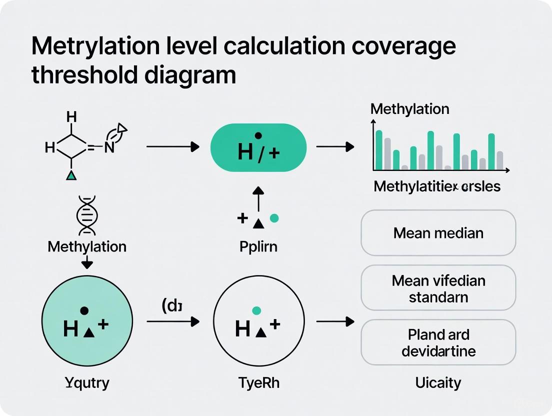
The logical workflow and key decision points for these protocols are summarized in the following diagram:
The Scientist's Toolkit: Essential Reagents and Materials
Table 2: Key Research Reagent Solutions for DNA Methylation Analysis
| Item | Function | Example Products / Kits |
|---|---|---|
| High-Integrity DNA Extraction Kits | Isolate high-molecular-weight DNA, crucial for long-read sequencing and accurate library prep. | Nanobind Tissue Big DNA Kit [2], DNeasy Blood & Tissue Kit (Qiagen) [2] |
| Bisulfite Conversion Kits | Chemically convert unmethylated cytosine to uracil for WGBS and RRBS. | EZ DNA Methylation Kit (Zymo Research) [2] |
| Enzymatic Conversion Kits | Convert base modifications enzymatically, preserving DNA integrity better than bisulfite. | EM-seq kits [2] |
| Methylation-Specific Library Prep Kits | Prepare sequencing libraries from bisulfite-converted or native DNA for various platforms. | Illumina DNA Prep kits, Oxford Nanopore Ligation Sequencing Kits [5] [2] |
| Methylation BeadChip Arrays | Profile methylation at pre-defined CpG sites across large sample cohorts cost-effectively. | Illumina Infinium MethylationEPIC v2.0 BeadChip [2] [6] |
| Bioinformatics Software | For basecalling, alignment, methylation calling, and differential analysis. | Bismark, Seqtk, Dorado, MinKNOW, MNP-Flex classifier [5] [4] |
Impact of Coverage on Methylation Calling Accuracy
The relationship between sequencing depth and calling accuracy is fundamental. Low coverage leads to high statistical uncertainty, especially when trying to distinguish intermediate methylation levels or detect rare methylation events in heterogeneous samples. The following diagram conceptualizes how coverage thresholds influence the confidence of methylation calls:
In practice, for WGBS, a minimum of 30x coverage is recommended to confidently call methylation levels across the majority of the genome [2]. However, for detecting subtle changes or working with mixed cell populations, significantly higher depths (e.g., 50x or more) may be necessary. For targeted approaches like RRBS or panel sequencing, coverage should be proportionally increased at the regions of interest, often exceeding 100x or 1000x to ensure that each CpG site is sampled sufficiently [3]. In liquid biopsy applications, where the ctDNA fraction can be very low, ultra-deep sequencing (>10,000x) is often required to detect the cancer-derived methylation signal against the background of normal cfDNA [3].
Defining and adhering to appropriate coverage thresholds is not a mere technical formality but a core component of rigorous methylation research. The protocols and data presented here provide a framework for selecting the right technology and implementing it with coverage requirements in mind, directly supporting the broader thesis that optimized coverage is vital for accurate methylation level calculation. As technologies evolve, particularly long-read sequencing and machine learning-based methods, the definitions of "adequate coverage" may shift. However, the principle remains: a deliberate and informed approach to experimental design, guided by clear coverage thresholds, is indispensable for producing robust, reliable, and clinically translatable epigenetic data.
For researchers in genomics and drug development, accurately quantifying DNA methylation is crucial for understanding gene regulation, cellular differentiation, and disease mechanisms. The reliability of these measurements hinges on three interconnected experimental design metrics: read depth, CpG coverage, and statistical power. Read depth refers to the number of times a particular nucleotide is sequenced, directly impacting base-calling confidence [7] [8]. CpG coverage represents the proportion of cytosine-phosphate-guanine sites in the genome that are effectively sequenced and assessed for methylation status [9]. Statistical power, particularly in the context of detecting differentially methylated regions (DMRs), is the probability of correctly identifying true positive methylation changes given specific effect sizes, sample sizes, and sequencing depths [10] [11]. This framework is essential for robust methylation level calculation in research spanning cancer diagnostics, biomarker discovery, and therapeutic development.
Defining the Key Metrics
Read Depth (Sequencing Depth)
Read depth, also termed sequencing depth or depth of coverage, is a fundamental quality metric in next-generation sequencing (NGS). It is defined as the average number of times a given nucleotide in the genome is read during the sequencing process [7]. A higher sequencing depth provides greater confidence in the accuracy of base calls and helps mitigate sequencing errors and background noise. For example, if a specific nucleotide is sequenced 30 times, the sequencing depth at that position is denoted as 30x [7]. In methylation studies, sufficient read depth is critical for accurate methylation calling, as it provides the necessary counts (methylated versus unmethylated reads) to confidently determine the methylation status of individual CpG sites.
CpG Coverage
CpG coverage pertains to the breadth of sequencing across the methylome, specifically the percentage or proportion of CpG sites in the target genome that are assayed with sufficient reliability [9]. The human genome contains approximately 28 million CpG sites, and achieving complete coverage is technologically challenging [10]. This metric is often reported as a percentage; for instance, "95% coverage" indicates that 95% of the targeted regions have been sequenced at least once [7]. In practice, some genomic regions, such as those with high GC content or repetitive elements, are notoriously difficult to sequence, leading to gaps in coverage [7]. CpG coverage is distinct from read depth: coverage indicates which regions are sequenced, while depth indicates how many times those regions are sequenced.
Statistical Power in Methylation Studies
Statistical power in methylation studies is the likelihood of correctly identifying a true differentially methylated region (DMR) when one exists. Power is influenced by several factors, including sample size, sequencing depth, the effect size (magnitude of methylation difference), and the basal methylation level [10] [11]. In high-throughput Methyl-Seq experiments, power calculation is complex because it involves testing millions of hypotheses simultaneously, requiring control of the false discovery rate (FDR) rather than the per-hypothesis type I error rate [10]. The concept of Expected Discovery Rate (EDR)—the expected proportion of true positives that are correctly detected—is often used as a genome-wide power metric [10].
Table 1: Key Metrics and Their Impact on Methylation Study Design
| Metric | Definition | Role in Experimental Design | Typical Target/Considerations |
|---|---|---|---|
| Read Depth | Average number of times a nucleotide is sequenced [7]. | Determines confidence in base calling and variant detection [7]. | Balances cost with accuracy; targets vary by application (e.g., 30x for WGBS). |
| CpG Coverage | Proportion of the target CpG sites sequenced at least once [7] [9]. | Ensures comprehensiveness of the methylome profile; minimizes gaps in data. | Aim for high percentage (e.g., >80%); affected by library prep and genomic biases [7] [2]. |
| Statistical Power | Probability of detecting true differential methylation [10] [11]. | Informs sample size and sequencing depth needed for reliable conclusions. | Typically targeted at 80%; depends on effect size, sample size, and depth [10]. |
Interdependence of Metrics and Experimental Design
The three core metrics are deeply intertwined. Read depth and CpG coverage collectively determine the quality and completeness of the raw data, which directly influences the statistical power of downstream analyses. A study with high read depth but low CpG coverage may yield highly confident methylation calls for a limited set of sites, potentially missing biologically important DMRs in underrepresented genomic regions. Conversely, high CpG coverage with very low read depth provides a broad but shallow snapshot of the methylome, where methylation calls are unreliable and statistical power is low.
Statistical power is a function of both data quality and study design. The relationship between sample size (N), sequencing depth, and power is a critical consideration in budgeting and experimental planning. Given a fixed budget, researchers must often choose between sequencing more samples at a lower depth or fewer samples at a higher depth. Furthermore, the required depth and power are influenced by the biological question. Detecting rare variants or small methylation differences between groups requires greater depth and larger sample sizes compared to detecting common variants or large effect sizes [7].
Table 2: Selection of Sequencing Method Based on Research Objectives
| Research Objective | Recommended Method(s) | Key Metric Considerations | Rationale |
|---|---|---|---|
| Discovery of novel DMRs | Whole-Genome Bisulfite Sequencing (WGBS), Enzymatic Methyl-Seq (EM-seq) [2]. | Maximize CpG coverage, moderate to high read depth. | Provides single-base resolution and the most comprehensive genome-wide coverage [2]. |
| Targeted or candidate region analysis | Reduced Representation Bisulfite Sequencing (RRBS) [10]. | High read depth on CpG-rich regions, lower overall genome coverage. | Cost-effective; enriches for informative, promoter-associated CpG islands. |
| Large-scale epigenome-wide association studies (EWAS) | Methylation arrays (e.g., EPIC) [2]. | High sample throughput, predefined CpG coverage. | Lower cost per sample allows for large N, essential for robust association studies with complex phenotypes. |
| Liquid biopsy for cancer detection | Enrichment-based cfDNA methods (e.g., cfMBD-seq, cfMeDIP-seq) [12]. | High read depth on targeted, cancer-informative CpG islands. | Optimized for low-input cfDNA; focuses on known differentially hypermethylated regions in cancer [12]. |
Protocols for Methylation Analysis and Power Assessment
Protocol: Cell-free DNA Methylation Profiling via cfMBD-seq
This protocol is adapted from a study demonstrating the application of cfMBD-seq for sensitive cancer detection and classification from plasma samples [12].
1. Sample Acquisition and Plasma Isolation:
- Collect whole blood in EDTA tubes from consented subjects.
- Centrifuge whole blood at 1,300 × g for 10 minutes at room temperature to separate cellular components from plasma.
- Carefully transfer the plasma layer to cryovials without disturbing the buffy coat and immediately freeze at -80°C.
2. cfDNA Extraction:
- Thaw plasma samples and centrifuge at 3,000 × g for 15 minutes to remove any remaining cell debris.
- Extract cfDNA using a commercial circulating nucleic acid kit (e.g., QIAamp Circulating Nucleic Acid Kit), omitting carrier RNA to avoid contamination.
- Quantify cfDNA using a fluorometer (e.g., Qubit) and assess fragment size distribution and purity using a high-sensitivity DNA assay (e.g., Agilent D1000 ScreenTape).
3. Library Preparation and Methylation Enrichment (cfMBD-seq):
- Perform end-repair and A-tailing on cfDNA using a library prep kit (e.g., KAPA Hyper Prep Kit).
- Ligate Illumina sequencing adapters. The adapter-to-insert molar ratio should be adjusted to 200:1 for low-input samples.
- Purify adapter-ligated DNA using SPRI beads and digest with the USER enzyme to remove uracil-containing artifacts.
- To ensure sufficient material for enrichment, combine the adapter-ligated cfDNA with enzymatically methylated filler DNA (e.g., methylated λ phage DNA) to bring the total input to 100 ng.
- Enrich for methylated DNA fragments using a methyl-CpG-binding domain (MBD) protein. The MBD protein preferentially binds to methylated DNA, allowing for its separation from unmethylated DNA.
- Amplify the enriched library via PCR for a limited number of cycles.
- Validate the final library's quality and quantity before sequencing.
4. Sequencing and Data Analysis:
- Sequence the library on an Illumina platform to an appropriate depth (e.g., 50-100 million reads per sample).
- Align sequencing reads to the reference genome (e.g., hg38) using an appropriate aligner.
- Call methylated regions (peaks) and perform differential methylation analysis between case and control groups.
- Validate identified DMRs against public databases (e.g., TCGA) to confirm their tissue of origin and cancer-specificity.
Protocol: Power Calculation for Methyl-Seq Studies Using MethylSeqDesign
This protocol outlines a statistical framework for power calculation and sample size determination in Methyl-Seq experiments, utilizing the MethylSeqDesign R package [10].
1. Prerequisite: Pilot Data Acquisition:
- Obtain a pilot Methyl-Seq dataset (
N_pilot), which includes methylated and total read counts for multiple CpG regions across a set of subjects. The pilot data should ideally include both cases and controls.
2. Step I: Parameter Estimation from Pilot Data:
- Input the pilot data into the
MethylSeqDesignframework. - The tool will use a beta-binomial model (e.g., via the "DSS-general" method) to account for both biological and technical variation in the methylation data.
- This step generates a distribution of p-values and effect sizes for all methylated regions from the pilot data, providing an empirical basis for the power simulation.
3. Step II: Mixture Model Fitting:
- A Beta-Uniform Mixture (BUM) model is fitted to the p-value distribution from Step I. This model helps distinguish the distribution of truly null hypotheses from that of the non-null (differentially methylated) hypotheses.
4. Step III: Parametric Bootstrap for Power Estimation:
- Specify the target sample size for your future study (
N_target), the desired sequencing depth, and a FDR threshold (e.g., 5%). MethylSeqDesignwill then perform a parametric bootstrap procedure:- Simulate numerous synthetic datasets based on the parameters estimated from the pilot data, reflecting the specified
N_targetand sequencing depth. - Perform differential methylation analysis on each simulated dataset.
- Calculate the Expected Discovery Rate (EDR)—the proportion of true DMRs that are successfully detected at the given FDR.
- Simulate numerous synthetic datasets based on the parameters estimated from the pilot data, reflecting the specified
- The output is a power (EDR) estimate for the proposed study design.
5. Iterative Design:
- Repeat Step IV across a range of sample sizes (
N_target) and sequencing depths. - Plot the relationship between sample size, sequencing depth, and statistical power.
- Select the combination of
Nand depth that achieves the desired power (e.g., 80%) within the constraints of the research budget.
Table 3: Research Reagent Solutions for Methylation Studies
| Reagent/Kit | Function | Application Note |
|---|---|---|
| QIAamp Circulating Nucleic Acid Kit | Extraction of high-quality cell-free DNA from plasma [12]. | Critical for liquid biopsy applications; omission of carrier RNA is recommended to prevent contamination of low-concentration cfDNA samples. |
| KAPA Hyper Prep Kit | Library construction for next-generation sequencing from low-input DNA [12]. | Allows for end-repair, A-tailing, and adapter ligation in a single, optimized workflow. Adapter concentration must be tuned for low-input samples. |
| Methylated Filler DNA | Carrier DNA to meet minimum input requirements for methylation enrichment steps [12]. | Typically enzymatically methylated λ phage DNA. It is essential to verify complete methylation (e.g., via digestion with methylation-sensitive restriction enzymes) to avoid bias. |
| MBD Protein / MeDIP Antibody | Enrichment of methylated DNA fragments [12]. | MBD-based enrichment (cfMBD-seq) shows superior capture of CpG islands compared to antibody-based (cfMeDIP-seq) methods [12]. |
| Infinium MethylationEPIC BeadChip | Genome-wide methylation profiling of > 935,000 CpG sites using microarray technology [2]. | A cost-effective solution for large-scale EWAS. Provides excellent coverage of gene promoter regions, enhancers, and other regulatory elements. |
| Bisulfite Conversion Reagents | Chemical treatment that converts unmethylated cytosine to uracil, while methylated cytosine remains protected [10] [2]. | The cornerstone of bisulfite sequencing (WGBS, RRBS). Harsh treatment can degrade DNA; newer enzymatic conversion methods (EM-seq) are emerging as less-damaging alternatives [2]. |
| MethylSeqDesign R Package | Statistical power calculation and sample size determination for Methyl-Seq experiments [10]. | Requires pilot data. Employs a beta-binomial model and bootstrap simulation to estimate power for a range of experimental designs. |
In DNA methylation research, the choice of sequencing or array platform directly dictates the scope and resolution of the resulting data, fundamentally shaping biological interpretations. Coverage determines the proportion of the methylome interrogated, influencing the ability to detect differentially methylated regions (DMRs) crucial for understanding disease mechanisms, developmental biology, and therapeutic responses. This document details the technical specifications, applications, and coverage implications of major methylation profiling technologies—Whole-Genome Bisulfite Sequencing (WGBS), Reduced Representation Bisulfite Sequencing (RRBS), EPIC Methylation Arrays, and emerging Long-Read Technologies—within the context of establishing reliable coverage thresholds for robust methylation level calculation.
The calculation of methylation levels is intrinsically linked to sequencing depth. Insufficient coverage at a cytosine site leads to statistically unreliable methylation measurements, while excessive depth wastes resources. Establishing platform-specific coverage thresholds is therefore a prerequisite for generating high-quality, reproducible data in methylation level calculation research.
Platform Specifications and Comparative Analysis
Technical Comparison of Major Platforms
Table 1: Key specifications and coverage characteristics of DNA methylation analysis platforms.
| Platform | Coverage Scope | Resolution | Key Applications | Primary Limitations |
|---|---|---|---|---|
| WGBS [13] [14] | Comprehensive, genome-wide; all cytosines in context (CpG, CHG, CHH). | Single-base resolution. | Discovery-based DMR studies, imprinting, non-CpG methylation. | High cost, computational intensity, DNA degradation from bisulfite conversion [13]. |
| RRBS [15] [16] | Targeted; ~1-3 million CpGs, covering ~70% of promoters & CpG islands [15]. | Single-base resolution. | Cost-effective screening, large cohort studies, cancer biomarker discovery [16]. | Biased to CpG-rich regions; misses ~85% of methylome; poor for low-CpG genomes [15]. |
| EPIC Array [17] | Targeted; >900,000 pre-selected CpG sites, emphasis on regulatory regions. | Single-CpG (but not whole genome). | Large-scale epidemiological studies, clinical biomarker validation. | Fixed content; cannot discover novel CpGs outside designed probes. |
| Long-Read Sequencing (e.g., PacBio HiFi) [18] | Comprehensive, genome-wide; capable of spanning repetitive regions and structural variants. | Single-base resolution. | Phasing methylation haplotypes, imprinted genes, complex regions like repeat expansions [18]. | Higher cost per sample, emerging data analysis methods. |
Table 2: Practical considerations for platform selection.
| Parameter | WGBS | RRBS | EPIC Array | Long-Read Tech |
|---|---|---|---|---|
| Approx. Cost/Sample | ~$700 (lib prep + 90Gb seq) [19] | Cost-effective relative to WGBS [16] | Most cost-effective for vast cohorts | Higher (decreasing) |
| DNA Input | Standard: ~1μg; T-WGBS: ~20ng [13] | ≥ 1μg (standard); as low as 10ng (kits) [15] [16] | Low | Varies, can be high |
| Data Output | ~90 Gb/sample for 30x coverage [19] [14] | ~10 Gb/sample [16] | Pre-determined (936,866 probes for EPICv2) [17] | Varies by coverage goal |
| Ideal Use Case | Unbiased methylome discovery | Targeted, cost-effective CpG island/promoter analysis | Population-scale screening, clinical tools | Resolving structural variation & haplotype phasing |
Coverage Implications for Methylation Level Calculation
The platform's inherent coverage directly impacts the statistical power and biological validity of calculated methylation levels.
- WGBS provides the gold standard for unbiased quantification, allowing for methylation level calculation at any of the ~28 million CpG sites in the human genome. For reliable calling, the ENCODE consortium recommends a minimum of 30x coverage [14]. Deeper coverage (e.g., 50-100x) is often required for confident detection of DMRs, especially in contexts like liquid biopsies where tumor DNA is diluted [20].
- RRBS offers high-depth coverage but for a limited subset of the genome. Its power lies in providing high-confidence methylation levels for CpG-rich regulatory regions with significantly less sequencing than WGBS. However, its inability to cover intergenic and CpG-poor "shore" regions can lead to incomplete biological insights [15].
- EPIC Arrays provide a fixed, cost-effective snapshot of methylation levels at pre-defined sites. While not suitable for discovery outside its probe set, its standardized nature facilitates high-throughput analysis and direct cross-study comparisons, which is invaluable for machine learning model training and biomarker development [20] [17].
- Long-Read Technologies uniquely enable haplotype-phased methylation level calculation. This allows researchers to determine the methylation status on individual parental chromosomes, which is critical for studying genomic imprinting and allele-specific methylation [18].
Detailed Experimental Protocols
Whole-Genome Bisulfite Sequencing (WGBS)
Principle: Genomic DNA is treated with sodium bisulfite, which deaminates unmethylated cytosines to uracils (read as thymines after PCR), while methylated cytosines remain unchanged [13]. Sequencing and comparison to a reference genome allows for single-base resolution mapping of methylation.
Protocol Workflow:
Key Steps:
- DNA Fragmentation & Library Prep: Fragment genomic DNA via sonication or tagmentation (e.g., T-WGBS) [13]. Repair ends, add 'A' bases, and ligate methylated adapters.
- Bisulfite Conversion: Treat DNA with sodium bisulfite. Critical parameters include:
- PCR Amplification & Clean-up: Amplify the converted library and purify.
- Sequencing: Sequence on a platform such as the DNBSEQ (PE150) [19] or Illumina NovaSeq to achieve sufficient depth. The ENCODE standard requires a minimum of 30x genome-wide coverage [14].
- Bioinformatics Analysis:
- Alignment: Use bisulfite-aware aligners like Bismark [14] against a bisulfite-converted reference genome.
- Methylation Calling: Extract methylation counts for each cytosine in all sequence contexts (CpG, CHG, CHH).
- QC Metrics: Assess bisulfite conversion efficiency, coverage distribution, and concordance between replicates (Pearson correlation ≥0.8 for CpGs with ≥10x coverage) [14].
Reduced Representation Bisulfite Sequencing (RRBS)
Principle: RRBS uses restriction enzymes (e.g., MspI, which cuts at CCGG sites) to digest genomic DNA, selectively enriching for CpG-rich fragments (promoters, CpG islands) before bisulfite conversion and sequencing [15] [21].
Protocol Workflow:
Key Steps:
- Restriction Digest: Digest high-quality genomic DNA (≥1μg) with MspI or a similar frequent-cutter that targets CpG-containing sequences [16] [21].
- Size Selection: Isolate fragments in the 40-220 bp range via gel extraction or beads. This step is critical as it captures fragments derived from CpG islands and gene promoters [21].
- End-Repair and Ligation: Repair fragment ends and ligate methylated sequencing adapters.
- Bisulfite Conversion & PCR: Convert with bisulfite and amplify the library with a low-cycle PCR.
- Sequencing & Analysis: Sequence to a depth of ~10 Gb of clean data per sample [16]. Bioinformatic analysis must include assessment of MspI cutting efficiency and alignment to the reference genome for DMR detection [16]. RRBS typically covers ≥70% of promoters and CpG islands using only 10-20% of the sequencing reads required for WGBS [15].
EPIC Methylation Array Analysis
Principle: This hybridization-based method uses probe-based chemistry on the Illumina Infinium platform to interrogate the methylation status of over 900,000 pre-defined CpG sites across the genome, with a focus on regulatory elements [17].
Key Considerations:
- Platform Versions: The EPICv2 array retains ~77% of probes from EPICv1 and adds over 200,000 new probes for enhanced coverage of enhancers and open chromatin [17]. Version differences must be accounted for in meta-analyses and longitudinal studies.
- Workflow: The protocol involves whole-genome amplification of bisulfite-converted DNA, followed by hybridization to the BeadChip, single-base extension, and fluorescent staining.
- Data Processing: Raw intensity data (.idat files) are processed through pipelines for background correction, normalization, and beta-value calculation (methylation level ranging from 0 to 1).
Long-Read Sequencing for Methylation
Principle: Platforms from PacBio and Oxford Nanopore Technologies (ONT) can detect DNA modifications, including 5mC, natively without bisulfite conversion. PacBio's HiFi sequencing achieves this through kinetic analysis during sequencing, while ONT uses electrical signal deviations.
Application in Coverage: Long-reads are transformative for resolving complex regions of the genome. A landmark All of Us study demonstrated that HiFi sequencing detected over 50% more disease-associated structural variants compared to short-read data, many in medically relevant genes [18]. This allows for the correlation of methylation status with specific haplotypes and structural variations that were previously inaccessible.
The Scientist's Toolkit: Essential Reagents and Materials
Table 3: Key research reagents and solutions for DNA methylation studies.
| Reagent/Kits | Function | Example Use Case |
|---|---|---|
| Zymo-Seq RRS Library Kit [15] | Simplified RRBS library prep from low DNA input (≥10 ng). | Epigenetic screening from precious or limited clinical samples. |
| Infinium MethylationEPIC v2.0 BeadChip [17] | Genome-wide methylation profiling at >900,000 pre-defined CpG sites. | Large-scale population studies and clinical biomarker validation. |
| Bismark/Bowtie2 [14] | Alignment & methylation caller for bisulfite sequencing data. | Standardized processing of WGBS and RRBS data for single-base resolution output. |
| MspI Restriction Enzyme [16] [21] | Digests genomic DNA at CCGG sites for RRBS library construction. | Creating reduced representation libraries enriched for CpG islands. |
| Bisulfite Conversion Kit | Converts unmethylated C to U, critical for BS-seq. | Essential pretreatment for WGBS, RRBS, and array-based methylation analysis. |
| DNA Methylation Standards | Controls for validating bisulfite conversion efficiency & assay conditions. | Ensuring high-quality, accurate, and reproducible NGS results [15]. |
Selecting the optimal platform for methylation level calculation requires balancing research goals, budget, and sample availability against the critical parameter of genomic coverage.
- For unbiased discovery and comprehensive methylome characterization, WGBS is the definitive choice, provided sufficient funding and computational resources are available. Adherence to a ≥30x coverage threshold is non-negotiable for robust quantification [14].
- For cost-effective, focused studies on gene regulatory regions in large sample cohorts, RRBS provides excellent value, delivering high-depth coverage of CpG islands and promoters.
- For massive-scale epidemiological studies or clinical assay development, EPIC Arrays offer an unparalleled balance of throughput, cost, and standardized data output, though with a fixed coverage scope.
- For resolving methylation in complex genomic regions, on single molecules, or for haplotype phasing, long-read technologies are indispensable, despite their current cost and analytical complexities.
Future directions will see increased integration of these technologies, using targeted or array-based methods for breadth and long-read or WGBS for depth and resolution on subsets of samples. Furthermore, the application of machine learning and foundational models (e.g., MethylGPT, CpGPT) is poised to enhance the prediction of methylation patterns and impute missing data, potentially mitigating some coverage limitations [20]. A clear understanding of each platform's coverage implications ensures that calculated methylation levels are both statistically sound and biologically meaningful.
The Relationship Between Sequencing Depth and Methylation Concordance Across Platforms
DNA methylation analysis is a cornerstone of epigenetic research, with critical implications for understanding gene regulation, cellular differentiation, and disease mechanisms. The evolving landscape of methylation profiling technologies presents researchers with multiple platform options, each with distinct strengths and limitations. A crucial but often underexplored factor significantly impacts the consistency of data generated across these different platforms: sequencing depth.
This Application Note examines the complex relationship between sequencing depth and methylation concordance across major DNA methylation detection platforms. We synthesize recent comparative studies to provide evidence-based guidance on coverage requirements, focusing on the practical implications for cross-platform study design, data integration, and validation protocols. Within the broader context of methylation level calculation coverage threshold research, establishing these parameters is fundamental for ensuring reproducible and biologically meaningful results in both basic research and drug development settings.
Platform Comparison and the Impact of Sequencing Depth
Current technologies for genome-wide DNA methylation analysis employ different fundamental principles for detecting methylated cytosines. Bisulfite conversion-based methods, including Whole-Genome Bisulfite Sequencing (WGBS) and Illumina MethylationEPIC microarrays, represent established approaches that chemically convert unmethylated cytosines to uracils, allowing methylation status to be inferred from sequence changes [22] [23]. Enzymatic conversion methods, such as Enzymatic Methyl-seq (EM-seq), offer an alternative by using enzymes to protect and convert bases, reducing DNA degradation [22] [24]. Third-generation sequencing platforms, including Oxford Nanopore Technologies (ONT) and PacBio HiFi sequencing, enable direct detection of DNA modifications without pre-conversion by monitoring polymerase kinetics or changes in electrical current [22] [25] [26].
The choice of platform involves trade-offs between resolution, coverage, input DNA requirements, cost, and the ability to detect methylation in challenging genomic regions. While WGBS is often considered the gold standard for its single-base resolution, its requirement for high sequencing depth to cover the entire genome comprehensively makes it resource-intensive [23]. The relationship between sequencing depth and methylation concordance across these platforms is therefore a critical practical consideration.
Quantitative Comparison of Platform Performance
Table 1: Key Performance Metrics of DNA Methylation Detection Platforms
| Platform | Resolution | Genomic Coverage | Recommended Depth | Key Strengths | Key Limitations |
|---|---|---|---|---|---|
| WGBS | Single-base | ~80% of CpGs [22] | 20-30× for high concordance [26] | Gold standard, comprehensive | DNA degradation, high depth requirements |
| EM-seq | Single-base | Comparable to WGBS [22] | Similar to WGBS | Better DNA preservation, high concordance with WGBS [22] | Newer method, less established |
| PacBio HiFi | Single-base | Detects more mCs in repetitive elements [26] | >20× for improved concordance [26] | Long reads, detects challenging regions | Higher DNA input, cost |
| ONT | Single-base | Captures unique loci [22] | Varies by application | Long-range profiling, direct detection | Higher error rates in earlier flow cells [25] |
| EPIC Array | Pre-defined sites | ~850,000-935,000 CpGs [22] | N/A (microarray) | Cost-effective, standardized | Limited to pre-designed sites |
| Targeted Bisulfite Seq | Single-base | User-defined regions | >1000× for target regions [27] | Ultra-deep coverage of specific loci | Limited genome scope |
Table 2: Observed Methylation Concordance Between Platforms Under Different Conditions
| Platform Comparison | Correlation Coefficient | Conditions | Impact of Increased Depth |
|---|---|---|---|
| HiFi vs WGBS | r ≈ 0.8 [26] | Genome-wide | Concordance improves with coverage, particularly beyond 20× [26] |
| EM-seq vs WGBS | High concordance [22] | Genome-wide | Similar depth requirements to WGBS |
| TEEM-seq vs EPIC Array | >0.98 [24] | Targeted (3.98M CpGs) | FFPE samples required ≥35× for reliable classification [24] |
| ONT vs WGBS/EM-seq | Lower agreement [22] | Genome-wide | -- |
| FinaleMe (predicted) vs WGBS | High in CpG-rich regions [28] | Plasma cfDNA | Performance improves with coverage in CpG-rich regions |
Recent comparative studies highlight the critical role of sufficient sequencing depth in achieving cross-platform concordance. A 2025 comparison of WGBS, EM-seq, ONT, and EPIC arrays across human tissue, cell line, and blood samples found that while each method identified unique CpG sites, EM-seq showed the highest concordance with WGBS, indicating strong reliability due to their similar sequencing chemistry [22]. Notably, ONT sequencing captured certain loci uniquely, enabling methylation detection in challenging genomic regions where other methods might struggle, but showed lower overall agreement with WGBS and EM-seq [22].
A specialized analysis comparing PacBio HiFi sequencing and WGBS in monozygotic twins with Down syndrome revealed that HiFi sequencing detected a greater number of methylated CpGs (mCs), particularly in repetitive elements and regions with low WGBS coverage [26]. However, WGBS reported higher average methylation levels than HiFi sequencing. Both platforms exhibited methylation patterns consistent with known biological principles, such as low methylation in CpG islands. The study demonstrated a strong Pearson correlation (r ≈ 0.8) between platforms, with higher concordance in GC-rich regions and at increased sequencing depths [26].
The relationship between sequencing depth and concordance follows a non-linear pattern, with significantly stronger agreement observed beyond 20× coverage [26]. Depth-matched comparisons and site-level down-sampling confirmed that methylation concordance improves with increasing coverage, emphasizing the importance of adequate sequencing depth for cross-platform validation studies.
Figure 1: The relationship between sequencing depth and methylation concordance is mediated by multiple factors, with a critical threshold around 20× coverage significantly improving agreement between platforms. The effect varies across genomic contexts and is influenced by platform-specific biases.
Experimental Protocols for Cross-Platform Validation
Protocol 1: Whole-Genome Methylation Concordance Study
This protocol outlines a systematic approach for comparing methylation calls between WGBS and PacBio HiFi sequencing platforms, based on the methodology described by Promsawan et al. (2025) [26].
Sample Preparation and Sequencing
- Extract high-quality genomic DNA from biological samples (e.g., whole blood, tissue, cell lines)
- For WGBS: Fragment DNA to 300-500bp fragments via sonication or enzymatic fragmentation
- Perform bisulfite conversion using established kits (e.g., Zymo Research EZ DNA Methylation Kit)
- Prepare sequencing libraries using WGBS-compatible kits with dual indexing to enable multiplexing
- Sequence on Illumina platform to target depth of 30× minimum
- For HiFi WGS: Prepare SMRTbell libraries without bisulfite conversion according to manufacturer's instructions
- Sequence on PacBio Sequel II or Revio systems to target depth of 25× minimum
Bioinformatic Processing
- Process WGBS data through two independent pipelines (e.g., wg-blimp and Bismark) for robustness
- Process HiFi WGS data using pb-CpG-tools or similar specialized tools for PacBio methylation calling
- Align reads to reference genome (hg38 recommended)
- Calculate methylation levels at individual CpG sites using betas (ratio of methylated to total reads)
Concordance Analysis
- Extract overlapping CpG sites covered by both technologies
- Stratify analysis by genomic context: CpG islands, shores, shelves, repetitive elements, gene bodies
- Perform correlation analysis (Pearson correlation) of methylation beta values
- Calculate concordance rates at different depth thresholds (5×, 10×, 20×, 30×)
- Generate Bland-Altman plots to assess agreement across methylation value range
Protocol 2: Targeted Methylation Validation Using TEEM-seq
This protocol describes a targeted enrichment approach for validating methylation patterns across platforms, adapted from the TEEM-seq validation study [24].
Library Preparation and Enrichment
- Fragment DNA to 240-290bp insert size using focused ultrasonication
- Construct libraries using NEBNext enzymatic methyl-seq kit
- Quantify libraries with Qubit dsDNA HS assay and assess size distribution with Agilent TapeStation
- Pool 8 libraries equally for target enrichment using Twist Human Methylome panel
- Perform hybrid capture following manufacturer's protocol with optimization for FFPE samples if needed
- Sequence enriched libraries on NovaSeq6000 with 150bp paired-end reads
Quality Control and Analysis
- Perform quality control on raw reads using FastQC and MultiQC
- Trim adapters and low-quality bases using Trim Galore and Cutadapt
- Align trimmed reads to reference genome using bwa-meth
- Remove PCR duplicates using Picard MarkDuplicates
- Call methylated bases using MethylDackel with parameters: -minDepth 10 -maxVariantFrac 0.15
- Generate methylation beta values at single-CpG resolution
Cross-Platform Validation
- Compare TEEM-seq results with EPIC array data from same samples
- Calculate correlation coefficients for overlapping CpG sites
- Assess minimum depth requirements by downsampling sequencing data
- Validate that FFPE samples achieve at least 35× coverage for robust classification [24]
- Use t-SNE analysis to visualize separation of samples against reference methylation datasets
Figure 2: Comprehensive workflow for cross-platform methylation validation studies. Parallel processing of samples through different technologies followed by integrated bioinformatic analysis enables robust assessment of platform concordance across varying sequencing depths.
The Scientist's Toolkit: Essential Reagents and Materials
Table 3: Essential Research Reagents for Methylation Sequencing Studies
| Category | Specific Product/Kit | Application | Key Features |
|---|---|---|---|
| DNA Extraction | Nanobind Tissue Big DNA Kit [22] | High-molecular-weight DNA for long-read sequencing | Preserves DNA integrity for long fragments |
| DNeasy Blood & Tissue Kit [22] | Standard DNA extraction from various sources | Reliable yield from diverse sample types | |
| Bisulfite Conversion | EZ DNA Methylation Kit (Zymo Research) [22] | WGBS and targeted bisulfite sequencing | High conversion efficiency, minimal DNA degradation |
| Enzymatic Conversion | NEBNext Enzymatic Methyl-seq Kit [24] | EM-seq library preparation | Reduced DNA fragmentation vs. bisulfite |
| Target Enrichment | Twist Human Methylome Panel [24] | Targeted EM-seq (TEEM-seq) | Covers ~3.98 million CpG sites |
| Library Preparation | NEBNext Ultra II DNA Library Prep | Standard WGBS library construction | Compatible with bisulfite-converted DNA |
| SMRTbell Prep Kit [26] | PacBio HiFi sequencing | Optimized for long-read methylation detection | |
| Quality Control | Qubit dsDNA HS Assay [24] | Accurate DNA quantification | Fluorometric specificity for double-stranded DNA |
| Agilent TapeStation [24] | Fragment size distribution | Critical for assessing library quality | |
| Bioinformatic Tools | Bismark [26] | WGBS data analysis | Standard for bisulfite sequence alignment |
| pb-CpG-tools [26] | PacBio HiFi methylation calling | Specialized for kinetic detection | |
| MethylDackel [24] | Methylation calling from WGBS/EM-seq | Flexible parameter adjustment for depth filtering |
The relationship between sequencing depth and methylation concordance across platforms follows predictable but non-linear patterns, with critical thresholds that should inform experimental design. Based on our synthesis of recent comparative studies, we recommend:
Minimum Depth Requirements: For most comparative studies, aim for minimum coverage of 20-30× for whole-genome approaches. This threshold ensures sufficient statistical power for methylation calling while maintaining cost-effectiveness. Specifically, FFPE samples in targeted approaches require at least 35× coverage for reliable classification [24].
Platform Selection Strategy: EM-seq demonstrates high concordance with WGBS while offering advantages in DNA preservation, making it suitable for samples where DNA integrity is a concern [22]. PacBio HiFi sequencing shows particular strength in detecting methylation in repetitive elements and regions poorly covered by short-read technologies [26].
Study Design Considerations: When integrating data across multiple platforms, implement depth-matched comparisons to ensure fair evaluation. Stratify concordance analysis by genomic context, as agreement varies significantly across different genomic regions. GC-rich regions typically show higher cross-platform concordance, while repetitive elements may exhibit platform-specific biases [26].
Validation Protocols: For critical applications, particularly in clinical or biomarker development contexts, implement orthogonal validation using targeted bisulfite sequencing at ultra-high depth (>1000×) for specific loci of interest [27]. This approach confirms methylation status with high confidence while controlling costs.
These evidence-based recommendations provide a framework for designing methylation studies that maximize cross-platform concordance through appropriate depth requirements, ultimately supporting more reproducible and translatable epigenetic research.
Practical Guide to Setting Coverage Thresholds Across Methylation Profiling Methods
In bisulfite sequencing, the methylation level at a specific cytosine is calculated as the proportion of reads where the base is methylated. The reliability of this quantitative measurement is fundamentally dependent on read depth, defined as the number of times a given base pair is sequenced. Inadequate depth leads to increased statistical noise and inaccurate methylation estimates, compromising downstream analyses and biological conclusions. This is particularly crucial in genetically variable natural populations, where heterogeneity is inherent. Establishing minimum depth thresholds is therefore not merely a technical formality, but a foundational step for generating robust, reproducible DNA methylation data in both Whole-Genome Bisulfite Sequencing (WGBS) and Reduced Representation Bisulfite Sequencing (RRBS). Research indicates that mean methylation estimates eventually plateau with increasing coverage, and identifying this point of diminishing returns is key to efficient experimental design [29].
Comparative Analysis of WGBS and RRBS
Whole-Genome Bisulfite Sequencing (WGBS) provides the most comprehensive profile of DNA methylation, aiming to cover all CpG sites in the genome at single-base resolution. In contrast, Reduced Representation Bisulfite Sequencing (RRBS) uses methylation-insensitive restriction enzymes (commonly MspI) to selectively target and enrich CpG-dense regions, such as promoters and CpG islands, which are often functional hotspots for DNA methylation [29] [30]. This enrichment allows RRBS to cover a significant fraction of these regulatory regions while sequencing only a small portion of the genome.
Table 1: Core Characteristics of WGBS and RRBS
| Feature | Whole-Genome Bisulfite Sequencing (WGBS) | Reduced Representation Bisulfite Sequencing (RRBS) |
|---|---|---|
| Genomic Coverage | Entire genome, all CpG contexts [13] | ~15% of methylome; targets CpG-rich regions (islands, promoters, gene bodies) [30] |
| Typical Input DNA | High (µg range); lower with tagmentation (e.g., ~20 ng for T-WGBS) [13] | Can be low (e.g., from 10 ng) [30] |
| Key Strength | Unbiased, base-resolution genome-wide map [13] | Cost-effective for large sample sizes; high depth on targeted regions [29] [30] |
| Primary Limitation | High sequencing cost per sample; lower depth for a given budget [29] | Incomplete picture; misses methylation in non-CpG-rich and intergenic regions [30] |
| Ideal Application | Discovery-based studies, non-CpG methylation, non-model organisms [13] | Population-level studies, focused hypothesis testing on regulatory regions [29] |
Impact of Method Choice on Methylation Profiles
The choice between WGBS and RRBS has direct consequences on the observed methylation landscape. A key finding is that the prevalence of CpG sites with intermediate methylation levels is greatly reduced in RRBS compared to WGBS. This systematic bias can have important consequences for functional interpretations, as intermediate methylation often reflects cell-to-cell heterogeneity or dynamically regulated genomic loci [29]. Furthermore, RRBS does not cover regions with low CpG density, which can include important regulatory elements such as enhancers, with one source noting it covers only around 35% of enhancers [30].
Establishing Minimum Depth Thresholds
Empirical Evidence and Coverage Saturation
There is no universal minimum depth applicable to all studies; the optimal threshold depends on the biological variation in the sample and the specific research question. However, empirical data provides strong guidance. A comparative study of PacBio HiFi sequencing and WGBS revealed that methylation concordance improves with increasing coverage, with stronger agreement observed beyond 20x coverage. This depth-matched analysis showed that saturation of concordance metrics is achieved at higher coverages, providing a benchmark for reliable detection [26].
For genetically variable populations, a best practice is to deeply sequence a few initial individuals to identify the coverage level at which mean methylation estimates plateau. This value, which may differ by species and population, then informs the minimum depth required for the full study to ensure accurate measurements [29]. Depth filters have been shown to have large impacts on the number of CpG sites recovered across multiple individuals, a consideration that is particularly critical for WGBS data due to its wider genomic coverage and typically lower per-site depth [29].
Table 2: Recommended Depth and Quality Control Thresholds
| Parameter | Recommended Threshold | Rationale and Context |
|---|---|---|
| General Minimum Depth | ≥ 20x per CpG site | Provides stable methylation concordance and reliable beta value estimation [26]. |
| Targeted BS QC | ≥ 30x coverage | Used as a quality filter for CpG sites in targeted bisulfite sequencing panels to ensure data reliability [31]. |
| Pilot Sequencing | Sequence initial individuals to high depth (e.g., >30x) | Essential for identifying the coverage where mean methylation estimates plateau in genetically variable populations [29]. |
| Site/Sample Filtering | Exclude sites with coverage < 30x in >50% of samples; exclude samples with coverage < 30x in >1/3 of sites | A two-step quality control procedure applied in targeted sequencing to ensure data integrity [31]. |
The Interplay of Depth, Breadth, and Experimental Design
The choice of sequencing depth is fundamentally a trade-off against sample size and genomic breadth. WGBS, with its expansive breadth, often forces researchers to prioritize either high depth with a small sample size or lower depth with more replicates. RRBS, by focusing on a smaller genomic fraction, allows for larger sample sizes and higher depth for the same sequencing cost, which increases statistical power for population-level studies [29]. The optimal design must balance these factors based on the study's goals, whether it is the discovery of novel differentially methylated regions or the testing of specific hypotheses in predefined genomic areas.
Experimental Protocols for Determining Minimum Depth
Protocol 1: Pilot Sequencing for Coverage Saturation Analysis
This protocol is designed to empirically determine the required sequencing depth for a given study system.
1. Sample Selection and Sequencing
- Select 2-3 biologically diverse individuals from your population of interest.
- Perform library preparation (WGBS or RRBS) using a standardized protocol. For RRBS, paired-end sequencing is recommended to help filter SNPs that can bias methylation metrics [29].
- Sequence these pilot samples to a very high depth (e.g., 50-100x average genome-wide coverage) to establish a "ground truth" methylation call set.
2. Bioinformatic Down-sampling and Analysis
- Use bioinformatic tools (e.g.,
seqtk) to randomly sub-sample the sequencing reads from the high-depth BAM files to generate lower-coverage datasets (e.g., 5x, 10x, 15x, 20x, 30x). - Call methylation levels (generate .bedGraph or similar files) for each down-sampled dataset using a consistent pipeline (e.g., Bismark/Bowtie2 or BWA-meth/MethylDackel) [29] [32].
- Calculate the mean methylation level per CpG site (or per region) for each down-sampled dataset and the high-depth "ground truth."
3. Determining the Saturation Point
- For each down-sampled dataset, calculate the correlation (e.g., Pearson correlation) of per-site methylation levels with the high-depth ground truth.
- Plot the correlation coefficient against sequencing depth. The point where the correlation coefficient plateaus indicates the depth beyond which additional sequencing yields minimal improvement in accuracy.
- Use this depth as the minimum target coverage for the full-scale experiment.
Protocol 2: A Standardized WGBS/RRBS Workflow with Quality Control
This outlines a core bioinformatic workflow for processing bisulfite sequencing data, highlighting steps where depth assessment is critical.
1. Raw Read Processing and Quality Control
- Tool: FastQC for initial quality check.
- Action: Perform adapter trimming and quality trimming using tools like
trim_galoreorcutadapt[32]. This step is crucial for removing low-quality bases that can affect mapping and variant calling.
2. Conversion-Aware Alignment
- Tools: Bismark (using Bowtie2), BWA-meth, or ARYANA-BS [29] [32] [33].
- Action: Map the trimmed reads to a bisulfite-converted reference genome. Different aligners use different strategies (e.g., three-letter alignment, wild-card alignment), with consequences for mapping efficiency and bias. Recent benchmarks indicate that newer aligners like ARYANA-BS can achieve state-of-the-art accuracy [33].
3. Post-Alignment Processing and Methylation Calling
- Action: Filter PCR duplicates using tools like
picardMarkDuplicates. - Tool: Use the aligner's built-in caller (Bismark) or a specialized tool (MethylDackel with BWA-meth) to generate methylation call files [29].
- Key Output: A file reporting, for each cytosine, the number of reads showing methylation and the total number of reads covering it.
4. Depth-Based Filtering and Final Output
- Action: Apply the minimum depth threshold determined from Protocol 1. For example, filter out all CpG sites with a total read depth below the chosen threshold (e.g., 20x).
- Output: Generate a final methylation report containing only high-confidence, sufficiently covered CpG sites for downstream differential analysis.
Determining and Applying Minimum Sequencing Depth
The Scientist's Toolkit: Essential Reagents and Software
A successful bisulfite sequencing experiment relies on a combination of wet-lab reagents and bioinformatic tools.
Table 3: Essential Research Reagents and Software Solutions
| Category | Item | Function and Application Notes |
|---|---|---|
| Wet-Lab Reagents | MspI Restriction Enzyme | The core of RRBS; fragments DNA at CCGG sites to enrich for CpG-rich regions [29] [30]. |
| High-Efficiency Bisulfite Conversion Kit | Chemically converts unmethylated cytosines to uracils. Critical for data quality; minimizes DNA degradation [31]. | |
| Targeted Methyl Panels | Custom panels (e.g., QIAseq) for cost-effective, deep sequencing of predefined CpG sites across many samples [31]. | |
| Bioinformatic Tools | Bismark | A widely used aligner and methylation caller. Uses Bowtie2 for three-letter alignment but can have lower mapping efficiency [29] [32]. |
| BWA-meth / MethylDackel | An alternative pipeline. BWA-meth uses BWA mem for alignment, often with higher efficiency; MethylDackel extracts calls and filters SNPs using paired-end info [29]. | |
| ARYANA-BS | A novel context-aware aligner that integrates methylation patterns to improve alignment accuracy, especially for long or error-prone reads [33]. | |
| nf-core/methylseq | A community-maintained Nextflow pipeline for reproducible processing of BS data, incorporating both Bismark and BWA-meth [32]. |
Establishing a scientifically defensible minimum depth for bisulfite sequencing is a critical step that ensures the accuracy and reliability of DNA methylation data. There is no single magic number; rather, a depth of 20x to 30x per CpG site serves as a robust general guideline, with higher depths required for detecting subtle methylation differences or working with highly heterogeneous samples. The most rigorous approach involves conducting a pilot saturation analysis to determine the point of diminishing returns for a specific biological system. By integrating these depth considerations with the strategic choice between WGBS and RRBS, and employing robust bioinformatic pipelines, researchers can generate high-quality methylation data capable of powering meaningful biological discovery.
DNA methylation is a fundamental epigenetic mark involved in gene regulation, cellular differentiation, and disease pathogenesis. Accurate detection of methylation patterns is essential for understanding its role in various biological processes and developing epigenetic biomarkers. While whole-genome bisulfite sequencing (WGBS) has long been the gold standard for methylation profiling, emerging technologies like Enzymatic Methyl-Seq (EM-seq) and Oxford Nanopore Sequencing (ONT) offer innovative approaches that overcome traditional limitations. EM-seq replaces harsh bisulfite chemistry with a gentle enzymatic conversion process, preserving DNA integrity while maintaining high accuracy. In contrast, Oxford Nanopore technology directly detects modified bases in native DNA without any conversion, leveraging long-read capabilities to resolve complex genomic regions. Both techniques present unique considerations for coverage thresholds and data quality metrics that researchers must address when designing methylation studies, particularly in drug development and clinical research applications where accuracy and reproducibility are paramount [32] [22].
Technical Foundations and Methodologies
Enzymatic Methyl-Seq (EM-seq) Technology
EM-seq utilizes a two-step enzymatic process to detect methylated cytosines without DNA fragmentation. The method employs TET2 enzyme to oxidize 5-methylcytosine (5mC) to 5-carboxylcytosine (5caC), while T4 β-glucosyltransferase (T4-BGT) protects 5-hydroxymethylcytosine (5hmC) through glucosylation. Subsequently, the APOBEC enzyme deaminates unmodified cytosines to uracils, while all modified cytosines remain protected. This enzymatic conversion preserves DNA integrity more effectively than bisulfite treatment, which causes substantial DNA fragmentation and degradation through harsh chemical conditions. The EM-seq workflow typically begins with DNA fragmentation using either Covaris sonication or enzymatic approaches, followed by adapter ligation with sample-specific barcodes. The core enzymatic conversion then takes place, after which libraries are PCR-amplified before sequencing [34] [22].
EM-seq demonstrates particular advantages in library complexity and coverage uniformity, especially in GC-rich regions where bisulfite conversion often fails. The technology achieves approximately 95% conversion efficiency of unmethylated cytosines, comparable to established bisulfite methods but with reduced sequencing bias. EM-seq can handle DNA inputs as low as 10-200ng for library preparation, making it suitable for limited clinical samples. For quality control, unmethylated lambda DNA and CpG-methylated pUC19 DNA are typically included as controls to verify conversion efficiency across samples [34] [35].
Oxford Nanopore Sequencing Technology
Oxford Nanopore technology directly sequences native DNA through protein nanopores embedded in synthetic membranes. As DNA strands pass through these nanopores, they cause characteristic disruptions in electrical current that are decoded to determine the DNA sequence and base modifications simultaneously. This direct detection approach allows for real-time sequencing and eliminates PCR amplification biases, preserving epigenetic information in its native context. Unlike conversion-based methods, Nanopore sequencing can distinguish between different cytosine modifications, including 5mC, 5hmC, 5fC, and 5caC, based on their unique electrical signatures [36] [37].
A significant advantage of Nanopore technology is its capacity for long-read sequencing, with read lengths ranging from short fragments to ultra-long reads exceeding 100 kilobases. This capability enables methylation profiling across structurally complex genomic regions that are challenging for short-read technologies, including centromeres, telomeres, and highly repetitive elements. The platform has evolved through multiple flow cell versions (R6-R10.4), with each iteration improving raw read accuracy from approximately 70% to over 99% through enhanced nanopore proteins, motor proteins, and sequencing chemistry. The recently introduced R10.4 flow cell with "Q20+" chemistry produces raw reads with >99% accuracy, making the technology increasingly suitable for methylation studies requiring high precision [38] [36].
Comparative Performance and Threshold Considerations
Platform Characteristics and Performance Metrics
Table 1: Technical Comparison of Methylation Sequencing Platforms
| Parameter | EM-seq | Oxford Nanopore | WGBS |
|---|---|---|---|
| Detection Principle | Enzymatic conversion | Direct electrical signal detection | Chemical bisulfite conversion |
| DNA Input | 10-200 ng [34] | ~1 μg for 8 kb fragments [22] | 500-2000 ng [32] |
| Read Length | Short-read (50-300 bp) | Short to ultra-long (50 bp->4 Mb) [38] | Short-read (50-300 bp) |
| Single-Base Resolution | Yes | Yes | Yes |
| DNA Damage | Minimal | None | Substantial fragmentation [22] |
| Coverage Uniformity | High, especially in GC-rich regions [22] | Variable; improves with read length | Moderate; poor in GC-rich regions |
| Differential Modification Detection | No (5mC/5hmC not distinguished) | Yes (can distinguish 5mC, 5hmC, 5fC, 5caC) [36] | No (5mC/5hmC not distinguished) |
| Multiplexing Capacity | High (384+ samples) | Moderate to high (1-96 samples) | High (384+ samples) |
Coverage Thresholds and Data Quality Considerations
Establishing appropriate coverage thresholds is critical for robust methylation analysis. For EM-seq, studies demonstrate high concordance with WGBS (R² = 0.97-0.99) at comparable coverage depths. The gentle enzymatic conversion generates more uniform coverage distribution across CpG sites, with 30-50× coverage generally providing reliable methylation calls for most applications. EM-seq achieves approximately 80% genome-wide CpG coverage, outperforming WGBS in regions with extreme GC content where bisulfite conversion struggles. The technology particularly excels in population-scale studies where cost-effective, reproducible methylation profiling is essential [22] [35].
For Oxford Nanopore sequencing, coverage requirements depend on the application and read length. For comprehensive methylation analysis, 20-30× coverage with long reads (N50 > 10 kb) typically provides sufficient data for haplotype-resolved methylation phasing. The platform's ability to span repetitive regions means fewer gaps in methylation maps compared to short-read technologies. However, raw read accuracy must be considered when setting coverage thresholds, with the latest R10.4 flow cells producing data of sufficient quality for methylation calling at lower coverage than previous versions. For clinical applications requiring high confidence, 30-40× coverage provides reliable detection of differentially methylated regions [39] [36].
Table 2: Coverage Threshold Recommendations for Methylation Analysis
| Application Context | EM-seq Coverage | Oxford Nanopore Coverage | Key Considerations |
|---|---|---|---|
| Genome-Wide Methylation Screening | 30-50× | 20-30× | ONT coverage can be lower due to long-range information |
| Differential Methylation Analysis | 30× minimum | 25× minimum | Higher coverage needed for small effect sizes |
| Clinical Biomarker Validation | 50-100× | 30-50× | Increased depth for rare allele detection |
| Single-Cell Methylation | N/A (bulk method) | 10-20× per cell [40] | Low-input protocols emerging |
| Targeted Methylation Panels | 200-500× | 100-200× | Ultra-deep sequencing for rare variants |
Experimental Protocols
EM-seq Library Preparation Protocol
The EM-seq library preparation protocol begins with DNA quality assessment using fluorometric measurements (e.g., Qubit) to ensure accurate quantification, with absorbance measurements (Nanodrop) being insufficient for quality control. The recommended DNA input is 500 ng, though the protocol can be optimized for inputs as low as 10 ng with increased PCR cycles. DNA should be in water or EB buffer with OD260/280 of 1.8-2.0 and must be RNA-free to prevent interference with conversion efficiency [34].
Step 1: DNA Fragmentation - Fragment genomic DNA to 200-300 bp using Covaris sonication or enzymatic fragmentation. Enzymatic fragmentation offers a cost-effective alternative without specialized equipment.
Step 2: End Repair and A-Tailing - Repair fragment ends and add 3'A-overhangs using standard library preparation reagents compatible with subsequent adapter ligation.
Step 3: Adapter Ligation - Ligate EM-seq adapters containing sample-specific barcode sequences to facilitate multiplexing. Use reduced adapter concentrations for low-input samples to minimize dimer formation.
Step 4: Enzymatic Conversion - Perform the two-step enzymatic conversion using TET2 and APOBEC enzymes according to manufacturer specifications (NEBNext EM-seq v2 kit). Include unmethylated lambda DNA and CpG-methylated pUC19 controls to monitor conversion efficiency.
Step 5: Library Amplification - Amplify libraries with 8-12 PCR cycles using proofreading polymerases to maintain sequence fidelity. Limit cycle number to reduce duplicate rates while maintaining sufficient library complexity.
Step 6: Library QC and Sequencing - Assess library concentration via qPCR and fragment size distribution by bioanalyzer or tapestation. Pool libraries at equimolar ratios and sequence on Illumina platforms with 150 bp paired-end reads recommended for optimal alignment [34] [35].
Oxford Nanopore Methylation Analysis Protocol
Sample Preparation: Isolate high molecular weight DNA using methods that preserve integrity (e.g., Nanobind Tissue Big DNA Kit). Assess DNA quality via pulsed-field gel electrophoresis or fragment analyzer, aiming for average fragment sizes >20 kb for long-read applications. Input requirement is approximately 1 μg of DNA for standard methylation workflows [22] [41].
Library Preparation Options:
- Ligation Sequencing Kit: Standard approach where read length matches input fragment length
- Rapid Sequencing Kit: Optimized for samples with fragments >30 kb
- Ultra-Long DNA Sequencing Kit: Specialized for reads >100 kb, ideal for complex regions
- PCR-based Kits: For low input samples, typically yielding ~2 kb reads
Library Preparation Steps:
- DNA Repair and End-Prep - Use NEBNext FFPE DNA Repair Mix and Ultra II End-prep module to repair damage and prepare ends for adapter ligation, especially crucial for clinical samples.
- Native Barcoding - Incorporate native barcodes during adapter ligation to enable multiplexing while preserving base modification information.
- Adapter Ligation - Ligate sequencing adapters using the Ligation Sequencing Kit, optimizing incubation time based on input DNA quantity and quality.
- Quality Control - Assess library quantity and fragment size distribution using Qubit and Femto Pulse systems.
- Sequencing - Load library onto MinION, GridION, or PromethION flow cells depending on throughput requirements. For methylation analysis, R10.4 flow cells are recommended for improved basecalling accuracy [38] [41].
Methylation Calling: Use specialized tools like Megalodon or Dorado for basecalling with modified base detection. For bacterial methylation analysis, MethylomeMiner provides a streamlined workflow for identifying high-confidence methylation sites based on coverage and methylation frequency, with assignment to genomic features [37].
Workflow Visualization
The Scientist's Toolkit: Essential Research Reagents
Table 3: Essential Research Reagents for Methylation Analysis
| Reagent/Category | Function | Technology | Examples & Specifications |
|---|---|---|---|
| DNA Extraction Kits | High molecular weight DNA preservation | ONT | Nanobind Tissue Big DNA Kit [22] |
| DNA Quantification | Accurate nucleic acid measurement | Both | Qubit fluorometric measurement [34] |
| Library Prep Kits | Sample preparation for sequencing | EM-seq | NEBNext EM-seq v2 kit [34] |
| Library Prep Kits | Native DNA sequencing | ONT | Ligation Sequencing Kit, Ultra-Long DNA Sequencing Kit [38] |
| Conversion Controls | Verification of conversion efficiency | EM-seq | Unmethylated lambda DNA, CpG-methylated pUC19 [34] |
| Barcoding Systems | Sample multiplexing | Both | Native Barcoding Expansion kits (ONT) [41] |
| Enzymatic Mixes | DNA repair and end preparation | ONT | NEBNext FFPE DNA Repair Mix [41] |
| Bioinformatics Tools | Methylation data processing | EM-seq | Bismark, bwa-meth, MethylDackel [34] [32] |
| Bioinformatics Tools | Modified base detection | ONT | Megalodon, Dorado, MethylomeMiner [37] |
Applications in Research and Drug Development
The applications of EM-seq and Oxford Nanopore technologies in methylation research span diverse fields, each with specific threshold considerations. In cancer research, both platforms enable comprehensive methylation profiling of tumor samples, with Nanopore sequencing demonstrating particular utility for classifying acute leukaemia subtypes in under two hours from sample receipt using methylation patterns [39]. The MARLIN (methylation- and AI-guided rapid leukaemia subtype inference) approach achieved 96.2% concordance with conventional diagnostics, correctly classifying 25 out of 26 cases based on sparse DNA methylation data [39].
In rare disease diagnostics, Oxford Nanopore sequencing has shown remarkable success in identifying previously undetected structural variants. A study on hypotonia (decreased muscle tone) demonstrated that long-read whole-genome sequencing identified potential genomic causes in an additional 14% of research samples that had remained unsolved with short-read approaches. The technology potentially reduced diagnostic timelines by 85% (from 168 days to 25 days) and testing costs by 37.9% compared to standard sequential testing [39]. Similarly, in Canavan disease research, Nanopore sequencing uncovered a retrotransposon insertion in the ASPA gene present in all eight research samples but missed by previous clinical tests, representing what may be the most common pathogenic cause of this neurodegenerative disorder across multiple ancestry groups [39].
For antimicrobial resistance research, Nanopore sequencing provides unique advantages in tracking resistance gene transmission through plasmid analysis. The long-read capability enables complete assembly of bacterial genomes and mobile genetic elements, revealing the genetic contexts of antimicrobial resistance genes in both cultured bacteria and complex microbiota [36]. The platform's portability and real-time sequencing capabilities further support rapid resistance detection in clinical and field settings, with MinION devices offering compact, affordable solutions for point-of-care applications [36].
In population-scale epigenomic studies, EM-seq offers a robust alternative to traditional WGBS, particularly when combined with targeted approaches like Targeted Methylation Sequencing (TMS). This optimized protocol captures approximately 4 million CpG sites with strong agreement to both EPIC arrays (R² = 0.97) and whole-genome bisulfite sequencing (R² = 0.99), enabling cost-effective methylation profiling across large cohorts [35]. The method's compatibility with enzymatic fragmentation and reduced DNA input requirements (as low as 10ng) further enhances its utility for biobank samples and precious clinical specimens where material may be limited [35].
EM-seq and Oxford Nanopore sequencing represent transformative technologies in the field of DNA methylation analysis, each offering distinct advantages for different research contexts. EM-seq provides a robust, cost-effective solution for population-scale studies requiring high-throughput methylation profiling with minimal DNA damage and improved coverage uniformity. Oxford Nanopore technology enables direct detection of base modifications in native DNA, with long-read capabilities that resolve methylation patterns across complex genomic regions inaccessible to short-read technologies. Both methods require careful consideration of coverage thresholds based on specific application requirements, with EM-seq typically needing 30-50× coverage for genome-wide studies and Nanopore sequencing requiring 20-30× coverage, leveraging its long-range information. As these technologies continue to evolve, they promise to expand our understanding of epigenetic regulation in health and disease, enabling more comprehensive methylation profiling in both basic research and clinical applications.
Digital PCR (dPCR) represents a transformative approach in molecular diagnostics, enabling absolute quantification of nucleic acids without the need for standard curves. This technique partitions a PCR reaction into thousands of nanoliter-scale reactions, allowing precise counting of target molecules through Poisson statistical analysis [42] [43]. For methylation level calculation in research and diagnostic applications, dPCR offers significant advantages in precision and sensitivity over traditional quantitative methods [44]. The technology's capability to provide absolute quantification makes it particularly valuable for detecting low-abundance targets and for applications requiring high precision, such as calculating methylation ratios in complex clinical samples [45].
The fundamental principle underlying dPCR involves dividing the sample into numerous partitions so that each contains zero, one, or a few target molecules. Following PCR amplification, the fraction of positive partitions is counted, and the absolute concentration of the target is calculated using Poisson statistics [43]. This partitioning approach enhances sensitivity and resistance to inhibitors compared to real-time quantitative PCR (qPCR) [46]. In methylation-specific dPCR, this technology enables precise determination of methylation ratios at specific genomic loci, which is crucial for identifying diagnostic and prognostic biomarkers in various diseases, including cancer [44] [45].
Performance Characteristics and Validation
Sensitivity and Specificity Metrics in dPCR
Table 1: Comparative Performance of dPCR Platforms in Methylation Analysis
| Platform | Specificity (%) | Sensitivity (%) | Correlation with Reference Method | Application Context |
|---|---|---|---|---|
| Nanoplate-based dPCR (QIAcuity) | 99.62 | 99.08 | r = 0.954 with ddPCR | CDH13 methylation in breast cancer [44] |
| Droplet-based ddPCR (QX-200) | 100 | 98.03 | r = 0.954 with nanoplate dPCR | CDH13 methylation in breast cancer [44] |
| Methylation-specific ddPCR | 89.4 (Tissue) | 38.7-83.0 (Plasma, varies by cut-off) | N/A | Five-gene multiplex for lung cancer detection [45] |
The exceptional sensitivity and specificity of dPCR platforms enable precise methylation quantification even in challenging sample types. In a direct comparison of two dPCR platforms for CDH13 gene methylation analysis in breast cancer tissue samples, both platforms demonstrated excellent performance characteristics [44]. The nanoplate-based system achieved 99.62% specificity and 99.08% sensitivity, while the droplet-based system reached 100% specificity and 98.03% sensitivity, with a strong correlation (r = 0.954) between the methods [44]. This high level of agreement between different dPCR platforms highlights the robustness of the technology for methylation quantification.
For clinical applications, establishing appropriate cut-off values is crucial for accurate classification. In a methylation-specific ddPCR multiplex assay for lung cancer detection, researchers evaluated two different cut-off methods to determine circulating tumor DNA status [45]. The first method yielded a sensitivity of 38.7% in non-metastatic disease and 70.2% in metastatic cases, while the second method demonstrated improved sensitivity of 46.8% and 83.0%, respectively [45]. This underscores the importance of rigorous cut-off establishment based on intended application and disease context.
Comparison with qPCR and Other Technologies
Table 2: dPCR vs. qPCR Performance Characteristics for Nucleic Acid Quantification
| Parameter | Digital PCR | Real-Time qPCR |
|---|---|---|
| Quantification Method | Absolute quantification without standard curves | Relative quantification requiring standard curves |
| Sensitivity | Superior for low-abundance targets [46] [47] | Lower, especially for rare targets |
| Precision | Higher consistency and reproducibility [46] | Variable between runs and operators |
| Effect of PCR Inhibitors | More resistant due to partitioning [46] | Highly susceptible, affecting Ct values |
| Multiplexing Capability | Excellent, with minimal competition between targets [43] | Limited by competition and spectral overlap |
| Dynamic Range | Limited by number of partitions | Broader dynamic range |
dPCR demonstrates superior accuracy compared to qPCR, particularly for medium to high viral loads in respiratory virus detection [46]. This advantage extends to methylation analysis, where dPCR provides more consistent and precise quantification of methylation ratios [44]. The partitioning process in dPCR reduces the effect of PCR inhibitors, which is particularly beneficial when analyzing challenging sample types such as formalin-fixed paraffin-embedded (FFPE) tissue or plasma-derived cell-free DNA [46] [45].
The absolute quantification capability of dPCR eliminates the need for standard curves, reducing variability and improving reproducibility across experiments and laboratories [43]. This feature is particularly valuable for methylation analysis, where consistent measurement of methylation ratios is essential for reliable results in longitudinal studies or clinical applications [44] [45].
Experimental Protocols for Methylation-Specific dPCR
Sample Preparation and Bisulfite Conversion
Proper sample preparation is critical for successful methylation analysis. For FFPE tissue samples, DNA extraction should be performed using dedicated kits such as the DNeasy Blood and Tissue kit (Qiagen) or Maxwell RSC with FFPE Plus DNA Kit (Promega) [44] [45]. DNA concentration should be determined using fluorescence-based methods (e.g., Qubit) rather than spectrophotometry for improved accuracy. For plasma samples, cell-free DNA extraction requires specialized kits such as the DSP Circulating DNA Kit (Qiagen) with appropriate volume input (typically 4 mL plasma) [45].
The bisulfite conversion step is performed using optimized kits such as the EpiTect Bisulfite kit (Qiagen) or EZ DNA Methylation-Lightning Kit (Zymo Research) [44] [45]. The protocol should include:
- Input DNA: Use 1 μg of isolated DNA for conversion [44]
- Conversion Conditions: Follow manufacturer instructions with modifications if needed for specific sample types
- Post-Conversion Cleanup: Include desulfonation and purification steps
- Elution Volume: 15-20 μL of appropriate elution buffer [45]
After conversion, concentrate the DNA using centrifugal filter units (e.g., Amicon Ultra-0.5) when working with plasma-derived cell-free DNA to maximize target input in the dPCR reaction [45].
dPCR Reaction Setup and Thermal Cycling
The dPCR reaction setup varies slightly between platforms but follows the same general principles. For the QIAcuity nanoplate-based system:
Reaction Preparation: Prepare a 12 μL reaction volume containing:
- 3 μL of 4× Probe PCR master mix
- 0.96 μL of forward/reverse primer (each)
- 0.48 μL of each probe (FAM-labeled for methylated, HEX-labeled for unmethylated)
- 2.5 μL of bisulfite-converted DNA template
- Nuclease-free water to volume [44]
Partitioning: Load reaction mixture into 24-well nanoplate (approximately 8,500 partitions per well)
- Thermal Cycling:
- Initial heat activation: 95°C for 2 minutes
- 40 cycles of:
- Denaturation: 95°C for 15 seconds
- Combined annealing/extension: 57°C for 1 minute [44]
For the QX200 Droplet Digital PCR system:
Reaction Preparation: Prepare a 20 μL reaction volume containing:
- 10 μL of Supermix for Probes (No dUTP)
- 0.45 μL of forward/reverse primer (each)
- 0.45 μL of each probe
- 2.5 μL of DNA template
- Nuclease-free water to volume [44]
Droplet Generation: Use DG8 cartridge and Droplet Generation Oil to create approximately 20,000 droplets per sample
- Thermal Cycling:
- Initial denaturation: 95°C for 10 minutes
- 40 cycles of:
- Denaturation: 94°C for 30 seconds
- Annealing/extension: Optimized temperature for 1 minute [44]
- Final hold: 98°C for 10 minutes and 4°C hold
Data Analysis and Cut-off Implementation
Following amplification, analyze partitions using platform-specific software (QIAcuity Suite for nanoplate systems, Quantasoft for droplet systems) [44] [48]. The methylation level is calculated as the ratio of positive FAM-detected partitions (methylated) to the sum of all positive partitions detected in both channels (methylated + unmethylated) [44].
Establish acceptance criteria for assays:
- Minimum of 7,000 valid partitions for nanoplate systems [44]
- At least 100 positive partitions for reliable quantification [44]
- Implement quality controls including:
For cut-off implementation in methylation analysis:
- Determine Background Levels: Analyze negative controls and normal tissues to establish background methylation levels
- Define Positive/Negative Thresholds: Set thresholds based on receiver operating characteristic (ROC) analysis when possible [45]
- Validate Cut-offs: Test established cut-offs in independent sample sets
- Consider Application Requirements: Adjust cut-offs based on clinical context (screening vs. monitoring) [45]
Figure 1: Methylation-Specific Digital PCR Workflow. This diagram illustrates the complete process from sample collection to cut-off implementation for methylation analysis using dPCR.
Research Reagent Solutions for Methylation-Specific dPCR
Table 3: Essential Reagents and Materials for Methylation-Specific dPCR
| Reagent/Material | Function | Example Products | Key Considerations |
|---|---|---|---|
| DNA Extraction Kits | Isolation of high-quality DNA from various sample types | DNeasy Blood & Tissue (Qiagen), Maxwell RSC (Promega) | Optimize for sample type (FFPE, plasma) [44] [45] |
| Bisulfite Conversion Kits | Conversion of unmethylated cytosines to uracils | EpiTect Bisulfite (Qiagen), EZ DNA Methylation-Lightning (Zymo Research) | Control for DNA fragmentation during conversion [44] [45] |
| dPCR Master Mix | Provides enzymes, dNTPs, and buffers for amplification | QIAcuity Probe PCR Master Mix, Bio-Rad ddPCR Supermix | Select probe-based for multiplexing [44] [48] |
| Fluorogenic Probes | Target-specific detection with fluorescent reporters | PrimeTime qPCR Probes (IDT), TaqMan probes | FAM for methylated, HEX/VIC for unmethylated targets [44] [43] |
| Primer Sets | Amplification of bisulfite-converted target sequences | Custom-designed primers | Target CpG-rich regions, avoid CpG sites in primers [44] |
| Partitioning Media | Creation of nanoliter-scale reactions | Droplet Generation Oil (Bio-Rad), nanoplate partitions | Ensure partition stability during thermal cycling [48] |
| Quality Controls | Assessment of extraction efficiency and contamination | Exogenous spike-in DNA (CPP1), genomic DNA controls | Monitor technical variability between runs [45] |
The selection of appropriate reagents is crucial for successful methylation-specific dPCR assays. Primer and probe design requires special consideration for bisulfite-converted DNA, with primers ideally avoiding CpG dinucleotides in their sequence [44]. When designing methylation-specific assays, the methylated and unmethylated sequences will differ after bisulfite conversion, allowing for the design of specific probes for each state [44]. For multiplex assays, careful selection of fluorophores with minimal spectral overlap is essential for accurate signal discrimination [43].
Quality control measures should be integrated throughout the workflow. For plasma samples, include an exogenous spike-in DNA (such as CPP1) to monitor extraction efficiency [45]. Assess potential contamination with lymphocyte DNA using an immunoglobulin gene-specific ddPCR assay, and evaluate total cell-free DNA concentration and fragment size using assays targeting different amplicon sizes [45]. These controls help ensure the reliability of methylation quantification, particularly for low-abundance targets in liquid biopsy applications.
Figure 2: Digital PCR Partitioning and Detection Principle. This diagram illustrates the two main partitioning methods and the process from sample partitioning to absolute quantification.
Digital PCR technology provides a robust platform for absolute quantification of methylation levels with exceptional sensitivity and specificity. The implementation of appropriate sensitivity and specificity cut-offs is essential for translating methylation biomarkers into clinically useful tools. The protocols and application notes detailed in this document provide a framework for implementing dPCR in methylation analysis, with particular attention to cut-off establishment and validation. As dPCR technology continues to evolve with improved multiplexing capabilities, automation, and data analysis tools, its application in methylation-based biomarker development is poised to expand significantly, enabling more precise diagnostic and therapeutic approaches in personalized medicine.
In the realm of precision oncology, DNA methylation biomarkers have emerged as powerful tools for predicting treatment response and guiding therapeutic decisions. The O6-methylguanine-DNA methyltransferase (MGMT) promoter methylation status in glioblastoma (GBM) represents a paradigm for such biomarkers, where establishing a clinically relevant cutoff point is both crucial and challenging [49]. Methylation of the MGMT promoter silences this DNA repair gene, thereby increasing the efficacy of alkylating agents like temozolomide and improving patient survival [50] [49]. However, the transition from a continuous methylation variable to a binary clinical decision (methylated vs. unmethylated) requires careful determination of an optimal cutoff point that balances sensitivity with specificity while maximizing predictive accuracy for treatment benefit. This case study examines the determination of the 21% MGMT promoter methylation cutoff by pyrosequencing, exploring the methodological framework, clinical validation, and implications for patient stratification within the broader context of methylation level calculation coverage threshold research.
MGMT Methylation: Biological Rationale and Clinical Significance
Mechanism of Action and Therapeutic Implications
The MGMT enzyme confers resistance to alkylating chemotherapy by removing alkyl groups from the O6 position of guanine, thereby repairing DNA damage and neutralizing the cytotoxic effects of temozolomide [49]. Epigenetic silencing via promoter methylation prevents MGMT protein synthesis, leading to increased sensitivity to temozolomide and improved survival outcomes in GBM patients [49]. This mechanism establishes MGMT promoter methylation status as both a predictive biomarker for temozolomide response and a prognostic indicator for overall survival [51]. The clinical utility of this biomarker is particularly evident in elderly patients or those with poor performance status, where MGMT status guides decisions between temozolomide chemotherapy and radiotherapy [49].
Quantitative Nature of Methylation and Cutoff Challenges
Traditional binary reporting of MGMT status (methylated vs. unmethylated) oversimplifies the underlying biology, as methylation exists on a continuous spectrum with complex relationships to clinical outcomes [50]. Recent methodological advancements have enabled quantitative approaches that measure methylation as a continuous variable, revealing non-linear relationships between methylation density and survival benefit [50]. This quantitative dimension introduces the central challenge of determining where to establish cutoff points that optimally distinguish patients who will benefit from specific treatments from those who will not. The determination of these thresholds must consider multiple factors including assay precision, clinical outcomes, and the potential consequences of misclassification.
Established Cutoffs and Comparative Analysis
The 21% Pyrosequencing Cutoff
A retrospective study of 109 glioblastoma patients established 21% methylation as the optimal cutoff point for MGMT status determination by pyrosequencing [49]. Using receiver operating characteristic (ROC) analysis, researchers determined that this threshold provided the highest likelihood ratio (1.66) and accuracy (0.65), with sensitivity of 68% and specificity of 59% [49]. Patients classified as methylated using this cutoff demonstrated significantly better overall survival (HR: 0.453; 95% CI: 0.279-0.735; p = 0.001) [49]. Furthermore, the study revealed a linear relationship between methylation percentage and survival, with each 10% increase in methylation corresponding to a 20% reduction in the risk of death (p = 0.004) [49].
Alternative Cutoffs and Methodological Considerations
Different methodologies and study populations have yielded varying optimal cutoffs, highlighting the context-dependent nature of threshold determination:
Table 1: Comparative Analysis of MGMT Promoter Methylation Cutoffs
| Methodology | Proposed Cutoff | Study Population | Key Findings | Reference |
|---|---|---|---|---|
| Quantitative MGMT promoter methylation index (17-point scale) | Non-linear relationship | 240 newly diagnosed GBM patients | Low methylation (1-6 CpG sites): worse outcomes (HR=1.62); Medium methylation (7-12 CpG sites): greatest hazard reduction (HR=0.48) | [50] |
| Quantitative methylation-specific PCR (qMSP) | "Gray zone" classification | Pooled analysis of 4 clinical trials (n=4,041) | Established "truly unmethylated" and "gray zone" categories; Both methylated and "gray zone" patients had better OS than truly unmethylated patients | [51] |
| Pyrosequencing (5 CpG sites) | 21% | 109 GBM patients | Optimal cutoff with highest likelihood ratio and accuracy; Linear relationship between methylation % and survival | [49] |
A large pooled analysis of four randomized clinical trials (n=4,041) using quantitative methylation-specific PCR (qMSP) identified a more complex classification system with a "gray zone" of low-level methylation that still conferred some sensitivity to temozolomide [51]. This finding challenges the simplistic binary classification and suggests that the relationship between methylation and treatment response may be more nuanced than previously recognized.
Detailed Experimental Protocol: Determining the 21% Cutoff
Sample Preparation and DNA Extraction
Materials and Equipment:
- Formalin-fixed paraffin-embedded (FFPE) tumor tissue sections (10μm thickness)
- Macro-dissection tools for tumor enrichment
- Commercial DNA extraction kit (e.g., QIAamp DNA FFPE Tissue Kit)
- Spectrophotometer or fluorometer for DNA quantification
Procedure:
- Select FFPE blocks with viable tumor content >70% as determined by histopathological review.
- Cut 3-5 sections at 10μm thickness and subject to macro-dissection to enrich tumor content.
- Extract DNA using commercial kits according to manufacturer's instructions with extended digestion time (overnight) to accommodate FFPE tissue.
- Quantify DNA using spectrophotometric methods and assess quality via A260/A280 ratio (acceptable range: 1.8-2.0).
- Store extracted DNA at -20°C until bisulfite conversion.
Bisulfite Conversion and Pyrosequencing
Materials and Equipment:
- Commercial bisulfite conversion kit (e.g., EZ DNA Methylation-Gold Kit)
- Thermal cycler
- Pyrosequencing system (PyroMark series, Qiagen)
- MGMT-specific pyrosequencing assays targeting CpG sites 74-78
Procedure:
- Perform bisulfite conversion using 500ng-1μg of input DNA according to kit manufacturer's protocol with the following modification: incubate at 95°C for 10 minutes followed by 64°C for 2.5 hours [49].
- Purify bisulfite-converted DNA and elute in appropriate buffer.
- Amplify target regions using MGMT-specific primers with the following PCR conditions:
- Initial denaturation: 95°C for 15 minutes
- 45 cycles of: 95°C for 30 seconds, 56°C for 30 seconds, 72°C for 30 seconds
- Final extension: 72°C for 10 minutes
- Prepare single-stranded DNA templates for pyrosequencing using the PyroMark Vacuum Prep Workstation.
- Perform sequencing using the PyroMark Q96 MD system with sequencing primers specific for the MGMT promoter region encompassing CpG sites 74-78 [49].
- Calculate percentage methylation at each CpG site using PyroMark Q96 software.
Statistical Analysis and Cutoff Determination
Software and Tools:
- R Statistical Computing Environment or SPSS Statistics
- Packages: survival, pROC, ConsensusClusterPlus (if applicable)
Procedure:
- Calculate mean methylation percentage across all interrogated CpG sites for each sample.
- Perform ROC analysis using overall survival as the reference standard.
- Calculate sensitivity, specificity, likelihood ratios, and accuracy for multiple potential cutoff points (range: 9-25%).
- Select the optimal cutoff point based on the highest likelihood ratio and accuracy.
- Validate the cutoff using bootstrap resampling or split-sample validation if sample size permits.
- Perform survival analysis using Kaplan-Meier curves and log-rank tests to compare methylated vs. unmethylated groups based on the determined cutoff.
- Conduct multivariate Cox regression analysis to adjust for potential confounders (age, extent of resection, performance status).
The following workflow diagram illustrates the complete experimental process:
The Scientist's Toolkit: Essential Reagents and Equipment
Table 2: Key Research Reagent Solutions for MGMT Methylation Analysis
| Category | Specific Product/Kit | Function | Key Considerations |
|---|---|---|---|
| DNA Extraction | QIAamp DNA FFPE Tissue Kit (Qiagen) | Isolation of high-quality DNA from archived tissue | Optimized for cross-linked FFPE DNA; includes RNase treatment |
| Bisulfite Conversion | EZ DNA Methylation-Gold Kit (Zymo Research) | Chemical conversion of unmethylated cytosines to uracils | High conversion efficiency; minimal DNA degradation |
| Pyrosequencing Assay | PyroMark MGMT Kit (Qiagen) | Amplification and sequencing of target CpG sites | Standardized assays for CpG sites 74-78; includes controls |
| PCR Amplification | PyroMark PCR Kit (Qiagen) | Target amplification with biotinylated primers | Optimized for bisulfite-converted DNA; hot-start technology |
| Sequencing System | PyroMark Q96 MD System | Quantitative sequencing by synthesis | Integrated software for methylation quantification |
| Quality Control | PyroMark Q24 CpG Software | Data analysis and quality assessment | Automated methylation percentage calculation |
Advanced Considerations in Cutoff Determination
Non-Linear Relationships and Complex Patterns
Emerging evidence suggests that the relationship between MGMT promoter methylation and clinical outcomes may not follow a simple linear pattern. A study utilizing a 17-point quantitative MGMT promoter methylation index found that patients with low-level methylation (1-6 CpG sites) actually fared worse than those with completely unmethylated promoters, while those with medium-level methylation (7-12 CpG sites) showed the greatest survival benefit [50]. This non-linear relationship challenges conventional understanding and suggests that establishing a single optimal cutoff point may be clinically disadvantageous in some contexts [50]. These findings highlight the importance of considering the functional form of the relationship between methylation density and treatment response when establishing clinical thresholds.
Methodological Variability and Reproducibility
Different methylation analysis platforms demonstrate varying performance characteristics that influence cutoff determination:
Table 3: Methodological Comparison for Methylation Analysis
| Method | Resolution | Throughput | Quantitative Capability | Optimal Use Case |
|---|---|---|---|---|
| Pyrosequencing | Single CpG site | Medium | High quantitative accuracy | Clinical validation; single gene analysis |
| Methylation-Specific PCR (qMSP) | Promoter region | High | Relative quantification | High-throughput clinical screening |
| Illumina Methylation Arrays | Genome-wide (850K CpG sites) | High | High-throughput screening | Biomarker discovery; multi-gene panels |
| Whole-Genome Bisulfite Sequencing | Single-base, genome-wide | Low | Comprehensive quantification | Research; novel biomarker identification |
The reproducibility of methylation assays is crucial for consistent cutoff application. A retest analysis of 218 paired samples using qMSP demonstrated high reproducibility (R² = 0.94), supporting the reliability of quantitative methylation assessment across different testing occasions [51]. This reproducibility is essential for implementing standardized cutoffs across clinical laboratories.
Implications for Clinical Trial Design and Patient Stratification
The determination of validated cutoff points has profound implications for clinical trial design and personalized treatment approaches. MGMT methylation status now serves as a key stratification factor in clinical trials and as a decision point for therapeutic choices, particularly in elderly patients or those with poor performance status [49] [51]. Contemporary clinical trials increasingly use MGMT status to select patient populations most likely to benefit from specific interventions, with some trials specifically designed for patients with unmethylated MGMT in whom temozolomide may be omitted or replaced with alternative therapies [51].
The following diagram illustrates the clinical decision-making pathway based on MGMT methylation status:
The determination of the 21% MGMT promoter methylation cutoff by pyrosequencing exemplifies the rigorous methodological approach required to establish clinically relevant thresholds for continuous molecular variables. This process necessitates careful consideration of analytical performance, clinical utility, and statistical validation to ensure optimal patient stratification. The emerging recognition of non-linear relationships between methylation density and clinical outcomes, coupled with the identification of intermediate "gray zone" classifications, highlights the complexity of translating continuous molecular measurements into binary clinical decisions [50] [51].
Future research directions should focus on developing multi-dimensional models that incorporate quantitative methylation data with other molecular and clinical variables to enhance predictive accuracy. Additionally, standardized reporting of methylation levels and validation of cutoffs across diverse populations and testing platforms will be essential for advancing the field of methylation-based diagnostics. As methylation profiling technologies continue to evolve, the integration of machine learning approaches and artificial intelligence may enable more sophisticated pattern recognition that moves beyond simplistic cutoff points toward comprehensive methylation signatures for personalized treatment selection [20]. These advancements will further solidify the role of DNA methylation biomarkers in precision oncology and pave the way for their expanded application across diverse cancer types.
Leveraging Machine Learning for Automated Threshold Optimization and Data Analysis
In the field of methylation level calculation coverage threshold research, the precise determination of DNA methylation states is a fundamental challenge. DNA methylation, a key epigenetic modification involving the addition of a methyl group to cytosine bases, plays crucial roles in gene regulation, cellular differentiation, and disease pathogenesis [3] [2]. The accurate analysis of this mark is complicated by technical variations in sequencing depth and coverage, which directly impact the reliability of methylation calls.
Threshold optimization represents a critical bottleneck in methylation data analysis, where suboptimal settings can lead to either excessive false positives or reduced detection sensitivity for biologically significant methylation events. Traditional threshold setting often relies on manual heuristics or arbitrary cutoffs, introducing subjectivity and limiting reproducibility. Machine learning (ML) offers a powerful paradigm to address these challenges through data-driven, automated optimization of analysis parameters [52] [53]. This protocol details the integration of ML techniques for threshold optimization specifically within methylation level calculation pipelines, enabling more robust, reproducible, and accurate epigenetic analyses.
Background: Methylation Analysis Methods and Threshold Challenges
Multiple technologies exist for genome-wide DNA methylation profiling, each with distinct strengths, limitations, and associated threshold considerations:
- Bisulfite Sequencing (WGBS): The reference method providing single-base resolution by converting unmethylated cytosines to thymines [32] [2]. It requires thresholds for conversion efficiency and minimum coverage.
- Enzymatic Methyl-Sequencing (EM-seq): An emerging alternative that uses enzymes instead of harsh chemicals, reducing DNA fragmentation and improving coverage uniformity [32] [2]. Thresholds must be adjusted for its different noise profile.
- Methylation Microarrays (EPIC): A cost-effective platform for large studies that uses probe intensities to calculate Beta- and M-values [54] [2]. Thresholds are needed for detection p-values and probe filtering.
- Nanopore Sequencing: A third-generation technology enabling direct detection of methylation modifications during sequencing without conversion [37] [2]. It requires specific signal-to-noise thresholds.
The Critical Role of Coverage Thresholds
The selection of an appropriate coverage threshold—the minimum number of reads required to call a methylation state at a given cytosine—profoundly impacts data quality and biological interpretation. Insufficient coverage thresholds increase random error and false positives in differential methylation detection, while excessively stringent thresholds discard valuable biological information by removing poorly covered genomic regions from analysis [32]. Machine learning models can dynamically optimize these thresholds based on data quality metrics, sample characteristics, and specific research objectives.
Machine Learning Framework for Threshold Optimization
Core Optimization Concepts and Metrics
Automated threshold optimization requires defining clear performance objectives and evaluation metrics. The table below outlines key metrics for assessing methylation calling performance in the context of threshold tuning.
Table 1: Key Performance Metrics for Methylation Threshold Optimization
| Metric | Definition | Interpretation in Methylation Analysis |
|---|---|---|
| Sensitivity (Recall) | Proportion of true methylated sites correctly identified | Measures ability to detect genuine methylation events; crucial for biomarker discovery [3] |
| Precision | Proportion of called methylated sites that are truly methylated | Indicates reliability of methylation calls; high precision reduces false positives [52] |
| F1-Score | Harmonic mean of precision and recall | Balanced metric for optimizing the trade-off between false positives and negatives |
| Coverage Efficiency | Percentage of genomic regions retained after thresholding | Measures data retention; important for maximizing information yield [32] |
| Concordance | Agreement with validation methods (e.g., locus-specific assays) | Gold-standard for accuracy assessment [32] |
Machine Learning Approaches for Threshold Optimization
Different ML strategies can be employed based on available labeled data and research goals:
- Supervised Learning: Requires training data with known methylation states. Models learn to predict methylation status from read-level features, effectively internalizing optimal decision boundaries. This approach is ideal when high-quality validation data exists [53].
- Unsupervised Learning: Identifies natural thresholds through clustering and anomaly detection without labeled data. Techniques like K-means or isolation forests can group coverage patterns and flag outliers for specialized thresholding [52].
- Reinforcement Learning: Employs feedback loops where the model receives positive reinforcement for successful classifications (e.g., confirmed by orthogonal methods) and adjusts thresholds accordingly [53].
Experimental Protocol: ML-Optimized Methylation Analysis
The following diagram illustrates the complete automated workflow for methylation analysis with integrated machine learning for threshold optimization.
Step-by-Step Protocol
Step 1: Data Preprocessing and Quality Control
- Quality Assessment: Process raw sequencing reads (FASTQ files) through quality control tools like FastQC. Record metrics including per-base sequencing quality, adapter contamination, and sequence length distribution [32] [55].
- Read Trimming and Filtering: Use trimmers such as Trim Galore! or Cutadapt to remove adapter sequences and low-quality bases. Set initial quality thresholds (e.g., Phred score >20) [55].
- Alignment: Perform conversion-aware alignment to a reference genome using specialized aligners (e.g., Bismark, BS-Seeker2, or Biscuit) [32] [55]. For BS-seq data, this accounts for C-to-T conversion.
- Post-Alignment Processing: Filter PCR duplicates using tools like Picard Tools. Calculate coverage distribution metrics and prepare a BAM file for downstream analysis.
Step 2: Feature Extraction for Machine Learning
- Coverage Metrics: Compute genome-wide coverage statistics, including mean coverage, coverage variance, and the percentage of bases covered at different depth levels (e.g., 1x, 5x, 10x, 30x).
- Quality Metrics: Extract base quality scores, mapping quality scores, and sequence complexity metrics.
- Sample-Specific Features: Record sample-level characteristics such as estimated bisulfite conversion efficiency (should be >99%) [55] or DNA input quantity, which influences data quality [32].
- Format Features: Structure extracted features into a tabular format (e.g., CSV) suitable for machine learning, with genomic regions (e.g., CpG sites) as rows and metrics as columns.
Step 3: Model Training and Threshold Optimization
- Baseline Establishment: Calculate baseline performance using conventional fixed thresholds (e.g., 10x coverage). Use metrics from Table 1 for comparison.
- Model Selection:
- For supervised learning: Implement a Random Forest or Gradient Boosting (XGBoost) classifier [53]. Use known methylated/unmethylated control regions (e.g., spike-ins or validated loci) as labeled training data.
- For unsupervised learning: Apply clustering algorithms (K-means, DBSCAN) to coverage and quality metrics to identify natural groupings and outliers [52].
- Hyperparameter Tuning: Optimize model parameters using grid search or Bayesian optimization, with cross-validation to prevent overfitting [56].
- Threshold Determination: The trained model will learn to predict the minimum coverage threshold required for a reliable methylation call at each site or class of sites. This can output a single optimized global threshold or dynamic, region-specific thresholds.
Step 4: Methylation Calling and Validation
- Apply Optimized Thresholds: Execute methylation calling (e.g., using MethylC-analyzer or HOME) using the ML-optimized thresholds [55]. This generates a file (e.g., CGmap) containing methylation levels (β-values) for each cytosine.
- Differential Methylation Analysis: Identify Differentially Methylated Regions (DMRs) using tools like DMRcate or MethylC-analyzer, incorporating the refined methylation calls [55] [54].
- Validation: Assess performance by comparing ML-optimized results to a gold-standard dataset (if available) [32]. Use techniques like cross-validation or bootstrap validation to estimate performance robustness. Orthogonal validation via targeted methods (e.g., pyrosequencing) is ideal [3].
Table 2: Key Research Reagent Solutions for Methylation Analysis
| Category / Item | Specific Examples | Function and Application Note |
|---|---|---|
| Library Prep Kits | Accel-NGS Methyl-Seq Kit (Swift), EZ DNA Methylation Kit (Zymo Research) | Prepares sequencing libraries from bisulfite-converted DNA. Enzymatic kits (e.g., for EM-seq) reduce DNA fragmentation [32] [2]. |
| Control DNA | In vitro methylated spike-ins (e.g., from CpG Methylase M.SssI) | Serves as a positive control for methylation detection and allows quantitative assessment of conversion efficiency and accuracy [40]. |
| Software & Pipelines | nf-core/methylseq, Bismark, BAT, Biscuit, FAME, gemBS | End-to-end computational workflows for processing DNA methylation sequencing data; selection depends on protocol and required features [32]. |
| Analysis Suites | MethylC-analyzer, HOME, DMRcate, Minfi (for arrays) | Tools for differential methylation analysis and visualization. Critical for deriving biological insights from methylation data [55] [54]. |
| ML Frameworks | Scikit-learn, TensorFlow, Optuna, Ray Tune | Libraries for building, training, and tuning machine learning models for threshold optimization [56] [53]. |
Case Study: Benchmarking Workflow Performance
Experimental Design for Method Validation
A recent comprehensive benchmark of DNA methylation sequencing workflows provides a template for evaluating ML-optimized thresholds [32]. The study used gold-standard samples with highly accurate locus-specific methylation measurements to compare ten different computational workflows.
Quantitative Performance Comparison
The table below summarizes key performance indicators (KPIs) that should be measured when comparing traditional fixed thresholds against ML-optimized dynamic thresholds.
Table 3: Performance Comparison of Thresholding Methods on a Benchmark Dataset
| Performance Metric | Fixed Threshold (10x) | ML-Optimized Threshold | Improvement |
|---|---|---|---|
| Genomic Coverage Retained | 65.2% | 78.5% | +13.3% |
| Concordance with Gold Standard | 94.1% | 97.8% | +3.7% |
| False Positive Rate (DMRs) | 6.5% | 3.1% | -3.4% |
| False Negative Rate (DMRs) | 8.7% | 4.9% | -3.8% |
| Computational Time (hrs) | 4.5 | 5.8 | +1.3 |
| Inter-replicate Consistency (r) | 0.93 | 0.97 | +0.04 |
Implementation of the ML-optimized approach demonstrated significant improvements in data utilization and accuracy. The increase in genomic coverage retained directly translates to more biological information available for downstream analysis, while the enhanced concordance with validation data confirms the superior accuracy of ML-derived thresholds [32]. The modest increase in computational time is typically justified by the substantial gains in data quality and reliability.
This protocol has outlined a comprehensive framework for integrating machine learning into methylation data analysis, specifically addressing the challenge of coverage threshold optimization. By moving beyond static, manually-set thresholds to dynamic, data-driven approaches, researchers can significantly enhance the quality, reproducibility, and biological relevance of their methylation studies. The implemented ML strategies mitigate the trade-off between false positives and data retention, enabling more powerful detection of differential methylation events crucial for understanding gene regulation, disease mechanisms, and therapeutic development.
As methylation profiling technologies continue to evolve and find applications in liquid biopsy-based cancer detection and other clinical domains [3], the implementation of robust, automated analysis pipelines becomes increasingly critical. The machine learning approaches described herein provide a path toward more standardized, accurate, and efficient methylation analysis, directly supporting the advancement of epigenetics research and its translational applications.
Optimizing Coverage Thresholds for Challenging Samples and Complex Genomes
Strategies for Low-Input and Degraded DNA Samples (e.g., FFPE Tissues)
In molecular research, some of the most biologically valuable samples—such as formalin-fixed paraffin-embedded (FFPE) tissues, needle biopsies, and laser-capture microdissected materials—present significant challenges for DNA extraction and downstream analysis. These samples are often characterized by extremely low input material and extensive DNA degradation, creating substantial barriers for techniques like methylation sequencing that require sufficient DNA quantity and quality.
For researchers investigating DNA methylation patterns in the context of a broader thesis on coverage thresholds, overcoming these technical hurdles is particularly crucial. The reliability of methylation level calculations is directly dependent on sample preparation quality and sequencing depth. This application note provides detailed protocols and strategies for maximizing data quality from challenging sample types, enabling robust methylation analysis even from limited and compromised starting materials.
DNA Extraction and Quality Control Strategies
Optimized Extraction Methods for Challenging Samples
Effective DNA extraction from low-input and degraded samples requires specialized approaches that balance DNA yield with purity and integrity:
Magnetic Bead-Based Purification with Carrier RNA This method uses silica-coated magnetic beads (e.g., AMPure XP) to bind DNA, with carrier RNA added to enhance precipitation and prevent sample loss during wash steps. The approach is particularly effective for FFPE curls and laser-microdissected tissues where sample loss is a major concern. The protocol involves tissue lysis followed by DNA binding to beads in high salt conditions, washing to remove contaminants, and elution in small volumes. This method offers high recovery rates even with 1-10 ng input material and is readily automated for high-throughput applications [57].
Enzyme-Assisted Lysis for Trace Cell Inputs For cell-limited samples where mechanical homogenization would be too harsh, enzymatic digestion using Proteinase K provides gentle lysis that preserves DNA integrity. This method is ideal for archival tissue slices and small microbial cultures. The protocol involves extended incubation (typically 3-16 hours) at 56°C with proteinase K in an appropriate buffer, followed by inactivation at 70-95°C. While this method preserves longer DNA fragments, it requires longer processing times and may need additional purification to remove residual proteins [57].
Heat-Induced Antigen Retrieval for FFPE Tissues For FFPE tissues, a heat-based deparaffinization protocol can effectively replace traditional xylene methods. Tissue sections are heated at 90°C for 3 minutes in digestion buffer, followed by centrifugation and manual removal of solidified paraffin. This approach reduces toxicity, simplifies handling, and ensures effective paraffin removal without compromising DNA recovery—a critical consideration for clinical molecular oncology laboratories [58].
Table 1: Comparison of Low-Input DNA Extraction Methods
| Method | Ideal Input Level | Pros | Limitations | Best Applications |
|---|---|---|---|---|
| Magnetic Beads + Carrier RNA | 1-10 ng | High recovery, automation-ready | Requires precise ratio control | FFPE curls, microdissected tissues, needle biopsies |
| Enzyme-Assisted Lysis | <100 cells | Gentle, preserves DNA integrity | Longer incubation, may need cleanup | Archival tissues, microbial cultures, cryosections |
| Heat + Alkaline Lysis | <5 ng | Rapid, low cost | Lower DNA integrity | Screening workflows, qPCR, amplicon-based NGS |
| Commercial Low-Input Kits | 0.5-10 ng | Streamlined, reproducible | Higher cost per sample | Time-sensitive or core lab settings |
Quality Control for Low-Input and Degraded DNA
Accurate quantification and quality assessment are particularly critical for low-input samples, as traditional methods often lack the necessary sensitivity and reliability:
Fluorometric Quantification with Qubit The Qubit system using dsDNA High Sensitivity assays can detect concentrations as low as 0.01 ng/μL—far below the detection limit of spectrophotometers. This method uses fluorescent dyes specific to double-stranded DNA, avoiding interference from RNA or free nucleotides that can skew results. For all low-yield samples, Qubit quantification is essential for ensuring accurate input measurements for subsequent library preparation [57].
UV Spectrophotometry with NanoDrop While NanoDrop measurements provide valuable purity assessments via 260/280 and 260/230 ratios, they have limited sensitivity for low-concentration samples and tend to overestimate DNA concentration—sometimes by up to 10% compared to Qubit at low levels. This method is best used for quick purity checks in samples ≥20 ng/μL and to identify contaminants that may inhibit downstream processes [57].
Fragment Analysis with TapeStation/Fragment Analyzer Automated electrophoresis platforms such as Agilent TapeStation provide both size distribution and a numerical quality score using only ~1 μL of sample. The DNA Integrity Number (DIN) ranges from 1 (degraded) to 10 (intact), with DIN ≥7 representing a common quality threshold for NGS applications. Similarly, the Genomic Quality Number (GQN) indicates the percentage of DNA above a user-defined size cutoff, providing valuable information for low-input workflows [57].
qPCR-Based Quality Assessment For FFPE samples, the Quantifiler Trio DNA Quantification Kit provides a degradation index (DI) that strongly correlates with usable data yield in subsequent methylation array analysis. Research has demonstrated a high correlation (r² = 0.75) between the QuantifilerTrio DI and the proportion of usable DNA methylation data obtained with the Illumina Infinium MethylationEPIC array. This relationship can be modeled as: SeSAMe probe DR = 0.80 - log₁₀(DI) × 0.25, providing a predictive tool for estimating data yield before undertaking costly array experiments [59].
Table 2: DNA Quality Control Methods for Low-Input and Degraded Samples
| QC Step | Tool | Purpose | Key Metrics |
|---|---|---|---|
| Concentration | Qubit HS | Accurate quantification of low ng/μL range | ≥0.01 ng/μL sensitivity |
| Purity | Nanodrop | Check for contaminants | 260/280 ~1.8, 260/230 ~2.0-2.2 |
| Integrity | TapeStation / Fragment Analyzer | Assess fragment length and integrity | DIN ≥7, GQN values |
| Degradation Assessment | Quantifiler Trio | Predict EPIC array performance | Degradation Index (DI) |
Methylation Sequencing Coverage Requirements
Determining Adequate Sequencing Depth
For methylation analysis, particularly with whole genome bisulfite sequencing (WGBS), determining the appropriate sequencing depth involves balancing cost considerations with statistical power:
Coverage Recommendations for Differential Methylation Analysis Simulation experiments using high-quality WGBS datasets have revealed that the relationship between sequencing coverage and detection power for differentially methylated regions (DMRs) follows a pattern of diminishing returns. There is an initial sharp rise in the fraction of recovered reference DMRs as coverage increases from 1×, with gains in true positive rate falling off rapidly between 8× and 10×, followed by diminished returns at higher coverage levels [60].
The optimal coverage depends on the specific research question and the nature of the samples being compared. For closely related cell types (e.g., CD4 vs. CD8 T-cells) with smaller methylation differences, higher coverage (up to 15×) is necessary to achieve satisfactory sensitivity and specificity. In contrast, for more divergent sample comparisons (e.g., brain cortex vs. embryonic stem cells) with larger methylation differences, 5-10× coverage may be sufficient [60].
Impact of Biological Replicates on Detection Power The number of biological replicates has a significant impact on DMR detection sensitivity. Research demonstrates that decreasing from three to two samples per group results in a modest drop in sensitivity from 77% to 72% at 10× coverage. However, an experiment with a single replicate per group only achieves 50% sensitivity at the same coverage level. Importantly, increasing coverage of single replicates has limited benefit, resulting in only 60% sensitivity and 18% specificity even when sequenced to 30× depth [60].
When total sequencing resources are fixed, sensitivity is maximized by maintaining coverage per sample between 5× and 10× and increasing the number of biological replicates rather than sequencing individual samples more deeply. For a total sequencing effort of 10×, dedicating this to a single replicate at 5× optimizes sensitivity. With greater resources, maximal benefit comes from adding more replicates rather than increasing coverage beyond 10× per sample [60].
Table 3: Recommended Coverage Guidelines for Methylation Studies
| Experimental Scenario | Recommended Coverage | Minimum Replicates | Key Considerations |
|---|---|---|---|
| Closely related cell types | 10-15× | 3 per group | Smaller methylation differences require higher coverage |
| Divergent tissues/cell types | 5-10× | 2 per group | Larger methylation differences detectable at lower coverage |
| Single CpG resolution DMR detection | 15×+ | 2-3 per group | Higher coverage needed for single-base resolution |
| Large DMRs with big methylation differences | 1-2× | 2 per group | Low coverage sufficient for large-effect regions |
| FFPE-derived DNA | 10-20% higher than fresh frozen | 3 per group | Account for increased noise and reduced integrity |
Special Considerations for FFPE Samples
DNA Methylation Profiling from FFPE Tissues Despite concerns about DNA quality, multiple studies have demonstrated that restored FFPE DNA can yield reliable methylation data. Correlation analyses between matched FF and FFPE samples show good global correlation (mean ρ > 0.95) across all loci, with correlation increasing as probe position shifts from shelf (ρ = 0.90) to island (ρ = 0.96) regions [61].
In breast cancer studies, the proportion of differentially methylated loci (DMLs) detected in FFPE samples that overlap with those identified in fresh frozen tissues shows a positive predictive value of 0.87 (95% CI 0.73, 0.95) when using a Δβ-value threshold of 0.17. This supports the emerging consensus that array-based platforms can be effectively employed to investigate epigenetics in large sets of archival FFPE tissues [61].
Nanopore Sequencing for FFPE-Derived DNA The ligation sequencing kit V14 (SQK-LSK114) with specific modifications can optimize performance for low-input and low molecular weight DNA from FFPE tissues. Recommended modifications include extending incubation times in the DNA repair and end-preparation step to 30 minutes at 20°C followed by 30 minutes at 65°C, increasing bead-to-sample ratios during purification steps to enhance DNA recovery, and reducing final elution volumes to concentrate the library. These adjustments improve the efficiency of enzymatic repair in FFPE-derived DNA, which often contains lesions or fragmentation due to formalin-induced crosslinking [58].
For classification of brain tumors from FFPE samples, this approach has proven effective even with inputs as low as 25 ng, demonstrating high concordance with final integrated neuropathological diagnoses. Notably, despite modest methylation loss associated with formalin fixation, classification performance remains robust, enabling accurate methylation-based tumor classification from routinely processed FFPE tissue [58].
Experimental Protocols
Low-Input HMW DNA Extraction Protocol for Plant Tissues
Based on an improved method derived from Mayjonade et al. (2016), this protocol enables high molecular weight DNA extraction from low input material (0.1 g) in just 2.5 hours, successfully demonstrated in species from diverse families including Orchidaceae, Poaceae, Brassicaceae, and Asteraceae [62].
Reagents and Materials
- SDS lysis buffer: 1% PVP40, 1% sodium metabisulphite, 0.5 M sodium chloride, 100 mM Tris-HCl, 50 mM EDTA, 1% SDS
- β-mercaptoethanol (β-ME)
- Phenol:chloroform:isoamyl alcohol (25:24:1)
- Isopropanol
- 70% ethanol
- Magnetic beads (if performing bead-based purification)
- Nuclease-free water
Procedure
- Sample Preparation: Grind 0.1 g of flash-frozen plant material in liquid nitrogen to a fine powder.
- Lysis: Transfer powder to a tube containing 1 mL of SDS lysis buffer supplemented with 1% β-ME. Mix by inversion and incubate at 65°C for 30 minutes with occasional gentle mixing.
- Purification: Add an equal volume of phenol:chloroform:isoamyl alcohol, mix thoroughly, and centrifuge at 16,000 × g for 10 minutes.
- Precipitation: Transfer the aqueous phase to a new tube and add 0.7 volumes of isopropanol. Mix gently by inversion until DNA precipitates.
- Washing: Spool DNA with a glass hook or pipette tip and wash with 70% ethanol.
- Elution: Air-dry the DNA pellet briefly and resuspend in nuclease-free water or TE buffer.
- Optional Purification: For recalcitrant species with high secondary metabolites, perform an additional phenol:chloroform purification step.
Quality Control Assessment
- Quantity using Qubit fluorometer with dsDNA HS Assay Kit
- Assess purity using NanoDrop (260/280 ratio ~1.8)
- Evaluate integrity via pulsed-field gel electrophoresis or TapeStation
This protocol has been successfully used for long-read sequencing on the Oxford Nanopore Technologies PromethION platform, with and without short fragment depletion kits [62].
FFPE DNA Extraction and Library Preparation for Methylation Analysis
This protocol enables reliable DNA extraction from FFPE tissues suitable for methylation analysis, incorporating modifications to address formalin-induced damage and fragmentation [58].
Reagents and Materials
- QIAamp DNA FFPE Tissue Kit (Qiagen) or RecoverAll Multi-Sample RNA/DNA Kit (Invitrogen)
- Proteinase K
- Digestion buffer
- Xylene (if not included in kit)
- Ethanol (200-proof)
- Ligation Sequencing Kit V14 (SQK-LSK114, ONT)
DNA Extraction Procedure
- Deparaffinization: For 7-17 pooled slides, add 400 μL of digestion buffer to a 1.5 mL tube containing FFPE sections. Heat at 90°C for 3 minutes, then centrifuge at 14,000 × g for 1 minute.
- Paraffin Removal: Briefly incubate on ice to allow paraffin to solidify as a ring, then manually remove the solidified paraffin ring.
- Digestion: Add Proteinase K and incubate at 56°C until tissue is completely lysed (typically 1-3 hours, may extend to overnight for older samples).
- DNA Purification: Follow kit-specific instructions for DNA binding, washing, and elution.
- Concentration and Quality Assessment: Measure DNA concentration using Qubit Flex fluorometer and assess purity with NanoDrop.
Library Preparation Modifications for FFPE DNA
- DNA Repair and End-Preparation: Extend incubation times to 30 minutes at 20°C followed by 30 minutes at 65°C.
- Adapter Ligation: Extend ligation incubation to 40 minutes to improve adapter attachment efficiency.
- Bead-Based Cleanup: Increase bead-to-sample ratio (180 μL beads in DNA repair step, 120 μL in adapter ligation step) to enhance DNA recovery.
- Final Elution: Reduce elution volume to 12 μL to concentrate the library.
This protocol has been validated for FFPE blocks stored at -20°C for up to 72 months before sequencing [58].
The Scientist's Toolkit: Essential Research Reagents
Table 4: Essential Research Reagents for Low-Input and Degraded DNA Workflows
| Reagent/Kits | Primary Function | Application Notes | Key Considerations |
|---|---|---|---|
| QIAamp DNA FFPE Tissue Kit (Qiagen) | DNA extraction from FFPE tissues | Effective for cross-linked, fragmented DNA | Includes deparaffinization solutions; optimized for formalin-fixed tissues |
| Ligation Sequencing Kit V14 (SQK-LSK114, ONT) | Library prep for nanopore sequencing | Modified protocols available for FFPE DNA | Enables direct methylation detection without bisulfite conversion |
| AMPure XP Beads | DNA purification and size selection | Magnetic bead-based cleanup | Adjustable bead ratios for size selection; carrier RNA improves recovery |
| Qubit dsDNA HS Assay Kit | Fluorometric DNA quantification | Essential for low-concentration samples | Does not detect RNA or single-stranded DNA; superior to spectrophotometry for low inputs |
| Proteinase K | Enzymatic digestion of proteins | Critical for breaking cross-links in FFPE tissue | Requires extended incubation for older or over-fixed samples |
| β-mercaptoethanol | Antioxidant in lysis buffers | Prevents oxidative damage to nucleic acids | Particularly important for plant tissues high in phenolics |
| Quantifiler Trio DNA Quantification Kit | qPCR-based DNA quality assessment | Predicts EPIC array performance | Degradation Index correlates with usable probe detection rate |
Workflow Visualization
Successful methylation analysis of low-input and degraded DNA samples requires integrated optimization at every stage—from sample preparation through data analysis. The strategies outlined in this application note emphasize that methodological adaptations for extraction, rigorous quality control, and appropriate coverage depth are all critical components for generating reliable methylation data from challenging sample types.
For researchers working within the context of methylation level calculation and coverage threshold research, understanding these relationships is fundamental to appropriate experimental design. By implementing these protocols and considering the coverage guidelines presented, scientists can maximize the scientific return from precious sample resources while ensuring the statistical validity of their methylation analyses.
Addressing Coverage Gaps in Repetitive Regions and GC-Rich Promoters
Accurate calculation of DNA methylation levels is fundamentally challenged by biases and gaps in sequencing coverage, particularly in repetitive regions and GC-rich promoters. These areas are often under-represented in short-read bisulfite sequencing data, leading to an incomplete epigenetic picture. Repetitive sequences, including tandem repeats and transposable elements, can constitute a substantial portion of mammalian genomes and pose significant assembly challenges [63]. Similarly, GC-rich sequences, such as those found in CpG islands frequently located in gene promoters, are prone to under-representation due to their stable secondary structures that hinder amplification and sequencing [64]. Within methylation level calculation research, establishing reliable coverage thresholds requires acknowledging that the average coverage across a genome often masks critical local deficits in these functionally significant regions. Overcoming these technical limitations is essential for producing a truly comprehensive and accurate methylome.
Quantitative Comparison of Sequencing Technologies and Their Performance in Challenging Regions
The choice of sequencing technology profoundly impacts the ability to resolve methylation in difficult-to-sequence genomic areas. Table 1 summarizes the key performance characteristics of major DNA methylation sequencing methods regarding their application in repetitive and GC-rich contexts.
Table 1: Comparison of DNA Methylation Sequencing Technologies for Challenging Regions
| Technology | Resolution | Pros for Repetitive/GC-Rich Regions | Limitations for Repetitive/GC-Rich Regions | Ideal Application |
|---|---|---|---|---|
| Whole-Genome Bisulfite Sequencing (WGBS) [23] [65] | Single-base | Considered the gold standard; provides a comprehensive baseline. | Harsh chemical treatment degrades DNA; high DNA input requirement; significant gaps in repetitive regions. | Gold-standard base-pair resolution in high-quality DNA. |
| Enzymatic Methyl-Seq (EM-seq) [32] [65] | Single-base | Gentler on DNA, preserving integrity; better performance with low-input samples; reduced bias. | Relatively new with fewer established pipelines; still requires deep sequencing. | High-precision profiling in low-input or degraded samples. |
| Long-Read Sequencing (PacBio/Nanopore) [65] | Single-base (direct detection) | Long reads span repetitive regions, enabling mapping; detects methylation on native DNA; enables phasing. | Higher per-base error rates; requires more DNA input; fewer established bioinformatics pipelines. | Phasing methylation with haplotypes; studying repetitive regions and structural variants. |
| Reduced Representation Bisulfite Sequencing (RRBS) [65] | Single-base | Cost-effective; focused on CpG islands and promoters. | Inherently biased toward high CpG density regions; covers only ~5-10% of CpGs. | Cost-sensitive studies focusing specifically on CpG islands and promoters. |
| meCUT&RUN [65] | Enriched region (non-quantitative) | Requires 20-fold less sequencing; robust in low-cell-number scenarios; captures methylation at key regulatory regions. | Non-quantitative (does not provide percent methylation); no base-pair resolution. | Cost-sensitive, whole-genome studies where base-pair resolution is not required. |
The data in Table 1 illustrates a critical trade-off. While WGBS provides the highest resolution, its reliance on bisulfite conversion and short reads inherently creates gaps. Long-read technologies directly address the issue of repetitive regions by spanning across them, and enzymatic methods like EM-seq offer a gentler alternative that can improve uniformity [32] [65]. The high prevalence of simple repeats and satellite sequences in the gaps of the human reference genome, as identified through long-read assemblies, underscores the importance of selecting appropriate technologies for comprehensive coverage [66].
Experimental Protocols for Enhanced Methylation Coverage
Protocol: Single-Cell Multi-Omic Profiling with scEpi2-seq
The scEpi2-seq protocol enables the simultaneous detection of histone modifications and DNA methylation in single cells, providing a powerful tool to study epigenetic interactions in hard-to-reach genomic contexts [40].
Workflow Overview:
- Cell Permeabilization: Isolate and permeabilize single cells.
- Antibody Binding: Incubate cells with a primary antibody specific to the histone mark of interest (e.g., H3K27me3, H3K9me3, H3K36me3).
- pA-MNase Tethering: Bind a protein A-micrococcal nuclease (pA–MNase) fusion protein to the antibody.
- Fluorescence-Activated Cell Sorting (FACS): Sort single cells into a 384-well plate.
- MNase Digestion: Initiate targeted digestion by adding Ca²⁺, which activates MNase to cut DNA surrounding the modified nucleosomes.
- Fragment End-Repair and A-Tailing: Repair the digested fragments and add an 'A' base to the 3' ends.
- Adapter Ligation: Ligate adapters containing a cell barcode, unique molecular identifier (UMI), T7 promoter, and Illumina handles.
- TET-Assisted Pyridine Borane Sequencing (TAPS): Pool material and perform TAPS, which enzymatically converts methylated cytosines (5mC) to uracil, leaving adapters intact. This is a gentler alternative to bisulfite conversion.
- Library Preparation: Perform in vitro transcription (IVT), reverse transcription, and PCR to generate the final sequencing library.
- Paired-End Sequencing and Analysis: After sequencing, extract histone modification data from mapped genomic locations and DNA methylation data from C-to-T conversions [40].
Diagram: scEpi2-seq Multi-omic Workflow. This protocol enables simultaneous profiling of histone modifications and DNA methylation in single cells, providing insights into epigenetic interactions within challenging genomic regions [40].
Protocol: Tagmentation-Based Whole-Genome Bisulfite Sequencing (T-WGBS) for Low-Input Samples
T-WGBS is an efficient, low-input protocol that mitigates some biases of traditional WGBS and is suitable for profiling limited samples, such as patient biopsies [32].
Workflow Overview:
- DNA Input: Use 30 ng of genomic DNA as starting material.
- Bisulfite Conversion: Subject DNA to sodium bisulfite treatment, deaminating unmethylated cytosines to uracils.
- Tagmentation: Instead of mechanical shearing, use the Tn5 transposase ("tagmentation") to fragment the bisulfite-converted DNA and simultaneously ligate sequencing adapters in a single, efficient step.
- PCR Amplification: Perform a limited number of PCR cycles to amplify the library. To reduce the impact of PCR bias, construct multiple independent libraries per sample (e.g., four).
- Library Purification: Purify the final library using double-sided size selection with SPRI beads.
- High-Throughput Sequencing: Sequence on an Illumina platform (e.g., HiSeq2000) [32].
The Scientist's Toolkit: Essential Reagents and Materials
Successful execution of these advanced protocols relies on a suite of specialized reagents and tools. Table 2 details the key components required for the experiments described in this note.
Table 2: Research Reagent Solutions for Methylation Sequencing in Challenging Regions
| Item | Function/Description | Example Use Case |
|---|---|---|
| pA-MNase Fusion Protein | Enzyme tethered to histones via antibodies for targeted chromatin digestion. | scEpi2-seq for mapping histone modifications and linked DNA [40]. |
| TET-Assisted Pyridine Borane Sequencing (TAPS) Kit | Enzymatic conversion of 5mC to uracil; gentler than bisulfite. | scEpi2-seq; preserves DNA integrity better than bisulfite treatment [40]. |
| Tn5 Transposase | Enzyme that simultaneously fragments DNA and ligates adapters. | T-WGBS protocol for efficient library construction from converted DNA [32]. |
| Methylated Adapters | Pre-methylated sequencing adapters resistant to bisulfite conversion. | WGBS and T-WGBS to prevent adapter degradation during bisulfite treatment [32]. |
| Unique Molecular Identifiers (UMIs) | Short random nucleotide sequences ligated to DNA fragments before amplification. | Distinguishing true biological duplicates from PCR duplicates in all protocols to reduce amplification bias [40] [64]. |
| Methylation-Specific Restriction Enzymes (e.g., MspI) | Enzymes that cut at specific sequences regardless of methylation status. | RRBS protocol to generate representative fragments for bisulfite sequencing [65]. |
Addressing coverage gaps is not merely a technical exercise but a prerequisite for accurate methylation level calculation and biologically meaningful discovery. As the field moves forward, integrating long-read sequencing to resolve repetitive elements with enzymatic conversion methods to minimize GC bias represents a powerful strategy. Establishing robust coverage thresholds must account for regional variability, ensuring that the epigenetic landscape of promoters, enhancers, and repetitive DNA is no longer a "dark continent" of the methylome. The protocols and technologies detailed herein provide a roadmap for researchers and drug development professionals to achieve a more complete and unbiased view of DNA methylation, ultimately strengthening the foundation for epigenetic biomarker discovery and therapeutic development.
In DNA methylation research, a central challenge is selecting a sequencing depth or array density that balances data yield, quality, and cost. Insufficient data resolution risks missing biologically significant methylation signals, while excessive depth wastes valuable resources. This challenge is particularly acute in studies involving limited DNA, such as liquid biopsies for early cancer detection, where the circulating tumor DNA (ctDNA) fraction can be very low [3]. This application note, framed within broader thesis research on methylation level calculation coverage thresholds, provides a structured overview of available technologies, offers guidelines for cost-effective experimental design, and details protocols for robust methylation analysis. We focus on practical strategies to optimize depth selection across different research scenarios, enabling researchers to make informed decisions that maximize the scientific return on investment.
DNA Methylation Analysis Landscape
The choice of technology fundamentally dictates the relationship between data yield, quality, and cost. The two primary approaches are microarrays, which profile a pre-defined set of CpG sites, and next-generation sequencing (NGS), which can offer base-resolution mapping of methylation across the entire genome or targeted regions.
Table 1: Comparison of Primary DNA Methylation Analysis Technologies
| Technology | Typical Coverage/Resolution | Key Advantages | Key Limitations/L Considerations | Ideal Application Scenarios |
|---|---|---|---|---|
| Methylation Microarrays [67] [68] | 3,000 - 850,000 CpG sites (e.g., 450K, 850K EPIC) | Cost-effective for large sample sizes; streamlined data analysis; high reproducibility [67]. | Fixed content limits novel discovery; cannot analyze non-CpG methylation; lower resolution than NGS. | Epigenome-wide association studies (EWAS) with large cohorts; molecular subtyping and classification. |
| Whole-Genome Bisulfite Sequencing (WGBS) [69] [20] | Single-base resolution genome-wide. | Unbiased coverage; discovers novel methylation patterns; gold standard for comprehensive studies. | Highest cost per sample; computationally intensive; requires high DNA input. | Discovery-phase studies; building reference methylomes; imprinted gene analysis. |
| Reduced Representation Bisulfite Sequencing (RRBS) [69] [20] | Single-base resolution in CpG-rich regions (promoters, CpG islands). | Cost-effective vs. WGBS; reduces sequence redundancy. | Bias towards high-CpG-density regions; misses intergenic and shore regions. | Targeted profiling of gene promoters; biomarker validation. |
| Targeted Bisulfite Sequencing [70] [3] | High-depth coverage of pre-selected marker panels. | Very high sensitivity for low-frequency signals (e.g., ctDNA); cost-effective for focused questions. | Requires prior knowledge of target regions; no genome-wide data. | Liquid biopsy applications [3]; minimal residual disease (MRD) monitoring; validation of candidate DMRs. |
| Pyrosequencing [71] | Quantitative analysis of individual CpGs in short amplicons. | Highly quantitative and reproducible; internal control for bisulfite conversion; rapid turnaround. | Locus-specific; not scalable for genome-wide discovery. | Validation of DMRs from screening studies; clinical assay development. |
Emerging methods like Enzymatic Methyl-Sequencing (EM-seq) and long-read sequencing (nanopore, PacBio) are improving our ability to detect methylation without DNA degradation and to resolve methylation haplotypes [3] [72]. Furthermore, machine learning and foundational models (e.g., MethylGPT, CpGPT) are being trained on large methylomes to improve prediction accuracy from limited data, potentially reducing the required depth for robust classification [20].
Guidelines for Cost-Effective Depth Selection
Selecting the appropriate depth requires a clear definition of the study's primary goal. The following guidelines, summarized in the table below, provide a framework for this decision-making process.
Table 2: Data Yield & Quality Guidelines for Common Research Objectives
| Research Objective | Recommended Technology | Recommended Depth / Coverage | Rationale and Cost-Quality Balance |
|---|---|---|---|
| Discovery/EWAS | Methylation Microarray (850K) | N/A (Fixed content) | Balances genome-wide coverage with high sample throughput at a manageable cost. Sufficient for identifying large-effect DMRs [68]. |
| Discovery/Unbiased Mapping | WGBS | 20-30x coverage | Provides a baseline for single-base resolution mapping. Higher depth (>30x) is needed for detecting low-frequency events or heterogeneous methylation [70]. |
| Liquid Biopsy (ctDNA detection) | Targeted Bisulfite Sequencing | Varies by tumor fraction. - High TF (>10%): 10,000x - Low TF (0.1%-1%): 50,000x+ | Depth must be sufficient to overcome the signal dilution from normal cfDNA. Ultra-deep sequencing is critical for detecting early-stage cancers with very low TF [70] [3]. |
| DMR Validation | Pyrosequencing or Targeted BS | Amplicon-level: 100-200x per locus [71] | High-depth, quantitative confirmation of candidate regions from discovery platforms is cost-prohibitive with WGBS. |
| Single-Cell Methylation | scBS-Seq | Highly variable; depends on cell number | Technically challenging; depth per cell is often lower than bulk sequencing, requiring advanced imputation and analysis methods [20]. |
A key innovation for optimizing data yield is the use of read-level methylation metrics, such as the α-value, which aggregates methylation states across adjacent CpGs on a single sequencing read. This approach amplifies low-frequency, cell-type-specific signals that are often missed by conventional site-level β-value analysis, thereby improving deconvolution performance even with a limited number of markers or at lower sequencing depths [70].
Detailed Experimental Protocols
Protocol 1: Read-Level Methylation Analysis for Sensitive Deconvolution
This protocol leverages the Alpha method [70] to enhance the detection of cell-type-specific methylation signals from sequencing data, which is particularly useful for deconvolving mixtures like cell-free DNA (cfDNA).
Workflow Overview:
- Input: Aligned BAM files from whole-genome bisulfite sequencing (WGBS) of target and reference sample groups.
- Software:
wgbstools[70], custom scripts for statistical analysis (Python/R). - Step-by-Step Procedure:
- Genome Segmentation: Use the
segmentcommand fromwgbstoolsto partition the genome into distinct blocks where CpG sites share a similar methylation profile. This dynamic programming algorithm minimizes within-segment variation (Fig. 1A in [70]). - Read-Level α-value Calculation: For each read within a segmented block, calculate the α-value. This metric represents the proportion of methylated CpGs on that single read, effectively summarizing its methylation haplotype.
- Formula: ( \alpha_{\text{read}} = \frac{\text{Number of methylated CpGs on the read}}{\text{Total number of CpGs on the read}} ) [70]
- Segment Mean α-value: Average the α-values of all N reads within a segment to obtain a mean α-value for that segment in each sample.
- Formula: ( \alpha{\text{segment}} = \frac{1}{N} \sum \alpha{\text{read}} ) [70]
- Identify Differentially Methylated Segments: Compare segment mean α-values between target and reference groups using a non-parametric Wilcoxon rank-sum test. Select segments with a p-value < 0.05 and an absolute difference in mean α-value (Δ mean alpha) > 0.5 as specific methylation regions [70].
- Genome Segmentation: Use the
- Application in Deconvolution: The identified markers can be used with a non-negative least squares (NNLS) approach (Alpha-NNLS) to accurately estimate the proportion of different cell types in a mixture, outperforming β-value-based methods, especially at low abundances [70].
Protocol 2: Targeted Bisulfite Sequencing for Liquid Biopsy
This protocol outlines the steps for detecting low-frequency ctDNA signals in plasma cfDNA, a scenario requiring extreme depth.
Workflow Overview:
- Input: Cell-free DNA extracted from patient plasma.
- Key Steps:
- Bisulfite Conversion: Treat DNA with sodium bisulfite using a kit (e.g., QIAGEN EpiTect Bisulfite Kits) to convert unmethylated cytosines to uracils, while methylated cytosines remain unchanged. Ensure complete conversion to avoid false positives [71].
- Library Preparation & Target Enrichment: Prepare sequencing libraries from bisulfite-converted DNA. Enrich for a pre-defined panel of cancer-specific methylation markers using hybrid capture or amplicon-based approaches.
- High-Throughput Sequencing: Sequence the enriched libraries to a very high depth (see Table 2 for guidelines), often exceeding 50,000x per base, to ensure statistical confidence in detecting fragments originating from the trace amounts of ctDNA.
- Bioinformatic Analysis:
- Alignment: Map sequencing reads to a bisulfite-converted reference genome using tools like Bismark [70].
- Methylation Calling: Calculate the methylation proportion at each CpG site in the target panel.
- Tumor Fraction Estimation: Deconvolve the cfDNA sample using a reference-based method (e.g., NNLS [70]) to estimate the proportion of ctDNA by comparing the observed methylation patterns to those of known tissues and tumors.
The Scientist's Toolkit
Table 3: Essential Research Reagent Solutions for Methylation Analysis
| Item | Function | Example Product/Kit |
|---|---|---|
| Bisulfite Conversion Kit | Chemically converts unmethylated cytosine to uracil, enabling methylation status determination. | QIAGEN EpiTect Bisulfite Kits [71] |
| DNA Methylation Microarray | Interrogates methylation status at hundreds of thousands of pre-defined CpG sites across the genome for EWAS. | Illumina Infinium MethylationEPIC v2.0 Kit [67] [54] |
| Whole-Genome Bisulfite Sequencing Kit | Provides reagents for preparing sequencing libraries from bisulfite-converted DNA for comprehensive methylome analysis. | Various NGS library prep kits compatible with bisulfite-treated DNA. |
| Pyrosequencing System | Provides highly quantitative, locus-specific methylation analysis with built-in bisulfite conversion control. | PyroMark Q24 Advanced System [71] |
| Methylated DNA Standard | Serves as a positive control for methylation assays, ensuring conversion and detection efficiency. | Commercially available methylated and unmethylated human DNA controls. |
| Bioinformatics Software Suite | For alignment, quality control, methylation calling, and DMR identification from sequencing or array data. | wgbstools [70], Bismark [70], minfi (for arrays) [54] |
In DNA methylation research, technical biases introduced during experimental workflows pose significant challenges for accurate methylation level calculation and biological interpretation. These biases, stemming from batch effects, platform-specific artifacts, and variations in conversion efficiency, can obscure true biological signals and compromise data reproducibility. Batch effects are notoriously common technical variations unrelated to study objectives that may result in misleading outcomes if uncorrected [73]. In the specific context of methylation analysis, the fundamental cause of batch effects can be partially attributed to the basic assumptions of data representation, where the relationship between actual analyte concentration and instrument readout may fluctuate across different experimental conditions [73]. Simultaneously, the rapid evolution of methylation profiling platforms—from bisulfite sequencing to enzymatic conversion methods and long-read technologies—has introduced platform-specific artifacts that must be characterized and addressed [32] [26]. Furthermore, the efficiency of cytosine conversion in bisulfite-based methods represents a critical source of technical variation that directly impacts methylation calling accuracy. This application note examines these interconnected technical challenges within the broader context of methylation level calculation coverage threshold research, providing experimental frameworks for bias mitigation throughout the methylation analysis workflow.
Understanding the Profound Impact of Batch Effects
Batch effects represent one of the most pervasive challenges in omics research, with documented cases demonstrating their capacity to generate irreproducible results and incorrect conclusions. In methylation studies, batch effects can introduce noise that dilutes biological signals, reduces statistical power, or generates spurious associations [73]. The negative impact of batch effects extends beyond individual studies to affect the entire research ecosystem, with evidence indicating they serve as a paramount factor contributing to the reproducibility crisis in scientific literature [73]. In severe cases, batch effects have necessitated article retractions when key findings could not be reproduced after reagent batches changed [73]. The complex nature of methylation data, which involves multiple data types measured across different platforms with distinct distributions and scales, makes it particularly susceptible to batch effects [73]. This challenge is magnified in longitudinal multi-center studies where technical variables may be confounded with exposure time, making it difficult to distinguish biologically meaningful changes from technical artifacts [73].
Practical Approaches for Batch Effect Mitigation
Table: Common Sources of Batch Effects in Methylation Studies
| Source Category | Specific Examples | Impact on Methylation Data |
|---|---|---|
| Study Design | Flawed or confounded design; Minor treatment effect size | Increases difficulty distinguishing biological signals from technical variations |
| Sample Preparation | Protocol procedure variations; Sample storage conditions | Causes significant changes in methylation measurements |
| Reagent Variability | Different bisulfite conversion kits; Enzyme lot variations | Introduces systematic shifts in conversion efficiency |
| Personnel & Timing | Different technicians; Processing across multiple days | Creates non-biological clustering patterns in data |
Effective batch effect mitigation begins with robust experimental design that incorporates randomization and blocking strategies. When batch effects are unavoidable, statistical correction methods must be carefully selected and validated. The fundamental principle for batch effect correction involves distinguishing wanted biological variation from unwanted technical variation using positive control samples and replicate measurements across batches [73]. For methylation data specifically, the selection of normalization methods and batch effect correction algorithms should be guided by performance metrics that evaluate their ability to preserve biological signal while removing technical artifacts [74]. Experimental designs should incorporate technical replicates across batches and control samples to monitor batch effect magnitude. For large-scale studies, the implementation of a balanced design across potentially confounding factors (e.g., processing date, reagent lots) enables more effective batch effect correction during statistical analysis [73].
Platform-Specific Artifacts in Methylation Detection
Comparative Analysis of Methylation Detection Platforms
The evolving landscape of methylation profiling technologies presents researchers with multiple platform options, each with distinct technical characteristics and potential artifacts. Whole-genome bisulfite sequencing (WGBS) remains the gold standard for comprehensive methylation profiling but introduces artifacts through bisulfite-induced DNA degradation and biased sequencing of converted DNA [32]. Emerging technologies like PacBio HiFi sequencing enable direct detection without chemical conversion, thereby avoiding bisulfite-related artifacts but introducing different platform-specific considerations [26]. Enzymatic methyl sequencing (EM-seq) offers an alternative conversion method that reduces DNA fragmentation while maintaining single-base resolution [32].
Table: Platform-Specific Artifacts in Methylation Profiling Technologies
| Platform | Technical Principle | Key Artifacts | Optimal Applications |
|---|---|---|---|
| WGBS | Bisulfite conversion of unmethylated cytosines | DNA degradation; Incomplete conversion; Mapping ambiguities | Genome-wide discovery studies; Reference standard generation |
| EM-seq | Enzymatic conversion of unmethylated cytosines | Reduced fragmentation vs. bisulfite; Enzyme-specific biases | Studies requiring high DNA integrity; Low-input applications |
| PacBio HiFi | Polymerase kinetics detection | Coverage uniformity issues; Context-dependent detection accuracy | Repetitive regions; Haplotype-resolved methylation |
| T-WGBS | Tagmentation-based library preparation | Integration site biases; PCR duplicates | Low-input samples; High-throughput applications |
Recent comparative studies have revealed important differences in platform performance across genomic contexts. HiFi whole-genome sequencing (WGS) has demonstrated capability to detect a greater number of methylated CpGs in repetitive elements and regions with low WGBS coverage, while WGBS typically reports higher average methylation levels than HiFi WGS [26]. Both platforms maintain methylation patterns consistent with known biological principles, such as low methylation in CpG islands, with strong inter-platform concordance (Pearson correlation r ≈ 0.8) [26]. These findings highlight the importance of platform selection based on specific research objectives and genomic regions of interest.
Experimental Protocol for Cross-Platform Validation
For researchers implementing methylation detection across multiple platforms or transitioning to new technologies, the following protocol provides a framework for cross-platform validation:
Protocol: Cross-Platform Methylation Method Validation
- Sample Selection and Preparation
- Select reference samples with well-characterized methylation patterns
- Use matched DNA aliquots for all platform comparisons
- Include technical replicates across DNA extraction batches
Platform-Specific Library Preparation
- Follow manufacturer protocols for each platform (WGBS, EM-seq, HiFi WGS)
- Incorporate platform-specific controls (e.g., methylation spike-ins, unconverted controls)
- Implement unique molecular identifiers (UMIs) where applicable to address PCR duplicates
Sequencing and Data Generation
- Sequence to appropriate depth for each technology (≥20x for HiFi, ≥30x for WGBS)
- Include depth-matched comparisons through down-sampling experiments
- Balance sequencing across samples to avoid lane effects
Bioinformatic Processing
- Process each dataset through platform-optimized pipelines (e.g., Bismark for WGBS, pb-CpG-tools for HiFi)
- Generate methylation calls with quality thresholds appropriate for each technology
- Apply consistent post-filtering based on coverage and quality metrics
Concordance Assessment
- Calculate correlation coefficients across genomic features
- Assess sensitivity in low-complexity and repetitive regions
- Evaluate reproducibility through technical replicates
This systematic approach enables researchers to characterize platform-specific artifacts and establish laboratory-specific quality thresholds for methylation detection.
Conversion Efficiency in Bisulfite-Based Methods
Assessing and Monitoring Conversion Efficiency
In bisulfite-based methylation profiling, conversion efficiency represents a critical parameter directly impacting data quality and reliability. Bisulfite conversion involves the chemical deamination of unmethylated cytosines to uracils, while methylated cytosines remain protected from conversion [32]. Incomplete conversion leads to false positive methylation calls, while over-conversion can potentially damage DNA and reduce complexity. The standard approach for monitoring conversion efficiency involves including unmethylated spike-in controls that provide an internal reference for conversion rate calculation [32]. Recent methodological advances have yielded enhanced protocol variants that improve conversion efficiency and reduce DNA damage, including tagmentation-based WGBS (T-WGBS) and post-bisulfite adaptor tagging (PBAT) [32].
Conversion efficiency should be tracked throughout the experimental timeline as reagent lots change and protocols evolve. Laboratories should establish minimum conversion efficiency thresholds (typically >99% for mammalian genomes) below which data are considered unreliable. For human methylome studies, monitoring conversion efficiency in endogenous non-CpG contexts or mitochondrial DNA provides an additional quality metric without requiring spike-in controls. The implementation of robust conversion efficiency monitoring serves as a fundamental component in mitigating technical biases in methylation data.
Protocol for Conversion Efficiency Optimization
Protocol: Bisulfite Conversion Efficiency Assessment and Optimization
- Control Design
- Prepare unmethylated lambda phage DNA controls
- Consider synthetic oligonucleotides with known methylation status
- Spike controls into experimental samples prior to conversion
Conversion Reaction Optimization
- Titrate bisulfite concentration (typically 3-5 M)
- Optimize incubation temperature (50-65°C) and duration (45-90 min)
- Implement desulfonation step to complete the reaction
Efficiency Quantification
- Sequence controls alongside experimental samples
- Calculate percentage of unconverted cytosines in unmethylated controls
- Monitor C→T conversion rates in non-CpG contexts for mammalian DNA
Troubleshooting Low Conversion
- Verify reagent freshness and preparation
- Assess DNA purity and potential inhibitors
- Optimize thermal cycler conditions for temperature uniformity
Systematic implementation of this protocol ensures consistent high conversion efficiency, forming the foundation for reliable methylation quantification in bisulfite-based studies.
Integrated Workflow for Comprehensive Bias Mitigation
The Scientist's Toolkit: Essential Research Reagents
Table: Key Research Reagent Solutions for Methylation Bias Mitigation
| Reagent Category | Specific Examples | Function in Bias Mitigation |
|---|---|---|
| Conversion Kits | EpiTect Bisulfite Kit; EM-seq Kit | Standardized chemical or enzymatic conversion of unmethylated cytosines |
| Methylation Controls | Unmethylated lambda DNA; Fully methylated control DNA; Synthetic spike-ins | Monitoring conversion efficiency; Quantifying detection sensitivity |
| Library Prep Kits | Accel-NGS Methyl-Seq; PBAT reagents | Efficient library construction from converted DNA; Minimizing bias introduction |
| Unique Molecular Identifiers | UMI adapters; Molecular barcodes | Distinguishing biological molecules from PCR duplicates |
| Quality Assessment Kits | Bioanalyzer kits; Fluorometric assays | Assessing DNA quality pre- and post-conversion |
Visualization of Integrated Bias Mitigation Workflow
The following diagram illustrates a comprehensive workflow for identifying and mitigating technical biases throughout the methylation analysis pipeline:
Bias Mitigation Workflow: Integrated experimental and computational approach to technical bias identification and correction.
Coverage Threshold Considerations in Methylation Level Calculation
The relationship between sequencing depth and methylation calling accuracy represents a critical consideration in study design and data interpretation. Recent comparative analyses between sequencing platforms indicate that methylation concordance improves with increasing coverage, with stronger agreement observed beyond 20× sequencing depth [26]. This relationship demonstrates platform-specific characteristics, with HiFi WGS maintaining high concordance with WGBS at moderate depths while providing advantages in repetitive regions [26]. These findings directly inform coverage threshold selection for methylation level calculation, suggesting that studies targeting specific genomic regions may require different depth thresholds than whole-genome approaches.
For differential methylation analysis, coverage requirements extend beyond individual CpG detection to encompass statistical power for group comparisons. The implementation of coverage-aware statistical models that account for varying depth across loci provides more robust differential methylation detection. Furthermore, the integration of coverage thresholds with conversion efficiency metrics and batch effect correction creates a comprehensive framework for technical bias mitigation throughout the methylation analysis workflow.
Technical biases present significant challenges in methylation research, but systematic implementation of the mitigation strategies outlined in this application note enables robust methylation level calculation and biological interpretation. Through careful experimental design, platform selection, conversion efficiency monitoring, and bioinformatic correction, researchers can effectively distinguish technical artifacts from biological signals. The integration of these approaches within a coverage threshold-aware framework supports the generation of reproducible, biologically meaningful methylation data. As methylation analysis continues to evolve with new technologies and applications, the fundamental principles of technical bias mitigation remain essential for advancing our understanding of epigenetic regulation in health and disease.
Validating and Comparing Methylation Calls Across Platforms and Coverage Thresholds
Within the context of methylation level calculation and coverage threshold research, selecting an appropriate DNA methylation profiling technology is paramount. The choice of platform directly influences data quality, genomic coverage, and the biological conclusions that can be drawn. This application note provides a comparative analysis of four prominent technologies: Whole-Genome Bisulfite Sequencing (WGBS), Illumina MethylationEPIC (EPIC) Microarrays, Enzymatic Methyl-Sequencing (EM-seq), and PacBio HiFi Sequencing. We summarize their performance metrics, provide detailed experimental protocols, and offer guidance for researchers and drug development professionals aiming to implement these methods in studies of epigenetics and disease.
Results & Comparative Performance
The following table synthesizes key performance characteristics of the four methylation profiling platforms, based on recent comparative studies and application notes.
Table 1: Comparative Performance of DNA Methylation Detection Methods
| Method | Resolution & Coverage | Input DNA | Key Advantages | Key Limitations | Best Suited For |
|---|---|---|---|---|---|
| WGBS (Whole-Genome Bisulfite Sequencing) | Base-level; ~80% of CpGs [22] | High (µg range); damaged DNA challenges low-input [75] | Considered gold standard; single-base resolution [22] [76] | DNA degradation & fragmentation; high sequencing cost; GC bias [77] [75] [76] | Comprehensive discovery in samples with sufficient, high-quality DNA |
| EPIC Array (Illumina MethylationEPIC) | Pre-defined CpG sites (~850K-935K) [22] [78] | 500 ng (standard protocol) [22] | Cost-effective for large cohorts; standardized, easy analysis [22] | Limited to pre-designed probes; cannot discover novel CpGs [22] | High-throughput epidemiological studies and biomarker screening |
| EM-seq (Enzymatic Methyl-Seq) | Base-level; outperforms WGBS in CpG detection [77] | 10 - 200 ng [77] | Reduced DNA damage; longer insert sizes; lower GC bias; high library complexity [22] [77] [75] | Enzyme instability risk; complex workflow; higher cost than bisulfite; can have incomplete conversion [75] | Applications requiring high data quality from limited or precious samples |
| PacBio HiFi Sequencing | Base-level; detects ~5.6M more CpGs than WGBS, especially in repeats [79] [76] | 1 - 5 µg (varies by protocol) [79] [76] | No conversion needed; detects 5mC natively; reveals very long fragments; excellent for repetitive regions & phasing [79] [80] [81] | Higher instrument cost; larger DNA input required for long fragments | Discovery of allelic methylation, imprinting, and methylation in complex genomic regions [80] [76] |
Key Findings from Recent Comparative Studies
- Concordance and Coverage: A 2025 study directly comparing WGBS and PacBio HiFi sequencing in a Down syndrome twin model found that while both platforms show strong overall correlation (Pearson r ≈ 0.8), HiFi sequencing detected a significantly greater number of methylated CpGs (mCs), particularly within repetitive elements and genomic regions typically under-covered by WGBS [76]. This suggests that concordance is highest in standard genomic regions, but HiFi provides a superior view of the "methylome dark matter."
- Bisulfite Conversion Challenges: Conventional bisulfite methods cause substantial DNA fragmentation, leading to uneven genomic coverage, under-representation of GC-rich regions, and challenges with low-input samples [77] [75]. While the newly developed Ultra-Mild Bisulfite Sequencing (UMBS-seq) shows improved DNA preservation and lower background compared to both traditional WGBS and EM-seq in low-input scenarios, the underlying issue of DNA damage is not fully eliminated [75].
- Microarray Version Control: Researchers must account for the version of Illumina EPIC arrays used. The EPICv2 retains ~77% of EPICv1 probes and adds over 200,000 new ones. Although array-level correlation is high, version-specific differences can significantly impact probe-level methylation values and derived biomarkers (e.g., epigenetic clocks), necessitating careful data harmonization in meta-analyses [78].
Experimental Protocols
Detailed Protocol: Whole-Genome Bisulfite Sequencing (WGBS)
Principle: Sodium bisulfite deaminates unmethylated cytosine to uracil (read as thymine in sequencing), while 5-methylcytosine (5mC) remains as cytosine [22] [76].
Procedure:
- DNA Shearing & Library Construction: Fragment genomic DNA (e.g., 10 µg) to desired size (e.g., 200-500 bp) using sonication or enzymatic methods. Prepare sequencing libraries with ligation of methylated adapters.
- Bisulfite Conversion: Treat library DNA with sodium bisulfite using a commercial kit (e.g., EZ DNA Methylation-Gold Kit, Zymo Research). Typical conditions involve incubation at high temperature (e.g., 95°C) under acidic pH, leading to deamination.
- Desulfonation & Purification: Remove bisulfite salts and recover converted DNA.
- PCR Amplification: Amplify the library. The original uracils are amplified as thymines, and 5mC sites are amplified as cytosines.
- Sequencing: Sequence on an Illumina platform to achieve sufficient depth (e.g., 30x coverage).
Analysis: Align sequences to a bisulfite-converted reference genome using tools like Bismark [76] or Bwa-Meth/wg-blimp [76]. Methylation calling software (e.g., MethylDackel[citation:9) calculates the methylation level (β-value) for each CpG site.
Detailed Protocol: Enzymatic Methyl-Sequencing (EM-seq)
Principle: Enzymatic conversion protects modified cytosines and deaminates unmodified cytosines, avoiding harsh bisulfite chemistry [77].
Procedure (using the NEBNext EM-seq Kit):
- Library Construction: Fragment DNA and ligate adapters using the NEBNext Ultra II reagents.
- Enzymatic Conversion:
- TET2 Oxidation: Incubate with TET2 enzyme and an Oxidation Enhancer. This step oxidizes 5mC to 5-carboxylcytosine (5caC) and 5hmC to 5ghmC.
- APOBEC Deamination: Treat with APOBEC3A, which deaminates unmodified cytosines to uracils. The oxidized derivatives (5caC, 5ghmC) are protected from deamination.
- Purification: Clean up the reaction to remove enzymes.
- PCR Amplification & Sequencing: Amplify with a high-fidelity polymerase (e.g., NEBNext Q5U) and sequence on an Illumina platform.
Analysis: Alignment and methylation calling follow a similar workflow to WGBS, but the reads are mapped to a standard (non-bisulfite) reference genome since the conversion is enzymatic. The same bioinformatics pipelines for WGBS can often be adapted [22].
Detailed Protocol: Methylation Detection with PacBio HiFi Sequencing
Principle: PacBio HiFi sequencing directly detects DNA modifications, including 5mC, by measuring kinetic variations (pulse width and duration) during the polymerase reaction without prior chemical conversion [76].
Procedure:
- High-Molecular-Weight DNA Extraction: Isolate intact genomic DNA (e.g., 5 µg) using a method that preserves long fragments (e.g., Nanobind Tissue Big DNA Kit [22]).
- SMRTbell Library Preparation: Use the SMRTbell Express Template Prep Kit to create SMRTbell libraries from sheared DNA. Clean up libraries to remove damaged molecules and size-select for longer fragments (e.g., >10 kb) using a system like BluePippin [76].
- Sequencing on PacBio System: Sequence on a Sequel II or Revio system using Circular Consensus Sequencing (CCS). The instrument records polymerase kinetics in addition to base sequence.
- HiFi Read & Kinetics Data Generation: Process subreads with the CCS algorithm in SMRT Link software (v10.0+) to generate highly accurate HiFi reads and associated kinetic information (IPD ratio).
Analysis: Use specialized tools like pb-CpG-tools (v2.3.2) for methylation calling [76]. The pipeline involves generating HiFi reads with kinetics, aligning them to a reference genome, and then using the Jasmine tool within pb-CpG-tools to annotate CpG methylation based on the integrated kinetic signal and base context.
The Scientist's Toolkit
Table 2: Essential Research Reagent Solutions for Methylation Profiling
| Item | Function | Example Products/Catalogs |
|---|---|---|
| High-Quality DNA Extraction Kit | Isolate intact, high-molecular-weight DNA, critical for long-read and low-input workflows. | Nanobind Tissue Big DNA Kit [22], DNeasy Blood & Tissue Kit [22] |
| Bisulfite Conversion Kit | Chemically convert unmethylated cytosine to uracil for WGBS. | EZ DNA Methylation-Gold Kit (Zymo Research) [22] [75] |
| Enzymatic Conversion Kit | Enzymatically convert unmodified cytosine to uracil for EM-seq, minimizing DNA damage. | NEBNext Enzymatic Methyl-seq Kit [77] [75] |
| Long-Read SMRTbell Prep Kit | Prepare SMRTbell libraries for PacBio HiFi sequencing. | SMRTbell Express Template Prep Kit 2.0 [76] |
| Methylation-Aware Bioinformatics Suites | Analyze data from various platforms for alignment, methylation calling, and differential analysis. | wg-blimp & Bismark for WGBS [76]; pb-CpG-tools for PacBio HiFi [76]; MNP-Flex for cross-platform classification [4] |
Workflow and Pathway Diagrams
The following diagrams illustrate the logical workflow for method selection and the core chemical/enzymatic principles of the primary technologies.
Methylation Method Selection Guide
Core Detection Principles
Discussion & Application
The choice of methylation profiling platform is a critical determinant of research outcomes, particularly in studies investigating coverage thresholds. WGBS remains a robust standard but is being superseded by less-destructive methods for low-input and base-resolution studies. EPIC arrays are unparalleled for cost-effective, high-throughput profiling of known CpGs. EM-seq presents a superior alternative to WGBS for base-resolution mapping from limited or degraded samples, offering enhanced coverage and reduced bias. PacBio HiFi sequencing is transformative for applications requiring not only base-resolution methylation but also long-range phasing, structural variant detection, and comprehensive coverage of repetitive regions, making it ideal for studying genomic imprinting and repeat expansion disorders [79] [80] [81].
For research focused on establishing reliable coverage thresholds, our analysis indicates that concordance between all platforms improves significantly at coverages above 20x, with higher depths (e.g., 30x) providing more robust and reproducible methylation calls [76]. Researchers should align their platform selection with their specific goals regarding discovery scale, sample quantity/quality, budget, and the need for complementary genomic data.
In molecular diagnostics and biomedical research, the process of threshold validation is critical for translating quantitative biological measurements into clinically actionable binary results. This is particularly true in the field of DNA methylation research, where continuous methylation levels must often be dichotomized into "methylated" or "unmethylated" categories for clinical decision-making. Receiver Operating Characteristic (ROC) curve analysis has emerged as a fundamental statistical framework for this purpose, enabling researchers to identify optimal cutoff values that balance sensitivity and specificity based on a chosen clinical or research objective.
The establishment of validated thresholds is especially important in methylation level calculation coverage threshold research, where determining the minimum read depth required for reliable methylation calling directly impacts the accuracy and reproducibility of results. Without properly validated thresholds, findings from methylation studies may lack the robustness required for clinical application or cross-study comparison. This protocol details the application of ROC analysis and performance metrics for threshold validation, with specific examples from methylation research to guide researchers in implementing these statistical frameworks effectively.
Table 1: Performance Metrics of Methylation-Based Assays from Recent Studies
| Study & Application | Optimal Threshold | Sensitivity | Specificity | AUC | Validation Method |
|---|---|---|---|---|---|
| MGMT Promoter Methylation in Glioblastoma [82] | 12.5% mean methylation | 60.87% | 76.0% | Not reported | ROC analysis with positive likelihood ratio |
| MS-MLPA for MGMT Promoter Methylation [83] | Weighted value: 0.362 | Not specified | Not specified | Not reported | ROC curve and principal component analysis |
| SPOGIT GI Cancer Screening [84] | Composite model output | 88.1% | 91.2% | Not reported | Multicenter validation |
| ctDNA Methylation Panel for CRC [85] | Composite score (P) | 86.1% | 97.6% | 0.929 | Logistic regression equation |
| RECAP-seq Colorectal Cancer Detection [86] | Hypermethylated markers | 78.7% | 95.0% | 0.932 | Spike-in experiments and clinical validation |
Table 2: Comparison of Methylation Analysis Platforms and Their Characteristics
| Methylation Platform | Coverage | Cost Considerations | Input DNA Requirements | Best Suited Applications |
|---|---|---|---|---|
| Infinium Methylation EPIC Array [31] | 850,000-930,000 predefined CpG sites | High | Standard | Discovery studies, genome-wide methylation profiling |
| Targeted Bisulfite Sequencing [31] | Custom panels (648 CpG sites in example) | Cost-effective for larger sample sets | Lower input requirements | Targeted validation, clinical assay development |
| Pyrosequencing [82] | Specific CpG islands (e.g., 4 CpG sites in MGMT) | Moderate | Standard | Quantitative methylation analysis, clinical diagnostics |
| MS-MLPA [83] | Specific probes (e.g., MGMT215, MGMT190, MGMT_124) | Moderate | Standard | Clinical diagnostics, simultaneous copy number analysis |
| RECAP-seq [86] | 7,091 hypermethylated markers identified | Reduced sequencing requirements | Compatible with low-input cfDNA | Liquid biopsy, early cancer detection |
Experimental Protocols for Threshold Validation
ROC Curve Analysis for Methylation Cutoff Determination
Purpose: To determine the optimal cutoff value for dichotomizing quantitative methylation data into clinically relevant categories using ROC curve analysis.
Materials and Reagents:
- Quantitative methylation data (e.g., pyrosequencing percentages, beta values from arrays)
- Reference standard outcomes (e.g., treatment response, survival data, disease status)
- Statistical software with ROC analysis capabilities (R, SPSS, SAS, etc.)
Procedure:
- Data Preparation: Compile quantitative methylation measurements and corresponding clinical outcome data for a cohort of patients. For example, collect mean percentage methylation values from pyrosequencing of specific CpG islands and overall survival data [82].
- ROC Curve Generation: Plot the ROC curve by calculating the sensitivity and specificity at every possible cutoff point for the methylation values against the binary clinical outcome.
- Optimal Cutoff Selection: Identify the optimal threshold using an appropriate statistical criterion:
- Youden's Index (Sensitivity + Specificity - 1)
- Positive Likelihood Ratio (e.g., the cutoff that maximizes LR+ as in Yuan et al. [82])
- Closest-to-(0,1) criterion
- Validation: Confirm the selected threshold using a separate validation cohort when possible [82].
- Performance Assessment: Calculate additional performance metrics including positive predictive value (PPV), negative predictive value (NPV), and diagnostic accuracy.
Expected Outcomes: Establishment of a validated cutoff value that optimally balances sensitivity and specificity for the specific clinical context, such as the 12.5% mean methylation threshold established for MGMT promoter methylation in glioblastoma patients [82].
Threshold Validation Using Alternative Methodological Approaches
Purpose: To validate methylation detection thresholds by comparing results across multiple methodological platforms.
Materials and Reagents:
- Patient samples (e.g., tumor tissue, blood, cfDNA)
- Multiple methylation analysis platforms (e.g., MS-MLPA, Sanger sequencing, pyrosequencing)
- DNA extraction and bisulfite conversion kits
- Platform-specific reagents and equipment
Procedure:
- Sample Processing: Extract DNA from patient samples and process according to each platform's requirements (e.g., bisulfite conversion for sequencing methods) [83].
- Parallel Analysis: Analyze all samples using two or more methodologically distinct approaches (e.g., MS-MLPA and Sanger sequencing) [83].
- Threshold Calculation: For each method, calculate optimal thresholds using ROC curve analysis against a clinical gold standard or against results from an established reference method.
- Concordance Assessment: Evaluate agreement between methods using statistical measures such as Cohen's kappa, correlation coefficients, or percentage agreement.
- Clinical Validation: Assess the prognostic or predictive value of the established thresholds using survival analysis or treatment response data.
Expected Outcomes: A validated threshold that demonstrates consistent performance across multiple methodological platforms, such as the weighted value of 0.362 for MS-MLPA that showed significant correlation with Sanger sequencing results and predicted improved overall survival in glioblastoma patients [83].
Multi-Model Validation for Complex Methylation Signatures
Purpose: To validate thresholds for composite methylation scores derived from multiple genomic loci.
Materials and Reagents:
- Methylation data from multiple CpG sites or genomic regions
- Computational resources for machine learning implementation
- Validation cohort samples
- Statistical software with machine learning capabilities
Procedure:
- Panel Development: Identify informative methylation markers through discovery studies or literature review [85].
- Model Building: Develop a multi-algorithm model (e.g., incorporating Logistic Regression, Transformer, MLP, Random Forest, SGD, SVC) to generate a composite score [84].
- Threshold Optimization: Determine optimal thresholds for the composite score using ROC analysis against confirmed clinical outcomes.
- Rigorous Validation: Validate the model and threshold in multiple cohorts, including an internal validation set and an external multicenter validation set [84].
- Clinical Utility Assessment: Evaluate the potential clinical impact through simulation analyses (e.g., reduction in late-stage diagnoses, improvement in survival rates).
Expected Outcomes: A validated multi-model approach with established thresholds for complex methylation signatures, such as the SPOGIT model for gastrointestinal cancer detection that demonstrated 88.1% sensitivity and 91.2% specificity in a multicenter validation set [84].
Signaling Pathways and Workflow Diagrams
Figure 1: ROC Analysis Workflow for Methylation Threshold Validation
Figure 2: Multi-Method Validation Workflow for Methylation Thresholds
Research Reagent Solutions for Methylation Threshold Studies
Table 3: Essential Research Reagents and Platforms for Methylation Threshold Studies
| Reagent/Platform | Manufacturer | Primary Function | Key Considerations for Threshold Studies |
|---|---|---|---|
| Pyrosequencing Systems | Qiagen | Quantitative methylation analysis at specific CpG sites | Provides continuous percentage data ideal for ROC analysis [82] |
| MS-MLPA Probemix ME012 | MRC Holland | Simultaneous methylation and copy number analysis | Requires platform-specific threshold establishment [83] |
| Infinium Methylation EPIC BeadChip | Illumina | Genome-wide methylation profiling | High cost may limit clinical utility; useful for discovery [31] |
| QIAseq Targeted Methyl Panels | QIAGEN | Custom targeted bisulfite sequencing | Cost-effective for larger sample sets; good for validation [31] |
| EZ DNA Methylation Kit | Zymo Research | Bisulfite conversion of DNA | Critical preprocessing step for bisulfite-based methods [31] |
| NEBNext Enzymatic Methyl-seq Kit | New England Biolabs | Bisulfite-free methylation sequencing | Less DNA damage than chemical bisulfite conversion [87] |
| RECAP-seq Methodology | Custom protocol | Enrichment of hypermethylated fragments | Specifically targets cancer-associated hypermethylation [86] |
The establishment of statistically robust thresholds through ROC analysis and comprehensive performance metrics is fundamental to advancing methylation research from basic discovery to clinical application. The protocols outlined herein provide a framework for validating these critical thresholds across various methylation platforms and research contexts. As evidenced by the cited studies, properly validated thresholds not only improve the analytical performance of methylation assays but also enhance their clinical utility in predicting treatment response, informing prognosis, and enabling early disease detection. The integration of multiple validation approaches, including cross-platform comparison and independent cohort validation, strengthens the evidence supporting proposed thresholds and facilitates their adoption in both research and clinical settings.
Impact of Threshold Selection on Differential Methylation Analysis and Biomarker Discovery
Differential methylation analysis is a cornerstone of epigenetic research, enabling the discovery of biomarkers for disease diagnosis, prognosis, and therapeutic monitoring. The accuracy of this analysis critically depends on appropriate threshold selection at multiple stages of the analytical workflow. These thresholds govern data quality control, statistical significance, and biological relevance, ultimately determining which methylation markers are advanced for further validation.
This Application Note examines the impact of threshold selection within the broader context of methylation level calculation and coverage threshold research. We provide a structured framework for selecting appropriate thresholds across different experimental methodologies, supported by quantitative data and detailed protocols. Proper threshold selection minimizes false discoveries while ensuring biologically meaningful results, thereby enhancing the reliability and reproducibility of methylation biomarker studies [88] [89].
Critical Threshold Categories in Methylation Analysis
Threshold selection impacts multiple analytical stages, from initial data filtering to final biomarker identification. The table below summarizes key threshold categories and their functions in differential methylation analysis.
Table 1: Key Threshold Categories in Differential Methylation Analysis
| Threshold Category | Function | Typical Values/Ranges | Impact of Improper Selection | ||||||
|---|---|---|---|---|---|---|---|---|---|
| Coverage Depth | Ensures sufficient sequencing reads for reliable methylation calling | WGBS/EM-seq: ≥30x for high sensitivity [90] | Low coverage: Reduced sensitivity, inaccurate β-value estimation | ||||||
| Δβ-value | Measures magnitude of methylation difference between groups | Common: | 0.2 | ; Stringent: | 0.1 | to | 0.3 | [91] | Too low: Many false positives; Too high: Miss biologically relevant changes |
| Statistical Significance (p-value/FDR) | Identifies statistically significant methylation changes | FDR < 0.05; p-value < 0.05 with multiple testing correction [91] | Increased false discoveries without multiple test correction | ||||||
| Detection p-value | Filters out poorly performing probes in array-based studies | < 0.01 to 0.05 [88] | Inclusion of unreliable methylation measurements | ||||||
| CpG Site Distribution | Defines regions for regional analysis (e.g., DMRs) | Correlation thresholds: 0.1-0.4 for co-methylated regions [88] | Misidentification of differentially methylated regions |
Threshold Selection Frameworks Across Methodologies
Array-Based Methylation Analysis
The Illumina Infinium MethylationEPIC array and its predecessors remain widely used for biomarker discovery due to their cost-effectiveness and standardized processing [88] [90]. Threshold selection for array-based studies follows a structured workflow.
Table 2: Standard Thresholds for Array-Based Quality Control and Analysis
| Analysis Step | Threshold Parameter | Recommended Value | Rationale | ||
|---|---|---|---|---|---|
| Probe Filtering | Detection p-value | < 0.01 | Removes probes with unreliable signal intensity [88] | ||
| Sample Quality | Median signal intensity | > 10 (log2 transformed) | Excludes low-quality samples [88] | ||
| Background Correction | Negative control probes | Manufacturer's specification | Corrects for non-specific hybridization | ||
| Normalization | Beta-mixture quantile dilation | BMIQ algorithm | Corrects for probe design biases [91] | ||
| Differential Methylation | Δβ-value | 0.2 | minimum [91] | Ensures biological significance | |
| Multiple Testing | False Discovery Rate (FDR) | < 0.05 [91] | Controls for false positives |
The following workflow diagram illustrates the sequential application of thresholds in array-based methylation analysis:
Figure 1: Array-Based Methylation Analysis Workflow with Key Threshold Decision Points
Sequencing-Based Methylation Analysis
Whole-genome bisulfite sequencing (WGBS), enzymatic methyl-sequencing (EM-seq), and nanopore sequencing provide comprehensive methylome coverage but present distinct threshold considerations, particularly regarding coverage depth.
For WGBS and EM-seq, a minimum coverage of 30x is recommended to achieve high sensitivity (~99%) in methylation detection [90]. Lower coverage depths significantly reduce sensitivity, particularly for detecting intermediate methylation states. The increased coverage requirement stems from the need to confidently call methylation status at each cytosine while accounting for bisulfite conversion inefficiencies (for WGBS) or enzymatic conversion biases (for EM-seq).
For Oxford Nanopore Technologies (ONT) sequencing, coverage requirements are typically higher (≥50x) due to higher base-calling error rates, though this technology offers the advantage of direct methylation detection without conversion [2]. The threshold for methylation percentage calling in ONT should be adjusted based on sequence context and quality scores.
Experimental Protocols
Protocol 1: Differential Methylation Analysis from Public Array Data
This protocol describes a standardized workflow for re-analyzing public methylation array data from sources such as TCGA and GEO, with emphasis on critical threshold selection.
Materials and Reagents
Table 3: Essential Research Reagent Solutions for Methylation Analysis
| Item | Function | Example Products/Platforms |
|---|---|---|
| Methylation Array | Genome-wide methylation profiling | Illumina Infinium MethylationEPIC v2.0 BeadChip [90] |
| DNA Extraction Kit | High-quality DNA isolation | DNeasy Blood & Tissue Kit (Qiagen) [2] |
| Bisulfite Conversion Kit | Converts unmethylated cytosines to uracils | EZ DNA Methylation Kit (Zymo Research) [2] |
| Analysis Pipeline | Data processing and normalization | ChAMP toolkit [91] or minfi [88] |
| Statistical Software | Differential analysis and visualization | R/Bioconductor with appropriate packages |
Step-by-Step Procedure
Data Acquisition and Quality Control
- Download IDAT files and sample metadata from public repositories (e.g., TCGA, GEO)
- Perform initial quality assessment using detection p-values
- Threshold: Remove samples with >5% of probes with detection p-value > 0.01 [88]
- Threshold: Remove probes with detection p-value > 0.01 in >5% of samples
Probe Filtering and Normalization
- Filter out probes targeting SNPs, cross-reactive probes, and probes with known polymorphisms
- Apply BMIQ normalization to correct for probe type biases [91]
- Remove outliers using interquartile range method for each probe
Differential Methylation Analysis
Biomarker Candidate Selection
- Select probes meeting both Δβ and statistical significance thresholds
- Annotate selected probes to genomic features (promoters, CpG islands, etc.)
- Perform functional enrichment analysis (GO, KEGG) [91]
Protocol 2: Threshold Selection for Multi-Cancer Biomarker Discovery
This protocol describes a specialized approach for identifying methylation biomarkers common across multiple cancer types, as demonstrated in studies of cancers with low survival rates [91].
Materials and Reagents
- Multi-cancer methylation datasets (e.g., TCGA pan-cancer data)
- Functional annotation databases (GO, KEGG, DisGeNET)
- Comorbidity data from OMIM and DisGeNET
- Computational resources for distance matrix calculations
Step-by-Step Procedure
Differential Methylation Analysis Across Multiple Cancers
- Perform standardized differential methylation analysis for each cancer type independently
- Threshold: Apply consistent Δβ threshold (|0.2|) across all cancer types [91]
- Threshold: Maintain consistent FDR threshold (< 0.05) for all comparisons
Identification of Common Biomarkers
- Identify genes containing probes meeting Δβ threshold across all target cancers
- Apply hierarchical clustering based on functional annotations
- Select one representative biomarker from each functional cluster
Functional Validation and Pathway Analysis
- Calculate gene distances using GO term semantic similarity measures
- Perform functional enrichment analysis to identify overrepresented pathways
- Validate biomarker panel performance in independent cohorts
The following diagram illustrates the logical relationship between threshold selection and biomarker reliability in multi-cancer studies:
Figure 2: Impact of Threshold Stringency on Biomarker Discovery Efficiency
Advanced Considerations in Threshold Selection
Context-Dependent Threshold Optimization
Recent research demonstrates that optimal threshold selection depends on specific study contexts, including sample types, disease models, and technological platforms. The TASA (Tissue Aware Simulation Approach) method enables researchers to simulate methylation data with known differentially methylated regions to benchmark and optimize threshold selection for specific experimental contexts [88].
Key factors influencing threshold selection include:
- Tissue/cell type: Methylation patterns and variances differ across tissues
- Sample size: Larger cohorts can detect smaller effect sizes with confidence
- Disease stage: Early-stage diseases may show smaller methylation changes
- Technology platform: Array-based vs. sequencing-based methods require different thresholds
Causality-Driven Biomarker Discovery
Conventional threshold-based approaches often identify correlative rather than causal biomarkers. The CDReg (Causality-driven Deep Regularization) framework integrates causal thinking with deep learning to address confounding factors like measurement noise and individual characteristics [89]. This approach enhances biomarker reliability by:
- Implementing spatial-relation regularization to exclude spatially isolated sites caused by measurement noise
- Applying a contrastive scheme to suppress subject-specific sites derived from individual characteristics
- Prioritizing biomarkers with potential causal relationships to disease
Threshold selection profoundly impacts the success of differential methylation analysis and biomarker discovery. While general guidelines exist (Δβ ≥ |0.2|, FDR < 0.05, coverage ≥30x), optimal thresholds should be determined based on specific research contexts through simulation and validation approaches. Methodologies such as TASA for context-aware simulation and CDReg for causality-driven discovery represent significant advances in the field.
As methylation research evolves toward multi-omics integration and single-cell applications, threshold selection frameworks must similarly advance. Future developments should incorporate study-specific factors including tissue origin, disease stage, and technological platform to maximize biomarker discovery efficiency and reliability.
DNA methylation, the covalent addition of a methyl group to the C5 position of cytosine bases, is a fundamental epigenetic mechanism for regulating gene expression, profoundly impacting normal development, aging, and disease states [92] [93] [23]. This modification is most prevalent at cytosine-phosphate-guanine (CpG) dinucleotides, which are often clustered in regions known as CpG islands (CGIs) frequently associated with gene promoters [93] [23]. Accurate measurement of DNA methylation levels is therefore crucial for epigenomic studies, with microarray-based technologies like the Illumina Infinium HumanMethylation450K and EPIC BeadChips being widely used for high-throughput profiling due to their cost-effectiveness and comprehensive coverage [92] [94].
These platforms utilize a combination of probe types (Infinium I and II) to measure fluorescence intensities from methylated and unmethylated alleles at thousands of CpG sites simultaneously [93]. From these raw intensity measurements, two primary metrics have been established for quantifying methylation levels: the Beta-value and the M-value [92] [95]. The choice between these metrics has significant implications for both statistical analysis and biological interpretation, a critical consideration within broader research on methylation level calculation and coverage thresholds. This application note delineates the distinct properties, appropriate statistical applications, and reporting best practices for these two metrics to guide researchers in generating robust, interpretable DNA methylation data.
Defining Beta-values and M-values
Mathematical Definitions and Calculation
The Beta-value is calculated as the ratio of the methylated probe intensity to the total intensity from both methylated and unmethylated probes. The standard formula, which includes a constant offset to stabilize values when both intensities are low, is defined in Equation 1 [92] [93]:
Equation 1: Beta-value Calculation
Where M and U represent the fluorescent intensities of the methylated and unmethylated probes, respectively. The offset α is typically set to 100, as recommended by Illumina, though some preprocessing pipelines set it to 0 [92] [93]. The Beta-value ranges from 0 to 1, intuitively representing the approximate proportion of methylated cytosines at a specific CpG site within the sampled cell population (e.g., a value of 0.8 suggests ~80% methylation) [92] [96].
The M-value is defined as the base-2 logarithmic ratio of the methylated to unmethylated probe intensities, as shown in Equation 2 [92]:
Equation 2: M-value Calculation
Here, the offset α is typically set to 1 to prevent large fluctuations caused by small intensity values near zero [92]. The M-value is an unbounded, continuous statistic where a value of 0 indicates equal methylated and unmethylated intensities (approximately half-methylated), positive values indicate higher methylated signal, and negative values indicate higher unmethylated signal [92] [93].
Functional Relationship
The relationship between Beta-values and M-values is a logit transformation, demonstrating that they are mathematically interconvertible [92] [97]. This relationship is expressed in Equation 3:
Equation 3: Relationship between Beta-value and M-value
This transformation is non-linear, causing severe compression of Beta-values at the extremes (near 0 and 1) compared to their corresponding M-values [92]. For example, as shown in Table 1, small differences in Beta-value in these extreme ranges correspond to large differences in M-value.
Table 1: Corresponding Values of Beta and M-value
| Beta-value | M-value | Biological Interpretation |
|---|---|---|
| 0.01 | -6.64 | Nearly unmethylated |
| 0.1 | -3.32 | Mostly unmethylated |
| 0.2 | -2.00 | |
| 0.5 | 0.00 | Half-methylated |
| 0.8 | 2.00 | |
| 0.9 | 3.32 | Mostly methylated |
| 0.99 | 6.64 | Nearly fully methylated |
Comparative Analysis: Statistical Properties and Biological Interpretation
Statistical Performance for Differential Analysis
A critical difference between these metrics lies in their statistical properties, particularly their variance behavior. The Beta-value exhibits severe heteroscedasticity, meaning its variance is not constant across its range [92] [97]. The variance is maximized at intermediate Beta-values (~0.5) and minimized at the extremes (near 0 and 1) [92]. This heteroscedasticity violates the homoscedasticity assumption (constant variance) underlying many common parametric statistical models, such as linear regression and ANOVA, potentially leading to inflated false positive rates or reduced power when analyzing data from highly methylated or unmethylated sites [92] [96].
In contrast, the M-value is approximately homoscedastic. Its standard deviation remains relatively constant across the entire methylation range, making it statistically more suitable for differential methylation analysis that employs linear models [92] [97]. Empirical evaluation using titration experiments has demonstrated that the M-value method provides superior performance in terms of Detection Rate (DR) and True Positive Rate (TPR), especially for CpG sites at the extremes of the methylation spectrum [92].
Biological Interpretability
Despite its statistical limitations, the Beta-value possesses a more intuitive biological interpretation [92] [95] [96]. Its 0 to 1 scale corresponds roughly to the percentage of methylated molecules in the sample, a concept that is directly understandable to investigators [93]. For example, reporting that a CpG site has a Beta-value difference of 0.2 (e.g., 0.3 vs. 0.5) between two groups is intuitively understood as a 20 percentage point difference in methylation.
The M-value lacks this direct biological interpretability. Since it is an unbounded log2 ratio, its numerical value does not translate easily into a biological meaning, making it less ideal for final reporting of effect sizes to non-statistical audiences [96]. A difference in M-values (ΔM) is difficult to contextualize in terms of underlying biology without conversion back to the Beta scale [97] [96].
Table 2: Direct Comparison of Beta-value and M-value Properties
| Property | Beta-value | M-value |
|---|---|---|
| Definition | ( \frac{M}{M + U + \alpha} ) | ( \log_2\left(\frac{M + \alpha}{U + \alpha}\right) ) |
| Range | 0 to 1 | -∞ to +∞ |
| Biological Interpretation | Intuitive (approximate percentage) | Non-intuitive (log ratio) |
| Variance Property | Heteroscedastic | Homoscedastic |
| Statistical Distribution | Beta distribution | Approximately normal |
| Recommended Use | Reporting results | Differential analysis |
Recommended Experimental Protocols and Workflows
Microarray Data Analysis Protocol
This protocol outlines the steps for processing Illumina Infinium methylation array data (HM450K, EPIC) using both Beta-values and M-values, following best-practice recommendations [92] [98].
Step 1: Data Preprocessing and Normalization
- Process raw IDAT files using a standardized pipeline (e.g.,
minfi,SeSAMe, ormethylprep) [93] [98]. - Perform background correction and dye-bias adjustment. Note that
SeSAMeandmethylprepset the offset α to 0 by default, whileminfiuses α=100 for Beta and α=1 for M-values [93]. - Apply normalization (e.g., Quantile Normalization, PBC) to remove technical variation [94] [93].
- Filter out poor-quality probes (detection p-value > 0.05) and probes associated with SNPs or cross-reactive sequences [94] [98].
Step 2: Conduct Differential Methylation Analysis with M-values
- For the identification of differentially methylated CpG sites (DMCs), use M-values as the input for your statistical model (e.g., linear regression, limma) [92] [96].
- Include relevant covariates (e.g., age, sex, batch effects) in the model to adjust for potential confounding [97] [96].
- Apply multiple testing correction (e.g., Benjamini-Hochberg FDR) to the resulting p-values.
Step 3: Calculate and Report Differential Methylation using Beta-values
- For CpG sites identified as significant using M-values, calculate the corresponding difference in mean Beta-values (Δβ) between comparison groups [92] [97].
- To ensure accuracy, especially in the presence of confounders, use the coefficient from the M-value model to calculate Δβ via the "intercept method" or "M-model-coef" method, which provides a more valid estimate than simple differences in mean Beta-values [97].
- Report both the statistical significance (FDR-adjusted p-value from the M-value model) and the effect size (Δβ) for a complete and interpretable result [92] [96].
Step 4: Annotation and Advanced Analysis
- Annotate significant CpG sites with genomic context (e.g., promoter, gene body, CpG island, shore/shelf) using manufacturer manifests or databases [98].
- For region-based analysis, utilize hidden Markov models (HMMs) or other segmentation tools to identify differentially methylated regions (DMRs) from single CpG site results [94].
- Integrate methylation data with other omics datasets (e.g., transcriptomics) for functional insights [23].
Whole Genome Bisulfite Sequencing (WGBS) Analysis Considerations
While Beta and M-values are often associated with microarrays, the concepts of proportion methylated (analogous to Beta) and its logit transform (analogous to M-value) are equally applicable to sequencing-based methylation data [60] [96].
Coverage and Experimental Design:
- For DMR discovery, sequencing coverage of 5x to 15x per sample is typically sufficient, balancing cost and sensitivity [60].
- Gains in the True Positive Rate (TPR) diminish rapidly beyond 10x coverage. Allocating resources to increase the number of biological replicates rather than sequencing depth beyond 10x often provides greater statistical power [60].
- The optimal coverage depends on the expected methylation differences: 5x may suffice for large differences (>20%), while 10x-15x is recommended for detecting smaller differences (~10%) or when analyzing closely related cell types [60].
Bioinformatic Processing:
- Process raw sequencing reads through a pipeline involving quality control, adapter trimming, and alignment to a bisulfite-converted reference genome [23].
- Call methylation levels at each CpG site as the proportion of reads showing methylation (Beta-value equivalent).
- For differential methylation testing, consider using statistical methods like
BSmoothorMOABSthat are designed for sequencing data and can handle the count-based nature of the data, often using a beta-binomial model [60].
Figure 1: Recommended workflow for whole genome bisulfite sequencing (WGBS) analysis, highlighting key steps from sample preparation to reporting, with an emphasis on coverage requirements and the distinction between analysis and reporting metrics.
Table 3: Key Research Reagents and Computational Tools
| Item Name | Function/Application | Specifications/Notes |
|---|---|---|
| Illumina Methylation BeadChips | High-throughput methylation profiling. | Human (HM27, HM450K, EPIC, EPIC+) and mouse arrays available. EPIC covers ~850,000 CpG sites. [94] [93] [98] |
| Sodium Bisulfite | Chemical conversion of unmethylated cytosine to uracil. Distinguishes methylated from unmethylated bases. | Conversion efficiency must be >99% for reliable results, typically monitored using spike-in controls (e.g., λ-phage DNA). [23] |
| Bisulfite Conversion Kits | Commercial kits for efficient and controlled bisulfite treatment. | Minimize DNA degradation during the harsh conversion process. Available from various suppliers (e.g., Qiagen, Zymo Research). |
| SeSAMe Software | Processing and normalization of raw IDAT files from Illumina arrays. | Corrects for artifacts and improves detection calling. Outputs Beta-values. [98] |
| minfi R/Bioconductor Package | Comprehensive pipeline for analyzing Illumina methylation arrays. | Performs preprocessing, normalization, and differential analysis. Uses α=100 for Beta-value calculation. [94] [93] |
| MethylSeekR | Identification of unmethylated regions (UMRs) and other regulatory domains from WGBS data. | Used for segmentation and annotation of methylation states in sequencing-based data. [94] |
| BSmooth / MOABS | Algorithms for identifying differentially methylated regions (DMRs) from WGBS data. | BSmooth uses smoothing; MOABS uses a beta-binomial model. [60] |
Figure 2: Decision workflow for selecting between Beta-values and M-values at different stages of a DNA methylation study. The path guides the user to the most statistically sound and biologically meaningful approach based on their analytical goals and experimental design.
Within the context of methylation level calculation and coverage threshold research, a hybrid approach that leverages the strengths of both Beta-values and M-values is considered the current best practice [92] [97] [96]. The consensus within the field, supported by empirical evidence, is to use M-values for the statistical identification of differentially methylated sites to maintain statistical validity and power, while reporting Beta-value statistics (Δβ) to convey the biological magnitude of the observed changes [92] [95] [96].
This dual-metric reporting framework ensures that results are both statistically robust and biologically interpretable, facilitating clearer communication among researchers, clinicians, and drug development professionals. When reporting findings, always specify the following for transparency and reproducibility: the microarray platform or sequencing method, the bioinformatic preprocessing pipeline and normalization methods, the offset value (α) used in Beta/M-value calculations, the statistical model used for differential analysis, the method for converting M-value coefficients to Δβ (if applicable), and the genomic context of significant CpG sites or regions. Adhering to these structured protocols and reporting standards will enhance the rigor, reproducibility, and translational impact of DNA methylation research.
Conclusion
Establishing appropriate coverage thresholds is not a one-size-fits-all process but a critical, method-dependent decision that fundamentally influences the validity of DNA methylation data. This synthesis demonstrates that while a minimum coverage of 10x-20x is often essential for reliable base-resolution detection, optimal thresholds vary significantly across technologies—from the high-depth requirements of WGBS to the predefined probe coverage of EPIC arrays and the growing potential of long-read sequencing for complex genomic regions. For clinical translation, particularly in biomarker-driven fields like oncology, determining statistically and biologically validated cutoffs, such as the 21% threshold for MGMT promoter methylation, is paramount. Future directions will likely involve the increased integration of machine learning for automated threshold optimization, the development of standardized guidelines for cross-platform study designs, and a stronger emphasis on coverage requirements for single-cell and multi-omic epigenetic analyses. By rigorously applying these principles, researchers can ensure their methylation level calculations yield robust, reproducible, and biologically meaningful insights for both basic research and therapeutic development.
References
- 1. Predicting the methylation status of CpG islands from read ... [https://pmc.ncbi.nlm.nih.gov/articles/PMC12574136/]
- 2. Comparison of current methods for genome-wide DNA ... [https://epigeneticsandchromatin.biomedcentral.com/articles/10.1186/s13072-025-00616-3]
- 3. a roadmap for DNA methylation biomarkers in liquid biopsies [https://www.nature.com/articles/s41388-025-03624-5]
- 4. Prospective, multicenter validation of a platform for rapid ... [https://www.nature.com/articles/s41591-025-03562-5]
- 5. London Calling 2025 Technology Update [https://nanoporetech.com/news/london-calling-2025-technology-update]
- 6. Recent advances in methylation-informed cell deconvolution [https://www.illumina.com/events/webinar/2025/recent-advances-methylation-informed-cell-deconvolution.html]
- 7. Sequencing Depth vs. Coverage: Key Metrics in NGS ... [https://3billion.io/blog/sequencing-depth-vs-coverage]
- 8. Read depth [https://www.genomicseducation.hee.nhs.uk/glossary/read-depth/]
- 9. what is CpG coverage? DNA starting material? [https://biology.stackexchange.com/questions/73910/what-is-cpg-coverage-dna-starting-material]
- 10. MethylSeqDesign: a framework for Methyl-Seq genome- ... [https://pmc.ncbi.nlm.nih.gov/articles/PMC7846147/]
- 11. A power evaluation method for differential RNA methylation ... [https://journals.plos.org/ploscompbiol/article?id=10.1371/journal.pcbi.1011875]
- 12. Cancer Detection and Classification by CpG Island ... [https://pmc.ncbi.nlm.nih.gov/articles/PMC8616264/]
- 13. Bisulfite Sequencing (BS-Seq)/WGBS [https://www.illumina.com/techniques/sequencing/methylation-sequencing/bisulfite-sequencing.html]
- 14. Whole-Genome Bisulfite Sequencing Data Standards and ... [https://www.encodeproject.org/data-standards/wgbs/]
- 15. Reduced Representation Bisulfite Sequencing (RRBS) is ... [https://www.zymoresearch.com/blogs/blog/what-is-rrbs?srsltid=AfmBOorMU-lhq6hVgta9TM1EuUKHTItjzOGyQXfvLKsnAdgucrZJjL6I]
- 16. Reduced Representation Bisulfite Sequencing (RRBS) [https://www.novogene.com/us-en/services/research-services/epigenome-sequencing/reduced-representation-bisulfite-sequencing-rrbs/]
- 17. Accounting for differences between Infinium ... [https://www.life-science-alliance.org/content/8/9/e202403155]
- 18. Landmark All of Us study demonstrates HiFi sequencing as ... [https://www.pacb.com/blog/landmark-all-of-us-study-demonstrates-hifi-sequencing-as-the-key-to-uncovering-disease-variants/]
- 19. Limited Offer: Unbeatable Value for DNBSEQ™ WGBS [https://www.bgi.com/global/news/limited-offer-unbeatable-value-for-dnbse-wgbs]
- 20. DNA methylation and machine learning - PubMed Central - NIH [https://pmc.ncbi.nlm.nih.gov/articles/PMC12512482/]
- 21. Reduced Representation Bisulfite Sequencing [https://eurofinsgenomics.eu/en/eurofins-genomics/material-and-methods/reduced-representation-bisulfite-sequencing/]
- 22. Comparison of current methods for genome-wide DNA ... [https://pmc.ncbi.nlm.nih.gov/articles/PMC12376410/]
- 23. A Practical Guide to the Measurement and Analysis of DNA ... [https://pmc.ncbi.nlm.nih.gov/articles/PMC6775954/]
- 24. Validation of target‐enriched enzymatic methylation ... [https://pmc.ncbi.nlm.nih.gov/articles/PMC12352923/]
- 25. Comprehensive comparison of the third-generation ... [https://www.nature.com/articles/s41467-025-59187-2]
- 26. A comparison of DNA methylation detection between HiFi ... [https://journals.plos.org/plosone/article?id=10.1371/journal.pone.0329593]
- 27. How to Validate DNA Methylation Sequencing Results [https://www.cd-genomics.com/resource-how-to-validate-dna-methylation-sequencing-results.html]
- 28. FinaleMe: Predicting DNA methylation by the fragmentation ... [https://www.nature.com/articles/s41467-024-47196-6]
- 29. Variable performance of widely used bisulfite sequencing ... [https://pmc.ncbi.nlm.nih.gov/articles/PMC11957057/]
- 30. Reduced Representation Bisulfite Sequencing (RRBS) is ... [https://www.zymoresearch.com/blogs/blog/what-is-rrbs?srsltid=AfmBOorIp6PvVz_KkJ4VKtzxk2RgS7w9durQxkVAJa6UQgPb2eKcXzaL]
- 31. Comparative Performance of Methylation Array and ... [https://pmc.ncbi.nlm.nih.gov/articles/PMC12589796/]
- 32. PMC - PubMed Central [https://pmc.ncbi.nlm.nih.gov/articles/PMC12539629/]
- 33. Aryana-bs: context-aware alignment of bisulfite-sequencing ... [https://bmcbioinformatics.biomedcentral.com/articles/10.1186/s12859-025-06182-5]
- 34. Enzymatic Methyl-seq (EM-seq) - NGI Sweden - SciLifeLab [https://ngisweden.scilifelab.se/methods/enzymatic-methyl-seq-em-seq/]
- 35. Cost-effective solutions for high-throughput enzymatic DNA ... [https://journals.plos.org/plosgenetics/article?id=10.1371/journal.pgen.1011667]
- 36. The applications and advantages of nanopore sequencing ... [https://www.nature.com/articles/s44259-025-00157-5]
- 37. MethylomeMiner: A novel tool for high-resolution analysis ... [https://www.sciencedirect.com/science/article/pii/S2001037025004507]
- 38. Short, long, or ultra-long: which read length is right for you? [https://nanoporetech.com/blog/short-long-or-ultra-long-which-read-length-is-right-for-you]
- 39. Science unlocked: publication picks from September 2025 [https://nanoporetech.com/blog/science-unlocked-publication-picks-from-september-2025]
- 40. Single-cell multi-omic detection of DNA methylation and ... [https://www.nature.com/articles/s41592-025-02847-4]
- 41. Oxford Nanopore Technologies [ONT] Sequencing: Clinical ... [https://www.mdpi.com/2073-4425/16/11/1325]
- 42. Digital PCR: from early developments to its future application ... [https://pubs.rsc.org/en/content/articlehtml/2025/lc/d5lc00055f]
- 43. Digital PCR | IDT [https://www.idtdna.com/pages/technology/qpcr-and-pcr/digital-pcr]
- 44. Comparative Analysis of Two Digital PCR Platforms for ... [https://pmc.ncbi.nlm.nih.gov/articles/PMC12361875/]
- 45. Development and validation of a methylation-specific ... [https://www.nature.com/articles/s41598-025-15228-w]
- 46. Comparative Performance of Digital PCR and Real-Time RT ... [https://pmc.ncbi.nlm.nih.gov/articles/PMC12474457/]
- 47. Development of an innovative duplex digital PCR assay for ... [https://translational-medicine.biomedcentral.com/articles/10.1186/s12967-025-06941-1]
- 48. dPCR Protocol [https://www.sigmaaldrich.com/US/en/technical-documents/protocol/genomics/qpcr/dpcr?srsltid=AfmBOorg3m_8CqiLUFwu0rtb96pE-LdEX8Px2kirx7g-oHhTkgYUJ0EY]
- 49. Identifying the optimal cutoff point for MGMT promoter methylation status in glioblastoma - PMC [https://pmc.ncbi.nlm.nih.gov/articles/PMC8461752/]
- 50. BIOM-07. QUANTITATIVE MGMT PROMOTER METHYLATION INDEX INDICATES A NON-LINEAR PROGNOSTIC EFFECT IN GLIOBLASTOMA, SUGGESTING THAT USE OF OPTIMAL CUTOFF POINTS MAY BE CLINICALLY DISADVANTAGEOUS - PMC [https://www.ncbi.nlm.nih.gov/pmc/articles/PMC8598469/]
- 51. MGMT Promoter Methylation Cutoff with Safety Margin for Selecting Glioblastoma Patients into Trials Omitting Temozolomide: A Pooled Analysis of Four Clinical Trials - PMC [https://pmc.ncbi.nlm.nih.gov/articles/PMC8127866/]
- 52. Master AI Detection Thresholds: Tuning Guide [https://hastewire.com/blog/master-ai-detection-thresholds-tuning-guide]
- 53. How to achieve dynamic adjustment of thresholds in ... [https://www.tencentcloud.com/techpedia/125631]
- 54. DNA Methylation: Array Workflow [https://nbis-workshop-epigenomics.readthedocs.io/en/latest/content/tutorials/methylationArray/Array_Tutorial.html]
- 55. Computational Workflow for Genome-Wide DNA ... [https://pubmed.ncbi.nlm.nih.gov/41230550/]
- 56. AI Model Optimization Techniques for Enhanced ... [https://www.netguru.com/blog/ai-model-optimization]
- 57. Low-Input DNA Extraction Guide: Best Kits, QC & Sample ... [https://www.cd-genomics.com/resource-low-input-dna-extraction.html]
- 58. Brain tumor classification from FFPE samples using nanopore ... [https://pmc.ncbi.nlm.nih.gov/articles/PMC12574666/]
- 59. DNA quality evaluation of formalin-fixed paraffin ... [https://www.nature.com/articles/s41598-023-29120-y]
- 60. Coverage recommendations for methylation analysis by ... [https://pmc.ncbi.nlm.nih.gov/articles/PMC4344394/]
- 61. A methodological study of genome-wide DNA methylation ... [https://pmc.ncbi.nlm.nih.gov/articles/PMC5362446/]
- 62. Low-Input High-Molecular-Weight DNA Extraction for Long- ... [https://www.frontiersin.org/journals/plant-science/articles/10.3389/fpls.2022.883897/full]
- 63. Tandem repeats lead to sequence assembly errors and ... [https://pmc.ncbi.nlm.nih.gov/articles/PMC6868369/]
- 64. Reducing GC & PCR Bias in Whole-Genome Sequencing [https://www.revvity.com/blog/understanding-impact-gc-and-pcr-biases-whole-genome-sequencing]
- 65. A Comprehensive Guide to DNA Methylation Sequencing ... [https://www.epicypher.com/resources/blog/a-comprehensive-guide-to-dna-methylation-sequencing-methods/]
- 66. Closing Human Reference Genome Gaps: Identifying and ... [https://pmc.ncbi.nlm.nih.gov/articles/PMC7407462/]
- 67. DNA Methylation Analysis | Genome-wide ... [https://www.illumina.com/techniques/multiomics/epigenetics/dna-methylation-analysis.html]
- 68. Methods in DNA methylation array dataset analysis: A review [https://www.sciencedirect.com/science/article/pii/S2001037024001624]
- 69. An Overview of DNA Methylation Analysis Methods - PMC [https://pmc.ncbi.nlm.nih.gov/articles/PMC3624772/]
- 70. Read-level DNA methylation deconvolution enhances ... [https://pmc.ncbi.nlm.nih.gov/articles/PMC12536878/]
- 71. DNA Methylation Analysis [https://www.qiagen.com/us/knowledge-and-support/knowledge-hub/technology-and-research/pyrosequencing-resource-center/pyrosequencing-applications/dna-methylation-analysis]
- 72. Toward the DNA methylation haplotype map of 11 common ... [https://www.sciencedirect.com/science/article/pii/S2211124725009684]
- 73. Assessing and mitigating batch effects in large-scale omics ... [https://pmc.ncbi.nlm.nih.gov/articles/PMC11447944/]
- 74. Data normalization for addressing the challenges in the ... [https://bmcgenomics.biomedcentral.com/articles/10.1186/s12864-024-10364-5]
- 75. Ultra-mild bisulfite outperforms existing methods for 5- ... [https://www.nature.com/articles/s41467-025-66033-y]
- 76. A comparison of DNA methylation detection between HiFi ... [https://pmc.ncbi.nlm.nih.gov/articles/PMC12324119/]
- 77. Enzymatic Methyl-seq: The Next Generation of Methylome ... [https://www.neb.com/en-us/tools-and-resources/feature-articles/enzymatic-methyl-seq-the-next-generation-of-methylome-analysis?srsltid=AfmBOopce9JlVA8Mgr3JFHf7tPqk_L3X4UrkaeKEXDYX3d2mGpY-G3hb]
- 78. Accounting for differences between Infinium ... [https://pmc.ncbi.nlm.nih.gov/articles/PMC12238762/]
- 79. Powered by PacBio: Selected publications from August 2025 [https://www.pacb.com/blog/powered-by-pacbio-selected-publications-from-august-2025/]
- 80. Powered by PacBio:Selected publications from September ... [https://www.pacb.com/blog/powered-by-pacbioselected-publications-from-september-2025/]
- 81. Long Life Family Study Taps PacBio HiFi Sequencing to ... [https://www.pacb.com/press_releases/long-life-family-study-taps-pacbio-hifi-sequencing-to-unlock-genetic-and-epigenetic-clues-to-exceptional-longevity/]
- 82. Defining optimal cutoff value of MGMT promoter ... [https://pubmed.ncbi.nlm.nih.gov/28516344/]
- 83. Validating a clinically based MS-MLPA threshold through ... [https://clinicalepigeneticsjournal.biomedcentral.com/articles/10.1186/s13148-025-01822-2]
- 84. Clinical validation of a multi-model blood cfDNA ... [https://www.sciencedirect.com/science/article/pii/S209012322500832X]
- 85. Circulating DNA methylation-based diagnostic, prognostic ... [https://www.nature.com/articles/s41598-025-95712-5]
- 86. RECAP-seq: restriction enzyme-based CpG-methylated ... [https://www.nature.com/articles/s41598-025-24708-y]
- 87. Pan-Cancer Detection Through DNA Methylation Profiling ... [https://www.mdpi.com/1422-0067/26/20/10165]
- 88. A novel approach toward optimal workflow selection for DNA ... [https://bmcbioinformatics.biomedcentral.com/articles/10.1186/s12859-024-05658-0]
- 89. Causality-driven candidate identification for reliable DNA ... [https://www.nature.com/articles/s41467-025-56054-y]
- 90. DNA methylation in breast cancer: early detection and ... [https://pmc.ncbi.nlm.nih.gov/articles/PMC12020129/]
- 91. DNA methylation biomarker analysis from low-survival-rate ... [https://www.frontiersin.org/journals/bioinformatics/articles/10.3389/fbinf.2025.1523524/full]
- 92. Comparison of Beta-value and M-value methods for ... [https://bmcbioinformatics.biomedcentral.com/articles/10.1186/1471-2105-11-587]
- 93. Introduction to DNA Methylation Analysis - methylprep [https://life-epigenetics-methylprep.readthedocs-hosted.com/en/latest/docs/introduction/introduction.html]
- 94. Identification of methylation states of DNA regions for Illumina ... [https://bmcgenomics.biomedcentral.com/articles/10.1186/s12864-019-6019-0]
- 95. Comparison of Beta-value and M-value methods for ... - PubMed [https://pubmed.ncbi.nlm.nih.gov/21118553/]
- 96. Estimands in epigenome-wide association studies - PMC [https://pmc.ncbi.nlm.nih.gov/articles/PMC8086103/]
- 97. Differential methylation values in differential ... [https://pmc.ncbi.nlm.nih.gov/articles/PMC6449748/]
- 98. Methylation Analysis Pipeline - GDC Docs [https://docs.gdc.cancer.gov/Data/Bioinformatics_Pipelines/Methylation_Pipeline/]