Solving Batch Effects in Microarray Data: A Comprehensive Guide for Robust Genomic Analysis
This article provides a detailed roadmap for researchers, scientists, and drug development professionals tackling the pervasive challenge of batch effects in microarray data.
Solving Batch Effects in Microarray Data: A Comprehensive Guide for Robust Genomic Analysis
Abstract
This article provides a detailed roadmap for researchers, scientists, and drug development professionals tackling the pervasive challenge of batch effects in microarray data. It covers the foundational understanding of how technical variations arise and their profound negative impact on data integrity and research reproducibility. The guide delves into established and novel correction methodologies, including ComBat, Limma, and ratio-based scaling, offering practical application advice. It further addresses critical troubleshooting and optimization strategies for complex real-world scenarios and provides a framework for the rigorous validation and comparative assessment of correction performance. By synthesizing insights from recent multiomics studies and benchmarking efforts, this resource aims to empower scientists to enhance the reliability and biological relevance of their microarray analyses.
Understanding Batch Effects: The Hidden Threat to Microarray Data Integrity
What Are Batch Effects? Defining Technical Variation in High-Throughput Experiments
What is a batch effect?
A batch effect is a type of non-biological variation that occurs when non-biological factors in an experiment cause systematic changes in the produced data [1]. These technical variations become a major problem when they are correlated with an outcome of interest, potentially leading to incorrect biological conclusions [2].
In high-throughput experiments, batch effects represent sub-groups of measurements that have qualitatively different behavior across conditions that are unrelated to the biological or scientific variables in a study [2]. They are notoriously common technical variations in omics data and may result in misleading outcomes if uncorrected [3] [4].
What causes batch effects?
Batch effects can arise from multiple sources throughout the experimental process. The table below summarizes the most common causes:
Table: Common Sources of Batch Effects in High-Throughput Experiments
| Source Category | Specific Examples | Affected Stages |
|---|---|---|
| Personnel & Time [2] [1] | Different technicians, processing dates, time of day | Experiment execution |
| Reagents & Equipment [2] [1] | Different reagent lots, instrument calibration, laboratory conditions | Sample processing, data generation |
| Experimental Conditions [1] [5] | Atmospheric ozone levels, laboratory temperatures | Sample processing, data generation |
| Sample Handling [3] | Sample storage conditions, freeze-thaw cycles, centrifugation protocols | Sample preparation and storage |
| Study Design [3] | Non-randomized sample collection, confounded batch and biological groups | Study design |
Common causes of batch effects grouped by category.
How do I detect batch effects in my data?
Detecting batch effects is a crucial first step before attempting correction. The table below outlines common qualitative and quantitative assessment methods:
Table: Methods for Detecting Batch Effects
| Method Type | Specific Technique | How It Works | Interpretation |
|---|---|---|---|
| Visualization [5] [6] | Principal Component Analysis (PCA) | Projects data onto top principal components | Data separates by batch rather than biological source |
| Visualization [5] [6] | t-SNE/UMAP | Non-linear dimensionality reduction | Cells from different batches cluster separately |
| Visualization [5] | Clustering & Heatmaps | Creates dendrograms of sample similarity | Samples cluster by batch instead of treatment |
| Quantitative Metrics [5] [6] | k-Nearest Neighbor Batch Effect Test (kBET) | Measures batch mixing at local level | Values closer to 1 indicate better batch mixing |
| Quantitative Metrics [5] [6] | Adjusted Rand Index (ARI) | Compares clustering similarity | Lower values suggest stronger batch effects |
| Quantitative Metrics [5] [6] | Normalized Mutual Information (NMI) | Measures batch-clustering dependency | Lower values indicate less batch dependency |
Workflow for detecting batch effects using visualization and quantitative methods.
What methods can correct for batch effects?
Various statistical techniques have been developed to correct for batch effects. The choice of method often depends on your data type and study design:
General Purpose & Microarray Methods
- ComBat: Empirical Bayesian method that adjusts for location and scale (additive and multiplicative) batch effects [1] [7]. It is one of the best-known BECAs and assumes batch effects have both additive and multiplicative loadings [8].
- Surrogate Variable Analysis (SVA): Identifies and estimates surrogate variables for unknown batch effects and other unwanted variation [9].
- Remove Unwanted Variation (RUV): Uses factor analysis on control genes (genes not differentially expressed) to estimate and remove batch effects [9].
- Ratio-Based Methods (e.g., Ratio-G): Scales absolute feature values of study samples relative to those of concurrently profiled reference materials [4]. This approach has been shown to be particularly effective when batch effects are completely confounded with biological factors [4].
Specialized Methods for Specific Scenarios
- BRIDGE (Batch effect Reduction of mIcroarray data with Dependent samples usinG empirical Bayes): Specifically designed for longitudinal microarray studies with "bridge samples" - technical replicates profiled at multiple timepoints/batches [7].
- Longitudinal ComBat: Extension of ComBat that accounts for within-subject repeated measures by including subject-specific random effects [7].
- Harmony: Iterative clustering method that maximizes diversity within each cluster while calculating correction factors [5] [6]. Particularly effective for single-cell RNA-seq data [10].
- Mutual Nearest Neighbors (MNN): Corrects batch effects by identifying mutual nearest neighbors between datasets and using them as anchors for correction [1] [6].
Table: Batch Effect Correction Algorithms and Their Applications
| Algorithm | Primary Data Type | Key Feature | Considerations |
|---|---|---|---|
| ComBat [1] [7] | Microarray, bulk RNA-seq | Empirical Bayes adjustment | Assumes sample independence |
| SVA [9] | Microarray, bulk RNA-seq | Estimates surrogate variables | May remove biological signal |
| Ratio-G [4] | Multi-omics | Uses reference materials | Requires reference samples |
| BRIDGE [7] | Longitudinal microarray | Uses bridge samples | Specific to dependent samples |
| Harmony [5] [6] | Single-cell RNA-seq | Iterative clustering | Good for complex data |
| MNN Correct [1] [6] | Single-cell RNA-seq | Mutual nearest neighbors | Computationally intensive |
What are common troubleshooting issues with batch effect correction?
Overcorrection: Removing Biological Signal
One common issue is overcorrection, where biological signals are mistakenly removed along with technical variation. Signs of overcorrection include [5] [6]:
- Distinct cell types clustering together on dimensionality reduction plots
- Complete overlap of samples from very different biological conditions
- Cluster-specific markers comprising genes with widespread high expression (e.g., ribosomal genes)
- Absence of expected canonical markers for known cell types
- Scarcity of differential expression hits in pathways expected based on sample composition
Sample Imbalance
Sample imbalance - differences in cell type numbers, proportions, or cells per type across samples - significantly impacts integration results and biological interpretation [5]. This is particularly problematic in cancer biology with significant intra-tumoral and intra-patient discrepancies [5].
Confounded Study Designs
When biological factors and batch factors are completely confounded (e.g., all controls in one batch and all cases in another), most batch effect correction methods struggle to distinguish technical variations from true biological differences [4]. In such extreme scenarios, ratio-based methods using reference materials have shown promise [4].
How is batch effect correction different for single-cell RNA-seq versus bulk RNA-seq?
While the purpose of batch correction (mitigating technical variations) remains the same, the algorithmic approaches differ significantly due to data characteristics [6]:
- Data Scale & Sparsity: Single-cell RNA-seq data is much larger (thousands of cells vs. tens of samples) and sparser (with high dropout rates) than bulk RNA-seq data [3] [6].
- Algorithm Suitability: Bulk RNA-seq methods may be insufficient for single-cell data due to size and sparsity, while single-cell methods may be excessive for bulk experimental designs [6].
- Specialized Tools: Single-cell specific methods like Harmony, Seurat, and MNN Correct are designed to handle the unique challenges of single-cell data [5] [6].
Essential Research Reagent Solutions for Batch Effect Management
Table: Key Research Materials for Batch Effect Mitigation
| Material/Reagent | Function in Batch Effect Management | Application Context |
|---|---|---|
| Reference Materials [4] | Provides stable benchmark for ratio-based correction | Multi-batch studies, quality control |
| Standardized Reagents [2] | Minimizes lot-to-lot variability | All experimental phases |
| Control Samples [9] | Enables monitoring of technical variation | Quality assurance across batches |
| "Bridge Samples" [7] | Technical replicates profiled across batches | Longitudinal studies, method validation |
| Multiplexed Reference Standards [4] | Multi-omics quality control and integration | Large-scale multi-omics studies |
How do I choose the right batch effect correction method?
Selecting an appropriate batch effect correction algorithm (BECA) requires considering multiple factors:
- Assess Your Entire Workflow: Choose BECAs compatible with your complete data processing workflow, not just what is popular [8]. The compatibility of a BECA with other workflow steps (normalization, missing value imputation, etc.) is crucial.
- Evaluate with Downstream Sensitivity Analysis: Test how different BECAs affect your biological findings by comparing differentially expressed features before and after correction [8].
- Don't Rely Solely on Visualization: While PCA and t-SNE plots are useful, they can be misleading for subtle batch effects. Combine visualization with quantitative metrics [8].
- Consider Using Evaluation Frameworks: Tools like SelectBCM can help rank BECAs based on multiple evaluation metrics, though you should examine raw evaluation measurements rather than just ranks [8].
Decision process for selecting an appropriate batch effect correction method.
Batch effects are technical variations introduced during the processing of microarray experiments that are unrelated to the biological factors of interest. These non-biological variations can originate at multiple stages of the workflow—from initial sample preparation through final data acquisition—and can profoundly impact data quality and interpretation. When uncorrected, batch effects can mask true biological signals, reduce statistical power, or even lead to incorrect conclusions that compromise research validity and reproducibility [11]. This technical support guide identifies common sources of batch effects in microarray workflows and provides practical troubleshooting solutions to help researchers maintain data integrity.
Frequently Asked Questions (FAQs) on Microarray Batch Effects
1. What are the most critical steps in the microarray workflow where batch effects originate?
Batch effects can emerge at virtually every stage of microarray processing. Key vulnerability points include:
- Sample preparation and storage: Variations in sample collection, protocol procedures, and reagent lots [11]
- Hybridization process: Evaporation due to improper sealing, incorrect temperature, or insufficient humidifying buffer [12]
- Data acquisition: Variations in scanner performance, environmental conditions, and reagent flow patterns [12] [13]
2. How can I determine if my microarray data is affected by batch effects?
Technical issues that suggest batch effects include:
- High background signal indicating impurities binding nonspecifically to the array [13]
- Unusual reagent flow patterns in BeadChip images [12]
- Inconsistent results from different probe sets for the same gene [13]
- Poor clustering of quality control replicates in principal component analysis
3. What are the consequences of not addressing batch effects in microarray data?
Uncorrected batch effects can:
- Mask genuine biological signals and reduce statistical power in differential expression analyses
- Generate false positive results when batch conditions correlate with biological outcomes [11]
- Compromise research reproducibility, potentially leading to retracted findings and economic losses [11]
- Invalidate cross-study comparisons and meta-analyses
Troubleshooting Guide: Common Microarray Batch Effects and Solutions
Table: Common Batch Effect Issues and Resolutions in Microarray Workflows
| Symptoms | Probable Causes | Recommended Solutions | Stage |
|---|---|---|---|
| Insufficient reagent coverage on BeadChip | Reagents stuck to tube lids/sides; Incorrect pipettor settings | Centrifuge tubes after thawing; Verify pipettor calibration and settings [12] | Sample Preparation |
| High background signal | Impurities (cell debris, salts) binding nonspecifically to array | Improve sample purification; Ensure proper washing steps [13] | Data Acquisition |
| Unusual reagent flow patterns | Dirty glass backplates; Debris trapped between components | Thoroughly clean glass backplates before and after each use [12] | Data Acquisition |
| Wet BeadChips after vacuum desiccation | Insufficient drying time; Old or contaminated reagents | Extend drying time; Replace with fresh ethanol and XC4 solutions [12] | Processing |
| Uncoated areas on BeadChips after XC4 coating | Air bubbles preventing solution contact | Briefly reposition chips in solution with back-and-forth movement [12] | Processing |
| Evaporation during hybridization | Loose chamber clamps; Brittle gaskets; Incorrect oven temperature | Ensure tight seals; Verify gasket condition; Monitor oven temperature [12] [13] | Hybridization |
| Inconsistent results for same gene across probe sets | Alternative splicing; Sequence variations; Probe homology issues | Verify transcript variants; Check for sample sequence variations [13] | Data Analysis |
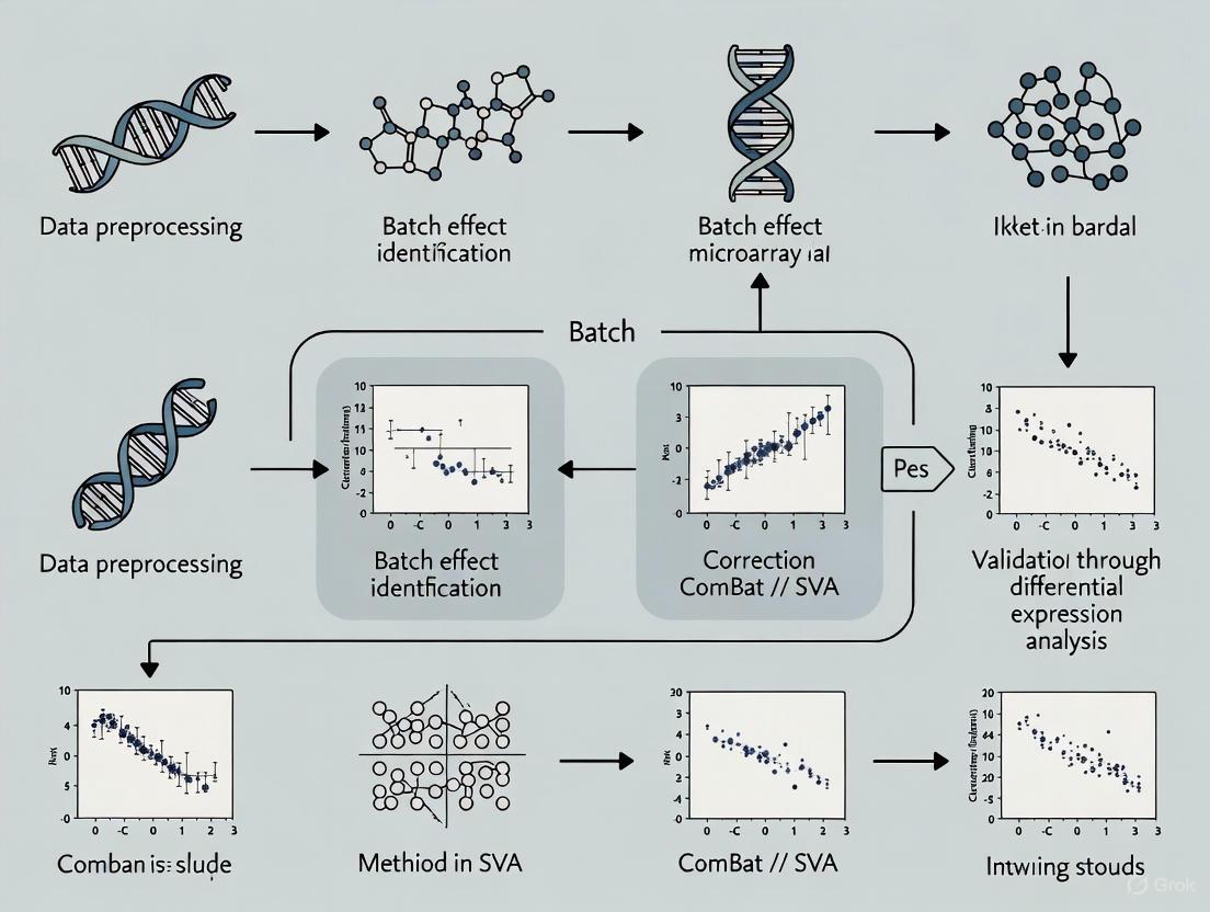
Microarray Workflow with Batch Effect Risk Points
The following diagram maps the microarray workflow and highlights critical control points where batch effects commonly originate:
Experimental Protocols for Batch Effect Evaluation
Establishing Quality Control Standards (QCS)
Implementing systematic quality controls enables objective monitoring of technical variations throughout the microarray workflow:
Tissue-Mimicking QCS Preparation:
- Create a controlled quality control standard using propranolol in a gelatin matrix (concentrations of 10, 20, 40, 80 mg/mL)
- Prepare QCS solution by mixing propranolol or propranolol-d7 (internal standard) with gelatin solution in a 1:20 ratio
- Spot QCS solution alongside experimental samples on multiple slides (recommended: 18 spots per slide) [14]
- Use these standards to evaluate variation caused by sample preparation and instrument performance
Batch Effect Assessment Protocol:
- Process QCS slides alongside experimental samples across multiple batches
- Measure technical variation using the QCS signals across batches
- Apply computational batch effect correction methods (ComBat, limma) to QCS data
- Evaluate correction efficiency by measuring reduction in QCS variation and improved sample clustering in multivariate principal component analysis [14]
Table: Key Research Reagent Solutions for Batch Effect Mitigation
| Item | Function | Considerations |
|---|---|---|
| Tissue-mimicking QCS (propranolol in gelatin) | Monitors technical variation across full workflow; Evaluates ion suppression effects [14] | Prepare fresh; Standardize spotting volume and pattern |
| Internal standards (e.g., propranolol-d7) | Controls for technical variation in sample processing; Normalization reference [14] | Use stable isotope-labeled versions of analytes |
| Fresh ethanol solutions | Prevents absorption of atmospheric water during processing | Replace regularly; Verify concentration |
| Fresh XC4 solution | Ensures consistent BeadChip coating | Reuse only up to six times during a two-week period [12] |
| Calibrated pipettors | Ensures accurate reagent dispensing | Perform yearly gravimetric calibration using water [12] |
| Humidifying buffer (PB2) | Prevents evaporation during hybridization | Verify correct volume in chamber wells [12] |
Batch effects remain a significant challenge in microarray workflows that can compromise data quality and research validity. By implementing systematic quality control measures, adhering to standardized protocols, and applying appropriate computational corrections when necessary, researchers can significantly reduce technical variations. The troubleshooting guidelines and experimental protocols provided here offer practical approaches to identify, mitigate, and correct batch effects, ultimately enhancing the reliability and reproducibility of microarray data in biomedical research.
FAQs: Understanding the Batch Effect Problem
What are batch effects and how do they arise? Batch effects are systematic technical variations introduced into data due to differences in experimental conditions rather than biological factors. These unwanted variations can arise from multiple sources, including:
- Different processing times, instruments, or machines
- Different laboratory personnel or sites
- Different reagent lots or analysis pipelines
- Sample storage conditions and freeze-thaw cycles In essence, any technical variable that creates consistent patterns of variation separate from your biological question of interest can constitute a batch effect [3] [15].
Why are batch effects particularly problematic in microarray research? Batch effects introduce non-biological variability that can confound your results in several ways:
- They can mask genuine biological signals, reducing statistical power
- They can create false associations that lead to incorrect conclusions
- In worst-case scenarios, they can completely drive observed differences between groups when batch is confounded with experimental conditions [8] [3] The high-dimensional nature of microarray data makes it especially vulnerable as these technical variations can systematically affect hundreds or thousands of data points simultaneously [16].
What is the difference between balanced and confounded study designs?
- Balanced Design: Your experimental groups are equally distributed across batches. For example, both case and control samples are processed on each chip and across different processing days. This allows technical variability to be "averaged out" during analysis [15] [17].
- Confounded Design: Your experimental groups are completely or partially separated by batch. For example, all control samples were processed in January while all case samples were processed in February. In this scenario, biological and technical effects become indistinguishable, making valid conclusions nearly impossible [15] [16].
Can batch effects really lead to paper retractions? Yes. The literature contains documented cases where batch effects directly contributed to irreproducible findings and subsequent retractions. In one prominent example, a study developing a fluorescent serotonin biosensor had to be retracted when the sensitivity was found to be highly dependent on reagent batch (specifically, the batch of fetal bovine serum), making key results unreproducible [3]. Another retracted study on personalized ovarian cancer treatment falsely identified gene expression signatures due to uncorrected batch effects [8].
Troubleshooting Guides
Problem: Unexpectedly Large Number of Significant Findings After Batch Correction
Symptoms:
- Thousands of significant differentially expressed genes appear only AFTER batch correction
- These genes show no or minimal significance before correction
- Biological interpretation of results seems implausible
Diagnosis: This pattern suggests possible over-correction or false signal introduction by your batch correction method, particularly when using empirical Bayes methods like ComBat with unbalanced designs [16] [18].
Solutions:
- Verify study design balance: Check if your biological groups are confounded with batch factors
- Apply more conservative correction: Consider using simpler methods like including batch as a covariate in linear models
- Validate with positive controls: Use genes known to be associated with your biological question as validation
- Try multiple correction approaches: Compare results across different algorithms to identify consistent findings
Prevention: Always randomize sample processing to ensure balanced distribution of experimental groups across batches. If complete randomization isn't possible, ensure each batch contains at least some samples from each biological group [16].
Problem: Persistent Batch Clustering After Correction
Symptoms:
- Samples continue to cluster by batch in PCA plots after correction
- Biological signal remains weak compared to technical variation
- Batch effects appear stronger than biological effects
Diagnosis: Your batch correction method may be insufficient for the magnitude of technical variation in your data, or you may have unidentified batch sources [8].
Solutions:
- Identify hidden batch factors: Use PCA to identify unknown sources of technical variation
- Increase correction stringency: Adjust parameters or try more aggressive algorithms
- Apply multiple correction steps: Address different batch sources sequentially
- Consider data removal: In extreme cases, exclude batches with irreconcilable technical issues
Problem: Loss of Biological Signal After Correction
Symptoms:
- Known biological differences disappear after batch correction
- Samples become overly homogenized
- Biological groups that previously separated well now mix completely
Diagnosis: Your correction method may be over-removing biological variation, especially when batch and biological factors are partially confounded [8].
Solutions:
- Use biological controls: Include samples with known differences to monitor signal preservation
- Try less aggressive methods: Switch to harmony, limma, or other more conservative approaches
- Adjust correction parameters: Reduce strength of correction where possible
- Apply supervised methods: Use methods that specifically protect biological variables of interest
Quantitative Impact Assessment
Table 1: Documented Cases of Batch Effect Consequences in Biomedical Research
| Study Type | Impact of Batch Effects | Consequences | Citation |
|---|---|---|---|
| Ovarian cancer biomarker study | False gene expression signatures identified | Study retraction | [8] |
| Clinical trial risk classification | Incorrect classification of 162 patients, 28 received wrong chemotherapy | Clinical harm potential | [3] |
| DNA methylation pilot study (n=30) | 9,612-19,214 significant differentially methylated sites appearing only after ComBat correction | False discoveries | [16] |
| Cross-species gene expression analysis | Apparent species differences greater than tissue differences; reversed after correction | Misinterpretation of fundamental biological relationships | [3] |
| Serotonin biosensor development | Sensitivity dependent on reagent batch | Key results unreproducible, paper retracted | [3] |
Table 2: Performance of Batch Effect Correction Methods Under Different Conditions
| Correction Method | Balanced Design Performance | Confounded Design Performance | Key Limitations | Citation |
|---|---|---|---|---|
| ComBat | Excellent | Risk of false positives | Can introduce false signals in unbalanced designs | [16] [18] |
| limma removeBatchEffect() | Good | Moderate | Less aggressive, may leave residual batch effects | [8] [19] |
| BRIDGE (for longitudinal data) | Excellent | Good | Requires bridging samples | [7] |
| SVA/RUV | Good for unknown batch effects | Variable performance | May capture biological signal if confounded | [8] |
| Harmony | Good | Good | Developed for single-cell, adapting to microarrays | [20] |
Experimental Protocols
Protocol 1: Systematic Batch Effect Assessment in Microarray Data
Purpose: Identify and quantify batch effects in your microarray dataset before proceeding with differential expression analysis.
Materials:
- Normalized microarray expression data
- Experimental metadata (batch information, biological groups)
- R statistical environment with following packages: limma, sva, pcaMethods
Procedure:
- Prepare data matrix: Start with your normalized expression values (log2-transformed recommended)
- Perform Principal Component Analysis (PCA):
- Test association between PCs and experimental variables:
- For each principal component (PC1-PC10), test association with:
- Batch factors (chip, row, processing date)
- Biological variables (disease status, treatment group)
- Sample characteristics (age, sex, BMI if relevant)
- Use ANOVA for categorical variables, correlation tests for continuous variables
- For each principal component (PC1-PC10), test association with:
- Visualize associations: Create boxplots of PC loadings colored by batch and biological groups
- Calculate batch effect magnitude:
- Compute variance explained by batch factors in each PC
- Use PVCA (Principal Variance Component Analysis) to partition variance sources
Interpretation:
- Strong association of early PCs (PC1-PC3) with batch factors indicates significant batch effects
- Biological variables should explain more variance than technical factors in well-controlled experiments
- If batch explains >25% of variance in early PCs, correction is necessary [16]
Protocol 2: Comparative Batch Effect Correction Evaluation
Purpose: Systematically evaluate multiple batch correction methods to select the most appropriate approach for your specific dataset.
Materials:
- Raw normalized microarray data
- Batch information (categorical)
- Biological group information
- R environment with: sva, limma, pamr
Procedure:
- Split data by batch: If you have multiple batches, analyze each batch separately for differential expression to establish batch-specific results [8]
- Create reference sets:
- Identify differentially expressed features in each batch (FDR < 0.05)
- Create a union set (all unique significant features across batches)
- Create an intersect set (features significant in all batches)
- Apply multiple correction methods:
- Process your data with 3-4 different BECAs (e.g., ComBat, limma, SVA, Harmony)
- Use default parameters initially
- Evaluate performance:
- For each corrected dataset, perform differential expression analysis
- Calculate recall: proportion of union reference set detected
- Calculate false positive rate: proportion of significant features not in union set
- Check preservation of intersect set: these should remain significant
Interpretation:
- The optimal method maximizes recall while minimizing false positives
- Methods that miss many features from the intersect set may be over-correction
- Consistent performance across multiple evaluation metrics indicates robustness [8]
Visual Guide to Batch Effect Concepts
Title: Impact of Batch Effect Management on Research Outcomes
Title: Balanced vs Confounded Study Design Impact
Table 3: Key Computational Tools for Batch Effect Management
| Tool Name | Primary Function | Best Use Scenario | Implementation | |
|---|---|---|---|---|
| ComBat | Empirical Bayes batch correction | When batch factors are known and design is balanced | R/sva package | |
| limma removeBatchEffect() | Linear model-based correction | Mild batch effects with balanced design | R/limma package | |
| BRIDGE | Longitudinal data correction | Time series studies with bridging samples | Custom R implementation | [7] |
| SelectBCM | Automated method selection | Initial screening of multiple BECAs | Available as described in literature | [8] |
| PCA | Batch effect visualization | Initial diagnostic assessment | Multiple R packages |
Table 4: Experimental Quality Control Materials
| Material Type | Purpose | Implementation Example | |
|---|---|---|---|
| Reference Samples | Monitor technical variation | Include same reference sample in each batch | |
| Bridging Samples | Connect batches technically | Split same biological sample across batches | [7] |
| Positive Controls | Verify biological signal preservation | Samples with known large biological differences | |
| Randomized Processing Order | Prevent confounding | Randomize sample processing across experimental groups | |
| Balanced Design | Enable statistical separation | Ensure each batch contains all experimental groups |
Advanced Troubleshooting: Special Scenarios
Longitudinal Studies with Time-Batch Confounding
Special Challenge: When batch is completely confounded with time points (all time point 1 samples in batch 1, all time point 2 in batch 2), traditional correction methods fail.
Solution: Apply specialized methods like BRIDGE that use "bridging samples" - technical replicates measured across multiple batches/timepoints to inform the correction [7].
Protocol:
- Include a subset of samples measured at multiple timepoints in both batches
- Use these bridging samples to estimate true biological temporal changes
- Apply empirical Bayes framework that incorporates bridging sample information
- Correct all samples based on the bridging sample-informed model
Challenge: Most real-world datasets have multiple, interacting batch effects (e.g., chip, row, processing date, technician).
Solution Approach:
- Identify all potential batch sources through systematic PCA association testing
- Determine correction order: Address larger sources first, or correct simultaneously if using multivariate methods
- Validate after each correction: Check if one batch correction introduces artifacts for other batch types
- Use conservative approaches: When multiple strong batch effects exist, consider including them as covariates in your final model rather than aggressive pre-correction
When to Abandon a Dataset
In some cases, batch effects may be irreconcilable. Consider excluding batches or entire datasets when:
- Batch effects are larger than the strongest biological effects in your system
- The experimental design is perfectly confounded with no bridging samples
- Multiple correction approaches yield completely different results with no consensus
- Positive controls (known biological differences) disappear after any reasonable correction attempt
Remember that publishing results from irredeemably confounded studies risks contributing to the reproducibility crisis, so ethical considerations may warrant dataset exclusion rather than forced analysis [3] [16].
What are the primary visual tools for diagnosing batch effects?
The most common visual tool for an initial assessment of batch effects is Principal Component Analysis (PCA). When you plot your data, typically using the first two principal components, a clear separation of data points by batch (rather than by biological condition) is a strong visual indicator that batch effects are present [21] [22].
For a more advanced visualization, Uniform Manifold Approximation and Projection (UMAP) is widely used. Like PCA, a UMAP plot that shows clusters corresponding to their source batch suggests a significant batch effect. The open-source platform Batch Effect Explorer (BEEx), for instance, incorporates UMAP specifically for this purpose, allowing researchers to qualitatively assess batch effects in medical image data [23].
The following diagram illustrates a typical diagnostic workflow that integrates these visual tools:
Which statistical metrics quantify the severity of batch effects?
While visual tools are intuitive, statistical metrics are essential for quantifying the severity of batch effects. The following table summarizes key diagnostic metrics:
| Metric Name | What It Measures | Interpretation | Common Tools |
|---|---|---|---|
| Silhouette Score [22] | How similar a sample is to its own batch vs. other batches (on a scale from -1 to 1). | Scores near 1 indicate strong batch clustering (strong batch effect). Scores near 0 or negative indicate no batch structure. | BEEx [23], Custom scripts |
| k-Nearest Neighbor Batch Effect Test (kBET) [24] [22] | The proportion of a sample's neighbors that come from different batches. | A high rejection rate indicates that batches are not well-mixed (strong batch effect). A low rate suggests successful correction. | HarmonizR [25], FedscGen [24] |
| Average Silhouette Width (ASW) [25] | Similar to the Silhouette Score, but often reported specifically for batch (ASWbatch) and biological label (ASWlabel). | A high ASWbatch indicates a strong batch effect. A high ASWlabel after correction indicates biological signal was preserved. | BERT [25] |
| Principal Variation Component Analysis (PVCA) [23] | The proportion of total variance in the data explained by batch versus biological factors. | A high proportion of variance attributed to "batch" indicates a significant batch effect. | BEEx [23] |
| Batch Effect Score (BES) [23] | A composite score designed to quantify the extent of batch effects from multiple analysis perspectives. | A higher score indicates a more pronounced batch effect. | BEEx [23] |
I've applied a correction method. How do I check if it worked?
Evaluating the success of a batch-effect correction procedure involves using the same diagnostic tools on the corrected data and comparing the results to the original, uncorrected data.
- Visual Inspection: Regenerate PCA and UMAP plots using the corrected data. Successful correction is indicated by the intermingling of data points from different batches, with clusters now ideally forming based on biological conditions rather than technical origins [24] [22].
- Statistical Validation: Recalculate the quantitative metrics.
- The kBET acceptance rate should increase, indicating better mixing [24].
- The Silhouette Score with respect to batch should decrease significantly, moving closer to zero [22].
- The ASW Batch score should decrease, while the ASW Label score (measuring biological cluster cohesion) should be maintained or improved, showing that biological signal was preserved while technical noise was removed [25].
What is a recommended experimental protocol for a comprehensive batch effect diagnosis?
Below is a detailed workflow you can follow to systematically diagnose batch effects in your microarray dataset, incorporating tools like BEEx [23] and BERT [25].
Objective: To qualitatively and quantitatively determine the presence and magnitude of batch effects in a multi-batch microarray dataset.
Materials and Inputs:
- Data Matrix: A normalized, preprocessed gene expression matrix (features x samples).
- Batch Metadata: A file specifying the batch ID for each sample.
- Biological Covariates: A file specifying biological conditions (e.g., disease state, treatment) for each sample.
- Software/Tools: R/Python environment with packages like
sva(for ComBat),limma,umap, and access to specialized tools like BEEx [23] or BERT [25].
Procedure:
Data Preprocessing: Ensure your data is normalized and filtered. Log-transformation is often applied to microarray data to stabilize variance.
Qualitative (Visual) Assessment:
- Generate PCA Plot: Perform PCA on your expression matrix. Color the data points by
batchand, separately, bybiological condition. A clear separation by batch in the PCA plot is an initial red flag. - Generate UMAP Plot: Create a UMAP projection of your data. Again, color points by batch and biological condition. Look for clustering driven by batch identity.
- Generate Heatmap & Dendrogram: Perform hierarchical clustering on the samples and visualize it with a heatmap. A dendrogram that groups samples primarily by batch indicates a strong batch effect.
- Generate PCA Plot: Perform PCA on your expression matrix. Color the data points by
Quantitative (Statistical) Assessment:
- Calculate Silhouette Scores: Compute the silhouette score where the "cluster" label is the batch ID. A high average score confirms the visual observation from the plots.
- Perform kBET: Run the k-nearest neighbor batch effect test on your data. A high rejection rate across many samples quantifies the failure of batches to mix.
- Run PVCA: Use Principal Variation Component Analysis to partition the total variance in your dataset. Note the percentage of variance attributed to "batch" versus your biological factors of interest.
Interpretation and Reporting:
- Correlate the findings from all visual and statistical methods.
- A consensus across multiple diagnostics (e.g., clear batch clustering in PCA/UMAP, high silhouette score, high kBET rejection rate, and high batch variance in PVCA) provides robust evidence for the presence of batch effects.
- This comprehensive diagnosis forms the basis for deciding whether and how to proceed with batch-effect correction.
The Scientist's Toolkit: Essential Research Reagents & Solutions
The following table lists key computational tools and statistical solutions used in the field of batch effect diagnostics and correction, as identified in the search results.
| Tool/Solution Name | Type/Function | Key Application Context |
|---|---|---|
| BEEx (Batch Effect Explorer) [23] | Open-source platform for qualitative & quantitative batch effect detection. | Medical images (Pathology & Radiology); provides visualization and a Batch Effect Score (BES). |
| ComBat [26] [21] [22] | Empirical Bayes framework for location/scale adjustment. | Microarray, Proteomics, Radiomics; robust for small sample sizes. |
Limma (removeBatchEffect) [25] [22] |
Linear models to remove batch effects as a covariate. | General omics data (Transcriptomics, Proteomics), Radiomics. |
| BERT [25] | High-performance, tree-based framework for data integration. | Large-scale, incomplete omic data (Proteomics, Transcriptomics, Metabolomics). |
| HarmonizR [25] | Imputation-free framework using matrix dissection. | Integration of arbitrarily incomplete omic profiles. |
| kBET [24] [22] | Statistical test to quantify batch mixing. | Evaluation of batch effect correction efficacy in single-cell RNA-seq and other data. |
| Silhouette Width (ASW) [25] | Metric for cluster cohesion and separation. | Global evaluation of data integration quality, applicable to any clustered data. |
| RECODE/iRECODE [27] | High-dimensional statistics-based tool for technical noise reduction. | Single-cell omics data (scRNA-seq, scHi-C, spatial transcriptomics). |
Batch Effect Correction Tools: From ComBat to Cutting-Edge Ratio Methods
Frequently Asked Questions (FAQs)
1. What is the fundamental difference between ComBat and Limma's removeBatchEffect?
ComBat uses an empirical Bayes framework to actively adjust your data by shrinking batch effect estimates toward a common mean, making it particularly powerful for small sample sizes. In contrast, Limma's removeBatchEffect function performs a linear model adjustment, simply subtracting the estimated batch effect from the data without any shrinkage. Crucially, removeBatchEffect is intended for visualization purposes and not for data that will be used in downstream differential expression analysis; for formal analysis, the batch factor should be included directly in the design matrix of your statistical model [28] [29].
2. When should I use SVA instead of ComBat or Limma?
You should use Surrogate Variable Analysis (SVA) when the sources of batch effects are unknown or unmeasured [8] [30]. While ComBat and removeBatchEffect require you to specify the batch factor, SVA is designed to identify and adjust for these hidden sources of variation by estimating surrogate variables from the data itself. These surrogate variables can then be included as covariates in your downstream models [30].
3. I'm getting a "non-conformable arguments" error when running ComBat. What should I do?
This error often relates to issues with the data matrix or model structure [31]. A common solution is to filter out low-varying or zero-variance genes from your dataset before running ComBat. You should also check that your batch vector does not contain any NA values and that it has the same number of samples as your data matrix [31].
4. Can these batch correction methods be used for data types other than gene expression? Yes, the core principles of these algorithms are applied across various data types. For instance, they have been successfully used in radiogenomic studies of lung cancer patients [22]. Furthermore, specialized variants like ComBat-met have been developed for DNA methylation data (β-values), which use a beta regression framework to account for the unique distributional properties of such data [32].
5. What is the most important consideration for a successful batch correction? A balanced study design is paramount [15]. If your biological conditions of interest are perfectly confounded with batch (e.g., all controls are in batch 1 and all treatments are in batch 2), no statistical method can reliably disentangle the technical artifacts from the true biological signal. Whenever possible, ensure that each batch contains a mixture of all biological conditions you plan to study [15] [33].
Troubleshooting Guides
Problem 1: Poor Batch Correction Performance
Symptoms: After correction, Principal Component Analysis (PCA) plots still show strong clustering by batch, or downstream analysis (e.g., differential expression) yields unexpected or biologically implausible results.
| Potential Cause | Recommended Action |
|---|---|
| Severe design imbalance | Review your experimental design. If the batch is perfectly confounded with a condition, correction is not advised. Re-assess the feasibility of the analysis [15]. |
| Incorrect algorithm selection | Re-evaluate your choice. For known batches, use ComBat or include batch in the model. For unknown batches, use SVA or RUV [8] [30]. |
| Incompatible data preprocessing | Ensure the batch correction method is compatible with your entire workflow (e.g., normalization, imputation). The choice of preceding steps can significantly impact the BECA's performance [8]. |
| Over-correction | Aggressive correction can remove biological signal. Use sensitivity analysis to check if key biological findings are consistent across different BECAs [8]. |
Problem 2: Errors During ComBat Execution
Symptoms: Errors such as "non-conformable arguments" or "missing value where TRUE/FALSE needed" [31].
| Potential Cause | Recommended Action |
|---|---|
| Genes with zero variance | Filter your data matrix to remove genes with zero variance across all samples. This is a very common fix [31]. |
| Zero variance within a batch | Remove genes that have zero variance in any of the batches, not just across all samples [31]. |
NA values in the data or batch vector |
Check for and remove any NA values in your batch vector or data matrix [31]. |
Performance and Methodology Comparison
The table below summarizes the core methodologies and applications of ComBat, Limma, and SVA.
| Algorithm | Core Methodology | Primary Use Case | Key Assumptions | Data Types |
|---|---|---|---|---|
| ComBat | Empirical Bayes framework that shrinks batch effect estimates towards a common mean [8]. | Correcting for known batch effects, especially with small sample sizes [29]. | Batch effects fit a predefined model (e.g., additive, multiplicative) [8]. | Microarray data, RNA-seq count data (ComBat-seq) [32]. |
Limma's removeBatchEffect |
Fits a linear model and subtracts the estimated batch effect [22]. | Preparing data for visualization (e.g., PCA plots). Not for downstream DE analysis [28]. | Batch effects are linear and additive [22]. | Normalized, continuous data (e.g., log-CPMs from microarray or RNA-seq). |
| SVA | Identifies latent factors ("surrogate variables") that capture unknown sources of variation [30]. | Correcting for unknown batch effects or unmeasured confounders [8]. | Surrogate variables represent technical noise and can be estimated from the data [30]. | Can be applied after appropriate normalization for various data types. |
Experimental Protocols
Detailed Methodology for Benchmarking Batch Effect Correction Algorithms
This protocol outlines a sensitivity analysis to evaluate the performance of different BECAs, ensuring robust and reproducible results [8].
1. Experimental Setup and Data Splitting
- Begin with a dataset comprising multiple batches.
- Split the data into its individual batches for a ground-truth comparison (e.g., Batch A, Batch B, etc.) [8].
2. Establishing Reference Sets via Differential Expression Analysis
- Perform a differential expression (DE) analysis separately on each individual batch.
- From these individual analyses, create two crucial reference sets:
- The Union Set: Combine all unique differentially expressed (DE) features found in any of the individual batches.
- The Intersect Set: Identify the DE features that are consistently found in every single batch. This set acts as a high-confidence biological signal [8].
3. Applying and Evaluating Batch Correction Methods
- Apply a variety of BECAs (e.g., ComBat, Limma, SVA) to the original, full dataset.
- Conduct a DE analysis on each of the batch-corrected datasets.
- For each BECA, calculate performance metrics by comparing its DE results to the reference sets:
- Recall: The proportion of features in the Union Set that were successfully rediscovered after correction.
- False Positive Rate: The proportion of features called significant after correction that were not present in the Union Set.
- A reliable BECA will show high recall and a low false positive rate. Additionally, it should retain most features from the Intersect Set; missing these suggests the correction may be too aggressive and is removing real biological signal [8].
Workflow for a Standard Limma-voom Analysis with Batch Covariates
For RNA-seq count data, this is a statistically sound workflow that incorporates batch information directly into the model for differential expression [28] [29].
- Create a DGEList object using your raw count data and sample metadata.
- Normalize the data using the Trimmed Mean of M-values (TMM) method with
calcNormFactors. - Apply the
voomtransformation, which converts counts to log2-counts per million (log-CPM) and calculates observation-level weights for linear modeling. Plot thevoomobject to check data quality. - Create a design matrix that includes both your biological condition of interest and the known batch factor(s).
- Fit a linear model using the
lmFitfunction with thevoom-transformed data and your design matrix. - Apply empirical Bayes moderation to the standard errors using the
eBayesfunction. - Extract the results of your differential expression analysis using the
topTablefunction.
The Scientist's Toolkit: Essential Research Reagents & Materials
| Item | Function/Brief Explanation |
|---|---|
| High-Dimensional Data | The primary input (e.g., from microarrays, RNA-seq, or methylation arrays) requiring correction for technical noise [8]. |
| Batch Metadata | A critical file (often a CSV) that maps each sample to its processing batch. Essential for ComBat and Limma [29]. |
| R Statistical Software | The standard environment for running these analyses. Key packages include sva (for ComBat and SVA), limma (for removeBatchEffect and linear modeling), and edgeR or DESeq2 for normalization and DE analysis [29]. |
| Negative Control Genes | A set of genes known not to be affected by the biological conditions of interest. Required for methods like RUV but can be challenging to define. In practice, non-differentially expressed genes from a preliminary analysis are sometimes used as "pseudo-controls" [30]. |
| Reference Batch | A specific batch chosen as the baseline to which all other batches are adjusted. This is an option in tools like ComBat and can be useful when one batch is considered a "gold standard" [22]. |
| Visualization Tools (PCA) | Essential for diagnosing batch effects before and after correction. PCA plots provide an intuitive visual assessment of whether sample clustering is driven by batch or biology [8] [33]. |
BECA Selection and Evaluation Workflow
The following diagram outlines a logical workflow for selecting, applying, and evaluating a batch effect correction strategy, incorporating key considerations from the FAQs and troubleshooting guides.
What is the fundamental principle behind Empirical Bayes frameworks like ComBat? Empirical Bayes frameworks, such as ComBat, address the pervasive issue of batch effects in high-throughput genomic datasets. Batch effects are technical artifacts that introduce non-biological variability into data due to processing samples in different batches, at different times, or by different personnel. If left uncorrected, this noise can reduce statistical power, dilute true biological signals, and potentially lead to spurious or misleading scientific conclusions [7] [34] [35]. ComBat uses an Empirical Bayes approach to robustly estimate and adjust for these batch-specific artifacts, allowing for the more valid integration of datasets from multiple studies or processing batches [34].
How does the Empirical Bayes method in ComBat differ from a standard linear model? While a standard linear model might directly estimate and subtract batch effects, this can be unstable for studies with small sample sizes per batch. ComBat's key innovation is its use of shrinkage estimation. It assumes that batch effect parameters (e.g., the amount by which a batch shifts a gene's expression) across all genes in a dataset follow a common prior distribution (e.g., a normal distribution for additive effects). ComBat then uses the data itself to empirically estimate the parameters of this prior distribution and "shrinks" the batch effect estimates for individual genes toward the common mean. This pooling of information across genes makes the estimates more robust and prevents overfitting, especially for genes with high variance or batches with small sample sizes [7] [34].
Troubleshooting Guides and FAQs
Model Selection and Application
Q: My study has a longitudinal design where the same subjects are profiled over time, and time is completely confounded with batch. Is standard ComBat appropriate? A: No, standard ComBat, which assumes sample independence, is not ideal for dependent longitudinal samples and may overcorrect the data [7]. For such designs, you should consider specialized methods:
- Longitudinal ComBat: This extension incorporates subject-specific random effects into the ComBat model to account for the within-subject correlation introduced by repeated measurements [7].
- BRIDGE (Batch effect Reduction of mIcroarray data with Dependent samples usinG empirical Bayes): This method is specifically designed for confounded longitudinal studies and requires the inclusion of "bridge samples"–technical replicates from a subset of participants that are profiled across multiple batches. These bridges explicitly inform the batch-effect correction [7].
Q: When should I use a reference batch in ComBat? A: Using a reference batch is highly recommended in biomarker development pipelines [34]. In this scenario:
- The initial training set is designated as the reference batch.
- All future validation or test batches are adjusted to align with this reference.
- This ensures the training data and the derived biomarker signature remain fixed, avoiding the "sample set bias" where adding new batches alters the adjusted values of previously processed samples. This guarantees the biomarker can be consistently applied to new data without retraining [34].
Data Input and Preprocessing
Q: What are the basic data structure requirements for running ComBat? A: Your data should be structured as a features-by-samples matrix (e.g., Genes x Samples). The model requires you to specify a batch covariate (e.g., processing site or date) for each sample. You can also optionally include other biological or technical covariates in the design matrix to preserve their effects during correction [7] [34].
Q: My data is distributed across multiple institutions and cannot be centralized due to privacy regulations. Can I still use ComBat? A: Yes, a Decentralized ComBat (DC-ComBat) algorithm has been developed for this purpose. It uses a federated learning approach where local nodes (institutions) calculate summary statistics from their data. These statistics are then aggregated by a central node to compute the grand mean and variance needed for the Empirical Bayes estimation. The individual patient data never leaves the local institution, preserving privacy while achieving harmonization results nearly identical to the pooled-data approach [36].
Interpretation and Output
Q: After running ComBat, how can I validate the success of the batch correction? A: You should use both visual and quantitative diagnostics:
- Principal Component Analysis (PCA) Plots: Visualize the data before and after correction. Samples should no longer cluster strongly by batch in the corrected PCA plot.
- Distributional Metrics: Examine the moments of the data distribution (mean, variance, skewness, kurtosis) across batches before and after correction. Effective harmonization should align these distributions [34].
ComBat Workflow and Signaling Pathways
The following diagram illustrates the logical workflow and data flow of the Empirical Bayes estimation process in ComBat.
Key Parameter Estimates in the ComBat Model
ComBat corrects for two types of batch effects by estimating the following parameters for each gene in each batch. These are adjusted using the Empirical Bayes shrinkage method [7] [34] [36].
Table 1: Core Batch Effect Parameters in the ComBat Model
| Parameter | Symbol | Type of Batch Effect | Interpretation |
|---|---|---|---|
| Additive Batch Effect | (\gamma_{i,v}) | Location / Mean | A gene- and batch-specific term that systematically shifts the mean expression level. |
| Multiplicative Batch Effect | (\delta_{i,v}) | Scale / Variance | A gene- and batch-specific term that scales the variance (spread) of the expression values. |
The Scientist's Toolkit: Essential Research Reagents and Solutions
For researchers conducting microarray experiments and subsequent batch effect correction, the following tools and conceptual "reagents" are essential.
Table 2: Key Research Reagents and Solutions for Batch Effect Correction
| Item | Function / Interpretation | Considerations for Use |
|---|---|---|
| Bridge Samples | Technical replicate samples from a subset of participants profiled in multiple batches. They serve as a direct link to inform batch-effect correction in longitudinal studies [7]. | Logistically challenging and costly to obtain, but are crucial for confounded longitudinal designs. |
| Reference Batch | A single, high-quality batch designated as the standard to which all other batches are aligned. Preserves data integrity in biomarker studies [34]. | Prevents "sample set bias" and ensures a fixed training set for biomarker development. |
| Sensitive Attribute (Z) | A protected variable (e.g., race, age) the model is explicitly prevented from using, often enforced via adversarial training in fairness-focused applications [37]. | Requires careful specification and is part of advanced de-biasing techniques beyond standard batch correction. |
| Covariate Matrix (X) | A design matrix specifying known biological or treatment conditions of interest. ComBat uses this to model and preserve these effects during batch removal [34] [36]. | Critical for preventing the removal of true biological signal along with batch noise. |
| Shrinkage Estimators | The mathematical mechanism that stabilizes batch effect estimates by borrowing information across all genes, reducing the influence of high-variance genes [7] [34]. | The core of the Empirical Bayes approach, providing more robust corrections, especially with small batch sizes. |
Frequently Asked Questions (FAQs) on Ratio-Based Batch Effect Correction
FAQ 1: What is the core principle behind ratio-based batch effect correction? The ratio-based method, sometimes referred to as Ratio-G, works by scaling the absolute feature values (e.g., gene expression, protein intensity) of study samples relative to the values of one or more concurrently profiled reference materials analyzed in the same batch [4]. This transforms the raw measurements into a ratio scale, effectively canceling out batch-specific technical variations. The underlying assumption is that any technical variation affecting the study samples will also affect the reference material, allowing the ratio to isolate the biological signal [4] [38].
FAQ 2: When is a ratio-based approach particularly advantageous over other methods? Ratio-based correction is especially powerful in confounded scenarios, where batch effects are completely confounded with the biological factors of interest [4]. For instance, if all samples from biological Group A are processed in Batch 1 and all samples from Group B in Batch 2, it becomes impossible for many algorithms to distinguish technical from biological variation. In such cases, the ratio-based method, which uses an internal anchor (the reference material), performs significantly better at preserving true biological differences while removing batch effects [4].
FAQ 3: What are the critical considerations when selecting a reference material? An ideal reference material should be both stable and representative.
- Stability: The material must be homogenous and available in sufficient quantity to be profiled alongside every batch in a long-term study [4].
- Representativeness: Its composition should broadly reflect the study samples. For example, in proteomics, the Quartet Project's matched DNA, RNA, protein, and metabolite reference materials derived from B-lymphoblastoid cell lines are designed for this purpose [4]. In large-scale plasma proteomics studies, using pooled plasma from multiple healthy donors as a quality control (QC) sample has been shown to be effective [38].
FAQ 4: My data is on a different scale after ratio transformation. Does this impact downstream analysis? Yes, applying a ratio-based transformation will change the scale of your data. This is a fundamental characteristic of the method. While this scaling is precisely what corrects the batch effects, it is crucial to ensure that the statistical models and algorithms used in downstream analyses (e.g., differential expression, clustering) are compatible with ratio-scaled data. Always verify that your downstream tools can handle this data type appropriately.
FAQ 5: Can the ratio method be combined with other normalization techniques? Yes, ratio-based correction is often part of a larger data preprocessing workflow. It is common to perform initial normalization (e.g., for library size in RNA-seq) on the raw data before calculating the ratios relative to the reference material. The ratio step itself is the primary batch-effect correction, and its output can then be used directly for downstream statistical modeling.
Troubleshooting Common Experimental Issues
Problem: Inconsistent Correction Across Features
- Symptoms: After correction, some genes or proteins still show strong batch-associated variance, while others are over-corrected.
- Possible Causes: The chosen reference material might not be optimal for all feature types. For example, a reference material with a narrow dynamic range may not effectively correct features with very high or low expression.
- Solutions:
- Validate the dynamic range of your reference material against your study samples prior to large-scale deployment.
- Consider using a pooled reference comprising multiple samples to better capture the diversity of your study's features [38].
Problem: Introduction of Noise by Low-Abundance Features
- Symptoms: Increased variability in measurements for low-intensity genes/proteins after ratio application.
- Possible Causes: When the reference material's value for a specific feature is very low or near the detection limit, the ratio calculation can become unstable and amplify noise.
- Solutions:
- Implement a filtering step to remove features where the reference material's signal is consistently low or undetectable across batches.
- As a quality control flag, consider excluding proteins where more than half of the bridging control measurements fall below the limit of detection, as this indicates unreliable data [39].
Problem: Poor Batch Effect Removal in PCA Plots
- Symptoms: Samples still cluster by batch in a Principal Component Analysis (PCA) plot after ratio correction.
- Possible Causes:
- The batch effect may be non-additive or non-linear, which a simple scaling factor cannot fully address.
- Strong sample-specific batch effects might be present, which require a method capable of modeling these complex variations [39].
- Solutions:
- Visually inspect the data to diagnose the type of batch effect. Plotting measurements from two batches against each other can reveal if effects are protein-specific, sample-specific, or plate-wide [39].
- For complex, multi-type batch effects, consider more robust regression-based methods like BAMBOO that are specifically designed to handle them using bridging controls [39].
Performance Comparison of Batch Effect Correction Algorithms
The table below summarizes the performance of various batch effect correction algorithms (BECAs) across different data types and experimental scenarios, as evidenced by benchmarking studies.
Table 1: Performance Comparison of Batch-Effect Correction Algorithms
| Algorithm | Underlying Principle | Recommended Data Type(s) | Strengths | Key Limitations |
|---|---|---|---|---|
| Ratio-Based | Scaling to reference material(s) | Multi-omics (Transcriptomics, Proteomics, Metabolomics) [4] | Superior in confounded batch-group scenarios; broadly applicable [4]. | Requires carefully characterized reference materials. |
| ComBat | Empirical Bayes framework | Microarray, RNA-seq (ComBat-seq) [32] [40] | Widely adopted; effective for mean shifts in balanced designs [38]. | Assumes normal distribution; can be impacted by outliers in bridging controls [39]. |
| Harmony | PCA-based iterative clustering | Single-cell RNA-seq, Multi-omics [4] | Performs well in balanced and some confounded scenarios [4]. | Performance may vary across omics types. |
| BAMBOO | Robust regression on bridging controls | Proximity Extension Assay (PEA) Proteomics [39] | Robust to outliers; corrects protein-, sample-, and plate-wide effects [39]. | Requires multiple (e.g., 10-12) bridging controls. |
| ComBat-met | Beta regression | DNA Methylation (β-values) [32] | Tailored for proportional data (0-1); controls false positives [32]. | Specifically designed for methylation data. |
| Median Centering | Mean/median scaling per batch | Proteomics [38] | Simple and fast. | Lower accuracy; significantly impacted by outliers [39]. |
Standard Experimental Protocol for Ratio-Based Correction
This protocol provides a step-by-step guide for implementing a ratio-based batch effect correction in a multi-batch study, using the Quartet Project as a model [4].
Step 1: Experimental Design and Reference Material Selection
- Identify a stable and representative reference material. For multi-omics studies, consider using matched reference materials like the Quartet suites [4].
- Design your experiment so that the same reference material is profiled in every batch. The number of technical replicates for the reference material should be determined based on desired precision.
Step 2: Data Generation and Preprocessing
- Generate your multi-batch data (e.g., transcriptomics, proteomics).
- Perform initial, basic normalization on the raw data as required by your platform (e.g., library size normalization for RNA-seq, log transformation for microarray data).
Step 3: Ratio Calculation
- For each feature (gene, protein) ( i ) in a study sample from batch ( b ), calculate the ratio value as follows: ( \text{Ratio}{i,b} = \frac{\text{Normalized value of study sample}{i,b}}{\text{Normalized value of reference material}_{i,b}} )
- Here, the "Normalized value of reference material" is typically the mean or median of the technical replicates of the reference material profiled in the same batch ( b ).
Step 4: Data Integration and Downstream Analysis
- The resulting ratio-scaled matrix is your batch-corrected dataset.
- Proceed with downstream analyses such as differential expression, clustering, or predictive modeling. Ensure that the methods used are compatible with ratio-scaled data.
The workflow below summarizes this process.
The Scientist's Toolkit: Essential Research Reagents & Materials
The successful implementation of a ratio-based correction strategy relies on key reagents and resources. The table below lists essential items for setting up such an approach.
Table 2: Key Research Reagent Solutions for Ratio-Based Methods
| Item | Function & Role in Batch Correction | Example from Literature |
|---|---|---|
| Cell Line-Derived Reference Materials | Provides a stable, renewable source of DNA, RNA, protein, and metabolites for system-wide batch correction. | Quartet Project's matched multiomics reference materials from four family members' B-lymphoblastoid cell lines [4]. |
| Pooled Plasma/Serum QC Samples | Serves as a reference material for clinical proteomics and metabolomics studies, mimicking the sample matrix. | Pooled plasma from 16 healthy males used as a QC sample in a large-scale T2D patient proteomics study [38]. |
| Bridging Controls (BCs) | Identical samples included on every processing plate (e.g., in PEA protocols) to directly measure and model plate-to-plate variation. | At least 8-12 bridging controls per plate are recommended for robust correction using methods like BAMBOO [39]. |
| Commercial Reference Standards | Well-characterized, commercially available standards (e.g., Universal Human Reference RNA) that can be used as a common denominator across labs. | Various sources; often used in method development and cross-platform comparisons to anchor measurements. |
Frequently Asked Questions
Q1: My batch-corrected data shows unexpected clustering. What could be wrong? In a fully confounded study design, where your biological groups of interest perfectly separate by batch, it may be impossible to disentangle biological signals from technical batch effects [15]. If a batch correction method is applied in this scenario, it might remove biological signal along with the batch effect, leading to misleading clustering. Always check your experimental design for balance before proceeding.
Q2: What should I do if my ComBat model fails to converge?
Try increasing the number of genes used in the empirical Bayes estimation by adjusting the gene_subset_n parameter [41]. Using a larger subset of genes can stabilize the model fitting process. Additionally, ensure that your model matrix for covariates (covar_mod) is correctly specified and contains only categorical variables.
Q3: How do I handle missing values in my batch or covariate data?
The pycombat_seq function offers the na_cov_action parameter to control this. You can choose to:
"raise"an error and stop execution."remove"samples with missing covariates and issue a warning."fill"by creating a distinct covariate category per batch for the missing values [41]. Your choice should be guided by the extent and nature of the missing data.
Q4: Should I correct for batch effects before or after normalization? Batch effect correction is typically performed after data normalization. In RNA-Seq analyses, upstream processing steps like quality control and normalization should be performed within each batch before applying a batch effect correction method like ComBat-Seq [42].
Q5: After correction, a known biological signal seems weakened. Is this normal? Overly aggressive correction is a known risk. Some methods, especially those that do not retain "true" between-batch differences, can inadvertently remove or weaken strong biological signals if they are correlated with a batch [8] [43]. It is crucial to use downstream sensitivity analyses to verify that key biological findings are preserved after correction.
Troubleshooting Common Scenarios
Scenario 1: Correcting RNA-Seq Count Data in Python Problem: You have a raw count matrix from an RNA-Seq experiment conducted over several batches and need to correct for batch effects using a method designed for count data.
Solution: Use the pycombat_seq function, which is a Python port of the ComBat-Seq method.
Key Parameters:
covar_mod: A model matrix if you need to preserve signals from specific covariates.ref_batch: Specify a batch id to use as a reference, against which all other batches will be adjusted [41].
Scenario 2: Comparing Multiple Batch Correction Methods in R Problem: You are unsure which batch correction method is most appropriate for your biomarker data and want to compare several approaches.
Solution: Use the batchtma R package, which provides a unified interface for multiple methods.
Method Selection Guide from batchtma: [43]
| Method | Approach | Retains "True" Between-Batch Differences? |
|---|---|---|
simple |
Simple means | No |
standardize |
Standardized batch means | Yes |
ipw |
Inverse-probability weighting | Yes |
quantreg |
Quantile regression | Yes |
quantnorm |
Quantile normalization | No |
Scenario 3: Integrating Single-Cell RNA-Seq Data in R Problem: You have multiple batches of single-cell RNA-seq data where the cell population composition is unknown or not identical across batches.
Solution: Use the batchelor package and its quickCorrect() function, which is designed for this context.
Critical Pre-Correction Steps: [42]
- Subset to Common Features: Ensure both datasets use the same set of genes.
- Rescale Batches: Use
multiBatchNorm()to adjust for differences in sequencing depth between batches. - Select Highly Variable Genes (HVGs): Use
combineVar()andgetTopHVGs()to select genes that drive population structure.
Experimental Protocols & Evaluation
Protocol: Evaluating Correction Performance with Downstream Sensitivity Analysis
This protocol helps you assess how different BECAs affect your biological conclusions, a recommended best practice [8].
- Split Data by Batch: Treat each of your batches as an individual dataset.
- Perform DEA Per Batch: Conduct a differential expression analysis (DEA) on each batch separately to obtain lists of differentially expressed (DE) features for each.
- Create Reference Sets:
- Union Reference: Combine all unique DE features from all individual batches.
- Intersect Reference: Identify the DE features that are common to all batches.
- Apply Multiple BECAs: Correct your full dataset using several batch correction methods.
- Perform DEA on Corrected Data: Run DEA on each batch-corrected dataset.
- Calculate Performance Metrics:
- Recall: The proportion of the Union Reference found by the DEA on the corrected data.
- Check Intersect: Ensure that features in the Intersect Reference are still present after correction; their absence may indicate over-correction.
The method that yields the highest recall while preserving the intersect features can be considered the most reliable for your data.
The Scientist's Toolkit
| Essential Material / Software | Function |
|---|---|
sva / inmoose R/Package |
Provides the standard ComBat (for normalized data) and ComBat-Seq (for count data) algorithms for batch effect adjustment using empirical Bayes frameworks [41] [40]. |
limma R Package |
Contains the removeBatchEffect() function, a linear-model-based method for removing batch effects, commonly used for microarray and RNA-Seq data [8] [42]. |
batchelor R Package (Bioconductor) |
A specialized package for single-cell data, offering multiple correction algorithms (e.g., MNN, rescaleBatches) that do not assume identical cell population composition across batches [42]. |
batchtma R Package |
Provides a suite of methods for adjusting batch effects in biomarker data, with a focus on retaining true between-batch differences caused by confounding sample characteristics [43]. |
| Principal Component Analysis (PCA) | A dimensionality reduction technique used to visualize batch effects before and after correction. Persistent batch clustering in PCA plots after correction suggests residual batch effects [8] [42]. |
Batch Effect Correction Workflow
The following diagram outlines the logical workflow for a standard batch effect correction process, from data preparation to evaluation.
Batch Effect Correction Workflow
Method Selection Logic
Choosing the right batch correction method is critical. The following diagram provides a logical pathway for selecting an appropriate algorithm based on your data type and experimental design.
Algorithm Selection Guide
ComBat-met FAQs: Core Methodology and Application
Q1: What is ComBat-met and how does it fundamentally differ from standard ComBat?
ComBat-met is a specialized batch effect correction method designed specifically for DNA methylation data. Unlike standard ComBat, which assumes normally distributed data, ComBat-met employs a beta regression framework that accounts for the unique characteristics of DNA methylation β-values, which are constrained between 0 and 1 and often exhibit skewness and over-dispersion. The method fits beta regression models to the data, calculates batch-free distributions, and maps the quantiles of the estimated distributions to their batch-free counterparts [32].
Q2: When should I choose ComBat-met over other batch correction methods?
ComBat-met is particularly advantageous when:
- Your data consists of β-values from DNA methylation studies
- You require high statistical power for differential methylation analysis
- Controlling false positive rates is a critical concern
- You need to handle data with both additive and multiplicative batch effects
Simulation studies demonstrate that ComBat-met followed by differential methylation analysis achieves superior statistical power compared to traditional approaches while correctly controlling Type I error rates in nearly all cases [32].
Q3: What are the key preprocessing steps before applying ComBat-met?
Proper preprocessing is essential for effective batch correction:
- Quality Control: Remove poor-quality samples and probes using standard methylation QC pipelines
- Normalization: Apply appropriate normalization for your platform (450K/EPIC)
- M-Value Conversion: While ComBat-met works with β-values, the underlying model uses M-values (logit-transformed β-values) for statistical modeling [32] [44]
Q4: Can ComBat-met handle reference-based adjustments?
Yes, ComBat-met supports both common batch effect adjustment (adjusting all batches to a common mean) and reference-based adjustment, where all batches are adjusted to the mean and precision of a specific reference batch. This is particularly useful when you have a gold-standard batch or when integrating new data with previously established datasets [32].
Performance Comparison of Batch Effect Correction Methods
Table 1: Comparative performance of DNA methylation batch effect correction methods based on simulation studies
| Method | Underlying Model | Data Type | Key Advantages | Limitations/Considerations |
|---|---|---|---|---|
| ComBat-met | Beta regression | β-values | Specifically designed for methylation data; maintains β-value constraints; improved power in simulations | Newer method with less established track record |
| Standard ComBat | Empirical Bayes (Gaussian) | M-values | Widely adopted; robust for small batch sizes | Can introduce false positives if misapplied to unbalanced designs [18] [16] |
| M-value ComBat | Empirical Bayes (Gaussian) | M-values | Uses established M-value transformation | Requires back-transformation to β-values for interpretation |
| SVA | Surrogate variable analysis | M-values | Handles unknown batch effects; doesn't require batch labels | May capture biological signal if confounded with technical variation |
| RUVm | Remove unwanted variation | M-values | Uses control probes/features; flexible framework | Requires appropriate control features |
| BEclear | Latent factor models | β-values | Directly models β-values; imputes missing values | Different statistical approach than ComBat family |
Troubleshooting Guide: Common Issues and Solutions
Problem: Unexpected False Positives After Batch Correction
Symptoms: Thousands of significant CpG sites appear after batch correction that weren't present before correction, particularly with unbalanced study designs [18] [16].
Solutions:
- Balance Study Design: Ensure biological groups are distributed evenly across batches before processing
- Include Covariates: Specify known biological covariates in the model matrix when running ComBat-met
- Validate with Null Data: Run negative controls to assess false positive rates
- Check Batch-Biology Confounding: Use PCA to verify batch effects are not confounded with biological variables
Table 2: Troubleshooting common ComBat-met implementation issues
| Issue | Potential Causes | Diagnostic Steps | Solutions |
|---|---|---|---|
| Poor batch effect removal | Incorrect batch labels; Severe batch effects; Biological signal confounded with batch | PCA coloring by batch before/after correction; Check association of PCs with batch | Verify batch labels; Consider reference batch correction; Check for confounding |
| Over-correction | Biological signal correlates with batch; Too aggressive parameter estimation | Compare results with uncorrected data; Check if biological signal strength decreased dramatically | Use shrinkage parameters; Adjust model specifications; Validate with known biological controls |
| Computational performance issues | Large datasets; Many batches; Many features | Monitor memory usage; Check parallelization settings | Use parallel processing; Filter low-quality probes first; Increase system resources |
| Values outside expected range | Extreme batch effects; Model misspecification | Check distribution of corrected values | Ensure proper data preprocessing; Consider using M-value transformation approach |
Problem: Persistent Batch Effects After Correction
Symptoms: Samples still cluster by batch in PCA plots after applying ComBat-met.
Solutions:
- Check for Additional Batch Factors: There may be multiple sources of batch effects (chip, row, processing date) that all need correction
- Verify Data Quality: Extreme outliers or poor-quality samples can interfere with correction
- Consider Alternative Normalization: Some batch effects may be better addressed at the normalization stage
- Explore Parameter Settings: Adjust the shrinkage parameters in ComBat-met for optimal performance
Experimental Protocols and Workflows
ComBat-met Standard Implementation Protocol
Step-by-Step Procedure:
Data Input Preparation
- Format data as a matrix of β-values (features × samples)
- Ensure β-values range between 0-1 with no missing values
- Prepare batch annotation vector (length = number of samples)
- Prepare covariate matrix if adjusting for biological variables
Quality Control (Pre-correction)
- Remove probes with detection p-value > 0.01 in >5% of samples
- Exclude samples with >5% missing probes
- Filter out cross-reactive and SNP-affected probes
Model Specification
- Define primary batch variable (essential)
- Specify biological covariates of interest (optional but recommended)
- Choose between common batch effect adjustment or reference batch adjustment
Parameter Estimation
- ComBat-met fits beta regression models for each feature
- Estimates location (mean) and scale (precision) parameters
- Calculates batch-free distributions using maximum likelihood estimation
Quantile Matching Adjustment
- Maps quantiles of original distributions to batch-free counterparts
- Preserves the distributional properties of β-values
- Outputs corrected β-values ready for downstream analysis
Validation Protocol for Batch Correction Effectiveness
Post-Correction Diagnostic Steps:
Principal Components Analysis (PCA)
- Visualize first 2-3 principal components colored by batch
- Compare pre- and post-correction clustering patterns
- Check if biological groups separate better after correction
Statistical Tests for Residual Batch Effects
- Test association of principal components with batch variables
- Perform ANOVA on a subset of probes to check for residual batch effects
Technical Replicate Concordance
- Calculate correlation between technical replicates across batches
- Improved concordance after correction indicates successful batch removal
Research Reagent Solutions and Computational Tools
Table 3: Essential tools and resources for DNA methylation batch effect correction
| Resource Category | Specific Tools/Packages | Primary Function | Implementation |
|---|---|---|---|
| Primary Analysis | ComBat-met, iComBat [45] [26] | Core batch effect correction | R/Bioconductor |
| Quality Control | minfi, ChAMP, SeSAMe [44] | Preprocessing and quality control | R/Bioconductor |
| Normalization | BMIQ, SWAN, Functional normalization | Probe-type and dye bias correction | R/Bioconductor |
| Visualization | PCA, Hierarchical clustering | Diagnostic plots and assessment | Various R packages |
| Differential Methylation | methylKit, limma, DMRcate | Downstream analysis post-correction | R/Bioconductor |
Advanced Applications and Future Directions
Incremental Batch Correction with iComBat
For longitudinal studies with repeated measurements, the newly proposed iComBat framework enables correction of newly added data without reprocessing previously corrected datasets. This is particularly valuable for:
- Clinical trials with ongoing participant enrollment
- Long-term epidemiological studies
- Aging research with repeated epigenetic assessments
iComBat maintains consistency across timepoints while avoiding computational bottlenecks associated with reprocessing entire datasets [45] [26].
Integration with Emerging Methylation Technologies
While initially developed for bisulfite conversion-based microarray data, ComBat-met's principles are adaptable to:
- Enzymatic conversion techniques (TET-assisted pyridine borane sequencing, APOBEC-coupled sequencing)
- Nanopore sequencing for direct methylation detection
- Single-cell methylation protocols
The fundamental challenge of technical variability across batches persists across these emerging technologies, though specific parameter adjustments may be necessary [32].
Best Practices for Experimental Design to Minimize Batch Effects
Proactive design considerations can significantly reduce batch effect challenges:
- Randomization: Distribute biological groups evenly across batches
- Balancing: Ensure key covariates (age, sex, condition) are balanced across batches
- Reference Samples: Include technical replicates or reference standards in each batch
- Metadata Collection: Document all potential batch variables for later adjustment
By implementing these specialized solutions and troubleshooting approaches, researchers can effectively address the unique challenges of batch effect correction in DNA methylation data, leading to more reliable and reproducible epigenetic research.
Optimizing Your Pipeline: Strategies for Complex and Confounded Data Scenarios
Technical support for researchers navigating the challenges of confounded experimental designs in microarray data analysis.
In longitudinal microarray studies, a confounded design occurs when batch effects—technical variations from processing samples in different groups—are entangled with the biological factors of interest, most critically, time. This confounding makes it challenging or impossible to distinguish whether observed changes in gene expression are genuine biological signals or artifacts of technical variation. This technical support center provides guidelines and solutions for identifying, troubleshooting, and correcting for these confounded designs.
FAQs: Understanding Confounded Designs and Batch Effects
What is a confounded design in the context of microarray data?
A confounded design is one where a technical factor (like the batch in which samples were processed) is perfectly correlated with a biological factor of interest (like a time point or treatment group). For example, if all samples from Time Point 1 are processed in Batch 1, and all samples from Time Point 2 are processed in Batch 2, any observed difference could be due to time, batch, or both. This entanglement obscures the true biological signal [7] [11].
Why are confounded designs particularly problematic in longitudinal studies?
Longitudinal studies aim to identify genes whose expression changes over time within the same subjects. When batch is confounded with time, it becomes statistically difficult to isolate the temporal effect. This can lead to:
- Reduced statistical power to detect real time-dependent changes.
- Increased false positives, where batch effects are mistaken for genuine biological effects.
- Misleading and non-reproducible results, which can invalidate conclusions [7] [11].
What are "bridge samples" and how can they help?
Bridge samples, also known as technical replicates, are samples from the same subject that are profiled in multiple batches. For instance, samples from M subjects at Time Point 1 are split and run in both Batch 1 and Batch 2. These samples serve as a technical "bridge," providing a direct measure of the batch effect that can be used to inform and improve batch-effect correction algorithms, such as the BRIDGE method [7].
Can I correct for a confounded design if I didn't include bridge samples in my experiment?
While bridge samples are ideal, other statistical methods can be applied. Methods like longitudinal ComBat extend standard batch correction by incorporating a subject-specific random effect to account for within-subject correlations in longitudinal data. Furthermore, general statistical techniques like linear mixed models or ANCOVA can be used to control for confounding factors during the data analysis stage, provided the confounding variables were measured [7] [46].
Troubleshooting Guide: Identifying and Solving Common Problems
Problem: Inability to Distinguish Time Effects from Batch Effects
Symptoms: Strong batch clustering in PCA/UMAP plots that aligns perfectly with time points; few or no genes with plausible longitudinal profiles.
Solutions:
- Leverage Bridge Samples: If available, use a method specifically designed for this scenario, such as BRIDGE (Batch effect Reduction of mIcroarray data with Dependent samples usinG empirical Bayes). BRIDGE uses the technical replicate samples to directly inform the batch-effect correction, leading to more accurate estimates of time effects [7].
- Use a Longitudinal-Specific Method: Apply longitudinal ComBat, which accounts for within-subject repeated measures, unlike standard ComBat which assumes sample independence and may over-correct in longitudinal settings [7].
- Statistical Control: Employ a linear mixed model (LMM) with time as a fixed effect and a subject-specific random intercept. This model can help isolate within-subject changes over time from technical variations.
Problem: Over-Correction and Loss of Biological Signal
Symptoms: Biological groups that should be distinct (e.g., different cell types) become mixed after batch-effect correction.
Solutions:
- Method Selection: Choose a method that is sensitive to biological variance. A benchmark of single-cell RNA-seq methods (insights from which are often applicable to microarray data) found that Harmony, LIGER, and Seurat are effective at integrating batches while preserving biological separation [47].
- Validate Results: After correction, use metrics like Average Silhouette Width (ASW) to quantify both batch mixing and cell-type separation. A good correction should have high mixing across batches but high separation across cell types [47].
Problem: Flawed Study Design Leading to Confounding
Symptoms: The experiment was designed such that batch and treatment are inherently linked, with no balancing or randomization.
Solutions:
- Prevention at Design Stage: The best solution is prevention through careful experimental design.
- Statistical Adjustment: If a flawed design is already in place, use multivariate statistical models (like linear or logistic regression) to adjust for the confounder during analysis. This involves including the confounding variable (e.g., batch) as a covariate in the model [46].
Experimental Protocols for Correction
Protocol 1: Batch Effect Correction using the BRIDGE Method
BRIDGE is a three-step empirical Bayes approach designed for confounded longitudinal studies with bridge samples [7].
Workflow:
Methodology:
- Model Specification: Assume the observed data follows a location-and-scale (L/S) model, where batch effects exert both additive (mean-shifting) and multiplicative (variance-scaling) effects on gene expression.
- Parameter Estimation: Leverage the "bridging data"—the paired measurements from technical replicates profiled in multiple batches—to inform empirical Bayes estimates of the batch-effect parameters. This step systematically borrows information across genes to improve estimation.
- Data Adjustment: Adjust the raw expression data using the estimated parameters to remove the additive and multiplicative batch effects. The output is corrected data that can be analyzed as if all samples were run in a single batch [7].
Protocol 2: Diagnostic Analysis for Confounding
Before correction, it is crucial to diagnose the presence and severity of confounding.
Steps:
- Principal Component Analysis (PCA): Perform PCA on the normalized expression data. Color the data points by batch and by the biological factor of interest (e.g., time). If the primary principal components separate samples perfectly by batch and this aligns with the biological groups, confounding is likely present.
- Statistical Testing: Use metrics like the k-nearest neighbor batch-effect test (kBET) to quantitatively assess whether local batch mixing is worse than expected by chance. A high rejection rate indicates strong batch effects [47].
Comparison of Batch Effect Correction Methods
The table below summarizes key methods for handling batch effects, particularly in challenging confounded scenarios.
| Method Name | Key Principle | Handles Confounded Designs? | Requires Bridge Samples? | Best For |
|---|---|---|---|---|
| BRIDGE [7] | Empirical Bayes leveraging technical replicates | Yes | Yes | Longitudinal microarray studies with bridge samples |
| Longitudinal ComBat [7] | Empirical Bayes with a subject-specific random effect | Yes | No | Longitudinal studies with repeated measures |
| ComBat [7] [47] | Empirical Bayes standard adjustment | No (can over-correct) | No | Cross-sectional studies with independent samples |
| Harmony [49] [47] | Iterative clustering in PCA space to maximize batch diversity | Yes (can handle some) | No | General purpose; single-cell and microarray data |
| LIGER [47] | Integrative non-negative matrix factorization | Yes (separates shared & batch-specific factors) | No | Integrating datasets with biological differences |
Research Reagent Solutions
This table lists key materials and their functions for designing robust experiments that minimize confounding.
| Item | Function in Experimental Design |
|---|---|
| Technical Replicate Samples (Bridge Samples) | Profiled across multiple batches to directly measure and correct for batch effects [7]. |
| Reference RNA Pools | A standardized control sample run in every batch to monitor technical variation and aid in normalization. |
| Randomized Sample List | A list dictating the order of sample processing to avoid systematically correlating batch with any biological group [46] [48]. |
| Balanced Block Design | An experimental layout ensuring each batch contains a balanced representation of all biological conditions and time points. |
Frequently Asked Questions (FAQs)
1. What are batch effects and why are they a critical concern in microarray research? Batch effects are systematic technical variations introduced during the processing of samples in different batches, such as on different days, by different operators, or using different reagent lots [7] [50]. These non-biological variations can obscure true biological signals, lead to misleading outcomes, reduce statistical power, and, in worst-case scenarios, result in false-positive or false-negative findings, thereby compromising the reliability and reproducibility of your study [4] [16]. In highly confounded designs where batch is completely mixed with a biological factor of interest, the risk of false discoveries is particularly severe [4].
2. How can thoughtful experimental design prevent batch effect problems? A well-planned design is the most effective antidote to batch effects. The core principle is to avoid confounding your biological variable of interest with technical batch variables [16]. This is primarily achieved through randomization and balancing. In a balanced design, samples from different biological groups are distributed evenly across all batches [4]. For example, if you are comparing healthy and diseased samples across four processing batches, you should ensure each batch contains an equal number of healthy and diseased samples. This prevents the technical variability of a batch from being misinterpreted as a biological difference.
3. What are reference materials and how do they help correct for batch effects? Reference materials are well-characterized control samples that are profiled concurrently with your study samples in every batch [4]. In a microarray context, these are often standardized RNA or DNA samples. By measuring how the expression or methylation profile of these reference samples shifts from one batch to another, you can quantify the technical batch effect. This measured technical variation can then be used to adjust the data from your study samples, effectively "subtracting out" the batch effect. Ratio-based methods that scale study sample data relative to the reference data are particularly effective, especially in confounded study designs [4].
4. My study has a longitudinal design where time is completely confounded with batch. What is the best correction approach? When your study involves repeated measurements over time and each time point is processed in a separate batch (a fully confounded design), standard correction methods may fail or remove the biological signal of interest. In this specific scenario, the BRIDGE method is recommended [7]. BRIDGE uses "bridging samples" – technical replicate samples from a subset of participants that are profiled at multiple timepoints/batches – to accurately inform the batch-effect correction while preserving the longitudinal biological signal.
5. I've used ComBat but got suspiciously high numbers of significant results. What might have gone wrong? A dramatic increase in significant findings after applying ComBat is a classic warning sign of an unbalanced or confounded study design [16]. ComBat uses an empirical Bayes framework to estimate and adjust for batch effects. If your biological groups are not represented in every batch (e.g., all "Control" samples were run in Batch 1 and all "Treatment" samples in Batch 2), ComBat may incorrectly attribute the large biological differences to a batch effect and over-correct the data, thereby introducing false signal [16]. The solution is to ensure a balanced design from the outset.
Troubleshooting Guides
Problem 1: Confounded Batch-Group Design
- Symptoms: A Principal Component Analysis (PCA) plot shows samples clustering perfectly by batch instead of biological group; an overwhelming number of significant differentially expressed genes appear after applying a batch-effect correction algorithm like ComBat [16].
- Root Cause: The study's biological variable of interest (e.g., disease status) is completely or heavily confounded with the batch variable [4] [16].
- Solution:
- Prevention at Design Stage: Implement a stratified randomization procedure. List your samples by the biological factor (e.g., Disease A, Disease B, Control). Within each group, randomly assign samples to the available batches to ensure balanced representation across all batches [51] [16].
- Correction Post-Hoc:
- If available, use a reference-material-based ratio method. By transforming your data relative to the stable reference sample run in each batch, you can correct for technical variation without relying on the confounded study samples [4].
- If bridging samples are available, use the BRIDGE method, which is specifically designed for such dependent data structures [7].
- Symptoms: After batch-effect correction, you observe a large, unexpected increase in the number of significant features (e.g., genes, CpG sites) compared to the uncorrected data [16].
- Root Cause: The batch correction method has over-adjusted the data, often due to a confounded design or the use of an overly aggressive correction method that removes true biological signal along with the technical noise [16].
- Solution:
- Always Visualize: Perform PCA and other visualization techniques on your data before and after correction. A successful correction should show batches mixing together while biological groups remain distinct.
- Validate with Negative Controls: Use positive and negative control genes or samples where the biological truth is known (or highly suspected) to verify that the correction is not altering expected results.
- Switch Methods: If using a parametric method like ComBat leads to over-correction, try a non-parametric method or a ratio-based approach, which may be less prone to introducing false signals [4].
Problem 3: Batch Effects in Longitudinal or Repeated Measures Studies
- Symptoms: In a study where samples from the same subjects are collected over time but processed in different batches, you are unable to detect temporal changes, or the changes detected are driven by batch.
- Root Cause: Standard batch-effect correction methods like ComBat assume statistical independence between samples. They do not account for the within-subject correlation in longitudinal data, which can lead to over-correction and loss of the time-dependent signal [7].
- Solution:
- Incorporate Bridging Samples: At the design phase, plan for a set of "bridge samples." These are technical replicates from a subset of participants that are re-profiled across multiple batches (timepoints). They serve as a direct link to measure and correct for batch effects [7].
- Use Specialized Methods: Apply a method like BRIDGE (Batch effect Reduction of mIcroarray data with Dependent samples usinG empirical Bayes) or longitudinal ComBat, which are explicitly designed to model within-subject dependence while correcting for batch [7].
Table 1: Comparison of Common Batch Effect Correction Methods
| Method | Core Principle | Best For | Key Advantage | Key Limitation |
|---|---|---|---|---|
| Ratio-Based Scaling [4] | Scales feature values of study samples relative to a concurrently profiled reference material. | Confounded designs; multi-omics studies. | Highly effective even when batch and group are completely confounded. | Requires profiling of a reference material in every batch. |
| ComBat [7] [16] | Empirical Bayes framework to estimate and adjust for location/scale (additive/multiplicative) batch effects. | Balanced study designs with independent samples. | Powerful and widely used; good for small sample sizes. | Can introduce false signal in unbalanced/confounded designs [16]. |
| BRIDGE [7] | Empirical Bayes using "bridge samples" (technical replicates across batches). | Longitudinal studies with dependent samples. | Specifically preserves time-dependent biological signals. | Requires forward planning to include bridging samples. |
| Harmony [4] | Iterative clustering and integration based on principal components. | Single-cell RNA-seq; balanced or moderately confounded designs. | Effective at integrating datasets while preserving fine cellular identities. | Output is an embedding, not a corrected expression matrix. |
Table 2: Common Randomization Techniques in Experimental Design
| Technique | Description | Application Scenario |
|---|---|---|
| Simple Randomization [51] | Assigning samples to batches completely at random (e.g., using a random number generator). | Preliminary studies or when sample size is very large. Can lead to imbalanced groups. |
| Random Permuted Blocks [51] | Randomization occurs in small blocks (e.g., 4 or 6 samples) to ensure perfect balance at the end of each block. | Clinical trials or any study where samples are processed or recruited sequentially. Ensures balance over time. |
| Stratified Randomization [51] [16] | First, split samples into strata based on a known confounding factor (e.g., sex, age group). Then, randomize within each stratum to batches. | When a known biological factor (e.g., sex) strongly influences the outcome. Ensures this factor is balanced across batches. |
Experimental Protocols
Protocol 1: Implementing a Reference Material-Based Ratio Correction
Purpose: To correct for batch effects in a multi-batch microarray study using a reference material.
Reagents & Equipment:
- Study RNA/DNA samples
- Certified reference material (e.g., from Quartet Project [4] or other source)
- Microarray platform and standard processing reagents
Procedure:
- Experimental Design: For every experimental batch that includes your study samples, also profile a fixed aliquot of your chosen reference material.
- Data Generation: Process all samples and generate raw expression (or methylation) data.
- Ratio Calculation: For each gene
jand each study sampleiin batchk, calculate the ratio-adjusted value:Adjusted_Value_ij = Raw_Value_ij / Reference_Mean_jkwhereReference_Mean_jkis the average expression of genejin the reference material replicates from batchk. - Downstream Analysis: Use the ratio-adjusted values for all integrative and differential expression analyses as if they were generated in a single batch [4].
Protocol 2: Randomized Block Design for Sample Processing
Purpose: To ensure a balanced distribution of biological groups across all processing batches.
Procedure:
- Define Blocks: Determine your batch as the "block." If your batch size is 12 and you have 3 biological groups (A, B, C), each block will process 4 samples from each group.
- Create Allocation List: Within each block, create a random ordering for the 12 samples (4xA, 4xB, 4xC). This can be done using statistical software or a random number table. For example, for one block, a random permutation might be [A, C, B, A, B, C, B, A, C, B, A, C].
- Blind Processing: The list should be used by a technician who is blinded to the biological hypotheses to process the samples, thereby preventing conscious or unconscious bias.
Workflow Visualization
Batch Effect Correction Decision Workflow
The Scientist's Toolkit
Table 3: Essential Research Reagent Solutions for Batch Effect Management
| Item | Function | Example/Notes |
|---|---|---|
| Certified Reference Material (CRM) | Provides a stable, well-characterized benchmark to quantify and correct for technical variation across batches. | Quartet Project reference materials (DNA, RNA, protein, metabolite) [4]; External RNA Controls Consortium (ERCC) controls. |
| Bridging Samples | Technical replicates profiled in multiple batches to directly measure and model batch effects in dependent data. | Aliquots of the same patient sample stored and used in different processing batches in a longitudinal study [7]. |
| Blocking/Randomization Software | To implement stratified or block randomization for balanced sample allocation across batches. | Functions in R (sample, blockrand), Python (numpy.random), or dedicated statistical software. |
| Batch Effect Correction Algorithms | Software tools to statistically remove batch effects from data post-hoc. | ComBat [7], BRIDGE [7], Harmony [4], Ratio-based scripts. |
FAQs on Managing Missing Data
1. What are the main causes of missing data in microarray experiments? Missing values in transcriptomics data can arise from several technical sources, including incomplete RNA extraction, low reverse transcription efficiency, insufficient sequencing depth, or data filtering during processing [52].
2. What is the difference between MCAR, MAR, and MNAR? Understanding the mechanism behind missing data is crucial for selecting the right handling method [53].
- MCAR (Missing Completely At Random): The probability of a value being missing is unrelated to any observed or unobserved data. Example: a random lab equipment failure.
- MAR (Missing At Random): The probability of missingness depends on observed data but not on the missing value itself. Example: the likelihood of a missing BMI value might depend on the observed age of a patient.
- MNAR (Missing Not At Random): The probability of missingness depends on the unobserved missing value. Example: individuals with very high BMI may systematically avoid reporting it [53].
3. What are the common methods for handling missing values, and when should I use them? The choice of method depends on the data context and the volume of missing values [52].
Table 1: Common Methods for Handling Missing Values
| Method | Description | Best Use Case | Considerations |
|---|---|---|---|
| Deletion | Removing samples or features with missing values. | When the amount of missing data is very small and random (MCAR). | Risky as it can discard biologically significant information and reduce statistical power [52] [53]. |
| Fixed-Value Imputation | Replacing missing values with a constant (e.g., 0, minimum, mean, or median). | A simple first approach for small, non-random datasets. | Can introduce significant bias, especially if the missingness is not random [52]. |
| k-Nearest Neighbors (KNN) | Estimating the missing value from the mean of the 'k' most similar samples. | Datasets with complex patterns where similar samples can inform the missing value. | Computationally intensive and sensitive to noise; requires selection of optimal 'k' [52]. |
| Random Forest (RF) | Predicting missing values by training models on observed data. | Non-linear data with complex structures and interactions. | Requires substantial computational resources and careful hyperparameter tuning [52]. |
| Multiple Imputation by Chained Equations (MICE) | Iteratively imputes missing values using regression models for each variable. | Data assumed to be MAR; provides a robust estimate of the uncertainty around the imputed values. | Computationally complex but often provides less biased estimates than single imputation [52] [53]. |
4. How do outliers impact analysis, and how can I detect them? Outliers can significantly bias statistical inference and lead to misleading conclusions. They can stem from experimental errors or represent genuine biological variation [52]. Common detection methods include:
- Box Plot: A visual method where data points falling above Q3 + 1.5×IQR or below Q1 - 1.5×IQR are classified as outliers. This method is robust and ideal for exploratory analysis [52].
- Z-Score: For normally distributed data, data points with an absolute Z-score greater than 3 are typically considered outliers [52].
- Isolation Forest: An efficient, tree-based algorithm that isolates outliers by randomly partitioning data. Outliers are isolated in fewer splits [52].
FAQs on Normalization and Integration with Batch Effect Correction
1. Why is normalization a critical preprocessing step? Normalization adjusts for technical biases such as differences in sequencing depth (library size) or RNA capture efficiency between samples [54]. Without it, cells with higher sequencing depth may appear to have higher expression, and downstream analyses like clustering and differential expression can yield incorrect results [54].
2. What are some standard normalization methods for gene expression data? Several methods are commonly used, each with its own assumptions.
Table 2: Common Normalization Methods for Gene Expression Data
| Method | Principle | Strengths | Limitations |
|---|---|---|---|
| Log Normalization | Counts are divided by the total library size, multiplied by a scale factor (e.g., 10,000), and log-transformed. | Simple, easy to implement, and the default in many tools like Seurat and Scanpy [54]. | Assumes cells have similar RNA content; does not address high sparsity from dropout events [54]. |
| Quantile Normalization | Aligns the distribution of gene expression values across samples by sorting and averaging ranks. | Forces identical expression distributions across samples. | Can distort true biological differences in gene expression; primarily used for microarray data and is generally unsuitable for scRNA-seq [54] [55]. |
| SCTransform | Models gene expression using a regularized negative binomial regression, accounting for sequencing depth and technical covariates. | Provides excellent variance stabilization and seamlessly integrates with Seurat workflows [54]. | Computationally demanding and relies on the assumption of a negative binomial distribution [54]. |
| Non-linear Normalization (e.g., Cubic Splines) | Uses array signal distribution analysis and splines to reduce variability. | Can outperform linear methods in reducing variability between replicate arrays [56]. | Method-specific parameters may need optimization. |
3. What is the correct order for integrating missing value imputation, normalization, and batch effect correction? The sequence of preprocessing steps is critical, as each step influences the next [8]. A typical and recommended workflow is: Imputation of Missing Values → Normalization → Batch Effect Correction.
Batch effect correction algorithms (BECAs) often assume that the input data has already been cleaned and normalized. Applying them to data with missing values or unadjusted technical biases can lead to suboptimal correction and artifacts [8]. It is crucial to check the assumptions of your chosen BECA and ensure they are compatible with the preceding steps in your workflow [8].
4. How can I assess if my preprocessing steps, including batch correction, were successful? Do not rely solely on a single metric or visualization [8].
- Quantitative Metrics: Use metrics like the Local Inverse Simpson's Index (LISI) to quantify batch mixing (higher is better) and cell-type separation [54]. The k-nearest neighbor Batch Effect Test (kBET) is another statistical test that assesses local batch mixing [54] [57].
- Downstream Sensitivity Analysis: Perform differential expression analysis on your corrected data. Compare the list of differentially expressed features (the "union" and "intersect") to those found in individual, uncorrected batches. A good BECA should maximize the recall of true biological signals while minimizing false positives [8].
- Visual Inspection: Use PCA plots to see if samples cluster by biological group rather than by batch. However, be cautious, as this only captures batch effects correlated with the first few principal components and may miss more subtle effects [8].
The following diagram illustrates a robust workflow for integrating these preprocessing steps and evaluating their success.
Workflow for Integrated Preprocessing and Evaluation
The Scientist's Toolkit: Key Research Reagents & Solutions
Table 3: Essential Computational Tools for Microarray Preprocessing
| Tool Name | Category | Primary Function | Application Note |
|---|---|---|---|
| ComBat / limma [8] [57] | Batch Effect Correction | Adjusts for batch effects using empirical Bayes methods (ComBat) or linear models (limma's removeBatchEffect()). | Best used when the sources of variation are known. Assumes batch effects fit a model with specific loading assumptions (e.g., additive, multiplicative) [8]. |
| RUV / SVA [8] | Batch Effect Correction | Removes unwanted variation or identifies surrogate variables when the source of batch effects is unknown. | Useful for complex studies where not all technical factors are recorded. |
| mice [53] | Missing Value Imputation | Performs Multiple Imputation by Chained Equations for robust handling of missing data. | Ideal for data assumed to be MAR, as it accounts for uncertainty in the imputations. |
| missForest [53] | Missing Value Imputation | A Random Forest-based method for imputing missing values. | Handles non-linear relationships and complex data structures effectively. |
| SelectBCM [8] | Evaluation | Applies and ranks multiple batch effect correction methods based on several evaluation metrics. | A convenient tool, but users should inspect the raw evaluation metrics and not blindly trust the top rank. |
| Harmony [54] [57] | Batch Effect Correction | Integrates datasets by iteratively clustering and correcting in a low-dimensional space. | Fast and scalable, particularly good for single-cell data while preserving biological variation. |
| Affymetrix TAC [55] | Normalization | Uses the Robust Multi-array Average (RMA) algorithm for background adjustment, quantile normalization, and summarization. | A standard workflow for preprocessing Affymetrix microarray data (CEL files). |
Frequently Asked Questions (FAQs)
Q1: What is over-correction and why is it a problem in batch effect correction?
Over-correction occurs when batch effect removal methods inadvertently remove true biological variation alongside technical noise. This is problematic because it can lead to false conclusions in downstream analysis, such as masking真实的 differentially expressed genes or methylation sites, ultimately compromising the biological validity of your research findings. The core challenge lies in the fact that both batch effects and biological signals manifest as systematic variations in the data, making them difficult to disentangle.
Q2: For DNA methylation microarray data, what specific method helps avoid the statistical issues of standard ComBat?
For DNA methylation data comprised of β-values (which are constrained between 0 and 1), using the standard ComBat method that assumes a Gaussian distribution is not ideal and can lead to problems. ComBat-met is specifically designed for this data type. It employs a beta regression framework that directly models the statistical distribution of β-values, thereby providing a more appropriate and effective correction that better preserves biological signals [32].
Q3: How can I quantitatively assess if my integration has preserved biological signal after correction?
Beyond visual inspection of plots, use quantitative metrics. Key benchmarks include [58]:
- Biological Conservation Metrics: Normalized Mutual Information (NMI), Average Silhouette Width (ASW) for cell types (ASW_C), and Graph Connectivity (GC) assess how well cell-type identities are maintained.
- Batch Correction Metrics: The k-nearest neighbor batch-effect test (kBET) and Empirical Batch Mixing (EBM) evaluate the effectiveness of batch mixing. It's crucial to monitor both sets of metrics simultaneously; successful integration shows good batch mixing without a significant drop in biological cluster quality.
Q4: What is a key limitation of current benchmarking metrics that I should be aware of?
Current benchmarking frameworks, like the single-cell integration benchmarking (scIB) metrics, can fall short in fully capturing unsupervised intra-cell-type variation [58]. This means that subtle but biologically important variations within a single cell type (e.g., differentiation gradients) might be lost during correction even if standard metrics look good. Newer metrics and loss functions are being developed to address this specific issue.
Troubleshooting Guides
Problem: Loss of Biological Signal After Batch Correction
Symptoms:
- Distinct biological groups (e.g., different cell types or disease states) are poorly separated in visualizations like UMAP after correction.
- Known, validated differentially methylated regions or expressed genes are no longer significant after correction.
- Quantitative metrics like NMI or ASW_C show a significant decrease post-correction.
Solutions:
Choose a Distribution-Aware Method:
- Context: You are working with DNA methylation β-value data.
- Action: Use ComBat-met instead of standard ComBat. ComBat-met uses a beta regression model that respects the bounded nature of β-values, preventing distortion that can occur with Gaussian-based models and helping to preserve true biological differences [32].
- Workflow: The method fits a beta regression, calculates a batch-free distribution, and maps quantiles to adjust the data. See the Detailed Experimental Protocol section for the workflow diagram.
Incorporate Biological Supervision:
- Context: You have prior knowledge of some cell-type or sample group labels.
- Action: Utilize semi-supervised integration methods like scANVI (for single-cell data) or leverage loss functions that incorporate cell-type information [58]. These methods use the known labels as anchors to guide the correction process, ensuring that the variation associated with these biological groups is protected during batch effect removal.
- Principle: By providing biological labels, you inform the algorithm what constitutes a "signal" to be preserved versus "noise" to be removed.
Validate with Multi-Layer Annotations and Refined Metrics:
- Context: You are working with complex atlas-level data with hierarchical annotations (e.g., cell type -> cell state).
- Action: Go beyond standard benchmarks. Use datasets with multi-layered annotations (e.g., from the Human Lung Cell Atlas) and employ emerging metrics designed to capture intra-cell-type biological conservation [58]. This helps ensure that fine-grained biological processes are not smoothed over.
Problem: Inconsistent Correction Across Features or Samples
Symptoms:
- High variance in the performance of differential analysis after correction.
- Some known biological signals are preserved while others are lost.
Solutions:
- Employ a Reference-Based Adjustment Strategy:
- Context: You have a designated high-quality control batch or a gold-standard reference dataset.
- Action: Use a reference-based correction method. For example, ComBat-ref for RNA-seq data selects the batch with the smallest dispersion as a reference and adjusts all other batches towards it, improving consistency and statistical power [59] [60]. This anchors the correction to a stable baseline.
- Protocol: The process involves estimating batch-specific dispersions, selecting the minimal-dispersion batch as reference, and using a negative binomial model to adjust counts. See the Detailed Experimental Protocol section for the workflow.
Detailed Experimental Protocols
Protocol 1: Batch Effect Correction for DNA Methylation Data using ComBat-met
This protocol is tailored for β-values from microarray or bisulfite sequencing data [32].
- Input Data Preparation: Format your data as a matrix of β-values (features x samples), with associated batch and biological condition covariates.
- Model Fitting: For each feature, a beta regression model is fit using maximum likelihood estimation. The model accounts for batch effects and biological conditions.
- Parameter Estimation: The common cross-batch average (
α), batch-associated effects (δ), and precision parameters (φ) are estimated. - Calculate Batch-Free Distribution: The parameters for the target, batch-free distribution are calculated (
α*,φ*). - Quantile Matching Adjustment: Each data point is adjusted by matching the quantile of its original distribution to the corresponding quantile of the batch-free distribution.
The workflow is designed to be computationally efficient and allows for parallel processing across features.
Protocol 2: Evaluating Integration Performance with scIB-E Metrics
This protocol outlines a refined evaluation strategy based on benchmarks from deep learning approaches [58].
- Data Integration: Apply your chosen batch correction method to your dataset(s).
- Metric Calculation - Batch Correction:
- Calculate kBET to assess mixing of batches in a local neighborhood.
- Calculate ASW on batch labels (ASW_B) to gauge batch mixing at a global level.
- Metric Calculation - Biological Conservation:
- Calculate NMI and ASW on cell-type labels (ASW_C) to evaluate the preservation of known biological groups.
- Calculate Graph Connectivity (GC) to check if cells of the same type remain connected in a neighborhood graph.
- Intra-Cell-Type Analysis: For a more rigorous test, assess the preservation of continuous biological processes (e.g., trajectories) or subtle sub-populations within major cell types using dedicated metrics or visual inspection.
Table 1: Performance Comparison of Batch Correction Methods in Simulations
| Method | Data Type | Key Feature | Reported Performance Advantage |
|---|---|---|---|
| ComBat-met [32] | DNA Methylation (β-values) | Beta regression model | Superior statistical power in differential methylation analysis while controlling false positive rates. |
| ComBat-ref [59] | RNA-seq (Counts) | Reference batch (min dispersion) | Maintains high True Positive Rate (TPR) comparable to batch-free data, even with high batch dispersion. |
| FedscGen [24] | scRNA-seq | Privacy-preserving federated learning | Matches centralized method (scGen) on key metrics (NMI, ASW_C, kBET). |
| scANVI & Correlation Loss [58] | scRNA-seq | Semi-supervised & intra-cell-type conservation | Improved biological signal preservation, especially for intra-cell-type variation. |
Table 2: Key Metrics for Evaluating Batch Correction Performance [58]
| Metric Category | Metric Name | What it Measures | Ideal Outcome |
|---|---|---|---|
| Batch Correction | kBET | Local mixing of batches | High acceptance rate |
| ASW_B | Global separation by batch | Score close to 0 (no separation) | |
| Biological Conservation | NMI | Overlap of cell-type clusters | High score (close to 1) |
| ASW_C | Separation by cell type | High score (close to 1) | |
| Graph Connectivity | Preservation of same-type cell neighborhoods | High score (close to 1) |
The Scientist's Toolkit
Table 3: Essential Research Reagents & Computational Tools
| Item / Resource | Function / Description | Relevance to Avoiding Over-Correction |
|---|---|---|
| ComBat-met | Beta regression-based correction for DNA methylation β-values. | Core tool for methylation data; model respects data distribution to protect biology [32]. |
| scANVI | Semi-supervised VAE for single-cell data integration. | Uses known cell-type labels to guide correction and preserve biological variation [58]. |
| Reference Batch | A high-quality, low-dispersion batch used as an adjustment target. | Provides a stable baseline for correction, improving consistency (e.g., in ComBat-ref) [59]. |
| scIB / scIB-E Metrics | A suite of benchmarking metrics for single-cell data integration. | Enables quantitative validation that biological signal is maintained post-correction [58]. |
| Multi-Layer Annotations | Hierarchical cell labels (e.g., type -> state). | Used for rigorous validation to ensure intra-cell-type variation is preserved [58]. |
| FedscGen | Federated learning framework for scRNA-seq batch correction. | Allows collaborative correction without data sharing, addressing privacy concerns [24]. |
Methodology Visualization
Diagram 1: ComBat-met Beta Regression Workflow.
Diagram 2: Evaluation Workflow for Biological Signal Preservation.
A technical guide for resolving key challenges in microarray data analysis
This guide addresses common technical issues in microarray data research, providing actionable solutions to ensure data reliability and biological validity within the broader context of batch effect correction.
High Background and Signal Noise
Issue: What causes high background noise and how can it be mitigated? High background noise often arises from technical variations in sample preparation, dye incorporation, and hybridization efficiencies. This noise is particularly problematic for weakly expressed genes, where background noise can approach the signal intensity itself, increasing variance and confounding the detection of true expression changes [61].
Solutions:
- Variance Stabilization: Use the
vsn(variance stabilization normalization) method to stabilize variance across the intensity range. This transformation makes variance approximately independent of mean intensities, providing a more reliable measure for differential gene expression [61]. - Quality Control Filters: Implement rigorous spot selection during image analysis to exclude low-quality measurements from downstream analysis [61].
- External Controls: For experiments with global mRNA shifts (e.g., yeast stationary phase), use external RNA controls (e.g., Bacillus subtilis mRNA) added in known concentrations to monitor changes more accurately [61].
Experimental Protocol: Variance Stabilization Normalization
- Install the
vsnpackage available in Bioconductor (R environment) - Apply the variance-stabilizing transformation to your raw expression data
- Validate the transformation by checking the dependence between variance and mean intensities
- Use the transformed ratios for downstream differential expression analysis [61]
Data Heterogeneity and Batch Effects
Issue: How to identify and correct for batch effects in microarray data? Batch effects are systematic technical biases that occur when data is generated in different batches, at different times, or under different experimental conditions. These effects can be stronger than the biological signals of interest and act as confounding variables if not properly addressed [9].
Solutions:
- Batch Effect Signature Correction (BESC): A novel method that uses pre-computed batch effect signatures from reference datasets to predict and remove technical variations without removing biological differences. This approach is particularly valuable for high-throughput correction of microarray data repositories [9].
- Empirical Bayes Methods (ComBat): Uses an empirical Bayes framework to correct for both additive and multiplicative batch effects. The ComBat algorithm effectively adjusts for batch effects while protecting known biological covariates of interest [59] [47].
- Cross-Platform Normalization: For data integration across different platforms, methods like XPN (Cross-Platform Normalization) and DWD (Distance Weighted Discrimination) have shown effectiveness in correcting platform-specific biases [62].
Table 1: Comparison of Batch Effect Correction Methods
| Method | Approach | Best Use Cases | Advantages |
|---|---|---|---|
| BESC | Batch effect signature correction | Blind correction of new samples | Conservative; doesn't remove biological differences |
| ComBat | Empirical Bayes | Known batch identities | Adjusts for additive/multiplicative effects |
| XPN | Cross-platform normalization | Integrating different microarray platforms | High inter-platform concordance |
| DWD | Distance weighted discrimination | Differently sized treatment groups | Robust to unbalanced group sizes |
Experimental Protocol: Batch Effect Signature Correction
- Reference Selection: Compile a reference dataset representing technical variations without biological differences
- Signature Calculation: Compute orthogonal batch effect signatures from the reference set
- Application: Use these signatures to predict and remove batch effects in new datasets
- Validation: Verify that technical variation is reduced while biological differences are preserved [9]
Platform Differences and Cross-Platform Integration
Issue: How to address systematic differences when combining data from multiple platforms? Different microarray platforms use distinct manufacturing techniques, labeling methods, hybridization protocols, probe lengths, and probe sequences, all of which contribute to systematic platform effects. These differences make direct comparison of raw expression values problematic [62].
Solutions:
- Gene Set Enrichment Transformation: Convert high-dimensional gene expression data into enrichment scores based on biologically relevant gene sets. This transformation filters out platform-specific noise and increases comparability between microarray and RNA-Seq data [63].
- Sequence-Based Re-annotation: Improve cross-platform reproducibility by mapping probe sequences to current genome annotations, which can substantially improve agreement between different platforms [62] [64].
- Leverage Heterogeneous References: Build basis matrices using data from multiple platforms and diverse biological conditions (including disease states) to reduce both technical and biological biases in downstream analyses like cell-mixture deconvolution [65].
Table 2: Cross-Platform Normalization Performance Comparison
| Normalization Method | Inter-Platform Concordance | Robustness to Different Group Sizes | Gene Detection Loss |
|---|---|---|---|
| XPN | High | Moderate | Low |
| DWD | Moderate | High | Lowest |
| EB/ComBat | Moderate | Moderate | Moderate |
| GQ | Moderate | Moderate | Moderate |
Experimental Protocol: Gene Set Enrichment for Cross-Platform Analysis
- Gene Set Selection: Choose biologically relevant gene set collections (e.g., pathways, functional categories)
- Score Calculation: Compute enrichment scores for each gene set in every sample using methods like single-sample GSEA
- Data Transformation: Replace raw gene expression values with gene set enrichment scores
- Downstream Analysis: Perform comparative analyses using the transformed dataset [63]
The Scientist's Toolkit
Table 3: Essential Research Reagent Solutions
| Reagent/Resource | Function | Application Context |
|---|---|---|
| External RNA Controls | Monitor global mRNA shifts | Experiments with substantial transcriptome changes |
| BESC Reference Sets | Pre-computed batch effect signatures | Blind batch correction of new samples |
| Multi-Platform Basis Matrices | Reference for cell-mixture deconvolution | Estimating cell proportions from mixed samples |
| Variance Stabilization Packages | Stabilize measurement variance | Normalization of intensity-dependent variance |
| Gene Set Collections | Biological context for data transformation | Cross-platform data integration |
Experimental Design for Prevention
Issue: How can experimental design minimize these common issues? Proper experimental design can prevent many common issues before data collection begins. Strategic planning addresses potential sources of technical variation at the outset.
Solutions:
- Randomization: Distribute biological conditions across batches and processing times to avoid confounding technical and biological factors
- Balanced Design: Ensure all treatment groups are equally represented across batches to facilitate distinguishing batch effects from biological signals [62]
- Reference Standards: Include control samples or reference materials in each batch to monitor technical variation
- Metadata Documentation: Systematically record all experimental conditions, array designs, and sample treatments using established standards like MIAME (Minimum Information About a Microarray Experiment) [61]
Computational Workflows for Troubleshooting
The following workflow integrates multiple solutions for comprehensive data troubleshooting:
Data Troubleshooting Workflow
Implementation Notes:
- Quality Control: Assess spot quality, signal intensity distributions, and spatial artifacts
- Background Correction: Apply spatial detrending and local background subtraction
- Normalization: Use variance-stabilizing methods like
vsnor quantile normalization - Batch Effect Detection: Perform PCA to identify batches as primary variance components
- Platform Integration: Apply cross-platform normalization when combining datasets
By implementing these troubleshooting strategies, researchers can significantly improve data quality, enhance comparability across studies, and ensure that biological conclusions are based on true biological signals rather than technical artifacts.
Benchmarking Correction Performance: Metrics and Validation Frameworks
Frequently Asked Questions (FAQs)
1. What are signal-to-noise ratio (SNR) and classification accuracy, and why are they important for my microarray data?
Signal-to-noise ratio (SNR) quantifies how well your true biological signal can be distinguished from technical background variations. Classification accuracy measures how effectively your data can be used to correctly categorize samples into their true biological groups (e.g., diseased vs. healthy). In the context of batch effect correction, these metrics are vital because a successful correction should enhance the true biological signal (improving SNR) and facilitate correct sample classification, rather than introducing artifacts or removing real biological differences. High SNR is a key indicator of data quality, ensuring that spots on the microarray can be accurately detected above the background level [66]. Simultaneously, robust classification accuracy validates that the biological patterns remain interpretable after technical corrections [67].
2. How can I calculate the Signal-to-Noise Ratio for my dataset?
Different SNR calculation methods exist, and choosing an appropriate one is important. The table below summarizes three methods, including a newer approach called the Signal-to-Both-Standard-Deviations Ratio (SSDR), which has been shown to yield a lower percentage of false positives and false negatives [68].
| Calculation Method | Formula | Typical Threshold | Key Feature |
|---|---|---|---|
| Signal-to-Standard-Deviation Ratio (SSR) | (Signal Mean - Background Mean) / Background Standard Deviation | 2.0 - 3.0 [68] | Commonly used in signal processing. |
| Signal-to-Background Ratio (SBR) | Signal Median / Background Median | ~1.60 [68] | A simpler, commonly used ratio. |
| Signal-to-Both-Standard-Deviations Ratio (SSDR) | (Signal Mean - Background Mean) / (Signal SD + Background SD) | 0.70 - 0.80 [68] | Incorporates variability from both signal and background; can provide more accurate results [68]. |
3. What is a good SNR threshold to use for my analysis?
There is no universal SNR threshold, as it can be influenced by factors like hybridization stringency, the type of target template (e.g., oligonucleotide vs. genomic DNA), and the presence of background DNA [68]. The thresholds provided in the table above are general guidance. It is recommended to empirically determine a suitable threshold for your specific experimental conditions. A value above 85 for a 4x180k array is considered excellent, while values between 30 and 85 are considered "good" [66].
4. How do I use classification accuracy to evaluate batch effect correction?
After applying a batch effect correction algorithm (BECA), you can treat the integrated data as a new dataset and run a classification analysis. The performance of various machine learning algorithms (e.g., Support Vector Machine, Random Forest) can be evaluated using k-fold cross-validation to calculate accuracy [67]. An effective batch correction should maintain or improve the accuracy of classifying samples into their correct biological groups across batches, without forcing artificial mixing of distinct cell types or biological conditions [6].
5. What are the signs that my batch effect correction has failed or over-corrected?
Failed correction (under-correction) is often visible in dimensionality reduction plots like PCA or t-SNE, where samples still cluster strongly by batch rather than by biological group [4] [6]. Overcorrection is more insidious and can remove biological signal. Key signs of overcorrection include [6]:
- A significant loss of known, expected cluster-specific markers.
- Cluster-specific markers being replaced by genes with widespread high expression (e.g., ribosomal genes).
- A substantial overlap in the markers for different clusters, indicating lost distinction.
- A scarcity of differential expression hits in pathways known to be active in your samples.
Troubleshooting Guides
Problem: Poor Signal-to-Noise Ratio after Labelling and Hybridization
A low SNR makes it difficult to detect true aberrations or expression changes accurately [66].
| Step | Check | Solution |
|---|---|---|
| 1. | DNA Labelling Efficiency | Evaluate your DNA labelling kit. Use kits optimized for maximum enzyme efficiency and uniform incorporation of fluorescent nucleotides to ensure high signal intensity without high background [66]. |
| 2. | Purification Step | Ensure the clean-up step after labelling effectively removes unincorporated dye molecules, as these contribute to background noise [66]. |
| 3. | Washing Procedure | Verify that all post-hybridization washing steps are performed correctly with the right solutions and stringencies to minimize non-specific hybridization [66]. |
Problem: Low Classification Accuracy After Batch Effect Correction
If your data fails to classify samples correctly after batch correction, it may be due to either residual batch effects or over-correction.
| Step | Action | Details |
|---|---|---|
| 1. | Visual Inspection | Use PCA or t-SNE plots to visualize your data, coloring points by batch and by biological group. Effective correction should show mixing of batches but preservation of biological group separation [4] [6]. |
| 2. | Quantitative Metrics | Calculate integration scores like the local inverse Simpson's index (LISI) to quantitatively assess batch mixing (iLISI) and biological separation (cLISI) [27]. |
| 3. | Downstream Sensitivity Analysis | Compare the list of differentially expressed (DE) features found in individual batches versus the list found after batch correction. A good method should recover the union and intersect of DE features from individual batches, minimizing both false positives and false negatives [8]. |
| 4. | Try a Different BECA | If accuracy is low, test a different batch correction algorithm. The performance of BECAs can vary significantly with data traits [67] [8]. Consider ratio-based methods like Ratio-G, which can be particularly effective when batch effects are confounded with biological factors [4]. |
Experimental Protocols
Protocol: Evaluating Batch Effect Correction Algorithms Using Classification Accuracy
This protocol provides a framework for assessing the performance of different BECAs in a manner aligned with the thesis on solving batch effects.
1. Data Preparation:
- Acquire a multi-batch dataset with known biological groups. Public repositories like the Gene Expression Omnibus (GEO) are suitable sources [67].
- Perform initial preprocessing and normalization specific to your microarray platform [69].
2. Create Balanced and Confounded Scenarios (Optional but Recommended):
- To rigorously test algorithms, subset your data to simulate both ideal (balanced) and challenging (confounded) experimental designs [4].
- Balanced: Ensure each biological group is equally represented in every batch.
- Confounded: Deliberately confound one biological group with a specific batch to test the algorithm's robustness.
3. Apply Batch Effect Correction:
- Apply several BECAs to your dataset(s). Common algorithms to test include:
- ComBat: Adjusts for additive and multiplicative batch effects [8].
- Ratio-based (Ratio-G): Scales feature values relative to a concurrently profiled reference material [4].
- Harmony: Uses iterative clustering and PCA to correct batch effects [4] [27].
- RUVseq / SVA: Models and removes unwanted variation using control genes or surrogate variables [4] [8].
4. Perform Classification Analysis:
- For each corrected dataset, apply multiple machine learning classification algorithms.
- Common Algorithms: Support Vector Machine (SVM), Random Forest (RF), k-Nearest Neighbors (KNN), Decision Tree (DT), and MultiLayer Perceptron (MLP) [67].
- Use k-fold cross-validation (e.g., k=5 or k=10) to train and test the models, ensuring a robust estimate of performance.
5. Evaluate and Compare Performance:
- The primary metric is the classification accuracy from the cross-validation for each algorithm and BECA combination.
- Summarize the results in a table for clear comparison. The table below provides a hypothetical example.
Table: Example Comparison of Classification Accuracy (%) After Applying Different BECAs
| Biological Group | ComBat | Ratio-G | Harmony | No Correction |
|---|---|---|---|---|
| Balanced Scenario | 95% | 96% | 94% | 65% |
| Confounded Scenario | 75% | 92% | 78% | 60% |
Essential Workflow and Relationships
Assessment Workflow for Batch Correction
Common Problems and Causes
The Scientist's Toolkit
Table: Key Research Reagent Solutions for Microarray Analysis
| Item | Function in Experiment |
|---|---|
| CytoSure Genomic DNA Labelling Kit | Enzymatically labels sample and reference DNA with fluorescent dyes (e.g., Cy3/Cy5). Optimized for high efficiency to ensure strong signals and low background noise [66]. |
| Reference Material (e.g., Quartet Project RM) | A well-characterized control sample profiled concurrently with study samples in every batch. Enables ratio-based correction methods (e.g., Ratio-G) that are highly effective for confounded batch effects [4]. |
| Brainarray Annotation Packages | Updated probe-set annotation packages that re-annotate older microarray chips to current genome annotations. Helps ensure you are analyzing the correct genes and avoids issues with obsolete probes [70]. |
| SCAN Normalization Algorithm | A single-sample normalization method that can help mitigate probe-sequence biases (like GC bias) and other technical variations before data integration [70]. |
Batch effects are a pervasive technical challenge in microarray data research, introduced by variations in experimental conditions such as reagent lots, personnel, sequencing platforms, or processing times [49] [6]. These non-biological variations can obscure true biological signals, leading to inaccurate conclusions in downstream analyses. Several computational methods have been developed to address this issue, among which ComBat, Limma, and simple ratio-based adjustments are widely used. This guide provides a comparative analysis of these methods, offering troubleshooting advice and protocols to help researchers select and implement the most appropriate batch effect correction for their microarray datasets.
Methodologies and Experimental Protocols
ComBat and Its Specialized Variants
ComBat is a popular method that uses an empirical Bayes framework to adjust for batch effects. Its core strength is its ability to "shrink" batch effect estimates towards the overall mean, making it particularly robust for studies with small sample sizes per batch by borrowing information across all features [32] [25].
- ComBat-met for DNA Methylation Data: Standard ComBat assumes a Gaussian distribution, which is unsuitable for DNA methylation β-values (ranging from 0 to 1). ComBat-met addresses this by using a beta regression model to fit the data, calculating a batch-free distribution, and then mapping the quantiles of the original data to this new distribution [32].
- Reference-Based Adjustment: ComBat-met allows adjustment of all batches to the mean and precision of a user-specified reference batch, which is crucial when one batch serves as a gold standard [32].
- Protocol Workflow:
- Input Preparation: Format your data as a matrix of β-values (features × samples).
- Model Fitting: For each feature, fit a beta regression model where the mean is modeled as a function of batch and relevant biological covariates.
- Parameter Estimation: Use maximum likelihood estimation to obtain parameters for the batch-free distribution.
- Quantile Mapping: Adjust each data point by matching its quantile in the original, batch-affected distribution to the corresponding quantile in the estimated batch-free distribution [32].
Limma for Batch Effect Correction
The limma package in R uses a linear modeling framework to account for known batch effects. It is not a correction method per se but rather incorporates batch as a covariate directly into the statistical model during differential analysis [19] [30].
- Protocol Workflow:
- Create Design Matrix: Specify a design matrix that includes both the biological groups of interest and the known batch groups.
- Model Fitting: Fit a linear model to the expression data using this design matrix.
- Differential Analysis: Proceed with the standard
limmapipeline for empirical Bayes moderation and hypothesis testing. The resulting p-values for the biological condition will already be adjusted for the batch effect included in the model [19].
Ratio-Based Methods
Ratio methods are a simpler approach, often involving the scaling of samples or features to a reference profile (e.g., a control sample or a per-feature median). While not always classified as a standalone "ratio method," the principle is central to many normalization and correction techniques.
- Implementation Concept:
- Choose a Reference: Select a reference sample or compute a reference profile (e.g., median value for each feature across a control batch).
- Compute Ratios: For each feature in every sample, calculate a ratio relative to the reference value.
- Adjust Data: Use these ratios to scale the data, effectively removing global scaling differences between batches.
The table below summarizes the key characteristics and performance considerations of ComBat, Limma, and ratio-based methods based on benchmarking studies and established best practices.
| Method | Underlying Model | Data Type Suitability | Handling of Known vs. Unknown Batch Effects | Key Advantages | Key Limitations |
|---|---|---|---|---|---|
| ComBat | Empirical Bayes (Gaussian) [32] | Normalized, continuous data (e.g., microarray, normalized RNA-seq) [32] [71] | Known batch effects [32] | Robust for small sample sizes via parameter shrinkage; widely adopted [32]. | Standard ComBat unsuitable for beta-values or raw counts [32] [71]. |
| ComBat-met | Beta Regression [32] | DNA methylation β-values (0-1 range) [32] | Known batch effects [32] | Specifically models the distribution of β-values; improves power in differential methylation analysis [32]. | --- |
| Limma | Linear Model [19] [30] | Continuous data (e.g., microarray, log-transformed counts) [19] [30] | Known batch effects [19] | Simple implementation within a powerful differential analysis framework; no pre-correction needed [19]. | Cannot handle unknown batch effects; relies on correct model specification [30]. |
| Ratio-Based Methods | Scaling/Normalization | Various data types | Known batches or global technical variation | Simple, fast, and intuitive [49]. | May not correct for complex batch effects; risk of removing biological signal. |
Visualizing the ComBat-met Workflow
The following diagram illustrates the core quantile-matching adjustment process of the ComBat-met method:
Frequently Asked Questions (FAQs)
Q1: How do I choose between ComBat and Limma for my microarray dataset?
- A: The choice hinges on your specific data and analytical goals. Use Limma when you are primarily conducting a differential analysis and can confidently identify all major batch effects in advance. Its linear model framework efficiently corrects for these known batches during the statistical testing phase. Use ComBat if you need a batch-corrected expression matrix for other types of downstream analysis (e.g., clustering, visualization) or if your study has small sample sizes per batch, as its empirical Bayes shrinkage provides more stable estimates [32] [19].
Q2: What should I do if my data doesn't follow a normal distribution?
- A: Applying standard ComBat (which assumes normality) to non-Gaussian data, such as DNA methylation β-values or raw counts, is inappropriate and can yield poor results [32] [71]. For such data:
- For DNA methylation β-values, use ComBat-met, which is based on a beta regression model [32].
- For raw RNA-seq counts, use ComBat-seq, which uses a negative binomial model [72] [71].
- Always check the distribution of your data (e.g., using histograms or Q-Q plots) before selecting a batch correction method.
Q3: What are the signs of overcorrection in batch effect adjustment?
- A: Overcorrection occurs when a batch correction method removes not only technical variation but also genuine biological signal. Key signs include [6]:
- The loss of expected cluster-specific markers in a dimensionality reduction plot (e.g., UMAP).
- A significant overlap in the marker genes identified for different cell types or conditions.
- The emergence of ubiquitous genes (e.g., ribosomal genes) as top markers.
- A scrambled or overly mixed visualization where batches are perfectly integrated, but known biological groups can no longer be distinguished.
Q4: How can I validate the success of my batch effect correction?
- A: Validation should involve both visual and quantitative assessments:
- Visual Inspection: Use Principal Component Analysis (PCA) or UMAP/t-SNE plots. Before correction, samples often cluster strongly by batch. After successful correction, samples should cluster primarily by biological condition, with batch-driven separation minimized [6].
- Quantitative Metrics: Calculate metrics like the Average Silhouette Width (ASW) with respect to batch. A lower ASW batch score after correction indicates that samples from different batches are more intermixed. Conversely, the ASW for biological labels should be preserved or improved [25].
The Scientist's Toolkit: Essential Research Reagents & Materials
The following table lists key resources used in experiments for developing and benchmarking the batch effect methods discussed.
| Item Name | Function/Description | Relevance in Batch Effect Research |
|---|---|---|
| The Cancer Genome Atlas (TCGA) Data | A public repository containing multi-omics data from thousands of cancer patients [32]. | Serves as a gold-standard real-world dataset for demonstrating a method's ability to recover biological signals (e.g., in breast cancer subtypes) after batch correction [32]. |
| Simulated DNA Methylation Data | Data generated in silico using packages like methylKit in R, where the true differential methylation status and batch effects are known [32]. |
Allows for rigorous benchmarking by enabling the calculation of True Positive Rates (TPR) and False Positive Rates (FPR) to compare the statistical power and error control of different methods [32]. |
| Reference Batch | A specific batch (e.g., the first batch processed or a batch with the highest data quality) chosen as a baseline [32] [25]. | Enables "reference-based" correction, where all other batches are adjusted to align with the mean and precision of this reference, crucial for integrating new data with a legacy dataset [32]. |
| Negative Control Features | Genes or genomic loci assumed to be unaffected by the biological conditions of interest [30]. | Required for methods like RUV2 and RUV4 to estimate and remove unwanted variation (batch effects) when the exact batch structure is unknown [30]. |
FAQs on Batch Effect Correction & Reference Materials
Q1: What are batch effects, and why is their correction critical in microarray data research?
Batch effects are unwanted technical variations introduced in experiments due to differences in reagent lots, processing times, laboratory personnel, or sequencing platforms [6]. In microarray data, failure to correct for these effects can obscure true biological signals, leading to false discoveries and impeding the accuracy and reproducibility of downstream analyses [32].
Q2: How can reference materials be used to validate batch effect correction methods?
Reference materials, such as those provided by large-scale consortium projects, are stable, well-characterized samples profiled across multiple batches or labs. By comparing data from these reference samples before and after batch correction, researchers can quantify the removal of technical variation. Metrics like the coefficient of variation (CV) across technical replicates from different batches can be used to assess the effectiveness of the correction [20].
Q3: What are the common signs of a successful versus an overcorrected batch effect adjustment?
Successful batch correction is indicated by the integration of samples from different batches in dimensionality reduction plots (like PCA or UMAP) based on biological similarities rather than batch origin, while preserving known biological signals [6]. Overcorrection, however, can be identified by:
- A significant loss of expected cluster-specific biological markers.
- The predominance of widely expressed genes (e.g., ribosomal genes) as top markers.
- A notable absence of differential expression hits in pathways expected from the sample composition [6].
Q4: At which data level should batch effect correction be performed for optimal results in omics studies?
Benchmarking studies in proteomics have shown that performing batch-effect correction at the aggregated protein level is more robust than at the precursor or peptide level. This late-stage correction interacts favorably with protein quantification methods and helps retain biological variance while effectively removing technical noise [20]. The optimal stage may vary by data type, but the principle of correcting at the level used for downstream biological interpretation is widely applicable.
Troubleshooting Guides for Batch Effect Correction
Issue 1: Incomplete Batch Effect Removal After Applying Correction Algorithms
Problem: After applying a batch correction method (e.g., ComBat), samples still cluster by batch in a PCA plot instead of by biological group.
| Possible Cause | Diagnostic Steps | Solution |
|---|---|---|
| Confounded Design | Review experimental design. Check if biological groups are perfectly correlated with batches. | If confounded, include external reference material data for adjustment [20] or use a method like Ratio that leverages reference samples [20]. |
| Incorrect Model | Verify the design matrix. Check if all relevant batch and biological covariates are correctly specified. | Ensure the linear model includes both the batch and the biological group of interest. For example, in limma, use design <- model.matrix(~Group + Batch) [19]. |
| Strong Batch Effect | Check the magnitude of batch-associated variation using Principal Variance Component Analysis (PVCA) [20]. | Consider using a reference-based correction approach, which aligns all batches to a designated reference batch's mean and precision [32]. |
Issue 2: Loss of Biological Signal or Suspected Overcorrection
Problem: After batch correction, expected differential expression between biological groups is diminished or absent.
| Possible Cause | Diagnostic Steps | Solution |
|---|---|---|
| Over-aggressive Correction | Check for the key signs of overcorrection, such as the loss of canonical markers [6]. | Re-run the correction with parameter shrinkage disabled (if using an empirical Bayes method) or try a different, less aggressive algorithm [32]. |
| Inappropriate Algorithm | Evaluate the performance of different Batch-Effect Correction Algorithms (BECAs) using quantitative metrics like kBET or ARI [6]. | Switch to a method demonstrated to be robust for your data type. For DNA methylation β-values, use a method like ComBat-met based on beta regression instead of standard ComBat [32]. |
Experimental Protocols for Validation
Protocol: Validating Batch Correction Using Consortium Reference Materials
This protocol outlines how to use large-scale consortium data, like that from the Quartet project, to benchmark batch correction methods [20].
1. Data Acquisition and Scenario Design:
- Obtain a dataset where the same reference materials (e.g., D5, D6, F7, M8) have been profiled across multiple batches.
- Design two analysis scenarios:
- Balanced (B): Where sample groups are evenly distributed across batches.
- Confounded (C): Where sample groups are correlated with batches to test robustness.
2. Application of Batch Correction:
- Apply multiple BECAs (e.g., ComBat, Ratio, RUV-III-C, Harmony) to the data.
- Perform correction at different data levels (e.g., precursor, peptide, protein for proteomics; or probe, summarized signal for microarrays) if applicable.
3. Performance Assessment with Quantitative Metrics:
- Feature-based Metrics: Calculate the Coefficient of Variation (CV) within technical replicates of reference materials across batches. A lower post-correction CV indicates better performance.
- Sample-based Metrics:
- Signal-to-Noise Ratio (SNR): Evaluate the resolution in differentiating known sample groups after PCA.
- Principal Variance Component Analysis (PVCA): Quantify the proportion of variance explained by biological versus batch factors before and after correction.
- Differential Analysis Assessment (for simulated data): Use the Matthews Correlation Coefficient (MCC) and Pearson correlation to assess the accuracy of recovering known differential expression.
Workflow Diagram: Batch Effect Correction Validation
The following diagram illustrates the core workflow for validating batch effect correction using reference materials:
The Scientist's Toolkit: Key Research Reagent Solutions
The following table details essential reagents and materials for conducting robust batch effect correction and validation.
| Item | Function & Application |
|---|---|
| Quartet Reference Materials | A set of four well-characterized, multi-omics reference samples from one family. Used as a gold standard for cross-batch and cross-platform performance assessment in multi-omics studies, including microarray data integration [20]. |
| Universal Reference Standards | A single, pooled sample profiled concurrently with study samples in every batch. Enables the use of Ratio-based correction methods, where study sample intensities are scaled by the reference's intensities on a feature-by-feature basis [20]. |
| ComBat-met Algorithm | A specialized beta regression framework for correcting batch effects in DNA methylation β-value data. It accounts for the bounded (0-1), often non-Gaussian distribution of methylation values, preventing violations of model assumptions [32]. |
| Harmony Algorithm | An integration algorithm that uses iterative clustering to remove batch effects from dimensionality-reduced data. While popular in single-cell RNA-seq, it is flexible and can be extended to other omics data types for integrating multi-batch datasets [20]. |
| Polly Verified Datasets | An example of a data quality assurance service that employs batch effect correction (e.g., Harmony) and quantitative metrics to deliver harmonized datasets with a verified absence of batch effects [6]. |
Frequently Asked Questions (FAQs)
Batch Effect Correction & Experimental Design
Q1: What is a batch effect and why does it matter for differential expression analysis?
Batch effects are systematic technical variations in your data that arise from processing samples in different batches, at different times, with different reagents, or by different personnel [15]. These non-biological variations can confound true biological signals, leading to false positives or false negatives in your differential expression analysis and potentially invalidating your biomarker discovery efforts [15].
Q2: My design matrix for limma shows one less batch column than my batch factors. Is this an error?
No, this is expected behavior. When you include an intercept in your linear model, one batch category is automatically used as the reference level to make the model solvable [19]. For example, if you have three batches (Batch1, Batch2, Batch3), your design matrix will only show two batch columns. Samples with (Batch1=1, Batch2=0) are Batch1; (Batch1=0, Batch2=1) are Batch2; and (Batch1=0, Batch2=0) are Batch3 [19].
Q3: How can I check if my dataset has significant batch effects?
You can use these methods to identify batch effects:
- Principal Component Analysis (PCA): Visualize the top principal components; sample separation by batch rather than biological group suggests batch effects [6].
- t-SNE/UMAP Plots: Before correction, cells from different batches may cluster separately rather than by biological similarity [6].
- Quantitative Metrics: Use metrics like k-nearest neighbor batch effect test (kBET), normalized mutual information (NMI), or adjusted rand index (ARI) to quantitatively assess batch effects [6].
Troubleshooting Differential Expression Analysis
Q4: My batch-corrected results show unexpected or biologically implausible genes as significant. What might be wrong?
This could indicate overcorrection, where true biological signal is being removed along with technical noise. Signs of overcorrection include [6]:
- Cluster-specific markers comprise widely expressed genes (e.g., ribosomal genes)
- Substantial overlap among cluster-specific markers
- Absence of expected canonical markers for known cell types
- Scarcity of differential expression hits in biologically expected pathways
Q5: How do I properly specify contrasts in limma after including batch in my model?
When your design matrix includes both group and batch effects, specify contrasts only for your biological comparisons of interest. For example, if comparing groups MGO vs NMGO while correcting for batch, your contrast should be "GO_MvsNM = GroupM_GO - GroupNM_GO" [19]. There's no need to form contrasts for the batch terms themselves when your goal is differential expression between biological groups [19].
Biomarker Discovery Challenges
Q6: Why do biomarker signatures from similar studies often show little gene overlap?
This reproducibility challenge stems from multiple factors:
- Study-specific batch effects that weren't properly accounted for
- Different microarray platforms with distinct probe sets and technologies
- Biological heterogeneity within sample populations
- Statistical challenges from large variances and small sample sizes [73]
Despite different gene lists, successful biomarker panels often capture similar underlying biology, such as proliferation-associated pathways in breast cancer classifiers [74].
Q7: What are the key considerations for biomarker validation after microarray analysis?
- Clinical Utility: Match the biomarker's intended use (diagnosis, prognosis, treatment selection) with appropriate validation [74]
- Multi-omics Integration: Combine genomics, proteomics, and metabolomics data for comprehensive validation [75]
- Independent Cohorts: Validate findings in separate patient populations to ensure generalizability [73]
- Standardization: Establish standardized protocols for biomarker validation to enhance reproducibility [75]
Troubleshooting Guides
Guide 1: Solving Common limma Batch Correction Issues
| Problem | Possible Cause | Solution |
|---|---|---|
| Model matrix not full rank | Too many factors or confounded variables | Check for perfect confounding between group and batch; simplify model [19] |
| Unexpected results after correction | Overcorrection removing biological signal | Use combat, removeBatchEffect, or other methods with appropriate parameters [76] [15] |
| Batch effects remain after correction | Severe batch effects or unbalanced design | Ensure balanced study design; consider stronger correction methods like Harmony or ComBat [6] [15] |
| Poor differential expression results | Incorrect contrast specification | Specify contrasts for biological comparisons only, not batch terms [19] |
Guide 2: Quality Control Metrics for Batch Correction
Use this table to evaluate the success of your batch correction:
| Metric | Purpose | Ideal Value |
|---|---|---|
| PCA Visualization | Visual assessment of batch mixing | Samples cluster by biology, not batch [6] |
| kBET Acceptance Rate | Quantitative batch mixing assessment | Closer to 1 indicates better mixing [6] |
| ASW (Average Silhouette Width) | Cluster cohesion and separation | Higher values indicate better preservation of biological structure [77] |
| NMI (Normalized Mutual Information) | Cell type identification preservation | Values closer to 1 indicate better biological preservation [77] |
Batch Effect Correction and Analysis Workflow
Research Reagent Solutions for Microarray Analysis
| Reagent/Software | Function | Application Notes |
|---|---|---|
| Limma R Package | Differential expression analysis with batch correction | Uses linear models; includes removeBatchEffect function [78] [15] |
| ComBat | Batch effect adjustment | Empirical Bayes method for strong batch effects [15] |
| Harmony | Integration of multiple datasets | Iterative clustering approach; good for complex batch structures [6] |
| Clariom D Assay | Whole transcriptome microarray analysis | Requires strand-specific reagents for accurate results [76] |
| WT Pico/WT Plus Reagents | Sample preparation for microarrays | Strand-specific reagents needed for Clariom D arrays [76] |
| TAC Software | Microarray data analysis platform | Includes limma integration and batch correction tools [76] |
Biomarker Validation and Implementation Framework
Advanced Troubleshooting: Complex Experimental Designs
Handling Confounded Designs
When biological variables are perfectly correlated with batch (fully confounded), batch correction becomes extremely challenging [15]. Solutions include:
- Advanced Statistical Methods: Use methods that can handle partial confounding
- Sample Matching: Employ techniques like NPmatch that use sample pairing [15]
- Meta-analysis: Combine with external datasets if available
- Acknowledgment of Limitations: Clearly state the design limitations in your publications
Multi-Study Integration
For integrating data across multiple studies or platforms:
- Cross-Platform Normalization: Use methods like SST-RMA to address platform-specific biases [76]
- Federated Learning Approaches: Consider privacy-preserving methods like FedscGen for sensitive clinical data [77]
- Multi-Omics Verification: Confirm microarray findings with RNA-seq or proteomic data [75]
By systematically addressing these batch effect challenges and following robust analytical workflows, researchers can significantly improve the reliability and reproducibility of their differential expression results and biomarker discovery efforts.
Frequently Asked Questions
Q1: What are the most effective batch effect correction methods for radiogenomic studies?
In lung cancer radiogenomic studies comparing FDG PET/CT images with genomic data, ComBat and Limma methods demonstrated superior performance compared to traditional phantom correction. Research shows these methods effectively reduced batch effects from different PET/CT scanners while preserving biological signals. In one study, ComBat- and Limma-corrected data revealed more texture features significantly associated with TP53 mutations than phantom-corrected data, indicating better preservation of biologically relevant information [79].
Q2: How can I evaluate whether batch effect correction has been successful?
Multiple evaluation metrics should be used concurrently. For radiogenomic data, researchers recommend using principal component analysis (PCA) plots to visualize batch clustering, combined with quantitative measures like the k-nearest neighbor batch effect test (kBET) rejection rate and silhouette scores. A successful correction will show reduced batch clustering in PCA plots, lower kBET rejection rates, and improved silhouette scores indicating better sample grouping by biological conditions rather than technical batches [79].
Q3: What Python tools are available for batch effect correction?
pyComBat provides a Python implementation of both ComBat and ComBat-Seq algorithms, offering similar correction power to the original R implementations with improved computational efficiency. The tool includes both parametric and non-parametric approaches and handles both microarray (normal distribution) and RNA-Seq data (negative binomial distribution). Benchmarking shows pyComBat performs 4-5 times faster than the R implementation while producing nearly identical results [80].
Q4: How do I handle batch effects in multi-omics datasets?
MultiBaC is specifically designed for batch effect correction in multi-omics datasets where different omics modalities were measured in different batches. This method can correct batch effects across different omics types provided there is at least one common omics data type present in all batches. The approach uses PLS models to predict missing omics values and applies ARSyN to remove batch effects while preserving biological variation [81].
Troubleshooting Guides
Poor Batch Effect Correction Results
Symptoms: Batch clustering persists in PCA plots after correction, poor kBET/silhouette scores, or loss of biological signal.
Solutions:
- Verify data distribution assumptions: Ensure you're using the appropriate method for your data type (ComBat for normally distributed data, ComBat-Seq for count data) [80].
- Check for missing covariates: Include known biological covariates in the correction model to prevent over-correction.
- Try reference batch approach: Use ComBat-ref which selects the batch with smallest dispersion as reference, preserving its data while adjusting other batches toward it [60].
- Evaluate multiple methods: Compare ComBat, Limma, and phantom-based approaches using multiple metrics [79].
Installation and Technical Issues with Correction Tools
Common Issues: Package dependency conflicts, version incompatibilities, or memory issues with large datasets.
Solutions for pyComBat:
Solutions for R Packages:
Quality Control and Standardization Issues
Symptoms: Inconsistent correction results across studies, inability to compare corrected datasets.
Solutions:
- Implement quality control standards: Use tissue-mimicking quality control standards (QCS) like propranolol in gelatin matrix to monitor technical variation [14].
- Apply multiple evaluation metrics: Use combined approaches including PCA, kBET, silhouette scores, and linear models estimating variability attributed to batch effects [82].
- Establish preprocessing standards: Ensure consistent normalization (e.g., TMM for RNA-seq, RMA for microarrays) before batch correction [80].
Batch Effect Correction Performance Comparison
Table 1: Performance of different batch effect correction methods in lung cancer radiogenomic data [79]
| Method | PCA Visualization | kBET Rejection Rate | Silhouette Score | TP53 Association | Best Use Case |
|---|---|---|---|---|---|
| Uncorrected | Strong batch clustering | High | Poor | Limited | Baseline assessment |
| Phantom Correction | Moderate improvement | Reduced | Improved | Moderate | Scanner-specific calibration |
| ComBat | Minimal batch clustering | Low | Good | Strong | Multi-center studies |
| Limma | Minimal batch clustering | Low | Good | Strong | Studies with biological covariates |
| ComBat-ref | Not tested | Not tested | Not tested | Not tested | RNA-seq data with clear reference batch |
Table 2: Computational performance comparison of ComBat implementations [80]
| Implementation | Language | Parametric Runtime | Non-parametric Runtime | RNA-Seq Support | License |
|---|---|---|---|---|---|
| Original ComBat | R | Baseline (~60 min) | Baseline (~60 min) | Via ComBat-Seq | GPL |
| Scanpy | Python | ~1.5x faster | Not available | No | BSD |
| pyComBat | Python | 4-5x faster | 4-5x faster | Yes (pyComBat-Seq) | GPL-3.0 |
Experimental Protocols
Protocol 1: Batch Effect Correction for Radiogenomic Data
This protocol follows the methodology used in the lung cancer FDG PET/CT study [79]:
Sample Preparation and Data Collection:
- Acquire FDG PET/CT images from lung cancer patients using standardized protocols
- Ensure consistent patient preparation (6-hour fasting, blood glucose <200 mg/dL)
- Extract texture features using validated tools (e.g., Chang-Gung Image Texture Analysis toolbox)
- Generate genomic data using targeted sequencing platforms (e.g., CancerSCAN)
Batch Correction Workflow:
- Data Organization: Structure features into matrices (samples × features) with associated batch labels
- Method Selection: Choose based on data type:
- ComBat: For normally distributed radiomic features
- Limma: When including biological covariates
- Phantom correction: For scanner-specific calibration
- Parameter Optimization: Use default parameters initially, then optimize based on evaluation metrics
- Quality Assessment: Apply multiple evaluation methods (PCA, kBET, silhouette scores)
Evaluation Steps:
- Perform PCA visualization to assess batch clustering
- Calculate kBET rejection rates (lower indicates better correction)
- Compute silhouette scores for biological groupings
- Test associations with known biological variables (e.g., TP53 mutations)
Protocol 2: Quality Control Standard Implementation for MSI Data
This protocol adapts the quality control approach for mass spectrometry imaging data [14]:
QCS Preparation:
- Prepare tissue-mimicking quality control standards using propranolol in gelatin matrix
- Create concentration series (0.1-5 mM) for response linearity assessment
- Spot QCS solutions alongside biological samples on same slides
- Include internal standards (e.g., propranolol-d7) for normalization
Batch Effect Monitoring:
- Acquire data from QCS and biological samples in same analytical batch
- Monitor technical variation using QCS signal intensity and spatial homogeneity
- Apply computational batch effect correction methods (ComBat, EigenMS, WaveICA)
- Assess correction efficiency by reduction in QCS variation and improved sample clustering
Workflow Diagrams
Batch Effect Correction Evaluation Workflow
Quality Control Standard Implementation
Research Reagent Solutions
Table 3: Essential research reagents and tools for batch effect correction studies
| Reagent/Tool | Function | Application Note |
|---|---|---|
| pyComBat | Python implementation of ComBat/ComBat-Seq | 4-5x faster than R implementation; supports both microarray and RNA-Seq data [80] |
| MultiBaC R Package | Batch effect correction for multi-omics data | Requires at least one common omics type across all batches [81] |
| Gelatin-based QCS | Tissue-mimicking quality control standard | Propranolol in gelatin matrix monitors technical variation in MSI [14] |
| MBECS Package | Microbiome batch effect correction suite | Integrates multiple BECAs with evaluation metrics for microbiome data [82] |
| Phantom Materials | Scanner calibration for radiomics | Cylinder phantom (NEMA NU2-1994) with hot cylinder and background [79] |
| CancerSCAN | Targeted sequencing platform | Customizable gene panels for mutation detection in cancer studies [79] |
Conclusion
Effective batch effect correction is not a one-size-fits-all process but a critical, iterative component of rigorous microarray data analysis. The journey from understanding the sources of technical variation to applying and validating a correction method is essential for ensuring data quality and biological validity. As the field advances, the integration of reference materials and ratio-based methods offers a powerful strategy for confounded scenarios common in longitudinal and multi-center studies. Future directions will likely involve more automated and integrated pipelines, improved methods for multiomics data integration, and a stronger emphasis on reproducibility from the initial experimental design. By adopting the comprehensive strategies outlined here, researchers can significantly enhance the robustness of their findings, leading to more reliable biomarkers, drug targets, and clinical insights.
References
- 1. Batch effect [https://en.wikipedia.org/wiki/Batch_effect]
- 2. Tackling the widespread and critical impact of batch effects in high-throughput data - PMC [https://pmc.ncbi.nlm.nih.gov/articles/PMC3880143/]
- 3. Assessing and mitigating batch effects in large-scale omics ... [https://pmc.ncbi.nlm.nih.gov/articles/PMC11447944/]
- 4. Correcting batch effects in large-scale multiomics studies using a reference-material-based ratio method | Genome Biology | Full Text [https://genomebiology.biomedcentral.com/articles/10.1186/s13059-023-03047-z]
- 5. 4 Handy Tips For Correcting Batch Effects | ScRNA-seq ... [https://www.pythiabio.com/post/4-handy-tips-for-correcting-batch-effects-scrna-seq-analysis-101]
- 6. Batch Effect in Single-cell RNA-seq: Frequently Asked ... [https://www.elucidata.io/blog/batch-effect-in-single-cell-rna-seq-frequently-asked-questions-and-answers]
- 7. Batch effect Reduction of mIcroarray data with Dependent ... [https://pmc.ncbi.nlm.nih.gov/articles/PMC9617207/]
- 8. Thinking points for effective batch correction on biomedical ... [https://pmc.ncbi.nlm.nih.gov/articles/PMC11471903/]
- 9. Blind estimation and correction of microarray batch effect [https://journals.plos.org/plosone/article?id=10.1371/journal.pone.0231446]
- 10. Are batch effects still relevant in the age of big data? [https://www.sciencedirect.com/science/article/pii/S0167779922000361]
- 11. Assessing and mitigating batch effects in large-scale omics ... [https://genomebiology.biomedcentral.com/articles/10.1186/s13059-024-03401-9]
- 12. Hybridization and X stain Infinium Assay Troubleshooting ... [https://knowledge.illumina.com/microarray/general/microarray-general-troubleshooting-list/000005025]
- 13. Microarray Basics Support - Troubleshooting [https://www.thermofisher.com/us/en/home/technical-resources/technical-reference-library/microarray-analysis-support-center/microarray-basics-support/microarray-basics-support-troubleshooting.html]
- 14. Quality Control Standards for Batch Effect Evaluation and ... [https://pmc.ncbi.nlm.nih.gov/articles/PMC12120824/]
- 15. What are Batch Effects in Omics Data and How to Correct ... [https://bigomics.ch/blog/the-tricky-problem-of-batch-effects-in-biological-data/]
- 16. Adjusting for Batch Effects in DNA Methylation Microarray ... [https://www.frontiersin.org/journals/genetics/articles/10.3389/fgene.2018.00083/full]
- 17. Examining the practical limits of batch effect-correction ... [https://www.sciencedirect.com/science/article/abs/pii/S1673852719301481]
- 18. Simulating ComBat: how batch correction can lead to the ... [https://bmcbioinformatics.biomedcentral.com/articles/10.1186/s12859-020-03559-6]
- 19. Limma for batch effect correction [https://www.biostars.org/p/9554216/]
- 20. Protein-level batch-effect correction enhances robustness ... [https://www.nature.com/articles/s41467-025-64718-y]
- 21. Comparative evaluation of imputation and batch-effect ... [https://www.medrxiv.org/content/10.1101/2025.08.14.25333694v1.full-text]
- 22. Comparative analysis of batch correction methods for FDG ... [https://www.nature.com/articles/s41598-023-45296-9]
- 23. BEEx is An Open-Source Tool that Evaluates Batch Effects in ... [https://pmc.ncbi.nlm.nih.gov/articles/PMC11735318/]
- 24. FedscGen: privacy-preserving federated batch effect ... [https://genomebiology.biomedcentral.com/articles/10.1186/s13059-025-03684-6]
- 25. High performance data integration for large-scale analyses ... [https://www.nature.com/articles/s41467-025-62237-4]
- 26. An incremental framework for batch effect correction in DNA ... [https://pmc.ncbi.nlm.nih.gov/articles/PMC12495439/]
- 27. Comprehensive noise reduction in single-cell data with the ... [https://pmc.ncbi.nlm.nih.gov/articles/PMC12570323/]
- 28. Batch effects : ComBat or removebatcheffects (limma ... [https://www.biostars.org/p/266507/]
- 29. How to Analyze RNAseq Data for Absolute Beginners 21 [https://ngs101.com/how-to-analyze-rnaseq-data-for-absolute-beginners-21-a-comprehensive-guide-to-batch-effects-covariates-adjustment/]
- 30. Chapter 3 Batch effect adjustment [https://evayiwenwang.github.io/Managing_batch_effects/adjust.html]
- 31. Error when using ComBat [https://stackoverflow.com/questions/21532998/error-when-using-combat]
- 32. ComBat-met: adjusting batch effects in DNA methylation data [https://pmc.ncbi.nlm.nih.gov/articles/PMC12086544/]
- 33. Batch Correction Analysis [https://rnabio.org/module-03-expression/0003/06/02/Batch-Correction/]
- 34. Alternative empirical Bayes models for adjusting for batch ... [https://pmc.ncbi.nlm.nih.gov/articles/PMC6044013/]
- 35. Risk-conscious correction of batch effects - BMC Bioinformatics [https://bmcbioinformatics.biomedcentral.com/articles/10.1186/s12859-016-1212-5]
- 36. A Decentralized ComBat Algorithm and Applications to ... [https://pmc.ncbi.nlm.nih.gov/articles/PMC8965063/]
- 37. Algorithmic Solutions to Algorithmic Bias: A Technical Guide [https://medium.com/data-science/algorithmic-solutions-to-algorithmic-bias-a-technical-guide-aef59eaf6565]
- 38. Protein-level batch - effect enhances correction in... robustness [https://www.nature.com/articles/s41467-025-64718-y?error=cookies_not_supported&code=65014dd1-7d5a-4ea8-bf0c-a7f02a997002]
- 39. The BAMBOO method for correcting batch effects in high ... [https://www.nature.com/articles/s41598-024-84320-4]
- 40. Adjusting batch in effects expression microarray using empirical... data [https://pubmed.ncbi.nlm.nih.gov/16632515/]
- 41. pycombat_seq: batch effect correction for RNASeq data [https://inmoose.readthedocs.io/en/stable/pycombatseq.html]
- 42. Chapter 1 Correcting batch effects | Multi-Sample Single- ... [https://bioconductor.org/books/3.14/OSCA.multisample/integrating-datasets.html]
- 43. Batch Effect Adjustments • batchtma - Konrad Stopsack [https://stopsack.github.io/batchtma/]
- 44. Batch-effect detection, correction and characterisation in ... [https://clinicalepigeneticsjournal.biomedcentral.com/articles/10.1186/s13148-022-01277-9]
- 45. iComBat: An incremental framework for batch effect ... [https://www.sciencedirect.com/science/article/pii/S2001037025003745]
- 46. How to control confounding effects by statistical analysis [https://pmc.ncbi.nlm.nih.gov/articles/PMC4017459/]
- 47. A benchmark of batch-effect correction methods for single-cell ... [https://genomebiology.biomedcentral.com/articles/10.1186/s13059-019-1850-9]
- 48. Confounding: What it is and how to deal with it [https://www.sciencedirect.com/science/article/pii/S0085253815529748]
- 49. Batch Effect Correction [https://www.10xgenomics.com/analysis-guides/introduction-batch-effect-correction]
- 50. [5] Standards in Gene Expression Microarray Experiments [https://www.sciencedirect.com/science/chapter/bookseries/abs/pii/S0076687906110058]
- 51. How to: Randomized experiment Statistical designs [https://influentialpoints.com/Training/randomized_experiments.htm]
- 52. Handling Missing Values and Outliers in Bioinformatics [https://www.metwarebio.com/bioinformatics-missing-values-outliers-processing/]
- 53. Handling Missing Data in R: A Comprehensive Guide [https://www.r-bloggers.com/2025/08/handling-missing-data-in-r-a-comprehensive-guide/]
- 54. Batch effect correction and normalization of scRNA-Seq data [https://www.nygen.io/resources/blog/batch-effect-normalization-techniques-scrna-seq]
- 55. An updated comparison of microarray and RNA-seq for ... [https://pmc.ncbi.nlm.nih.gov/articles/PMC12016467/]
- 56. A new non-linear normalization method for reducing variability ... [https://catalog.data.gov/dataset/a-new-non-linear-normalization-method-for-reducing-variability-in-dna-microarray-experimen]
- 57. Opinion Are batch effects still relevant in the age of big data? [https://www.sciencedirect.com/science/article/abs/pii/S0167779922000361]
- 58. Benchmarking deep learning methods for biologically ... [https://pmc.ncbi.nlm.nih.gov/articles/PMC12636214/]
- 59. Highly effective batch effect correction method for RNA-seq ... [https://pmc.ncbi.nlm.nih.gov/articles/PMC11718288/]
- 60. Highly effective batch effect correction method for RNA-seq ... [https://www.sciencedirect.com/science/article/pii/S200103702400432X]
- 61. From microarray data to results - PMC [https://pmc.ncbi.nlm.nih.gov/articles/PMC1299059/]
- 62. Empirical comparison of cross-platform normalization methods ... [https://bmcbioinformatics.biomedcentral.com/articles/10.1186/1471-2105-12-467]
- 63. Increased comparability between RNA-Seq and microarray ... [https://journals.plos.org/ploscompbiol/article?id=10.1371/journal.pcbi.1008295]
- 64. Performance comparison of two microarray platforms to ... [https://pmc.ncbi.nlm.nih.gov/articles/PMC2464609/]
- 65. Leveraging heterogeneity across multiple datasets ... [https://www.nature.com/articles/s41467-018-07242-6]
- 66. Evaluation of microarray DNA labellng kits [https://www.ogt.com/us/resources/array-resources-and-support/array-resources/evaluation-of-dna-labelling-kits-for-enhanced-microarray-results/]
- 67. Comparative Study of Classification Algorithms for Various ... [https://pmc.ncbi.nlm.nih.gov/articles/PMC8951024/]
- 68. Empirical Evaluation of a New Method for Calculating ... [https://pmc.ncbi.nlm.nih.gov/articles/PMC2394959/]
- 69. Analysis of microarray data | Functional genomics II [https://www.ebi.ac.uk/training/online/courses/functional-genomics-ii-common-technologies-and-data-analysis-methods/microarrays/analysis-of-microarray-data/]
- 70. Introduction to Microarray Data - GitHub Pages [https://alexslemonade.github.io/refinebio-examples/02-microarray/00-intro-to-microarray.html]
- 71. Problems in conducting batch effect correction [https://bioinformatics.stackexchange.com/questions/19526/problems-in-conducting-batch-effect-correction]
- 72. zhangyuqing/ComBat-seq: Batch effect adjustment based ... [https://github.com/zhangyuqing/ComBat-seq]
- 73. Identification of potential biomarkers from microarray ... [https://pmc.ncbi.nlm.nih.gov/articles/PMC3639664/]
- 74. Challenges in Biomarker Discovery: Combining Expert ... [https://pmc.ncbi.nlm.nih.gov/articles/PMC3548234/]
- 75. Challenges and Future Trends in Biomarker Analysis for ... [https://blog.crownbio.com/challenges-and-future-trends-in-biomarker-analysis-for-clinical-research-whats-ahead-in-2025]
- 76. Clariom™ D Assay, human, 10 reactions - FAQs [https://www.thermofisher.com/store/v3/products/faqs/902922]
- 77. FedscGen: privacy-preserving federated batch effect ... [https://pmc.ncbi.nlm.nih.gov/articles/PMC12285155/]
- 78. limma powers differential expression analyses for RNA ... [https://pmc.ncbi.nlm.nih.gov/articles/PMC4402510/]
- 79. Comparative analysis of batch correction methods for FDG ... [https://pmc.ncbi.nlm.nih.gov/articles/PMC10600181/]
- 80. pyComBat, a Python tool for batch effects correction in high ... [https://pmc.ncbi.nlm.nih.gov/articles/PMC10701943/]
- 81. MultiBaC:去除多组学数据批次效应 [http://www.biocloudservice.com/wordpress/?p=27736]
- 82. MBECS: Microbiome Batch Effects Correction Suite [https://bmcbioinformatics.biomedcentral.com/articles/10.1186/s12859-023-05252-w]