Validating Protein Structure Prediction for Snake Venom Toxins: From Computational Models to Therapeutic Applications
This article provides a comprehensive overview of the validation of protein structure prediction tools for snake venom toxins, a diverse and challenging class of proteins.
Validating Protein Structure Prediction for Snake Venom Toxins: From Computational Models to Therapeutic Applications
Abstract
This article provides a comprehensive overview of the validation of protein structure prediction tools for snake venom toxins, a diverse and challenging class of proteins. Aimed at researchers, scientists, and drug development professionals, it explores the foundational principles of venom complexity, details the application of cutting-edge tools like AlphaFold2, ColabFold, and RFdiffusion, and addresses key troubleshooting and optimization strategies for low-abundance and difficult targets. Furthermore, it critically examines validation frameworks that integrate computational predictions with experimental data, such as epitope mapping and neutralization assays. By synthesizing recent advances, this review outlines a path for leveraging computational structural biology to accelerate the development of next-generation antivenoms and toxin-derived therapeutics.
The Complex Landscape of Snake Venom Toxins and Prediction Challenges
Understanding Snake Venom Proteome Diversity and Its Biomedical Significance
Snake venoms represent sophisticated biochemical arsenals comprising complex mixtures of proteins and peptides that have evolved to facilitate prey subjugation and digestion [1]. The profound diversity of these venoms, driven by factors such as evolutionary lineage, geographical distribution, and ecological pressures, presents significant challenges for comprehensive characterization while simultaneously offering immense potential for biomedical discovery [1]. Understanding this diversity at structural and functional levels is critical for developing effective antivenoms and harnessing venom components for therapeutic applications, particularly in an era of rising antibiotic resistance [1] [2].
The global health burden of snakebite envenomation is substantial, with the World Health Organization estimating 5.4 million bites annually resulting in 81,000 to 138,000 deaths worldwide [3]. Traditional antivenoms, derived from animal immunization with crude venoms, often suffer from limited specificity, batch-to-batch variability, and inadequate neutralization of key toxins [3] [4]. These limitations underscore the urgent need for rationally designed therapeutics based on detailed understanding of venom composition and structure-function relationships [5].
This review examines contemporary approaches for characterizing snake venom proteomes, with particular emphasis on methodological advances in proteomics, structural prediction tools, and computational biology that are transforming venom research. By comparing the capabilities and limitations of these technologies, we provide researchers with a framework for selecting appropriate strategies based on their specific investigative goals.
Compositional Diversity of Snake Venoms: Major Toxin Families
Snake venoms contain numerous protein families that contribute to their pathological effects. The relative abundance of these families varies considerably across species, influencing venom toxicity and medical implications.
Table 1: Major Toxin Families in Snake Venoms and Their Characteristics
| Toxin Family | Key Enzymatic Activities | Primary Pathological Effects | Relative Abundance in Venoms |
|---|---|---|---|
| Metalloproteinases (SVMPs) | Proteolysis of extracellular matrix proteins | Hemorrhage, tissue damage, coagulopathy | 20-60% in viperid venoms [1] |
| Phospholipases A₂ (PLA₂s) | Hydrolysis of phospholipids | Neurotoxicity, myotoxicity, hemolysis, antimicrobial activity | 5-47% depending on species [3] [6] |
| Three-Finger Toxins (3FTxs) | Receptor antagonism (e.g., nAChRs) | Neurotoxicity, cytolysis, anticoagulation | Up to 54-81% in elapid venoms [6] |
| Serine Proteases | Fibrinogenolysis, factor activation | Coagulopathy, hypotension | ~11-17% in some viperid venoms [3] |
| C-type Lectins | Platelet receptor binding | Platelet aggregation/inhibition | Variable (e.g., 17% in E. ocellatus) [3] |
Comparative proteomic studies reveal striking interspecies variations in venom composition. For instance, iTRAQ-based proteomic analysis of Nigerian snake venoms demonstrated that while Echis ocellatus and Bitis arietans venoms are dominated by metalloproteinases (53% and 47%, respectively) and share similar profiles of C-type lectins and serine proteases, Naja nigricollis venom contains three-finger toxins as its most abundant component (9%) with substantially lower metalloproteinase content (3%) [3]. Similarly, analysis of Micrurus ephippifer venom revealed a predominance of 3FTxs (54%) and PLA₂s (29%), characteristic of elapid venoms [6].
Beyond these quantitative differences, venom complexity is amplified by the presence of multiple proteoforms - structural variants of proteins generated through post-translational modifications, alternative splicing, and genetic polymorphisms [5]. These proteoforms exhibit functional variations that are not captured by conventional bottom-up proteomics approaches, necessitating advanced analytical techniques for comprehensive characterization.
Methodological Approaches in Venom Proteomics
Mass Spectrometry-Based Proteomics
Mass spectrometry has become the cornerstone of modern venom proteomics, enabling high-resolution characterization of venom compositions. Two primary workflows dominate the field:
Bottom-up proteomics involves digesting venom proteins with trypsin followed by liquid chromatography-tandem mass spectrometry (LC-MS/MS) analysis of the resulting peptides. This approach, exemplified in the characterization of Micrurus ephippifer venom where tryptic peptide sequences matched 46 proteins in the SwissProt/UniProt database, provides extensive coverage of venom components but often fails to distinguish between protein isoforms and proteoforms [6].
Top-down proteomics analyzes intact proteins without enzymatic digestion, preserving information about post-translational modifications and proteoforms. While this method offers more comprehensive characterization of venom heterogeneity, it faces challenges in detecting low-abundance proteins and requires specialized instrumentation [5].
Table 2: Comparison of Venom Proteomics Methodologies
| Methodology | Key Advantages | Limitations | Representative Applications |
|---|---|---|---|
| iTRAQ Proteomics | Multiplexing capability (4-8 samples), relative quantification, high reproducibility | Requires extensive sample preparation, limited dynamic range | Quantification of toxin abundance across species [3] |
| Bottom-up Proteomics | High sensitivity, comprehensive protein identification, well-established workflows | Loss of proteoform and structural information, incomplete sequence coverage | Cataloging venom compositions at toxin family level [6] [5] |
| Top-down Proteomics | Preservation of proteoform information, identification of PTMs | Technical complexity, limited throughput, detection challenges for large proteins | Characterization of protein isoforms and modified variants [5] |
| Transcriptomics | Identification of low-abundance toxins, complete sequence information | May not reflect secreted venom composition, tissue availability limitations | Venom gland analysis of M. ephippifer (2,885 transcripts assembled) [6] |
iTRAQ (Isobaric Tags for Relative and Absolute Quantitation) technology has emerged as a powerful tool for comparative venom proteomics, enabling simultaneous quantification of protein abundance across multiple samples [3]. The iTRAQ workflow involves several critical steps: (1) protein extraction and reduction/alkylation of disulfide bonds; (2) tryptic digestion of proteins into peptides; (3) labeling of peptides with isobaric tags; (4) LC-MS/MS analysis; and (5) database searching and quantitative analysis [3]. This approach has been successfully applied to identify fine-scale differences in toxin families across species and geographical populations, providing insights crucial for targeted antivenom development.
Structural Characterization Techniques
Understanding venom protein function requires detailed structural information that goes beyond primary sequences. Several biophysical techniques are employed for this purpose:
X-ray crystallography provides atomic-resolution structures but requires high-quality crystals that can be challenging to obtain for flexible venom proteins [5]. Cryogenic electron microscopy (cryo-EM) enables structure determination of large complexes without crystallization, making it suitable for studying venom protein interactions [5]. Nuclear magnetic resonance (NMR) spectroscopy offers solution-state structural information and insights into dynamics but is limited by protein size [5].
The integration of these high-resolution structural techniques with functional assays has proven particularly valuable for understanding mechanisms of toxin action. For example, structural studies of three-finger toxins have revealed how conserved structural folds can produce diverse pharmacological effects through variations in loop sequences and molecular surfaces [4].
Computational and Bioinformatics Approaches
Protein Structure Prediction Tools
The application of machine learning-based structure prediction tools has transformed venom toxin characterization, particularly for proteins lacking experimental structures. A comprehensive evaluation of three modeling tools on over 1,000 snake venom toxin structures revealed that AlphaFold2 (AF2) performed best across all assessed parameters, with ColabFold (CF) scoring slightly worse while being computationally less intensive [7].
These tools exhibit varying performance depending on toxin characteristics. For small toxins like 3FTxs, predictions are highly accurate, while larger toxins with flexible regions (e.g., snake venom metalloproteinases) present greater challenges [7]. All tools struggled with regions of intrinsic disorder, particularly flexible loops and propeptide regions, though they performed well in predicting functional domains [7]. This underscores the importance of exercising caution when working with computational models, especially for large toxins containing flexible regions.
Deep Learning and De Novo Design
Recent advances in deep learning have enabled not only structure prediction but also the de novo design of proteins that interact with venom toxins. Researchers have used RFdiffusion to design proteins targeting three-finger toxins (3FTxs), achieving remarkable results [4]. Through limited experimental screening, they obtained designs with high thermal stability (Tm > 78°C for short-chain neurotoxin binder SHRT, Tm > 95°C for long-chain neurotoxin binder LNG), high binding affinity (Kd values in the nanomolar range), and near-atomic-level agreement with computational models [4].
These designed proteins effectively neutralized α-neurotoxins and cytotoxins in vitro and protected mice from lethal neurotoxin challenges, demonstrating the potential of computational approaches to generate novel therapeutics against venom toxins [4]. Similarly, the Venomics AI platform has been used to mine global venomics datasets, identifying 386 venom-encrypted peptides (VEPs) with potent antimicrobial activity against drug-resistant pathogens [2]. From 58 peptides selected for experimental validation, 53 (91.4%) exhibited activity against at least one pathogenic strain, with Arachnoserver-derived peptides showing particularly strong antimicrobial potential [2].
Epitope Prediction and Immunoinformatics
Computational immunology offers promising approaches for improving antivenom development through epitope prediction. Systematic reviews have identified that multitool prediction strategies consistently outperform single-tool approaches, particularly when structural and sequence-based models are combined [8]. However, reporting inconsistencies, limited negative data, and variable study designs impair direct comparison across studies [8].
The availability of structural data and toxin family characteristics emerged as key factors influencing prediction success [8]. Standardized frameworks for dataset selection, algorithm parameters, and validation rigor are critically needed to advance computational epitope discovery in venom research [8].
Experimental Protocols for Key Methodologies
iTRAQ-Based Quantitative Proteomics Protocol
Sample Preparation:
- Extract venom proteins using EDTA-containing buffer without SDS
- Reduce disulfide bonds with 10 mM DTT at 56°C for 1 hour
- Alkylate with 55 mM iodoacetamide in the dark for 45 minutes
- Quantity protein concentration using Bradford assay with BSA standards
Protein Digestion and Labeling:
- Digest 100 μg of protein with trypsin (enzyme-to-protein ratio 1:20) at 37°C for 4 hours
- Desalt resulting peptides using C18 spin columns
- Label peptides with iTRAQ 4plex reagent kit according to manufacturer's instructions
Mass Spectrometry Analysis:
- Analyze labeled peptides via LC-MS/MS using high-resolution mass spectrometer
- Perform data-dependent acquisition with dynamic exclusion
- Process data using Mascot and IQuant for protein identification and quantification
Validation:
- Perform one-dimensional SDS-PAGE for quality control
- Use biological and technical replicates to ensure reproducibility
- Confirm key findings with orthogonal methods when possible
Venom Toxin Neutralization Assay Protocol
Toxin Preparation:
- Isolate target toxins via size exclusion chromatography and RP-HPLC
- Characterize purity by SDS-PAGE and mass spectrometry
- Determine toxin activity using specific functional assays
In Vitro Neutralization:
- Pre-incubate toxins with neutralizing agents (designed proteins or antivenom) for 30 minutes at 37°C
- Assess receptor binding inhibition via ELISA or surface plasmon resonance
- Evaluate cellular effects using cell-based assays (e.g., neurite outgrowth for neurotoxins)
In Vivo Validation:
- Use murine models of envenomation with IACUC approval
- Administer toxin-neutralizing agent complexes via appropriate routes
- Monitor survival, clinical signs, and physiological parameters
- Perform histopathological examination of affected tissues
Research Reagent Solutions for Venom Proteomics
Table 3: Essential Research Reagents and Tools for Venom Proteomics
| Reagent/Tool Category | Specific Examples | Research Applications | Key Considerations |
|---|---|---|---|
| Proteomic Reagents | iTRAQ/TMT labeling kits, trypsin, C18 columns, LC-MS grade solvents | Protein identification and quantification | Multiplexing capacity, labeling efficiency, compatibility with MS instrumentation |
| Bioinformatics Tools | AlphaFold2, ColabFold, HADDOCK, EVcouplings, APEX | Structure prediction, molecular docking, epitope mapping | Computational resources, accuracy for specific toxin families, validation requirements |
| Structural Biology | Crystallization kits, cryo-EM grids, NMR isotopes | High-resolution structure determination | Sample requirements, technical expertise, access to specialized facilities |
| Antivenom Development | Coralmyn antivenom, various polyvalent antivenoms | Neutralization studies, efficacy assessment | Species specificity, batch-to-batch variability, regulatory considerations |
| Cell-Based Assay Systems | PC12 cells (neurite outgrowth), SH-SY5Y cells, primary neurons | Functional characterization of neurotoxins | Cell line authentication, relevance to human physiology, standardization of protocols |
The field of snake venom proteomics has evolved from simple cataloging of venom components to sophisticated integrative approaches that combine multiple omics technologies, structural biology, and computational predictions. This multidimensional characterization is essential for understanding venom complexity and harnessing its components for biomedical applications.
Future advancements will likely focus on several key areas: (1) improved computational methods that better handle flexible regions and rare proteoforms; (2) standardized frameworks for validating predicted epitopes and structures; (3) high-throughput screening platforms for functional characterization; and (4) integrative databases that combine structural, functional, and immunological data.
The convergence of these technologies promises to accelerate the development of next-generation antivenoms with improved efficacy and specificity while simultaneously unlocking the therapeutic potential of venom components for treating conditions ranging from antibiotic-resistant infections to neurodegenerative diseases. As these tools become more accessible and standardized, they will help democratize venom research, particularly in resource-limited settings where the burden of snakebite envenomation is highest.
Snake venoms represent a complex library of biologically active proteins and polypeptides, with key toxin families including Three-Finger Toxins (3FTx), Snake Venom Metalloproteinases (SVMPs), Phospholipases A2 (PLA2s), and Cysteine-Rich Secretory Proteins (CRISPs). These families provide an exceptional testing ground for validating computational protein structure prediction methods due to their diverse structural architectures, varied biological activities, and medical importance in snakebite envenoming—a neglected tropical disease claiming over 100,000 lives annually [9] [10] [4]. The accurate prediction of these toxin structures is a critical step toward developing novel therapeutics, including next-generation antivenoms, and offers insights into structure-function relationships that govern their pathological mechanisms.
Table 1: Core Characteristics of Major Snake Venom Toxin Families
| Toxin Family | Typical Molecular Weight | Key Structural Features | Primary Biological Activities |
|---|---|---|---|
| Three-Finger Toxins (3FTx) | 6-9 kDa [10] | Three β-stranded loops extending from a central core stabilized by 4-5 conserved disulfide bonds [9] | Neurotoxicity, cytotoxicity, cardiotoxicity [9] [10] |
| Snake Venom Metalloproteinases (SVMPs) | 20-100 kDa [11] [10] | Zinc-dependent enzymes with HEXXHXXGXXHD consensus sequence and Met-turn [12] | Hemorrhage, fibrin(ogen)olysis, apoptosis, platelet aggregation inhibition [11] [12] |
| Phospholipases A2 (PLA2s) | 13-15 kDa [10] | Compact, multi-stranded β-sheet structure with α-helices [13] | Neurotoxicity, myotoxicity, membrane disruption [10] |
| Cysteine-Rich Secretory Proteins (CRISPs) | Information missing | Information missing | Information missing |
Structural Characteristics and Comparative Analysis
Three-Finger Toxins (3FTx)
3FTxs exhibit a characteristic three-finger fold maintained by four conserved disulfide bonds in the core, creating three β-stranded loops resembling three fingers of a hand [9]. These toxins typically contain 60-74 amino acid residues with 4-5 disulfide bridges, and while all share a similar fold, they recognize a broad range of molecular targets resulting in diverse biological activities [9]. Some 3FTxs feature an additional fifth disulfide bridge in loop I or II, as seen in non-conventional toxins and long-chain neurotoxins [9]. The conserved cysteine residues, along with invariant residues like Tyr25 and Phe27, contribute to proper folding [9]. Structural variations occur in the length and conformation of the loops, with some toxins having longer C-terminal or N-terminal extensions [9].
Snake Venom Metalloproteinases (SVMPs)
SVMPs are zinc-dependent enzymes classified into three primary structural categories based on their domain composition [11] [12]:
- P-I SVMPs (20-30 kDa) contain only a metalloproteinase (M) domain [11] [12]
- P-II SVMPs (30-60 kDa) contain metalloproteinase and disintegrin domains [11] [12]
- P-III SVMPs (60-100 kDa) contain metalloproteinase, disintegrin-like, and cysteine-rich domains [11] [12]
The catalytic M domain features a conserved zinc-binding sequence (HEXXHXXGXXH) followed by a "Methionine-turn" motif, with the active site zinc ion coordinated by three histidine residues within this sequence [11] [12]. The P-III class shows the greatest structural diversity due to proteolytic cleavage, repeated domain loss, and presence of ancillary domains [11].
Table 2: SVMP Classification and Functional Properties
| SVMP Class | Domains Present | Molecular Weight | Representative Activities |
|---|---|---|---|
| P-I | Metalloproteinase | 20-30 kDa [11] [12] | Hemorrhage, fibrin(ogen)olysis, apoptosis [11] |
| P-II | Metalloproteinase, Disintegrin | 30-60 kDa [11] [12] | Hemorrhage, platelet aggregation inhibition [11] |
| P-III | Metalloproteinase, Disintegrin-like, Cysteine-rich | 60-100 kDa [11] [12] | Hemorrhage, prothrombin activation, inflammation [11] |
Phospholipases A2 (PLA2s) and CRISPs
While this review focuses on the structural prediction of 3FTxs and SVMPs, PLA2s and CRISPs represent additional important toxin families. PLA2s exhibit both neurotoxic and cytotoxic properties through their ability to disrupt plasma membranes [10]. CRISPs, though not detailed in the available search results, constitute another significant toxin family in snake venoms. The structural prediction challenges for these families parallel those discussed for 3FTxs and SVMPs.
Experimental Validation of Computational Predictions
De Novo Design of 3FTx Inhibitors
Recent advances in deep learning methods have enabled the de novo design of proteins to bind and neutralize 3FTxs [4]. In a landmark study, researchers used RFdiffusion to design binding proteins targeting both short-chain and long-chain α-neurotoxins and cytotoxins from the 3FTx family [4]. The designed proteins exhibited remarkable thermal stability (Tm > 78°C for SHRT and >95°C for LNG) and high binding affinity (Kd values of 0.9 nM for SHRT against short-chain neurotoxins and 1.9 nM for LNG against α-cobratoxin) [4].
X-ray crystallography confirmed near-atomic-level agreement between the computational models and experimental structures, with root-mean-square deviation (RMSD) values of 1.04 Å for SHRT and 0.42 Å for LNG [4]. The designed binders effectively neutralized 3FTxs in vitro and protected mice from lethal neurotoxin challenge, demonstrating the power of accurate structural prediction for therapeutic development [4].
Structural Characterization of Hemachatoxin
The isolation and characterization of hemachatoxin, a novel 3FTx from Hemachatus haemachatus venom, provides a case study in experimental structure validation [9]. Researchers determined the complete amino acid sequence through Edman degradation and mass spectrometry (calculated mass 6836.4 Da, experimental mass 6835.68±0.94 Da), then solved the crystal structure at 2.43 Å resolution using molecular replacement with Naja nigricollis toxin-γ coordinates as a search model [9].
The structure revealed the characteristic three-finger fold with four conserved disulfide bonds and six anti-parallel β-strands forming two β-sheets [9]. Analysis of B factors indicated flexibility in loop II, suggesting conformational adaptability during membrane interaction [9]. This detailed experimental characterization provides valuable validation data for computational predictions of 3FTx structures.
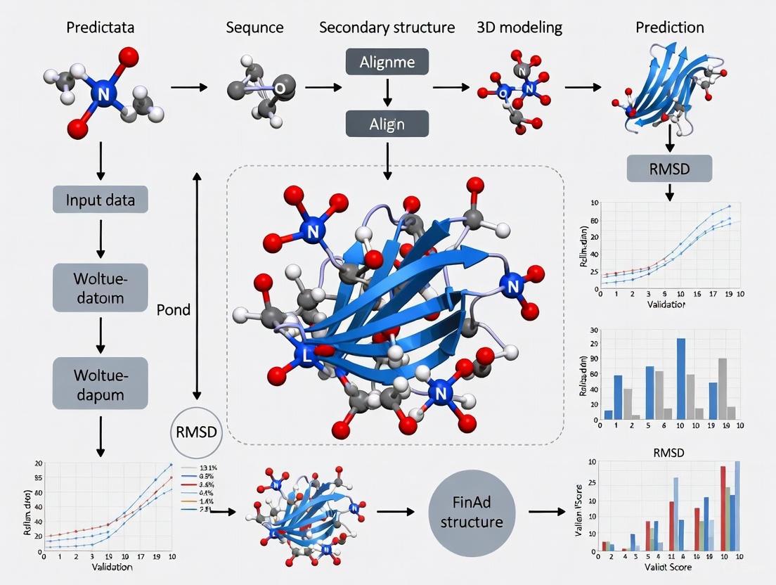
Figure 1: Workflow for Computational Design and Experimental Validation of 3FTx Inhibitors
Research Reagent Solutions for Toxin Studies
Table 3: Essential Research Reagents for Toxin Characterization
| Reagent / Method | Specific Application | Key Function in Research |
|---|---|---|
| X-ray Crystallography | 3D structure determination | Provides atomic-resolution structures for validation of computational predictions [9] [14] |
| Cryo-EM | Structural analysis of large complexes | Enables structure determination of toxin complexes without crystallization [15] |
| Liquid Chromatography Mass Spectrometry (LC-MS) | Venom proteomics and toxin identification | Identifies and quantifies toxin components in complex venoms [10] |
| Surface Plasmon Resonance (SPR) / Bio-Layer Interferometry (BLI) | Binding affinity measurements | Quantifies binding kinetics between toxins and designed inhibitors [4] |
| Circular Dichroism (CD) Spectroscopy | Protein secondary structure and stability | Assesses thermal stability of designed binding proteins [4] |
| Edman Degradation | Protein sequencing | Determines amino acid sequences of purified toxins [9] |
| Yeast Surface Display | Binder screening | Identifies high-affinity binders from designed libraries [4] |
The integration of computational protein design with experimental structural biology has created new opportunities for understanding and neutralizing snake venom toxins. The successful de novo design of 3FTx-binding proteins demonstrates how accurate structure prediction can generate potent inhibitors with therapeutic potential [4]. Similarly, detailed structural characterization of toxins like hemachatoxin and various SVMPs provides essential validation data for improving prediction algorithms [9] [11] [12]. As these methods continue to advance, they promise to accelerate the development of safer, more effective treatments for snakebite envenoming while providing fundamental insights into protein structure-function relationships.
Why Venom Toxins Are Challenging Targets for Structure Prediction
The accurate prediction of protein three-dimensional structures is a cornerstone of modern biochemistry, critical for understanding function and guiding therapeutic development. Within this field, snake venom toxins represent a particularly challenging class of proteins for computational modeling. These toxins, which include diverse families such as three-finger toxins (3FTxs), phospholipases A2 (PLA2s), and snake venom metalloproteinases (SVMPs), have evolved complex structural features that often defy conventional prediction methods [16]. The limitations of current computational tools against these toxins highlight significant gaps in our understanding of protein-biomembrane interactions and reveal critical dependencies on experimental structural data for training algorithms.
This analysis examines the specific molecular characteristics that make venom toxins difficult to model, compares the performance of various structure prediction platforms on these challenging targets, and details experimental methodologies for validation. By examining both successful applications and notable failures in venom toxin modeling, this guide provides researchers with a framework for assessing the reliability of computational predictions in toxinology and related fields.
Molecular Complexity of Venom Toxins
Snake venom toxins possess unique structural and functional properties that collectively contribute to their challenging nature for prediction algorithms. These difficulties arise from several interconnected factors:
Disulfide Bond Complexity: Many venom toxins, including three-finger toxins and phospholipases B, are stabilized by intricate disulfide bond networks. For instance, SVPLBs from Bothrops moojeni contain five conserved cysteine residues in their mature form, four of which form two disulfide bonds (Cys88–Cys500 and Cys499–Cys523) that are critical for structural integrity [17]. These cross-links create unique topological constraints that are difficult to predict accurately from sequence alone.
Membrane Interaction Mechanisms: Perhaps the most significant challenge lies in predicting how cytotoxins and phospholipases interact with biological membranes. The molecular basis of membrane disruption remains incompletely understood, and current datasets severely underrepresent protein-lipid interactions [18]. This knowledge gap directly impacts prediction accuracy for toxins like three-finger cytotoxins, which exert toxicity through membrane disruption rather than specific receptor binding [4].
Conformational Flexibility: Many toxins exhibit significant structural plasticity, adopting different conformations in various environments. Deep learning-based exploration of venom-encrypted peptides has revealed that many antimicrobial candidates transition from flexible conformations in solution to α-helical structures in membrane-mimicking environments [2], a dynamic behavior that static structure prediction struggles to capture.
Evolutionary Rapid Diversification: Venom toxins evolve under strong positive selection pressure, resulting in sequences that are highly variable yet maintain conserved structural folds. This creates a challenge for homology-based methods, as even toxins with similar structures may share limited sequence identity [16].
Table 1: Key Challenging Features of Major Snake Venom Toxin Families
| Toxin Family | Representative Toxins | Key Structural Challenges | Impact on Prediction Accuracy |
|---|---|---|---|
| Three-Finger Toxins (3FTx) | α-neurotoxins, cytotoxins | Multistranded β-structure with three extended loops, conserved disulfide bridges [4] | High accuracy for neurotoxins targeting specific receptors; poor for cytotoxins targeting membranes [18] |
| Phospholipases B (PLB) | PLB_Bm from Bothrops moojeni | Four-layer αββα sandwich core, 18 β-strands, 17 α-helices, N-terminal nucleophile aminohydrolase fold [17] | Homology modeling possible with 70% identity templates, but membrane interaction sites difficult to predict |
| Snake Venom Nerve Growth Factors | sNGF from Daboia russelii, Naja naja | Dimeric structure, extensive disulfide bonds, protease resistance [19] | Enhanced stability factors complicate dynamics predictions; coevolutionary analysis needed for interface predictions |
Performance Comparison of Prediction Methods
Various computational approaches have been applied to venom toxin structure prediction, with markedly different success rates across toxin families and functional classes. The performance disparity highlights how methodological limitations affect practical applications in antivenom development and therapeutic discovery.
Success with Neurotoxins Versus Failure with Cytotoxins
Recent research demonstrates a striking contrast in prediction accuracy between different three-finger toxin subfamilies. AI-based approaches have successfully designed inhibitory proteins against α-neurotoxins, which function through specific receptor interactions. The RFdiffusion software generated designs that formed extensive backbone hydrogen bonds with target neurotoxins, achieving remarkable binding affinity (Kd = 0.9 nM for short-chain α-neurotoxin binder SHRT) and providing complete protection in mouse models [4]. The successful application involved:
- Using RFdiffusion conditioned on secondary structure and block adjacency tensors to generate backbones forming extended β-sheets with target neurotoxins
- Applying ProteinMPNN for sequence design on the generated backbones
- Filtering designs using AlphaFold2 and Rosetta metrics
- Experimental validation through yeast surface display and biophysical methods (BLI, SPR)
- Structural verification via X-ray crystallography (2.58 Å resolution for SHRT-ScNtx complex) [4]
In stark contrast, the same computational pipeline failed to produce effective inhibitors for cytotoxins from the same structural family. Despite creating designs with high binding affinity in vitro, these inhibitors showed no protective effect in mouse models against toxin-induced skin lesions [18]. This failure underscores a fundamental limitation: current AI tools like AlphaFold and Rosetta excel at predicting protein-protein interactions but perform poorly with protein-lipid interactions [18].
Homology Modeling Limitations and Successes
Homology modeling remains valuable for toxins with suitable templates, though with significant limitations. For SVPLBs, modeling using phospholipase B-like protein from Bos taurus (70% sequence identity) as a template produced viable structures validated through PROCHECK, ERRAT, and Verif3D [17]. However, this approach revealed substantial variations in surface charge distribution, active site cavity volume, and depth across homologs—features critical for understanding function and developing inhibitors that are poorly captured by modeling [17].
Table 2: Performance Comparison of Computational Methods on Venom Toxins
| Method | Best For | Key Limitations | Experimental Validation Required |
|---|---|---|---|
| RFdiffusion + ProteinMPNN | De novo design of protein-protein inhibitors (e.g., against α-neurotoxins) [4] | Fails for membrane-disrupting toxins; requires extensive experimental screening | Essential (yeast display, BLI/SPR, X-ray crystallography, in vivo models) |
| AlphaFold2 | Structure prediction of soluble toxins, complex prediction [16] | Poor performance on protein-lipid interactions; limited by training dataset gaps [18] | Recommended, especially for novel folds or membrane-associated toxins |
| Molecular Dynamics Simulations | Understanding binding stability (e.g., sNGF-TrkA interactions) [19] | Computationally intensive; limited timescales; force field inaccuracies for non-standard interactions | Validation through binding assays, functional studies |
| Homology Modeling | Toxins with high-sequence identity templates (>70%) [17] | Misses subtle structural variations critical for function; template availability limited | Necessary, particularly for active site characterization |
Experimental Validation Workflows
Rigorous experimental validation is essential for confirming computational predictions of venom toxin structures, particularly given the known limitations of in silico methods. The following protocols represent state-of-the-art approaches for verifying predicted structures and interactions.
Structural Determination and Binding Assays
The successful development of neurotoxin inhibitors combined computational design with extensive experimental validation through this comprehensive workflow:
For binding characterization, Bio-Layer Interferometry (BLI) and Surface Plasmon Resonance (SPR) provide quantitative measurements of toxin-inhibitor interactions. The standard protocol involves:
- Immobilization: Toxins are immobilized on biosensor chips (BLI) or sensor surfaces (SPR) via amine coupling
- Association: Serial dilutions of designed binders are flowed over the surface at 25°C in HBS-EP buffer
- Dissociation: Buffer without binder is flowed to monitor complex dissociation
- Analysis: Binding curves are fitted to 1:1 binding models to calculate kinetic parameters (Kd, kon, koff) [4]
Functional and In Vivo Validation
Beyond structural and binding studies, functional assays are critical for confirming that designed inhibitors actually neutralize toxin activity:
In Vitro Neutralization: For neurotoxins, cell-based assays measuring protection against acetylcholine receptor blockade are essential. Cultured neuroblastoma cells expressing muscle-type nAChRs are pre-treated with toxins followed by addition of designed inhibitors, with viability and receptor function as endpoints [4].
In Vivo Protection Models: Mouse challenge models provide the most clinically relevant validation. For neurotoxins, mice are injected with lethal doses of toxin (LD99) concurrently with or followed by inhibitor administration. Survival, symptom progression (paralysis, respiratory distress), and histological analysis of tissues provide comprehensive efficacy data [4].
The critical importance of functional validation is highlighted by the cytotoxin inhibitor failure—despite excellent binding metrics in vitro (Kd in nM range), the inhibitors provided no protection against tissue damage in mouse models [18].
Research Reagent Solutions
Advancing venom toxin structure prediction requires specialized reagents and computational resources. The table below details essential research tools for this field.
Table 3: Essential Research Reagents and Resources for Venom Toxin Structure Prediction
| Category | Specific Tools/Reagents | Function/Application | Key Features |
|---|---|---|---|
| Computational Platforms | RFdiffusion [4], AlphaFold2 [16], Rosetta [16], APEX [2] | De novo protein design, structure prediction, antimicrobial peptide mining | Specialized for different interaction types (protein-protein vs. protein-lipid) |
| Structural Databases | UniProt [20] [19], VenomZone [20] [2], ConoServer [2], ArachnoServer [2] | Source of toxin sequences, structures, and functional annotations | Varying coverage of different venomous species; integration challenges |
| Experimental Validation | Yeast Surface Display [4], BLI/SPR [4], X-ray Crystallography [4] | Binding affinity measurement, structural verification | High-throughput screening (yeast display) vs. high-precision (crystallography) |
| Specialized Datasets | DBAASP [2], ISOB [2], Custom venom proteomics datasets [16] | AMP comparison, species-specific toxins, training machine learning models | Variable quality; require curation and standardization |
The structural prediction of venom toxins remains a formidable challenge, with success heavily dependent on toxin class and the nature of its biological interactions. Current AI-driven methods have demonstrated remarkable capabilities for designing inhibitors against toxin-receptor interactions but show significant limitations for toxins targeting lipid membranes. This disparity underscores fundamental gaps in our understanding of protein-membrane interactions and highlights the critical importance of comprehensive experimental validation.
The field requires advances in several key areas: improved representation of membrane-associated proteins in training datasets, development of specialized algorithms for protein-lipid interactions, and standardized validation protocols that include functionally relevant assays. As computational methods continue to evolve, integrating multiple approaches—from deep learning-based design to molecular dynamics simulations—will likely provide the most productive path forward for overcoming the unique challenges posed by venom toxins.
The Critical Role of Genomic and Transcriptomic Data in Foundation Building
The field of toxinology has been transformed by the integration of large-scale biological data. Genomic and transcriptomic information now provides the essential foundation for understanding the complex composition and evolution of snake venoms, which are primarily composed of numerous proteins and peptides. This data-driven approach has become particularly critical for validating and advancing computational protein structure prediction tools, which rely on high-quality sequence information to model toxin structures with atomic-level precision. The shift from traditional, low-throughput experimental methods to a bioinformatics-guided framework has accelerated the pace of discovery, enabling researchers to move from venom characterization to therapeutic development with unprecedented speed and accuracy. This comparison guide examines how different computational approaches perform when grounded in comprehensive genomic and transcriptomic data, with a specific focus on applications in snake venom research.
Performance Comparison of Structure Prediction Tools on Venom Toxins
Recent studies have systematically evaluated how well modern computational tools predict the structures of snake venom toxins, which often lack experimental structures due to their complexity and the challenges in isolation and crystallization.
Table 1: Performance Comparison of Protein Structure Prediction Tools on Snake Venom Toxins
| Tool Name | Primary Function | Performance on Small Toxins (e.g., 3FTx) | Performance on Large, Complex Toxins (e.g., SVMPs) | Key Limitations |
|---|---|---|---|---|
| AlphaFold2 | Monomer structure prediction | High accuracy [7] | Moderate accuracy [7] | Struggles with flexible loops [7] |
| ColabFold | Monomer structure prediction | Slightly lower than AF2 [7] | Moderate accuracy [7] | Computationally intensive [7] |
| AlphaFold-Multimer | Protein complex prediction | Not specifically evaluated | Not specifically evaluated | Lower accuracy than monomer predictions [21] |
| DeepSCFold | Protein complex prediction | Superior for antibody-antigen interfaces [21] | Improved TM-score over other methods [21] | Requires multiple sequence alignments [21] |
The table above highlights a critical finding from a 2024 Toxicon study which demonstrated that while machine-learning tools can successfully predict toxin structures, their performance varies significantly based on toxin size and complexity [7]. All tools face challenges with intrinsically disordered regions common in venom proteins, particularly flexible loops that may be crucial for functional activity.
For protein complexes common in venom systems, newer methods like DeepSCFold have shown remarkable improvements. In benchmark testing, DeepSCFold achieved an 11.6% improvement in TM-score compared to AlphaFold-Multimer on CASP15 multimer targets, and enhanced the success rate for predicting antibody-antigen binding interfaces by 24.7% over AlphaFold-Multimer [21]. This demonstrates how specialized tools leveraging sequence-derived structural complementarity can overcome limitations of more general approaches.
Experimental Protocols for Validation Studies
Protocol for Benchmarking Prediction Tools on Toxin Targets
A comprehensive comparative study published in 2024 established a standardized protocol for evaluating structure prediction tools on challenging toxin targets [7]:
- Dataset Curation: Compile a diverse set of over 1,000 snake venom toxin sequences with no experimentally determined structures, representing major toxin families (3FTxs, SVMPs, PLA2s, etc.).
- Model Generation: Process all sequences through multiple prediction tools (AlphaFold2, ColabFold, Modeller) using standardized computing resources.
- Quality Assessment: Evaluate predictions using multiple metrics including per-residue confidence scores (pLDDT), root-mean-square deviation (RMSD) from reference structures when available, and visual inspection of functionally important regions.
- Functional Validation: Compare predicted structures with known functional data, including receptor binding sites and enzymatic active sites, to assess biological relevance beyond structural accuracy.
This protocol revealed that while global structures were often well-predicted, local regions corresponding to functional domains showed variation between tools, emphasizing the need for multi-tool consensus approaches.
Protocol for De Novo Binder Design Against Venom Toxins
A groundbreaking 2025 Nature study established a protocol for designing synthetic proteins to neutralize snake venom toxins using structure prediction and design tools [4]:
- Target Selection: Focus on three-finger toxin (3FTx) subfamilies (short-chain α-neurotoxins, long-chain α-neurotoxins, and cytotoxins) responsible for severe envenoming pathology.
- Computational Design: Use RFdiffusion to generate protein backbones conditioned on binding to specific toxin epitopes, followed by ProteinMPNN for sequence design.
- In Silico Screening: Filter designs using AlphaFold2 predictions and interface metrics, selecting top candidates for experimental testing.
- Experimental Validation:
- Affinity Measurement: Validate binding using surface plasmon resonance (SPR) and bio-layer interferometry (BLI).
- Structural Verification: Determine crystal structures of toxin-binder complexes to validate computational models.
- Functional Neutralization: Test neutralization capacity in vitro (e.g., toxin receptor binding assays) and in vivo (mouse protection models).
This protocol yielded remarkable results, with designed binders achieving picomolar to nanomolar affinities (e.g., 0.9 nM for short-chain neurotoxin binder SHRT) and demonstrating potent toxin neutralization in animal models [4] [22]. The close agreement between computational models and experimental structures (RMSDs of 0.42-1.04 Å) strongly validated the structure prediction approach.
Visualization of Research Workflows
Diagram 1: Integrated workflow for venom research showing how genomic and transcriptomic data foundation enables downstream applications.
Diagram 2: AI-driven pipeline for antivenom development showing the closed-loop design process.
Table 2: Key Research Reagent Solutions for Venom Genomics and Structure Prediction
| Resource Category | Specific Tools/Databases | Primary Function | Application in Toxin Research |
|---|---|---|---|
| Genome Databases | AlphaSync [23] | Provides continuously updated predicted protein structures | Ensures researchers work with current structural models; addressed backlog of 60,000 outdated structures |
| Structure Prediction Tools | AlphaFold2, ColabFold [7] | Predicts 3D structures from amino acid sequences | Models toxin structures when experimental data unavailable; superior for small toxins like 3FTxs |
| Complex Structure Prediction | DeepSCFold [21] | Predicts protein complex structures using sequence-derived complementarity | Models toxin-receptor interactions; improves antibody-antigen interface prediction by 24.7% |
| De Novo Design Tools | RFdiffusion, ProteinMPNN [4] [22] | Generates novel protein binders for specific targets | Designs toxin-neutralizing proteins; achieved 0.9 nM affinity for α-neurotoxins |
| Transcriptomic Resources | Venom gland transcriptome databases [24] [25] | Catalog toxin gene expression profiles | Identifies full toxin repertoire; reveals heterogeneity in venom production |
| Validation Resources | SAbDab [21] | Database of antibody structures | Benchmark for antibody-toxin complex predictions |
The integration of genomic and transcriptomic data has fundamentally transformed snake venom research, creating a robust foundation for validating and applying protein structure prediction tools. Performance comparisons clearly demonstrate that while general-purpose tools like AlphaFold2 provide excellent starting points, specialized methods like DeepSCFold offer significant advantages for modeling the complex protein interactions relevant to toxin neutralization. The experimental success of de novo designed toxin binders - achieving near-atomic accuracy and potent neutralization in vivo - provides compelling validation of this integrated approach. As resources like AlphaSync ensure ongoing data currency, and tools continue to evolve, the research community is positioned to accelerate the development of next-generation therapeutics for snakebite and other toxin-mediated conditions.
A Practical Guide to Structure Prediction Tools and Workflows for Venom Research
In the field of structural biology, accurately predicting protein three-dimensional (3D) structures from amino acid sequences is crucial for understanding function and facilitating drug discovery. This is particularly relevant for snake venom toxins, which are challenging targets due to their flexibility, limited homologous sequences, and the scarcity of experimentally determined structures for validation. This guide objectively compares the performance of four computational tools—AlphaFold2, ColabFold, Modeller, and RFdiffusion—within the specific context of snake venom toxin research, providing experimental data and protocols to inform scientists in their selection process.
Performance Comparison on Snake Venom Toxins
A 2024 comparative study in Toxicon evaluated AlphaFold2, ColabFold, and Modeller on over 1,000 snake venom toxins for which no experimental structures existed [7]. The following table summarizes their key performance metrics.
Table 1: Performance of structure prediction tools on snake venom toxins
| Tool | Overall Performance | Performance on Small Toxins (e.g., 3FTxs) | Performance on Large Toxins (e.g., SVMPs) | Challenges and Limitations | Computational Intensity |
|---|---|---|---|---|---|
| AlphaFold2 (AF2) | Best performance across all assessed parameters [7] | Superior prediction accuracy [7] | Struggled more compared to small toxins [7] | Struggles with flexible loop regions and intrinsic disorder [7] | High [7] |
| ColabFold (CF) | Slightly worse than AF2, but still strong [7] | High prediction accuracy [7] | Struggled more compared to small toxins [7] | Struggles with flexible loop regions and intrinsic disorder [7] | Less intensive than AF2 [7] |
| Modeller | Lower performance compared to AF2 and CF [7] | Not specified | Not specified | Struggles with flexible loop regions and intrinsic disorder [7] | Not specified |
| RFdiffusion | Not directly tested in this study, but used successfully for de novo binder design against toxins [4] | Successfully used to design binders for small 3FTx neurotoxins [4] | Not specified | Not primarily a prediction tool; used for generative binder design [4] | Not specified |
The study concluded that while all tools performed well in predicting stable functional domains, they consistently struggled with regions of intrinsic disorder, such as flexible loops and propeptide regions, which are common in toxins [7]. Therefore, researchers should exercise caution when interpreting predictions of these dynamic elements.
Key Experimental Protocols and Workflows
The tools compared here encompass two main types of computational approaches: protein structure prediction (deducing the structure of a given sequence) and de novo binder design (creating new proteins that bind to a target of interest). RFdiffusion falls into the latter category. Below are detailed methodologies for key experiments cited in the results.
Comparative Prediction of Snake Venom Toxin Structures
The protocol from the 2024 Toxicon study provides a direct comparison of AF2, ColabFold, and Modeller [7].
- Objective: To evaluate the accuracy of multiple structure prediction tools on a large set of snake venom toxins without experimentally-solved structures.
- Dataset: Over 1,000 snake venom toxin sequences from protein families such as three-finger toxins (3FTxs) and snake venom metalloproteinases (SVMPs). Sequences were collected from UniProt and RefSeq, with redundancy reduced at a threshold of 80% sequence identity [7].
- Methodology:
- Input Sequences: The same toxin sequences were processed independently by AlphaFold2, ColabFold, and Modeller.
- Structure Prediction:
- AlphaFold2: Searches large sequence databases (e.g., UniRef, BFD, MGnify) with HHblits and HMMer to build multiple sequence alignments (MSAs) as input for its deep neural network [26].
- ColabFold: Utilizes the MMseqs2 algorithm for drastically faster (40-60x) MSA generation against similar databases, including its own ColabFoldDB, before feeding features into a modified AlphaFold2 or RoseTTAFold network [26].
- Modeller: A comparative modeling tool that uses known protein structures as templates to model the structure of a target sequence. The specific version and template database used were not detailed in the study [7].
- Analysis: The predicted models were assessed on parameters of accuracy, which likely included metrics like RMSD (Root Mean Square Deviation) and confidence scores, though the specific metrics are not listed in the search results [7].
Workflow for predicting toxin structures
De Novo Binder Design against Toxins using RFdiffusion
A landmark 2025 study in Nature detailed the use of RFdiffusion to design proteins that neutralize lethal snake venom toxins [4]. This demonstrates an advanced application of structure prediction models for therapeutic development.
- Objective: To computationally design de novo proteins that bind and neutralize three-finger toxins (3FTxs) like α-neurotoxins and cytotoxins.
- Targets: A consensus short-chain α-neurotoxin (ScNtx) and the native long-chain α-neurotoxin α-cobratoxin [4].
- Methodology:
- Conditioned Generation: The RFdiffusion network was conditioned on the target toxin's structure. For α-neurotoxins, the generation was guided to form extended β-sheets with exposed edge β-strands on the toxin. For cytotoxins, "hotspot" residues were defined on the three-finger loops to direct binding [4].
- Sequence Design: Generated protein backbones were assigned sequences using ProteinMPNN, a deep learning-based sequence design tool [4].
- Filtering & Selection: Designed proteins were filtered using a combination of:
- Experimental Validation: Top candidates were tested via:
- Yeast Surface Display: For initial binding confirmation.
- Bio-Layer Interferometry (BLI) / Surface Plasmon Resonance (SPR): To measure binding affinity (Kd).
- X-ray Crystallography: To verify that the designed complex matched the computational model (achieving RMSD as low as 0.42 Å) [4].
- In Vitro and In Vivo Neutralization Assays: To confirm protective efficacy against venom toxicity [4].
Workflow for designing toxin binders
Advanced Workflows and Emerging Tools
BindCraft: An Enhanced ColabFold Pipeline for Binder Design
BindCraft is a protocol built upon ColabFold's implementation of AlphaFold2 Multimer (AF2M) to improve de novo binder design [27] [28].
- How it Works:
- It starts with a random amino acid sequence for the binder.
- AF2M co-folds this random sequence with the target protein.
- An iterative optimization loop begins, using gradient-based backpropagation through the AF2M network. This process suggests sequence changes that improve scores for binding confidence, interface interaction strength, and other metrics.
- The final optimized sequence is refined with ProteinMPNN and filtered again with AF2M to ensure quality and solubility [27].
- Performance: In tests on 10 targets, BindCraft produced binders with sub-nanomolar affinity and achieved significantly better experimental hit rates than many other de novo design methods [27]. It was also the winning method in an EGFR binder design competition, producing a binder with a Kd of 4.91e-7 M [28].
AfCycDesign: Adapting ColabFold for Cyclic Peptides
Cyclic peptides are a promising therapeutic modality. AfCycDesign is a modification of the AlphaFold2/ColabDesign framework that introduces a custom cyclic offset to the model's relative positional encoding, enabling accurate structure prediction and de novo design of cyclic peptides [29]. In tests, it predicted cyclic peptide structures with a median RMSD of 0.8 Å to experimental structures and was successfully used to design binders against targets like MDM2 and Keap1 [29].
Research Reagent Solutions for Computational Toxinology
The following table details key computational tools and databases essential for conducting the experiments described in this guide.
Table 2: Key research reagents and tools for computational toxin research
| Item Name | Type | Primary Function in Research |
|---|---|---|
| AlphaFold2 | Software | Highly accurate protein structure prediction from sequence using MSAs and deep learning [26] [7]. |
| ColabFold | Software | Accessible, accelerated protein structure prediction combining MMseqs2 fast homology search with AF2 or RoseTTAFold [26] [7]. |
| RFdiffusion | Software | Generative AI model for creating novel protein structures and binders conditioned on a target [4]. |
| Modeller | Software | Comparative protein structure modeling by satisfaction of spatial restraints, useful when templates are available [7]. |
| ProteinMPNN | Software | Neural network for designing amino acid sequences that fold into a given protein backbone structure [27] [4]. |
| UniProt/RefSeq | Database | Source of canonical protein sequences for collecting toxin and non-toxin datasets [7] [30]. |
| ColabFoldDB | Database | Custom environmental database for MSA generation, combining BFD/MGnify with eukaryotic/metagenomic sequences [26]. |
| RoseTTAFold | Software | A deep learning-based protein structure prediction tool, similar to AlphaFold2 [26]. |
| Rosetta | Software Suite | A comprehensive suite for macromolecular modeling, used for energy scoring and refinement in design protocols [4] [29]. |
Workflow for de Novo Design of Toxin-Neutralizing Proteins
Snakebite envenoming is a neglected tropical disease claiming over 100,000 lives annually, with current antivenom treatments relying on century-old methods of animal immunization that yield variable efficacy and potential adverse effects [4] [31]. The emergence of artificial intelligence (AI) and deep learning methods has revolutionized protein science, shifting the paradigm from structure prediction to de novo creation of therapeutic proteins with optimized properties [32]. This guide examines the workflow for designing toxin-neutralizing proteins, comparing computational tools and experimental methods while contextualizing performance within snake venom toxin research.
Comparative Analysis of Computational Protein Design Tools
Tool Performance Characteristics
AI-driven platforms have demonstrated remarkable capabilities in generating novel protein structures, though their performance varies significantly across different design challenges.
Table 1: Comparison of Key Protein Design and Prediction Platforms
| Tool Name | Primary Function | Strengths | Limitations | Reported Success Rate/Performance |
|---|---|---|---|---|
| RFdiffusion | De novo protein design | Creates novel proteins with high stability and affinity; enables β-strand pairing for toxin neutralization [4] | Requires experimental validation; may generate false positives [32] | Designed binders with Kd values of 0.9-271 nM for various 3FTx subfamilies [4] |
| AlphaFold2 (AF2) | Structure prediction | Superior accuracy for well-characterized proteins and functional domains; best overall performance [7] [33] | Struggles with flexible loops and disordered regions [7] [33] | Highest performance across assessed parameters for toxin structure prediction [7] |
| ColabFold (CF) | Structure prediction | Slightly worse than AF2 but computationally less intensive [7] [33] | Similar limitations with flexible regions as AF2 [7] | Near-AF2 performance with reduced computational requirements [7] |
| ProteinMPNN | Sequence design | Robust sequence optimization for designed backbones [4] [31] | Dependent on quality of input backbone structures | Critical for achieving stable, expressible designs [4] |
Tool Selection Considerations
For snake venom toxins specifically, a multitool prediction strategy consistently outperforms single-tool approaches [8]. The integration of structural and sequence-based models is particularly important when working with toxins lacking experimental structures, as all tools struggle with regions of intrinsic disorder such as loops and propeptide regions [7] [33]. The workflow typically employs RFdiffusion for backbone generation, ProteinMPNN for sequence design, and AlphaFold2 for initial structural validation [4] [31].
Experimental Validation Workflow
Quantitative Assessment of Designed Proteins
The efficacy of de novo designed toxin-neutralizing proteins has been rigorously validated through multiple experimental approaches, yielding compelling quantitative data on their performance.
Table 2: Experimental Performance Metrics of Designed Toxin-Binding Proteins
| Designed Binder | Target Toxin | Binding Affinity (Kd) | Thermal Stability (Tm) | Structural Validation | Neutralization Efficacy |
|---|---|---|---|---|---|
| SHRT | Short-chain α-neurotoxin (consensus) | 0.9 nM (SPR) [4] | 78°C [4] | X-ray crystallography (1.04 Å RMSD) [4] | Protection in lethal mouse challenge [4] |
| LNG | α-cobratoxin (long-chain neurotoxin) | 1.9 nM (SPR) [4] | >95°C [4] | X-ray crystallography (0.42 Å RMSD over design) [4] | Protection in lethal mouse challenge [4] |
| CYTX | Cytotoxin (consensus) | 271 nM (SPR) [31] | High solubility, monomeric [31] | Not specified | Neutralized whole venoms (N. pallida, N. nigricollis) [31] |
Detailed Experimental Protocols
Binding Affinity Measurement (SPR/BLI)
Purpose: Quantify binding kinetics between designed proteins and toxin targets.
- Immobilization: Toxins or designed binders are immobilized on sensor chips (SPR) or biosensor tips (BLI)
- Association Phase: Introduced binding partners at varying concentrations in solution phase
- Dissociation Phase: Monitored complex dissociation in buffer-only flow
- Data Analysis: Determined dissociation constants (Kd) by fitting binding curves to 1:1 binding models
- Key Equipment: Surface plasmon resonance instruments (SPR) or bio-layer interferometry (BLI) systems [4] [31]
Structural Validation (X-ray Crystallography)
Purpose: Verify computational models with experimental structural data.
- Crystallization: Designed proteins (apo or complexed with toxins) crystallized using vapor diffusion methods
- Data Collection: X-ray diffraction data collected at synchrotron facilities
- Structure Solution: Molecular replacement or experimental phasing methods
- Model Validation: Computational models compared to electron density maps, RMSD calculated [4]
In Vitro Neutralization Assays
Purpose: Assess functional efficacy of designed binders.
- Cell Viability Assays: Luminescent assays measuring protection against cytotoxin-induced cell death
- nAChR Binding Interference: Measured prevention of neurotoxin binding to nicotinic acetylcholine receptors
- Venom Neutralization: Tested against whole venoms with high target toxin content [4] [31]
In Vivo Protection Studies
Purpose: Evaluate protective efficacy in biologically relevant systems.
- Animal Model: Mice challenged with lethal doses of neurotoxins
- Dosing Regimens: Pre-incubation of toxin with binder; post-challenge administration (e.g., 15 minutes after toxin)
- Endpoint Monitoring: Survival tracked over specified period (e.g., up to two weeks)
- Molar Ratios: Tested various toxin:binder ratios (e.g., 1:10) [4] [34]
The Scientist's Toolkit: Essential Research Reagents
Table 3: Key Research Reagent Solutions for Toxin-Neutralizing Protein Design
| Reagent/Category | Specific Examples | Function/Application |
|---|---|---|
| Computational Design Platforms | RFdiffusion, ProteinMPNN, AlphaFold2 [4] [7] | De novo backbone generation, sequence design, and structural validation |
| Expression Systems | E. coli recombinant expression [4] [31] | High-yield production of designed proteins; cost-effective manufacturing |
| Binding Affinity Assays | Surface Plasmon Resonance (SPR), Bio-Layer Interferometry (BLI) [4] | Quantitative measurement of protein-toxin binding kinetics and affinity |
| Structural Biology Tools | X-ray crystallography, Size Exclusion Chromatography (SEC) [4] | Experimental structure determination and validation of computational models |
| Stability Assessment | Circular Dichroism (CD) melting experiments [4] | Measurement of thermal stability and folding properties |
| In Vitro Neutralization | Luminescent cell viability assays, nAChR binding interference [4] [31] | Functional assessment of toxin neutralization in cellular systems |
| In Vivo Models | Mouse lethal challenge models [4] [34] | Preclinical validation of protective efficacy in whole organisms |
Workflow Visualization
Computational Design and Validation Workflow
Experimental Validation Pipeline
The workflow for de novo design of toxin-neutralizing proteins represents a transformative approach to antivenom development, leveraging AI-driven computational design integrated with rigorous experimental validation. This methodology has demonstrated remarkable success in creating stable, high-affinity binders against challenging three-finger toxin targets, with binding affinities in the nanomolar range and proven efficacy in protecting against lethal toxin challenges [4] [34]. As the field advances, the integration of multiple prediction tools, careful attention to flexible regions in toxin structures, and systematic experimental validation will be crucial for developing the next generation of antivenom therapeutics that are safer, more effective, and more accessible than current options [7] [8] [35].
Application in Epitope Mapping for Antivenom Development (B-cell and T-cell)
Snakebite envenomation remains a significant global health challenge, classified as a Category A neglected tropical disease by the World Health Organization [36]. Traditional antivenom production, which relies on animal immunization with whole venom, faces considerable limitations including batch variability, limited cross-reactivity, and the risk of adverse immune reactions [8]. Epitope mapping—the process of identifying specific regions on antigens recognized by immune cells—has emerged as a transformative approach for developing safer, more effective, and rationally designed antivenoms. Within this context, validating protein structure prediction tools for snake venom toxins is paramount, as accurate structural models provide the foundation for reliable epitope identification and characterization.
The immune response to snake venom toxins involves both B-cell epitopes (BCEs), which are regions directly recognized by B-cell receptors or secreted antibodies, and T-cell epitopes (TCEs), which are peptides presented by major histocompatibility complex (MHC) molecules to activate T-cell help [37] [8]. While BCEs can be linear (continuous sequences) or conformational (discontinuous residues brought together by protein folding), most B-cell epitopes are thought to be conformational, necessitating accurate structural information for their identification [38]. Understanding both epitope types is crucial for developing effective antivenoms, as T-cell help is essential for generating high-affinity, long-lasting antibody responses against venom toxins [37].
Computational Approaches for Epitope Prediction
Computational immunoinformatics has revolutionized epitope prediction by enabling rapid, large-scale screening of venom toxins, significantly reducing reliance on venom immunizations and accelerating the discovery process [8]. These methods leverage algorithms trained on physicochemical properties, structural features, and machine learning to identify potential immunogenic regions.
Comparison of Computational Prediction Methods
Table 1: Comparison of Computational Epitope Prediction Approaches
| Method Type | Examples | Key Features | Performance Metrics | Best Use Cases |
|---|---|---|---|---|
| Sequence-Based BCE Predictors | ABCpred, BepiPred, TEPRF | Uses amino acid propensity scales, hydrophilicity, flexibility; ABCpred employs machine learning | AUC: 0.445-0.538; High false positive rates [39] | Initial screening of linear epitopes |
| Structure-Based BCE Predictors | DiscoTope, ElliPro | Leverages 3D structural data to identify surface-accessible residues | Better for conformational epitopes [38] | Toxins with known or well-predicted structures |
| T-cell Epitope Predictors | NetMHCIIPan, TEPITOPE | Predicts peptide binding to MHC class II molecules | Identifies peptides with T-cell activation potential [37] [36] | Assessing immunogenicity risk and vaccine design |
| Protein Class-Specific Models | Custom decision tree classifier for metalloendopeptidases | Trained on curated epitopes from specific protein families | Lower false positive rate (0.33 vs 0.40-0.58); Improved AUC [39] | Toxins belonging to well-characterized protein families |
| Deep Learning Approaches | Deep-STP | Uses g-gap, natural vector, and Word2Vec features with CNN | Accuracy: 82.00%; Effective for toxin classification [30] | General snake toxin protein identification |
Protein Structure Prediction Tools for Venom Toxins
Accurate structural models are essential for conformational epitope mapping. Recent advances in machine learning-based structure prediction have shown promising results for snake venom toxins, though with important limitations. A 2024 comparative study evaluated three modelling tools on over 1000 snake venom toxin structures [7]:
- AlphaFold2 (AF2) performed best across all assessed parameters, demonstrating remarkable accuracy for small toxins like three-finger toxins (e.g., cytotoxins, neurotoxins).
- ColabFold (CF) scored slightly worse than AF2 but offered computational efficiency advantages.
- All tools struggled with regions of intrinsic disorder, particularly flexible loops and propeptide regions, which are common in larger toxins like Snake Venom Metalloproteinases (SVMPs).
- Predictions were most reliable for functional domains, highlighting the importance of exercising caution when working with toxins lacking experimental structures [7].
These structural predictions enable computational epitope mapping through molecular docking simulations, with studies successfully identifying potential T-cell and B-cell epitopes in cobra venom cytotoxins through computational approaches [36].
Experimental Validation of Predicted Epitopes
Computational predictions require experimental validation to confirm their immunological relevance. Multiple techniques have been developed to characterize epitopes at the molecular level, each with distinct advantages and limitations.
Key Experimental Techniques for Epitope Mapping
Table 2: Experimental Methods for Epitope Validation
| Method | Principle | Epitope Type Detected | Throughput | Key Advantages | Major Limitations |
|---|---|---|---|---|---|
| SPOT Synthesis | Cellulose-bound peptide arrays probed with antibodies | Linear B-cell epitopes | High | Economically viable; assesses many peptides simultaneously [40] | Misses conformational epitopes |
| Peptide ELISA | Peptides immobilized on plates for antibody binding | Linear B-cell epitopes | Medium | Fast and cost-effective with high specificity [38] | Limited to linear epitopes only |
| Phage Display | Phage libraries expressing random peptides screened with antibodies | Mainly linear B-cell epitopes | High | Can mimic some conformational epitopes [38] | Biased toward high-affinity binders |
| X-ray Crystallography | Determines 3D structure of antibody-antigen complexes | Conformational B-cell epitopes | Low | Atomic-level resolution of interactions [38] [5] | Technically challenging; requires crystals |
| MELD/LC-MS | Multi-enzymatic limited digestion with mass spectrometry | Linear B-cell epitopes | Medium | Provides sequence-level epitope information [36] | Complex data interpretation |
| LIBRA-seq | Links BCR sequencing to antigen specificity | Both linear and conformational | High | High-throughput; connects sequence to function [38] | Specialized equipment required |
| T-cell Assays (ELISpot, Proliferation) | Measures T-cell activation upon peptide exposure | T-cell epitopes | Medium | Confirms functional immunogenicity [37] | Requires viable immune cells |
Integrated Epitope Mapping Workflows
The most successful epitope mapping strategies combine multiple computational and experimental approaches. The following workflow illustrates a comprehensive strategy for epitope identification and validation in antivenom development:
Case Studies in Epitope-Driven Antivenom Development
Multiepitope Antivenom Against Loxosceles Spider Venom
A landmark study demonstrated the power of epitope mapping for developing broad-spectrum antivenoms. Researchers identified three linear epitopes for Loxosceles astacin-like protease 1 (LALP-1) and two for hyaluronidase (LiHYAL) from Loxosceles intermedia spider venom using SPOT-synthesis technique [40]. These were combined with a previously characterized epitope of sphingomyelinase D (SMase D) to generate a recombinant multiepitopic protein (rMEPLox).
Key findings included:
- rMEPLox was non-toxic and elicited antibodies reactive with multiple Loxosceles species venoms (L. intermedia, L. laeta, L. gaucho, and L. similis)
- Anti-rMEPLox antibodies efficiently neutralized sphingomyelinase, hyaluronidase, and metalloproteinase activities of L. intermedia venom
- The multiepitope approach provided broader protection compared to single-epitope immunogens [40]
This epitope-driven strategy successfully addressed the challenge of venom scarcity for immunization and produced a candidate suitable for both antivenom production and experimental vaccination.
Cobra Venom Cytotoxin Epitope Analysis
A 2023 study addressed the critical challenge of cytotoxin (CTX) neutralization in cobra envenomation. CTX constitutes approximately 70% of cobra venom and is responsible for dermonecrotic symptoms, but exhibits low immunogenicity, making conventional antivenoms ineffective against its toxicity [36].
The research combined:
- Immunoinformatic analyses predicting T-cell and B-cell epitopes
- Molecular docking simulations identifying HLA-B62 as the supertype with greatest binding affinity
- MELD/LC-MS epitope-omics revealing three potential epitope sequences
- Site-directed mutagenesis confirming epitope locations at functional loops
This integrated approach identified four potential epitope sites residing within CTX's functional loops, providing crucial targets for developing CTX-targeted antivenoms [36].
Neutralizing Antibodies Against Snake Venom Metalloproteinases
Research on metalloendopeptidases from Bothrops snake venoms demonstrated that computational predictions could identify epitopes missed by experimental mapping alone. A custom decision tree classifier trained specifically on metalloendopeptidase epitopes achieved a lower false positive rate (0.3266) compared to general prediction tools like ABCpred (0.5752) and BepiPred (0.3961) [39].
Notably, immunization with a computationally predicted epitope that was undetected by SPOT immunoassays successfully induced neutralizing antibody production in mice, demonstrating that computational methods can reveal epitopes with significant therapeutic potential that might be overlooked by standard experimental approaches [39].
Table 3: Key Research Reagent Solutions for Epitope Mapping
| Reagent/Resource | Function | Application Examples |
|---|---|---|
| SPOT Synthesis Kit | Parallel peptide synthesis on cellulose membranes | Mapping linear B-cell epitopes in LALP-1 and LiHYAL [40] |
| HLA Allele Panels | Assessing peptide binding to diverse MHC molecules | T-cell epitope prediction for cytotoxin [36] |
| Peptide ELISA Kits | Measuring antibody binding to synthetic peptides | Validating epitope immunoreactivity [38] |
| Deep Learning Frameworks | Training custom epitope prediction models | Deep-STP for snake toxin identification [30] |
| Structural Biology Software | Molecular docking and dynamics simulations | HLA-epitope interaction studies [36] |
| Multi-enzymatic Digestion Kits | Sample preparation for epitope-omics | MELD/LC-MS epitope mapping [36] |
Epitope mapping represents a paradigm shift in antivenom development, moving from empirical whole-venom immunization toward rational, targeted approaches. The integration of computational predictions with experimental validation creates a powerful framework for identifying key immunogenic regions on venom toxins. Advances in protein structure prediction, particularly through tools like AlphaFold2, provide increasingly reliable structural models that enhance conformational epitope detection, though challenges remain for flexible regions and larger toxins.
The future of epitope-based antivenom development lies in multi-epitope strategies that incorporate both B-cell and T-cell epitopes from multiple toxins, creating broadly protective and highly specific therapeutics. As computational methods continue to improve and experimental techniques become more sophisticated, epitope-driven approaches promise to revolutionize the treatment of snakebite envenomation, potentially saving thousands of lives annually in affected regions worldwide.
The application of artificial intelligence (AI) to protein design is revolutionizing the development of therapeutics for neglected tropical diseases, with snakebite envenoming being a prime example. This case study examines a breakthrough in the field: the use of the AI platform RFdiffusion to design de novo mini-binders that neutralize lethal three-finger toxin (3FTx) neurotoxins from elapid snakes [4] [18]. This approach represents a paradigm shift from century-old, animal-based antivenom production methods toward a precise, computational design process [41] [42].
The research, published in Nature, demonstrates that these computationally designed proteins exhibit exceptional binding affinity and thermal stability, and provide complete protection in mouse models of envenomation [4] [43]. The following analysis compares the performance of these AI-designed mini-binders against traditional and other emerging alternatives, situating the findings within the broader thesis of validating protein structure prediction tools for challenging targets like snake venom toxins.
Experimental Protocols & Methodologies
AI-Driven Protein Design and Validation Workflow
The development of the anti-neurotoxin mini-binders followed a structured, multi-stage workflow that integrated computational design with experimental validation.
1. Target Selection and Analysis: The process targeted key toxins from the 3FTx family: a consensus short-chain α-neurotoxin (ScNtx) and a native long-chain α-neurotoxin (α-cobratoxin) [4]. These toxins disrupt nerve signaling by inhibiting nicotinic acetylcholine receptors (nAChRs) [4] [18].
2. Computational Binder Design: The team used the deep learning-based RFdiffusion tool to generate novel protein backbones predicted to form complementary interfaces with the target toxins [4] [44]. The design strategy focused on having the binder form an extended β-sheet with an edge β-strand on the toxin, a method known to produce high-affinity interactions [4] [18]. A second AI tool, ProteinMPNN, was then used to determine the optimal amino acid sequences for the designed protein structures [18] [45].
3. Computational Filtering: Thousands of initial designs were filtered down using AlphaFold2 (for structural prediction) and Rosetta metrics (for energy and interaction scoring) to identify the most promising candidates for experimental testing [4] [44]. This step drastically reduced the number of designs requiring laboratory synthesis.
4. Experimental Expression and Characterization: Selected designer proteins were synthesized in yeast or bacteria [4] [42]. Their binding affinity to the target toxins was quantified using bio-layer interferometry (BLI) and surface plasmon resonance (SPR) [4]. The stability of the proteins was assessed through circular dichroism (CD) melting experiments [4].
5. Structural Validation: The atomic-level structures of the designed binders, both alone and in complex with their toxin targets, were determined using X-ray crystallography. This confirmed the near-atomic-level accuracy of the computational models [4].
6. Functional Neutralization Assays:
- In Vitro: The ability of the designed proteins to inhibit toxin binding to nAChRs was tested in cell-based assays [4].
- In Vivo: Mice were challenged with a lethal dose of neurotoxin. The designed proteins were administered either mixed with the toxin beforehand or as a rescue treatment 15-30 minutes after the toxin. Survival rates were monitored to determine efficacy [4] [18] [45].
Comparative Validation Framework
A key context for this research is the critical need to validate protein structure prediction tools on challenging, understudied targets. A separate 2024 study in Toxicon directly evaluated tools like AlphaFold2 and ColabFold on over 1,000 snake venom toxins, which often lack experimental structures [33]. It found that while these tools performed well on structured domains, they struggled with flexible loops and disordered regions [33]. This underscores the importance of the successful experimental validation—particularly the X-ray crystallography data—reported in the Nature study, which confirms the high predictive accuracy for the designed mini-binder-toxin complexes [4].
Performance Comparison: AI Mini-Binders vs. Alternative Technologies
The performance of AI-designed mini-binders can be objectively assessed against traditional antivenoms and other experimental approaches across key metrics, including efficacy, development efficiency, and physicochemical properties.
Table 1: Comparative Therapeutic Performance In Vivo
| Technology | Target Specificity | Survival Rate (Pre-incubated) | Survival Rate (Rescue, 15-min delay) | Stability (Thermal) |
|---|---|---|---|---|
| AI-Designed Mini-Binders [4] [43] [44] | High (Designed for 3FTx) | 100% | 100% | High (Tm >78°C to >95°C) |
| Traditional Plasma-Derived Antivenom [4] [41] [44] | Polyclonal, variable | 72-89% (hospital) | ~30-50% (field) | Low (requires cold chain) |
| Antisense Peptides (Experimental) [46] | High (for specific epitopes) | Data not available | Data not available | Data not available |
Table 2: Development Workflow and Efficiency Comparison
| Parameter | AI-Designed Mini-Binders | Traditional Antivenom | Conventional Antibody Discovery |
|---|---|---|---|
| Discovery Time | 8.5 minutes (design) [44] | 6-12 months (animal immunization) [41] | Months to years [4] |
| Candidates Tested | 200-500 [44] | N/A (Relies on animal immune response) | 50,000-100,000 [44] |
| Experimental Success Rate | 18-42% [44] | N/A (Limited antibody response to 3FTx) [4] | 0.1-0.5% [44] |
| Production System | Microbial fermentation (E. coli, yeast) [4] [42] | Large animal plasma (horses, sheep) [41] [42] | Mammalian cell culture [4] |
| Key Advantage | Rapid, cost-effective, precise design [4] [43] | Established regulatory pathway | High specificity and affinity |
Key Findings from Comparative Data:
- Superior Efficacy and Speed: The AI-designed binder SHRT achieved a binding affinity (Kd = 0.9 nM) for a short-chain neurotoxin, while the binder LNG achieved a Kd of 1.9 nM for the long-chain α-cobratoxin [4]. The entire process from AI design to preclinical validation was completed in 21 days, a fraction of the 2-5 year industry standard [44].
- Overcoming Traditional Limitations: Traditional antivenoms are often ineffective against 3FTxs because these small toxins fail to elicit a strong immune response in production animals [4] [44]. AI design bypasses this biological limitation, enabling direct targeting of critical functional sites on the toxin [4].
- Advantages Over Other Novel Approaches: While other technologies like antisense peptides show promise, with reported dissociation constants in the low micromolar range (1–10 μM) [46], the AI-designed mini-binders demonstrate significantly higher (nanomolar) affinity, and their efficacy has been robustly validated in live animal models [4].
The Scientist's Toolkit: Research Reagent Solutions
The experimental workflow relies on a suite of specialized reagents, software, and biological materials. The following table details key components essential for replicating or building upon this research.
Table 3: Essential Research Reagents and Tools
| Item Name | Function/Application in Research | Specific Example / Vendor |
|---|---|---|
| RFdiffusion | Generative AI model for de novo protein backbone design. | University of Washington, Baker Lab [4] [18] |
| ProteinMPNN | AI tool for assigning optimal amino acid sequences to designed protein structures. | University of Washington, Baker Lab [18] [45] |
| AlphaFold2 | Protein structure prediction tool used for computational filtering of designs. | DeepMind [4] [33] |
| Rosetta | Software suite for protein structure modeling and energy calculations. | Rosetta Commons [4] [18] |
| Three-Finger Toxins (3FTx) | Target antigens for binder design; includes α-neurotoxins and cytotoxins. | Recombinantly expressed or isolated from venom (e.g., α-cobratoxin from Naja kaouthia) [4] |
| Yeast Surface Display | Platform for screening and initial characterization of designed binder proteins. | N/A [4] |
| Bio-Layer Interferometry (BLI) | Label-free technique for measuring binding affinity and kinetics. | N/A [4] |
| Surface Plasmon Resonance (SPR) | Label-free technique for quantifying biomolecular interactions. | N/A [4] |
Mechanistic Insights and Pathway Analysis
The designed mini-binders function through a sophisticated mechanism of steric hindrance, directly competing with the toxin's native receptor interactions.
Mechanism Explanation:
- Pathological Pathway (Right): Free 3FTx neurotoxins, such as α-cobratoxin, bind with high affinity to nicotinic acetylcholine receptors (nAChRs) at the neuromuscular junction [4] [18]. This binding physically blocks the receptor, preventing acetylcholine from transmitting signals and leading to flaccid paralysis, respiratory failure, and death.
- Therapeutic Intervention (Left): The AI-designed mini-binders are engineered to form extensive complementary surfaces with the toxin. The neutralization mechanism is primarily achieved through steric hindrance; by binding tightly to key regions of the toxin (e.g., loop II and III, which are critical for nAChR interaction), the mini-binder physically prevents the toxin from accessing its biological target [4]. Structural data confirms that binders like LNG interact with Arg33 on the toxin, a residue known to be crucial for binding to the acetylcholine-binding protein [4].
This case study demonstrates that AI-designed mini-binders are not merely an incremental improvement but a transformative technology for targeting snake venom toxins. They consistently outperform traditional antivenoms in key metrics such as development speed, binding affinity, thermal stability, and rescue efficacy in preclinical models [4] [43] [44].
Within the broader thesis of validating protein structure prediction, the high-resolution crystal structures of the designer protein-toxin complexes provide compelling evidence for the accuracy of modern computational tools, even for de novo designed proteins and their challenging toxin targets [4] [33]. This success validates the entire workflow—from computational prediction to functional therapeutic.
The implications extend far beyond snakebite. The platform nature of this AI-driven design approach, with its radically reduced timelines and costs, is already being adapted to other global health threats, including viral infections like influenza and SARS-CoV-2 [44]. It promises to democratize the development of effective biologics, particularly for neglected diseases that have been historically underserved by conventional drug discovery paradigms.
Characterizing Low-Abundance Proteins from Venom Gland Transcriptomes
Venom gland transcriptomics has revolutionized our understanding of the molecular composition of animal venoms, providing invaluable insights into their evolution and therapeutic potential. While major venom components like phospholipases A2 and snake venom metalloproteinases have been extensively studied, low-abundance proteins remain a largely unexplored frontier. These minor components often escape detection in conventional proteomic analyses due to technical limitations, yet they may possess unique bioactivities and play crucial roles in envenomation pathology.
The validation of protein structure predictions for these scarce toxins represents a critical challenge and opportunity in toxinology. This guide objectively compares the predominant methodological frameworks used to characterize these elusive proteins, with a specific focus on snake venom toxins. We evaluate traditional and emerging approaches based on their sensitivity, throughput, and accuracy in identifying and validating low-abundance components, providing researchers with a clear comparison of available alternatives and their experimental underpinnings.
Methodological Comparison for Characterization
Research in this field relies on a multi-faceted approach, combining transcriptomic, proteomic, and computational strategies. The table below summarizes the core methodologies, their applications, and key performance considerations.
Table 1: Comparison of Methodologies for Characterizing Low-Abundance Venom Proteins
| Methodology | Primary Application | Key Advantages | Inherent Limitations | Suitability for Low-Abundance Proteins |
|---|---|---|---|---|
| Venom Gland Transcriptomics [47] [48] | Cataloging toxin transcripts and constructing species-specific databases. | High sensitivity; does not require venom extraction; identifies novel toxin sequences. | Prone to false positives; reveals expression, not secretion; may miss post-translational modifications [49]. | Excellent (Theoretically detects all expressed transcripts) |
| Shotgun Proteomics [50] | Global profiling of venom protein composition. | High-throughput; provides direct evidence of secreted proteins. | Lower sensitivity; minor components can be masked by abundant proteins [50]. | Poor (Vulnerable to signal suppression) |
| Chromatography-Fractionated Proteomics [49] [50] | In-depth venom profiling and identification of protein isoforms. | Reduces venom complexity; significantly improves detection of low-abundance components [49]. | Time-consuming; requires higher quantities of starting material. | Good (Enhances proteomic coverage) |
| Computational Structure Prediction (e.g., AlphaFold2) [47] | De novo 3D modeling of proteins from sequence data. | Predicts structures for proteins difficult to isolate experimentally; high speed [47]. | Model accuracy must be validated; limited predictive power for dynamics. | Excellent (Requires only sequence data) |
| AI-Powered De Novo Protein Design (e.g., RFdiffusion) [4] [41] | Designing therapeutic toxin-neutralizing proteins. | Creates high-affinity binders without animal immunization; high thermal stability [51]. | A nascent technology; requires complex in vitro/in vivo validation. | N/A (Therapeutic application) |
Experimental Protocols and Data
Integrated Transcriptomic and Proteomic Workflow
The most robust protocol for characterizing low-abundance proteins involves a convergent workflow that couples transcriptomic discovery with proteomic validation [49] [50].
Detailed Protocol:
Transcriptome Assembly:
- Tissue Source: Total RNA is extracted from venom glands. Some studies use "regenerating" glands, activated by preliminary milking, to boost toxin expression [48] [49].
- Sequencing & Assembly: PolyA-enriched RNA is sequenced (e.g., Illumina platforms). Reads are trimmed, quality-filtered, and de novo assembled into contigs using tools like Trinity [49].
- Open Reading Frame (ORF) Prediction: All possible coding sequences (CDSs) are translated from the assembled contigs [48].
- Toxin Annotation: CDSs are analyzed using a combination of homology searches (BLAST against toxin databases), domain identification (e.g., with CD-Search), and signal peptide prediction (e.g., SignalP) to identify putative toxins [48] [49].
Proteomic Validation:
- Venom Fractionation: Crude venom is pre-fractionated using Reverse-Phase High-Performance Liquid Chromatography (RP-HPLC) to reduce complexity and increase dynamic range [49] [50].
- Protein Digestion: Fractionated proteins are digested with specific endoproteinases (e.g., trypsin, Lys-C, Asp-N, Glu-C) to generate peptides for mass spectrometry [48].
- LC-MS/MS Analysis: Peptide mixtures are separated by liquid chromatography and analyzed by tandem mass spectrometry (e.g., on an Orbitrap instrument) [48].
- Database Searching: MS/MS spectra are searched against a custom protein database built from the transcriptome to confirm the translation and secretion of predicted toxins [48] [49]. This step is critical for validating the transcriptome and confirming the existence of low-abundance proteins.
Computational Structure Prediction and Validation
For proteins confirmed via proteomics, or those impossible to isolate, computational modeling determines their 3D structure.
Detailed Protocol:
- Sequence Selection: Mature protein sequences of interest (e.g., CRISPs, vWFD) are selected from the validated transcriptome [47].
- Model Generation: 3D structures are generated de novo using AI-based tools like AlphaFold2 [47].
- Model Validation:
- Geometric Quality: Tools like Procheck and Verify3D assess the stereochemical quality and residue environment compatibility of the predicted models [47].
- Overall Model Quality: ERRAT is used to analyze non-bonded atomic interactions [47].
- Structural Comparison: The predicted model is compared to any available crystallographic structures of homologs from the Protein Data Bank (PDB) by calculating the Root Mean Square Deviation (RMSD). An RMSD below 2 Å generally indicates high structural similarity [47].
- Domain Annotation: Conserved motifs and domains are identified using databases like Pfam and InterPro to infer potential function [47].
Table 2: Quantitative Structural Validation Metrics for Low-Abundance Protein Models (Exemplary Data) [47]
| Protein Model | Source | Reference Structure (PDB) | RMSD (Å) | Validation Tools Used |
|---|---|---|---|---|
| CRISP Model | Bothrops asper | Vespula germanica Allergen 5 | 0.813 | Procheck, Verify3D, ERRAT |
| CRISP Model | Bothrops jararaca | Vespula germanica Allergen 5 | 0.717 | Procheck, Verify3D, ERRAT |
| CRISP Model | Bothrops asper | Trimeresurus flavoviridis Triflin | Not Specified | Phylogenetic & Structural Alignment |
The Scientist's Toolkit: Essential Research Reagents and Solutions
Successful characterization of low-abundance venom proteins relies on a suite of specialized reagents and computational tools.
Table 3: Key Research Reagent Solutions for Venom Protein Characterization
| Reagent / Solution | Function / Application | Specific Examples / Notes |
|---|---|---|
| SV Total RNA Isolation System | Extraction of high-quality total RNA from venom gland tissue for transcriptome sequencing [48]. | A key first step in generating a species-specific database. |
| SMART cDNA Library Construction Kit | Creation of high-fidelity cDNA libraries from limited RNA samples for Sanger or next-generation sequencing [48]. | Ensures comprehensive coverage of expressed transcripts. |
| Endoproteinases (Trypsin, Lys-C, Asp-N, Glu-C) | Specific digestion of venom proteins into peptides for mass spectrometric identification [48]. | Using multiple enzymes increases proteome coverage and sequence confidence. |
| AlphaFold2 | De novo prediction of 3D protein structures from amino acid sequences [47]. | Critical for modeling proteins that cannot be isolated for crystallography. |
| RFdiffusion | Generative AI for de novo design of proteins that bind specific targets, such as venom toxins [4] [41]. | Used to create novel "miniprotein" antitoxins. |
| Procheck / Verify3D / ERRAT | Computational validation of the geometric and structural quality of predicted protein models [47]. | Essential for assessing the reliability of computational models. |
| RP-HPLC Columns (e.g., C18) | High-resolution separation and fractionation of complex venom mixtures prior to proteomic analysis [48] [49]. | Vital for decomplexing venom to detect low-abundance components. |
The characterization of low-abundance proteins in venom gland transcriptomes is a technically demanding but highly rewarding endeavor. As the data demonstrates, no single method is sufficient; confidence in identification and structural modeling is greatest when a convergent approach is employed. Transcriptomics provides the sensitive, initial discovery layer, proteomics offers essential validation of secretion, and computational tools like AlphaFold2 unlock rapid structural insights for proteins resistant to traditional isolation methods.
The emerging paradigm, powered by AI and integrated multi-omics, is transforming the field. It allows researchers to move beyond simply cataloging components to actively designing sophisticated molecular countermeasures and therapeutics. By leveraging the comparative methodologies and experimental data outlined in this guide, researchers can effectively illuminate the dark corners of the venom proteome, revealing new biology and pioneering new treatments for neglected tropical diseases like snakebite.
Overcoming Obstacles: Strategies for Enhanced Accuracy and Handling Low-Abundance Toxins
Addressing Low-Abundance and Orphan Toxins with Bioinformatics Pipelines
Snake venom represents a complex mixture of proteins and peptides, with three-finger toxins (3FTxs) constituting 40-70% of the venom proteome in elapid species [52] [53]. Despite advances in venom research, a significant knowledge gap persists regarding the structural and functional characteristics of numerous toxins. The term "orphan toxins" refers to three-finger toxins with unknown biological functions and target specificity [52] [53]. Systematic analyses have identified over 550 amino acid sequences of 3FTxs whose functions remain undetermined, classified into more than 150 distinct subgroups based on structural features [53]. Concurrently, low-abundance toxins—present in minute quantities within venom—pose distinct challenges for isolation and characterization using conventional biochemical approaches [50]. These unexplored toxin libraries represent valuable pharmacophores with significant potential for therapeutic and diagnostic applications [52] [53].
The limitations of traditional venom characterization methodologies are particularly pronounced for orphan and low-abundance toxins. Conventional proteomics techniques struggle to detect and quantify scarce toxin variants, while functional assays require substantial protein quantities often unavailable for rare toxins [50]. Furthermore, the dynamic nature of venom composition, influenced by ecological and genetic factors, compounds these challenges. Bioinformatics pipelines have emerged as transformative tools to overcome these limitations, enabling researchers to predict structures, infer functions, and prioritize experimental validation for the most promising toxin candidates [7] [30].
Comparative Performance of Bioinformatics Tools for Toxin Research
Protein Structure Prediction Tools
Accurate structural prediction is fundamental to understanding toxin function and mechanism. Recent advances in machine learning have yielded powerful tools for protein structure prediction, though their performance varies significantly when applied to toxin targets.
Table 1: Comparison of Protein Structure Prediction Tools for Toxin Targets
| Tool | Methodology | Performance on Small Toxins (e.g., 3FTxs) | Performance on Large Toxins (e.g., SVMPs) | Key Limitations |
|---|---|---|---|---|
| AlphaFold2 | Deep learning neural networks | High accuracy | Moderate accuracy | Struggles with flexible loop regions |
| ColabFold | Optimized AlphaFold2 implementation | Slightly reduced accuracy vs. AlphaFold2 | Moderate accuracy | Computationally less intensive |
| MODELLER | Comparative modeling | Variable depending on template availability | Poor without suitable templates | Limited for novel folds |
A comprehensive evaluation of these tools on over 1000 snake venom toxins revealed that AlphaFold2 performed best across assessed parameters, with ColabFold providing a strong balance between accuracy and computational efficiency [7]. Importantly, all tools demonstrated superior performance for compact toxins like 3FTxs compared to larger, more complex toxins such as snake venom metalloproteinases (SVMPs) [7]. A significant limitation observed across all platforms was inadequate prediction of flexible loop regions and areas of intrinsic disorder, which are often critical for toxin function [7].
Toxin Identification and Classification Tools
Beyond structure prediction, bioinformatics pipelines offer capabilities for toxin identification and functional classification from sequence data.
Table 2: Specialized Tools for Toxin Identification and Characterization
| Tool | Approach | Application | Performance | Considerations |
|---|---|---|---|---|
| Deep-STP | Deep learning with feature optimization | Snake toxin protein identification | 82.00% accuracy (10-fold CV) | First dedicated predictor for snake toxins |
| BLAST/FASTA | Sequence homology search | General toxin identification | Limited for novel toxins without homologs | Rapid but limited novelty detection |
| SLING | Machine learning-based prediction | Toxin-antitoxin system identification | Comprehensive for TA systems in bacteria | Primarily for prokaryotic systems |
The Deep-STP model represents a specialized approach for snake toxin identification, utilizing three feature descriptors (g-gap, natural vector, and word2vec) with feature optimization through ANOVA and gradient-boosted decision trees [30]. This tool achieved 81.14% accuracy on independent validation data, demonstrating robust performance for toxin classification [30]. For orphan toxin investigation, these tools enable researchers to prioritize targets for experimental validation based on predicted structural features and potential functional novelty.
Experimental Protocols for Tool Validation
Protocol 1: Benchmarking Structure Prediction Tools
Objective: To evaluate the performance of structure prediction tools on toxin proteins with known experimental structures.
Materials:
- Curated dataset of toxin structures with experimental validation
- Computational infrastructure for AlphaFold2, ColabFold, and MODELLER
- Analysis software (PyMOL, ChimeraX) for structural alignment and RMSD calculation
Methodology:
- Dataset Curation: Compile a diverse set of toxin structures representing major families (3FTxs, PLA2s, SVMPs, SVSPs) from the Protein Data Bank
- Structure Prediction: Run each prediction tool on the toxin sequences without providing structural templates
- Model Evaluation: Calculate root-mean-square deviation (RMSD) between predicted and experimental structures
- Functional Site Analysis: Assess accuracy in predicting key functional residues and binding interfaces
- Statistical Analysis: Compare performance across toxin classes and structural features
This protocol mirrors the approach used by researchers who evaluated prediction tools on over 1000 snake venom toxins, finding that AlphaFold2 consistently outperformed other methods, particularly for well-structured domains [7].
Protocol 2: Orphan Toxin Functional Annotation Pipeline
Objective: To predict biological function for orphan toxins through integrated bioinformatics analysis.
Materials:
- Orphan toxin sequences from databases (UniProt, NCBI)
- Multiple sequence alignment tools (ClustalOmega, MAFFT)
- Structural comparison software (DALI, CE)
- Functional annotation databases (InterPro, Gene Ontology)
Methodology:
- Sequence Collection and Filtering: Gather orphan toxin sequences using curated search terms and eliminate redundancies with 80% sequence identity cutoff [30]
- Phylogenetic Analysis: Construct phylogenetic trees to identify evolutionary relationships and cluster orphans into subgroups [53]
- Structural Prediction and Comparison: Generate 3D models and compare to toxins with known functions using structural alignment
- Binding Site Prediction: Identify conserved residues and potential functional surfaces through evolutionary coupling analysis
- Functional Inference: Propose biological activities based on structural similarities to characterized toxins
This systematic approach has enabled researchers to classify previously orphaned toxins into functional groups, such as identifying non-conventional neurotoxins and toxins targeting novel receptors [53].
Visualization of Bioinformatics Workflows
The following diagram illustrates the integrated bioinformatics pipeline for addressing orphan and low-abundance toxins:
Integrated Bioinformatics Pipeline for Toxin Research
Table 3: Key Research Reagent Solutions for Toxin Bioinformatics
| Reagent/Resource | Category | Function | Example Applications |
|---|---|---|---|
| AlphaFold2 | Structure Prediction | Predicts 3D protein structures from sequence | Orphan toxin structure determination, functional site mapping |
| Deep-STP | Toxin Identification | Recognizes snake toxin proteins from sequence | Screening proteomic data for novel toxins, venom composition analysis |
| UniProtKB/NCBI | Data Repository | Provides curated protein sequences and annotations | Source of orphan toxin sequences, functional annotation data |
| RFdiffusion | De Novo Design | Designs novel proteins to bind specific targets | Creating toxin-neutralizing proteins, functional probes |
| PyMOL/ChimeraX | Molecular Visualization | Enables 3D structure analysis and comparison | Evaluating prediction accuracy, mapping functional surfaces |
These resources collectively enable researchers to navigate the challenges of orphan and low-abundance toxin characterization. The integration of multiple tools is essential, as each provides complementary capabilities. For instance, while AlphaFold2 excels at structural prediction, Deep-STP offers specialized classification for toxin proteins [7] [30]. The recent application of RFdiffusion for designing toxin-neutralizing proteins demonstrates how these tools can translate basic research into therapeutic applications [4].
Bioinformatics pipelines have revolutionized the study of orphan and low-abundance toxins, transforming these challenging targets from neglected curiosities into accessible research opportunities. The comparative analysis presented here demonstrates that while current tools like AlphaFold2 and specialized deep learning models show impressive performance, opportunities for improvement remain—particularly in predicting flexible regions and rare structural motifs [7] [30].
The ongoing expansion of venom databases, coupled with advances in deep learning algorithms, promises to further enhance our capabilities in toxin research. Future developments may include specialized predictors for different toxin functional classes, improved models for toxin-receptor interactions, and integrated platforms that combine structural prediction with functional annotation. As these tools become more sophisticated and accessible, they will accelerate the discovery of novel pharmacologically active compounds from venom, potentially yielding new therapeutics for conditions ranging from cardiovascular disease to neurological disorders [52] [53].
For researchers in this field, the recommended strategy involves employing multiple complementary tools rather than relying on a single method, validating computational predictions with targeted experimental studies, and contributing to community resources by depositing newly characterized toxin structures and functions. Through this integrated approach, the scientific community can systematically explore the vast landscape of orphan and low-abundance toxins, unlocking their potential for basic research and therapeutic applications.
Feature Optimization and Model Selection with Deep Learning (e.g., Deep-STP)
The study of snake venom toxins is crucial for developing treatments for snakebites and for repurposing toxin components for cardiovascular and neurological drugs [54] [30]. Traditional biochemical methods for identifying and characterizing these toxins are expensive, time-consuming, and labor-intensive [54]. Computational approaches have emerged as powerful alternatives, with feature optimization and model selection becoming critical steps in developing reliable deep learning tools for toxin research [54] [55].
This guide compares the performance of Deep-STP, a deep learning-based predictor specifically designed for snake toxin proteins, against other computational tools and methodologies, framing the comparison within the broader context of validating protein structure prediction for snake venom toxins.
Performance Comparison of Predictive Models
The table below summarizes the performance metrics of various predictive models mentioned in the search results, providing a quantitative basis for comparison.
Table 1: Performance Comparison of Toxin Prediction and Related Models
| Model Name | Primary Application | Reported Accuracy | Key Metrics | Feature Selection Method | Model Architecture |
|---|---|---|---|---|---|
| Deep-STP [54] [30] | Snake Toxin Protein Prediction | 82.00% (10-fold CV), 81.14% (Independent test) | Precision, Recall, F1-Score (values not specified) | ANOVA & GBDT with IFS [54] | 1-D Convolutional Neural Network (CNN) |
| ProToxin [55] | General Protein Toxicity Prediction | 90.6% (Blind test) | MCC: 0.796, OPM: 0.796 [55] | LightGBM & RFECV | XGBoost |
| SRADHO [56] | Disease Classification | 98.2% | Precision: 97.2%, Recall: 98.3%, F1-Score: 98.1% [56] | Statistical Reduction & Deep Hyper Optimization | AI Classifier (Logistic Regression, SVM, etc.) |
| HR Diagnosis Model [57] | Hypertensive Retinopathy Diagnosis | 94.66% | (After feature selection with HHO algorithm) [57] | HHO, GA, ABC, PSO | SVM on combined CNN features |
Comparative Analysis
- Deep-STP vs. ProToxin: While ProToxin demonstrates higher overall accuracy for general protein toxicity, Deep-STP represents a specialized tool for the specific domain of snake venom toxins [54] [55]. Its robust performance on an independent test set (81.14%) confirms its generalizability [54]. The choice between them depends on the research focus: broad toxicity screening or targeted snake toxin identification.
- Impact of Feature Selection: The performance of Deep-STP underscores the importance of sophisticated feature optimization. The model achieved its results after using ANOVA and Gradient-Boosted Decision Trees (GBDT) with Incremental Feature Selection (IFS) to refine a large set of initial features derived from protein sequences [54]. This process eliminates redundant and irrelevant features, improving model accuracy and robustness [54].
Experimental Protocols and Workflows
Deep-STP Methodology
The following diagram illustrates the end-to-end workflow for the Deep-STP model, from data preparation to final prediction.
Deep-STP Model Workflow
1. Data Curation:
- Source: Positive samples (snake toxin sequences) and negative samples were collected from UniProt and RefSeq databases [54] [30].
- Preprocessing: Sequences with more than 80% identity were removed to reduce redundancy, resulting in 270 positive and 339 negative sequences [54]. The dataset was split into 80% for training and 20% for independent testing [54].
2. Feature Encoding (Sequence to Vector):
- g-gap Dipeptide Composition: Captures the relationship between two amino acids separated by 'g' positions in the sequence, representing important linkages between residues [54] [30].
- Natural Vector (NV): A alignment-free method that represents a protein sequence using a 60-dimensional vector based on the statistical moments of each amino acid's position [54] [30].
- Word2Vector (W2V): A Natural Language Processing (NLP) technique that learns vector representations for "words" (in this case, sequence fragments) such that similar words are closer in the vector space. The Continuous Bag of Words model was used to generate 200-dimensional embeddings [54] [30].
3. Feature Optimization:
- ANOVA (Analysis of Variance): Used as an initial filter to identify and retain features with the highest statistical significance for distinguishing between toxin and non-toxin classes [54].
- Gradient-Boosted Decision Tree (GBDT) with Incremental Feature Selection (IFS): The GBDT algorithm ranks features by their importance. The IFS strategy then constructs models by progressively adding features in the order of their importance, stopping when the model performance peaks, thus identifying the optimal feature subset [54].
4. Model Training & Evaluation:
- Architecture: A one-dimensional Convolutional Neural Network (1-D CNN) was used, which is effective for extracting patterns from sequential data like protein sequences [54] [30].
- Evaluation: Model performance was rigorously assessed using 10-fold cross-validation on the training data and further validated on the held-out independent test set [54].
Structure Prediction Workflow for Toxins
For the broader context of protein structure prediction on snake venom toxins, the following workflow is recommended based on comparative studies.
Toxin Structure Validation Protocol
1. Multi-Tool Prediction:
- A comparative study recommends using AlphaFold2 (AF2), ColabFold (CF), and other tools like MODELLER in parallel [7]. AF2 consistently performed best across assessed parameters, with CF being a close, computationally less intensive alternative [7].
2. Consensus Analysis and Validation:
- Consensus Building: Predictions from different tools should be compared. Regions where models agree are considered high-confidence [7].
- Handling Uncertainty: All tools struggle with predicting regions of intrinsic disorder, such as flexible loops and propeptide regions [7]. These areas require special caution and should be flagged as low-confidence.
- Downstream Application: The validated models can provide valuable insights for designing molecular binders, studying protein interactions, and identifying potential binding sites [7].
The Scientist's Toolkit: Research Reagent Solutions
The table below lists key computational tools and resources essential for research in this field.
Table 2: Essential Research Reagents & Tools for Computational Toxin Research
| Tool/Resource Name | Type/Category | Primary Function in Research | Relevance to Snake Venom Toxin Studies |
|---|---|---|---|
| Deep-STP [54] [30] | Specialized Predictor | Identifies snake toxin proteins from amino acid sequences. | First deep learning-based tool for large-scale screening of snake toxin proteins. |
| ProToxin [55] | Generic Toxin Predictor | Predicts protein toxicity from sequences using XGBoost. | Useful for initial broad screening of potential toxicity in venom components. |
| AlphaFold2 & ColabFold [7] | Structure Prediction Tools | Predicts 3D protein structures from amino acid sequences. | Key for modeling toxin structures lacking experimental data; performs well on small toxins like 3FTxs [7]. |
| RFdiffusion [4] [45] | De Novo Protein Designer | AI-based tool that designs novel proteins that bind to specific targets. | Used to design proteins that neutralize lethal snake venom toxins (e.g., 3FTxs) [4]. |
| UniProtKB [54] [55] | Protein Sequence Database | Provides curated protein sequence and functional information. | Primary source for collecting known toxin (positive) and non-toxin (negative) sequences for model training [54]. |
The validation of protein structure prediction and function in snake venom research is increasingly reliant on robust deep-learning models. Deep-STP provides a tailored solution for snake toxin protein identification, demonstrating how specialized model design and rigorous feature optimization are critical for achieving high performance in this niche domain. For comprehensive toxin research, a multi-tool strategy is recommended: using specialized predictors like Deep-STP for identification, high-accuracy tools like AlphaFold2 for structural insights, and emerging technologies like RFdiffusion for therapeutic development. This integrated approach accelerates the pace of discovery and therapeutic innovation in the field of toxinology.
The Power of Multi-Tool Prediction Strategies Over Single-Tool Approaches
In the field of structural biology, the choice of computational tools for protein structure prediction is a fundamental decision that directly impacts research outcomes. This guide provides an objective comparison of single-tool versus multi-tool prediction strategies, contextualized within cutting-edge research on snake venom toxins. We analyze performance metrics, experimental validation data, and practical implementation workflows to assist researchers, scientists, and drug development professionals in optimizing their structural prediction pipelines.
Determining protein three-dimensional structures is crucial for understanding function, yet experimental methods like X-ray crystallography and cryo-EM remain resource-intensive [58]. Computational structure prediction has emerged as a transformative alternative, with methods ranging from homology-based modeling to advanced deep learning approaches such as AlphaFold2 (AF2) and RFdiffusion [59] [4].
The prediction of snake venom toxin structures presents a particularly challenging test case. These proteins often lack experimental structures, contain flexible regions, and feature complex functional mechanisms, making them ideal benchmarks for comparing computational strategies [7] [33]. Recent studies have demonstrated that while individual tools can achieve remarkable accuracy, integrated multi-tool approaches consistently deliver superior performance across critical parameters including accuracy, reliability, and functional applicability.
Performance Comparison: Single-Tool vs. Multi-Tool Approaches
Quantitative Assessment of Prediction Tools
Table 1: Performance comparison of protein structure prediction tools on challenging targets like snake venom toxins
| Prediction Tool | Primary Methodology | Performance on Well-Folded Domains | Performance on Flexible/Loop Regions | Computational Intensity | Key Limitations |
|---|---|---|---|---|---|
| AlphaFold2 (AF2) | Deep Learning (Evoformer) | High accuracy [7] [33] | Struggles with intrinsic disorder [7] [33] | High | Limited conformational sampling [60] |
| ColabFold (CF) | AF2-based with MMseqs2 | Slightly worse than AF2 [7] [33] | Similar limitations to AF2 [7] [33] | Moderate | Reduced accuracy vs. AF2 [33] |
| RFdiffusion | Diffusion-based generative AI | High (designed binders) [4] | Targeted interface design [4] | Very High | Requires experimental validation [4] |
| MODELLER | Homology/Comparative Modeling | Variable (template-dependent) [7] [33] | Limited without templates [7] | Low | Highly template-dependent [7] |
| Multi-Tool Consensus | Integrated pipeline | Highest reliability [7] [4] | Improved coverage [7] [4] | Variable (workflow-dependent) | Increased complexity |
Experimental Validation Data
Table 2: Experimental validation metrics for computationally predicted and designed proteins
| Validation Metric | Single-Tool Prediction | Multi-Tool Strategy | Experimental Context |
|---|---|---|---|
| Root-mean-square deviation (RMSD) | 0.74-1.83 Å (AF2 vs. experimental) [58] | 0.42-1.04 Å (AF2+RFdiffusion+ProteinMPNN) [4] | Comparison to X-ray crystallography [4] [58] |
| Binding Affinity (Kd) | N/A (structure prediction only) | 0.9-1.9 nM (designed toxin binders) [4] | Surface plasmon resonance [4] |
| Thermal Stability (Tm) | N/A (structure prediction only) | 78->95°C (designed proteins) [4] | Circular dichroism melting [4] |
| In Vivo Efficacy | N/A (structure prediction only) | Complete protection at 5x molar ratio [4] | Mouse lethal neurotoxin challenge [4] |
| Region-Specific Accuracy | Poor loop prediction [7] | Comprehensive functional insights [4] [19] | Molecular dynamics & functional assays [19] |
Multi-Tool Workflow for Protein Structure Prediction and Design
The following diagram illustrates a robust multi-tool workflow validated in recent snake venom toxin research:
Multi-Tool Prediction and Design Workflow
Experimental Protocols for Method Validation
Comparative Assessment Protocol for Prediction Tools
- Target Selection: Identify proteins lacking experimental structures (e.g., snake venom toxins) [7]
- Multi-Tool Prediction:
- Generate structures using AlphaFold2, ColabFold, and MODELLER in parallel [33]
- Apply consistent computing resources and parameters where possible
- Quality Assessment:
- Experimental Benchmarking:
Integrated Design Protocol for Therapeutic Proteins
- Target Identification: Define specific functional regions (e.g., neurotoxin receptor-binding sites) [4]
- Binder Design:
- Sequence Optimization:
- Apply ProteinMPNN to design amino acid sequences for generated backbones [4]
- Computational Screening:
- Experimental Validation:
The Scientist's Toolkit: Essential Research Reagents and Solutions
Table 3: Key research reagents and computational tools for advanced protein structure prediction
| Tool/Category | Specific Examples | Function/Purpose | Implementation Considerations |
|---|---|---|---|
| Structure Prediction | AlphaFold2, ColabFold, MODELLER | Generate 3D models from sequence | ColabFold offers faster, less intensive computation with slightly reduced accuracy [33] |
| Generative Design | RFdiffusion, ProteinMPNN | Create novel binding proteins | Requires significant computational resources [4] |
| Quality Assessment | pLDDT, MolProbity, APOLLO | Evaluate model reliability | pLDDT scores correlate with accuracy [58] |
| Molecular Visualization | PyMOL, ChimeraX | Model analysis and figure generation | Essential for interpreting multi-domain proteins [58] |
| Interaction Analysis | HADDOCK, Rosetta | Protein-protein docking | Critical for functional validation [19] |
| Dynamics Simulation | GROMACS, AMBER | Assess conformational flexibility | Reveals limitations in static predictions [19] |
| Experimental Validation | SPR, BLI, X-ray crystallography | Verify computational predictions | X-ray crystallography provides atomic-level validation [4] |
Case Study: Snake Venom Toxin Research
Recent groundbreaking research on snake venom toxins exemplifies the power of multi-tool approaches. Scientists targeting deadly three-finger toxins (3FTxs) from elapid snakes employed an integrated pipeline with remarkable results:
The strategy combined RFdiffusion for initial binder design, ProteinMPNN for sequence optimization, and AlphaFold2 for interaction screening [4]. This pipeline produced de novo proteins that bound α-neurotoxins with nanomolar affinity (0.9-1.9 nM) and demonstrated remarkable thermal stability (Tm >78°C) [4]. Crucially, these designed proteins neutralized lethal neurotoxins in mice, providing complete protection when administered post-exposure [4] [61].
In contrast, a comparative study evaluating individual tools on snake venom toxins found that while AlphaFold2 performed best overall, all single tools struggled with flexible loop regions and intrinsic disorder [7] [33]. This limitation is particularly significant for toxins, as these flexible regions often mediate critical biological functions [19].
The evidence from snake venom toxin research clearly demonstrates that multi-tool prediction strategies outperform single-tool approaches across multiple metrics. While tools like AlphaFold2 represent monumental advances, their limitations in predicting flexibility, protein-protein interactions, and complex assembly processes necessitate integrated approaches.
Future developments will likely focus on better incorporating physicochemical properties of multimeric complexes [60], improving conformational sampling for flexible regions [7], and enhancing protein-lipid interaction prediction for membrane-associated targets [18]. As computational methods continue evolving, the strategic integration of multiple tools will remain essential for tackling the most challenging problems in structural biology and therapeutic design.
For researchers embarking on protein structure prediction, particularly for challenging targets like snake venom toxins, implementing a consensus strategy that leverages the complementary strengths of multiple computational tools provides the most reliable path to accurate, biologically relevant results.
Refining Models with Molecular Dynamics Simulations and Stability Analysis
Molecular dynamics (MD) simulations have emerged as an indispensable tool for validating computational models of snake venom toxins, providing atomic-level insights into stability, conformational dynamics, and intermolecular interactions that are difficult to capture experimentally. With over 100,000 annual fatalities from snakebites globally and many more suffering permanent disabilities, the need for effective therapeutics is urgent [4]. While recent advances in deep learning-based protein structure prediction tools like AlphaFold2 have revolutionized our ability to model toxin structures, these static models require dynamic validation to assess their biological relevance and stability [7]. MD simulations address this gap by enabling researchers to observe protein behavior under physiologically relevant conditions, test computational predictions, and refine models against experimental data.
Within snake venom research, this validation pipeline is particularly crucial for translating structural models into functional insights for drug development. For example, computational designs of toxin-neutralizing proteins have demonstrated remarkable agreement with experimental structures (0.42-1.04 Å RMSD) and high thermal stability (Tm > 95°C), properties that were confirmed through combined MD analysis and experimental validation [4] [22]. This review systematically compares the performance of various protein structure prediction tools when applied to challenging snake venom targets and outlines comprehensive protocols for validating these models through MD simulations and stability analysis, providing researchers with a framework for accelerating antivenom development and toxin-based therapeutic design.
Comparative Performance of Protein Structure Prediction Tools for Snake Venom Toxins
Quantitative Assessment of Modeling Accuracy
The evaluation of protein structure prediction tools for snake venom toxins reveals significant differences in performance across toxin families and structural features. A comprehensive study assessing over 1000 snake venom toxin structures demonstrated that while machine-learning tools generally achieve high accuracy, their performance varies considerably based on toxin size, structural complexity, and intrinsic disorder [7].
Table 1: Comparative Performance of Protein Structure Prediction Tools for Snake Venom Toxins
| Assessment Parameter | AlphaFold2 | ColabFold | MODELLER |
|---|---|---|---|
| Overall Accuracy | Highest | Slightly lower than AF2 | Lowest |
| Computational Intensity | High | Moderate | Low |
| Small Toxins (e.g., 3FTxs) | Excellent | Excellent | Good |
| Large Toxins (e.g., SVMPs) | Good | Moderate | Poor |
| Flexible Loop Regions | Moderate | Moderate | Poor |
| Structured Domains | Excellent | Good | Moderate |
| Reference Dependence | Low | Low | High |
The data reveal that AlphaFold2 consistently outperforms other tools across most assessment parameters, particularly for small toxins like three-finger toxins (3FTxs) where structural predictions show near-experimental accuracy [7]. However, all tools struggle with regions of intrinsic disorder, such as flexible loops and propeptide regions, highlighting a critical area where MD simulations provide essential complementary information. ColabFold presents a compelling alternative with only slightly reduced performance compared to AlphaFold2 but with significantly lower computational requirements, making it more accessible for resource-limited settings [7].
Tool-Specific Strengths and Limitations
Each prediction tool exhibits distinct characteristics that determine its suitability for specific research applications in toxin modeling. AlphaFold2 demonstrates exceptional capability in predicting functional domains and binding sites, which is crucial for understanding toxin-receptor interactions and designing targeted inhibitors [7]. Its predictions have been experimentally validated in multiple studies, including the design of proteins that neutralize lethal snake venom toxins with high binding affinity (Kd values in nanomolar range) [4] [22].
MODELLER, as a homology-based approach, remains heavily dependent on the availability and quality of reference structures in databases [62]. While it can generate reasonable models for toxins with close structural homologs, its performance deteriorates significantly for novel folds or highly divergent sequences. This limitation is particularly relevant for snake venom toxins, which often evolve rapidly and exhibit substantial structural variation even within closely related species [7].
The integration of multiple prediction tools followed by consensus evaluation and MD refinement has emerged as a best practice in the field. This approach leverages the complementary strengths of different algorithms while mitigating their individual limitations, ultimately yielding more robust and reliable structural models for downstream applications in drug design and mechanism elucidation [7].
Experimental Protocols for Model Validation
Molecular Dynamics Simulation Workflow
MD simulations provide a critical bridge between static computational models and biological function by assessing structural stability, flexibility, and interaction dynamics under physiologically relevant conditions. The following protocol outlines a comprehensive approach for validating predicted toxin structures through MD simulations:
System Preparation: Begin with the predicted toxin structure in PDB format. Place the protein in a simulation box with appropriate dimensions (typically 1.0-1.5 nm padding from the protein surface). Solvate the system using water models (TIP3P or SPC/E) and add ions (150 mM NaCl) to achieve physiological ionic concentration and neutralization. Apply force field parameters (CHARMM36, AMBER, or OPLS-AA) compatible with the chosen simulation software [62] [19] [63].
Energy Minimization: Conduct 5,000-10,000 steps of steepest descent energy minimization to remove steric clashes and unfavorable contacts. Verify that the maximum force is below 1000 kJ/mol/nm, indicating stable minimization. This step ensures the system reaches a local energy minimum before initiating dynamics [62].
Equilibration Phases: Perform equilibration in two phases: (1) NVT ensemble for 100-500 ps with position restraints on heavy atoms to stabilize temperature (300K using Berendsen or Nosé-Hoover thermostats), and (2) NPT ensemble for 100-500 ps with similar restraints to stabilize pressure (1 bar using Parrinello-Rahman barostat). Monitor system parameters (density, temperature, pressure) for stability before proceeding to production runs [19].
Production Simulation: Conduct unrestrained MD simulations for 100 ns to 1 μs, depending on system size and research question. Save coordinates every 10-100 ps for analysis. For enhanced sampling of conformational space, consider replica-exchange MD (REMD) or accelerated MD (aMD) techniques, particularly for studying toxin folding or large-scale conformational changes [62] [64].
Trajectory Analysis: Calculate root-mean-square deviation (RMSD) to assess structural stability, root-mean-square fluctuation (RMSF) to identify flexible regions, radius of gyration (Rg) to monitor compactness, and solvent-accessible surface area (SASA) to evaluate surface properties. For toxin-receptor complexes, additionally analyze hydrogen bonding patterns, salt bridge persistence, and binding free energies using methods such as MM/PBSA or MM/GBSA [19] [63].
This workflow was successfully applied in studies of snake venom defensins, where simulations of 38 defensin structures demonstrated remarkable stability (0.445 ± 0.23 nm) and revealed key functional dyads responsible for Kv1.3 channel blockade [64]. Similarly, MD simulations of phospholipase B-like enzymes from Crotalus adamanteus provided insights into activation mechanisms and classification as Ntn-hydrolases [63].
Diagram 1: MD simulation workflow for toxin model validation.
Stability Analysis and Experimental Correlations
Complementary to MD simulations, experimental biophysical techniques provide essential validation of model stability and functional properties. The following integrated protocol ensures comprehensive assessment:
Thermal Stability Assessment: Use circular dichroism (CD) spectroscopy to monitor secondary structure changes as a function of temperature (20-95°C). Determine melting temperature (Tm) by tracking signal change at specific wavelengths (e.g., 222 nm for α-helical content). Combine with differential scanning calorimetry (DSC) for direct measurement of thermal denaturation profiles. Designed toxin-neutralizing proteins have demonstrated exceptional thermal stability with Tm values exceeding 78°C, with some reaching >95°C [4].
Binding Affinity Measurements: Employ surface plasmon resonance (SPR) or bio-layer interferometry (BLI) to quantify toxin-binder interactions. Immobilize the target toxin on sensor chips and measure association/dissociation rates of designed binders. Use serial dilutions to determine equilibrium dissociation constants (Kd). Optimized designs have achieved Kd values in nanomolar range (0.9-271 nM), confirming high-affinity interactions predicted computationally [4] [22].
Structural Validation: When possible, validate computational models with experimental structures. X-ray crystallography of designed toxin-binding proteins has confirmed near-atomic agreement with computational models (0.42-1.32 Å RMSD) [4] [22]. Cryo-EM has also been successfully employed for structural characterization of toxin-receptor complexes, such as muscarinic toxin 3 (MT3) bound to adrenergic and muscarinic receptors [65].
This multi-faceted validation approach ensures that computational models not only resemble experimental structures but also demonstrate appropriate stability and functional characteristics, providing confidence for downstream applications in therapeutic development.
Case Studies in Snake Venom Toxin Research
De Novo Designed Toxin-Neutralizing Proteins
A landmark application of computational design validated through MD simulations and stability analysis involves the creation of de novo proteins targeting three-finger toxins (3FTxs) from elapid snakes. Researchers used RFdiffusion to design proteins targeting short-chain α-neurotoxins, long-chain α-neurotoxins, and cytotoxins from the 3FTx family [4]. Following computational design, the team employed extensive MD simulations to assess binding interface stability and conformational dynamics before experimental testing.
The validation pipeline yielded remarkably stable designs with melting temperatures (Tm) of 78°C for the short-chain α-neurotoxin binder (SHRT) and >95°C for the long-chain α-neurotoxin binder (LNG) [4]. X-ray crystallography confirmed near-atomic agreement with design models (0.42-1.04 Å RMSD), while in vitro and in vivo assays demonstrated potent neutralization of venom toxicity [4] [22]. This case exemplifies the power of integrating computational design with rigorous validation protocols to develop therapeutics for neglected tropical diseases like snakebite.
Snake Venom Defensins and Ion Channel Interactions
MD simulations have proven invaluable for elucidating the molecular mechanisms underlying toxin-ion channel interactions, as demonstrated in studies of snake venom defensins targeting Kv1.3 potassium channels. Researchers conducted MD simulations on 38 defensin structures, revealing exceptional structural stability (0.445 ± 0.23 nm) despite sequence variations [64].
The simulations identified conserved basic-hydrophobic dyads (Y-K, R-W, R-W) that mediate interactions with channel pore residues. These dyads form a structural motif in the γ-core region with seven distinct phenotypes (RWKW, RWRW, PWRR, PWKR, RWKR, RLGW, and GWRR) that dictate binding specificity and affinity [64]. This detailed mechanistic understanding, enabled by MD simulations, provides a foundation for designing selective ion channel modulators based on toxin scaffolds.
Nerve Growth Factor-TrkA Recognition Mechanisms
Comparative MD simulations of snake venom nerve growth factors (sNGFs) from Daboia russelii and Naja naja in complex with human TrkA receptor revealed enhanced binding stability compared to human NGF (hNGF) [19]. Principal component analysis and free energy landscape calculations demonstrated constrained conformational flexibility in sNGF complexes, suggesting an adaptive binding mechanism for effective receptor engagement.
Network coevolutionary analysis further identified conserved residue pairs that maintained interactions throughout simulations, highlighting structurally and functionally critical regions [19]. These insights position sNGFs as promising therapeutic candidates for neurodegenerative disorders and illustrate how MD simulations can guide the selection of natural products for drug development.
Research Reagent Solutions for Toxin Modeling and Validation
Table 2: Essential Research Reagents and Computational Tools for Toxin Modeling
| Reagent/Tool | Specific Function | Application Examples |
|---|---|---|
| AlphaFold2 | Protein structure prediction from sequence | Modeling 3FTx, PLA2, and SVMP structures [7] |
| ColabFold | Accelerated structure prediction using MMseqs2 | Rapid modeling of toxin variants [7] |
| GROMACS | Molecular dynamics simulation package | Simulating toxin-receptor complexes [62] [19] |
| HADDOCK | Protein-protein docking | Modeling toxin interactions with receptors [62] [19] |
| RFdiffusion | De novo protein backbone generation | Designing toxin-neutralizing proteins [4] [22] |
| ProteinMPNN | Protein sequence design | Optimizing sequences for stable folds [4] [22] |
| CHARMM36 | Force field parameters | MD simulations of toxin-membrane systems [63] |
| PyMOL | Molecular visualization | Analysis and presentation of toxin structures [62] |
This toolkit enables researchers to establish a comprehensive workflow from initial structure prediction through dynamic validation and functional analysis. The integration of these resources was demonstrated in the successful design of toxin-neutralizing proteins, where RFdiffusion and ProteinMPNN generated initial designs that were subsequently validated through MD simulations and experimental characterization [4] [22].
Diagram 2: Integrated computational workflow for toxin research.
The integration of molecular dynamics simulations with stability analysis has transformed the validation pipeline for computational models of snake venom toxins, enabling researchers to bridge the gap between static structures and dynamic biological function. As demonstrated across multiple case studies, this approach provides critical insights into conformational stability, binding mechanisms, and functional properties that inform therapeutic development.
Looking forward, several emerging technologies promise to further enhance this validation framework. The integration of machine learning potentials with traditional force fields may extend MD simulations to longer timescales and larger systems, while advanced sampling techniques could more efficiently explore conformational landscapes. Additionally, the growing availability of experimental structures for toxin-receptor complexes provides essential benchmarks for validating computational predictions [65].
For researchers tackling the global challenge of snakebite envenoming and exploring therapeutic applications of venom components, the rigorous validation pipeline outlined in this review offers a robust methodology for transforming computational predictions into biologically relevant models with tangible applications in drug discovery and toxin biology.
Benchmarking Success: Integrating Computational Predictions with Experimental Validation
Snakebite envenoming remains a pressing global health issue, classified by the World Health Organization as a highest-priority neglected tropical disease, causing an estimated 100,000 fatalities annually and over 300,000 permanent disabilities [4]. The traditional drug discovery pipeline, particularly for antivenom development, has been plagued by high costs, extensive timelines, and alarming failure rates—with safety concerns responsible for 56% of project failures [66]. These challenges are especially pronounced in snake venom research due to the incredible complexity of venoms; a single snake species produces hundreds of distinct toxins, creating a vast pool of potentially bioactive proteins that requires sophisticated methods to characterize [67].
The establishment of a gold standard integrating in silico (computational) and in vitro/vivo (experimental) approaches represents a paradigm shift for the field. This integrated framework addresses critical limitations of standalone methods: computational predictions require experimental validation, while traditional experimental approaches benefit tremendously from computational prioritization. Modern bioinformatics tools have evolved from supplemental aids to essential components of the research pipeline, enabling scientists to mine the approximately 2,200 known snake toxin sequences and more than 400 three-dimensional structures in public repositories with unprecedented efficiency [67]. This review examines the current state of integrated methodologies, providing a comparative analysis of approaches and establishing a validated framework for protein structure prediction and functional analysis in snake venom toxin research.
Comparative Analysis of Integrated Methodologies
Table 1: Comparison of Integrated In Silico and Experimental Approaches in Venom Research
| Research Focus | In Silico Methods | In Vitro/Vivo Validation | Key Performance Metrics | References |
|---|---|---|---|---|
| Toxin Neutralizer Design | RFdiffusion deep learning for de novo protein design; ProteinMPNN for sequence optimization; AlphaFold2 for structural validation | Yeast surface display affinity screening; Bio-layer interferometry (Kd: 0.9 nM for SHRT design); In vivo murine protection from lethal neurotoxin challenge | 100% survival in mice with designed binders; Picomolar binding affinity (0.9 nM); Thermal stability (Tm >78°C) | [4] |
| Plant-Derived Toxin Inhibitors | SwissADME, pkCSM, ADMETlab for drug-likeness; ProTox3, Toxtree for toxicity prediction | In vitro PLA2 inhibition assays; In vivo lethality neutralization (100% for anisic acid) | Complete (100%) neutralization of lethality; 83% inhibition at 22.7 μM (labdane lactone) | [68] |
| Toxin-Receptor Interactions | HADDOCK molecular docking; Molecular dynamics simulations (100-200ns); MM/PBSA binding free energy calculations | Experimental validation of sNGF-TrkA interactions from literature; Comparison with known hNGF-TrkA crystal structure | Stronger binding affinities for sNGFs vs hNGF; Constrained conformational flexibility in complexes | [19] |
| Toxin Classification & Similarity | V-ToCs (Venom Toxin Clustering) for sequence/structure analysis; AlphaFold2 for structure prediction | Benchmarking against experimentally determined structures (PDB); Conservation mapping across toxin families | Identification of conserved epitopes for broadly-neutralizing antibody design | [69] |
Table 2: Performance Metrics of AI-Based Toxicity Prediction Tools
| Tool/Database | Prediction Scope | Key Features | Validation Accuracy | Application in Venom Research | |
|---|---|---|---|---|---|
| ProTox3 | Hepatotoxicity, cardiotoxicity, carcinogenicity | Structural similarity-based profiling; Molecular fragment analysis | AUROC >0.80 for hepatotoxicity | Plant-derived inhibitor safety profiling | [68] [70] |
| Tox21 | 12 toxicity pathways nuclear receptor signaling | High-throughput screening data; ~8,249 compounds | Concordance 70-80% with experimental results | Preliminary toxin hazard assessment | [70] |
| hERG Central | Cardiotoxicity (hERG channel inhibition) | >300,000 experimental records; Classification & regression | Balanced accuracy ~0.75-0.85 | Cardiac toxin interaction profiling | [70] |
| DILIrank | Drug-induced liver injury | 475 annotated compounds; Human hepatotoxicity focus | Critical for preclinical safety | Antivenom therapeutic candidate screening | [70] |
Experimental Protocols for Integrated Workflows
De Novo Toxin Neutralizer Design and Validation
Computational Protocol: The RFdiffusion deep learning algorithm generates protein backbones conditioned on target toxin epitopes, with particular focus on edge β-strands of three-finger toxins (3FTxs) to enable extended β-sheet formation [4]. Secondary structure and block adjacency tensors guide the generation process. Subsequently, ProteinMPNN performs sequence design, and the resulting designs are filtered using AlphaFold2 initial guess metrics and Rosetta energy scores. Structural alignment comparisons between neurotoxin-nAChR complexes with and without designed binders guide candidate selection for experimental testing.
Experimental Validation: Synthetic genes encoding top designs undergo affinity screening via yeast surface display. Positive candidates proceed to quantitative binding characterization using bio-layer interferometry (BLI) and surface plasmon resonance (SPR), determining dissociation constants (Kd). In vitro neutralization efficacy is assessed through toxin binding interference assays with nicotinic acetylcholine receptors (nAChRs). Finally, in vivo validation involves murine protection studies, where mice are challenged with lethal doses of neurotoxins (e.g., α-cobratoxin) followed by administration of designed neutralizers, monitoring survival rates and symptom progression [4].
Molecular Dynamics of Toxin-Receptor Interactions
Computational Protocol: Active sites are first identified through literature mining and multiple sequence alignment using Clustal Omega [19]. Molecular docking is performed with HADDOCK, generating initial toxin-receptor complexes. Subsequently, molecular dynamics simulations run for 100-200 nanoseconds to analyze complex stability, principal component analysis to examine conformational flexibility, and free energy landscape mapping. Binding affinities are calculated using PRODIGY and molecular mechanics Poisson-Boltzmann surface area (MM/PBSA) methods. A coevolutionary analysis using the EVcouplings server identifies evolutionarily conserved residue-residue contacts within 5Å distance cutoffs.
Experimental Correlation: Results are benchmarked against experimentally determined structures from the Protein Data Bank (e.g., 1WWW for hNGF-TrkA). HotSpot analysis performs in silico alanine scanning of protein-protein interfaces using PPCheck, SpotOn, and DrugScore PPI tools, with predictions compared to known mutational studies [19]. The stronger binding affinities and constrained conformational flexibilities observed for snake venom nerve growth factors (sNGFs) compared to human NGF provide molecular insights for their potential as therapeutic alternatives for neurodegenerative disorders.
Plant-Derived Inhibitor Screening
Computational Protocol: A systematic review following PRISMA guidelines identifies plant-derived compounds with reported anti-venom activity [68]. SwissADME, pkCSM, ADMETlab, ProTox3, Toxtree, and DataWarrior collectively assess absorption, distribution, metabolism, excretion, and toxicity (ADMET) characteristics. Compounds are evaluated for drug-likeness based on Lipinski's rule of five and other medicinal chemistry parameters. Toxicity endpoints include hepatotoxicity, cardiotoxicity, and mutagenicity predictions.
Experimental Validation: Promising computational candidates proceed to in vitro phospholipase A2 (PLA2) inhibition assays. Effective compounds advance to in vivo murine studies, where venom (from Naja kaouthia, Daboia russelii, Ophiophagus hannah, or Echis carinatus) pre-incubated with plant compounds is injected into mice [68]. Lethality neutralization percentage is calculated, along with protection against defibrinogenation. Notable successes include anisic acid achieving 100% neutralization of lethality, and labdane lactone demonstrating 83% inhibition against Ophiophagus hannah venom at 22.7 μM concentration.
Visualization of Integrated Workflows
Integrated Research Workflow for Venom Toxin Studies
Toxin Neutralizer Design and Validation Process
The Scientist's Toolkit: Essential Research Reagent Solutions
Table 3: Key Research Reagents and Computational Tools for Integrated Venom Research
| Tool/Reagent | Type | Primary Function | Application Examples | Validation Standards | |
|---|---|---|---|---|---|
| RFdiffusion | Computational Algorithm | De novo protein backbone generation | Designer toxin neutralizers targeting 3FTx epitopes | Near-atomic-level agreement with models (0.42-1.04 Å RMSD) | [4] |
| AlphaFold2 | Computational Tool | Protein structure prediction | Toxin and neutralizer protein structure validation | Experimental crystallographic verification | [4] [71] |
| V-ToCs | Bioinformatics Tool | Sequence/structure similarity analysis | Identifying conserved epitopes across toxin families | Benchmarking against known structural families | [69] |
| SwissADME | Computational Tool | Drug-likeness prediction | Screening plant-derived toxin inhibitors | Correlation with experimental bioavailability | [68] |
| HADDOCK | Computational Tool | Molecular docking | Studying toxin-receptor interactions (e.g., sNGF-TrkA) | Comparison with experimental binding data | [19] |
| Yeast Surface Display | Experimental Platform | Protein affinity screening | Initial testing of designed toxin binders | Correlation with SPR/BLI binding constants | [4] |
| Bio-layer Interferometry | Experimental Instrument | Biomolecular binding quantification | Measuring binder-toxin dissociation constants (Kd) | Comparison with SPR reference standards | [4] |
| Molecular Dynamics | Computational Method | Complex stability analysis | Studying toxin-receptor binding dynamics | Convergence testing; experimental correlation | [19] |
The establishment of a gold standard combining in silico and in vitro/vivo methodologies represents a transformative approach to snake venom toxin research and therapeutic development. The integrated framework demonstrated by de novo designed toxins neutralizers achieving picomolar binding affinity and complete in vivo protection showcases the power of this approach [4]. Similarly, the identification of plant-derived compounds like anisic acid with 100% lethality neutralization through computational screening followed by experimental validation highlights the efficiency gains possible through integrated methodologies [68].
The future of this gold standard will be shaped by several critical developments. First, the expansion of specialized databases and tools like V-ToCs will enable more comprehensive toxin similarity analyses and conserved epitope identification [69]. Second, advances in AI-based toxicity prediction tools will improve early safety profiling, potentially reducing the 56% of drug failures currently attributed to safety concerns [66] [70]. Third, the integration of new approach methodologies (NAMs) including organ-on-a-chip technologies and advanced in vitro systems will enhance the human relevance of validation while reducing ethical concerns associated with animal testing [66] [72].
For researchers in snake venom toxinology and antivenom development, the adoption of this integrated framework offers a path to safer, more efficacious, and broadly neutralizing therapeutics. By systematically combining computational predictions with rigorous experimental validation, the field can accelerate the development of next-generation treatments for snakebite envenoming while establishing a robust methodology applicable to other neglected tropical diseases.
Comparative Analysis of Tool Performance on Challenging Toxin Targets
Snakebite envenoming is a critical global health issue, claiming over 100,000 lives annually and causing severe disabilities for hundreds of thousands more [4]. This neglected tropical disease poses particular challenges in low-resource settings across sub-Saharan Africa, South Asia, and Latin America [73]. The complex nature of snake venom toxins, particularly the diverse three-finger toxin (3FTx) family, has complicated the development of effective antivenoms [4]. Traditional antivenom production relies on animal-derived polyclonal antibodies, which face limitations including high cost, batch-to-batch variability, and limited efficacy against specific neurotoxins [4] [8].
Computational approaches have emerged as transformative tools for addressing these challenges. The application of protein structure prediction and design algorithms to venom research represents a paradigm shift from traditional methods [8]. This review provides a comprehensive comparative analysis of computational tool performance on challenging toxin targets, evaluating their capabilities, limitations, and experimental validation within snake venom research. By examining the intersection of computational biology and toxin neutralization strategies, we aim to provide researchers with actionable insights for tool selection and therapeutic development.
Performance Comparison of Structure Prediction Tools
Key Metrics for Tool Evaluation
Evaluating protein structure prediction tools requires multiple performance dimensions. For snake venom toxins, which often lack extensive experimental structural data, assessment parameters must extend beyond simple accuracy metrics to include functional relevance and practical utility [7]. The most informative evaluation encompasses structural accuracy, domain precision, loop region handling, computational efficiency, and applicability to different toxin classes.
Recent systematic assessments have revealed significant variations in tool performance across these parameters. A 2024 study evaluating over 1,000 snake venom toxin structures found that while modern machine learning tools generally produce high-quality predictions, their performance is not uniform across all toxin types and structural features [7]. Understanding these nuanced differences is crucial for selecting appropriate tools for specific research applications.
Comparative Performance Analysis
Table 1: Comprehensive Performance Comparison of Structure Prediction Tools on Toxin Targets
| Tool | Overall Accuracy | Small Toxins (3FTx) | Large Toxins (SVMPs) | Loop Regions | Computational Demand | Key Limitations |
|---|---|---|---|---|---|---|
| AlphaFold2 | Highest performing | Excellent prediction quality | Good performance | Struggles with flexible loops | High | Limited accuracy for disordered regions |
| ColabFold | Slightly below AF2 | Very good prediction quality | Moderate performance | Similar loop challenges as AF2 | Moderate | Slightly reduced accuracy vs. AF2 |
| RoseTTAFold | Good performance | Good prediction quality | Variable performance | Moderate loop handling | Moderate | Less consistent than AF2 |
| ESMFold | Fast but less accurate | Moderate prediction quality | Limited performance | Poor flexible region prediction | Low | Reduced accuracy for complex toxins |
| Modeller | Variable | Dependent on templates | Template-dependent | Limited de novo capability | Low | Requires good templates |
The comparative data reveals several critical patterns. First, machine-learning-based tools consistently outperform traditional homology modeling approaches, particularly for toxins with limited structural templates [7]. AlphaFold2 emerges as the top-performing tool across most metrics, achieving superior accuracy for small toxins like three-finger toxins (3FTxs) while maintaining good performance for larger, more complex toxins such as snake venom metalloproteinases (SVMPs) [7].
However, all tools demonstrate limitations in predicting flexible loop regions, which are often critical for toxin function and immune recognition [7]. This structural feature represents a significant challenge across computational methods and highlights an important area for methodological improvement. Additionally, the trade-off between computational efficiency and prediction accuracy varies substantially between tools, with ESMFold offering speed advantages while sacrificing some precision compared to AlphaFold2 [7].
The performance differential between tool classes is most pronounced for specific toxin structural characteristics. Small, stable toxins with conserved structural folds typically yield high-quality predictions across multiple tools, while larger toxins with flexible domains or intrinsic disorder regions present greater challenges [7] [74]. This suggests that researchers should consider both toxin properties and intended applications when selecting computational approaches.
Experimental Validation of Computational Predictions
Case Study: De Novo Designed Toxin Neutralizers
The most compelling validation of computational tools comes from experimental confirmation of their predictions. A landmark 2025 study demonstrated the power of deep learning-based protein design for creating toxin-neutralizing proteins [4]. Researchers utilized RFdiffusion to design proteins targeting short-chain α-neurotoxins, long-chain α-neurotoxins, and cytotoxins from the 3FTx family [4]. The designed binders achieved remarkable affinities, with the top candidates SHRT, LNG, and CYTX binding their respective targets with dissociation constants of 0.9 nM, 1.9 nM, and 271 nM [4] [22].
Structural validation through X-ray crystallography confirmed the computational designs with near-atomic precision. The SHRT-neurotoxin complex showed a root-mean-square deviation (RMSD) of 1.04 Å from the computational model, while the LNG-α-cobratoxin complex achieved an RMSD of 0.42 Å over the design [4]. These results demonstrate that computational approaches can generate designs that closely match experimental structures, even for challenging toxin targets.
Functional validation in biological assays further confirmed the utility of these designed proteins. The designed binders effectively neutralized all three 3FTx subfamilies in vitro and protected mice from lethal neurotoxin challenge [4]. This comprehensive validation—from structural accuracy to functional efficacy in animal models—represents a significant milestone for computational toxin research.
Experimental Methodology for Validation
Table 2: Standard Experimental Protocols for Computational Prediction Validation
| Validation Method | Protocol Summary | Key Metrics | Typical Duration | Applications in Toxin Research |
|---|---|---|---|---|
| Yeast Surface Display | Library screening via FACS | Binding affinity, specificity | 2-4 weeks | Initial binder identification and affinity maturation |
| Bio-Layer Interferometry (BLI) | Immobilization-based binding assays | Dissociation constant (Kd), on/off rates | 1-2 days | Quantitative binding affinity measurement |
| Surface Plasmon Resonance (SPR) | Real-time binding kinetics | Binding kinetics, affinity constants | 1-3 days | High-precision binding characterization |
| Size Exclusion Chromatography (SEC) | Separation by hydrodynamic radius | Monomeric state, complex formation | 1 day | Assessing oligomeric state and stability |
| Circular Dichroism (CD) | Far-UV spectrum analysis | Secondary structure, thermal stability (Tm) | 1 day | Structural integrity and thermal stability |
| X-ray Crystallography | Protein crystallization and diffraction | Atomic-resolution structure, RMSD | 2-12 months | Structural validation of computational models |
| In Vitro Neutralization | Cell-based toxicity assays | IC50, neutralization efficiency | 1-2 weeks | Functional efficacy assessment |
| In Vivo Protection | Animal challenge models | Survival rate, symptom reduction | 1-4 weeks | Preclinical therapeutic validation |
The experimental workflow for validating computationally predicted or designed toxin binders typically follows a sequential process [4]. Initial screening often employs display technologies like yeast surface display to identify promising candidates from designed libraries. This is followed by biophysical characterization using techniques such as BLI and SPR to quantify binding affinities and kinetics [4]. Structural validation through X-ray crystallography provides atomic-level confirmation of computational models, while functional assays in cells and animals establish therapeutic potential [4].
This multi-layered validation approach is essential for establishing both the accuracy and utility of computational predictions. The integration of computational design with rigorous experimental testing creates a closed-loop system that continuously improves model performance through iterative refinement [22].
Structural Bioinformatics Workflow
The computational analysis of toxin targets follows a structured workflow that integrates multiple tools and validation steps. The diagram below illustrates this process, highlighting critical decision points and methodology selection.
Workflow for Toxin Structure Analysis
This workflow emphasizes the decision-making process based on template availability, which significantly influences tool selection and methodology. When high-quality templates exist, homology modeling approaches often provide efficient and accurate results. For toxins with limited template information, ab initio and free modeling methods like AlphaFold2 offer viable alternatives despite higher computational demands [7] [75].
The iterative nature of this process enables continuous refinement, where experimental results feed back into computational model improvement. This closed-loop validation is particularly valuable for challenging toxin targets that may require multiple design-test cycles to achieve desired binding affinities or neutralization capabilities [4] [22].
Integrated Computational-Experimental Pipeline
The most successful applications in computational toxin research combine multiple tools in integrated pipelines that leverage the strengths of different approaches. The following diagram illustrates a representative pipeline for developing toxin-neutralizing proteins.
Integrated Pipeline for Toxin Binder Development
This integrated approach demonstrates how computational and experimental methods complement each other in modern toxin research. The computational phase generates initial designs and prioritizes candidates for experimental testing, significantly reducing the time and resources required for initial discovery [4] [22]. The experimental phase then provides critical validation data and functional insights that feed back into computational model refinement [22].
The successful application of this pipeline to snake venom toxins highlights its potential for broader applications in toxicology and therapeutic development. The demonstrated ability to design proteins that neutralize lethal toxins with high affinity and specificity represents a significant advancement over traditional approaches [4] [61].
Essential Research Reagents and Tools
Table 3: Research Reagent Solutions for Computational Toxinology
| Category | Specific Tools/Reagents | Function | Application Examples |
|---|---|---|---|
| Structure Prediction | AlphaFold2, ColabFold, RoseTTAFold, ESMFold | 3D structure prediction from sequence | Predicting toxin structures, complex modeling |
| Protein Design | RFdiffusion, ProteinMPNN, Rosetta | De novo protein design and sequence optimization | Creating toxin-binding proteins |
| Molecular Dynamics | GROMACS, AMBER, NAMD | Simulating molecular movements and interactions | Studying toxin-receptor binding, stability |
| Docking & Interaction | HADDOCK, AutoDock, SwissDock | Predicting protein-protein and protein-ligand interactions | Mapping toxin binding sites |
| Experimental Validation | BLI, SPR, SEC, CD spectroscopy | Biophysical characterization of binding and stability | Measuring binding affinity, structural integrity |
| Structural Biology | X-ray crystallography, Cryo-EM | High-resolution structure determination | Validating computational models |
| Bioinformatics | MODELLER, I-TASSER, SWISS-MODEL | Homology modeling and structure analysis | Template-based structure prediction |
| Epitope Prediction | B-cell epitope predictors, T-cell epitope predictors | Identifying immunogenic regions | Vaccine development, antivenom design |
The toolkit for computational toxin research has expanded significantly with the advent of machine learning and AI-driven approaches [22] [76]. While traditional bioinformatics tools remain valuable for specific applications, modern deep learning methods have demonstrated superior capabilities for challenging targets with limited template information [7] [75].
The integration of these tools into cohesive workflows enables researchers to address complex questions in toxin structure-function relationships, neutralization mechanisms, and therapeutic development. The availability of both commercial and academic tools creates a diverse ecosystem that supports various research applications and resource levels [22] [76].
The comparative analysis of computational tools for toxin research reveals both remarkable progress and persistent challenges. Machine learning-based methods, particularly AlphaFold2 and de novo design platforms like RFdiffusion, have demonstrated unprecedented capabilities for predicting and designing protein structures relevant to snake venom toxins [4] [7]. The experimental validation of computationally designed toxin-neutralizing proteins with high affinity and in vivo efficacy represents a watershed moment for the field [4] [61].
However, important limitations remain, particularly for flexible loop regions and large, complex toxins [7] [74]. The performance differential between tools highlights the importance of selecting appropriate methods based on specific research questions and toxin characteristics. Integrated computational-experimental pipelines that leverage the strengths of multiple approaches show particular promise for advancing both basic research and therapeutic development [4] [22].
As computational methods continue to evolve, their application to venom research offers hope for addressing the global burden of snakebite envenoming through safer, more effective, and more accessible treatments. The democratization of these tools, particularly through web servers and open-source platforms, may help bridge resource gaps in affected regions and accelerate progress against this neglected tropical disease [4] [8].
In the field of snake venom research, accurately predicting the structure of toxin proteins is fundamental to developing novel therapeutics. However, the true value of these computational models is only realized through rigorous experimental validation across multiple biological scales. This guide systematically compares the key metrics used to assess protein design success, from initial biochemical characterization to definitive proof of therapeutic efficacy in living organisms. We examine the experimental protocols and data interpretation for each validation level, providing researchers with a framework for evaluating computational predictions against empirical evidence, with a specific focus on applications against snake venom toxins.
Comparative Analysis of Validation Metrics
Table 1: Hierarchy of Experimental Validation Metrics for Protein Design in Venom Research
| Validation Tier | Key Metric(s) | Experimental Method | Typical Value Range for Success | Biological Insight Gained | Throughput |
|---|---|---|---|---|---|
| Biophysical & In Vitro Binding | Dissociation Constant (Kd) | Bio-Layer Interferometry (BLI), Surface Plasmon Resonance (SPR) | Low nM (e.g., 0.9-1.9 nM) [4] | Binding affinity and kinetics | High |
| Structural Validation | Root-Mean-Square Deviation (RMSD) | X-ray Crystallography | Near-atomic level (e.g., 0.42-1.04 Å) [4] | Atomic-level accuracy of design model | Low |
| In Vitro Functional | Half Maximal Inhibitory Concentration (IC50) | Cell-based assays, nAChR binding interference | Not specified in results | Neutralization of pathological function | Medium |
| In Vivo Efficacy | Survival Rate | Lethal challenge models in mice | 100% protection from lethal dose [4] | Therapeutic efficacy in a whole organism | Low |
Table 2: Performance Comparison of Computational Tools for Challenging Venom Toxin Targets
| Prediction Tool | Reported Performance on Snake Venom Toxins | Key Application in Venom Research | Noted Limitations |
|---|---|---|---|
| AlphaFold2 (AF2) | Best performance across assessed parameters [77] | High-accuracy monomeric toxin structure prediction | Struggles with flexible loops and disordered regions [77] |
| ColabFold (CF) | Slightly worse than AF2, but computationally less intensive [77] | Rapid, accessible prediction of toxin structures | Similar issues with intrinsic disorder as AF2 [77] |
| RFdiffusion | Used to design proteins neutralizing lethal 3FTx toxins [4] | De novo design of toxin-binding proteins | Requires experimental validation of designed binders [4] |
| DeepSCFold | Improves complex interface prediction (e.g., for antibody-antigen) [21] | Modeling toxin-antivenom protein complexes | Newer method; broader validation in venom field pending [21] |
Experimental Protocols for Key Validation Assays
Measuring Binding Affinity (Kd) via Surface Plasmon Resonance (SPR)
Objective: To quantitatively determine the binding affinity (Kd) between a designed protein and a snake venom toxin.
Detailed Workflow:
- Ligand Immobilization: The venom toxin (e.g., α-cobratoxin) is covalently immobilized on a sensor chip surface [4].
- Analyte Injection: The designed binding protein (analyte) is flowed over the chip surface in a series of concentrations (e.g., in a two-fold dilution series) [4].
- Association & Dissociation Monitoring: The SPR instrument measures the change in mass on the sensor surface (Response Units, RU) in real-time as the analyte binds (association phase) and is then replaced by buffer (dissociation phase).
- Data Fitting: The resulting sensorgrams (RU vs. time) are fitted to a binding model (e.g., 1:1 Langmuir) to calculate the association rate (kon), dissociation rate (koff), and the equilibrium dissociation constant Kd = koff/kon [4].
Interpretation: A low nanomolar Kd (e.g., 0.9-1.9 nM), as achieved for designed α-neurotoxin binders, indicates high-affinity binding, a prerequisite for effective neutralization [4].
Structural Validation via X-ray Crystallography
Objective: To experimentally determine the atomic structure of a designed protein-toxin complex and compare it to the computational model.
Detailed Workflow:
- Crystallization: The purified protein-toxin complex is concentrated and subjected to screening crystallization conditions to form ordered, three-dimensional crystals [4].
- Data Collection: A synchrotron X-ray source is used to bombard the crystal, producing a diffraction pattern.
- Phase Solving and Model Building: The electron density map is calculated from diffraction data. The computational design model is often used as a starting point for molecular replacement to solve the "phase problem" [4] [78].
- Model Refinement and Validation: The atomic model is iteratively refined to fit the electron density. The final step is calculating the Root-Mean-Square Deviation (RMSD) between the experimental structure and the computational design model [4].
Interpretation: A low RMSD (e.g., 0.42 Å over the design, 0.61 Å over the toxin) indicates near-atomic-level agreement, validating the accuracy of the design process [4].
In Vivo Survival Studies
Objective: To assess the ability of a designed protein to protect live animals from a lethal challenge of snake venom toxin.
Detailed Workflow:
- Pre-clinical Model Establishment: A murine model is typically used. The LD99 (lethal dose for 99% of animals) of the purified toxin (e.g., a neurotoxin) is determined.
- Therapeutic Intervention: Mice are administered the designed neutralizing protein, either pre- or post-toxin challenge. Doses and timing are critical variables [4].
- Monitoring and Endpoint: Animals are monitored for a defined period (e.g., 24 hours) for signs of envenoming (e.g., paralysis, respiratory distress) and survival is recorded [4].
- Control Groups: The study must include control groups (e.g., toxin-only, buffer-only) to validate the model and results.
Interpretation: Survival rates are a direct measure of therapeutic efficacy. For example, designed 3FTx-binding proteins have demonstrated 100% protection against a lethal neurotoxin challenge, a critical milestone for antivenom development [4].
Visualization of Experimental Workflows
Diagram: Multi-Tiered Validation Pathway
The following diagram illustrates the logical progression of experiments from computational design to in vivo validation.
The Scientist's Toolkit: Essential Research Reagents and Solutions
Table 3: Key Research Reagent Solutions for Venom Toxin Research and Validation
| Reagent / Solution | Critical Function | Application Examples |
|---|---|---|
| Purified Snake Venom Toxins | Target proteins for binding and neutralization studies | α-Cobratoxin (from Naja kaouthia), Ammodytoxin A (from Vipera ammodytes) [4] [79] |
| High-Affinity Designed Binders | Recombinant neutralizing agents | SHRT (binds short-chain neurotoxins), LNG (binds long-chain α-cobratoxin) [4] |
| Nicotinic Acetylcholine Receptor (nAChR) | Key physiological target of α-neurotoxins | In vitro functional assays to measure toxin inhibition [4] |
| Stable Cell Lines | Expressing target receptors (e.g., nAChR) | High-throughput screening of toxin neutralization [4] |
| Pre-clinical Animal Models | In vivo efficacy and safety testing | Murine lethal challenge model for survival studies [4] |
The validation of computationally designed proteins for snake venom research demands a multi-faceted approach that spans from atomic-level structural accuracy to life-saving efficacy in vivo. While high-affinity binding (Kd) and low structural RMSD provide foundational evidence of success, these in vitro metrics must ultimately correlate with functional neutralization and survival in animal models. The recent success of de novo designed proteins in achieving all these validation milestones signals a transformative era for antivenom development, demonstrating the power of integrating computational design with rigorous, tiered experimental testing.
The validation of therapeutic efficacy against snakebite envenoming represents a critical bridge between toxin research and clinical application. As snake venoms comprise complex mixtures of proteins and peptides, assessing how effectively potential treatments neutralize their deadly effects requires a sophisticated arsenal of biological assays and preclinical models. Within the broader context of validating protein structure predictions for snake venom toxins, these functional assays provide the essential experimental proof necessary to confirm that computationally-predicted structures translate to biologically relevant neutralizing capabilities. This review systematically compares the current landscape of neutralization methodologies, from gold-standard in vivo models to innovative in vitro approaches, providing researchers with a comprehensive guide to experimental design in antivenom development.
Established Neutralization Assays: Mechanisms and Applications
In Vivo Murine Lethality Model
The in vivo murine lethality neutralization assay remains the undisputed gold standard for evaluating antivenom efficacy, required by regulatory agencies worldwide for antivenom quality control and development [80]. This assay quantitatively measures the most critical outcome—prevention of death—providing a holistic assessment of neutralization against the complex synergistic actions of entire venoms.
The experimental protocol involves pre-incubating varying quantities of venom with a fixed volume of antivenom (or alternatively, incubating a fixed venom dose with varying antivenom volumes) for approximately 30 minutes at 37°C before administering the mixture to groups of mice via intravenous, intraperitoneal, or subcutaneous routes [80]. The primary endpoint is survival over a predetermined observation period, typically 24 hours, with results expressed as the Effective Dose 50 (ED50)—the amount of antivenom required to protect 50% of animals from a lethal venom challenge. For example, in preclinical evaluation of Echis-specific antivenoms, this assay demonstrated that while all tested antivenoms (EchiTAbG, SAIMR Echis, and Echiven) neutralized lethality against E. romani from Nigeria, their potency varied significantly against E. romani from Cameroon and E. ocellatus from Ghana [81]. SAIMR Echis provided 80% survival against E. romani (Cameroon), whereas EchiTAbG failed to prevent lethality beyond three hours against E. ocellatus (Ghana) under comparable testing conditions [81].
Despite its regulatory status, this model presents significant limitations including ethical concerns regarding animal suffering, high variability influenced by factors such as rodent strain and injection route, substantial operational costs, and limited throughput that restricts the number of conditions that can be practically evaluated [80].
Functional In Vitro Neutralization Assays
In vitro functional assays provide mechanistic insights into neutralization of specific toxin activities, serving as essential supplements to lethality testing by quantifying inhibition of pathophysiologically relevant venom effects.
Table 1: Key In Vitro Functional Assays for Venom Toxin Neutralization
| Assay Type | Target Toxin Family | Measured Endpoint | Example Application | Key Findings |
|---|---|---|---|---|
| Phospholipase A₂ (PLA₂) Activity | Secreted phospholipases A₂ | Hydrolysis of synthetic or natural phospholipid substrates | Evaluation of E. ocellatus vs E. romani venom variations [81] | E. ocellatus (Ghana) showed strongest PLA₂ activity; E. romani (Cameroon) weak activity; E. romani (Nigeria) insignificant activity |
| Metalloproteinase (SVMP) Activity | Snake venom metalloproteinases | Proteolytic degradation of synthetic peptides or natural proteins (e.g., azocasein, gelatin) | Assessment of marimastat inhibition across species [82] | Marimastat exhibited potent SVMP inhibition with IC₅₀ values ranging from 0.0042 to 3.06 μM |
| Coagulopathy Assays | Procoagulant toxins, serine proteases | Plasma clotting time measurement | Comparison of E. ocellatus and E. romani venom effects [81] | No differences observed in coagulopathy between E. ocellatus (Ghana) and E. romani (Cameroon and Nigeria) venoms |
| Serine Protease (SVSP) Activity | Snake venom serine proteases | Hydrolysis of chromogenic substrates | Inhibition by nafamostat [82] | Nafamostat inhibited SVSP in Causus rhombeatus venom with IC₅₀ of 0.261 μM |
These assays enable researchers to dissect neutralization profiles against specific toxin families, complementing whole venom neutralization data. For example, they can reveal whether a novel therapeutic candidate specifically neutralizes cytotoxic PLA₂s while having minimal effect on haemorrhagic SVMPs—information crucial for developing targeted or combination therapies.
Emerging Modalities and Novel Therapeutic Approaches
Small Molecule Therapeutics (SMTs)
Small molecule therapeutics represent a paradigm shift in snakebite treatment, offering potential advantages in stability, cost, and administration compared to traditional antibody-based antivenoms. These compounds target specific enzymatic toxin families with high potency, as demonstrated by their nanomolar to micromolar inhibition constants.
Table 2: Promising Small Molecule Therapeutics for Snakebite Envenoming
| Therapeutic Agent | Primary Target | Mechanism of Action | Reported Potency (IC₅₀) | Venom/Species Tested |
|---|---|---|---|---|
| Varespladib | Secreted PLA₂ (svPLA₂) | Direct enzyme inhibition | 0.221-0.276 μM [82] | Bitis arietans and B. gabonica |
| Marimastat | Snake venom metalloproteinases (svMP) | Zinc-chelating broad-spectrum matrix metalloproteinase inhibitor | 0.0042-3.06 μM [82] | Multiple viper species |
| Nafamostat | Snake venom serine proteases (svSP) | Serine protease inhibition | 0.261-3.80 μM [82] | Causus rhombeatus, Bitis species |
| Dimercaprol | Snake venom metalloproteinases (svMP) | Metal chelator | 5.01-79.8 μM [82] | Multiple snake species |
SMTs are particularly promising as adjunct therapies to conventional antivenoms, potentially administered orally in pre-hospital settings to inhibit tissue-damaging toxins before definitive care. Their evaluation requires specialized neutralization assays focusing on enzymatic inhibition rather than lethality endpoints.
De Novo Designed Proteins and Plant-Derived Inhibitors
Computational approaches have enabled the de novo design of toxin-neutralizing proteins with remarkable properties. Using deep learning methods like RFdiffusion, researchers have created proteins that bind with nanomolar affinity (Kd = 0.9 nM for short-chain neurotoxins) to three-finger toxins (3FTxs), effectively protecting mice from lethal neurotoxin challenges [4]. These designed proteins exhibit exceptional thermal stability (Tm > 95°C) and can be manufactured recombinantly, representing a next-generation approach to antivenom development [4].
Concurrently, plant-derived phytochemicals continue to show promise as toxin inhibitors. Systematic screening has identified compounds like anisic acid, which achieved complete (100%) neutralization of lethality and defibrinogenation induced by multiple snake venoms including Naja kaouthia, Daboia russelii, Ophiophagus hannah, and Echis carinatus in both in vivo and in vitro studies [68]. Terpenes such as labdane lactone and labdane trialdehyde have also demonstrated significant venom-inhibitory activity at micromolar concentrations [68].
The Research Toolkit: Essential Reagents and Methodologies
Table 3: Essential Research Reagents and Materials for Neutralization Assays
| Reagent/Material | Specific Examples | Research Application | Key Function in Assays |
|---|---|---|---|
| Reference Venoms | E. ocellatus (Ghana), E. romani (Nigeria/Cameroon) pools [81] | All neutralization assays | Provide standardized toxic challenge for consistent evaluation across laboratories |
| Commercial Antivenoms | EchiTAbG, SAIMR Echis, Echiven [81] | Positive controls in neutralization studies | Benchmark efficacy against experimental treatments |
| Enzyme Substrates | Synthetic phospholipids, chromogenic protease substrates, azocasein [81] [82] | In vitro enzymatic assays | Quantify specific toxin activities and their inhibition |
| Small Molecule Inhibitors | Varespladib, marimastat, nafamostat, dimercaprol [82] | Target-specific neutralization studies | Probe structure-activity relationships and combination therapies |
| Animal Models | Murine models (CD-1, Swiss Webster) [80] | In vivo lethality and pathology assessments | Provide whole-organism response to envenomation and treatment |
| Immunoassay Reagents | ELISA plates, venom-specific antibodies, detection systems [83] | Venom detection and antibody quantification | Measure venom concentrations and antivenom binding kinetics |
Experimental Workflow: From Protein Prediction to Validation
The following diagram illustrates the integrated experimental workflow connecting computational protein structure prediction with functional validation through neutralization assays:
Figure 1: Integrated workflow for validating predicted protein structures through functional neutralization assays. This pipeline connects computational predictions with experimental validation across increasing biological complexity.
This integrated workflow demonstrates how computational advances in protein structure prediction are transforming antivenom development. Deep learning methods like RFdiffusion enable de novo design of toxin-binding proteins by identifying conserved structural epitopes critical for neutralization [4]. Subsequent experimental validation through the described assay cascade confirms both prediction accuracy and biological efficacy, creating a virtuous cycle of model improvement and therapeutic optimization.
The landscape of neutralization assays and preclinical models for assessing therapeutic efficacy against snake venom toxins is evolving rapidly, blending established gold-standard methods with innovative approaches. The murine lethality model remains essential for regulatory approval, while increasingly sophisticated in vitro assays provide mechanistic insights and higher throughput screening capabilities. Emerging technologies—including de novo designed proteins, small molecule therapeutics, and computational structure prediction—are expanding the toolkit available to researchers. As these fields converge, the integration of predictive modeling with functional validation through the assays described herein will accelerate the development of next-generation snakebite treatments with enhanced efficacy, stability, and accessibility. This multidisciplinary approach exemplifies how computational biology and experimental toxicology can synergize to address a critical neglected tropical disease.
Standardized Frameworks for Bias Assessment and Reporting in Venom Studies
Venom research stands at a transformative juncture, where traditional biochemical approaches are increasingly integrated with high-throughput omics technologies and computational tools. This integration has revolutionized our understanding of venom composition, evolution, and therapeutic applications [84]. However, this rapid advancement has exposed significant methodological challenges that threaten the validity and reproducibility of research outcomes. The field of venom studies is characterized by inherent complexities—from the tremendous diversity of venomous species and their geographically variable toxin profiles to the technical limitations in venom collection, storage, and analysis [85] [84]. These complexities, combined with varying methodological standards across laboratories, have created an urgent need for standardized frameworks for bias assessment and reporting.
The absence of such standardization is particularly problematic for the validation of protein structure prediction methods applied to snake venom toxins. As computational tools like AlphaFold2 and ColabFold revolutionize our ability to model toxin structures without experimental data, the field lacks consensus on how to rigorously evaluate these predictions against ground truth [7]. This challenge is compounded by documented biases in venom research toward specific snake families (particularly Viperidae and Elapidae), medically prominent species, and certain biogeographic regions, while neglecting others despite their medical importance [85]. This systematic review objectively compares current approaches for bias assessment and validation in venom research, with particular emphasis on protein structure prediction, to provide researchers with clear methodological guidance and highlight critical gaps requiring community attention.
Quantifying Current Biases in Venom Research
A comprehensive analysis of 267 articles published between 1964 and 2021 reveals profound taxonomic and geographical biases in venom research [85]. The distribution of research effort across snake families shows significant imbalance (χ²(6) = 243.1, p < 0.0001), with Viperidae being the most studied family (144 species, 48.3% of total), followed by Elapidae (110 species, 36.9%), while other families such as Atractaspididae, Homalopsidae, Psammophiidae, and Pseudoxyrhophiidae are severely underrepresented [85]. This bias extends to the genus level (χ²(95) = 196.7, p < 0.0001), with Bothrops (26 studied species, 43 articles) and Crotalus (24 studied species, 39 articles) receiving disproportionate attention compared to other genera.
Table 1: Taxonomic Distribution of Snake Species in Venom Research Literature
| Snake Family | Number of Studied Species | Percentage of Total | Number of Articles |
|---|---|---|---|
| Viperidae | 144 | 48.3% | 223 |
| Elapidae | 110 | 36.9% | 178 |
| Colubridae | 35 | 11.7% | 42 |
| Atractaspididae | 4 | 1.3% | 5 |
| Homalopsidae | 3 | 1.0% | 3 |
| Psammophiidae | 1 | 0.3% | 1 |
| Pseudoxyrhophiidae | 1 | 0.3% | 1 |
Geographical representation in venom research also shows significant imbalance. The Neotropics are the most represented biogeographic realm in terms of number of studied species, while the Indomalayan and Afrotropical realms—regions experiencing severe snakebite impacts—remain notably underrepresented [85]. This bias is particularly problematic because venom composition can vary considerably based on geographical distribution, ontogeny, and sex of the specimens [84]. The hazard category assigned to each species significantly affects research attention (p < 0.0001), with critically relevant species (Category 1) receiving disproportionately more study than those categorized as having moderate (Category 3) or low (Category 4) clinical relevance [85].
Beyond taxonomic and geographical biases, venom research exhibits methodological imbalances. A systematic review of computational epitope prediction in venom research found that only 11 articles met inclusion criteria for both computational prediction and experimental validation of venom toxin epitopes [8]. This scarcity highlights a critical gap in translational validation studies. Furthermore, reporting inconsistencies, limited negative data publication, and variable study designs impair direct comparison across studies [8]. These biases collectively compromise the development of broad-spectrum antivenoms and limit our fundamental understanding of venom evolution and function.
Computational Tools for Protein Structure Prediction in Toxinology
The accurate prediction of protein structures is fundamental to understanding toxin function, mechanism of action, and developing targeted neutralization strategies. Recent advances in deep learning have revolutionized this field, but performance varies considerably across tools and toxin types. A comparative study evaluated three modeling tools (AlphaFold2, ColabFold, and MODELLER) on over 1000 snake venom toxins lacking experimental structures [7]. The findings demonstrate that all tools struggle with regions of intrinsic disorder, such as loops and propeptide regions, while performing well in predicting functional domains.
Table 2: Performance Comparison of Protein Structure Prediction Tools for Snake Venom Toxins
| Prediction Tool | Methodology | Performance on Small Toxins (e.g., 3FTx) | Performance on Large Toxins (e.g., SVMPs) | Computational Intensity | Key Limitations |
|---|---|---|---|---|---|
| AlphaFold2 | Deep Learning | Superior (high accuracy) | Moderate (structural challenges) | High | Struggles with flexible loops and disordered regions |
| ColabFold | Deep Learning | Slightly inferior to AlphaFold2 | Moderate (similar to AlphaFold2) | Medium | Similar to AlphaFold2 but less computationally intensive |
| MODELLER | Comparative Modeling | Lower than deep learning tools | Poor (limited reference structures) | Low | Highly dependent on template availability |
The evaluation established that AlphaFold2 performed best across all assessed parameters, with ColabFold scoring only slightly worse while being computationally less intensive [7]. This performance differential is particularly relevant for resource-limited settings, where computational capacity may constrain tool selection. For challenging toxin targets like snake venom metalloproteinases (SVMPs), which often contain flexible loops and multiple domains, all tools showed reduced performance compared to smaller toxins like three-finger toxins (3FTxs) [7].
The integration of multiple prediction tools consistently outperforms single-tool approaches. Multitool prediction strategies, particularly those combining structural and sequence-based models, demonstrate superior performance in epitope prediction studies [8]. This consensus approach helps mitigate individual tool limitations and provides more robust structural hypotheses for experimental validation. The availability of structural data for specific toxin families emerges as a key factor influencing prediction success, highlighting the need for expanded toxin-specific training datasets [8] [7].
Figure 1: Computational Workflow for Toxin Structure Prediction and Validation. This integrated approach combines multiple prediction tools to generate consensus structures before experimental validation.
Standardized Frameworks for Bias Assessment
The development of standardized frameworks for bias assessment represents a critical advancement in venom research methodology. A custom 20-item bias assessment checklist has been specifically created for studies that combine in silico epitope prediction with experimental validation [8]. This checklist addresses core scientific principles that directly impact reproducibility and validity, with items scored equally (1 = reported, 0 = absent, 0.5 = partial compliance). "NA" items are excluded from the total score, ensuring studies aren't penalized for irrelevant methodological aspects.
The bias assessment framework encompasses four critical domains: (1) computational reproducibility, including items related to software versions, tool parameters, and dataset selection; (2) experimental rigor, covering controls, replication, animal randomization, and ethical statements; (3) reporting transparency, including negative results and availability of supplementary materials; and (4) analytical appropriateness, covering structural validation and docking parameters [8]. This comprehensive approach ensures that both computational and experimental components of venom research meet minimum standards for scientific validity.
For protein structure prediction specifically, standardized evaluation must address several unique challenges. Snake venom toxins often contain flexible loops, disulfide bridges, and intrinsic disorder regions that complicate accurate modeling [7]. Evaluation protocols should specifically report performance metrics for these challenging structural elements. Additionally, the field must develop venom-specific benchmarks that account for the distinct structural properties of major toxin families (3FTxs, phospholipases A2, SVMPs, serine proteases, etc.) rather than relying on general protein structure assessment metrics.
Figure 2: Comprehensive Bias Assessment Framework for Venom Studies. This standardized checklist addresses computational reproducibility, experimental rigor, reporting transparency, and analytical appropriateness.
Experimental Validation Protocols
Computational predictions require rigorous experimental validation to establish biological relevance. For protein structure prediction, validation typically involves multiple complementary techniques. X-ray crystallography provides high-resolution structural data, as demonstrated in the determination of de novo designed proteins binding to three-finger toxins, where structures showed near-atomic-level agreement with computational models (2.58 Å resolution; 1.04 Å RMSD) [4]. Surface plasmon resonance (SPR) and bio-layer interferometry (BLI) quantify binding affinity between predicted structures and their targets, with high-affinity interactions (e.g., Kd of 0.9 nM for designed neurotoxin binders) confirming computational design accuracy [4].
For epitope prediction studies, experimental validation typically employs immunoassays including ELISA, Western blot, and neutralization assays to confirm immunogenicity and protective efficacy [8]. These should be complemented by negative controls to establish specificity. Functional assays are particularly important for validating structure-function predictions, such as the ability of designed proteins to interfere with toxin binding to nicotinic acetylcholine receptors, thereby preventing neurotoxicity [4]. For in vivo validation, animal challenge models (e.g., mouse protection assays) provide critical evidence of therapeutic efficacy, with studies demonstrating that designed toxin-neutralizing proteins can protect mice from otherwise lethal neurotoxin challenges [4].
A critical aspect of experimental validation is the reporting of negative results. Currently, publication bias favors positive findings, creating an incomplete picture of prediction tool performance [8]. Standardized validation protocols should require reporting of false positive and false negative rates to provide realistic assessments of computational tool performance. Additionally, validation should assess not just successful predictions but also the confidence metrics generated by prediction tools, establishing reliable thresholds for experimental prioritization.
Research Reagent Solutions for Venom Studies
Standardized venom research requires carefully selected reagents and methodologies to ensure reproducibility and comparability across laboratories. The following table details essential research reagents and their applications in computational and experimental venom research.
Table 3: Essential Research Reagents and Platforms for Venom Studies
| Reagent/Platform | Category | Specific Function | Application Example |
|---|---|---|---|
| AlphaFold2 | Computational Tool | Protein structure prediction | Predicting 3D structures of snake venom toxins from sequence data [7] |
| ColabFold | Computational Tool | Protein structure prediction | Rapid modeling of toxin structures with reduced computational requirements [7] |
| RFdiffusion | Computational Tool | De novo protein design | Designing binding proteins to neutralize three-finger toxins [4] |
| ProteinMPNN | Computational Tool | Protein sequence design | Optimizing sequences for de novo designed toxin-binding proteins [4] |
| Surface Plasmon Resonance (SPR) | Analytical Instrument | Biomolecular interaction analysis | Quantifying binding affinity between predicted structures and toxin targets [4] |
| Bio-Layer Interferometry (BLI) | Analytical Instrument | Binding kinetics measurement | Screening designed proteins for toxin binding affinity [4] |
| X-ray Crystallography | Structural Biology | High-resolution structure determination | Validating computational models of toxin-binding protein complexes [4] |
| Yeast Surface Display | Screening Platform | Protein-protein interaction screening | Identifying designed proteins with high affinity for toxin targets [4] |
| Circular Dichroism (CD) Spectroscopy | Biophysical Tool | Protein secondary structure and stability | Assessing thermal stability of designed toxin-binding proteins [4] |
| Size Exclusion Chromatography (SEC) | Purification Tool | Protein complex separation | Verifying monomeric state and purity of designed proteins [4] |
The selection of appropriate research reagents should align with standardized frameworks to minimize methodological variability. For example, when using computational tools like AlphaFold2 or ColabFold, researchers should report specific version numbers, parameter settings, and confidence metrics to enable replication [8] [7]. Similarly, experimental validation should include appropriate positive and negative controls, with detailed documentation of assay conditions, venom sources, and quantification methods.
The field of venom research requires immediate implementation of standardized frameworks for bias assessment and reporting to enhance reproducibility, comparability, and translational impact. Current research demonstrates significant biases in taxonomic coverage, geographical representation, and methodological approaches that limit the development of comprehensive solutions to snakebite envenoming and biodiscovery [85]. Computational tools for protein structure prediction show tremendous promise but require venom-specific benchmarks and standardized validation protocols to realize their full potential [7].
Future progress depends on addressing several critical challenges. First, the field must expand venom-specific training datasets for computational tools to improve prediction accuracy across diverse toxin families [8] [30]. Second, international collaboration should prioritize filling geographical and taxonomic gaps in venom research, with particular attention to neglected species in high-impact regions [85] [84]. Third, journals and funding agencies should mandate adherence to standardized reporting guidelines that encompass both computational and experimental components [8]. Finally, the venom research community should establish shared repositories for negative results and standardized datasets to accelerate method development and validation.
The integration of computational design with experimental validation represents a paradigm shift in venom research, offering opportunities to develop safer, more effective therapeutics for snakebite envenoming [4]. By adopting standardized frameworks for bias assessment and reporting, researchers can accelerate progress toward this goal while ensuring the reliability and reproducibility of their findings. As computational methods continue to advance, their thoughtful integration with rigorous experimental validation will be essential for unlocking the full potential of venom-derived discoveries.
Conclusion
The validation of protein structure prediction for snake venom toxins marks a paradigm shift in toxinology and therapeutic development. The integration of robust computational tools like AlphaFold2 and RFdiffusion with rigorous experimental validation has demonstrated remarkable success, exemplified by the design of stable, high-affinity proteins that neutralize lethal toxins in vivo. Future directions must focus on standardizing validation protocols, expanding venom-specific training datasets for AI models, and tackling the full spectrum of toxin diversity. By closing the loop between prediction and experimental confirmation, computational approaches are poised to democratize the development of safer, more effective, and broadly accessible antivenoms and novel biologics, ultimately transforming the treatment landscape for snakebite and other neglected tropical diseases.
References
- 1. Snake Venom Compounds: A New Frontier in the Battle ... [https://pmc.ncbi.nlm.nih.gov/articles/PMC12116190/]
- 2. Computational exploration of global venoms for ... [https://www.nature.com/articles/s41467-025-60051-6]
- 3. Comparative Label-Based Proteomics of Venoms from Echis ... [https://pmc.ncbi.nlm.nih.gov/articles/PMC12286013/]
- 4. De novo designed proteins neutralize lethal snake venom ... [https://www.nature.com/articles/s41586-024-08393-x]
- 5. Exploring snake venoms beyond the primary sequence [https://www.sciencedirect.com/science/article/pii/S0041010124004136]
- 6. Integrative transcriptomic, proteomic, biochemical and ... [https://www.sciencedirect.com/science/article/abs/pii/S1874391925000430]
- 7. A comparative study of protein structure prediction tools for ... [https://www.sciencedirect.com/science/article/pii/S0041010123003707]
- 8. Computational immunology in venom research: a systematic ... [https://pmc.ncbi.nlm.nih.gov/articles/PMC12494218/]
- 9. Identification and Structural Characterization of a New Three ... [https://pmc.ncbi.nlm.nih.gov/articles/PMC3483290/]
- 10. Categorization and Characterization of Snake Venom ... [https://pmc.ncbi.nlm.nih.gov/articles/PMC11894662/]
- 11. Snake Venom Metalloproteinases (SVMPs): A structure- ... [https://pmc.ncbi.nlm.nih.gov/articles/PMC7399193/]
- 12. Snake venom metalloproteinases [https://www.sciencedirect.com/science/article/abs/pii/S0041010112007477]
- 13. Platelet-activating factor acetylhydrolases: An overview ... [https://www.sciencedirect.com/science/article/abs/pii/S1388198118301744]
- 14. Crystal Structure of Human Plasma Platelet-activating ... [https://pmc.ncbi.nlm.nih.gov/articles/PMC2581546/]
- 15. Structural variation of types IV-A1- and IV-A3-mediated ... [https://www.nature.com/articles/s41467-024-53778-1]
- 16. Current Technologies in Snake Venom Analysis and ... [https://www.mdpi.com/2072-6651/16/11/458]
- 17. The Sequence and Three-Dimensional Structure ... [https://www.frontiersin.org/journals/molecular-biosciences/articles/10.3389/fmolb.2020.00175/full]
- 18. Researchers use AI to design proteins that block snake ... [https://arstechnica.com/science/2025/01/researchers-use-ai-to-design-proteins-that-block-snake-venom-toxins/]
- 19. Unraveling the molecular basis of snake venom nerve ... [https://www.frontiersin.org/journals/bioinformatics/articles/10.3389/fbinf.2025.1674791/full]
- 20. Computational Studies of Snake Venom Toxins - PMC [https://pmc.ncbi.nlm.nih.gov/articles/PMC5793095/]
- 21. High-accuracy protein complex structure modeling based ... [https://www.nature.com/articles/s41467-025-65090-7]
- 22. Beyond evolution: De novo designed protein toolkit ... [https://pmc.ncbi.nlm.nih.gov/articles/PMC12624534/]
- 23. AlphaSync database keeps protein structure prediction ... [https://www.stjude.org/media-resources/news-releases/2025-medicine-science-news/alphasync-database-keeps-protein-structure-prediction-resource-up-to-date.html]
- 24. The gene regulatory mechanisms shaping the heterogeneity ... [https://genomebiology.biomedcentral.com/articles/10.1186/s13059-025-03602-w]
- 25. Genomic, transcriptomic, and epigenomic analysis of a ... [https://www.sciencedirect.com/science/article/pii/S221138352200483X]
- 26. ColabFold: making protein folding accessible to all [https://www.nature.com/articles/s41592-022-01488-1]
- 27. Improving Binder Success Rates with BindCraft [https://levitate.bio/improving-binder-success-rates-with-bindcraft/]
- 28. Lessons from the protein design competition [https://www.adaptyvbio.com/blog/po102/]
- 29. Cyclic peptide structure prediction and design using ... [https://pmc.ncbi.nlm.nih.gov/articles/PMC12095755/]
- 30. a deep learning-based approach to predict snake toxin ... [https://pmc.ncbi.nlm.nih.gov/articles/PMC10829051/]
- 31. De novo designed proteins neutralize lethal snake venom toxins [https://pmc.ncbi.nlm.nih.gov/articles/PMC11118692/]
- 32. How AI Is Transforming De Novo Protein Design in Drug ... [https://www.pharmasalmanac.com/articles/reprogramming-the-rules-how-ai-is-transforming-de-novo-protein-design-in-drug-development]
- 33. A comparative study of protein structure prediction tools for ... [https://orbit.dtu.dk/en/publications/a-comparative-study-of-protein-structure-prediction-tools-for-cha/]
- 34. De novo-designed protein binders neutralize snake toxins [https://www.nature.com/articles/s41587-025-02575-2]
- 35. Susana Vázquez Torres: the power of computational protein ... [https://pmc.ncbi.nlm.nih.gov/articles/PMC12057216/]
- 36. Unveiling the functional epitopes of cobra venom cytotoxin ... [https://www.nature.com/articles/s41598-023-39222-2]
- 37. T and B cell epitope analysis for the immunogenicity ... [https://pmc.ncbi.nlm.nih.gov/articles/PMC10962608/]
- 38. B cell epitope mapping: The journey to better vaccines and ... [https://www.sciencedirect.com/science/article/abs/pii/S030441652400117X]
- 39. Computational B-cell epitope identification and production ... [https://www.nature.com/articles/s41598-018-33298-x]
- 40. Recombinant Protein Containing B-Cell Epitopes of Different ... [https://pmc.ncbi.nlm.nih.gov/articles/PMC5891610/]
- 41. 2025: The Year of the Snake – How AI is Shaping Cheaper, Faster, and More Effective Antivenoms - GeneOnline News [https://www.geneonline.com/2025-the-year-of-the-snake-ai-for-more-effective-antivenoms/]
- 42. Scientists develop AI-designed snake antivenom [https://sciencenews.dk/en/scientists-develop-ai-designed-snake-antivenom]
- 43. AI-designed proteins neutralise snake toxins [https://www.dtu.dk/english/newsarchive/2025/01/ai-designed-proteins-neutralise-snake-toxins]
- 44. Designer Bags? Try Designer Proteins [https://www.colorado.edu/studentgroups/colorado-engineer/2024/neuromorphic]
- 45. AI-designed proteins target toxins in deadly snake venom [https://www.snexplores.org/article/ai-proteins-counter-snake-venom-toxin]
- 46. Design and Binding Affinity of Antisense Peptides for Snake ... [https://pmc.ncbi.nlm.nih.gov/articles/PMC11858715/]
- 47. Computational Modeling of Low-Abundance Proteins in ... [https://www.mdpi.com/2072-6651/17/6/262]
- 48. Venom-gland transcriptomics and venom proteomics of the ... [https://www.nature.com/articles/s41597-023-02703-0]
- 49. True Lies: Using Proteomics to Assess the Accuracy of ... [https://pmc.ncbi.nlm.nih.gov/articles/PMC5869384/]
- 50. Investigating Toxin Diversity and Abundance in Snake ... [https://pmc.ncbi.nlm.nih.gov/articles/PMC8795951/]
- 51. Neutralizing deadly snake toxins - Institute for Protein Design [https://www.ipd.uw.edu/2025/01/neutralizing-deadly-snake-toxins/]
- 52. Orphan Three-Finger Toxins from Snake Venoms [https://pubmed.ncbi.nlm.nih.gov/41009361/]
- 53. Orphan Three-Finger Toxins from Snake Venoms [https://www.mdpi.com/1422-0067/26/18/8792]
- 54. Deep-STP: a deep learning-based approach to predict ... [https://www.frontiersin.org/journals/medicine/articles/10.3389/fmed.2023.1291352/full]
- 55. ProToxin, a Predictor of Protein Toxicity [https://www.mdpi.com/2072-6651/17/10/489]
- 56. SRADHO: statistical reduction approach with deep hyper ... [https://www.nature.com/articles/s41598-024-82838-1]
- 57. Triple-Stream Deep Feature Selection with Metaheuristic ... [https://www.mdpi.com/2076-3417/15/12/6485]
- 58. Structural validation and assessment of AlphaFold2 ... [https://www.nature.com/articles/s42003-022-03269-0]
- 59. Protein Structure Prediction: A Beginner's Guide to Free ... [https://bitesizebio.com/74900/protein-structure-prediction/]
- 60. Multimeric protein interaction and complex prediction [https://pmc.ncbi.nlm.nih.gov/articles/PMC12149419/]
- 61. Deep learning designs proteins against deadly snake venom [https://newsroom.uw.edu/news-releases/deep-learning-designs-proteins-against-deadly-snake-venom]
- 62. Using yeast two-hybrid system and molecular dynamics ... [https://pmc.ncbi.nlm.nih.gov/articles/PMC8320608/]
- 63. Modeling and molecular dynamics indicate that snake ... [https://www.sciencedirect.com/science/article/pii/S004101011830360X]
- 64. Snake venom defensins: Defining the structural and ... [https://www.sciencedirect.com/science/article/pii/S2590152425000108]
- 65. Structure-guided engineering of snake toxins for selective ... [https://www.nature.com/articles/s41467-025-61695-0]
- 66. Advancing predictive toxicology: overcoming hurdles and ... [https://pubs.rsc.org/en/content/articlehtml/2025/dd/d4dd00257a]
- 67. Computational Studies of Snake Venom Toxins [https://www.mdpi.com/2072-6651/10/1/8]
- 68. A review and in silico screening of plant-derived snake venom ... [https://pmc.ncbi.nlm.nih.gov/articles/PMC12527186/]
- 69. V-ToCs (Venom Toxin Clustering): A tool for the ... [https://www.sciencedirect.com/science/article/pii/S0041010124006603]
- 70. Recent advances in AI-based toxicity prediction for drug ... [https://www.frontiersin.org/journals/chemistry/articles/10.3389/fchem.2025.1632046/full]
- 71. Transformative Role of Artificial Intelligence in Drug ... [https://pmc.ncbi.nlm.nih.gov/articles/PMC12406033/]
- 72. A call to action: Advancing new approach methodologies ... [https://www.sciencedirect.com/science/article/pii/S0273230025001345]
- 73. Anti-Venom Market Trends & Summary: Rising Demand, New ... [https://vocal.media/longevity/anti-venom-market-trends-and-summary-rising-demand-new-technologies-and-global-accessibility-efforts]
- 74. Comparative Analysis of Deep Learning-Based Algorithms ... [https://pubmed.ncbi.nlm.nih.gov/41047732/]
- 75. Before and after AlphaFold2: An overview of protein ... [https://www.frontiersin.org/journals/bioinformatics/articles/10.3389/fbinf.2023.1120370/full]
- 76. List of protein structure prediction software [https://en.wikipedia.org/wiki/List_of_protein_structure_prediction_software]
- 77. A comparative study of protein structure prediction tools for ... [https://pubmed.ncbi.nlm.nih.gov/38113945/]
- 78. Protein Structure Prediction: Is It Useful? - PubMed Central [https://pmc.ncbi.nlm.nih.gov/articles/PMC2673339/]
- 79. Design and Binding Affinity of Antisense Peptides for ... [https://www.mdpi.com/1420-3049/30/4/903]
- 80. The saga of the development of laboratory tests to ... [https://www.sciencedirect.com/science/article/abs/pii/S0041010125000480]
- 81. Preclinical evaluation of the neutralising efficacy of three ... [https://pmc.ncbi.nlm.nih.gov/articles/PMC12321083/]
- 82. In vitro inhibition of snake venom toxins by varespladib, ... [https://www.sciencedirect.com/science/article/pii/S0041010125004015]
- 83. Review article The application of immunoassay techniques ... [https://www.sciencedirect.com/science/article/pii/0041010183900909]
- 84. Snake Venomics: Fundamentals, Recent Updates, and a Look ... [https://pmc.ncbi.nlm.nih.gov/articles/PMC9028316/]
- 85. An Analysis of General Trends and Biases in Snake Venom ... [https://pmc.ncbi.nlm.nih.gov/articles/PMC9783912/]