Why Your PCA Clusters Aren't Separating: A Biomedical Researcher's Guide to Diagnosis and Solutions
This guide provides a comprehensive framework for researchers and drug development professionals struggling with poor cluster separation in PCA plots.
Why Your PCA Clusters Aren't Separating: A Biomedical Researcher's Guide to Diagnosis and Solutions
Abstract
This guide provides a comprehensive framework for researchers and drug development professionals struggling with poor cluster separation in PCA plots. It covers foundational principles of PCA and clustering, explores advanced methodological approaches, details a systematic troubleshooting protocol for optimizing results, and establishes robust validation techniques. By addressing common pitfalls in high-dimensional, noisy biomedical data—such as genomic, metabolomic, and patient stratification datasets—this article delivers practical strategies to enhance analytical reproducibility, ensure biological interpretability, and derive meaningful insights from unsupervised learning.
Understanding PCA and Clustering: Why Your Biomedical Data Resists Clear Grouping
The Core Objective of PCA in Exploratory Data Analysis
Frequently Asked Questions
Q1: What is the primary goal of PCA in exploratory data analysis? The core objective of Principal Component Analysis (PCA) is to reduce the dimensionality of a dataset while retaining as much of the original variation as possible. It does this by transforming the data to a new coordinate system, where the new axes (principal components) are ordered by the amount of variance they capture from the data. [1] [2] In the context of clustering, this simplification helps to reveal the intrinsic grouping structure of the data in a lower-dimensional space that is easier to visualize and interpret. [3] [1]
Q2: I performed PCA, but the clusters in my plot are not well-separated. What does this mean? Poor cluster separation in a PCA plot can indicate several things. It might mean that distinct groups do not exist in your data based on the features you provided. Alternatively, it could signal that the principal components you are visualizing do not capture the data patterns that differentiate the clusters. [4] It is not guaranteed that the first few PCs, which capture the most variance, are also the most informative for clustering. [4] Finally, it could mean that your clusters are inherently overlapping and not well-defined, which is common when characterizing closely related cell types or subtypes. [5]
Q3: Should I always standardize my data before performing PCA? Standardization (scaling your features to have a mean of 0 and a standard deviation of 1) is generally recommended, especially when your variables are on different scales. [3] [1] Without standardization, variables with larger numeric ranges will dominate the principal components, potentially leading to a biased analysis. [3] However, there are specific situations where standardization might "ruin" your results, for instance, if the relative scale of your variables is meaningful for your biological question. [6] It is good practice to try both approaches and see which leads to more interpretable results.
Q4: How many principal components should I use for clustering? There is no definitive rule, but a common strategy is to choose the number of components that capture a sufficient amount of your data's total variance. You can use a scree plot (a plot of the variance explained by each component) and look for an "elbow" point where the explained variance starts to level off. [1] You can also consider the total cumulative variance explained. For example, you might choose the smallest number of components that explain more than 90% of the total variance. [1] [4] For clustering, you can also evaluate cluster separation (e.g., using silhouette width) for different numbers of PCs. [5]
Troubleshooting Guide: Poor Cluster Separation in PCA Plots
This guide walks you through a systematic approach to diagnose and address unclear clustering results.
Step 1: Evaluate Your Clustering Quality Before changing your approach, quantify the current cluster separation.
- Metric to Use: Silhouette Score. It measures how similar a data point is to its own cluster compared to other clusters. Scores range from -1 to 1, where:
- How to Proceed: Calculate the average silhouette score for your clustering. A low or negative average score confirms poor separation. You can also compute the score per cluster to identify which specific clusters are poorly defined. [3] [5]
Step 2: Diagnose the Cause of Poor Separation
| Potential Cause | Diagnostic Questions | Supporting Metric/Tool |
|---|---|---|
| Insufficient PCs Used | Does your 2D/3D plot ignore higher PCs that might contain cluster information? [4] | Scree Plot: Look for components beyond the "elbow" that still explain meaningful variance. |
| Irrelevant Features | Are all provided features relevant for distinguishing the groups you expect? | Variable Loadings: Examine the PCA loadings (the weight of each original variable in the PC). PCs driven by uninformative features won't aid separation. |
| Incorrect Data Preprocessing | Was the data standardized? Would a different transformation (e.g., log) be more appropriate? [6] | Data Summary: Check the mean and variance of your original variables. |
| Genuine Overlap | Is the biological reality that your subgroups are very similar? [5] | Domain Knowledge: Consult the biological context of your experiment. |
Step 3: Apply Corrective Methodologies Based on your diagnosis from Step 2, apply the following experimental protocols.
Protocol 1: Feature Selection and Engineering
- Objective: Ensure the features fed into PCA are relevant for distinguishing clusters.
- Methodology:
- Leverage Domain Knowledge: Choose attributes known to be biologically relevant (e.g., specific gene markers for cell types). [3]
- Drop Correlated Features: Use a correlation matrix to identify and remove highly correlated features that provide redundant information. [3]
- Use Feature Importance: If you have any labeled data, use supervised models like Random Forest to identify the most important features for classification and use those for PCA. [3]
- Expected Outcome: Principal components are constructed from meaningful features, improving the potential for clear cluster separation.
Protocol 2: Systematic PCA Dimensionality and Algorithm Tuning
- Objective: Find the optimal number of principal components and clustering algorithm for your data.
- Methodology:
- Determine Optimal PCs: Generate a scree plot and calculate cumulative explained variance. Don't just use the first 2-3 PCs by default; experiment with more. [1] [4]
- Choose a Clustering Algorithm: Test different algorithms on the PCA-transformed data. K-Means is common, but may not work for complex, non-spherical clusters. [3]
- Determine Optimal Clusters (k): If using K-Means, use the Elbow Method on within-cluster variance or directly optimize the Average Silhouette Score for different values of k. [3]
- Expected Outcome: A more robust clustering based on a principled choice of parameters.
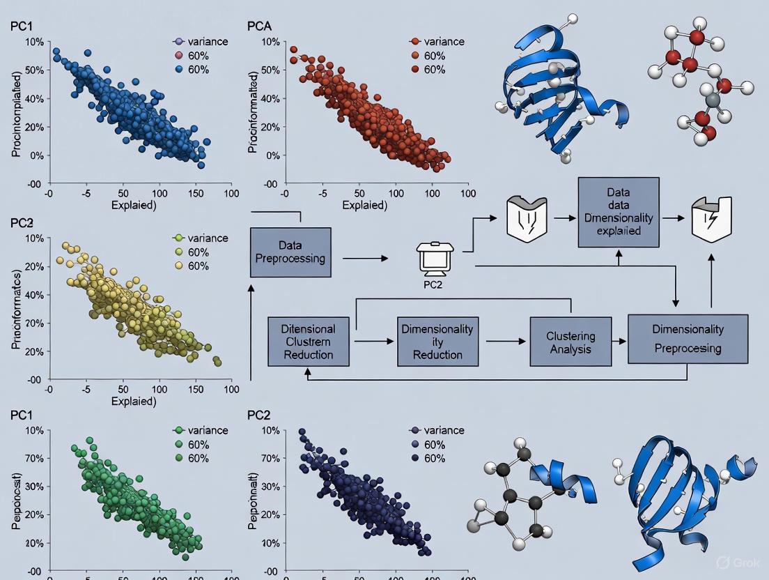
The following workflow summarizes the troubleshooting process:
The Scientist's Toolkit: Key Research Reagents & Materials
The following table details key computational "reagents" and metrics essential for diagnosing and troubleshooting PCA-based clustering.
| Research Reagent / Metric | Function & Purpose in Analysis |
|---|---|
| Silhouette Score | A diagnostic metric that quantifies the separation and compactness of resulting clusters. Values near +1 indicate well-defined clusters. [3] [5] |
| Scree Plot | A visual tool (plot of eigenvalues) used to decide how many principal components to retain by showing the variance explained by each component. [1] |
| Elbow Method | A heuristic used in conjunction with a scree plot or within-cluster variance to identify the optimal number of clusters (k) by looking for an "elbow" point. [3] |
| PCA Loadings | The weights assigned to each original variable in the linear combination that forms a principal component. Critical for interpreting what each PC represents biologically. [1] [7] |
| Correlation Matrix | Used during feature selection to identify and remove highly correlated variables that can bias the PCA transformation and subsequent clustering. [3] |
| StandardScaler / Z-score | A standard preprocessing step that normalizes features to have a mean of 0 and standard deviation of 1, preventing variables with large scales from dominating the PCA. [3] [1] |
Frequently Asked Questions (FAQs)
Q1: My PCA plot shows poor separation between presumed clusters. Does this mean my data has no meaningful groups?
A: Not necessarily. Poor separation in a Principal Component Analysis (PCA) plot can indicate that the underlying cluster structure in your data is non-linear. PCA is a linear dimensionality reduction technique and may fail to preserve complex cluster shapes, making distinct clusters appear overlapped [8]. Before abandoning your analysis, consider applying non-linear dimensionality reduction techniques (such as t-SNE) prior to clustering, or using clustering algorithms capable of identifying non-spherical clusters [9] [8].
Q2: Why does my K-Means clustering produce biologically implearable results on gene expression data?
A: This is a common issue. K-Means operates on several restrictive assumptions that are often violated in biomedical data:
- It assumes clusters are spherical and of similar size [10].
- It is sensitive to outliers and noise, which are common in experimental data [10].
- It requires you to pre-specify the number of clusters (k), which is rarely known a priori in exploratory research [10]. Biomedical data often contains clusters of irregular shapes, varying densities, and outliers. Using K-Means in such contexts can lead to unreliable results [11] [10].
Q3: How can I objectively determine the optimal number of clusters for my data?
A: There is no single best method, but several established techniques can guide your decision:
- Elbow Method: Plot the within-cluster sum of squares (WSS) against the number of clusters. The "elbow" point, where the rate of WSS decrease sharply slows, suggests a suitable k.
- Gap Statistic: Compares the total intra-cluster variation of your data to that of a reference null dataset. The cluster number that maximizes the "gap" statistic is optimal [12].
- Model-Based Methods: For model-based clustering, use statistical metrics like the Bayesian Information Criterion (BIC) to choose the best-fitting model and number of clusters [9]. It is best practice to use multiple indices and combine them with domain knowledge for a biologically defensible conclusion [13].
Troubleshooting Guide: Poor Cluster Separation
Problem Diagnosis
The following table outlines common symptoms, their potential causes, and initial diagnostic steps.
| Symptom | Potential Cause | Diagnostic Check |
|---|---|---|
| Overlapping clusters in PCA plot | Non-linear cluster structure [8] | Visualize data with t-SNE or UMAP. Check if separation improves. |
| Inconsistent cluster results | Noise and outliers in the data [11] | Conduct exploratory data analysis to identify and inspect outliers. |
| K-Means produces long, elongated clusters | Violation of spherical cluster assumption [10] | Run a density-based algorithm like DBSCAN and compare the results. |
| High variability in cluster assignments | Incorrect number of clusters (k) [9] | Apply the Elbow Method or Gap Statistic to re-estimate k. |
| Clusters seem driven by a few strong variables | Features on different scales dominating the distance calculation [9] | Ensure all features were standardized (e.g., Z-score normalization) before clustering. |
Solution Protocols
Protocol 1: Addressing Non-Linear Data and Poor PCA Separation
Objective: To achieve effective clustering when linear separation methods fail.
- Dimensionality Reduction: Apply a non-linear dimensionality reduction technique to your data.
- t-SNE: Effective for visualizing high-dimensional data in 2D or 3D, often revealing complex structures.
- UMAP: A newer technique that often preserves more of the global data structure than t-SNE.
- Algorithm Selection: Choose a clustering algorithm that does not assume spherical clusters.
- Validation: Cluster the data in the new non-linear space and validate the biological coherence of the results using domain knowledge and internal validation indices.
Protocol 2: Handling Noisy Biomedical Data with Outliers
Objective: To obtain robust and reliable clusters from data containing outliers and noise.
- Data Preprocessing:
- Robust Clustering:
- Consider Trimmed Clustering: Use algorithms that automatically "trim" or exclude a proportion of potential outliers during the clustering process, enhancing robustness [11].
- Use DBSCAN: This algorithm explicitly defines outliers as points in low-density regions, effectively separating them from core clusters [12].
- Validation: Use cluster validity indices that are robust to noise. Compare the stability of your results with and without the suspected outliers.
Algorithm Selection Workflow
The following diagram outlines a logical decision process for selecting an appropriate clustering algorithm based on your data characteristics and research goals.
The Scientist's Toolkit: Research Reagent Solutions
Essential Computational Tools
| Tool / Resource | Function | Application Notes |
|---|---|---|
| R Programming Language | A statistical computing environment with extensive packages for clustering and PCA. | Essential packages: evaluomeR (for automated trimmed clustering) [11], cluster, factoextra (for visualization and validation). |
| Python (Scikit-learn) | A machine learning library providing robust implementations of major clustering algorithms. | Modules: sklearn.cluster, sklearn.decomposition (for PCA), sklearn.preprocessing (for data scaling) [15]. |
| StandardScaler / Z-Normalization | A data preprocessing technique to standardize feature scales. | Critical for K-Means and PCA, which are sensitive to variable magnitude. Ensures all features contribute equally to distance calculations [9] [15]. |
| Silhouette Score | An internal validation metric to evaluate cluster quality and aid in determining k. | Values range from -1 to 1; higher positive values indicate better-defined clusters [12]. |
| Gap Statistic | A statistical method to estimate the optimal number of clusters by comparing data to a null reference. | More objective than the Elbow Method for choosing k [12]. |
| DBSCAN Algorithm | A density-based clustering algorithm that identifies arbitrary-shaped clusters and marks outliers. | Ideal for noisy biomedical datasets where the number of clusters is unknown and clusters are non-spherical [12] [14]. |
The 'Variance-as-Relevance' Assumption and Its Pitfalls in Biological Data
You've run your experiment, processed your high-dimensional biological data, and generated a Principal Component Analysis (PCA) plot, only to find a messy overlap of data points instead of the distinct clusters you expected. This common frustration often stems from a fundamental misconception known as the "Variance-as-Relevance" assumption—the flawed expectation that the directions of greatest variance in your dataset always correspond to biologically meaningful patterns.
In reality, the largest sources of variance in biological data often represent technical noise, batch effects, or biologically irrelevant variation that can obscure the signals you care about. This technical support guide will help you diagnose and resolve the issues causing poor cluster separation in your PCA plots, providing practical methodologies to extract meaningful biological insights from your data.
Frequently Asked Questions (FAQs)
FAQ 1: My PCA shows poor cluster separation despite strong biological signals in my raw data. What's wrong?
Answer: Poor cluster separation often indicates that technically sourced variance is dominating your biologically relevant variance. The "Variance-as-Relevance" assumption fails when systematic errors create larger data dispersion than your experimental effects.
Common causes include:
- Batch effects: Samples processed at different times or locations exhibit systematic technical differences
- Sample quality issues: Degradation or contamination affecting subsets of samples
- Inadequate normalization: Failure to account for technical variance before dimensionality reduction
- Hidden covariates: Unrecorded experimental variables influencing your measurements
FAQ 2: How can I determine if my variance structure is problematic?
Answer: Investigate the relationship between variance and signal intensity in your data. In many biological measurements, particularly gene expression studies, variance is intensity-dependent—with low-abundance features exhibiting proportionally higher variance that can dominate PCA results [16].
Diagnostic approach:
- Create a mean-variance relationship plot to identify problematic patterns
- Check if the features contributing most to principal components are biologically relevant or known technical artifacts
- Use spike-in controls if available to distinguish technical from biological variance
FAQ 3: What are the practical alternatives when PCA fails due to non-linear data structures?
Answer: When your data contains non-linear relationships that PCA cannot capture, consider these alternatives:
- t-SNE: Effective for visualizing complex local structures but distances between clusters are not meaningful
- UMAP: Generally preserves more global structure than t-SNE with similar local clustering capabilities
- Phate: Specifically designed for biological data with trajectory structures
- Non-linear PCA variants: Kernel PCA or autoencoder-based approaches
Table: Dimensionality Reduction Methods for Different Data Structures
| Method | Best For | Limitations | Non-Linear Capture |
|---|---|---|---|
| PCA | Linear data, Gaussian distributions | Fails with circular/non-linear patterns | No |
| t-SNE | Local structure visualization | Loses global structure, computational cost | Yes |
| UMAP | Preserving local and global structure | Parameter sensitivity | Yes |
| Kernel PCA | Non-linear manifolds | Computational complexity, kernel choice | Yes |
Troubleshooting Guide: Step-by-Step Protocols
Protocol 1: Data Quality Assessment and Preprocessing
Objective: Identify and mitigate data quality issues before PCA.
Generate quality control metrics
- For sequencing data: Calculate Phred scores, alignment rates, and GC content
- Use tools like FastQC to identify issues in sequencing runs or sample preparation [17]
- Establish minimum quality thresholds before proceeding with analysis
Assess mean-variance relationship
- Plot feature variance against mean expression/intensity
- Identify if low-abundance features with high relative variance are dominating your data
- Consider variance-stabilizing transformations if needed
Implement appropriate normalization
- Select normalization method based on your data type (e.g., TPM for RNA-seq, RLE for count data)
- Account for library size differences and other technical biases
- Validate normalization by checking if technical artifacts are reduced
Protocol 2: Batch Effect Detection and Correction
Objective: Identify and correct for batch effects that may obscure biological signals.
Detect batch effects
- Color PCA plot by potential batch variables (processing date, operator, etc.)
- Use statistical tests like PVCA or surrogate variable analysis
- Check if batch variables explain more variance than biological variables
Apply batch correction methods
- Choose appropriate method: ComBat, limma's removeBatchEffect, or SVA
- Preserve biological variance of interest while removing technical variance
- Validate correction by confirming batch variables no longer drive clustering
Experimental design to minimize batch effects
- Randomize samples across batches when possible
- Include technical replicates across batches
- Balance biological groups within batches
Protocol 3: Variance Modeling for Improved Signal Detection
Objective: Use advanced variance modeling approaches to enhance biological signal detection.
Select appropriate variance modeling approach
- For small sample sizes: Implement information-borrowing methods like Cyber-T or Limma [16]
- For complex experimental designs: Consider VAMPIRE's global variance modeling
- For RNA-seq data: Use DESeq2 or edgeR's dispersion estimation
Implement variance-stabilizing transformation
- Apply techniques that account for mean-variance dependence
- For count data: Consider regularized log transformation or variance stabilizing transformation (VST)
- Validate that transformation reduces technical noise while preserving biological signal
Feature selection based on biologically relevant variance
- Identify features with high biological coefficient of variation
- Prioritize features with consistent patterns within biological groups
- Avoid selecting features based solely on overall variance
Experimental Workflow for Robust Dimensionality Reduction
The diagram below illustrates a comprehensive workflow for addressing variance-related issues in PCA analysis:
The Scientist's Toolkit: Essential Research Reagents and Solutions
Table: Key Reagents and Computational Tools for Variance Troubleshooting
| Item | Function | Application Notes |
|---|---|---|
| Spike-in Controls | Distinguish technical from biological variance | Use ERCC RNA spike-ins for RNA-seq; add at known concentrations |
| Quality Control Tools | Assess data quality before analysis | FastQC for sequencing data; Qualimap for alignment metrics |
| Variance Modeling Software | Improve signal detection in small samples | Cyber-T, Limma, VAMPIRE, DESeq2 |
| Batch Correction Packages | Remove technical artifacts | ComBat, sva, limma's removeBatchEffect in R |
| Alternative Dimensionality Tools | Handle non-linear data structures | UMAP, t-SNE, PHATE, Kernel PCA |
| Visualization Libraries | Create diagnostic plots | ggplot2, plotly, seaborn, matplotlib |
Advanced Technique: Global Variance Modeling
For researchers dealing with particularly challenging datasets where traditional approaches fail, global variance modeling provides a powerful alternative:
Implementation protocol:
- Model the variance structure using the relationship: σ² = μ²A + B, where A represents expression-dependent variance and B represents expression-independent variance [16]
- Estimate parameters using Markov chain Monte Carlo (MCMC) algorithms for maximum likelihood estimation
- Incorporate variance estimates into statistical testing to identify truly significant changes
- Validate model fit by comparing observed versus expected variance patterns
This approach is particularly valuable for studies with limited replicates, where traditional methods like the t-test have low power and high false-positive rates for low-abundance features [16].
Successfully troubleshooting poor cluster separation in PCA requires abandoning the simplistic "Variance-as-Relevance" assumption and adopting a more nuanced understanding of data variance. By implementing the quality control measures, variance modeling techniques, and diagnostic approaches outlined in this guide, researchers can significantly improve their ability to extract meaningful biological insights from high-dimensional data.
Remember that PCA is just one tool in your dimensionality reduction arsenal—when your data contains complex non-linear structures, don't hesitate to explore alternative methods that might better capture the biological relationships you're studying.
Frequently Asked Questions (FAQs)
Why do my clusters separate well in raw data but disappear after standardization? This occurs when the original cluster separation was driven primarily by differences in feature scales rather than underlying correlations. Variables with larger ranges dominate the first principal components in unstandardized PCA, creating illusory clusters. Standardization ensures all features contribute equally, revealing the true underlying structure, which may show poorer separation [18] [6]. This is particularly common when data features have different measurement units or scales.
What does it mean when my PCA plot shows two distinct clusters for what should be identical gestures or samples? This typically indicates a preprocessing or data collection inconsistency between batches. In motion capture data, for example, slight differences in sensor calibration or positioning between recording sessions can cause identical gestures to form separate clusters in PCA space. This signals that technical artifacts, rather than biological or meaningful variation, are driving your principal components [19].
Why does my PCA clustering not correspond to known sample groupings? The principal components capturing the most variance may represent noise, batch effects, or biologically irrelevant variation (like population structure in genetics) rather than variation relevant to your grouping of interest. This violates the "variance-as-relevance" assumption that high-variance components necessarily contain meaningful cluster information [20].
How can I determine if my lack of cluster separation indicates genuine similarity or a methodological issue? First, verify your data preprocessing pipeline includes proper standardization, as scale differences can mask true separation [18]. Next, calculate the variance explained by your principal components; if the first few components capture minimal cumulative variance (e.g., <70%), your data may be too noisy for clear separation. Finally, conduct sensitivity analyses with different preprocessing approaches to see if separation improves [20].
Troubleshooting Guide: Poor Cluster Separation in PCA
Quick Diagnosis Table
| Symptom | Possible Causes | Diagnostic Steps | Potential Solutions |
|---|---|---|---|
| Distinct clusters disappear after standardization [6] | Clusters driven by scale differences, not correlation | Compare feature variances pre/post standardization; check if high-variance features defined original clusters | Focus on biological interpretation; use domain knowledge to select relevant features |
| Multiple clusters for identical sample types [19] | Batch effects, sensor calibration drift, collection protocol variations | Color points by collection date/batch; check for technical correlations with PCs | Implement batch correction; apply sensor calibration; standardize protocols |
| Diffuse, overlapping clusters with no clear separation | High noise-to-signal ratio; too many irrelevant features; genuine sample similarity | Calculate variance explained by first 2-3 PCs; assess feature quality; add known positives | Apply feature selection; increase sample size; use regularization; try alternative methods (t-SNE, UMAP) |
| Known groups don't separate in expected directions | PC axes capture irrelevant variance; group differences are subtle | Color points by known groups; check which features load strongly on early PCs | Apply supervised approaches (LDA); use weighted PCA; select group-informative features |
Comprehensive Experimental Protocol for Diagnosing Separation Issues
Step 1: Data Quality Assessment Begin by examining your raw data structure. Calculate basic descriptive statistics (mean, variance, range) for each feature to identify variables with dramatically different scales. For the sarcoidosis radiomics data discussed in the literature, researchers found that 9,706 feature pairs had correlations beyond 0.9, indicating severe redundancy that can distort PCA results [20]. Document any missing data patterns and assess whether they correlate with potential batch effects.
Step 2: Systematic Preprocessing Evaluation Process your data through multiple preprocessing pathways in parallel:
- Raw data (unstandardized)
- Z-score standardized data (mean-centered, unit variance)
- Range-scaled data (scaled to [0,1])
- Log-transformed data (if appropriate for your data type)
For each pathway, apply PCA and generate 2D and 3D plots of the first 2-3 principal components. Color points by known experimental factors (batch, date, operator) and hypothesized biological groups.
Step 3: Principal Component Analysis Compute PCA for each preprocessed dataset. Examine the scree plot to determine the variance explained by each component. As shown in PCA tutorials, the first component should capture the most variance, with each subsequent component capturing progressively less [18] [21]. Calculate the cumulative variance explained by the first 2-3 components, as these will determine your visualization clarity. If these components capture less than 60-70% of total variance, cluster separation will likely be poor.
Step 4: Cluster Validation Metrics Apply multiple clustering algorithms (K-means, Gaussian Mixture Models) to the principal components. Calculate silhouette scores, within-cluster sum of squares, and other validity measures for different numbers of hypothesized clusters. Compare these metrics across preprocessing methods to identify optimal analysis conditions.
Step 5: Sensitivity Analysis Systematically investigate how robust your results are to different analytical choices. This includes testing different feature subsets, applying various normalization schemes, and using alternative dimension reduction techniques. The goal is to determine whether poor separation persists across methodological variations or is specific to certain analysis decisions [20].
Advanced Diagnostic Framework
Research Reagent Solutions for PCA Cluster Analysis
| Reagent/Resource | Function | Application Notes |
|---|---|---|
| StandardScaler (sklearn.preprocessing) | Standardizes features by removing mean and scaling to unit variance | Essential for preventing high-variance features from dominating PCA [19] [18] |
| PCA (sklearn.decomposition) | Performs principal component analysis | Use n_components=None initially to examine all components; random_state for reproducibility [21] |
| Shapiro-Wilk Filter | Preprocessing filter to counter variance-as-relevance assumption | Identifies and removes features where high variance doesn't correlate with cluster relevance [20] |
| VarSelLCM Package (R) | Variable selection for model-based clustering | Implements diagonal GMM with models indexed by variable relevance; uses BIC for model selection [20] |
| Dynamic Time Warping | Aligns time-series data before PCA | Critical for motion capture or temporal data to align sequences despite timing variations [19] |
| Procrustes Analysis | Shape-based alignment of datasets | Aligns new recordings with reference gestures to ensure consistency in PCA space [19] |
Quantitative Thresholds for Cluster Separation Assessment
| Metric | Good Separation | Marginal Separation | Poor Separation |
|---|---|---|---|
| Variance Explained (PC1+PC2) | >80% | 60-80% | <60% |
| Silhouette Score | 0.7-1.0 | 0.5-0.7 | <0.5 |
| Between:Within Cluster SS Ratio | >3.0 | 1.5-3.0 | <1.5 |
| Cluster Distinctness (Visual) | Clear separation, minimal overlap | Partial separation, some overlap | No clear boundaries, heavy overlap |
Intervention Protocol Based on Diagnostic Results
Troubleshooting Guide: Poor Cluster Separation in PCA
Is the observed cluster separation in my PCA plot real or an artifact?
Problem You have run a single-cell RNA-sequencing experiment and performed PCA. The resulting plot shows clear clusters, but you are unsure if these groups represent true biological cell types or are technical artifacts.
Explanation Cluster separation in PCA can be driven by both biological and technical sources of variation. Batch effects—technical variations from processing cells in different laboratories, at different times, or with different reagents—create consistent fluctuations in gene expression that can be mistaken for biological signal [22]. Furthermore, the inherent population structure of your cells, such as a hierarchical relationship between cell types, can be misinterpreted by standard clustering algorithms, leading to either over-clustering or the false discovery of novel cell populations [23] [24].
Solution Follow the diagnostic workflow below to systematically evaluate your clustering results. This will help you determine if your clusters need correction for batch effects or merging due to over-clustering.
How do I definitively identify a batch effect in my data?
Problem You suspect a batch effect but are not sure how to confirm it.
Explanation A batch effect is present when technical factors (e.g., sequencing date, lane, or protocol) systematically explain more of the variance in your data than biological factors. This can be observed visually and confirmed with quantitative metrics [22].
Solution Follow this experimental protocol to detect batch effects.
Experimental Protocol: Batch Effect Detection
- Step 1: Visual Inspection with PCA. Perform PCA on your raw, uncorrected gene expression matrix. Create a scatter plot of the top principal components (e.g., PC1 vs. PC2). Color the data points by their batch of origin (e.g., experiment date). If cells cluster strongly by batch rather than by expected biological condition, a batch effect is likely present [22].
- Step 2: Visualization with UMAP/t-SNE. Similarly, generate a UMAP or t-SNE plot from your data and color the points by batch. The presence of separate, batch-specific sub-clusters within a group of cells that should be biologically homogeneous is a key indicator of a batch effect [22].
- Step 3: Calculate Quantitative Metrics. Use metrics to objectively measure the degree of batch integration. Common metrics include:
- kBET (k-nearest neighbor batch effect test): Rejects the null hypothesis (good batch mixing) if batches are not well mixed [22].
- ARI (Adjusted Rand Index): Measures the similarity between two clusterings. A low ARI between batch labels and cluster labels is desirable [24].
- NMI (Normalized Mutual Information): Measures the information shared between batch and cluster labels. Lower values indicate less influence from batch [22].
Table: Key Quantitative Metrics for Batch Effect Assessment
| Metric | What It Measures | Interpretation | Desired Value |
|---|---|---|---|
| kBET | Mixing of batches in local neighborhoods | Lower rejection rate indicates better mixing | Closer to 0 |
| ARI | Agreement between batch labels and cluster labels | Lower value indicates batch has less impact on clustering | Closer to 0 |
| NMI | Shared information between batch and cluster labels | Lower value indicates batch and clusters are independent | Closer to 0 |
My clusters are statistically significant but don't make biological sense. What now?
Problem Your data has passed batch effect checks and clustering algorithms report statistically distinct groups, but these groups lack known cell type markers or have unstable definitions.
Explanation This is a classic sign of over-clustering. Widely used clustering algorithms like Louvain and Leiden are heuristic and will partition data even when only random variation is present [23]. They do not formally account for statistical uncertainty, leading to overconfidence in the discovery of novel cell types [23]. This is especially problematic when the true biological structure of the cell population is hierarchical (e.g., T-cells and B-cells are both lymphocytes), but the clustering metric treats all groups as unrelated [24].
Solution Incorporate significance analysis into your clustering workflow.
Experimental Protocol: Significance Analysis for Clustering
- Step 1: Use Model-Based Hypothesis Testing. Employ methods like single-cell Significance of Hierarchical Clustering (sc-SHC). This approach defines a realistic parametric distribution for a cell population and tests whether a proposed split into two clusters could have arisen by chance from a single population [23].
- Step 2: Assess Pre-computed Clusters. If you have already generated clusters with a tool like Seurat, you can apply a significance analysis framework retrospectively. The method will hierarchically cluster the centers of your provided clusters and recursively apply statistical tests to determine which clusters should be merged [23].
- Step 3: Use Hierarchical Metrics for Evaluation. When comparing your results to a reference, use metrics that account for cell type hierarchy.
- Weighted Rand Index (wRI): Assigns different weights to pairwise cell relationships based on their place in the known cell type hierarchy. Mistakes between closely related subtypes (e.g., CD4 and CD8 T-cells) are penalized less than mistakes between distinct types (e.g., T-cells and neurons) [24].
- Weighted Normalized Mutual Information (wNMI): Uses a structured entropy that considers hierarchical relationships to reflect the accuracy of recovering the true cell population structure [24].
How do I choose and apply a batch effect correction method?
Problem You have identified a batch effect and need to correct it without removing true biological signal.
Explanation Batch effect correction methods use various algorithms to align cells from different batches in a shared space, assuming that a subset of the cell population is shared across batches [25]. The goal is to remove technical variation while preserving biological variation. Different methods are suited to different data types and sizes.
Solution Select an appropriate algorithm and be vigilant for overcorrection.
Table: Comparison of Common Batch Effect Correction Methods
| Method | Core Algorithm | Key Principle | Best For |
|---|---|---|---|
| Harmony [22] | Iterative clustering and correction | Removes batch effects by clustering similar cells across batches and maximizing diversity within each cluster. | Datasets with complex batch structures. |
| MNN Correct [25] [22] | Mutual Nearest Neighbors (MNNs) | Finds cells in different batches that have similar expression profiles (MNNs) and uses them as anchors to correct the data. | Datasets where not all cell types are present in all batches. |
| Seurat CCA [22] | Canonical Correlation Analysis (CCA) & MNNs | Projects data into a subspace using CCA, finds MNNs in this subspace, and uses them as anchors for integration. | Integrating large, complex datasets. |
| Scanorama [22] | Mutual Nearest Neighbors in reduced space | Finds MNNs in dimensionally reduced spaces and uses a similarity-weighted approach for integration. | Large datasets with high computational demands. |
Warning: Signs of Overcorrection After applying batch correction, check for these signs that you may have removed biological signal along with the batch effect [22]:
- Cluster-specific markers are dominated by common, non-informative genes (e.g., ribosomal genes).
- Significant overlap exists between markers for different clusters.
- Expected canonical cell type markers are absent.
- Few differential expression hits are found for pathways known to be active in your samples.
The Scientist's Toolkit
Table: Essential Research Reagents & Computational Tools
| Item | Function / Purpose | Example Tools / R Packages |
|---|---|---|
| Batch Effect Correction | Algorithms to remove technical variation from different experiments. | Harmony, MNN Correct, Seurat (CCA), Scanorama [22] |
| Significance Testing for Clusters | Statistically validates whether clusters represent distinct populations. | sc-SHC (single-cell Significance of Hierarchical Clustering) [23] |
| Hierarchical Evaluation Metrics | Evaluates clustering results while accounting for known cell type relationships. | Weighted Rand Index (wRI), Weighted NMI (wNMI) [24] |
| Dimensionality Reduction | Visualizes high-dimensional data to assess clustering and batch effects. | PCA, UMAP, t-SNE [22] |
| Quantitative Integration Metrics | Provides objective scores to assess the success of batch correction. | kBET, ARI, NMI [22] |
Experimental Protocol: A Rigorous Clustering Workflow
For robust results, follow this integrated protocol that incorporates batch correction and significance testing.
Workflow: An Integrated Approach to Valid Clustering
Step-by-Step Instructions:
- Start with Normalized Data. Begin with a properly normalized count matrix (cells x genes) to control for sequencing depth and other library-size biases [22].
- Perform Dimensionality Reduction. Run PCA on a set of highly variable genes. This reduces noise and computational cost for subsequent steps [23].
- Diagnose Batch Effects. As detailed in FAQ #2, use PCA/UMAP plots and quantitative metrics (kBET, ARI) to check for batch effects [22].
- Apply Batch Correction. If a batch effect is detected, select and apply a correction method from the table above (e.g., Harmony). Visualize and re-run the quantitative metrics to confirm the effect has been mitigated [22].
- Perform Clustering. Use a standard algorithm (e.g., Louvain in Seurat) to get an initial partition of the data into clusters [23].
- Run Significance Analysis. Apply a method like sc-SHC to your clusters. This will test each proposed split in the clustering tree and automatically merge clusters that do not represent statistically distinct populations, effectively correcting for over-clustering [23].
- Annotate and Validate Clusters. Use known marker genes to assign biological cell type labels to the statistically validated clusters. The use of hierarchical metrics (wRI, wNMI) for comparison with a reference can provide a more biologically plausible evaluation [24].
Beyond Basic PCA: Advanced Preprocessing and Clustering Techniques for Robust Analysis
Troubleshooting Guide: Poor Cluster Separation in PCA Plots
This guide addresses common data preparation issues that lead to poor cluster separation in Principal Component Analysis (PCA), a key step in many drug development and research pipelines. Proper data preprocessing is critical because PCA is sensitive to the scale, quality, and consistency of your input data [19].
Problem 1: Inconsistent Data Scaling
The Issue After applying PCA, your data forms unexpected or poorly separated clusters, even when you know the underlying groups should be similar. This often manifests as identical gestures or samples splitting into two distinct clusters [19].
Root Causes
- Dominant Features: Variables with larger numerical ranges (e.g., 0-1000) dominate the PCA, overshadowing variables with smaller ranges (e.g., 0-0.1), as PCA is sensitive to variance [19] [26].
- Inconsistent Preprocessing: Data collected in different batches or with slightly different sensor calibrations may have different baseline scales, causing them to cluster separately [19].
Solutions
- Apply Standardization: Use StandardScaler (Z-score normalization) to transform features to have a mean of 0 and a standard deviation of 1. This ensures all features contribute equally to the PCA [19] [26].
- Validate Across Batches: Ensure the same scaler fit to your original reference data is applied to new datasets to maintain consistency [19].
Experimental Protocol: Standardization
Problem 2: Sensor Drift or Misalignment
The Issue Newly recorded time-series data (e.g., from motion sensors) does not align with previous recordings in the PCA plot, despite representing the same biological or physical phenomenon [19].
Root Cause Small, consistent errors in sensor calibration, such as a 5-degree rotational offset, can systematically shift the data in the high-dimensional space, leading PCA to perceive it as a different cluster [19].
Solutions
- Sensor Calibration: Apply rotational or translational transformations to realign new data to a reference frame. The
scipy.spatial.transform.Rotationlibrary can be used for this purpose [19]. - Advanced Alignment: For time-series data, use Dynamic Time Warping (DTW) or Procrustes analysis to align sequences before applying PCA [19].
Experimental Protocol: Sensor Calibration
Problem 3: High-Dimensional, Correlated, and Noisy Data
The Issue In high-dimensional data (e.g., from genomics, metabolomics, or imaging), the first few Principal Components (PCs) capture a low percentage of the total variance, and cluster separation is poor [4] [20].
Root Causes
- "Variance as Relevance" Fallacy: PCA prioritizes high-variance features. In biological data, the largest sources of variance (e.g., population structure, batch effects) may not be relevant for discriminating the disease subtypes of interest [20].
- Correlated Noise: Highly correlated and noisy features can create large-variance principal components that are irrelevant for clustering, masking the true discriminatory signal [20].
Solutions
- Filter for Relevant Variance: Instead of using all PCs, employ a Shapiro-Wilk (SW) filter to select PCs that deviate from a normal distribution, as they are more likely to contain cluster structure [20].
- Explore Alternative Preprocessing: Investigate decorrelation filters or other dimensionality reduction techniques like autoencoders that do not rely solely on variance [19] [20].
Problem 4: Improper Handling of Missing Values
The Issue Clusters appear distorted, or the analysis fails entirely due to the presence of missing values in the dataset.
Root Causes
- Information Loss: Simply removing cases with missing values (complete case analysis) can introduce bias and reduce the effective sample size, leading to overfitting and imprecise clusters [9] [27].
- Inaccurate Imputation: Replacing missing values with a simple mean can distort the relationships between variables and the natural structure of the data [9].
Solutions
- Use Advanced Imputation: Apply multiple imputation or k-nearest neighbor (KNN) imputation to estimate missing values in a way that better preserves data structure [9].
- Avoid Dichotomania: Do not handle missingness by dichotomizing continuous variables, as this wastes information and reduces statistical power [27].
Frequently Asked Questions (FAQs)
Q1: Why do my identical biological replicates form separate clusters in the PCA plot?
This is a classic sign of batch effects or inconsistent preprocessing. Ensure that all data is scaled using the same parameters (e.g., the same StandardScaler object fit on your control data). Investigate whether technical artifacts (e.g., different sample preparation days) are introducing systematic variation that PCA is detecting [19].
Q2: My explained variance for the first few PCs is low (~20%). Can I still use PCA for clustering? Yes, but with caution. A low explained variance suggests that the key differences between your clusters might not be the largest sources of variance in the data. The PCs that capture most of the variance are not guaranteed to be the ones that are informative for clustering. You should investigate lower-order PCs or use pre-processing filters (like the Shapiro-Wilk filter) to find components that better separate your clusters [4] [20].
Q3: What is the single most important preprocessing step for PCA-based clustering? Standardization (Z-score normalization) is often the most critical step. Without it, PCA will be unduly influenced by the scale of your measurements, and variables measured in larger units (e.g., concentration in mmol/L) will dominate those in smaller units (e.g., expression fold-change), regardless of their biological importance [19] [26].
Q4: How can I align new data with my original reference dataset in PCA space? Beyond standardization, you may need a calibration or alignment step. For kinematic data, this could be a rotational transformation. For other data types, Procrustes analysis can be used to rotate, translate, and scale the new dataset to match the configuration of the original reference data as closely as possible [19].
Q5: Can autoencoders be a better alternative to PCA for clustering? Yes, in some cases. Autoencoders are neural networks that can learn non-linear latent representations of your data. By training an autoencoder on your original data, you can map new recordings into a shared latent space, which can be more robust to certain types of noise and variation, potentially leading to better-aligned clusters [19].
Data Presentation: Scaling and Normalization Techniques
The table below summarizes key techniques to prepare your data for PCA and clustering.
| Technique | Method Description | Sensitivity to Outliers | Best Use Cases for Clustering |
|---|---|---|---|
| Standardization (Z-Score) | Centers data to mean=0 and scales to standard deviation=1 [26]. | Moderate | Most common starting point; assumes near-normal data [26]. |
| Min-Max Scaling | Scales data to a specified range (e.g., [0, 1]) [26]. | High | Neural networks; data with bounded ranges [26]. |
| Robust Scaling | Centers data using the median and scales using the Interquartile Range (IQR) [26]. | Low | Data with significant outliers or skewed distributions [26]. |
| Absolute Maximum Scaling | Divides values by the maximum absolute value per feature. Scales to [-1, 1] [26]. | High | Sparse data; simple scaling needs. |
| Vector Normalization | Scales each individual sample (row) to have a unit norm (length=1) [26]. | Varies | Algorithms relying on cosine similarity or sample direction. |
The Scientist's Toolkit: Essential Research Reagents & Solutions
| Item | Function in Data Preparation |
|---|---|
| StandardScaler (sklearn) | Standardizes features by removing the mean and scaling to unit variance. Critical for PCA [19] [26]. |
| RobustScaler (sklearn) | Scales features using statistics that are robust to outliers. Use when your dataset contains many extreme values [26]. |
| Multiple Imputation | A statistical technique for handling missing data by creating several complete datasets and pooling results. Superior to mean imputation [27] [9]. |
| Dynamic Time Warping (DTW) | An algorithm for measuring similarity between two temporal sequences. Useful for aligning time-series data before clustering [19]. |
| Shapiro-Wilk (SW) Filter | A pre-processing filter used to select Principal Components that deviate from normality, as they are more likely to contain cluster-relevant information [20]. |
Experimental Workflow Diagrams
Data Preprocessing for Optimal PCA Clustering
Addressing Poor Cluster Separation
Automated and Sparse Clustering Methods for High-Dimensional Biomarker Data
Frequently Asked Questions
Q1: My PCA plot shows poor cluster separation. Does this mean my biomarkers have no meaningful patterns? Not necessarily. PCA can fail to separate clusters if the data has a non-linear structure or if the primary source of variance is not aligned with class boundaries [8]. Before abandoning your analysis, investigate using Linear Discriminant Analysis (LDA), which is designed specifically to maximize separation between known groups [28], or explore non-linear dimensionality reduction techniques.
Q2: What is the fundamental difference between traditional clustering and automated clustering for biomarker discovery? Traditional clustering methods (like k-means) often require you to specify the number of clusters in advance and can struggle with high-dimensional noise. Automated Clustering solves the Automatic Clustering Problem (ACP) by simultaneously determining the optimal number of clusters and the best assignment of data objects, maximizing intra-cluster cohesion and inter-cluster separation without prior information [29].
Q3: My high-dimensional proteomics data is very noisy. Which clustering method should I use? For high-dimensional, noisy biomarker data (e.g., from mass spectrometry), Automated Trimmed and Sparse Clustering (ATSC) is highly suitable. It automatically determines the optimal number of clusters while suppressing noise by emphasizing significant features and excluding outliers, all without manual parameter tuning [11].
Q4: How can I ensure my clustering results are biologically interpretable and not a black box? Seek out methods that provide interpretable results. For instance, the Interpretable Graph Neural Additive Network (GNAN) can be used to analyze sparse temporal biomarker data, providing node and feature importance metrics that trace which biomarkers and time points contribute most to a classification decision [30]. Furthermore, algorithms generated by Automatic Generation of Algorithms (AGA) are symbolic and human-readable, allowing researchers to understand and refine their structure [29].
Q5: What is a key advantage of using sparse clustering methods like ST-CS? Sparse clustering methods, such as Soft-Thresholded Compressed Sensing (ST-CS), integrate feature selection directly into the model training. This results in a parsimonious feature set, identifying a small subset of the most discriminative biomarkers. This enhances model interpretability and predictive accuracy by eliminating redundant or non-informative features [31].
Troubleshooting Guides
Guide 1: Troubleshooting Poor Cluster Separation in PCA
Poor cluster separation in a PCA plot is a common issue in biomarker research. The flowchart below outlines a systematic diagnostic and resolution process.
Resolution Steps:
- If Class Labels Are Known: Apply Linear Discriminant Analysis (LDA). Unlike PCA, which maximizes total variance, LDA finds the axes that maximize the separation between multiple predefined classes [28].
- If Class Labels Are Unknown:
- Investigate Non-Linearity: PCA is a linear technique and will fail if the data has a complex, non-linear structure (e.g., a circular distribution) [8]. Plot your raw data to check for such patterns.
- Apply Non-Linear Dimensionality Reduction: If non-linearity is suspected, use techniques like t-Distributed Stochastic Neighbor Embedding (t-SNE) or Neighbourhood Components Analysis (NCA) [28]. These methods are designed to reveal complex, non-linear cluster structures in low-dimensional projections.
- Address Data Quality Issues:
- Outliers and Noise: PCA is sensitive to outliers [32]. Use outlier detection methods from Exploratory Data Analysis (EDA) to identify and remove them [32].
- Data Scaling: If variables are on different scales (e.g., heart rate vs. a 1-5 symptom score), scaling is crucial before PCA to ensure that variables with larger values don't disproportionately influence the components [33].
Guide 2: Optimizing Clustering in High-Dimensional, Noisy Biomarker Data
High-dimensional biomarker data from proteomics or transcriptomics is often plagued by noise and redundant features. The following workflow is designed for this specific challenge.
Detailed Methodologies:
Automated Trimmed and Sparse Clustering (ATSC) Protocol [11]:
- Implementation: The ATSC method is available within the
evaluomeRpackage for R. - Process: The method automatically calibrates its tuning parameters—the trimming proportion (to exclude outliers) and the sparsity level (to suppress noisy features).
- Output: It outputs a robust clustering solution where the optimal number of clusters is determined without manual intervention, effectively handling noise and redundancy.
- Implementation: The ATSC method is available within the
Soft-Thresholded Compressed Sensing (ST-CS) Protocol [31]:
- Objective: Recover a sparse coefficient vector (ω) where non-zero coefficients correspond to discriminative biomarkers.
- Optimization Framework: The coefficients are estimated by solving a constrained optimization problem that includes dual ℓ₁ and ℓ₂ regularization. The ℓ₁-norm enforces sparsity, while the ℓ₂-norm stabilizes estimates and handles multicollinearity.
- Automated Feature Selection: Instead of manual thresholding, the resulting coefficients are partitioned into "signal" vs. "noise" using K-Medoids clustering on the coefficient magnitudes, fully automating the selection of the most important biomarkers.
Automatic Generation of Algorithms (AGA) for Clustering [29]:
- Concept: Uses Genetic Programming (GP) to assemble fundamental algorithmic components into novel, complete clustering algorithms.
- Output: The result is a human-readable and executable algorithm that is specifically tailored to a given dataset, potentially outperforming state-of-the-art general-purpose methods.
Comparative Analysis of Clustering Methods
The following table summarizes key automated and sparse clustering methods relevant for biomarker research.
| Method Name | Core Functionality | Key Advantages | Ideal Use Case in Biomarker Research |
|---|---|---|---|
| Automated Trimmed & Sparse Clustering (ATSC) [11] | Automatically determines cluster number (k) with noise trimming & sparsity. | Fully automated; robust to outliers & high-dimensional noise. | Unsupervised patient stratification from noisy transcriptomic/proteomic data. |
| Soft-Thresholded Compressed Sensing (ST-CS) [31] | Integrates classification with automated, sparse feature selection. | Outputs a minimal, discriminative biomarker panel; high specificity. | Identifying a parsimonious serum protein signature for disease diagnosis. |
| Automatic Algorithm Generation (AGA) [29] | Automatically constructs novel clustering algorithms from components. | Generates a custom, interpretable algorithm for a specific dataset. | Tackling novel, complex dataset structures where standard methods fail. |
| Interpretable Graph Learning (GNAN) [30] | Models sparse temporal biomarker data as graphs for classification. | Provides feature & time-point importance; no data imputation needed. | Analyzing irregularly sampled blood test data to find critical pre-diagnostic windows. |
The Scientist's Toolkit: Essential Research Reagents & Solutions
This table lists key computational tools and their functions for implementing the methods discussed.
| Item | Function in Analysis | Key Parameter / Consideration |
|---|---|---|
| evaluomeR Package (R) [11] | Implements the Automated Trimmed and Sparse Clustering (ATSC) method. | Accessible via Bioconductor; requires minimal computational background. |
| ST-CS Framework (Python/MATLAB) [31] | Provides the code for Soft-Thresholded Compressed Sensing. | Look for published code alongside the manuscript (e.g., on GitHub). |
| Genetic Programming (GP) Library (e.g., DEAP) | Serves as the engine for Automatic Algorithm Generation (AGA) [29]. | Requires definition of a set of elementary algorithmic components. |
| Silhouette Index (SI) [29] | An internal validation metric used as an objective function to evaluate clustering quality. | Does not assume cluster shape; values range from -1 (poor) to +1 (excellent). |
| 1-Bit Compressed Sensing [31] | A signal processing technique that quantizes data to binary values for robust sparse recovery. | Reduces noise and computational complexity, aligning with classification tasks. |
Integrating Sensor Calibration and Data Alignment to Correct for Technical Variance
Core Problem: Technical Variance Masks Biological Signals
In high-dimensional biological data analysis, technical variances from sensor drift or misalignment can obscure true biological clusters in Principal Component Analysis (PCA). These inconsistencies cause identical experimental conditions to appear as separate clusters, complicating interpretation [19]. Proper sensor calibration and data alignment are critical for ensuring that PCA visualizations reflect biological reality rather than technical artifacts.
Troubleshooting Guide: Poor Cluster Separation in PCA
| Problem | Root Cause | Diagnostic Steps | Solution |
|---|---|---|---|
| Separate PCA clusters for identical gestures/conditions [19] | Inconsistent sensor calibration or improper data scaling [19]. | Check for unit-to-unit sensor variation; review preprocessing and scaling pipelines [19] [34]. | Apply sensor calibration and use StandardScaler before PCA [19]. |
| Cluster drift between experimental batches | Sensor sensitivity changes over time and use (e.g., piezoelectric accelerometers) [34]. | Compare initial calibration certificates with recent performance data [34]. | Recalibrate sensors annually or after heavy use [34]. |
| Failure of new data to align with reference in PCA space | Slight changes in sensor placement or environmental conditions [19]. | Use Dynamic Time Warping (DTW) or Procrustes analysis to quantify misalignment [19]. | Apply rotation transformations or affine alignment to new datasets [19]. |
Experimental Protocols for Calibration & Alignment
Sensor Calibration Procedure
This protocol corrects for structural errors in inertial measurement units (IMUs) like accelerometers and gyroscopes [35].
- Objective: Determine scale factor, misalignment, and bias parameters for each sensor axis using a linear sensor model [35].
- Equipment: Precision rate table (for gyroscopes), multi-axis turntable (for accelerometer tumble test), thermal chamber [35].
- Methodology:
- Accelerometer Tumble Test: Mount the sensor and collect static measurements in multiple orientations to align each axis with, opposite to, and perpendicular to gravity, providing a ±1 g measurement [35].
- Gyroscope Rate Table Calibration: Secure the sensor on a rate table and rotate it across a range of precise angular rates to characterize response [35].
- Thermal Calibration: Perform the above processes inside a thermal chamber across the sensor's operational temperature range to model temperature-dependent parameter changes [35].
- Data Processing: A least-squares fit is performed on the collected data to populate the parameters in the sensor model for correcting future measurements [35].
Data Alignment Protocol for PCA
This corrects for misalignment between new recordings and a reference dataset in PCA space [19].
- Objective: Apply transformations so that identical biological conditions or gestures form unified clusters in a PCA plot.
- Preprocessing:
- Alignment Techniques:
- Rotation Transformation: For rotational misalignment, use Euler angles to realign data [19].
- Procrustes Analysis: Apply scaling, rotation, and translation to optimally align one dataset (new recording) to another (reference) [19].
- Dynamic Time Warping (DTW): Align time-series data by minimizing temporal distortions between sequences [19].
The following workflow integrates these protocols into a cohesive analysis pipeline to ensure data integrity from collection to visualization.
Cluster Diagnostics and Visualization
After calibration and alignment, validate clustering performance.
- Determining Cluster Number (k):
- Elbow Plot: Use the
yellowbrickpackage to visualize the within-cluster sum of squares against the number of clusters (k). The optimal k is often at the "elbow" of the plot [36]. - Silhouette Analysis: Plot silhouette scores for different k values. The highest average score suggests the most coherent cluster structure [36].
- Elbow Plot: Use the
- Dimensionality Reduction for Visualization:
- PaCMAP Recommendation: For 2D visualization of clusters, use PaCMAP, which better preserves both local and global data structure compared to PCA, t-SNE, or UMAP [36].
- Linear Discriminant Analysis (LDA): If cluster labels are known, LDA can be used to find the projection that maximizes between-cluster separation, directly addressing the visualization goal [28].
The Scientist's Toolkit: Essential Research Reagents & Materials
| Item | Function |
|---|---|
| Precision Rate Table | Provides precise angular rates for gyroscope calibration, characterizing scale factor and bias [35]. |
| Multi-Axis Turntable | Enables accelerometer tumble testing by rotating the sensor into multiple static orientations relative to gravity [35]. |
| Thermal Chamber | Allows calibration across a range of temperatures to model and correct for temperature-sensitive parameter drift [35]. |
| Reference Accelerometer | A NIST-traceable, calibrated reference sensor used to validate and calibrate the sensors under test [34]. |
| StandardScaler | A preprocessing tool that standardizes features by removing the mean and scaling to unit variance, preventing high-variance features from dominating PCA [19]. |
Frequently Asked Questions (FAQs)
Q1: Why do my identical gestures or experimental conditions form two separate clusters in my PCA plot? This is typically caused by technical variance, such as inconsistent sensor calibration between recording sessions or slight changes in sensor placement. PCA is sensitive to these systematic differences and will interpret them as separate sources of variance, breaking what should be one cluster into two [19].
Q2: How often should I recalibrate my sensors? The need for recalibration depends on the sensor technology and usage. Piezoelectric accelerometers can show noticeable sensitivity drift over time and may require annual recalibration. In contrast, MEMS-based sensors (variable capacitance, piezoresistive) are often more stable, with many units showing gain variations of less than 2% over time, making frequent recalibration less critical [34].
Q3: I have calibrated my sensors, but my new data still doesn't align with my original reference set in the PCA space. What else can I do? Calibration corrects internal sensor errors. For external misalignment (e.g., different orientation), apply data alignment techniques before PCA. Use Procrustes analysis to find the optimal rotation, translation, and scaling to align your new dataset to the reference. For time-series data, Dynamic Time Warping (DTW) can correct temporal misalignments [19].
Q4: Is PCA the best method for visualizing my clusters? PCA is excellent for preserving the global structure of your data. However, if your goal is to maximize the visual separation between known clusters, Linear Discriminant Analysis (LDA) is a more suitable technique, as it explicitly finds axes that maximize between-cluster variance [28]. For a more balanced preservation of local and global structure, consider PaCMAP [36].
Q5: Can machine learning solve this clustering issue without manual calibration? Advanced techniques like autoencoders can learn a shared latent space that is more robust to minor technical variations. By training a model on your original data, it can potentially map new, slightly misaligned recordings into the correct cluster. However, this requires a large and well-characterized training set, and proper sensor calibration remains the most reliable foundation [19].
Frequently Asked Questions
1. Why would my classification model perform well even when my PCA plot shows poor cluster separation?
This common scenario occurs because PCA only uses the first few principal components for visualization, which maximize the variance of the entire dataset but may not capture the features most relevant for class discrimination. Your classification model likely uses many more components or original features, allowing it to detect subtle patterns invisible in a 2D PCA plot [37]. The separation might be present in higher, un-plotted principal components.
2. I am using PCA for clustering, but the results are poor. What is the issue?
PCA is a linear technique designed to preserve global data variance, not to identify clusters, which are concentrations of data points (neighborhoods) [38]. Using neighborhood-preserving methods like t-SNE or UMAP before clustering often yields better results because their objective aligns directly with the goal of clustering [39] [38].
3. When should I avoid using PCA altogether?
PCA has known limitations in specific, advanced research contexts. In quantitative genetic association studies on human data, especially with family or multiethnic cohorts, PCA can perform poorly compared to Linear Mixed Models (LMMs) due to its inability to adequately model complex relatedness structures [40]. It is also generally inadequate for data with strong non-linear relationships [39] [41].
4. My t-SNE plot looks different every time I run it. Is this normal?
Yes, this is expected. The t-SNE algorithm is stochastic, meaning it contains random elements during the optimization process. While the random_state parameter can be set for reproducibility, different initializations can lead to visually distinct layouts, though the core cluster relationships should remain similar [39] [42].
5. For visualizing a very large dataset (e.g., >100,000 points), is t-SNE a good choice?
For very large datasets, UMAP is generally recommended over t-SNE. t-SNE is computationally intensive and slow on large data, while UMAP is designed for scalability and can handle millions of points efficiently, producing results in a fraction of the time [43] [44].
Troubleshooting Guides
Guide 1: Troubleshooting Poor Cluster Separation in PCA Plots
This guide helps diagnose and resolve situations where PCA fails to reveal expected data clusters.
Step 1: Confirm the Nature of Your Data
- Action: Determine if your data has non-linear relationships. PCA can only capture linear patterns.
- Interpretation: If the underlying data manifold is non-linear (e.g., a spiral or "S" curve), PCA will be ineffective. This is the primary reason to switch to a non-linear method [41].
Step 2: Check the Variance Explained by Plotted Components
- Action: Examine the
explained_variance_ratio_of your PCA model. A low cumulative variance for the first two components indicates that your 2D plot is missing most of the data's information [37]. - Interpretation: If the first two components explain a small percentage of the total variance (e.g., <30%), separation may exist in higher components not visualized.
- Action: Examine the
Step 3: Switch to a Non-Linear Dimensionality Reduction Method
- Action: Apply t-SNE or UMAP to your data using the workflow below.
- Interpretation: If clear, separable clusters appear with t-SNE or UMAP but not with PCA, your data contains non-linear structures that PCA cannot capture.
The following workflow outlines the decision path and primary considerations when troubleshooting poor PCA results:
Guide 2: Choosing Between t-SNE and UMAP
Once you've decided a non-linear method is needed, this guide helps select the most appropriate one.
Step 1: Evaluate Your Need for Speed and Scalability
Step 2: Determine Your Structural Priorities
- Action: Decide if understanding tight local neighborhoods or the global layout of clusters is more important for your analysis.
- Interpretation: t-SNE excels at creating tight, well-separated clusters that emphasize local similarities. UMAP provides a more balanced view, preserving more of the global structure (e.g., the relative distances between clusters) [44] [45].
Step 3: Consider Parameter Tuning and Reproducibility
The table below summarizes the core differences to guide your choice:
| Feature | t-SNE | UMAP |
|---|---|---|
| Primary Strength | Excellent for visualizing tight local clusters [44] | Balances local and global structure preservation [39] [44] |
| Speed | Slow, especially on large datasets [39] [43] | Fast and highly scalable [39] [43] |
| Global Structure | Poor; can distort relative positions of clusters [44] [45] | Better; more faithfully represents overall data layout [44] [45] |
| Parameter Sensitivity | High sensitivity to perplexity [39] [44] |
Less sensitive; more robust to parameter changes [44] |
| Ideal Use Case | Exploring small/medium datasets for fine-grained clustering (e.g., single-cell RNA-seq) [39] [44] | Visualizing large datasets and understanding broader relationships between groups [39] [44] |
Experimental Protocols
Protocol 1: Implementing t-SNE for Cluster Visualization
This protocol provides a standard method for using t-SNE to visualize clusters in a 2D scatter plot.
1. Research Reagent Solutions
- Python (v3.8+): Programming language environment.
- scikit-learn (
sklearn.manifold): Library containing theTSNEimplementation [39]. - Matplotlib/Seaborn: Libraries for creating static, publication-quality visualizations [39].
- StandardScaler (
sklearn.preprocessing): (Recommended) For standardizing features before analysis [39].
2. Methodology
- Data Preprocessing: Standardize your data matrix
XusingStandardScaler. This ensures all features contribute equally to the distance calculations [39]. - Model Initialization: Create a
TSNEobject. Key parameters to set are: - Model Fitting and Transformation: Call the
.fit_transform()method on your standardized dataXto generate the 2D embedding. - Visualization: Create a scatter plot of the resulting embedding, coloring points by their known labels if available.
- Data Preprocessing: Standardize your data matrix
3. Code Template
Protocol 2: Implementing UMAP for Scalable Dimensionality Reduction
This protocol details the use of UMAP for efficient visualization of both small and large datasets.
1. Research Reagent Solutions
2. Methodology
- Data Preprocessing: While UMAP is less sensitive to scaling than PCA, standardizing your data is still considered good practice.
- Model Initialization: Create a
UMAPobject. Key parameters are:n_components=2: For 2D projection.random_state: For reproducibility.n_neighbors: (Default=15) Controls the scale of structure captured. Lower values focus on local, higher values on global structure [44].min_dist: (Default=0.1) Controls the minimum distance between points in the embedding, affecting cluster tightness.
- Model Fitting and Transformation: Call
.fit_transform()on your data. - Visualization: Generate a scatter plot from the UMAP embedding.
3. Code Template
Comparative Analysis & Data
For a quantitative comparison, the table below summarizes benchmark performance and key characteristics of PCA, t-SNE, and UMAP.
| Feature | PCA | t-SNE | UMAP |
|---|---|---|---|
| Type / Preserved Structure | Linear / Global variance [39] | Non-linear / Local neighborhoods [39] [44] | Non-linear / Local & some Global [39] [44] |
| Speed (Relative) | Very Fast [39] [43] | Slow [39] [43] | Fast (slower than PCA, faster than t-SNE) [39] [43] |
| Use in ML Pipelines | Yes (e.g., as feature preprocessor) [39] | No (visualization only) [39] | Yes [39] |
| Inverse Transform | Yes [39] | No [39] | No [39] |
| Handles Non-Linear Data | No [39] | Yes [39] | Yes [39] |
| Typical Runtime on 70k samples (MNIST) | ~Seconds [43] | ~Hours (sklearn) / ~Minutes (Multicore) [43] | ~Minutes [43] |
The following diagram illustrates the fundamental algorithmic differences that lead to the performance and structural preservation characteristics outlined in the table above.
In the analysis of high-dimensional biological and chemical data, particularly in drug development research, Principal Component Analysis (PCA) is a fundamental technique for dimensionality reduction and visualization. However, researchers frequently encounter the challenge of poor cluster separation in PCA plots, which can obscure meaningful patterns in datasets related to compound screening, genomic profiling, or patient stratification. This technical support guide addresses the implementation of robust preprocessing and model-based clustering workflows in R and Python to diagnose and resolve these separation issues, framed within a broader thesis on troubleshooting cluster visualization.
Poor cluster separation often stems from inappropriate data scaling, high-dimensional noise, or the inherent limitations of linear techniques like PCA when applied to complex biological relationships. Through systematic troubleshooting methodologies and optimized code implementations, researchers can enhance their analytical workflows to extract more reliable insights from their experimental data.
Technical Support Center: Troubleshooting Guides and FAQs
Frequently Asked Questions
Q1: Why do my clusters appear poorly separated in PCA plots despite clear experimental groupings?
Poor cluster separation in PCA visualization can result from several factors:
- Inadequate preprocessing: Data may not be properly scaled, allowing features with larger variances to dominate the principal components disproportionately [46].
- High-dimensional noise: Biological datasets often contain technical noise that obscures meaningful biological signal in the first few principal components.
- Non-linear relationships: PCA is a linear technique and may fail to capture complex non-linear relationships present in the data [46].
- Insufficient variance capture: The first two principal components may not explain enough of the total variance in your dataset to reveal separation.
Q2: What Python and R packages are most suitable for implementing preprocessing and clustering workflows?
For Python:
- Preprocessing: Scikit-learn's
StandardScaler,MinMaxScaler, andPCAmodules [46] - Clustering: Scikit-learn's
KMeans,DBSCAN, andAgglomerativeClustering[47] - Visualization: Matplotlib, Seaborn, and Plotly
For R:
- Preprocessing: Built-in
scale()function andfactoextrapackage - Clustering:
kmeans(),clusterpackage for PAM,dbscanpackage - Visualization:
ggplot2withfactoextrafor PCA visualization
Q3: How can I determine whether poor cluster separation reflects true biological similarity versus analytical artifacts?
Implement the following diagnostic approach:
- Evaluate clustering metrics: Calculate silhouette scores, Calinski-Harabasz index, or Davies-Bouldin index on both the original data and PCA projection [47].
- Assess variance explained: Check if the first 2-3 principal components capture sufficient variance (typically >70-80%) to represent the data structure [46].
- Compare with alternative visualizations: Use t-SNE or UMAP as complementary non-linear visualization techniques.
- Validate with known positives: Include control samples with expected separation to verify the methodology.
Troubleshooting Guides
Issue 1: Inadequate Data Preprocessing
Symptoms:
- Clusters appear overlapped or poorly defined in PCA space
- One or two features dominate the principal component loadings
- Similar results across different clustering algorithms
Diagnosis: Check feature variances before and after scaling:
Resolution: Implement appropriate scaling based on your data type:
Issue 2: Suboptimal Principal Component Selection
Symptoms:
- First two principal components explain low percentage of total variance
- Cluster separation improves when using higher components but visualization becomes difficult
- PCA biplot shows many features with similar contribution weights
Diagnosis: Evaluate variance explained by each component:
Resolution: Select optimal number of components and consider alternative approaches:
Issue 3: Inappropriate Clustering Algorithm Selection
Symptoms:
- Clusters do not match experimental expectations
- Algorithm is sensitive to parameter changes
- Different algorithms produce wildly different results
Diagnosis: Compare multiple clustering approaches:
Resolution: Select algorithm based on data characteristics:
Experimental Protocols and Methodologies
Comprehensive Preprocessing Protocol
Objective: Standardize data preprocessing to enhance cluster separation in PCA projections.
Materials:
- High-dimensional dataset (e.g., gene expression, compound screening results)
- R or Python programming environment
- Preprocessing libraries as detailed in Section 2.1
Procedure:
- Data Cleaning:
- Identify and handle missing values using appropriate imputation
- Detect and address outliers using robust statistical methods
- Remove features with near-zero variance that contribute little information
Data Transformation:
- Apply log transformation for right-skewed distributions common in biological data
- Implement quantile normalization for between-sample technical variation
- Use variance stabilizing transformations for count-based data (e.g., RNA-seq)
Data Scaling:
- Select appropriate scaling method based on data distribution
- Apply chosen scaling consistently across all samples
- Verify that post-scaling features have comparable variances
Quality Control:
- Generate pre- and post-processing visualizations
- Calculate quality metrics (e.g., variance ratios, distribution statistics)
- Document all processing steps for reproducibility
Table 1: Preprocessing Methods Comparison
| Method | Use Case | Advantages | Limitations | R Function | Python Class |
|---|---|---|---|---|---|
| Z-score Standardization | Normally distributed data | Preserves outlier information | Sensitive to extreme outliers | scale() |
StandardScaler |
| Min-Max Normalization | Bounded ranges required | Preserves original distribution | Compressed variance with outliers | custom function |
MinMaxScaler |
| Robust Scaling | Data with outliers | Reduces outlier influence | May obscure legitimate extreme values | custom function |
RobustScaler |
| Mean Normalization | Directional data | Maintains sign of values | Limited application scope | custom function |
Custom transformer |
PCA Optimization Protocol
Objective: Maximize meaningful variance capture in principal components to improve cluster separation.
Materials:
- Preprocessed dataset from Protocol 3.1
- PCA implementation (R:
prcomp(), Python:sklearn.decomposition.PCA) - Visualization tools for component evaluation
Procedure:
- Covariance Analysis:
- Compute covariance matrix of preprocessed data
- Examine feature correlations to identify potential redundancies
- Consider feature removal or combination based on high correlations
Component Extraction:
- Perform eigendecomposition of covariance matrix
- Extract eigenvalues and eigenvectors
- Sort components by decreasing eigenvalue magnitude
Component Selection:
- Apply Kaiser criterion (eigenvalue > 1) for initial selection
- Use scree plot analysis to identify "elbow" point
- Apply cumulative variance threshold (typically 80-90%)
- Consider parallel analysis for statistical selection
Interpretation Enhancement:
- Apply varimax rotation for improved interpretability when appropriate
- Examine component loadings to assign meaning to principal components
- Identify features with strongest contributions to each component
Table 2: PCA Performance Metrics for Cluster Separation Assessment
| Metric | Calculation | Interpretation | Optimal Range | Implementation |
|---|---|---|---|---|
| Variance Explained | λ_i/Σλ | Proportion of total variance captured by component | >70% cumulative for first 3 components | pca.explained_variance_ratio_ (Python) |
| Cluster Silhouette Width | (b-a)/max(a,b) | Measures separation between clusters | 0.5-1.0 (good separation) | silhouette_score (Python) |
| Calinski-Harabasz Index | SSB/SSW × (N-k)/(k-1) | Ratio of between to within cluster dispersion | Higher values indicate better separation | calinski_harabasz_score (Python) |
| Davies-Bouldin Index | 1/k × Σ max(i≠j) (σi+σj)/d(ci,cj) | Average similarity between clusters | Lower values indicate better separation | davies_bouldin_score (Python) |
Model-Based Clustering Validation Protocol
Objective: Implement and validate clustering approaches that accommodate different data structures.
Materials:
- Dimensionally-reduced data from Protocol 3.2
- Multiple clustering algorithm implementations
- Validation metrics and visualization tools
Procedure:
- Algorithm Selection:
- Choose 2-3 algorithm types based on expected cluster structures
- Include both centroid-based and density-based approaches
- Consider model-based methods (Gaussian Mixture Models) for probabilistic assignments
Parameter Optimization:
- Perform grid search for key parameters (e.g., k in k-means, eps in DBSCAN)
- Use internal validation metrics to guide parameter selection
- Apply cross-validation when appropriate to avoid overfitting
Cluster Validation:
- Calculate internal validation metrics (silhouette, Dunn index)
- Apply stability analysis using bootstrapping or subsampling
- Implement external validation when ground truth labels are available
Result Interpretation:
- Visualize clusters in PCA space and original feature space
- Examine cluster characteristics through descriptive statistics
- Relate cluster assignments to experimental conditions or biological groups
Workflow Visualization
Comprehensive Troubleshooting Workflow
Integrated Preprocessing and Clustering Pipeline
Research Reagent Solutions
Table 3: Essential Computational Tools for Cluster Analysis
| Tool/Category | Specific Implementation | Function | Application Context |
|---|---|---|---|
| Data Preprocessing | Scikit-learn Preprocessors (Python) | Standardization, normalization, transformation | Preparing data for PCA and clustering algorithms |
| Dimensionality Reduction | PCA (prcomp in R, sklearn.decomposition in Python) | Linear dimensionality reduction | Initial visualization and noise reduction |
| Clustering Algorithms | K-means, DBSCAN, Hierarchical | Grouping similar data points | Identifying patterns in high-dimensional data |
| Validation Metrics | Silhouette score, Calinski-Harabasz | Quantifying cluster quality | Objective assessment of separation quality |
| Visualization | ggplot2 (R), Matplotlib/Seaborn (Python) | Data exploration and result presentation | Communicating findings and diagnosing issues |
| Alternative Methods | t-SNE, UMAP | Non-linear dimensionality reduction | When PCA fails to reveal meaningful separation |
The Diagnostic Protocol: A Step-by-Step Guide to Fixing Blurry Clusters
Frequently Asked Questions
- Q: My PCA plot shows poor separation between known biological samples. Where should I start troubleshooting?
- Q: I've standardized my data, but clusters are still unclear. What's the next step?
- A: Investigate whether the variance in your data is primarily driven by non-biological factors (e.g., batch effects). Use the provided diagnostic workflow to determine if the issue is data-specific or requires a different analytical approach [49].
- Q: Are there alternatives to PCA for visualizing clustered data?
- Q: How can I be sure I've found the true root cause of the poor separation?
Troubleshooting Guide: Poor Cluster Separation in PCA
This guide provides a structured methodology to diagnose and resolve the common issue of poor cluster separation in Principal Component Analysis (PCA). Follow the steps and refer to the associated diagrams and tables to identify the root cause in your experiment.
Problem Identification Guide
Use the following table to quickly identify potential root causes based on the symptoms observed in your PCA plot.
| Symptom | Most Likely Root Cause | Secondary Factors to Investigate |
|---|---|---|
| Clusters are overlapping and do not align with known sample groups. | Data Issues: Lack of meaningful variance, high noise, or strong confounding factors (e.g., batch effects) in the data itself [49]. | Algorithm limitations; Number of components is too low. |
| Known groups are mixed, but visible trends are aligned with technical artifacts. | Data Issues: Data not properly standardized before performing PCA [18] [48]. | Parameter selection (e.g., scaling parameters). |
| The first two principal components (PCs) capture a very low proportion of total variance. | Data/Algorithm Issues: The underlying data structure is non-linear, which PCA, a linear technique, cannot capture effectively [48]. | The number of components (k) is set too low. |
| Varying results and separation quality when using different software or tools. | Parameter Issues: Different default settings for data scaling, centering, or algorithm implementations [48]. | - |
Diagnostic Workflow
Follow this systematic workflow to pinpoint the root cause of poor clustering in your analysis. The corresponding diagram outlines this logical process.
Root Cause Analysis and Solution Protocols
Once a potential root cause is identified through the diagnostic workflow, use these detailed protocols to confirm and resolve the issue.
Protocol 1: Investigating Data Issues
- Objective: To confirm and resolve data quality and preprocessing issues that prevent effective cluster separation.
- Methodology:
- Standardization Check: Verify that each variable (feature) has been standardized to have a mean of zero and a standard deviation of one. This ensures all variables contribute equally to the analysis [18].
- Variance Examination: Calculate the variance for each variable. Variables with extremely low variance may not contribute to separation and can be filtered out.
- Covariance Matrix Analysis: Compute the covariance matrix to identify if variables are highly correlated. Redundant variables can skew the principal components [18] [48].
- Noise and Artifact Assessment: Use domain expertise to review the data for potential batch effects or other technical confounders that may be dominating the biological signal [49].
Protocol 2: Investigating Algorithm Limitations
- Objective: To determine if the linear assumptions of PCA are violated by the data's underlying structure.
- Methodology:
- Variance Explained Analysis: Create a scree plot of the eigenvalues. If the first two principal components account for a very low cumulative variance (e.g., <50-60%), the data may not be well-suited for linear dimensionality reduction [18] [48].
- Linearity Check: Plot variables against each other to look for non-linear relationships (curves, circles). PCA will perform poorly on such data.
- Alternative Method Testing: Apply a non-linear dimensionality reduction technique (e.g., t-SNE, UMAP) to the same dataset. If separation improves dramatically, the root cause is an algorithm limitation [28].
Protocol 3: Investigating Parameter Issues
- Objective: To ensure the parameters of the PCA algorithm are optimized for the specific dataset.
- Methodology:
- Number of Components: Re-run PCA while varying the number of components (
n_components). Use the scree plot to identify the "elbow" point, which indicates the optimal number of components to retain for capturing most of the variance [48]. - Scaling Parameters: If using a custom scaler (e.g.,
RobustScaler), ensure its parameters (e.g., quantile range) are appropriate for your data's distribution. Incorrect scaling can suppress meaningful variance.
- Number of Components: Re-run PCA while varying the number of components (
Key Reagents and Computational Tools
Essential materials and software for performing the diagnostic experiments outlined in this guide.
| Item Name | Function / Purpose |
|---|---|
| StandardScaler (Scikit-learn) | A standard tool for data standardization; subtracts the mean and scales to unit variance, which is critical for PCA performance [18]. |
| Covariance Matrix | A symmetric matrix that identifies correlations between all possible pairs of variables in the dataset, forming the basis for PCA computation [18] [48]. |
| Scree Plot | A line plot of the eigenvalues of the principal components. It is used to visually determine the optimal number of components to retain [48]. |
| Linear Discriminant Analysis (LDA) | A dimensionality reduction technique that maximizes separation between known classes, used as an alternative when PCA fails to separate pre-defined groups [28]. |
| t-SNE / UMAP | Modern non-linear dimensionality reduction algorithms used to test if poor separation in PCA is due to non-linear data structures [28]. |
Data Presentation and Visualization
Table 1: Quantitative Indicators for Root Cause Diagnosis
This table provides concrete thresholds and values to look for during your analysis to guide root cause identification.
| Metric | Calculation Method | Indicator of Data Issue | Indicator of Algorithm Issue | ||
|---|---|---|---|---|---|
| Variance Explained by PC1 & PC2 | Cumulative sum of first two eigenvalues. | Low variance (<60%) suggests data variance is spread thinly or is dominated by noise [18]. | Consistent low variance across multiple components suggests non-linear data. | ||
| Eigenvalue Distribution | Scree plot visualization. | A gentle, gradual slope suggests no single strong component, often due to noisy data. | N/A | ||
| Correlation Coefficient | Pearson correlation between variables. | Many highly correlated variables ( | r | > 0.9) can indicate redundancy and distort PCs [48]. | N/A |
Optimizing Feature Selection and Filtering to Suppress Noise and Redundancy
Frequently Asked Questions (FAQs)
1. Why does my PCA plot show poor cluster separation even when I know my data has subgroups?
Poor cluster separation in PCA often occurs because the primary principal components capture the highest variance in the data, but this variance may not be related to the subgroup structure you are trying to find. This is known as the "variance-as-relevance" assumption—the incorrect idea that high-variance features are always the most important for discrimination. In reality, the highest variance signals can often be due to noise, batch effects, or biologically irrelevant sources of variation (e.g., technical artifacts in gene sequencing or population structure in genetic data) rather than the latent subgroups of interest [20]. Furthermore, highly correlated and noisy features, common in domains like biomedicine, can obscure the true clustering structure, causing standard PCA to perform poorly [20].
2. My data has many highly correlated features. How does this affect clustering after PCA?
High correlation among features can significantly degrade clustering performance. When features are highly correlated, the principal components from PCA may consolidate this correlated noise into dominant components. This means the top PCs reflect this correlated technical noise rather than the biological signals defining your subgroups [20]. Consequently, clustering on these PCs will group data based on noise, not underlying biology. Pre-processing to address this correlation is often necessary.
3. What are the practical alternatives if PCA is not effectively revealing clusters?
If PCA is not effective, you should consider two main strategies:
- Alternative Projection Methods: Neighborhood-based methods like t-SNE and UMAP, or manifold learning techniques like Isomap, often outperform PCA for clustering because they are specifically designed to preserve local data structures and neighborhoods, which is more aligned with the goals of clustering [38].
- Alternative Pre-processing: Instead of using all features or PCA, use a feature selection step before clustering. Techniques like the Shapiro-Wilk (SW) filter have been developed as a pre-processing step to counter the "variance-as-relevance" assumption and can improve subsequent clustering performance [20].
4. How can I choose the right feature selection method for my clustering problem?
The choice depends on your data and goals. The table below summarizes the main types of feature selection methods [52] [53]:
| Method Type | How It Works | Key Advantages | Key Limitations & Best Use |
|---|---|---|---|
| Filter Methods | Selects features based on statistical scores (e.g., correlation, variance). | - Fast and computationally efficient. [52]- Model-agnostic. [52]- Good for initial screening to remove irrelevant features. [54] | - Ignores feature interactions. [54]- May select redundant features. [52] Best for: Large datasets as a pre-processing step. [52] |
| Wrapper Methods | Uses a model's performance to evaluate feature subsets (e.g., forward/backward selection). | - Considers feature interactions. [52]- Can yield high-performing feature sets. | - Computationally expensive. [52]- High risk of overfitting. [52] Best for: Smaller datasets where model performance is critical. [52] |
| Embedded Methods | Performs feature selection as part of the model training process (e.g., LASSO, tree-based importance). | - More efficient than wrapper methods. [52]- Model-specific, often highly effective. | - Less interpretable than filter methods. [52]- Tied to a specific learning algorithm. [52] Best for: Efficiently building models with built-in feature selection. |
For a purely unsupervised scenario where you have no target variable, you can use PCA or other dimensionality reduction techniques as a form of feature selection [53].
Troubleshooting Guides
Issue: Poor Cluster Separation in PCA
Problem: A PCA plot of your high-dimensional data (e.g., from transcriptomics or metabolomics) fails to show clear separation between expected subgroups.
Diagnosis Flowchart: The following workflow outlines a systematic approach to diagnose and resolve this issue.
Experimental Protocols:
Protocol 1: Implementing a Shapiro-Wilk (SW) Filter to Counter Variance-as-Relevance This protocol is designed to pre-process data by selecting features based on non-Gaussianity, which can be more indicative of cluster structure than high variance alone [20].
- Standardize the Data: Z-score normalize each feature to have a mean of 0 and a standard deviation of 1.
- Apply Shapiro-Wilk Test: For each standardized feature, compute the Shapiro-Wilk test statistic, which assesses the deviation from a normal distribution.
- Rank Features: Rank all features by their Shapiro-Wilk p-value in ascending order. A lower p-value indicates stronger evidence against normality.
- Select Feature Subset: Retain the top k features with the smallest p-values. The value of k can be chosen based on a predefined threshold (e.g., top 100 features) or by finding an "elbow" in a plot of p-values vs. feature rank.
- Proceed with Clustering: Perform your chosen clustering algorithm (e.g., Gaussian Mixture Models, k-means) on the dataset containing only the selected features.
Protocol 2: Comparative Assessment of Projection and Clustering Methods This protocol helps you empirically determine the best method combination for your specific dataset [38].
- Data Preparation: Clean and normalize your dataset. Handle missing values appropriately (e.g., via imputation).
- Define Method Combinations:
- Projection Methods: Prepare to run PCA, t-SNE, UMAP, and Isomap.
- Clustering Algorithms: Prepare to run k-means, k-medoids, and hierarchical clustering (Ward's method).
- Execute Analysis: For each combination of projection and clustering method, generate cluster labels.
- Evaluate Performance: Use internal validation metrics (e.g., silhouette score) and, if ground truth is available, external metrics (e.g., adjusted Rand index) to score each combination.
- Visual Inspection: Create visualizations of the projected data (e.g., using a Voronoi tessellation plot) colored by the derived cluster labels to qualitatively assess the results [38].
- Select Optimal Combination: Choose the projection and clustering method pair that yields the best and most biologically interpretable separation.
Issue: High Correlation and Noise in Features
Problem: Your dataset contains many highly correlated or noisy features, which is diluting the true signal.
Experimental Protocol: Decorrelation and Noise Filtering
- Correlation Analysis: Calculate the pairwise correlation matrix for all features.
- Identify Redundant Features: Identify groups of features with correlation coefficients exceeding a threshold (e.g., |r| > 0.9).
- Select Representative Feature: From each group of highly correlated features, retain one feature. The choice can be based on:
- Highest variance (simple, but reinforces variance-as-relevance).
- Highest connection to a prior biological knowledge.
- Results from a univariate statistical test against an auxiliary target.
- Apply Variance Thresholding: Use a Variance Threshold filter to remove all features with variance below a specified cutoff, as low-variance features are often non-informative [54] [53].
- Validate: Proceed with PCA or direct clustering on the decorrelated and filtered feature set and assess the improvement in cluster separation.
The Scientist's Toolkit: Research Reagent Solutions
| Item | Function & Application |
|---|---|
| Shapiro-Wilk (SW) Filter | An unsupervised pre-processing filter used to select features that deviate from a normal distribution, helping to bypass the "variance-as-relevance" assumption that can hinder clustering [20]. |
| t-SNE & UMAP | Non-linear dimensionality reduction techniques ideal for visualization and pre-processing for clustering. They excel at preserving local neighborhood structures, often revealing clusters that PCA misses [38]. |
| Gaussian Mixture Models (GMMs) | A probabilistic clustering method that is more flexible than k-means. It is particularly useful for identifying overlapping clusters and can be combined with variable selection methods (e.g., in the VarSelLCM package) [20]. |
| Variance Threshold Filter | A simple filter method that removes all features whose variance does not exceed a defined threshold. It is a fast and effective way to eliminate low-information, near-constant features [54] [53]. |
| Fisher's Score | A filter method for feature selection that calculates the ratio of between-class variance to within-class variance. A higher score indicates a feature with greater discriminatory power, useful for supervised settings [54] [53]. |
| LASSO (L1 Regularization) | An embedded feature selection method that penalizes the absolute size of model coefficients. It drives the coefficients of less important features to zero, effectively performing feature selection during model training [53]. |
A technical support guide for researchers tackling poor cluster separation.
Evaluation Metrics for Determining the Optimal k
The two most common metrics for determining the number of clusters (k) are the Elbow Method and the Silhouette Score. The table below summarizes their core characteristics.
| Metric | Calculation | Interpretation | Best For |
|---|---|---|---|
| Elbow Method [55] [56] [57] | Sum of squared distances of samples to their closest cluster center (Inertia). Plots inertia for a range of k values. | Identify the "elbow" point where the rate of decrease in inertia sharply shifts. This point suggests the optimal k. [56] | A quick, initial assessment on relatively simple, well-separated datasets. [55] |
| Silhouette Score [55] [57] | For each sample: (b - a) / max(a, b), where a=mean intra-cluster distance, b=mean nearest-cluster distance. Averages across all samples. [55] | Score between -1 and 1. +1 = excellent clustering, 0 = overlapping clusters, -1 = poor clustering. [55] [57] | A more robust evaluation, especially for data with potential overlap or noise. [55] |
Step-by-Step Experimental Protocols
Protocol 1: Implementing the Elbow Method
This protocol helps you find k by analyzing the reduction in within-cluster variance.
- Run K-Means for a Range of k: Execute the K-means algorithm for a range of potential k values (e.g., from 1 to 10) [56] [57].
- Extract Inertia: For each value of k, record the model's inertia, which is the sum of squared distances from each point to its assigned cluster center [57].
- Plot and Identify the Elbow: Plot k on the x-axis against the corresponding inertia values on the y-axis. The optimal k is often located at the "elbow" of the curve—the point where the rate of decrease in inertia sharply slows down, forming a bent arm shape [56].
Protocol 2: Implementing Silhouette Analysis
This protocol evaluates cluster quality based on both cohesion and separation.
- Run K-Means and Generate Labels: Similar to the Elbow Method, run K-means for a range of k values. For each k, obtain the cluster labels for all data points [57].
- Calculate the Silhouette Score: Compute the average silhouette score for each k. This score measures how similar a point is to its own cluster compared to other clusters [55] [57].
- Select the Optimal k: Choose the k value that yields the highest average silhouette score. This indicates a clustering structure where points are well-matched to their own cluster and poorly-matched to neighboring clusters [55].
The Scientist's Toolkit: Essential Research Reagents
| Item | Function |
|---|---|
| K-Means Clustering Algorithm | A partitioning method used to group data into a pre-defined number (k) of spherical clusters based on Euclidean distance [10] [9]. |
| Principal Component Analysis (PCA) | A dimensionality reduction technique used to visualize high-dimensional data in 2D or 3D plots. It can help reveal underlying cluster structure but can sometimes obscure clusters if the highest-variance components are not related to the cluster separation [20]. |
| Shapiro-Wilk (SW) Filter | A pre-processing filter that can be applied to counter the "variance-as-relevance" assumption. It helps select features for clustering based on non-Gaussianity rather than high variance, which can improve performance when discriminatory signals are not in the high-variance principal components [20]. |
| MAP-DP Algorithm | A flexible, model-based clustering alternative to K-means. It automatically estimates the number of clusters (k) from the data, does not assume spherical clusters, and can handle outliers more effectively [10]. |
Troubleshooting Poor Cluster Separation in PCA Plots
The following workflow outlines a systematic approach for diagnosing and resolving issues with cluster separation in your analysis.
Diagnosing Poor Separation: A Logical Workflow
Frequently Asked Questions (FAQs)
Q1: My Elbow Method plot does not show a clear "elbow." What should I do? This is a common scenario, especially with real-world, noisy data. When the elbow is not sharp or is ambiguous, you should not rely on this method alone [55]. Proceed by:
- Using the Silhouette Score as your primary metric, as it provides a direct quantitative measure of cluster quality [55] [57].
- Considering domain knowledge about your data to guide the choice of k.
- Exploring alternative clustering algorithms like MAP-DP, which does not require k to be specified in advance [10].
Q2: I have a high-dimensional dataset. Why does clustering on PCA plots sometimes fail to reveal clear groups? This failure is often due to the "variance-as-relevance" assumption inherent in PCA and many clustering algorithms [20]. PCA reduces dimensions by keeping the components with the highest variance. However, in biological data, the features or components with the highest variance may be driven by noise, batch effects, or biologically irrelevant signals (e.g., patient ancestry), while the subtle, low-variance signals actually discriminate your clusters of interest (e.g., disease subtypes) [20]. Solution: Instead of using the top principal components, try applying a Shapiro-Wilk (SW) filter to select features that are non-normally distributed before clustering, as these may be more likely to reveal true subgroups [20].
Q3: My clusters are identified, but they are not compact and have high internal variance. How can I improve this? High variation within clusters suggests poor boundaries or that the clusters are capturing multiple behaviors [3]. To address this:
- Check for Outliers: Outliers can pull cluster centroids and distort the overall partitioning. Identify and treat them appropriately [10] [3].
- Feature Engineering: Create new, more meaningful features that better capture the underlying patterns you wish to isolate [3].
- Local Re-clustering: Filter your data to a high-variance cluster and apply K-means again to split it into more homogeneous sub-clusters. Only retain these new clusters if they provide more meaningful insights [3].
- Revisit Feature Scaling: Ensure all features are standardized (e.g., using Z-score), as variables on larger scales can dominate the distance calculation [3].
Addressing Overfitting and Underfitting in Cluster Models
Technical Support Center Guide
FAQ 1: What are overfitting and underfitting in the context of cluster analysis?
In unsupervised learning like clustering, overfitting and underfitting are conceptualized through the lens of cluster validity rather than prediction error on a test set.
- Overfitting occurs when a clustering model captures noise and spurious patterns in the specific dataset rather than the true underlying group structure. An overfit model will produce clusters that are overly complex and do not generalize well; the cluster solution would change drastically if the analysis were run on a new sample from the same population [58] [59].
- Underfitting happens when the model is too simple to capture the meaningful natural groupings in the data. An underfit model will fail to identify distinct clusters, often merging separate groups into a single, non-informative cluster [58].
The table below summarizes the core differences.
| Aspect | Underfitting | Overfitting |
|---|---|---|
| Model Complexity | Too simple [58] | Too complex [58] |
| Analogy | A student who didn't study enough, performing poorly on both practice and real exams [58] | A student who memorized answers without understanding, failing on new exam questions [58] |
| Typical Cause | High bias, low variance [58] | Low bias, high variance [58] |
| Result in Clustering | Fails to find distinct, separated clusters; results in low intra-cluster cohesion and poor inter-cluster separation [59] | Finds too many micro-clusters based on noise; clusters are not reproducible [59] |
FAQ 2: Why do my clusters show poor separation in a PCA plot, even when my model seems complex?
This is a common issue that highlights a critical point: Principal Component Analysis (PCA) is not a clustering algorithm. PCA is a dimension-reduction technique that finds directions of maximum variance in the data [4] [20].
Poor separation in the first two principal components (PCs) can occur for several reasons:
- The variance that discriminates clusters is not in the first few PCs: PCA prioritizes high-variance directions. The features that best separate your clusters might be low-variance components. A model using many PCs can capture this separation, even if it's not visible in a 2D PCA plot [4] [37].
- Violation of the "Variance-as-Relevance" Assumption: Many clustering algorithms implicitly assume that high-variance features are the most relevant for discrimination. However, in biomedical data (e.g., genomics, metabolomics), the highest variance signals might be due to technical noise, population structure, or other factors unrelated to the disease subtypes you seek to find [20].
FAQ 3: How can I objectively diagnose and measure overfitting or underfitting in my cluster model?
Since there is no "ground truth" in unsupervised clustering, diagnosis relies on Cluster Validity Indices (CVIs). These internal validation metrics evaluate the quality of a clustering solution based on intra-cluster cohesion (how compact clusters are) and inter-cluster separation (how well-separated clusters are) [59] [60].
You should run your clustering algorithm (e.g., K-means) across a range of possible numbers of clusters (k) and calculate one or more CVIs for each solution. The optimal k is often suggested by a peak or trough in the CVI plot, indicating a balance between complexity and generalization.
The table below summarizes key cluster validity indices.
| Validity Index | Type | Interpretation | Best Value | Brief Description |
|---|---|---|---|---|
| Silhouette Index [59] | Internal | Higher is better [59] | Maximum | Measures how similar an object is to its own cluster compared to other clusters. |
| Calinski-Harabasz Index [59] | Internal | Higher is better [59] | Maximum | Ratio of between-cluster dispersion to within-cluster dispersion. |
| Davies-Bouldin Index [59] [60] | Internal | Lower is better [59] [60] | Minimum | Average similarity between each cluster and its most similar one. |
| Dunn Index [59] | Internal | Higher is better [59] | Maximum | Ratio of the smallest inter-cluster distance to the largest intra-cluster distance. |
| Xie-Beni Index [59] | Internal | Lower is better [59] | Minimum | A fuzzy clustering index that measures the ratio of cluster compactness to separation. |
FAQ 4: What are the practical steps to fix an underfit cluster model?
An underfit model fails to capture the structure in your data. To increase its complexity and representational power:
- Increase Model Complexity: If using K-means, try increasing the value of
k. Consider using algorithms that can find non-spherical clusters, like DBSCAN or Gaussian Mixture Models [58]. - Enhance Feature Engineering: Perform feature engineering to create more informative variables. Add new, potentially relevant features to the dataset [58].
- Reduce Excessive Regularization: If your clustering algorithm has regularization parameters (e.g., in model-based clustering), reduce their strength [58].
- Clean the Data: Remove noise from the data that might be obscuring the true patterns [58].
FAQ 5: What are the practical steps to fix an overfit cluster model?
An overfit model is too tuned to the noise in your specific dataset. To improve its generalization:
- Simplify the Model: Reduce the number of clusters (
k) in algorithms like K-means. Use a simpler clustering algorithm [58]. - Increase Training Data: If possible, collect more data. A larger dataset helps the algorithm discern true patterns from noise [58].
- Use Regularization: Employ clustering methods that incorporate regularization or variable selection to prevent the model from fitting noise [58] [20].
- Apply Dimensionality Reduction: Use PCA or other techniques to project the data onto a lower-dimensional space of the most meaningful components, filtering out some noise [20]. However, be aware of the "variance-as-relevance" limitation [20].
- Utilize Validity Indices for Model Selection: Let a CVI (see FAQ 3) guide your choice of the number of clusters and other parameters, rather than arbitrarily selecting them [59] [60].
The Scientist's Toolkit: Essential Reagents for Cluster Validation
The following table lists key "research reagents" – in this case, software tools and metrics – essential for diagnosing and troubleshooting cluster models.
| Tool/Reagent | Function/Brief Explanation |
|---|---|
| Cluster Validity Indices (CVIs) | Quantitative metrics (e.g., Silhouette, DBI) used as objective functions to evaluate cluster quality and select the optimal number of clusters [59] [60]. |
| PCA Plot | A visualization tool for inspecting the first few components of variance in the data. Used to check for gross patterns and potential outliers, but not definitive for clustering [4] [37]. |
| Shapiro-Wilk (SW) Filter | A proposed pre-processing filter to select PCA components based on non-Normality, countering the standard "variance-as-relevance" assumption and potentially improving clustering performance on biological data [20]. |
| Gaussian Mixture Models (GMMs) | A probabilistic clustering method that assumes data points are generated from a mixture of Gaussian distributions. Useful for modeling different cluster covariances [20]. |
| K-means | A classic centroid-based clustering algorithm that partitions data into a pre-defined number (k) of spherical clusters. Prone to the variance-as-relevance assumption [20]. |
| Metaheuristic Automatic Clustering | Optimization algorithms (e.g., based on nature-inspired metaheuristics) that use a CVI as a fitness function to automatically determine the number of clusters and their partitioning [59]. |
Correcting for Outliers and Non-Gaussian Distributions in Patient Data
Frequently Asked Questions
1. Why does my PCA plot show poor cluster separation even when I know my patient groups are distinct?
Poor cluster separation in PCA can occur for several reasons. PCA is a linear method that identifies global data structure by maximizing variance [8]. If your patient groups separate along a non-linear axis (e.g., a circular or curved pattern), PCA will not capture this structure effectively [8]. Furthermore, the presence of outliers or strong skewness in your data can heavily influence the principal components, pulling them in suboptimal directions and obscuring true group separations [32] [61].
2. How can I identify outliers in my dataset before performing PCA?
Outliers can be detected using several methods. For univariate data, a simple plot (like a quantile plot) can often reveal outliers and suggest whether a data transformation (like a log-transform) is appropriate [62]. For the high-dimensional data typical of patient studies, robust multivariate methods are recommended. Tools like EnsMOD use ensemble methods, combining robust PCA algorithms (like PcaGrid and ROBPCA) with hierarchical cluster analysis to statistically test for sample outliers [61].
3. My data isn't normally distributed. What should I do before applying PCA?
Many biological datasets have skewed distributions. In such cases, applying a transformation is a critical preprocessing step.
- Logarithmic transformation: This is often used for data with a positive skew, such as gene expression or proteomics data from LC-MS experiments [61] [62].
- Other transformations: Depending on the data, square root or Box-Cox transformations can also help stabilize variance and make the data more symmetric [61]. The goal is to make the data variation as close to normal as possible, which improves the performance of downstream statistical analyses, including PCA [61].
4. Are there alternatives to PCA if my data has a strong non-linear structure?
Yes. If your data has a complex, non-linear structure, PCA might distort the patterns you are trying to find [8]. In these cases, non-linear dimensionality reduction techniques are more appropriate. These include:
- t-Distributed Stochastic Neighbor Embedding (t-SNE): Excellent for visualizing high-dimensional data in 2D or 3D by preserving local structures [28].
- Neighbourhood Components Analysis (NCA): A method designed specifically to learn a transformation that maximizes separation between known classes [28].
Troubleshooting Guide: Improving Cluster Separation
| Problem Area | Diagnostic Check | Corrective Action & Solution |
|---|---|---|
| Data Distribution | Check histograms or Q-Q plots for each variable. Is the data heavily skewed? | Apply a log transformation or other normalizing transformation to variables with skewed distributions [61] [62]. |
| Outliers | Use robust outlier detection algorithms (e.g., ROBPCA, PcaGrid) or the ROUT method for nonlinear regression fits [61] [63]. | Remove confirmed technical outliers. For biological outliers, assess if they represent a rare but valid state [61]. |
| Linearity Assumption | Does a scatterplot matrix of original variables suggest a curved or circular relationship between groups? | Use a non-linear dimensionality reduction technique like t-SNE or NCA instead of standard PCA [8] [28]. |
| Clarity of PCA Results | The PCA biplot appears rotated, making interpretation difficult. | Consider a small, orthogonal rotation (e.g., 14 degrees) of the principal components to align features with axes for better interpretability, but use cautiously to preserve objectivity [32]. |
Experimental Protocols
Protocol 1: Detecting Multivariate Outliers Using EnsMOD
This protocol uses the EnsMOD software, which incorporates robust PCA algorithms [61].
- Input Data Preparation: Format your data as a matrix (samples x variables). Ensure data is normalized or scaled if variables are on different scales.
- Parameter Setting: Set the outlier detection stringency parameter. A common starting point is 97.5% for both the score distance and orthogonal distance, meaning an estimated 2.5% of samples may be classified as outliers [61].
- Run Hierarchical Cluster Analysis (HCA): EnsMOD performs HCA using multiple linkage functions. It selects the function with the highest Cophenetic Correlation Coefficient (CCC), requiring a CCC ≥0.8 for a reliable result [61].
- Calculate Silhouette Coefficients (SC): For each sample, the SC is computed. Samples with an SC < 0.25 are flagged as potential outliers [61].
- Perform Robust PCA (rPCA): EnsMOD runs rPCA (e.g., PcaGrid or ROBPCA) with the same 97.5% stringency threshold to identify outliers based on statistical deviation [61].
- Result Integration: Outliers identified by both HCA and rPCA should be carefully reviewed. After removal, re-run standard PCA on the cleaned dataset.
Protocol 2: The ROUT Method for Outlier Detection in Model Fitting
This method is particularly useful when fitting non-linear regression models to data, as it is robust to outliers that would otherwise dominate the sum-of-squares calculation [63].
- Robust Nonlinear Regression: Fit your data using a robust regression method based on the assumption that residuals follow a Lorentzian distribution. This distribution has wider tails than a Gaussian, making the fit less sensitive to outliers [63].
- Calculate Robust Standard Deviation (RSDR): Quantify the scatter around the robust fit by calculating the 68.27 percentile of the absolute values of the residuals [63].
- Compute P-values for Residuals: Divide each residual by the RSDR and compute a two-tailed P-value for each data point, approximating a t-distribution [63].
- Apply False Discovery Rate (FDR): Use the Benjamani and Hochberg FDR approach to identify significant outliers from the set of P-values. A Q value of 1% is typically recommended, meaning fewer than 1% of the identified outliers are expected to be false positives [63].
- Final Least-Squares Fit: Remove the identified outliers and perform a final, standard least-squares regression on the remaining data [63].
Workflow for Troubleshooting Poor PCA Separation
The following diagram outlines a logical workflow for diagnosing and addressing poor cluster separation in your analysis.
The Scientist's Toolkit: Key Research Reagents & Solutions
| Item Name | Function / Explanation |
|---|---|
| EnsMOD Software | An open-source tool that ensembles robust PCA and hierarchical clustering to statistically identify outliers in omics datasets with normally distributed variance [61]. |
| ROUT Method | A robust statistical method (Q=1%) that combines Lorentzian-based regression with False Discovery Rate control to identify outliers in nonlinear and linear model fitting [63]. |
| Robust PCA (rPCA) | A family of PCA algorithms (e.g., PcaGrid, ROBPCA) less sensitive to outliers than classical PCA, useful for reliable outlier detection and data cleaning [61]. |
| Linear Discriminant Analysis (LDA) | A dimensionality reduction technique that finds axes which maximize separation between known classes instead of maximizing overall variance, unlike PCA [28]. |
| t-SNE & NCA | Non-linear and supervised dimensionality reduction techniques, respectively, used as alternatives to PCA when data separation is based on complex, non-linear patterns [28]. |
A technical support guide for resolving data misalignment in multivariate analysis.
FAQ: Addressing Common Procrustes Analysis Issues
Q: My identical gestures or samples form separate clusters in PCA plots instead of aligning. What is wrong? A: This is typically not a problem with your biological data but an issue of data preprocessing and consistency. Slight variations in sensor calibration between recording sessions or improper data scaling can cause identical samples to appear misaligned in PCA space, as PCA is sensitive to these technical variances [19]. Procrustes analysis is designed to correct for these inconsistencies.
Q: When should I use Procrustes analysis instead of other alignment methods? A: Use Procrustes analysis when your goal is to compare the configuration or shape of your data while preserving the internal distances between samples [64]. It is ideal for aligning two ordinations (like two PCA solutions) or matching a dataset to a reference template. If your data involves multiple datasets (more than two), you would use its extension, Generalized Procrustes Analysis (GPA) [65].
Q: What is the difference between Procrustes Analysis and regression? A: While they may seem similar, Procrustes analysis is not a regression technique. Regression allows any linear transformation to minimize errors. Procrustes is restricted to only translation, rotation, and reflection—rigid body transformations that preserve the distances between points within a dataset [64].
Q: The sign of my PCA loadings seems arbitrary after Procrustes rotation. Is this a problem? A: No, this is expected. The signs of the eigenvectors (and thus loadings) in PCA are mathematically arbitrary and can be flipped without changing the solution. Procrustes analysis may reflect components to find the best fit; this does not affect the statistical interpretation [66].
Troubleshooting Guides
Guide 1: Correcting Cluster Misalignment in Motion Capture Data
This guide addresses the issue where newly recorded data from identical experiments fails to cluster with original data in PCA space [19].
Problem: Two sets of the same hand gestures, recorded at different times, form two distinct clusters for each gesture in a PCA plot, suggesting a false difference.
Primary Solution: Standardized Preprocessing and PCA The core issue is often inconsistent scaling. Ensure all features are normalized to a uniform scale before applying PCA.
Experimental Protocol:
- Load Data: Combine all data files (e.g.,
gesture_set1.csv,gesture_set2.csv) into a single dataset [19]. - Preprocess Data: Apply
StandardScaler(or similar function) to normalize positional and rotational sensor values. This prevents features with larger numerical ranges from dominating the PCA [19]. - Apply PCA: Perform Principal Component Analysis on the standardized data to reduce it to three main components for visualization [19].
- Visualize: Plot the results in a 3D scatter plot, grouped by gesture labels.
Troubleshooting: If misalignment persists, proceed to sensor calibration.
Alternative Solution: Sensor Calibration If preprocessing alone fails, a systematic sensor miscalibration might be the cause. Apply a rotation transformation to realign the new data to the original reference space [19].
Experimental Protocol:
- Extract Features: Isolate the positional and rotational values (e.g.,
['X', 'Y', 'Z', 'RX', 'RY', 'RZ']) [19]. - Define Calibration: Use a library like
scipy.spatial.transform.Rotationto define a corrective rotation using Euler angles [19]. - Apply Transformation: Use the
apply()function to rotate all data points in the new dataset [19]. - Re-run PCA: Perform PCA on the calibrated data and check for unified clusters.
Guide 2: Using Procrustes Analysis to Compare Two Ordinations
This guide details the use of Procrustes analysis to statistically assess the similarity between two different ordinations, such as a PCA on environmental data and a PCA on species data [67].
Problem: You have two multivariate analyses of the same samples and want to know how similar their underlying structures are.
Solution: Use Procrustes analysis to rotate, translate, and reflect one configuration to best match the other.
Experimental Protocol (using R and vegan package):
- Prepare Data: Ensure both datasets have the same samples (rows). If one dataset has fewer variables, add columns of zeros to the smaller set—a process called "zero padding" [64] [67].
- Perform Ordinations: Conduct separate ordinations (e.g., PCA) on each dataset.
- Run Procrustes: Use the
procrustes()function to fit the second ordination (Y) to the first (X). Settingsymmetric = TRUEensures a scale-independent, symmetric statistic [67]. - Visualize Results:
- Kind 1 Plot: Shows the rotation between the two ordinations and how well each sample matches.
- Kind 2 Plot: Shows residuals for each sample, helping identify outliers with a poor fit [67].
- Test Significance: Use the
protest()function for a permutational test of significance. A significant p-value indicates a true statistical similarity between the two configurations [67].
The following diagram illustrates the core workflow of a Procrustes analysis for aligning two data configurations:
Guide 3: Diagnosing Overfitting in Projection Pursuit with Procrustes
Projection Pursuit (PP) is a powerful visualization tool but can overfit data with a small sample-to-variable ratio. Procrustes analysis can act as a diagnostic tool [68].
Problem: PP results are unstable and seem to exploit random noise, especially with many variables and few samples.
Solution: Use Procrustes maps to find stable regions of PP projections across different variable compression parameters [68].
Experimental Protocol:
- Apply Compression: Run PCA on your data and truncate the solution to a varying number of components (
k). - Run Projection Pursuit: Perform PP on the truncated scores for each value of
k. - Procrustes Comparison: Use Procrustes analysis to compare the PP result for
kcomponents to the result fork+1components. - Create Procrustes Map: Plot the Procrustes similarity (or residual sum of squares) for each
k. A stable, high-similarity region indicates a robust number of components where overfitting is minimized [68].
Research Reagent Solutions
The following table lists key computational tools and their functions for implementing Procrustes analysis and related alignment methods.
| Tool / Algorithm | Function / Application | Key Feature |
|---|---|---|
| Procrustes Analysis [64] [67] | Relating two multivariate configurations (e.g., two PCA solutions). | Preserves internal structure; allows only rotation, translation, reflection. |
| Generalized Procrustes (GPA) [65] | Obtaining a consensus from more than two configurations (e.g., multiple sensory panels). | Iteratively transforms multiple datasets to a common consensus. |
| Piecewise Procrustes [69] | Functional alignment in neuroimaging (fMRI). | Aligns data within non-overlapping brain regions for efficiency. |
| Optimal Transport [69] | Functional alignment in neuroimaging (fMRI). | Alternative method with high inter-subject decoding accuracy. |
| Shared Response Model (SRM) [69] | Functional alignment in neuroimaging (fMRI). | Learns a common latent space across subjects. |
The table below summarizes key metrics and outcomes from the discussed methodologies.
| Method / Context | Key Metric | Outcome / Value |
|---|---|---|
| Procrustes Analysis (Symmetric) [67] | Procrustes Sum of Squares (m²) | Lower value indicates better fit (e.g., 0.4041). |
| Procrustes Significance Test [67] | Correlation / p-value | High correlation & p < 0.05 indicates significant similarity between configurations. |
| Sensor Calibration [19] | Corrective Rotation | Applied via Euler angles (e.g., [10, -5, 2] degrees). |
| Functional Alignment Benchmark [69] | Inter-subject Decoding Accuracy | SRM and Optimal Transport showed the highest accuracy gains. |
Ensuring Rigor: How to Validate Your Clusters and Compare Method Performance
This guide provides technical support for researchers encountering poor cluster separation in PCA plots, a common issue in biomedical data analysis. You will find clear, actionable answers to frequently asked questions, detailed protocols for quantitative evaluation, and visual guides to troubleshoot your clustering experiments.
Frequently Asked Questions (FAQs)
1. Why do my clusters show poor separation after applying PCA and K-means?
Poor cluster separation can stem from several issues. The principal components (PCs) that capture the most variance in your data are not always the same ones that contain clustering information [4]. If your dataset has many noisy or highly correlated features (common in genomic or metabolomic data), the high-variance PCs may represent this noise rather than meaningful cluster structure, a problem known as the "variance as relevance" assumption [20]. Furthermore, the K-means algorithm itself assumes clusters are spherical and of similar size, and performance degrades when this assumption is violated [70].
2. A high Silhouette Score indicates good clustering, but my results are not biologically interpretable. Why?
A high Silhouette Score (near +1) confirms that your clusters are compact and well-separated [71] [57]. However, it does not guarantee biological relevance. The algorithm groups data based on mathematical distance within the feature space you provide. If the features used for clustering do not capture the underlying biology, or if biologically distinct subgroups are mathematically similar in your feature set, the results will lack interpretability. Always validate clusters with external biological knowledge.
3. My inertia keeps decreasing as I increase the number of clusters (k). How do I find the right k?
This is expected behavior, as inertia measures the sum of squared distances of samples to their nearest cluster center, and this value will naturally decrease as more clusters are added [70]. Relying on inertia alone to choose k is not sufficient. You should use the Elbow Method, which involves plotting inertia against various k values and looking for the "elbow" point where the rate of decrease sharply slows [57]. For a more robust approach, combine this with Silhouette Analysis, which selects the k that yields the highest average silhouette score, indicating a structure with good separation and cohesion [57] [70].
4. When I rerun K-means, I get different clusters. How can I stabilize my results?
K-means is sensitive to the random initial placement of centroids [57] [70]. To stabilize your results:
- Use K-Means++ Initialization: Always prefer this over random initialization, as it spreads out the initial centroids, leading to better and more consistent results [70].
- Run Multiple Iterations: Execute the algorithm multiple times with different random seeds and select the clustering result with the lowest inertia [70].
- Set a Random Seed: For full reproducibility, use a fixed random seed (
random_statein Python's scikit-learn) so that results are identical every time the code is run.
Troubleshooting Guides
Guide 1: Diagnosing Poor Cluster Separation in PCA Plots
If your clusters are overlapping or poorly separated in a PCA plot, follow this logical workflow to identify the root cause.
Guide 2: A Protocol for Quantitative Cluster Validation
This step-by-step protocol provides a robust methodology for evaluating the quality and stability of your clustering results.
Objective: To systematically assess cluster quality using inertia, silhouette scores, and stability metrics. Applications: Validating clusters derived from patient subtypes, drug response groups, or any biomedical cohort.
Step-by-Step Procedure:
- Data Preprocessing: Standardize your features (e.g., using
StandardScalerin Python) to have a mean of 0 and a standard deviation of 1. This prevents variables with larger scales from dominating the distance calculations [19] [70]. - Dimensionality Reduction (PCA): Apply PCA to your standardized data. Retain enough components to explain a sufficient amount of variance (e.g., 90%), but be aware that informative signals for clustering might reside in lower-variance components [4].
- Cluster with a Range of k Values: Run your chosen clustering algorithm (e.g., K-means) for a range of k (number of clusters) values, typically from 2 to 10 [57].
- Calculate Metrics for Each k: For each value of k, calculate the key quality metrics as shown in the table below.
- Determine Optimal k: Plot the metrics against k. Use the Elbow Method on the inertia plot and identify the k that gives the highest average silhouette score [57] [70].
- Assess Stability: With the optimal k chosen, run the clustering algorithm multiple times (e.g., 50-100) with different random seeds. Calculate the frequency with which samples are grouped together across these runs to assess cluster stability.
Table 1: Core Quantitative Metrics for Cluster Analysis
| Metric | Definition | Interpretation | Ideal Value |
|---|---|---|---|
| Inertia | Sum of squared distances of samples to their nearest cluster center [57] [70] | Measures cluster compactness. Lower is better, but it always decreases with larger k. | Look for an "elbow" in the plot [57]. |
| Silhouette Score | For each sample: (b - a) / max(a, b), where a=mean intra-cluster distance, b=mean nearest-cluster distance [71] [57]. |
Measures both cohesion (a) and separation (b). | +1 (ideal), 0 (overlapping), -1 (wrong clusters) [71]. |
| Average Silhouette Score | The mean silhouette score across all samples [57]. | Evaluates the overall quality of the clustering configuration. | 0.7+ (Strong), 0.5-0.7 (Reasonable), <0.25 (No structure) [70]. |
| Stability | The consistency of cluster assignments across multiple algorithm runs with different random initializations. | High stability increases confidence in the results. | Higher is better. Look for consistent core clusters. |
The Scientist's Toolkit
Table 2: Essential Research Reagents for Computational Experiments
| Tool / Resource | Function in Analysis |
|---|---|
| Scikit-learn (Python) | A comprehensive library containing implementations of PCA, K-means, and functions for calculating inertia and silhouette scores [57]. |
| StandardScaler | A critical preprocessing tool that standardizes features by removing the mean and scaling to unit variance, ensuring equal weight in analysis [19] [70]. |
| K-Means++ | The recommended initialization method for K-means, which speeds up convergence and leads to better results than random initialization [70]. |
| Elbow Method | A graphical technique for estimating the optimal number of clusters (k) by finding the point where inertia's rate of decrease sharply shifts [57]. |
| Shapiro-Wilk (SW) Filter | An emerging pre-processing technique designed to counter the "variance as relevance" assumption by filtering out high-variance principal components that are likely noise, potentially improving cluster detection [20]. |
Benchmarking Clustering Algorithms on Controlled and Real-World Datasets
Core Concepts and Common Challenges
What is the primary purpose of internal clustering validation, and what are its main challenges? Internal clustering validation aims to determine the best clustering solution from a set of candidates using only the internal information of the data, without reference to a ground-truth label. This is crucial for real-world applications where true labels are unknown. Key challenges include:
- Bias in Evaluation: Some internal validation indexes can be biased, for example, by systematically favoring a higher or lower number of clusters regardless of the true data structure [72].
- Limitations of "Correct" Number of Clusters: A common flaw is evaluating an index solely on its ability to find the "correct" number of clusters (k) that matches a ground truth. This can be misleading, as a clustering algorithm might produce a poor solution for k but an excellent one for a different k. An index that selects the good solution with the "wrong" k would be incorrectly penalized [72].
- Robustness in Ranking: A high-quality index should not only identify the best partition but also effectively rank all candidate solutions from best to worst. This robustness is vital when a single ideal candidate is not available or when multiple reasonable partitions exist for a given dataset [72].
My PCA plot shows distinct clusters for the same gesture from different data batches. What went wrong? This is a common issue in dimensionality reduction for time-series data, often stemming from batch effects. Even for the same gesture, data collected in separate recording sessions can be influenced by the following factors [73]:
- Inconsistent Sensor Calibration: Minor differences in sensor calibration between recording sessions can introduce systematic variations that PCA interprets as the primary source of variance, leading to separate clusters.
- Data Scaling: While you may have used the same scaler, if it was fitted on a combined dataset, the underlying distribution of each batch might be different. These distributional shifts can cause batch separation in the PCA space.
- Need for Alignment: The data might require additional preprocessing, such as dynamic time warping, to align the temporal sequences before applying PCA and clustering [73].
Methodologies and Protocols
What is a comprehensive methodology for benchmarking internal validity indexes? A robust benchmarking methodology should move beyond simply counting how often an index selects the "correct" number of clusters. An enhanced approach involves three complementary sub-methodologies to assess different aspects of an index's behavior [72]:
- Correlation with External Validation: This measures the correlation between the rankings produced by an internal validity index and those produced by an external index (which has access to ground-truth labels) across a wide range of candidate partitions. A high correlation indicates that the internal index can reliably distinguish between good and poor clustering solutions, even if it doesn't always pick the single best one [72].
- Controlled Single-Best Selection: This assesses the index's performance in the specific task of identifying the single best partition, which is often the end goal in practical applications.
- Behavioral Analysis on Controlled Data: This involves testing indexes on specially designed datasets (e.g., data with no actual clusters) to investigate complex behaviors and potential biases in a controlled environment [72].
Detailed Experimental Protocol: Benchmarking an Internal Validity Index This protocol is based on the methodology used in a large-scale study of 26 internal validity indexes [72].
- Dataset Curation: Assemble a large and diverse collection of datasets. For example, a benchmark might use 16,177 unique datasets featuring a variety of properties like number of clusters, cluster density, and overlap. This ensures results are not specific to a single data type [72].
- Generate Candidate Partitions: Use multiple clustering algorithms (e.g., K-Means, Spectral Clustering, HDBSCAN*, Single Linkage) with varying hyperparameters (like the number of clusters, k) on each dataset. This produces a broad collection of candidate clustering solutions for each dataset [72].
- Evaluate Partitions with External Index: Calculate an external validity index (e.g., Jaccard index) for every candidate partition against the known ground truth. This establishes a "quality score" for each partition [72].
- Evaluate Partitions with Internal Indexes: Calculate the value of each internal validity index for every candidate partition.
- Performance Calculation: For the correlation-based sub-methodology, calculate the rank correlation (e.g., Spearman's correlation) between the internal index's values and the external index's values for the list of candidate partitions from a single dataset. Repeat this for all datasets to get an average performance [72].
Data Analysis and Visualization
What are some advanced techniques for visualizing clustered data to maximize separation? If your goal is to visualize known clusters with maximum separation, consider alternatives to PCA, which is designed to preserve global variance, not necessarily to separate pre-defined groups.
- Linear Discriminant Analysis (LDA): This is the standard technique for this purpose. LDA finds a projection of the data that explicitly maximizes the ratio of between-cluster variance to within-cluster variance. This directly enhances the visual separation between known classes in the projected space [28].
- Neighbourhood Components Analysis (NCA): This method learns a linear transformation by directly optimizing a cost function based on nearest-neighbor classification performance. It aims to project the data into a space where points from the same cluster are close together and points from different clusters are far apart [28].
- t-Distributed Stochastic Neighbor Embedding (t-SNE): This non-linear technique is excellent for visualizing high-dimensional data in 2D or 3D. It focuses on preserving local structure, often resulting in clear separation of clusters, though the relative distances between clusters are not always meaningful [28].
Workflow for Dimensionality Reduction in Cluster Visualization
The diagram below illustrates a decision workflow for choosing a dimensionality reduction technique based on your research goal.
The Scientist's Toolkit
Research Reagent Solutions for Clustering Benchmarks
| Item | Function in Experiment |
|---|---|
| Diverse Dataset Collection | A large set (e.g., 16,000+ datasets) with varied properties (cluster shapes, densities, noise) ensures benchmark results are generalizable and not biased toward specific data characteristics [72]. |
| Clustering Algorithm Suite | A collection of algorithms from different families (partitioning, hierarchical, density-based) is used to generate a wide range of candidate clustering solutions for evaluation [72]. |
| External Validity Indexes | Indexes like Adjusted Rand Index (ARI) and Normalized Mutual Information (NMI) provide a ground-truth-based quality score for candidate partitions, serving as a benchmark for internal indexes [74]. |
| Internal Validity Indexes | Measures like Silhouette Index or Davies-Bouldin Index are the subjects of the benchmark. They evaluate cluster quality using only the data and the clustering solution itself [72]. |
| Robust Benchmarking Framework | Software that implements the multi-faceted evaluation methodology, calculating metrics like correlation and success rate, and aggregating results across all datasets and algorithms [72]. |
Performance of Top Single-Cell Clustering Algorithms
Recent benchmarking of 28 algorithms on single-cell transcriptomic and proteomic data revealed the following top performers, assessed using metrics like Adjusted Rand Index (ARI) and Normalized Mutual Information (NMI) [74].
| Algorithm | Transcriptomic Data (Rank) | Proteomic Data (Rank) | Key Characteristic |
|---|---|---|---|
| scAIDE | 2 | 1 | Top performance across omics types [74]. |
| scDCC | 1 | 2 | Excellent for memory efficiency [74]. |
| FlowSOM | 3 | 3 | Offers excellent robustness [74]. |
| TSCAN | High (Time Efficiency) | High (Time Efficiency) | Recommended for users who prioritize time efficiency [74]. |
FAQ: Troubleshooting Poor Cluster Separation
Q: I've tried LDA, but my clusters still aren't well separated. What should I check? A: Poor separation after LDA suggests that the features in your original high-dimensional space may not contain enough discriminative information to cleanly separate the clusters. Re-examine your feature engineering and selection. It is also critical to ensure that the class labels you are providing to LDA are accurate.
Q: How does the choice of clustering algorithm affect my benchmark results? A: The algorithm's bias significantly impacts results. Algorithms based on different principles (e.g., K-Means vs. HDBSCAN*) will produce different types of cluster structures. A validity index that works well for compact, spherical clusters might perform poorly on elongated, density-based clusters. Therefore, benchmarking must use a diverse suite of algorithms [72] [74].
Q: What are the most robust internal validity indexes according to recent benchmarks? A: While the "best" index can depend on the data, a large-scale benchmark study that includes both classic and newer indexes can identify generally robust performers. For example, a comprehensive study of 26 indexes found that certain modern indexes designed for specific clustering paradigms (like density-based clustering) can offer more reliable performance across diverse scenarios. Always consult recent, large-scale benchmark studies for the most current recommendations [72]. In the specific field of single-cell omics, scAIDE, scDCC, and FlowSOM have been identified as top performers [74].
Frequently Asked Questions
1. The clusters in my original data became less distinct and overlapped after applying PCA. What went wrong? PCA operates on the assumption that the most important structures in your data are linear and can be captured by maximizing global variance. If your data contains distinct subgroups that are separated by non-linear boundaries (e.g., circular or curved patterns), PCA may fail to preserve these separations. In such cases, the projection onto principal components can distort the true cluster structure, causing them to overlap [8]. You should investigate using non-linear dimensionality reduction techniques.
2. Can a principal component with very low explained variance still be useful for identifying subgroups? Yes. There is no guarantee that the first few principal components (PCs), which capture the most variance, are the same components that reveal clustering structure. Sometimes, meaningful subgroup separation can be present in later components with lower explained variance, especially if the clusters are oblong, close to each other, or parallel to the direction of the first PC [4]. Visual examination of patterns in each PC is recommended.
3. How many principal components should I retain for clustering analysis? While a common approach is to choose PCs that cumulatively explain 70-90% of the total variance, a more robust method for clustering is to select components with eigenvalues greater than or equal to 1 (if you are using correlation matrices). Furthermore, you should also consider the interpretability of the components and whether they reveal discernible cluster structures [4].
4. My data is on different scales. Should I preprocess it before performing PCA? Yes, it is highly recommended. PCA is sensitive to the scales of variables. Variables with a larger scale will dominate the principal components. You should center your data (subtract the mean) and often scale it (divide by the standard deviation) so that each variable contributes equally to the analysis.
Troubleshooting Guide: Poor Cluster Separation in PCA
Diagnostic Steps
1. Check Data Quality and Preprocessing
- Action: Examine your raw data for outliers and missing values. Ensure that continuous variables have been standardized (centered and scaled) to prevent variables with larger variances from disproportionately influencing the PCA.
- Outcome: Clean, standardized data provides a more reliable foundation for PCA and subsequent clustering.
2. Assess the Variance Explained by Components
- Action: Create a scree plot to visualize the proportion of variance explained by each principal component. Look for an "elbow" point where the explained variance drops off significantly.
- Outcome: This helps you decide how many components to retain. Retaining too few can lose cluster information, while too many can introduce noise.
3. Visualize Cluster Separation on Multiple Components
- Action: Do not limit your visualization to just PC1 vs. PC2. Generate scatter plots for combinations of later components (e.g., PC1 vs. PC3, PC2 vs. PC4) to check if separation exists in lower-variance dimensions [4].
- Outcome: You may discover that distinct clusters are revealed in components beyond the first two.
4. Evaluate the Linearity Assumption
- Action: Visually inspect your data for non-linear patterns. You can plot the raw data (if possible) and also check the PCA residuals.
- Outcome: If the data has a strong non-linear structure (e.g., a "manifold" shape), you will know that PCA is not the appropriate technique and should consider non-linear alternatives.
Experimental Protocol for Validating Subgroups
Objective: To determine if the subgroups identified through PCA and clustering are statistically significant and not due to random chance.
Materials:
- Dataset with sample measurements.
- Statistical software (e.g., R, Python with
scikit-learnandscipylibraries).
Procedure:
Perform PCA and Dimensionality Reduction:
- Standardize the data.
- Perform PCA.
- Using the scree plot and eigenvalue criteria, select
kprincipal components for subsequent clustering.
Perform Clustering on PCA Output:
- Apply a clustering algorithm (e.g., k-means) to the reduced data (the
kselected components). - Assign each sample in your data a cluster label.
- Apply a clustering algorithm (e.g., k-means) to the reduced data (the
Statistically Validate Cluster Quality:
- Internal Validation: Calculate internal indices like the Silhouette Score or Davies-Bouldin Index to assess the compactness and separation of the clusters formed in the PCA-reduced space.
- Stability Validation: Use the Jaccard similarity method to assess cluster stability: a. Randomly subsample 90% of your dataset. b. Repeat steps 1 and 2 on this subsample to get new cluster labels. c. Compare the new clusters to the original clusters on the overlapping samples using the Jaccard similarity coefficient. d. Repeat this process many times (e.g., 100 iterations) to generate a distribution of Jaccard coefficients.
Interpret Results:
- High mean Jaccard scores (e.g., >0.85) indicate that the cluster structure is stable and robust to variations in the sample.
- Low scores suggest that the identified subgroups are not reliable and may be artifacts of the algorithm or noise.
The following table summarizes key metrics and thresholds used for diagnosing poor cluster separation in PCA.
Table 1: Key Metrics for Diagnosing PCA Cluster Separation
| Metric | Interpretation | Target/Benchmark |
|---|---|---|
| Cumulative Variance (First k PCs) | Proportion of total information retained. | Often 70-90%, but depends on the field. |
| Eigenvalue of a PC | Amount of variance captured by a single PC. | Retain PCs with eigenvalue ≥ 1 (Kaiser's rule). |
| Silhouette Score | How well samples fit their own cluster vs. neighboring clusters. | Range: -1 to +1. Values near +1 indicate good separation. |
| Mean Jaccard Similarity | Stability of clusters upon data resampling. | > 0.85 indicates highly stable clusters. |
The Scientist's Toolkit
Table 2: Essential Research Reagents & Computational Tools
| Item | Function in Analysis |
|---|---|
Standardization Software (e.g., R's scale, Python's StandardScaler) |
Preprocessing tool to center and scale variables, ensuring each feature contributes equally to PCA. |
PCA Library (e.g., R's prcomp, Python's sklearn.decomposition.PCA) |
Core computational engine to perform the principal component analysis and reduce data dimensionality. |
| Clustering Algorithm (e.g., k-means, Hierarchical Clustering) | Method to identify potential subgroups within the dimension-reduced data from PCA. |
| Internal Validation Indices (e.g., Silhouette Score, Davies-Bouldin Index) | Quantitative metrics to evaluate the quality and distinctness of the clusters without external labels. |
| Stability Analysis Script (Custom code for Jaccard similarity) | A computational protocol to test the reliability of clustering results against minor perturbations in the input data. |
Workflow Visualization
The following diagram illustrates the logical workflow for troubleshooting and validating subgroups when faced with poor cluster separation in a PCA plot.
PCA Subgroup Validation Workflow
The following diagram contrasts the outcomes of applying PCA to different types of data structures.
PCA Outcomes Based on Data Structure
Troubleshooting Guide: Poor Cluster Separation in PCA
FAQ: My PCA clusters do not show clear biological relevance. What could be wrong?
Answer: Poor biological relevance in identified clusters is a common challenge. The issue often stems from the "variance-as-relevance" assumption, where the principal components (PCs) capturing the most variance in your dataset are not necessarily the ones that are biologically discriminatory for the subgroups you are trying to identify [20]. A cluster found computationally must be validated to ensure it represents a true biological phenomenon rather than an artifact of the data structure.
- Underlying Cause: High-variance signals in biomedical data (e.g., from genomics, imaging) often reflect technical variations, population structure (e.g., ancestry), or healthy physiological variation, not the underlying disease biology [20]. For instance, in a lung imaging study, the highest variance PCs might correlate with lung size or image acquisition protocols, not disease subtypes.
- Diagnostic Check: Investigate the loadings of your top PCs. If they are dominated by variables you would not expect to be drivers of the biology in question, your clustering may lack biological plausibility.
FAQ: How can I improve the biological relevance of my clusters?
Answer: Moving beyond a purely computational clustering to a biologically plausible one requires a multi-fethod strategy.
- Pre-Processing Filters: Consider applying a pre-processing filter, like the Shapiro-Wilk (SW) filter, which identifies and removes non-informative, high-variance principal components before clustering. This can counteract the "variance-as-relevance" assumption and improve subsequent clustering performance [20].
- Integrated Validation: Always validate computational clusters against external biological data. A successful example from metabolic liver disease (MASLD) research identified two distinct clusters ("Liver-Specific" and "Cardio-Metabolic") by validating them with liver transcriptomics, plasma metabolomics, and genetic risk scores, confirming their distinct biological profiles [75].
FAQ: My explained variance is low in the first few PCs. Can I still use them for clustering?
Answer: Yes, you can. There is no guarantee that the first few Principal Components (PCs), which capture the most variance, are the most informative for clustering or for representing the biological signal of interest [4]. A later PC with low explained variance might be the one that actually separates your clusters.
- Actionable Advice: Do not limit your clustering analysis only to the first two or three PCs based on a scree plot. Explore the clustering results when using different combinations of PCs, including those with lower explained variance. The meaningfulness of a cluster should be judged by its biological and clinical coherence, not solely by the proportion of variance explained by the PCs used to find it [4].
Experimental Protocols for Cluster Validation
Protocol: Clinical & Biological Validation of Identified Clusters
This protocol outlines steps to ensure identified clusters are clinically meaningful and not data artifacts, based on established research methodologies [76] [75].
1. Define a Comprehensive Validation Panel: Collect data across multiple domains to build a robust profile for each cluster.
- Clinical Characteristics: Document demographics, disease severity, co-morbidities, and standard laboratory values.
- Genetic Data: When available, calculate polygenic risk scores (PRS) for relevant traits or test for enrichment of specific genetic variants within clusters [75].
- Molecular Phenotypes: Use transcriptomics (e.g., RNA-seq from tissue) and metabolomics (e.g., plasma mass spectrometry) to uncover distinct biological pathways active in each cluster [75].
2. Perform Association Analysis: Statistically compare the validation metrics from Step 1 across the identified clusters.
- Objective: Test for significant differences in clinical outcomes, genetic predispositions, and molecular signatures between clusters.
- Example: In the MASLD study, the "Liver-Specific" cluster was significantly enriched for a high genetic risk score for hepatic fat content (PRS-HFC) and the PNPLA3 rs738409 variant, while the "Cardio-Metabolic" cluster was not [75].
3. Longitudinal Outcome Tracking: The most powerful validation is demonstrating that clusters predict future clinical events.
- Method: In a cohort with follow-up data, track the incidence of key outcomes (e.g., disease progression, complications) for each cluster.
- Example: The same MASLD study showed that over a 13-year follow-up, both the "Liver-Specific" and "Cardio-Metabolic" clusters had a similarly high risk of chronic liver disease, but only the "Cardio-Metabolic" cluster had a significantly elevated risk of cardiovascular disease [75]. This proved the clusters had distinct, clinically relevant trajectories.
Protocol: Addressing the "Variance-as-Relevance" Assumption in Pre-Processing
This protocol provides an alternative to standard PCA pre-processing to improve the likelihood of finding biologically relevant clusters [20].
1. Perform Standard PCA: Generate the principal components (PCs) for your high-dimensional dataset as usual.
2. Apply the Shapiro-Wilk (SW) Filter:
- Objective: To identify and retain PCs that show a deviation from multivariate normality, which may be more likely to contain cluster-specific signals.
- Method: Perform a Shapiro-Wilk test for multivariate normality on the scores of each PC. Retain only those PCs for which the null hypothesis of normality is rejected at a chosen significance level (e.g., p < 0.05).
3. Proceed with Clustering: Use the filtered set of PCs (those that failed the normality test) as input for your chosen clustering algorithm (e.g., Gaussian Mixture Models, k-means).
Data Presentation
Table 1: Composite Indicators for Enhanced Cluster Discrimination
The use of mechanistically informed composite indicators can provide superior discriminatory capacity over analyzing variables in isolation [76].
| Indicator Name | Formula / Construction | Clinical Rationale & Biological Mechanism |
|---|---|---|
| Inflammation–Nutrition Ratio | CRP (mg/L) / Albumin (g/L) | Integrates opposing acute-phase responses to identify malnutrition–inflammation complex syndrome (MICS). Cytokines suppress albumin synthesis while stimulating CRP production [76]. |
| Middle-Small Molecule Clearance Index | β2-microglobulin reduction ratio (%) × Kt/V | Provides a comprehensive dialysis adequacy assessment by integrating small molecule clearance (Kt/V) with middle molecule removal (β2-microglobulin) [76]. |
| Ferritin–Hemoglobin Ratio | Ferritin (ng/mL) / Hemoglobin (g/dL) | Quantifies functional iron deficiency, where inflammation causes iron sequestration despite adequate stores, affecting erythropoiesis [76]. |
| Calcium–Phosphorus Product | Serum Calcium (mg/dL) × Serum Phosphorus (mg/dL) | Quantifies the thermodynamic driving force for vascular calcification. Exceeding a threshold (e.g., 55 mg²/dL²) increases risk of spontaneous precipitation [76]. |
Table 2: Real-World Cluster Validation Outcomes
This table summarizes how validated clusters from published studies were linked to distinct clinical outcomes.
| Study & Condition | Identified Clusters | Key Validation Method | Clinical Outcome Correlation |
|---|---|---|---|
| MASLD (Liver Disease) [75] | 1. Liver-Specific2. Cardio-Metabolic | Genetics (PRS, PNPLA3), Liver Histology, Longitudinal Follow-up | Cluster 1: High risk of chronic liver disease progression.Cluster 2: High risk of chronic liver disease, cardiovascular disease, and type 2 diabetes. |
| Hemodialysis [76] | 1. High Retention-Inflammatory2. Optimal Clearance3. Intermediate-Stable | Composite Biomarker Profiles (See Table 1) | Informs tailored interventions: intensified dialysis for Cluster 1, clearance optimization for Cluster 2, and proactive monitoring for Cluster 3. |
| Cancer Symptoms [77] | 1. Higher Symptom Burden2. Lower Symptom Burden | Prevalence of depression, anxiety, and drowsiness | Enables nurses to provide tailored interventions for improved symptom management based on cluster assignment. |
The Scientist's Toolkit
Research Reagent Solutions
| Item | Function in Analysis |
|---|---|
| Shapiro-Wilk (SW) Filter | A pre-processing filter applied to Principal Components (PCs) to identify and retain those that deviate from multivariate normality, countering the unverified "variance-as-relevance" assumption and improving cluster detection [20]. |
| Mechanistically Informed Composite Indicators | Constructed variables that mathematically integrate pathophysiological domains (e.g., inflammation and nutrition). They often have superior discriminatory capacity for phenotyping compared to analyzing single variables [76]. |
| t-SNE (t-distributed Stochastic Neighbor Embedding) | A non-linear dimensionality reduction technique useful for visualizing high-dimensional data in a low-dimensional space (e.g., 2D or 3D) where PCA may be ineffective, often used prior to clustering [77]. |
| Polygenic Risk Score (PRS) | A single value summarizing an individual's genetic predisposition to a trait or disease. Used to validate clusters by testing for enrichment of specific genetic profiles [75]. |
| Gaussian Mixture Model (GMM) | A probabilistic model for clustering that assumes data points are generated from a mixture of a finite number of Gaussian distributions. Useful for estimating the likelihood of cluster membership [20]. |
Workflow Visualization
Diagram 1: Cluster Validation Workflow
Diagram 2: PCA Troubleshooting Pathway
Frequently Asked Questions (FAQs)
FAQ 1: Why do my clusters overlap or become less distinct after applying PCA? This often occurs because the principal components that capture the most variance in your data are not the same components that best separate the clusters. This is a violation of the "variance-as-relevance assumption," which is a core limitation of PCA. PCA prioritizes directions of maximum variance in the dataset, but this variance may be driven by noise, healthy biological variation, or technical artifacts (e.g., batch effects) rather than the underlying subgroup structure you wish to find [20].
FAQ 2: My data has a known circular or nonlinear structure. Will PCA work well? No, PCA is a linear technique and will typically fail to preserve nonlinear structures. For data arranged in a circle, manifold, or other complex shapes, PCA cannot bend its components to capture the pattern. The orthogonal components will distort the true relationships, causing clusters to overlap [8]. In these cases, nonlinear dimensionality reduction techniques like UMAP or t-SNE are more appropriate [8] [48].
FAQ 3: How does high correlation among features affect PCA-based clustering? Highly correlated features are common in biomedical data (e.g., genomics, radiomics) and can dominate the first few principal components. While PCA consolidates correlated variables, it does not automatically make these components discriminatory for clustering. The resulting components may reflect a latent variable, like population ancestry in genetics, that is unrelated to your disease of interest, leading to misleading subgroups [20].
FAQ 4: Can I use PCA for clustering if my data has missing values? Standard PCA algorithms require a complete dataset. While methods exist to handle missing values—such as the Orthogonalized-Alternating Least Squares (O-ALS) algorithm, which performs PCA without an imputation step—their performance can vary with the percentage and pattern of the missing data [78]. It is crucial to choose an algorithm that preserves the orthogonality of components when dealing with missing values.
Troubleshooting Guide: Poor Cluster Separation in PCA Plots
This guide provides a systematic approach to diagnosing and resolving poor cluster separation.
Table 1: Checklist for Diagnosing Poor PCA Cluster Separation
| Step | Question to Ask | Implication |
|---|---|---|
| 1. Data Structure | Is the underlying cluster structure non-linear? | If yes, PCA is likely an inappropriate choice [8]. |
| 2. Variance vs. Relevance | Do the high-variance PCs align with known class labels? | Poor alignment suggests the "variance-as-relevance" assumption is violated [20]. |
| 3. Feature Correlation | Are there many highly correlated or redundant features? | High correlation can cause PCA to find components that do not discriminate clusters [20]. |
| 4. Data Scaling | Was the data standardized before applying PCA? | Without standardization (mean=0, std=1), high-variance features can artificially dominate the first PCs [15] [79] [48]. |
Experimental Protocol 1: Testing the Variance-as-Relevance Assumption
Objective: To determine if the principal components with the highest variance are relevant for discriminating clusters.
Methodology:
- Standardize your data to have a mean of 0 and a standard deviation of 1 for each feature [15] [79].
- Apply PCA and obtain all principal components and their explained variance.
- Perform clustering (e.g., using Gaussian Mixture Models or k-means) on the following representations and compare the results [20]:
- Full PCA Model: Use the top k components that explain, for example, 95% of the variance.
- Lower-Variance PCs Only: Use a set of components from the middle or end of the ranked list (e.g., components 5-10).
- Validation: If cluster quality (e.g., measured by silhouette score) improves when using lower-variance components, it confirms that the variance-as-relevance assumption is violated for your dataset.
Experimental Protocol 2: Pre-processing with the Shapiro-Wilk (SW) Filter
Objective: To preemptively counter the variance-as-relevance assumption by filtering out features whose variation is likely due to noise.
Methodology:
- Conduct a normality test: Apply the Shapiro-Wilk test to each feature in your dataset to get a p-value [20].
- Filter features: Retain features with low p-values (e.g., p < 0.05). These features significantly deviate from a normal distribution, and their variation is more likely to contain a structured, potentially cluster-relevant signal rather than just noise.
- Apply PCA and cluster: Perform PCA on the filtered dataset and proceed with your clustering analysis.
- Compare results: Validate whether clustering performance improves compared to using the unfiltered dataset.
Experimental Workflow and Alternative Pathways
The following diagram illustrates the logical workflow for troubleshooting poor cluster separation and highlights alternative methodological pathways.
Table 2: Research Reagent Solutions for Clustering Analysis
| Tool / Method | Function | Considerations for Use |
|---|---|---|
| Standard PCA | Linear dimensionality reduction for data exploration and preprocessing [2] [48]. | Assumes high-variance components are relevant. Prone to failure with non-linear data [20] [8]. |
| Shapiro-Wilk (SW) Filter | A pre-processing filter to select features with non-normal variation, potentially enriching for cluster-relevant signals [20]. | A practical approach to counter the variance-as-relevance assumption. |
| Gaussian Mixture Models (GMM) | A probabilistic model-based clustering method that fits a mixture of Gaussian distributions to the data [20]. | Flexible but can make implicit variance-as-relevance assumptions. |
| VarSelLCM | A GMM-based method that includes explicit variable selection, identifying which features are relevant for clustering [20]. | Helps mitigate the issue of noisy, non-discriminatory features. |
| Fisher-EM | A clustering algorithm that projects data into a discriminative latent subspace, combining clustering and dimensionality reduction [20]. | Designed to find a subspace that optimizes cluster separation. |
| Sparse K-means | A version of K-means that performs variable selection through L1 regularization on feature weights [20]. | Useful for high-dimensional data where only a subset of features defines the clusters. |
Table 3: Quantitative Evidence from Empirical Data (from [20])
| Dataset | Features (p) | Observations (n) | Highly Correlated Feature Pairs (>0.9) | Clustering Challenge |
|---|---|---|---|---|
| Sarcoidosis (GRADS) | 566 | 321 | 9706 | Radiomics features include linear rescalings, creating dominant but non-discriminatory variance. |
| COPDGene (Metabolite) | 995 | 1130 | 86 | High correlation can cause PCA components to reflect metabolic pathways unrelated to disease subtypes. |
| TCGA (Gene Expression) | 15,832 | 801 | 1,850 | Top PCs may capture population structure or batch effects rather than tumor subtype differences. |
Conclusion
Achieving clear cluster separation in PCA plots is not a single-step process but a rigorous analytical journey. By mastering the foundational principles, adopting advanced methodological tools, applying a systematic diagnostic protocol, and insisting on robust validation, researchers can transform ambiguous scatterplots into reliable, biologically meaningful discoveries. Moving forward, the field must prioritize methods that move beyond the simplistic 'variance-as-relevance' assumption, embracing more sophisticated, automated, and robust clustering techniques. This evolution is critical for enhancing the reproducibility of subgroup identification in complex diseases, ultimately accelerating the development of targeted therapies and personalized medicine approaches. Future work should focus on integrating domain knowledge directly into the clustering process and developing standardized reporting frameworks for unsupervised analyses.
References
- 1. Principal Component Analysis [https://www.codecademy.com/article/principal-component-analysis-intro]
- 2. Principal component analysis [https://en.wikipedia.org/wiki/Principal_component_analysis]
- 3. A Practical Guide to Tackling Clustering Problems in ... [https://medium.com/@nedwinvivek/a-practical-guide-to-tackling-clustering-problems-in-machine-learning-6cf39dff2c36]
- 4. if my "explained" variance is low in my PCA component ... [https://stats.stackexchange.com/questions/501226/if-my-explained-variance-is-low-in-my-pca-component-is-it-still-useful-for-cl]
- 5. Chapter 5 Clustering, redux | Advanced Single-Cell ... [https://bioconductor.org/books/3.14/OSCA.advanced/clustering-redux.html]
- 6. PCA clustering results 'ruined' by standartization [https://stats.stackexchange.com/questions/123239/pca-clustering-results-ruined-by-standartization]
- 7. How to best define a "contrast" in a Principal Component ... [https://stats.stackexchange.com/questions/91791/how-to-best-define-a-contrast-in-a-principal-component-analysis-pca]
- 8. When Principal Component Analysis (PCA) Fails [https://medium.com/@nailaratudianti/when-principal-component-analysis-pca-fails-e1a6327a7855]
- 9. Comprehensive Guide to Cluster Analysis [https://www.displayr.com/understanding-cluster-analysis-a-comprehensive-guide/]
- 10. What to Do When K-Means Clustering Fails: A Simple yet ... [https://pmc.ncbi.nlm.nih.gov/articles/PMC5036949/]
- 11. Optimizing clustering-based analytical methods with ... [https://www.sciencedirect.com/science/article/pii/S0010482525007875]
- 12. How to Do a Cluster Analysis: A Step-by-Step Guide for ... [https://creatum.online/2025/03/31/how-to-do-a-cluster-analysis-a-step-by-step-guide-for-beginners/]
- 13. An analysis framework for clustering algorithm selection with ... [https://journals.plos.org/plosone/article?id=10.1371/journal.pone.0266369]
- 14. Clustering in Machine Learning [https://training.galaxyproject.org/training-material/topics/statistics/tutorials/clustering_machinelearning/tutorial.html]
- 15. Principal Component Analysis (PCA) [https://www.geeksforgeeks.org/data-analysis/principal-component-analysis-pca/]
- 16. variance-modeling considerations for robust data analysis [https://pmc.ncbi.nlm.nih.gov/articles/PMC4358796/]
- 17. Garbage In, Garbage Out: Dealing with Data Errors ... [https://blog.omni-inc.com/blog/garbage-in-garbage-out-dealing-with-data-errors-bioinformatics]
- 18. Principal Component Analysis (PCA): Explained Step-by- ... [https://builtin.com/data-science/step-step-explanation-principal-component-analysis]
- 19. Resolving PCA Clustering Issues in Time Series Motion ... [https://medium.com/@python-javascript-php-html-css/resolving-pca-clustering-issues-in-time-series-motion-capture-data-8f4dd8653708]
- 20. Limitations of Clustering with PCA and Correlated Noise [https://pmc.ncbi.nlm.nih.gov/articles/PMC11338589/]
- 21. Principal Component Analysis (PCA) Made Easy [https://medium.com/@sahin.samia/principal-component-analysis-pca-made-easy-a-complete-hands-on-guide-e26a3680c0bc]
- 22. Batch Effect in Single-cell RNA-seq: Frequently Asked ... [https://www.elucidata.io/blog/batch-effect-in-single-cell-rna-seq-frequently-asked-questions-and-answers]
- 23. Significance analysis for clustering with single-cell RNA- ... [https://pmc.ncbi.nlm.nih.gov/articles/PMC11282907/]
- 24. Accounting for cell type hierarchy in evaluating single cell ... [https://genomebiology.biomedcentral.com/articles/10.1186/s13059-020-02027-x]
- 25. Batch effects in single-cell RNA-sequencing data are ... [https://www.nature.com/articles/nbt.4091]
- 26. Scaling, Normalization and Standardization [https://www.geeksforgeeks.org/machine-learning/feature-engineering-scaling-normalization-and-standardization/]
- 27. A Very Short List of Common Pitfalls in Research Design ... [https://pmc.ncbi.nlm.nih.gov/articles/PMC9477699/]
- 28. Dimensionality reduction technique to maximize separation ... [https://stats.stackexchange.com/questions/16305/dimensionality-reduction-technique-to-maximize-separation-of-known-clusters]
- 29. Automatic clustering by automatically generated algorithms [https://www.sciencedirect.com/science/article/abs/pii/S0952197625015982]
- 30. Identifying Critical Phases for Disease Onset with Sparse ... [https://arxiv.org/html/2503.14561v1]
- 31. Automated sparse feature selection in high-dimensional ... [https://bmcbioinformatics.biomedcentral.com/articles/10.1186/s12859-025-06193-2]
- 32. Means and Issues for Adjusting Principal Component ... [https://www.mdpi.com/1999-4893/18/3/129]
- 33. Unmasking Long Covid: PCA & Clustering Analysis of ... [https://drmowinckels.io/blog/2025/visible-pca/]
- 34. Factory Calibration & Recalibration - endaq Help Center [https://support.endaq.com/article/190-factory-calibration-recalibration]
- 35. Calibration and Characterization of IMUs · VectorNav [https://www.vectornav.com/resources/inertial-navigation-primer/specifications--and--error-budgets/specs-imucal]
- 36. Best Practices for Visualizing Your Cluster Results [https://towardsdatascience.com/best-practices-for-visualizing-your-cluster-results-20a3baac7426/]
- 37. High Classification Accuracy Despite Poor Separation in ... [https://stats.stackexchange.com/questions/660658/high-classification-accuracy-despite-poor-separation-in-pca-for-multi-class-data]
- 38. Comparative assessment of projection and clustering ... [https://www.sciencedirect.com/science/article/pii/S2352914824001291]
- 39. PCA vs t-SNE vs UMAP: Visualizing the Invisible in Your Data [https://medium.com/@laakhanbukkawar/pca-vs-t-sne-vs-umap-visualizing-the-invisible-in-your-data-92cb2baebdbb]
- 40. Limitations of principal components in quantitative genetic ... [https://elifesciences.org/articles/79238]
- 41. Dimensionality Reduction in Machine Learning for ... [https://www.guvi.in/blog/dimensionality-reduction-in-machine-learning/]
- 42. A practical guide to dimensionality reduction techniques - Hex [https://hex.tech/blog/dimensionality-reduction-techniques/]
- 43. Performance Comparison of Dimension Reduction ... - UMAP [https://umap-learn.readthedocs.io/en/latest/benchmarking.html]
- 44. Comparing T-SNE And UMAP: When To Use One Over ... [https://aicompetence.org/comparing-t-sne-and-umap/]
- 45. Which Dimension Reduction Method Should I Use for Data ... [https://sites.duke.edu/dimensionreduction/]
- 46. 主成分分析有哪些应用?数据降维提效实操方法 [https://www.fanruan.com/finepedia/article/68cc021bf7a2e71297035603]
- 47. 备战2025数学建模国赛(算法71):聚类分析原创 [https://blog.csdn.net/m0_52343631/article/details/150620271]
- 48. What Is Principal Component Analysis (PCA)? [https://www.ibm.com/think/topics/principal-component-analysis]
- 49. Manufacturing Root Cause Analysis [https://causalai.causalens.com/manufacturing-rca-for-data-scientists/]
- 50. What Is Root Cause Analysis? The Complete RCA Guide [https://www.splunk.com/en_us/blog/learn/root-cause-analysis.html]
- 51. 4 Best Practices for Root Cause Analysis [https://www.scoutapm.com/blog/root-cause-analysis]
- 52. Feature Selection Techniques in Machine Learning [https://www.geeksforgeeks.org/machine-learning/feature-selection-techniques-in-machine-learning/]
- 53. What is Feature Selection? | IBM [https://www.ibm.com/think/topics/feature-selection]
- 54. Focus on filter methods for feature selection | by Carlo C. [https://medium.com/aimonks/focus-on-filter-methods-for-feature-selection-c49197d69e47]
- 55. Elbow Method vs Silhouette Co-efficient in Determining ... [https://adria708.medium.com/elbow-method-vs-silhouette-co-efficient-in-determining-the-number-of-clusters-33baff2fbeee]
- 56. Elbow Method for optimal value of k in KMeans [https://www.geeksforgeeks.org/machine-learning/elbow-method-for-optimal-value-of-k-in-kmeans/]
- 57. K-Means: Getting the Optimal Number of Clusters [https://www.analyticsvidhya.com/blog/2021/05/k-mean-getting-the-optimal-number-of-clusters/]
- 58. ML | Underfitting and Overfitting [https://www.geeksforgeeks.org/machine-learning/underfitting-and-overfitting-in-machine-learning/]
- 59. Cluster validity indices for automatic clustering - PubMed Central [https://pmc.ncbi.nlm.nih.gov/articles/PMC11787482/]
- 60. New indices for cluster validity assessment [https://www.sciencedirect.com/science/article/abs/pii/S016786550500125X]
- 61. EnsMOD: A Software Program for Omics Sample Outlier ... [https://pmc.ncbi.nlm.nih.gov/articles/PMC10282819/]
- 62. Finding outlier values for non-normally distributed data [https://stats.stackexchange.com/questions/213624/finding-outlier-values-for-non-normally-distributed-data]
- 63. Detecting outliers when fitting data with nonlinear regression [https://bmcbioinformatics.biomedcentral.com/articles/10.1186/1471-2105-7-123]
- 64. Procrustes Analysis - an overview [https://www.sciencedirect.com/topics/computer-science/procrustes-analysis]
- 65. Generalized Procrustes Analysis (GPA) [https://www.xlstat.com/solutions/features/generalized-procrustes-analysis-gpa]
- 66. Comparing PCA solutions - procrustes analysis [https://stats.stackexchange.com/questions/299059/comparing-pca-solutions]
- 67. Procrustes Analysis [https://john-quensen.com/tutorials/procrustes-analysis/]
- 68. Procrustes rotation as a diagnostic tool for projection ... [https://www.sciencedirect.com/science/article/abs/pii/S0003267015003013]
- 69. An empirical evaluation of functional alignment using inter- ... [https://www.sciencedirect.com/science/article/pii/S1053811921009563]
- 70. K-Means Clustering - ML Simplified [https://machinelearningsimplified.com/modules/clustering/k-means/]
- 71. Silhouette Score - Biased-Algorithms [https://medium.com/biased-algorithms/silhouette-score-d85235e7638b]
- 72. Benchmarking of Clustering Validity Measures Revisited [https://arxiv.org/html/2511.05983v1]
- 73. PCA Plot Clustering Issue with Time Series Motion Capture ... [https://stackoverflow.com/questions/79263104/pca-plot-clustering-issue-with-time-series-motion-capture-data]
- 74. Comparative benchmarking of single-cell clustering ... [https://pmc.ncbi.nlm.nih.gov/articles/PMC12406447/]
- 75. Data-driven cluster analysis identifies distinct types of ... [https://www.nature.com/articles/s41591-024-03283-1]
- 76. Data-driven cluster analysis identifies three clinical ... [https://pmc.ncbi.nlm.nih.gov/articles/PMC12636659/]
- 77. Patient clusters based on demographics, clinical ... [https://www.sciencedirect.com/science/article/pii/S1462388925000201]
- 78. Solving the missing value problem in PCA by ... [https://www.sciencedirect.com/science/article/pii/S0169743924000935]
- 79. Understanding Principal Component Analysis (PCA) [https://medium.com/@roshmitadey/understanding-principal-component-analysis-pca-d4bb40e12d33]